Functional recovery of the dentate gyrus after a focal lesion is accompanied by structural...
19
1 23 Brain Structure and Function ISSN 1863-2653 Volume 218 Number 2 Brain Struct Funct (2013) 218:437-453 DOI 10.1007/s00429-012-0407-4 Functional recovery of the dentate gyrus after a focal lesion is accompanied by structural reorganization in the adult rat Angélica Zepeda, Andrea Aguilar- Arredondo, Gabriela Michel, Laura Elisa Ramos-Languren, Martha L. Escobar & Clorinda Arias
Transcript of Functional recovery of the dentate gyrus after a focal lesion is accompanied by structural...

1 23
Brain Structure and Function ISSN 1863-2653Volume 218Number 2 Brain Struct Funct (2013) 218:437-453DOI 10.1007/s00429-012-0407-4
Functional recovery of the dentate gyrusafter a focal lesion is accompanied bystructural reorganization in the adult rat
Angélica Zepeda, Andrea Aguilar-Arredondo, Gabriela Michel, Laura ElisaRamos-Languren, Martha L. Escobar &Clorinda Arias

1 23
Your article is protected by copyright and
all rights are held exclusively by Springer-
Verlag. This e-offprint is for personal use only
and shall not be self-archived in electronic
repositories. If you wish to self-archive your
work, please use the accepted author’s
version for posting to your own website or
your institution’s repository. You may further
deposit the accepted author’s version on a
funder’s repository at a funder’s request,
provided it is not made publicly available until
12 months after publication.

ORIGINAL ARTICLE
Functional recovery of the dentate gyrus after a focal lesionis accompanied by structural reorganization in the adult rat
Angelica Zepeda • Andrea Aguilar-Arredondo •
Gabriela Michel • Laura Elisa Ramos-Languren •
Martha L. Escobar • Clorinda Arias
Received: 25 October 2011 / Accepted: 14 March 2012 / Published online: 6 April 2012
� Springer-Verlag 2012
Abstract The adult brain is highly plastic and tends to
undergo substantial reorganization after injury to com-
pensate for the lesion effects. It has been shown that such
reorganization mainly relies on anatomical and biochemi-
cal modifications of the remaining cells which give rise to a
network rewiring without reinstating the original mor-
phology of the damaged region. However, few studies have
analyzed the neurorepair potential of a neurogenic struc-
ture. Thus, the aim of this work was to analyze if the DG
could restore its original morphology after a lesion and to
establish if the structural reorganization is accompanied by
behavioral and electrophysiological recovery. Using a
subepileptogenic injection of kainic acid (KA), we induced
a focal lesion in the DG and assessed in time (1) the loss
and recovery of dependent and non dependent DG cogni-
tive functions, (2) the anatomical reorganization of the DG
using a stereological probe and immunohistochemical
markers for different neuronal maturation stages and, (3)
synaptic plasticity as assessed through the induction of in
vivo long-term potentiation (LTP) in the mossy fiber
pathway (CA3-DG). Our results show that a DG focal
lesion with KA leads to a well delimited region of neuronal
loss, disorganization of the structure, the loss of associated
mnemonic functions and the impairment to elicit LTP.
However, behavioral and synaptic plasticity expression
occurs in a time dependent fashion and occurs along the
morphological restoration of the DG. These results provide
novel information on neural plasticity events associated to
functional reorganization after damage.
Keywords Excitotoxicity � LTP � Damage � Contextual
fear memory � Plasticity � Hippocampus
Introduction
The central nervous system is a plastic structure which may
reorganize as a consequence of naturally occurring events,
such as learning and memory, but also in response to
injury. It is now well established that central nervous sys-
tem lesions lead to functional loss, but that remaining cells
may display plastic adaptations that contribute to the
recovery of the function (Nudo et al. 1996; Carmichael and
Chesselet 2002; Zepeda et al. 2004; Brown et al. 2007;
Winship and Murphy 2008; Sigler et al. 2009). However,
reinstatement of the original morphology of the damaged
structure does not necessarily accompany such recovery
(Zepeda et al. 2004). Recently in view of the continuous
generation of new neurons in the adult mammalian sub-
ventricular zone (SVZ) (Doetsch and Alvarez-Buylla 1996)
and dentate gyrus of the hippocampus (DG) (Kuhn et al.
1996), the process of neurogenesis has gained attention as a
possible mechanism for neurorepair. Thus, the DG repre-
sents a unique structure to evaluate the impact of plastic
events on functional and morphological reorganization
after injury given its cellular proliferative potential.
Several works have shown the increased production of
endogenous progenitors in the SVZ and DG and the
migration of new proliferating cells toward the lesion area
in ischemia (Jin et al. 2001; Arvidsson et al. 2002; Parent
A. Zepeda (&) � A. Aguilar-Arredondo � G. Michel � C. Arias
Departamento de Medicina Genomica y Toxicologıa Ambiental,
Instituto de Investigaciones Biomedicas, Universidad Nacional
Autonoma de Mexico, AP 70-228, 04510 Mexico, D.F., Mexico
e-mail: [email protected]
A. Zepeda � L. E. Ramos-Languren � M. L. Escobar
Division de Investigacion y Estudios de Posgrado,
Facultad de Psicologıa, Universidad Nacional Autonoma
de Mexico, 04510 Mexico, D.F., Mexico
123
Brain Struct Funct (2013) 218:437–453
DOI 10.1007/s00429-012-0407-4
Author's personal copy

et al. 2002; Bendel et al. 2005) and epilepsy models
(Bengzon et al. 1997; Gray and Sundstrom 1998; Nakagawa
et al. 2000; Scharfman et al. 2000). However, naturally
occurring structural and functional reorganization is still
under debate (for a review see Zepeda et al. 2009).
Stimulation of adult neural progenitors leading to hip-
pocampal morphological and functional reorganization has
been achieved after infusion of growth factors (Nakatomi
et al. 2002). However, regeneration of injured brain regions
where proliferation occurs at a very low rate has not been
shown to occur beyond doubt (for review see Okano and
Sawamoto 2008). Plasticity in the hippocampus has been
mainly shown to occur only when particular combinations
of trophic factors are exogenously provided (Nakatomi
et al. 2002) although these results have been challenged
(Bendel et al. 2005; Sun et al. 2007; Ogita et al. 2005).
Thus, it remains controversial whether naturally occurring
neurorepair events (i.e. non-stimulated) may be sufficient
to account for the morphological restoration of a damaged
structure. Although few studies have shown that the DG
tends to reorganize morphologically after damage in young
(Dong et al. 2003) and adult rodents (Ogita et al. 2005;
Hernandez-Ortega et al. 2007), the cellular mechanisms
leading to such restoration and its time-dependent func-
tional significance remain unknown.
Given that the DG constitutes a highly plastic structure
which is clearly associated with cognitive functions such
as the contextual fear memory, we evaluated the struc-
tural, behavioral and the long-term potentiation (LTP) of
the DG after an excitotoxic focal lesion induced with
kainic acid (KA) and addressed the time-dependent cor-
relation between naturally occurring neuronal prolifera-
tion and maturation within the structure with functional
recovery.
Our results show a time-dependent morphological
reorganization of the DG which correlates with the resto-
ration of its volume and is accompanied by modifications
in histochemical markers associated to different stages of
neuronal maturation. Moreover, induction of in vivo LTP
in the DG-CA3 pathway along with functional recovery of
DG dependent behavioral tasks was observed in a time-
dependent manner.
Materials and methods
Animals
Adult male Wistar rats (n = 54), weighing between 250
and 300 g (3–4 months old) were used throughout the
study and handled in accordance with local government
rules and the Society for Neuroscience Guide for the Care
and Use of Laboratory Animals with approval of the
Animal Care Committee of the Instituto de Investigaciones
Biomedicas, UNAM. Efforts were made to minimize ani-
mal suffering and to reduce the number of subjects used.
The total number of animals per group is shown in Fig. 1.
During the whole experimental procedure, four rats per
cage were housed in a laboratory environment with an
inverted 12 h artificial light/dark cycle with free access to
water and food.
Lesion procedures
For all the experiments, animals were anesthetized with
2–3 % isoflurane in a mixture of 95 % O2 and 5 % CO2
and placed in a stereotaxic system (Stoelting, Wood Dale,
Ill). The dentate gyrus (DG) of the hippocampus was
localized according to bregma (Paxinos and Watson 1986):
AP-3.8, LM-2.4, and DV-3.5. The excitotoxic lesion was
induced by a unilateral injection of 1 ll (0.75 nM) of
kainic acid (KA; Sigma-Aldrich, Chemie, St. Louis, MO)
at a rate of 1 ll/min in the right DG. The KA was dissolved
in 1 M NaOH pH 7.0–7.4, and the solution was brought to
the desired volume with 10 mM phosphate buffer (pH 7.4).
KA was infused using a micro syringe mounted on a
microinjection pump (Stoelting, Co., WoodDale, IL, USA).
Sham animals received 1 ll/min of 10 mM phosphate
buffer. The skin was then sutured, anesthesia was discon-
tinued, and animals were returned to their acrylic cages
until the time of perfusion.
During the first hours after the experiment and for the
days that followed, the rats were observed until the day of
perfusion. None of the rats presented seizures at any time
point and only scarce wet-rat shakes were observed in
some rats during the first hour after KA infusion.
Fig. 1 Schematic representation of experimental procedures. On top of the time line, experimental procedures are depicted, on bottom, days
corresponding to each procedure. CFC contextual fear conditioning, cued FC cued fear conditioning
438 Brain Struct Funct (2013) 218:437–453
123
Author's personal copy

Apparatus
Open field arena
For the open field motor activity task, an 80 cm
long 9 80 cm wide 9 30 cm high black floor and wall
acrylic arena was used. The arena was divided in
20 cm 9 20 cm squares for quantifiable locomotion
recording.
Place recognition arena and objects
A 60 cm long 9 60 cm wide 9 30 cm height black floor
and wall acrylic arena was used. Objects used for this task
were plastic and glass boxes, lids and containers; all were
6–7 cm high, 4–10 cm wide and 4–10 cm deep.
Contextual fear-conditioning chamber
For the contextual fear-conditioning task, a conditioning
chamber 25 cm long 9 25 cm wide 9 20 cm high was
used (San Diego Instruments, San Diego, CA, USA). The
walls and the roof consisted of transparent acrylic and
the removable floor consisted of 23 stainless steel rods. The
chamber was equipped with a matrix of 32 infrared beams
at the level of the floor. Movements inside the chamber
were registered by the interruption of a beam and the
information was sent to a computer. The roof of the
chamber was equipped with a speaker producing an 80 dB
and 2 kHz sound.
Altered context chamber
The fear-conditioning chamber was employed with radical
context modifications: the floor was covered with a white
acrylic platform, the walls were covered on the outside
with a black–white stripe pattern, an acrylic insert was
vertically placed inside the chamber so it was divided in
two triangular compartments, and a cherry scented ball of
cotton was placed below the acrylic floor.
Behavioral testing
A schematic representation of the time line of experimental
procedures is shown in Fig. 1.
Animals were tested during the dark cycle, the training
and test room was dark and no visual cues were available.
At day 4 (n = 15; KA = 7, sham = 8), 19 (n = 16;
KA = 8, sham = 8) or 54 (n = 15; KA = 7, sham = 8)
after the lesion and for three consecutive days, animals
were habituated to the experimenter and to the open field
arena. For the next 3 days, animals were trained and tested
as follows: on day 7, 22 or 57 subjects were evaluated in a
place recognition task; on day 8, 23 or 58, motor behavior
in the open field test was assessed and subjects were sub-
sequently trained in contextual fear conditioning and on
day 9, 24 or 59 postlesion, we evaluated contextual fear
memory and cued fear memory in an altered context.
Behavioral procedures
Open field testing
To evaluate general motor performance animals were
individually placed in one corner of the open field arena as
previously described, and the arena was divided into 16
squares marked by white lines. Number of crossings was
scored for 5 min.
Place recognition test
Place recognition is a spatial memory task particularly
modulated by the CA1 of the hippocampus (Ennaceur et al.
1997; Brun et al. 2002; Mumby et al. 2002). Subjects were
individually placed in the open field arena with two iden-
tical objects located in two opposite corners of the box at
25 cm distance from each corner. Subjects were allowed to
freely explore the objects for 5 min (Acquisition period
‘‘A’’) and then returned to their homecage for 15 min (this
period constituted the retention interval), and the time
(s) that the subject spent exploring each object was recor-
ded. In the mean time, the objects and the arena were wiped
with cleaning solution: 10 % EtOH, 10 % Extran (Merck,
Darmstadt, Germany) diluted in water. After this period,
one of the objects was located in a new position (novel
location) while the other remained at the same location
(familiar location). The subject was then allowed to explore
both objects for 5 min (Recall period ‘‘R’’). The session was
recorded and exploring time was registered. A recognition
index was obtained after calculating: time spent exploring
the novel location/(time spent exploring the novel loca-
tion ? time spent exploring the familiar location).
Contextual fear memory
Contextual memories are modulated mainly by the hippo-
campal DG (Phillips and LeDoux 1992; Lee and Kesner
2004; Saxe et al. 2006). Briefly, a tone (conditioned
stimulus) is paired to an electric shock (unconditioned
stimulus) in a given context. As a consequence of the
conditioning process, subjects develop a conditioned
response of freezing, which reflects aversion and is defined
as ‘‘total absence of movement except for those generated
by breathing’’ (Phillips and LeDoux 1992). The task was
performed in the fear-conditioning chamber previously
described in the Apparati section. The task consisted of one
Brain Struct Funct (2013) 218:437–453 439
123
Author's personal copy

training and one evaluation session separated by 24 h.
Eight, 23 or 58 days after the lesion, subjects were indi-
vidually trained by placing them in the conditioning
chamber where they were allowed to explore for 2 min
(pretone) before the first pairing of tone–shock was deliv-
ered. The tone had a 20 s duration and co-terminated with a
2 s, and 1 mA foot-shock. Movement was recorded for the
following 4 min and four consecutive tone–shock pairings
were administered. The total duration of the training ses-
sion was 22 min. 24 h later, subjects were evaluated for
aversive contextual memory: animals were placed indi-
vidually in the training context and freezing was scored for
the entire 5 min session. Subjects were then returned to
their home cages. After each session, the chamber was
wiped with cleaning solution.
Cued fear memory
Two hours after evaluation in the aversive context, subjects
were evaluated for cued aversive memory, which mainly
depends on the basolateral amygdala (LeDoux et al. 1990).
Each subject was placed in the altered context chamber for
400 s; the tone (20 s duration) used in the contextual fear-
conditioning protocol was presented twice, 120 and 260 s
after the session had started. No shock was administered
and freezing was scored for the next 3 min.
Electrophysiological recordings
Mossy fiber (MF) pathway long-term potentiation (LTP)
After the last behavioral test, animals were prepared for
electrophysiological recording for in vivo LTP. Electro-
physiological recordings at the MF pathway were per-
formed in anesthetized animals as previously described
(Escobar et al. 1997, 2003; Gomez-Palacio-Schjetnan and
Escobar 2008, 2010). Responses were recorded using a
monopolar microinfusion electrode placed in the CA3
pyramidal layer of the dorsal hippocampus at the follow-
ing stereotaxic coordinates: AP -2.9 mm, ML ?2.2 mm,
and DV -3.0 mm (Paxinos and Watson 1986). Responses
were evoked using a bipolar electrode via direct and
unilateral stimulation of the MF at the coordinates AP
-3.5 mm, ML -2.0 mm, and DV -3.1 mm (Paxinos and
Watson 1986). Low frequency responses were evoked
once every 20 s throughout a 20 min baseline period, after
which high frequency stimulation (HFS) (3 trains/100 Hz/
1 s/20 s intertrain) was applied and evoked responses were
collected for 3 h (Escobar et al. 1997; Schjetnan and
Escobar 2010). Mossy fiber LTP was calculated as a per-
centage of change compared to the mean slope of the field
potentials of the entire baseline period prior to the delivery
of HFS.
Mossy fiber synapses express presynaptic inhibitory
group II of metabotropic glutamate receptors (mGluRs).
Thus, a criterion that allows to distinguish MF EPSPs from
non-MF EPSPs as well as from area CA3 population
spikes, is the selective reduction of this synaptic response
caused by the metabotropic glutamate receptor II agonist,
2-(2,3-dicarboxy-cyclopropyl) glycine (DCG-IV).
Microinfusion of DCG-IV (0.5 ll of 5 lM in 5 min)
was used at the end of the electrophysiological recordings
to verify that the signal was generated by mossy fiber
inputs (Calixto et al. 2003). DCG-IV was delivered via
microinfusion electrodes which consisted on 33-gauge
stainless steel epoxylite-covered cannulae coupled to
polyethylene tubing through a 26-gauge stainless steel ring,
connected to a Teflon-coated stainless steel wire (0.005-in.
diameter). Polyethylene tubing was attached to a 10-ll
Hamilton syringe that was driven by a microinfusion pump
(Cole Palmer Co. Vernon Hills, IL).
Histological procedures
All chemicals were purchased from Baker (Austin, Tx)
unless otherwise stated. After 1, 10, 25 and 60 days of
surgery, subjects were anesthetized with sodium pento-
barbital and transcardially perfused with 250 ml of 0.9 %
chilled saline followed by 250 ml of 4 % chilled formal-
dehyde in 0.1 M phosphate buffer (pH 7.4). A group of
three to five subjects per time point was evaluated for
histological analysis. Brains were removed and left in
fixative for an additional 24 h. They were successively
transferred to 15 and 30 % sucrose, and 30 lm coronal
sections were cut in a cryostat (Microm HM550, Thermo
Scientific, Waltham, MA, USA); one of each five was
mounted on gelatin covered slides and stained with cresyl
violet. The rest of the sections were collected in 24-well
plates (Corning, NY, USA) filled with cryoprotection
solution (25 % ethylene glycol, 25 % glycerol, 50 % PB
0.2 M pH7.4) and stored at 4 �C until ready to be pro-
cessed for immunohistochemical procedures.
Immunohistochemistry
Free floating sections were processed for brain lipid bind-
ing protein (BLBP), calretinin (CR), doublecortin (DCX),
neuronal antigen (NeuN), glial fibrillary acidic protein
(GFAP) and proliferating cell nuclear antigen (PCNA).
Table 1 describes the details of utilized antibodies.
Sections were washed three times for 10 min in PBS–
Triton (0.3 %). All sections were then incubated at room
temperature for 2 h in 5 % horse serum (1:25; Vector
Laboratories, Burlingame, CA) diluted in 0.3 % Triton-X-
100 in PBS, except for those treated for PCNA where the
antigen was first recovered at 80 �C for 20 min in 10 mM
440 Brain Struct Funct (2013) 218:437–453
123
Author's personal copy

sodium citrate solution, pH 6.0. After blocking, sections
were incubated overnight at 4 �C in anti NeuN or in
combination of two primary antibodies, BLBP/PCNA or
DCX/CR or DCX/NeuN or GFAP/NeuN (see Table 1 for
details) diluted in blocking solution. Sections were washed
three times for 10 min with PBS and then incubated for 1 h
at room temperature with the corresponding secondary
antibodies (see Table 2) diluted in blocking solution. In
some cases, sections were counterstained with DAPI (1 lg/
ml) (Invitrogen, Carlsbad, CA). Sections were mounted in
Superfrost glass slides (Thermo Scientific, Waltham; MA)
with the antifading medium; 1,4-diazabicyclo octane
solution (DABCO) (Sigma, St. Louis, MO).
Confocal microscopy and image analysis
Histological sections where cell counting was performed
were captured and analyzed using the Zeiss LSM5 confocal
microscope (Oberkochen, Germany). Cell counting was
performed on three to four 20 lm sections from three
animals for each experimental condition. Each section
analyzed was separated by 40 lm from the next and con-
tained information from a 14 lm optical section in the
axial plane. Four fields per section at a 409 magnification
within the granular layer were randomly analyzed for the
different cell markers. Sections in which counting was
performed corresponded to the injured region (as observed
by the disorganized granular layer) and were contained
within coordinates AP -3.14 and -4.30 mm according to
Paxinos and Watson (1986). Images were exported to
Image J software (version 1.42q, Wayne Rasband, National
Institutes of Health); the number of positive cells per
optical field was recorded and average was calculated.
A student0s t test was performed to validate significant
differences between sham and lesion groups.
Volumetric analysis of the Dentate Gyrus
Dentate granule cell layer volume of Nissl stained and
NeuN immunohistochemically processed sections was
calculated using a stereological method, the Cavalieri
Estimator Protocol from StereoInvestigator software (MBF
Bioscience, Inc., Williston, VT). 12 sections per animal
each separated by 120 lm and contained within AP -3.2
and -4.8 were randomly evaluated. The contour of the DG
from all sections was delineated, the volume of the gran-
ular layer from each subject was automatically calculated
and the average per group (10, 25, or 60 day postlesion;
sham and KA) was obtained and reported in mm3. Con-
tours were exported to Neurolucida software (MBF
Table 1 Primary antibodies used for immunohistochemical procedures
Primary antibodies
Antigen Stage Binding Host Dilution Company
Brain lipid binding protein
(BLBP)
Neural progenitors Cytoplasmic proteins Rabbit 1:500 Chemicon; Billerica, MA, USA
Calretinin (CR) Early postmitotic
neurons
Calcium-binding protein Rabbit 1:500 Swant; Marly 1 Switerland
Doublecortin (DCX) Migratory neuroblasts Microtubules Goat 1:400 Santa Cruz Biotechnology; Santa
Cruz, CA, USA
Glial fibrillary acidic protein
(GFAP)
Radial glia and mature
astrocytes
Intermediate filament
proteins
Rabbit 1:1,000 DAKO; Glostrup, Denmark
Neuronal nuclei (NeuN) Postmitotic neurons Nuclear proteins Mouse 1:800 Sigma; St. Louis, MO, USA
Proliferating cell nuclear
antigen (PCNA)
Proliferation DNA polymerase delta
auxiliary protein
Mouse 1:1,000 DAKO; Glostrup, Denmark
Table 2 Secondary antibodies
used for immunohistochemical
procedures
Secondary antibodies
Antibody Host Dilution Company
CY3, CY5 and FITC anti-rabbit. anti-goat
and anti-mouse.
Donkey 1:500 Jackson Immuno Research: West
Grove, PA, USA
Dylight 594 anti-goat Donkey 1:600 Jackson Immuno Research; West
Grove, PA, USA
Alexa Fluor 488 anti-mouse Donkey 1:500 Invitrogen: Carlsband, CA, USA
Alexa Fluor 546 anti-rabbit Donkey 1:250 Invitrogen: Carlsband, CA, USA
Brain Struct Funct (2013) 218:437–453 441
123
Author's personal copy
Bioscience Inc., Williston, VT) and reconstructions from
the DG were produced to create a visual model of the
volume of the structure.
Results
Excitotoxic focal lesion in the dentate gyrus in time
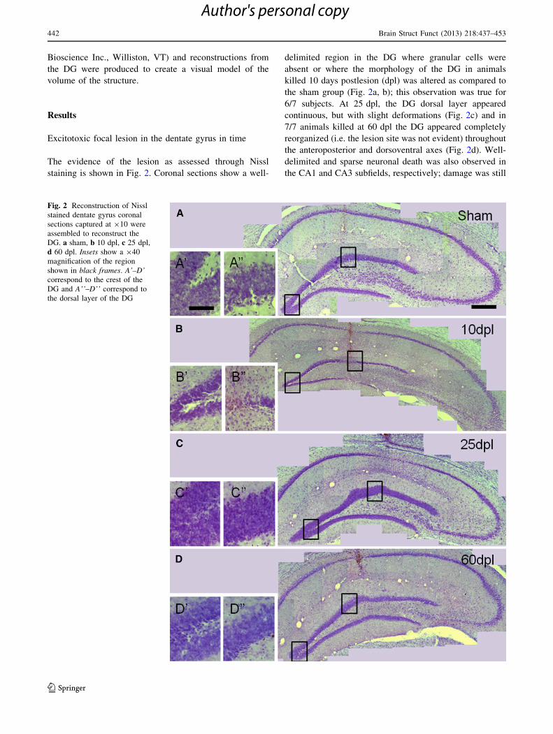
The evidence of the lesion as assessed through Nissl
staining is shown in Fig. 2. Coronal sections show a well-
delimited region in the DG where granular cells were
absent or where the morphology of the DG in animals
killed 10 days postlesion (dpl) was altered as compared to
the sham group (Fig. 2a, b); this observation was true for
6/7 subjects. At 25 dpl, the DG dorsal layer appeared
continuous, but with slight deformations (Fig. 2c) and in
7/7 animals killed at 60 dpl the DG appeared completely
reorganized (i.e. the lesion site was not evident) throughout
the anteroposterior and dorsoventral axes (Fig. 2d). Well-
delimited and sparse neuronal death was also observed in
the CA1 and CA3 subfields, respectively; damage was still
Fig. 2 Reconstruction of Nissl
stained dentate gyrus coronal
sections captured at 910 were
assembled to reconstruct the
DG. a sham, b 10 dpl, c 25 dpl,
d 60 dpl. Insets show a 940
magnification of the region
shown in black frames. A’–D’correspond to the crest of the
DG and A’’–D’’ correspond to
the dorsal layer of the DG
442 Brain Struct Funct (2013) 218:437–453
123
Author's personal copy

evident in these regions until 25 and 60 dpl corroborating
the selectivity of DG morphological reorganization
(Fig. 2b–d).
A volumetric analysis using the Cavalieri probe (MBF,
Bioscience Inc., Williston, VT) for measuring the DG
(Fig. 3a) showed that the average granular layer volume in
the 10 dpl group was significantly diminished as compared
to the sham, 25 and 60 dpl groups (F(3,15) = 11.232;
p \ 0.01, one way ANOVA). However, the DG volume
from the 25 dpl group did not differ from the sham or
60 dpl groups (Fig. 3b). All the Cavalieri analyses yielded
a Gundersen coefficient \0.05 (Table 3) (Gundersen and
Jensen 1987). These results show that the granular layer
recovers its original volume with time after the lesion.
Morphological reorganization of the DG
To analyze cellular proliferation and neuronal maturation
events associated to the morphological reorganization of
the DG, a multiple immunofluorescence and confocal
microscopy analysis for DCX- and CR-positive neurons
and NeuN-positive nuclei was performed in brains from
animals killed at 1, 10 and 60 dpl. DCX is a microtubule
marker widely used to evaluate neurogenesis levels, as its
expression occurs in neuronal committed newly born cells
(Rao and Shetty 2004; Couillard-Despres et al. 2005),
whereas CR expression relates to an early postmitotic step
of neuronal differentiation (Kempermann et al. 2003). The
DG of animals that received a KA injection and were killed
at 1 dpl, showed a twofold increase in DCX expression as
compared to sham subjects (p \ 0.05; t test). Such increase
progressed reaching the highest level both in the ipsi and
contralateral sides to the lesion at 10 dpl. A student’s t test
showed significant differences between 10 dpl and sham
groups (p \ 0.05) (Fig. 4a–c and right middle panel). After
60 dpl, when the DG appeared completely restructured, the
number of DCX-positive cells was similar to that observed
in the hippocampus from sham animals (Fig. 4d and right
middle panel). In sham animals, DCX-positive cells
exhibited the phenotype of undifferentiated granule cells
with short dendrites mostly oriented horizontally toward
the SGZ (Fig. 4e). In addition to the rise in DCX-positive
cell number at 1 dpl, a more differentiated phenotype was
observed and consisted in an increased number of elon-
gated dendrites vertically oriented toward the granular cell
layer (Fig. 4f) which contrasted with the short and hori-
zontally oriented neurites found in sham injected animals.
At 10 dpl, there were abundant DCX-expressing dense cell
clusters in the SGZ (Fig. 4g), suggesting that an intense
process of neuronal proliferation was taking place. How-
ever, at 60 dpl, the total number and the appearance of
DCX-positive cells returned to control levels (Fig. 4h and
right middle panel). Although KA was applied unilaterally,
an increase in markers of cell proliferation was also
observed in the intact hemisphere. It is thus conceivable
that the neuronal excitation elicited by KA might propagate
to the contralateral hippocampus through commissural
fibers and transiently increase cell proliferation without
producing structural changes (Fig. 4, bottom panel). In this
sense, it has been reported that the unilateral administration
of neurotoxic compounds leads to the expression of stress
Fig. 3 Visual modeling and
volumetric analysis of the
dentate gyrus. a Neurolucida
projection from a Nissl stained
coronal section showing the DG
of a sham animal,
b stereological analysis of the
granular layer of the DG reveals
that the volume in the 10 dpl
group significantly differs from
sham, 25 and 60 dpl
(F(3,15) = 11.232; **p \ 0.01)
Table 3 Volume and Gundersen0s coefficient corresponding to the
average per group of the granular layer of the DG
Group Volume granular cell layer
(mm3)
Gundersen’s
coefficient
Sham/intact 0.667 (n = 5) 0.010
10 dpl 0.305 (n = 4)** 0.021
25 dpl 0.547 (n = 4) 0.016
60 dpl 0.633 (n = 6) 0.015
Data show mean ± S.E.M. of the volume of the granular layer of the
DG and the corresponding Gundersen0s coefficient of error. Volume
of dentate granular layer of 10 dpl group is diminished and differs
from that of sham, 25 and 60 dpl groups (F(3,15) = 11.232;
**p \ 0.01; one way ANOVA). No differences among the rest of the
groups were observed
Brain Struct Funct (2013) 218:437–453 443
123
Author's personal copy

proteins in the intact hemisphere (Arias et al. 1998; Ayala
and Tapia 2008).
The inner molecular and infragranular layers of the DG
displayed CR immunoreactivity in sham animals (Fig. 4a).
A three-fold increase in CR-positive cells was observed as
soon as 1 dpl and until 60 dpl in the SGZ as well as in the
hilus (Fig. 4b–d and right bottom panel). Interestingly,
after 10 and 60 dpl, the CR immunoreactivity was also
strongly observed in the inner molecular and infragranular
layers of DG. Increase in CR expression was significantly
Fig. 4 Immunohistochemistry
for doublecortin (DCX) and
calretinin (CR) in coronal
sections from an injured DG.
Blue, cell nuclei stained with
DAPI. DCX (green) and CalR
(red) (a–d). Representative
sections from a sham lesion
animal (a); and subjects killed at
1 dpl (b); 10 dpl (c) and; 60 dpl
(d). e–h Higher magnifications
of different regions of the DG
from a sham animal and
subjects killed at 1 dpl (f),10 dpl (g) and 60 dpl (h); DCX
(green), NeuN (red). Arrowspoint at morphological
differences between DCX-
positive cells from a sham
subject (e) and another killed
1 dpl (f); arrowhead indicates
the appearance of DCX-
expressing dense cell clusters at
10 dpl (g). Photomicrographs
are representative of 3–4
sections from 3–5 different
animals of each experimental
group. Values represent
mean ± S.E.M. of three
different experiments where
DCX-positive cells (rightmiddle panel) and CalR-positive
cells (right bottom panel) were
counted; I ipsilateral,
C contralateral. Asterisks denote
significant differences as
compared to sham (student’s
t test) p \ 0.05. Scale bars50 lm
444 Brain Struct Funct (2013) 218:437–453
123
Author's personal copy

different from the sham group at all time points in the
ipsilateral DG, but also in the contralateral DG at 10 dpl
(p \ 0.05; t test).
To determine changes in the contents of neural pro-
genitor cells, we performed a quantitative analysis of
BLBP-positive cells in view that this protein is considered
as a specific marker of radial glia-like cells in the SGZ
(Steiner et al. 2006). At 1 dpl, few BLBP/PCNA-positive
co-localizing cells were observed (Fig. 5b). The content of
BLBP-positive cells significantly increased at 10 dpl in the
damaged DG (including the SGZ and the hilus) as com-
pared to the sham group (p \ 0.05; t test) (Fig. 5a–c and
right middle panel), and at 60 dpl, labeling of this marker
returned to basal levels (Fig. 5d).
The proliferation cell nuclear antigen (PCNA) showed
an increase from day 1 to 60 postlesion as compared to
sham levels (p \ 0.05, t test) (Fig. 5a–d) reaching the
highest point at 10 dpl (Fig. 5c and right bottom panel).
However, colocalization with BLBP was mostly evident at
1 dpl (Fig. 5e–h).
To evaluate the presence of the glial response to dam-
age, we analyzed GFAP-positive cells. Figure 6 shows the
distribution of radial glia-like cells in the sham DG. Loss of
GFAP-positive cells became evident as soon as 1 dpl in the
SGZ (compare Fig. 6A, a with B,b) and processes in
remaining cells shortened and diminished in number.
However, at 10 dpl radial glia-like cells became distin-
guishable again (compare Fig. 6a with c). Remarkably, the
GFAP-positive astrocytic response typical of damage was
confined to the hilus but was not observed within the
injured SGZ region (Fig. 6B, C).
To further analyze the cellular contents of the reorga-
nized region, we performed a NeuN immunohistochemistry
at all time points. Interestingly the DG of 6/7 animals killed
at 10 dpl showed an area devoid of NeuN-positive nuclei
(compare Fig. 7a, b) within the granular layer, whereas at
25 and 60 dpl none of the analyzed sections showed such a
region, instead NeuN-positive cells were uniformly dis-
tributed along the DG in the whole AP axis (Fig. 7c, d).
Moreover, the hilar region appeared devoid of NeuN-
positive cells at 10 and 25 dpl, while scarce cells were
observed at 60 dpl. Thus, structural reorganization at the
later time points studied seems to be particular of the
granular layer of the DG.
Focal dentate gyrus lesion impairs contextual fear
memory, but functional recovery occurs in time
To address the functional impact of the lesion on behavior,
we evaluated all groups in the cued fear memory and con-
textual fear memory tasks. Cued memory is known to rely
on the amygdala, whereas contextual fear memory depends
on the hippocampus and particularly on the DG (Phillips
and LeDoux 1992). Intact, sham and lesioned animals
received five tone–shock pairings in a single session. Fig-
ure 8a shows that in the 2 min lapse before the tone–shock
pairing, subjects displayed a normal movement behavior,
which translates into low percentage of freezing. However,
freezing increased gradually after each pairing in all groups,
thus animals were successfully conditioned regardless of
the time postlesion when they were trained (Fig. 8b). No
significant differences in freezing were obtained among
groups (F(6,47) = 1.309, p = 0.1624, repeated measures
ANOVA). 24 h after the training session, animals were
evaluated for contextual fear memory and, 2 h later, in cued
fear memory in an altered context. Figure 8c shows that
cued fear memory was preserved in all lesion groups (i.e.
percentage of freezing time was not different from sham
groups at any time point) (F(6,47) = 0.827, p = 0.55, fac-
torial ANOVA). These results differed from those observed
after evaluating contextual fear memory. Figure 8d shows
that only the 10 dpl group spent significantly less time
freezing than its control group in the aversive context
(F(6,47) = 13.587, p \ 0.0001, factorial ANOVA) whereas
freezing time in such context in the 25 and 60 dpl groups
was not significantly different from the sham or intact
groups, thus revealing an initial loss and a subsequent
functional recovery of the impaired behavior.
Given that freezing behavior implies lack of movement,
we tested the animals in the open field test to guarantee that
freezing was not caused by motor alterations or by the lack
of interest in exploring the environment. Figure 9a shows
no significant differences in the number of visited squares
in the open field test among groups at any time point
evaluated thereby confirming that freezing was dependent
on the conditioning and not on motor impairment or
emotional distress.
In addition, we explored the functional outcome of the
lesion in the place recognition task which has been shown
to be particularly modulated by the CA1 subfield of the
hippocampus. Figure 9b shows that place memory was not
impaired by the DG lesion at any time point evaluated and
that injured animals spent a similar amount of time
exploring the new location of a known object as the sham
and intact groups (F(6,47) = 0.304, p = 0.9316, repeated
measures ANOVA). Thus, the deficit provoked by the
lesion was restricted to a function mainly modulated by the
DG.
Long-term potentiation recovers in time
To evaluate whether behavioral recovery and structural
reorganization correlated with synaptic plasticity of the
DG, we induced high frequency LTP in vivo on the mossy
fiber pathway (DG-CA3). Figure 10a (top left panel) shows
a schematic representation of the mossy fiber pathway and
Brain Struct Funct (2013) 218:437–453 445
123
Author's personal copy

Fig. 5 BLBP-positive glia-like
neuroprecursor cells in DG at
different time points after KA
lesion. Coronal sections
showing a triple
immunofluorescence of BLBP
cells (red), DAPI-labeled cell
nuclei (blue) and proliferating
cells with PCNA (green) (a–d).
At higher magnifications of
regions shown in a–c, BLBP-
positive cells with PCNA nuclei
were observed at 1 dpl
(f, arrowhead), but not at 10 or
60 dpl (g–h). At 10 dpl a robust
increase of BLBP was evident
in the subgranular zone and in
the hilar region (g, arrows).
Quantification of cell number
positive to BLBP and PCNA in
the injected (I) and contralateral
hippocampus (C) is shown.
Photomicrographs are
representative of 3–4 sections
from 3–5 different animals of
each experimental group.
Values represent
mean ± S.E.M. of three
different experiments. Asterisksdenote significant differences as
compared to sham (student’s
t test) p \ 0.05. Scale bars50 lm
446 Brain Struct Funct (2013) 218:437–453
123
Author's personal copy

electrode placement. The CA3 EPSP consisted of poten-
tials of 0.38 ± 0.07 mV (mean ± SEM) elicited with
20–40 lA current pulses of 0.1–0.25 ms duration. These
responses began at 1.8–3.3 ms poststimulation and pre-
sented their valley at 7.1 ms with an average slope of
0.15 ± 0.007 (mean ± SEM) in agreement with previous
studies (Escobar et al. 1997; Gomez-Palacio-Schjetnan and
Escobar 2008; Schjetnan and Escobar 2010). Microinfu-
sion of DCG-IV at the end of the electrophysiological
recordings selectively blocked mossy fiber responses to
15 % of baseline signals, presenting potentials of
0.09 ± 0.02 mV with a mean slope of 0.06 ± 0.001
(mean ± S.E.M.), verifying that the signal was generated
by mossy fiber inputs (Fig. 10a, right panel).
The present results show that high frequency stimulation
(HFS) (three trains 100 Hz/1 s/20 s intertrain) was capable
of inducing LTP at the DG-CA3 pathway in adult rats in
vivo in accordance with previous studies (Escobar et al.
1997; Gomez-Palacio-Schjetnan and Escobar 2008; Sch-
jetnan and Escobar 2010). Mossy fiber LTP was charac-
terized by a slow initial increase in the EPSP slope that has
been related to the independence of NMDA receptors
activation and opioid peptide relevant modulation (Derrick
et al. 1991, 1992; Escobar et al. 1997). At 1 h post-stim-
ulation, the intact group presented a mean EPSP increase
(mean ± SEM) of 147.71 ± 1.45 (Fig. 10b). However, at
10 dpl DG-CA3-LTP could not be elicited (Fig. 10c), as
opposed to the 10-day sham lesion group (Fig. 10d).
Repeated–measures ANOVA for slope increases revealed
significant differences among groups (F6,23 = 7.15) and a
post hoc Fisher’s test showed significant differences
between the 10 dpl group and all of the other groups
(p \ 0.001). Interestingly, at 25 and 60 dpl, LTP could be
elicited and did not differ from the corresponding sham or
intact groups (Fig. 10e–h). HFS-induced potentiation was
not associated with after discharges or other overt epilep-
tiform activity as assessed by the continuously recorded
hippocampal EEG (data not shown).
Discussion
In this study, we show that the DG is capable of reorga-
nizing functionally and structurally in time after
Fig. 6 GFAP positive cells at 1 and 10 d after KA lesion. Coronal
sections labeled with NeuN (red) and GFAP (green) of sham injected
animals show the presence of radial-like glial cells (A and a). At
1 dpl, GFAP-positive radial processes shortened and diminished in
number (arrow, compare a and b) and at this time point as well as at
10 dpl a gliotic reaction was observed in the injured dorsal blade of
DG without entering into the granular cell layer (B, C arrowheads).
At 10 dpl some radial-like glial cells became evident (arrow, c).
Photomicrographs are representative of 3–4 sections from 3–5
different animals per group. Scale bars 40 lm
Brain Struct Funct (2013) 218:437–453 447
123
Author's personal copy

undergoing a unilateral focal lesion and that reorganization
relied exclusively on endogenous mechanisms. We dem-
onstrate that the granular cell layer of DG is able to
repopulate after a focal lesion and that the structural
reorganization correlated in time with the recovery of
contextual memory function and with in vivo restoration of
LTP in the DG-CA3 pathway.
It is feasible that newly added granule cells played a role
in this remodeling process, as we detected a long-lasting
increase in hippocampal neurogenic markers corresponding
in time with the refill of the lesion site. We evaluated
several markers to make a follow up of the morphological
reorganization process. Cells in the SGZ expressing BLBP
are undifferentiated, radial glia-like neural stem cells (Suh
et al. 2007). As early as 24 h after the lesion, few BLBP-
positive cells with PCNA-positive nuclei were already
present reaching the highest levels at 10 dpl. Thus, the
beginning of proliferation of these precursors started soon
after the lesion. PCNA remained high at all time points as
compared to sham levels reflecting proliferation of several
kinds of cells, but also likely suggesting an ongoing pro-
cess of DNA repair (Eisch and Mandyam 2006).
Fig. 7 NeuN
immunohistochemistry reveals
the morphological
reorganization of the DG in
time. Six to seven images
captured at 910 were assembled
to reconstruct the DG. a Sham;
b 10 dpl; c 25 dpl; d 60 dpl;
scale bar 400 lm. Insets show a
940 magnification of the region
shown in white frames. A’, B’,C’ and D’ correspond to the
dorsal layer of the DG and A’’,B’’, C’’ and D’’ correspond to
the crest of the DG; scale bar100 lm
448 Brain Struct Funct (2013) 218:437–453
123
Author's personal copy

As soon as 24 h after KA injection, we also detected the
increase in DCX-positive cells with vertically oriented
cellular processes as opposed to the short and horizontally
oriented neurites found in sections from sham animals.
This occurred prior to a robust rise in cells expressing
BLBP in agreement with the idea that most radial glial-like
cells are quiescent and only a short subset is recruited to
proliferate (Ledergerber et al. 2006). Thus, a group of
DCX-expressing cells might contain proliferative type-2
progenitor cells (Jessberger et al. 2005; Hodge et al. 2008)
which may start a neurorepair process. It has been shown
that DCX-expressing cells within the DG reemerge after
traumatic brain injury and it has been suggested that they
are the more likely contributors to stable neurogenesis (Yu
et al. 2008; Kernie and Parent 2010). Although similar
changes in number and morphology of DCX-positive cells
have been reported after systemic KA-induced seizures (Gu
et al. 2010) we cannot attribute this observation to seizures
because we used a subepileptogenic dose and only few rats
showed isolated wet-rat shakes during the first hour after
infusion.
It was noteworthy that the number of CR-positive cells
increased as early as 24 h after KA lesion. CR has been
considered as a marker of interneurons and Cajal–Retzius
cells in the DG, which co-express the glycoprotein reelin
(Tissir and Goffinet 2003) a pivotal factor in maintaining
the lamination in the hippocampus as well as in the rest of
the brain (Duveau et al. 2011). Interestingly, CR immu-
noreactivity was increased also at the sharp borders toward
the outer molecular layer of the DG as reported in studies
of morphological organization of this structure (Del Turco
et al. 2004). In cortical lesion models, newly generated
cells may differentiate into neurons and glia in the regen-
erating cortex, but lamination is not reinstated (Kolb et al.
2007).
Our results show that at 60 dpl, the zone originally
devoid of cells was refilled with NeuN-positive nuclei.
Thus, even when we cannot rule out the possibility that
plastic events involving neurite reorganization of preex-
isting neurons is contributing to the observed recovery
(Zepeda et al. 2004) the replenishing of the granule cell
layer is associated in time with functional reorganization.
Fig. 8 Contextual fear-conditioning recovery in time. a Freezing
percent of time spent during the pretone period (120 s). All groups
displayed a similar low percentage (F(6,47) = 1.312, p = 0.27;
factorial ANOVA) of freezing in the conditioning context before
the tone–shock pairings were administered. b Percentage of time
spent freezing during the conditioning session. All groups increase
the freezing time according to the presentation of tone–shock
pairings; ‘‘n’’ per group is depicted in parenthesis. (F(6,47) = 1.309,
p = 0.1624, Repeated measures ANOVA). c Evaluation of freezing
in the cued fear memory task, all groups display an elevated
percentage of time spent freezing which did not differ among each
other (F(6,47) = 0.827, p = 0.55; factorial ANOVA). d Freezing
behavior in the contextual fear memory task. Significant differences
were observed between 10 dpl and the rest of the groups
(F(6,47) = 13.587, **p \ 0.0001; factorial ANOVA)
Brain Struct Funct (2013) 218:437–453 449
123
Author's personal copy

However, further studies are required for establishing the
precise adaptive mechanisms underlying DG restoration.
Our results show an increase of neurogenic markers also
in the contralateral hippocampus, which are not necessarily
associated to the morphological reorganization process in
the damaged granular layer, but with molecular and cel-
lular signals which may activate the contralateral hippo-
campus through commissural fibers. The cellular response
of a lesion in the contralateral region of an injured structure
has been previously documented (Arias et al. 1998; Ayala
and Tapia 2008).
Functional recovery has been extensively shown to
occur after brain injury (for review see (Murphy and
Corbett 2009). However, structural disorganization may
prevail despite behavioral recovery (Kolb et al. 2007).
Our results showed that 60 days after a focal lesion to
the DG it reorganizes anatomically to the point that (1) the
volume of the structure recovers to control levels and, (2)
the lesion site is no longer evident as revealed by NeuN
immunohistochemistry. Interestingly, such structural reor-
ganization has not been documented after cortical damage
and we did not detect at any time point the glial scar typical
of cortical lesions (Zepeda et al. 2003, 2004). Both, the
core and the scar have been proposed to form a restrictive
environment for neural repair processes to occur (see Robel
et al. 2011 for review). Thus, the cortical and DG milieus
may substantially differ and the latter may provide a more
permissive environment for cellular plastic events to occur.
Fig. 9 Behavior in control tasks independent from the DG. a Perfor-
mance in the open field shows motor facilitation in all subjects
without group differences (F(6,47) = 1.561, p = 0.1797; repeated
measures ANOVA). b Performance in a place recognition task shows
a similar exploratory index among groups between the familiar and
novel location. A Acquisition period, R recall period (F(6,47) = 0.304,
p = 0.9316, repeated measures ANOVA)
Fig. 10 Mossy fiber LTP induction in vivo by HFS recovers in
time. a Left schematic representation of the mossy fiber pathway and
electrode placement showing the stimulated (CA3) (a) and recorded
(DG) (b) sites in a coronal plane. Right representative traces of the
EPSP obtained at baseline (full line) and after DCG-IV application
(dotted line). Note that DCG-IV selectively blocked mossy fiber
responses to 15 % of baseline signals. Plot of mossy fiber evoked
responses from the intact group (B); 10 dpl (C); 10 d sham lesion
(D); 25 dpl (E); 25 d sham lesion (F); 60 dpl (G); 60 d sham lesion
(H). DG-CA3-LTP induction was only blocked in the 10 dpl group.
Inner numbers show mean ± S.E.M. EPSPs slope (percent of
baseline) obtained 1 h after HFS for each group. The trace on the
top left in each graph shows the average of MF field potentials
obtained immediately before (full line) and 170 min after (dottedline) HFS
450 Brain Struct Funct (2013) 218:437–453
123
Author's personal copy

Morphological restoration may in turn give rise to the
recovery of the aversive memory function, which impor-
tantly relies on the DG integrity (Saxe et al. 2006). We can
be certain that the animals formed an aversive association
through the learning period, given that freezing increased
within the session in all groups. This result is in line with
previous studies showing that the dorsal hippocampal
neurons are not required for learning context fear, but are
required for fear context retrieval (Maren et al. 1997).
Thus, the initial failure in freezing reflects a memory loss
and not an acquisition deficit, whereas freezing after 25 and
60 dpl reveals the ability to remember the aversive context.
Generalized freezing can be discarded given that motor
behavior in the open field as well as in the 2 min period
prior to the tone appearance in the altered context remained
intact (data not shown). A work by Winocur et al. (2006)
has previously shown that inhibition of neurogenesis after
irradiation provokes a deficit in fear memory for as long as
2 months after ablation, thus suggesting a role of new
neurons for the adequate performance on hippocampus-
dependent memory function. Also a previous work has
shown that after a traumatic brain injury, reappearance of
neurons correlate with behavioral recovery and the DG has
been proposed as the neurogenic source (Sun et al. 2007).
Our results show that reorganization, as evaluated with
NeuN, occurs along functional recovery and within
25 days which correlates with the time-window in which
newly generated neurons have been shown to mature (Zhao
et al. 2006).
In addition to the morphological and behavioral reor-
ganization, the parallel reinstatement of LTP occurred. Our
results show that at 25 and 60 dpl, HFS was capable of
inducing in vivo LTP at the DG-CA3 pathway that was
blocked 10 dpl. These results are supported by previous
studies showing that low dose of gamma radiation, drasti-
cally reduced new cell proliferation, selectively blocked
LTP at DG in vitro (Snyder et al. 2001) and prevented the
LTP recovery after ischemic damage (Wang et al. 2005).
To our knowledge, mossy fiber LTP after a DG lesion has
not been previously addressed. Thus, we can only speculate
that the parallel reinstatement of LTP along with the
morphological and behavioral reorganization could rely on
well described plastic adaptive mechanisms such as
sprouting of fibers, and modifications in the excitatory/
inhibitory balance, but the idea that new neurons may be
participating in the organization of a preexisting neuronal
network should also be considered (Kempermann et al.
2002, 2004).
Of particular interest, functional and morphological
reorganization in our study occurred in the absence of
exogenous molecules known to enhance plastic responses
in the CNS (Yoshimura et al. 2001; Kolb et al. 2007).
Moreover, the time lapse in which recovery occurred is
consistent with the time that takes new neurons to migrate
and integrate into the rodent DG circuit (Cameron et al.
1993; Zhao et al. 2006).
In conclusion, our results provide information on the
progression of the cellular processes that are triggered after
a neurogenic niche injury in the adult and show that the
restoration of the original morphology of the damaged
structure occurs in parallel to the functional recovery of the
DG, as assessed through behavioral and electrophysiolog-
ical approaches.
Acknowledgments We thank Pedro Medina, Miguel Tapia, Patricia
Ferrera and Alicia Sampieri for technical assistance. We thank Ver-
don Taylor for providing facilities in the development of histo-
chemical procedures. This project was supported by DGAPA-PAPIIT
IN215609, IN213210 and CONACyT 60851.
Conflict of interest None.
References
Arias C, Becerra-Garcıa F, Arrieta I, Tapia R (1998) The protein
phosphatase inhibitor okadaic acid induces heat shock protein
expression and neurodegeneration in rat hippocampus in vivo.
Exp Neurol 153(2):242–254
Arvidsson A, Collin T, Kirik D, Kokaia Z, Lindvall O (2002)
Neuronal replacement from endogenous precursors in the adult
brain after stroke. Nat Med 8:963–970
Ayala GX, Tapia R (2008) HSP70 expression protects against
hippocampal neurodegeneration induced by endogenous gluta-
mate in vivo. Neuropharmacology 55(8):1383–1390
Bendel O, Bueters T, von Euler M, Ove Ogren S, Sandin J, von Euler
G (2005) Reappearance of hippocampal CA1 neurons after
ischemia is associated with recovery of learning and memory.
J Cerel Blood Flow Metab 25:1586–1595
Bengzon J, Kokaia Z, Elmer E, Nanobashvili A, Kokaia M, Lindvall
O (1997) Apoptosis and proliferation of dentate gyrus neurons
after single and intermittent limbic seizures. Proc Natl Acad Sci
USA 94:10432–10437
Brown CE, Li P, Boyd JD, Delaney KR, Murphy TH (2007)
Extensive turnover of dendritic spines and vascular remodeling
in cortical tissues recovering from stroke. J Neurosci
27:4101–4109
Brun VH, Otnass MK, Molden S, Steffenach HA, Witter MP, Moser
MB, Moser EI (2002) Place cells and place recognition
maintained by direct entorhinal-hippocampal circuitry. Science
296:2243–2246
Calixto E, Thiels E, Klann E, Barrionuevo G (2003) Early mainte-
nance of hippocampal mossy fiber–long-term potentiation
depends on protein and RNA synthesis and presynaptic granule
cell integrity. J Neurosci 23:4842–4849
Cameron HA, Woolley CS, McEwen BS, Gould E (1993) Differen-
tiation of newly born neurons and glia in the dentate gyrus of the
adult rat. Neuroscience 56:337–344
Carmichael ST, Chesselet MF (2002) Synchronous neuronal activity
is a signal for axonal sprouting after cortical lesions in the adult.
J Neurosci 22:6062–6070
Couillard-Despres S, Winner B, Schaubeck S, Aigner R, Vroemen M,
Weidner N, Bogdahn U, Winkler J, Kuhn HG, Aigner L (2005)
Doublecortin expression levels in adult brain reflect neurogen-
esis. Eur J Neurosci 21:1–14
Brain Struct Funct (2013) 218:437–453 451
123
Author's personal copy

Del Turco D, Gebhardt C, Burbach GJ, Pleasure SJ, Lowenstein DH,
Deller T (2004) Laminar organization of the mouse dentate
gyrus: insights from BETA2/Neuro D mutant mice. J Comp
Neurol 477:81–95
Derrick BE, Weinberger SB, Martinez JL Jr (1991) Opioid receptors
are involved in an NMDA receptor-independent mechanism of
LTP induction at hippocampal mossy fiber-CA3 synapses. Brain
Res Bull 27:219–223
Derrick BE, Rodriguez SB, Lieberman DN, Martinez JL Jr (1992) Mu
opioid receptors are associated with the induction of hippocam-
pal mossy fiber long-term potentiation. J Pharmacol Exp Ther
263:725–733
Doetsch F, Alvarez-Buylla A (1996) Network of tangential pathways
for neuronal migration in adult mammalian brain. Proc Natl
Acad Sci USA 93:14895–14900
Dong H, Csernansky CA, Goico B, Csernansky JG (2003) Hippo-
campal neurogenesis follows kainic acid-induced apoptosis in
neonatal rats. J Neurosci 23:1742–1749
Duveau V, Madhusudan A, Caleo M, Knuesel I, Fritschy JM (2011)
Impaired reelin processing and secretion by Cajal–Retzius cells
contributes to granule cell dispersion in a mouse model of
temporal lobe epilepsy. Hippocampus 21:935–944
Eisch AJ, Mandyam CD (2006) Adult neurogenesis and central
nervous system cell cycle analysis: novel tools for exploration of
the neural causes and correlates of psychiatric disorders. In:
Janigro D (ed) The cell cycle in the Central Nervous System.
Humana Press, NJ, pp 335–356
Ennaceur A, Neave N, Aggleton JP (1997) Spontaneous object
recognition and object location memory in rats: the effects of
lesions in the cingulate cortices, the medial prefrontal cortex, the
cingulum bundle and the fornix. Exp Brain Res 113:509–519
Escobar ML, Barea-Rodriguez EJ, Derrick BE, Reyes JA, Martinez
JL Jr (1997) Opioid receptor modulation of mossy fiber
synaptogenesis: independence from long-term potentiation.
Brain Res 751:330–335
Escobar ML, Figueroa-Guzman Y, Gomez-Palacio-Schjetnan A
(2003) In vivo insular cortex LTP induced by brain-derived
neurotrophic factor. Brain Res 991:274–279
Gomez-Palacio-Schjetnan A, Escobar ML (2008) In vivo BDNF
modulation of adult functional and morphological synaptic
plasticity at hippocampal mossy fibers. Neurosci Lett 445:62–67
Gray WP, Sundstrom LE (1998) Kainic acid increases the prolifer-
ation of granule cell progenitors in the dentate gyrus of the adult
rat. Brain Res 790:52–59
Gu P, Li Y, Shang Y, Hou Y, Zhao S (2010) Proliferation changes in
dentate gyrus of hippocampus during the first week following
kainic acid-induced seizures. Yakugaku Zasshi 130:1751–1754
Gundersen HJ, Jensen EB (1987) The efficiency of systematic
sampling in stereology and its prediction. J Microsc 147(Pt
3):229–263
Hernandez-Ortega K, Ferrera P, Arias C (2007) Sequential expression
of cell-cycle regulators and Alzheimer’s disease-related proteins
in entorhinal cortex after hippocampal excitotoxic damage.
J Neurosci Res 85:1744–1751
Hodge RD, Kowalczyk TD, Wolf SA, Encinas JM, Rippey C,
Enikolopov G, Kempermann G, Hevner RF (2008) Intermediate
progenitors in adult hippocampal neurogenesis: Tbr2 expression
and coordinate regulation of neuronal output. J Neurosci
28:3707–3717
Jessberger S, Romer B, Babu H, Kempermann G (2005) Seizures
induce proliferation and dispersion of doublecortin-positive
hippocampal progenitor cells. Exp Neurol 196:342–351
Jin K, Minami M, Lan JQ, Mao XO, Batteur S, Simon RP, Greenberg
DA (2001) Neurogenesis in dentate subgranular zone and rostral
subventricular zone after focal cerebral ischemia in the rat. Proc
Natl Acad Sci USA 98:4710–4715
Kempermann G (2002) Why new neurons? Possible functions for
adult hippocampal neurogenesis. J Neurosci 22:635–638
Kempermann G, Gast D, Kronenberg G, Yamaguchi M, Gage FH
(2003) Early determination and long-term persistence of adult-
generated new neurons in the hippocampus of mice. Develop-
ment 130:391–399
Kempermann G, Wiskott L, Gage FH (2004) Functional significance
of adult neurogenesis. Curr Opin Neurobiol 14:186–191
Kernie SG, Parent JM (2010) Forebrain neurogenesis after focal
Ischemic and traumatic brain injury. Neurobiol Dis 37:267–274
Kolb B, Morshead C, Gonzalez C, Kim M, Gregg C, Shingo T, Weiss
S (2007) Growth factor-stimulated generation of new cortical
tissue and functional recovery after stroke damage to the motor
cortex of rats. J Cereb Blood Flow Metab 27:983–997
Kuhn HG, Dickinson-Anson H, Gage FH (1996) Neurogenesis in the
dentate gyrus of the adult rat: age-related decrease of neuronal
progenitor proliferation. J Neurosci 16:2027–2033
Ledergerber D, Fritschy JM, Kralic JE (2006) Impairment of dentate
gyrus neuronal progenitor cell differentiation in a mouse model
of temporal lobe epilepsy. Exp Neurol 199:130–142
LeDoux JE, Cicchetti P, Xagoraris A, Romanski LM (1990) The
lateral amygdaloid nucleus: sensory interface of the amygdala in
fear conditioning. J Neurosci 10:1062–1069
Lee I, Kesner RP (2004) Differential contributions of dorsal
hippocampal subregions to memory acquisition and retrieval in
contextual fear-conditioning. Hippocampus 14:301–310
Maren S, Aharonov G, Fanselow M (1997) Neurotoxiclesions of the
dorsal hippocampus and Pavlovian fear conditioning in rats.
Behav Brain Res 88:261–274
Mumby DG, Gaskin S, Glenn MJ, Schramek TE, Lehmann H (2002)
Hippocampal damage and exploratory preferences in rats:
memory for objects, places, and contexts. Learn Mem 9:49–57
Murphy TH, Corbett D (2009) Plasticity during stroke recovery: from
synapse to behaviour. Nat Rev Neurosci 10:861–872
Nakagawa E, Aimi Y, Yasuhara O, Tooyama I, Shimada M, McGeer
PL, Kimura H (2000) Enhancement of progenitor cell division in
the dentate gyrus triggered by initial limbic seizures in rat
models of epilepsy. Epilepsia 41:10–18
Nakatomi H, Kuriu T, Okabe S, Yamamoto S, Hatano O, Kawahara
N, Tamura A, Kirino T, Nakafuku M (2002) Regeneration of
hippocampal pyramidal neurons after ischemic brain injury
by recruitment of endogenous neural progenitors. Cell 110:429–
441
Nudo RJ, Wise BM, SiFuentes F, Milliken GW (1996) Neural
substrates for the effects of rehabilitative training on motor
recovery after ischemic infarct. Science 272:1791–1794
Ogita K, Nishiyama N, Sugiyama C, Higuchi K, Yoneyama M,
Yoneda Y (2005) Regeneration of granule neurons after
lesioning of hippocampal dentate gyrus: evaluation using adult
mice treated with trimethyltin chloride as a model. J Neurosci
Res 82:609–621
Okano H, Sawamoto K (2008) Neural stem cells: involvement in
adult neurogenesis and CNS repair. Philos Trans R Soc Lond B
Biol Sci 363:2111–2122
Parent JM, Valentin VV, Lowenstein DH (2002) Prolonged seizures
increase proliferating neuroblasts in the adult rat subventricular
zone-olfactory bulb pathway. J Neurosci 22:3174–3188
Paxinos G, Watson C (1986) The rat brain stereotaxic coordinates.
Academic Press, Sydney
Phillips RG, LeDoux JE (1992) Differential contribution of amygdala
and hippocampus to cued and contextual fear conditioning.
Behav Neurosci 106:274–285
Rao MS, Shetty AK (2004) Efficacy of doublecortin as a marker to
analyse the absolute number and dendritic growth of newly
generated neurons in the adult dentate gyrus. Eur J Neurosci
19:234–246
452 Brain Struct Funct (2013) 218:437–453
123
Author's personal copy

Robel S, Berninger B, Gotz M (2011) The stem cell potential of glia:
lessons from reactive gliosis. Nat Rev Neurosci 12:88–104
Saxe MD, Battaglia F, Wang JW, Malleret G, David DJ, Monckton
JE, Garcia AD, Sofroniew MV, Kandel ER, Santarelli L, Hen R,
Drew MR (2006) Ablation of hippocampal neurogenesis impairs
contextual fear conditioning and synaptic plasticity in the dentate
gyrus. Proc Natl Acad Sci USA 103:17501–17506
Scharfman HE, Goodman JH, Sollas AL (2000) Granule-like neurons
at the hilar/CA3 border after status epilepticus and their
synchrony with area CA3 pyramidal cells: functional implica-
tions of seizure-induced neurogenesis. J Neurosci 20:6144–6158
Schjetnan AG, Escobar ML (2010) In vivo BDNF modulation of
hippocampal mossy fiber plasticity induced by high frequency
stimulation. Hippocampus. doi:10.1002/hipo.20866
Sigler A, Mohajerani MH, Murphy TH (2009) Imaging rapid
redistribution of sensory-evoked depolarization through existing
cortical pathways after targeted stroke in mice. Proc Natl Acad
Sci USA 106:11759–11764
Snyder JS, Kee N, Wojtowicz JM (2001) Effects of adult neurogen-
esis on synaptic plasticity in the rat dentate gyrus. J Neurophysiol
85:2423–2431
Steiner B, Klempin F, Wang L, Kott M, Kettenmann H, Kempermann
G (2006) Type-2 cells as link between glial and neuronal lineage
in adult hippocampal neurogenesis. Glia 54:805–814
Suh H, Consiglio A, Ray J, Sawai T, D’Amour KA, Gage FH (2007)
In vivo fate analysis reveals the multipotent and self-renewal
capacities of Sox2? neural stem cells in the adult hippocampus.
Cell Stem Cell 1:515–528
Sun D, McGinn MJ, Zhou Z, Harvey HB, Bullock MR, Colello RJ
(2007) Anatomical integration of newly generated dentate
granule neurons following traumatic brain injury in adult rats
and its association to cognitive recovery. Exp Neurol 204:
264–272
Tissir F, Goffinet AM (2003) Reelin and brain development. Nat Rev
Neurosci 4:496–505
Wang S, Kee N, Preston E, Wojtowicz JM (2005) Electrophysiological
corre lates of neural plasticity compensating for ischemia-induced
damage in the hippocampus. Exp Brain Res 165:250–260
Winocur G, Wojtowicz JM, Sekeres M, Snyder JS, Wang S (2006)
Inhibition of neurogenesis interferes with hippocampus-depen-
dent memory function. Hippocampus 16:296–304
Winship IR, Murphy TH (2008) In vivo calcium imaging reveals
functional rewiring of single somatosensory neurons after stroke.
J Neurosci 28:6592–6606
Yoshimura S, Takagi Y, Harada J, Teramoto T, Thomas SS, Waeber
C, Bakowska JC, Breakefield XO, Moskowitz MA (2001) FGF-2
regulation of neurogenesis in adult hippocampus after brain
injury. Proc Natl Acad Sci USA 98:5874–5879
Yu TS, Zhang G, Liebl DJ, Kernie SG (2008) Traumatic brain injury-
induced hippocampal neurogenesis requires activation of early
nestin-expressing progenitors. J Neurosci 28:12901–12912
Zepeda A, Vaca L, Arias C, Sengpiel F (2003) Reorganization of
visual cortical maps after focal ischemic lesions. J Cereb Blood
Flow Metab 23:811–820
Zepeda A, Sengpiel F, Guagnelli MA, Vaca L, Arias C (2004)
Functional reorganization of visual cortex maps after ischemic
lesions is accompanied by changes in expression of cytoskeletal
proteins and NMDA and GABA(A) receptor subunits. J Neurosci
24:1812–1821
Zepeda A, Michel G, Aguilar-Arredondo A, Arias C (2009)
Neurogenesis after brain stroke: is there a relationship with
functional recovery? Curr Trends Neurol 3:33–44
Zhao C, Teng EM, Summers RG Jr, Ming GL, Gage FH (2006)
Distinct morphological stages of dentate granule neuron matu-
ration in the adult mouse hippocampus. J Neurosci 26:3–11
Brain Struct Funct (2013) 218:437–453 453
123
Author's personal copy