Dentate gyrus-selective colchicine lesion and performance in temporal and spatial tasks
18
Behavioural Brain Research 160 (2005) 286–303 Research report Dentate gyrus-selective colchicine lesion and performance in temporal and spatial tasks Val´ eria Catelli Infantozzi Costa a,∗ , Jos´ e Lino Oliveira Bueno a , Gilberto Fernando Xavier b a Setor de Psicobiologia, Departamento de Psicologia e Educa¸ c˜ ao da Faculdade Filosofia, Ciˆ encias e Letras de Ribeir˜ ao Preto, Universidade de S˜ ao Paulo, Av. dos Bandeirantes, 3900, 14040-901 Ribeir˜ ao Preto, SP, Brazil b Departamento de Fisiologia do Instituto de Biociˆ encias, Universidade de S˜ ao Paulo, S˜ ao Paulo, SP, Brazil Received 18 August 2004; received in revised form 10 December 2004; accepted 16 December 2004 Available online 10 February 2005 Abstract The effects of multiple-site, intradentate, colchicine injections on the performance of a temporal, ‘differential reinforcement of low rates of responding’ (DRL-20 s) task and a spatial, ‘delayed non-matching-to-place’ (DNMTP) task in a plus-maze were investigated in rats trained in both tasks prior to the lesion. Quantitative analysis revealed a greater than 86% reduction in the dentate gyrus (DG) of the colchicine- injected rats compared to the sham-operated controls. Dentate gyrus damage rendered rats less efficient than sham-operated controls in the performance of the DRL-20s task. The DRL inter-response time (IRT) distribution for the DG-lesioned rats and the sham-operated controls was similar; however, while the distribution peak for the control rats was 20 s, it was 16 s for the DG-lesioned rats, indicating that the latter rats underestimated time. Performance of the DG-lesioned rats was also disrupted in the DNMTP task. However, DG-lesioned rats recovered control levels of performance during repeated training with an intertrial interval equal to 3 s. An increase in intertrial interval in lesioned and sham-operated controls disrupted performance in both groups; however, while DG-lesioned rats performed at chance levels when the intertrial interval was increased to 4min or longer, the sham-operated controls performed at chance levels only when the intertrial interval was increased to 16 min. These results seem most parsimoniously interpreted following the cognitive map theory of hippocampal function. © 2004 Elsevier B.V. All rights reserved. Keywords: Hippocampus; Dentate gyrus; Cognitive map theory; Working memory; Timing process; Spatial discrimination; NMTP task; DRL task 1. Introduction The hippocampal formation plays a critical role in brain function by regulating behavior and experience. While sev- eral different models of the hippocampal function coincide in ascribing memory functions to this brain structure, they disagree strongly with regard to the nature of the memory involved. O’Keefe and Nadel [52] distinguished among alternative strategies used by animals to navigate through the environ- ment, and suggested that more than one strategy may be ∗ Corresponding author. Tel.: +55 16 6023697; fax: +55 16 6335668. E-mail addresses: [email protected], [email protected] (V.C.I. Costa). used simultaneously to solve spatial tasks. According to these authors, while place (or locale) strategies involve cognitive mapping, guidance (or taxon) strategies depend on a particu- lar, prominent object or stimulus to indicate the goal location; egocentric orientation strategies are based on the rotation of the body axis relative to other axes. These strategies may be sustained by different neural systems; the hippocampal formation may be necessary for place learning. In addition, O’Keefe [53] suggested that when the use of one of these strategies is not possible, e.g., after lesions of the related system, the animal may rely on the remaining systems to solve the task, when this is possible. Normal rats apparently use these strategies simultaneously to solve spatial naviga- tion challenges in the Morris’ water maze task [81]. Further, Eichenbaum et al. [18] suggest that the integrity of the hip- 0166-4328/$ – see front matter © 2004 Elsevier B.V. All rights reserved. doi:10.1016/j.bbr.2004.12.011
-
Upload
hydra-aqua -
Category
Documents
-
view
0 -
download
0
Transcript of Dentate gyrus-selective colchicine lesion and performance in temporal and spatial tasks
Behavioural Brain Research 160 (2005) 286–303
Research report
Dentate gyrus-selective colchicine lesion and performancein temporal and spatial tasks
Valeria Catelli Infantozzi Costaa,∗, Jose Lino Oliveira Buenoa,Gilberto Fernando Xavierb
a Setor de Psicobiologia, Departamento de Psicologia e Educa¸cao da Faculdade Filosofia, Ciˆencias e Letras de Ribeir˜ao Preto,Universidade de S˜ao Paulo, Av. dos Bandeirantes, 3900, 14040-901 Ribeir˜ao Preto, SP, Brazil
b Departamento de Fisiologia do Instituto de Biociˆencias, Universidade de S˜ao Paulo, S˜ao Paulo, SP, Brazil
Received 18 August 2004; received in revised form 10 December 2004; accepted 16 December 2004Available online 10 February 2005
Abstract
The effects of multiple-site, intradentate, colchicine injections on the performance of a temporal, ‘differential reinforcement of low rates ofr ts trainedi colchicine-i ontrols in thep d controlsw t the latterr s recoveredc lesioneda ls when thei ial intervalw l function.©
K
1
feidi
sm
i
theseitiverticu-ion;ion ofmay
mpaltion,ese
lateds to
entlyviga-
,ip-
0d
esponding’ (DRL-20 s) task and a spatial, ‘delayed non-matching-to-place’ (DNMTP) task in a plus-maze were investigated in ran both tasks prior to the lesion. Quantitative analysis revealed a greater than 86% reduction in the dentate gyrus (DG) of thenjected rats compared to the sham-operated controls. Dentate gyrus damage rendered rats less efficient than sham-operated cerformance of the DRL-20 s task. The DRL inter-response time (IRT) distribution for the DG-lesioned rats and the sham-operateas similar; however, while the distribution peak for the control rats was 20 s, it was 16 s for the DG-lesioned rats, indicating tha
ats underestimated time. Performance of the DG-lesioned rats was also disrupted in the DNMTP task. However, DG-lesioned ratontrol levels of performance during repeated training with an intertrial interval equal to 3 s. An increase in intertrial interval innd sham-operated controls disrupted performance in both groups; however, while DG-lesioned rats performed at chance leve
ntertrial interval was increased to 4 min or longer, the sham-operated controls performed at chance levels only when the intertras increased to 16 min. These results seem most parsimoniously interpreted following the cognitive map theory of hippocampa2004 Elsevier B.V. All rights reserved.
eywords:Hippocampus; Dentate gyrus; Cognitive map theory; Working memory; Timing process; Spatial discrimination; NMTP task; DRL task
. Introduction
The hippocampal formation plays a critical role in brainunction by regulating behavior and experience. While sev-ral different models of the hippocampal function coincide
n ascribing memory functions to this brain structure, theyisagree strongly with regard to the nature of the memory
nvolved.O’Keefe and Nadel[52] distinguished among alternative
trategies used by animals to navigate through the environ-ent, and suggested that more than one strategy may be
∗ Corresponding author. Tel.: +55 16 6023697; fax: +55 16 6335668.E-mail addresses:[email protected],
[email protected] (V.C.I. Costa).
used simultaneously to solve spatial tasks. According toauthors, while place (or locale) strategies involve cognmapping, guidance (or taxon) strategies depend on a palar, prominent object or stimulus to indicate the goal locategocentric orientation strategies are based on the rotatthe body axis relative to other axes. These strategiesbe sustained by different neural systems; the hippocaformation may be necessary for place learning. In addiO’Keefe [53] suggested that when the use of one of thstrategies is not possible, e.g., after lesions of the resystem, the animal may rely on the remaining systemsolve the task, when this is possible. Normal rats apparuse these strategies simultaneously to solve spatial nation challenges in the Morris’ water maze task[81]. FurtherEichenbaum et al.[18] suggest that the integrity of the h
166-4328/$ – see front matter © 2004 Elsevier B.V. All rights reserved.oi:10.1016/j.bbr.2004.12.011

V.C.I. Costa et al. / Behavioural Brain Research 160 (2005) 286–303 287
pocampal formation is necessary for creating a cognitive mapthat supports flexible, spatial navigation in the environment.Their results show that rats with hippocampal damage man-age to adopt taxon and egocentric orientation strategies todeal with the requirements of the task to be solved.
Honig [30] and Olton[55,57]distinguished between ref-erence memory and working memory; working memorycontains information relevant to a given trial, and is con-text specific, while reference memory contains informationrelevant to several trials, and is context independent (see[34,54,56,57]). Olton et al.[54] proposed that the hippocam-pal formation is involved in working memory but not in refer-ence memory (see[37,57,62,78]). However, Meck et al.[45]noted that most demonstrations that the hippocampal systemis involved in working memory, but is not required by refer-ence memory, are based largely on tests providing spatial in-formation. This begs the question of whether the hippocampallesion effect results from interference with working memoryfor spatial but not for non-spatial information.
Different laboratories have reported that damage to thehippocampus impairs performance in a non-spatial, ‘differ-ential reinforcement of low rates of responding’ (DRL) task[1,3,7,8,10,33,64,66,71]. In a DRL task, reinforcement iscontingent on responses occurring a pre-defined time intervalafter the preceding response; that is, the rats must suppressresponse until a minimum time interval has elapsed since thel em-o ation[p ffectt ingm gaps.
ed-u ory;t ls isn ecke itedb n ofw
re-s mentt em,w ron-mt hibiti andt DRLt
nt oft tion[ ard[ heh anulec sub-fi theb so-
called conventional lesion techniques (e.g., aspiration, andelectrolytic and thermocoagulation), damage is not restrictedto the target area; passage fibers may be destroyed and theamount of damage to the vasculature is unknown[31,32].Thus, many of the behavioral changes observed after lesionstargeting the hippocampal formation may result from extra-hippocampal damage or a combination of hippocampal andextra-hippocampal damage. Since the use of colchicine min-imizes some of this problem, several laboratories have usedthis alkaloid to investigate the effects of damage to the den-tate gyrus (DG) granule cells and mossy fibers on memoryfunction[16,17,43,44,73,79,82,83].
Although colchicine exhibits preferential toxicity forgranule cells, some damage to hilar and pyramidal cellsalso has been reported[15,40,75,76,83]; for instance, thelength of the pyramidal cell layer of Ammon’s horn is sig-nificantly reduced following intradentate colchicine injection[20,75,76,79].
The aim of the present study was to investigate learningand memory changes induced by the selective loss of DGgranule cells in parallel tasks involving spatial and tempo-ral processes in the same animal. The effect of selective,colchicine-induced, DG granule cell loss was investigatedon performance of both (1) a temporal task (DRL-20 s), in-cluding a detailed analysis of the time-course of bar pressresponses to evaluate an animal’s ability to estimate time in-t lace(
f thep ro-c f thet for-m
2p
te-i ientp , wee celll task.T on-m
2
2acil-
i her ghingf wereh on a1 ratsw itumb g the
ast response, which is considered to require working mry and thus to be dependent on the hippocampal form
42,46,50]. Further to this discussion, Meck et al.[45] em-hasized that transection of the fimbria-fornix does not a
he rats’ sensitivity to time, but does affect temporal workemory as revealed in a peak procedure situation withOlton [56] argued that memory for a fixed interval sch
le of reinforcement is processed by the reference memhis may explain why performance by lesioned animaot affected, rendering them able to complete the task. Mt al. [45] suggested that the performance deficit exhiby hippocampal rats in DRL tasks results from disruptioorking memory rather than a deficit in estimating time.Rawlins[65] proposed that hippocampectomy would
trict memory storage capacity, thus generating impairo “bridging gaps” between stimuli in order to associate thhich is necessary to build up an overall map of the envient. Consistent with this view, Bannerman et al.[3] showed
hat rats with cytotoxic-induced hippocampal damage exmpairments in spatial tasks, including the water mazehe elevated T-maze, and non-spatial tasks, including theask.
Different sources of evidence suggest the involvemehe dentate granule cells in encoding mnemonic informa16,17,82]. The demonstration by Goldschmidt and Stew26,27], that the topical application of colchicine into tippocampus produces the selective loss of dentate grells and mossy fibers, while leaving other hippocampalelds reasonably intact, provided a model for studyingehavioral effects of selective neuronal loss. Using the
ervals precisely, and (2) a delayed non-matching-to-pDNMTP) task in a plus-maze.
Thus, the experimental procedure allows analysis oarticipation of DG granule cells in different cognitive pesses. Additionally, the study provides a discussion oheoretical underpinnings of both theories as to how ination is processed in temporal and spatial tasks.
. Experiment IA—Effect of DG-selective lesion onerformance of a DRL-20 s task
Sinden et al.[71] showed that complete, ibotenanduced, hippocampal pyramidal cell loss disrupts efficerformance of a DRL task. In the present experimentvaluated the effect of colchicine-induced, DG granule
oss on performance of a pre-lesion acquired, DRL-20 shis experiment was run in parallel with the delayed natching-to-place task (see Experiment IB, below).
.1. Materials and methods
.1.1. AnimalsTwenty naive, male Wistar rats, bred at the Central Colony F
ty of the University of Sao Paulo at Ribeirao Preto were used. Tats were 90 days old at the beginning of the experiments, weirom 220 to 280 g. Throughout all experiments, the animalsoused singly in steel cages in the laboratory colony room,2 h light:12 h dark cycle (lights on from 8:00 to 20:00 h). Theere kept on a food deprivation schedule at 80% of their ad libody weight by limiting access to food. On the 3 days precedin
288 V.C.I. Costa et al. / Behavioural Brain Research 160 (2005) 286–303
start of the behavioral pre-lesion training, the animals were individ-ually handled for 1 min. They were also manipulated daily duringweighing.
2.1.2. ApparatusThree, identical, operant test chambers (Lafayette model 80201)
were used, each measuring 20 cm× 20 cm× 23 cm. Each chamberpossessed a response lever 7 cm above the floor, in the center ofone of the walls. Below and to the left of the lever, was a cir-cular opening through which food pellets (45 mg) were releasedas reinforcement by a dispenser. A 5 W lamp located in the cen-ter of the ceiling constantly illuminated the chamber. An interface(MRA—Electronic Equipment, Ribeirao Preto, Brazil) connectedthe conditioning boxes to a PC computer, which controlled the ex-periment and registered the data. Each experimental chamber washeld within a sound proof wooden box (55 cm× 55 cm× 55 cm)provided with a 20 cm× 15 cm, transparent, acrylic window. Thesesets were located in a 6.0 m× 1.6 m× 3.0 m room; the interface andthe computer were located in an adjacent room.
2.1.3. Pre-lesion training in the DRL-20 s taskIn the first session, each rat was placed into the experimental
chamber and trained to bar press for food. In the second session,which lasted 30 min, the animals were submitted to a continuousreinforcement frequency (CRF) schedule in which bar pressing, atany moment, was always followed by reinforcement. Subsequently,the rats were trained in the DRL-20 s task; bar presses were rein-forced only if a minimum time of 20 s had elapsed from the previousr last re-s em andr of ther tely2 food.E inglet in theD ningi
ed tot intaint
2itted
tKopf
s w thei wasp ortex.S madeu ttea e andh re of1 np cells( wlyt e andt icinei re6 thisp ticald
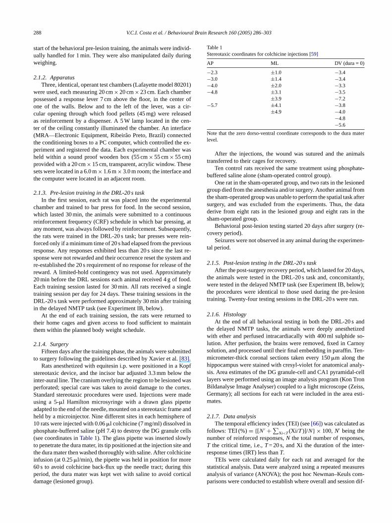
Table 1Stereotaxic coordinates for colchicine injections[59]
AP ML DV (dura = 0)
−2.3 ±1.0 −3.4−3.0 ±1.4 −3.4−4.0 ±2.0 −3.3−4.8 ±3.1 −3.5
±3.9 −7.2−5.7 ±4.1 −3.8
±4.9 −4.0−4.8−5.6
Note that the zero dorso-ventral coordinate corresponds to the dura materlevel.
After the injections, the wound was sutured and the animalstransferred to their cages for recovery.
Ten control rats received the same treatment using phosphate-buffered saline alone (sham-operated control group).
One rat in the sham-operated group, and two rats in the lesionedgroup died from the anesthesia and/or surgery. Another animal fromthe sham-operated group was unable to perform the spatial task aftersurgery, and was excluded from the experiments. Thus, the dataderive from eight rats in the lesioned group and eight rats in thesham-operated group.
Behavioral post-lesion testing started 20 days after surgery (re-covery period).
Seizures were not observed in any animal during the experimen-tal period.
2.1.5. Post-lesion testing in the DRL-20 s taskAfter the post-surgery recovery period, which lasted for 20 days,
the animals were tested in the DRL-20 s task and, concomitantly,were tested in the delayed NMTP task (see Experiment IB, below);the procedures were identical to those used during the pre-lesiontraining. Twenty-four testing sessions in the DRL-20 s were run.
2.1.6. HistologyAt the end of all behavioral testing in both the DRL-20 s and
the delayed NMTP tasks, the animals were deeply anesthetizedwith ether and perfused intracardiacally with 400 ml sulphide so-lution. After perfusion, the brains were removed, fixed in Carnoysolution, and processed until their final embedding in paraffin. Ten-mh naly-s l-celll TronB eiss,G esti-m
2s
fn es,T r-r
r thes asuresa om-p on dif-
esponse. Any responses exhibited less than 20 s since theponse were not rewarded and their occurrence reset the syste-established the 20 s requirement of no response for releaseeward. A limited-hold contingency was not used. Approxima0 min before the DRL sessions each animal received 4 g ofach training session lasted for 30 min. All rats received a s
raining session per day for 24 days. These training sessionsRL-20 s task were performed approximately 30 min after trai
n the delayed NMTP task (see Experiment IB, below).At the end of each training session, the rats were return
heir home cages and given access to food sufficient to mahem within the planned body weight schedule.
.1.4. SurgeryFifteen days after the training phase, the animals were subm
o surgery following the guidelines described by Xavier et al.[83].Rats anesthetized with equitesin i.p. were positioned in a
tereotaxic device, and the incisor bar adjusted 3.3 mm belonter-aural line. The cranium overlying the region to be lesionederforated; special care was taken to avoid damage to the ctandard stereotaxic procedures were used. Injections weresing a 5-�l Hamilton microsyringe with a drawn glass pipedapted to the end of the needle, mounted on a stereotaxic frameld by a microinjector. Nine different sites in each hemisphe0 rats were injected with 0.06�l colchicine (7 mg/ml) dissolved ihosphate-buffered saline (pH 7.4) to destroy the DG granulesee coordinates inTable 1). The glass pipette was inserted sloo penetrate the dura mater, its tip positioned at the injection sithe dura mater then washed thoroughly with saline. After colchnfusion (at 0.25�l/min), the pipette was held in position for mo0 s to avoid colchicine back-flux up the needle tract; duringeriod, the dura mater was kept wet with saline to avoid coramage (lesioned group).
icrometer-thick coronal sections taken every 150�m along theippocampus were stained with cresyl-violet for anatomical ais. Area estimates of the DG granule-cell and CA1 pyramida
ayers were performed using an image analysis program (Konildanalyse Image Analyser) coupled to a light microscope (Zermany); all sections for each rat were included in the areaates.
.1.7. Data analysisThe temporal efficiency index (TEI) (see[66]) was calculated a
ollows: TEI (%) = {[N ′ + ∑Xi<T (Xi/T )]/N} × 100,N′ being the
umber of reinforced responses,N the total number of responsthe critical time, i.e.,T= 20 s, and Xi the duration of the inte
esponse times (IRT) less thanT.TEIs were calculated daily for each rat and averaged fo
tatistical analysis. Data were analyzed using a repeated menalysis of variance (ANOVA); the post hoc Newman–Keuls carisons were conducted to establish where overall and sessi
V.C.I. Costa et al. / Behavioural Brain Research 160 (2005) 286–303 289
ferences existed among groups. Only differences with significancelevels equal to or less than 0.05 were considered.
The areas of the DG in both lesioned and sham-operated controlrats were compared using aT-test.
2.2. Histology—results and discussion
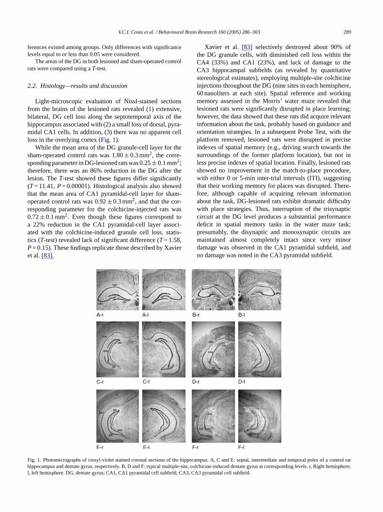
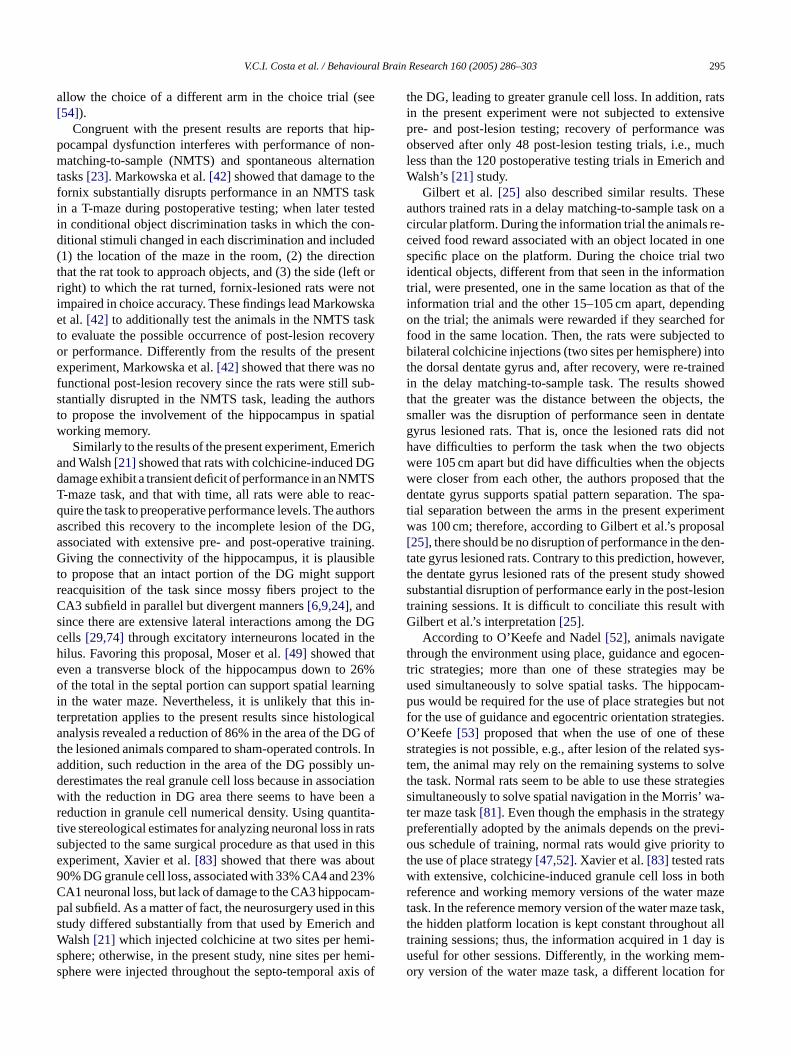
Light-microscopic evaluation of Nissl-stained sectionsfrom the brains of the lesioned rats revealed (1) extensive,bilateral, DG cell loss along the septotemporal axis of thehippocampus associated with (2) a small loss of dorsal, pyra-midal CA1 cells. In addition, (3) there was no apparent cellloss in the overlying cortex (Fig. 1).
While the mean area of the DG granule-cell layer for thesham-operated control rats was 1.80± 0.3 mm2, the corre-sponding parameter in DG-lesioned rats was 0.25± 0.1 mm2;therefore, there was an 86% reduction in the DG after thelesion. TheT-test showed these figures differ significantly(T= 11.41,P= 0.00001). Histological analysis also showedthat the mean area of CA1 pyramidal-cell layer for sham-operated control rats was 0.92± 0.3 mm2, and that the cor-responding parameter for the colchicine-injected rats was0.72± 0.1 mm2. Even though these figures correspond toa 22% reduction in the CA1 pyramidal-cell layer associ-ated with the colchicine-induced granule cell loss, statis-tics (T-test) revealed lack of significant difference (T= 1.58,P aviere
Xavier et al. [83] selectively destroyed about 90% ofthe DG granule cells, with diminished cell loss within theCA4 (33%) and CA1 (23%), and lack of damage to theCA3 hippocampal subfields (as revealed by quantitativestereological estimates), employing multiple-site colchicineinjections throughout the DG (nine sites in each hemisphere,60 nanoliters at each site). Spatial reference and workingmemory assessed in the Morris’ water maze revealed thatlesioned rats were significantly disrupted in place learning;however, the data showed that these rats did acquire relevantinformation about the task, probably based on guidance andorientation strategies. In a subsequent Probe Test, with theplatform removed, lesioned rats were disrupted in preciseindexes of spatial memory (e.g., driving search towards thesurroundings of the former platform location), but not inless precise indexes of spatial location. Finally, lesioned ratsshowed no improvement in the match-to-place procedure,with either 0 or 5-min inter-trial intervals (ITI), suggestingthat their working memory for places was disrupted. There-fore, although capable of acquiring relevant informationabout the task, DG-lesioned rats exhibit dramatic difficultywith place strategies. Thus, interruption of the trisynapticcircuit at the DG level produces a substantial performancedeficit in spatial memory tasks in the water maze task;presumably, the disynaptic and monosynaptic circuits aremaintained almost completely intact since very minord andn
Fhl
= 0.15). These findings replicate those described by Xt al.[83].
ig. 1. Photomicrographs of cresyl-violet stained coronal sections of the hipippocampus and dentate gyrus, respectively. B, D and F: typical multiple-si
, left hemisphere. DG, dentate gyrus; CA1, CA1 pyramidal cell subfield; CA3
amage was observed in the CA1 pyramidal subfield,o damage was noted in the CA3 pyramidal subfield.
pocampus. A, C and E: septal, intermediate and temporal poles of a control ratte, colchicine-induced dentate gyrus at corresponding levels. r, Right hemisphere;, CA3 pyramidal cell subfield.
290 V.C.I. Costa et al. / Behavioural Brain Research 160 (2005) 286–303
Moser et al.[49] reported that a small transverse blockof the hippocampus (down to 26% of the total, includingall hippocampal subfields) can support spatial learning inthe water maze as long as it is located in the septal pole.In the present experiment, quantitative analysis revealed thatcolchicine-injected rats, relative to controls, exhibit a 22% re-duction in the CA1 pyramidal cell layer. Since this CA1 areareduction was distributed throughout the septo-temporal axisof the hippocampus, it seems unlikely that it is responsiblefor the observed behavioral changes.
2.3. Behavior—results
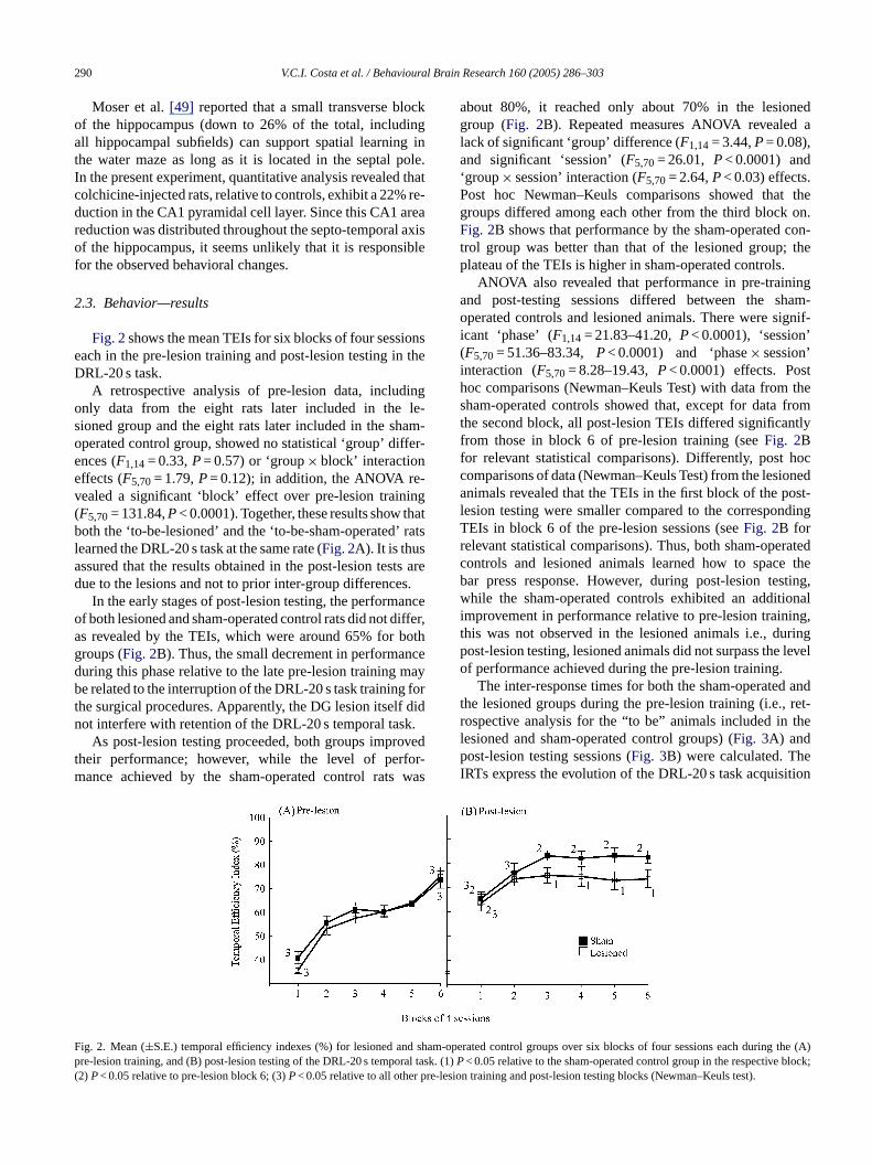
Fig. 2shows the mean TEIs for six blocks of four sessionseach in the pre-lesion training and post-lesion testing in theDRL-20 s task.
A retrospective analysis of pre-lesion data, includingonly data from the eight rats later included in the le-sioned group and the eight rats later included in the sham-operated control group, showed no statistical ‘group’ differ-ences (F1,14= 0.33,P= 0.57) or ‘group× block’ interactioneffects (F5,70= 1.79,P= 0.12); in addition, the ANOVA re-vealed a significant ‘block’ effect over pre-lesion training(F5,70= 131.84,P< 0.0001). Together, these results show thatboth the ‘to-be-lesioned’ and the ‘to-be-sham-operated’ ratsla ts ared s.
anceo iffer,a bothg nced mayb fort f didn k.
ovedt for-m was
about 80%, it reached only about 70% in the lesionedgroup (Fig. 2B). Repeated measures ANOVA revealed alack of significant ‘group’ difference (F1,14= 3.44,P= 0.08),and significant ‘session’ (F5,70= 26.01, P< 0.0001) and‘group× session’ interaction (F5,70= 2.64,P< 0.03) effects.Post hoc Newman–Keuls comparisons showed that thegroups differed among each other from the third block on.Fig. 2B shows that performance by the sham-operated con-trol group was better than that of the lesioned group; theplateau of the TEIs is higher in sham-operated controls.
ANOVA also revealed that performance in pre-trainingand post-testing sessions differed between the sham-operated controls and lesioned animals. There were signif-icant ‘phase’ (F1,14= 21.83–41.20,P< 0.0001), ‘session’(F5,70= 51.36–83.34, P< 0.0001) and ‘phase× session’interaction (F5,70= 8.28–19.43,P< 0.0001) effects. Posthoc comparisons (Newman–Keuls Test) with data from thesham-operated controls showed that, except for data fromthe second block, all post-lesion TEIs differed significantlyfrom those in block 6 of pre-lesion training (seeFig. 2Bfor relevant statistical comparisons). Differently, post hoccomparisons of data (Newman–Keuls Test) from the lesionedanimals revealed that the TEIs in the first block of the post-lesion testing were smaller compared to the correspondingTEIs in block 6 of the pre-lesion sessions (seeFig. 2B forrelevant statistical comparisons). Thus, both sham-operatedc theb sting,w onali ing,t ringp levelo
d andt ret-r thelp eI ition
F d sham ing the (A)p ral taskP block;( pre-les
earned the DRL-20 s task at the same rate (Fig. 2A). It is thusssured that the results obtained in the post-lesion tesue to the lesions and not to prior inter-group difference
In the early stages of post-lesion testing, the performf both lesioned and sham-operated control rats did not ds revealed by the TEIs, which were around 65% forroups (Fig. 2B). Thus, the small decrement in performauring this phase relative to the late pre-lesion traininge related to the interruption of the DRL-20 s task training
he surgical procedures. Apparently, the DG lesion itselot interfere with retention of the DRL-20 s temporal tas
As post-lesion testing proceeded, both groups imprheir performance; however, while the level of perance achieved by the sham-operated control rats
ig. 2. Mean (±S.E.) temporal efficiency indexes (%) for lesioned anre-lesion training, and (B) post-lesion testing of the DRL-20 s tempo2)P< 0.05 relative to pre-lesion block 6; (3)P< 0.05 relative to all other
ontrols and lesioned animals learned how to spacear press response. However, during post-lesion tehile the sham-operated controls exhibited an additi
mprovement in performance relative to pre-lesion trainhis was not observed in the lesioned animals i.e., duost-lesion testing, lesioned animals did not surpass thef performance achieved during the pre-lesion training.
The inter-response times for both the sham-operatehe lesioned groups during the pre-lesion training (i.e.,ospective analysis for the “to be” animals included inesioned and sham-operated control groups) (Fig. 3A) andost-lesion testing sessions (Fig. 3B) were calculated. Th
RTs express the evolution of the DRL-20 s task acquis
-operated control groups over six blocks of four sessions each dur. (1)< 0.05 relative to the sham-operated control group in the respectiveion training and post-lesion testing blocks (Newman–Keuls test).

V.C.I. Costa et al. / Behavioural Brain Research 160 (2005) 286–303 291
Fig. 3. Mean (±S.E.) inter-response times (IRT) for sham-operated controls and lesioned animals in both (A) the pre-lesion training, and (B) post-lesion testingof the DRL-20 s task. (1)P< 0.05 relative to the sham-operated control group in the respective block; (2)P< 0.05 relative to pre-lesion block 6; (3)P< 0.05relative to all other pre-lesion training and post-lesion testing blocks (Newman–Keuls test).
and performance during both the pre-lesion training and post-lesion testing sessions; progress is revealed by an increasein the IRTs. AsFig. 3A shows, in the first block of pre-lesion training sessions, IRTs were around 7–8 s; however,as training proceeded, the IRTs increased to about 16 s forboth groups in the last block of sessions (note that this is aretrospective analysis). ANOVA revealed a lack of signifi-cant IRT differences for ‘group’ (F1,14= 0.36,P= 0.56) and‘group× session’ interaction (F5,70= 0.96,P= 0.45) effects,in the pre-lesion training; conversely, there was a signifi-cant ‘session’ effect (F5,70= 71.42,P< 0.0001), indicatingthat “both groups” learned equally well how to increase IRTduring the pre-lesion training (Fig. 3A).
In the first block of post-lesion testing, the IRTs of both le-sioned and sham-operated controls did not differ (seeFig. 2Bfor relevant statistical comparisons) and were around 14 s;thus, selective damage to the DG does not disrupt the re-tention of the IRT acquired previously to the lesion. Repeti-tive, post-lesion testing lead sham-operated controls to reacha mean IRT of about 20 s, and the lesioned rats an IRT ofabout 16 s (Fig. 3B). ANOVA just failed to reach a signifi-cant ‘group’ effect (F1,14= 4.08,P< 0.06). On the other hand,there were significant ‘session’ (F5,70= 17.78,P< 0.0001)and ‘group× session’ interaction (F5,70= 2.96,P< 0.01) ef-fects, showing that there is a marked difference in the rate ofincrease in IRT both groups.Fig. 3B clearly shows this differ-e n IRTs thoseo heser theD sionI or-rr thel
ngt asp es-s ons
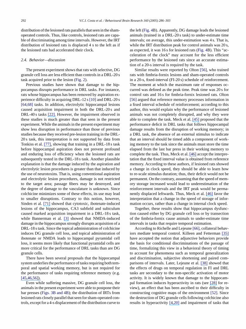
(Fig. 4B). While ANOVA, as expected, revealed a lack of sig-nificant ‘group’ effects for IRT scores in the four final sessionsof the pre-lesion training (F1,7798= 0.04,P= 0.84 (Fig. 4A),it revealed a significant ‘group’ difference for the IRT scoresin the four final post-lesion testing sessions (F1,6468= 253.27,P< 0.0001) (Fig. 4B). Inspection ofFig. 4B reveals that IRT
Fig. 4. Inter-response time distribution in the four final pre-lesion trainingsessions (A), and the four last post-lesion testing sessions (B) in the DRL-20 stask for both the sham-operated controls and the lesioned animals.
nce. Post hoc comparisons showed that the post-lesiocores of the sham-operated controls were greater thanf corresponding pre-lesion session 6, indicating that tats improved with the additional post-surgical testing inRL-20 s task; conversely, for the lesioned group, post-le
RT scores from blocks 2 to 6 did not differ from those of cesponding block 6 in the pre-lesion training (seeFig. 3B forelevant statistical differences), indicating that IRTs inesioned rats did not increase after damage to the DG.
To evaluate the animals’ ability to discriminate amoime intervals after training, an IRT distribution analysis werformed, including the four final pre-lesion training sions (Fig. 4A) and the four final post-lesion testing sessi
292 V.C.I. Costa et al. / Behavioural Brain Research 160 (2005) 286–303
distribution of the lesioned rats parallels that seen in the sham-operated controls. Thus, like controls, lesioned rats are capa-ble of discriminating among time intervals. However, the IRTdistribution of lesioned rats is displaced 4 s to theleft as ifthe lesioned rats had accelerated their clock.
2.4. Behavior—discussion
The present experiment shows that rats with selective, DGgranule cell loss are less efficient than controls in a DRL-20 stask acquired prior to the lesion (Fig. 2).
Previous studies have shown that damage to the hip-pocampus disrupts performance in DRL tasks. For instance,rats whose hippocampus has been removed by aspiration ex-perience difficulty in acquiring DRL-12 s[10] and DRL-20 s[64,68] tasks. In addition, electrolytic hippocampal lesionscaused acquisition impairment in both the DRL-20 s andDRL-40 s tasks[22]. However, the impairment observed inthese studies is much greater than that seen in the presentexperiment. While the animals in the present experiment mayshow less disruption in performance than those of previousstudies because they received pre-lesion training in the DRL-20 s task, this interpretation is not supported by data fromTonkiss et al.[77], showing that training in a DRL-18 s taskbefore hippocampal aspiration does not prevent profoundand enduring loss of efficiency when the lesioned rats ares siblee n ande d byt ationa ictedt , andt Sincec leadt ver,S edl lumc ask,w dd of aD inei n ofi celll arem DGg
mpals tem-p fort (e.g.[
thea theirbl con-t e to
the left (Fig. 4B). Apparently, DG damage leads the lesionedanimals (trained in a DRL-20 s task) to under-estimate timeintervals; on average, this under-estimation was 4 s. That is,while the IRT distribution peak for control animals was 20 s,as expected, it was 16 s for lesioned rats (Fig. 4B). This “ac-celeration of the clock” may account for the less efficientperformance by the lesioned rats since an accurate estima-tion of a 20-s interval is required by the task.
Similar results were reported by Olton[56], who trainedrats with fimbria-fornix lesions and sham-operated controlsin a 20-s, fixed-interval (FI-20 s) schedule of reinforcement.The moment at which the maximum rate of responses oc-curred was defined as the peak time. Peak time was 20 s forcontrol rats and 16 s for fimbria-fornix lesioned rats. Olton[56] argued that reference memory processes information ina fixed interval schedule of reinforcement; according to thisauthor, this would explain why the performance of lesionedanimals was not completely disrupted, and why they wereable to complete the task. Meck et al.[45] proposed that theperformance deficit in DRL tasks that follows hippocampaldamage results from the disruption of working memory; ina DRL task, the absence of an external stimulus to indicatethat an interval should be timed adds a component of work-ing memory to the task since the animals must store the timeelapsed from the last bar press in their working memory tocomplete the task. Thus, Meck et al.[46] accept the interpre-t ncem weda howt t bep mem-o f ther rma-ni nfor-m eed.
func-t ctiono timei
-ih videt e oft ngt ationa om-p tt RLt otora m-pv y inct lsor at
ubsequently tested in the DRL-18 s task. Another plauxplanation is that the damage induced by the aspiratiolectrolytic lesion procedures is greater than that induce
he use of neurotoxins. That is, using conventional aspirnd electrolytic lesion procedures, damage is not restr
o the target area; passage fibers may be destroyedhe degree of damage to the vasculature is unknown.olchicine minimizes some of these effects, its use mayo smaller disruptions. Contrary to this notion, howeinden et al.[71] showed that cytotoxic, ibotenate-induc
esions of the hippocampus, CA3 subfield and subicuaused marked acquisition impairment in a DRL-18 s thile Bannerman et al.[3] showed that NMDA-induceamage in the hippocampus strongly disrupts acquisitionRL-18 s task. Since the topical administration of colchic
nduces DG granule cell loss, and topical administratiobotenate or NMDA leads to hippocampal pyramidaloss, it seems more likely that functional pyramidal cells
ore critical for the performance of DRL tasks than areranule cells.
There have been several proposals that the hippocaystem underlies the performance of tasks requiring bothoral and spatial working memory, but is not required
he performance of tasks requiring reference memory45,46,56]).
Even while suffering massive, DG granule cell loss,nimals in the present experiment were able to postponear presses (Figs. 3B and 4B); the distribution of IRTs from
esioned rats closely parallel that seen for sham-operatedrols, except for a 4-s displacement of the distribution curv
ation that the fixed interval value is obtained from refereemory. According to these authors, if lesioned rats shochange in clock speed, they should be able to learn
o re-scale stimulus duration; thus, their deficit would noermanent. On the contrary, assuming that the speed ofry storage increased would lead to underestimation oeinforcement intervals and the IRT peak would be peently displaced leftwards. Thus, Meck et al.[46] favor the
nterpretation that a change in the speed of storage of iation occurs, rather than a change in internal clock spTogether, these results show that hippocampus dys
ion caused either by DG granule cell loss or by transef the fimbria-fornix cause animals to under-estimate
ntervals in tasks that require temporal estimation.According to Richelle and Lejeune[66], collateral behav
ors mediate temporal control. Killeen and Fetterman[35]ave accepted the notion that adjunctive behaviors pro
he basis for conditional discriminations of the passagime, formalizing this view in a behavioral theory of timio account for phenomena such as temporal generaliznd discrimination, subjective shortening and paired carisons of intervals. Later, Lejeune et al.[38] showed tha
he effects of drugs on temporal regulation in FI and Dasks are secondary to the non-specific activation of mctivity. It is widely known that damage to the hippocaal formation induces hyperactivity in rats (see[28] for re-iew), an effect that has been ascribed to their difficultonstructing cognitive maps of the environment[52]. Sincehe destruction of DG granule cells following colchicine aesults in hyperactivity[4,20] and impairment of tasks th
V.C.I. Costa et al. / Behavioural Brain Research 160 (2005) 286–303 293
require the construction of cognitive maps (e.g.[73,83]), it istempting to propose that the underestimation of time seen inthe DG-lesioned rats is related to this spatial difficulty.
3. Experiment IB—Effect of DG-selective lesion onperformance of an NMTP task
Extensive damage to the DG granule cells strongly dis-rupts acquisition of working and reference memory tasks inthe water maze[83]. In the present experiment, we evalu-ated the effect colchicine-induced, DG granule cell loss onperformance of a pre-lesion acquired, NMTP task. Note thatthis experiment was run in parallel with the DRL-20 s taskreported above.
3.1. Materials and methods
3.1.1. AnimalsSince the same rats used in the DRL-20 s task were trained
and tested in parallel in the NMTP task, the extent of DG gran-ule cell damage and time-course of pre-lesion training and post-lesion testing were exactly the same, making inter-task comparisonspossible.
3.1.2. Apparatus
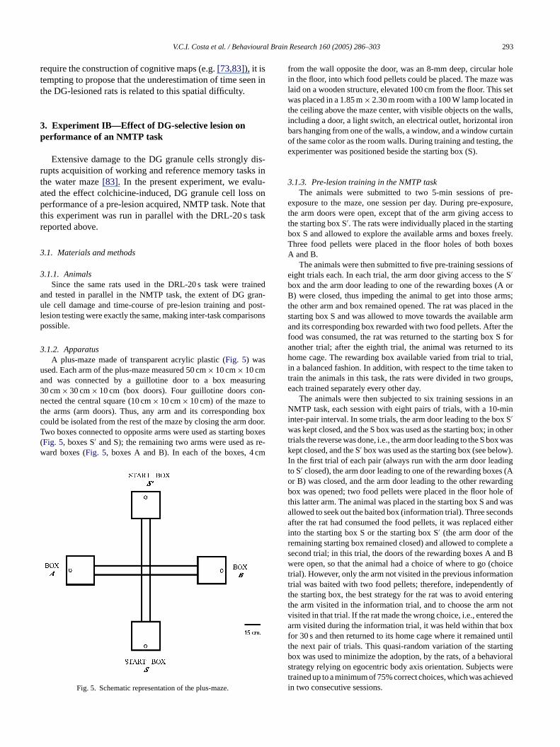
ua ring3 n-n ot boxc door.T boxes( s re-w cm
from the wall opposite the door, was an 8-mm deep, circular holein the floor, into which food pellets could be placed. The maze waslaid on a wooden structure, elevated 100 cm from the floor. This setwas placed in a 1.85 m× 2.30 m room with a 100 W lamp located inthe ceiling above the maze center, with visible objects on the walls,including a door, a light switch, an electrical outlet, horizontal ironbars hanging from one of the walls, a window, and a window curtainof the same color as the room walls. During training and testing, theexperimenter was positioned beside the starting box (S).
3.1.3. Pre-lesion training in the NMTP taskThe animals were submitted to two 5-min sessions of pre-
exposure to the maze, one session per day. During pre-exposure,the arm doors were open, except that of the arm giving access tothe starting box S′. The rats were individually placed in the startingbox S and allowed to explore the available arms and boxes freely.Three food pellets were placed in the floor holes of both boxesA and B.
The animals were then submitted to five pre-training sessions ofeight trials each. In each trial, the arm door giving access to the S′
box and the arm door leading to one of the rewarding boxes (A orB) were closed, thus impeding the animal to get into those arms;the other arm and box remained opened. The rat was placed in thestarting box S and was allowed to move towards the available armand its corresponding box rewarded with two food pellets. After thefood was consumed, the rat was returned to the starting box S foranother trial; after the eighth trial, the animal was returned to itsh trial,i en tot ups,e
in anN mini x Sw othert x wask ow).I ingt s (Ao rdingb le oft wasa ondsa eitheri er lete as d Bw hoicet tiont ly oft eringt notv d thea oxf untilt rtingb iorals weret ievedi
A plus-maze made of transparent acrylic plastic (Fig. 5) wassed. Each arm of the plus-maze measured 50 cm× 10 cm× 10 cmnd was connected by a guillotine door to a box measu0 cm× 30 cm× 10 cm (box doors). Four guillotine doors coected the central square (10 cm× 10 cm× 10 cm) of the maze t
he arms (arm doors). Thus, any arm and its correspondingould be isolated from the rest of the maze by closing the armwo boxes connected to opposite arms were used as startingFig. 5, boxes S′ and S); the remaining two arms were used aard boxes (Fig. 5, boxes A and B). In each of the boxes, 4
Fig. 5. Schematic representation of the plus-maze.
ome cage. The rewarding box available varied from trial ton a balanced fashion. In addition, with respect to the time takrain the animals in this task, the rats were divided in two groach trained separately every other day.
The animals were then subjected to six training sessionsMTP task, each session with eight pairs of trials, with a 10-
nter-pair interval. In some trials, the arm door leading to the bo′
as kept closed, and the S box was used as the starting box; inrials the reverse was done, i.e., the arm door leading to the S boept closed, and the S′ box was used as the starting box (see beln the first trial of each pair (always run with the arm door leado S′ closed), the arm door leading to one of the rewarding boxer B) was closed, and the arm door leading to the other rewaox was opened; two food pellets were placed in the floor ho
his latter arm. The animal was placed in the starting box S andllowed to seek out the baited box (information trial). Three secfter the rat had consumed the food pellets, it was replaced
nto the starting box S or the starting box S′ (the arm door of themaining starting box remained closed) and allowed to compecond trial; in this trial, the doors of the rewarding boxes A anere open, so that the animal had a choice of where to go (c
rial). However, only the arm not visited in the previous informarial was baited with two food pellets; therefore, independenthe starting box, the best strategy for the rat was to avoid enthe arm visited in the information trial, and to choose the armisited in that trial. If the rat made the wrong choice, i.e., entererm visited during the information trial, it was held within that b
or 30 s and then returned to its home cage where it remainedhe next pair of trials. This quasi-random variation of the staox was used to minimize the adoption, by the rats, of a behavtrategy relying on egocentric body axis orientation. Subjectsrained up to a minimum of 75% correct choices, which was achn two consecutive sessions.
294 V.C.I. Costa et al. / Behavioural Brain Research 160 (2005) 286–303
3.1.4. SurgerySurgical procedures were as described in Experiment IA; the
same rats were used both experiments.
3.1.5. Post-lesion testing in the NMTP taskAfter the post-surgical recovery period, which lasted for 20 days,
the animals were tested in the DRL-20 s task (see Experiment IA)and, in parallel, were tested in the NMTP task. Initially, the rats weregiven two pre-test sessions, a procedure identical to the pre-trainingphase described in Section3.1.3. The animals were then tested sixsessions using the procedures described in the training sessions ofSection3.1.3.
3.1.6. Data analysisA spatial efficiency index (SEI) was calculated using the formula:
SEI (%) = (8− number the incorrect choices)× 12.5.Data were analyzed using ANOVA and post hoc Newman–Keuls
comparisons were conducted to establish where overall and ses-sions differences existed among groups. Only differences withsignificance levels equal to or less than 0.05 were consideredrelevant.
3.2. Results
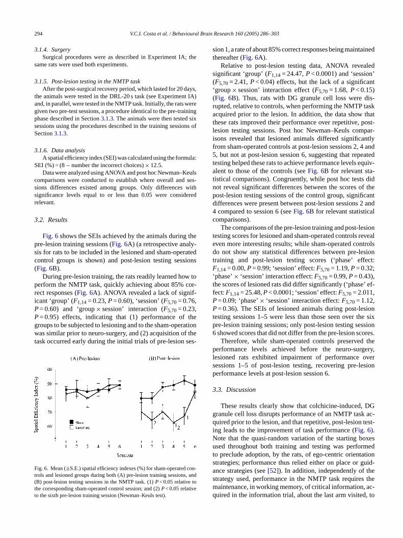
Fig. 6shows the SEIs achieved by the animals during thepre-lesion training sessions (Fig. 6A) (a retrospective analy-sis for rats to be included in the lesioned and sham-operatedc sions(
w top or-r -iPP theg rationw thet ses-
F con-t s, and(tt
sion 1, a rate of about 85% correct responses being maintainedthereafter (Fig. 6A).
Relative to post-lesion testing data, ANOVA revealedsignificant ‘group’ (F1,14= 24.47,P< 0.0001) and ‘session’(F5,70= 2.41,P< 0.04) effects, but the lack of a significant‘group× session’ interaction effect (F5,70= 1.68,P< 0.15)(Fig. 6B). Thus, rats with DG granule cell loss were dis-rupted, relative to controls, when performing the NMTP taskacquired prior to the lesion. In addition, the data show thatthese rats improved their performance over repetitive, post-lesion testing sessions. Post hoc Newman–Keuls compar-isons revealed that lesioned animals differed significantlyfrom sham-operated controls at post-lesion sessions 2, 4 and5, but not at post-lesion session 6, suggesting that repeatedtesting helped these rats to achieve performance levels equiv-alent to those of the controls (seeFig. 6B for relevant sta-tistical comparisons). Congruently, while post hoc tests didnot reveal significant differences between the scores of thepost-lesion testing sessions of the control group, significantdifferences were present between post-lesion sessions 2 and4 compared to session 6 (seeFig. 6B for relevant statisticalcomparisons).
The comparisons of the pre-lesion training and post-lesiontesting scores for lesioned and sham-operated controls revealeven more interesting results; while sham-operated controlsdo not show any statistical differences between pre-lesiont fect:F‘t ’ ef-f ,PP siont he sixp ssion6 ores.
thep gery,l overs sionp
3
DGg ac-q test-iN oxesu medt tions guid-a es s them ac-q , to
ontrol groups is shown) and post-lesion testing sesFig. 6B).
During pre-lesion training, the rats readily learned hoerform the NMTP task, quickly achieving about 85% cect responses (Fig. 6A). ANOVA revealed a lack of signifcant ‘group’ (F1,14= 0.23,P= 0.60), ‘session’ (F5,70= 0.76,= 0.60) and ‘group× session’ interaction (F5,70= 0.23,= 0.95) effects, indicating that (1) performance ofroups to be subjected to lesioning and to the sham-opeas similar prior to neuro-surgery, and (2) acquisition of
ask occurred early during the initial trials of pre-lesion
ig. 6. Mean (±S.E.) spatial efficiency indexes (%) for sham-operatedrols and lesioned groups during both (A) pre-lesion training sessionB) post-lesion testing sessions in the NMTP task. (1)P< 0.05 relative tohe corresponding sham-operated control session; and (2)P< 0.05 relativeo the sixth pre-lesion training session (Newman–Keuls test).
raining and post-lesion testing scores (‘phase’ ef1,14= 0.00,P= 0.99; ‘session’ effect:F5,70= 1.19,P= 0.32;
phase’× ‘session’ interaction effect:F5,70= 0.99,P= 0.43),he scores of lesioned rats did differ significantly (‘phaseect:F1,14= 25.48,P< 0.0001; ‘session’ effect:F5,70= 2.011= 0.09; ‘phase’× ‘session’ interaction effect:F5,70= 1.12,= 0.36). The SEIs of lesioned animals during post-le
esting sessions 1–5 were less than those seen over tre-lesion training sessions; only post-lesion testing seshowed scores that did not differ from the pre-lesion scTherefore, while sham-operated controls preserved
erformance levels achieved before the neuro-suresioned rats exhibited impairment of performanceessions 1–5 of post-lesion testing, recovering pre-leerformance levels at post-lesion session 6.
.3. Discussion
These results clearly show that colchicine-induced,ranule cell loss disrupts performance of an NMTP taskuired prior to the lesion, and that repetitive, post-lesion
ng leads to the improvement of task performance (Fig. 6).ote that the quasi-random variation of the starting bsed throughout both training and testing was perfor
o preclude adoption, by the rats, of ego-centric orientatrategies; performance thus relied either on place ornce strategies (see[52]). In addition, independently of thtrategy used, performance in the NMTP task requireaintenance, in working memory, of critical information,uired in the information trial, about the last arm visited
V.C.I. Costa et al. / Behavioural Brain Research 160 (2005) 286–303 295
allow the choice of a different arm in the choice trial (see[54]).
Congruent with the present results are reports that hip-pocampal dysfunction interferes with performance of non-matching-to-sample (NMTS) and spontaneous alternationtasks[23]. Markowska et al.[42] showed that damage to thefornix substantially disrupts performance in an NMTS taskin a T-maze during postoperative testing; when later testedin conditional object discrimination tasks in which the con-ditional stimuli changed in each discrimination and included(1) the location of the maze in the room, (2) the directionthat the rat took to approach objects, and (3) the side (left orright) to which the rat turned, fornix-lesioned rats were notimpaired in choice accuracy. These findings lead Markowskaet al.[42] to additionally test the animals in the NMTS taskto evaluate the possible occurrence of post-lesion recoveryor performance. Differently from the results of the presentexperiment, Markowska et al.[42] showed that there was nofunctional post-lesion recovery since the rats were still sub-stantially disrupted in the NMTS task, leading the authorsto propose the involvement of the hippocampus in spatialworking memory.
Similarly to the results of the present experiment, Emerichand Walsh[21] showed that rats with colchicine-induced DGdamage exhibit a transient deficit of performance in an NMTST-maze task, and that with time, all rats were able to reac-q thorsa DG,a ning.G iblet portr theCs e DGc theh te 26%o ningi in-t gicala G oft ls. Ina un-d iationw en ar tita-t ratss in thise ut9 23%C am-p thiss andW mi-s emi-s is of
the DG, leading to greater granule cell loss. In addition, ratsin the present experiment were not subjected to extensivepre- and post-lesion testing; recovery of performance wasobserved after only 48 post-lesion testing trials, i.e., muchless than the 120 postoperative testing trials in Emerich andWalsh’s[21] study.
Gilbert et al.[25] also described similar results. Theseauthors trained rats in a delay matching-to-sample task on acircular platform. During the information trial the animals re-ceived food reward associated with an object located in onespecific place on the platform. During the choice trial twoidentical objects, different from that seen in the informationtrial, were presented, one in the same location as that of theinformation trial and the other 15–105 cm apart, dependingon the trial; the animals were rewarded if they searched forfood in the same location. Then, the rats were subjected tobilateral colchicine injections (two sites per hemisphere) intothe dorsal dentate gyrus and, after recovery, were re-trainedin the delay matching-to-sample task. The results showedthat the greater was the distance between the objects, thesmaller was the disruption of performance seen in dentategyrus lesioned rats. That is, once the lesioned rats did nothave difficulties to perform the task when the two objectswere 105 cm apart but did have difficulties when the objectswere closer from each other, the authors proposed that thedentate gyrus supports spatial pattern separation. The spa-t imentw osal[ den-t ever,t oweds siont ithG
et ocen-t y beu cam-p t notf gies.O eses sys-t olvet tegiess wa-t egyp revi-o tot sw othr azet task,t t allt ay isu m-o for
uire the task to preoperative performance levels. The auscribed this recovery to the incomplete lesion of thessociated with extensive pre- and post-operative traiiving the connectivity of the hippocampus, it is plaus
o propose that an intact portion of the DG might supeacquisition of the task since mossy fibers project toA3 subfield in parallel but divergent manners[6,9,24], andince there are extensive lateral interactions among thells [29,74] through excitatory interneurons located inilus. Favoring this proposal, Moser et al.[49] showed thaven a transverse block of the hippocampus down tof the total in the septal portion can support spatial lear
n the water maze. Nevertheless, it is unlikely that thiserpretation applies to the present results since histolonalysis revealed a reduction of 86% in the area of the D
he lesioned animals compared to sham-operated controddition, such reduction in the area of the DG possiblyerestimates the real granule cell loss because in associth the reduction in DG area there seems to have be
eduction in granule cell numerical density. Using quanive stereological estimates for analyzing neuronal loss inubjected to the same surgical procedure as that usedxperiment, Xavier et al.[83] showed that there was abo0% DG granule cell loss, associated with 33% CA4 andA1 neuronal loss, but lack of damage to the CA3 hippocal subfield. As a matter of fact, the neurosurgery used intudy differed substantially from that used by Emerichalsh[21] which injected colchicine at two sites per he
phere; otherwise, in the present study, nine sites per hphere were injected throughout the septo-temporal ax
ial separation between the arms in the present experas 100 cm; therefore, according to Gilbert et al.’s prop
25], there should be no disruption of performance in theate gyrus lesioned rats. Contrary to this prediction, howhe dentate gyrus lesioned rats of the present study shubstantial disruption of performance early in the post-leraining sessions. It is difficult to conciliate this result wilbert et al.’s interpretation[25].According to O’Keefe and Nadel[52], animals navigat
hrough the environment using place, guidance and egric strategies; more than one of these strategies mased simultaneously to solve spatial tasks. The hippous would be required for the use of place strategies bu
or the use of guidance and egocentric orientation strate’Keefe [53] proposed that when the use of one of th
trategies is not possible, e.g., after lesion of the relatedem, the animal may rely on the remaining systems to she task. Normal rats seem to be able to use these straimultaneously to solve spatial navigation in the Morris’er maze task[81]. Even though the emphasis in the stratreferentially adopted by the animals depends on the pus schedule of training, normal rats would give priority
he use of place strategy[47,52]. Xavier et al.[83] tested ratith extensive, colchicine-induced granule cell loss in b
eference and working memory versions of the water mask. In the reference memory version of the water mazehe hidden platform location is kept constant throughouraining sessions; thus, the information acquired in 1 dseful for other sessions. Differently, in the working mery version of the water maze task, a different location

296 V.C.I. Costa et al. / Behavioural Brain Research 160 (2005) 286–303
the hidden platform is used on each day of training; therefore,the information acquired in 1 day is useful only for the trialsrun during that day, and is not applicable to other sessions.It is important to mention that during training in both thereference and working memory versions of the water mazetask, rats departed from different starting points at the pool’sedge; therefore, optimal performance required knowledge ofthe relative positions of the multiple extra-maze cues andof the platform relative to these cues, involving navigationbased on place strategies (see[81]). The results show thatDG-lesioned rats were disrupted in the reference memoryversion of the water maze task, indicating their inability to useplace strategies; however, the data suggest that some relevantinformation about the requirements of the task were acquiredby the lesioned rats, implying that this improvement reliedon guidance and orientation strategies obtained throughoutrepeated training[83]. In addition, lesioned rats showed noimprovement in the working memory version of the watermaze task, indicating that not only their working memory forplaces was disrupted but also that guidance and orientationstrategies were not efficient in supporting performance in thewater maze task.
The animals in the present experiment were trained inthe NMTP task prior to surgery; thus, considering the usualpriority given by normal rats to rely on place strategy to per-form this type of task[47,52], it is reasonable to suggest thatt Afters y onpt lainw e im-pI ratsr g ong edr l ratsa ew whyt lesst sibleh singe rente mays hep rpre-t
sents ory;i iont /ore ed fora incet alf beenm f thisb uire
this function[37,42,45,46,50,57,62,78]. Similar results wereobserved when damage was restricted to the DG granulecells[43,79,83]. The data gathered in the present experiment,however, do not support this view since, after a transient,post-operatory disruption of performance, rats exhibitingDG granule cell loss achieved control performance level(Fig. 6). Whatever the strategy used by the rats to performthe NMTP task (place, guidance or ego-centric), this strategyinvolves the temporary maintenance of information inworking memory. Note, however, that the time intervalbetween the information and the choice trials in the presentexperiment was only 3 s. It is possible that an increase inthis time interval might reveal a time-dependent, workingmemory deficit. Experiment II addressed this possibility bytesting the rats’ performance with ITIs varying from 0 to16 min.
The experimental design used in this study attempted topreclude the use of an egocentric orientation strategy; thestarting box in the choice trials varied quasi-randomly fromtrial to trial. Experiment III tested whether this behavioralmanipulation was effective in preventing the animals fromadopting an egocentric orientation strategy.
4. Experiment II—Effect of increasing the intervalbetween information and choice trials onp
sultsr ilityt non-m ,r denti taski triali .[ theh g-to-p l.p ms tob i.e.,t oice,a yedn ten-t t theo f thei oryd y eta que,d thato ingt iredp tial)w hh ntricd mals
hey learned the task based primarily on place strategy.urgery, however, DG-lesioned rats could no longer rellace strategy to perform the task (see[83]), differing from
he sham-operated controls that could. This would exphy lesioned rats, but not sham-operated controls, weraired in the early, post-operatory testing sessions (Fig. 6B).
n addition, as post-lesion testing proceeded, lesionede-acquired the pre-lesion performance, but now relyinuidance strategy (see[63]); that is, even though lesionats reached a level of performance equivalent to controt the sixth post-lesion session (Fig. 6B), this performancould be based on a different strategy. It is not clear
he post-lesion acquisition rate for DG-lesioned rats washan that seen in pre-lesion acquisition training. One posypothesis is that acquisition rates for this NMTP task, uither place or guidance strategies, are intrinsically diffeven for normal rats. Alternatively, damage to the DGlow down acquisition of this task relative to controls. Tresent results do not allow deciding among these inte
ations.Performance in NMTP tasks like that used in the pre
tudy is believed to depend on a functional working mem.e., the critical information collected during the informatrial (about the box visited, including related intra- andxtra-maze cues) must be maintained as long as requircorrect decision of where to go in the choice trial. S
he initial proposal by Olton et al.[54], that the hippocampormation is necessary for working memory, there haveany reports showing that damage or disconnection orain structure disrupts performance in tasks that req
erformance of the NMTP task
Previous studies have produced controversial reegarding the effect of hippocampal damage on rats’ abo perform delayed matching-to-sample and delayedatching-to-sample tasks (e.g.[2,48,60,80]). For instance
ats with hippocampal lesions exhibit delay-depenmpairments in a delayed non-matching-to-samplenvolving two arms selected at random during everyn an eight-arm radial maze[60]. Similarly, Morris et al48] showed that an ibotenate acid-induced lesion ofippocampus impairs performance in a delayed matchinlace task in the water maze. Interestingly, Aggleton et a[2]rovided evidence suggesting that this impairment seee associated with tasks that involve spatial information;
he same rats that were disrupted in a spatial, forced-chlternation task were not disrupted in an object, delaon-matching-to-sample task in a Y-maze, even with re
ion delays of as long as 60 s. This result suggests tharigin of the controversy may be related to the nature o
nformation the animals must maintain in working memuring the delay to perform the task. However, Pruskl. [61] developed a non-spatial, picture-based, trial-unielayed matching-to-sample task for rats analogous toften used for testing working memory in primates, show
hat selective lesions of the rat hippocampus impaerformance in this delay-dependent visual (non-spaorking memory task. Long and Kesner[39] tested rats witippocampal lesion in working memory tasks for egoceistance and place information, showing that the ani

V.C.I. Costa et al. / Behavioural Brain Research 160 (2005) 286–303 297
were disrupted in both types of task; however, impairmentfor the egocentric distance information was mild.
In the present experiment, a delayed non-matching-to-place test allowed evaluation of to what extent animals withcolchicine-induced, granule cell loss are capable of main-taining the level of performance achieved by the end of thepost-lesion testing in Experiment IB, when the interval be-tween the information and the choice trials was 3 s or 1, 2, 4,8, or 16 min.
4.1. Materials and methods
The subjects and apparatus were the same as used in Experi-ment IB. Thirty days after the end of the Experiment I, seven DG-lesioned rats and another seven sham-operated controls were re-tested during nine sessions, employing a behavioral procedure iden-tical to that used in the pre-lesion training and post-lesion testingof Experiment IB (Retest); the animals were returned to their homecages for approximately 3 s between the information and choice tri-als. By the ninth session, the rats had achieved the minimum criterionof six correct responses per session, over two consecutive sessions;this criterion was defined based on a binomial distribution, withPless than 0.05 (two animals in the lesioned group did not reach thelimiting criterion and were not tested in the DNMTP; thus, the datarefer to five lesioned animals and seven sham-operated controls).The DNMTP procedure was then initiated and consisted of main-taining the animals in their home cages for an interval of 1, 2, 4, 8o rvalw inter-v als, ina delaysv hus,r y be-f sions,t etests r theD hus,t ssionc
4
othl earlys by thees e inb testi 30-d per-f ove-mm sig-n i-a‘ ;p spa-t k of
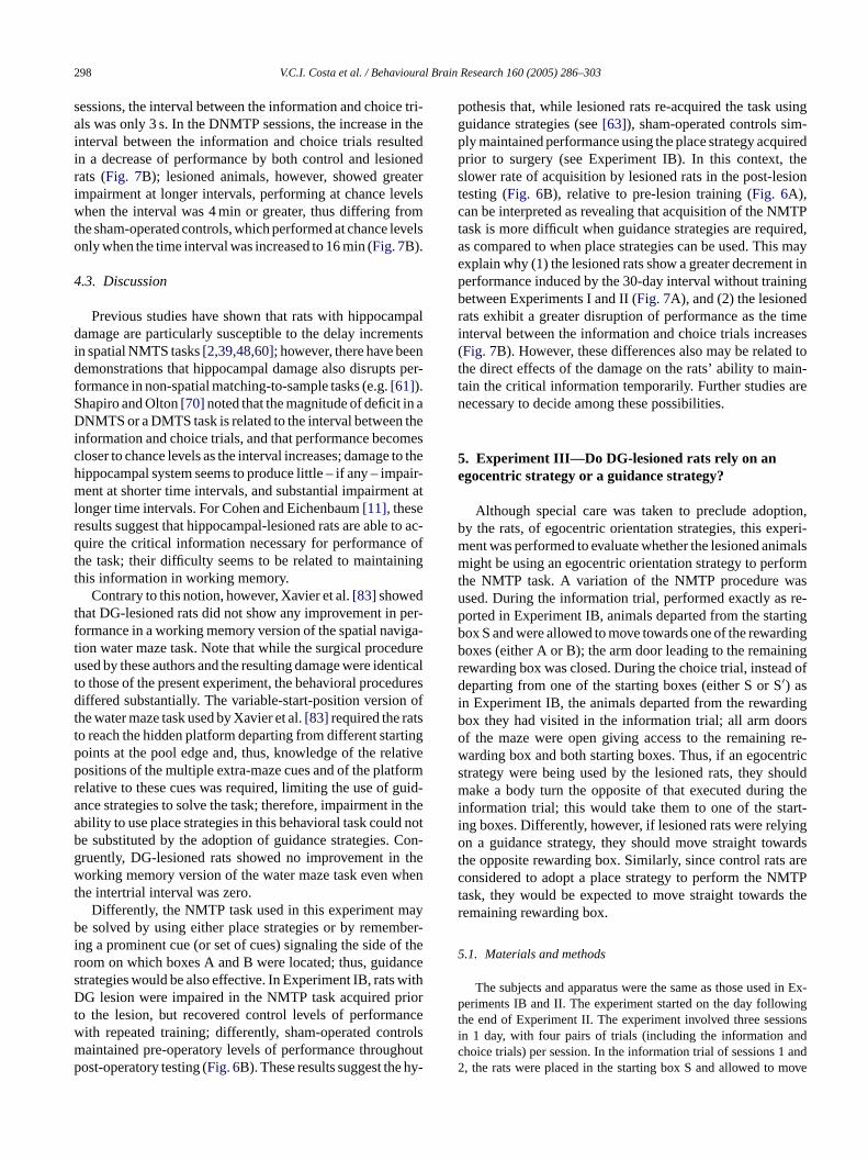
Fig. 7. Mean (±S.E.) efficiency indexes (%) for sham-operated controls andlesioned animals in the Retest (blocks of three sessions) and DNMTP ses-sions using time intervals of 0.05 (mean scores of five sessions), 1, 2, 4, 8or 16 min. (1)P< 0.05 relative to sham-operated controls in the correspond-ing block (Retest) or delays (DNMTP); (2)P< 0.05 relative to other blocks(Retest) or delays (DNMTP) within the same group; (3)P< 0.05 relative tothe remaining delays (DNMTP) (Newman–Keuls test).
the Retest were less than those for the second and third blocks(seeFig. 7A for relevant statistical comparisons). This wasnot seen for the sham-operated controls, which showed nostatistical differences in their scores.
Fig. 7B shows the effect of increasing the time intervalbetween the information and choice trials on animals per-formance; both lesioned animals and sham-operated controlsdecreased their spatial efficiency indexes as the interval be-tween the information and choice trials increased (ANOVA‘time interval’ effect:F5,50= 11.35,P< 0.0001); even thoughthis effect was significantly greater for the lesioned rats(ANOVA ‘group’ effect: F1,10= 10.93,P< 0.0001), the rateat which it occurs is similar for both groups (ANOVA‘group× session’ interaction effect:F5,50= 1.86,P= 0.10).Inspection of Fig. 7B reveals that the lesioned animalsreached chance levels of performance when the time inter-val between the information and choice trials was 4 min ormore; for the sham-operated controls, the same effect wasseen only for the 16-min time interval (seeFig. 7B for rel-evant post hoc statistical comparisons). For the interspersedsessions, in which the interval between the information andchoice trials was 0.05 min (3 s), the efficiency index was 85%for the sham-operated animals and 72% for DG-lesioned rats(Fig. 7B); these scores were not significantly different.
During the early Retest sessions, performed 30 days (with-out behavioral training) after the end of the post-lesion test-i atedr e tot pareF tlyg con-t psi for-m firmst test
r 16 min between the information and choice trials. Each inteas tested in a different session every other day; the each-dayals were randomly distributed among the sessions and animcounter balanced design. There were five sessions where
aried (1, 2, 4, 8 or 16 min), one session for each interval. Tats were tested once at each of five different delays. One daore each delay session, i.e., interspersed with the DNMTP seshere were sessions with a 3-s (0.05 min) delay, similar to the Ressions, to aid the animals in maintaining performance oveNMTP sessions, and to allow comparison between them. T
here were five sessions with a 3-s (0.05 min) delay. Each seonsisted of eight pairs of trials.
.2. Results
Fig. 7A shows that the level of performance of besioned and sham-operated control animals at thetages of Retest was reduced compared to that seennd of the post-lesion testing in Experiment IB (Fig. 6B);ince the behavioral procedure was exactly the samoth the testing phase of Experiment IB and the Re
n this experiment, we conclude that, as expected, theay interval without testing lead to a decrement in
ormance. However, repeated testing lead to an imprent in performance by both groups (Fig. 7A); repeatedeasures ANOVA including Retest data revealed aificant ‘session’ effect (F2,20= 10.92,P< 0.0001) assocted with non-significant ‘group’ (F1,10= 1.78,P= 0.20) and
group× session’ interaction (F2,20= 1.35,P= 0.27) effectsost hoc Newman–Keuls comparisons revealed that the
ial efficiency indexes of the lesioned rats in the first bloc
ng in Experiment IB, both lesioned and sham-operats showed a small decrement in performance relativheir performance in the late post-lesion testing (comigs. 6B and 7A); this decline in performance was slighreater in the lesioned rats compared to sham-operated
rols (Fig. 7A). However, with repeated training, both groumproved their efficiency indexes, achieving levels of per
ance equivalent to those seen previously, which conhe results of Experiment IB; note that over the nine Re
298 V.C.I. Costa et al. / Behavioural Brain Research 160 (2005) 286–303
sessions, the interval between the information and choice tri-als was only 3 s. In the DNMTP sessions, the increase in theinterval between the information and choice trials resultedin a decrease of performance by both control and lesionedrats (Fig. 7B); lesioned animals, however, showed greaterimpairment at longer intervals, performing at chance levelswhen the interval was 4 min or greater, thus differing fromthe sham-operated controls, which performed at chance levelsonly when the time interval was increased to 16 min (Fig. 7B).
4.3. Discussion
Previous studies have shown that rats with hippocampaldamage are particularly susceptible to the delay incrementsin spatial NMTS tasks[2,39,48,60]; however, there have beendemonstrations that hippocampal damage also disrupts per-formance in non-spatial matching-to-sample tasks (e.g.[61]).Shapiro and Olton[70] noted that the magnitude of deficit in aDNMTS or a DMTS task is related to the interval between theinformation and choice trials, and that performance becomescloser to chance levels as the interval increases; damage to thehippocampal system seems to produce little – if any – impair-ment at shorter time intervals, and substantial impairment atlonger time intervals. For Cohen and Eichenbaum[11], theseresults suggest that hippocampal-lesioned rats are able to ac-quire the critical information necessary for performance oft ingt
t per-f iga-t dureu nticat duresd n oft st tingp ativep formr uid-a in thea d notb Con-g thew hent
ayb ber-i f ther nces withD riort ncew trolsm houtp hy-
pothesis that, while lesioned rats re-acquired the task usingguidance strategies (see[63]), sham-operated controls sim-ply maintained performance using the place strategy acquiredprior to surgery (see Experiment IB). In this context, theslower rate of acquisition by lesioned rats in the post-lesiontesting (Fig. 6B), relative to pre-lesion training (Fig. 6A),can be interpreted as revealing that acquisition of the NMTPtask is more difficult when guidance strategies are required,as compared to when place strategies can be used. This mayexplain why (1) the lesioned rats show a greater decrement inperformance induced by the 30-day interval without trainingbetween Experiments I and II (Fig. 7A), and (2) the lesionedrats exhibit a greater disruption of performance as the timeinterval between the information and choice trials increases(Fig. 7B). However, these differences also may be related tothe direct effects of the damage on the rats’ ability to main-tain the critical information temporarily. Further studies arenecessary to decide among these possibilities.
5. Experiment III—Do DG-lesioned rats rely on anegocentric strategy or a guidance strategy?
Although special care was taken to preclude adoption,by the rats, of egocentric orientation strategies, this experi-m imalsm formt asu re-p rtingb rdingb ningr d ofdi dingb orso g re-w ntrics houldm thei art-i ingo ardst arec MTPt s ther
5
in Ex-p ingt sionsi ndc and2 move
he task; their difficulty seems to be related to maintainhis information in working memory.
Contrary to this notion, however, Xavier et al.[83] showedhat DG-lesioned rats did not show any improvement inormance in a working memory version of the spatial navion water maze task. Note that while the surgical procesed by these authors and the resulting damage were ide
o those of the present experiment, the behavioral proceiffered substantially. The variable-start-position versio
he water maze task used by Xavier et al.[83] required the rato reach the hidden platform departing from different staroints at the pool edge and, thus, knowledge of the relositions of the multiple extra-maze cues and of the platelative to these cues was required, limiting the use of gnce strategies to solve the task; therefore, impairmentbility to use place strategies in this behavioral task coule substituted by the adoption of guidance strategies.ruently, DG-lesioned rats showed no improvement inorking memory version of the water maze task even w
he intertrial interval was zero.Differently, the NMTP task used in this experiment m
e solved by using either place strategies or by rememng a prominent cue (or set of cues) signaling the side ooom on which boxes A and B were located; thus, guidatrategies would be also effective. In Experiment IB, ratsG lesion were impaired in the NMTP task acquired p
o the lesion, but recovered control levels of performaith repeated training; differently, sham-operated conaintained pre-operatory levels of performance througost-operatory testing (Fig. 6B). These results suggest the
l
ent was performed to evaluate whether the lesioned anight be using an egocentric orientation strategy to per
he NMTP task. A variation of the NMTP procedure wsed. During the information trial, performed exactly asorted in Experiment IB, animals departed from the staox S and were allowed to move towards one of the rewaoxes (either A or B); the arm door leading to the remaiewarding box was closed. During the choice trial, insteaeparting from one of the starting boxes (either S or S′) as
n Experiment IB, the animals departed from the rewarox they had visited in the information trial; all arm dof the maze were open giving access to the remaininarding box and both starting boxes. Thus, if an egocetrategy were being used by the lesioned rats, they sake a body turn the opposite of that executed during
nformation trial; this would take them to one of the stng boxes. Differently, however, if lesioned rats were relyn a guidance strategy, they should move straight tow
he opposite rewarding box. Similarly, since control ratsonsidered to adopt a place strategy to perform the Nask, they would be expected to move straight towardemaining rewarding box.
.1. Materials and methods
The subjects and apparatus were the same as those usederiments IB and II. The experiment started on the day follow
he end of Experiment II. The experiment involved three sesn 1 day, with four pairs of trials (including the information ahoice trials) per session. In the information trial of sessions 1, the rats were placed in the starting box S and allowed to
V.C.I. Costa et al. / Behavioural Brain Research 160 (2005) 286–303 299
towards one of the rewarding boxes (A or B) where they were re-warded; the arm door leading towards the other box was closed. Therats were then transferred to their home cages where they stayed for3 s. They were then replaced in the box from which they had justbeen removed (A or B) and allowed to perform their choice trial,with all arm doors of the maze open; thus, the animals could movetowards any box of the maze. In the information trial of session 3,the animals departed from the starting box S, with the arm doorsleading to the starting box S′ and one of the arm doors leading tothe rewarding boxes A or B open; thus, in this case, the rats couldchoose where to go in the information trial. Since the animals hadnot been rewarded in the starting box S′, this choice was consideredan error; differently, the choice to move towards the rewarding boxwas considered a correct response. Three seconds after the informa-tion trial, the animals were subjected to the choice trial, which wasidentical to the previous procedures.
5.2. Results and discussion
The spatial efficiency indexes revealed that both groupsexhibit levels of performance similar to those seen in the pre-vious experiments (data not shown); in addition, the lesionedrats did not differ significantly from the sham-operated con-trols. On the third session of this experiment, when the ani-mals could choose where to go in the information trial, theaverage number of correct responses was 77.5% for the sham-operated controls and 75.0% for the lesioned rats; similarl VAra
con-fi nta-t e int ntrics ositea e wasn
ctlyt rsis-t g attap callya nt byo ings ningt ox,i thea leftt ares armw here-f ce orh dis-c
d ratsd that
lesioned rats use intra- and/or extra-maze cues to performthis task.
6. General discussion
The results constitute evidence for multiple functions in-volving the DG granule cells, including time control mod-ulation, and spatial and working memories, congruent withprevious proposals for the hippocampal formation (e.g.[3]).However, the data can be also interpreted following O’Keefeand Nadel’s[52] line of reasoning (see below).
6.1. DG lesion and performance in a DRL-20 s task
Olton [56] proposed that reference memory processedinformation in a fixed interval schedule of reinforcement,providing an explanation of why the performance of lesionedanimals was not completely disrupted, and why they wereable to complete the FI-20 s task. In addition, Meck et al.[45]proposed that the less efficient performance in DRL tasksfollowing hippocampal damage results from disruption ofworking memory. Note that the DRL task differs from the FItask in other respects. In the FI task, a bar press is associatedwith both reinforcement after a time interval has elapsedand lack of reinforcement during the interval; however, thea ssedb barp en ito g thel pitet nedr rD hiss botht
ter-f be-t et al.[ am-p en eta agem hilel ffer-e cine-i lesse erea e re-s ropera ent.
sks,O tt g ina er-v e (got nless
evels of performance were seen in the choice trials. ANOevealed a lack of significant ‘group’ (F2,16= 0.00,P= 0.99)nd ‘trial’ (F3,48= 0.04,P= 0.99) significant effects.
The results of the present experiment unequivocallyrm that the lesioned rats did not use egocentric orieion strategies to achieve control levels of performanche NMTP task; had the lesioned rats been using egocetrategies, they would not move straight towards the opprm of the maze in the choice trial because this responsot rewarded during previous testing in the task.
It could be argued that the lesioned rats move direhrough the adjoining arms because they exhibit a peent running response of a hyperactivity instead of aiminhe opposite maze arm. In fact, hippocampal damage[14,84]nd colchicine-induced, DG-granule cell loss[83] do induceersistent responses in rats. This question was specifiddressed in the third session of the present experimeffering the animals the opportunity of either (1) movtraight through the arm towards an unbaited box, or turo the right or to the left towards a previously baited bn the information trial, or (2) moving straight throughrm towards a baited box, or turning to the right or to the
owards an unbaited box, in the choice trial. The resultstraightforward: the rats only move straight through thehen this response takes them to the rewarding box. T
ore, one can discard the interpretation that perseveranyperactivity was driving the animals’ response can bearded.
Taken together, these results indicate that the lesioneo not rely on egocentric orientation; the data suggest
nimal is not punished, including when the bar is preefore the correct time. Differently, in the DRL task, aress may be rewarded or punished, depending on whccurs; thus, rats must solve the conflict of approachin
ever at certain times and avoiding it at other times. Deshese differences, the time-course for fimbria-fornix lesioats in the FI-20 s task[56] is very similar to that seen foG-lesioned rats in a DRL-20 s task (Experiment IA). Tuggests that a similar strategy is being used to solveasks, which does not involve working memory.
Rawlins [65] proposed that hippocampal damage ineres with the ability to deal with temporal discontinuityween events to be associated. Congruently, Bannerman3] showed that complete cytotoxic damage to the hippocus disrupts performance in a DRL-18 s task and Sindl. [71] showed that complete hippocampal ibotenate damarkedly affected performance in a DRL-18 s task, w
eaving performance in a DRL-12 s task almost intact. Dintly, the present experiments show that rats with colchi
nduced, DG granule cell loss, even though slightlyfficient than controls when performing a DRL-20 task, wble to (under-) estimate time intervals. Together, thesults suggest that the contribution of the hippocampus pnd the DG for performance of DRL tasks may be differ
When discussing performance of animals in DRL ta’Keefe and Nadel[52] proposed that “. . . rats cannot ‘coun
ime’, but rather bridge temporal intervals by engaginny of a variety of behaviors which fill the required intal. The normal rat can call on behaviors based on placo the other side of the box), guidance (do not press u
300 V.C.I. Costa et al. / Behavioural Brain Research 160 (2005) 286–303
light is on), or orientation (engage in a sequence of collat-eral displacement behaviors) hypotheses.” (p. 325). In agree-ment with this notion, Richelle and Lejeune[66] proposeda synthesis as to how collateral behaviors mediate temporalcontrol, and Killeen and Fetterman[35] defended the notionthat adjunctive behaviors serve as the basis for conditionaldiscriminations of the passage of time. Further, Costa et al.[13] showed that there is a defined sequence of adjunctivebehaviors during performance of a DRL task. Congruently, ithas been shown that the presence of objects in the condition-ing chamber facilitates performance in DRL tasks[1,36,72],possibly because such objects contribute to the appearanceof adjunctive behaviors on which temporal discriminationwould be based.
O’Keefe and Nadel[52] hypothesized that animals withhippocampal damage, even though unable to use a place strat-egy, taking them away from the lever, should be able to gener-ate some form of collateral behaviors, relying on guidance ororientation strategies, to perform DRL tasks. In this context,the results of the present study suggest that these strategiesare not as efficient as the place strategy, leading to time under-estimation. In addition, these authors emphasized that sincemost DRL experiments are preceded by training on a contin-uous reinforcement schedule, and hippocampal rats acquirethe CRF schedule more rapidly than do controls, this superioracquisition in the CRF schedule would lead to difficulty infi thes hilee RLt Lp r-i ; ina ere-f y,e hyp thant esist ong d ex-p hoseo
ofa ct ist anced task,w tiont witht , thea mpo-r cifica n,r n top ben-e hus,s gs( -
creased the spontaneous motor activity in rats, decreasingperformance in DRL tasks. Hippocampal damage in rats iswidely known to induce hyperactivity (see[28] for review)associated with difficulty in constructing cognitive maps ofthe environment[52]. Similarly, colchicine-induced, selec-tive DG granule cell loss in rats increases motor activity[4,20]and decreases performance in tasks that require the adoptionof the place strategy (e.g.[73,83]).
Together, these findings suggest that DG-lesioned ratsadopt guidance and/or orientation strategies, engaging them-selves in a sequence of behaviors to measure time intervals(see[35,52,66]). However, since they also exhibit activationof motor behaviors (see[4,20]), it would take them less time toconclude such sequence. Consequently, these animals showsmaller inter-response times and, therefore, a less efficientperformance in the DRL task (Experiment IA). Furthermore,Costa et al.[12] showed that the disruption of performancein the DRL-20 s seen after colchicine-induced damage tothe dentate gyrus depends upon the size of the experimen-tal chamber; that is, the larger is the experimental chamberthe smaller is the behavioral disruption.
6.2. DG lesion and performance in an NMTP task
Performance of rats in both delayed matching-to-sampleand delayed non-matching-to-sample tasks is believed to de-p arymf for-m asks[ re isa tioni atd k ac-q test-i otet oicet It isd rk-i ancer f then for-m
ilityt bilityt sa taskr in theN blea er thel , nowr ts IBa eventt esultso ina
nding collateral behaviors in DRL tasks to compete withtrategy developed during CRF training; congruently, wxtensive CRF pre-training results in persistent deficit in Dasks[19], minimal CRF pre-training results in normal DRerformance by hippocampal rats[67]. In the present expe
ments, rats acquired the DRL task prior to the lesionddition, the degree of CRF training was very small. Th
ore, it is likely that the prior CRF training had little, if anffect on performance of the DRL. This helps explain werformance of these rats was only slightly less efficient
hat of the controls after surgery. However, if the hypothhat DG-lesioned rats perform the DRL task by relyinguidance and orientation strategies is correct, this woullain why their inter-response times were shorter than tf the controls.
Lejeune et al.[38] showed that the administrationmineptine (a tricyclic antidepressant whose major effe
o inhibit dopamine uptake and, at higher doses, to enhopamine release) decreases performance in a DRLhile leaving performance in a time duration discrimina
ask intact. Thus, the hypothesis that this drug interfereshe timing mechanism was discarded; on the contraryuthors suggested that the effects of amineptine on teal regulation in the DRL task are secondary to non-spectivation of motor activity. In favor of this interpretatioetraction of the bars during time estimation was showrevent expression of the activated motor behavior to thefit of the expression of unimpaired time estimation. Timilarly to other hyperactivity-inducing dopaminergic drue.g., nomifensin[51] and buproprione[69]), amineptive in
end on the integrity of working memory for the temporaintenance of critical information for later use[54]. While
ormer studies on the involvement of the hippocampalation in working memory are based largely on spatial t
45], there have been proposals that this brain structulso required for the maintenance of non-spatial informa
n working memory (e.g.[46]). The present study shows thamage to the DG disrupts performance of an NMTP tasuired prior to the lesion; however, repeated post-lesion
ng lead to recovery of performance (Experiment IB). Nhat the time interval between the information and chrials throughout all phases of Experiment IB was 3 s.ifficult to conciliate these results with the notion that wo
ng memory is disrupted since the recovered performequires a preserved working memory, independently oature of the information maintained temporarily for perance of the task.The demonstration that DG damage disrupts the ab
o navigate relying on place strategies, preserving the ao use guidance and orientation strategies[83], corroboratenother more parsimonious interpretation of the NMTPesults. That is, place strategies supported performanceMTP task prior to the lesion, but were no longer availafter damage; thus, performance was disturbed soon aft
esion. As training proceeded, the rats relearned the taskelying on the available guidance strategies (Experimennd II). The training schedule was designed such as to pr
he use of orientation strategies (see above), and the rf Experiment III indicate that our design was effectivevoiding the adoption of orientation strategies.
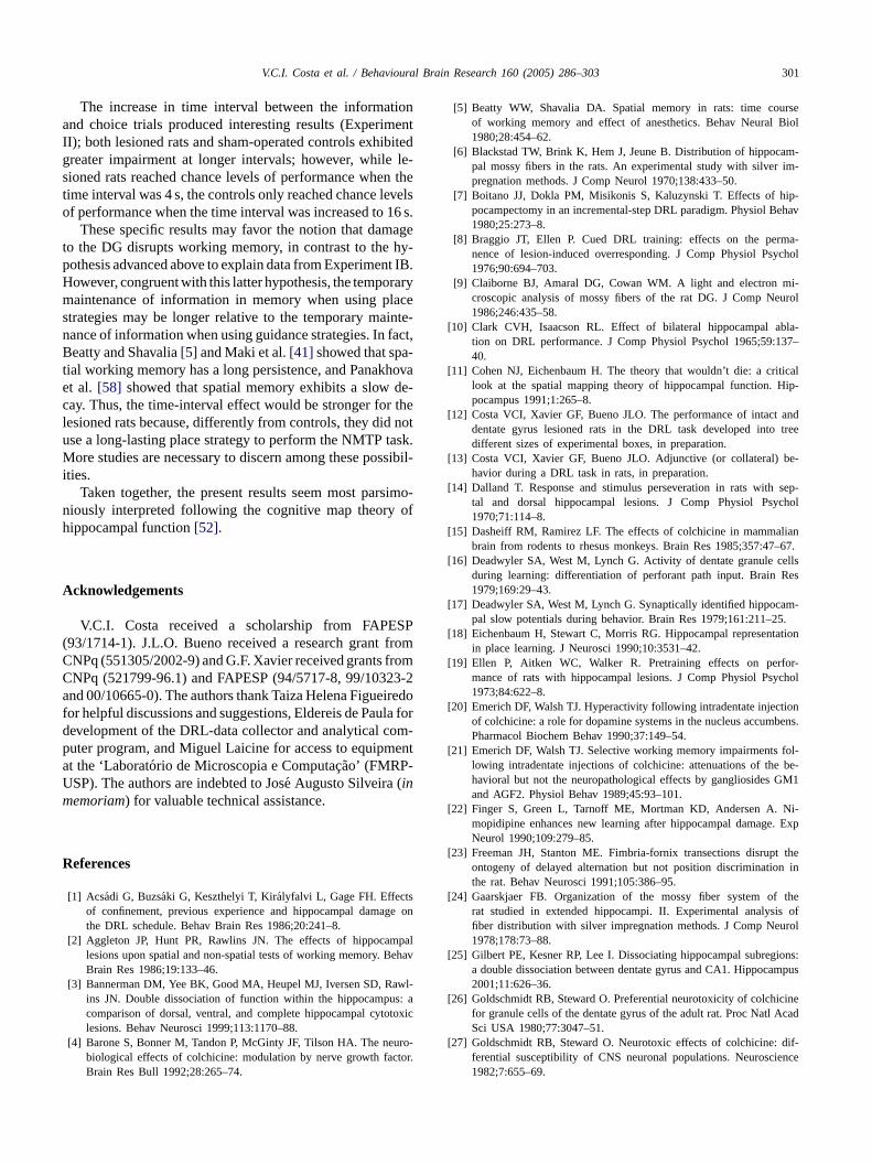
V.C.I. Costa et al. / Behavioural Brain Research 160 (2005) 286–303 301
The increase in time interval between the informationand choice trials produced interesting results (ExperimentII); both lesioned rats and sham-operated controls exhibitedgreater impairment at longer intervals; however, while le-sioned rats reached chance levels of performance when thetime interval was 4 s, the controls only reached chance levelsof performance when the time interval was increased to 16 s.
These specific results may favor the notion that damageto the DG disrupts working memory, in contrast to the hy-pothesis advanced above to explain data from Experiment IB.However, congruent with this latter hypothesis, the temporarymaintenance of information in memory when using placestrategies may be longer relative to the temporary mainte-nance of information when using guidance strategies. In fact,Beatty and Shavalia[5] and Maki et al.[41] showed that spa-tial working memory has a long persistence, and Panakhovaet al. [58] showed that spatial memory exhibits a slow de-cay. Thus, the time-interval effect would be stronger for thelesioned rats because, differently from controls, they did notuse a long-lasting place strategy to perform the NMTP task.More studies are necessary to discern among these possibil-ities.
Taken together, the present results seem most parsimo-niously interpreted following the cognitive map theory ofhippocampal function[52].
A
SP( fromC fromC 23-2a iredof la ford om-p entaUm
R
sge on
palehav
wl-s: atoxic
uro-tor.
[5] Beatty WW, Shavalia DA. Spatial memory in rats: time courseof working memory and effect of anesthetics. Behav Neural Biol1980;28:454–62.
[6] Blackstad TW, Brink K, Hem J, Jeune B. Distribution of hippocam-pal mossy fibers in the rats. An experimental study with silver im-pregnation methods. J Comp Neurol 1970;138:433–50.
[7] Boitano JJ, Dokla PM, Misikonis S, Kaluzynski T. Effects of hip-pocampectomy in an incremental-step DRL paradigm. Physiol Behav1980;25:273–8.
[8] Braggio JT, Ellen P. Cued DRL training: effects on the perma-nence of lesion-induced overresponding. J Comp Physiol Psychol1976;90:694–703.
[9] Claiborne BJ, Amaral DG, Cowan WM. A light and electron mi-croscopic analysis of mossy fibers of the rat DG. J Comp Neurol1986;246:435–58.
[10] Clark CVH, Isaacson RL. Effect of bilateral hippocampal abla-tion on DRL performance. J Comp Physiol Psychol 1965;59:137–40.
[11] Cohen NJ, Eichenbaum H. The theory that wouldn’t die: a criticallook at the spatial mapping theory of hippocampal function. Hip-pocampus 1991;1:265–8.
[12] Costa VCI, Xavier GF, Bueno JLO. The performance of intact anddentate gyrus lesioned rats in the DRL task developed into treedifferent sizes of experimental boxes, in preparation.
[13] Costa VCI, Xavier GF, Bueno JLO. Adjunctive (or collateral) be-havior during a DRL task in rats, in preparation.
[14] Dalland T. Response and stimulus perseveration in rats with sep-tal and dorsal hippocampal lesions. J Comp Physiol Psychol1970;71:114–8.
[15] Dasheiff RM, Ramirez LF. The effects of colchicine in mammalianbrain from rodents to rhesus monkeys. Brain Res 1985;357:47–67.
[ ellsRes
[ am-5.
[ ation
[ for-ychol
[ tionbens.
[ fol-be-
GM1
[ Ni-. Exp
[ t then in
[ theis ofurol
[ ions:mpus
[ cinecad
[ dif-ence
cknowledgements
V.C.I. Costa received a scholarship from FAPE93/1714-1). J.L.O. Bueno received a research grantNPq (551305/2002-9) and G.F. Xavier received grantsNPq (521799-96.1) and FAPESP (94/5717-8, 99/103nd 00/10665-0). The authors thank Taiza Helena Figue
or helpful discussions and suggestions, Eldereis de Pauevelopment of the DRL-data collector and analytical cuter program, and Miguel Laicine for access to equipmt the ‘Laboratorio de Microscopia e Computac¸ao’ (FMRP-SP). The authors are indebted to Jose Augusto Silveira (inemoriam) for valuable technical assistance.
eferences
[1] Acsadi G, Buzsaki G, Keszthelyi T, Kiralyfalvi L, Gage FH. Effectof confinement, previous experience and hippocampal damathe DRL schedule. Behav Brain Res 1986;20:241–8.
[2] Aggleton JP, Hunt PR, Rawlins JN. The effects of hippocamlesions upon spatial and non-spatial tests of working memory. BBrain Res 1986;19:133–46.
[3] Bannerman DM, Yee BK, Good MA, Heupel MJ, Iversen SD, Rains JN. Double dissociation of function within the hippocampucomparison of dorsal, ventral, and complete hippocampal cytolesions. Behav Neurosci 1999;113:1170–88.
[4] Barone S, Bonner M, Tandon P, McGinty JF, Tilson HA. The nebiological effects of colchicine: modulation by nerve growth facBrain Res Bull 1992;28:265–74.
16] Deadwyler SA, West M, Lynch G. Activity of dentate granule cduring learning: differentiation of perforant path input. Brain1979;169:29–43.
17] Deadwyler SA, West M, Lynch G. Synaptically identified hippocpal slow potentials during behavior. Brain Res 1979;161:211–2
18] Eichenbaum H, Stewart C, Morris RG. Hippocampal representin place learning. J Neurosci 1990;10:3531–42.
19] Ellen P, Aitken WC, Walker R. Pretraining effects on permance of rats with hippocampal lesions. J Comp Physiol Ps1973;84:622–8.
20] Emerich DF, Walsh TJ. Hyperactivity following intradentate injecof colchicine: a role for dopamine systems in the nucleus accumPharmacol Biochem Behav 1990;37:149–54.
21] Emerich DF, Walsh TJ. Selective working memory impairmentslowing intradentate injections of colchicine: attenuations of thehavioral but not the neuropathological effects by gangliosidesand AGF2. Physiol Behav 1989;45:93–101.
22] Finger S, Green L, Tarnoff ME, Mortman KD, Andersen A.mopidipine enhances new learning after hippocampal damageNeurol 1990;109:279–85.
23] Freeman JH, Stanton ME. Fimbria-fornix transections disrupontogeny of delayed alternation but not position discriminatiothe rat. Behav Neurosci 1991;105:386–95.
24] Gaarskjaer FB. Organization of the mossy fiber system ofrat studied in extended hippocampi. II. Experimental analysfiber distribution with silver impregnation methods. J Comp Ne1978;178:73–88.
25] Gilbert PE, Kesner RP, Lee I. Dissociating hippocampal subrega double dissociation between dentate gyrus and CA1. Hippoca2001;11:626–36.
26] Goldschmidt RB, Steward O. Preferential neurotoxicity of colchifor granule cells of the dentate gyrus of the adult rat. Proc Natl ASci USA 1980;77:3047–51.
27] Goldschmidt RB, Steward O. Neurotoxic effects of colchicine:ferential susceptibility of CNS neuronal populations. Neurosci1982;7:655–69.
302 V.C.I. Costa et al. / Behavioural Brain Research 160 (2005) 286–303
[28] Gray JA, McNaughton N. Comparison between the behavioural ef-fects of septal and hippocampal lesions: a review. Neurosci BiobehavRev 1983;7:119–88.
[29] Hjorth-Simonsen A, Laurberg S. Commissural connections of thedentate area in the rat. J Comp Neurol 1977;174:591–606.
[30] Honig WK. Studies of working memory in the pigeon. In: HulseSH, Fowler H, Honig WK, editors. Cognitive processes in animalbehavior. Hillsdale: Erlbaum; 1978. p. 211–48.
[31] Jarrard LE. Selective hippocampal lesions and behavior: effects ofkainic acid lesions on performance of place and cue tasks. BehavNeurosci 1983;97:873–89.
[32] Jarrard LE. Use of excitotoxins to lesion the hippocampus: update.Hippocampus 2002;12:405–14.
[33] Johnson CT, Olton DS, Gage FH, Jenko PG. Damage to hippocampalconnections: effects on DRL and spontaneous alternation. J CompPhysiol Psychol 1977;91:508–22.
[34] Kesner RP. Neurobiological views of memory. In: Marinez Jr JL,Kesner RP, editors. Learning and memory. A biological view. SanDiego: Academic Press; 1986. p. 399–438.
[35] Killeen PR, Fetterman JG. A behavioral theory of timing. PsycholRev 1988;95:274–95.
[36] Kirk WT, Berntson GG, Hothersall D. Effects of paleocerebellarlesions on DRL performance in the Albino rat. J Comp PhysiolPsychol 1982;96:348–60.
[37] Knowlton BJ, Shapiro ML, Olton DS. Hippocampal seizures disruptworking memory performance but not reference memory acquisition.Behav Neurosci 1989;103:1144–7.
[38] Lejeune H, Hermans I, Mocaer E, Rettori MC, Poignant JC, RichelleM. Amineptine, response timing, and time discrimination in the Al-bino rat. Pharmacol Biochem Behav 1995;51:165–73.
[39] Long JM, Kesner RP. Effects of hippocampal and parietal cortexn in-
[ ha-ls by
[ tialnot
y rats.
[ iverats.
[ is-sk in
[ am-but
Res
[ ory.
[ ag-tem-
[ tialrats.
[ s ofocen-
[ atialcad
[ ofory
[51] O’Donnell JM, Seiden LS. Differential-reinforcement-of-low-rate 72-second schedule: selective effects of antidepressant drugs. J Pharma-col Exp Ther 1983;224(1):80–8.
[52] O’Keefe J, Nadel L. The hippocampus as a cognitive map. Oxford:Oxford University Press; 1978.
[53] O’Keefe J. Two spatial systems in the rat brain-implicationsfor the neural basis of learning and memory. Prog Brain Res1983;58:453–64.
[54] Olton DS, Becker JT, Handelmann GE. Hippocampus, space andmemory. Behav Brain Sci 1979;2:313–22.
[55] Olton DS. Characteristics of spatial memory. In: Hulse SH, FowlerH, Honig WK, editors. Cognitive processes in animal behavior. Hills-dale: Erlbaum; 1978. p. 327–42.
[56] Olton DS. Hippocampal function and memory for temporal context.In: Isaacson RL, Pribran KA, editors. The hippocampus, vol. 4. NewYork: Plenum Press; 1986. p. 281–98.
[57] Olton DS. Mnemonic functions of the hippocampus: past, presentand future. In: Squire LR, Lindenlaub E, editors. The biologyof memory: symposium Bernried, Germany, October 15–19, 1989.Stuttgart: Schattauer Verlag; 1990. p. 427–43.
[58] Panakhova E, Buresova O, Bures J. Persistence of spatial memoryin the Morris water tank task. Int J Psychophysiol 1984;2:5–10.
[59] Paxinos G, Watson C. The rat brain in stereotaxic coordinates. Lon-don: Academic Press; 1986.
[60] Porter MC, Burk JA, Mair RG. A comparison of the effects of hip-pocampal or prefrontal cortical lesions on three versions of delayednon-matching-to-sample based on positional or spatial cues. BehavBrain Res 2000;109:69–81.
[61] Prusky GT, Douglas RM, Nelson L, Shabanpoor A, Sutherland RJ.Visual memory task for rats reveals an essential role for hippocampusand perirhinal cortex. Proc Natl Acad Sci USA 2004;101:5064–8.
[ lve-sci
[ pal
[ l be-
[ mpo-
[ mon
[ hip-hysiol
[ iontion
[ rats-rate
[ . In:MIT
[ oxicrats.
[ ficit1–6.
[ spa-edRes
[ terald therosci
lesions on memory for egocentric distance and spatial locatioformation in rats. Behav Neurosci 1998;112:480–95.
40] Lothman EW, Stein DA, Wooten GF, Zucker DK. Potential mecnisms underlying the destruction of dentate gyrus granule celcolchicines. Exp Neurol 1982;78:293–302.
41] Maki WS, Beatty WW, Hoffman N, Bierley RA, Clouse BA. Spamemory over long retention intervals: non-memorial factors arenecessary for accurate performance on the radial-arm maze bBehav Neural Biol 1984;41:1–6.
42] Markowska AL, Olton DS, Murray EA, Gaffan D. A comparatanalysis of the role of fornix and cingulate cortex in memory:Exp Brain Res 1989;74:187–201.
43] McLamb RL, Mundy WR, Tilson HA. Intradentate colchicine drupts the acquisition and performance of a working memory tathe radial arm maze. Neurotoxicology 1988;9:521–8.
44] McNaughton BL, Barnes CA, Meltzer J, Sutherland RJ. Hippocpal granule cells are necessary for normal spatial learningnot for spatially-selective pyramidal cell discharge. Exp Brain1989;76:485–96.
45] Meck WH, Church RM, Olton DS. Hippocampus, time, and memBehav Neurosci 1984;98:3–22.
46] Meck WH, Church RM, Wenk GL, Olton DS. Nucleus basalis mnocellularis and medial septal area lesions differentially impairporal memory. J Neurosci 1987;404:3505–11.
47] M’Harzi M, Jarrard LE. Strategy selection in a task with spaand non-spatial components: effects of fimbria-fornix lesions inBehav Neural Biol 1992;58:171–9.
48] Morris RG, Schenk F, Tweedie F, Jarrard LE. Ibotenate lesionhippocampus and/or subiculum: dissociating components of alltric spatial learning. Eur J Neurosci 1990;2:1016–28.
49] Moser MB, Moser EI, Forrest E, Andersen P, Morris RG. Splearning with a minislab in the dorsal hippocampus. Proc Natl ASci USA 1995;92:9697–701.
50] Murray EA, Davidson M, Gaffan D, Olton DS, Suomi S. Effectsfornix transection and cingulate cortical ablation on spatial memin rhesus monkeys. Exp Brain Res 1989;74:173–86.
62] Raffaele KC, Olton DS. Hippocampal and amygdaloid invoment in working memory for non-spatial stimuli. Behav Neuro1988;102:349–55.
63] Ramos JM. Long-term spatial memory in rats with hippocamlesions. Eur J Neurosci 2000;12:3375–84.
64] Rawlins JN, Winocur G, Gray JA. The hippocampus, collaterahavior, and timing. Behav Neurosci 1983;97:857–72.
65] Rawlins JN. Associations across time: the hippocampus as a terary memory store. Behav Brain Sci 1985;8:479–96.
66] Richelle M, Lejeune H. Time in animal behaviour. Oxford: PergaPress; 1980.
67] Schmaltz LW, Isaacson RL. Retention of a DRL 20 schedule bypocampectomized and partially neodecorticate rats. J Comp PPsychol 1966;62:128–32.
68] Schmaltz LW, Wolf BP, Trejo WR. FR, DRL, and discriminatlearning in rats following aspiration lesions and penicillin injecinto hippocampus. Physiol Behav 1973;11:17–22.
69] Seiden LS. Effects of trazadone, fluoxetine and bupropion onresponding under a schedule of differential-reinforcement-of-low72 s (DRL 72 s). Fed Proc 1983;42:1163.
70] Shapiro ML, Olton DS. Hippocampal function and interferenceSchacter DL, Tulving E, editors. Memory systems. Cambridge:Press; 1994. p. 87–117.
71] Sinden JD, Rawlins JN, Gray JA, Jarrard LE. Selective cytotlesions of hippocampal formation and DRL performance inBehav Neurosci 1986;100:320–9.
72] Slonaker RL, Hothersall D. Collateral behaviors and the DRL deof rats with septal lesions. J Comp Physiol Psychol 1972;80:9
73] Sutherland RJ, Whishaw IQ, Kolb B. A behavioural analysis oftial localization following electrolytic, kainate- or colchicine-inducdamage to the hippocampal formation in the rat. Behav Brain1983;7:133–53.
74] Swanson LW, Sawchenko PE, Cowan WM. Evidence for collaprojections by neurons in Ammon’s horn, the dentate gyrus, ansubiculum: a multiple retrograde labeling study in the rat. J Neu1981;1:548–59.

V.C.I. Costa et al. / Behavioural Brain Research 160 (2005) 286–303 303
[75] Tilson HA, Harry GJ, MacLamb RL, Peterson NJ, Rodgers BC,Pediaditakis P, et al. Role of dentate gyrus cells in retention of aradial arm maze task and sensitivity of rats to cholinergic drugs.Behav Neurosci 1988;102:835–42.
[76] Tilson HA, Rogers BC, Grimes L, Harry GJ, Peterson NJ, HongJS, et al. Time-dependent neurobiological effects of colchicineadministered directly into the hippocampus of rats. Brain Res1987;408:163–72.
[77] Tonkiss J, Morris RG, Rawlins JN. Intra-ventricular infusion ofNMDA antagonist AP5 impairs performance on a non-spatial op-erant DRL task in the rat. Exp Brain Res 1988;73:181–8.
[78] Walker JA, Olton DS. Fimbria-fornix lesions impair spatialworking memory but not cognitive mapping. Behav Neurosci1984;98:226–42.
[79] Walsh TJ, Schulz DW, Tilson HA, Schmechel DE. Colchicine-induced granule cell loss in rat hippocampus: selective behavioraland histological alterations. Brain Res 1986;398:23–36.
[80] Weiner I, Feldon J, Tarrasch R, Hairston I, Joel D. Fimbria-fornixcut affects spontaneous activity, two-way avoidance and delayednon-matching to sample, but not latent inhibition. Behav Brain Res1998;96:59–70.
[81] Whishaw IQ, Mittleman G. Visits to starts, routes, and places byrats (Rattus norvegicus) in swimming pool navigation tasks. J CompPsychol 1986;100:422–31.
[82] Winson J, Abzug C. Gating of neuronal transmission in the hip-pocampus: efficacy of transmission varies with behavioral state. Sci-ence 1977;196:1223–5.
[83] Xavier GF, Oliveira-Filho FJB, Santos AMG. Dentate gyrus-selectivecolchicine lesion and disruption of performance in spatial tasks: dif-ficulties in ‘place strategy’ because of a lack of flexibility in the useof environmental cues? Hippocampus 1999;9:668–81.
[84] Xavier GF, Stein C, Bueno OFA. Rats with dorsal hippocampallesions do react to new stimuli but not to spatial changes of knownstimuli. Behav Neural Biol 1990;54:172–83.