Environmental enrichment increases progenitor cell survival in the dentate gyrus following lateral...
21
Environmental Enrichment Increases Progenitor Cell Survival in the Dentate Gyrus following Lateral Fluid Percussion Injury Lindsey J. Gaulke 1 , Philip J. Horner 2 , Andrew J. Fink 1 , Courtney L. McNamara 1 , and Ramona R. Hicks 1,2,* 1Department of Rehabilitation Medicine and 2Department of Neurological Surgery, University of Washington, Seattle, WA 98195 Abstract Neurons in the hilus of the dentate gyrus are lost following a lateral fluid percussion injury. Environmental enrichment is known to increase neurogenesis in the dentate in intact rats, suggesting that it might also do so following fluid percussion injury, and potentially provide replacements for lost neurons. We report that 1 hour of daily environmental enrichment for 3 weeks increased the number of progenitor cells in the dentate following fluid percussion injury, but only on the ipsilesional side. In the dentate granule cell layer, but not the hilus, most progenitors had a neuronal phenotype. The rate of on going cell proliferation was similar across groups. Collectively these results suggest that the beneficial effects of environmental enrichment on behavioral recovery following FP injury are not attributable to neuronal replacement in the hilus, but may be related to increased neurogenesis in the granule cell layer. Keywords Astrogenesis; Bromodeoxyuridine; Neurogenesis; Hippocampus; Traumatic Brain Injury Introduction Of the two million people per year who sustain head injuries in the United States, approximately 100,000 die, and 100,000 are left with lifelong disabilities [39]. A lifelong disability following a traumatic brain injury (TBI) may last more than 50 years because the incidence is greatest in young adults [63]. However, people can and do recover from TBI, but the recovery is often incomplete. Following a severe head injury, outcome studies have shown that peak recovery occurred at 6 months post-trauma in 90% of the cases (Roberts, 1979; Jennett, 1998). Although many of these patients remained significantly disabled, approximately 25% recovered from a severe injury to a level of moderate disability, and another 25% actually recovered to a level of mild disability (Roberts, 1979). Thus, given the number of people who sustain a TBI, the duration of the disability, and the potential for recovery, it is important to determine ways to increase these recovery processes after brain injury. Among the many consequences of TBI, persistent cognitive deficits, including impairments in memory and learning, are especially common and a major obstacle to functional recovery [40,64,69]. The specific neuropathological causes of the learning and memory impairments following TBI have been difficult to define because of the heterogeneity of the disorder, but damage to the temporal lobes, including the hippocampus is common [24]. Rodent models of *Corresponding author: Ramona R. Hicks, Ph.D., Dept. of Rehabilitation Medicine, University of Washington, 1959 NE Pacific St., Seattle, WA 98195-4490 Ph. 206-598-5350, FAX: 206-8=685-3244, E-mail: [email protected]. NIH Public Access Author Manuscript Brain Res Mol Brain Res. Author manuscript; available in PMC 2006 August 24. Published in final edited form as: Brain Res Mol Brain Res. 2005 November 30; 141(2): 138–150. NIH-PA Author Manuscript NIH-PA Author Manuscript NIH-PA Author Manuscript
Transcript of Environmental enrichment increases progenitor cell survival in the dentate gyrus following lateral...
Environmental Enrichment Increases Progenitor Cell Survival inthe Dentate Gyrus following Lateral Fluid Percussion Injury
Lindsey J. Gaulke1, Philip J. Horner2, Andrew J. Fink1, Courtney L. McNamara1, and RamonaR. Hicks1,2,*1Department of Rehabilitation Medicine and
2Department of Neurological Surgery, University of Washington, Seattle, WA 98195
AbstractNeurons in the hilus of the dentate gyrus are lost following a lateral fluid percussion injury.Environmental enrichment is known to increase neurogenesis in the dentate in intact rats, suggestingthat it might also do so following fluid percussion injury, and potentially provide replacements forlost neurons. We report that 1 hour of daily environmental enrichment for 3 weeks increased thenumber of progenitor cells in the dentate following fluid percussion injury, but only on the ipsilesionalside. In the dentate granule cell layer, but not the hilus, most progenitors had a neuronal phenotype.The rate of on going cell proliferation was similar across groups. Collectively these results suggestthat the beneficial effects of environmental enrichment on behavioral recovery following FP injuryare not attributable to neuronal replacement in the hilus, but may be related to increased neurogenesisin the granule cell layer.
KeywordsAstrogenesis; Bromodeoxyuridine; Neurogenesis; Hippocampus; Traumatic Brain Injury
IntroductionOf the two million people per year who sustain head injuries in the United States, approximately100,000 die, and 100,000 are left with lifelong disabilities [39]. A lifelong disability followinga traumatic brain injury (TBI) may last more than 50 years because the incidence is greatest inyoung adults [63]. However, people can and do recover from TBI, but the recovery is oftenincomplete. Following a severe head injury, outcome studies have shown that peak recoveryoccurred at 6 months post-trauma in 90% of the cases (Roberts, 1979; Jennett, 1998). Althoughmany of these patients remained significantly disabled, approximately 25% recovered from asevere injury to a level of moderate disability, and another 25% actually recovered to a levelof mild disability (Roberts, 1979). Thus, given the number of people who sustain a TBI, theduration of the disability, and the potential for recovery, it is important to determine ways toincrease these recovery processes after brain injury.
Among the many consequences of TBI, persistent cognitive deficits, including impairments inmemory and learning, are especially common and a major obstacle to functional recovery[40,64,69]. The specific neuropathological causes of the learning and memory impairmentsfollowing TBI have been difficult to define because of the heterogeneity of the disorder, butdamage to the temporal lobes, including the hippocampus is common [24]. Rodent models of
*Corresponding author: Ramona R. Hicks, Ph.D., Dept. of Rehabilitation Medicine, University of Washington, 1959 NE Pacific St.,Seattle, WA 98195-4490 Ph. 206-598-5350, FAX: 206-8=685-3244, E-mail: [email protected].
NIH Public AccessAuthor ManuscriptBrain Res Mol Brain Res. Author manuscript; available in PMC 2006 August 24.
Published in final edited form as:Brain Res Mol Brain Res. 2005 November 30; 141(2): 138–150.
NIH
-PA Author Manuscript
NIH
-PA Author Manuscript
NIH
-PA Author Manuscript
TBI, including the lateral fluid percussion (FP) brain injury model also produce persistentimpairments in learning and memory [61], which have been attributed to neuronal injury anddysfunction in the hippocampus [11,27,33,47]. Within the hippocampus, there is a significantloss of neurons in the CA3 pyramidal cell layer and the hilus of the dentate gyrus ontheipsilesional side following a FP injury [23]. In addition, in the hilus there is evidence ofbilateral injury, which includes damage to neuronal cytoskeletal proteins [34], activation ofinjury related signal transduction pathways [62], and abnormal electrophysiology [47]. It hasbeen suggested that the learning and memory deficits associated with FP injury may beattributable to the bilateral injury to the hilus.
Environmental enrichment (EE) is a well established intervention for attenuating cognitivedeficits associated with FP injury [4,28,35,59,76], as well as other experimental brain injurymodels [13,79]. Although our understanding of how EE mediates functional recovery afterbrain injury is incomplete, one explanation is that it promotes adaptations of surviving neuralnetworks, enabling them to take over functions of damaged regions of the brain. Theseadaptations include synaptogenesis, dendritogenesis, axon sprouting, receptor up and downregulation, unmasking of synapses, and remyelination [14,38,55,80].
A second and emerging hypothesis regarding the mechanisms by which EE mediates cognitiverecovery is that neural progenitors may replace neurons and restore neural networks damagedby brain injury [1,45,60]. In support of this hypothesis, EE improves spatial learning in intactanimals [13,54,81] and increases neurogenesis in the dentate gyrus of the hippocampus [54,81]. Furthermore, antimitotic drugs that block cell proliferation, also block the beneficialeffects of EE on hippocampal-dependent learning [7].
Following FP injury, transplantating embyronic tissue into the hippocampus attenuates celldeath [72] and improves neuromotor and cognitive function [71]. Endogenous neural stem cellsmay also be capable of replacing lost neurons [1,15,29,48,49,58,66]. Although brain injury byitself produces a rapid and profound increase in neural progenitors, the increase is transientand the majority of cells do not survive [10,12,41,42,46,70,74]. The importance of thesetransient increases in endogenous neural progenitors following brain injury is unknown, but ithas been suggested that it may be an attempt at self repair [2].
The purpose of this study was to evaluate the effects of environmental enrichment on stem cellproliferation, survival and fate in the dentate gyrus in animals subjected to a FP injury. Weused bromodeoxyuridine (BrdU) to label mitotic cells during the subacute periods followinga FP injury and then compared their numbers between animals given EE or standard housingconditions. We also used cell specific markers in conjunction with BrdU-labeling to determineif EE altered the phenotype of the progenitor cells following FP injury. Lastly, we used anantibody (Ki67) to label cells currently undergoing mitosis to investigate the effects ofenvironmental enrichment following FP injury on long-term cell proliferation in the dentate.
Materials and MethodsAll animal procedures in this study were approved by the Institutional Animal Care and UseCommittee of the University of Washington and were performed in accordance with standardspublished in the National Institute of Health Guide for the Care and use of Laboratory AnimalsNational Research Council. Adult Sprague-Dawley male rats (n = 57, 325–375 g,approximately 120 days old, Charles River) kept in a 12 h on/12 h off light/dark cycle withfree access to food and water, were used for this study. The animals were housed 2 – 3 per cageand adapted to standard laboratory conditions for at least 5 days before being used in anyexperimental studies. Animals were then randomly assigned to following groups: FP injury,
Gaulke et al. Page 2
Brain Res Mol Brain Res. Author manuscript; available in PMC 2006 August 24.
NIH
-PA Author Manuscript
NIH
-PA Author Manuscript
NIH
-PA Author Manuscript
standard housing (FP, n = 17); FP injury, environmental enrichment (FP+EE, n = 18), shaminjury, standard housing (SH, n = 15), or naïve, environmental enrichment (naïve, n = 7).
Lateral Fluid Percussion Brain InjuryThe procedure for the lateral FP injury has previously been described in detail [50]. Briefly,animals were anesthetized with 65 mg/kg sodium pentobarbital (i.p.) and secured in astereotaxic frame. Animals were given a 5 mm diameter craniotomy, which was centered onthe left side, 3 mm lateral to the sagittal suture and 4.5 mm posterior to bregma. A Luer-lochub was rigidly fixed with dental cement to the craniotomy. A lateral FP injury of moderateseverity was induced in animals by attaching the hub to the FP injury device (ScientificInstruments, University of Washington, Seattle, WA), and transmitting a pressure fluid pulseof 3.5 atm to the epidural space. All animals were placed on heating pads while anesthetized,which has been shown to maintain normothermic brain temperature [56]. Age-matched sham-injured animals underwent all of the surgical procedures used for the FP animals, except theactual application of the fluid pulse. Following FP injury or sham injury, animals were returnedto their home cages. Animals from the same treatment group (FP, FP+EE, or SH) were housedtogether.
Environmental enrichmentAnimals assigned to the FP+EE group were given EE for one hour/day, beginning the day afterinjury and lasting for 20 days. We designed our experiment with 1 h/day of EE for severalreasons. First, specific pathogen free (SPF) guidelines and space restrictions of our animalvivarium prevented us from housing animals in EE on a continuous basis. Second, whereasmost previous studies have used continuous housing in an enriched environment, there havebeen a few reports demonstrating that short daily periods of EE, ranging from 45 min to 3 hoursper day [13,18], could produce alterations in the brain. Third, shorter periods of EE may bemore translatable to certain clinical situations than continuous EE.
The enrichment occurred at the end of their light cycle in a dark room. The EE consisted ofbeing placed in a large plastic cage (122 cm L, 61 cm W, 46 cm H) that contained a variety ofobjects, including climbing ladders, plastic tubing and racks of different dimensions, whichallowed the animals to explore and interact with many different objects. The arrangement andtypes of objects in the cage were varied every day in order to introduce novel stimulation tothe animals [35]. Animals were placed in the EE cages in groups of 7 – 8 rats, consisting of 5FP and 2 – 3 naïve rats. When animals were not in the enriched environment, they were housedin standard cages with 1 or 2 other rats, under identical conditions to animals in the FP and SHgroups.
Bromodeoxyuridine injectionBromodeoxyuridine (Boehringer-Mannheim, Sigma, St. Louis), a thymidine analog that isincorporated into the DNA during the S phase of the cell cycle, was administered to animals48 hours after the FP or sham injury (300 mg/kg of a 10 mg/ml in 0.007 N NaOH/0.9% NaClsolution, i.p.). The dose was based on a previous report, which demonstrated that lower dosessignificantly underestimated the number of mitotic cells, that higher doses did not interferewith uptake of BrdU, and that this dose was not toxic [8]. The time point was selected in orderto coincide with peak increases in neural progenitors previously described following rodentTBI [10,12].
Tissue processingThree weeks after FP or SH injury, animals were euthanized by an overdose of pentobarbital(120 mg/kg, i.p.) followed by brain perfusion with 4% paraformaldehyde. Brains were post-
Gaulke et al. Page 3
Brain Res Mol Brain Res. Author manuscript; available in PMC 2006 August 24.
NIH
-PA Author Manuscript
NIH
-PA Author Manuscript
NIH
-PA Author Manuscript
fixed overnight, cryoprotected in sucrose, and cut into 40 μm thick coronal sections throughoutthe level of the hippocampus (n=5/group). Sections were collected and placed in cryoprotectant(30% ethylene glycol, 20% glycerol, 50 mM sodium phosphate buffer, pH 7.4), and stored at−20oC until ready for processing. Alternate sections were mounted onto slides and stained withcresyl violet or Fluoro Jade, or processed with immunocytochemistry as described below.
ImmunocytochemistryDetection of BrdU in combination with a marker for neurons (NeuN) or astrocytes (S100β)was performed using a triple-labeling immunofluorescence protocol as previously described[8]. In brief, free-floating sections were rinsed in 0.08 M tris-buffer saline, incubated in 2 NHCl for 1 h, rinsed in 0.1 M boric acid for 10 min, rinsed in TBS 3 x 10 min, incubated in ablocking solution (TBS/5% horse serum/0.1% Triton-X) for 30 min, and then incubated in aprimary antibody cocktail of rat anti-BrdU (1:100 Accurate, Westbury, NY), mouse anti-NeuN(1:500 Chemicon, Temecula, CA), rabbit anti-S100β (1:2000 Swant, Bellazona, Switzerland)in blocking solution overnight on a shaker at room temperature. The next day, sections wererinsed in TBS 3 x 10 min, and then incubated in a cocktail of appropriate fluorochrome-labeledsecondary antibodies (anti-rat AlexaFluor488 and anti-mouse AlexaFluor594, 1:250,Molecular Probes, Eugene, OR, and anti-rabbit Cy5, 1:250, Jackson ImmunoResearch, WestGrove, PA) for 1 h, followed by rinses in TBS 3 x 10 min. Sections were mounted on pre-coated slides, counterstained with DAPI (1 μg/ml TBS, Sigma, St. Louis), and coverslippedusing an anti-fade mounting medium (Vectashield, Vector Laboratories, Burlinghame, CA).
A single-label immunocytochemical protocol, based on a modification of the avidin-biotin-peroxidase complex (ABC) method for light microscopy [37], was used to detect cellsundergoing cell cycle activation. Free-floating tissue sections were rinsed in 0.08 M TBS, thenincubated in 1% hydrogen peroxide in TBS for 10 minutes to remove endogenous peroxidase,rinsed well in TBS, incubated in an antigen retrieval solution (DAKO Cytomation, Carpenteria,CA) for 30 min at 80o C, rinsed in TBS 3 x 10 min, incubated in a blocking solution (TBS/5%horse serum/0.1% Triton-X) for 30 min, and then incubated in the primary antibody, mouseanti-Ki67 (1:200 Vector, Burlinghame, CA) in the block solution overnight at roomtemperature. The following day, sections were rinsed in TBS 3 x 10 min, and incubated for 1hour in a biotinylated secondary antibody (rat adsorbed anti-mouse IgG, 1:200 Vector Labs,Burlinghame, CA,), rinsed well, and then incubated for 1 hour in ABC complex (Vector Labs,1:100). Sections were rinsed, then pre-incubated in a 0.05% solution of 3,3’diaminiobenzidine(DAB, Sigma, St. Louis, MO) for 10 minutes, followed by incubation in DAB with 0.01%hydrogen peroxide for 2 minutes. Sections were rinsed several times, mounted onto pre-coatedslides and air-dried overnight. Sections were dehydrated through a series of graded ethanols,followed by xylene solutions, and then coverslipped with Permount.
Fluoro-Jade stainingAlternate tissue sections were stained with Fluoro-Jade (Chemicon, Temecula, CA) to visualizedegenerating neurons, as previously described [67]. Briefly, sections were mounted onto poly-L-lysine coated slides and air-dried overnight. The next day, slides were rehydrated throughgraded alcohol solutions into water, and then incubated in 0.06% potassium permanganate for15 min, rinsed with water, and incubated in 0.001% Fluoro-Jade for 30 min, rinsed in water,and air dried overnight in the dark. Slides were coverslipped with DPX mounting medium(Sigma, St. Louis) and stored at 4o C until ready for viewing under a fluorescent microscopewith a FITC filter.
Quantification of cellsDesign-based stereological sampling was used to estimate the numbers of BrdU- and Ki67-labeled cells in the granule cell layer and hilar regions of the ipsilesional (left) and
Gaulke et al. Page 4
Brain Res Mol Brain Res. Author manuscript; available in PMC 2006 August 24.
NIH
-PA Author Manuscript
NIH
-PA Author Manuscript
NIH
-PA Author Manuscript
contralesional (right) sides of the brain. Sections were collected throughout the rostral-caudalextent of the dentate gyrus and every eighth section (spaced 320 μm apart) was analyzed. Thissampling frequency resulted in the analysis of 9 tissue sections/rat. Fluorescent-labeled BrdUcells were visualized using a confocal scanning laser microscope (MRC 2000; Bio-Rad,Hercules, CA) and a 40x oil immersion lens. DAB-labeled Ki67-positive cells were visualizedusing a brightfield microscope (Axioskope 2000, Zeiss, NY) and a 40x objective. BecauseBrdU- and Ki67-positive cells were a comparatively rare event, stereological procedures [52]were modified so as to exclude cells in the bottommost focal plane [16]. Other than thisexclusion, all positive neural cells were counted in the right and left granule cell layer (includingthe subgranular zone) and hilus of the dentate gyrus. Cells that were at least 1 cell away fromthe subgranular zone layer were classified as being in the hilus of the dentate. Only cells thathad a clearly defined nuclear outline and speckled or solid labeling dispersed over the nucleuswere counted. Cell counts were conducted by an observer who was blinded to the treatmentgroups.
Numbers of labeled cells in each region for each tissue section were summed for individualanimals. The sum from each animal was then multiplied by 8 (since we used a 1 in 8 series) toobtain an estimate of the total number of labeled cells per region. This data was then used tocalculate group means for estimates of total BrdU- and Ki67-labeled cells. We also calculatedgroup means of the actual number of BrdU-labeled cells from tissue sections at each bregmalevel order to determine if there were differences along a rostral-caudal gradient.
To determine the relative distribution of the phenotypes of the BrdU-labeled cells, a z-seriesreconstruction with the confocal microscope was used to evaluate co-localization with NeuN-or S100β-labeled cells. We analyzed 10 cells per region (90 BrdU+ cells/region for each animal,or as many cells that were present if less than 10/section) from a 1 in 8 series of sectionscollected throughout the dentate, as described above. This modification of a previouslydescribed method for determining phenotypes of BrdU-labeled cells [16,51] was undertakento determine if cell fate differed along a rostral-caudal gradient.
Morris Water MazeIn order to validate the effects of 1 h/d of EE on behavior, additional animals were tested witha 2-day Morris Water Maze (MWM) procedure (n=10/group), as previously described (Hickset al., 1998). Testing began 20 days after FP or sham injury, and consisted of performing twoblocks of four acquisition trials per day for 2 days, for a total of 16 trials. The time requiredfor each animal to find the platform (goal latency) was recorded for each trial with a videomotion analyzer (HVS Image, Buckingham, UK), and the means were calculated for each trialblock. An observer blinded to each animal's treatment performed all testing. Upon completionof the MWM testing, animals were euthanized by an overdose of pentobarbital (120 mg/kg,i.p.), followed by decapitation.
Statistical AnalysisOne-way ANOVA followed by Tukey’s post-hoc test was used for comparing results of cellcounts. A repeated measures ANOVA followed by the Bonferonni’s post-hoc test was usedfor comparison of MWM goal latencies across trial blocks and groups, and for a comparisonof cell numbers at different rostral-caudal levels of the brain (SYSTAT Software, Inc.,Richmond, CA). Differences were accepted as statistically significant at p < 0.05. All errorsare given as standard errors of the mean.
Gaulke et al. Page 5
Brain Res Mol Brain Res. Author manuscript; available in PMC 2006 August 24.
NIH
-PA Author Manuscript
NIH
-PA Author Manuscript
NIH
-PA Author Manuscript
ResultsFP injury produced a brief period of apnea in all animals, but breathing resumed spontaneouslyin all but 5 animals that subsequently died. Of the surviving animals, all displayed normalbehavior after recovery from anesthesia as determined by visual inspection of posture,grooming, inquisitiveness in the recovery and home cages, and response to handling. Grossmotor impairments were not observed in the injured rats. No significant differences wereobserved in animal weights across injury or intervention groups at the time of injury or ateuthanasia. Animals that were exposed to EE following FP injury behaved similarly to thenaïve rats. Upon initial placement into the cage they would actively explore the cage, oftenchasing each other in and out of the tubes, and climbing up ladders and objects in small groupsof 2 or 3 rats. Animals also commonly rearranged the objects in the cage.
Environmental Enrichment Increases Progenitor Cells after FP InjuryBrdU-positive cells were brightly labeled and clearly distinguishable from unlabeled cells inthe bilateral dentate gyrus for all animals (Fig.1). The labeling of cells though was not uniform,with some cells uniformly dark and almost completely filled, whereas others were lighter andwith a speckled distribution of the BrdU (Fig. 1B–D). These differences in labeling may beattributable to differences in their mitotic stage at the time of injection, the number of timesthey divided since the injection, or their cell fate. Most of the BrdU-positive cells appeared tobe in the subgranular zone or the granule cell layer, with fewer cells observed in the hilus (Fig.2). In addition to single-labeled BrdU, NeuN, and S100β cells (Fig.1B,C,E,F), double-labeledBrdU/NeuN and BrdU/S100β cells were also clearly visible with the confocal microscope (Fig.1).
The total number of BrdU-positive cells was significantly increased following FP+EEcompared to FP and SH (p < 0.002 and 0.001, respectively) in the left granule cell layer (Fig.2). FP+EE also increased BrdU+ cells in the left hilus, although this only reached significancein comparison to SH (p < 0.02). On the right side (contralesional), there were no significantdifferences in the number of BrdU+ cells in the dentate for any of the groups.
To control for the possibility that BrdU-positive cells might be labeling dying cells rather thanneural progenitors, we stained alternate brain sections with Fluoro-Jade [68,75]. Althoughsome on-going neuronal degeneration was still present in the penumbra of the cortical lesionin injured animals, we found no evidence of Fluoro-Jade staining in the dentate gyrus at thistime point (data not shown). Nor did we see Fluoro-Jade staining in any of the sham injuredanimals.
Environmental Enrichment Increases Neurogenesis in the Granule Cell Layer, but not in theHilus
In the left granule cell layer, there was a significant increase in the number of double-labeledBrdU/NeuN cells for FP+EE compared to FP and SH (p < 0.003 and 0.001, respectively) (Fig.3A). Although the number increased, the percentage of progenitors with a neuronal phenotypefollowing FP+EE actually decreased compared to SH (68 ± 4 % vs. 87 ± 2 %, p < 0.05) (Fig3B). There was also a decrease in neurogenesis as a percentage of total BrdU+ cells followingFP by itself (62 ± 7%) compared to SH (p < 0.02) (Fig. 3B). Although comparatively few BrdU+ cells were co-labeled with S100β, there was a significant increase in the number followingFP and FP+EE compared to SH (p < 0.01 and 0.001, respectively) (Fig. 3C). The percentageof BrdU/S100β cells also increased in the left granule cell layer following both FP (24 ± 7%)and FP+EE (19 ± 2%) compared to SH (1 ± 1%) (p < 0.003 for FP and 0.02 for FP+EE) (Fig.3D). The remainder of the BrdU+ cells in the left granule cell layer were single-labeled and ofunknown phenotype. These single-labeled cells were greater in number in the left granule cell
Gaulke et al. Page 6
Brain Res Mol Brain Res. Author manuscript; available in PMC 2006 August 24.
NIH
-PA Author Manuscript
NIH
-PA Author Manuscript
NIH
-PA Author Manuscript
layer in FP+EE compared to SH (p < 0.05) (Fig. 3E), but the percentage did not vary by group(Fig. 3F). Nor, were group differences in number or percentage of single- or double-labeledcells observed in the right granule cell layer.
In the left hilus, few of the progenitors were co-labeled with NeuN and there were no differencesbetween groups (means ranged from 4 ± 2% to 9 ± 4%) (Fig. 3A). A majority of the progenitorswere double-labeled with S100β (Fig. 3C,D) or single-labeled (Fig. 3, E,F). Although thepercentage of double-labeled BrdU/S100β cells was almost twice as large following FP (64 ±6%) compared to FP+EE (33 ± 15%) (Fig. 3D), and similarly, the percentage of single-labeledcells following FP (29 ± 6%) was half that of FP+EE (58 ± 15%) (Fig. 3F), these differencesdid not reach significance because of the large variability within groups. In the right hilus, therewere no group differences in the number or percentage of single- or double-labeled cells.
Gliogenesis Varies by Rostral-Caudal Level after FP InjuryDouble-labeled BrdU/S100β cells were significantly greater in caudal regions of the leftgranule cell layer (p < 0.002) and left hilus (p < 0.002) following FP and FP+EE than in morerostral levels (Fig. 4A,C). However, cells double-labeled with BrdU/NeuN, tended to be greaterin more rostral regions, although this did not reach significance (p = 0.120) (Fig. 4B). Single-labeled BrdU cells in the left hilus did not show a differential rostral-caudal distribution (p =0.336) (Fig. 4D).
Environmental Enrichment Increases Progenitor Survival, not Proliferation in the Dentateafter FP Injury
Ki67-positive cells were used to identify mitotic cells and were clearly labeled with a brownprecipitate following incubation in the DAB solution and distinguishable from unlabeled cellsin the dentate for all animals (Fig. 5A). As expected, most of the Ki67-positive cells residedin the subgranular zone, with lesser numbers in the hilus and deeper layers of the granule celllayer. Neither FP nor FP+EE produced significant differences in the numbers of Ki67-positivecells for the bilateral granule cell layers and hilus of the dentate gyrus compared to SH (Fig.5B).
One-Hour of Daily Environmental Enrichment Attenuates Cognitive Deficits after FP InjuryTo determine if the experimental manipulations that we used in this study for the EE (1 h/d X3 weeks) produced improvements in behavior following FP injury similar to those reported inprevious studies (24 h/d X 2 weeks) [28,35,59], we added additional animals to the study andevaluated spatial learning and memory in a MWM across groups. In general, performanceimproved over trial blocks [F(3,36) = 32.75, p <0.001] for all groups (Fig. 6). There was alsoa significant main effect of group [F(2,27) = 18.93, p < 0.001), but not a group by trialinteraction. Post hoc tests indicated that the FP+EE group and the sham group performedsignificantly better than the FP group (p < 0.001), which is in agreement with previous studies[28,35,59]. Swim speeds did not vary between groups.
DiscussionThe major finding of this study is that EE increased the number of progenitor cells in theipsilesional granule cell layer and hilus of the dentate gyrus following a FP injury. This findingis in general agreement with several previous studies that have demonstrated that EE canincrease neural progenitors in the hippocampus [6,7,16,22,54,79,81]. However, this findingwas also novel and somewhat unexpected in comparison to previous studies because 1) in intactanimals, EE produced bilateral increases in neural progenitors in the granule cell layer, and noincreases in the hilus [6,54,81], and 2) following an ischemic injury, two weeks of EE did nothave an additive effect on the number of progenitor cells in the dentate gyrus [5]. There are
Gaulke et al. Page 7
Brain Res Mol Brain Res. Author manuscript; available in PMC 2006 August 24.
NIH
-PA Author Manuscript
NIH
-PA Author Manuscript
NIH
-PA Author Manuscript

several possible explanations for the differences between our findings and those of previousstudies.
One possibility is that the increases in BrdU labeling that we observed in the ipsilesional dentatereflect cell death rather than proliferation. We believe this is unlikely for the following reasons.First, we observed no Fluoro Jade staining in the dentate gyrus in any of our animals, suggestingthat neuronal degeneration is no longer present in this region of the brain 3 weeks post-injury.This observation is in agreement with a previous time course study, which reported an absenceof neuronal degeneration (based on both Fluoro Jade and TUNEL labeling) in the hippocampusbetween 2 and 4 weeks post-FP injury [65]. Second, although we did observe chronic neuronaldegeneration in the cortex following FP injury, we have not found any cells that are double-labeled with Fluoro Jade and BrdU, suggesting that at least in the penumbra of the corticallesion, neural progenitors and degenerating cells are two distinct populations (unpublisheddata). Third, although following a hypoxic-ischemic injury dying cells appear to re-enter thecell cycle and become positively labeled with BrdU and Ki67, this phenomenon was notobserved following TBI [44]. Thus, it appears that reactivation of cell cycling in apoptoticneurons may or may not occur, and depends upon unknown factors that may be associated withspecific types of CNS injury. Fourth, if apoptosis and re-entry into the cell cycle pathway wasresponsible for the unilateral increases in BrdU labeling then one might also expect to seeunilateral increases in anti-Ki67 labeling, but that was not the case. Ki67-labeled cells in thedentate were symmetrical between the right and left hemispheres.
Another possible explanation for the lack of a bilateral effect of EE on cell proliferation in thedentate is that we provided animals with just 1 hour of daily EE, whereas most previous studiesprovided EE for 24 h/d [79]. However, 1 h/d of EE was sufficient to attenuate deficits in spatiallearning in the MWM following FP (Fig. 6), and 3 h/d of EE produced increases in neurogenesisand improvements in memory and learning in intact mice [18] and rats [7]. Even shorterdurations, as little as 40 min/d, was sufficient to produce alterations in mRNA and brain weightin developing animals [13]. Thus, it appears that alterations to the brain can be produced withrelatively short periods of EE, but whether this confers optimal benefits remains to bedetermined.
In addition to the daily amount of EE, there are also several other factors that could explainwhy we did not observe bilateral alterations in the number of progenitor cells. For example,EE has been reported to improve behavior following FP injury in male rats, but not in femalerats [77]. Besides gender [54,76], there are numerous other factors, including species, [6], age[81], the onset and duration of the EE [20,54], and the type of injury [5] that may also explainthe differential effects of EE among various studies. Furthermore, differences in the number,timing, and dose of BrdU injections [8,54] may also contribute to different study outcomes.Interestingly, in sham injured animals EE did not improve behavior [31], nor did it increasethe number of BrdU+ cells in the dentate (unpublished pilot data). Therefore, the lack of aneffect of EE on cell proliferation in the contralesional dentate following FP injury, as well asthe lack of an effect following sham injury suggests that numerous factors can interact withEE and modulate its effects on behavior and neurogenesis.
A second important finding of this study is that progenitor cell fate varied by region and bygroup. In the left granule cell layer following SH injury, approximately 85% of the progenitorshad a neuronal phenotype, compared to 62% following FP, and 68% following FP+EE. Ourdata for FP injury is in close agreement to that reported following an ischemic injury, whereapproximately 60% of the progenitor cells in the granule cell layer were neurons [70]. Thereason for the proportional decrease in neurogenesis in the left granule cell layer following FPis unknown. Although it may be caused by inhibition of neurogenesis, this seems unlikelybecause the absolute number of new neurons is not decreased after FP, and is actually increased
Gaulke et al. Page 8
Brain Res Mol Brain Res. Author manuscript; available in PMC 2006 August 24.
NIH
-PA Author Manuscript
NIH
-PA Author Manuscript
NIH
-PA Author Manuscript
after FP+EE. Rather than inhibition of neurogenesis, it may be the increase in astrogenesis thatexplains the proportional decrease of newly born neurons following FP injury.
Progenitor cell fate following FP injury was not further modulated by EE. The lack of an effectof EE on cell fate has also been reported following seizures, where the percent of BrdU/NeuNcells in the dentate granule cell layer ranged from 70 – 80% in animals that did and did notreceive enrichment [17]. In contrast to the lack of an effect of EE on progenitor fate followingFP injury, age did have an effect, with greater neurogenesis in younger animals [74].
In the left hilar region, EE increased progenitor cells following FP, but neurogenesis was rare.Most of the progenitors in the FP and FP+EE groups were astrocytes or single-labeled BrdUcells. The left hilus was of particular interest because it has a significant loss of neuronsfollowing FP injury [23,32,47], and is adjacent to the subgranular zone. Since recent studiessuggest that stem cell proliferation, fate and migration are modulated by the extracellularenvironment [25,30,57], we hypothesized that the neuropathology in the hilus following FPinjury, in combination with unknown molecular events associated with EE, would provide apermissive environment for neurogenesis. However, it appears that FP+EE does not enhanceneurogenesis in this region, but it does provide a permissive environment for progenitor cells,as single-labeled BrdU cells in the hilus were significantly increased in FP+EE compared toFP. This is important because if these progenitors are undifferentiated, then they could providea substrate for additional manipulations aimed at driving them to a neuronal fate.
Although we did not see evidence of neuronal replacement following brain injury with ourintervention (EE), a recent study suggests that other interventions may be effective. Followingan ischemic injury, administration of endothelial growth factor (EGF) and fibroblast growthfactor (FGF-2) significantly increased neurogenesis in the CA1 pyramidal cell layer [53].Interestingly, FGF-2 is increased in the hippocampus by exercise [21]. Since EE includesmotor, social, and sensory components, it is possible that the increases in progenitors in thehippocampus following FP+EE may be linked to increases in FGF-2 protein levels, as well.Whether neuronal replacement in CA1 is required for behavioral recovery, though, is unclear.Even without growth factor administration, neurogenesis increased in the dentate gyrus andwas correlated with recovery of electrophysiological and behavioral function, despite the lossof cells in the CA1 region [78]. Similarly, the attenuation of cognitive deficits associated withEE following FP injury may be attributable to the increases in newly born neurons in the granulecell layer, rather than neuronal replacement in the hilus.
FP injury increased increase astrogenesis in the left hilus, but EE did not have an additiveeffect. This data is in agreement with a previous study on intact animals, which demonstratedthat wheel-running, but not EE, increased new astrocytes [73]. In comparison to adult rats,juveniles had less astrogenesis in the hilus following FP injury [74], and we observed a similartrend in the FP+EE group compared to FP. Several previous studies have reported an increasein glial fibrillary acidic protein (GFAP), a marker for astrocytes, in the hippocampus followingexperimental TBI [3,19,36]. Although one might predict that astrogenesis would account forthe increase in GFAP, actually very few GFAP+ cells were found to be co-localized with BrdU[3]. Furthermore, the number of astrocytes in the dentate gyrus was not significantly increasedfollowing FP injury [23]. Thus, it appears that number of new astrocytes in the hilus followingFP injury is small relative to the total number of astrocytes in the entire dentate gyrus orhippocampus. In addition, it is possible that the number of new astrocytes may be even smallerthan our estimates, as a recent report suggests that S100β may also label oligodendrocytes[26].
Astrogenesis, but not neurogenesis, varied by rostral-caudal levels within the dentate.Astrocytes were more common in the left granule cell layer and hilus in sections collected 4.9
Gaulke et al. Page 9
Brain Res Mol Brain Res. Author manuscript; available in PMC 2006 August 24.
NIH
-PA Author Manuscript
NIH
-PA Author Manuscript
NIH
-PA Author Manuscript

mm posterior to bregma than in more rostral sections following FP and FP+EE. Thecraniotomy, where the fluid percussive wave is transmitted, is centered approximately atbregma − 4.5 mm. The rostral-caudal variability observed in this study highlights the need forsampling BrdU+ cells at multiple levels of the brain when determining cell phenotypesfollowing brain injury. Although neural stem cell fate may be relatively homogenous in naïveand sham injured animals, it appears that proximity to the injury may be a major factorfollowing injury. This data also suggests that neurogenesis and astrogenesis are differentiallyregulated following brain injury, as the proximity to the impact site only affected the BrdU/S100β+ cells.
We also evaluated the effect of EE on cell cycle activation at the time of euthanasia. Unlikethe BrdU data, which demonstrated increased survival of cells that were proliferating at thetime of injury, the ki67 labeling did not differ between groups or sides of the brain. Thesefindings are in agreement with previous studies where EE was demonstrated to increase cellsurvival, but not ongoing proliferation in control animals [6,9,43,54,73,81]. Interestingly,running increases both cell proliferation and survival of progenitors in the dentate gyrus [16],which underscores the importance of clarifying how specific components of these behavioralinterventions can differentially modulate progenitor cells.
In conclusion, this study suggest that 1 hour of daily EE following a FP injury increases thesurvival of endogenous neural progenitors in the ipsilesional dentate gyrus, but does not altertheir on going rate of proliferation or their fate. Whereas a majority of the progenitors in thegranule cell layer of the dentate differentiate into neurons, neurogenesis in the hilus is rare.Thus, the beneficial effects of EE on behavioral recovery following FP injury do not appear tobe attributable to neuronal replacement in the hilus, but may be related to increasedneurogenesis in the granule cell layer. This study lays the groundwork for further investigationson the regulation of endogenous neural stem cells following brain injury and their role inrecovery from FP injury.
Acknowledgements
This work was supported by the Royalty Research Foundation at the University of Washington, RRF65-4875, and byNIH grant NS46792 from the National Institute of Neurological Disorders. This work was also facilitated by NIHgrant P30 HD02274 from the National Institute of Child Health and Human Development. We also thank LeslieDanielson, Melissa Delauo, Silver Denton, Michael Haberspointer, Ted Hansen, Susie Kozawa, Amber Racine, andErin Simon for all of their preliminary investigations, which contributed to the methodology used in the present study.
References1. Ambrogini P, Cuppini R, Cuppini C, Ciaroni S, Cecchini T, Ferri P, Sartini S, Del Grande P. Spatial
learning affects immature granule cell survival in adult rat dentate gyrus. Neurosci Lett 2000;286:21–4. [PubMed: 10822143]
2. Arvidsson A, Collin T, Kirik D, Kokaia Z, Lindvall O. Neuronal replacement from endogenousprecursors in the adult brain after stroke. Nat Med 2002;8:963–70. [PubMed: 12161747]
3. Baldwin SA, Scheff SW. Intermediate filament change in astrocytes following mild cortical contusion.Glia 1996;16:266–75. [PubMed: 8833197]
4. Bramlett HM, Dietrich WD. Quantitative structural changes in white and gray matter 1 year followingtraumatic brain injury in rats. Acta Neuropathol (Berl) 2002;103:607–14. [PubMed: 12012093]
5. Briones TL, Suh E, Hattar H, Wadowska M. Dentate gyrus neurogenesis after cerebral ischemia andbehavioral training. Biol Res Nurs 2005;6:167–79. [PubMed: 15583357]
6. Brown J, Cooper-Kuhn CM, Kempermann G, Van Praag H, Winkler J, Gage FH, Kuhn HG. Enrichedenvironment and physical activity stimulate hippocampal but not olfactory bulb neurogenesis. Eur JNeurosci 2003;17:2042–6. [PubMed: 12786970]
Gaulke et al. Page 10
Brain Res Mol Brain Res. Author manuscript; available in PMC 2006 August 24.
NIH
-PA Author Manuscript
NIH
-PA Author Manuscript
NIH
-PA Author Manuscript
7. Bruel-Jungerman E, Laroche S, Rampon C. New neurons in the dentate gyrus are involved in theexpression of enhanced long-term memory following environmental enrichment. Eur J Neurosci2005;21:513–21. [PubMed: 15673450]
8. Cameron HA, McKay RD. Adult neurogenesis produces a large pool of new granule cells in the dentategyrus. J Comp Neurol 2001;435:406–17. [PubMed: 11406822]
9. Cheng Y, Black IB, DiCicco-Bloom E. Hippocampal granule neuron production and population sizeare regulated by levels of bFGF. Eur J Neurosci 2002;15:3–12. [PubMed: 11860501]
10. Chirumamilla S, Sun D, Bullock MR, Colello RJ. Traumatic brain injury induced cell proliferationin the adult mammalian central nervous system. J Neurotrauma 2002;19:693–703. [PubMed:12165131]
11. D'Ambrosio R, Maris DO, Grady MS, Winn HR, Janigro D. Selective loss of hippocampal long-termpotentiation, but not depression, following fluid percussion injury. Brain Res 1998;786:64–79.[PubMed: 9554957]
12. Dash PK, Mach SA, Moore AN. Enhanced neurogenesis in the rodent hippocampus followingtraumatic brain injury. J Neurosci Res 2001;63:313–9. [PubMed: 11170181]
13. Diamond MC. Response of the brain to enrichment. An Acad Bras Cienc 2001;73:211–20. [PubMed:11404783]
14. Dobkin BH. Functional rewiring of brain and spinal cord after injury: the three Rs of neural repairand neurological rehabilitation. Curr Opin Neurol 2000;13:655–9. [PubMed: 11148665]
15. Doetsch F, Scharff C. Challenges for brain repair: insights from adult neurogenesis in birds andmammals. Brain Behav Evol 2001;58:306–22. [PubMed: 11978948]
16. Ehninger D, Kempermann G. Regional effects of wheel running and environmental enrichment oncell genesis and microglia proliferation in the adult murine neocortex. Cereb Cortex 2003;13:845–51. [PubMed: 12853371]
17. Faverjon S, Silveira DC, Fu DD, Cha BH, Akman C, Hu Y, Holmes GL. Beneficial effects of enrichedenvironment following status epilepticus in immature rats. Neurology 2002;59:1356–64. [PubMed:12427884]
18. Feng R, Rampon C, Tang YP, Shrom D, Jin J, Kyin M, Sopher B, Miller MW, Ware CB, Martin GM,Kim SH, Langdon RB, Sisodia SS, Tsien JZ. Deficient neurogenesis in forebrain-specific presenilin-1knockout mice is associated with reduced clearance of hippocampal memory traces. Neuron2001;32:911–26. [PubMed: 11738035]
19. Floyd CL, Golden KM, Black RT, Hamm RJ, Lyeth BG. Craniectomy position affects morris watermaze performance and hippocampal cell loss after parasagittal fluid percussion. J Neurotrauma2002;19:303–16. [PubMed: 11939498]
20. Giza CC, Griesbach GS, Hovda DA. Experience-dependent behavioral plasticity is disturbedfollowing traumatic injury to the immature brain. Behav Brain Res 2005;157:11–22. [PubMed:15617766]
21. Gomez-Pinilla F, Dao L, So V. Physical exercise induces FGF-2 and its mRNA in the hippocampus.Brain Res 1997;764:1–8. [PubMed: 9295187]
22. Gould E, Tanapat P, Rydel T, Hastings N. Regulation of hippocampal neurogenesis in adulthood.Biol Psychiatry 2000;48:715–20. [PubMed: 11063968]
23. Grady MS, Charleston JS, Maris D, Witgen BM, Lifshitz J. Neuronal and glial cell number in thehippocampus after experimental traumatic brain injury: analysis by stereological estimation. JNeurotrauma 2003;20:929–41. [PubMed: 14588110]
24. Greenfield, J.G., Graham, D.I. and Lantos, P.L., Greenfield's Neuropathology, Oxford UniversityPress, London, 2002.
25. Gustafsson E, Lindvall O, Kokaia Z. Intraventricular infusion of TrkB-Fc fusion protein promotesischemia-induced neurogenesis in adult rat dentate gyrus. Stroke 2003;34:2710–5. [PubMed:14563966]
26. Hachem S, Aguirre A, Vives V, Marks A, Gallo V, Legraverend C. Spatial and temporal expressionof S100B in cells of oligodendrocyte lineage. Glia. 2005
27. Hamm RJ, Lyeth BG, Jenkins LW, O'Dell DM, Pike BR. Selective cognitive impairment followingtraumatic brain injury in rats. Behav Brain Res 1993;59:169–73. [PubMed: 8155285]
Gaulke et al. Page 11
Brain Res Mol Brain Res. Author manuscript; available in PMC 2006 August 24.
NIH
-PA Author Manuscript
NIH
-PA Author Manuscript
NIH
-PA Author Manuscript
28. Hamm RJ, Temple MD, O'Dell DM, Pike BR, Lyeth BG. Exposure to environmental complexitypromotes recovery of cognitive function after traumatic brain injury. J Neurotrauma 1996;13:41–7.[PubMed: 8714862]
29. Hastings NB, Gould E. Rapid extension of axons into the CA3 region by adult-generated granulecells. J Comp Neurol 1999;413:146–54. [PubMed: 10464376]
30. Hastings NB, Gould E. Neurons inhibit neurogenesis. Nat Med 2003;9:264–6. [PubMed: 12612572]31. Hicks R, Boggs A, Leider D, Kraemer P, Brown R, Scheff SW, Seroogy KB. Effects of exercise
following lateral fluid percussion brain injury in rats. Restorative Neurology and Neuroscience1998;12:41–47. [PubMed: 12671319]
32. Hicks RR, Baldwin SA, Scheff SW. Serum extravasation and cytoskeletal alterations followingtraumatic brain injury in rats. Comparison of lateral fluid percussion and cortical impact models. MolChem Neuropathol 1997;32:1–16. [PubMed: 9437655]
33. Hicks RR, Smith DH, Lowenstein DH, Saint Marie R, McIntosh TK. Mild experimental brain injuryin the rat induces cognitive deficits associated with regional neuronal loss in the hippocampus. JNeurotrauma 1993;10:405–14. [PubMed: 8145264]
34. Hicks RR, Smith DH, McIntosh TK. Temporal response and effects of excitatory amino acidantagonism on microtubule-associated protein 2 immunoreactivity following experimental braininjury in rats. Brain Res 1995;678:151–60. [PubMed: 7620884]
35. Hicks RR, Zhang L, Atkinson A, Stevenon M, Veneracion M, Seroogy KB. Environmentalenrichment attenuates cognitive deficits, but does not alter neurotrophin gene expression in thehippocampus following lateral fluid percussion brain injury. Neuroscience 2002;112:631–7.[PubMed: 12074904]
36. Hill SJ, Barbarese E, McIntosh TK. Regional heterogeneity in the response of astrocytes followingtraumatic brain injury in the adult rat. J Neuropathol Exp Neurol 1996;55:1221–9. [PubMed:8957445]
37. Hsu S, Raine L, Fanter H. Use of avidn-biotin-peroxidase complex (ABC) in immunoperoxidasetechniques; a comparison between ABC and unlabelled antibody (PAP) procedures. J HistochemCytochem 1981;29:577–580. [PubMed: 6166661]
38. Ip EY, Giza CC, Griesbach GS, Hovda DA. Effects of enriched environment and fluid percussioninjury on dendritic arborization within the cerebral cortex of the developing rat. J Neurotrauma2002;19:573–85. [PubMed: 12042093]
39. Jennett B. Epidemiology of head injury. Arch Dis Child 1998;78:403–6. [PubMed: 9659083]40. Jennett B, Snoek J, Bond MR. Disability after severe head injury: observation on the use of the
Glasgow Outcome Scale. J Neurol Neurosurg Psychiatry 1981;44:285–293. [PubMed: 6453957]41. Jin K, Minami M, Lan JQ, Mao XO, Batteur S, Simon RP, Greenberg DA. Neurogenesis in dentate
subgranular zone and rostral subventricular zone after focal cerebral ischemia in the rat. Proc NatlAcad Sci U S A 2001;98:4710–5. [PubMed: 11296300]
42. Kee N, Sivalingam S, Boonstra R, Wojtowicz JM. The utility of Ki-67 and BrdU as proliferativemarkers of adult neurogenesis. J Neurosci Methods 2002;115:97–105. [PubMed: 11897369]
43. Kempermann G, Kuhn HG, Gage FH. Experience-induced neurogenesis in the senescent dentategyrus. J Neurosci 1998;18:3206–12. [PubMed: 9547229]
44. Kuan CY, Schloemer AJ, Lu A, Burns KA, Weng WL, Williams MT, Strauss KI, Vorhees CV, FlavellRA, Davis RJ, Sharp FR, Rakic P. Hypoxia-ischemia induces DNA synthesis without cellproliferation in dying neurons in adult rodent brain. J Neurosci 2004;24:10763–72. [PubMed:15564594]
45. Kuhn HG, Palmer TD, Fuchs E. Adult neurogenesis: a compensatory mechanism for neuronaldamage. Eur Arch Psychiatry Clin Neurosci 2001;251:152–8. [PubMed: 11697579]
46. Liu J, Solway K, Messing RO, Sharp FR. Increased neurogenesis in the dentate gyrus after transientglobal ischemia in gerbils. J Neurosci 1998;18:7768–78. [PubMed: 9742147]
47. Lowenstein DH, Thomas MJ, Smith DH, McIntosh TK. Selective vulnerability of dentate hilarneurons following traumatic brain injury: a potential mechanistic link between head trauma anddisorders of the hippocampus. J Neurosci 1992;12:4846–53. [PubMed: 1464770]
Gaulke et al. Page 12
Brain Res Mol Brain Res. Author manuscript; available in PMC 2006 August 24.
NIH
-PA Author Manuscript
NIH
-PA Author Manuscript
NIH
-PA Author Manuscript

48. Lu D, Mahmood A, Zhang R, Copp M. Upregulation of neurogenesis and reduction in functionaldeficits following administration of DEtA/NONOate, a nitric oxide donor, after traumatic brain injuryin rats. J Neurosurg 2003;99:351–61. [PubMed: 12924710]
49. Markakis EA, Gage FH. Adult-generated neurons in the dentate gyrus send axonal projections to fieldCA3 and are surrounded by synaptic vesicles. J Comp Neurol 1999;406:449–60. [PubMed:10205022]
50. McIntosh TK, Vink R, Noble L, Yamakami I, Fernyak S, Soares H, Faden AL. Traumatic brain injuryin the rat: characterization of a lateral fluid-percussion model. Neuroscience 1989;28:233–44.[PubMed: 2761692]
51. Mohapel P, Ekdahl CT, Lindvall O. Status epilepticus severity influences the long-term outcome ofneurogenesis in the adult dentate gyrus. Neurobiol Dis 2004;15:196–205. [PubMed: 15006689]
52. Mouton, P.R., Principles and Practices of Unbiased Stereology: An Introduction for Bioscientists,Johns Hopkins University Press, Baltimore, 2002.
53. Nakatomi H, Kuriu T, Okabe S, Yamamoto S, Hatano O, Kawahara N, Tamura A, Kirino T, NakafukuM. Regeneration of hippocampal pyramidl neurons after ischemic brain injury by recruitment ofendogenous neural progenitors. Cell 2002;110:429–441. [PubMed: 12202033]
54. Nilsson M, Perfilieva E, Johansson U, Orwar O, Eriksson PS. Enriched environment increasesneurogenesis in the adult rat dentate gyrus and improves spatial memory. J Neurobiol 1999;39:569–78. [PubMed: 10380078]
55. Nudo RJ, Plautz EJ, Frost SB. Role of adaptive plasticity in recovery of function after damage tomotor cortex. Muscle Nerve 2001;24:1000–19. [PubMed: 11439375]
56. Okiyama K, Smith DH, Thomas MJ, McIntosh TK. Evaluation of a novel calcium channel blocker,(S)-emopamil, on regional cerebral edema and neurobehavioral function after experimental braininjury. J Neurosurg 1992;77:607–15. [PubMed: 1527621]
57. Palmer TD, Willhoite AR, Gage FH. Vascular niche for adult hippocampal neurogenesis. J CompNeurol 2000;425:479–94. [PubMed: 10975875]
58. Parent JM, Tada E, Fike JR, Lowenstein DH. Inhibition of dentate granule cell neurogenesis withbrain irradiation does not prevent seizure-induced mossy fiber synaptic reorganization in the rat. JNeurosci 1999;19:4508–19. [PubMed: 10341251]
59. Passineau MJ, Green EJ, Dietrich WD. Therapeutic effects of environmental enrichment on cognitivefunction and tissue integrity following severe traumatic brain injury in rats. Exp Neurol2001;168:373–84. [PubMed: 11259125]
60. Payne BR, Lomber SG. Reconstructing functional systems after lesions of cerebral cortex. Nat RevNeurosci 2001;2:911–9. [PubMed: 11733798]
61. Pierce JE, Smith DH, Trojanowski JQ, McIntosh TK. Enduring cognitive, neurobehavioral andhistopathological changes persist for up to one year following severe experimental brain injury inrats. Neuroscience 1998;87:359–69. [PubMed: 9740398]
62. Raghupathi R, Muir JK, Fulp CT, Pittman RN, McIntosh TK. Acute activation of mitogen-activatedprotein kinases following traumatic brain injury in the rat: implications for posttraumatic cell death.Exp Neurol 2003;183:438–48. [PubMed: 14552884]
63. Roberts, A.H., Severe Accidental Head Injury: An Assessment of Longterm Prognosis, Macmillan,London, 1979.
64. Rosenthal, M., Traumatic braininjury: neurobehavioral consequences. In B. Caplan (Ed.),Rehabilitation Psychology Desk Reference, Aspen, Rockville, 1987, pp. 37–63.
65. Sato M, Chang E, Igarashi T, Noble LJ. Neuronal injury and loss after traumatic brain injury: timecourse and regional variability. Brain Res 2001;917:45–54. [PubMed: 11602228]
66. Scharfman HE, Goodman JH, Sollas AL. Granule-like neurons at the hilar/CA3 border after statusepilepticus and their synchrony with area CA3 pyramidal cells: functional implications of seizure-induced neurogenesis. J Neurosci 2000;20:6144–58. [PubMed: 10934264]
67. Schmued LC, Albertson C, Slikker W Jr. Fluoro-Jade: a novel fluorochrome for the sensitive andreliable histochemical localization of neuronal degeneration. Brain Res 1997;751:37–46. [PubMed:9098566]
68. Schmued LC, Hopkins KJ. Fluoro-Jade B: a high affinity fluorescent marker for the localization ofneuronal degeneration. Brain Res 2000;874:123–30. [PubMed: 10960596]
Gaulke et al. Page 13
Brain Res Mol Brain Res. Author manuscript; available in PMC 2006 August 24.
NIH
-PA Author Manuscript
NIH
-PA Author Manuscript
NIH
-PA Author Manuscript

69. Schretlen DJ, Shapiro AM. A quantitative review of the effects of traumatic brain injury on cognitivefunctioning. Int Rev Psychiatry 2003;15:341–9. [PubMed: 15276955]
70. Sharp FR, Liu J, Bernabeu R. Neurogenesis following brain ischemia. Brain Res Dev Brain Res2002;134:23–30.
71. Sinson G, Voddi M, McIntosh TK. Combined fetal neural transplantation and nerve growth factorinfusion: effects on neurological outcome following fluid-percussion brain injury in the rat. JNeurosurg 1996;84:655–62. [PubMed: 8613859]
72. Soares HD, Sinson GP, McIntosh TK. Fetal hippocampal transplants attenuate CA3 pyramidal celldeath resulting from fluid percussion brain injury in the rat. J Neurotrauma 1995;12:1059–67.[PubMed: 8742134]
73. Steiner B, Kronenberg G, Jessberger S, Brandt MD, Reuter K, Kempermann G. Differential regulationof gliogenesis in the context of adult hippocampal neurogenesis in mice. Glia 2004;46:41–52.[PubMed: 14999812]
74. Sun D, Colello RJ, Daugherty WP, Kwon TH, McGinn MJ, Harvey HB, Bullock MR. Cellproliferation and neuronal differentiation in the dentate gyrus in juvenile and adult rats followingtraumatic brain injury. J Neurotrauma 2005;22:95–105. [PubMed: 15665605]
75. Tong W, Igarashi T, Ferriero DM, Noble LJ. Traumatic brain injury in the immature mouse brain:characterization of regional vulnerability. Exp Neurol 2002;176:105–16. [PubMed: 12093087]
76. Wagner AK, Kline AE, Sokoloski J, Zafonte RD, Capulong E, Dixon CE. Intervention withenvironmental enrichment after experimental brain trauma enhances cognitive recovery in male butnot female rats. Neurosci Lett 2002;334:165–8. [PubMed: 12453621]
77. Wagner AK, Willard LA, Kline AE, Wenger MK, Bolinger BD, Ren D, Zafonte RD, Dixon CE.Evaluation of estrous cycle stage and gender on behavioral outcome after experimental traumaticbrain injury. Brain Res 2004;998:113–21. [PubMed: 14725974]
78. Wang S, Kee N, Preston E, Wojtowicz JM. Electrophysiological correlates of neural plasticitycompensating for ischemia-induced damage in the hippocampus. Exp Brain Res. 2005
79. Will B, Galani R, Kelche C, Rosenzweig MR. Recovery from brain injury in animals: relative efficacyof environmental enrichment, physical exercise or formal training (1990–2002). Prog Neurobiol2004;72:167–82. [PubMed: 15130708]
80. York GK, Steinberg DA. Contralateral control. Neurology 1995;45:2297–8. [PubMed: 8848216]81. Young D, Lawlor PA, Leone P, Dragunow M, During MJ. Environmental enrichment inhibits
spontaneous apoptosis, prevents seizures and is neuroprotective. Nat Med 1999;5:448–53. [PubMed:10202938]
Gaulke et al. Page 14
Brain Res Mol Brain Res. Author manuscript; available in PMC 2006 August 24.
NIH
-PA Author Manuscript
NIH
-PA Author Manuscript
NIH
-PA Author Manuscript
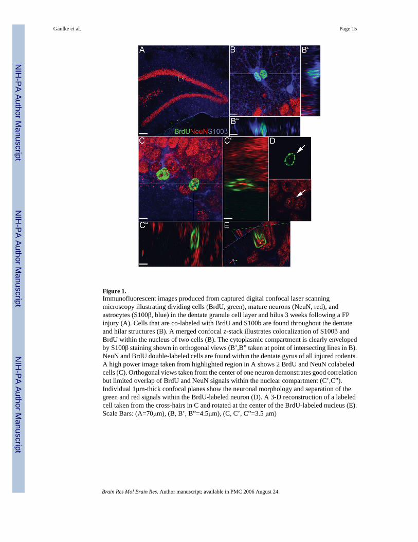
Figure 1.Immunofluorescent images produced from captured digital confocal laser scanningmicroscopy illustrating dividing cells (BrdU, green), mature neurons (NeuN, red), andastrocytes (S100β, blue) in the dentate granule cell layer and hilus 3 weeks following a FPinjury (A). Cells that are co-labeled with BrdU and S100b are found throughout the dentateand hilar structures (B). A merged confocal z-stack illustrates colocalization of S100β andBrdU within the nucleus of two cells (B). The cytoplasmic compartment is clearly envelopedby S100β staining shown in orthogonal views (B’,B” taken at point of intersecting lines in B).NeuN and BrdU double-labeled cells are found within the dentate gyrus of all injured rodents.A high power image taken from highlighted region in A shows 2 BrdU and NeuN colabeledcells (C). Orthogonal views taken from the center of one neuron demonstrates good correlationbut limited overlap of BrdU and NeuN signals within the nuclear compartment (C’,C”).Individual 1μm-thick confocal planes show the neuronal morphology and separation of thegreen and red signals within the BrdU-labeled neuron (D). A 3-D reconstruction of a labeledcell taken from the cross-hairs in C and rotated at the center of the BrdU-labeled nucleus (E).Scale Bars: (A=70μm), (B, B’, B”=4.5μm), (C, C’, C”=3.5 μm)
Gaulke et al. Page 15
Brain Res Mol Brain Res. Author manuscript; available in PMC 2006 August 24.
NIH
-PA Author Manuscript
NIH
-PA Author Manuscript
NIH
-PA Author Manuscript
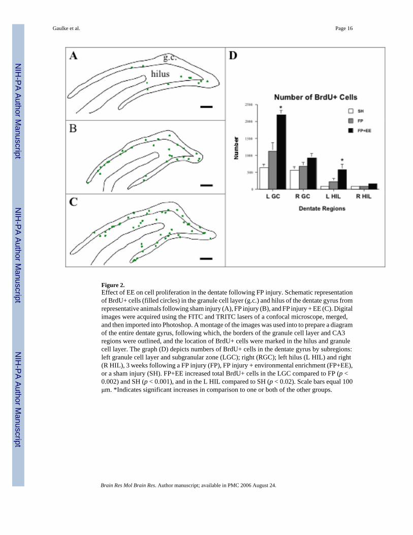
Figure 2.Effect of EE on cell proliferation in the dentate following FP injury. Schematic representationof BrdU+ cells (filled circles) in the granule cell layer (g.c.) and hilus of the dentate gyrus fromrepresentative animals following sham injury (A), FP injury (B), and FP injury + EE (C). Digitalimages were acquired using the FITC and TRITC lasers of a confocal microscope, merged,and then imported into Photoshop. A montage of the images was used into to prepare a diagramof the entire dentate gyrus, following which, the borders of the granule cell layer and CA3regions were outlined, and the location of BrdU+ cells were marked in the hilus and granulecell layer. The graph (D) depicts numbers of BrdU+ cells in the dentate gyrus by subregions:left granule cell layer and subgranular zone (LGC); right (RGC); left hilus (L HIL) and right(R HIL), 3 weeks following a FP injury (FP), FP injury + environmental enrichment (FP+EE),or a sham injury (SH). FP+EE increased total BrdU+ cells in the LGC compared to FP (p <0.002) and SH (p < 0.001), and in the L HIL compared to SH (p < 0.02). Scale bars equal 100μm. *Indicates significant increases in comparison to one or both of the other groups.
Gaulke et al. Page 16
Brain Res Mol Brain Res. Author manuscript; available in PMC 2006 August 24.
NIH
-PA Author Manuscript
NIH
-PA Author Manuscript
NIH
-PA Author Manuscript
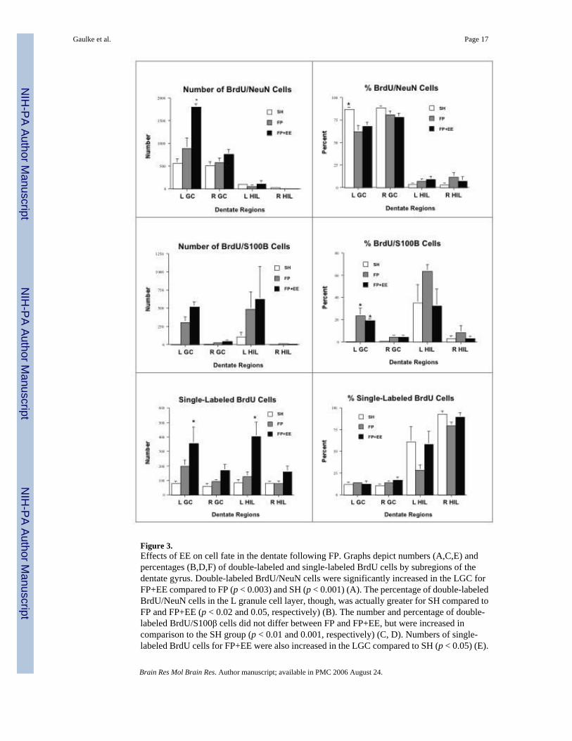
Figure 3.Effects of EE on cell fate in the dentate following FP. Graphs depict numbers (A,C,E) andpercentages (B,D,F) of double-labeled and single-labeled BrdU cells by subregions of thedentate gyrus. Double-labeled BrdU/NeuN cells were significantly increased in the LGC forFP+EE compared to FP (p < 0.003) and SH (p < 0.001) (A). The percentage of double-labeledBrdU/NeuN cells in the L granule cell layer, though, was actually greater for SH compared toFP and FP+EE (p < 0.02 and 0.05, respectively) (B). The number and percentage of double-labeled BrdU/S100β cells did not differ between FP and FP+EE, but were increased incomparison to the SH group (p < 0.01 and 0.001, respectively) (C, D). Numbers of single-labeled BrdU cells for FP+EE were also increased in the LGC compared to SH (p < 0.05) (E).
Gaulke et al. Page 17
Brain Res Mol Brain Res. Author manuscript; available in PMC 2006 August 24.
NIH
-PA Author Manuscript
NIH
-PA Author Manuscript
NIH
-PA Author Manuscript
In the L hilus, there was a significant increase in single-labeled BrdU cells compared to FP(p < 0.03) and to SH (p < 0.01) (E). *Indicates significant increases in comparison to one orboth of the other groups.
Gaulke et al. Page 18
Brain Res Mol Brain Res. Author manuscript; available in PMC 2006 August 24.
NIH
-PA Author Manuscript
NIH
-PA Author Manuscript
NIH
-PA Author Manuscript

Figure 4.Effects of rostral-caudal level on cell proliferation and fate following FP or FP+EE comparedto SH. Graphs depict numbers of single- and double-labeled BrdU cells at various bregmalevels for the left granule cell layer (A, B) or hilus (C, D) for each group. A repeated measuresANOVA demonstrated that in the left granule cell layer and hilus the number of BrdU/Sl00Bcells varied significantly by bregma level (p < 0.002). In contrast to the effects of rostral-caudallocation on gliogenesis, no differences were observed for neurogenesis (B) or single-labeledcells (D).
Gaulke et al. Page 19
Brain Res Mol Brain Res. Author manuscript; available in PMC 2006 August 24.
NIH
-PA Author Manuscript
NIH
-PA Author Manuscript
NIH
-PA Author Manuscript
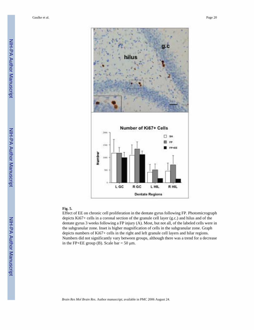
Fig. 5.Effect of EE on chronic cell proliferation in the dentate gyrus following FP. Photomicrographdepicts Ki67+ cells in a coronal section of the granule cell layer (g.c.) and hilus and of thedentate gyrus 3 weeks following a FP injury (A). Most, but not all, of the labeled cells were inthe subgranular zone. Inset is higher magnification of cells in the subgranular zone. Graphdepicts numbers of Ki67+ cells in the right and left granule cell layers and hilar regions.Numbers did not significantly vary between groups, although there was a trend for a decreasein the FP+EE group (B). Scale bar = 50 μm.
Gaulke et al. Page 20
Brain Res Mol Brain Res. Author manuscript; available in PMC 2006 August 24.
NIH
-PA Author Manuscript
NIH
-PA Author Manuscript
NIH
-PA Author Manuscript
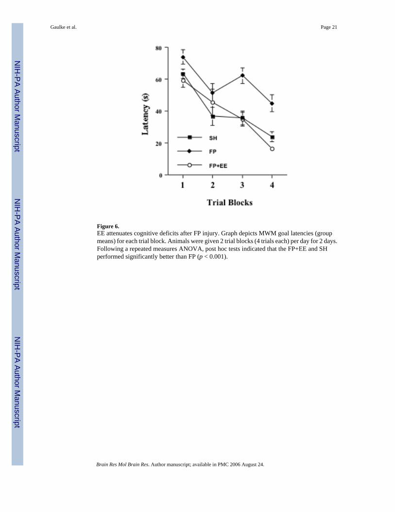
Figure 6.EE attenuates cognitive deficits after FP injury. Graph depicts MWM goal latencies (groupmeans) for each trial block. Animals were given 2 trial blocks (4 trials each) per day for 2 days.Following a repeated measures ANOVA, post hoc tests indicated that the FP+EE and SHperformed significantly better than FP (p < 0.001).
Gaulke et al. Page 21
Brain Res Mol Brain Res. Author manuscript; available in PMC 2006 August 24.
NIH
-PA Author Manuscript
NIH
-PA Author Manuscript
NIH
-PA Author Manuscript