Colchicine-Induced Polyploidy in Rhododendron fortunei Lindl
13
plants Article Colchicine-Induced Polyploidy in Rhododendron fortunei Lindl Lan Mo, Junhao Chen, Xiongzhen Lou, Qiangwei Xu, Renhui Dong, Zaikang Tong, Huahong Huang and Erpei Lin * State Key Laboratory of Subtropical Silviculture, Zhejiang A&F University, Hangzhou 311300, Zhejiang, China; [email protected] (L.M.); [email protected] (J.C.); [email protected] (X.L.); [email protected] (Q.X.); [email protected] (R.D.); [email protected] (Z.T.); [email protected] (H.H.) * Correspondence: [email protected] Received: 13 February 2020; Accepted: 23 March 2020; Published: 31 March 2020 Abstract: Polyploidy in Rhododendron fortunei has great potential to improve its horticultural and commercial value, and to also meet market demands. In this study, a feasible method for polyploid induction in R. fortunei via colchicine treatment was established, and the obtained polyploid plants were identified and characterized. As a result, the stem bases of tissue-cultured plantlets treated with 0.1% colchicine for 24 h showed the highest polyploid induction with a rate of 36.67%. By flow cytometric analysis, 69 tetraploids and 29 octoploids were identified in the regenerated plants that were examined. Phenotypic analysis indicated that the leaves of tetraploid and octoploid plants were smaller, rounder and thicker with more abundant and longer epidermal hairs than those of diploids. Furthermore, the stomata of polyploids were larger and sparser than those of diploids. An increase in chlorophyll content was also detected in polyploids, which resulted in darker green leaves. In conclusion, our study established an effective method to induce polyploidy in R. fortunei, which could be used to develop new genetic resources for breeding R. fortunei and other Rhododendron species in the future. Keywords: polyploid; tetraploidy; plant stomata; flow cytometry 1. Introduction Rhododendron fortunei Lindl, one of the alpine rhododendrons, is an evergreen small tree or shrub of genus Rhododendron, and mainly grows at an altitude of 620 to 2000 meters in Southern China [1]. Owing to the beautiful and bright color of its flowers, and their elegant shape and charming aroma, R. fortunei has a high ornamental value and great commercial potential in landscaping and horticultural markets [2]. However, the use of R. fortunei in landscaping and horticultural markets is severely limited by its slow growth rate and reliance on an alpine climate. Therefore, its environmental adaption and horticultural traits need to be improved in order to advance the development and utilization of R. fortunei. Naturally occurring polyploidy is widespread in the plant kingdom, and polyploidization is often responsible for the generation of new species during plant evolution [3]. Compared with their diploid relatives, polyploid plants often exhibit advantageous features, including greater adaptability and resistance, and they have giant flowers, fruits, and other prominent features [4–6]. To exploit these advantages, artificial methods of inducing polyploidy have become an important tool for plant breeding and they have been widely applied in the breeding programs of crops, vegetables and flowers [7]. Herbicides, such as colchicine, trifluralin and oryzalin, are chemicals that are frequently used to induce polyploidy in plants, as they effectively arrest mitosis at the anaphase stage [8,9]. Polyploids of several Rhododendron species have been successfully induced using these herbicides, resulting in varieties with Plants 2020, 9, 424; doi:10.3390/plants9040424 www.mdpi.com/journal/plants
-
Upload
khangminh22 -
Category
Documents
-
view
3 -
download
0
Transcript of Colchicine-Induced Polyploidy in Rhododendron fortunei Lindl

plants
Article
Colchicine-Induced Polyploidy in Rhododendronfortunei Lindl
Lan Mo, Junhao Chen, Xiongzhen Lou, Qiangwei Xu, Renhui Dong, Zaikang Tong,Huahong Huang and Erpei Lin *
State Key Laboratory of Subtropical Silviculture, Zhejiang A&F University, Hangzhou 311300, Zhejiang, China;[email protected] (L.M.); [email protected] (J.C.); [email protected] (X.L.);[email protected] (Q.X.); [email protected] (R.D.); [email protected] (Z.T.);[email protected] (H.H.)* Correspondence: [email protected]
Received: 13 February 2020; Accepted: 23 March 2020; Published: 31 March 2020�����������������
Abstract: Polyploidy in Rhododendron fortunei has great potential to improve its horticultural andcommercial value, and to also meet market demands. In this study, a feasible method for polyploidinduction in R. fortunei via colchicine treatment was established, and the obtained polyploid plantswere identified and characterized. As a result, the stem bases of tissue-cultured plantlets treatedwith 0.1% colchicine for 24 h showed the highest polyploid induction with a rate of 36.67%. By flowcytometric analysis, 69 tetraploids and 29 octoploids were identified in the regenerated plants thatwere examined. Phenotypic analysis indicated that the leaves of tetraploid and octoploid plantswere smaller, rounder and thicker with more abundant and longer epidermal hairs than those ofdiploids. Furthermore, the stomata of polyploids were larger and sparser than those of diploids.An increase in chlorophyll content was also detected in polyploids, which resulted in darker greenleaves. In conclusion, our study established an effective method to induce polyploidy in R. fortunei,which could be used to develop new genetic resources for breeding R. fortunei and other Rhododendronspecies in the future.
Keywords: polyploid; tetraploidy; plant stomata; flow cytometry
1. Introduction
Rhododendron fortunei Lindl, one of the alpine rhododendrons, is an evergreen small tree or shrubof genus Rhododendron, and mainly grows at an altitude of 620 to 2000 meters in Southern China [1].Owing to the beautiful and bright color of its flowers, and their elegant shape and charming aroma,R. fortunei has a high ornamental value and great commercial potential in landscaping and horticulturalmarkets [2]. However, the use of R. fortunei in landscaping and horticultural markets is severely limitedby its slow growth rate and reliance on an alpine climate. Therefore, its environmental adaption andhorticultural traits need to be improved in order to advance the development and utilization of R. fortunei.
Naturally occurring polyploidy is widespread in the plant kingdom, and polyploidization is oftenresponsible for the generation of new species during plant evolution [3]. Compared with their diploidrelatives, polyploid plants often exhibit advantageous features, including greater adaptability andresistance, and they have giant flowers, fruits, and other prominent features [4–6]. To exploit theseadvantages, artificial methods of inducing polyploidy have become an important tool for plant breedingand they have been widely applied in the breeding programs of crops, vegetables and flowers [7].Herbicides, such as colchicine, trifluralin and oryzalin, are chemicals that are frequently used to inducepolyploidy in plants, as they effectively arrest mitosis at the anaphase stage [8,9]. Polyploids of severalRhododendron species have been successfully induced using these herbicides, resulting in varieties with
Plants 2020, 9, 424; doi:10.3390/plants9040424 www.mdpi.com/journal/plants
Plants 2020, 9, 424 2 of 13
improved ornamental value that provide new genetic resources for crossbreeding [10–12]. However,at present, there are no reported instances of polyploidy induction in R. fortunei species. Therefore, it isimportant to establish a polyploidy induction method for generating new breeding resources, whichshould help to improve the horticultural traits of R. fortunei.
The aim of our study was to establish a highly efficient method for polyploidy induction inR. fortunei. The shoot apex and stem bases of aseptic tissue-cultured plantlets were used as explants, andcolchicine treatments of varying concentration and time were assessed. The ploidy level of regeneratedplants was estimated by flow cytometry, and the morphological and physiological characteristics ofpolyploids were also determined.
2. Results
2.1. Effects of Colchicine Treatment on Explants’ Survival
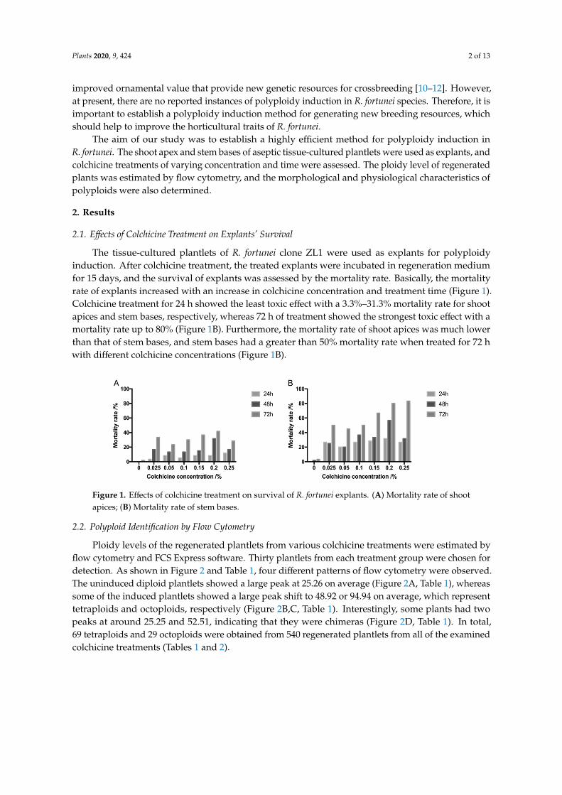
The tissue-cultured plantlets of R. fortunei clone ZL1 were used as explants for polyploidyinduction. After colchicine treatment, the treated explants were incubated in regeneration mediumfor 15 days, and the survival of explants was assessed by the mortality rate. Basically, the mortalityrate of explants increased with an increase in colchicine concentration and treatment time (Figure 1).Colchicine treatment for 24 h showed the least toxic effect with a 3.3%–31.3% mortality rate for shootapices and stem bases, respectively, whereas 72 h of treatment showed the strongest toxic effect with amortality rate up to 80% (Figure 1B). Furthermore, the mortality rate of shoot apices was much lowerthan that of stem bases, and stem bases had a greater than 50% mortality rate when treated for 72 hwith different colchicine concentrations (Figure 1B).
Plants 2020, 9, x FOR PEER REVIEW 2 of 13
important to establish a polyploidy induction method for generating new breeding resources, which should help to improve the horticultural traits of R. fortunei.
The aim of our study was to establish a highly efficient method for polyploidy induction in R. fortunei. The shoot apex and stem bases of aseptic tissue-cultured plantlets were used as explants, and colchicine treatments of varying concentration and time were assessed. The ploidy level of regenerated plants was estimated by flow cytometry, and the morphological and physiological characteristics of polyploids were also determined.
2. Results
2.1. Effects of Colchicine Treatment on Explants’ Survival
The tissue-cultured plantlets of R. fortunei clone ZL1 were used as explants for polyploidy induction. After colchicine treatment, the treated explants were incubated in regeneration medium for 15 days, and the survival of explants was assessed by the mortality rate. Basically, the mortality rate of explants increased with an increase in colchicine concentration and treatment time (Figure 1). Colchicine treatment for 24 h showed the least toxic effect with a 3.3%–31.3% mortality rate for shoot apices and stem bases, respectively, whereas 72 h of treatment showed the strongest toxic effect with a mortality rate up to 80% (Figure 1B). Furthermore, the mortality rate of shoot apices was much lower than that of stem bases, and stem bases had a greater than 50% mortality rate when treated for 72 h with different colchicine concentrations (Figure 1B).
Figure 1. Effects of colchicine treatment on survival of R. fortunei explants. (A) Mortality rate of shoot apices; (B) Mortality rate of stem bases.
2.2. Polyploid Identification by Flow Cytometry
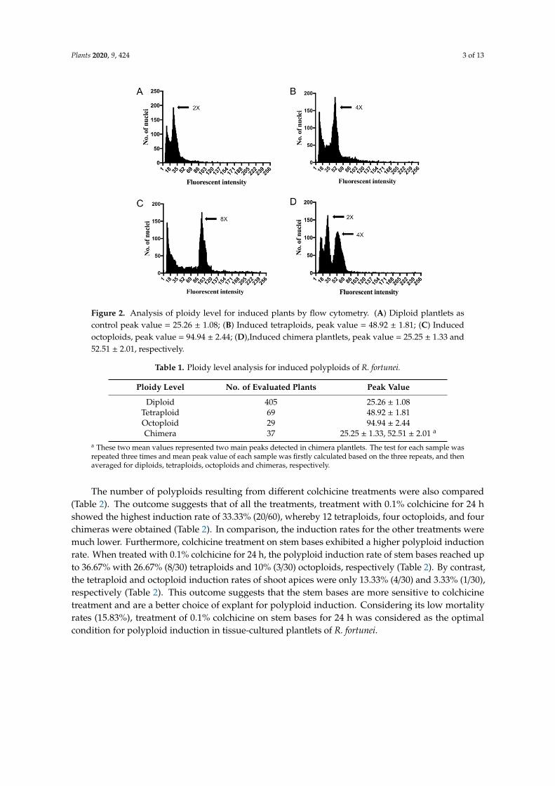
Ploidy levels of the regenerated plantlets from various colchicine treatments were estimated by flow cytometry and FCS Express software. Thirty plantlets from each treatment group were chosen for detection. As shown in Figure 2 and Table 1, four different patterns of flow cytometry were observed. The uninduced diploid plantlets showed a large peak at 25.26 on average (Figure 2A, Table 1), whereas some of the induced plantlets showed a large peak shift to 48.92 or 94.94 on average, which represent tetraploids and octoploids, respectively (Figure 2B,C, Table 1). Interestingly, some plants had two peaks at around 25.25 and 52.51, indicating that they were chimeras (Figure 2D, Table 1). In total, 69 tetraploids and 29 octoploids were obtained from 540 regenerated plantlets from all of the examined colchicine treatments (Tables 1 and 2).
Figure 1. Effects of colchicine treatment on survival of R. fortunei explants. (A) Mortality rate of shootapices; (B) Mortality rate of stem bases.
2.2. Polyploid Identification by Flow Cytometry
Ploidy levels of the regenerated plantlets from various colchicine treatments were estimated byflow cytometry and FCS Express software. Thirty plantlets from each treatment group were chosen fordetection. As shown in Figure 2 and Table 1, four different patterns of flow cytometry were observed.The uninduced diploid plantlets showed a large peak at 25.26 on average (Figure 2A, Table 1), whereassome of the induced plantlets showed a large peak shift to 48.92 or 94.94 on average, which representtetraploids and octoploids, respectively (Figure 2B,C, Table 1). Interestingly, some plants had twopeaks at around 25.25 and 52.51, indicating that they were chimeras (Figure 2D, Table 1). In total,69 tetraploids and 29 octoploids were obtained from 540 regenerated plantlets from all of the examinedcolchicine treatments (Tables 1 and 2).
Plants 2020, 9, 424 3 of 13Plants 2020, 9, x FOR PEER REVIEW 3 of 13
Figure 2. Analysis of ploidy level for induced plants by flow cytometry. (A) Diploid plantlets as control peak value = 25.26 ± 1.08; (B) Induced tetraploids, peak value = 48.92 ± 1.81; (C) Induced octoploids, peak value = 94.94 ± 2.44; (D),Induced chimera plantlets, peak value = 25.25 ± 1.33 and 52.51 ± 2.01, respectively.
Table 1. Ploidy level analysis for induced polyploids of R. fortunei.
Ploidy Level No. of Evaluated Plants Peak Value
Diploid 405 25.26 ± 1.08
Tetraploid 69 48.92 ± 1.81
Octoploid 29 94.94 ± 2.44
Chimera 37 25.25 ± 1.33, 52.51 ± 2.01 a a These two mean values represented two main peaks detected in chimera plantlets. The test for each sample was repeated three times and mean peak value of each sample was firstly calculated based on the three repeats, and then averaged for diploids, tetraploids, octoploids and chimeras, respectively.
The number of polyploids resulting from different colchicine treatments were also compared (Table 2). The outcome suggests that of all the treatments, treatment with 0.1% colchicine for 24 h showed the highest induction rate of 33.33% (20/60), whereby 12 tetraploids, four octoploids, and four chimeras were obtained (Table 2). In comparison, the induction rates for the other treatments were much lower. Furthermore, colchicine treatment on stem bases exhibited a higher polyploid induction rate. When treated with 0.1% colchicine for 24 h, the polyploid induction rate of stem bases reached up to 36.67% with 26.67% (8/30) tetraploids and 10% (3/30) octoploids, respectively (Table 2). By contrast, the tetraploid and octoploid induction rates of shoot apices were only 13.33% (4/30) and 3.33% (1/30), respectively (Table 2). This outcome suggests that the stem bases are more sensitive to colchicine treatment and are a better choice of explant for polyploid induction. Considering its low mortality rates (15.83%), treatment of 0.1% colchicine on stem bases for 24 h was considered as the optimal condition for polyploid induction in tissue-cultured plantlets of R. fortunei.
Figure 2. Analysis of ploidy level for induced plants by flow cytometry. (A) Diploid plantlets ascontrol peak value = 25.26 ± 1.08; (B) Induced tetraploids, peak value = 48.92 ± 1.81; (C) Inducedoctoploids, peak value = 94.94 ± 2.44; (D),Induced chimera plantlets, peak value = 25.25 ± 1.33 and52.51 ± 2.01, respectively.
Table 1. Ploidy level analysis for induced polyploids of R. fortunei.
Ploidy Level No. of Evaluated Plants Peak Value
Diploid 405 25.26 ± 1.08Tetraploid 69 48.92 ± 1.81Octoploid 29 94.94 ± 2.44Chimera 37 25.25 ± 1.33, 52.51 ± 2.01 a
a These two mean values represented two main peaks detected in chimera plantlets. The test for each sample wasrepeated three times and mean peak value of each sample was firstly calculated based on the three repeats, and thenaveraged for diploids, tetraploids, octoploids and chimeras, respectively.
The number of polyploids resulting from different colchicine treatments were also compared(Table 2). The outcome suggests that of all the treatments, treatment with 0.1% colchicine for 24 hshowed the highest induction rate of 33.33% (20/60), whereby 12 tetraploids, four octoploids, and fourchimeras were obtained (Table 2). In comparison, the induction rates for the other treatments weremuch lower. Furthermore, colchicine treatment on stem bases exhibited a higher polyploid inductionrate. When treated with 0.1% colchicine for 24 h, the polyploid induction rate of stem bases reached upto 36.67% with 26.67% (8/30) tetraploids and 10% (3/30) octoploids, respectively (Table 2). By contrast,the tetraploid and octoploid induction rates of shoot apices were only 13.33% (4/30) and 3.33% (1/30),respectively (Table 2). This outcome suggests that the stem bases are more sensitive to colchicinetreatment and are a better choice of explant for polyploid induction. Considering its low mortalityrates (15.83%), treatment of 0.1% colchicine on stem bases for 24 h was considered as the optimalcondition for polyploid induction in tissue-cultured plantlets of R. fortunei.
Plants 2020, 9, 424 4 of 13
Table 2. Comparison of the number of polyploid induction from different colchicine treatments.
ColchicineConcentration
/%
TreatmentTime /h
TetraploidsFrequency
OctoploidsFrequency
ChimerasFrequency
PolyploidsFrequency
ShootApex
StemBase
ShootApex
StemBase
ShootApex
StemBase
ShootApex
StemBase
0 24 0/30 0/30 0/30 0/30 0/30 0/30 0/30 0/3048 0/30 0/30 0/30 0/30 0/30 0/30 0/30 0/3072 0/30 0/30 0/30 0/30 0/30 0/30 0/30 0/30
0.025 24 0/30 0/30 0/30 0/30 0/30 0/30 0/30 0/3048 0/30 0/30 0/30 0/30 0/30 0/30 0/30 0/3072 0/30 0/30 0/30 0/30 0/30 0/30 0/30 0/30
0.05 24 1/30 3/30 0/30 0/30 0/30 0/30 1/30 3/3048 1/30 3/30 0/30 2/30 1/30 2/30 2/30 7/3072 1/30 4/30 1/30 2/30 1/30 1/30 3/30 7/30
0.1 24 4/30 8/30 1/30 3/30 2/30 2/30 7/30 13/3048 1/30 4/30 0/30 1/30 2/30 1/30 3/30 6/3072 2/30 3/30 1/30 2/30 1/30 2/30 4/30 7/30
0.15 24 1/30 3/30 1/30 3/30 0/30 0/30 2/30 6/3048 1/30 4/30 0/30 2/30 1/30 3/30 2/30 9/3072 2/30 2/30 0/30 1/30 1/30 2/30 3/30 5/30
0.2 24 1/30 2/30 0/30 1/30 2/30 3/30 3/30 6/3048 2/30 3/30 1/30 1/30 1/30 2/30 4/30 6/3072 2/30 2/30 0/30 0/30 1/30 1/30 3/30 3/30
0.25 24 1/30 3/30 0/30 2/30 0/30 0/30 1/30 5/3048 1/30 2/30 0/30 1/30 2/30 1/30 3/30 4/3072 1/30 1/30 1/30 2/30 1/30 1/30 3/30 4/30
Total 22/540 47/540 6/540 23/540 16/540 21/540 44/540 91/540
2.3. Morphological Characteristics of R. fortunei Polyploids
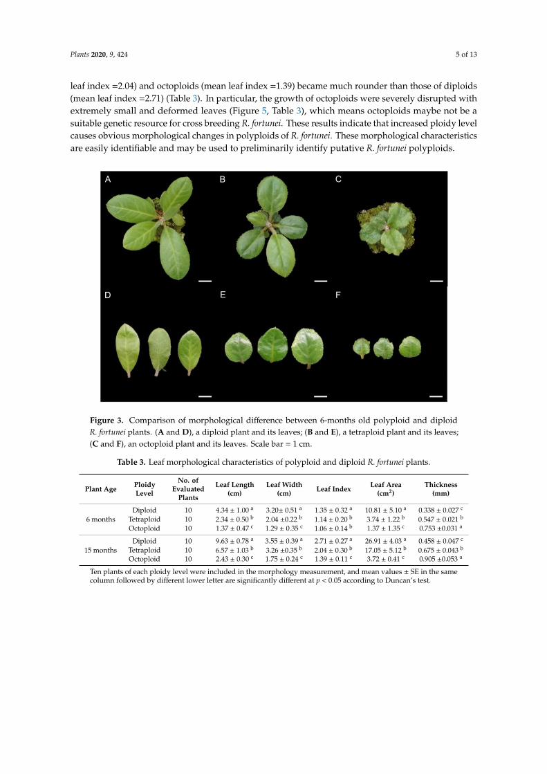
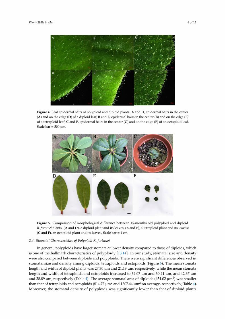
After the ploidy levels were analyzed, plantlets with different ploidy levels were transplanted to agreenhouse and grown for a further six months. The morphological characteristics of these six-monthsold plants were investigated and diploid and polyploid plants were compared. As shown in Figure 3,the leaves of tetraploids and octoploids were considerably rounder, harder and thicker with a slightlyrough surface and darker color compared to those of the diploids. For the diploid plants, the meanleaf length was 4.34 cm with a mean leaf width of 3.20 cm and mean leaf area of 10.81 cm2 (Table 3).For tetraploid and octoploid plants, the mean leaf length was 2.34 cm and 1.37 cm, the mean leaf widthwas 2.04 cm and 1.29 cm, and the mean leaf area was 3.74 cm2 and 1.37 cm2, respectively (Table 3). Thisshowed that that the polyploid leaves were dramatically smaller than the diploid leaves. In addition,the leaves of diploid plants showed an elliptical shape with a mean leaf index of 1.35, whereas theleaves of tetraploids and octoploids were much rounder with a mean leaf index of 1.14 and 1.06,respectively, which are very close to 1.00 (Table 3). Additionally, the thickness of leaves increased withploidy level, the mean thickness of octoploids leaves was 0.753 mm, which was much thicker than that oftetraploids (0.574 mm) and diploids (0.338 mm) (Table 3). Leaf epidermal hairs were also observed underthe stereoscope. As shown in Figure 4, denser and longer epidermal hairs were observed on leaf surfacesand edges of octoploids and tetraploids, which meant they had a rougher leaf surface than diploids.
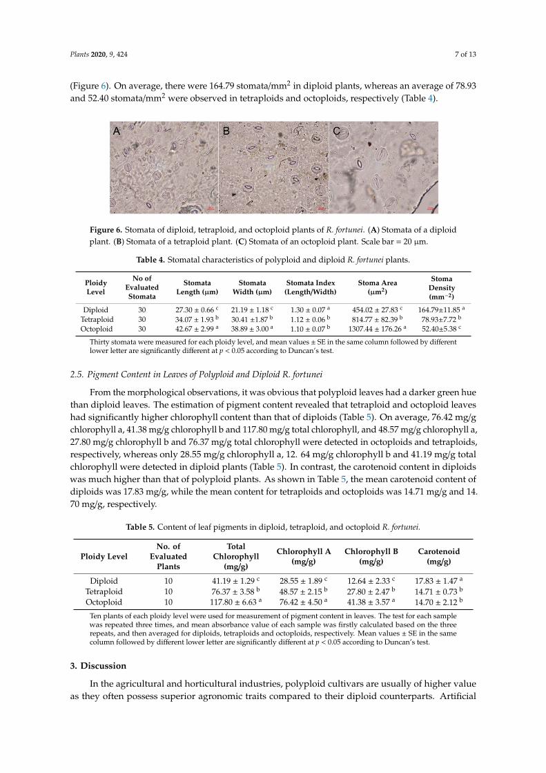
Furthermore, to evaluate the stability in the morphology of these polyploid plants, the leafcharacteristics of 15-months old plants were also investigated and compared. As a result, significantdifferences were also observed between diploids and polyploids (Figure 5). Compared with diploids,the polyploid plants still exhibited rounder, smaller and thicker leaves (Figure 5, Table 3). The mean leafarea of diploid plants was 26.91 cm2, while that of tetraploids and octoploids were only 17.05 cm2 and3.72 cm2, respectively (Table 3). The average leaf length and width of diploid plants were 9.63 cm and3.55 cm, while that of tetraploids and octoploids were 6.57 cm and 3.26 cm, and 2.43 cm and 1.75 cm,respectively (Table 3). The average leaf index value also indicated that the leaves of tetraploids (mean
Plants 2020, 9, 424 5 of 13
leaf index =2.04) and octoploids (mean leaf index =1.39) became much rounder than those of diploids(mean leaf index =2.71) (Table 3). In particular, the growth of octoploids were severely disrupted withextremely small and deformed leaves (Figure 5, Table 3), which means octoploids maybe not be asuitable genetic resource for cross breeding R. fortunei. These results indicate that increased ploidy levelcauses obvious morphological changes in polyploids of R. fortunei. These morphological characteristicsare easily identifiable and may be used to preliminarily identify putative R. fortunei polyploids.
Plants 2020, 9, x FOR PEER REVIEW 5 of 13
Furthermore, to evaluate the stability in the morphology of these polyploid plants, the leaf characteristics of 15-months old plants were also investigated and compared. As a result, significant differences were also observed between diploids and polyploids (Figure 5). Compared with diploids, the polyploid plants still exhibited rounder, smaller and thicker leaves (Figure 5, Table 3). The mean leaf area of diploid plants was 26.91 cm2, while that of tetraploids and octoploids were only 17.05 cm2 and 3.72 cm2, respectively (Table 3). The average leaf length and width of diploid plants were 9.63 cm and 3.55 cm, while that of tetraploids and octoploids were 6.57 cm and 3.26 cm, and 2.43 cm and 1.75 cm, respectively (Table 3). The average leaf index value also indicated that the leaves of tetraploids (mean leaf index =2.04) and octoploids (mean leaf index =1.39) became much rounder than those of diploids (mean leaf index =2.71) (Table 3). In particular, the growth of octoploids were severely disrupted with extremely small and deformed leaves (Figure 5, Table 3), which means octoploids maybe not be a suitable genetic resource for cross breeding R. fortunei. These results indicate that increased ploidy level causes obvious morphological changes in polyploids of R. fortunei. These morphological characteristics are easily identifiable and may be used to preliminarily identify putative R. fortunei polyploids.
Figure 3. Comparison of morphological difference between 6-months old polyploid and diploid R. fortunei plants. (A and D), a diploid plant and its leaves; (B and E), a tetraploid plant and its leaves; (C and F), an octoploid plant and its leaves. Scale bar = 1 cm.
Table 3. Leaf morphological characteristics of polyploid and diploid R. fortunei plants.
Plant Age Ploidy
Level
No. of
Evaluated
Plants
Leaf Length
(cm)
Leaf Width
(cm) Leaf Index
Leaf Area
(cm2)
Thickness
(mm)
6 months
Diploid 10 4.34 ± 1.00 a 3.20± 0.51 a 1.35 ± 0.32 a 10.81 ± 5.10 a 0.338 ± 0.027 c
Tetraploid 10 2.34 ± 0.50 b 2.04 ±0.22 b 1.14 ± 0.20 b 3.74 ± 1.22 b 0.547 ± 0.021 b
Octoploid 10 1.37 ± 0.47 c 1.29 ± 0.35 c 1.06 ± 0.14 b 1.37 ± 1.35 c 0.753 ±0.031 a
15 months
Diploid 10 9.63 ± 0.78 a 3.55 ± 0.39 a 2.71 ± 0.27 a 26.91 ± 4.03 a 0.458 ± 0.047 c
Tetraploid 10 6.57 ± 1.03 b 3.26 ±0.35 b 2.04 ± 0.30 b 17.05 ± 5.12 b 0.675 ± 0.043 b
Octoploid 10 2.43 ± 0.30 c 1.75 ± 0.24 c 1.39 ± 0.11 c 3.72 ± 0.41 c 0.905 ±0.053 a Ten plants of each ploidy level were included in the morphology measurement, and mean values ± SE in the same column followed by different lower letter are significantly different at p < 0.05 according to Duncan’s test.
Figure 3. Comparison of morphological difference between 6-months old polyploid and diploidR. fortunei plants. (A and D), a diploid plant and its leaves; (B and E), a tetraploid plant and its leaves;(C and F), an octoploid plant and its leaves. Scale bar = 1 cm.
Table 3. Leaf morphological characteristics of polyploid and diploid R. fortunei plants.
Plant Age PloidyLevel
No. ofEvaluated
Plants
Leaf Length(cm)
Leaf Width(cm) Leaf Index Leaf Area
(cm2)Thickness
(mm)
6 monthsDiploid 10 4.34 ± 1.00 a 3.20± 0.51 a 1.35 ± 0.32 a 10.81 ± 5.10 a 0.338 ± 0.027 c
Tetraploid 10 2.34 ± 0.50 b 2.04 ±0.22 b 1.14 ± 0.20 b 3.74 ± 1.22 b 0.547 ± 0.021 b
Octoploid 10 1.37 ± 0.47 c 1.29 ± 0.35 c 1.06 ± 0.14 b 1.37 ± 1.35 c 0.753 ±0.031 a
15 monthsDiploid 10 9.63 ± 0.78 a 3.55 ± 0.39 a 2.71 ± 0.27 a 26.91 ± 4.03 a 0.458 ± 0.047 c
Tetraploid 10 6.57 ± 1.03 b 3.26 ±0.35 b 2.04 ± 0.30 b 17.05 ± 5.12 b 0.675 ± 0.043 b
Octoploid 10 2.43 ± 0.30 c 1.75 ± 0.24 c 1.39 ± 0.11 c 3.72 ± 0.41 c 0.905 ±0.053 a
Ten plants of each ploidy level were included in the morphology measurement, and mean values ± SE in the samecolumn followed by different lower letter are significantly different at p < 0.05 according to Duncan’s test.
Plants 2020, 9, 424 6 of 13Plants 2020, 9, x FOR PEER REVIEW 6 of 13
Figure 4. Leaf epidermal hairs of polyploid and diploid plants. A and D, epidermal hairs in the center (A) and on the edge (D) of a diploid leaf; B and E, epidermal hairs in the center (B) and on the edge (E) of a tetraploid leaf; C and F, epidermal hairs in the center (C) and on the edge (F) of an octoploid leaf. Scale bar = 500 μm.
Figure 5. Comparison of morphological difference between 15-months old polyploid and diploid R. fortunei plants. (A and D), a diploid plant and its leaves; (B and E), a tetraploid plant and its leaves; (C and F), an octoploid plant and its leaves. Scale bar = 1 cm.
2.4. Stomatal Characteristics of Polyploid R. fortunei
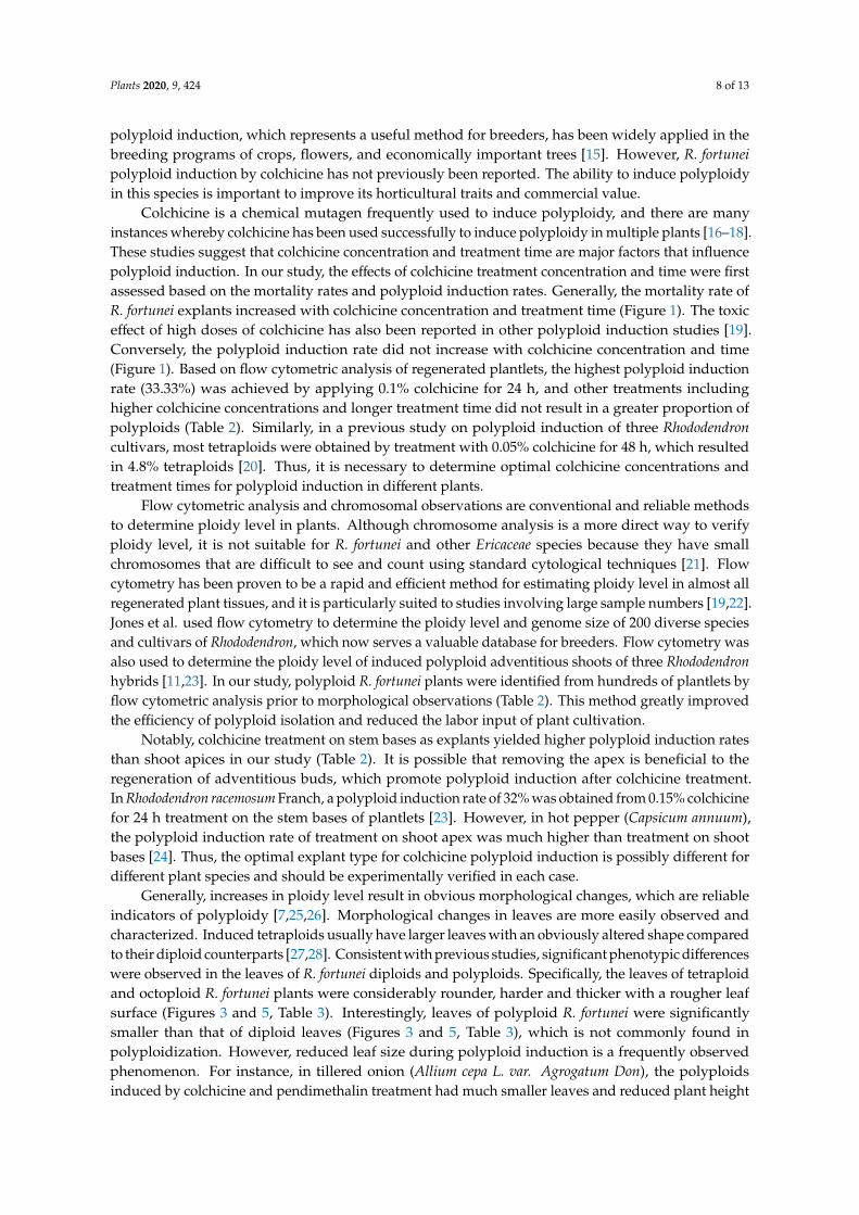
In general, polyploids have larger stomata at lower density compared to those of diploids, which is one of the hallmark characteristics of polyploidy [13,14]. In our study, stomatal size and density were also compared between diploids and polyploids. There were significant differences observed in stomatal size and density among diploids, tetraploids and octoploids (Figure 6). The mean stomata length and width of diploid plants was 27.30 μm and 21.19 μm, respectively, while the mean stomata length and width of tetraploids and octoploids increased to 34.07 μm and 30.41 μm, and 42.67 μm and 38.89 μm, respectively (Table 4). The average stomatal area of diploids (454.02 μm2) was smaller than that of tetraploids and octoploids (814.77 μm2 and 1307.44 μm2 on average, respectively; Table 4). Moreover, the stomatal density of polyploids was significantly lower than that of diploid plants (Figure 6). On average, there were 164.79
Figure 4. Leaf epidermal hairs of polyploid and diploid plants. A and D, epidermal hairs in the center(A) and on the edge (D) of a diploid leaf; B and E, epidermal hairs in the center (B) and on the edge (E)of a tetraploid leaf; C and F, epidermal hairs in the center (C) and on the edge (F) of an octoploid leaf.Scale bar = 500 µm.
Plants 2020, 9, x FOR PEER REVIEW 6 of 13
Figure 4. Leaf epidermal hairs of polyploid and diploid plants. A and D, epidermal hairs in the center (A) and on the edge (D) of a diploid leaf; B and E, epidermal hairs in the center (B) and on the edge (E) of a tetraploid leaf; C and F, epidermal hairs in the center (C) and on the edge (F) of an octoploid leaf. Scale bar = 500 μm.
Figure 5. Comparison of morphological difference between 15-months old polyploid and diploid R. fortunei plants. (A and D), a diploid plant and its leaves; (B and E), a tetraploid plant and its leaves; (C and F), an octoploid plant and its leaves. Scale bar = 1 cm.
2.4. Stomatal Characteristics of Polyploid R. fortunei
In general, polyploids have larger stomata at lower density compared to those of diploids, which is one of the hallmark characteristics of polyploidy [13,14]. In our study, stomatal size and density were also compared between diploids and polyploids. There were significant differences observed in stomatal size and density among diploids, tetraploids and octoploids (Figure 6). The mean stomata length and width of diploid plants was 27.30 μm and 21.19 μm, respectively, while the mean stomata length and width of tetraploids and octoploids increased to 34.07 μm and 30.41 μm, and 42.67 μm and 38.89 μm, respectively (Table 4). The average stomatal area of diploids (454.02 μm2) was smaller than that of tetraploids and octoploids (814.77 μm2 and 1307.44 μm2 on average, respectively; Table 4). Moreover, the stomatal density of polyploids was significantly lower than that of diploid plants (Figure 6). On average, there were 164.79
Figure 5. Comparison of morphological difference between 15-months old polyploid and diploidR. fortunei plants. (A and D), a diploid plant and its leaves; (B and E), a tetraploid plant and its leaves;(C and F), an octoploid plant and its leaves. Scale bar = 1 cm.
2.4. Stomatal Characteristics of Polyploid R. fortunei
In general, polyploids have larger stomata at lower density compared to those of diploids, whichis one of the hallmark characteristics of polyploidy [13,14]. In our study, stomatal size and densitywere also compared between diploids and polyploids. There were significant differences observed instomatal size and density among diploids, tetraploids and octoploids (Figure 6). The mean stomatalength and width of diploid plants was 27.30 µm and 21.19 µm, respectively, while the mean stomatalength and width of tetraploids and octoploids increased to 34.07 µm and 30.41 µm, and 42.67 µmand 38.89 µm, respectively (Table 4). The average stomatal area of diploids (454.02 µm2) was smallerthan that of tetraploids and octoploids (814.77 µm2 and 1307.44 µm2 on average, respectively; Table 4).Moreover, the stomatal density of polyploids was significantly lower than that of diploid plants
Plants 2020, 9, 424 7 of 13
(Figure 6). On average, there were 164.79 stomata/mm2 in diploid plants, whereas an average of 78.93and 52.40 stomata/mm2 were observed in tetraploids and octoploids, respectively (Table 4).
Plants 2020, 9, x FOR PEER REVIEW 7 of 13
stomata/mm2 in diploid plants, whereas an average of 78.93 and 52.40 stomata/mm2 were observed in tetraploids and octoploids, respectively (Table 4).
Figure 6. Stomata of diploid, tetraploid, and octoploid plants of R. fortunei. (A) Stomata of a diploid plant. (B) Stomata of a tetraploid plant. (C) Stomata of an octoploid plant. Scale bar = 20 μm.
Table 4. Stomatal characteristics of polyploid and diploid R. fortunei plants.
Ploidy
Level
No of
Evaluated
Stomata
Stomata
Length (μm)
Stomata
Width (μm)
Stomata Index
(Length/Width) Stoma Area (μm2)
Stoma
Density
(mm−2)
Diploid 30 27.30 ± 0.66 c 21.19 ± 1.18 c 1.30 ± 0.07 a 454.02 ± 27.83 c 164.79±11.85 a
Tetraploid 30 34.07 ± 1.93 b 30.41 ±1.87 b 1.12 ± 0.06 b 814.77 ± 82.39 b 78.93±7.72 b
Octoploid 30 42.67 ± 2.99 a 38.89 ± 3.00 a 1.10 ± 0.07 b 1307.44 ± 176.26 a 52.40±5.38 c Thirty stomata were measured for each ploidy level, and mean values ± SE in the same column followed by different lower letter are significantly different at p < 0.05 according to Duncan’s test.
2.5. Pigment Content in Leaves of Polyploid and Diploid R. fortunei
From the morphological observations, it was obvious that polyploid leaves had a darker green hue than diploid leaves. The estimation of pigment content revealed that tetraploid and octoploid leaves had significantly higher chlorophyll content than that of diploids (Table 5). On average, 76.42 mg/g chlorophyll a, 41.38 mg/g chlorophyll b and 117.80 mg/g total chlorophyll, and 48.57 mg/g chlorophyll a, 27.80 mg/g chlorophyll b and 76.37 mg/g total chlorophyll were detected in octoploids and tetraploids, respectively, whereas only 28.55 mg/g chlorophyll a, 12. 64 mg/g chlorophyll b and 41.19 mg/g total chlorophyll were detected in diploid plants (Table 5). In contrast, the carotenoid content in diploids was much higher than that of polyploid plants. As shown in Table 5, the mean carotenoid content of diploids was 17.83 mg/g, while the mean content for tetraploids and octoploids was 14.71 mg/g and 14. 70 mg/g, respectively.
Table 5. Content of leaf pigments in diploid, tetraploid, and octoploid R. fortunei.
Ploidy
Level
No. of Evaluated
Plants
Total
Chlorophyll
(mg/g)
Chlorophyll A
(mg/g)
Chlorophyll B
(mg/g)
Carotenoid
(mg/g)
Diploid 10 41.19 ± 1.29 c 28.55 ± 1.89 c 12.64 ± 2.33 c 17.83 ± 1.47 a
Tetraploid 10 76.37 ± 3.58 b 48.57 ± 2.15 b 27.80 ± 2.47 b 14.71 ± 0.73 b
Octoploid 10 117.80 ± 6.63 a 76.42 ± 4.50 a 41.38 ± 3.57 a 14.70 ± 2.12 b Ten plants of each ploidy level were used for measurement of pigment content in leaves. The test for each sample was repeated three times, and mean absorbance value of each sample was firstly calculated based on the three repeats, and then averaged for diploids, tetraploids and octoploids, respectively. Mean values ± SE in the same column followed by different lower letter are significantly different at p < 0.05 according to Duncan’s test.
Figure 6. Stomata of diploid, tetraploid, and octoploid plants of R. fortunei. (A) Stomata of a diploidplant. (B) Stomata of a tetraploid plant. (C) Stomata of an octoploid plant. Scale bar = 20 µm.
Table 4. Stomatal characteristics of polyploid and diploid R. fortunei plants.
PloidyLevel
No ofEvaluatedStomata
StomataLength (µm)
StomataWidth (µm)
Stomata Index(Length/Width)
Stoma Area(µm2)
StomaDensity(mm−2)
Diploid 30 27.30 ± 0.66 c 21.19 ± 1.18 c 1.30 ± 0.07 a 454.02 ± 27.83 c 164.79±11.85 a
Tetraploid 30 34.07 ± 1.93 b 30.41 ±1.87 b 1.12 ± 0.06 b 814.77 ± 82.39 b 78.93±7.72 b
Octoploid 30 42.67 ± 2.99 a 38.89 ± 3.00 a 1.10 ± 0.07 b 1307.44 ± 176.26 a 52.40±5.38 c
Thirty stomata were measured for each ploidy level, and mean values ± SE in the same column followed by differentlower letter are significantly different at p < 0.05 according to Duncan’s test.
2.5. Pigment Content in Leaves of Polyploid and Diploid R. fortunei
From the morphological observations, it was obvious that polyploid leaves had a darker green huethan diploid leaves. The estimation of pigment content revealed that tetraploid and octoploid leaveshad significantly higher chlorophyll content than that of diploids (Table 5). On average, 76.42 mg/gchlorophyll a, 41.38 mg/g chlorophyll b and 117.80 mg/g total chlorophyll, and 48.57 mg/g chlorophyll a,27.80 mg/g chlorophyll b and 76.37 mg/g total chlorophyll were detected in octoploids and tetraploids,respectively, whereas only 28.55 mg/g chlorophyll a, 12. 64 mg/g chlorophyll b and 41.19 mg/g totalchlorophyll were detected in diploid plants (Table 5). In contrast, the carotenoid content in diploidswas much higher than that of polyploid plants. As shown in Table 5, the mean carotenoid content ofdiploids was 17.83 mg/g, while the mean content for tetraploids and octoploids was 14.71 mg/g and 14.70 mg/g, respectively.
Table 5. Content of leaf pigments in diploid, tetraploid, and octoploid R. fortunei.
Ploidy LevelNo. of
EvaluatedPlants
TotalChlorophyll
(mg/g)
Chlorophyll A(mg/g)
Chlorophyll B(mg/g)
Carotenoid(mg/g)
Diploid 10 41.19 ± 1.29 c 28.55 ± 1.89 c 12.64 ± 2.33 c 17.83 ± 1.47 a
Tetraploid 10 76.37 ± 3.58 b 48.57 ± 2.15 b 27.80 ± 2.47 b 14.71 ± 0.73 b
Octoploid 10 117.80 ± 6.63 a 76.42 ± 4.50 a 41.38 ± 3.57 a 14.70 ± 2.12 b
Ten plants of each ploidy level were used for measurement of pigment content in leaves. The test for each samplewas repeated three times, and mean absorbance value of each sample was firstly calculated based on the threerepeats, and then averaged for diploids, tetraploids and octoploids, respectively. Mean values ± SE in the samecolumn followed by different lower letter are significantly different at p < 0.05 according to Duncan’s test.
3. Discussion
In the agricultural and horticultural industries, polyploid cultivars are usually of higher valueas they often possess superior agronomic traits compared to their diploid counterparts. Artificial
Plants 2020, 9, 424 8 of 13
polyploid induction, which represents a useful method for breeders, has been widely applied in thebreeding programs of crops, flowers, and economically important trees [15]. However, R. fortuneipolyploid induction by colchicine has not previously been reported. The ability to induce polyploidyin this species is important to improve its horticultural traits and commercial value.
Colchicine is a chemical mutagen frequently used to induce polyploidy, and there are manyinstances whereby colchicine has been used successfully to induce polyploidy in multiple plants [16–18].These studies suggest that colchicine concentration and treatment time are major factors that influencepolyploid induction. In our study, the effects of colchicine treatment concentration and time were firstassessed based on the mortality rates and polyploid induction rates. Generally, the mortality rate ofR. fortunei explants increased with colchicine concentration and treatment time (Figure 1). The toxiceffect of high doses of colchicine has also been reported in other polyploid induction studies [19].Conversely, the polyploid induction rate did not increase with colchicine concentration and time(Figure 1). Based on flow cytometric analysis of regenerated plantlets, the highest polyploid inductionrate (33.33%) was achieved by applying 0.1% colchicine for 24 h, and other treatments includinghigher colchicine concentrations and longer treatment time did not result in a greater proportion ofpolyploids (Table 2). Similarly, in a previous study on polyploid induction of three Rhododendroncultivars, most tetraploids were obtained by treatment with 0.05% colchicine for 48 h, which resultedin 4.8% tetraploids [20]. Thus, it is necessary to determine optimal colchicine concentrations andtreatment times for polyploid induction in different plants.
Flow cytometric analysis and chromosomal observations are conventional and reliable methodsto determine ploidy level in plants. Although chromosome analysis is a more direct way to verifyploidy level, it is not suitable for R. fortunei and other Ericaceae species because they have smallchromosomes that are difficult to see and count using standard cytological techniques [21]. Flowcytometry has been proven to be a rapid and efficient method for estimating ploidy level in almost allregenerated plant tissues, and it is particularly suited to studies involving large sample numbers [19,22].Jones et al. used flow cytometry to determine the ploidy level and genome size of 200 diverse speciesand cultivars of Rhododendron, which now serves a valuable database for breeders. Flow cytometry wasalso used to determine the ploidy level of induced polyploid adventitious shoots of three Rhododendronhybrids [11,23]. In our study, polyploid R. fortunei plants were identified from hundreds of plantlets byflow cytometric analysis prior to morphological observations (Table 2). This method greatly improvedthe efficiency of polyploid isolation and reduced the labor input of plant cultivation.
Notably, colchicine treatment on stem bases as explants yielded higher polyploid induction ratesthan shoot apices in our study (Table 2). It is possible that removing the apex is beneficial to theregeneration of adventitious buds, which promote polyploid induction after colchicine treatment.In Rhododendron racemosum Franch, a polyploid induction rate of 32% was obtained from 0.15% colchicinefor 24 h treatment on the stem bases of plantlets [23]. However, in hot pepper (Capsicum annuum),the polyploid induction rate of treatment on shoot apex was much higher than treatment on shootbases [24]. Thus, the optimal explant type for colchicine polyploid induction is possibly different fordifferent plant species and should be experimentally verified in each case.
Generally, increases in ploidy level result in obvious morphological changes, which are reliableindicators of polyploidy [7,25,26]. Morphological changes in leaves are more easily observed andcharacterized. Induced tetraploids usually have larger leaves with an obviously altered shape comparedto their diploid counterparts [27,28]. Consistent with previous studies, significant phenotypic differenceswere observed in the leaves of R. fortunei diploids and polyploids. Specifically, the leaves of tetraploidand octoploid R. fortunei plants were considerably rounder, harder and thicker with a rougher leafsurface (Figures 3 and 5, Table 3). Interestingly, leaves of polyploid R. fortunei were significantlysmaller than that of diploid leaves (Figures 3 and 5, Table 3), which is not commonly found inpolyploidization. However, reduced leaf size during polyploid induction is a frequently observedphenomenon. For instance, in tillered onion (Allium cepa L. var. Agrogatum Don), the polyploidsinduced by colchicine and pendimethalin treatment had much smaller leaves and reduced plant height

Plants 2020, 9, 424 9 of 13
compared to its diploid counterpart [29]. Eeckhaut et al. obtained a tetraploid R. simsii that showedsmaller and rounder leaves and grew more slowly than its diploid [30]. Lyuchun, P et al. also foundthat there were smaller and larger leaves were observed in different colchicine induced polyploids ofR. racemosum Franch [23]. Besides, in colchicine-induced polyploid Lilium pumilum DC, compared withthe diploid plant, the growth of tetraploid leaves was shortened and the plant grew slower in the earlystage, while the tetraploid plant accelerated growth in the later stage, and the size of leaf increasedobviously [31]. So, we proposed the hypothesis that accumulation of colchicine in the polyploidyplantlets influences the mitosis, which leads to the limited size of leaves. Another possible explanationfor this phenomenon is the increased demand for cellular resources and energy to support cell divisionin polyploids [32]. It is conceivable that genome duplication results in a need for greater resources toreplicate the genome before cell division, thus creating a higher demand for resources in polyploidplants [33]. A disturbance in cell division may result in smaller organ size with higher ploidy level.
Stomata characteristics are another frequently used indicator of the ploidy level of plants [34].Compared with their diploid counterparts, the stomata of induced R. fortunei polyploids were larger,whereas their stomatal density sharply decreased (Figure 6, Table 4). This is consistent with manyprevious studies. The stomata of tetraploid passion fruit (Passiflora edulis Sims.) was significantly largerthan those of diploid counterparts, whereas stomatal density was significantly reduced with higherploidy [35]. Stomata of tetraploid Gladiolus grandiflorus had increased length and width but decreaseddensity [36]. According to these studies, stomata characteristics may be a more reliable indicator ofpolyploids [37,38].
Higher chlorophyll content is a frequently observed event in polyploid induction in diverse plantspecies [39,40]. In our study, higher chlorophyll content was detected in polyploid R. fortunei plantlets,explaining the observed darker green of polyploid leaves (Table 5). Similarly, tetraploids of cassava(Manihot esculenta) were found to have higher chlorophyll content, which increased photosyntheticcapacity and led to higher yield compared with diploids [40]. In Stevia rebaudiana, tetraploids alsohad a significantly higher chlorophyll content than diploid plants, resulting in higher photosyntheticcapacity [39].
In summary, it is possible to obtain induced polyploids from tissue-cultured plantlets of R. fortuneiusing colchicine treatments, and an optimal induction method was established based on systematicscreening of colchicine concentration and treatment time. However, the efficiency of tetraploid inductioncan still be improved. As a valuable ornamental plant, polyploid induction will provide new geneticresources for future breeding programs of R. fortunei and Rhododendron species. Polyploid inductionalso provides the opportunity to improve the seed or pollen vigor of the subgenus hybrid betweenevergreen and deciduous Rhododendron species. The observed morphological changes resulting frompolyploidization require further characterization. The obtained polyploids are currently maintained ina greenhouse for further use in breeding programs once they flower.
4. Materials and Methods
4.1. Plant Materials
R. fortunei clone ‘ZL1’ came from a superior tree of Huading mountain in Zhejiang province,China. This clone exhibits superior horticultural traits including a long flowering time and strongresistance and it has been successfully propagated through tissue culture. In this study, tissue-culturedplantlets of the clone ZL1 were used as the explants for polyploidy induction. These aseptic plantletswere propagated in WPM (woody plant medium) with 30 g/L sucrose and 9 g/L agar, pH5.2, and whenthey had grown up to about 5 cm they were used for colchicine treatment.
4.2. Colchicine Treatment and Shoot Regeneration
Seven colchicine concentrations (0, 0.024, 0.05, 0.1, 0.15, 0.2 and 0.25%) and three treatment times(24, 48 and 72 h) were set up, thus creating 21 experimental treatment groups. Ninety tissue-cultured
Plants 2020, 9, 424 10 of 13
plantlets were used in each treatment. All plantlets were soaked with colchicine solution for 24, 48and 72 h, and were then thoroughly washed three times with sterilized water. Finally, the shoot apexand stem bases were cut from plantlets and transferred to regeneration medium (WPM medium with2 mg/L ZT, 0.1 mg/L NAA, 30 g/L sucrose and 9 g/L agar, pH 5.2), respectively. Shoot regenerationwas performed in a growth room at 25 ± 2 ◦C with 1500–2000 Lx light intensity and a 12-h light/12-hdark photoperiod [41]. After approximately two months, callus was induced from explants, and theadventitious shoots regenerated from the callus were used for further analysis.
4.3. Mortality Rate Determination
The mortality rate of explants was determined after 15 days of cultivation in the regenerationmedium. The mortality rate was calculated as follows:
Mortality rate (%) = (number of dead explants/number of treated explants) × 100.
4.4. Flow Cytometry for Ploidy Level Estimation
After cultivation in the regeneration medium for two months, and when the regeneratedadventitious shoots grew to 1.5 cm, they were transplanted to the rooting medium (WPM mediumcontaining 1 mg/L NAA, 30 g/L sucrose and 9 g/L agar, pH 5.2) to grow for another one month.When plantlets were approximately 3–5 cm in height, newly formed young leaves of 30 plantletsrandomly chosen from each treatment were used for ploidy level estimation by flow cytometry. In total,540 plantlets regenerated from colchicine treatments were tested by flow cytometry. Briefly, newleaves of each sample were finely chopped with a razor blade in a petri dish with 500 µL of CystainUltraviolet Precise P Nuclei Extraction Buffer (Sysmex Partec, Germany). The solution was filteredusing CellTrics 50 µm disposable filters (Sysmex Partec, Germany) to remove leaf tissue debris. Then,the extracted nuclei were stained with 1.5 mL 4,6-diamidino-2 phenylindole (DAPI) staining buffer(Sysmex Partec, Germany), and stood at rest, avoiding light for 5–10 mins. The suspension containingstained nuclei was analyzed using CyFlow Counter flow cytometry (Sysmex Partec, Germany). Whenthe number of nuclei was greater than 5000 and the variation coefficient (CV) value was less than5%, data were recorded and visualized by FCS Express software. The test was repeated at least threetimes for each sample and the mean peak value of each sample was first calculated based on the threerepeats, and then averaged for diploids, tetraploids, octoploids and chimeras, respectively. Ploidylevel was determined by comparing mean peak value of each sample with that of the diploid [22].The induction rate was calculated as follows:
Induction rate (%) = number of polyploid plantlets/number of analyzed plantlets × 100.
4.5. Leaf Morphology Measurement
After the flow cytometric analysis, identified diploid, tetraploid and octoploid plantlets weretransferred to a greenhouse, and after growing for 6 months and 15 months, the mature leaves from themiddle part of the plants were sampled for morphology measurements. The leaves were imaged by ascanner, then leaf length, width, and area were determined using the Image J software and millimetergraph paper method [42]. Leaf thickness was measured using a leaf thickness gauge (YH-1, China).At least ten plants of each ploidy level were included in the leaf morphology measurement [43].
4.6. Determination of Leaf Pigment Content
Ten plants of each ploidy level were used for measurement of the pigment content in leaves.Mature leaves (0.1 g) were chopped into small pieces and soaked in 95% ethanol for 48 h in the dark [44].Absorbance measurements of each extraction were made using a spectrophotometer at 665 nm, 649 nm,and 470 nm. The test for each sample was repeated three times, and the mean absorbance value ofeach sample was first calculated based on the three repeats, and then averaged for diploids, tetraploids
Plants 2020, 9, 424 11 of 13
and octoploids. According to the respective formulas for pigment concentration in alcohol extract, thecontents of chlorophyll a and b, total chlorophyll and carotenoids were calculated [45]; chlorophyllcontent was measured by using the method of Makeen et al [46].
4.7. Determination of Stomatal Cell Size and Density
The abaxial leaf surface of mature leaves was coated with transparent nail polish, then the lowerepidermis was peeled off and put on a glass microscope slide for stomata analyses [47,48]. Stomatalfeatures were examined using a DXM 1200 microscope (Nikon, Japan). Stomatal length, width, anddensity were measured and compared between diploid and polyploid plants [49]. Stomatal densitywas determined by counting the number of stomata that were evenly distributed across six microscopicfields [50]. The length, width and area of 30 stomata of each ploidy level were measured and calculated.
4.8. Data Processing
One-way ANOVA and Duncan’s test were performed using SPSS ver.17.0 (SPSS, Chicago, IL,USA). Differences were considered statistically significant when p < 0.05.
5. Conclusion
In our study, an efficient polyploid induction method was established for tissue-cultured plantletsof R. fortunei. Based on mortality rate and polyploid induction rate, the optimal treatment to inducepolyploids was 0.1% colchicine for 24 h. Stem bases of plantlets were shown to be more amenableto polyploid induction by colchicine treatment than shoot apices. Through ploidy level analysis,69 tetraploids and 29 octoploids plants of R. fortunei were obtained from the 540 plantlets examinedfrom all treatment groups. In comparison to diploids, polyploid plants exhibited dramatic changes inleaf characteristics, specifically smaller, rounder and thicker leaves with more and longer epidermalhairs. On the other hand, polyploids exhibited a larger stomatal apparatus, lower stomatal density andhigher chlorophyll content. These changes were more apparent in octoploids than in tetraploids. Theseresults indicate that R. fortunei polyploidy is successfully induced by colchicine treatment and presentsan efficient and reliable methodology for breeding programs of Rhododendron species in the future.
Author Contributions: Conceptualization, L.M. and E.L.; Methodology, L.M. and E.L.; Software, L.M. and Q.X.;Validation, L.M. and Z.T.; Formal Analysis, L.M. and J.C.; Investigation, Z.T. and E.L.; Resources, Z.T. and E.L.;Data Curation, L.M. and J.C.; Writing – Original Draft Preparation, L.M.; Writing – Review & Editing, E.L. andZ.T.; Visualization, L.M. and R.D.; Supervision, E.L. and Z.T.; Project Administration, E.L., X.L. and Z.T.; FundingAcquisition, Z.T. and H.H. All authors have read and agreed to the published version of the manuscript.
Funding: This research was funded by the Tree Breeding Research Project of Zhejiang province, China (Grantnumber 2016C02056-12).
Acknowledgments: The authors are very grateful to Huading Forestry Station for giving kind help to collect theplant materials from Huading mountain of Zhejiang Province. The authors also admire the contributions of all thelaboratory staff for their tremendous cooperation in completing this work.
Conflicts of Interest: The authors declare no conflict of interest.
References
1. Zhengyi, W.; Peter, R.; Deyuan, H. Flora of China. Volume 9: Pittosporaceae through Connaraceae; Science Press:Beijing, China, 2013.
2. Bian, C.; Jin, Z. The Flowering and Fruit Set Features of Rhododendron fortunei in Tiantai Mountains.Acta Horticulturae Sinica 2006, 33, 101.
3. Soltis, D.E.; Soltis, P.S.; Tate, J.A. Advances in the study of polyploidy since Plant speciation. New Phytol.2003, 161, 173–191. [CrossRef]
4. Eeckhaut, T.; Van Huylenbroek, J.; De Schepper, S.; Van Labeke, M.C. Breeding for polyploidy in Belgianazalea (Rhododendron simsii hybrids). Acta Horticulturae 2006, 714, 113–118. [CrossRef]

Plants 2020, 9, 424 12 of 13
5. Manzoor, A.; Ahmad, T.; Bashir, M.A.; Hafiz, I.A.; Silvestri, C. Studies on colchicine induced chromosomedoubling for enhancement of quality traits in ornamental plants. Plants 2019, 8, 194. [CrossRef]
6. Predieri, S. Mutation induction and tissue culture in improving fruits. Plant Cell Tissue Organ Cult. 2001, 64,185–210. [CrossRef]
7. Sattler, M.C.; Carvalho, C.R.; Clarindo, W.R. The polyploidy and its key role in plant breeding. Planta 2016,243, 281–296. [CrossRef]
8. Madon, M.; Clyde, M.M.; Hashim, H.; Yusuf, Y.M.; Mat, H.; Saratha, S. Polyploidy induction of oil palmthrough colchicine and oryzalin treatments. J. Oil Palm Res. 2005, 17, 110–123.
9. Eeckhaut, T.G.R.; Werbrouck, S.P.O.; Leus, L.W.H.; Bockstaele, E.J.V.; Debergh, P.C. Chemically InducedPolyploidization in Spathiphyllum wallisii Regel through Somatic Embryogenesis. Plant Cell Tissue Organ.Cult. 2004, 78, 241–246. [CrossRef]
10. Väinölä, A. Polyploidization and early screening of Rhododendron hybrids. Euphytica. 2000, 112, 239–244.[CrossRef]
11. Jones, J.R.; Ranney, G.T.; Eaker, T.A. A novel method for inducing polyploidy in Rhododendron seedlings.J. Am. Rhododendron Soc. 2008, 62, 130–135.
12. Hua, Y.; Hongxing, L.; Xuzhong, S.; Guojian, D.; Zhuomei, C. Experiment on induction of three species ofRhododendron by colchicines. J. Zhejiang For. Sci. Technol. 2009, 29, 53–56.
13. De Schepper, S.; Leus, L.; Eeckhaut, T.; Van Bockstaele, E.; Debergh, P.; De Loose, M. Somatic polyploidpetals: Regeneration offers new roads for breeding Belgian pot azaleas. Plant Cell Tissue Organ Cult. 2004, 76,183–188. [CrossRef]
14. De Schepper, S.; Leus, L.; Mertens, M.; Debergh, P.; Van Bockstaele, E.; De Loose, M. Somatic polyploidy andits consequences for flower coloration and flower morphology in azalea. Plant Cell Rep. 2001, 20, 583–590.[CrossRef]
15. Yue, F.; Yunhui, R.; Xiuyu, L.; Yuhan, S.; Yongping, D.; Jihong, Z.; Hong, Y.; Yun, L. Artificial Induction ofPolyploid Robinia pseudoacacia L. and Identification of Its Ploidy. Mol. Plant Breed. 2018, 16, 7124–7131.
16. Müntzing, A.; Runquist, E. Note on some colchicine-induced polyploids. Hereditas 2010, 25, 491–495.[CrossRef]
17. Urwin, N.A.R. Generation and characterisation of colchicine-induced polyploid Lavandula × intermedia.Euphytica 2014, 197, 331–339. [CrossRef]
18. Zhou, H.; Feng, D.; Yan, H. The Progress of Polyploids Induced In Vitro via Colchicine. J. Nucl. Agric. Sci.2015, 29, 1307–1315.
19. De, K.K.; Saha, A.; Tamang, R.; Sharma, B. Investigation on relative genome sizes and ploidy levels ofDarjeeling-Himalayan Rhododendron species using flow cytometer. Indian J. Biotechnol. 2010, 9, 64–68.
20. Väinölä, A.; Repo, T. Polyploidisation of Rhododendron Cultivars In Vitro And How It Affects Cold Hardiness.Acta Horticulturae 2001, 560, 319–322. [CrossRef]
21. Rounsaville, T.J.; Ranney, T.G. Ploidy Levels and Genome Sizes of Berberis, L. and Mahonia Nutt. Species,Hybrids, and Cultivars. Hortsci. Publ. Am. Soc. Horticult. Sci. 2010, 45, 1029–1033. [CrossRef]
22. Eeckhaut, T.; Leus, L.; Van Huylenbroeck, J. Exploitation of flow cytometry for plant breeding. ActaPhysiologiae Plantarum 2005, 27, 743–750. [CrossRef]
23. Lyuchun, P.; Junfeng, T.; Xiu’an, D.; Jie, S.; Weijia, X.; Wenling, G.; Shifeng, L. Polyploidy Induction andIdentification of Rhododendron racemosum Franch. J. Nucl. Agric. Sci. 2018, 32, 0257–0265.
24. Kulkarni, M.; Borse, T. Induced polyploidy with gigas expression for root traits in Capsicum annuum (L.).Plant Breed. 2009, 129, 461–464. [CrossRef]
25. Miguel, T.P.; Leonhardt, K.W. In Vitro polyploid induction of orchids using oryzalin. Scientia Horticulturae2011, 130, 314–319. [CrossRef]
26. Chung, M.Y.; Kim, C.Y.; Min, J.S.; Lee, D.J.; Naing, A.H.; Chung, J.D.; Kim, C.K. In Vitro induction oftetraploids in an interspecific hybrid of Calanthe (Calanthe discolor). Plant Biotechnol. Rep. 2014, 8, 251–257.[CrossRef]
27. Paniel, S.; Marhold, K.; Filová, B.; Zozomová-Lihová, J. Genetic and morphological variation in thediploid–polyploid Alyssum montanum in Central Europe: Taxonomic and evolutionary considerations.Plant Syst. Evol. 2011, 294, 1–25. [CrossRef]
28. Liu, G.; Li, Z.; Bao, M. Colchicine-induced chromosome doubling in Platanus acerifolia and its effect on plantmorphology. Euphytica 2007, 157, 145–154. [CrossRef]
Plants 2020, 9, 424 13 of 13
29. Ren, J.; Wu, X.; Song, C.; Liang, Y.Y.; Gao, W.; Wang, Y. Induction of polyploid tillered onion using colchicineand pendimethalin. Sains Malaysiana 2018, 47, 2617–2624. [CrossRef]
30. Eeckhaut, T.; Samyn, G.; Van Bockstaele, E. In Vitro polyploidy induction in Rhododendron simsii hybrids.Acta Horticulturae 2002, 572, 43–49. [CrossRef]
31. Yingjie, Y.; Beibei, G.; Qian, W.; Junping, G.; Bo, H. Colchicines-induced polyploid plants and identificationin Lilium pumilum DC. J. China Agric. Univ. 2013, 18, 128–133.
32. Comai, L. The advantages and disadvantages of being polyploid. Nat. Rev. Genet. 2005, 6, 836–846.[CrossRef] [PubMed]
33. Corneillie, S.; De Storme, N.; Van Acker, R.; Fangel, J.U.; De Bruyne, M.; De Rycke, R.; Geelen, D.;Willats, W.G.T.; Vanholme, B.; Boerjan, W. Polyploidy Affects Plant Growth and Alters Cell Wall Composition.Plant Physiol. 2019, 179, 74–87. [CrossRef] [PubMed]
34. Duren, M.V.; Morpurgo, R.; Dolezel, J.; Afza, R. Induction and verification of autotetraploids in diploidbanana (Musa acuminata) by In Vitro techniques. Euphytica 1996, 88, 25–34. [CrossRef]
35. Sakhanokho, H.; Rajasekaran, K.; Kelley, R.Y.; Faridi, N.F. Induced polyploidy in diploid ornamental ginger(Hedychium muluense R.M. Smith) using Colchicine and Oryzalin. HortScience 2009, 44, 1809–1814. [CrossRef]
36. Manzoor, A.; Ahmad, T.; Bashir, M.A.; Baig, M.M.Q.; Quresh, A.A.; Shah, M.; Nawaz, K.; Hafiz, I.A.Induction and identification of colchicine induced polyploidy in Gladiolus grandiflorus ”White Prosperity”.Folia Horticulturae 2018, 30, 307–319. [CrossRef]
37. Inceer, H.; Hayirlioglu-Ayaz, S. Chromosome numbers in Tripleurospermum Sch. Bip. (Asteraceae) and closelyrelated genera: Relationships between ploidy level and stomatal length. Plant Syst. Evol. 2010, 285, 149–157.[CrossRef] [PubMed]
38. Marinho, R.; Mendes-Rodrigues, C.; Bonetti, A.; Oliveira, P. Pollen and stomata morphometrics andpolyploidy in Eriotheca (Malvaceae-Bombacoideae). Plant. Biol. 2014, 16, 508–511. [CrossRef]
39. Smarda, P.; Horova, L.; Knapek, O.; Dieck, H.; Dieck, M.; Ražna, K.; Hrubik, P.; Orloci, L.; Papp, L.F.; Vesela, K.Multiple haploids, triploids, and tetraploids found in modern-day “living fossil” Ginkgo biloba. Horticult. Res.2018, 5, 1–12. [CrossRef]
40. Gomes, V.; Aud, F.; Santos-Serejo, J.; Oliveira, E. Inducing autotetraploids in cassava using oryzalin andcolchicine and their in vitro morphophysiological effects. Genet. Mol. Res. GMR 2016, 15. [CrossRef]
41. Wei, X.; Chen, J.; Zhang, C.; Wang, Z. In Vitro shoot culture of Rhododendron fortunei: An important plant forbioactive phytochemicals. Ind. Crops Prod. 2018, 126, 459–465. [CrossRef]
42. Pandey, S.K.; Singh, H.A. Simple, Cost-Effective Method for Leaf Area Estimation. J. Botany 2011, 2011, 1–6.[CrossRef]
43. Ye, Y.; Tong, J.; Shi, X.; Yuan, W.; Li, G. Morphological and cytological studies of diploid and colchicine-inducedtetraploid lines of crape myrtle (Lagerstroemia indica L.). Scientia Horticulturae 2010, 124, 95–101. [CrossRef]
44. Pápista, É.; Ács, É.; Böddi, B. Chlorophyll-a determination with ethanol—A critical test. Hydrobiologia 2002,485, 191–198. [CrossRef]
45. Zucchelli, G.; Jennings, R.C.; Garlaschi, F.M.; Cinque, G.; Bassi, T.R.; Cremonesi, O. The Calculated In Vitroand In Vivo Chlorophyll a Absorption Bandshape. Biophys. J. 2002, 82, 378–390. [CrossRef]
46. Makeen, K.; Babu, G.S.; Lavanya, G.R.; Abraham, G. Studies of Chlorophyll Content by Different Methods inBlack Gram (Vigna mungo L.). Int. J. Agric. Res. 2007, 2, 651–654.
47. Rego, M.M.D.; Rego, E.R.D.; Bruckner, C.H.; Finger, F.L.; Otoni, W.C. In Vitro induction of autotetraploidsfrom diploid yellow passion fruit mediated by colchicine and oryzalin. Plant Cell Tissue Organ Cult. 2011,107, 451–459. [CrossRef]
48. Omidbaigi, R.; Mirzaee, M.; Hassani, M.E.; Moghadam, M.S. Induction and identification of polyploidy inbasil (Ocimum basilicum L.) medicinal plant by colchicine treatment. Int. J. Plant. Prod. 2010, 4, 87–98.
49. Mishra, M.K. Tomatal characteristics at different ploidy levels in Coffea L. Ann. Botany 1997, 80, 689–692.[CrossRef]
50. Yang, X.; Cao, Z.Y.; An, L.; Wang, Y.M.; Fang, X. In Vitro tetraploid induction via colchicine treatment fromdiploid somatic embryos in grapevine (Vitis vinifera L.). Euphytica 2006, 152, 217–224. [CrossRef]
© 2020 by the authors. Licensee MDPI, Basel, Switzerland. This article is an open accessarticle distributed under the terms and conditions of the Creative Commons Attribution(CC BY) license (http://creativecommons.org/licenses/by/4.0/).