Epiphytic and epixylic lichen species diversity in Estonian natural forests
Flora 209 (2014) 88–94
Contents lists available at ScienceDirect
Flora
j o ur nal ho me page: www.elsev ier .com/ locate / f lora
Exodermis structure controls fungal invasion in the leafless epiphyticorchid Dendrophylax lindenii (Lindl.) Benth. ex Rolfe
Guillaume Chomickia,∗, Luc P.R. Bidelb, Christian Jay-Allemandc
a Systematic Botany and Mycology, University of Munich (LMU), Munich 80680, Germanyb INRA Montpellier, UMR AGAP, Montpellier, Francec University of Montpellier 2, UMR DIADE, Place Eugène Bataillon, F-34 095 Montpellier, France
a r t i c l e i n f o
Article history:Received 8 May 2013Accepted 10 December 2013Edited by R. LöschAvailable online 8 January 2014
Keywords:Leafless orchidsExodermisPassage cellMycorrhizaFlavonoidsAngraecinae
a b s t r a c t
Leafless and shootless epiphytic orchids rely essentially on CAM photosynthesis in roots for carbon gain.However, it is believed that a proportion of carbon is obtained by endomycorrhizal associations. In thisstudy, we show that Dendrophylax lindenii possesses a dimorphic exodermis with smaller, thin-walledpassage cells that are depleted in flavonoids. No hyphae succeeded in penetrating into the cortex froma non-passage cell, but 20% of the hyphae in contact with a passage cell managed to penetrate into thecortex. The passage cells represent 40% of the amount of cells in the centre of the side that touches thesubstrate, but no passage cells are observed in the upper side of the root. This distribution and densityof exodermal passage cells define a strategy for controlling the extent and location of fungal invasionin the orchid root. This strategy provides a mechanism for restricting fungal growth to the lower cortexand thus maximising carbon gain from photosynthesis while enabling further trophic exchanges frommycorrhizal associations.
© 2014 Elsevier GmbH. All rights reserved.
Introduction
Mycorrhiza are a key element in plant–fungi symbioses thatarose in the late Silurian/early Devonian and are thought to havebeen of paramount importance in the conquest of the land by plants(Remy et al., 1994; Stubblefield and Taylor, 1988; Taylor et al.,1995). Over 80% of all angiosperm species possess mycorrhizalassociations (Smith and Read, 1997). In Orchidaceae Jussieu, themost diverse angiosperm family with distinctly more than 26,000species described (The Plant List, 2010), mycorrhizal associationsare present in most species (Arditti, 1992). One of the peculiaritiesof orchids with regards to mycorrhizae lies in their germinationphysiology: orchid seeds are very small and possess little nutri-tive reserve (Arditti, 1967, 1992; Arditti et al., 1990; Leake, 1994).As a result, all species are dependent upon mycorrhizal associa-tion for germination and establishment of protocorms, a structurethat develops following germination and is myco-heterotrophicuntil the first leaves develop (Arditti, 1967, 1992). Following theestablishment of the plantlet, mycorrhizal associations generallypersist throughout the life of the orchid, with the rare excep-tions of some tropical epiphytic species (Smith and Read, 2008).The distinctiveness of orchid mycorrhiza was early noticed when
∗ Corresponding author.E-mail addresses: [email protected], [email protected]
(G. Chomicki).
Bernard (1908) identified saprophytic or pathogenic Rhizoctonia-like fungi forming endomycorrhizae with cultures of protocorms.In contrast to the majority of vascular plants, which form vesicular-arbuscular mycorrhiza (VAM) with Glomeromycota, the fungalpartner has shifted in the common ancestor of Orchidaceae fromGlomeromycetes fungi to a particular clade of Basidiomycota, theorder Cantharellales (Yukawa et al., 2009). Most orchid mycorrhi-zae occur with Cantharellales fungi and are referred to as OrchidMycorrhizae (OM): Yukawa et al. (2009); Rasmussen (2002). Fur-thermore, accumulating evidence suggests that the relationshipbetween orchids and fungi is not bi-directional and that orchidsdo not pay back fungi in carbon (Rasmussen and Rasmussen,2009). However, recent evidence indicates that bi-directionality(i.e. mutualism) occurs at least in some ectomycorrhizal species(Cameron et al., 2008). Unique associations with these free-livingsaprophytic, often pathogenic Basidiomycotean fungi are thoughtto have broadened the ecological niche of orchids and may havehad an important role in the evolutionary success of Orchidaceae(Ogura-Tsujita et al., 2009). Not surprisingly, 69% of all Orchidaceaeare epiphytic species (Zotz, 2013), the remainder being terrestrialand a smaller fraction lithophytes (Dearnaley, 2007). Most orchidsare leafy and only partially myco-heterotrophic, however over 100species of orchids are fully achlorophyllous (Leake, 2005) and arehenceforth myco-heterotrophic. A few terrestrial, often but notalways achlorophyllous orchids, forms ectomycorrhiza (Girlandaet al., 2006; Liebel et al., 2010; Zimmer et al., 2007) but it is clear thatthis has been secondarily acquired during orchid evolution since
0367-2530/$ – see front matter © 2014 Elsevier GmbH. All rights reserved.http://dx.doi.org/10.1016/j.flora.2014.01.001
G. Chomicki et al. / Flora 209 (2014) 88–94 89
in all cases they share an OM-forming common ancestor (Yukawaet al., 2009). This idea has also been conveyed by the identifica-tion of partially myco-heterotrophic orchids that are associatedwith trees through ectomycorrhiza but can simultaneously asso-ciate with Rhizoctonia-forming fungi (Bidartondo et al., 2004).
Although the mycorrhizal status of several terrestrial leaflessorchids (e.g. Corallorhiza trifida) has been investigated, little isknown about the mycorrhizae of leafless epiphytic orchids. Epi-phytic leafless orchids occur in three subtribes of the tribe Vandae(subfamily Epidendroideae), in total 283 species: Aeridinae (Tae-niophyllum, Chiloschista, Microtatorchis and Phalaenopsis sectionAphyllae; in total 230 spp.), occurring mostly in Asia; Aerangidinae(Microcoelia, Solenangis, Chauliodon, in total 28 spp.) from Africa andMadagascar; and lastly Angraecinae with the New World generaCampylocentrum and Dendrophylax (Carlsward et al., 2006; Stewartet al., 2006; Summerhayes, 1943).
In this study, the anatomy and histochemistry of Dendrophylaxlindenii (Lindl.) Benth. ex Rolfe roots has been examined. Compari-son of the exodermis histology with the invasion pattern of fungalhyphae in the roots revealed a major role of the exodermis structureand histochemistry in the control of fungal invasion in the leaflessepiphytic orchid Dendrophylax lindenii.
Materials and methods
Histochemistry and DPBA staining of D. lindenii roots
Dendrophylax lindenii (Fig. 1) root samples were collected inEast Cuba, South of Guisa, Provincia of Granma, and stored in 70%ethanol. Hand sections of roots of D. lindenii were observed undera Nikon epifluorescence microscope eclipse E600 connected to aNikon camera Ds-Fi 1. For histolocalisation of flavonoids, a B2A filter
(excitation spectrum: 450–490 nm) was used in combination withdiphenylboric acid 2-aminoethyl ester (DBPA) staining, which wasperformed as previously described (Sheahan and Rechnitz, 1992).For histolocalisation of lignin, a DAPI filter was used (excitationspectrum: 385–400 nm).
Image and statistical analysis
All measurements were effectuated using MacBiophoton-ics ImageJ (http://rsb.info.nih.gov/ij). For the quantification offlavonoid fluorescence in Fig. 4E, the outline of the outer cell wall ofthe exodermis portion shown has been traced with the segmentedline tool of ImageJ. The plot profile function was used to obtain thefluorescence intensity values, expressed as grey value. The 3019intensity values representing the profile were then retrieved ofImageJ and plotted against the corresponding distance (position)in Microsoft Excel. Significant statistical support for differencesbetween two series was sought with Two-sample Student’s t-tests.Two-sample Student’s t-tests were performed in R (The R founda-tion for statistical computing). The null and alternative hypotheseswere as follow (H0): there is no difference in means between thetwo groups; (H1): there is a difference in means between the twogroups.
Results
Anatomy and histochemistry of Dendrophylax lindenii roots
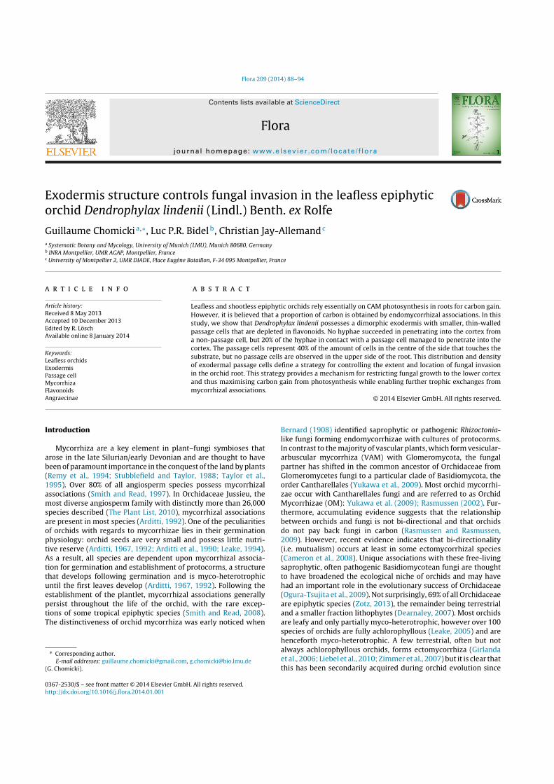
Dendrophylax lindenii roots (Fig. 1) exhibit a clear polarity andbilateral symmetry with differentiated upper and lower sides. Thevelamen is two-layered, and the second layer is shed at the upperroot side. Some cells of the epivelamen, that are in contact to the
Fig. 1. Habit and morphology of Dendrophylax lindenii. (A) Habit and general morphology. (B) Flower morphology. Ds, distal sepal; Ls, lateral sepal; P, petal; L, labellum; Ac,anther cap; St, stigma; S, spur. (C) Root morphology. P, pneumatode. (D) Root cross-section. V, velamen; TI, tracheoidal idioblast. Scale bars: (A) 3 cm; (B) 0.5 cm; (C) 2.4 mm;(D) 0.7 mm.
90 G. Chomicki et al. / Flora 209 (2014) 88–94
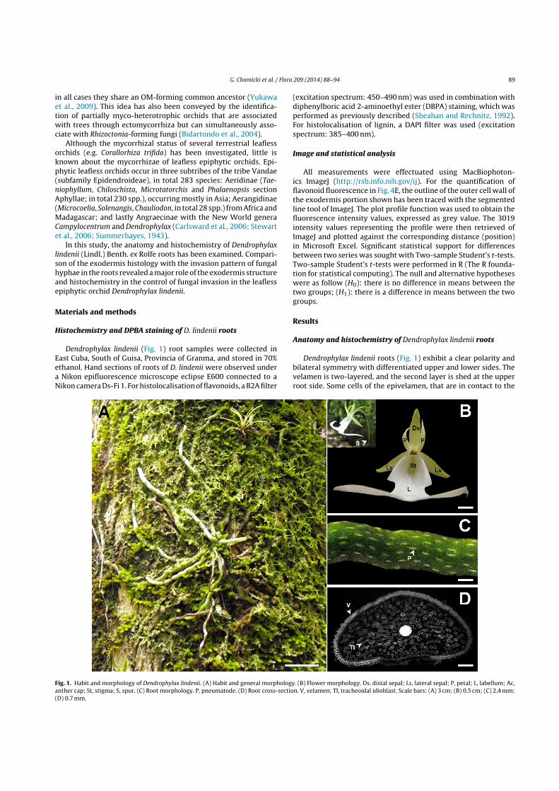
Fig. 2. Lignin and flavonoid distribution in D. lindenii root as revealed by histochemistry. (A) Root transection observed with DAPI filter, showing lignified cells. (B) Roottransection observed with B2A filter and DPBA staining, revealing flavonoid distribution. (C) Flavonoids in the stele. E, endodermis; P, passage cell; Ph, phloem; px, protoxylem;mx, metaxylem; Pi, pith. (D) Flavonoid distribution in the lower root region. White arrowhead points to fungal sporangia and grey arrowhead points to intramatricial hyphae.Exo, exodermis; P, passage cell; EpiV, epivelamen; EndoV, endovelamen. (E) Lignin in the lower root region. (F) Close up of the exodermis. Arrowhead points to the secondarycell walls. Scale bars: (A and B) 350 !m; (C) 90 !m; (D) 65 !m; (E) 80 !m; (F) 12 !m.
support (=second (outer) velamen layer, V2), elongate and formroot hairs which have the function of crampons. The endovelamencell wall shows thick secondary cell walls thickening in the upperside of the root but is thin-walled in the lower side of the root.Endodermal cells are O-shaped.
Lignified tissues were visualised by histochemistry using a DAPIfilter. In the stele, the xylem poles (7) exhibited the strongest signal.The endodermis was slightly lignified but essentially in the sec-ondary cell walls (Fig. 2A). The whole cortex is photosynthetic, witha higher chloroplast concentration in the upper side of the root.Non-living tracheoidal idioblasts are interspersed into parenchy-matous cells of the cortex and are lignified. The exodermis presentsa distinct pattern in the upper and lower sides of the root, withlignin present in the lower parts of the secondary cell wall layers inthe upper side (Fig. 2A, arrowhead) and in all cell walls in the lowerside [Fig. 2A, E and F (arrowhead)]. In the passage cells of both endo-dermis and exodermis lignin is absent or very scarce (Fig. 2A, E andF). The first layer of the velamen or epivelamen is not lignified, butthe second velamen layer – the endovelamen – is lignified in thecentral part of the lower side only (Fig. 2A and E).
The flavonoid distribution in D. lindenii root was examined usingDPBA staining, which specifically binds to flavonoids (Sheahan andRechnitz, 1992). The thick tertiary cell walls of the endodermis
exhibit a strong yellow-gold fluorescence which is characteristicof flavonoids related to quercetin (Sheahan and Rechnitz, 1992).Flavonoids are essentially absent in the cortical cells, including tra-cheoidal idioblasts, but they are present in the thick tertiary cellwalls of the outer part of the exodermis (Fig. 2B and D). The epivela-men is exempt of flavonoids in the central part of the lower side, butthe flavonoid level subsequently increases dramatically towardsthe upper side and is correlated to a cell size increase (Fig. 2B).However, the fluorescence in the epivelamen is green-yellow, notyellow-gold as in the second velamen layer (Figs. 2B and 4A). Thisis a colour which is generally associated to kaempherol derivatives(Sheahan and Rechnitz, 1992). In the endovelamen, flavonoids arepresent in the central part of the lower side only and form a gradi-ent which reaches a maximum at the point of contact between theorchid root and the support (Fig. 2B and D).
An OM-type fungus is associated with D. lindenii roots
Sporangial structures have been observed adjacent to the lowerside of the root and numerous hyphae lie within the upper vela-men and in the cortex as well (Figs. 2D and 3C–H). Arbuscular-likestructures were present in the velamen (Fig. 3E and H), vesicularstructures at the apex of a hypha were also observed (Fig. 3D and

G. Chomicki et al. / Flora 209 (2014) 88–94 91
Fig. 3. Passage of hyphae through the exodermis layer and fungal structures. (A) Frequency of hyphae that fail or succeed to cross the exodermis. (B) Exodermal flavonoidsand blocked hyphae endings (arrowheads). (C) Digested peloton in the cortex (arrowhead). (D) Pelotons in course of digestion in the cortex (black arrowheads). Whitearrowhead points a vesicular structure. (E) Hyphae that passed the exodermis. Exo, exodermis; P, passage cell; EpiV, epivelamen; EndoV, endovelamen. (F and G) Detailhyphae that crossed the exodermis. The red dotted line represents the exodermis outer wall. Black arrowhead point hyphae, white arrowhead points a vesicular structurethat terminate a hypha. (H) Detail of mycorrhizal fungi in the velamen. White arrowhead points hypha, black arrowhead points an arbuscular structure. Abbreviations: Exo,Exodermis; Cort, Cortex; P, passage cell; EpiV, epivelamen; EndoV, endovelamen. Scale bars: (B and D) 25 !m; (C and E) 20 !m.
G). Importantly, digested pelotons or pelotons in course of digestionwere present in cortical cells (Fig. 3C and D).
Most of the hyphae end upon touching the outer tangential cellwall of the exodermis (Figs. 2D and 3B). The hyphae ‘blocked’ atthe exodermis versus those that succeeded to penetrate the exo-dermis barrier into the cortex were quantified (Fig. 3A). All hyphae(n = 32) that touched a non-passage cell (i.e. a standard exodermalcell) failed to penetrate further into the cortex (Fig. 3A). By contrast,when hyphae reached the exodermis by touching a passage cell,they penetrated the exodermis towards the cortex at a frequency of0.2 (n = 24). Hence, penetration of the fungi into the cortex occurredonly through a passage cell.
Passage cells occur primarily in the lower side of theexodermis in D. lindenii
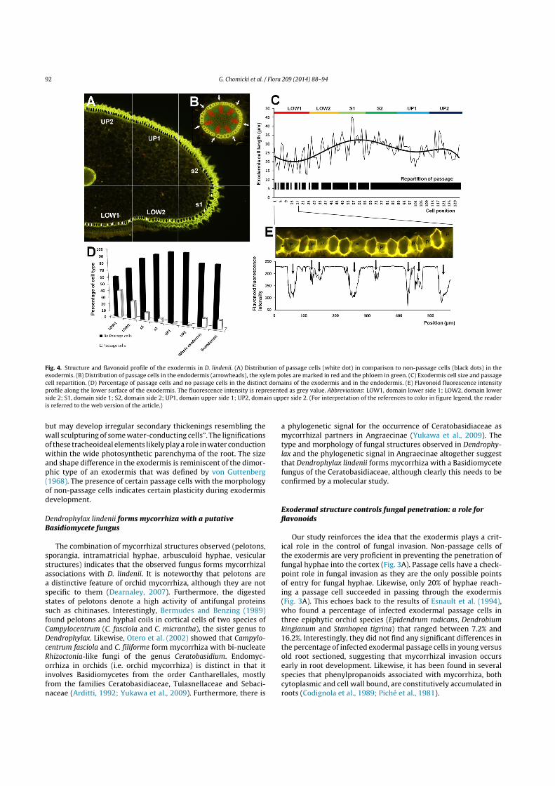
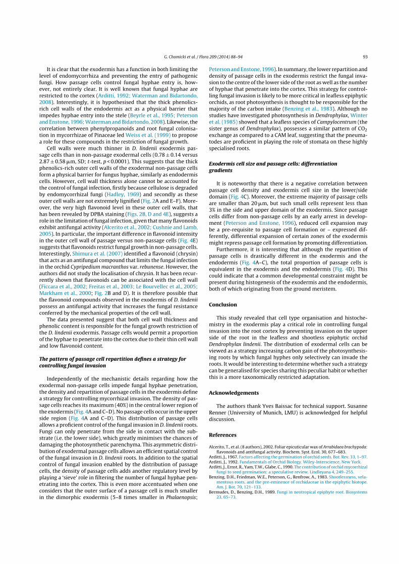
In order to gain further insights into how D. lindenii controlsfungal invasion, the distribution of passage cells was studied.By contrast to the endodermal passage cells which are regularlyspaced and generally inserted very close to the protoxylem poles(Figs. 2C and 4B, D), the exodermal passage cells are irregularlyrepartitioned around the exodermis (Fig. 4A, C and D). Exodermalpassage cells have a maximum density of 40% of the total amountof exodermal cells in the central zone of the lower side of the root(LOW1): Fig. 4D. The density of exodermal cells diminishes fromthe lower centre side of the root (LOW1) to the side of the root(region S2) where passage cells represent 4.5% of the exodermalcells (Fig. 4A, C and E). Exodermal passage cells were absent fromthe upper side regions (UP1 and UP2): Fig. 4A, C and E.
In order to test the hypothesis that cell wall flavonoids wereinvolved in the exodermal-mediated restriction of fungal invasion,the flavonoid intensity profile of the exodermis outer surface was
determined in a portion of the central lower zone (LOW1), wherethe density of passage cells is highest (Fig. 4E). The flavonoid fluo-rescence pattern depicted the exodermal cell types with saturatingintensity for non-passage cells and low intensity for passage cells(Fig. 4E). Given that fungal hyphae were found to be able to pen-etrate into the cortex only through passage cells (Fig. 3A), theprobability of fungal hyphae penetration was thus also correlatedwith flavonoid level.
Passage cells were significantly smaller than non-passage cells(Fig. 4G; t-test, p < 0.0001) and generally elongated by contrastto the more isodiametric shape of non-passage cells (Fig. 3B). Avery few passage cells exhibited an isodiametric shape and weresimilar to the non-passage cells (e.g. Fig. 2F). They were only distin-guished by thin cell walls and depletion in lignin and most notablyflavonoids.
The exodermis cell width fluctuates but follows distinctivetrends from the ab1 to the AD2 region (Fig. 4F and H). From ab1to s1, exodermal cell length increases linearly (Fig. 4F and H), ab2exodermal cells being significantly longer than ab1 cells (t-test,p < 0.01) and s1 cells being significantly longer than ab1 cells (t-test,p < 0.0001) as well as ab2 cells (t-test, p < 0.01).
Discussion
Anatomy of the Dendrophylax lindenii root
Our anatomical analyses confirmed the earlier account ofBenzing et al. (1983) who first reported the 2-layered structureand shedding of the epivelamen on the upper side of the root inthis species. The lignification of tracheoideal elements found inD. lindenii contrasts with the claims of Benzing et al. (1983) whostated that “[tracheoidal idioblasts] are not lignified or suberised,
92 G. Chomicki et al. / Flora 209 (2014) 88–94
Fig. 4. Structure and flavonoid profile of the exodermis in D. lindenii. (A) Distribution of passage cells (white dot) in comparison to non-passage cells (black dots) in theexodermis. (B) Distribution of passage cells in the endodermis (arrowheads), the xylem poles are marked in red and the phloem in green. (C) Exodermis cell size and passagecell repartition. (D) Percentage of passage cells and no passage cells in the distinct domains of the exodermis and in the endodermis. (E) Flavonoid fluorescence intensityprofile along the lower surface of the exodermis. The fluorescence intensity is represented as grey value. Abbreviations: LOW1, domain lower side 1; LOW2, domain lowerside 2; S1, domain side 1; S2, domain side 2; UP1, domain upper side 1; UP2, domain upper side 2. (For interpretation of the references to color in figure legend, the readeris referred to the web version of the article.)
but may develop irregular secondary thickenings resembling thewall sculpturing of some water-conducting cells”. The lignificationsof these tracheoideal elements likely play a role in water conductionwithin the wide photosynthetic parenchyma of the root. The sizeand shape difference in the exodermis is reminiscent of the dimor-phic type of an exodermis that was defined by von Guttenberg(1968). The presence of certain passage cells with the morphologyof non-passage cells indicates certain plasticity during exodermisdevelopment.
Dendrophylax lindenii forms mycorrhiza with a putativeBasidiomycete fungus
The combination of mycorrhizal structures observed (pelotons,sporangia, intramatricial hyphae, arbusculoid hyphae, vesicularstructures) indicates that the observed fungus forms mycorrhizalassociations with D. lindenii. It is noteworthy that pelotons area distinctive feature of orchid mycorrhiza, although they are notspecific to them (Dearnaley, 2007). Furthermore, the digestedstates of pelotons denote a high activity of antifungal proteinssuch as chitinases. Interestingly, Bermudes and Benzing (1989)found pelotons and hyphal coils in cortical cells of two species ofCampylocentrum (C. fasciola and C. micrantha), the sister genus toDendrophylax. Likewise, Otero et al. (2002) showed that Campylo-centrum fasciola and C. filiforme form mycorrhiza with bi-nucleateRhizoctonia-like fungi of the genus Ceratobasidium. Endomyc-orrhiza in orchids (i.e. orchid mycorrhiza) is distinct in that itinvolves Basidiomycetes from the order Cantharellales, mostlyfrom the families Ceratobasidiaceae, Tulasnellaceae and Sebaci-naceae (Arditti, 1992; Yukawa et al., 2009). Furthermore, there is
a phylogenetic signal for the occurrence of Ceratobasidiaceae asmycorrhizal partners in Angraecinae (Yukawa et al., 2009). Thetype and morphology of fungal structures observed in Dendrophy-lax and the phylogenetic signal in Angraecinae altogether suggestthat Dendrophylax lindenii forms mycorrhiza with a Basidiomycetefungus of the Ceratobasidiaceae, although clearly this needs to beconfirmed by a molecular study.
Exodermal structure controls fungal penetration: a role forflavonoids
Our study reinforces the idea that the exodermis plays a crit-ical role in the control of fungal invasion. Non-passage cells ofthe exodermis are very proficient in preventing the penetration offungal hyphae into the cortex (Fig. 3A). Passage cells have a check-point role in fungal invasion as they are the only possible pointsof entry for fungal hyphae. Likewise, only 20% of hyphae reach-ing a passage cell succeeded in passing through the exodermis(Fig. 3A). This echoes back to the results of Esnault et al. (1994),who found a percentage of infected exodermal passage cells inthree epiphytic orchid species (Epidendrum radicans, Dendrobiumkingianum and Stanhopea tigrina) that ranged between 7.2% and16.2%. Interestingly, they did not find any significant differences inthe percentage of infected exodermal passage cells in young versusold root sectioned, suggesting that mycorrhizal invasion occursearly in root development. Likewise, it has been found in severalspecies that phenylpropanoids associated with mycorrhiza, bothcytoplasmic and cell wall bound, are constitutively accumulated inroots (Codignola et al., 1989; Piché et al., 1981).
G. Chomicki et al. / Flora 209 (2014) 88–94 93
It is clear that the exodermis has a function in both limiting thelevel of endomycorrhiza and preventing the entry of pathogenicfungi. How passage cells control fungal hyphae entry is, how-ever, not entirely clear. It is well known that fungal hyphae arerestricted to the cortex (Arditti, 1992; Waterman and Bidartondo,2008). Interestingly, it is hypothesised that the thick phenolics-rich cell walls of the endodermis act as a physical barrier thatimpedes hyphae entry into the stele (Beyrle et al., 1995; Petersonand Enstone, 1996; Waterman and Bidartondo, 2008). Likewise, thecorrelation between phenylpropanoids and root fungal colonisa-tion in mycorrhizae of Pinaceae led Weiss et al. (1999) to proposea role for these compounds in the restriction of fungal growth.
Cell walls were much thinner in D. lindenii exodermis pas-sage cells than in non-passage exodermal cells (0.78 ± 0.14 versus2.87 ± 0.58 !m, SD; t-test, p < 0.0001). This suggests that the thickphenolics-rich outer cell walls of the exodermal non-passage cellsform a physical barrier for fungus hyphae, similarly as endodermiscells. However, cell wall thickness alone cannot be accounted forthe control of fungal infection, firstly because cellulose is degradedby endomycorrhizal fungi (Hadley, 1969) and secondly as theseouter cell walls are not extremely lignified (Fig. 2A and E–F). More-over, the very high flavonoid level in these outer cell walls, thathas been revealed by DPBA staining (Figs. 2B, D and 4E), suggests arole in the limitation of fungal infection, given that many flavonoidsexhibit antifungal activity (Alcerito et al., 2002; Cushnie and Lamb,2005). In particular, the important difference in flavonoid intensityin the outer cell wall of passage versus non-passage cells (Fig. 4E)suggests that flavonoids restrict fungal growth in non-passage cells.Interestingly, Shimura et al. (2007) identified a flavonoid (chrysin)that acts as an antifungal compound that limits the fungal infectionin the orchid Cypripedium macranthos var. rebunense. However, theauthors did not study the localisation of chrysin. It has been recur-rently shown that flavonoids can be associated with the cell wall(Ficcara et al., 2002; Freitas et al., 2003; Le Bourvellec et al., 2005;Markham et al., 2000; Fig. 2B and D). It is therefore possible thatthe flavonoid compounds observed in the exodermis of D. lindeniipossess an antifungal activity that increases the fungal resistanceconferred by the mechanical properties of the cell wall.
The data presented suggest that both cell wall thickness andphenolic content is responsible for the fungal growth restriction ofthe D. lindenii exodermis. Passage cells would permit a proportionof the hyphae to penetrate into the cortex due to their thin cell walland low flavonoid content.
The pattern of passage cell repartition defines a strategy forcontrolling fungal invasion
Independently of the mechanistic details regarding how theexodermal non-passage cells impede fungal hyphae penetration,the density and repartition of passage cells in the exodermis definea strategy for controlling mycorrhizal invasion. The density of pas-sage cells reaches its maximum (40%) in the central lower region ofthe exodermis (Fig. 4A and C–D). No passage cells occur in the upperside region (Fig. 4A and C–D). This distribution of passage cellsallows a proficient control of the fungal invasion in D. lindenii roots.Fungi can only penetrate from the side in contact with the sub-strate (i.e. the lower side), which greatly minimises the chances ofdamaging the photosynthetic parenchyma. This asymmetric distri-bution of exodermal passage cells allows an efficient spatial controlof the fungal invasion in D. lindenii roots. In addition to the spatialcontrol of fungal invasion enabled by the distribution of passagecells, the density of passage cells adds another regulatory level byplaying a ‘sieve’ role in filtering the number of fungal hyphae pen-etrating into the cortex. This is even more accentuated when oneconsiders that the outer surface of a passage cell is much smallerin the dimorphic exodermis (5–8 times smaller in Phalaenopsis;
Peterson and Enstone, 1996). In summary, the lower repartition anddensity of passage cells in the exodermis restrict the fungal inva-sion to the centre of the lower side of the root as well as the numberof hyphae that penetrate into the cortex. This strategy for control-ling fungal invasion is likely to be more critical in leafless epiphyticorchids, as root photosynthesis is thought to be responsible for themajority of the carbon intake (Benzing et al., 1983). Although nostudies have investigated photosynthesis in Dendrophylax, Winteret al. (1985) showed that a leafless species of Campylocentrum (thesister genus of Dendrophylax), possesses a similar pattern of CO2exchange as compared to a CAM leaf, suggesting that the pneuma-todes are proficient in playing the role of stomata on these highlyspecialised roots.
Exodermis cell size and passage cells: differentiationgradients
It is noteworthy that there is a negative correlation betweenpassage cell density and exodermis cell size in the lower/sidedomain (Fig. 4C). Moreover, the extreme majority of passage cellsare smaller than 20 !m, but such small cells represent less than3% in the side and upper domain of the exodermis. Since passagecells differ from non-passage cells by an early arrest in develop-ment (Peterson and Enstone, 1996), reduced cell expansion maybe a pre-requisite to passage cell formation or – expressed dif-ferently, differential expansion of certain zones of the exodermismight repress passage cell formation by promoting differentiation.
Furthermore, it is interesting that although the repartition ofpassage cells is drastically different in the exodermis and theendodermis (Fig. 4A–C), the total proportion of passage cells isequivalent in the exodermis and the endodermis (Fig. 4D). Thiscould indicate that a common developmental constraint might bepresent during histogenesis of the exodermis and the endodermis,both of which originating from the ground meristem.
Conclusion
This study revealed that cell type organisation and histoche-mistry in the exodermis play a critical role in controlling fungalinvasion into the root cortex by preventing invasion on the upperside of the root in the leafless and shootless epiphytic orchidDendrophylax lindenii. The distribution of exodermal cells can beviewed as a strategy increasing carbon gain of the photosynthesis-ing roots by which fungal hyphes only selectively can invade theroots. It would be interesting to determine whether such a strategycan be generalised for species sharing this peculiar habit or whetherthis is a more taxonomically restricted adaptation.
Acknowledgements
The authors thank Yves Baissac for technical support. SusanneRenner (University of Munich, LMU) is acknowledged for helpfuldiscussion.
References
Alcerito, T., et al. (8 authors), 2002. Foliar epicuticular wax of Arrabidaea brachypoda:flavonoids and antifungal activity. Biochem. Syst. Ecol. 30, 677–683.
Arditti, J., 1967. Factors affecting the germination of orchid seeds. Bot. Rev. 33, 1–97.Arditti, J., 1992. Fundamentals of Orchid Biology. Wiley-Interscience, New York.Arditti, J., Ernst, R., Yam, T.W., Glabe, C., 1990. The contribution of orchid mycorrhizal
fungi to seed germination: a speculative review. Lindleyana 4, 249–255.Benzing, D.H., Friedman, W.E., Peterson, G., Renfrow, A., 1983. Shootlessness, vela-
mentous roots, and the pre-eminence of orchidaceae in the epiphytic biotope.Am. J. Bot. 70, 121–133.
Bermudes, D., Benzing, D.H., 1989. Fungi in neotropical epiphyte root. Biosystems23, 65–73.

94 G. Chomicki et al. / Flora 209 (2014) 88–94
Bernard, N., 1908. La culture des orchidees, dans ses rapports avec la symbiose. Soc.Roy. Agr. et Bot. Gand, Ghent/Belgium, 16 pp.
Beyrle, H.F., Smith, S.E., Franco, C.M.M., Peterson, R.L., 1995. Colonization of Orchismorio protocorms by a mycorrhizal fungus: effects of nitrogen nutrition andglyphosate in modifying the responses. Can. J. Bot. 73, 1128–1140.
Bidartondo, M.I., Burghardt, B., Gebauer, G., Bruns, T.D., Read, D.J., 2004. Chang-ing partners in the dark: isotopic and molecular evidence of ectomycorrhizalliaisons between forest orchids and trees. Proc. R. Soc. Lond. 271 (1550),1799–1806.
Cameron, D.D., Johnson, I., Read, D.J., Leake, J.R., 2008. Giving and receiving: mea-suring the carbon cost of mycorrhizas in the green orchid, Goodyera repens. NewPhytol. 180, 176–184.
Carlsward, B.S., Stern, W.L., Bytebier, B., 2006. Comparative vegetative anatomy andsystematics of the angraecoids (Vandeae, Orchidaceae) with an emphasis on theleafless habit. Bot. J. Linn. Soc. 151, 165–218.
Codignola, A., Verotta, L., Spanu, P., Maffei, M., Scannerini, S., Bonfante-Fasolo, P.,1989. Cell wall bound-phenols in roots of vesicular-arbuscular mycorrhizalplants. New Phytol. 112, 221–228.
Cushnie, T.P.T., Lamb, A.J., 2005. Antimicrobial activity of flavonoids. Int. J. Antimi-crob. Agents 26, 343–356.
Dearnaley, J., 2007. Further advances in orchid mycorrhizal research. Mycorrhiza 17,475–486.
Esnault, A., Masuhara, G., McGee, P.A., 1994. Involvement of exodermal passage cellsin mycorrhizal infection of some orchids. Mycol. Res. 98, 672–676.
Ficcara, R., et al. (8 authors), 2002. Study of flavonoids/b cyclodextrins inclusioncomplexes by NMR, FT-IR, DSC, X-ray investigation. J. Pharm. Biomed. Anal. 29,1005–1014.
Freitas, V., Carvalho, E., Mateus, N., 2003. Study of carbohydrate influence on protein-tannin aggregation by nephelometry. Food Chem. 81, 503–509.
Girlanda, M., et al. (12 authors), 2006. Inefficient photosynthesis in the Mediter-ranean orchid Limodorum abortivum is mirrored by specific association toectomycorrhizal Russulaceae. Mol. Ecol. 15, 491–504.
Hadley, G., 1969. Cellulose as a carbon source for orchid mycorrhiza. New Phytol.68, 933–939.
Le Bourvellec, C., Bouchet, B., Renard, C.M.G.C., 2005. Non-covalent interactionbetween procyanidins and apple cell wall material. Part III. Study on modelpolysaccharides. Biochim. Biophys. Acta 1725, 10–18.
Leake, J.R., 1994. The biology of myco-heterotrophic (‘saprophytic’) plants. NewPhytol. 127, 171–216.
Leake, J.R., 2005. Plants parasitic on fungi: unearthing the fungi in myco-heterotrophs and debunking the “saprophytic” plant myth. Mycologist 19,113–122.
Liebel, H.T., Bidartondo, M.I., Preiss, K., Segreto, R., Stockel, M., Rodda, M., Gebauer,G., 2010. C and N stable isotope signatures reveal constraints to nutritionalmodes in orchids from the Mediterranean and Macaronesia. Am. J. Bot. 97,903–912.
Markham, K.R., Ryan, K.G., Gould, K.S., Richards, G.K., 2000. Cell wall sited flavonoidsin lisianthus flower petals. Phytochemistry 54, 681–687.
Ogura-Tsujita, Y., Gebauer, G., Hashimoto, T., Umata, H., Yukawa, T., 2009. Evidencefor novel and specialised mycorrhizal parasitism: the orchid Gastrodia confusagains carbon from saprotrophic Mycena. Proc. R. Soc. Lond. B 276, 761–767.
Otero, J.T., Ackerman, J.D., Bayman, P., 2002. Diversity and host specificity of endo-phytic Rhizoctonia-like fungi from tropical orchids. Am. J. Bot. 89, 1852–1858.
Peterson, P.A., Enstone, D.E., 1996. Functions of passage cells in the endodermis andexodermis of roots. Physiol. Plant 97, 592–598.
Piché, Y., Fortin, J.A., Lafontaine, J.G., 1981. Cytoplasmic phenols and polysaccharidesin ectomycorrhizal and non-mycorrhizal short roots of pine. New Phytol. 88,695–703.
Rasmussen, H.N., 2002. Recent developments in the study of orchid mycorrhiza.Plant Soil 244, 149–163.
Rasmussen, H.N., Rasmussen, F.N., 2009. Orchid mycorrhiza: implications of amycophagous life style. Oikos 118, 334–345.
Remy, W., Taylor, T.N., Hass, H., Kerp, H., 1994. Four hundred-million-year-old vesic-ular arbuscular mycorrhizae. Proc. Natl. Acad. Sci. U. S. A. 91, 11841–11843.
The Plant List, 2010. Version 1. Published on the Internet, available at:http://www.theplantlist.org/
Sheahan, J.J., Rechnitz, G.A., 1992. Flavonoid-specific staining in Arabidopsis thaliana.Biotechniques 13, 880–883.
Shimura, H., Matsuura, M., Takada, N., Koda, Y., 2007. An antifungal compoundinvolved in symbiotic germination of Cypripedium macranthos var. rebunense(Orchidaceae). Phytochemistry 68, 1442–1447.
Smith, S.E., Read, D.J., 1997. Mycorrhizal Symbiosis, 2nd ed. Acad. Press, San Diego.Smith, S., Read, D., 2008. Colonization of roots and anatomy of arbuscular mycor-
rhiza. In: Mycorrhizal Symbiosis. Academic Press, London, pp. 42–90.Stewart, J., Hermans, J., Campbell, J., 2006. Angraecoid Orchids: Species from the
African Region. Timber Press, Portland.Stubblefield, S.P., Taylor, T.N., 1988. Recent advances in palaeomycology. New Phy-
tol. 108, 3–25.Summerhayes, V.S., 1943. African orchids XIII: the leafless angraecoid orchids. Bot.
Mus. Leaflets Harvard Univ. 11, 137–170.Taylor, T.N., Remy, W., Hass, H., Kerp, H., 1995. Fossil arbuscular mycorrhizae from
the Early Devonian. Mycologia 87, 560–573.Von Guttenberg, H., 1968. Der primäre Bau der Angiospermenwurzel. Handbuch der
Pflanzenanatomie. Bd, 8. Borntraeger, Leipzig.Waterman, R.J., Bidartondo, M.I., 2008. Deception above, deception below: linking
pollination and mycorrhizal biology of orchids. J. Exp. Bot. 59, 1085–1096.Weiss, M., Schmidt, J., Neumann, D., Wray, V., Christ, R., Strack, D., 1999. Phenyl-
propanoids in mycorrhizas of the Pinaceae. Planta 208, 491–502.Winter, K., Medina, E., Garcia, V., Mayoral, M.L., Muniz, R., 1985. Crassulacean acid
metabolism in roots of a leafless orchid, Campylocentrum tyrridion Garay & Dun-sterv. J. Plant Physiol. 118, 73–78.
Yukawa, T., Oruga-Tsujita, Y., Shefferson, R.P., Yokiyama, J., 2009. Mycorrhizal diver-sity in Apostasia (Orchidaceae) indicates the origin and evolution or orchidmycorrhiza. Am. J. Bot. 96, 1997–2009.
Zimmer, K., Hynson, N.A., Gebauer, G., Allen, E.B., Allen, M.F., Read, D.J., 2007. Widegeographical and ecological distribution of nitrogen and carbon gains from fungiin pyroloids and monotropoids (Ericaceae) and in orchids. New Phytol. 175,166–175.
Zotz, G., 2013. The systematic distribution of vascular epiphytes – a critical update.Bot. J. Linn. Soc. 171, 453–481.
Copyright © 2022 FDOKUMEN