Effect of Different Sprouting Conditions on Alpha Amylase ...
Cerebral Cortex
doi:10.1093/cercor/bhq091
Focal Damage to the Adult Rat Neocortex Induces Wound Healing Accompanied byAxonal Sprouting and Dendritic Structural Plasticity
Catherine A. Blizzard1, Jyoti A. Chuckowree1, Anna E. King1, Katherine A. Hosie1, Graeme H. McCormack1, Jamie A. Chapman2,
James C. Vickers1,2 and Tracey C. Dickson1
1Wicking Dementia Research and Education Centre, Menzies Research Institute and 2School of Medicine, University of Tasmania,
Private Bag 34, Hobart Tasmania 7000, Australia
Address correspondence to Dr Tracey Dickson, Menzies Research Institute, University of Tasmania, Private Bag 23, Hobart, Tasmania 7000, Australia.
Email: [email protected].
Accumulating evidence indicates that damage to the adultmammalian brain evokes an array of adaptive cellular responsesand may retain a capacity for structural plasticity. We haveinvestigated the cellular and architectural alterations followingfocal experimental brain injury, as well as the specific capacity forstructural remodeling of neuronal processes in a subset of corticalinterneurons. Focal acute injury was induced by transient insertionof a needle into the neocortex of anesthetized adult male Hooded--Wistar rats and thy1 green fluorescent protein (GFP) mice.Immunohistochemical, electron microscopy, and bromodeoxyuri-dine cell proliferation studies demonstrated an active and evolvingresponse of the brain to injury, indicating astrocytic but notneuronal proliferation. Immunolabeling for the neuron-specificmarkers phosphorylated neurofilaments, a-internexin and calretininat 7 days post injury (DPI) indicated phosphorylated neurofilamentsand a-internexin but not calretinin immunopositive axonal sproutswithin the injury site. However, quantitative studies indicateda significant realignment of horizontally projecting dendrites ofcalretinin-labeled interneurons at 14 DPI. This remodeling wasspecific to calretinin immunopositive interneurons and did not occurin a subpopulation of pyramidal neurons expressing GFP in theinjured mouse cortex. These data show that subclasses of corticalinterneurons are capable of adaptive structural remodeling.
Keywords: cortical injury, interneuron, neurogenesis, plasticity,regeneration
Introduction
Structural injury to the brain evokes a distinct sequence of
events indicative of an attempt to repair and heal, including the
activation of microglial, oligodendroglial precursor, meningeal,
astrocyte, and stem cell populations (reviewed in Fawcett and
Asher 1999). It is now established that neurons can also actively
react to injury, either through the formation of reactive axonal
sprouts or via altered connectivity of preexisting pathways
(reviewed in Chuckowree et al. 2004; Carmichael 2006;
Fitzgerald and Fawcett 2007; Macias 2008).
Ultimately, an adaptive brain response to trauma may require
the induction of neurogenesis and either appropriate re-
generation or compensatory plasticity of neural pathways.
Alterations in neural progenitor populations and cell pro-
liferation have been demonstrated in diverse models of
experimental brain lesion and stroke conditions and are
proposed to contribute to postlesion brain recovery (Clarke
et al. 1994; Duggal et al. 1997; Holmin et al. 1997; Kernie et al.
2001; Arvidsson et al. 2002; Chen et al. 2003; Douen et al. 2004;
Salman et al. 2004). However, whether brain injury evokes
a neurogenic response that contributes to functional recovery
currently remains contentious, with the presence of neuro-
genic events differing for different injury paradigms (Holmin
et al. 1997; Kernie et al. 2001; Kuroda et al. 2002; Rice et al.
2003; Salman et al. 2004; Yu et al. 2008).
Examples of neuronal plasticity, manifesting as alterations in
synaptic strength and wiring, remodeling of axonal and
dendritic arbors, and changes in dendritic spine and axonal
bouton turnover, have been observed in the naı̈ve brain and in
response to alterations in sensory experience and injury/lesion
(for a selection of recent examples, see Grutzendler et al. 2002;
Knott et al. 2002; Trachtenberg et al. 2002; Majewska and Sur
2003; Portera-Cailliau et al. 2003; Holtmaat et al. 2005; Tailby
et al. 2005; Lee et al. 2006, 2008; Majewska et al. 2006; Brown
et al. 2007, 2008, 2010; Yamahachi et al. 2009). However, the
specificity and degree of remodeling that can occur in response
to injury has not been fully elucidated. Cortical neurons
demonstrate axonal and synaptic remodeling in response to
various models of injury (e.g., King et al. 2001; Knott et al. 2002;
Trachtenberg et al. 2002; Majewska et al. 2006). Reorganization
such as this may correlate with functional recovery not only in
stroke (Carmichael 2003, 2006; Brown et al. 2008; Di Filippo
et al. 2008) but comparable plasticity may also account for the
recovery observed after forms of spinal cord injury (reviewed
in Edgerton et al. 2004; Dunlop 2008).
Recent research indicates that populations of interneurons
within the barrel cortex demonstrate adaptive plasticity,
specifically with regards to the remodeling of dendritic arbors
(Lee et al. 2006, 2008). Modifications of the dendritic tree and
synaptic contacts may also occur following injury, with studies
showing that the plastic potential of cortical dendrites is
enhanced following injury (Kolb and Gibb 1991; Jones and
Schallert 1992) lasting over 2 weeks post injury (Jones and
Schallert 1992). Interestingly, studies have indicated that not all
subpopulations of neurons are equal in their capacity for such
adaptive structural plasticity. For example, interneurons in the
intact neocortex have been demonstrated to be capable of
dendritic arbor remodeling while pyramidal neuronal subpo-
pulations remain stable (Lee et al. 2006).
Thecurrent investigationutilizedamodelof focal brain injury to
generate a discrete unilateral lesion in the adult rodent somato-
sensory cortex. The cellular response to injury was investigated
over a time course of up to 14 days post injury (DPI). Acute focal
injury to the somatosensory cortex induced an evolving cellular
response, initially characterizedby tissuedestruction andbleeding
within the injury site. This was followed by an infiltration of the
injury site with glial cells and subsequent tissue remodeling
including the formation of a glial scar and neovascularization.
� The Author 2010. Published by Oxford University Press. All rights reserved.
For permissions, please e-mail: [email protected]
Cerebral Cortex Advance Access published May 28, 2010 by guest on June 7, 2016
http://cercor.oxfordjournals.org/D
ownloaded from
Analysis of the morphological plasticity of resident interneuron
and pyramidal neuron populations demonstrated cell type--
specific dendritic arbor remodeling following injury; calretinin
positive interneurons reorganized their dendrites with a distal
orientation shift away from the lesion site, whereas the dendritic
arbors of green fluorescent protein (GFP)-expressing pyramidal
neurons were not altered. Additionally, peri-lesion axonal sprout-
ing was observed for pyramidal neurons but not for interneurons
adjacent to the injury site. These data suggest that the brain is
capable of significant remodeling following injury, specific to
neuronal type.
Materials and Methods
In vivo Brain InjuryAll experimental procedures utilizing adult male Hooded--Wistar rats
(250--270 g, corresponding to approximately 8 weeks old) and adult
male thy1 GFP-M mice (between 8 and 10 weeks old) were approved
by the Animal Ethics Committee of the University of Tasmania and are
in accordance with the Australian code of practice for the care and use
of animals for scientific purposes. Animals were housed in standard
conditions (20 �C, 12/12 h light/dark cycle), with access to food and
water ad libitum and monitored daily for signs of illness and stress.
Acute focal neocortical injuries were performed as previously
described (King et al. 1997, 2001; Dickson et al. 2005). Briefly, following
intraperitoneal administration of anesthetic (pentobarbitone sodium, 72
mg/kg, Abbot Laboratories) and analgesic (Carprogen, 4 mg/kg, Pfizer),
a burr holewas drilled into the skull, (5-mmanterior and4.5-mm lateral to
lambda for rat, 2.5-mmanterior and 2-mm lateral to lambda formice), and
a focal injury was made in the somatosensory cortex (Par1 region) by
lowering a 25-gageHamilton needle (Reno) to a depth of 1.5mm into the
graymatter for rats and a 29-gage needle to a depth of 1mm into the gray
matter for mice. The needle was left in place for 10 min prior to removal
and suturing of the wound. Following recovery, animals were terminally
anesthetized (pentabarbitone sodium, 140 mg/kg) at a range of
postinjury time intervals up to 14 days following injury (n > 5 animals
per time point at 1, 7, and 14 DPI), and brains were processed for either
immunohistochemistry or transmission election microscopy as de-
scribed in the relevant sections below.
To determine the phenotype of cells proliferating within the first
7 days following injury, animals were administered with 12.5 mg/mL
solution of the thymidine analog bromodeoxyuridine (BrdU) (Sigma) in
7 mM NaOH/0.9% NaCl by intraperitoneal injection. Animals were
injected with BrdU (25 mg/kg/day), between 1 and 6 DPI, and were
transcardially perfused (4% paraformaldehyde [PFA]/0.01 M phosphate-
buffered saline [PBS]) at 7 days following injury. Prior to immunohis-
tochemistry, antigen retrieval was performed by incubating sections in
2 M HCl for 1 h at 37 �C, followed by 3 neutralization washes in 0.1 M
borate buffer (pH 8.5).
ImmunohistochemistryTo determine alterations in specific cell populations following cortical
injury, rats were terminally anesthetized and transcardially perfused as
above. Noninjured, age-matched controls corresponding to the 1, 7, and
14 DPI time points (n = 5 animals per time point) were also perfused.
Brain sections (50-lm vibratome) were collected coronally and also
horizontal to the pial surface, through the injury site, from control and
experimental animals and were immunohistochemically labeled in
single as well as double-labeling combinations, with a range of cell-
specific antibodies (Table 1). Primary antibody binding was visualized
using species-specific fluorescent secondary antibodies, Alexa488 and
Alexa594 (Molecular Probes); however, BrdU labeling was visualized
with an anti-mouse rat-adsorbed fluorescein secondary antibody
(dilution 1:500, Vector Laboratories). Fluorescence imaging was
performed using an inverted DMIRB Leica microscope equipped with
a Magnafire (Optronics) digital camera. Adobe Photoshop 7 was utilized
to prepare figures.
Ultrastructural StudiesAnimals were injured, as above, and then perfused at 7 or 14 DPI with 4%
PFA/2% glutaraldehyde/0.01 M PBS. Brains were postfixed in the same
fixative solution overnight at 4 �C and then stored in PBS until sectioned.
Small regions of the injury site (or equivalent regions of control noninjured
brains) were removed and trimmed to 1--2 mm cubes. Tissue blocks were
then osmicated, stainedwith uranyl acetate, dehydrated, and embedded in
Epon resin. Thick (1 lm) plastic survey sections were cut and stainedwith
1% toluidine blue in 1% borax until a suitable region was found. Thin
sections (70--90 nm)were then cut on a Reichert Ultracut ultramicrotome
and placed onto copper-palladium mesh grids. Following staining with
uranyl acetate and lead citrate, grids were examined using a Philips CM100
transmission electron microscope.
Analysis of Dendrite OrientationFor these investigations, 5 male rats and 5 male thy1 GFP mice at 14 DPI
werematched to 5-control animals, for both species,with focal injuries in
the somatosensory cortex, as detailed above, performed postmortem. In
this line of mice, (thy1 GFP-M), GFP expression is localized to
a subpopulation of pyramidal neurons within the cortex (Feng et al.
2000). Vibratome brain sections (80 lm)were serially sectioned through
the injury site for each control and experimental animal, and for rat
sections, immunolabeled with calretinin, a calcium-binding protein
localized to a subset of c-aminobutyric acidergic (GABAergic) neurons
(Pappas and Parnavelas 1998). A series of digital images were captured at
optimal z-axis intervals (maximum 2.5 lm), using a Zeiss LSM 510
multiphoton/confocal dual purposemicroscopewithZen software. Four
203 fields of view were captured around each injury site for 2 separate
sections from each animal and all cells within the field of view identified
for analysis. Flattened 3D projection stacks were overlaid with a target
that had an 80-lm circumference (e.g., see Fig. 6A,B) and analyzed
blinded to experimental conditions, using ImageJ freeware.
To quantify the orientation of the dendrites, the angle of all dendrites
intersecting the 80-lm circumference for each neuron was measured,
relative to the direction of the injury site. This was calculated by
drawing a line from the injury site to the center of each soma and then
measuring the acute angle between the intersection point for each
dendrite at the 80-lm circumference. Thus, for each dendrite this angle
of intersection ranged between 0 and 90�. Therefore, an angle of 0�indicates that the dendrite is orientated directly in line with the injury
site (either toward or away from), and angle of 90� indicates that the
dendrite is orientated laterally relative to the injury site, with a random
distribution having an expected angle of 45� (Fig. 6A). From this data,
the mean dendrite angle per neuron (h) was calculated. Thus, the null
hypothesis was that there was no significant change in the mean
dendrite angle, relative to the injury site, at 14 DPI in comparison with
sham-injured control. Mean dendrite angle in correlation with distance
from the injury site was investigated for significance using 2-way
analysis of variance (ANOVA), t-tests, and linear regression, respectively
with the InStat statistical package (GraphPad). Circular variance of
angle distribution was analyzed with the Oriana 3.0 statistical package.
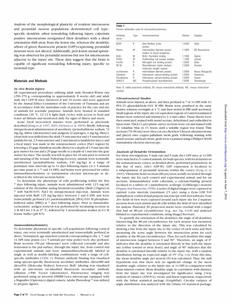
Table 1Primary antibodies used for immunohistochemistry
Antibodyname
Type Immunoreactivity Dilution Supplier
GFAP R Glial fibrillary acidicprotein
1:2000 Dako
Nestin M Intermediate filament proteinmarker of neural progenitor
1:1000 BD Biosciences
BrdU M BrdU, thymidine analog 1:1000 SigmaPCNA M Proliferating cell nuclear antigen 1:500 ZymedFerritin R Microglial iron binding protein 1:2000 DakoNF-M R Neurofilament triplet medium
molecular weight subunit1:2000 Serotec
a-internexin R Intermediate filament protein 1:2000 Novus BiologicalsCalretinin R Interneuron calcium-binding protein 1:2000 ChemiconParvalbumin R Interneuron calcium-binding protein 1:2000 SwantSMI312 MC Phosphorylated neurofilaments 1:5000 Sternberger
Note: R, rabbit polyclonal antibody; M, mouse monoclonal antibody; MC, mouse monoclonal
cocktail.
Page 2 of 11 Focal Damage to the Adult Rat Neocortex d Blizzard et al.
by guest on June 7, 2016http://cercor.oxfordjournals.org/
Dow
nloaded from
A limitation to analyzing changes in mean dendrite angle is that the
statistical analysis can demonstrate significant changes in orientation
relative to the control but cannot give an indicationof the directionof the
alteration. To determine the direction of any significant changes in
dendrite orientation, the polarity of all dendrites was determined for
calretinin positive neurons. The number of dendrites in 4 quadrants—
proximal, lateral 1 and2, anddistal (Fig. 7A), to the injury site, intersecting
at 80 lm from the cell body, was calculated to determine dendrite
polarity and analyzed using a 2-way ANOVA (InStat, GraphPad).
Confocal and Multiphoton MicroscopyAll confocal and multiphoton images collected in this study were
obtained on a Zeiss LSM 510 microscope, with Zen software. For
analysis of axon sprouting at 7 DPI, images were obtained with an argon
488 laser and a HeLa 543 laser. Both lasers were passed through either
a 203/0.8 NA plan-apochromat (Zeiss) objective to visualize the entire
injury site and a 643/0.8 NA plan-apochromat (Zeiss) oil immersion
objective for high-power images. For quantification of potential
dendritic changes, calretinin-immunolabeled cells were visualized with
the HeLa 594 laser passing through a 203/0.8 NA plan-apochromat
(Zeiss), the laser and pinhole settings remaining constant. GFP cells
were visualized with a Mai Tai multiphoton laser, at the wavelength 850
nm, with constant power, passing through a 203/1.0 DIC VIS IR W
plan-apochromat (Zeiss) water immersion objective.
Results
Focal Injury Induces an Active Healing ResponseCulminating in the Formation of a Glial Scar andNeovascularization
The cellular alterations following localized injury to the
neocortex were examined at 1, 7, and 14 DPI. Analysis of
cortical postinjury material sectioned in the horizontal plane
revealed distinct cellular changes as the response to injury
progressed, within this tissue core and surrounding the injury
site, that were less evident using traditional coronal sectioning.
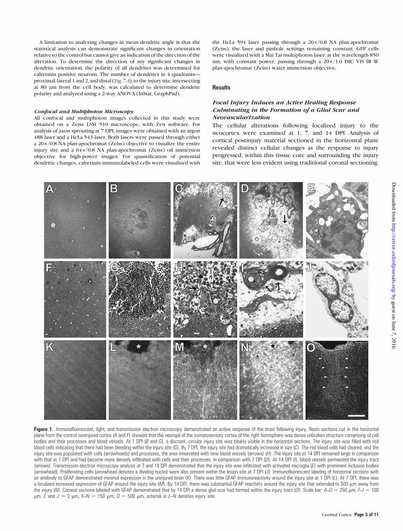
Figure 1. Immunofluorescent, light, and transmission electron microscopy demonstrated an active response of the brain following injury. Resin sections cut in the horizontalplane from the control noninjured cortex (A and F) showed that the neuropil of the somatosensory cortex of the right hemisphere was dense unbroken structure comprising of cellbodies and their processes and blood vessels. At 1 DPI (B and G), a discreet, circular injury site was clearly visible in the horizontal sections. The injury site was filled with redblood cells indicating that there had been bleeding within the injury site (G). By 7 DPI, the injury site had dramatically increased in size (C). The red blood cells had cleared, and theinjury site was populated with cells (arrowheads) and processes, the area innervated with new blood vessels (arrows) (H). The injury site at 14 DPI remained large in comparisonwith that at 1 DPI and had become more densely infiltrated with cells and their processes, in comparison with 7 DPI (D). At 14 DPI (I), blood vessels permeated the injury tract(arrows). Transmission electron microscopy analysis at 7 and 14 DPI demonstrated that the injury site was infiltrated with activated microglia (E) with prominent inclusion bodies(arrowhead). Proliferating cells (arrowhead denotes a dividing nuclei) were also present within the lesion site at 7 DPI (J). Immunofluorescent labeling of horizontal sections withan antibody to GFAP demonstrated minimal expression in the uninjured brain (K). There was little GFAP immunoreactivity around the injury site at 1 DPI (L). At 7 DPI, there wasa localized increased expression of GFAP around the injury site (M). By 14 DPI, there was substantial GFAP reactivity around the injury site that extended to 500 lm away fromthe injury (N). Coronal sections labeled with GFAP demonstrated that by 14 DPI a dense glial scar had formed within the injury tract (O). Scale bar: A--D 5 250 lm, F--I 5 100lm, E and J 5 2 lm, K--N 5 150 lm, O 5 500 lm, asterisk in L--N denotes injury site.
Cerebral Cortex Page 3 of 11
by guest on June 7, 2016http://cercor.oxfordjournals.org/
Dow
nloaded from
Focal neocortical injury resulted in substantial tissue de-
struction at the site of the lesion and subsequent alterations
indicative of attempted brain repair. Microscopic analysis
demonstrated no detectable histopathological changes in
control brains or in brain regions contralateral to the lesion
site, evidenced with both resin sections (Fig. 1A,F) and also
with immunolabeling for glial fibrillary acidic protein (GFAP)
(Fig. 1K). The neuropil in control noninjured resin sections was
essentially homogenous, tightly associated and dense (Fig.
1A,F). At 1 DPI, tissue destruction was noted within the injury
site, with a clear needle tract being visible (Fig. 1B). This lesion
site at 1 DPI was characterized by massive red blood cell
infiltration (Fig. 1G). By 7 DPI, the injury site had increased in
size and now ranged from 200 to 300 lm in diameter. The
blood cells that were present at 1 DPI had cleared, and
neovascularization of the lesion site had commenced with the
area infiltrated by occasional new blood vessels and sparsely
populated with cells and processes (Fig. 1C). The neuropil
within the injury site, however, remained significantly less
dense than surrounding tissue (Fig. 1H). Immunolabeling for
GFAP demonstrated that the injury and peri-lesion site had now
become filled with reactive astrocytes with their processes
directed toward the central lesion site (Fig. 1M). At 14 DPI,
blood vessels permeated the injury tract (Fig. 1D,I, arrows) and
a significant proportion of the tract had closed together leaving
a central dense GFAP-labeled core (Fig. 1N) (King et al. 2001),
with a glial scar extending approximately 500 lm from the
edges of the lesion (Fig. 1N,O). GFAP immunolabeling was
greatest directly adjacent to the injury site, as indicated by
increased immunoreactivity, with this labeling progressively
decreasing with increasing distance from the injury (Fig. 1N,O).
Ultrastructural studies of the central lesion site at 7 and 14 DPI
demonstrate, in support of immunohistochemical analysis, the
presence of activated microglia with inclusions (Fig. 1E) and
also proliferating cells (Fig. 1J).
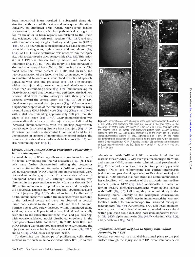
Cortical Injury Induces Neural Progenitor Proliferationbut not Neurogenesis
As noted above, proliferating cells were a prominent feature of
the tissue surrounding the injured neocortex (Fig. 1J). These
cells were further characterized utilizing the progenitor
marker nestin and the mitotic markers, BrdU and proliferating
cell nuclear antigen (PCNA). Nestin immunoreactive cells were
not evident in the gray matter of the neocortex of control
noninjured brains (Fig. 2A), although some labeling was
observed in the periventricular region (data not shown). By 7
DPI, nestin immunoreactive profiles were localized throughout
the neocortical laminae and were especially abundant adjacent
to the injury site (Fig. 2B,C). Alterations in the localization of
nestin immunoreactivity within the neocortex were restricted
to the ipsilateral cortex and were not observed in cortical
tissue contralateral to the lesion. BrdU and PCNA immuno-
positive nuclei were rarely observed within noninjured tissue
sections, where cell proliferation was limited and generally
restricted to the subventricular zone (SVZ) and pial covering,
with occasional-labeled nuclei distributed elsewhere in the
brain parenchyma (data not shown). By 7 DPI, both BrdU and
PCNA labeling was distributed in a broad zone surrounding the
injury site and extending into the corpus callosum (Fig. 2D,E)
and SVZ (Fig. 2F,G), colocalizing with nestin.
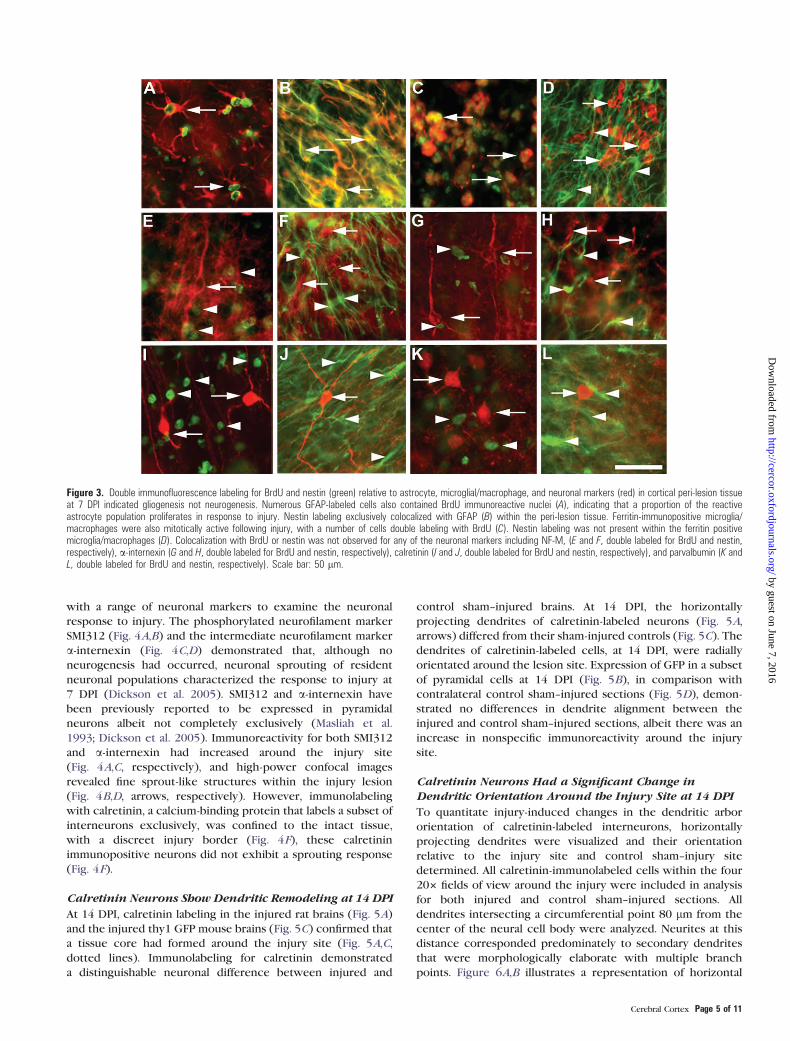
To determine the phenotype of proliferating cells, tissue
sections were double immunolabeled for either BrdU, in animals
administered with BrdU at 1 DPI, or nestin, in addition to
markers for astrocytes (GFAP), microglia/macrophages (ferritin),
and neurons (NF-M, a-internexin, calretinin, and parvalbumin)
(Fig. 3). Neuronal markers were selected to represent pyramidal
neuron (NF-M and a-internexin) and cortical interneuron
(calretinin and parvalbumin) populations. Examination of injured
tissue at 7 DPI showed that both BrdU and nestin immunolabel-
ing colocalized with expression of the astrocytic intermediate
filament protein, GFAP (Fig. 3A,B). Additionally, a number of
ferritin positive microglia/macrophages were double labeled
with BrdU (Fig. 3C) indicating they were mitotically active
following injury. Contrary to the widespread colocalization
between nestin and GFAP, nestin immunoreactivity was not
localized within ferritin-immunopositive activated microglia/
macrophages (Fig. 3D). Furthermore, BrdU and nestin immuno-
reactivity were absent from all neuronal populations examined
within peri-lesion tissue, including those immunopositive for NF-
M (Fig. 3E,F), alpha-internexin (Fig. 3G,H), calretinin (Fig. 3I,J),
and parvalbumin (Fig. 3K,L).
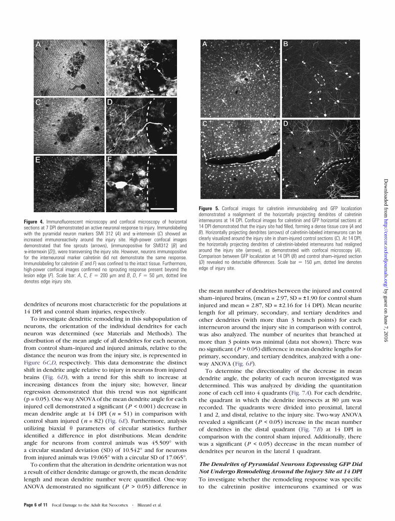
Pyramidal Neurons Respond to Injury with AxonalSprouting by 7 DPI
Cortical sections, cut in a parallel/horizontal plane to the pial
surface through the injury site at 7 DPI, were immunolabeled
Figure 2. Immunofluorescence labeling for nestin was increased within the cortex at7 DPI. Nestin immunoreactive cells were not evident in the gray matter of theneocortex of control noninjured brains (A), but by 7 DPI were abundant withinthe lesioned tissue (B). Nestin immunoreactive profiles were present in tissueextending from the SVZ and corpus callosum up to the injury site (C). Doubleimmunofluorescence labeling for BrdU, administered at 1 DPI, (D) and nestin(E) demonstrated localization of these markers to the same cells within peri-lesiontissue. Double labeling for PCNA (F) relative to nestin (G) confirmed the proliferationof nestin-labeled cells within the SVZ. Scale bar: A and B 5 100 lm, C 5 800 lm,D--G 5 50 lm.
Page 4 of 11 Focal Damage to the Adult Rat Neocortex d Blizzard et al.
by guest on June 7, 2016http://cercor.oxfordjournals.org/
Dow
nloaded from
with a range of neuronal markers to examine the neuronal
response to injury. The phosphorylated neurofilament marker
SMI312 (Fig. 4A,B) and the intermediate neurofilament marker
a-internexin (Fig. 4C,D) demonstrated that, although no
neurogenesis had occurred, neuronal sprouting of resident
neuronal populations characterized the response to injury at
7 DPI (Dickson et al. 2005). SMI312 and a-internexin have
been previously reported to be expressed in pyramidal
neurons albeit not completely exclusively (Masliah et al.
1993; Dickson et al. 2005). Immunoreactivity for both SMI312
and a-internexin had increased around the injury site
(Fig. 4A,C, respectively), and high-power confocal images
revealed fine sprout-like structures within the injury lesion
(Fig. 4B,D, arrows, respectively). However, immunolabeling
with calretinin, a calcium-binding protein that labels a subset of
interneurons exclusively, was confined to the intact tissue,
with a discreet injury border (Fig. 4F), these calretinin
immunopositive neurons did not exhibit a sprouting response
(Fig. 4F).
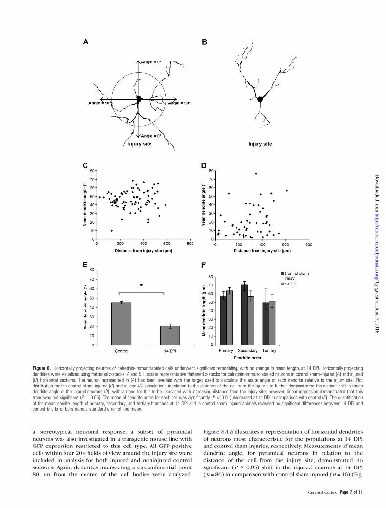
Calretinin Neurons Show Dendritic Remodeling at 14 DPI
At 14 DPI, calretinin labeling in the injured rat brains (Fig. 5A)
and the injured thy1 GFP mouse brains (Fig. 5C) confirmed that
a tissue core had formed around the injury site (Fig. 5A,C,
dotted lines). Immunolabeling for calretinin demonstrated
a distinguishable neuronal difference between injured and
control sham--injured brains. At 14 DPI, the horizontally
projecting dendrites of calretinin-labeled neurons (Fig. 5A,
arrows) differed from their sham-injured controls (Fig. 5C). The
dendrites of calretinin-labeled cells, at 14 DPI, were radially
orientated around the lesion site. Expression of GFP in a subset
of pyramidal cells at 14 DPI (Fig. 5B), in comparison with
contralateral control sham--injured sections (Fig. 5D), demon-
strated no differences in dendrite alignment between the
injured and control sham--injured sections, albeit there was an
increase in nonspecific immunoreactivity around the injury
site.
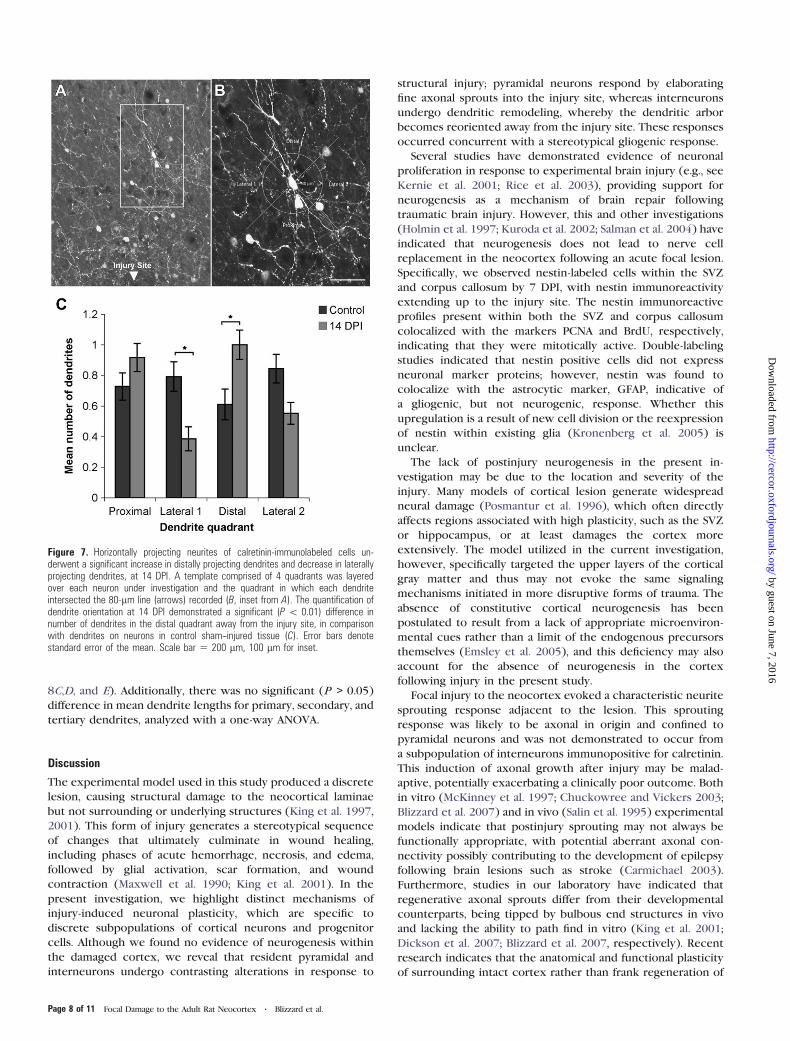
Calretinin Neurons Had a Significant Change inDendritic Orientation Around the Injury Site at 14 DPI
To quantitate injury-induced changes in the dendritic arbor
orientation of calretinin-labeled interneurons, horizontally
projecting dendrites were visualized and their orientation
relative to the injury site and control sham--injury site
determined. All calretinin-immunolabeled cells within the four
203 fields of view around the injury were included in analysis
for both injured and control sham--injured sections. All
dendrites intersecting a circumferential point 80 lm from the
center of the neural cell body were analyzed. Neurites at this
distance corresponded predominately to secondary dendrites
that were morphologically elaborate with multiple branch
points. Figure 6A,B illustrates a representation of horizontal
Figure 3. Double immunofluorescence labeling for BrdU and nestin (green) relative to astrocyte, microglial/macrophage, and neuronal markers (red) in cortical peri-lesion tissueat 7 DPI indicated gliogenesis not neurogenesis. Numerous GFAP-labeled cells also contained BrdU immunoreactive nuclei (A), indicating that a proportion of the reactiveastrocyte population proliferates in response to injury. Nestin labeling exclusively colocalized with GFAP (B) within the peri-lesion tissue. Ferritin-immunopositive microglia/macrophages were also mitotically active following injury, with a number of cells double labeling with BrdU (C). Nestin labeling was not present within the ferritin positivemicroglia/macrophages (D). Colocalization with BrdU or nestin was not observed for any of the neuronal markers including NF-M, (E and F, double labeled for BrdU and nestin,respectively), a-internexin (G and H, double labeled for BrdU and nestin, respectively), calretinin (I and J, double labeled for BrdU and nestin, respectively), and parvalbumin (K andL, double labeled for BrdU and nestin, respectively). Scale bar: 50 lm.
Cerebral Cortex Page 5 of 11
by guest on June 7, 2016http://cercor.oxfordjournals.org/
Dow
nloaded from
dendrites of neurons most characteristic for the populations at
14 DPI and control sham injuries, respectively.
To investigate dendritic remodeling in this subpopulation of
neurons, the orientation of the individual dendrites for each
neuron was determined (see Materials and Methods). The
distribution of the mean angle of all dendrites for each neuron,
from control sham--injured and injured animals, relative to the
distance the neuron was from the injury site, is represented in
Figure 6C,D, respectively. This data demonstrate the distinct
shift in dendrite angle relative to injury in neurons from injured
brains (Fig. 6D), with a trend for this shift to increase at
increasing distances from the injury site; however, linear
regression demonstrated that this trend was not significant
(p = 0.05). One-way ANOVA of the mean dendrite angle for each
injured cell demonstrated a significant (P < 0.001) decrease in
mean dendrite angle at 14 DPI (n = 51) in comparison with
control sham injured (n = 82) (Fig. 6E). Furthermore, analysis
utilizing biaxial h parameters of circular statistics further
identified a difference in plot distributions. Mean dendrite
angle for neurons from control animals was 45.509� with
a circular standard deviation (SD) of 10.542� and for neurons
from injured animals was 19.065� with a circular SD of 17.065�.To confirm that the alteration in dendrite orientation was not
a result of either dendrite damage or growth, the mean dendrite
length and mean dendrite number were quantified. One-way
ANOVA demonstrated no significant (P > 0.05) difference in
the mean number of dendrites between the injured and control
sham--injured brains, (mean = 2.97, SD = ±1.90 for control sham
injured and mean = 2.87, SD = ±2.16 for 14 DPI). Mean neurite
length for all primary, secondary, and tertiary dendrites and
other dendrites (with more than 3 branch points) for each
interneuron around the injury site in comparison with control,
was also analyzed. The number of neurites that branched at
more than 3 points was minimal (data not shown). There was
no significant (P > 0.05) difference in mean dendrite lengths for
primary, secondary, and tertiary dendrites, analyzed with a one-
way ANOVA (Fig. 6F).
To determine the directionality of the decrease in mean
dendrite angle, the polarity of each neuron investigated was
determined. This was analyzed by dividing the quantitation
zone of each cell into 4 quadrants (Fig. 7A). For each dendrite,
the quadrant in which the dendrite intersects at 80 lm was
recorded. The quadrants were divided into proximal, lateral
1 and 2, and distal, relative to the injury site. Two-way ANOVA
revealed a significant (P < 0.05) increase in the mean number
of dendrites in the distal quadrant (Fig. 7B) at 14 DPI in
comparison with the control sham injured. Additionally, there
was a significant (P < 0.05) decrease in the mean number of
dendrites per neuron in the lateral 1 quadrant.
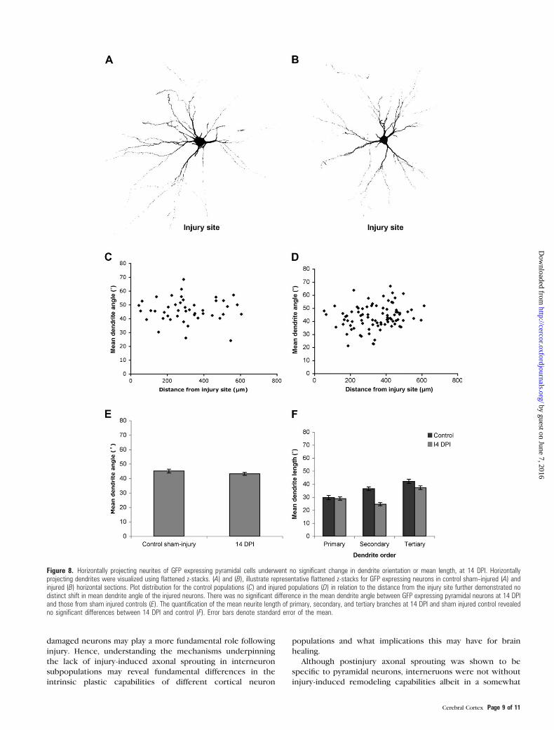
The Dendrites of Pyramidal Neurons Expressing GFP DidNot Undergo Remodeling Around the Injury Site at 14 DPI
To investigate whether the remodeling response was specific
to the calretinin positive interneurons examined or was
Figure 4. Immunofluorescent microscopy and confocal microscopy of horizontalsections at 7 DPI demonstrated an active neuronal response to injury. Immunolabelingwith the pyramidal neuron markers SMI 312 (A) and a-internexin (C) showed anincreased immunoreactivity around the injury site. High-power confocal imagesdemonstrated that fine sprouts (arrows), (immunopositive for SMI312 [B] anda-internexin [D]), were transversing the injury site. However, neurons immunopositivefor the interneuronal marker calretinin did not demonstrate the same response.Immunolabeling for calretinin (E and F) was confined to the intact tissue. Furthermore,high-power confocal images confirmed no sprouting response present beyond thelesion edge (F). Scale bar: A, C, E 5 200 lm and B, D, F 5 50 lm, dotted linedenotes edge injury site.
Figure 5. Confocal images for calretinin immunolabeling and GFP localizationdemonstrated a realignment of the horizontally projecting dendrites of calretinininterneurons at 14 DPI. Confocal images for calretinin and GFP horizontal sections at14 DPI demonstrated that the injury site had filled, forming a dense tissue core (A andB). Horizontally projecting dendrites (arrows) of calretinin-labeled interneurons can beclearly visualized around the injury site in sham-injured control sections (C). At 14 DPI,the horizontally projecting dendrites of calretinin-labeled interneurons had realignedaround the injury site (arrows), as demonstrated with confocal microscopy (A).Comparison between GFP localization at 14 DPI (B) and control sham--injured section(D) revealed no detectable differences. Scale bar 5 150 lm, dotted line denotesedge of injury site.
Page 6 of 11 Focal Damage to the Adult Rat Neocortex d Blizzard et al.
by guest on June 7, 2016http://cercor.oxfordjournals.org/
Dow
nloaded from
a stereotypical neuronal response, a subset of pyramidal
neurons was also investigated in a transgenic mouse line with
GFP expression restricted to this cell type. All GFP positive
cells within four 203 fields of view around the injury site were
included in analysis for both injured and noninjured control
sections. Again, dendrites intersecting a circumferential point
80 lm from the center of the cell bodies were analyzed.
Figure 8A,B illustrates a representation of horizontal dendrites
of neurons most characteristic for the populations at 14 DPI
and control sham injuries, respectively. Measurements of mean
dendrite angle, for pyramidal neurons in relation to the
distance of the cell from the injury site, demonstrated no
significant (P > 0.05) shift in the injured neurons at 14 DPI
(n = 86) in comparison with control sham injured (n = 46) (Fig.
Figure 6. Horizontally projecting neurites of calretinin-immunolabeled cells underwent significant remodeling, with no change in mean length, at 14 DPI. Horizontally projectingdendrites were visualized using flattened z-stacks. A and B illustrate representative flattened z-stacks for calretinin-immunolabeled neurons in control sham--injured (A) and injured(B) horizontal sections. The neuron represented in (A) has been overlaid with the target used to calculate the acute angle of each dendrite relative to the injury site. Plotdistribution for the control sham--injured (C) and injured (D) populations in relation to the distance of the cell from the injury site further demonstrated the distinct shift in meandendrite angle of the injured neurons (D), with a trend for this to be increased with increasing distance from the injury site; however, linear regression demonstrated that thistrend was not significant (P5 0.05). The mean of dendrite angle for each cell was significantly (P\ 0.01) decreased at 14 DPI in comparison with control (E). The quantificationof the mean neurite length of primary, secondary, and tertiary branches at 14 DPI and in control sham injured animals revealed no significant differences between 14 DPI andcontrol (F). Error bars denote standard error of the mean.
Cerebral Cortex Page 7 of 11
by guest on June 7, 2016http://cercor.oxfordjournals.org/
Dow
nloaded from
8C,D, and E). Additionally, there was no significant (P > 0.05)
difference in mean dendrite lengths for primary, secondary, and
tertiary dendrites, analyzed with a one-way ANOVA.
Discussion
The experimental model used in this study produced a discrete
lesion, causing structural damage to the neocortical laminae
but not surrounding or underlying structures (King et al. 1997,
2001). This form of injury generates a stereotypical sequence
of changes that ultimately culminate in wound healing,
including phases of acute hemorrhage, necrosis, and edema,
followed by glial activation, scar formation, and wound
contraction (Maxwell et al. 1990; King et al. 2001). In the
present investigation, we highlight distinct mechanisms of
injury-induced neuronal plasticity, which are specific to
discrete subpopulations of cortical neurons and progenitor
cells. Although we found no evidence of neurogenesis within
the damaged cortex, we reveal that resident pyramidal and
interneurons undergo contrasting alterations in response to
structural injury; pyramidal neurons respond by elaborating
fine axonal sprouts into the injury site, whereas interneurons
undergo dendritic remodeling, whereby the dendritic arbor
becomes reoriented away from the injury site. These responses
occurred concurrent with a stereotypical gliogenic response.
Several studies have demonstrated evidence of neuronal
proliferation in response to experimental brain injury (e.g., see
Kernie et al. 2001; Rice et al. 2003), providing support for
neurogenesis as a mechanism of brain repair following
traumatic brain injury. However, this and other investigations
(Holmin et al. 1997; Kuroda et al. 2002; Salman et al. 2004) have
indicated that neurogenesis does not lead to nerve cell
replacement in the neocortex following an acute focal lesion.
Specifically, we observed nestin-labeled cells within the SVZ
and corpus callosum by 7 DPI, with nestin immunoreactivity
extending up to the injury site. The nestin immunoreactive
profiles present within both the SVZ and corpus callosum
colocalized with the markers PCNA and BrdU, respectively,
indicating that they were mitotically active. Double-labeling
studies indicated that nestin positive cells did not express
neuronal marker proteins; however, nestin was found to
colocalize with the astrocytic marker, GFAP, indicative of
a gliogenic, but not neurogenic, response. Whether this
upregulation is a result of new cell division or the reexpression
of nestin within existing glia (Kronenberg et al. 2005) is
unclear.
The lack of postinjury neurogenesis in the present in-
vestigation may be due to the location and severity of the
injury. Many models of cortical lesion generate widespread
neural damage (Posmantur et al. 1996), which often directly
affects regions associated with high plasticity, such as the SVZ
or hippocampus, or at least damages the cortex more
extensively. The model utilized in the current investigation,
however, specifically targeted the upper layers of the cortical
gray matter and thus may not evoke the same signaling
mechanisms initiated in more disruptive forms of trauma. The
absence of constitutive cortical neurogenesis has been
postulated to result from a lack of appropriate microenviron-
mental cues rather than a limit of the endogenous precursors
themselves (Emsley et al. 2005), and this deficiency may also
account for the absence of neurogenesis in the cortex
following injury in the present study.
Focal injury to the neocortex evoked a characteristic neurite
sprouting response adjacent to the lesion. This sprouting
response was likely to be axonal in origin and confined to
pyramidal neurons and was not demonstrated to occur from
a subpopulation of interneurons immunopositive for calretinin.
This induction of axonal growth after injury may be malad-
aptive, potentially exacerbating a clinically poor outcome. Both
in vitro (McKinney et al. 1997; Chuckowree and Vickers 2003;
Blizzard et al. 2007) and in vivo (Salin et al. 1995) experimental
models indicate that postinjury sprouting may not always be
functionally appropriate, with potential aberrant axonal con-
nectivity possibly contributing to the development of epilepsy
following brain lesions such as stroke (Carmichael 2003).
Furthermore, studies in our laboratory have indicated that
regenerative axonal sprouts differ from their developmental
counterparts, being tipped by bulbous end structures in vivo
and lacking the ability to path find in vitro (King et al. 2001;
Dickson et al. 2007; Blizzard et al. 2007, respectively). Recent
research indicates that the anatomical and functional plasticity
of surrounding intact cortex rather than frank regeneration of
Figure 7. Horizontally projecting neurites of calretinin-immunolabeled cells un-derwent a significant increase in distally projecting dendrites and decrease in laterallyprojecting dendrites, at 14 DPI. A template comprised of 4 quadrants was layeredover each neuron under investigation and the quadrant in which each dendriteintersected the 80-lm line (arrows) recorded (B, inset from A). The quantification ofdendrite orientation at 14 DPI demonstrated a significant (P \ 0.01) difference innumber of dendrites in the distal quadrant away from the injury site, in comparisonwith dendrites on neurons in control sham--injured tissue (C). Error bars denotestandard error of the mean. Scale bar 5 200 lm, 100 lm for inset.
Page 8 of 11 Focal Damage to the Adult Rat Neocortex d Blizzard et al.
by guest on June 7, 2016http://cercor.oxfordjournals.org/
Dow
nloaded from
damaged neurons may play a more fundamental role following
injury. Hence, understanding the mechanisms underpinning
the lack of injury-induced axonal sprouting in interneuron
subpopulations may reveal fundamental differences in the
intrinsic plastic capabilities of different cortical neuron
populations and what implications this may have for brain
healing.
Although postinjury axonal sprouting was shown to be
specific to pyramidal neurons, interneruons were not without
injury-induced remodeling capabilities albeit in a somewhat
Figure 8. Horizontally projecting neurites of GFP expressing pyramidal cells underwent no significant change in dendrite orientation or mean length, at 14 DPI. Horizontallyprojecting dendrites were visualized using flattened z-stacks. (A) and (B), illustrate representative flattened z-stacks for GFP expressing neurons in control sham--injured (A) andinjured (B) horizontal sections. Plot distribution for the control populations (C) and injured populations (D) in relation to the distance from the injury site further demonstrated nodistinct shift in mean dendrite angle of the injured neurons. There was no significant difference in the mean dendrite angle between GFP expressing pyramidal neurons at 14 DPIand those from sham injured controls (E). The quantification of the mean neurite length of primary, secondary, and tertiary branches at 14 DPI and sham injured control revealedno significant differences between 14 DPI and control (F). Error bars denote standard error of the mean.
Cerebral Cortex Page 9 of 11
by guest on June 7, 2016http://cercor.oxfordjournals.org/
Dow
nloaded from
contrasting manner. In this investigation, we show the
particularly novel property of a subpopulation of interneurons,
namely those that were calretinin immunoreactive, to remodel
their dendritic arbors in response to acute brain lesion. Cortical
interneurons within the barrel cortex have previously been
shown to have a capacity for morphological plasticity (Lee et al.
2006); however, this was under physiologically normal con-
ditions. Furthermore, previous investigations have suggested
that the potential sites for structural plasticity are the
horizontal connections within the superficial layers of the
cortex (Kaas et al. 1990; Darian-Smith and Gilbert 1994; Lee
et al. 2008). Following acute brain injury in the current study,
the dendritic arbor of calretinin positive interneurons was
orientated away from the injury site, relative to sham-injured
controls. Although it was postulated that this change in
dendritic orientation was due to new dendrite growth in
response to injury, further analyses of both mean dendrite
number and mean neurite length revealed that neither
parameter was altered as a result of the injury. Hence, the
alteration in dendrite alignment was most likely a result of
remodeling of existing arbors, such that those close to the
injury retracted, while those distal to the injury grew. These
findings are consistent with studies demonstrating that GABA
positive interneurons exhibit dynamic arbor rearrangements
while pyramidal populations remain stable, under physiologi-
cally normal conditions (Frahm et al. 2004).
A subset of pyramidal neurons, specifically those expressing
GFP in the transgenic mouse, was also investigated to determine
if this remodeling was cell-type specific. While the injury site in
these mice had formed a comparable tissue core by 14 DPI,
indicating an active brain response, statistical analysis revealed
no significant remodeling in the dendrites of these pyramidal
neurons. Thus, the dendritic remodeling observed at around the
injury site, in our model of brain injury, may be confined to
interneurons, specifically the calretinin positive population,
demonstrating a cell type--specific response to injury. These
findings are in coherence with studies demonstrating that the
GABA positive interneurons exhibit dynamic arbor rearrange-
ments while pyramidal populations remain stable, under
physiologically normal conditions (Lee et al. 2006).
Previous investigations have demonstrated that the out-
growth of dendrites following injury can be correlated to
axonal rearrangement and, interestingly, that alterations in
dendritic morphology are associated with synaptic modifica-
tions (for recent review, see Macias 2008). Hence, the dendritic
reorientation observed in the current study may represent
structural plasticity directed toward the intact cortex. The
injury-induced generation of aberrant axonal connectivity may
contribute to the development of epilepsy following various
forms of brain lesion (Carmichael 2003). It is also possible that
alterations in the balance of inhibitory synapses, for example,
resulting from remodeling of dendritic architecture as ob-
served in the current injury paradigm, may also play a role in
the formation of aberrant connectivity following some forms of
brain lesion. However, whether this plasticity ultimately
culminates in the formation of new functional or dysfunctional
connections remains to be elucidated.
Conclusions
The adult brain has a frequently unappreciated capacity for
cytoarchitectural remodeling and repair following injury. These
studies demonstrate a novel cell type--specific response of
neuronal populations to acute injury; a subpopulation of
pyramidal neurons elaborated axonal sprouts into the injury site,
whereas a subpopulation of interneurons (calretinin neurons)
underwent a reorganization of their dendrites to project more
distally away for the injury site. These distinct responses,
exhibited by different cortical neuron populations, provide novel
insight into the plastic potential of the mature brain to injury.
Funding
National Health and Medical Research Council of Australia; the
JO and JR Wicking Trust (ANZ Trustees); Tasmanian Masonic
Centenary Research Foundation.
Notes
Conflict of Interest : None declared.
References
Arvidsson A, Collin T, Kirik D, Kokaia Z, Lindvall O. 2002. Neuronal
replacement from endogenous precursors in the adult brain after
stroke. Nat Med. 8(9):963--970.
Blizzard CA, Haas MA, Vickers JC, Dickson TC. 2007. Cellular dynamics
underlying regeneration of damaged axons differs from initial axon
development. Eur J Neurosci. 26(5):1100--1108.
Brown CE, Boyd JD, Murphy TH. 2010. Longitudinal in vivo imaging
reveals balanced and branch-specific remodeling of mature cortical
pyramidal dendritic arbors after stroke. J Cereb Blood Flow Metab.
30(4):783--791.
Brown CE, Li P, Boyd JD, Delaney KR, Murphy TH. 2007. Extensive
turnover of dendritic spines and vascular remodeling in cortical
tissues recovering from stroke. J Neurosci. 27(15):4101--4109.
Brown CE, Wong C, Murphy TH. 2008. Rapid morphologic plasticity of
peri-infarct dendritic spines after focal ischemic stroke. Stroke.
39(4):1286--1291.
Carmichael ST. 2003. Plasticity of cortical projections after stroke.
Neuroscientist. 9(1):64--75.
Carmichael ST. 2006. Cellular and molecular mechanisms of neural
repair after stroke: making waves. Ann Neurol. 59(5):735--742.
Chen XH, Iwata A, Nonaka M, Browne KD, Smith DH. 2003.
Neurogenesis and glial proliferation persist for at least one year in
the subventricular zone following brain trauma in rats. J Neuro-
trauma. 20(7):623--631.
Chuckowree JA, Dickson TC, Vickers JC. 2004. Intrinsic regenerative
ability of mature CNS neurons. Neuroscientist. 10(4):280--285.
Chuckowree JA, Vickers JC. 2003. Cytoskeletal and morphological
alterations underlying axonal sprouting after localized transection
of cortical neuron axons in vitro. J Neurosci. 23(9):3715--3725.
Clarke SR, Shetty AK, Bradley JL, Turner DA. 1994. Reactive astrocytes
express the embryonic intermediate neurofilament nestin. Neuro-
report. 5(15):1885--1888.
Darian-Smith C, Gilbert CD. 1994. Axonal sprouting accompanies
functional reorganization in adult cat striate cortex. Nature.
368(6473):737--740.
Di Filippo M, Tozzi A, Costa C, Belcastro V, Tantucci M, Picconi B,
Calabresi P. 2008. Plasticity and repair in the post-ischemic brain.
Neuropharmacology. 55(3):353--362.
Dickson TC, Chuckowree JA, Chuah MI, West AK, Vickers JC. 2005.
alpha-Internexin immunoreactivity reflects variable neuronal vul-
nerability in Alzheimer’s disease and supports the role of the beta-
amyloid plaques in inducing neuronal injury. Neurobiol Dis.
18(2):286--295.
Dickson TC, Chung RS, McCormack GH, Staal JA, Vickers JC. 2007.
Acute reactive and regenerative changes in mature cortical axons
following injury. Neuroreport. 18(3):283--288.
Douen AG, Dong L, Vanance S, Munger R, Hogan MJ, Thompson CS,
Hakim AM. 2004. Regulation of nestin expression after cortical
ablation in adult rat brain. Brain Res. 1008(2):139--146.
Page 10 of 11 Focal Damage to the Adult Rat Neocortex d Blizzard et al.
by guest on June 7, 2016http://cercor.oxfordjournals.org/
Dow
nloaded from

Duggal N, Schmidt-Kastner R, Hakim AM. 1997. Nestin expression in
reactive astrocytes following focal cerebral ischemia in rats. Brain
Res. 768(1--2):1--9.
Dunlop SA. 2008. Activity-dependent plasticity: implications for re-
covery after spinal cord injury. Trends Neurosci. 31(8):410--418.
Edgerton VR, Tillakaratne NJ, Bigbee AJ, de Leon RD, Roy RR. 2004.
Plasticity of the spinal neural circuitry after injury. Annu Rev
Neurosci. 27:145--167.
Emsley JG, Mitchell BD, Kempermann G, Macklis JD. 2005. Adult
neurogenesis and repair of the adult CNS with neural progenitors,
precursors, and stem cells. Prog Neurobiol. 75(5):321--341.
Fawcett JW, Asher RA. 1999. The glial scar and central nervous system
repair. Brain Res Bull. 49(6):377--391.
Feng G, Mellor RH, Bernstein M, Keller-Peck C, Nguyen QT, Wallace M,
Nerbonne JM, Lichtman JW, Sanes JR. 2000. Imaging neuronal
subsets in transgenic mice expressing multiple spectral variants of
GFP. Neuron. 28(1):41--51.
Fitzgerald J, Fawcett J. 2007. Repair in the central nervous system.
J Bone Joint Surg Br. 89(11):1413--1420.
Frahm C, Haupt C, Witte OW. 2004. GABA neurons survive focal
ischemic injury. Neuroscience. 127(2):341--346.
Grutzendler J, Kasthuri N, Gan WB. 2002. Long-term dendritic spine
stability in the adult cortex. Nature. 420(6917):812--816.
Holmin S, Almqvist P, Lendahl U, Mathiesen T. 1997. Adult nestin-
expressing subependymal cells differentiate to astrocytes in re-
sponse to brain injury. Eur J Neurosci. 9(1):65--75.
Holtmaat AJ, Trachtenberg JT, Wilbrecht L, Shepherd GM, Zhang X,
Knott GW, Svoboda K. 2005. Transient and persistent dendritic
spines in the neocortex in vivo. Neuron. 45(2):279--291.
Jones TA, Schallert T. 1992. Overgrowth and pruning of dendrites in
adult rats recovering from neocortical damage. Brain Res.
581(1):156--160.
Kaas JH, Krubitzer LA, Chino YM, Langston AL, Polley EH, Blair N. 1990.
Reorganization of retinotopic cortical maps in adult mammals after
lesions of the retina. Science. 248(4952):229--231.
Kernie SG, Erwin TM, Parada LF. 2001. Brain remodeling due to
neuronal and astrocytic proliferation after controlled cortical injury
in mice. J Neurosci Res. 66(3):317--326.
King CE, Canty AJ, Vickers JC. 2001. Alterations in neurofilaments
associated with reactive brain changes and axonal sprouting
following acute physical injury to the rat neocortex. Neuropathol
Appl Neurobiol. 27(2):115--126.
King CE, Jacobs I, Dickson TC, Vickers JC. 1997. Physical damage to rat
cortical axons mimics early Alzheimer’s neuronal pathology. Neuro-
report. 8(7):1663--1665.
Knott GW, Quairiaux C, Genoud C, Welker E. 2002. Formation of
dendritic spines with GABAergic synapses induced by whisker
stimulation in adult mice. Neuron. 34(2):265--273.
Kolb B, Gibb R. 1991. Environmental enrichment and cortical injury:
behavioral and anatomical consequences of frontal cortex lesions.
Cereb Cortex. 1(2):189--198.
Kronenberg G, Wang LP, Synowitz M, Gertz K, Katchanov J, Glass R,
Harms C, Kempermann G, Kettenmann H, Endres M. 2005. Nestin-
expressing cells divide and adopt a complex electrophysiologic
phenotype after transient brain ischemia. J Cereb Blood Flow Metab.
25(12):1613--1624.
Kuroda T, Nakamura H, Itoh K, Le WR, Yoshimura S, Takenaka K, Sakai N.
2002.Nestin immunoreactivity in localneuronsof the adult rat striatum
after remote cortical injury. J Chem Neuroanat. 24(2):137--146.
Lee WC, Chen JL, Huang H, Leslie JH, Amitai Y, So PT, Nedivi E. 2008. A
dynamic zone defines interneuron remodeling in the adult neo-
cortex. Proc Natl Acad Sci U S A. 105(50):19968--19973.
Lee WC, Huang H, Feng G, Sanes JR, Brown EN, So PT, Nedivi E. 2006.
Dynamic remodeling of dendritic arbors in GABAergic interneurons
of adult visual cortex. PLoS Biol. 4(2):e29.
Macias M. 2008. Injury induced dendritic plasticity in the mature
central nervous system. Acta Neurobiol Exp (Wars). 68(2):334--346.
Majewska A, Sur M. 2003. Motility of dendritic spines in visual cortex in
vivo: changes during the critical period and effects of visual
deprivation. Proc Natl Acad Sci U S A. 100(26):16024--16029.
Majewska AK, Newton JR, Sur M. 2006. Remodeling of synaptic
structure in sensory cortical areas in vivo. J Neurosci.
26(11):3021--3029.
Masliah E, Mallory M, Ge N, Godson C, Saitoh T. 1993. Phorbol ester-
induced neuritic alterations in the rat neocortex. Structural and
immunocytochemical studies. Mol Chem Neuropathol. 20(2):
125--145.
Maxwell WL, Follows R, Ashhurst DE, Berry M. 1990. The response of
the cerebral hemisphere of the rat to injury. I. The mature rat. Philos
Trans R Soc Lond B Biol Sci. 328(1250):479--500.
McKinney RA, Debanne D, Gahwiler BH, Thompson SM. 1997. Lesion-
induced axonal sprouting and hyperexcitability in the hippocampus
in vitro: implications for the genesis of posttraumatic epilepsy. Nat
Med. 3(9):990--996.
Pappas IS, Parnavelas JG. 1998. Basic fibroblast growth factor promotes
the generation and differentiation of calretinin neurons in the rat
cerebral cortex in vitro. Eur J Neurosci. 10(4):1436--1445.
Portera-Cailliau C, Pan DT, Yuste R. 2003. Activity-regulated dynamic
behavior of early dendritic protrusions: evidence for different types
of dendritic filopodia. J Neurosci. 23(18):7129--7142.
Posmantur RM, Kampfl A, Taft WC, Bhattacharjee M, Dixon CE, Bao J,
Hayes RL. 1996. Diminished microtubule-associated protein 2
(MAP2) immunoreactivity following cortical impact brain injury.
J Neurotrauma. 13(3):125--137.
Rice AC, Khaldi A, Harvey HB, Salman NJ, White F, Fillmore H,
Bullock MR, Eriksson PS, Perfilieva E, Bjork-Eriksson T, et al. 2003.
Proliferation and neuronal differentiation of mitotically active cells
following traumatic brain injury. Exp Neurol. 183(2):406--417.
Salin P, Tseng GF, Hoffman S, Parada I, Prince DA. 1995. Axonal
sprouting in layer V pyramidal neurons of chronically injured
cerebral cortex. J Neurosci. 15(12):8234--8245.
Salman H, Ghosh P, Kernie SG. 2004. Subventricular zone neural stem
cells remodel the brain following traumatic injury in adult mice.
J Neurotrauma. 21(3):283--292.
Tailby C, Wright LL, Metha AB, Calford MB. 2005. Activity-dependent
maintenance and growth of dendrites in adult cortex. Proc Natl
Acad Sci U S A. 102(12):4631--4636.
Trachtenberg JT, Chen BE, Knott GW, Feng G, Sanes JR, Welker E,
Svoboda K. 2002. Long-term in vivo imaging of experience-
dependent synaptic plasticity in adult cortex. Nature.
420(6917):788--794.
Yamahachi H, Marik SA, McManus JN, Denk W, Gilbert CD. 2009. Rapid
axonal sprouting and pruning accompany functional reorganization
in primary visual cortex. Neuron. 64(5):719--729.
Yu TS, Zhang G, Liebl DJ, Kernie SG. 2008. Traumatic brain injury-
induced hippocampal neurogenesis requires activation of early
nestin-expressing progenitors. J Neurosci. 28(48):12901--12912.
Cerebral Cortex Page 11 of 11
by guest on June 7, 2016http://cercor.oxfordjournals.org/
Dow
nloaded from
Copyright © 2022 FDOKUMEN