Layer-specific excitatory circuits differentially control recurrent network dynamics in the...
11
RESEARCH ARTICLE 3332 Development 139, 3332-3342 (2012) doi:10.1242/dev.078063 © 2012. Published by The Company of Biologists Ltd INTRODUCTION The neocortex of the mammalian brain is a highly organized laminar structure comprising hundreds of different cell types that originates from the development of two embryonic germinal zones: the ventricular zone (VZ) and the subventricular zone (SVZ) of the telencephalon. The VZ and SVZ are composed of distinct but related types of neural progenitor cells (Pinto and Götz, 2007; Fietz and Huttner, 2011; Lui et al., 2011). During cortical development, different subtypes of projection neurons born from neural progenitors migrate out of the germinal zones and cross the intermediate zone (IZ), in an orderly sequence. These neurons accumulate progressively in the cortical plate (CP) to form the six- layered structure of the mammalian neocortex (Molyneaux et al., 2007). Neuronal subtype specification and migration are finely orchestrated events in developing neocortex (Ayala et al., 2007). To date, several genes and pathways involved in neocortical development have been identified. However, these genes and pathways have been studied individually, leaving it unclear how these different regulators integrate as a network. In recent years, microRNAs (miRNAs) have rapidly emerged as a new layer of regulation of neocortical development in mammals (Saba and Schratt, 2010). miRNAs are a class of short (~22 nucleotide), non-coding RNAs that control gene expression at the post-transcriptional level, primarily by imperfect base pairing with specific mRNA targets (Krol et al., 2010). The expression patterns and targets of several miRNAs are conserved across chordate evolution, from amphioxus to mammals, suggesting an ancient origin and crucial function in evolutionarily conserved developmental processes (Candiani et al., 2011). Currently, more than 700 miRNAs have been identified in the mouse (http://www.mirbase.org/blog/2011/11/mirbase-18-released/). A unique feature of miRNAs is their ability to regulate many genes in parallel, and in some cases one miRNA can target similar families of genes (Baek et al., 2008; Selbach et al., 2008). Therefore, miRNAs are prime candidates for regulatory molecules that could orchestrate gene networks during complex developmental processes. To investigate the global function of miRNAs in embryonic mouse neocortex in vivo, recent studies have used depletion of miRNAs by means of genetic inactivation of Dicer1 (previously Dicer), an essential enzyme for the maturation of nearly all miRNAs. However, most of these studies did not lead to the identification of target genes (Makeyev et al., 2007; Choi et al., 2008; De Pietri Tonelli et al., 2008; Kawase-Koga et al., 2009). Other studies have addressed the function of specific miRNAs in embryonic mouse neocortex; however, the identification of targets was often motivated by a gene-specific approach (Arvanitis et al., 2010; Zhao et al., 2009) or in silico predictions (Shibata et al., 2008; Zhao et al., 2010). It is now well established that miRNAs can also accelerate the decay of target mRNAs, in addition to repressing their translation (Chekulaeva and Filipowicz, 2009; Guo et al., 2010). Therefore, recent studies have used microarrays to identify changes in the expression of potential targets upon manipulation of a single miRNA in cell cultures or in lower vertebrates (Chekulaeva and Filipowicz, 2009; Hendrickson et al., 2009). The identification of putative miRNA targets is only the first step. To validate target genes, the effect of miRNA manipulations on target expression needs to be assessed. Direct regulation of target gene expression by miRNAs has been typically tested by reporter assays. This approach has been mainly used in vitro, and most of these studies perturbed target repression by miRNA overexpression. Therefore, these studies have not provided evidence that physiological levels of miRNA can repress the mRNA target, nor that miRNA- 1 Department of Neuroscience and Brain Technologies, Fondazione Istituto Italiano di Tecnologia, Via Morego 30, 16163 Genova, Italy. 2 Max-Planck Institute for Evolutionary Anthropology, Deutscher Platz 6, D-04103 Leipzig, Germany. 3 Max- Planck Institute of Molecular Cell Biology and Genetics, Pfotenhauerstrasse 108, D-01307 Dresden, Germany. *Author for correspondence ([email protected]) Accepted 18 June 2012 SUMMARY MicroRNAs (miRNAs) are rapidly emerging as a new layer of regulation of mammalian brain development. However, most of the miRNA target genes remain unidentified. Here, we explore gene expression profiling upon miRNA depletion and in vivo target validation as a strategy to identify novel miRNA targets in embryonic mouse neocortex. By this means, we find that Foxp2, a transcription factor associated with speech and language development and evolution, is a novel miRNA target. In particular, we find that miR-9 and miR-132 are able to repress ectopic expression of Foxp2 protein by targeting its 3 untranslated region (3UTR) in vivo. Interestingly, ectopic expression of Foxp2 in cortical projection neurons (a scenario that mimics the absence of miRNA-mediated silencing of Foxp2 expression) delays neurite outgrowth in vitro and impairs their radial migration in embryonic mouse neocortex in vivo. Our results uncover a new layer of control of Foxp2 expression that may be required for proper neuronal maturation. KEY WORDS: MicroRNAs, Foxp2, In vivo validation, Mouse Convergent repression of Foxp2 3UTR by miR-9 and miR-132 in embryonic mouse neocortex: implications for radial migration of neurons Yoanne M. Clovis 1 , Wolfgang Enard 2 , Federica Marinaro 1 , Wieland B. Huttner 3 and Davide De Pietri Tonelli 1, * DEVELOPMENT Development ePress online publication date 8 August 2012
-
Upload
independent -
Category
Documents
-
view
3 -
download
0
Transcript of Layer-specific excitatory circuits differentially control recurrent network dynamics in the...

RESEARCH ARTICLE3332
Development 139, 3332-3342 (2012) doi:10.1242/dev.078063© 2012. Published by The Company of Biologists Ltd
INTRODUCTIONThe neocortex of the mammalian brain is a highly organizedlaminar structure comprising hundreds of different cell types thatoriginates from the development of two embryonic germinal zones:the ventricular zone (VZ) and the subventricular zone (SVZ) of thetelencephalon. The VZ and SVZ are composed of distinct butrelated types of neural progenitor cells (Pinto and Götz, 2007; Fietzand Huttner, 2011; Lui et al., 2011). During cortical development,different subtypes of projection neurons born from neuralprogenitors migrate out of the germinal zones and cross theintermediate zone (IZ), in an orderly sequence. These neuronsaccumulate progressively in the cortical plate (CP) to form the six-layered structure of the mammalian neocortex (Molyneaux et al.,2007).
Neuronal subtype specification and migration are finelyorchestrated events in developing neocortex (Ayala et al., 2007).To date, several genes and pathways involved in neocorticaldevelopment have been identified. However, these genes andpathways have been studied individually, leaving it unclear howthese different regulators integrate as a network.
In recent years, microRNAs (miRNAs) have rapidly emerged asa new layer of regulation of neocortical development in mammals(Saba and Schratt, 2010). miRNAs are a class of short (~22nucleotide), non-coding RNAs that control gene expression at thepost-transcriptional level, primarily by imperfect base pairing withspecific mRNA targets (Krol et al., 2010). The expression patternsand targets of several miRNAs are conserved across chordateevolution, from amphioxus to mammals, suggesting an ancient
origin and crucial function in evolutionarily conserveddevelopmental processes (Candiani et al., 2011). Currently, morethan 700 miRNAs have been identified in the mouse(http://www.mirbase.org/blog/2011/11/mirbase-18-released/). Aunique feature of miRNAs is their ability to regulate many genesin parallel, and in some cases one miRNA can target similarfamilies of genes (Baek et al., 2008; Selbach et al., 2008).Therefore, miRNAs are prime candidates for regulatory moleculesthat could orchestrate gene networks during complexdevelopmental processes.
To investigate the global function of miRNAs in embryonicmouse neocortex in vivo, recent studies have used depletion ofmiRNAs by means of genetic inactivation of Dicer1 (previouslyDicer), an essential enzyme for the maturation of nearly allmiRNAs. However, most of these studies did not lead to theidentification of target genes (Makeyev et al., 2007; Choi et al.,2008; De Pietri Tonelli et al., 2008; Kawase-Koga et al., 2009).Other studies have addressed the function of specific miRNAs inembryonic mouse neocortex; however, the identification of targetswas often motivated by a gene-specific approach (Arvanitis et al.,2010; Zhao et al., 2009) or in silico predictions (Shibata et al.,2008; Zhao et al., 2010).
It is now well established that miRNAs can also accelerate thedecay of target mRNAs, in addition to repressing their translation(Chekulaeva and Filipowicz, 2009; Guo et al., 2010). Therefore,recent studies have used microarrays to identify changes in theexpression of potential targets upon manipulation of a singlemiRNA in cell cultures or in lower vertebrates (Chekulaeva andFilipowicz, 2009; Hendrickson et al., 2009). The identification ofputative miRNA targets is only the first step. To validate targetgenes, the effect of miRNA manipulations on target expressionneeds to be assessed. Direct regulation of target gene expression bymiRNAs has been typically tested by reporter assays. Thisapproach has been mainly used in vitro, and most of these studiesperturbed target repression by miRNA overexpression. Therefore,these studies have not provided evidence that physiological levelsof miRNA can repress the mRNA target, nor that miRNA-
1Department of Neuroscience and Brain Technologies, Fondazione Istituto Italiano diTecnologia, Via Morego 30, 16163 Genova, Italy. 2Max-Planck Institute forEvolutionary Anthropology, Deutscher Platz 6, D-04103 Leipzig, Germany. 3Max-Planck Institute of Molecular Cell Biology and Genetics, Pfotenhauerstrasse 108, D-01307 Dresden, Germany.
*Author for correspondence ([email protected])
Accepted 18 June 2012
SUMMARYMicroRNAs (miRNAs) are rapidly emerging as a new layer of regulation of mammalian brain development. However, most of themiRNA target genes remain unidentified. Here, we explore gene expression profiling upon miRNA depletion and in vivo targetvalidation as a strategy to identify novel miRNA targets in embryonic mouse neocortex. By this means, we find that Foxp2, atranscription factor associated with speech and language development and evolution, is a novel miRNA target. In particular, we findthat miR-9 and miR-132 are able to repress ectopic expression of Foxp2 protein by targeting its 3� untranslated region (3�UTR) invivo. Interestingly, ectopic expression of Foxp2 in cortical projection neurons (a scenario that mimics the absence of miRNA-mediatedsilencing of Foxp2 expression) delays neurite outgrowth in vitro and impairs their radial migration in embryonic mouse neocortexin vivo. Our results uncover a new layer of control of Foxp2 expression that may be required for proper neuronal maturation.
KEY WORDS: MicroRNAs, Foxp2, In vivo validation, Mouse
Convergent repression of Foxp2 3�UTR by miR-9 and miR-132in embryonic mouse neocortex: implications for radialmigration of neuronsYoanne M. Clovis1, Wolfgang Enard2, Federica Marinaro1, Wieland B. Huttner3 and Davide De Pietri Tonelli1,*
DEVELO
PMENT
Development ePress online publication date 8 August 2012

3333RESEARCH ARTICLEmiRNAs repress Foxp2 in vivo
dependent regulation is biologically important (Thomas et al.,2010). For these reasons, the vast majority of targets still awaitexperimental validation (Friedman et al., 2009).
The identification and experimental validation of miRNA targetgenes is a crucial step towards the identification of miRNAfunctions. Here, to identify novel miRNA target genes inembryonic neocortex, we depleted miRNAs from the dorsaltelencephalon (dTel) of developing mouse embryos and performedgene-expression profiling. We identified a novel miRNA-targetgene, Foxp2, validated its regulation in vivo and investigated thebiological relevance of miRNA-mediated regulation of Foxp2 withrespect to progenitor proliferation, cell-type specification,differentiation and neuronal migration in developing mouseneocortex.
MATERIALS AND METHODSMouse lines and in utero electroporationMice were housed under standard conditions at the MPI-CBG Dresden(Germany) and IIT Genova (Italy). Emx1Cre mice (Iwasato et al., 2000)were crossed with Dicerflox mice (Murchison et al., 2005) and genotyped,as previously described (De Pietri Tonelli et al., 2008). Wild-typeC57BL/6NCrl females were purchased from Charles River laboratories;vaginal plug day was defined as E0.5. In utero electroporation wasperformed as previously described (De Pietri Tonelli et al., 2006) withpCAGGS-driven reporter plasmids (each at 1 g/l), or in combinationwith miRNAs inhibitors (Ambion) at final concentration of 25 M.Embryos were either immediately used (Luciferase assays) or fixed in 4%paraformaldehyde in PBS at 4°C overnight (for immunofluorescence andin situ hybridization). The experiments to investigate the effect of ectopicexpression of Foxp2 were initially performed by in utero electroporationof (1:1 ratio) pCAGGS-mCherry/pCAGGS-Foxp2-�-3�UTR or (as acontrol) of pCAGGS-mCherry/pCAGGS-empty plasmids. In case ofabsence of any relevant phenotype, we did not perform additional controls.This strategy allowed us to ‘reduce’ the number of animals used in eachexperiment in accordance with animal-welfare legislations.
Total RNA extraction and microarray analysisRNA was extracted from the dTel of Dicer knockout and control E13.5embryos with RNeasy kit (Qiagen). Total RNA (5 g) were labeled,hybridized to Affymetrix Mouse Genome 430 2.0 and scanned followingAffymetrix protocols. Data preparation and analysis was conducted inthe R statistical environment. Gene-expression levels were analyzedwith Affymetrix file using the MG-Mm430 Ensembl Custom CDF file(version 10) based on mouse Ensembl genes. The ‘Rma’ algorithm wasused for background correction and normalization of log2 transformedexpression. Genes with expression levels above background weredetermined based on the Wilcoxon test, with the ‘mas5’ function (R-Bioconductor ‘affy’ package) and a cut-off of P<0.05. Student’s t-testwas used to compare gene expression levels between the nine samples.The 125 possible permutations of the samples were used to assesssignificance and FDR (<5%). A list of annotated genes (Ensembl Genes60) and their statistics is provided in the NCBI GEO database(Accession Number GSE37610). We annotated the biological processesof these genes (Ashburner et al., 2000) using Ensembl Biomart(http://www.ensembl.org/biomart/index.html; Ensembl Genes 60;December 2010). Wilcoxon rank test implemented in FUNC (Prüfer etal., 2007) was used to test the enrichment of high and low ranking t-statistics in Dicer knockout samples. Expression values were clusteredand visualized using GeneCluster 3.0 and TreeView.
Immunofluorescence and in situ hybridizationFixed cryosections or vibratome sections (8-40 m) were prepared aspreviously described (De Pietri Tonelli et al., 2008). Primary antibodieswere used at dilution of 1:500. Mouse monoclonal antibodies were: anti-Cux1 (clone 2A10, Sigma) and anti-Foxp4 (clone 3B12 Abnova, 1:200).Rat monoclonal anti-histone H3 (phospho S28, Abcam) was used. Rabbitpolyclonal antibodies were: anti-Foxp2, anti-Foxp4 (1:200) and anti-Tbr2
(Abcam). Goat polyclonal anti-Brn2 (C-20) (Santa Cruz) and guinea-pigpolyclonal anti-vGluT1 (Millipore) antibodies were used. Secondaryantibodies were from Invitrogen. Detection of Tbr2, Brn2 and Cux1 wasperformed with antigen retrieval as previously described (De Pietri Tonelliet al., 2008). In situ hybridization was performed as previously described(De Pietri Tonelli et al., 2006; De Pietri Tonelli et al., 2008); sequences ofLNA-modified riboprobes (Exiqon) used can be found in supplementarymaterial Table S1. Images were acquired with on Olympus BX61 andLeica TCS SP5 microscopes.
Reporter plasmidsFoxp2 3�UTR, Foxp2, Foxp4, Renilla, firefly Luciferases and mCherry-coding regions were PCR amplified and cloned into pCAGGS vector(Niwa et al., 1991). The 3�UTR region of Foxp2 was cloned downstreamof Renilla Luciferase (either in sense or antisense orientation), ordownstream of the Foxp2 open reading frame (details available uponrequest). PCR templates were either BAC clones (BACPAC ResourcesCenter) [RP23-415H10 (Foxp2)] or full-length cDNAs clones (OpenBiosystems) [BC062926 (Foxp2); BC052407 (Foxp4)]. Mutations of miR-9 and miR-132 binding-sites in Foxp2 3�UTR sequence were achievedusing Quick Change Site-Directed Mutagenesis Kit (Stratagene), followingthe manufacturer’s instructions. Sequences of PCR oligonucleotides(Sigma) are available in supplementary material Table S1.
Preparation and morphological analyses of primary corticalneuron culturesE18.5 mouse embryonic cortices, electroporated in utero at E13.5, wereisolated from brains, dissociated by enzymatic digestion with 0.125%trypsin in Neurobasal medium (Invitrogen) for 20 minutes at 37°C andsubsequently dissociated mechanically with a fine-tipped Pasteur pipette.The resulting tissue was re-suspended in serum-free Neurobasal mediumsupplemented with 2% B-27 and 1% Glutamax-I (Invitrogen). Neuronswere plated at 600 cells/mm2 onto poly-D-lysine and laminin (Sigma)-coated glass coverslips (Enzel-Gläser GmbH). Cultures were maintainedwith antibiotics in Neurobasal medium supplemented with 2% B27 and 1%Glutamax-I for 4 or 7 days. Neurites were analyzed with NeuronJ(Meijering et al., 2004). Sholl analysis (radius step size of 1.85 m) wasperformed with ImageJ software (Wayne Rasband, NIH, USA).
Luciferase assaysEmbryos were harvested at the indicated times after in uteroelectroporation. Brains were homogenized in 600 l of PLB (Promega) at4°C using a SilentCrusher S (Heidolph) and centrifuged for 10 minutes at10,600 g at 4°C prior to Luciferase measurements. Data are the mean of atleast seven brains (or pools of two brains each), obtained from at least threepregnant females. Luciferase assays were performed with DLR assaysystem (Promega) using a Victor3-V luminometer (PerkinElmer).
Quantification and statistical analysesPhosphohistone-H3- (or Tbr2-) mCherry double-positive cells werequantified in embryonic cortices in the VZ/SVZ (determined as the regionof the cortical wall between the upper edge of the Tbr2-positive stainingand the ventricle boundary) and neuronal layers (determined by subtractingVZ/SVZ from the entire cortical wall) in each field (40� objective) andexpressed as a proportion of total mCherry+ cells. The number of mCherry+
cells in the intermediate zone (IZ) of targeted brains was expressed aspercent of total mCherry+ cells per field (20� objective). The IZ wasdetermined as the region between layer VI (Foxp2 positive) and SVZ.mCherry+ cells were counted across representative fields of theelectroporated postnatal cortices. Distribution of mCherry-positive neurons,or mCherry-Cux1 and mCherry-Brn2 double-positive neurons werequantified in each of 10 or five bins, respectively (three to five brain werecounted per condition; three to five sections along the rostrocaudal axiswere counted per brain) and overly conservative Bonferroni correction wasapplied. Eleven to 150 neurons per condition (four independentpreparations) were analyzed for quantification. Data are expressed asmean±s.e.m. for all quantifications and assays. Unless stated otherwise,two-tailed Student’s t-tests were performed and differences considered tobe significant when P<0.05. D
EVELO
PMENT

3334
RESULTSFoxp2 is prematurely expressed in the embryonicneocortex of Dicer knockout miceIn our previous study, to investigate the role of miRNAs inembryonic mouse neocortex, we depleted mature miRNAs in thedTel of developing mouse embryos by mean of genetic ablationof Dicer. This was carried out by crossing Dicerflox mice withEmx1Cre mice, which express the Cre-recombinase in the neuralprogenitors of the dTel starting from embryonic day 9.5 (E9.5)(De Pietri Tonelli et al., 2008). Here, we used the same geneticapproach to deplete miRNAs, but we applied a genome-wideexpression analysis using oligonucleotide microarrays to identifynovel miRNA target genes in embryonic mouse neocortex.Among the 12,198 Ensembl genes detected above background(out of the 15,758 present on the array), we found 3027differently expressed in dTel of E13.5 Dicer knockout mouseembryos (Dicerflox/flox Emx1Cre/wt, Fig. 1A, supplementarymaterial Fig. S1) compared with control littermates (Dicerflox/wt
Emx1Cre/wt, Fig. 1A, supplementary material Fig. S1), at a falsediscovery rate (FDR) threshold of 5%. Among the genesupregulated in dTel of Dicer knockout mice, the ten mostcommon gene-ontology (GO) categories were mainly related tosignaling and developmental processes (Wilcoxon rank test,P<2�10–16) (supplementary material Fig. S1A). These categoriesalso included ‘Gene silencing by RNAi’, a group containingDicer itself, Dgcr8, Adar and Lin28, genes that encode regulatorsof miRNA processing, as well as Mov10, Tnrc6a and Tnrc6b,which encode for effectors of miRNA activity (Krol et al., 2010).This finding suggests a potential feedback process upon depletionof mature miRNAs. By contrast, among the downregulatedgenes, the ten most common GO categories were related tometabolic functions (supplementary material Fig. S1B). Thiscould reflect the progressive decrease in progenitor proliferationthat we previously observed in the dTel of Dicer knockout mouseembryos (De Pietri Tonelli et al., 2008). Interestingly,considering only the 62 genes with the biggest modulation (agreater than twofold change), 51/62 were upregulated in dTel ofDicer knockout embryos (Fig. 1A; supplementary material Fig.S1C). This bias towards upregulated genes potentially reflects anenrichment of miRNA targets. Indeed, several known miRNAtargets such as Olig2, Tac1, Tnc, Rasgrp1 and Camta1 wereamong the upregulated genes (Fig. 1A; supplementary materialFig. S1C). By contrast, no previously known miRNA targetswere present among the downregulated genes. A particularlyinteresting candidate upregulated gene was Foxp2 (Fig. 1A;supplementary material Fig. S1C). Foxp2 is a member of theForkhead-box family of transcription factors. Two functionalcopies of this transcription factor are required for the properdevelopment of speech and language in humans (Fisher andScharff, 2009; Lai et al., 2001), and two amino acid changesduring human evolution might have tuned specific speech-relevant properties of corticobasal ganglia circuits (Enard, 2011;Enard et al., 2009; Enard et al., 2002). In the embryonic mouse,neocortex Foxp2 starts its expression in postmigratory neurons,and in the postnatal cortex it is restricted to projections neuronsof layers V-VI (Ferland et al., 2003; Takahashi et al., 2003;Campbell et al., 2009; Hisaoka et al., 2010; Reimers-Kipping etal., 2011). Remarkably, detectable levels of Foxp2 mRNA havebeen reported in embryonic germinal zones of the mousetelencephalon – but not Foxp2 protein (Ferland et al., 2003;Takahashi et al., 2003). Additionally, the 3�UTR of Foxp2mRNA is highly conserved among vertebrates and contains
RESEARCH ARTICLE Development 139 (18)
multiple predicted miRNA-binding sites (supplementary materialFig. S2). These data suggest that Foxp2 expression might beregulated by miRNAs in the embryonic neocortex.
To test whether the two- to sevenfold increase in Foxp2 mRNAexpression observed upon miRNA depletion (Fig. 1A) is paralleledby an increase in Foxp2 protein, we performedimmunofluorescence detection of endogenous Foxp2 protein in thebrain of E13.5 Dicer knockout embryos (Fig. 1C-F). Consistentwith previous reports (Ferland et al., 2003; Lai et al., 2003;Takahashi et al., 2003), Foxp2 protein was expressed in thethalamus of both control and Dicer knockout brains at E13.5 (Fig.1B,C). However, consistent with the array data, Foxp2 protein wasprematurely expressed in the dTel of Dicer knockout embryos (theregion in which Emx1-driven Cre expression triggers geneticinactivation of Dicer) (De Pietri Tonelli et al., 2008) (Fig. 1C) andalso in some neural progenitors of the VZ (Fig. 1D-F). Theexpression pattern of Foxp2 in the dTel of Dicer knockout embryosprobably reflected the expression pattern of Emx1 gene, which hasa high medial-to-lateral gradient (Simeone et al., 1992; Nakagawaet al., 1999; Muzio et al., 2002). The premature expression ofendogenous Foxp2 protein in miRNA-depleted embryonicneocortex raises the possibility that expression of Foxp2 is directlycontrolled by miRNAs in this tissue.
Fig. 1. Foxp2 is prematurely expressed in the embryonicneocortex of Dicer knockout mice. (A)Microarray analysis of totalRNA from dorsal telencephalon (dTel) of E13.5 control (Dicerflox/wt
Emx1Cre/wt; n4) and conditional Dicer knockout (Dicer knockout;Dicerflox/flox Emx1Cre/wt; n5) littermate mouse embryos. Black arrowsindicate either down- or upregulated genes; red arrow indicates Foxp2.(B-F)Immunoflorescence with anti-Foxp2 antibody in the dTel of E13.5control (B) and Dicer knockout (C-F) littermate embryos, counterstainedfor DNA with DAPI (D,F). Dashed box in C indicates a region similar tothe one shown in D-F. Arrows indicate ectopic Foxp2 expression in aneural progenitor cell of Dicer knockout region (D-F, above the dottedline). Asterisks indicate the lumen of the lateral ventricle. Th, thalamus;Cx, cortex; VZ, ventricular zone. Section orientation is indicated in D. D,dorsal; L, lateral. Scale bars: 100m in B,C; 25m in D-F.
DEVELO
PMENT
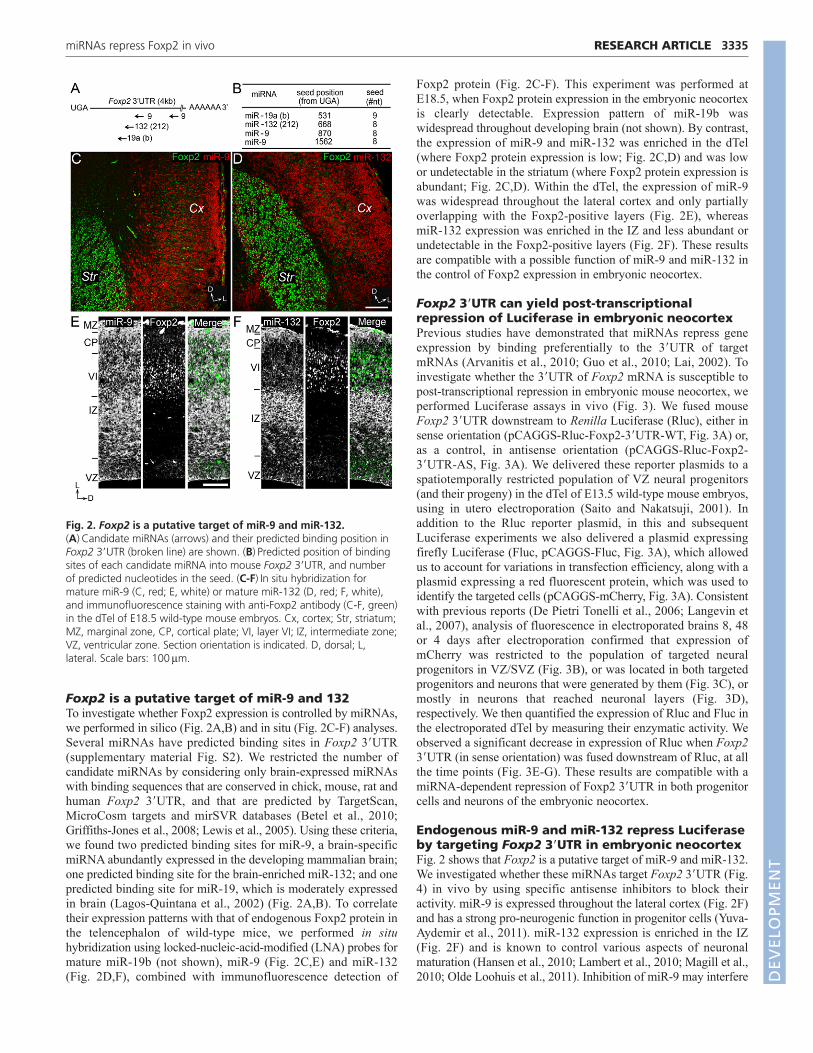
Foxp2 is a putative target of miR-9 and 132To investigate whether Foxp2 expression is controlled by miRNAs,we performed in silico (Fig. 2A,B) and in situ (Fig. 2C-F) analyses.Several miRNAs have predicted binding sites in Foxp2 3�UTR(supplementary material Fig. S2). We restricted the number ofcandidate miRNAs by considering only brain-expressed miRNAswith binding sequences that are conserved in chick, mouse, rat andhuman Foxp2 3�UTR, and that are predicted by TargetScan,MicroCosm targets and mirSVR databases (Betel et al., 2010;Griffiths-Jones et al., 2008; Lewis et al., 2005). Using these criteria,we found two predicted binding sites for miR-9, a brain-specificmiRNA abundantly expressed in the developing mammalian brain;one predicted binding site for the brain-enriched miR-132; and onepredicted binding site for miR-19, which is moderately expressedin brain (Lagos-Quintana et al., 2002) (Fig. 2A,B). To correlatetheir expression patterns with that of endogenous Foxp2 protein inthe telencephalon of wild-type mice, we performed in situhybridization using locked-nucleic-acid-modified (LNA) probes formature miR-19b (not shown), miR-9 (Fig. 2C,E) and miR-132(Fig. 2D,F), combined with immunofluorescence detection of
3335RESEARCH ARTICLEmiRNAs repress Foxp2 in vivo
Foxp2 protein (Fig. 2C-F). This experiment was performed atE18.5, when Foxp2 protein expression in the embryonic neocortexis clearly detectable. Expression pattern of miR-19b waswidespread throughout developing brain (not shown). By contrast,the expression of miR-9 and miR-132 was enriched in the dTel(where Foxp2 protein expression is low; Fig. 2C,D) and was lowor undetectable in the striatum (where Foxp2 protein expression isabundant; Fig. 2C,D). Within the dTel, the expression of miR-9was widespread throughout the lateral cortex and only partiallyoverlapping with the Foxp2-positive layers (Fig. 2E), whereasmiR-132 expression was enriched in the IZ and less abundant orundetectable in the Foxp2-positive layers (Fig. 2F). These resultsare compatible with a possible function of miR-9 and miR-132 inthe control of Foxp2 expression in embryonic neocortex.
Foxp2 3�UTR can yield post-transcriptionalrepression of Luciferase in embryonic neocortexPrevious studies have demonstrated that miRNAs repress geneexpression by binding preferentially to the 3�UTR of targetmRNAs (Arvanitis et al., 2010; Guo et al., 2010; Lai, 2002). Toinvestigate whether the 3�UTR of Foxp2 mRNA is susceptible topost-transcriptional repression in embryonic mouse neocortex, weperformed Luciferase assays in vivo (Fig. 3). We fused mouseFoxp2 3�UTR downstream to Renilla Luciferase (Rluc), either insense orientation (pCAGGS-Rluc-Foxp2-3�UTR-WT, Fig. 3A) or,as a control, in antisense orientation (pCAGGS-Rluc-Foxp2-3�UTR-AS, Fig. 3A). We delivered these reporter plasmids to aspatiotemporally restricted population of VZ neural progenitors(and their progeny) in the dTel of E13.5 wild-type mouse embryos,using in utero electroporation (Saito and Nakatsuji, 2001). Inaddition to the Rluc reporter plasmid, in this and subsequentLuciferase experiments we also delivered a plasmid expressingfirefly Luciferase (Fluc, pCAGGS-Fluc, Fig. 3A), which allowedus to account for variations in transfection efficiency, along with aplasmid expressing a red fluorescent protein, which was used toidentify the targeted cells (pCAGGS-mCherry, Fig. 3A). Consistentwith previous reports (De Pietri Tonelli et al., 2006; Langevin etal., 2007), analysis of fluorescence in electroporated brains 8, 48or 4 days after electroporation confirmed that expression ofmCherry was restricted to the population of targeted neuralprogenitors in VZ/SVZ (Fig. 3B), or was located in both targetedprogenitors and neurons that were generated by them (Fig. 3C), ormostly in neurons that reached neuronal layers (Fig. 3D),respectively. We then quantified the expression of Rluc and Fluc inthe electroporated dTel by measuring their enzymatic activity. Weobserved a significant decrease in expression of Rluc when Foxp23�UTR (in sense orientation) was fused downstream of Rluc, at allthe time points (Fig. 3E-G). These results are compatible with amiRNA-dependent repression of Foxp2 3�UTR in both progenitorcells and neurons of the embryonic neocortex.
Endogenous miR-9 and miR-132 repress Luciferaseby targeting Foxp2 3�UTR in embryonic neocortexFig. 2 shows that Foxp2 is a putative target of miR-9 and miR-132.We investigated whether these miRNAs target Foxp2 3�UTR (Fig.4) in vivo by using specific antisense inhibitors to block theiractivity. miR-9 is expressed throughout the lateral cortex (Fig. 2F)and has a strong pro-neurogenic function in progenitor cells (Yuva-Aydemir et al., 2011). miR-132 expression is enriched in the IZ(Fig. 2F) and is known to control various aspects of neuronalmaturation (Hansen et al., 2010; Lambert et al., 2010; Magill et al.,2010; Olde Loohuis et al., 2011). Inhibition of miR-9 may interfere
Fig. 2. Foxp2 is a putative target of miR-9 and miR-132.(A)Candidate miRNAs (arrows) and their predicted binding position inFoxp2 3�UTR (broken line) are shown. (B)Predicted position of bindingsites of each candidate miRNA into mouse Foxp2 3�UTR, and numberof predicted nucleotides in the seed. (C-F)In situ hybridization formature miR-9 (C, red; E, white) or mature miR-132 (D, red; F, white),and immunofluorescence staining with anti-Foxp2 antibody (C-F, green)in the dTel of E18.5 wild-type mouse embryos. Cx, cortex; Str, striatum;MZ, marginal zone, CP, cortical plate; VI, layer VI; IZ, intermediate zone;VZ, ventricular zone. Section orientation is indicated. D, dorsal; L,lateral. Scale bars: 100m.
DEVELO
PMENT

3336
with neurogenesis, whereas inhibition of miR-132 may interferewith neuronal maturation. We used in utero electroporation todeliver synthetic antisense inhibitors specific for miR-9 (Fig.4A,B), miR-132 (Fig. 4C,D) or scrambled control (Fig. 4B,D),along with pCAGGS-Rluc-Foxp2-3�UTR-WT (see Fig. 3A), in thedTel of E13.5 wild-type mouse embryos. To avoid interferenceswith neurogenesis, we examined the effect of miR-9 inhibition on
RESEARCH ARTICLE Development 139 (18)
Fig. 3. Foxp2 3�UTR can yield post-transcriptional repression ofLuciferase in embryonic neocortex. (A)CAG promoter-drivenplasmids express mCherry (pCAGGS-mCherry), Renilla Luciferase (Rluc,pCAGGS-Rluc) or firefly luciferase (Fluc, pCAGGS-Fluc). The wild-type(WT) 3�UTR of Foxp2 was inserted downstream of Rluc in sense(pCAGGS-Rluc-Foxp2-3�UTR-WT) or antisense orientation (pCAGGS-Rluc-Foxp2-3�UTR-AS, control). In utero electroporation was used todeliver plasmids in the neural progenitors of dorsal telencephalon (dTel)of E13.5 wild-type embryos. Embryos were allowed to develop in utero(IUDev) for the indicated times. (B-D)Fluorescence images ofcryosections through cortices of mouse embryos, electroporated withpCAGGS-mCherry, pCAGGS-Fluc and pCAGGS-Rluc-Foxp2-3�UTR-WTand developed for the indicated times, showing targeted cells (mCherrypositive) confined in ventricular zone (VZ) and sub-ventricular zone(SVZ) in B, or in both VZ/SVZ and intermediate zone (IZ) in C or in layersII-VI in D. Dotted lines indicate pial surface. Dashed lines indicateventricular surface. MZ, marginal zone; CP, cortical plate; II-VI indicatethe cortical layers. Section orientation is indicated. D, dorsal; L, lateral.Scale bar: 100m. (E-G)Rluc/Fluc ratio measured in lysates of brainselectroporated as in B-D. Black bars, pCAGGS-Rluc-Foxp2-3�UTR-AS;white bars, pCAGGS-Rluc-Foxp2-3�UTR-WT. Rluc/Fluc ratio ofpCAGGS-Rluc-Foxp2-3�UTR-AS samples was normalized to one. n,number of brains (or pool of two brains) analyzed per condition.*P<0.05; **P<0.01; ***P<0.001.
Fig. 4. Endogenous miR-9 and miR-132 repress Luciferase bytargeting Foxp2 3�UTR in embryonic neocortex. (A,C)Images ofcryosections through the dTel of embryos electroporated at E13.5 (as inB,D) and developed in utero for the indicated time, illustrating intrinsicmCherry fluorescence. MZ, marginal zone; CP, cortical plate; II-VI indicatethe cortical layers; VZ, ventricular zone; SVZ, sub-ventricular zone. Dottedlines indicate the pial surface; dashed lines indicate the ventricular surface.Scale bars: 50m. (B,D)Rluc/Fluc ratio measured in brain lysates fromwild-type mouse embryos co-electroporated with pCAGGS-mCherry,pCAGGS-Fluc, pCAGGS-Rluc-Foxp2-3�UTR-WT, and either scrambled(B,D, control, black bar), miR-9 inhibitor (B, anti-miR-9, gray bar) or miR-132 inhibitor (D, anti-miR-132, gray bar), and developed in utero for theindicated time. Rluc/Fluc ratio of control samples was normalized to one.***P<0.001. (E)Predicted binding sites of miR-9 (mmu-miR-9) and miR-132 (mmu-miR-132) in Foxp2 3�UTR (3�UTR WT). Four nucleotides (bold)were mutated in each of the putative binding sites of miR-9 (3�UTR MT1and MT2) or of miR-132 (3�UTR MT3). Vertical lines indicate putative basepairings; columns indicate G-U bindings; ‘X’ indicates a mismatch.(F,G)Rluc/Fluc ratio measured in brain lysates from wild-type mouseembryos co-electroporated in utero at E13.5 with pCAGGS-mCherry,pCAGGS-Fluc, and either pCAGGS-Rluc-Foxp2-3�UTR-WT (black bars),pCAGGS-Rluc-Foxp2-3�UTR-MT1-2 (white bars), pCAGGS-Rluc-Foxp2-3�UTR-MT3 (dark gray bars) or pCAGGS-Rluc-Foxp2-3�UTR-MT1-2-3 (light gray bars), and developed in utero for the indicated time. Rluc/Flucratio of pCAGGS-Rluc-Foxp2-3�UTR-WT samples was normalized to one.One-way analysis of variance (ANOVA) was used for statistical analysis.*P<0.05; **P<0.01; ***P<0.001; n.s., not significant. D
EVELO
PMENT

Foxp2 3�UTR in progenitors (8 hours after electroporation, Fig.4A). Conversely, given the enriched expression of miR-132 in theIZ, we analyzed the effect of miR-132 inhibition on Foxp2 3�UTRin neurons (4 days after electroporation, Fig. 4C). We thenquantified the expression of Rluc and Fluc in the electroporateddTel by measuring their enzymatic activity (Fig. 4B,D). Weobserved a significant rescue of Rluc expression uponadministration of miR-9 or miR-132 inhibitors. This experimentshows that endogenous miR-9 and miR-132 repress Luciferase bytargeting the Foxp2 3�UTR in progenitors and neurons,respectively. Cortices electroporated with either mir-9 or miR-132inhibitors were further analyzed by immunofluorescence for theexpression of endogenous Foxp2 protein; however, under theseconditions we did not detect an increased expression of theendogenous Foxp2 protein in targeted cells (data not shown). Thelack of de-repression of the endogenous Foxp2 expression may bedue to insufficient inhibition of the endogenous miR-9/132 (whichare very abundant in embryonic neocortex), to progressive dilutionof the synthetic miRNA inhibitors in proliferating progenitors or toa redundant action of additional miRNAs on Foxp2.
Next, we wanted to validate experimentally the predicted bindingsequences of miR-9 and miR-132 within Foxp2 3�UTR in theembryonic neocortex. Previous studies have shown that miRNA-target interaction occurs mostly by formation of base pairing betweenthe 5� region of the miRNA (seed) with its target (Brennecke et al.,2005; Lewis et al., 2005). We therefore prepared plasmids in which,downstream of Rluc, we fused a mutated 3�UTR of Foxp2 in whichsequences assumed to base pair with miR-9 or miR-132 (or both)were altered (Fig. 4E). Using this strategy, we also aimed toinvestigate the activity and relative efficiency of miR-9 and miR-132for the repression of Foxp2 3�UTR in both progenitors and neurons.We used in utero electroporation to deliver each mutated plasmid(Fig. 4E) into the dTel of E13.5 wild-type mouse embryos. As acontrol, we electroporated pCAGGS-Rluc-Foxp2-3�UTR-WT (seeFig. 3A). We then quantified Luciferase expression in neuralprogenitors in VZ/SVZ (Fig. 4F) and in neurons in the neuronallayers (Fig. 4G). Mutations that prevented the binding of eitherendogenous miR-9 or miR-132 to Foxp2 3�UTR rescued Rlucexpression in both progenitors (Fig. 4F) and neurons (Fig. 4G).Remarkably, mutations that affected the binding of both miR-9 andmiR-132 resulted in an additive rescue of Rluc expression inneurons, but not in progenitors (Fig. 4F,G). These results areconsistent with the natural expression patterns of miR-9 and of miR-132 (Fig. 2) (De Pietri Tonelli et al., 2008; Shibata et al., 2011) andsuggest that the activity of these miRNAs can converge on Foxp2-3�UTR in neurons.
Ectopic expression of Foxp2 in neural progenitorsdoes not impair their subtype specification anddifferentiation in embryonic neocortexTo investigate the possible biological relevance of the miRNA-mediated repression of Foxp2-3�UTR in the embryonic neocortex,we ectopically expressed Foxp2 protein in neural progenitors (ascenario that mimics the absence of miRNA-mediated silencing ofFoxp2 expression) and investigated the consequences on cell-typespecification and differentiation. Progenitors that divide in the SVZ(also known as basal progenitors) are generated from progenitorsthat divide in the VZ (also known as apical progenitors), and can beidentified by the expression of the transcription factor Tbr2 (Eomes)(Englund et al., 2005). We used in utero electroporation to deliverthe pCAGGS-mCherry plasmid into the dTel of E13.5 wild-typemouse embryos, along with either pCAGGS empty plasmid
3337RESEARCH ARTICLEmiRNAs repress Foxp2 in vivo
(control), or a plasmid expressing a transcript encoding Foxp2protein lacking its 3�UTR, so that it could not be targeted by miR-9 and miR-132 (pCAGGS-Foxp2-�-3�UTR) (supplementarymaterial Fig. S3A-C). Immunofluorescence analysis with anti-Foxp2 antibody 24 hours after electroporation revealed that almostall (96±4%) targeted cells in the VZ/SVZ that expressed mCherryalso expressed Foxp2 protein (supplementary material Fig. S3A-C).Therefore, in subsequent experiments, cells expressing mCherrywere also considered positive for Foxp2 protein when the latter wasco-electroporated. We next compared the cortices that ectopicallyexpressed Foxp2 with those not expressing ectopic Foxp2 40 hoursafter electroporation (supplementary material Fig. S3D-J).Quantification of the proportion of apical progenitors (Tbr2-negative mCherry-positive cells) and of basal progenitors (Tbr2 andmCherry double-positive cells) revealed no difference between thetwo conditions (supplementary material Fig. S4J). Similarly, wefound no difference in the proportion of targeted progenitorsundergoing basal and apical mitoses (identified by co-expression ofmCherry and of the mitotic marker phosphohistone H3) (notshown). Finally, we quantified the number of cells generated bytargeted progenitors, that delaminated from the VZ/SVZ (i.e.mCherry-positive cells that were located in neuronal layers) andagain we found no difference between the two conditions(supplementary material Fig. S4D,G,K). These results indicate thatprogenitor subtype specification or differentiation is not impairedafter ectopic Foxp2 expression.
Ectopic Foxp2 expression impairs radial migrationof targeted cells in embryonic neocortex, by amiR-9/132-dependent mechanismIn the embryonic mouse neocortex, Foxp2 starts its expression inpostmigratory neurons (Ferland et al., 2003; Takahashi et al., 2003).Endogenous miR-9 and miR-132 repressed Luciferase expression bytargeting Foxp2 3�UTR in neurons of the embryonic neocortex (Fig.4), raising the possibility that this control may be important forneuronal migration or maturation. To test this hypothesis, we used inutero electroporation to deliver the pCAGGS-mCherry plasmid intothe dTel of E13.5 wild-type mouse embryos, along with (1)pCAGGS empty plasmid (control, Fig. 5A,B), (2) pCAGGS-Foxp2-�-3�UTR (Fig. 5C,D), (3) pCAGGS-Foxp2-3�UTR-MT1+2+3[expressing a transcript for the Foxp2-coding sequence and amutated 3�UTR (as in Fig. 4) that is not targeted by miR-9 and miR-132 (Fig. 5E,F)] or (4) pCAGGS-Foxp2-3�UTR-WT [expressing atranscript for Foxp2-coding sequence and its WT 3�UTR (Fig.5G,H)]. Five days after electroporation, we first examined theexpression of Foxp2 protein in electroporated cortices (Fig.5B,D,F,H). Immunoreactivity of Foxp2 protein in control cortices(Fig. 5B) was consistent with previous studies (Ferland et al., 2003;Shu et al., 2001; Takahashi et al., 2003) and comparable with theexpression of Foxp2 protein in cortices electroporated with theFoxp2 construct carrying wild-type 3�UTR (Fig. 5H). By contrast,immunoreactivity of Foxp2 protein was strongly increased in corticeselectroporated with the Foxp2 construct lacking the 3�UTR (Fig.5D), as well as in cortices electroporated with the Foxp2 constructcarrying a mutated 3�UTR in miR-9/132 binding sites (Fig. 5F). Wenext analyzed the distribution of targeted cells in cortex. We foundcomparable distributions of targeted cells in control cortices and incortices electroporated with pCAGGS-Foxp2-3�UTR-WT (Fig.5A,G,I), consistent with previous studies (De Pietri Tonelli et al.,2006; Langevin et al., 2007). By contrast, in cortices electroporatedwith pCAGGS-Foxp2-�-3�UTR and pCAGGS-Foxp2-3�UTR-MT1+2+3, we found that many of the targeted cells were misplaced D
EVELO
PMENT

3338
in the IZ (Fig. 5C,E,I). These results show that ectopic expression ofFoxp2 protein impairs radial migration of neurons, and that thiseffect on migration is attenuated by the repressive action ofendogenous miR-9/132 on Foxp2 3�UTR in the embryonicneocortex.
We then asked whether the misplaced cells were delayed orarrested in their migration. To discriminate between these twopossibilities, electroporated cortices were analyzed at postnatal day15 (P15), when neuronal migration is largely complete (Fig. 5J-O).To exclude possible artifacts owing to the ectopic expression of atranscription factor, we used Foxp4 as an additional control. Foxp4is the closest paralog of Foxp2, and in the embryonic mouseneocortex Foxp4 protein is co-expressed with Foxp2 and interactswith it (Li et al., 2004; Takahashi et al., 2008). Cortices wereelectroporated at E13.5 with pCAGGS-mCherry plasmid alongwith (1) pCAGGS empty plasmid (Fig. 5J,M), (2) pCAGGS-Foxp4-�-3�UTR [expressing a transcript for Foxp4 lacking the3�UTR (to avoid eventual post-transcriptional effects) (Fig. 5K,N)]
RESEARCH ARTICLE Development 139 (18)
or (3) pCAGGS-Foxp2-�-3�UTR (Fig. 5L,O). At P15 we analyzedthe laminar distribution of targeted cells. Laminar distribution ofcells in control cortices (Fig. 5J,M) and in cortices electroporatedwith the Foxp4 construct was comparable (Fig. 5K,N;immunofluorescent verification of Foxp4 expression not shown).By contrast, in cortices electroporated with the Foxp2 construct, agreater proportion of cells were located in deeper cortical layers,and some were also found in the white matter (Fig. 5L,O). Thisfinding corroborates the results of the previous experiment (Fig.5A-I) and suggests that ectopic expression of Foxp2 protein inembryonic neocortex arrests radial migration of some targetedcells.
Ectopic expression of Foxp2 delays neuriteoutgrowth and branching in cortical neuronsTo investigate whether ectopic expression of Foxp2 protein canimpair the differentiation of cortical projection neurons, we brieflyexamined the cells after ectopic Foxp2 expression in P15 brains (as
Fig. 5. Ectopic Foxp2 expression impairs radial migration of targeted cells in embryonic neocortex, by a miR-9/132-dependentmechanism. (A-H)Images of cryosections through the telencephalon of E18.5 wild-type mouse embryos co-electroporated in utero at E13.5 withpCAGGS-mCherry along with either pCAGGS (control) (A,B), pCAGGS-Foxp2--3�UTR (C,D), pCAGGS-Foxp2-3�UTR-MT1+2+3 (E,F) or pCAGGS-Foxp2-3�UTR-WT (G,H), showing intrinsic fluorescence of mCherry (A,C,E,G) or Foxp2 immunostaining (B,D,F,H). CP, cortical plate; VI, layer VI; Str,striatum. Dashed lines indicate the limits of the intermediate zone (IZ). Scale bar: 50m. (I)Quantification of the proportion of targeted cells locatedin the IZ of the electroporated cortices per field (as shown in A,C,E,G). One-way analysis of variance (ANOVA) was used for statistical analysis. n,number of sections counted across rostrocaudal axes from at least four brains per condition. Data are mean±s.e.m. (J-L)Images of sections throughthe cerebral cortex of postnatal day 15 (P15) wild-type mice co-electroporated in utero at E13.5 with pCAGGS-mCherry and either pCAGGS (J,control), pCAGGS-Foxp4-�-3�UTR (K, Foxp4-�-3�UTR) or pCAGGS-Foxp2-�-3�UTR (L, Foxp2-�-3�UTR), showing intrinsic mCherry fluorescence.Arrow indicates ectopic cells in the white matter (WM); dashed lines indicate the bottom limit of WM; I-VI indicate cortical layers. Orientation ofsections is indicated. D, dorsal; L, lateral. Scale bar: 100m. Dotted lines in A-H,J-L indicate pial surface. (M-O)Quantification of the proportion ofmCherry-positive cells located in each of the 10 bins per field (as in J-L) along the rostrocaudal axis, counted from at least five brains per condition;n.s., non significant. P0.1; *P<0.05, ***P<0.001. Data are mean±s.e.m.
DEVELO
PMENT

in Fig. 5L). At the morphological level, cells arrested in the whitematter exhibited a tangential morphology and some of themextended processes; moreover, some of these cells acquiredglutamatergic fate (supplementary material Fig. S4). We alsostained the electroporated cortices (as in Fig. 5J,L) for Cux1 (Fig.6A-C) or Brn2 (also known as Pou3f2, Fig. 6D) protein, which are
3339RESEARCH ARTICLEmiRNAs repress Foxp2 in vivo
markers of cortical projection neurons of the upper layers(Molyneaux et al., 2007). We analyzed the distribution of targetedneurons (mCherry-positive cells), in P15 cortices targeted withFoxp2 or control (as in Fig. 5J,L), which also expressed Cux1 orBrn2 (Fig. 6E,F). We found that, upon ectopic expression of Foxp2,several of the targeted neurons that expressed Cux1 or Brn2 were
Fig. 6. Ectopic expression of Foxp2 delaysneurite outgrowth and branching in corticalneurons. (A-D)Images of sections through thecerebral cortices of wild-type P15 mice, co-electroporated in utero at E13.5 with pCAGGS-mCherry and either pCAGGS (A) or pCAGGS-Foxp2-�-3�UTR (B-D), showing intrinsicfluorescence of mCherry (A-D, red) and Cux1 (A-C,green) or Brn2 (D, green). Dashed box in B indicatesa region similar to the ones shown in C and D.Arrows indicate mCherry-Cux1 (C) or mCherry-Brn2(D) double-positive neurons. Scale bars: 100m inA,B; 50m in C,D. (E,F)Quantification of thedistribution of mCherry-Cux1 (E) or mCherry-Brn2(F) double-positive neurons in the cortical wall(divided in five bins along the dorsoventral axis) perfield (as in A,B). (G,H)Quantification of the relativeproportion of mCherry-Cux1 (G) or mCherry-Brn2(H) double-positive neurons among the entirepopulation of targeted cells (mCherry positive) perfield (as in A,B). WM, white matter; I-VI indicatecortical layers. Section orientations are indicated. D,dorsal; L, lateral. (I,J)Representative tracings ofprimary cortical projection neurons from E18.5wild-type mouse embryos co-electroporated inutero at E13.5 with pCAGGS-mCherry and eitherpCAGGS (I, control) or pCAGGS-Foxp2-�-3�UTR (J,Foxp2-�-3�UTR) and cultivated in vitro for 4 days (4DIV). Scale bar: 15m. (K-N)Quantification of totalneurite length (K,M) and branching (L,N) ofmCherry-positive primary cortical projectionneurons from control embryos (control, black barsin K,M; black diamonds in L,N) or co-electroporatedwith pCAGGS-Foxp2-�-3�UTR (white bars, K,M;white diamonds in L,N) and cultivated for 4 days invitro (K,L) or 7 days in vitro (M,N). In E-H,K-N, dataare mean±s.e.m. *P<0.05; **P<0.01; ***P<0.001.
DEVELO
PMENT

3340
misplaced in deeper cortical layers or in the white matter (Fig. 6B-F). However, the proportion of targeted neurons that acquired anupper layer identity was unaffected (Fig. 6G,H). Taken together,these experiments indicate that differentiation of cortical projectionneurons is not impaired after ectopic expression of Foxp2 protein.
Foxp2 has also been reported to be involved in the control ofneurite outgrowth in neurons (Vernes et al., 2007; Spiteri et al.,2007; Reimers-Kipping et al., 2011; Vernes et al., 2011). Previousstudies have shown that radial migration of cortical neuronsrequires precise coordination of leading process extension andbranching (Nadarajah et al., 2001; Nadarajah et al., 2003;Kriegstein and Noctor, 2004; Attardo et al., 2008; Falnikar et al.,2011). Early stages of migration include polarization and processesextension (Dotti et al., 1988). To study whether ectopic expressionof Foxp2 protein alters neurite outgrowth, we prepared primaryneuronal cultures from embryonic cortices electroporated withpCAGGS empty plasmid, or pCAGGS-Foxp2-�-3�UTR, alongwith pCAGGS-mCherry plasmid (Fig. 6I-N). We then investigatedneurite outgrowth specifically in cortical projection neurons (whichwere selectively targeted by mean of in utero electroporation ofdTel) (LoTurco et al., 2009) by analyzing the total length (Fig.6K,M) and branching (Fig. 6L,N) of neurites after 4 or 7 days invitro (in wild-type animals, it is widely accepted that polarizationand extension of neurites are already established within the first 4days in vitro; Polleux and Snider, 2010). We found that, comparedwith controls, cortical projection neurons targeted with pCAGGS-Foxp2-�-3�UTR had shorter and less branched neurites at 4 days(Fig. 6I-L) but not at 7 days in vitro (Fig. 6M,N). This resultindicates that ectopic expression of Foxp2 protein in developingcortical projection neurons alters the proper timing of neuriteoutgrowth. We speculate that this effect could explain the observedarrest in radial migration of neurons.
DISCUSSIONIn our study, we used genome-wide profiling of miRNA-depletedembryonic mouse neocortex to identify potential miRNA targetgenes. We then validated in vivo one of the identified targets,Foxp2, and found that convergent action of miR-9 and miR-132prevents ectopic expression of Foxp2 by targeting its 3�UTR.Moreover, we found that the ectopic expression of Foxp2 indeveloping projection neurons delays neurite outgrowth in vitroand impairs their radial migration in embryonic mouse neocortexin vivo.
Our study introduces a combined strategy of genome-wideprofiling of miRNA-depleted embryonic mouse neocortex and invivo target validation by in utero electroporation of Luciferasereporters, to experimentally validate novel miRNA targets withsignificance for neocortical development. Indeed, we have foundmore than 3000 genes that are regulated, either directly orindirectly, by the miRNA pathway in embryonic mouse neocortex.Which of these genes are directly controlled by miRNAs, and thefunction of such control in the orchestration of neocorticaldevelopment are matters for continued study. Importantly, our invivo validation of miRNA:mRNA interaction provides animprovement over the standard in vitro assays. Indeed, the cell-typespecificity of miRNA regulation is increasingly recognized(Bhattacharyya et al., 2006; Jopling et al., 2005; Kedde et al., 2007;Vasudevan et al., 2007; Shibata et al., 2011).
The most remarkable finding of our study is that ectopicexpression of Foxp2 in cortical projection neurons – a scenario thatresembles the absence of miRNA-mediated repression of Foxp2 –delays neurite outgrowth and arrests their radial migration. This
RESEARCH ARTICLE Development 139 (18)
effect is attenuated by endogenous miR-9 and miR-132, whichrepress Foxp2 in embryonic neocortex. Foxp2 is involved in thefine control of neurite outgrowth in developing neurons (Enard etal., 2009; Reimers-Kipping et al., 2011; Tam et al., 2011; Vernes etal., 2011). Moreover, miR-9 and miR-132 are also involved in thecontrol of neurite outgrowth and branching in neurons by targetingdistinct subsets of mRNAs (Vo et al., 2005; Wayman et al., 2008;Xu et al., 2008). Our results therefore raise the intriguingpossibility that, in migrating neurons, the precise coordination ofneurite extension and branching is achieved by the convergentaction of miR-9 and miR-132 to optimize Foxp2 dose.Interestingly, miR-9 was recently found to tune levels of Foxp1 (aparalog of Foxp2) to ensure proper development of motoneuronsin developing chick spinal cord (Otaegi et al., 2011). Thus,miRNA-dependent repression of Foxp expression might be abroader mechanism that occurs in other members of the Foxpfamily.
Finally, numerous studies on the role of Foxp2 in neocorticaldevelopment have focused on loss-of-function mutations (Shu etal., 2005; Groszer et al., 2008; Fujita et al., 2008; Takahashi et al.,2009). We found that ectopic expression of Foxp2 impairs neuriteextension and radial migration of cortical projection neurons.Although we do not directly address the role of miR-9/132 in thecontrol of endogenous Foxp2 expression [see Åkerblom et al.(Åkerblom et al., 2012) for extensive review about the currenttechnical limitations in functional studies on miRNAs in vivo], wehave uncovered a new layer of control of Foxp2 expression. Giventhat Foxp2 3�UTR and miR-9/132 are evolutionarily conserved,our finding sets the stage for future studies aimed at the directinvestigation of mutations in the 3�UTR of human FOXP2transcript. These studies could perhaps provide new explanationsfor defects observed in human pathologies involving alteredexpression of FOXP genes (Hannenhalli and Kaestner, 2009).
AcknowledgementsWe thank Dr L. Cancedda and Dr J. Assad (Istituto Italiano di Tecnologia, IIT)for critical reading of the manuscript; Dr Itohara (RIKEN, Japan) and Dr Hannon(Cold Spring Harbor Laboratory, MA, USA) for kindly providing Emx1Cre andDicerFlox mouse lines, respectively; IIT and Max-Plank Institute of Molecular CellBiology and Genetics (MPI-CBG) technical staff (M. Pesce, F. Succol, M. Nanni,F. Piccardi and C. Haffner) for help; B. Nickel and T. Giger [Max-Plank Instituteof Evolutionary Anthropology (MPI-EVA)] for help in microarray experiments;and Dr Schwamborn (Münster University, Germany) for advice in miRNAinhibitors.
FundingW.E. was supported by the Max-Planck Society; W.B.H. was supported bygrants from the Deutsche Forschungsgemeinschaft (DFG) [SFB 655, A2; TRR83, Tp6] and the ERC [250197], by the DFG-funded Center for RegenerativeTherapies Dresden, and by the Fonds der Chemischen Industrie. This work wassupported by Fondazione Istituto Italiano di Tecnologia.
Competing interests statementThe authors declare no competing financial interests.
Supplementary materialSupplementary material available online athttp://dev.biologists.org/lookup/suppl/doi:10.1242/dev.078063/-/DC1
ReferencesÅkerblom, M., Sachdeva, R. and Jakobsson, J. (2012). Functional studies of
microRNAs in neural stem cells: problems and perspectives. Front. Neurosci. 6,14.
Arvanitis, D. N., Jungas, T., Behar, A. and Davy, A. (2010). Ephrin-B1 reversesignaling controls a posttranscriptional feedback mechanism via miR-124. Mol.Cell. Biol. 30, 2508-2517.
Ashburner, M., Ball, C. A., Blake, J. A., Botstein, D., Butler, H., Cherry, J. M.,Davis, A. P., Dolinski, K., Dwight, S. S., Eppig, J. T. et al. (2000). Gene D
EVELO
PMENT
3341RESEARCH ARTICLEmiRNAs repress Foxp2 in vivo
ontology: tool for the unification of biology. The Gene Ontology Consortium.Nat. Genet. 25, 25-29.
Attardo, A., Calegari, F., Haubensak, W., Wilsch-Bräuninger, M. andHuttner, W. B. (2008). Live imaging at the onset of cortical neurogenesisreveals differential appearance of the neuronal phenotype in apical versus basalprogenitor progeny. PLoS ONE 3, e2388.
Ayala, R., Shu, T. and Tsai, L. H. (2007). Trekking across the brain: the journey ofneuronal migration. Cell 128, 29-43.
Baek, D., Villén, J., Shin, C., Camargo, F. D., Gygi, S. P. and Bartel, D. P.(2008). The impact of microRNAs on protein output. Nature 455, 64-71.
Betel, D., Koppal, A., Agius, P., Sander, C. and Leslie, C. (2010).Comprehensive modeling of microRNA targets predicts functional non-conserved and non-canonical sites. Genome Biol. 11, R90.
Bhattacharyya, S. N., Habermacher, R., Martine, U., Closs, E. I. andFilipowicz, W. (2006). Relief of microRNA-mediated translational repression inhuman cells subjected to stress. Cell 125, 1111-1124.
Brennecke, J., Stark, A., Russell, R. B. and Cohen, S. M. (2005). Principles ofmicroRNA-target recognition. PLoS Biol. 3, e85.
Campbell, P., Reep, R. L., Stoll, M. L., Ophir, A. G. and Phelps, S. M. (2009).Conservation and diversity of Foxp2 expression in muroid rodents: functionalimplications. J. Comp. Neurol. 512, 84-100.
Candiani, S., Moronti, L., De Pietri Tonelli, D., Garbarino, G. and Pestarino,M. (2011). A study of neural-related microRNAs in the developing amphioxus.Evodevo 2, 15.
Chekulaeva, M. and Filipowicz, W. (2009). Mechanisms of miRNA-mediatedpost-transcriptional regulation in animal cells. Curr. Opin. Cell Biol. 21, 452-460.
Choi, P. S., Zakhary, L., Choi, W. Y., Caron, S., Alvarez-Saavedra, E., Miska,E. A., McManus, M., Harfe, B., Giraldez, A. J., Horvitz, H. R. et al. (2008).Members of the miRNA-200 family regulate olfactory neurogenesis. Neuron 57,41-55.
De Pietri Tonelli, D., Calegari, F., Fei, J. F., Nomura, T., Osumi, N.,Heisenberg, C. P. and Huttner, W. B. (2006). Single-cell detection ofmicroRNAs in developing vertebrate embryos after acute administration of adual-fluorescence reporter/sensor plasmid. Biotechniques 41, 727-732.
De Pietri Tonelli, D., Pulvers, J. N., Haffner, C., Murchison, E. P., Hannon, G.J. and Huttner, W. B. (2008). miRNAs are essential for survival anddifferentiation of newborn neurons but not for expansion of neural progenitorsduring early neurogenesis in the mouse embryonic neocortex. Development135, 3911-3921.
Dotti, C. G., Sullivan, C. A. and Banker, G. A. (1988). The establishment ofpolarity by hippocampal neurons in culture. J. Neurosci. 8, 1454-1468.
Enard, W. (2011). FOXP2 and the role of cortico-basal ganglia circuits in speechand language evolution. Curr. Opin. Neurobiol. 21, 415-424.
Enard, W., Przeworski, M., Fisher, S. E., Lai, C. S., Wiebe, V., Kitano, T.,Monaco, A. P. and Pääbo, S. (2002). Molecular evolution of FOXP2, a geneinvolved in speech and language. Nature 418, 869-872.
Enard, W., Gehre, S., Hammerschmidt, K., Hölter, S. M., Blass, T., Somel, M.,Brückner, M. K., Schreiweis, C., Winter, C., Sohr, R. et al. (2009). Ahumanized version of Foxp2 affects cortico-basal ganglia circuits in mice. Cell137, 961-971.
Englund, C., Fink, A., Lau, C., Pham, D., Daza, R. A., Bulfone, A., Kowalczyk,T. and Hevner, R. F. (2005). Pax6, Tbr2, and Tbr1 are expressed sequentially byradial glia, intermediate progenitor cells, and postmitotic neurons in developingneocortex. J. Neurosci. 25, 247-251.
Falnikar, A., Tole, S. and Baas, P. W. (2011). Kinesin-5, a mitotic microtubule-associated motor protein, modulates neuronal migration. Mol. Biol. Cell 22,1561-1574.
Ferland, R. J., Cherry, T. J., Preware, P. O., Morrisey, E. E. and Walsh, C. A.(2003). Characterization of Foxp2 and Foxp1 mRNA and protein in thedeveloping and mature brain. J. Comp. Neurol. 460, 266-279.
Fietz, S. A. and Huttner, W. B. (2011). Cortical progenitor expansion, self-renewal and neurogenesis – a polarized perspective. Curr. Opin. Neurobiol. 21,23-35.
Fisher, S. E. and Scharff, C. (2009). FOXP2 as a molecular window into speechand language. Trends Genet. 25, 166-177.
Friedman, R. C., Farh, K. K., Burge, C. B. and Bartel, D. P. (2009). Mostmammalian mRNAs are conserved targets of microRNAs. Genome Res. 19, 92-105.
Fujita, E., Tanabe, Y., Shiota, A., Ueda, M., Suwa, K., Momoi, M. Y. andMomoi, T. (2008). Ultrasonic vocalization impairment of Foxp2 (R552H) knockinmice related to speech-language disorder and abnormality of Purkinje cells. Proc.Natl. Acad. Sci. USA 105, 3117-3122.
Griffiths-Jones, S., Saini, H. K., van Dongen, S. and Enright, A. J. (2008).miRBase: tools for microRNA genomics. Nucleic Acids Res. 36, D154-D158.
Groszer, M., Keays, D. A., Deacon, R. M., de Bono, J. P., Prasad-Mulcare, S.,Gaub, S., Baum, M. G., French, C. A., Nicod, J., Coventry, J. A. et al. (2008).Impaired synaptic plasticity and motor learning in mice with a point mutationimplicated in human speech deficits. Curr. Biol. 18, 354-362.
Guo, H., Ingolia, N. T., Weissman, J. S. and Bartel, D. P. (2010). MammalianmicroRNAs predominantly act to decrease target mRNA levels. Nature 466, 835-840.
Hannenhalli, S. and Kaestner, K. H. (2009). The evolution of Fox genes andtheir role in development and disease. Nat. Rev. Genet. 10, 233-240.
Hansen, K. F., Sakamoto, K., Wayman, G. A., Impey, S. and Obrietan, K.(2010). Transgenic miR132 alters neuronal spine density and impairs novelobject recognition memory. PLoS ONE 5, e15497.
Hendrickson, D. G., Hogan, D. J., McCullough, H. L., Myers, J. W., Herschlag,D., Ferrell, J. E. and Brown, P. O. (2009). Concordant regulation of translationand mRNA abundance for hundreds of targets of a human microRNA. PLoS Biol.7, e1000238.
Hisaoka, T., Nakamura, Y., Senba, E. and Morikawa, Y. (2010). The forkheadtranscription factors, Foxp1 and Foxp2, identify different subpopulations ofprojection neurons in the mouse cerebral cortex. Neuroscience 166, 551-563.
Iwasato, T., Datwani, A., Wolf, A. M., Nishiyama, H., Taguchi, Y.,Tonegawa, S., Knöpfel, T., Erzurumlu, R. S. and Itohara, S. (2000). Cortex-restricted disruption of NMDAR1 impairs neuronal patterns in the barrel cortex.Nature 406, 726-731.
Jopling, C. L., Yi, M., Lancaster, A. M., Lemon, S. M. and Sarnow, P. (2005).Modulation of hepatitis C virus RNA abundance by a liver-specific microRNA.Science 309, 1577-1581.
Kawase-Koga, Y., Otaegi, G. and Sun, T. (2009). Different timings of Dicerdeletion affect neurogenesis and gliogenesis in the developing mouse centralnervous system. Dev. Dyn. 238, 2800-2812.
Kedde, M., Strasser, M. J., Boldajipour, B., Oude Vrielink, J. A., Slanchev, K.,le Sage, C., Nagel, R., Voorhoeve, P. M., van Duijse, J., Ørom, U. A. et al.(2007). RNA-binding protein Dnd1 inhibits microRNA access to target mRNA.Cell 131, 1273-1286.
Kriegstein, A. R. and Noctor, S. C. (2004). Patterns of neuronal migration in theembryonic cortex. Trends Neurosci. 27, 392-399.
Krol, J., Loedige, I. and Filipowicz, W. (2010). The widespread regulation ofmicroRNA biogenesis, function and decay. Nat. Rev. Genet. 11, 597-610.
Lagos-Quintana, M., Rauhut, R., Yalcin, A., Meyer, J., Lendeckel, W. andTuschl, T. (2002). Identification of tissue-specific microRNAs from mouse. Curr.Biol. 12, 735-739.
Lai, C. S., Fisher, S. E., Hurst, J. A., Vargha-Khadem, F. and Monaco, A. P.(2001). A forkhead-domain gene is mutated in a severe speech and languagedisorder. Nature 413, 519-523.
Lai, C. S., Gerrelli, D., Monaco, A. P., Fisher, S. E. and Copp, A. J. (2003).FOXP2 expression during brain development coincides with adult sites ofpathology in a severe speech and language disorder. Brain 126, 2455-2462.
Lai, E. C. (2002). Micro RNAs are complementary to 3� UTR sequence motifs thatmediate negative post-transcriptional regulation. Nat. Genet. 30, 363-364.
Lambert, T. J., Storm, D. R. and Sullivan, J. M. (2010). MicroRNA132modulates short-term synaptic plasticity but not basal release probability inhippocampal neurons. PLoS ONE 5, e15182.
Langevin, L. M., Mattar, P., Scardigli, R., Roussigné, M., Logan, C., Blader, P.and Schuurmans, C. (2007). Validating in utero electroporation for the rapidanalysis of gene regulatory elements in the murine telencephalon. Dev. Dyn.236, 1273-1286.
Lewis, B. P., Burge, C. B. and Bartel, D. P. (2005). Conserved seed pairing, oftenflanked by adenosines, indicates that thousands of human genes are microRNAtargets. Cell 120, 15-20.
Li, S., Weidenfeld, J. and Morrisey, E. E. (2004). Transcriptional and DNAbinding activity of the Foxp1/2/4 family is modulated by heterotypic andhomotypic protein interactions. Mol. Cell. Biol. 24, 809-822.
LoTurco, J., Manent, J. B. and Sidiqi, F. (2009). New and improved tools for inutero electroporation studies of developing cerebral cortex. Cereb. Cortex 19Suppl. 1, i120-i125.
Lui, J. H., Hansen, D. V., and Kriegstein A. R. (2011). Development andevolution of the human neocortex. Cell 146, 18-36.
Magill, S. T., Cambronne, X. A., Luikart, B. W., Lioy, D. T., Leighton, B. H.,Westbrook, G. L., Mandel, G. and Goodman, R. H. (2010). microRNA-132regulates dendritic growth and arborization of newborn neurons in the adulthippocampus. Proc. Natl. Acad. Sci. USA 107, 20382-20387.
Makeyev, E. V., Zhang, J., Carrasco, M. A. and Maniatis, T. (2007). TheMicroRNA miR-124 promotes neuronal differentiation by triggering brain-specific alternative pre-mRNA splicing. Mol. Cell 27, 435-448.
Meijering, E., Jacob, M., Sarria, J.-C. F., Steiner, P., Hirling, H. and Unser, M.(2004). Design and validation of a tool for neurite tracing and analysis influorescence microscopy images. Cytometry 58, 167-176.
Molyneaux, B. J., Arlotta, P., Menezes, J. R. and Macklis, J. D. (2007).Neuronal subtype specification in the cerebral cortex. Nat. Rev. Neurosci. 8, 427-437.
Murchison, E. P., Partridge, J. F., Tam, O. H., Cheloufi, S. and Hannon, G. J.(2005). Characterization of Dicer-deficient murine embryonic stem cells. Proc.Natl. Acad. Sci. USA 102, 12135-12140. D
EVELO
PMENT

3342 RESEARCH ARTICLE Development 139 (18)
Muzio, L., Di Benedetto, B., Stoykova, A., Boncinelli, E., Gruss, P. andMallamaci, A. (2002). Emx2 and Pax6 control regionalization of the pre-neuronogenic cortical primordium. Cereb. Cortex 12, 129-139.
Nadarajah, B., Brunstrom, J. E., Grutzendler, J., Wong, R. O. and Pearlman,A. L. (2001). Two modes of radial migration in early development of thecerebral cortex. Nat. Neurosci. 4, 143-150.
Nadarajah, B., Alifragis, P., Wong, R. O. and Parnavelas, J. G. (2003).Neuronal migration in the developing cerebral cortex: observations based onreal-time imaging. Cereb. Cortex 13, 607-611.
Nakagawa, Y., Johnson, J. E. and O’Leary, D. D. (1999). Graded and arealexpression patterns of regulatory genes and cadherins in embryonic neocortexindependent of thalamocortical input. J. Neurosci. 19, 10877-10885.
Niwa, H., Yamamura, K. and Miyazaki, J. (1991). Efficient selection for high-expression transfectants with a novel eukaryotic vector. Gene 108, 193-199.
Olde Loohuis, N. F., Kos, A., Martens, G. J., Van Bokhoven, H., Nadif Kasri,N. and Aschrafi, A. (2012). MicroRNA networks direct neuronal developmentand plasticity. Cell. Mol. Life Sci. 69, 89-102.
Otaegi, G., Pollock, A., Hong, J. and Sun, T. (2011). MicroRNA miR-9 modifiesmotor neuron columns by a tuning regulation of FoxP1 levels in developingspinal cords. J. Neurosci. 31, 809-818.
Pinto, L. and Götz, M. (2007). Radial glial cell heterogeneity–the source of diverseprogeny in the CNS. Prog. Neurobiol. 83, 2-23.
Polleux, F. and Snider, W. (2010). Initiating and growing an axon. Cold SpringHarb. Perspect. Biol. 2, a001925.
Prüfer, K., Muetzel, B., Do, H. H., Weiss, G., Khaitovich, P., Rahm, E., Pääbo,S., Lachmann, M. and Enard, W. (2007). FUNC: a package for detectingsignificant associations between gene sets and ontological annotations. BMCBioinformatics 8, 41.
Reimers-Kipping, S., Hevers, W., Pääbo, S. and Enard, W. (2011). HumanizedFoxp2 specifically affects cortico-basal ganglia circuits. Neuroscience 175, 75-84.
Saba, R. and Schratt, G. M. (2010). MicroRNAs in neuronal development,function and dysfunction. Brain Res. 1338, 3-13.
Saito, T. and Nakatsuji, N. (2001). Efficient gene transfer into the embryonicmouse brain using in vivo electroporation. Dev. Biol. 240, 237-246.
Selbach, M., Schwanhäusser, B., Thierfelder, N., Fang, Z., Khanin, R. andRajewsky, N. (2008). Widespread changes in protein synthesis induced bymicroRNAs. Nature 455, 58-63.
Shibata, M., Kurokawa, D., Nakao, H., Ohmura, T. and Aizawa, S. (2008).MicroRNA-9 modulates Cajal-Retzius cell differentiation by suppressing Foxg1expression in mouse medial pallium. J. Neurosci. 28, 10415-10421.
Shibata, M., Nakao, H., Kiyonari, H., Abe, T. and Aizawa, S. (2011).MicroRNA-9 regulates neurogenesis in mouse telencephalon by targetingmultiple transcription factors. J. Neurosci. 31, 3407-3422.
Shu, W., Yang, H., Zhang, L., Lu, M. M. and Morrisey, E. E. (2001).Characterization of a new subfamily of winged-helix/forkhead (Fox) genes thatare expressed in the lung and act as transcriptional repressors. J. Biol. Chem.276, 27488-27497.
Shu, W., Cho, J. Y., Jiang, Y., Zhang, M., Weisz, D., Elder, G. A., Schmeidler,J., De Gasperi, R., Sosa, M. A., Rabidou, D. et al. (2005). Altered ultrasonicvocalization in mice with a disruption in the Foxp2 gene. Proc. Natl. Acad. Sci.USA 102, 9643-9648.
Simeone, A., Gulisano, M., Acampora, D., Stornaiuolo, A., Rambaldi, M. andBoncinelli, E. (1992). Two vertebrate homeobox genes related to theDrosophila empty spiracles gene are expressed in the embryonic cerebral cortex.EMBO J. 11, 2541-2550.
Spiteri, E., Konopka, G., Coppola, G., Bomar, J., Oldham, M., Ou, J., Vernes,S. C., Fisher, S. E., Ren, B. and Geschwind, D. H. (2007). Identification of thetranscriptional targets of FOXP2, a gene linked to speech and language, indeveloping human brain. Am. J. Hum. Genet. 81, 1144-1157.
Takahashi, K., Liu, F. C., Hirokawa, K. and Takahashi, H. (2003). Expression ofFoxp2, a gene involved in speech and language, in the developing and adultstriatum. J. Neurosci. Res. 73, 61-72.
Takahashi, K., Liu, F. C., Hirokawa, K. and Takahashi, H. (2008). Expression ofFoxp4 in the developing and adult rat forebrain. J. Neurosci. Res. 86, 3106-3116.
Takahashi, H., Takahashi, K. and Liu, F. C. (2009). FOXP genes, neuraldevelopment, speech and language disorders. Adv. Exp. Med. Biol. 665, 117-129.
Tam, W. Y., Leung, C. K., Tong, K. K. and Kwan, K. M. (2011). Foxp4 isessential in maintenance of Purkinje cell dendritic arborization in the mousecerebellum. Neuroscience 172, 562-571.
Thomas, M., Lieberman, J. and Lal, A. (2010). Desperately seeking microRNAtargets. Nat. Struct. Mol. Biol. 17, 1169-1174.
Vasudevan, S., Tong, Y. and Steitz, J. A. (2007). Switching from repression toactivation: microRNAs can up-regulate translation. Science 318, 1931-1934.
Vernes, S. C., Spiteri, E., Nicod, J., Groszer, M., Taylor, J. M., Davies, K. E.,Geschwind, D. H. and Fisher, S. E. (2007). High-throughput analysis ofpromoter occupancy reveals direct neural targets of FOXP2, a gene mutated inspeech and language disorders. Am. J. Hum. Genet. 81, 1232-1250.
Vernes, S. C., Oliver, P. L., Spiteri, E., Lockstone, H. E., Puliyadi, R., Taylor, J.M., Ho, J., Mombereau, C., Brewer, A., Lowy, E. et al. (2011). Foxp2regulates gene networks implicated in neurite outgrowth in the developingbrain. PLoS Genet. 7, e1002145.
Vo, N., Klein, M. E., Varlamova, O., Keller, D. M., Yamamoto, T., Goodman,R. H. and Impey, S. (2005). A cAMP-response element binding protein-inducedmicroRNA regulates neuronal morphogenesis. Proc. Natl. Acad. Sci. USA 102,16426-16431.
Wayman, G. A., Davare, M., Ando, H., Fortin, D., Varlamova, O., Cheng, H.Y., Marks, D., Obrietan, K., Soderling, T. R., Goodman, R. H. et al. (2008).An activity-regulated microRNA controls dendritic plasticity by down-regulatingp250GAP. Proc. Natl. Acad. Sci. USA 105, 9093-9098.
Xu, X. L., Li, Y., Wang, F. and Gao, F. B. (2008). The steady-state level of thenervous-system-specific microRNA-124a is regulated by dFMR1 in Drosophila. J.Neurosci. 28, 11883-11889.
Yuva-Aydemir, Y., Simkin, A., Gascon, E. and Gao, F. B. (2011). MicroRNA-9:functional evolution of a conserved small regulatory RNA. RNA Biol. 8, 557-564.
Zhao, C., Sun, G., Li, S. and Shi, Y. (2009). A feedback regulatory loop involvingmicroRNA-9 and nuclear receptor TLX in neural stem cell fate determination.Nat. Struct. Mol. Biol. 16, 365-371.
Zhao, C., Sun, G., Li, S., Lang, M. F., Yang, S., Li, W. and Shi, Y. (2010).MicroRNA let-7b regulates neural stem cell proliferation and differentiation bytargeting nuclear receptor TLX signaling. Proc. Natl. Acad. Sci. USA 107, 1876-1881.
DEVELO
PMENT