Accelerated long-term forgetting in children with temporal lobe epilepsy
Neuron
Article
Excitatory Interactionsbetween Olfactory Processing Channelsin the Drosophila Antennal LobeShawn R. Olsen,1,2 Vikas Bhandawat,1,2 and Rachel I. Wilson1,*1Department of Neurobiology, Harvard Medical School, 220 Longwood Avenue, Boston, MA 02115, USA2These authors contributed equally to this work.
*Correspondence: [email protected] 10.1016/j.neuron.2007.03.010
SUMMARY
Each odorant receptor gene defines a uniquetype of olfactory receptor neuron (ORN) anda corresponding type of second-order neuron.Because each odor can activate multiple ORNtypes, information must ultimately be inte-grated across these processing channels toform a unified percept. Here, we show that, inDrosophila, integration begins at the level ofsecond-order projection neurons (PNs). We ge-netically silence all the ORNs that normally ex-press a particular odorant receptor and findthat PNs postsynaptic to the silent glomerulusreceive substantial lateral excitatory inputfrom other glomeruli. Genetically confiningodor-evoked ORN input to just one glomerulusreveals that most PNs postsynaptic to otherglomeruli receive indirect excitatory input fromthe single ORN type that is active. Lateral con-nections between identified glomeruli vary instrength, and this pattern of connections is ste-reotyped across flies. Thus, a dense networkof lateral connections distributes odor-evokedexcitation between channels in the first brainregion of the olfactory processing stream.
INTRODUCTION
All the olfactory receptor neurons (ORNs) that express the
same odorant receptor gene respond similarly to odors,
and all project to the same sphere of neuropil (glomerulus)
in the brain (Buck, 1996). Furthermore, in most species,
each second-order olfactory neuron receives direct syn-
aptic input from only one glomerulus. Thus, each odorant
receptor gene defines a unique parallel processing chan-
nel in the olfactory system.
There is good evidence for crosstalk between these
glomerular channels very early in the olfactory system,
as early as the level of second-order neurons. Anatomi-
cally, glomeruli are interconnected by a network of local
interneurons. Functionally, synaptic connections have
been demonstrated between interneurons and second-
order principal neurons, both in the insect antennal lobe
and in the mammalian olfactory bulb (Jahr and Nicoll,
1980; Ernst and Boeckh, 1983; Hoskins et al., 1986;
Malun, 1991; Leitch and Laurent, 1996; MacLeod and
Laurent, 1996; Stocker et al., 1997; Christensen et al.,
1998; Isaacson and Strowbridge, 1998; Python and
Stocker, 2002; Urban and Sakmann, 2002; Aungst et al.,
2003; Hayar et al., 2004; Wilson et al., 2004; Wilson and
Laurent, 2005). It seems likely that these lateral connec-
tions have a role in shaping the output of the olfactory
bulb and the antennal lobe. However, none of these stud-
ies has directly assessed the net effect of glomerular
crosstalk on second-order neurons during in vivo odor
stimulation.
Understanding how the antennal lobe and olfactory bulb
transform olfactory representations requires an in vivo
characterization of interglomerular synaptic connections.
Is the net effect of these lateral connections inhibitory or
excitatory? How powerful are these connections com-
pared to direct ORN inputs? How does the amount of lat-
eral input to a second-order neuron vary across odor stim-
uli? What rules govern the pattern of synaptic connectivity
between glomeruli? Is a single glomerulus only connected
with glomeruli in its local vicinity? Is each glomerulus only
connected to a few others, or is the pattern of connectivity
more dense? Is there a correlation between glomerular
connectivity and odor tuning? To what extent is the pat-
tern of lateral connectivity stereotyped from one animal
to the next?
The most direct way to characterize lateral input to
a second-order olfactory neuron would be to silence its di-
rect ORN inputs while preserving ORN input to another
glomerulus or glomeruli. Any remaining odor responses
in cells postsynaptic to a ‘‘silent’’ glomerulus should re-
flect purely lateral inputs. This type of experiment is cur-
rently not possible in vertebrates, so we have turned to the
Drosophila antennal lobe. This structure shares the same
basic architecture as the vertebrate olfactory bulb but
presents several experimental advantages. First, the
odorant receptor gene expressed by most Drosophila
ORN types has already been identified and mapped
onto spatially stereotyped glomeruli in the antennal lobe
Neuron 54, 89–103, April 5, 2007 ª2007 Elsevier Inc. 89

Neuron
Excitatory Interactions in Drosophila Antennal Lobe
(Couto et al., 2005; Fishilevich and Vosshall, 2005; Hallem
and Carlson, 2006). Second, the circuitry of the Drosophila
antennal lobe develops properly in the absence of normal
olfactory activity, and ORNs target the correct glomerulus
even when they do not express a functional receptor
(Dobritsa et al., 2003; Berdnik et al., 2006). Finally there
are only �50 glomeruli in the Drosophila antennal lobe,
as compared to �1000 in the mouse olfactory bulb
(Laissue et al., 1999). This makes it possible to record
from second-order neurons (called projection neurons,
or PNs) postsynaptic to an identified ORN input. Impor-
tantly, like olfactory bulb mitral cells in most vertebrates,
PNs in the Drosophila antennal lobe receive direct ORN
input from a single glomerulus.
Circumstantial evidence suggests that interglomerular
connections shape PN odor responses in Drosophila. A
comparison of odor-evoked responses in ORNs and
PNs corresponding to the same glomerulus revealed
that the rank order of a PN’s odor preferences can be
different from the preferences of its presynaptic ORNs
(Wilson et al., 2004). This argues that a PN’s responses
are not completely determined by its direct ORN inputs.
Instead, it implies that PNs integrate input from multiple
ORN types. However, this conclusion has been chal-
lenged by functional imaging studies (Ng et al., 2002;
Wang et al., 2003).
Here, we use a variety of genetic manipulations and mi-
crodissections to remove direct ORN inputs to one or more
glomeruli in the Drosophila antennal lobe. In vivo record-
ings from PNs postsynaptic to ‘‘silent’’ glomeruli reveal
that these PNs receive lateral inputs from other glomeruli.
The net effect of lateral synaptic inputs to PNs is predom-
inantly excitatory and can be strong enough to trigger
a train of action potentials. In order to define the functional
connectivity between identified glomeruli, we have gener-
ated flies with only a single active ORN type. Stimulation of
just one ORN type is sufficient to recruit lateral excitatory
inputs to other glomeruli, but this sensitivity also leads to
saturation as more ORN types are activated. A single glo-
merulus provides indirect excitatory input to most, if not all,
glomeruli, thus defining a dense network of lateral connec-
tions spanning the entire antennal lobe. However, some
lateral excitatory connections are substantially stronger
than others. Finally, we find that this pattern of connection
strengths is relatively stereotyped across flies, suggesting
that it may be genetically hardwired. These findings di-
rectly demonstrate that synaptic crosstalk between glo-
meruli can shape PN odor responses in vivo. Furthermore,
our results reveal some of the fundamental rules governing
these interglomerular interactions.
RESULTS
Olfactory Stimuli Trigger Lateral Excitatory
Interactions among Glomeruli
The Drosophila antennal lobes receive olfactory input from
two peripheral organs, the antennae and the maxillary
palps. The palps contain �120 ORNs which fall into six
90 Neuron 54, 89–103, April 5, 2007 ª2007 Elsevier Inc.
distinct types not found in the antennae (de Bruyne
et al., 1999; Goldman et al., 2005). The antennae contain
an additional 43 ORN types not found in the palps (de
Bruyne et al., 2001; Hallem et al., 2004). Thus, each glo-
merulus receives direct ORN input exclusively from either
the palps or the antennae. Within the antennal lobe, the six
palp glomeruli are intermingled with the 43 antennal
glomeruli (Couto et al., 2005; Fishilevich and Vosshall,
2005). This anatomy provides a convenient way to inde-
pendently manipulate inputs to two groups of glomeruli.
In order to determine whether interactions between glo-
meruli shape PN odor responses, we began by acutely
severing the antennal nerves. This manipulation leaves
the palp ORNs intact and allows us to test whether anten-
nal PNs receive lateral input from palp glomeruli (Fig-
ure 1A). Consistent with previous reports (Berdnik et al.,
2006), we found that ablating ORN input to some glomeruli
does not induce morphological rearrangement of the re-
maining ORN axons, even five days postsurgery (Fig-
ure 1B). However, we cannot exclude the possibility that
removing ORN input to some glomeruli induces more sub-
tle synaptic plasticity of interglomerular connections.
Therefore, we performed PN recordings immediately after
severing the antennal nerves in order to rule out a role for
any such plasticity.
Whole-cell patch-clamp recordings from PNs typically
show abundant spontaneous synaptic input (Figure 1C).
In contrast, PNs postsynaptic to antennal glomeruli in
antennae-less flies show no spontaneous activity (Figures
1D and 1E). Nevertheless, odor stimulation of the maxillary
palps evokes a depolarization in these PNs (n = 6 PNs in
6 flies; Figures 1D and 1E). The magnitude of this depolar-
ization varied across cells but was sufficient to produce
a train of spikes in a PN postsynaptic to glomerulus
VC3, for example (Figure 1E). We confirmed that each of
the PNs we recorded from innervated an antennal glomer-
ulus by filling the cell with biocytin (see Experimental
Procedures).
We averaged the membrane potential across six pre-
sentations of the same odor in the same cell (Figure 1F)
before averaging across experiments (Figure 1G). All
these odors strongly activate one or more palp ORN types
(de Bruyne et al., 1999), and all elicited a substantial depo-
larization in each of the antennal PNs we recorded from.
The solvent we use to dilute our odors (paraffin oil) evoked
no response (Figure 1G). Removing both antennae and
maxillary palps abolished odor-evoked depolarizations
(n = 3), demonstrating that the maxillary palps mediate
this response (Figure 1G).
These results demonstrate that antennal PNs receive
lateral synaptic input from palp ORNs and that the net ef-
fect of this indirect input is excitatory. It is possible that
some interglomerular synapses hyperpolarize PNs, but if
so, this inhibition is evidently obscured by a larger excit-
atory component. It is important to note that ORN input
was removed acutely (�10–20 min before recording).
Therefore, the lateral excitation we observed cannot
reflect remodeling of the antennal lobe circuitry.
Neuron
Excitatory Interactions in Drosophila Antennal Lobe
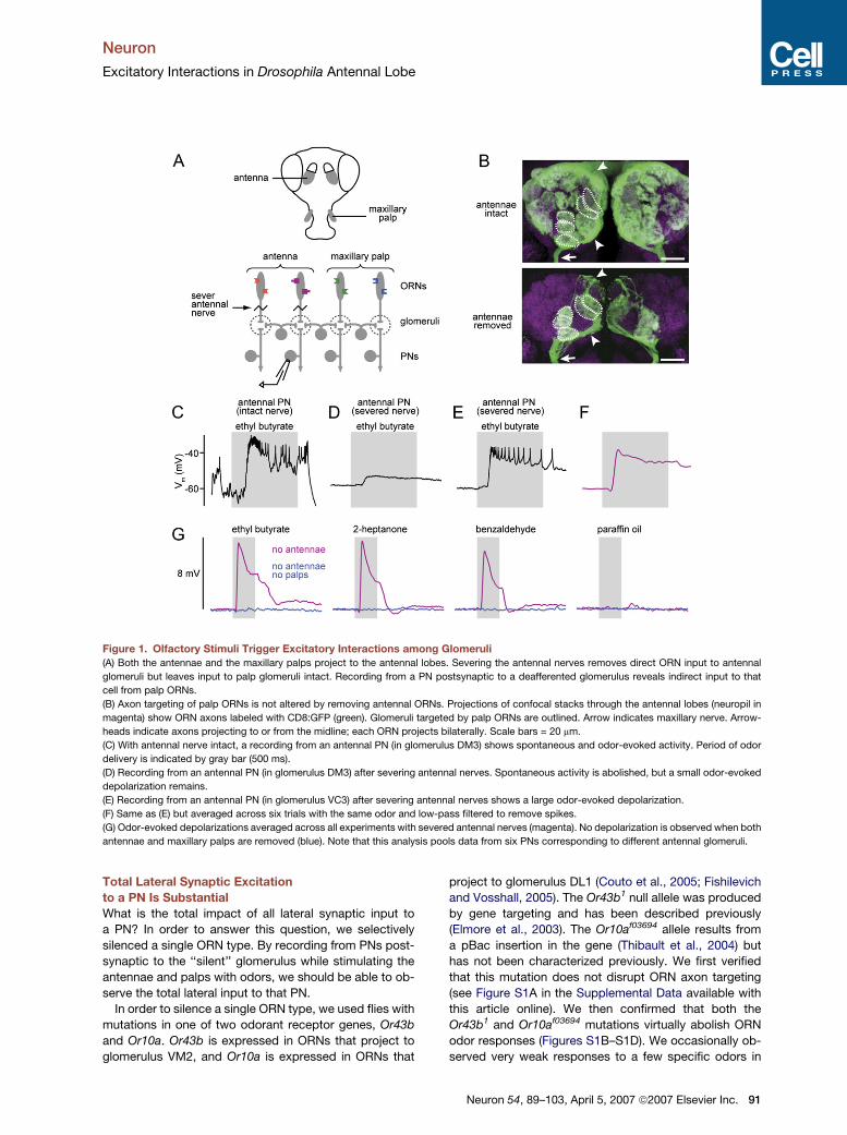
Figure 1. Olfactory Stimuli Trigger Excitatory Interactions among Glomeruli
(A) Both the antennae and the maxillary palps project to the antennal lobes. Severing the antennal nerves removes direct ORN input to antennal
glomeruli but leaves input to palp glomeruli intact. Recording from a PN postsynaptic to a deafferented glomerulus reveals indirect input to that
cell from palp ORNs.
(B) Axon targeting of palp ORNs is not altered by removing antennal ORNs. Projections of confocal stacks through the antennal lobes (neuropil in
magenta) show ORN axons labeled with CD8:GFP (green). Glomeruli targeted by palp ORNs are outlined. Arrow indicates maxillary nerve. Arrow-
heads indicate axons projecting to or from the midline; each ORN projects bilaterally. Scale bars = 20 mm.
(C) With antennal nerve intact, a recording from an antennal PN (in glomerulus DM3) shows spontaneous and odor-evoked activity. Period of odor
delivery is indicated by gray bar (500 ms).
(D) Recording from an antennal PN (in glomerulus DM3) after severing antennal nerves. Spontaneous activity is abolished, but a small odor-evoked
depolarization remains.
(E) Recording from an antennal PN (in glomerulus VC3) after severing antennal nerves shows a large odor-evoked depolarization.
(F) Same as (E) but averaged across six trials with the same odor and low-pass filtered to remove spikes.
(G) Odor-evoked depolarizations averaged across all experiments with severed antennal nerves (magenta). No depolarization is observed when both
antennae and maxillary palps are removed (blue). Note that this analysis pools data from six PNs corresponding to different antennal glomeruli.
Total Lateral Synaptic Excitation
to a PN Is Substantial
What is the total impact of all lateral synaptic input to
a PN? In order to answer this question, we selectively
silenced a single ORN type. By recording from PNs post-
synaptic to the ‘‘silent’’ glomerulus while stimulating the
antennae and palps with odors, we should be able to ob-
serve the total lateral input to that PN.
In order to silence a single ORN type, we used flies with
mutations in one of two odorant receptor genes, Or43b
and Or10a. Or43b is expressed in ORNs that project to
glomerulus VM2, and Or10a is expressed in ORNs that
project to glomerulus DL1 (Couto et al., 2005; Fishilevich
and Vosshall, 2005). The Or43b1 null allele was produced
by gene targeting and has been described previously
(Elmore et al., 2003). The Or10af03694 allele results from
a pBac insertion in the gene (Thibault et al., 2004) but
has not been characterized previously. We first verified
that this mutation does not disrupt ORN axon targeting
(see Figure S1A in the Supplemental Data available with
this article online). We then confirmed that both the
Or43b1 and Or10af03694 mutations virtually abolish ORN
odor responses (Figures S1B–S1D). We occasionally ob-
served very weak responses to a few specific odors in
Neuron 54, 89–103, April 5, 2007 ª2007 Elsevier Inc. 91
Neuron
Excitatory Interactions in Drosophila Antennal Lobe
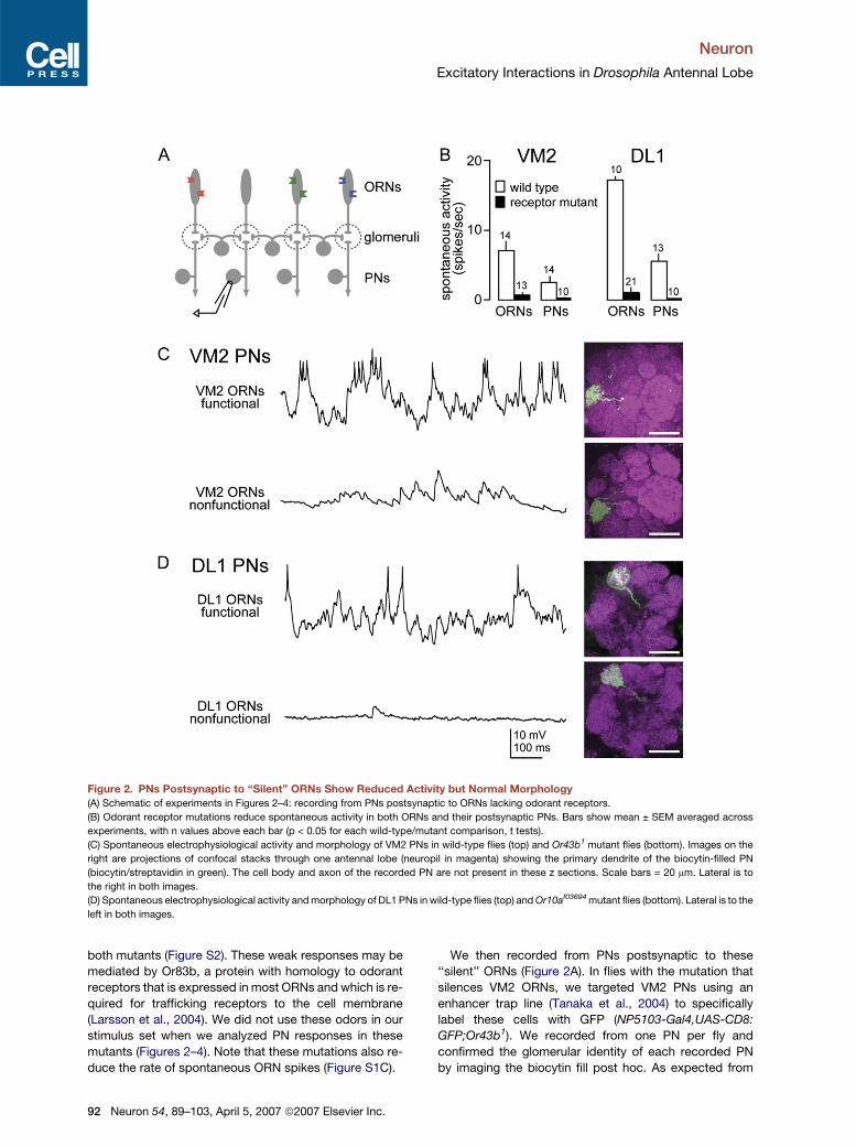
Figure 2. PNs Postsynaptic to ‘‘Silent’’ ORNs Show Reduced Activity but Normal Morphology
(A) Schematic of experiments in Figures 2–4: recording from PNs postsynaptic to ORNs lacking odorant receptors.
(B) Odorant receptor mutations reduce spontaneous activity in both ORNs and their postsynaptic PNs. Bars show mean ± SEM averaged across
experiments, with n values above each bar (p < 0.05 for each wild-type/mutant comparison, t tests).
(C) Spontaneous electrophysiological activity and morphology of VM2 PNs in wild-type flies (top) and Or43b1 mutant flies (bottom). Images on the
right are projections of confocal stacks through one antennal lobe (neuropil in magenta) showing the primary dendrite of the biocytin-filled PN
(biocytin/streptavidin in green). The cell body and axon of the recorded PN are not present in these z sections. Scale bars = 20 mm. Lateral is to
the right in both images.
(D) Spontaneous electrophysiological activity and morphology of DL1 PNs in wild-type flies (top) and Or10af03694 mutant flies (bottom). Lateral is to the
left in both images.
both mutants (Figure S2). These weak responses may be
mediated by Or83b, a protein with homology to odorant
receptors that is expressed in most ORNs and which is re-
quired for trafficking receptors to the cell membrane
(Larsson et al., 2004). We did not use these odors in our
stimulus set when we analyzed PN responses in these
mutants (Figures 2–4). Note that these mutations also re-
duce the rate of spontaneous ORN spikes (Figure S1C).
92 Neuron 54, 89–103, April 5, 2007 ª2007 Elsevier Inc.
We then recorded from PNs postsynaptic to these
‘‘silent’’ ORNs (Figure 2A). In flies with the mutation that
silences VM2 ORNs, we targeted VM2 PNs using an
enhancer trap line (Tanaka et al., 2004) to specifically
label these cells with GFP (NP5103-Gal4,UAS-CD8:
GFP;Or43b1). We recorded from one PN per fly and
confirmed the glomerular identity of each recorded PN
by imaging the biocytin fill post hoc. As expected from
Neuron
Excitatory Interactions in Drosophila Antennal Lobe
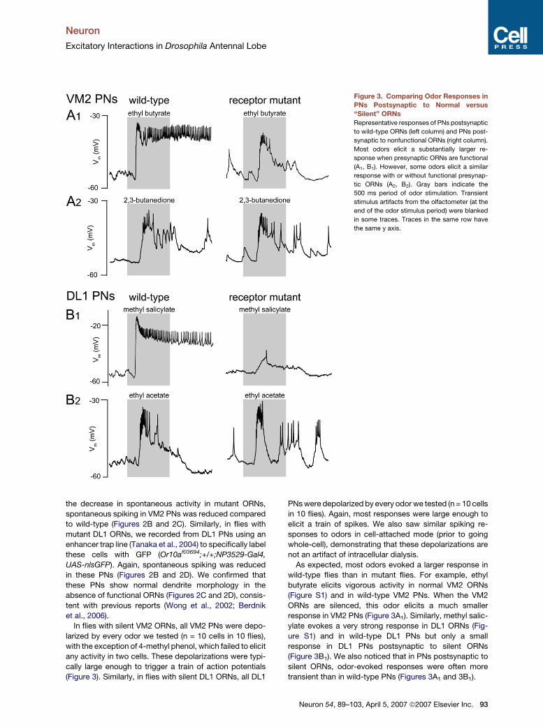
Figure 3. Comparing Odor Responses in
PNs Postsynaptic to Normal versus
‘‘Silent’’ ORNs
Representative responses of PNs postsynaptic
to wild-type ORNs (left column) and PNs post-
synaptic to nonfunctional ORNs (right column).
Most odors elicit a substantially larger re-
sponse when presynaptic ORNs are functional
(A1, B1). However, some odors elicit a similar
response with or without functional presynap-
tic ORNs (A2, B2). Gray bars indicate the
500 ms period of odor stimulation. Transient
stimulus artifacts from the olfactometer (at the
end of the odor stimulus period) were blanked
in some traces. Traces in the same row have
the same y axis.
the decrease in spontaneous activity in mutant ORNs,
spontaneous spiking in VM2 PNs was reduced compared
to wild-type (Figures 2B and 2C). Similarly, in flies with
mutant DL1 ORNs, we recorded from DL1 PNs using an
enhancer trap line (Tanaka et al., 2004) to specifically label
these cells with GFP (Or10af03694;+/+;NP3529-Gal4,
UAS-nlsGFP). Again, spontaneous spiking was reduced
in these PNs (Figures 2B and 2D). We confirmed that
these PNs show normal dendrite morphology in the
absence of functional ORNs (Figures 2C and 2D), consis-
tent with previous reports (Wong et al., 2002; Berdnik
et al., 2006).
In flies with silent VM2 ORNs, all VM2 PNs were depo-
larized by every odor we tested (n = 10 cells in 10 flies),
with the exception of 4-methyl phenol, which failed to elicit
any activity in two cells. These depolarizations were typi-
cally large enough to trigger a train of action potentials
(Figure 3). Similarly, in flies with silent DL1 ORNs, all DL1
PNs were depolarized by every odor we tested (n = 10 cells
in 10 flies). Again, most responses were large enough to
elicit a train of spikes. We also saw similar spiking re-
sponses to odors in cell-attached mode (prior to going
whole-cell), demonstrating that these depolarizations are
not an artifact of intracellular dialysis.
As expected, most odors evoked a larger response in
wild-type flies than in mutant flies. For example, ethyl
butyrate elicits vigorous activity in normal VM2 ORNs
(Figure S1) and in wild-type VM2 PNs. When the VM2
ORNs are silenced, this odor elicits a much smaller
response in VM2 PNs (Figure 3A1). Similarly, methyl salic-
ylate evokes a very strong response in DL1 ORNs (Fig-
ure S1) and in wild-type DL1 PNs but only a small
response in DL1 PNs postsynaptic to silent ORNs
(Figure 3B1). We also noticed that in PNs postsynaptic to
silent ORNs, odor-evoked responses were often more
transient than in wild-type PNs (Figures 3A1 and 3B1).
Neuron 54, 89–103, April 5, 2007 ª2007 Elsevier Inc. 93
Neuron
Excitatory Interactions in Drosophila Antennal Lobe
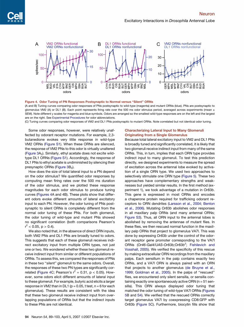
Figure 4. Odor Tuning of PN Responses Postsynaptic to Normal versus ‘‘Silent’’ ORNs
(A and B) Tuning curves comparing odor responses of PNs postsynaptic to wild-type (magenta) and mutant ORNs (blue). PNs are postsynaptic to
glomerulus VM2 (A) or DL1 (B). Each point represents firing rate over the 500 ms odor stimulus period, averaged across experiments (mean ±
SEM). Note different y scales for magenta and blue symbols. Odors are arranged so the smallest wild-type responses are on the left and the largest
are on the right. See Experimental Procedures for odor abbreviations.
(C) Tuning curves comparing odor responses of VM2 and DL1 PNs postsynaptic to mutant ORNs. Note correlated but not identical odor tuning.
Some odor responses, however, were relatively unaf-
fected by odorant receptor mutations. For example, 2,3-
butanedione evokes very little response in wild-type
VM2 ORNs (Figure S1). When these ORNs are silenced,
the response of VM2 PNs to this odor is virtually unaltered
(Figure 3A2). Similarly, ethyl acetate does not excite wild-
type DL1 ORNs (Figure S1). Accordingly, the response of
DL1 PNs to ethyl acetate is undiminished by silencing their
presynaptic ORNs (Figure 3B2).
How does the size of total lateral input to a PN depend
on the odor stimulus? We quantified odor responses by
computing mean firing rates over the 500 ms duration
of the odor stimulus, and we plotted these response
magnitudes for each odor stimulus to produce tuning
curves (Figures 4A and 4B). These plots show that differ-
ent odors evoke different amounts of lateral excitatory
input to each PN. However, the odor tuning of PNs post-
synaptic to silent ORNs is completely different from the
normal odor tuning of these PNs. For both glomeruli,
the odor tuning of wild-type and mutant PNs showed
no significant correlation (both comparisons Pearson’s
r2 < 0.05, p > 0.4).
We also noted that, in the absence of direct ORN inputs,
both VM2 PNs and DL1 PNs are broadly tuned to odors.
This suggests that each of these glomeruli receives indi-
rect excitatory input from multiple ORN types, not just
one or two. We wondered whether these two glomeruli re-
ceive indirect input from similar or different populations of
ORNs. To assess this, we compared the responses of PNs
in these two ‘‘silent’’ glomeruli to the same odors. Overall,
the responses of these two PN types are significantly cor-
related (Figure 4C; Pearson’s r2 = 0.31, p < 0.05). How-
ever, some odors elicit different amounts of lateral input
to these glomeruli. For example, butyric acid elicits a larger
response in VM2 than in DL1 (p = 0.05, t test, n = 6 for each
glomerulus). These results are consistent with the idea
that these two glomeruli receive indirect input from over-
lapping populations of ORNs but that the indirect inputs
to these PNs are not identical.
94 Neuron 54, 89–103, April 5, 2007 ª2007 Elsevier Inc.
Characterizing Lateral Input to Many Glomeruli
Originating from a Single Glomerulus
Because total lateral excitatory input to VM2 and DL1 PNs
is broadly tuned and significantly correlated, it is likely that
two glomeruli receive indirect input from many of the same
ORNs. This, in turn, implies that each ORN type provides
indirect input to many glomeruli. To test this prediction
directly, we designed experiments to measure the spread
of excitation across the antennal lobe evoked by activa-
tion of a single ORN type. We used two approaches to
selectively stimulate one ORN type (Figure 5). These two
approaches have complementary strengths and weak-
nesses but yielded similar results. In the first method (ex-
periment 1), we took advantage of a mutation in Or83b.
This gene is expressed in most ORNs and encodes
a chaperone protein required for trafficking odorant re-
ceptors to ORN dendrites (Larsson et al., 2004; Benton
et al., 2006). Mutating Or83b abolishes odor responses
in all maxillary palp ORNs (and many antennal ORNs;
Figure S3). Thus, all ORN input to the antennal lobes is
abolished by removing the antennae of mutant flies. In
these flies, we then rescued normal function in the maxil-
lary palp ORNs that project to glomerulus VA7l. This was
done by expressing Or83b under the control of the odor-
ant receptor gene promoter corresponding to the VA7l
ORNs (Or46-Gal4/UAS-Or83b;Or83b2; Fishilevich and
Vosshall, 2005). We verified the specificity of this rescue
by making extracellular ORN recordings from the maxillary
palps. Each sensillum in the palp contains exactly two
ORNs, and a VA7l ORN is always paired with an ORN
that projects to another glomerulus (de Bruyne et al.,
1999; Goldman et al., 2005). In the palps of ‘‘rescued’’
flies, we encountered only silent sensilla, or sensilla con-
taining exactly one spontaneously active ORN (n = 51 sen-
silla). This ORN always displayed odor tuning that
matched the odor tuning of wild-type VA7l ORNs (Figures
5B and S4). We verified that the rescued ORNs correctly
target glomerulus VA7l by coexpressing CD8:GFP with
Or83b (Figure 5C). Furthermore, biocytin fills show that
Neuron
Excitatory Interactions in Drosophila Antennal Lobe
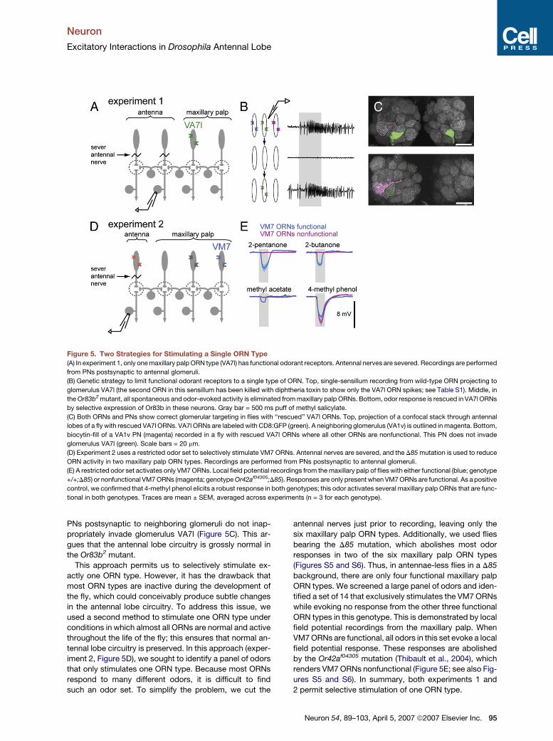
Figure 5. Two Strategies for Stimulating a Single ORN Type
(A) In experiment 1, only one maxillary palp ORN type (VA7l) has functional odorant receptors. Antennal nerves are severed. Recordings are performed
from PNs postsynaptic to antennal glomeruli.
(B) Genetic strategy to limit functional odorant receptors to a single type of ORN. Top, single-sensillum recording from wild-type ORN projecting to
glomerulus VA7l (the second ORN in this sensillum has been killed with diphtheria toxin to show only the VA7l ORN spikes; see Table S1). Middle, in
the Or83b2 mutant, all spontaneous and odor-evoked activity is eliminated from maxillary palp ORNs. Bottom, odor response is rescued in VA7l ORNs
by selective expression of Or83b in these neurons. Gray bar = 500 ms puff of methyl salicylate.
(C) Both ORNs and PNs show correct glomerular targeting in flies with ‘‘rescued’’ VA7l ORNs. Top, projection of a confocal stack through antennal
lobes of a fly with rescued VA7l ORNs. VA7l ORNs are labeled with CD8:GFP (green). A neighboring glomerulus (VA1v) is outlined in magenta. Bottom,
biocytin-fill of a VA1v PN (magenta) recorded in a fly with rescued VA7l ORNs where all other ORNs are nonfunctional. This PN does not invade
glomerulus VA7l (green). Scale bars = 20 mm.
(D) Experiment 2 uses a restricted odor set to selectively stimulate VM7 ORNs. Antennal nerves are severed, and the D85 mutation is used to reduce
ORN activity in two maxillary palp ORN types. Recordings are performed from PNs postsynaptic to antennal glomeruli.
(E) A restricted odor set activates only VM7 ORNs. Local field potential recordings from the maxillary palp of flies with either functional (blue; genotype
+/+;D85) or nonfunctional VM7 ORNs (magenta; genotype Or42af04305;D85). Responses are only present when VM7 ORNs are functional. As a positive
control, we confirmed that 4-methyl phenol elicits a robust response in both genotypes; this odor activates several maxillary palp ORNs that are func-
tional in both genotypes. Traces are mean ± SEM, averaged across experiments (n = 3 for each genotype).
PNs postsynaptic to neighboring glomeruli do not inap-
propriately invade glomerulus VA7l (Figure 5C). This ar-
gues that the antennal lobe circuitry is grossly normal in
the Or83b2 mutant.
This approach permits us to selectively stimulate ex-
actly one ORN type. However, it has the drawback that
most ORN types are inactive during the development of
the fly, which could conceivably produce subtle changes
in the antennal lobe circuitry. To address this issue, we
used a second method to stimulate one ORN type under
conditions in which almost all ORNs are normal and active
throughout the life of the fly; this ensures that normal an-
tennal lobe circuitry is preserved. In this approach (exper-
iment 2, Figure 5D), we sought to identify a panel of odors
that only stimulates one ORN type. Because most ORNs
respond to many different odors, it is difficult to find
such an odor set. To simplify the problem, we cut the
antennal nerves just prior to recording, leaving only the
six maxillary palp ORN types. Additionally, we used flies
bearing the D85 mutation, which abolishes most odor
responses in two of the six maxillary palp ORN types
(Figures S5 and S6). Thus, in antennae-less flies in a D85
background, there are only four functional maxillary palp
ORN types. We screened a large panel of odors and iden-
tified a set of 14 that exclusively stimulates the VM7 ORNs
while evoking no response from the other three functional
ORN types in this genotype. This is demonstrated by local
field potential recordings from the maxillary palp. When
VM7 ORNs are functional, all odors in this set evoke a local
field potential response. These responses are abolished
by the Or42af04305 mutation (Thibault et al., 2004), which
renders VM7 ORNs nonfunctional (Figure 5E; see also Fig-
ures S5 and S6). In summary, both experiments 1 and
2 permit selective stimulation of one ORN type.
Neuron 54, 89–103, April 5, 2007 ª2007 Elsevier Inc. 95
Neuron
Excitatory Interactions in Drosophila Antennal Lobe
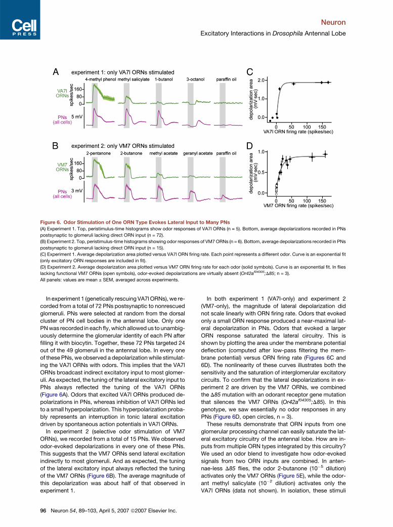
Figure 6. Odor Stimulation of One ORN Type Evokes Lateral Input to Many PNs
(A) Experiment 1. Top, peristimulus-time histograms show odor responses of VA7l ORNs (n = 5). Bottom, average depolarizations recorded in PNs
postsynaptic to glomeruli lacking direct ORN input (n = 72).
(B) Experiment 2. Top, peristimulus-time histograms showing odor responses of VM7 ORNs (n = 6). Bottom, average depolarizations recorded in PNs
postsynaptic to glomeruli lacking direct ORN input (n = 15).
(C) Experiment 1. Average depolarization area plotted versus VA7l ORN firing rate. Each point represents a different odor. Curve is an exponential fit
(only excitatory ORN responses are included in fit).
(D) Experiment 2. Average depolarization area plotted versus VM7 ORN firing rate for each odor (solid symbols). Curve is an exponential fit. In flies
lacking functional VM7 ORNs (open symbols), odor-evoked depolarizations are virtually absent (Or42af04305;D85; n = 3).
All panels: values are mean ± SEM, averaged across experiments.
In experiment 1 (genetically rescuing VA7l ORNs), we re-
corded from a total of 72 PNs postsynaptic to nonrescued
glomeruli. PNs were selected at random from the dorsal
cluster of PN cell bodies in the antennal lobe. Only one
PN was recorded in each fly, which allowed us to unambig-
uously determine the glomerular identity of each PN after
filling it with biocytin. Together, these 72 PNs targeted 24
out of the 49 glomeruli in the antennal lobe. In every one
of these PNs, we observed a depolarization while stimulat-
ing the VA7l ORNs with odors. This implies that the VA7l
ORNs broadcast indirect excitatory input to most glomer-
uli. As expected, the tuning of the lateral excitatory input to
PNs always reflected the tuning of the VA7l ORNs
(Figure 6A). Odors that excited VA7l ORNs produced de-
polarizations in PNs, whereas inhibition of VA7l ORNs led
to a small hyperpolarization. This hyperpolarization proba-
bly represents an interruption in tonic lateral excitation
driven by spontaneous action potentials in VA7l ORNs.
In experiment 2 (selective odor stimulation of VM7
ORNs), we recorded from a total of 15 PNs. We observed
odor-evoked depolarizations in every one of these PNs.
This suggests that the VM7 ORNs send lateral excitation
indirectly to most glomeruli. And as expected, the tuning
of the lateral excitatory input always reflected the tuning
of the VM7 ORNs (Figure 6B). The average magnitude of
this depolarization was about half of that observed in
experiment 1.
96 Neuron 54, 89–103, April 5, 2007 ª2007 Elsevier Inc.
In both experiment 1 (VA7l-only) and experiment 2
(VM7-only), the magnitude of lateral depolarization did
not scale linearly with ORN firing rate. Odors that evoked
only a small ORN response produced a near-maximal lat-
eral depolarization in PNs. Odors that evoked a larger
ORN response saturated the lateral circuitry. This is
shown by plotting the area under the membrane potential
deflection (computed after low-pass filtering the mem-
brane potential) versus ORN firing rate (Figures 6C and
6D). The nonlinearity of these curves illustrates both the
sensitivity and the saturation of interglomerular excitatory
circuits. To confirm that the lateral depolarizations in ex-
periment 2 are driven by the VM7 ORNs, we combined
the D85 mutation with an odorant receptor gene mutation
that silences the VM7 ORNs (Or42af04305;D85). In this
genotype, we saw essentially no odor responses in any
PNs (Figure 6D, open circles, n = 3).
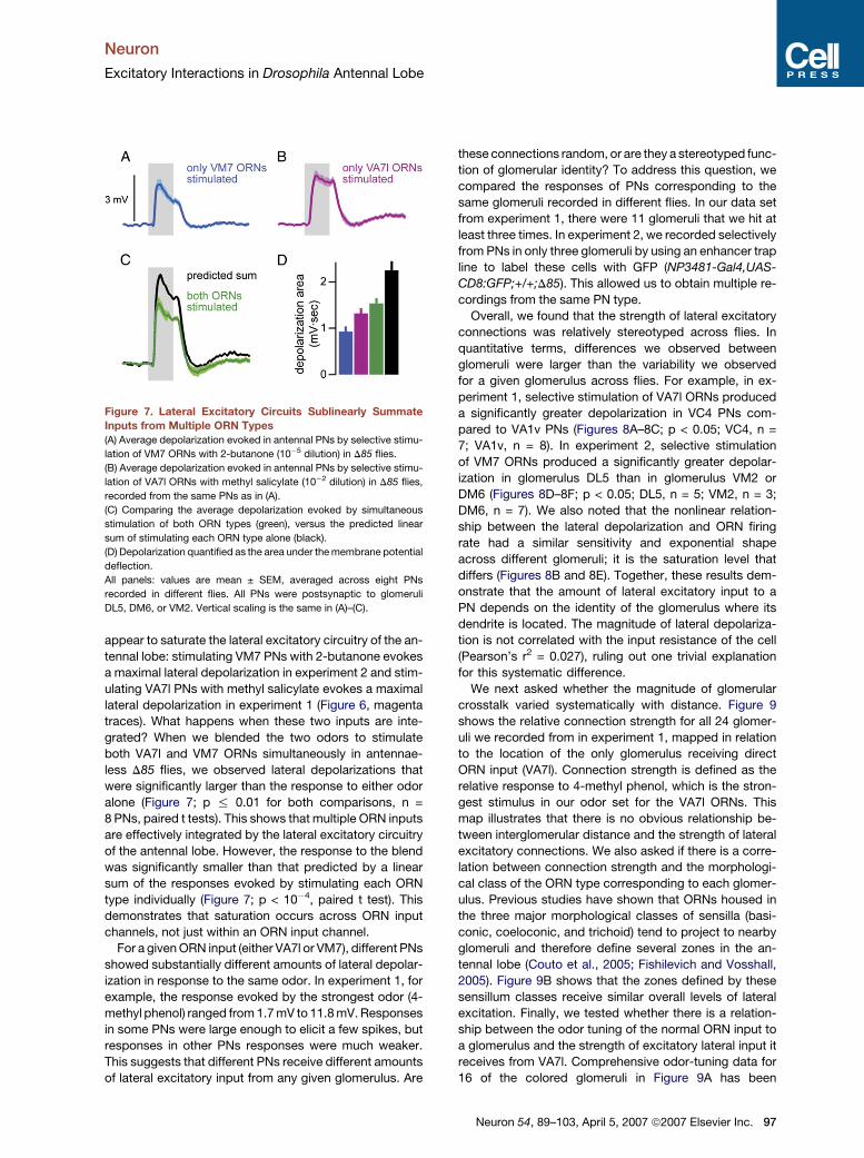
These results demonstrate that ORN inputs from one
glomerular processing channel can easily saturate the lat-
eral excitatory circuitry of the antennal lobe. How are in-
puts from multiple ORN types integrated by this circuitry?
We used an odor blend to investigate how odor-evoked
signals from two ORN inputs are combined. In anten-
nae-less D85 flies, the odor 2-butanone (10�5 dilution)
activates only the VM7 ORNs (Figure 5E), while the odor-
ant methyl salicylate (10�2 dilution) activates only the
VA7l ORNs (data not shown). In isolation, these stimuli
Neuron
Excitatory Interactions in Drosophila Antennal Lobe
appear to saturate the lateral excitatory circuitry of the an-
tennal lobe: stimulating VM7 PNs with 2-butanone evokes
a maximal lateral depolarization in experiment 2 and stim-
ulating VA7l PNs with methyl salicylate evokes a maximal
lateral depolarization in experiment 1 (Figure 6, magenta
traces). What happens when these two inputs are inte-
grated? When we blended the two odors to stimulate
both VA7l and VM7 ORNs simultaneously in antennae-
less D85 flies, we observed lateral depolarizations that
were significantly larger than the response to either odor
alone (Figure 7; p % 0.01 for both comparisons, n =
8 PNs, paired t tests). This shows that multiple ORN inputs
are effectively integrated by the lateral excitatory circuitry
of the antennal lobe. However, the response to the blend
was significantly smaller than that predicted by a linear
sum of the responses evoked by stimulating each ORN
type individually (Figure 7; p < 10�4, paired t test). This
demonstrates that saturation occurs across ORN input
channels, not just within an ORN input channel.
For a given ORN input (either VA7l or VM7), different PNs
showed substantially different amounts of lateral depolar-
ization in response to the same odor. In experiment 1, for
example, the response evoked by the strongest odor (4-
methyl phenol) ranged from 1.7 mV to 11.8 mV. Responses
in some PNs were large enough to elicit a few spikes, but
responses in other PNs responses were much weaker.
This suggests that different PNs receive different amounts
of lateral excitatory input from any given glomerulus. Are
Figure 7. Lateral Excitatory Circuits Sublinearly Summate
Inputs from Multiple ORN Types
(A) Average depolarization evoked in antennal PNs by selective stimu-
lation of VM7 ORNs with 2-butanone (10�5 dilution) in D85 flies.
(B) Average depolarization evoked in antennal PNs by selective stimu-
lation of VA7l ORNs with methyl salicylate (10�2 dilution) in D85 flies,
recorded from the same PNs as in (A).
(C) Comparing the average depolarization evoked by simultaneous
stimulation of both ORN types (green), versus the predicted linear
sum of stimulating each ORN type alone (black).
(D) Depolarization quantified as the area under the membrane potential
deflection.
All panels: values are mean ± SEM, averaged across eight PNs
recorded in different flies. All PNs were postsynaptic to glomeruli
DL5, DM6, or VM2. Vertical scaling is the same in (A)–(C).
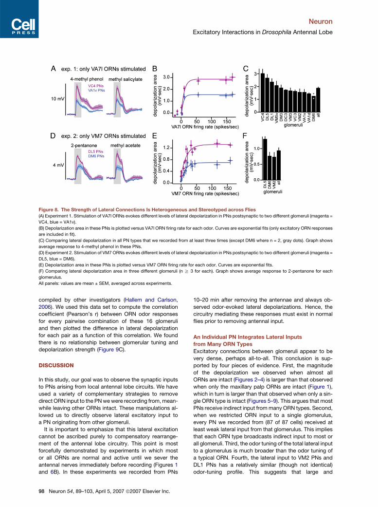
these connections random, or are they a stereotyped func-
tion of glomerular identity? To address this question, we
compared the responses of PNs corresponding to the
same glomeruli recorded in different flies. In our data set
from experiment 1, there were 11 glomeruli that we hit at
least three times. In experiment 2, we recorded selectively
from PNs in only three glomeruli by using an enhancer trap
line to label these cells with GFP (NP3481-Gal4,UAS-
CD8:GFP;+/+;D85). This allowed us to obtain multiple re-
cordings from the same PN type.
Overall, we found that the strength of lateral excitatory
connections was relatively stereotyped across flies. In
quantitative terms, differences we observed between
glomeruli were larger than the variability we observed
for a given glomerulus across flies. For example, in ex-
periment 1, selective stimulation of VA7l ORNs produced
a significantly greater depolarization in VC4 PNs com-
pared to VA1v PNs (Figures 8A–8C; p < 0.05; VC4, n =
7; VA1v, n = 8). In experiment 2, selective stimulation
of VM7 ORNs produced a significantly greater depolar-
ization in glomerulus DL5 than in glomerulus VM2 or
DM6 (Figures 8D–8F; p < 0.05; DL5, n = 5; VM2, n = 3;
DM6, n = 7). We also noted that the nonlinear relation-
ship between the lateral depolarization and ORN firing
rate had a similar sensitivity and exponential shape
across different glomeruli; it is the saturation level that
differs (Figures 8B and 8E). Together, these results dem-
onstrate that the amount of lateral excitatory input to a
PN depends on the identity of the glomerulus where its
dendrite is located. The magnitude of lateral depolariza-
tion is not correlated with the input resistance of the cell
(Pearson’s r2 = 0.027), ruling out one trivial explanation
for this systematic difference.
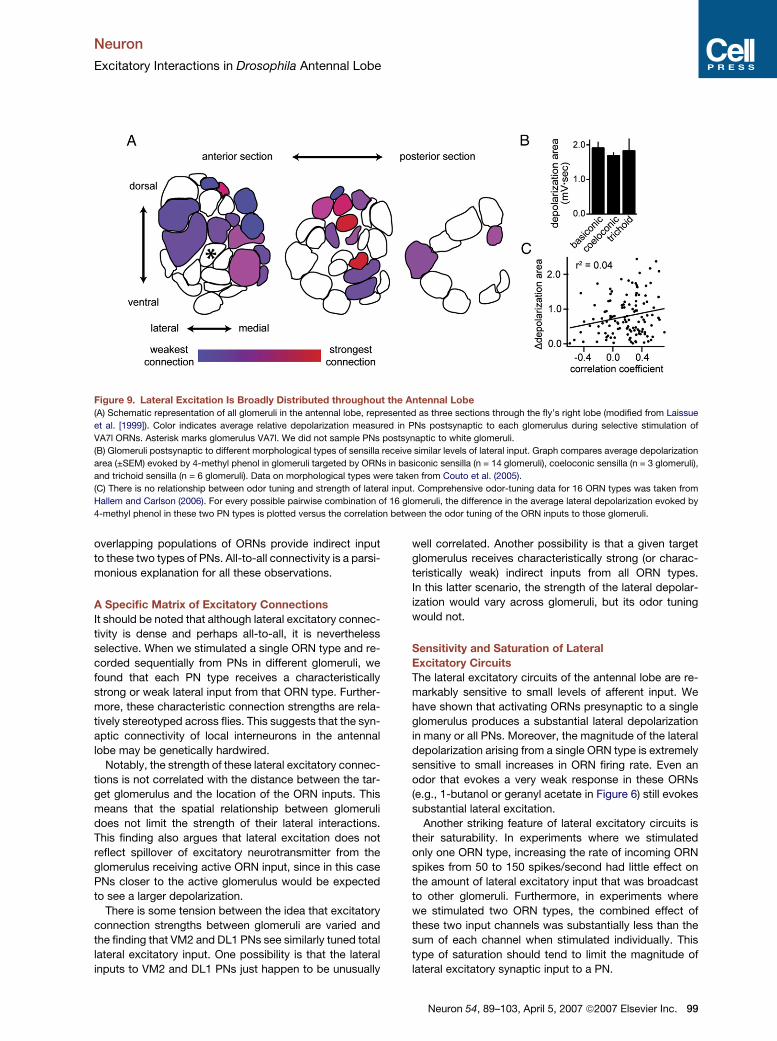
We next asked whether the magnitude of glomerular
crosstalk varied systematically with distance. Figure 9
shows the relative connection strength for all 24 glomer-
uli we recorded from in experiment 1, mapped in relation
to the location of the only glomerulus receiving direct
ORN input (VA7l). Connection strength is defined as the
relative response to 4-methyl phenol, which is the stron-
gest stimulus in our odor set for the VA7l ORNs. This
map illustrates that there is no obvious relationship be-
tween interglomerular distance and the strength of lateral
excitatory connections. We also asked if there is a corre-
lation between connection strength and the morphologi-
cal class of the ORN type corresponding to each glomer-
ulus. Previous studies have shown that ORNs housed in
the three major morphological classes of sensilla (basi-
conic, coeloconic, and trichoid) tend to project to nearby
glomeruli and therefore define several zones in the an-
tennal lobe (Couto et al., 2005; Fishilevich and Vosshall,
2005). Figure 9B shows that the zones defined by these
sensillum classes receive similar overall levels of lateral
excitation. Finally, we tested whether there is a relation-
ship between the odor tuning of the normal ORN input to
a glomerulus and the strength of excitatory lateral input it
receives from VA7l. Comprehensive odor-tuning data for
16 of the colored glomeruli in Figure 9A has been
Neuron 54, 89–103, April 5, 2007 ª2007 Elsevier Inc. 97
Neuron
Excitatory Interactions in Drosophila Antennal Lobe
Figure 8. The Strength of Lateral Connections Is Heterogeneous and Stereotyped across Flies
(A) Experiment 1. Stimulation of VA7l ORNs evokes different levels of lateral depolarization in PNs postsynaptic to two different glomeruli (magenta =
VC4, blue = VA1v).
(B) Depolarization area in these PNs is plotted versus VA7l ORN firing rate for each odor. Curves are exponential fits (only excitatory ORN responses
are included in fit).
(C) Comparing lateral depolarization in all PN types that we recorded from at least three times (except DM6 where n = 2, gray dots). Graph shows
average response to 4-methyl phenol in these PNs.
(D) Experiment 2. Stimulation of VM7 ORNs evokes different levels of lateral depolarization in PNs postsynaptic to two different glomeruli (magenta =
DL5, blue = DM6).
(E) Depolarization area in these PNs is plotted versus VM7 ORN firing rate for each odor. Curves are exponential fits.
(F) Comparing lateral depolarization area in three different glomeruli (n R 3 for each). Graph shows average response to 2-pentanone for each
glomerulus.
All panels: values are mean ± SEM, averaged across experiments.
compiled by other investigators (Hallem and Carlson,
2006). We used this data set to compute the correlation
coefficient (Pearson’s r) between ORN odor responses
for every pairwise combination of these 16 glomeruli
and then plotted the difference in lateral depolarization
for each pair as a function of this correlation. We found
there is no relationship between glomerular tuning and
depolarization strength (Figure 9C).
DISCUSSION
In this study, our goal was to observe the synaptic inputs
to PNs arising from local antennal lobe circuits. We have
used a variety of complementary strategies to remove
direct ORN input to the PN we were recording from, mean-
while leaving other ORNs intact. These manipulations al-
lowed us to directly observe lateral excitatory input to
a PN originating from other glomeruli.
It is important to emphasize that this lateral excitation
cannot be ascribed purely to compensatory rearrange-
ment of the antennal lobe circuitry. This point is most
forcefully demonstrated by experiments in which most
or all ORNs are normal and active until we sever the
antennal nerves immediately before recording (Figures 1
and 6B). In these experiments we recorded from PNs
98 Neuron 54, 89–103, April 5, 2007 ª2007 Elsevier Inc.
10–20 min after removing the antennae and always ob-
served odor-evoked lateral depolarizations. Hence, the
circuitry mediating these responses must exist in normal
flies prior to removing antennal input.
An Individual PN Integrates Lateral Inputs
from Many ORN Types
Excitatory connections between glomeruli appear to be
very dense, perhaps all-to-all. This conclusion is sup-
ported by four pieces of evidence. First, the magnitude
of the depolarization we observed when almost all
ORNs are intact (Figures 2–4) is larger than that observed
when only the maxillary palp ORNs are intact (Figure 1),
which in turn is larger than that observed when only a sin-
gle ORN type is intact (Figures 5–9). This argues that most
PNs receive indirect input from many ORN types. Second,
when we restricted ORN input to a single glomerulus,
every PN we recorded from (87 of 87 cells) received at
least weak lateral input from that glomerulus. This implies
that each ORN type broadcasts indirect input to most or
all glomeruli. Third, the odor tuning of the total lateral input
to a glomerulus is much broader than the odor tuning of
a typical ORN. Fourth, the lateral input to VM2 PNs and
DL1 PNs has a relatively similar (though not identical)
odor-tuning profile. This suggests that large and
Neuron
Excitatory Interactions in Drosophila Antennal Lobe
Figure 9. Lateral Excitation Is Broadly Distributed throughout the Antennal Lobe
(A) Schematic representation of all glomeruli in the antennal lobe, represented as three sections through the fly’s right lobe (modified from Laissue
et al. [1999]). Color indicates average relative depolarization measured in PNs postsynaptic to each glomerulus during selective stimulation of
VA7l ORNs. Asterisk marks glomerulus VA7l. We did not sample PNs postsynaptic to white glomeruli.
(B) Glomeruli postsynaptic to different morphological types of sensilla receive similar levels of lateral input. Graph compares average depolarization
area (±SEM) evoked by 4-methyl phenol in glomeruli targeted by ORNs in basiconic sensilla (n = 14 glomeruli), coeloconic sensilla (n = 3 glomeruli),
and trichoid sensilla (n = 6 glomeruli). Data on morphological types were taken from Couto et al. (2005).
(C) There is no relationship between odor tuning and strength of lateral input. Comprehensive odor-tuning data for 16 ORN types was taken from
Hallem and Carlson (2006). For every possible pairwise combination of 16 glomeruli, the difference in the average lateral depolarization evoked by
4-methyl phenol in these two PN types is plotted versus the correlation between the odor tuning of the ORN inputs to those glomeruli.
overlapping populations of ORNs provide indirect input
to these two types of PNs. All-to-all connectivity is a parsi-
monious explanation for all these observations.
A Specific Matrix of Excitatory Connections
It should be noted that although lateral excitatory connec-
tivity is dense and perhaps all-to-all, it is nevertheless
selective. When we stimulated a single ORN type and re-
corded sequentially from PNs in different glomeruli, we
found that each PN type receives a characteristically
strong or weak lateral input from that ORN type. Further-
more, these characteristic connection strengths are rela-
tively stereotyped across flies. This suggests that the syn-
aptic connectivity of local interneurons in the antennal
lobe may be genetically hardwired.
Notably, the strength of these lateral excitatory connec-
tions is not correlated with the distance between the tar-
get glomerulus and the location of the ORN inputs. This
means that the spatial relationship between glomeruli
does not limit the strength of their lateral interactions.
This finding also argues that lateral excitation does not
reflect spillover of excitatory neurotransmitter from the
glomerulus receiving active ORN input, since in this case
PNs closer to the active glomerulus would be expected
to see a larger depolarization.
There is some tension between the idea that excitatory
connection strengths between glomeruli are varied and
the finding that VM2 and DL1 PNs see similarly tuned total
lateral excitatory input. One possibility is that the lateral
inputs to VM2 and DL1 PNs just happen to be unusually
well correlated. Another possibility is that a given target
glomerulus receives characteristically strong (or charac-
teristically weak) indirect inputs from all ORN types.
In this latter scenario, the strength of the lateral depolar-
ization would vary across glomeruli, but its odor tuning
would not.
Sensitivity and Saturation of Lateral
Excitatory Circuits
The lateral excitatory circuits of the antennal lobe are re-
markably sensitive to small levels of afferent input. We
have shown that activating ORNs presynaptic to a single
glomerulus produces a substantial lateral depolarization
in many or all PNs. Moreover, the magnitude of the lateral
depolarization arising from a single ORN type is extremely
sensitive to small increases in ORN firing rate. Even an
odor that evokes a very weak response in these ORNs
(e.g., 1-butanol or geranyl acetate in Figure 6) still evokes
substantial lateral excitation.
Another striking feature of lateral excitatory circuits is
their saturability. In experiments where we stimulated
only one ORN type, increasing the rate of incoming ORN
spikes from 50 to 150 spikes/second had little effect on
the amount of lateral excitatory input that was broadcast
to other glomeruli. Furthermore, in experiments where
we stimulated two ORN types, the combined effect of
these two input channels was substantially less than the
sum of each channel when stimulated individually. This
type of saturation should tend to limit the magnitude of
lateral excitatory synaptic input to a PN.
Neuron 54, 89–103, April 5, 2007 ª2007 Elsevier Inc. 99
Neuron
Excitatory Interactions in Drosophila Antennal Lobe
Together, these results suggest that the impact of lat-
eral excitatory connections might be strongly dependent
on odor concentration. Testing this hypothesis will require
comparing the sensitivity of direct and lateral inputs to
a range of concentrations and understanding how these
inputs are integrated by PNs.
A Cellular Substrate for Lateral
Excitatory Connections
While this manuscript was under review, a report ap-
peared that identified a novel population of cholinergic
local neurons in the Drosophila antennal lobe (Shang
et al., 2007). There is no direct evidence that these local
neurons mediate the local excitatory connections we
have observed, but this hypothesis seems plausible.
Each cholinergic local neuron reportedly innervates most
glomeruli, and this morphology could easily explain our
observation that a single ORN type broadcasts excitatory
input to most or all PNs. Interestingly, excitatory (glutama-
tergic) local neurons were also recently identified in the ol-
factory bulb (Aungst et al., 2003), although it is not known
whether these cells make synapses onto mitral cells, the
analog of antennal lobe PNs.
Shang et al. (2007) also independently provided evi-
dence that PNs receive lateral excitatory input. As in our
study (Figures 2–4), these investigators measured activity
in PNs whose presynaptic ORNs have been silenced by an
odorant receptor gene mutation. Complementary to our
electrophysiological approach, Shang et al. (2007) used
a genetically-encoded ecliptic pHluorin to monitor the
balance of synaptic vesicle exocytosis and endocytosis
at presynaptic sites in PN dendrites. They found that
PNs whose presynaptic ORNs were silent still showed
odor-evoked dendritic synaptopHluorin signals, implying
that these PNs receive indirect excitatory input from other
ORNs.
Lateral Inhibition in the Drosophila Antennal Lobe
Models of olfactory processing in the insect antennal lobe
and the vertebrate olfactory bulb stress the importance of
inhibitory connections between glomeruli (Mori et al.,
1999; Laurent, 2002). What about lateral inhibition in the
Drosophila antennal lobe? It is known that GABAergic in-
terneurons ramify throughout the Drosophila antennal
lobe and release GABA in response to odor stimulation
(Stocker et al., 1997; Ng et al., 2002; Wilson and Laurent,
2005). Drosophila antennal lobe PNs have GABAA-like and
GABAB-like receptors, and antagonists of these receptors
disinhibit PN odor responses (Wilson and Laurent, 2005).
Given this, it is perhaps surprising that we have not
observed lateral synaptic inhibition in PNs.
Several considerations put this finding in perspective.
First, although the lateral inputs we observe are domi-
nated by excitation, it is possible that these responses
reflect the integration of both excitatory and inhibitory
inputs. As a result, inhibition could be masked by a larger
postsynaptic excitation. Second, although our results are
inconsistent with a dominant role for interglomerular post-
100 Neuron 54, 89–103, April 5, 2007 ª2007 Elsevier Inc.
synaptic inhibition of PNs, our findings do not preclude
a role for interglomerular presynaptic inhibition of ORN
axon terminals. Presynaptic inhibition of neurotransmitter
release from ORN axons is a well-known phenomenon in
the mammalian olfactory bulb (Ennis et al., 2001; McGann
et al., 2005; Murphy et al., 2005; Wachowiak et al., 2005)
and in the crustacean olfactory lobe (Wachowiak et al.,
2002). In this study, we abolished or severely reduced
direct ORN input to the PNs we were recording from,
which necessarily prevents us from observing any sub-
stantial presynaptic inhibition.
It is worth noting that neither GABAA nor GABAB recep-
tors can mediate the lateral depolarization we observe.
Both GABAA and GABAB conductances are hyperpolariz-
ing in PNs (Wilson and Laurent, 2005). And although
GABAA and GABAB receptor antagonists together com-
pletely block GABA-evoked hyperpolarizations in PNs
(Wilson and Laurent, 2005), they do not diminish the lateral
depolarization we describe in this study (Figure S7). This
result also demonstrates that the lateral depolarization
does not represent disinhibition (inhibition of inhibitory
input to PNs).
Implications of Lateral Excitatory Connections
for Odor Processing
A significant transformation in odor responses occurs be-
tween the ORN and PN layer in the Drosophila olfactory
system. First, the odor tuning of PNs can be broader
than the odor tuning of their presynaptic ORNs (Wilson
et al., 2004). This may reflect, in part, the effects of the lat-
eral excitatory connections we have described in this
study. Because we have observed that the odor tuning
of lateral input to a PN is different from the odor tuning
of its direct ORN input, it seems likely that these lateral in-
puts promote excitatory responses to odors that would
not have otherwise excited that PN. A second feature of
the ORN-to-PN transformation is that the rank order of
PN odor preferences can differ from the odor preferences
of their presynaptic ORNs (Wilson et al., 2004). Again, be-
cause the odor tuning of lateral input to a PN is different
from the odor tuning of its direct ORN input, it seems likely
that lateral excitatory connections between glomeruli con-
tribute to this phenomenon.
However, it would be misleading to neatly assign differ-
ent components of a PN’s odor response to direct versus
lateral excitatory inputs. Direct and lateral excitation may
coexist with pre- and postsynaptic inhibition, and all these
inputs are likely to be integrated by PNs in a nonlinear
fashion. Broad tuning in PNs could also reflect some non-
linearity in ORN-to-PN connections.
In general, bridging the gap between cellular and
systems neuroscience will require a deeper understand-
ing of how neurons integrate complex synaptic inputs
in vivo. Using a combination of genetic techniques and
in vivo electrophysiology, we have begun to dissect the
various synaptic interactions involved in odor processing
in the Drosophila antennal lobe. Our strategy has been
to eliminate one input to an identified neuron in order to
Neuron
Excitatory Interactions in Drosophila Antennal Lobe
unmask other relevant interactions. Here, this approach
has revealed broadly distributed but specific excitatory
connections between glomeruli. Although the behavior
of a neural circuit is ultimately a complex product of its
components, some insight can nevertheless be gained
by manipulating one element at a time, provided that ap-
propriate genetic tools are available. In this respect, the
Drosophila olfactory circuit represents a powerful system
for understanding the synaptic and cellular computations
performed on sensory stimuli that ultimately produce
perception and behavior.
EXPERIMENTAL PROCEDURES
Fly Stocks
Flies were reared at room temperature on conventional cornmeal agar.
All experiments were performed on adult female flies 2–7 days post-
eclosion. Fly stocks were kindly provided as follows: Or10a-Gal4
(Barry Dickson); Or43b1 (Dean Smith); NP5103-Gal4, NP3529-Gal4,
and NP3481-Gal4 (Kei Ito and Liqun Luo); UAS-Or83b, Or83b-Gal4,
Or83b2, Or46a-Gal4, Or92a-Gal4, and Or42b-Gal4 (Leslie Vosshall);
UAS-DTl/CyO and UAS-DTlIII (Leslie Stevens). UAS-CD8:GFPI, UAS-
CD8:GFPII, and UAS-CD8:GFPIII were obtained from the Bloomington
Stock Center. Or10af03694 and Or42af04305 are indexed on Flybase as
pBac insertions and were obtained from Bloomington. We found that
the Or42af04305 stock had a second mutation that specifically affects
the pb2A and pb3B neurons (Figures S5 and S6). This mutation is
on the third chromosome and most likely affects the odorant receptor
genes Or85e and Or85d, which are expressed in the pb2A and pb3B
neurons, respectively. We term this mutation D85. The Or42af04305
and D85 mutations were separated using standard genetic crosses.
The genotypes used in all experiments are listed in Table S1.
ORN Recordings
Flies were immobilized in the trimmed end of a plastic pipette tip. A ref-
erence electrode filled with Drosophila saline was inserted into the eye,
and a sharp saline-filled glass capillary (tip diameter < 1mm) was in-
serted into a sensillum. Sensilla were visualized using an Olympus
BX51WI microscope with a 503 air objective. Sensillum types were
identified based on their morphology and their characteristic re-
sponses to a panel of odors (de Bruyne et al., 1999, 2001). For record-
ings from flies with rescued VA7l ORNs, we recorded semirandomly
from all three types of sensilla on the maxillary palp (pb2 sensilla which
contain the VA7l ORNs are slightly thinner than pb1 and pb3, which al-
lowed us to bias our recordings toward pb2). Voltage signals were ac-
quired with an A-M Systems Model 2400 amplifier (10MU headstage).
Signals were low-pass filtered at 2 kHz and digitized at 10 kHz. ORN
spikes were detected off-line using routines in IgorPro (Wavemetrics).
In all cases except ab1 sensilla, spikes could be easily sorted based on
their different shapes. In the case of ab1 sensilla, there are four neu-
rons and hence four kinds of spikes, of which the DL1 ORN spikes
are the smallest. To get accurate estimates for the response of the
DL1 ORNs to some odors, we needed to kill other neurons in this sen-
sillum (see Table S1). Due to genetic constraints, this was not done for
the mutant DL1 ORNs. Hence, responses to some odors that activate
the other neurons in this sensillum strongly could not be determined,
and are marked (z) in Figure S1. Local field potentials were recorded
with an electrode inserted into the antenna or maxillary palp and a
reference electrode in the eye.
PN Recordings
Whole-cell recordings from PNs were performed in vivo as previously
described (Wilson and Laurent, 2005). For some experiments, the an-
tennal nerves were severed with fine forceps just prior to recording.
The composition of the internal patch-pipette solution was as follows:
140 mM potassium aspartate, 10 mM HEPES, 4 mM MgATP, 0.5 mM
Na3GTP, 1 mM EGTA, 1 mM KCl, 13 mM biocytin hydrazide (pH = 7.3;
osmolarity adjusted to �265 mOsm). The composition of the external
saline solution was as follows: 103 mM NaCl, 5 mM N-tris(hydroxy-
methyl) methyl-2-aminoethane-sulfonic acid, 8 mM trehalose, 10 mM
glucose, 26 mM NaHCO3, 1 mM NaH2PO4, CaCl2, and 1.5 mM
MgCl2. Osmolarity was adjusted to 270–275 mOsm. The saline was
bubbled with 95% O2/5% CO2 and reached a final pH = 7.3. Voltage
recordings were obtained with an A-M Systems Model 2400 amplifier
in the current clamp mode (10 MU headstage). Signals were low-pass
filtered at 5 kHz and digitized at 10 kHz. An Olympus BX51WI micro-
scope with a 403 water-immersion objective, IR-DIC optics, and a
fluorescence attachment was used to obtain recordings under visual
control. One neuron was recorded per brain, and the morphology of
each cell was visualized post hoc with biocytin histochemistry. Histo-
chemistry with biocytin-streptavidin and nc82 antibody was performed
as described previously (Wilson and Laurent, 2005), except that in the
secondary incubation we used 1:250 goat anti-mouse:AlexaFluor 633
and 1:1000 streptavidin:AlexaFluor 568 (Molecular Probes). The nc82
antibody (used to outline glomerular boundaries) was obtained from
the Developmental Studies Hybridoma Bank (University of Iowa). His-
tochemistry with a-CD8 antibody (Figures 1B, 5C, and S1A) was per-
formed as described previously (Wilson and Laurent, 2005). Glomeruli
were identified using published maps (Laissue et al., 1999; Couto et al.,
2005).
Olfactory Stimulation
Odors were diluted in paraffin oil at a ratio of 1:100 (except for the VM7-
only experiments, see below). Odors in Figure 4 are benzaldehyde
(BNZ), butyric acid (BUA), 2,3-butanedione (BUD), 1-butanol (BUT),
cyclohexanone (CYH), cis-3-hexen-1-ol (CIS), ethyl butyrate (EBA),
ethyl acetate (ETA), geranyl acetate (GER), methyl salicylate (MSL),
3-methylthio-1-propanol (MTP), 4-methyl phenol (MPH), g-valerolac-
tone (VAL). Odors used for stimulation of rescued VA7l ORNs (Figures
6A and 6C, 8A–8C, and 9) were 4-methyl phenol, benzaldehyde,
methyl salicylate, ethyl acetate, 1-butanol, cyclohexanone, 3-octanol,
and paraffin oil. For selective stimulation of VM7 ORNs (Figures 5E, 6B,
6D, 7A, and 8D–8F), dilution ratios in paraffin oil were adjusted for most
odors to achieve specific stimulation; odors were acetone (10�4 dilu-
tion), 2-butanone (10�5), ethyl acetate (10�4), geranyl acetate (10�3),
hexanal (10�4), hexyl acetate (10�4), isoamyl acetate (10�6), methyl
acetate (10�4), octanal (10�4), propanal (10�4), 2-pentanone (10�4),
trans-2-hexenal (10�4), butyric acid (10�2), and paraffin oil. Odor-
source details are at http://wilson.med.harvard.edu/odors.html. Odors
were delivered with a custom-built olfactometer. A continuous stream
of charcoal-filtered air (2.2 l/min) was directed over the fly. Switching of
a three-way solenoid redirected 200 ml/min of this air through an odor
vial, which rejoined the air stream 12 cm from the end of the odor tube.
Thus, all odors were diluted 10-fold in air just before reaching the fly. All
odor stimuli were applied for 500 ms. The odor tube was �8 mm in
diameter and terminated �8 mm from the fly.
Data Analysis
Data were analyzed using custom software written in Igor Pro (Wave-
metrics). In Figures 1, 6, 7, and 8, PN voltage traces were averaged
over six repeated presentations of each odor and low-pass filtered off-
line at 13 Hz to remove spikes before averaging across experiments.
Depolarization area was computed as the area under the baseline-
zeroed membrane potential over a 500 ms window starting 100 ms
after opening of the odor valve. Traces are presented as the mean ±
SEM across experiments. Tuning curves in Figure 4 were generated
by computing mean firing rate over the 500 ms odor presentation,
minus the baseline firing rate.
Supplemental Data
The Supplemental Data for this article can be found online at http://
www.neuron.org/cgi/content/full/54/1/89/DC1/.
Neuron 54, 89–103, April 5, 2007 ª2007 Elsevier Inc. 101
Neuron
Excitatory Interactions in Drosophila Antennal Lobe
ACKNOWLEDGMENTS
We thank Barry Dickson, Kei Ito, Liqun Luo, Leslie Vosshall, and Leslie
Stevens for gifts of fly stocks. We are grateful to Wade Regehr and
members of the Wilson lab for helpful conversations and comments
on the manuscript. This work was funded by a grant from the National
Institutes of Health (1R01DC008174-01), a Pew Scholars Award,
a Smith Family Foundation New Investigators Award, an Armenise-
Harvard Junior Faculty Award, and a Loreen Arbus Scholarship in Neu-
roscience (to R.I.W.). S.R.O is supported by a Predoctoral Fellowship
from the National Science Foundation.
Received: January 3, 2007
Revised: March 13, 2007
Accepted: March 15, 2007
Published: April 4, 2007
REFERENCES
Aungst, J.L., Heyward, P.M., Puche, A.C., Karnup, S.V., Hayar, A.,
Szabo, G., and Shipley, M.T. (2003). Centre-surround inhibition among
olfactory bulb glomeruli. Nature 426, 623–629.
Benton, R., Sachse, S., Michnick, S.W., and Vosshall, L.B. (2006).
Atypical membrane topology and heteromeric function of Drosophila
odorant receptors in vivo. PLoS Biol. 4, e20. 10.1371/journal.pbio.
0040020.
Berdnik, D., Chihara, T., Couto, A., and Luo, L. (2006). Wiring stability
of the adult Drosophila olfactory circuit after lesion. J. Neurosci. 26,
3367–3376.
Buck, L.B. (1996). Information coding in the vertebrate olfactory sys-
tem. Annu. Rev. Neurosci. 19, 517–544.
Christensen, T.A., Waldrop, B.R., and Hildebrand, J.G. (1998). Multi-
tasking in the olfactory system: context-dependent responses to
odors reveal dual GABA-regulated coding mechanisms in single olfac-
tory projection neurons. J. Neurosci. 18, 5999–6008.
Couto, A., Alenius, M., and Dickson, B.J. (2005). Molecular, anatomi-
cal, and functional organization of the Drosophila olfactory system.
Curr. Biol. 15, 1535–1547.
de Bruyne, M., Clyne, P.J., and Carlson, J.R. (1999). Odor coding in
a model olfactory organ: the Drosophila maxillary palp. J. Neurosci.
19, 4520–4532.
de Bruyne, M., Foster, K., and Carlson, J.R. (2001). Odor coding in the
Drosophila antenna. Neuron 30, 537–552.
Dobritsa, A.A., van der Goes van Naters, W., Warr, C.G., Steinbrecht,
R.A., and Carlson, J.R. (2003). Integrating the molecular and cellular
basis of odor coding in the Drosophila antenna. Neuron 37, 827–841.
Elmore, T., Ignell, R., Carlson, J.R., and Smith, D.P. (2003). Targeted
mutation of a Drosophila odor receptor defines receptor requirement
in a novel class of sensillum. J. Neurosci. 23, 9906–9912.
Ennis, M., Zhou, F.M., Ciombor, K.J., Aroniadou-Anderjaska, V.,
Hayar, A., Borrelli, E., Zimmer, L.A., Margolis, F., and Shipley, M.T.
(2001). Dopamine D2 receptor-mediated presynaptic inhibition of
olfactory nerve terminals. J. Neurophysiol. 86, 2986–2997.
Ernst, K.D., and Boeckh, J. (1983). A neuroanatomical study on the or-
ganization of the central antennal pathways in insects. III. Neuroana-
tomical characterization of physiologically defined response types of
deutocerebral neurons in Periplaneta americana. Cell Tissue Res.
229, 1–22.
Fishilevich, E., and Vosshall, L.B. (2005). Genetic and functional subdi-
vision of the Drosophila antennal lobe. Curr. Biol. 15, 1548–1553.
Goldman, A.L., van der Goes van Naters, W., Lessing, D., Warr, C.G.,
and Carlson, J.R. (2005). Coexpression of two functional odor recep-
tors in one neuron. Neuron 45, 661–666.
102 Neuron 54, 89–103, April 5, 2007 ª2007 Elsevier Inc.
Hallem, E.A., and Carlson, J.R. (2006). Coding of odors by a receptor
repertoire. Cell 125, 143–160.
Hallem, E.A., Ho, M.G., and Carlson, J.R. (2004). The molecular basis
of odor coding in the Drosophila antenna. Cell 117, 965–979.
Hayar, A., Karnup, S., Ennis, M., and Shipley, M.T. (2004). External
tufted cells: a major excitatory element that coordinates glomerular ac-
tivity. J. Neurosci. 24, 6676–6685.
Hoskins, S.G., Homberg, U., Kingan, T.G., Christensen, T.A., and
Hildebrand, J.G. (1986). Immunocytochemistry of GABA in the anten-
nal lobes of the sphinx moth Manduca sexta. Cell Tissue Res. 244,
243–252.
Isaacson, J.S., and Strowbridge, B.W. (1998). Olfactory reciprocal
synapses: Dendritic signaling in the CNS. Neuron 20, 749–761.
Jahr, C.E., and Nicoll, R.A. (1980). Dendrodendritic inhibition: demon-
stration with intracellular recording. Science 207, 1473–1475.
Laissue, P.P., Reiter, C., Hiesinger, P.R., Halter, S., Fischbach, K.F., and
Stocker, R.F. (1999). Three-dimensional reconstruction of the antennal
lobe in Drosophila melanogaster. J. Comp. Neurol. 405, 543–552.
Larsson, M.C., Domingos, A.I., Jones, W.D., Chiappe, M.E., Amrein, H.,
and Vosshall, L.B. (2004). Or83b encodes a broadly expressed odorant
receptor essential for Drosophila olfaction. Neuron 43, 703–714.
Laurent, G. (2002). Olfactory network dynamics and the coding of
multidimensional signals. Nat. Rev. Neurosci. 3, 884–895.
Leitch, B., and Laurent, G. (1996). GABAergic synapses in the antennal
lobe and mushroom body of the locust olfactory system. J. Comp.
Neurol. 372, 487–514.
MacLeod, K., and Laurent, G. (1996). Distinct mechanisms for syn-
chronization and temporal patterning of odor-encoding neural assem-
blies. Science 274, 976–979.
Malun, D. (1991). Synaptic relationships between GABA-immunoreac-
tive neurons and an identified uniglomerular projection neuron in the
antennal lobe of Periplaneta americana: a double-labeling electron
microscopic study. Histochemistry 96, 197–207.
McGann, J.P., Pirez, N., Gainey, M.A., Muratore, C., Elias, A.S., and
Wachowiak, M. (2005). Odorant representations are modulated by
intra- but not interglomerular presynaptic inhibition of olfactory sen-
sory neurons. Neuron 48, 1039–1053.
Mori, K., Nagao, H., and Yoshihara, Y. (1999). The olfactory bulb: coding
and processing of odor molecule information. Science 286, 711–715.
Murphy, G.J., Darcy, D.P., and Isaacson, J.S. (2005). Intraglomerular
inhibition: signaling mechanisms of an olfactory microcircuit. Nat. Neu-
rosci. 8, 354–364.
Ng, M., Roorda, R.D., Lima, S.Q., Zemelman, B.V., Morcillo, P., and
Miesenbock, G. (2002). Transmission of olfactory information between
three populations of neurons in the antennal lobe of the fly. Neuron 36,
463–474.
Python, F., and Stocker, R.F. (2002). Immunoreactivity against choline
acetyltransferase, gamma-aminobutyric acid, histamine, octopamine,
and serotonin in the larval chemosensory system of Drosophila mela-
nogaster. J. Comp. Neurol. 453, 157–167.
Shang, Y., Claridge-Chang, A., Sjulson, L., Pypaert, M., and Miesen-
bock, G. (2007). Excitatory local circuits and their implications for
olfactory processing in the fly antennal lobe. Cell 128, 601–612.
Stocker, R.F., Heimbeck, G., Gendre, N., and de Belle, J.S. (1997).
Neuroblast ablation in Drosophila P[GAL4] lines reveals origins of
olfactory interneurons. J. Neurobiol. 32, 443–456.
Tanaka, N.K., Awasaki, T., Shimada, T., and Ito, K. (2004). Integration
of chemosensory pathways in the Drosophila second-order olfactory
centers. Curr. Biol. 14, 449–457.
Thibault, S.T., Singer, M.A., Miyazaki, W.Y., Milash, B., Dompe, N.A.,
Singh, C.M., Buchholz, R., Demsky, M., Fawcett, R., Francis-Lang,
Neuron
Excitatory Interactions in Drosophila Antennal Lobe
H.L., et al. (2004). A complementary transposon tool kit for Drosophila
melanogaster using P and piggyBac. Nat. Genet. 36, 283–287.
Urban, N.N., and Sakmann, B. (2002). Reciprocal intraglomerular exci-
tation and intra- and interglomerular lateral inhibition between mouse
olfactory bulb mitral cells. J. Physiol. 542, 355–367.
Wachowiak, M., Cohen, L.B., and Ache, B.W. (2002). Presynaptic inhi-
bition of olfactory receptor neurons in crustaceans. Microsc. Res.
Tech. 58, 365–375.
Wachowiak, M., McGann, J.P., Heyward, P.M., Shao, Z., Puche, A.C.,
and Shipley, M.T. (2005). Inhibition of olfactory receptor neuron input
to olfactory bulb glomeruli mediated by suppression of presynaptic
calcium influx. J. Neurophysiol. 94, 2700–2712.
Wang, J.W., Wong, A.M., Flores, J., Vosshall, L.B., and Axel, R. (2003).
Two-photon calcium imaging reveals an odor-evoked map of activity
in the fly brain. Cell 112, 271–282.
Wilson, R.I., and Laurent, G. (2005). Role of GABAergic inhibition in
shaping odor-evoked spatiotemporal patterns in the Drosophila anten-
nal lobe. J. Neurosci. 25, 9069–9079.
Wilson, R.I., Turner, G.C., and Laurent, G. (2004). Transformation of
olfactory representations in the Drosophila antennal lobe. Science
303, 366–370.
Wong, A.M., Wang, J.W., and Axel, R. (2002). Spatial representation
of the glomerular map in the Drosophila protocerebrum. Cell 109,
229–241.
Neuron 54, 89–103, April 5, 2007 ª2007 Elsevier Inc. 103
Copyright © 2022 FDOKUMEN