Novel forms of forgetting in temporal lobe epilepsy
28
1 Novel forms of forgetting in temporal lobe epilepsy Adam Zeman 1* , Chris Butler 2 , Nils Muhlert 3 , Fraser Milton 4 1 Cognitive Neurology Research Group, Peninsula Medical School, University of Exeter, Exeter, UK 2 Nuffield Department of Clinical Neurosciences (Clinical Neurology) University of Oxford, Oxford, UK 3 NMR Research Unit, Department of Neuroinflammation, Institute of Neurology, University College, London, UK 4 Department of Psychology, University of Exeter, Exeter, UK *Corresponding author: Professor of Cognitive and Behavioural Neurology, Peninsula Medical School, St Luke's Campus, Magdalen Road, Exeter EX1 2LU email: [email protected]
-
Upload
manchester -
Category
Documents
-
view
0 -
download
0
Transcript of Novel forms of forgetting in temporal lobe epilepsy

1
Novel forms of forgetting in temporal lobe epilepsy
Adam Zeman1*, Chris Butler2, Nils Muhlert3, Fraser Milton4
1Cognitive Neurology Research Group, Peninsula Medical School, University
of Exeter, Exeter, UK
2Nuffield Department of Clinical Neurosciences (Clinical Neurology)
University of Oxford, Oxford, UK
3NMR Research Unit, Department of Neuroinflammation, Institute of
Neurology, University College, London, UK
4Department of Psychology, University of Exeter, Exeter, UK
*Corresponding author:
Professor of Cognitive and Behavioural Neurology, Peninsula Medical School, St Luke's Campus, Magdalen Road, Exeter EX1 2LU email: [email protected]

2
Abstract
Transient Epileptic Amnesia (TEA) is a recently defined subtype of Temporal Lobe Epilepsy,
principally affecting people in middle age with a male predominance. Its key manifestation is the
occurrence of recurring episodes of transient amnesia, usually lasting less than an hour and often
occurring on waking. One third of patients have exclusively amnestic attacks, while in two thirds at
least some attacks are accompanied by other manifestations of epilepsy, especially olfactory
hallucinations. Several lines of evidence point to a seizure focus in the medial temporal lobes. TEA is
accompanied by a striking loss of autobiographical memories in two thirds of sufferers, accelerated
loss of memories which had been acquired successfully in around one half, and topographical
amnesia in around one third. This paper reviews the findings of the TIME project (The Impairment of
Memory in Epilepsy - http://sites.pcmd.ac.uk/time/tea.php ) in relation to TEA, accelerated long-
term forgetting and remote memory impairment.
3
1 Introduction
Memory complaints are common in epilepsy generally, especially so among patients with temporal
lobe epilepsy (TLE), in whom the seizure focus lies within the brain region – the temporal lobe (TLs) –
most closely associated with memory formation and storage [1]. Much fruitful research has focused
on the memory deficits occurring in patients with intractable TLE, who often have structural
pathology in the TLs, such as mesial temporal sclerosis, and are likely to undergo intensive clinical,
neuropsychological, radiological and electroencephalographic assessment with a view to possible
epilepsy surgery [1]. This paper focuses on a different group of patients, whose TLE is usually
treatment responsive, but in whom problems with memory are an especially conspicuous clinical
feature. Such patients sometimes present with recurrent episodes of transient amnesia which prove
to be TL seizures, the syndrome of Transient Epileptic Amnesia (TEA) [2, 3]; others describe a striking
impairment in the ability to recollect important episodes from their past lives which they and their
relatives are sometimes confident were previously accessible, the phenomenon of ‘remote memory
impairment’ (RMI) [4]; a third set of patients complain of difficulty in retaining memories which they
are able to acquire initially, a phenomenon recently termed ‘accelerated long-term forgetting’ (ALF)
[5]. These memory difficulties are interrelated: around two thirds of patients with TEA complain of
RMI, and around half describe ALF [2]. But RMI and ALF also occur in association with more orthodox
presentations of TLE, in particular complex partial seizures, and they sometimes provide the clue to
the presence of subtle, underlying epilepsy in patients whose initial presentation is not, on the
surface, with epilepsy at all, but with disordered memory [4, 6, 7].
In the first part of this paper the senior author (AZ) introduces the historical background to the TIME
study (The Impairment of Memory in Epilepsy - http://sites.pcmd.ac.uk/time/tea.php ), a nationwide
UK study established to investigate TEA, ALF and RMI in 2003. In the second part, the study’s first
three research fellows describe the main findings of the study to date. We conclude by identifying
some important unanswered questions about this clinically important and theoretically intriguing
cluster of epilepsy-related memory disorders.
2 Transient Epileptic Amnesia
The hallmark of TEA is the occurrence of episodes of transient amnesia, during which other cognitive
functions are substantially intact, which prove to be due to epilepsy [3]. Not surprisingly the

4
diagnosis of epilepsy in these cases is often delayed. More surprisingly, given the evidence we
marshal below, the existence of this form of epilepsy remains controversial.
Hughlings-Jackson provided an early case example, in the published diary of his physician patient, Dr
Z. Dr Z recorded an occasion on which he assessed, diagnosed and treated a child with pneumonia
following the onset of the characteristic aura of his seizures [8]. He subsequently realized that he
had no recollection of the consultation, but his notes revealed that he had conducted it competently
and the seizure had apparently gone unnoticed. The most natural interpretation is that his seizure
had disabled his anterograde memory, while sparing other cognitive functions. Subsequent single
case reports and small case series highlighted the occurrence of temporal lobe seizures of which the
sole or chief manifestation was transient amnesia [4]. Several terms were used to describe these
including pure amnestic seizures, ictal amnesia, epileptic amnesia, epileptic amnesic attacks,
epileptic transient amnesia and transient epileptic amnesia.
A large study of Transient Global Amnesia (TGA) corroborated the suggestion that TLE can present in
this way [9]. TGA classically causes prolonged (typically 4-6 hour) episodes of dense anterograde and
variable retrograde amnesia in middle aged people, often following physical or emotional stress.
Recurrence rates are under 10%/year. In Hodges’ extensive study of TGA, 8/114 patients satisfying
research criteria for a diagnosis of TGA went on to develop TLE. Their amnesic attacks were
distinguished from typical episodes of TGA by their brevity – less than an hour - and their tendency
to recur.
In 1998 we reported 10 cases of TEA, reviewing a further 21 from the literature, using diagnostic
criteria which have since been adopted by others (Table 1) [10]. The clinical features were
consistent: TEA is a disorder of middle aged people, predominantly men, who present with
recurrent, brief episodes of transient anterograde and retrograde amnesia, often on waking (a useful
diagnostic clue). Patients sometimes ‘remember not being able to remember’ after the episode. The
majority had other seizure manifestations suggestive of TLE including olfactory hallucinations,
complex partial and secondarily generalised seizures, but in around one third of cases transient
amnesia was the sole manifestation. Neuroimaging was usually normal, but, when present,
epileptiform discharges on EEG pointed to an origin in the temporal lobes. The seizures almost
always remitted promptly on treatment. Sufferers were of high IQ, with a mean predicted value of
115. Interictal neuropsychological tests gave substantially normal results, but, despite this, the
majority of patients complained of a notable loss of recall for salient personal episodes which
persisted after successful treatment of the seizures.

5
3 Remote memory impairment (RMI)
Seven of the 10 patients described in 1998 reported an ‘unusual and persistent impairment of
remote memory’. In five cases the impairment affected memories predating the clinical onset of TEA
by up to 30 years. We studied a single case from the series, RG, in detail, revealing marked reduction
in autobiographical – but not public event – memory extending over a quarter of a century [11].
Specifically the deficit involved an inability to evoke rich autobiographical memories for events that
RG knew had occurred and that he had been able to recollect before the onset of his epilepsy, one
year prior to study. Given that RG was able to retrieve some memories vividly, and that these had
previously been accessible to him, we interpreted his retrograde memory impairment in terms of a
degradation of previously established remote memories by subclinical epileptiform activity. Single
case studies by Kapur [12]and Luchelli and Spinnler [13] suggested that focal retrograde impairment
affecting memory for personal and/or public events could also occur in ‘orthodox’ TLE presenting
with non-amnestic seizures.
4 Accelerated long-term forgetting (ALF)
While the majority of patients with TEA reported difficulties in recollecting previously salient
memories from the remote past, some also complained of a subtly different memory problem,
difficulty in retrieving memories from the recent past (from around a day to a few weeks ago)
despite substantially normal performance on memory tests at standard retention intervals. Case 3
[10] is illustrative: this patient was able to function normally as a heating engineer, preparing
quotations for work over a matter of days, but found that over weeks his memory for recent jobs
faded irrevocably. Kapur [14], O’Connor [15], Luchelli [13] and Mayes [16] described similar
phenomena in single patients with other forms of TLE. Martin [17] and Blake [18], but not Giovagnoli
[19], found evidence for ‘accelerated long-term forgetting’ in group studies.
5 Key questions
The evidence marshalled so far, which summarises the state of knowledge before 2003, suggests
that TLE sometimes presents with amnestic seizures, and that patients with this presentation are at
a high risk of associated interictal memory disorders, RMI and ALF, which can occur in the face of
normal performance on standard memory tests. But the existence of TEA remained controversial,
and the data described so far left many questions unanswered: what causes TEA? What is its
prognosis? Why does it so often cause amnesia on awakening? Is RMI really a deficit of memory
storage, or might acquisition, consolidation or retrieval be at fault? Is it the result of epileptiform
activity in the memory system, as we hypothesised, or might subtle structural damage be

6
responsible? Is ALF a disorder of memory consolidation, as its features suggest, and what is its
pathophysiology? The TIME study (The Impairment of Memory in Epilepsy) was established in 2003
to address these and related questions. The following sections review the progress made to date.
6 Promising areas of research and young investigators
6.1 Chris Butler
Clinical, neuropsychological and radiological features of Transient Epileptic Amnesia
Patient recruitment
With the help of the British Neurological Surveillance Unit
(http://www.theabn.org/AboutTheABN/BNSU.aspx), we recruited patients from around the United
Kingdom who met the criteria for TEA listed in Table 1 [10].
All patients underwent a clinical interview and a comprehensive battery of neuropsychological tests.
The available EEG data were collated and reviewed. Detailed magnetic resonance imaging (MRI) was
carried out including whole-brain, T1-weighted volumetric imaging. 24 healthy matched control
subjects also underwent neuropsychological testing and MRI.
6.2 Clinical Features of TEA
The clinical features of the patient cohort are summarized in table 2. A total of 50 patients (34
males) were recruited [2] .with a predominance of men. The mean age at the onset of amnesic
attacks was 62.1 years (range 44 to 77 years). The distribution of patients meeting each of the three
diagnostic criteria for TEA is shown in figure 1. The mean delay from symptom onset to the diagnosis
of TEA was 21 months. Other diagnoses previously received included transient global amnesia,
‘psychogenic’ attacks, transient ischaemic attacks, cardiac arrhythmia and dementia.
The amnesic attacks typically involved both loss of memory for recent events (retrograde amnesia)
and an inability to take on board new information (anterograde amnesia). Attack onset was sudden
and heralded by questions such as “Where am I?” “What are we doing here?” or “What day is it?”.
This questioning was typically repetitive but the anterograde amnesia was often incomplete, with
50% patients reporting at least partial recall of their attacks. Other cognitive domains, such as
language, perception and executive function were apparently preserved: witnesses reported
examples of patients being able to drive, play the piano, translate from French to English and play
golf during the amnesic period. The median duration of attacks was 30 minutes to 1 hour. The

7
median frequency of recurrence was 12 attacks per year. A strong association of the amnesic
episodes with waking was observed: 74% patients had experienced attacks in this context. No other
consistent triggers or warning symptoms were identified. Amnesia was the sole feature of all attacks
in 28% patients, and 92% experienced at least one such episode. However, certain other features
sometimes preceded or accompanied the amnesia: olfactory or gustatory hallucinations (42%);
automatisms such as chewing, lip-smacking or fiddling with clothing (36%); and brief episodes of
unresponsiveness (24%). Two patients suffered temporally distinct generalised tonic-clonic seizures.
Neuroimaging was clinically unremarkable in all but one patient, who had a meningioma abutting
the right temporal lobe. Clear, recurrent epileptiform abnormalities were present in 37% patients
(although only after sleep-deprivation in eight patients) and were consistently over temporal or
frontotemporal regions. Anticonvulsant monotherapy had abolished the attacks in 96% patients.
Even though the attacks responded well to treatment, persistent memory difficulties were common.
70% patients described a patchy loss of remote autobiographical memories. 44% reported problems
with “holding on to a memory for more than a few days”, suggesting the presence of accelerated
long-term forgetting. 36% had developed marked navigational difficulties.
6.3 Neuropsychological testing
Neuropsychological test results are shown in table 2. Patients and controls were well matched for
age and education. There were significant but small (<1 standard deviation) differences between the
groups on measures of anterograde memory. No differences were observed in measures of
language, visuospatial perception or executive function.
In order to investigate patients’ complaints of remote autobiographical memory loss, we
administered the Modified Autobiographical Memory Interview [2] which probes recollection of
personal facts and events from each decade of the patient’s life. This test demonstrated that
patients have impaired memory for events that occurred not only since the onset of their attacks,
but also extending back several decades (see section 8 for more details).
Finally, a subset of 24 patients who showed no deficit on the standard anterograde memory tests
were investigated for accelerated long-term forgetting. Subjects learned a list of words and a series
of meaningless designs until they achieved 90% accuracy at retrieval. They were then tested on this
material at delays of 30 minutes, 1 week and 3 weeks. Despite learning at a normal rate, patients
showed an accelerated rate of forgetting compared with controls, a difference that was particularly
8
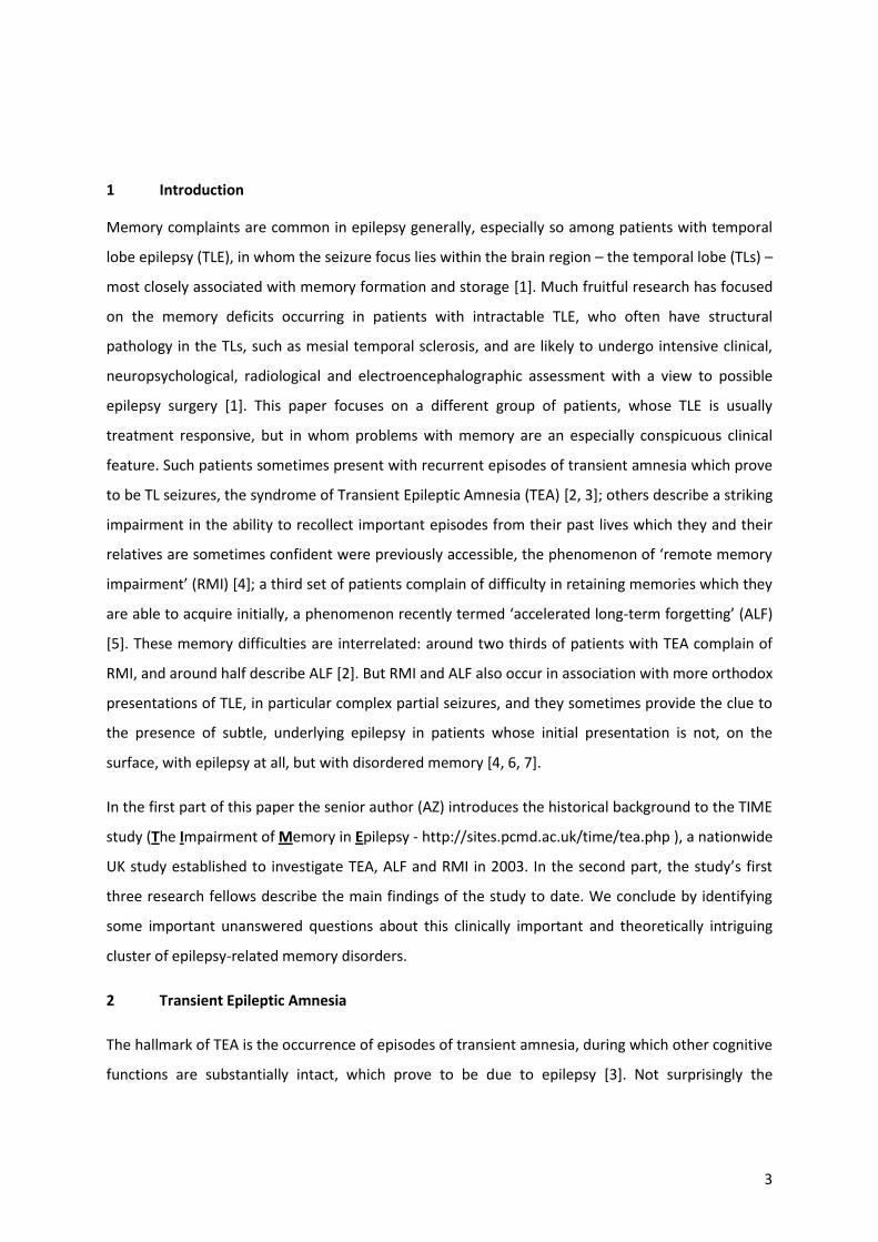
marked over the longer time intervals (figure 2). Importantly, the rate of long-term forgetting
correlated with patients’ subjective complaints of memory impairment, whereas performance on
standard memory tasks did not.
6.4 Brain imaging
As mentioned above, interictal neuroimaging rarely reveals clinically relevant abnormalities in TEA.
There is no imaging evidence of increased vascular disease in TEA patients compared with matched
healthy controls [20]. Nevertheless, across the whole group of patients in the TIME Project, we
identified subtle, bilateral hippocampal atrophy (~8% volume decrease compared with controls)
[21]. Recently, we have demonstrated additional gray matter atrophy bilaterally in the perirhinal and
orbitofrontal cortices, but in no other brain regions (Butler et al., submitted) The degree of medial
temporal lobe atrophy correlated with performance on standard tests of anterograde memory. This
relationship was identified using both manual volumetry and voxel-based morphometry. No brain
regions were found in which gray matter volume correlated with autobiographical memory or long-
term forgetting rate.
Perictal FDG-positron emission tomography (PET) was performed in one patient during a flurry of
amnestic seizures [22]. This revealed hypermetabolism in the left hippocampus, which had resolved
upon repeat scanning three months later.
6.5 Summary
We propose that TEA is a distinct but under-recognised syndrome of epilepsy characterised by
recurrent, brief amnestic attacks that begin in middle to old age, often occur upon wakening, and
are frequently associated with unusual memory deficits including accelerated long-term forgetting
and autobiographical amnesia. The clinical and neuroimaging features point towards a seizures focus
in the medial temporal lobes. As with many syndromes, TEA has debatable boundaries. A notable
example is the overlap with what Gallassi and colleagues have termed the Epileptic Amnesic
Syndrome (EAS) [6, 23]. Patients with EAS have late onset (mean age = 63 years) persistent memory
difficulties, often detectable on standard neuropsychological instruments, in association with subtle
temporal lobe seizures. In other patients (e.g.[7]), complaints of ALF and remote memory
impairment can precede the onset of amnesic seizures by several years. Further research is
necessary to clarify the relationship between TEA and other forms of late-onset temporal lobe
epilepsy. In the meantime, the concept serves to highlight this distinctive presentation of epilepsy,
and facilitate novel explorations of human memory function.

9
7 Promising areas of research and young investigators
7.1 Nils Muhlert
The nature of accelerated long term forgetting (ALF)
Reports of memory complaints are extremely common in epilepsy clinics. Whilst these are often
corroborated by standard tests of memory, a substantial minority (24%) of those who complain
about their memories perform within normal limits on standard tests [24]. This discrepancy has
previously been ascribed to mood disorders (e.g. [25]). But ALF provides an alternative or additional
explanation [5]: forgetting may be normal at the 30 minute delays used in traditional tests of
memory yet increased over days or weeks . In a recent study, perceived memory loss was found to
correlate more strongly with forgetting over four weeks than with forgetting over 30 minutes [24].
The study of ALF may lead to improvements in the clinical detection of memory impairment, and
shed light on memory processes.
This brief review outlines the prevalence of ALF in epilepsy, the methods used to assess it and the
current evidence on the cognitive and neurobiological basis of ALF.
7.2 What is the prevalence of ALF in epilepsy?
ALF appears to be particularly widespread among people with TEA [2, 26, 27], affecting nearly half
(44%) of patients with this condition [2]. It has also been reported in people with TLE and, to a lesser
extent, those with idiopathic generalised epilepsies (IGE). In people with TLE, the majority of studies
have found evidence for ALF [18, 28, 29]. Whilst some have failed to do so [30, 31], this may be
explained by differences in the methodology used to assess ALF (discussed later). In a case-by-case
analysis Muhlert et al. found impaired retention over a three week delay in 30-55% of patients with
TLE, depending on the test used [29]. In people with IGE, Davidson et al. found some evidence for
ALF in children with IGE [32] while a study in adults found similar rates of forgetting in those with IGE
and healthy controls [29]. The existence of ALF in those with frontal lobe epilepsies has not been
assessed, although Blake et al. reported ALF in a heterogeneous sample of patients with partial
epilepsies originating from the left hemisphere [18]. The fact that ALF is seen in some forms of
epilepsy but not others suggests that psychosocial factors common to people with epilepsy, such as
worry about seizures, does not explain ALF.
7.3 How is ALF assessed in epilepsy?

10
At present, the use of different tests by research groups complicates comparisons between studies.
In particular, the use of tests that are liable to ceiling effects and overlearning during acquisition or
to floor effects at long delays make it difficult to draw clear conclusions about the presence or
absence of ALF. Whilst experimental tests of memory have been designed to avoid such effects (e.g.
[28, 29]) a number of drawbacks prevent their use as standard clinical tools. For instance they lack
normative data, and require an additional appointment to assess very long-term memory. Use of
telephone assessments at delayed time points (e.g. [33]) may make clinical assessments of ALF more
cost-effective. There is need for a standardised set of measures supported by normative data to
facilitate the clinical diagnosis and theoretical investigation of ALF.
7.4 What is the cognitive basis of ALF?
As the clinical phenomenon of ALF involves apparently normal memory acquisition and retention at
short intervals, with impaired retention at longer delays, it is natural to suppose that it is due to a
disorder of memory consolidation or storage. This may be correct, but it remains possible that
memories are fragile from the outset in people with ALF. Learning is not always normal in patients
who show ALF (e.g.6, 10), and subtle abnormalities in encoding may become more apparent over
time. Yet if this were the main explanation for ALF, one would predict a direct relationship between
retention over short and long delays. Muhlert et al. [27] found no evidence for this: while retention
over 30 min was correlated with retention over 24 h in healthy controls, there was no correlation in
patients with TEA. This suggests a disruption of normal memory processing in the interval between
these time points.
7.5 What is the neurobiological basis of ALF?
The two most plausible explanations for ALF are clinical or subclinical epileptiform activity on the
one hand and structural brain damage on the other. The role of clinical seizures in ALF was
highlighted by the case of patient JT [15], who showed a ‘precipitous’ decrease in recall following
witnessed seizures. Further evidence came from a group study of people with TLE, in which those
who experienced frequent seizures (>4) over a four week delay showed greater forgetting over that
time [34]. However, a recent study found no association between seizures over a three week delay
and ALF in either TLE or IGE groups [29]. Similarly, ALF is seen in people with TEA who are typically
seizure-free (e.g. [2, 27]). Subclinical epileptiform activity could be relevant in such cases. Patients
with TLE with evidence of interictal discharges on EEG have been found to have worse recall after a
four week delay than those without interictal discharges [34]. There is an intriguing possibility that
subclinical epileptiform acitivity during sleep might lead to ALF. ALF is detectable over 24 hour
11
delays in people with TEA [27]. Prior to treatment with medication, people with TEA often report
seizures upon awakening [2]. Residual epileptiform activity during sleep might disrupt the memory
consolidation processes that are thought to occur during sleep [35]. Further research, including EEG
recordings carried out during sleep and waking between acquisition and delayed memory testing, is
needed to explore this possibility.
A second potential explanation for ALF is structural brain damage. A number of studies have
demonstrated the existence of structural damage in patients who show ALF, including increased T2
relaxation times in the hippocampus [28], reduced hippocampal volume [21, 29], hypometabolism
in the medial temporal lobes [36], decreased hippocampal NAA/Creatine on MR spectroscopic
imaging (a marker of neuronal density and mitochondrial function) [36] and mesial temporal
sclerosis [29]. Furthermore, patients with TEA demonstrate abnormalities on tests of declarative
memory, which rely on an intact medial temporal lobe, but normal rates of forgetting on tests of
procedural memory, which rely on the integrity of the basal ganglia ([27]; [37]). Yet, no study has
demonstrated a significant correlation between ALF and structural brain damage, and so further
evidence is needed to establish a causative role.
7.6 Conclusion
In summary, ALF is relatively common in people with TEA and TLE and helps to explain the
discrepancy between memory complaints and the results of standard memory tests in these groups.
It is likely to play a less important role in IGE. The development of robust clinical tests for ALF will
help to define its clinical role and theoretical implications. Whilst its cause is not yet known future
investigation of the relationships between ALF, structural brain imaging and epileptiform activity in
wakefulness and sleep should prove informative.
Promising areas of research and young investigators
8.1 Fraser Milton
Remote memory impairments in TEA
The remote memory deficits of patients with TEA are clinically striking and have received increasing
attention in recent years. The following section will briefly describe the current evidence for
impairments of different types of remote memory information: autobiographical event memory,
personal semantic memory, and public semantic memory. We then consider whether TEA can be
considered a form of focal retrograde amnesia before discussing the implications of these findings
for theories of long-term memory.
12
8.2 Autobiographical event memory
A number of recent group [2, 26, 38] and case studies [39, 40] provide evidence that TEA results in
deficits for autobiographical event memory. For instance, Butler et al. [2], who recruited 50 patients
diagnosed with TEA from across the UK as part of the TIME project, found that 70% of the patients
examined complained of autobiographical memory deficits. Using a modified version of the
Autobiographical Memory Interview (AMI [41]), Butler et al. found that the autobiographical
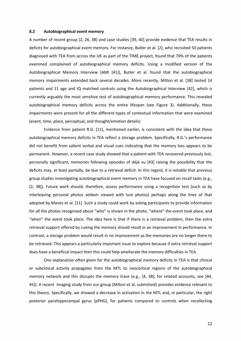
memory impairments extended back several decades. More recently, Milton et al. [38] tested 14
patients and 11 age and IQ matched controls using the Autobiographical Interview [42], which is
currently arguably the most sensitive test of autobiographical memory performance. This revealed
autobiographical memory deficits across the entire lifespan (see Figure 3). Additionally, these
impairments were present for all the different types of contextual information that were examined
(event, time, place, perceptual, and thought/emotion details).
Evidence from patient R.G. [11], mentioned earlier, is consistent with the idea that these
autobiographical memory deficits in TEA reflect a storage problem. Specifically, R.G.’s performance
did not benefit from salient verbal and visual cues indicating that the memory loss appears to be
permanent. However, a recent case study showed that a patient with TEA recovered previously lost,
personally significant, memories following episodes of déjà vu [43] raising the possibility that the
deficits may, at least partially, be due to a retrieval deficit. In this regard, it is notable that previous
group studies investigating autobiographical event memory in TEA have focused on recall tasks (e.g.,
[2, 38]). Future work should, therefore, assess performance using a recognition test (such as by
interleaving personal photos seldom viewed with lure photos) perhaps along the lines of that
adopted by Manes et al. [11]. Such a study could work by asking participants to provide information
for all the photos recognised about “who” is shown in the photo, “where” the event took place, and
“when” the event took place. The idea here is that if there is a retrieval problem, then the extra
retrieval support offered by cueing the memory should result in an improvement in performance. In
contrast, a storage problem would result in no improvement as the memories are no longer there to
be retrieved. This appears a particularly important issue to explore because if extra retrieval support
does have a beneficial impact then this could help ameliorate the memory difficulties in TEA.
One explanation often given for the autobiographical memory deficits in TEA is that clinical
or subclinical activity propagates from the MTL to neocortical regions of the autobiographical
memory network and this disrupts the memory trace (e.g., [4, 38]; for related accounts, see [44,
45]). A recent imaging study from our group (Milton et al, submitted) provides evidence relevant to
this theory. Specifically, we showed a decrease in activation in the MTL and, in particular, the right
posterior parahippocampal gyrus (pPHG), for patients compared to controls when recollecting
13
memories from their past. Furthermore, there was reduced effective connectivity between this right
pPHG region and the right middle temporal gyrus which is a key part of the autobiographical
memory network.
8.3 Personal semantic memory
Butler et al. [2], using the modified version of the AMI, found that deficits for personal semantic
memory stretched back several decades. Similarly, Milton et al. [38] found that patients had an
overall impairment for personal semantic information, although when examining different life
periods separately (childhood, teenage, young adult, middle age, and recent events) deficits only
reached significance for the middle age time period. These results indicate that the personal
semantic memory deficits may be less temporally extensive than the autobiographical event
memory impairments detected by Milton et al. [38]. An alternative explanation, however, for the
more restricted deficits is that the personal semantic tests used by Butler et al. [2] and Milton et al.
[38] may be less sensitive measures than the Autobiographical Interview used by Milton et al. [38] to
investigate autobiographical event memory. Indeed, in both studies performance was close to
ceiling for both patients and controls meaning that it is possible that relatively subtle deficits in
other time periods were not detected. It would, therefore, be valuable to further investigate
personal semantic memory using a more difficult and sensitive test to assess whether the personal
semantic memory deficits in TEA extend across the lifespan or whether they are indeed more
restricted than the autobiographical event memory deficits.
8.4 Public Semantic memory
Milton et al. [38] provided the first systematic examination of public semantic memory performance
in TEA. Significant deficits were apparent on the Dead-Or-Alive test [46] and these deficits were most
marked for recent rather than remote memories. No overall impairments emerged on a Famous
Events test, although, as for the Dead-or-Alive test, when examining the different time periods
separately, deficits were found for recent events. No impairments were found either on a test of
Famous Faces or on a New Words Acquisition test. One reasonable question is why an overall deficit
was found for the Dead-or-Alive test but not for the other three tests? One potential explanation is
that the Dead-or-Alive test appears to have a greater episodic component than the other tests used.
This would be consistent with the idea that the episodic and semantic memory systems are closely
intertwined [47]. For instance, autobiographical significance has been shown to modulate
performance on a semantic test of famous people in healthy participants [48]. It could, therefore, be
that the overall deficit in the Dead-or-Alive test is the result of patients benefiting less than controls

14
from episodic information about the person to supplement their semantic information. In future
work, it would be of interest to examine whether any neural differences between patients and
controls when retrieving public semantic information overlap with those found for autobiographical
event memory recollection. If the explanation we offer is correct, then one might expect
considerable similarity in the neural deficits.
8.5 Is TEA a form of focal retrograde amnesia?
Previous work has consistently demonstrated that the memory deficits in TEA constitute a form of
focal retrograde memory as the term is generally understood – an inability to retrieve memories that
have been successfully acquired in the past, in the absence of any deficit on standard tests of
anterograde memory (e.g., [2, 26, 38]). Furthermore, Hornberger et al. [7] have provided evidence
that focal retrograde amnesia can even be a prodromal symptom of TEA. Nevertheless, a careful
inspection of the autobiographical memory data (see Figure 1) collected by Milton et al. [38]
suggests that this conclusion may not be as straightforward as it first appears. Specifically, whilst
performance was preserved on a series of standard anterograde memory tests, patients’
autobiographical memory was significantly impaired the most recent time period. Critically, the
events recalled in this time period for all the patients occurred after the onset of their TEA, and in
most cases after the onset of successful treatment. Whilst this impairment may not be surprising for
a subset of these patients (5/14) who complain of accelerated long-term forgetting, it is notable that
all patients performed at least 1 standard deviation below the mean performance of the control
group. This pattern of results therefore suggests that some form of anterograde memory deficit may
be a general feature of TEA. Examining the time course of this apparent deficit using a prospective
design could provide considerable theoretical insight into the nature of this problem. One possibility
is that patients with TEA do, in fact, have a mild encoding deficit that cannot be detected using
standard tests but which may emerge when a more sensitive measure such as the Autobiographical
Interview is used. In this case, one would expect the impairment to be apparent immediately after
the event. Alternatively, initial encoding may be preserved but there may be a problem with early
consolidation processes resulting in accelerated long-term forgetting over the course of days or
weeks. This possibility would imply that accelerated long-term forgetting is more prevalent in TEA
than has commonly been acknowledged. Finally, performance may be preserved several weeks after
the event but the deficits emerge over a longer duration (e.g., months) than has previously been
examined. If any of these possibilities are the case, then it would provide some explanation for why
subjective reports of memory problems in epilepsy often do not correlate with objective measures
on standard neuropsychological tests (e.g., [49]).

15
8.6 Implications for theories of long-term memory.
We have already presented evidence consistent with the idea that TEA is a medial temporal lobe
(MTL) syndrome (e.g., [21, 22]). Consequently, the remote memory impairments in TEA have
implications for current theories of remote memory. The standard consolidation model posits that
the MTL, and in particular the hippocampus, has a time limited role in episodic memory (e.g., [50]).
The MTL, at first, is critical for the storage of new memories, but, over time, these memories
become independent of the MTL and are represented in the neocortex alone. In contrast, multiple
trace theory argues that the MTL is essential for episodic memories throughout the lifespan [51].
Each time a memory is retrieved a new trace is laid down leading to the formation of multiple traces
in the MTL. Both theories predict that the MTL has a time limited role for semantic memory. In this
regard, the finding of a temporally graded deficit on public semantic memory tests [38] would be
compatible with both the standard consolidation model and multiple trace theory. On the other,
hand, the finding of autobiographical event memory deficits extending across the lifespan appears
more in line with the assumptions of multiple trace theory. Nevertheless, the standard consolidation
model could potentially explain these results if it posits that the deficits are due to clinical or
subclinical activation from the MTL disrupting the memory trace along the lines suggested in Section
8.2.
9 Questions for future research
The work summarised in this paper has established that recurrent episodes of transient amnesia can
be caused by TLE in the syndrome of Transient Epileptic Amnesia. These are often accompanied by
two non-standard forms of memory impairment, accelerated long-term forgetting and remote
memory impairment, which can occur in the face of normal performance on standard memory tests.
While these interictal deficits are especially common in TEA, they are also seen in patients with
forms of TLE other than TEA [4]. The work of the young investigators summarised above has
advanced our knowledge of TEA, ALF and RMI considerably, but there are many unanswered
questions for future research. The following are particularly important:
TEA: what causes the syndrome to develop in middle age? What is the long term prognosis
of the syndrome? What is its underlying neuropathology?
ALF: is it, as it appears to be, a disorder of memory consolidation, or is there a subtle
impairment of memory acquisition? Is it due to a disorder of physiology (eg epileptiform
discharges during sleep or wakefulness) or to a disorder of structure? Does it respond, to
some degree, to antiepileptic treatment?

16
RMI: is the apparent loss of memories in some patients with RMI due to a disorder of
memory storage, or is there an impairment of memory retrieval? As for ALF, Is RMI due to a
disorder of physiology (eg epileptiform discharges during sleep or wakefulness) or to a
disorder of structure?

17
Box 1: TEA – an illustrative case report
A 58-year-old carpet fitter experienced 28 episodes of transient amnesia over 18 months. All occurred
upon waking in the night and lasted about 20 minutes. He repetitively questioned his wife, but was
responsive and coherent throughout. During one attack he was unable to recall the death of his
brother a few days earlier. Routine EEG and MRI were normal. Lamotrigine abolished the attacks but
they briefly returned, with associated olfactory hallucinations, during a period of non-compliance,
and ceased again when he restarted the medication. At interview, he described rapid forgetting of
recently acquired memories, patchy loss of salient autobiographical memories from the past 30
years, such as his wife’s abdominal surgery and the wedding of his son, and significant new
difficulties navigating around his local area.

18
FIGURE LEGENDS
Figure 1
Venn diagram showing the distribution of patients meeting the diagnostic criteria for TEA
employed by Butler et al [2].
Figure 2
Recall performance of patients with TEA trained to a 90% criterion on a 15 word list at 30
minutes, 1 week and 3 weeks. ‘Patients’ are the TEA group as a whole; ‘ALF+’ 12 patients
with TEA who complained subjectively of ALF; ‘ALF-‘ 12 patients who did not complain
subjectively of ALF [2].
Figure 3
Mean number of details recalled for events from five life periods by patients with TEA
compared to control participants [38].
19
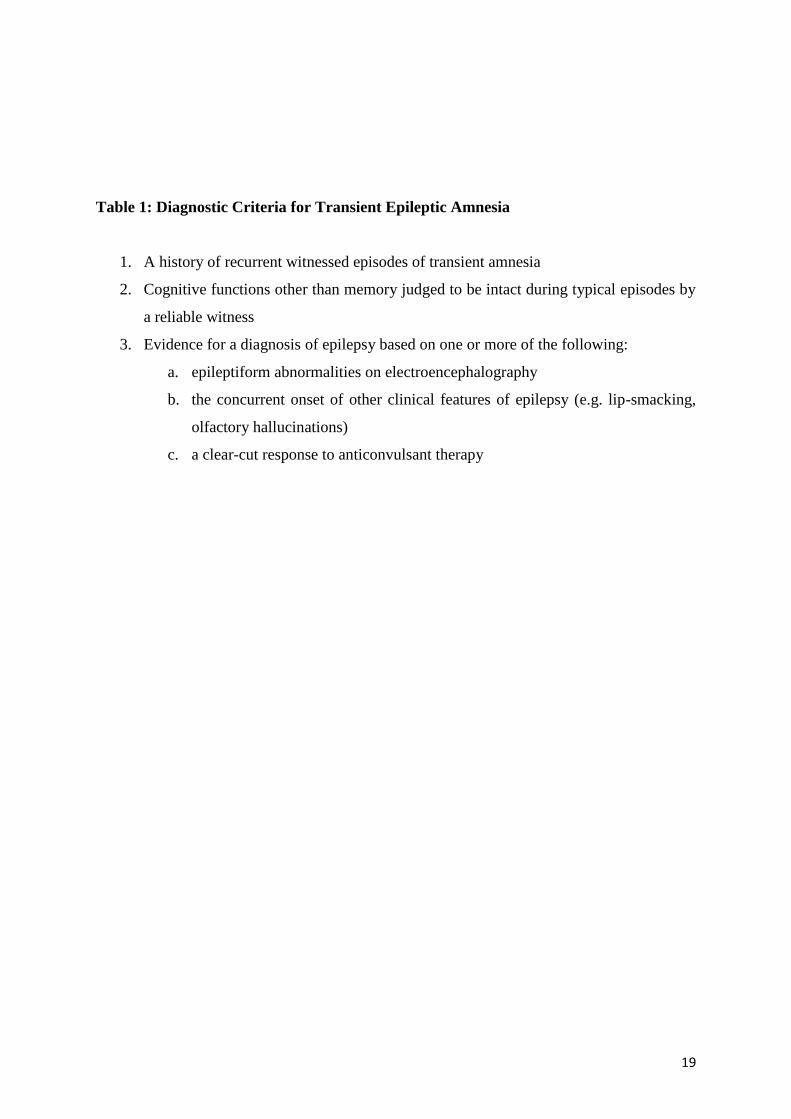
Table 1: Diagnostic Criteria for Transient Epileptic Amnesia
1. A history of recurrent witnessed episodes of transient amnesia
2. Cognitive functions other than memory judged to be intact during typical episodes by
a reliable witness
3. Evidence for a diagnosis of epilepsy based on one or more of the following:
a. epileptiform abnormalities on electroencephalography
b. the concurrent onset of other clinical features of epilepsy (e.g. lip-smacking,
olfactory hallucinations)
c. a clear-cut response to anticonvulsant therapy

20
Table 2: Core clinical features of Transient Epileptic Amnesia
Demographics age at onset (y) 62.1 (range 44 to 77)
sex distribution m=34; f=16
Amnestic attack
characteristics
number of attacks median = 10
(IQR 6 to 30)
frequency (attacks per year) median = 12
(IQR 5 to 20)
attack duration median = 30-60min (range
<1min to days)
cessation of attacks on AED 96%
some attacks on waking 74%
partial amnesia for attack 56%
repetitive questioning 50%
olfactory hallucinations 42%
motor automatisms 36%
brief unresponsiveness 24%
Interictal memory c/o autobiographical memory loss 70%
c/o accelerated forgetting 44%
c/o topographical memory loss 36%
Investigations interictal epileptiform EEG abnormalities 37%
structural lesion on MRI 2%

21
Table 3: Demographic and neuropsychological profile of TEA patients and controls
Raw scores are shown with the maximum possible score for each test given in parentheses in the left-hand
column
AF subgroup = subgroup of TEA patients selected for accelerated forgetting tests and matched pairwise with controls.
WCST = Wisconsin Card Sort Test 64
HAD = Hospital Anxiety and Depression scale
All patients (n=50) Controls
(n=24)
mean (SD) mean (SD)
Age (years) 68.3 (8.6) 67.7 (8.1)
Education (years) 12.2 (2.9) 12.5 (3.1)
Full scale IQ 118.3 (12.8) 120.0 (14.4)
Episodic memory
Story recall immediate (25) 14.0 (4.3) 15.9 (3.8)
Story recall delayed (25) 11.7 (5.0)* 14.7 (3.8)
Rey figure delayed recall (36) 15.0 (6.5)* 18.6 (6.1)
Word recognition (50) 46.1 (4.7)* 48.3 (1.9)
Face recognition (50) 40.7 (5.4)*** 45.1 (2.9)
Semantic memory
Graded faces (60)† 40.0 (9.6) 44.0 (7.6)
Graded naming (30) 21.4 (5.1) 23.5 (4.2)
Visuospatial perception
Rey figure copy (36) 34.5 (3.1) 35.5 (1.1)
Executive function
Letter fluency (words/3min) 42.5 (13.9) 43.8 (11.4)
Category fluency (words/min) 19.3 (5.9) 22.0 (4.4)
WCST categories completed 2.8 (1.3) 3.4 (1.4)

22
† for each face: one point for providing identifying information; * = p<0.05, *** = p<0.001
Reference List
[1] Zeman A, Jones-Gotman M, Kapur N. Epilepsy and Memory. 2012. Oxford, Oxford University
Press.
[2] Butler CR, Graham KS, Hodges JR, Kapur N, Wardlaw JM, Zeman AZ. The syndrome of
transient epileptic amnesia. Ann Neurol 2007;61:587-98.
[3] Zeman A, Butler C. Transient epileptic amnesia. Curr Opin Neurol 2010;23:610-6.
[4] Butler CR, Zeman AZ. Recent insights into the impairment of memory in epilepsy: transient epileptic amnesia, accelerated long-term forgetting and remote memory impairment. Brain 2008;131:2243-63.
[5] Muhlert N, Zeman A. The enigma of long-term forgetting. Seizure 2012;21:77-8.
[6] Gallassi R. Epileptic amnesic syndrome: an update and further considerations. Epilepsia 2006;47 Suppl 2:103-5.
[7] Hornberger M, Mohamed A, Miller L, Watson J, Thayer Z, Hodges JR. Focal retrograde amnesia: Extending the clinical syndrome of transient epileptic amnesia. J Clin Neurosci 2010;17:1319-21.
[8] Hughlings Jackson J. On a particular variety of epilepsy (intellectual aura), one case with symptoms of organic brain disease. Brain 1888:179-207.
[9] Hodges JR. Transient Amnesia. London: WB Saunders; 1991.
[10] Zeman AZ, Boniface SJ, Hodges JR. Transient epileptic amnesia: a description of the clinical and neuropsychological features in 10 cases and a review of the literature. J Neurol Neurosurg Psychiatry 1998;64:435-43.
[11] Manes F, Hodges JR, Graham KS, Zeman A. Focal autobiographical amnesia in association with transient epileptic amnesia. Brain 2001;124:499-509.
[12] Kapur N, Young A, Bateman D, Kennedy P. Focal retrograde amnesia: a long term clinical and neuropsychological follow-up. Cortex 1989;25:387-402.
[13] Lucchell F, Spinnler H. Ephemeral new traces and evaporated remote engrams: a form of neocortical temporal lobe amnesia? A preliminary case report. Neurocase 1998;4:447-59.
[14] Kapur N., Millar J, Colbourn C, Abbott P, Kennedy P, Docherty T. Very long-term amnesia in association with temporal lobe epilepsy: evidence for multiple-stage consolidation processes. Brain Cogn 1997;35:58-70.
[15] O'Connor M, Sieggreen MA, Ahern G, Schomer D, Mesulam M. Accelerated forgetting in association with temporal lobe epilepsy and paraneoplastic encephalitis. Brain Cogn 1997;35:71-84.

23
[16] Mayes AR, Isaac CL, Holdstock JS, Cariga P, Gummer A, Roberts N. Long-term amnesia: a review and detailed illustrative case study. Cortex 2003;39:567-603.
[17] Martin RC, Loring DW, Meador KJ, Lee Gp, Thrash N, Arena JG. Impaired long-term retention despite normal verbal learning in patients with temporal lobe dysfunction. Neuropsychology 1991;5:3-12.
[18] Blake RV, Wroe SJ, Breen EK, McCarthy RA. Accelerated forgetting in patients with epilepsy: evidence for an impairment in memory consolidation. Brain 2000;123 Pt 3:472-83.
[19] Giovagnoli AR, Casazza M, Avanzini G. Visual learning on a selective reminding procedure and delayed recall in patients with temporal lobe epilepsy. Epilepsia 1995;36:704-11.
[20] Butler CR, Zeman A. The causes and consequences of transient epileptic amnesia. Behav Neurol 2011;24:299-305.
[21] Butler CR, Bhaduri A, Acosta-Cabronero J, Nestor PJ, Kapur N, Graham KS et al. Transient epileptic amnesia: regional brain atrophy and its relationship to memory deficits. Brain 2009;132:357-68.
[22] Butler CR, Zeman A. A case of transient epileptic amnesia with radiological localization. Nat Clin Pract Neurol 2008;4:516-21.
[23] Gallassi R, Morreale A, Di Sarro R, Lugaresi E. Epileptic amnesic syndrome. Epilepsia 1992;33 Suppl 6:S21-S25.
[24] Witt JA, Glockner C, Helmstaedter C. Extended retention intervals can help to bridge the gap between subjective and objective memory impairment. Seizure 2012;21:134-40.
[25] Corcoran R, Thompson P. Epilepsy and poor memory: who complains and what do they mean? Br J Clin Psychol 1993;32 ( Pt 2):199-208.
[26] Manes F, Graham KS, Zeman A, de Lujan CM, Hodges JR. Autobiographical amnesia and accelerated forgetting in transient epileptic amnesia. J Neurol Neurosurg Psychiatry 2005;76:1387-91.
[27] Muhlert N, Milton F, Butler CR, Kapur N, Zeman AZ. Accelerated forgetting of real-life events in Transient Epileptic Amnesia. Neuropsychologia 2010;48:3235-44.
[28] Wilkinson H, Holdstock JS, Baker G, Herbert A, Clague F, Downes JJ. Long-term accelerated forgetting of verbal and non-verbal information in temporal lobe epilepsy. Cortex 2012;48:317-32.
[29] Muhlert N, Grunewald RA, Hunkin NM, Reuber M, Howell S, Reynders H et al. Accelerated long-term forgetting in temporal lobe but not idiopathic generalised epilepsy. Neuropsychologia 2011;49:2417-26.
[30] Bell BD, Fine J, Dow C, Seidenberg M, Hermann BP. Temporal lobe epilepsy and the selective reminding test: the conventional 30-minute delay suffices. Psychol Assess 2005;17:103-9.
[31] Bell BD. WMS-III Logical Memory performance after a two-week delay in temporal lobe epilepsy and control groups. J Clin Exp Neuropsychol 2006;28:1435-43.

24
[32] Davidson M, Dorris L, O'Regan M, Zuberi SM. Memory consolidation and accelerated forgetting in children with idiopathic generalized epilepsy. Epilepsy Behav 2007;11:394-400.
[33] Kemp S, Illman NA, Moulin CJ, Baddeley AD. Accelerated long-term forgetting (ALF) and transient epileptic amnesia (TEA): Two cases of epilepsy-related memory disorder. Epilepsy Behav 2012;24:382-8.
[34] Mameniskiene R, Jatuzis D, Kaubrys G, Budrys V. The decay of memory between delayed and long-term recall in patients with temporal lobe epilepsy. Epilepsy Behav 2006;8:278-88.
[35] Stickgold R. Sleep-dependent memory consolidation. Nature 2005;437:1272-8.
[36] Tramoni E, Felician O, Barbeau EJ, Guedj E, Guye M, Bartolomei F et al. Long-term consolidation of declarative memory: insight from temporal lobe epilepsy. Brain 2011;134:816-31.
[37] Deak MC, Stickgold R, Pietras AC, Nelson AP, Bubrick EJ. The role of sleep in forgetting in temporal lobe epilepsy: a pilot study. Epilepsy Behav 2011;21:462-6.
[38] Milton F, Muhlert N, Pindus DM, Butler CR, Kapur N, Graham KS et al. Remote memory deficits in transient epileptic amnesia. Brain 2010;133:1368-79.
[39] Illman NA, Rathbone CJ, Kemp S, Moulin CJ. Autobiographical memory and the self in a case of transient epileptic amnesia. Epilepsy Behav 2011;21:36-41.
[40] Ioannidis P, Balamoutsos G, Karabela O, Kosmidis MH, Karacostas D. Transient epileptic amnesia in a memory clinic setting: a report of three cases. Epilepsy Behav 2011;20:414-7.
[41] Kopelman MD, Wilson BA, Baddeley AD. The autobiographical memory interview: a new assessment of autobiographical and personal semantic memory in amnesic patients. J Clin Exp Neuropsychol 1989;11:724-44.
[42] Levine B, Svoboda E, Hay JF, Winocur G, Moscovitch M. Aging and autobiographical memory: dissociating episodic from semantic retrieval. Psychol Aging 2002;17:677-89.
[43] Milton F, Butler CR, Zeman AZ. Transient epileptic amnesia: deja vu heralding recovery of lost memories. J Neurol Neurosurg Psychiatry 2011;82:1178-9.
[44] Gallassi R, Morreale A, Lorusso S, Pazzaglia P, Lugaresi E. Epilepsy presenting as memory disturbances. Epilepsia 1988;29:624-9.
[45] Mendes MH. Transient epileptic amnesia: an under-diagnosed phenomenon? Three more cases. Seizure 2002;11:238-42.
[46] Kapur N, Ellison D, Smith MP, McLellan D, Burrrows EH. Focal retrograde amnesia following bilateral temporal lobe pathology. Brain 1992;115:73-85.
[47] Levine B, Turner GR, Tisserand D, Hevenor SJ, Graham SJ, McIntosh AR. The functional neuroanatomy of episodic and semantic autobiographical remembering: a prospective functional MRI study. J Cogn Neurosci 2004;16:1633-46.
[48] Westmacott R, Moscovitch M. The contribution of autobiographical significance to semantic memory. Mem Cognit 2003;31:761-74.

25
[49] Corcoran R, Thompson P. Memory failure in epilepsy: retrospective reports and prospective recordings. Seizure 1992;1:37-42.
[50] Squire LR, Bayley PJ. The neuroscience of remote memory. Curr Opin Neurobiol 2007;17:185-96.
[51] Moscovitch M, Rosenbaum RS, Gilboa A, Addis DR, Westmacott R, Grady C et al. Functional neuroanatomy of remote episodic, semantic and spatial memory: a unified account based on multiple trace theory. J Anat 2005;207:35-66.

26
Figure 1.

27
Figure 2.

28
0
10
20
30
40
50
60
70
80
90
Child Youth Young Adult Middle Age Recent
Time period
Nu
mb
er
of
inte
rnal
deta
ils
TEA patients
Controls
Figure 3.