Neuropsychological aspects of frontal lobe epilepsy. Dominic ...
629
Neuropsychological aspects of frontal lobe epilepsy. Dominic Upton, Doctor of Philosophy. Institute of Neurology. University of London. LONDON, WON 36G ROCKEFELLER MEOICAL LIBRARY INSTITUTE OF NEUROLOGY THE NATIONAL HOSPITAL QUEEN SQUARE, LONDON, WCIN 33G CLASS ACCN No. date W c
-
Upload
khangminh22 -
Category
Documents
-
view
1 -
download
0
Transcript of Neuropsychological aspects of frontal lobe epilepsy. Dominic ...

Neuropsychological aspects of frontal lobe
epilepsy.
Dominic Upton,Doctor of Philosophy.
Institute of Neurology. University of London.
LONDON,WON 36G
ROCKEFELLER MEOICAL LIBRARY INSTITUTE OF NEUROLOGY
T H E N A T I O N A L H O S P I T A LQUEEN SQUARE, LONDON, W C IN 3 3 G
CLASSACCN No.
d a t e W c

ProQuest Number: 10017771
All rights reserved
INFORMATION TO ALL USERS The quality of this reproduction is dependent upon the quality of the copy submitted.
In the unlikely event that the author did not send a complete manuscript and there are missing pages, these will be noted. Also, if material had to be removed,
a note will indicate the deletion.
uest.
ProQuest 10017771
Published by ProQuest LLC(2016). Copyright of the Dissertation is held by the Author.
All rights reserved.This work is protected against unauthorized copying under Title 17, United States Code.
Microform Edition © ProQuest LLC.
ProQuest LLC 789 East Eisenhower Parkway
P.O. Box 1346 Ann Arbor, Ml 48106-1346

Abstract.This study sought to increase our understanding of the neuropsychological consequences of frontal lobe epilepsy and surgery for this condition. In so doing, some suggestions were made on the role of the frontal lobes, and ways of assessing possible dysfunction in this area.
The performance of a large group of subjects with clearly defined frontal lobe epilepsy (n=74), was examined on a comprehensive battery of neuropsychological measures. The performance of this group was compared to a control group of subjects with clearly defined temporal lobe epilepsy (n=57). There were few consistent group differences in test performance. Neither were there any strong relationships between test scores and epilepsy related variables.
The frontal lobe group was sub-divided into more discrete regions (dorsolateral, orbital, mesial, m otor/prem otor, and extensive), and the neuropsychological characteristics associated with dysfunction in these regions was assessed. Although differences between the samples were small, some of the neuropsychological deficits previously associated with these regions were observed.
Since a num ber of m easures were used, their validity in assessm ent of frontal lobe dysfunction was assessed. A composite score of frontal lobe dysfunction was also derived using multivariate analysis. Comparisons revealed that the new composite score can be considered a more sensitive m easure in the assessm ent of frontal lobe epilepsy, than any individual neuropsychological measure investigated.
In addition to the group study, four cases were followed up before and a t regular intervals after surgery. The results highlighted a num ber of significant findings, from both a clinical and theoretical perspective.
Although the results of the comparisons between frontal and temporal lobe groups, and between different frontal regions.

revealed some significant functional differences, these were not consistently demonstrated. The results suggest the necessity to regard these groups collectively, since epileptic activity in any one specific region can cause deficits associated with other areas.

Index of contents.Titie PageTitle page 1Abstract 2Index of contents 4Index of tables 6Index of figures 9Acknowledgements 11
Chapter 1; Overview 121.1 Overview 13Chapter 2: Epilepsy: The condition and its treatment 182.1 Definition 192.2 Aetiology 202.3 Classification 222.4 T reatm ent 31Chapter 3: The frontal lobes: historical perspective 363.1 Morphological aspects 373.2 Functional aspects 37Chapter 4: Basic anatomy of the frontal lobes 424.1 Gross anatomy of the frontal lobes 434.2 Cytoarchitectonie classification 464.3 Vascular distribution 484.4 Connections with other cortical areas 534.5 Conclusion 54Chapter 5: Development of the frontal lobes 555.1 Anatomical development 565.2 Cognitive development 565.3 Summary and conclusions 63Chapter 6: Functions of the frontal lobes 646.1 Background 656.2 Memory 656.3 Movement 726.4 The frontal lobes and language 766.5 Visuo-spatial skills 786.6 Executive functions 806.7 Mood and personality 866.8 Moral judgem ent 876.9 Conclusion 88Chapter 7: Theories of frontal lobe function 897.1 Background 907.2 Luria's theory 947.3 ShaUice and the Supervisory Attentional
System98
7.4 Conclusion 107

Index of contents (cont).l i t i e Page
Chapter 8: Problems with previous research and introduction to current study
109
8.1 Overview 1108.2 Terminology 1108.3 Inappropriate assessm ents 1118.4 Inappropriate subject samples 1268.5 Clinical versus experimental 1328.6 Neuropsychological studies of frontal lobe
epilepsy136
8.7 Hypotheses and aims 141Chapter 9: Method 1449.1 Introduction 1459.2 Subjects 1479.3 Tests 1589.4 Test construction 1739.5 Procedure 1819.6 Statistical analysis 1829.7 Presentation of results 190Chapter 10: Results 19110.1 Results: Hypothesis 1 19210.2 Results: Hypothesis 2 19810.3 Results: Hypothesis 3 22510.4 Results: Hypothesis 4 23210.5 Results: Hypothesis 5 24710.6 Results: Hypothesis 6 25510.7 Results: Hypothesis 7 26210.8 Results: Hypothesis 8 27710.9 Results: Hypothesis 9 281Chapter 11: Discussion of results 30911.1 Hypothesis 1 and 2 discussion 31011.2 Hypothesis 3 discussion 33411.3 Hypothesis 4 discussion 33611.4 Hypothesis 5 discussion 34211.5 Hypothesis 6 discussion 34511.6 Hypothesis 7 discussion 35411.7 Hypothesis 8 discussion 35611.8 Hypothesis 9 discussion 358Chapter 12: General discussion 38412.1 Introduction 38512.2 Future research 393Chapter 13: Summary and Conclusions 395Chapter 14: References 398Chapter 15: Appendices 466

Index of tables.Number Title Page1.1 Conditions associated with frontal lobe dysfunction 142.1 Causes of epilepsy presenting at different ages 212.2 International classification of epileptic seizures 232.3 First and second line anti-epileptic medication 322.4 Cognitive consequences of modern anti-epileptic
medication32
2.5 Most frequent areas of brain areas resected for epilepsy
34
7.1 Models of frontal lobe functions 918.1 Tests thought sensitive to frontal lobe dysfunction 1169.1 Inclusion criteria for patient sample 1489.2 Number of subjects in each group 1489.3 Basic characteristics of subject groups 1529.4 Basic epilepsy characteristics of the groups 1539.5 Basic epilepsy variables of the groups 1549.6 Medication being taken by four groups 1549.7 Areas of MRI identified frontal lobe damage 1569.8 Areas of dysfunction according to EEG, MRI, and
seizure semiology156
9.9 Basic characteristics of normal subject sample 1759.10 Scores on four versions of cognitive estim ates 1779.11 Correlations between four versions of the cognitive
estimation task177
9.12 Scores on four parallel versions of DUFFER 1779.13 Correlations between four versions of the DUFFER 1779.14 Scores on four parallel versions of cost estimation 1799.15 Correlations between four versions of the cost
estimation task179
9.16 Scores on four parallel versions of temporal order task
179
9.17 Correlations between four versions of the temporal order task
179
9.18 Scores on four parallel versions of motor sequences task
180
9.19 Correlations between four versions of the motor sequences task
180

Index of tables (cent).
Number Title Page10.1 Frontal versus temporal lobe groups: background
tests194
10.2 Frontal versus temporal lobe groups: language and memory tests
194
10.3 Frontal versus temporal lobe groups: motor skill tests
195
10.4 Frontal versus temporal lobe groups: executive skills tasks
197
10.5 CUnical significance of background tests 25810.6 Clinical significance of language and memory tests 25810.7 Clinical significance of motor skill tests 25910.8 Cfinical significance of executive skill tests 26010.9 Variables entered into logistic regression equation 26510.10 Initial pass of logistic regression equation 26610.11 Values of B and Exp(B) for variables entered into
logistic regression equation267
10.12 Results of logistic regression equation as applied to validation set
269
10.13 Wilks' lambda and significance of discriminant analysis variables
272
10.14 Fisher's linear discriminant functions for the four subject groups
273
10.15 Classification table according to discriminant functions of the training set
275
10.16 Classification table according to discriminant functions of the validation set
276
10.17 Wfiks' lambda and significance level for discriminant analysis dependent upon frontal region
278
10.18 Fisher's linear discriminant functions for frontal regions
278
10.19 Classification table according to discriminant functions for individual frontal region (training set)
279
10.20 Classification table according to discriminant functions for individual frontal region (validation set)
280

Index of tables (cont).
Number Title Page10.21 Case MP: Background tests 28510.22 Case MP: Language and memory tests 28610.23 Case MP: Motor skills tests 28610.24 Case MP: Executive skill tests 28710.25 Case KW: Background tests 29110.26 Case KW: Language and memory tests 29210.27 Case KW: Motor skills tests 29210.28 Case KW: Executive skill tests 29310.29 Case JS: Background tests 29710.30 Case JS: Language and memory tests 29810.31 Case JS: Motor skills tests 29810.32 Case JS: Executive skill tests 29910.33 Case SC: Background tests 30410.34 Case SC: Language and memory tests 30510.35 Case SC: Motor skills tests 30510.36 Case SC: Executive skill tests 30611.1 Background tests: summary 35111.2 Language and memory tests: summary 35111.3 Motor skill tests: summary 35211.4 Executive skill tests: summary 353

Index of figures.Number Title Page2.1 Bancaud and Talairach's (1992) division of the
frontal lobes27
4.1 General anatomy of the lateral and mesial surface of the brain
44
4.2 Brodmann's classification of the frontal lobes 474.3 Division of the frontal lobes through their blood
supply49
4.4 Connections of the frontal lobes with other cortical regions
51
4.5 Complete ramifications of frontal lobe connections 526.1 Language deficits following lesions to different
cortical areas77
7.1 Steps necessary for successful completion of a task 957.2 Norman and ShaUice's model of frontal lobe
functioning100
7.3 Possible breakdown of the SAS 1038.1 Deficits associated with different frontal regions 1218.2 Effective frontal lobe test 1248.3 Ineffective frontal lobe test 1249.1 Borders of frontal regions used in this investigation 15710.1 Arithmetic by localisation and latéralisation 20010.2 Digit span by localisation and latéralisation 20010.3 Design Learning by localisation and latéralisation 20210.4 Naming by localisation and latéralisation 20410.5 Recognition memory for words by localisation and
latéralisation204
10.6 Bimanual hand movements by localisation and latéralisation
207
10.7 Bimanual hand movement errors by localisation and latéralisation
207
10.8 Motor sequences by localisation and latéralisation 21010.9 Left tapping by localisation and latéralisation 21010.10 Right tapping by localisation and latéralisation 21210.11 20 Questions- total number of questions asked by
localisation and latéralisation215
10.12 20 Questions- pseudo constraint questions asaked by localisation and latéralisation
215
10.13 Cognitive estimates by localisation and latéralisation 217

Index of figures (cont).Number Title Page10.14 Cost estimation by localisation and latéralisation 21710.15 GoUin by localisation and latéralisation 21910.16 Porteus maze time by localisation and latéralisation 21910.17 Stroop interference time by localisation and
latéralisation221
10.18 Trail making errors by localisation and latéralisation 22110.19 Visual counting by localisation and latéralisation 22310.20 WCST- category errors by localisation and
latéralisation223
10.21 Seizure frequency and latéralisation 22810.22 Cognitive estimates by seizure frequency and
latéralisation231
10.23 Frontal lobe time by seizure frequency and latéralisation
231
10.24 Age at onset probands and latéralisation 23510.25 Bimanual hand movements by latéralisation and age
at onset probands238
10.26 Left tapping by latéralisation and age at onset probands
238
10.27 Right tapping by latéralisation and age at onset probands
241
10.28 Temporal order for gestures by latéralisation and age at onset probands
241
10.29 GoUin by latéralisation and age at onset probands 24410.30 Porteus maze time by latéralisation and age a t onset
probands244
10.31 WCST- percentage perseverative errors by latéralisation and age a t onset probands
246
10.32 Cortical region affected and latéralisation 24810.33 Bimanual hand errors by area of frontal dysfunction
and latéralisation251
10.34 Bimanual hand errors by (collapsed) area of frontal dysfunction and latéralisation
251
10.35 DUFFER by area of frontal dysfunction and latéralisation
254
10.36 20 Questions- first guess by area of frontal dysfunction and latéralisation
254
12.1 Problems in assessing frontal lobe epilepsy 388
1 0

Acknowledgements.
I wish to acknowledge the help and support given to me by supervisor, Dr. Pam Thompson.
I also wish to thank Dr. Rhiannon Corcoran for her willingness to help and her critical comments at various stages of this project.
I should also like to acknowledge the assistance of Dr David Fish, Dr Mark Manford and Professor Simon Shorvon for the collection of the medical data that forms part of this thesis. Furthermore, I am indebted to both them and Dr. John Duncan for allowing me access to their patients.
Finally, I wish to acknowledge Penney, not ju s t for her assistance in translation of certain texts, and in proof reading, but more importantly, for the support provided during the study. Finally, for Francesca and Roseanna for their patience and quiet periods.
1 1

Chapter 1: Overview.
12

I.1. OVERVIEW.The frontal lobes are the most recently developed part of the hum an cerebral cortex accounting for approximately one third to one half of total cortical area. Phylogenetically they are suggested as being at the peak of the cortical hierarchy and representing classical hum an development:
"As a result o f phylogenetic differentiation [the frontal lobe] has become a precious part o f the patrimony o f our species."
Fuster, 1980 p. 5
In comparison to hum ans, other species have smaller frontal lobes- in the cat they represent only 3.5%, the dog 7%, the lem ur 8.5%,II.5% in gibbon and macaque, and in the chimp only 15% of the cortical region (Blinkov and Glaser, 1968; Goldman-Rakic, 1984).
The frontal lobes are thought to be associated with a num ber of pathological conditions (see table 1.1). Broadly speaking, researchers have suggested some form of frontal dysfunction either accounts for the disorder (eg psychopathy, juvenile delinquency), or that the frontal lobes may be excessively implicated in the disorder (eg multiple sclerosis, Parkinson’s disease). Researchers have tended to use both types of subject to produce evidence on the role of the frontal lobes (Stuss and Benson, 1984).
For many years the frontal lobes have remained an enigma for num erous researchers from a range of disciplines. These investigations have ranged from the Phineas Gage case (Harlow,1848, 1868), the anim al work in the early part of this century (Bianchi, 1895, 1922), and the careful description of behavioural and cognitive dysfunction following frontal lobe injury in the world wars (eg Feuchtwanger, 1923; Luria, 1969). More recently, examination of the effects of surgery for the relief of seizures (eg Milner, 1975) and psychiatric disorders (eg S tuss et al, 1981), the case of EVR (Eslinger and Damasio, 1985), and present day cerebral blood flow studies (eg Rezai et al, 1993) have added to our understanding of the frontal lobes.
1 3

Table 1.1. Conditions associated with frontal lobe dysfunction.C ondition R eferencels)Schizophrenia Gold et al, 1992
Goldberg et al, 1987ShaUice et al, 1991Weinberger et al, 1986, 1994
Autism Bishop, 1993Prior and Hoffmann, 1990Sherm an et al, 1984
Head traum a Levin and Kraus, 1994Mattson and Levin, 1990Stuss and Gow, 1992Stuss, 1987
Alcoholism Moscovich, 1982Pick's disease (PD) Gustafson et al, 1984
Risberg and Gustafson, 1983Obsessive-Compulsive (GOD) Ames et al, 1994
Baxter et al, 1987Toxic encephalopathy Orbaek et al, 1988
Risberg and Hagstadius, 1986Motor neurone disease (MND) Kieman and Hudson, 1994
Neary et al, 1990Parkinson's disease (PD) Brown and Marsden, 1987
Gotham et al, 1988Multiple sclerosis (MS) Amett et al, 1994
Rao, 1986Swirsky-Sacchetti et al, 1992
Self-Injurious Behaviour (SIB) Gedye, 1989, 1991Psychopathology EUiot, 1978
Gorenstein, 1982Lapierre et al, 1995
Juvenile delinquency Pontius, 1972Pontius and Yudowitz,1980
Pathological gambling Blaszczynski et al, 1991Nocturnal paroxysmal dystonia Tinuper et al, 1990Mood disorders Grafman et al, 1986
Stuss et al, 1992Anorexia nervosa RusseU and Roxanas, 1990
14

Table 1.1 .Conditions associated with frontal lobe dysfunction (cont).
C ondition Reference(s)Neoplasms Botez, 1974
Hebb and Penfield, 1940Luria et al, 1964
Huntington's disease Caine et al, 1978Vascular Adams and Victor, 1977Post surgical- epilepsy Milner, 1975Post surgical- psychiatric Benson et al, 1981
Naeser et al, 1981Stuss et al, 1981
Infectious disorders Greenfield, 1958Lishman, 1966
Normal aging Deutsch, 1991Mittenberg et al, 1989
Dementia of frontal lobe type Neary and Snowden, 1991Fragile X syndrome Mazzocco et al, 1992Focal vascular occlusion Stuss and Benson, 1986Multiple small infarcts of frontal lobe white m atter
Ishii et al, 1986
Herpes simplex encephalitis Damasio and van Hoesen,1983Mental Retardation Lôgdberg and Brun, 1993
1 5

Yet despite the interest, the expense in term s of money and time, and recent technological advances, it is still acknowledged th a t the frontal lobes are not fully understood. Despite the limited consensus on certain aspects, there is still an inability to say definitively what the frontal lobes do, and the consequences of frontal lobe damage are still unclear (Stuss and Benson, 1986). One condition tha t has received relatively little attention until recently, is frontal lobe epilepsy. This is surprising given the advances in neuropsychological knowledge gained from investigating the epileptic brain.
Much evidence regarding cortical functions has been revealed by the investigation of subjects with epilepsy. Such investigations include: careful descriptions of the behaviours exhibited during seizures- pioneered in the nineteenth century by Hughlings Jackson (Taylor, 1958); intra-operative stimulation studies of hum an cerebral cortex during the 1 9 4 0 /1950s (eg Jasper, 1941; Penfield and Rasmussen, 1950); careful matching of cognitive functions to seizure behaviours from known areas of cerebral dysfunction (eg Swartz et al, 1990; Zuelch et al, 1975). Furthermore, the analysis of post operative deficits following cortical resections for epilepsy in advancing our understanding of the brain should not be overlooked (Hebb and Penfield, 1940; Milner, 1964, 1975).
However, the neuropsychological insights gained from examining patients with epilepsy has not been fully developed as far as the frontal lobes are concerned. Indeed, minimal neuropsychological studies of pre-operative frontal lobe epilepsy subjects exist (Milner, 1988; Swartz et al, 1994; Waterman and Wada, 1990). It may be possible therefore, that neuropsychological studies of frontal lobe epilepsy may help unravel the "riddle of the frontal lobes" (Teuber, 1964).
This thesis attem pts to examine and integrate epilepsy and frontal lobe function. Although many neuropsychological investigations have been carried out examining frontal lobe functions or epilepsy, few have been carried out examining epilepsy, cognitive function and the frontal lobes.
1 6

Initially, the condition of epilepsy wiU be described- as will its treatm ent. However, by necessity the description will be parsimonious and concentrate on frontal epileptic symptoms, and their treatm ent. Secondly, the frontal lobes and their role in cognition will be discussed, although inevitably it will be a cautious description. Finally, some of the reasons for the riddle of the frontal lobes remaining unsolved will be highlighted.
1 7

Chapter 2: Epilepsy: the
condition and its treatment.
18

2.1. Definition.Epilepsy is a condition characterised by the recurrent tendency to non-febrile seizures and has been described for over 3000 years (Temkin, 1972). Despite being the most common chronic neurological condition (after migraine) and being recognised for centuries, epilepsy stiU remains difficult to define adequately. Jackson (1873), considered by some to be the father of epileptology, defined an epileptic seizure as:
"a sudden, excessive, and rapid discharge o f grey matter o f some part o f the brain"
Jackson, 1873 (Taylor, 1958 p.94)
A recent and more widely used definition is a clinical one (but one tha t unfortunately includes febrile seizures) in which to have epilepsy, is to suffer from recurrent seizures. Where seizures are:
"a paroxysmal discharge o f cerebral neurones that cause clinically detectable events that are apparent either to the subject or to an observer"
Hopkins, 1987 p. 1
Epilepsy is a heterogeneous condition with seizures being a symptom and not a disease entity per se. The major difficulty in making a diagnosis of epilepsy is the fact that diagnosis is essentially a clinical one. Between attacks, most people with epilepsy have normal physical and neurological examination.
Despite the difficulties in m easurem ent many epidemiological studies have produced concordant figures (Sander and Shorvon, 1987). Age adjusted point prevalence range from 2.7 to more than 40 per 1000, although for most studies the range is from 4 to 8 per 1000 (Goodridge and Shorvon, 1983; Hauser and Hesdorffer, 1990).
Most studies have found a higher rate in males. The incidence ratio of males to females varies from 1.1 to 1.7 (Hauser and Annergers,1993). The increased male incidence of epilepsy is thought to be due to a greater exposure to such potentially epileptogenic factors as head injury and alcohol (Hauser and Annergers, 1993).
1 9

2.2. Aetiology.Since epilepsy is a clinical expression of disordered physiology, any condition or damage that affects the brain’s neurones may precipitate seizures (see table 2.1). A factor suggested as being important, is the cortical area damaged; for example wounds of the motor and pre-motor cortex may have a higher risk of epilepsy than wounds to other cortical regions (Russell and Whitty, 1955). However, Grafman et al (1992) reported tha t 41.2% of their frontal lesioned subjects had seizures, compared to 51.4% of their non- frontal lesioned subjects despite equivalent cortical damage. It is also suggested the temporal regions have a higher epileptogenic susceptibility and evidence from both epidemiological and surgical centres certainly supports this latter view (eg Polkey, 1989; Rasmussen, 1987c).
Ironically, despite intensive investigation, in approximately 60% of cases no true cause can be found. This has been termed "cryptogenic" or "idiopathic" epilepsy. "Symptomatic" epilepsy is more readily recognised these days; tha t is where recurrent seizures are considered to be the clinical manifestation of disorders of the nervous system resulting from many different conditions (see table 2.1). Some researchers have estimated that up to 80% of people now classified as having "cryptogenic" epilepsy will have their aetiology diagnosed with the advent of more advanced cortical scanning procedures (Shorvon, 1992).
20

Table 2.1. Causes of epilepsy presenting at different ages.Neonatal (1st month)
Birth injury - anoxia or haemorrhage Congenital abnormalities Metabolic disorders Meningitis and other infections
Infancy (1-6 months)As above Infantile spasm s
Early childhood (6 months to 3 years)Febrile fits Birth injury Infection TraumaPoisons and metabolic defects Cerebral degeneration
Childhood and adolescenceIdiopathic or primary epilepsyBirth injuryTraumaInfectionsCerebral degeneration
E)arly adult lifeTraumaTumourIdiopathic or primary epilepsyBirth injuryInfectionCerebral degeneration
Late adult lifeVascular diseaseTraumaTumourCerebral degeneration
21

2.3. Classification.The latest classification and terminology of epileptic seizures was commissioned by the International League Against Epilepsy (ILAE) in 1981 (and officially adopted in 1982) with the aim of standardising terminologies, to allow greater comparability of research findings (see table 2.2).
2 .3 .1 . Generalised seizures.
There are a num ber of different types of generalised seizures: absence seizures (characterised by sudden onset consisting of brief interruptions in awareness, usually lasting 5 to 15 seconds); tonic- clonic seizures (typified by stiffening of the hmbs and body [tonic phase] followed by jerking movements [clonic phase]); myoclonic jerks (characterised by sudden, brief contractions of muscles which may be generalised or confined to one group of muscles); tonic seizures (involving a violent contraction of the muscles causing limbs to be fixed in strained positions); clonic seizures (convulsive seizures which have no tonic phase); atonic seizures (involving a slackening of muscle tone which may evolve gradually. Extremely brief and sudden loss of muscle tone will result in what are commonly known as "drop attacks").
2.3 .2 . Unclassifiable Seizures.
Despite the complicated classification system, about one third of seizures are unclassifiable; many may have features of several types.
2.3 .3 . Status epilepticus.
Status epilepticus consists of recurrent fits without recovery of consciousness between attacks, in contrast to serial fits where recurrent attacks occur frequently, but consciousness is regained between episodes. The specific name of epilepsia partialis continua is reserved for very localised motor status.
2 2

Table 2.2. International classification of epileptic seizures (1981).I. PARTIAL SEIZURES (seizures beginning locally)A. Simple partial seizures
(consciousness not impaired)1. With motor symptoms2. With somatosensory or special sensory symptoms3. With autonomic symptoms4. With psychic symptoms
B. Complex partial seizures(with impairment of consciousness)1. Beginning as simple partial seizures and progressing to impairment of consciousness
a. With no other featuresb. With features A. 1 -4c. With automatisms
2. With impairment of consciousness at onseta. With no other featuresb. With features A. 1 -4c. With automatisms
C. Partial seizures secondarily generalised____________________
11. GENERALISED SEIZURES (bilaterally symmetrical and without local onset)
A. 1. Absence seizures2. Atypical absences
B. Myoclonic seizuresC. Clonic seizuresD. Tonic seizuresE. Tonic-clonic seizuresF. Atonic seizures
111. UNCLASSIFIABLE EPILEPTIC SEIZURES (inadequate or incomplete data)____________
23

2.3 .4 . Partial seizures.
Partial seizures are those in which the first clinical and EEG signs suggest tha t original activation is confined to a "system" of neurones within a particular cortical region. Partial seizures are referred to as "complex partial" when consciousness is impaired, and "simple partial" when it is not. Partial seizures may terminate and therefore remain localised in activity or, may spread and progress into a generalised motor seizure (termed "secondary generalisation").
Partial seizures account for about one third of aU new cases, but as the prognosis is poorer they have a higher prevalence rate (Sander et al, 1990). They can arise from any area of the cortex, although seizures arising from the temporal lobes were thought to account for some 80%-85% of aU partial seizures (Jovanovic, 1974; Williamson, 1986). Because of their relative frequency, temporal lobe seizures are reasonably well described and understood, although problems of description and definition still exist (Engel, 1987).
Specific and general clinical characteristics of temporal lobe seizures can be combined to define certain seizure syndromes. Over 80% of temporal lobe seizures begin in the medial or basal temporal lobe structures (Dreifuss et al, 1985). Since seizures beginning in the lateral temporal lobe have a strong tendency to secondarily invade the hippocampus (Wieser, 1983) the clinical characteristics of medial and lateral onset seizures are often similar.
The seizures arising from the posterior cerebral cortex are considered to be the rarest of all partial seizures (Rasmussen,1987a). Sveinbjornsdottir and Duncan (1993) in a recent review have detailed the clinical characteristics associated with seizures from both parietal and occipital regions.
2.3.4.1. Frontal lobe seizures.
The characteristics of frontal lobe seizures have proved difficult to characterise and have often been overlooked and misinterpreted as either temporal lobe seizures or non-epüeptic attacks (Kanner et al, 1990; Saygi et al, 1992; Williamson et al, 1985). Original estimates suggested a maximum 2% of partial seizures originated from the
2 4

frontal lobes, but even this was felt by some to be an over estimate. For example, Fegersten and Roger (1961) found only 0.3% of their sample of 10,000 had seizures emanating from the frontal lobes. Later, the proportion of partial seizures thought to arise from the frontal regions was raised to 10-12%. More recently, in a community based and systematic investigation this figure has been raised even higher (Manford et al, 1992).
Research since the 1930s has mainly concentrated on the clinical semiology of temporal lobe seizures. However, there has been a recent resurgence in interest in frontal lobe epilepsy based in part, on more advanced technological approaches to investigation, which allow more accurate diagnosis of seizure type and localisation of its origin (Wieser, 1983). Simultaneously, there was the realisation that many of the failed temporal lobectomies might have represented an inaccurate diagnosis of frontal lobe epilepsy (Swartz and Delgardo- Escueta, 1987).
Since frontal lobe seizures are difficult to diagnose and the EEG provides, at best, mediocre information, the exact proportion of partial seizures arising from the frontal lobes has been difficult to ascertain. However, in a community based sample, the National General Practioner Study of Epilepsy (NGPSE) has attem pted to estimate the frequency of partial seizures, in particular those arising from the frontal regions (Manford et al, 1992).
Of the 1195 patients reported to the NGPSE panel 160 were classified as having partial seizures. Frontal lobe seizures accounted for 22.5% of those with partial seizures in the sample described. Of the patients with frontal lobe seizures, 12 had seizures with prominent posturing strongly suggestive of supplem entary motor area involvement and the rem ainder (24) experienced combinations of complex motor activity and versive tonic and clonic features, suggestive of other primary sites of frontal involvement.
The proportion of patients with frontal lobe involvement was high in this study: 22.5% consistent with pure frontal onset and a further 38% with fronto-parietal or fronto-temporal discharges. This contrasts with other opinion which has suggested that frontal seizures are relatively uncommon (eg Williamson et al, 1985).
25

Obviously, in the NGPSE study there are possible areas of bias. For example, frontal motor phenomena are strikingly objective seizure behaviour and more subtle earlier manifestations arising from other cortical sites may be missed. Perhaps more importantly, temporal lobe epilepsy may present with subtle symptoms which are unlocalisable in the scheme employed, leading to an underrepresentation of this group.
Attempting to define a syndrome of frontal lobe seizures is beset with problems. The size of the frontal lobes can lead to problems in seizure semiology; the frontal region occupies a third of the cerebral cortex and thus the characteristics of one frontal lobe seizure may be extremely different from that of another. The obvious way to overcome this problem is to further sub-divide the frontal lobes into distinct anatomical regions. This procedure has the advantage of partially responding to the needs of an anatomical classification of seizures and epilepsies. However, this itself is not without problems (ILAE, 1970, 1981).
In particular, an anatomical classification of epileptic seizures forces assignment to specific parts of the frontal lobe; yet seizures frequently reflect an "epileptogenic zone" tha t overlaps several cortical territories. Furthermore, an anatomical seizure classification discounts the dynamic nature of seizures. It emphasises only the most constant and recognisable inaugural signs. This reduces the importance of the propagation of seizure discharges. Propagation of seizures is of major importance, not only in defining the spatiotemporal organisation of seizures, but also for deducing their origin (Benson, 1985; Goldensohn and Gold, 1960; Goldman and Nauta, 1977; Goldman-Rakic et al, 1984; Mufson et al, 1981; Porter and MorseUi, 1985). However, despite these limitations the anatomical approach is simple and orientates aU researchers to the topography of the brain (Engel et al, 1982; ILAE, 1985). In the following discussion the frontal lobes are divided according to the proposal of Bancaud and Talairach (1992) and Engel et al (1982; see figure 2.1).
26

Figure 2.1. Bancaud and Talairach's (1992) division of the frontal lobes.
inte^T'ea d o . 'so ia!
Interm ed m e d ia l Fron;al reg
Area 6
Area 4
Polar F reg
Inf F. g y r u s
S M A (area 5) Lob p a r a c e n r a i s
LATERALASPECT
A r e a 2
C 'b io F'C";a i
MEDIALASPECT
27

A further difficulty with the diagnosis of frontal lobe seizures is that EEG studies undertaken are often inconclusive (Salanova et al,1994). Even with depth electrodes, the methodology for studying seizures is complex. The frontal lobe is large and its regions poorly delineated. It is difficult to securely implant num erous depth electrodes bilaterally in the frontal lobes and in other adjoining lobes. Additionally, epileptic discharges tend to diffuse rapidly, both ipsilaterally and bilaterally (Bancaud, 1969; Ludwig et al, 1976; Salanova et al, 1994; Wieser, 1983; Williamson et al, 1985).
However, with these caveats in mind, there follow descriptions of the seizure behaviours associated with epileptic activity arising from differing frontal regions.
2.3.4.1.1. Orbito-frontal seizures.
Chang et al (1991) reported the case of a 36 year old man with "proven seizures of the fronto-orbital region". The seizures were described as beginning with a sensation of whole body num bness, first felt in the feet, followed by flashbacks from the past, butterflies in the stomach or feelings of fear. On objective observation he vocalised and moved about as though frightened, but often quickly recovered and denied impairment of consciousness. On occasion, the seizure continued and he exhibited frightened and sometimes assaultive behaviour with loss of consciousness. These clinical features are similar to those described by Ludwig et al (1975),Tharp (1972), and Williamson et al (1985).
2.3.4.1.2. Mesial frontal lobe seizures.
Fusco et al (1990) report on a series of patients with mesial frontal lobe epilepsy. They described five components of their patient's seizures.
2.3.4.I.2.I. Aura.
Auras were difficult to describe exactly. A sensation of heat or warmth behind the back of the neck; "something in the back"; "cobweb sensation"; num bness and tingling on one side of the back;
2 8

warmth in the abdomen. Some of these sensations were also reported by Penfield and Welch (1951) after SMA stimulation.
2.3.4.1.2.2 Seizure onset.
In six of the patients, EEG/CCTV recordings suggested very early behavioural manifestations, of opening of the eye or small limb or trunk movements as if the subject was waking up. This kind of micro-arousal it is suggested, signals seizure onset (Bancaud et al, 1977b
2.3.4.1.2.3. Motor Pattern.
The movements frequently revolved around the longitudinal axis of the body (trunkle swaying) or around an imaginary transverse passage through the pelvis (flexion extension of the trunk).
2.3.4.1.2.4. States of consciousness.
Geier et al (1977) suggested that contact varied; in some contact was excellent, in others there was partial "breaking off. Lugaresi et al (1986) and HH Morris et al (1988) confirmed partial impairment of consciousness and an absence of post ictal confusion.
2.3.4.1.2.5. Vocalisation.
Six of the eight Fusco et al (1990) patients vocalised during the seizure; they screamed, shouted, grunted- occasionally in time with the motor manifestations- moaned, talked gibberish, sobbed, howled, or made animal noises.
2.3.4.1.3. Fronto-polar seizures.
Clinical manifestations of the seizures arising from the frontal pole are difficult to characterise and seem to take their semiology from the disorganisation of num erous sub-cortical systems, rather than from disturbances of the cortical area initially affected. Nonetheless they are characterised by an early and major disturbance of contact with the environment and secondary generalisation is rapid (Bancaud and Talairach, 1992; Boudouresques et al, 1977).
29

2.3.4.1.4. Primaiy motor cortex and pre-motor area seizures.
In general, seizures of frontal cortex 4 and 6 often involve tonic posturing of the limbs (Bancaud and Talairach, 1992).
2.3.4.1.5. Inferior frontal gyrus seizures.
Ictal discharges affecting this region produce arrested speech (Jurgens, 1976, Wyllie et al, 1988). Swallowing, salivation, and gustatory hallucinations are frequent (Aguglia and Tinisper, 1983).
2.3.4.1.6. Dorsolateral intermediate frontal Seizures.
Despite seizure discharge spreading rapidly from this region via multidirectional pathways, the most frequent sign is contralateral eye deviation (Bancaud et al, 1984; Wilson et al, 1980). The seizure can begin with an obsessive thought (forced thinking) which can be accompanied by a fairly weU adapted attem pt to act on the thoughts (forced acts) (Penfield and Jasper, 1954; Penfield and Perot, 1963).
2.3.4.1.7. Anterior cingulate gyrus seizures.
Seizures usually consist of intense fright with facial expression of fear (Williamson et al, 1985; WiUiamson and Spencer, 1986), and shouts and aggressive verbalisations directed to the outer world (Johnson, 1972).
2.3.4.1.8. Medial intermediate frontal seizures.
Seizures from this region produce two very different types of behavioural manifestation: firstly, frontal type absences which may last less than 10 seconds; secondly, complex motor seizures resembling primary generalised tonic-clonic seizures. The "generalisation” is often preceded by deviation of the head and eyes, most often contralateral to the discharge (Robülard et al, 1983).
30

2.4. TREATMENT.2.4 .1 . Background.
Prior to the sta rt of the use of phenobarbitone in 1912 bromides were considered the treatm ent of choice for epilepsy, along with "plenty of fresh air and healthy living". These drugs were not only of limited value in the treatm ent of epilepsy but were also capable of adversely affecting the mental state of the patient, in as much as bromism accounted for 4% of admissions to psychiatric hospitals in the early part of the century (Merritt, 1955).
2.4 .2 . Drug treatm ent.
Currently, there are some 20 to 30 different anti-epileptic drugs (AEDs) available (Hart and Sander, 1994), although five or six are routinely used in clinical practice either alone or in combination (Table 2.3). Reports of remission rates have suggested tha t some 60- 80% of people with epilepsy will become seizure free on currently available medication (Duncan, 1988; Shorvon, 1991).
The cognitive sequelea of current day anti convulsants is unclear; many investigations have been undertaken yet few conclusive results produced (see table 2.4 for a summary of some of these investigations). As Matthews (1992) states:
"The sheer number o f studies conducted on anticonvulsant effects may lure us into a fa lse sense o f confidence o f what is actually known and how securely. "
Matthews, 1992 p. 135
3 1

Table 2.3. First and second line anti- epileptic médication.
Seizure Type First line(in order o f choice)
Second Line (in order o f choice)
Primaiy Generalised Carbamazepine Sodium valproate Phenytoin Phenobarbitone Prim idone
ClobazamClonazepamVigabatrin
Absence Seizures Sodium Valproate Ethosuximide
Clonazepam
Myoclonic jerks Sodium Valproate Clonazepam
ClobazamEthosuximideNitrazepam
Tonic/Atonic Any of the aboveComplex partial seizures with or without secondary generalisation
CarbamazepinePhenytoinPhenobarbitonePrim idone
Clobazam Clonazepam Sodium Valproate Vigabatrin
Table 2.4. Cognitive consequences of m odem anti-epileptic medication.Drug Adverse effects No effects /P ositive
effectsPhenobarbitone Hutt et al, 1968 Camefield et al, 1979
Trimble, 1988 Meador et al, 1990
Phenytoin Rosen, 1968 Thompson et al, 1981 Thompson and Trimble, 1982
Dodrill and Troupin,1991 Meador et al, 1990
Carbamazepine Forsythe et al, 1991 Gilham et al, 1988
Thompson and Trimble, 1982Smith, 1988 Reynolds and Trimble, 1976
SodiumValproate
Brodie et al, 1987 Trimble and Thompson, 1984
Vigabatrin Sander and Hart, 1990 Grünewald et al, 1994
Mumford et al, 1990 Gilham et al, 1993
Lamotrigine - Smith et al, 1993Ethosuximide Guey et al, 1967 Smith et al, 1968
Browne et al, 1975
3 2

2.4 .3 . Surgery.
In those for whom seizures cannot be satisfactorily controlled surgical intervention can be considered. Some have argued the surgical option should be considered earlier, rather than later in treatm ent because of "secondary epileptogenesis" (Morrell, 1985; Morrell et al, 1985). Indeed, many investigators have found tha t older age a t surgery and longer duration of seizures correlates negatively with outcome (Bergen et al, 1984; Engel and Cahan,1986; Jensen, 1976).
2.4.3.1. Types of surgical intervention.
There are many different types of surgical intervention aimed at reducing, or indeed eliminating, epileptic seizures. Resection of an epileptogenic region rem ains the most popular (Polkey, 1993). However, there are other interventions available; corpus caUosotomy (Reutens et al, 1993; Van Wagenen and Herren, 1940), multiple sub-pial transection (Morrell et al, 1989), cerebellar stimulation (Wright et al, 1985), vagal nerve stimulation (Ben-Menachem,1992), centromedian thalamic stimulation (Fisher et al, 1992) and hemispherectomy (Beardsworth and Adams, 1988). These will not be discussed here,
2.4.3.2. Resective surgery.
This type of operation refers to the removal of a specific part or lobe of the brain. Certain operations are commoner than others which may relate to the predUication of certain parts of the brain to specific pathologies, the liability of that part of the brain to epilepsy, and the possible cognitive sequelea of removal (Polkey, 1993; see table 2.5).
33

Table 2.5. Most frequent areas of the brain resected for epilepsy.
Cortical region Montreal Neurological
Institute 1 1929-1980 (%)
n=2177
Maudsley Hospital^ 1975-1988 (%)
n=218
Tem poral 56 71Frontal 18 11Central 7 1Parietal 6 6Occipital 1 1Multilobe 11 9
1 Rasmussen, 1987b, c2 Polkey, 1989
3 4

2.4 .3 .21 . Frontal lobe resections.
Compared to the relief from seizures offered by temporal lobe resections, hemispherectomies and section of the corpus callosum (as some report them), results from surgery on the frontal lobes can, a t best, be described as "mediocre" (Talairach et al, 1992).
Talairach et al (1992) presented results that showed that 55% of patients were practically cured of their seizures and tha t 76% benefited (greater than 75% reduction of seizures). These results, however, were the worst presented by their institution. Rasm ussen (1975) reported that a worthwhile improvement was seen in 55% (23% had no seizures).
However, others have presented results that are more disappointing. Morris (1991), Hajek and Wieser (1988) and Van Ness et al (1989) suggest that only 20% of patients with frontal lobe epilepsy will have total seizure reduction. Fish et al (1993a), Engel et al (1993), and Cascino et al (1994) have also confirmed this contention.
The reasons for the poor results are numerous. Lesions on the frontal lobes can be large and extensive. Clinical semiology of seizures emanating in different portions of the frontal lobes have imprecise localising value (Williamson et al, 1985). Interictal spikes frequently have multiple sites, and initial ictal discharges in the same patient frequently come from different sites and use different routes of propagation. Epileptogenic zones are difficult to lateralise due to rapid corpus callosal spread. In addition, there is no subcortical relay structure that is likely to be disconnected by the surgical procedure.
Finally, the im portant role of num erous regions of the frontal lobe in cognition, language, and motion prohibits their excision (Salanova et al, 1994). There have been few studies that have reported on the global neuropsychological outcome following frontal resections for the relief of epilepsy, or indeed on the neuropsychological aspects of frontal lobe epilepsy itself (Milner, 1988; Swartz et al, 1994; Waterman and Wada, 1990; Williamson and Spencer, 1986).
35

Chapter 3:The frontal
lohes: historical perspective.
36

3.1. Morphological aspects.Varalio (1573-1591) is purported to be the first to docum ent the frontal regions when he divided the cortex into three "prominences"- the anterior, medial-inferior, and posterior. Ultimately, the beginning of cerebral division began in 1807 when Chaussier denoted the areas as "frontal", "temporal" and "occipital" with Arnold in 1838 completing the modern day classification by adding the "parietal" region (Meyer, 1971).
The division of the lobes into their constituent sulci and gyri was not completed until the late eighteenth century. The European anatom ists- Leuret, Gratiolet, Broca and Ecker- describing and subdividing the lobe into each of its constituent sulci and gyri.
The ambitious project of mapping the entire cortex was undertaken during the early twentieth century (eg Brodmann, 1909; Campbell, 1905; Von Economo, 1929). Brodmann's scheme, designating six layers and some 50 areas, was the one generally adopted by anatom ists and neurologists. However, there was, and still is, some disagreement about the number, boundaries, and functional significance of these areas (Damasio, 1991; Gorman and Unutzer,1993).
3.2. Functional Aspects.Prior to the early/middle eighteenth century the cortex was not well understood, and its association with higher mental function was minimised. Aristotle (384-322 BC) viewed the heart as the seat of mentation, with the brain merely acting as a cooler for the heart.
By the 1830s the concept that the cerebral hemispheres were the seat of intellect was almost universally accepted (eg Romber, 1846, 1853 [cited in Spfilane, 1981]). What was still debated was intrahem ispheric localisation of function (Benton, 1976, 1981; Clarke and Jacyna, 1987). Essentially, the argument was between holistic approaches as exemplified by the Flourens theory of "mass action"- which suggested that the am ount of brain tissue lost had to reach a critical level before any noticeable symptoms appeared, and
37

the view best espoused by Gall that the brain was an assemblage of organs each of which subsum ed a specific purpose (GaU and Spurzheim, 1809). The state of affairs was well summ arised by Andral (1823-1827):
"We cannot yet assign in the brain a distinct seat to the motions o f the upper and lower limbs. No doubt such distinct seats exist, since each o f these limbs may be paralysed separately, but we do not know it y e t "
Cited in Spillane, 1981 p. 197.
The frontal lobes were for a long period considered to be a "silent area". The first report of frontal lobe damage causing personality and psychological deficits can be considered to be that of the (in) famous "Phineas Gage" case, initially reported by Harlow (1848). The implications of this case for frontal lobe function were appreciated only in later years after the publication of Harlow's (1868) follow-up report. Some represent it as second only in importance to Broca’s cases of Messrs. Leborgne ("Tan") and belong for understanding localisation of function (MacMillan, 1986; Stuss and Benson, 1984).
Phineas Gage was involved in a construction accident which resulted in an iron bar passing through his skull which:
"entered the left cerebrum at the fissure o f Sylvius, possibly puncturing the cornu o f the left lateral ventricle, and in its passage and exit must have produced serious lesions o f the brain substance- the anterior and middle left lobes o f the cerebrum- disintegrating and pulpifying it"
Harlow, 1868, cited in MacMillan, 1986 p. 73.
Prior to this accident Gage was described as:
"a well-balanced mind, and was looked upon by those who knew him as a shrewd, smart business man, very energetic and persistent in executing all his plans o f operation. "
Harlow, 1848, p. 20.
38

The "shrewdness" and attention to detail of Gage m ust obviously be questioned given the cause of the accident. However, after the accident Gage was described by Harlow as:
"The equilibrium or balance between, so to speak, his intellectual faculties and his animal propensities, seems to have been destroyed. He is fitful, irreverent, indulging at times in the grossest profanity [which was not previously his custom], manifesting but little deference fo r his fellows, impatient o f restraint or advice when it conflicts with his desires, a t times pertinaciously obstinate, yet capricious and vacillating, desiring many plans o f future operation, which are no sooner arranged than they are abandoned in turn fo r others appearing more feasible. "
Harlow, 1868, cited in MacMillan, 1986 p. 85
In short the personality changes noted were consistent with those later shown to be associated with prefrontal lobe damage. Although Fuster (1980) suggests we should be careful of concluding too much from such a case.
In 1895 Blanchi produced further evidence from animal studies on the function of the frontal lobes. In his experiments, the frontal lobes of animals were ablated and the effects on behaviour examined. He noted that unilateral ablations:
"of the prefrontal region gave rise to but slight, or doubtful, disturbances. "
Blanchi, 1895 p. 497
Subsequently, he went on to observe the effects of bilateral ablation. The animals showed no sensory or motor defects but there were profound changes in character. They no longer showed affection for people and when approached they were likely to be fearful, and were no longer sociable nor engaged in play. These observations led him to conclude that bilateral ablations did not have any effect on "general intelligence" or a specific ability, but rather reflected the disintegration of the total personality.
39

Blanchi conducted his animal work for well over three decades, however, even after this time he was still forced to pen the question:
"What is the function o f that large m ass o f brain situatedin fron t o f the motor zone ?"
Blanchi, 1922, cited in Stuss and Benson 1986, p. 3
During the twentieth century a considerable advance was made in describing and evaluating the function of the frontal lobes. This was made possible both by the improvement in cytoarchitecture and in the large num ber of "subjects" with frontal lobe missile lesions- a consequence of two world wars.
World War I produced many thousands of cases of penetrating brain injuries, including many restricted to the frontal lobes. This "senseless loss of lobes" (sic) afforded ample opportunity for post war study of these patients. Feuchtwanger (1923) devoted a comprehensive monograph to his findings in subjects with prefrontal injuries. He concluded that the basic cognitive functions such as perceptual capacity, memory, and ideation were not impaired in frontally injured subjects. However, he suggested a profound disturbance of affect and of the capacity to control or integrate behaviour, which he considered to be a change in the total personality (cf Bianchi, 1895).
The Second World War and the use of prefrontal leucotomy to treat psychiatric patients provided further subjects in the quest to elucidate the function of the frontal lobes. The more recent use of advanced scanning procedures and the wider adoption of surgery for the relief of epilepsy has also added to the subject pool and evidence on frontal function.
The literature presented in this chapter has briefly outlined the early studies that were conducted attem pting to describe the functions of the frontal lobes. A num ber of different methods and subject groups (see table 1.1) have been used, but to a certain extent, the question posed by Bianchi (1922) remains.
4 0

One problem in the description of the functions associated with the frontal regions is in its anatomy; the focus of the next chapter. This can be considered of param ount importance when examining a neurologically damaged sample, especially one with epilepsy. The cortical damage may not be confined to one discrete region because of the num erous anatomical, physiological and biochemical connections the frontal regions have with other cortical areas.
4 1

Chapter 4: Basicanatomy of thefrontal lobes.
4 2

4.1. Gross anatomy of the frontal lobes.The general anatomy of both the lateral surface and the mesial surface of the frontal lobes is shown in figure 4.1. The frontal lobes posterior limit is the central sulcus and the mesial border the corpus callosum. The central sulcus runs in the lateral surface of the hemisphere, from approximately the mid-point of the hemisphere circumference, interiorly and anteriorly, towards the temporal lobe. The precentral sulcus lies parallel and anterior to the central sulcus. The two other sulci in the frontal lobe: the superior frontal and the inferior frontal both have an anteroposterior course. In the mesial surface the most important sulcus is the cingulate, which runs anteroposteriorly, parallel to the corpus callosum.
There are several gyral subcomponents of the frontal lobes:
(1). Precentral gyrus runs parallel to the central sulcus and constitutes the most caudal sector of the frontal lobe.
(2). First or superior frontal gyrus occupies the mesial and most anterior sector of the frontal lobe and is limited by the cingulate sulcus medially, by the superior frontal sulcus laterally, and by the superior sector of the precentral sulcus posteriorly.
(3). Second or middle frontal gyrus occupies the position between the superior and inferior frontal sulci. It runs parallel to the first frontal gyrus and occupies a more lateral and inferior position.
(4). Third or inferior frontal gyrus constitutes most of the frontal operculum.
(5). The orbital frontal gyri includes the gyrus rectus (which runs anteroposteriorly and forms the mesial limit of the orbital sector) and the orbital gyri themselves.
(6). The cingulate gyrus runs parallel to the corpus callosum and is separated from the remainder of the mesial frontal area by the cingulate sulcus. This gyrus continues into the region of the mesial parietal lobe. It is the anterior half that may be considered part of the frontal lobes.
43

Figure 4.1. General anatomy of the lateral and mesial surfaces of the brain.
ORBITAL GYRUS LEFT h e m is p h e r e
THAjJM JUS
COWPUS CAUOSOM
S£P T \JWPSU .U C IO U W
COMMISSURE
KYPOTXALAMUS
From Diamond et al 1985. Key overleaf.
4 4

Key to figure 4.1
A Frontal lobePrecentral sulcus
A2 Superior frontal sulcusA3 Inferior frontal sulcusA Precentral gyrusA3 Frontal gyri: Superior (A®), Middle [A ], Inferior (A®)A13 Gyrus rectusa 13 Paracentral sulcusAl7 Paracentral lobuleB Central sulcusC Lateral fissureD Parietal lobe
Postcentral sulcus E)2 Intra parietal sulcusE)3 Postcentral gyrusE)4 Parietal lobule: Superior (D^), Inferior (D®)DlO Paracentral lobuleE Parieto-occipital fissureF Preoccipital notchG Occipital lobeG3 Cuneus
Calcarine fissure G^ Lingual gyrusG® M edial/lateral occipital temporal gyrusH Temporal lobe
Superior temporal sulcus Inferior temporal sulcus
R3 Temporal gyri: Superior (R'^), Middle (R^), Inferior(H6)
Rio M edial/lateral occipital-temporal gyrusR ll Rhinal sulcusR12 Collateral Sulcus1 Cingulate sulcussJ Callosal sulcusK Limbic systemRi Paraterm gyrus
Subcallosal gyrus R3 Cingulate gyrus
Isthm usK3 Parahippocampal gyrusRO Uncus
45

4.2. Cytoarchitectonie classification.The cerebral cortex has not only been described dependent upon its gross anatomy and basic landmarks, but has also been described by many investigators on the basis of cellular organisation [cytoarchitectonies). The major investigations in this project were undertaken at the s ta rt of the twentieth century (eg Brodmann,1909, 1914; Campbell, 1905; von Bonin and Bailey, 1947; Von Economo, 1929). That most widely used is the Brodmann designations (see figure 4.2). This scheme designates six layers and some 50 areas. Although there was disagreement about the num ber and boundaries of these areas, the frontal pole was generally considered to occupy Brodmann's areas 9-10, the mesial surface to occupy areas 6, 8-12, 24, 25, 32 and 33, the orbital surface to occupy areas 11-14 and 47, and the lateral convex surface to occupy areas 45-46.
A functional classification based upon the Brodmann number has also been suggested (Jouandet and Gazzaniga, 1979). Area 4 denotes the primary motor area; the premotor area is designated by areas 6, 8, 43, 44 and 45; and the prefrontal cortex is denoted by areas 9- 15, 46 and 47 (with the possible addition of areas 13-15). Riegele (1931), however, suggested that areas 43 to 45 are cytoarchitectonically more similar to prefrontal cortex.
Walsh (1978), however, presented four major divisions of frontal cortex; motor (area 4); premotor (6 and part of 8); prefrontal (9,10, 45 and 46); and basomedial (9-13, 24 and 32), the latter two frequently grouped into one prefrontal division. Damasio (1991), whilst acknowledging the widespread use of this regional classification, em phasises the arbitrary nature of these borders and stresses the confusion surrounding the limits of these areas.
4 6

Figure 4.2. Brodmann's classification of the frontal lobes.
2
L
From Damasio (1991).
4 7

4.3. Vascular distribution.Another anatomical division of the frontal lobes is through their blood supply (see figure 4.3). Differentiation is less sharp and less consistent, but alterations, both developmental and acquired, may prove significant. The frontal lobes receive vascular supply exclusively from the bilateral anterior cerebral and the anterior branches of the middle cerebral arteries, all originating from the internal carotid artery (Carpenter and Sutin, 1983; Chusid, 1970).
The dorsal-lateral convexity is served by the middle cerebral artery, whereas the medial frontal area and the frontal tips are supplied by the anterior cerebral artery. Both feed the orbital surface, the division occurring between the lateral (middle cerebral) and medial (anterior cerebral) sections.
4.4. Connections with other cortical areas.The importance and multi-functions of the frontal lobes are in part a consequence of the connections they have with other areas of the brain.
The association tracts connecting cortical areas are shown in figure4.4. These are of two forms. The short association (U fibres) fibres connect adjacent gyri. The long association tracts consist of three major bundles, the cingulum, uncinate fasciculus, and arcuate fasciculus. The prefrontal region also serves as a tertiary zone for the limbic system as well as the motor system. It has rich connections, therefore, with all cortical and sub-cortical zones (see figure 4.5).
Through these connections the prefrontal regions, particularly the basal and mesial aspects of the lobes are intimately concerned with the state of alertness of the organism, while the rich connections with the posterior receptor areas and motor cortex, allow the lateral prefrontal regions to organise and execute the most complex of m an’s goal-directed or purposive activities (Luria, 1966).
4 8

Figure 4.3. Division of the frontal lobes through their blood supply.A: Lateral view of left hemisphere showing the area supplied by the anterior branches of the middle cerebral artery.
B: Lateral view of the right hemisphere showing the territory of the anterior cerebral artery.
Key: ACA: Anterior cerebral artery. MCA: Middle cerebral artery. OF: Orbitofrontal branches. PF: Prefrontal branch. PC: Precentral branch. C: Central branch. FP: Frontopolar branch. AIF: Anterior internal frontal branch. MIF: Middle internal frontal branch. PIF: Posterior internal frontal branch. PaC Paracentral branch. From Damsio, 1991.
49

Figure 4.3 (cont). Division of the frontal lobes through their blood supply.C: Mesial view of the right hemisphere showing the territory of the anterior cerebral artery.
D: Left hemisphere seen from above showing the area of overlap of the two vascular territories seen in A and B (the border zones).
Key: ACA: Anterior cerebral artery. MCA: Middle cerebral artery. OF: Orbitofrontal branches. PF: Prefrontal branch. PC: Precentral branch. C: Central branch. FP: Frontopolar branch. AIF: Anterior internal frontal branch. MIF: Middle internal frontal branch. PIF: Posterior internal frontal branch. PaC Paracentral branch. From Damsio, 1991.
5 0

Figure 4.4. Connections of the frontal lobes with other cortical regions.
C E R E B R A L
C O R T E X
PARIETAL
FR O N T A L L O B E
T E M P O R A L LOBE
OCCIPITALLOBE
Key. A: Short association fibres. Ai; Long association fibres. A : cingnlum. A^: Uncinate fasciculus. A : Inferior occipitofrontal fasciculus. A i Arcuate fasciculus. A : Superior longitudinal fasciculus. From Diamond et al, 1985
5 1

Figure 4.5. Complete ramifications of frontal lobe connections.
limbic zone
co rp u s
tha lam us
ec c r a s a ! part
/ ' mediobasai part
•s reticular fo rm a tio n
am ygdalay of the temporal lobe
subthalam ic region \hypophysis
From Luria, 1973.
5 2

4.5. Conclusion.The basic anatomy and physiology of the frontal lobes are important in any neuropsychological study, and an elementary understanding of such may be necessary in order to explain some of the disparate neuropsychological findings associated with studies of this region. It is apparent that the frontal regions have num erous connections with other cortical regions, which highlights the importance of this region in some of m an’s complex activities.
The anatomical and cognitive development of the region are also a pertinent area of investigation. For these factors too, may be implicated in explaining some of the cognitive consequences of frontal lobe damage. This will be the focus of the next chapter.
53

Chapter 5: Development of
the frontal lobes.
5 4

5.1. Anatomical development.Knowledge of anatomical development has been hampered by a number of methodological issues. These include the shortage of hum an cortex in early stages of development, the variation in maturity depending on which anatomical aspect is being studied, and the existence of anatomical individual differences (Stuss, 1992; Stuss and Benson, 1986). Despite this, several facts appear consistent. Many reports suggest a hierarchical model of cortical development, from primary motor and sensory areas to adjacent secondary areas, with associated regions (including prefrontal] developing last. At birth almost all of the major gyri are present and distinguishable (Chi et al, 1977). However, shaping of the cortical surface by tertiary sulcation continues throughout Hfe (Yakovlev, 1962).
The measurem ent of hum an neurones a t birth reaches a maximum at approximately the age of 2 months in the posterior cortex, and approximately 2 years in the frontal cortex (Goldman-Rakic, 1987). After this there is an exponential slow decline to approximately the age of 6 years (Rakic, 1988). The num ber of neurones then remains constant until approximately the age of 45 years, after which there is a slow decline (Blinkov and Glezer, 1968). Studies of myelination have also suggested that the frontal lobes are the last area to develop (Yakovlev, 1962; Yakovlev and Lecours, 1967).
Superimposed on this relatively smooth development of neurones, are a num ber of rhythmic oscillations of more specific anatomical features, such as developments in the thickness of cortical grey matter, density of cortical neurones and cortical volume (Blinkov and Glezer, 1968; Reines and Goldman, 1980; Rouke et al, 1983).
The frontal lobes also tend to show growth spurts both early (eg within the first year postnatally), and later (eg > 17 years) in the early adulthood life-span (Hudspeth and Pribram, 1990; Thatcher et al, 1987). Frontal growth spurts during the 6-8 year period are also evident but are of lower magnitude. Similarly, Epstein and Epstein's (1978) research on brain-to body ratios indicates that there are spurts of brain growth at ages 2-4, 6-8, 10-12, and 14-16. Positron
55

Emission Tomographic (PET) evidence has also suggested a m ultistage development, which reaches a peak a t the age of nine years (Chugani et al, 1987). This consistent feature of frontal lobe development has been related to Piaget's developmental stages with those growth spurts correlating with the early postnatal period, the period of concrete operations, and post-puberty stage of development (Phillips, 1969).
Even within the frontal lobes there is selectivity and specificity of development. The orbital prefrontal region appears to m ature before the dorsolateral, perhaps it is suggested, underlying the varied development of different behavioural correlates (Yakovlev, 1962).
5.2. Cognitive Development.Luria (1973) suggested the frontal lobes are not m ature in children until the ages of 4 to 7. He based this on a series of experiments examining children's verbal regulatory control over motor behaviour (Luria, 1959). Later Tinsley and Waters (1982) and Conrad (1971) obtained results for children aged 2 to 5 years which seemed to support Luria's hypothesis.
Golden (1981) however, suggests that the frontal lobes do not begin to mature until much later (approximately 12 years of age) and are essentially nonfunctional prior to adolescence. He bases this on the thesis that neurological development can be viewed as the end product of several factors, including myelinization, dendritic growth, growth of cell bodies, establishment of pathways among neurones, and other related physiological and biochemical processes. These process he suggests are not completed until early adolescence. However, given that there is no clear relationship between physical m aturation of the brain and psychological processes, then Golden's hypothesis is questionable. Similarly, as previously mentioned there is evidence from neuroanatomical and behavioural studies, to suggest that the frontal lobes show some evidence of m aturation as early as infancy (see Goldman-Rakic, 1987 and Welsh and Pennington, 1988 for reviews).
Other studies investigating the development of the frontal lobes have typically concentrated on the development of functions thought
5 6

to be associated with the frontal lobes, rather than inferring the developmental cognitive relationship from anatomical and physiological m easures (cf Golden, 1981). Two approaches have been adopted when attem pting to delineate some of the cognitive aspects associated with frontal lobe development. Firstly, researchers have examined children's performance on tasks tha t can be completed by normal adults, bu t not by those with frontal lobe dysfunction (eg Wisconsin Card Sorting Test; WCST). Alternatively, researchers have examined the consequences of damage to the prefrontal cortex sustained in childhood.
5.2 .1 . Normal childhood developm ent.
Studies examining children's abilities on tests thought sensitive to frontal dysfunction have provided conflicting evidence. Many reports have suggested that adult level performance on such tasks is usually observed in children after the age of 10-12 years [Chelune and Baer, 1986; Chelune and Thompson, 1987; Levin et al, 1991).
To take one example. Levin et al (1991) studied the performance of groups of 17 children in three age brackets (7-8 years, 9-12 years, 13-15 years) on several m easures thought sensitive to frontal lobe dysfunction (eg WCST, Twenty Questions, Tower of London, word fluency). Their findings were concordant with previous studies (eg Chelune and Baer, 1986; Passler et al, 1985), in that the results suggested that, by the age of 9-12 years most children performed at a level consistent with adult performance. Consequently, they suggested the implication for neurologicaUy damaged subjects, was that those children who sustain prefrontal damage after the age of 7-8 years are more likely to exhibit behaviour commonly associated with frontal dysfunction in adults:
"our findings support the postulation that frontal lobe lesions occurring in children cfter the age o f 8 years are more likely to result in unequivocal disruption o f cognitive and memory tasks that reflect concept formation, flexibility, and planning in problem solving and semantic organisation".
Levin et al, 1991 p.393
57

However, it has been suggested, that to use the total mastery of such tasks as evidence of the beginning of functioning of the frontal lobes is a mistake, and more developmentaUy appropriate m easures are needed (Welsh and Pennington, 1988). The evidence of such studies has revealed many of the behaviours associated with the frontal lobes are m ature by the age of eight years (Becker et al, 1987; Fiducia and O’Leary, 1990; Passler et al, 1985).
Passler et al (1985), for example, suggest certain skills associated with the frontal lobes develop at different ages. Thus the ability to perform tasks requiring flexibility of thought and action was apparent by the age of 6, whilst the skills necessary for correct completion of a non-verbal conflict task was not apparent until the age of 8, and more "advanced" tasks, reflecting complete development, by age 10 years. From their results it was apparent that all the skills associated with the frontal lobe were developed by the age of 10 years; the greatest period of development being between the ages of 6 and 8 years.
Similar results were obtained by Chelune and Baer (1986) in an interesting study of children’s performance on the WCST. They found tha t children's performance was indistinguishable from adult level by the age of 10 years. More importantly, however, they found developmental changes in different aspects of WCST performance corresponding to Piaget’s cognitive developmental stages.
Before the age of 6 years children were liable to perseverate and unable to generate alternative problem solving strategies. At this age, a child is still in the preoperational period, characterised by an "inability to shift his attention to other aspects of a situation" (Phillips, 1969).
Lesions in the prefrontal area may therefore be silent before the age of 7 years since children tend to perseverate prior to this age. The ages of 7-11 m arks the beginning of the concrete operational period, in which children begin to develop conservation and notions of class composition (Smith and Cowie, 1991). During this period therefore, lesions to the prefrontal region are not liable to be silent.
58

Piaget’s final period of cognitive development, formal operational, is characterised by the ability to engage in symbolic thought and hypothesis testing (Smith and Cowie, 1991). This being so, then children a t this age should be able to perform frontal tasks comparably to adults. Indeed, this appears to be the case from the evidence presented by Chelune and Baer (1986), Levin et al (1991) and Kirkorian et al (1994). Thus prefrontal lesions after the age of 10 should have an impact similar to that observed in adults.
From the evidence obtained with normal, non-neurologically damaged children, frontal lobe m aturation appears to be a multistage process with fuU mastery of some frontal lobe tasks occurring very early, but others not occuring until the age of 10 years or later.
5.2 .2 . E^ddence from neurological damage sustained in childhood.
There are a handful of reports on the effects of early prefrontal damage in children and the subsequent behaviour in adulthood of those children. One such patient is JP, described initially by Ackerly (1964; Ackerly and Benton, 1947), who was studied over a thirty year period (Benton, 1991). Scholastically, he was described as average, which was confirmed on formal psychometric testing. However, his school behaviour was less than ideal and was referred to a school guidance clinic:
"because o f repeated incidents over a period o f years such as masturbation, truancy, stealing from children, thrusting him self into the centre o f attention with no self- consciousness, excessive boastfulness and extreme unpopularity with his school mates"
Ackerly, 1964 p. 194
As an adult JP exhibited a combination of traits- among them, concrete thinking, impulsivity, weak interpersonal bonds, equanimity, and lack of concern- behaviours clearly suggestive of prefrontal damage. On retrospective examination of medical and school notes it was evident the behaviour recorded in adulthood had also been present in childhood. Surgical exploration revealed cystic
5 9

degeneration of the left frontal lobe sparing Broca’s area and motor cortex and a completely atrophied prefrontal region in the right frontal lobe. These changes apparently dated from an early age (approximately 3 years). However, with a retrospective report it is obviously impossible to date accurately.
In concluding the report, Ackerly (1964) calls for greater clinical studies and other longitudinal case studies of individuals with similar frontal lesions. This necessity has been observed to a limited degree. Other reports have also suggested tha t damage sustained to the prefrontal cortex in childhood can lead to characteristic behavioural changes that remain during adulthood (EsUnger et al, 1989; Eshnger et al, 1990; Grattan et al, 1989; Hebb, 1945; Marlowe, 1989, 1992; Norman, 1945; Price et al, 1990; Russell, 1948; SteUing et al, 1986).
For example, in a report of four cases of children who had sustained damage to the prefrontal region at the ages of 3 (2 cases), 7, and 9 years and followed up for between 3 and 7 years, but had not yet reached adulthood (Mateer and WiUiams, 1991), the authors describe behaviours characteristic of "frontal lobe syndrome". However, these behaviours were not observed immediately postinjury. Indeed, the important changes were not seen until many weeks or m onths post-injury. The changes in self-regulatory behaviour manifested as irritability, moodiness, distractibility, impulsivity, and impaired social awareness, persisted many years post-injury, and in no case was there complete resolution.
In another report. Price et al (1990) reported on two cases who sustained injuries in early childhood (0 and 4 years respectively) and whose behavioural abnormalities lasted during adulthood. In addition, these two cases were impaired on tests of moral development (Kohlberg, 1976) and appeared "stuck" a t the developmental stage when their injuries occurred. It was further suggested tha t the impairment may be greater when lesion occurs in childhood, rather than adulthood:
6 0

"It even appears as if bifrontal lesions yield behaviours that are more chaotic and aberrant when the damage is acquired early in life rather than during adulthood."
Price et al, 1990, p. 139
The reports so far summarised have suggested that childhood damage to the frontal lobes can lead to behavioural changes similar to those seen in adults, and that these changes can remain in adulthood with little chance of resolution. The question that m ust be posed, however, is why are these observations different from both the biological evidence and the developmental cognitive research with normal children ? That is, that the frontal lobes are not fuUy developed until much later (age 10 to 12 years) and that lesions prior to this age may be neuropsychologically silent (eg Chugani et al, 1987).
The work of Kennard is im portant in this regard. Her work suggested that primates operated on in infancy have a greater capacity for sparing and recovery than older animals on various m easures (Kennard, 1936, 1942, 1944), although this was not always a consistent finding (eg Kennard and Fulton, 1942). More recently, Chugani et al (1987) have suggested that frontal injury during a period of rapid growth in cerebral m aturation results in relative sparing of function because of enhanced plasticity. However, the evidence from the studies conducted with neurologicaUy damaged chUdren suggests there is Uttle chance of resolution of damage to the prefrontal cortex. Indeed, the opposite appears to be the case- early damage leads to behavioural changes that are apparent into adulthood (Levin et al, 1994; Price et al, 1990).
One proposal attempting to resolve these difficulties, is tha t the frontal lobes are continuaUy developing throughout both chUdhood and adulthood, but that this does not occur linearly but rather, growth spurts occur at certain ages (see previous discussion and Thatcher, 1991). Early frontal lobe damage disrupts the m aturation of the developing cerebral regions and disrupts both the neural and cognitive architecture; thus early damage results in disruption of part of a cognitive system and prevents the system from acting as a unified whole (Grattan and EsUnger, 1991).
61

A case study exemplifying this point was provided by Eslinger et al, 1992 (which raised similar issues to those of the study of Mateer and Williams, 1991). They described a thirty three year old woman who sustained a spontaneous intraparenchymal haemorrhage of unknown cause in the left frontal region a t the age of seven years. After the injury, the only noted difficulty was slower rate of academic learning. Three to five years after the lesion occurred, however, the patient's adjustm ent appeared to decline and social impairment became more obvious as she encountered progressively more complex social situations. She was promiscuous, hyper- religious, was unable to care for herself, her home or her child, and was unable to hold down full time employment and made several unsuccessful applications for jobs she was not qualified for, on the basis tha t they "looked interesting" (a similar observation, perhaps notably, was also made by Harlow, 1868 and Ackerly, 1964).
Eslinger et al (1992) suggested the impairment became obvious due to the increasing discrepancy between the burgeoning dem ands of adolescent development, and altered m aturation of the frontal lobe neural and cognitive systems. At a cognitive level, the patient failed to acquire the executive and self-regulatory processes associated with the frontal lobes. This became apparent in early adolescence at a time when m aturation of such processes is critical to psychological development (cf primate model of development; Goldman, 1971).
Furthermore, a t the neurobiological level, early frontal lobe ablation has been associated with subsequent anatomical changes in proximal and distal areas (Goldman and Galkin, 1978; Kolb and Gibb, 1990). This may have occurred in the case described.
Similarly, normal child studies have indicated tha t discrete "growth spurts" may occur in each frontal lobe throughout childhood and adolescence (Thatcher, 1991; and see discussion above). The Eslinger et al case's lesion occurred at age 7- an impediment in physiological m aturation would thus have been expected. However, impairments did not become apparent until three to five years later. It could be argued this is when the development of the cognitive and neurobiological system associated with the damaged area should have been completed and the functions come "on stream".
6 2

5.3. Summary and Conclusions.The evidence presented in this chapter suggests development of the frontal lobes to be far from linear, with distinct stages being observable both anatomically and cognitively. The evidence surrounding the consequences of frontal lobe damage sustained in childhood is on occasion contradictory. The thesis of EsUnger et al(1992) would, however, appear to offer one solution to this contradiction.
63

Chapter 6: Functions of the
frontal lobes.
6 4

6.1. Background.Attempts to assign discrete skills to specific areas of the brain have a long history (GaU, 1798; Liu, 1704; Spurtzheim, 1815; cited in Spfilane, 1981). Since the time of Phineas Gage, much systematic enquiry has been directed at illuminating the function of the frontal lobes, although the answers have not always been detailed in their description:
"...[the frontal lobes] are concerned with progressive development o f skill, primarily those o f handling the body, learning how to eat, walk, dance, play cards, smoke cigarettes, and so on."
Freeman and Watts, 1948, p.64
A variety of cognitive skills have been considered to be associated with the frontal regions and this diversity will be briefly outlined below. Again, the descriptions will be brief and it should be emphasised that considerable debate exists about the exact nature of the functional deficits associated with frontal lobe damage.
6.2. Memory.The significance of the role of the frontal lobes in memory functions is a rather controversial issue, although speculations have been around for well over 150 years (eg GaU, 1835; Rylander, 1939). Interestingly, however, no mention was made of a memory disorder with Phineas Gage (MacMUlan, 1986). Some researchers suggest the frontal lobes play an important role in memory but that this is characteristicaUy different from that played by the temporal lobe structures. In particular the primary role of the frontal lobes on memory tests such as delayed response, conditional associative learning, and memory for temporal order, has been em phasised (MUner, 1971; Petrides, 1985, 1989; Schacter, 1987; S tuss et al, 1994).
Alternatively, it has been suggested that memory functions per se are not impaired in patients with frontal lobe damage and decrements in performance are secondary to disorders of attention.
65

problem solving and disinhibition (Butters et al, 1994; Hécean and Albert, 1978).
6.2.1. Deficits on learning com plex material.
Luria (1973) suggested that patients with lesions of the lateral frontal zones could not form stable intentions to memorise new material and hence only acquired information in a passive fashion. For example with lists of words, they could retain four or five items without effort, but thereafter made no progress in learning the remaining material. Similarly, Signoret and Lhermitte (1976) reported that frontally lesioned patients were very poor at learning weakly associated word pairs. They also showed that the deficit could largely be abolished if the patient was shown how to construct images to form a link between the two words. The authors suggest that the deficit occurred because patients with frontal lobe lesions do not use such élaborative encoding strategies spontaneously.
A similar kind of deficit may underlie the recall/recognition differences associated with prefrontal lesions, since the subject does not have to leam or generate responses in recognition tasks, whereas in recall tasks this is obviously a necessity.
6.2.2. Recall versus recognition.
Patients with focal lesions of the frontal lobes are generally found to have intact recognition of recently presented material, even when their recall may be severely impaired (Janowsky et al, 1989; Parkin et al, 1988). Volpe and Hirst (1983) also found a marked discrepancy between recall and recognition performance in two amnesic patients. The former was impaired, whereas the latter they reported as being "comparable to controls" (p. 707). However, this m ust be treated with a degree of uncertainty since they scored at the 1st and 5th centile respectively (Burgess, 1992).
However, with many of these investigations there is a major methodological problem which may restrict the significance of the results. Namely, recall tasks are generally more "resource demanding" than recognition tasks (Schacter, 1987; Shallice,1988). Thus, one might get this pattern of results with any form of
6 6

cerebral impairment. For instance McNeil (1991) found a recall- recognition discrepancy in a group of schizophrenic patients. However, Parkin et al (1994) reported on a patient (CB) who was impaired on a recall measure tha t was of comparable demand to a recognition task.
In contrast to the above reports, Delbecq-Derouesne et al (1990) reported one of the most striking cases of recall-recognition discrepancy. Their patient, RW, with an anterior communicating artery aneurysm (ACoAA) :
"produced a normal number o f correct responses in tasks o f recally whereas his performance was comparable to that o f amnesics in recognition tasks"
Delbecq-Derouesne et al, 1990 p. 1046
This report is interesting because the patient shows a pattern of results inconsistent with that predicted, and most commonly dem onstrated in the literature.
6.2.3. M etamemoiy.
This deficit concerns the knowledge of the workings of the patient's memory system and the effectiveness of their memory strategies. A num ber of studies have suggested that disturbances in metamemory are attributable to focal cerebral lesions in the frontal lobes (eg Brandt, 1985; Hirst and Volpe, 1988; Shimam ura and Squire, 1986) although others have disputed these findings on a num ber of grounds, not least the inadequate conceptualisation of metamemory (eg O'Shea et al, 1994).
Hirst (1985) found that patients with bilateral frontal lesions were less effective at measuring their own metamemory than those with unilateral lesions. Such results are consistent with the view that frontal lesions impair the ability to monitor the effectiveness of one's plans (eg Luria, 1973; Shallice, 1988; Smith and Milner,1984). This is supported by evidence tha t frontally lesioned patients are poor at estimating the costs of certain items (eg Smith and Milner, 1984), or at cognitive estimation (Shallice and Evans,
6 7

1978)- tasks which require the subject to monitor different stages of the necessary plan in order to produce correct responses.
6.2 .4 . Confabulation.
Failure to monitor the effectiveness of plans and actions may also underlie another group of retrieval disorders. The best known of these is confabulation which is defined as the recall of incorrect, sometimes bizarre, information in response to standard questions. Two classes of confabulation have been described; fantastic confabulation (spontaneously produced) and provoked (or momentary) confabulation (Berlyne, 1972). S tuss and Benson (1984) reviewed available evidence that strongly suggests that confabulation is associated with frontal pathology. The assertion that confabulation occurs as a consequence of frontal lobe damage has more empirical support than some of the other frontal memory deficits described.
Some 17 studies have reported on param nesic syndromes with pathological evidence of frontal involvement (Burgess, 1992). Of the 41 cases in the literature which show radiological evidence of frontal damage, 13 have evidence of frontal without additional posterior involvement, and of these, 9 are unilateral. Thus the evidence seems compelling. However, of the 13 cases, 6 had lesions following traum atic brain injury, 4 had suffered ACoAAs, 1 had had a subarachnoid haemorrhage and two had suffered intraventicular haematomas (Burgess, 1992). Both traum atic brain injury and ACoAA can produce diffuse damage to both subcortical and posterior regions. Furthermore, cortical atrophy could not be excluded in the other cases, thus leaving only 1 out of 41 in which exclusive frontal pathology seems likely (Kapur and Coughlan, 1980). Despite these methodological reservations frontal pathology does appear to be associated with confabulation, although the evidence is a t best weak (Burgess, 1992).
6.2 .5 . Autobiographical memory.
From the above description of memory deficits (most particularly confabulation which can be considered a consequence of deficient autobiographical memory) it can assum ed that deficits in autobiographical memory would be apparent following frontal lobe
68

lesions. Recollection- the crucial component of autobiographical retrieval (Baddeley, 1982, 1990)- requires several steps which may be dependent on the integrity of the frontal lobes.
The first step requires the development of a strategy to solve the specific autobiographical question. This can be considered akin to the generation of a self-directed strategy employed to provide the solution to a problem and in which frontal lobe patients are deficient (eg Milner, 1964; Shallice and Evans, 1978; Smith and Milner, 1984).
The second step is to verify the remote memory trace that has "surfaced". This is accomplished by Judging its plausibility and the likelihood of it actually happening. Again, patients with frontal lobe damage have been shown to be deficient in this step (eg Berlyne, 1972; Moscovitich, 1989; see previous discussion).
The third step, is the organisation of a verbal account of the experience. This final step is obviously dependent upon the successful completion of the first two, and hence dependent on frontal lobe integrity.
Della-Sala et al (1993) examined the performance of subjects with frontal lobe damage on tests of autobiographical memory. They concluded that the frontal lobes are involved in autobiographical traces. However, they were unable to conclude that the frontal lobes were solely responsible. Indeed, some of the results presented could have been explained by the occurrence of widespread cortical damage. The majority of patients had large, bifrontal lesions in which damage to other regions could not be totally excluded. Furthermore, performance was assessed on a m easure of autobiographical memory (Kopelman et al, 1989), that is itself subject to criticisms of validity (Upton et al, 1992).
6.2 .6 . Increased susceptibility to interference.
Moscovitch (1982) presented five lists, containing 12 words each, to subjects with unilateral temporal or frontal lobe lesions. Subjects were required to recall as many words from a list as they could, immediately after it was presented. Words from the first four lists
6 9

were drawn from the same semantic category, and recall declined as subjects progressed from the first to the fourth list. This effect is usually ascribed to proactive interference (PI) from the semantically related words in the earlier lists. Words in the last list were drawn from a different semantic category, and normal people show improved recall of this list- known as release from proactive interference. Although patients with temporal lobe lesions showed poor recall of the words, they also showed a normal release from proactive interference on the fifth trial. In contrast patients with unilateral frontal lobe lesions showed far less release from proactive interference.
The other study that is most often cited in reference to interference effects, aside from the Moscovitch investigation, following frontal lobe dysfunction, is that of Stuss et al (1982). However, the authors acknowledge that in their leucotomised schizophrenic sample many factors such as:
"patient state [eg chronic versus acute), lesion location,and operational characteristics o f the interference:"
Stuss et al, 1982, p. 923
may affect the results of the interference paradigms.
S tuss et al (1982), Moscovitch (1982) and Freedman and Cermak (1986) aU failed to report whether their patients made more intrusion errors than their controls. In these studies, an intrusion would be made when a subject retrieved an item from a preceding list rather than from the target list on any given occasion.
Similar effects have, however, been claimed by Luria (1973). He describes how patients, when read two stories separated in time, were unable to recall the first because they repeatedly recalled the second or blurred the two stories together. As the patients were able to remember the two stories when presented in isolation it is possible that the patients did not have more intrusion errors because of a basic memory disorder per se, but because they had difficulty in deciding whether information came from the first or second story. This could have been the result of another kind of memory disorder (discussed below)- a problem in identifying the
7 0

temporal order in which items are presented, despite being able to recognise them. In other words, Luria's patients might not have been able to tell whether items came from the first or second story, because their temporal discrimination was poor.
6.2 .7 . Temporal order and frequency for events deficits.
It has been reported that frontal patients are poor at Judging which of two events was more recent (Butters et al, 1994; Luria, 1973; McAndrews and Milner, 1991; Milner et al, 1991). Milner et al (1991) also reported a latéralisation difference; patients with right frontal lobe lesions scored at chance on recency judgem ents involving visual material (abstract and representational pictures), whereas patients with left frontal lobe lesions did particularly badly with verbal material.
Smith and Milner (1988) have shown that frontal lesions cause difficulties with frequency judgements. Again a latéralisation difference was noted, with right frontal lesions particularly affecting frequency judgem ents involving abstract designs, whereas left frontal lesions more seriously affected judgem ents involving words. Despite these impaired frequency judgem ents, however, the patients showed normal recognition for the items from the series. Patients with unilateral temporal lobe lesions on the other hand, showed poor recognition but unimpaired frequency judgements.
Petrides and Milner (1982) have reported a related deficit with a self-ordered task, in which subjects were given stacks of cards, each having a regular array of stimuli whose position varied from card to card. Subjects had to work through the stacks touching one item on each card, without touching the same item twice on different cards. On this task, left frontally lesioned patients did very poorly whenever they had to point in sequence to more than six items, regardless of the nature of those items. The patients with right frontal lobe lesions showed only a müd impairment on the nonverbal tests.
Although Milner's patients rarely had lesions tha t were confined to ju s t one region of the frontal lobes, she was able to draw some provisional conclusions about the location of the lesions which had
71

the greatest disruptive effect: whereas lesions of the dorsolateral cortex were sufficient to cause deficits on the tasks, at least for recency discriminations; orbitofrontal lesions had little, or no effect.
The conclusion is also interesting because of Grafman's (1985) findings tha t orbitofrontal lesions were not associated with memory deficits in young missüe-wound patients, whereas dorsolateral frontal cortex lesions were associated with recall problems. It is possible, therefore, that dorsolateral frontal cortex lesions disrupt one function or several closely related functions which particularly affect the recall of recently presented information.
6.3. Movement.6 .3 .1 . Background.
Control of movement functions is the most evident of aU frontal functions. Frontal control of motor activity was first reported by Boyle in 1691, who observed a patient with unUateral paralysis of the arm and leg subsequent to a depressed frontal skuU fracture (Henneman, 1980). Jackson (1864 cited in Taylor, 1958) and subsequently Ferrier (1875) and Fritsch and Hitzig (1870) suggested and confirmed an organised motor area representing specific body zones (Spfilane, 1981).
6.3 .2 . Frontal neurological deficits.
Frontal release signs are often used to diagnose frontal pathology; for example, the grasp, rooting, sucking, snout and palmomental reflexes, Brun's ataxia and relative hyperactivity of the jaw jerk. However, many of these frontal signs can be seen with other, more diffuse neurological conditions- Alzheimer's disease or metabolic encephalopathy for example. Conversely, patients with known frontal lesions may have no or few frontal neurological signs or motor deficits (Ackerly and Benton, 1947; Benson and Stuss, 1982).
7 2

6.3 .3 . Simple and com plex m otor tasks.
Simple finger tapping tasks have been used in studies of motor performance in hum an subjects as tests of motor speed and coordination (Chavez et al, 1983; Dodrill, 1979; Halaand et al, 1977; Kimura and Davidson, 1975; King et al, 1978; Reitan and Davison, 1974). Two main findings have emerged from these studies; men tap faster than women (Dodrill, 1979; Kimura and Davidson, 1975; King et al, 1978), and in right handed subjects the right hand is consistently faster than the left (Chavez et al, 1983; Kimura and Davidson, 1975; Peters, 1980, 1981). This superiority is also evident when subjects use a stylus to tap a board repetitively (Corkin, 1968, Wyke, 1967). Localisation studies have implicated the left hemisphere (eg Wyke, 1967), although not consistently, with some suggesting the parietal and frontal regions being most affected (eg Laplane et al 1977a, b).
Human and non-hum an primate studies have indicated that lesions to the frontal lobes result in impairments in complex movement performance (Dodrill, 1979; Jason, 1985; Kolb and Milner, 1981; Luria, 1966; Moll and Kuypers, 1977; Passingham, 1985a and b, 1994). In the monkey, unilateral lesions of the SMA give rise to deficits on bimanual co-ordination tasks, in which the two hands m ust perform different actions simultaneously (Brinkman, 1981, 1984). A similar deficit has been recorded following SMA lesions in hum an subjects (Laplane et al, 1977a, b).
In hum an subjects, unilateral lesions of either frontal lobe produce bilateral impairments in the copying of complex arm and hand movements, whereas subjects with temporal-lobe lesions of either hemisphere perform such tasks normally (Kolb and Milner, 1981). In addition, bilateral deficits after large unilateral brain lesions have been observed on other complex motor tasks (eg pursuit tracking- Heap and Wyke, 1972; Terzuolo and Adey, 1960). These impairments are noted more frequently after damage to the left cerebral hemisphere, than after damage to the right (De Renzi et al, 1983; Kimura, 1982; Wyke, 1967).
73

Leonard et al (1988) studied the effect of circumscribed frontal lobe lesions on motor speed and co-ordination. Patients with temporal lobe lesions were included in the study for control purposes. This study revealed some interesting results. Firstly, the differences between men and women were as expected. In the simple tapping task, significant group differences for the female subjects were recorded; women with either left or right frontal lobe lesions obtained lower scores with both hands than the normal control subjects. This difference in the simple tapping was consistent with Kimura's (1983) hypothesis, that m anual praxic functions in women are more dependent on the anterior part of the left cerebral hemisphere than on the posterior part, where as in men, these two regions contribute equally to m anual functions. On the more complex tapping task (Thumstone's sequential tapping task), patients with frontal and temporal lobe lesions were impaired compared to normal controls- suggesting that cerebral lesions result in general motor slowing, but only when motor activity reaches a critical level.
Leonard et al (1988) suggest reasons for the impairment of frontal lobe lesioned subjects on the bimanual motor task. In the initial stages of learning a motor skill, performance is usually characterised by slow jerky movements- which perhaps reflects the reliance on visual and kinaesthetic feedback for the guidance of behaviour. With practise, the sequencing and timing of movements shifts from direct visual control, to a form of internal control, that stül utilises visual and kinaesthetic information (Pew, 1966), but that no longer requires constant reference to feedback. The impaired performance of patients with frontal lobe lesions on bim anual tapping could be attributed to difficulties in switching from an external to an internal form of control, and in particular to developing an internal representation of the complex movement sequence (cf Mazziotta and Phelps, 1986).
6 .3 .4 . Manual sequence learning.
As mentioned, the left hemisphere has been implicated in certain m anual task performance. Patients with left hemisphere lesions are also impaired in copying sequential meaningless hand movements
7 4

(De Renzi et al, 1983, 1980; Kimura and Archibald, 1974), or single postures of the hand or arm (De Renzi et al, 1980, 1983; Pieczuro and Vignolo, 1967], and in learning or remembering series of hand movements (Jason, 1983; Kimura, 1977; Lesser, 1976; Roy, 1981). There is a memory component to all of these tasks which seems to be important in demonstrating the asymmetry: no difference has been found between left and right hemisphere lesioned patients on either demanding speeded performance tasks or simple motor tasks with minimal memory component (Canavan et al, 1989).
Some progress has been made with regard to intra-hem ispheric organisation of these m anual tasks. Left-parietal lesions produce deficits in the ability to copy single or multiple hand movements (De Renzi et al, 1983; Kimura, 1982; Kolb and Milner, 1981). The role of the right frontal lobe in copying multiple hand movements is in some question; Kolb and Milner (1981) found impairments, whilst Kimura (1982) did not, although it should be stressed that the patient sample was different between the two studies (surgically induced lesions and anterior vascular lesions). In contrast to the right frontal region, there is considerable evidence which dem onstrates the importance of the left frontal regions in the learning of long m anual sequences (eg Jason, 1985).
Canavan et al (1989) compared different patient groups abilities in sequencing and learning of gesture sequences. Their findings with regard to patients with frontal lobe lesions, were entirely consistent with the literature (eg Jason, 1985; Kolb and Milner, 1981) in tha t such patients were impaired on sequencing tasks. Although, Canavan et al did not study the difference between left and right frontal lesioned patients (a consequence of limited patient numbers) they did however, find a difference between the right and left temporal lobe groups, with the latter making less errors. This difference opposed other findings in the Literature (eg Jason, 1983a, b, 1985, 1986; Kimura, 1977; Kimura and Archibald, 1974; Kolb and Milner,1981). The authors suggest that the impairment found in the right temporal lesioned patients may be a consequence of task procedure; the tests used in their study were simpler and reduced the learning and memory component of the task (known to be dependent on left
75

hemisphere integrity), and thus highlighted the right hem isphere's contribution.
6.4. Language.6.4 .1 . Expressive skills.
Language deficits, in the form of anomia are most commonly associated with left temporal lobe damage. Patients with left temporal lobe lesions are more impaired on naming tasks compared with left hemisphere lesions which spare the temporal lobes (Coughlan and Warrington, 1978; McKenna and Warrington, 1980). However, language does not depend solely on the temporal lobes. Hécean and Angelergues (1964) studied 214 cases with lesions of the left hemisphere. Although naming deficits were most pronounced following lesions to the temporal region, they were also observed in other cortical areas including the frontal lobes (see figure 6.1).
6 .4 .2 . Broca's aphasia.
One type of aphasia that deserves special mention is that described originally by Broca (1861) and which now bears his name (although it is often deemed expressive aphasia, motor aphasia or verbal aphasia). Traditionally, pathology involves Brodmann's area 44. The most visible feature is the non-fluent output, which is sparse, dysarthric, hesitant and effortful, has short phrase length and has agramatical verbal content. Comprehension is usually impaired, but is better than conversational speech.
7 6

Figure 6.1. Language deficits following lesions to different cortical areas.
V
Schematic histograms showing incidence of different aphasie impairments in relation to locus of lesion. Key: A, articulatory disturbance; F, difficulties in fiuency of speech; C, comprehension difficulties; D, naming disturbances; R, disturbances of repetition; L, disturbances in reading; E, disturbances of writing. From McCarthy and Warrington, 1990.
7 7

6.4 .3 . Comprehension deficits.
Likewise, lesions anywhere in the dominant hemisphere can lead to impairments on tasks of comprehension (see figure 6.1). Coughlan and Warrington (1978) found that performance on a shortened version of the De Renzi token test was impaired by lesions virtually anywhere in the left hemisphere, although in no sector was there a selective deficit. In contrast. Basso et al (1985) found tha t comprehension was specifically impaired with lesions either posteriorly (Wernicke's area, supram arginal gyrus and angular gyrus) or in the anterior regions (Broca's area).
6.4.4. Speech production deficits.
Schiff et al (1983) presented a review of all the published cases with a selective kinetic deficit (aphemia) for whom anatomical evidence was available. They concluded that, without exception, all of these cases had anterior dominant hemisphere lesions. SMA pathology can also lead to remarkable deficits including m uteness and decreased verbal output.
6.5. Visuo-spatial skills.6.5 .1 . Visual search.
The most commonly cited report of deficits in visual search following frontal lobe damage is that of Luria et al (1966). In contrast to the organised analytical searching pattern of normal subject's eye movements, frontally damaged patients were noted to be disorganised. These findings have been confirmed, although using a different technique (Tyler, 1969).
Teuber et al (1949), however, were the first to dem onstrate experimentally an impairment in visual searching. Patients with frontal lesions were slower to point to a matching stim ulus in an array of 48 items, and this deficit was observable in contrast to both normals and non-frontal lesion groups.
7 8

6.5 .2 . Impaired spatial orientation.
Semmes et al (1963) dem onstrated tha t patients with frontal lobe lesions have a deficit in spatial orientation. They reported a double dissociation of egocentric and allocentric spatial relations; that is, frontal lobe patients were impaired on the egocentric test but not the allocentric, and parietal lobe patients were impaired on the latter, but only slightly on the former. Performances on both tests were more severely affected by lesions of the left, rather than the right hemisphere. Semmes et al (1963) interpreted the impairment following frontal cortical damage as a difficulty with behaviours that depend on the accurate assessm ent of one's body orientation in space.
Other studies have suggested that patients with frontal lesions are impaired on tests of spatial orientation. In particular, patients with right frontal lobe lesions have difficulty in successfully solving finger- and stylus-maze tests (eg Corkin, 1965; Milner, 1964). Although this task has a large spatial component to it, the deficits in those with frontal lobe lesions are probably attributable to one of the following; spatial temporal memory deficits, the inability to follow rules, use feedback during the learning process, or impairments in flexibility (McCarthy and Warrington, 1990; Meenan and Miller, 1994).
This type of impairment may also underlie other types of visuoperceptual deficits noted after frontal lobe damage, for example; a decrease in the number of objects spontaneously observed in ambiguous figures (Harrower, 1939; Yacorzynski and Davis, 1945), a modification in the num ber of perceptual shifts occurring in reversible figures such as the Necker cube (Cohen, 1959; Teuber, 1964) and difficulties in changing perspective on a reversible figure task (Meenan and Miller, 1994; Ricci and Blundo, 1990).
7 9

6.6. Executive functions.6 .6 .1 . Background.
The discussion so far has centred on specific cognitive skills such as movement, language, and memory. One function that appears to be super-ordinate to all of these, is thought to be seated specifically in the frontal lobes- the "executive skills". They are, as S tuss and Benson suggest:
"the most significant o f human frontal lobe accomplishments. ”
Stuss and Benson, 1986 p.205
However good the clinical description, the neuropsychological concomitants of this type of behaviour have been difficult to both describe and measure, as Stuss and Benson point out:
"Executive functions after frontal lobe damage in the human may be quite disabling but has proved difficult to characterise. "
Stuss and Benson, 1986, p. 207
Early researchers also suggested the frontal lobes were critical for the integration of cognitive functions (eg Bianchi, 1895). Likewise, studies of individuals with war wounds dem onstrated similar types of deficits:
"Jfter partial resection of both frontal lobes... he showed great difficulty in alternating attention between two aspects o f a topic, in shifting hypotheses once started, in generating novel solutions to problems, and in envisaging the totality o f the situation."
Ruesch, 1943 p. 206
The theories proposed to account for these functions have tended to be descriptive in nature (Duncan, 1986). Thus, the frontal lobes have been seen as a system that sequences, or guides behaviour towards the attainm ent of some more immediate or distant goal. For example, Damasio (1985) viewed the dorsolateral region of the frontal lobes as critical for:
8 0

"The coherent organisation o f mental contents on which creative thinking and language depend, and that permit, in general, artistic activities and the planning o f fu ture activities. ”
Damasio, 1985, p.369.
Similarly, Luria also sees the frontal lobes as playing an integrating role:
"Man not only reacts passively to incoming irformation, but creates intentions, form s plans and programmes o f his actions, inspects their performance, and regulates his behaviour so that it conforms to these plans and programmes; finally, he verifies his conscious activity, comparing the effects o f his actions with the original intentions and correcting any mistakes he has made."
Luria, 1973 p. 78-80
These do provide a good description of the putative "function” of the frontal lobes, but critics have doubted their value as models of frontal lobe function (Duncan, 1986).
The term "problem solving" has often been used interchangeably with "executive skill", although there are some subtle differences. Even with these global terms there may be some slight differences of opinion in definition and fractionation. For instance, Weintraub and Mesulam (1985) classified problem solving tasks into the areas of "reasoning and abstraction", whereas Cronin and Golomb (1990) divided abstract reasoning into "concept formation", "concept shift" and "problem solving".
6 .6 .2 . Planning and Problem solving.
McCarthy and Warrington (1990) suggest that problem solving consists of five separate, although by no means autonomous, characteristics; (i). Focused attention, (ü). Higher order inferences, (üi). Formulation of strategies, (iv) Flexibility and (v). Evaluation of outcome.
It should be emphasised, that although this separation is useful in the description of problem solving (and is the method adopted here), the evidence presented for each could, and indeed has, been
8 1

applied to other sections. The categories themselves are separate, but they are not distinct- there is great deal of overlap between them.
6.6.2.1. Focused attention.
In order to solve a problem the individual has first to realise the problem exists and to focus on that problem whilst ignoring extraneous information.
Some patients with frontal lobe lesions have difficulty on tasks of focused attention. The highly distractable nature of those with frontal lesions has been noted for a num ber of years (eg Bianchi, 1895, 1922; Ferrier, 1875; Harlow, 1868; Hécean and Albert, 1978; Rylander, 1939). Some patients appear unable to ignore trivial events in the environment and may have difficulties concentrating. For example, Rylander described certain characteristics thus:
"Disturbed attention, increased distactability, a difficulty in grasping the whole o f a complicated state o f affairs. "
Rylander, 1939 p.20
Luria (1966, 1969) suggested that patients may fail to show normal patterns of "arousal" (cf epilepsy studies of seizures characteristic of "arousal" [Bancaud, et al, 1977]). More recently, Salamaso and Denes (1982), Alvisatos and Milner (1989) and Wilkins et al (1987) have dem onstrated impairments in patients with frontal lobe lesions on monotonous tasks requiring constant concentration.
Alternatively, some tests require selective attention to specific stimuli so the automatic or habitual response can be avoided. For example, Perret (1974) reported tha t patients were impaired on the Stroop test- either making num erous errors, or being abnormally slow.
More recently, observations of patients have interpreted "utilisation behaviour" as an attentional disorder (eg Brazelli et al, 1994; Lhermitte, 1986; Lhermitte et al, 1986; Shallice et al, 1989). These studies have reported that subjects with frontal lobe lesions may be more distractable and likely not to attend to relevant stimuli in the
8 2

environment. This has been interpreted as a deficit in Norman and Shallice’s (1980, 1986) supervisory attentional system (SAS); perceptual inputs activating uninhibited action schema, a t the mercy of environmental trigger or spontaneous inner drives. Hence, a patient with a deficient SAS would be unable to refrain from using objects at the mere sight of them.
6.6.2.2. Higher Order Inferences/ Abstraction.
The concept of "abstraction" and "higher order inferences" are remarkably similar, although the emphasis appears to differ sfightly. The necessary information from the elements of the problem m ust be abstracted in order to draw higher order inferences.
Benton (1968) used a simple test of abstraction and found that frontal lobe patients were impaired on a simple test of proverb definition. A more commonly used test of abstraction is Weigl's sorting task (Goldstein and Scheerer, 1941; Weigl, 1927)- a task that frontally damaged patients find extremely difficult (Benton, 1968; McFie and Piercy, 1952).
6.6.2.3. Formulation of Strategies/ Planning.
The formulation of a strategy implies that the individual is able to produce a plan of action which is suitable for the problem faced.
One type of test that examines strategy generation is verbal fiuency (Benton, 1968; Butler et al, 1993; Crowe, 1992; Milner, 1964).Again frontal lobe damage may result in impairment on such tasks, with the left hemisphere being particularly deficient. A non-verbal analogue of such tests has been devised by Jones-Gotman and Milner (1977), and results suggest right frontal lobe dysfunction can result in impairment on such tasks. Effective performance on fiuency tasks requires an efficient search strategy. These are not tests of memory per se, although they do appear to have at least one critical memory component. The impairment observed in subjects with frontal lobe lesions may involve a lowered abfiity to effectively search memory stores or perhaps, to restrict an active memory search (Butler et al, 1993).
8 3

Impairment on tasks such as cognitive estimation (Shallice and Evans, 1978), or price estimations (Smith and Milner, 1984) have also been interpreted as deficits in strategy formation. Similarly, impairments on other tasks that are presumed to depend on strategy formation, have been reported to be deficient in frontal lobe subjects (eg Karnath et al, 1991; Klouda and Cooper, 1990).
A further requirement in solving many problems is the necessity to anticipate, and keep in mind the consequences of one's action on others (McCarthy and Warrington, 1990). Patients with frontal lobe lesions have been shown to be impaired in such situations (Brazelli et al, 1994; EsHnger and Damasio, 1985; Goldstein et al, 1993; Shallice and Burgess, 1991a).
In the testing situation, subjects can be required to plan how they WÜ1 make a series of moves. A task from the artificial intelligence literature, the Tower of Hanoi, has been deemed a suitable task. There are a num ber of versions- "Tower of London test" (Shallice,1982), "Tower of Toronto" (Saint-Cyr et al, 1988), or the "Socks of Cambridge" (Morris et al, 1988; Owen et al, 1990) and impairments in patients with frontal lobe lesions have been dem onstrated on all (eg Andreasen et al, 1992; Lange et al, 1992; Levin et al, 1994;Owen et al, 1990). Similarly, in a normal population Morris et al(1993) dem onstrated the importance of the left frontal regions in completion of a computerised version of this task.
6.G.2.4. Flexibility.
A corollary to attentional deficits observed in patients with frontal lobe damage, can be dem onstrated on m easures of flexibility (Shallice and Burgess, 1991b). The impairment of patients with frontal pathology occurs when they are required to shift attention and thus change between responses (McCarthy and Warrington, 1990).
A lack of flexibility in patients with frontal lobe lesions may be shown on a range of "abstraction" tasks such as Weigl's category sorting task, or the Wisconsin Card Sorting Task. Patients may be
8 4

able to verbalise the correct shift in category but are not able to perform the task satisfactorily.
The WCST (Berg, 1948) is a widely used test of flexibility in problem solving strategies. The test procedure is outlined at length elsewhere. Briefly it requires the subject to sort cards according to a certain category and to shift these categories according to directions given by the examiner. Although, the subject is not informed of the category required to be sorted. Milner (1964) found tha t patients with dorsolateral frontal lobe lesions showed perseverative responses. Nelson (1976), using a modified, and simplified version of the task, found that frontal lobe subjects completed fewer categories and made more perseverative errors.
Perseveration is a particularly common consequence of prefrontal pathology (Goldberg and Bilder, 1987; Goldberg and Costa, 1985; Stuss and Benson, 1984). It does not ju s t occur in intellectually demanding tasks such as the WCST, it can also occur in simple tasks. Perseveration may affect virtually every domain of cognition and behaviour, regardless of the sensory modalities or the modes of output involved (Goldberg and Tucker, 1979; Luria, 1966/1980). Examples have been documented in a broad range of systems; motor (Luria, 1966/1980), oculomotor (Luria, 1966/1980), verbal (Kleist, 1934) and mnestic (Goldberg and Costa, 1985).
6.6.2.5. Evaluation of outcome/ Rule following.
The ability to evaluate the outcome of an operation has been investigated firstly by the patient’s ability in taking account of feedback, and secondly, the abüity to solve a problem "according to the rules".
In addition to the impairments in flexibility there may also be problems in rule following, whether these be implicit or explicit, socially, or environmentally constructed. Milner (1964; confirmed by Canavan, 1983) reported that some patients were unable to foUow the rules of a task in learning to trace a path through a stylus maze, commenting that:
8 5

"the pa tien t,, appears to simplify the problem, adopting the se t o f trying to get from the start to the fin ish as quickly as possible without regard to the one permissible route."
Milner, 1964, p.326
Patients with frontal lobe lesions often disobeyed the rules, even though, when asked, they can verbalise their instructions (Canavan, 1983; Milner, 1964). A similar dissociation between the ability to verbalise instructions, yet not follow them has also been reported in patients who fail to follow social norms, or implicit social rules (eg Brazelli et al, 1994; Eslinger and Damasio, 1985; Goldstein et al, 1993; Harlow, 1848; Shallice et al, 1989).
6.7. Mood and personality.Changes in personality and mood following frontal lobe pathology have been a consistent report throughout the early years of study (eg Blumer and Benson, 1975; Brickner, 1936; Faust, 1960; Greenblatt and Solomon, 1966; Harlow, 1868; Kleist, 1934). More recent conceptualisations have produced models of mood, in which the frontal lobe is but one part (Grafman et al, 1986). The range of mood disorder associated with frontal deficits is large and :
"includes altered mood [exultation or depression), decreased concern with social propriety, apathy and indifference, depression, automaticity, incontinence, restlessness, exuberance, euphoria, witzelsucht, dispontaneity (meaning a notable decrease in initiative, disorders in thinking, and emotional control], lack o f Judgement, diminished reliability or foresight, facetiousness, childish behaviour, dishinbition, anxiety, social withdrawal, outbursts o f irritability, decreased empathy, inertia, lack o f ambition, indifference to opinions o f others, poor judgement, satisfaction with inferior performance, extraversion, lack o f restraint, restlessness, purposelessness, decreased interest and drive, slowness in thinking, decreased self-concern, shallow affect, depressed outwardly directed behaviour and social sense, lethargy, impulsivity, distractibility, egocentricity, and grandiosity"
Stuss et al, 1992 p. 349
8 6

The range of disorders is wide and often contradictory. It has been suggested that damage to different locations within the frontal lobes can lead to different mood disorders. At its most basic, left hem isphere damage leads to "pseudo-depression", whilst right hemisphere damage results in "pseudo-psychopathology" (Blumer and Benson, 1975).
6.8. Moral judgment.Researchers have suggested that the prefrontal cortex may be essential for moral judgment:
"The frontal lobe plays an important part in the maturation o f complex comportmental skills, form al operational thought, social conduct and moral Judgem ent "
Price at al, 1990, p. 1383
Relevant evidence, such as it is, comes from two sources- either from the behavioural observation of frontally damaged patients (eg Eslinger et al, 1990; Price et al, 1990), or alternatively, the suggestion that moral development m ust be lacking in delinquents and psychopaths and the subsequent attem pts to dem onstrate these groups' limitations on tests of frontal dysfunction (eg Lewis et al, 1988; Pontius, 1972; Pontius and Yudowitz, 1980). The latter studies have been subjected to much criticism and evidence produced has been deemed weak (eg Hare, 1984). Although recently, Lapierre et al (1995) have taken into account some of the criticisms; producing firmer conclusions on the role of the frontal, specifically orbitofrontal, regions in psychopathic behaviour.
These studies have inferred that frontal lobe pathology may lead to deficits in moral judgement. However, it is more likely that the deficits underlying moral judgem ents are linked to the more executive skills associated with the frontal regions previously discussed.
8 7
6.9. Conclusion.This synopsis of frontal lobe cognitive skills has dem onstrated one consistent fact: frontal lobe damage can lead to a gam ut of specific cognitive problems. However, investigations have tended to indicate that these deficits appear to be qualitatively different from those caused by damage to other cortical areas. More importantly, the frontal lobes are thought to be responsible for the "executive" or "controlling" functions. Tests of specific cognitive function (as opposed to the more global "executive skills") are usually found to be impaired in patients with frontal lobe dysfunction compared to normal controls.
On tests thought to assess executive functions (or more properly, either components of executive skill or executive skills plus other sub-ordinate cognitive skills] patients with frontal lesions are found to be deficient- but not consistently. Indeed, one consistent finding is the lack of any consistent differences ! It could be concluded that whilst the frontal lobes control everything, they do nothing (cf David, 1992). Thus it is not surprising that deficits are found in many specific indices of cognitive functioning (cf Duncan, 1986).
Researchers have attem pted to resolve some of these disparate findings and formulate them into a coherent theory of frontal functioning, with varying degrees of success. These will be discussed in the next chapter.
8 8

Chapter 7 : Theories of frontal lobe
function.
8 9

7.1. Background.A num ber of different theories have been proposed to account for the functions associated with the frontal lobes. These have attem pted to give both an account of the specific deficits described, and an overall perspective of the function of the frontal lobes.
McCarthy and Warrington (1990) suggest that the theoretical interpretations of frontal function have been hampered by the emphasis on anatomical-functional levels of analysis (ie what do the frontal lobes do ?) to the detrim ent of theories concerning particular types or dissociations of cognitive deficit [ie what can go wrong with the skills ?]. In this respect, the neuropsychological analysis of frontal function has generally lagged behind the more "routine" cognitive skills. Similarly, as McCarthy and Warrington point out when considering the theoretical framework associated with the frontal lobes:
"Whereas their level o f analysis is often elaborate, theempirical data base is less robust "
McCarthy and Warrington, 1990, p.358
With these caveats in mind, theories of frontal lobe function will be discussed. The major focus of this chapter will be the models proposed by Luria and ShalUce, which can be considered two of the more prominent theories of frontal function. However, there are a num ber of proposals attempting to account for frontal lobe functions and these are summarised, in chronological order, in table 7.1.
9 0

Table 7.1. Models of frontal lobe function.
Abstract Attitude.Goldstein, 1936a,b , 1941.
1939a, b, 1944. Goldstein and Scheerer,
Summary of proposal EvaluationBased on the observation that behaviour after frontal lobe damage was characterised by "incapacity for abstract behaviour": "Their mental capacity may be sufficient fo r executing routine work, but they lack initiative, foresight, activity, and ability to handle new tasks. They are impaired in voluntarily shifting and choice. That correspond to the personality changes due to impairment o f abstract attitude."'Goldstein, 1944, p. 192
The concept of abstract thinking has now been replaced with a more detailed conceptualisation of frontal lobe functions. Luria (1969) was the first to suggest there was limited evidence for a general disturbance of abstract intelligence.
Temporal integration o f behaviour.Fus ter, 1980, 1991
Summary of proposal EvaluationThe basic position is that the frontal lobes are critical for the formation and realisation of temporal gestalts. Fuster postulates that: "the frontal lobe's superordinate function is the formation o f temporal structures o f behaviour with a unifying purpose or goal " Fuster, 1980 p. 126. The creation of these temporal gestalts requires both preparation of the organism for the event and the retention of information on a series of events.
Perhaps because it is based upon animal investigations, the model has not been widely accepted, and other theories proposed. However, some models have elements drawn from Fuster. Uses a theoretical framework little used in modern studies of problem solving (Shallice and Burgess, 1991b). Duffy and Campbell(1994) argue that it is simply a "working memory" model.
9 1

Table 7.1 (cont).Som atic markers and the guidance o f behaviour.Eslinger and Damasio, 1985. Damasio et al, 1991_____
Summary of proposal EvaluationProposed in response to patient EVR. Suggests a theory of "somatic markers". Suggests EVR is not able to select the most advantageous response, taking into account his autobiography and the immediate environment. EVR fails in the selection of one among many response options. The response selection impairment is due to a defect in the activation of somatic m arkers that m ust accompany the internal and automatic processing of possible response options
Does seem to satisfy an essential criteria of any model of frontal function; can explain why EVR- like patients succeed on tasks of frontal dysfunction but their social behaviour may be inappropriate. However, does tend to minimise the importance of the frontal lobes in tasks of "executive skills"- viewing the frontal lobes rather simplisticaUy as holders of somatic markers. It fails to describe or incorporate some of the more abstract skills associated with the frontal lobes.
N euroanatom ical/Behavioural theory.Stuss and Benson, 1987. Stuss, 1992
Summary of proposal EvaluationSuggests the frontal lobes are the pinnacle in a hierarchy of cognitive function. Its starting assum ption is that most brain function underlying overt behaviour is dependent on organised integrated fixed
functional system s, working independently of frontal lobe influence; localised in posterior /basal brain areas. The executive ability is conceived as providing conscious direction to the posterior/basal functional system s for the efficient processing of information in novel situations. Once an activity has become routine through practice, the organisation of the posterior/ basal functional systems is sufficient to maintain the behaviour.
Lacks from an integrated assessm ent. Its relative simplicity however, should foster appropriate assessm ent. Borrows heavily from the work of Luria, although diminishing the role of internal verbal regulation. It is suggested that theory can account for the failure of certain neuropsychological tests to discriminate frontal lesioned patients from other neurologically damaged patients, although the evidence thus far produced is weak. Similar, but simpler in description, to Shallice model.
9 2

Table 7.1 (cont).R epresentational m em ory.Goldman-Rakic, 1987
Siimmaiy of proposal EvaluationSuggests the prefrontal cortex receives sensory and mnemonic representations of reality as well as symbolic representations (eg concepts, plans) which have been elaborated in other cerebral areas. These are kept activated ("on line") by the prefrontal cortex in "representational memory" long enough for this live memory to modulate behaviour appropriately. Different prefrontal areas are postulated to house different representational memory units which are related to each other anatomically and functionally
Daigneault et al, (1992) concluded tha t the Goldman- Rakic model was "well articulated and integrated" and suggested "niany behavioural alterations known to result from prefrontal lesions in humans can be explained in interesting w ays by this model." Daigneault et ad, 1992, p.66. Main influence has been, however, in the basis of other models rather than a model in its own right. Comparable to the model of Eslinger and Damasio (1985), although difference in em phasis on where 'symbolic representations' are stored.
M anagerial Knowledge Units (MKUs).Grafman (1990)
Summary of proposal EXraluationSuggest Shalüce's attentional model is lacking a clear structural description of the information actually represented- the so called schema. Argues that information to be remembered and expressed often comes in large units such as stories, routine activities, or problem solving rules. MKUs are meant to represent large scale knowledge units. MKUs would be activated by a system similar to that proposed by Norman and Shallice (1986).
Grafman (1990) hope tha t the precise nature of the description will lead to the model being experimentally challenged- an advancement in frontal lobe research in itself. Indeed, this is proposed as a benefit of the model: 'We offer our model as a challenge to others to test or falsify. I f nothing else, debates over a cognitively based model o f frontal lobe irformation processing represent a step forward fo r human neuropsychological study." Grafman, 1990 p .I3I. Essentially offers the components of Shallice model.
93

7.2. Luria's Theory.7.2.1. The theory.
The most comprehensive framework for analysing frontal lobe function has been produced by Luria (eg Luria, 1966, 1969, 1973). His work has been a major influence in the field; indeed, many later theories have been developed on the basis of Luria's initial observations and hypotheses.
Luria considered the frontal lobes to be implicated in the formation of plans and programmes of actions requiring the recruitm ent of "lower" activating systems of the brain and hence permitting modulation of activity. The essence of the role of the frontal regions from initial idea through to outcome is represented in figure 7.1.
Firstly, Luria postulated that for any mental process to occur, the state of activity has to be regulated according to the task, and to the stage of the task. Luria reported on a num ber of studies with skin conductance, suggesting that patients with medial frontal lesions (compared to normal controls and patients with a posterior lesion) showed little change in autonomic responsivity when tasks required them to "make an effort". This Luria described as an abnormality in the voluntary control of attention (Luria, 1966, 1969). Thus, the first stage of Luria's model can be observed:
"The frontal lobes participate in the regulation o f the activation processes lying at the basis o f voluntary attention. "
Luria, 1973 p. 189
Subsequent investigations have confirmed this deficit in attention in frontal lobe subjects (eg Alivisatos and Milner, 1989; Rylander,1939; Salamaso and Denes, 1982; Wilkins et al, 1987; see chapter 6). Furthermore, physiological recordings have also confirmed this ANS deficit in subjects with frontal lobe damage (Damasio et al,1990; Eslinger and Damasio, 1985). Luria considered that voluntary attention was mediated via verbal processing, or inner speech. He therefore attributed these abnormalities to a failure of the verbal control of voluntary attention.
9 4

Figure 7.1. Steps necessary for completion of a task.
IDEA
Attention
ProgrammingI
RegulationI
Verification
OUTCOME
95

Thus the evidence both from Luria's investigations and subsequent reports, seems to support the hypothesis of an impairment of voluntary attention. However, it is unclear to what extent his em phasis on the verbal control of attentional processes is critical.
Luria (eg 1966, 1973) considered the disorders of problem solving (ie programming), could be interpreted in term s of the replacem ent of more complex types of reasoning and behaviour by simpler and more basic forms ("stereotypes"):
’’Disturbances o f the function o f the frontal lobes may lead to the disintegration o f complex programmes o f activity and to their ready replacement either by simpler and more basic form s o f behaviour, or by the repetition o f inert stereotypes.”
Luria, 1973 p. 199
Stereotyped behaviour was shown in experiments in which the patient was required to perform a task in which there was incompatibility, or conflict between the stim ulus and the response. Patients persisted in giving the most "compatible" or "obvious" response, simply echoing the examiner. More recent evidence on tasks such as the Stroop interference task (Ferret, 1974), or on utilisation behaviour (Brazelli et al, 1994; Lhermitte 1986;Lhermitte et al, 1986; Shallice et al, 1989), can be extended to fit th is concept.
In the planning stage, Luria attributed failure on problem solving tasks to different types of impairment. Patients might act impulsively without analysing the problem fully. Appropriate strategies for solving the problem were simply not generated:
”As a rule patients with lesions o f the frontal lobes with their distinctive impulsiveness never start with a preliminary analysis o f the ta sk ’s conditions but immediately attempt to solve. This leads to typical errors in planless solving attempts that usually remain uncorrected. ’’
Luria, 1973, p. 15
9 6

He also pointed out that these types of deficits could be overcome by the examiner’s provision of external supporting cues. Again, recent evidence seems to support this contention. Furthermore, there is also supporting evidence that the patients are able to verbalise the plan, but cannot act upon it (eg Eslinger and Damasio, 1985; Goldstein et al, 1993).
Patients with frontal lobe lesions not only lack the ability to plan effective strategies, they also lose the ability to regulate a process and verify the consequences of an action:
"The frontal lobes perform... the function o f allowing fo r the effect o f the action carried out and verification that it has taken the proper course."
Luria, 1973 p. 93
Once again, Luria emphasised the controlling function of language in this type of deficit. Patients lost the ability to modulate their behaviour in response to verbal commands, or even in response to their own overtly spoken instructions. Again, recent evidence supports this contention (as previously described).
7.2.2. Conclusion and evaluation.
A num ber of criticisms have been levelled at Luria’s work, both in terms of methodology and theoretical interpretations. Luria’s account of frontal lobe dysfunction was based on clinical observations of patients rather than on controlled quantitative studies. Although this does not necessarily negate the findings, it makes them less generalisable than those of more closely controlled experiments (Shallice, 1979, 1988). Furthermore, in many of Luria’s cases the lesions were large and spread to other cortical regions and thus it is difficult to make any firm anatomical-deficit connections.
Luria’s emphasis on the regulatory function of speech in attention and planning has received Little support. Some patients do show a dramatic dissociation between speech and action, either verbalising the correct solution to a problem whilst failing to solve it or failing to verbalise a solution whilst solving the problem (Stevenson, 1967
9 7

[cited in Shallice et al, 1991]), although such cases may be relatively rare (Drewe, 1975). Furthermore, patients with severe aphasie difficulties do not necessarily show impairments on tests of planning (Warrington, 1975; Warrington et al, 1986).
However, with these criticisms aside, a num ber of subsequent formulations of the roles of the frontal lobes have been heavily influenced by the work of Luria. For example Damasio (1985) has commented that deficits in problem solving are attributable to:
"impairment o f the cognitive programs... that allow the coherent organisation o f mental contents on which creative thinking and language depend and permit... the planning o f fu ture action."
Damasio, 1985, p.369
Similarly, recent formulations of frontal function proposed by Shallice (1988) and Stuss and Benson (1986) have been based on the work of Luria.
7.3. Shallice and the Supervisory Attentional System (SAS).7.3.1. The m odel
ShaUice and colleagues have advanced a theory of frontal lobe function over a number of years, based upon a model of "Supervisory Attentional System" and "Contention Scheduling" (Norman and Shallice, 1986; Shallice, 1988; ShaUice and Burgess, 1991b). It attem pts to further the proposal developed by Luria and establish it in a "cognitive science conceptual framework" (ShaUice, 1988) or "a possible reaUsation of Luria's theory in information processing terms" (ShaUice and Burgess, 1991b).
The model is based upon two premises: firstly, that routine selection of routine operations is decentraUsed; secondly, that nonroutine selection is quaUtatively different and involves a general purpose Supervisory System, which modulates rather than dictates the operation of the rest of the system.
9 8

The position is that a very large but finite set of actions and thought schemas can each be activated if the respective trigger is excited. A selected schema, one for which the activation level has reached threshold, in tu rn activates its component schemas and can exercise control over any of the special purpose processing resources it requires. Competition between schem as for control of the cognitive apparatus is affected by means of "Contention Scheduling" which prevents two competing schem as being selected. Modulating the activation level of schemas and so biasing their probability of being selected is the function of the Supervisory system (see Figure 7.2; From Shallice et al, 1989).
Attempting to assess the adequacy of such a model can be approached from four different directions according to ShaUice and Burgess (199lb). Firstly, what sort of characteristics would the lower level of the system. Contention Scheduling, have if operating without modulation from a Supervisory System- do these correspond to characteristics of patients with prefrontal lesions ? If there are strong trigger-schema contingencies present, it wUl be difficult to switch to a different set of responses, i.e. perseveration wiU occur. On the other hand, if aU trigger-schema contingencies are weaker, irrelevant aspects of the stim ulus situation would be liable to capture control of action or thought (Shallice and Burgess, 1991b).
Evidence exists for both positions in patients with frontal lobe lesions; both perseveration (eg Cicerone et al, 1983; Drewe, 1974; MUner, 1964; S tuss et al, 1983) and the inabUity to concentrate- ie distractibility (eg Knight, 1984; Knight et al, 1981; Rylander, 1939; Salamaso and Denes, 1982; Stuss et al, 1981; Wilkins et al, 1987).
Utilisation behaviour- a common component of frontal lobe syndrome according to some (eg BrazeUi et al, 1994; Goldstein et al, 1993; Lhermitte, 1983; ShaUice et al, 1989), is the type of behaviour expected from the model when no strong trigger-schema relations exist; actions being triggered by irrelevant environmental stimuU.
9 9
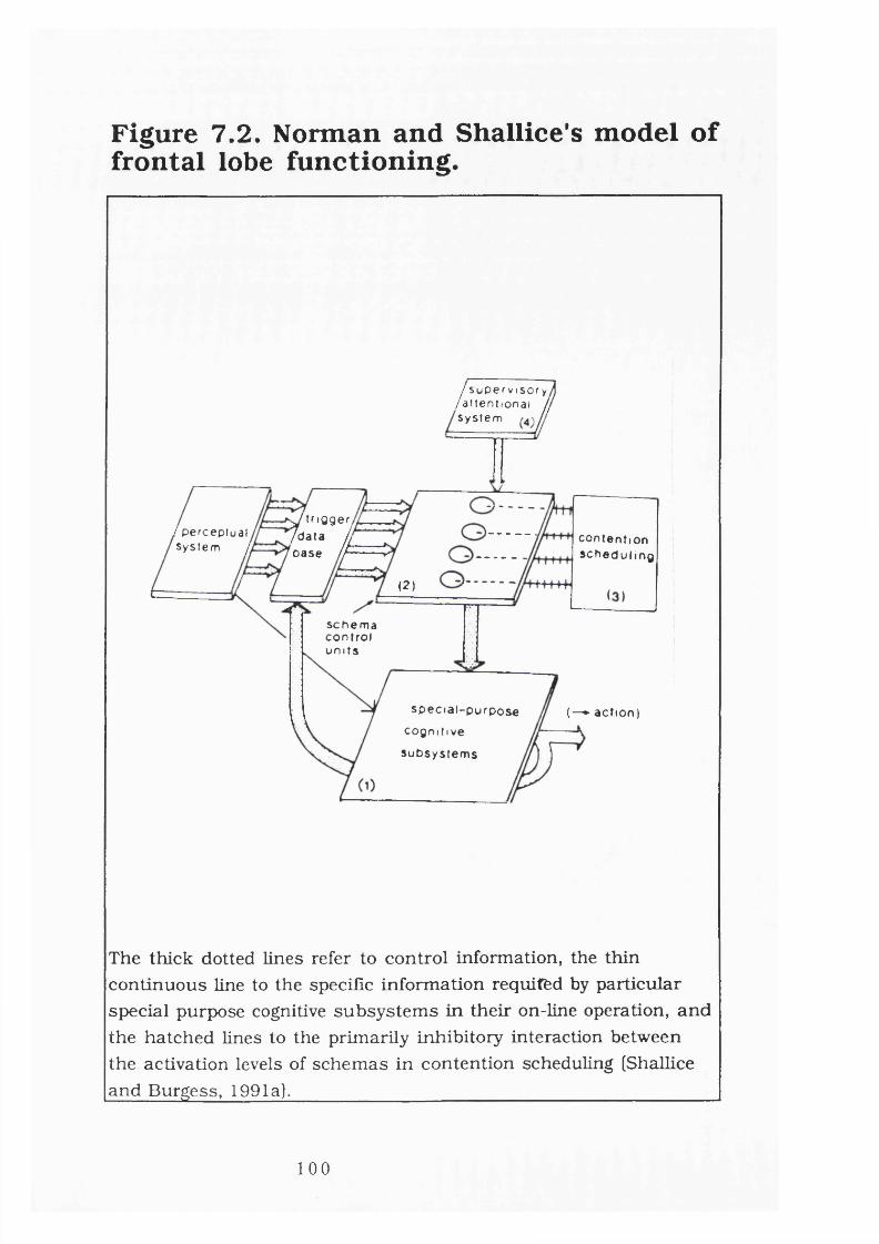
Figure 7.2. Norman and Shallice's model of frontal lobe functioning.
/ S u p e r v i s o r y / a t t e n t i o n a i j Sys t em
^ t r i g g e r / d a t a h o a s e h
! p e r c ep t u a l Sys t em y co n t en t i o n
s c h e d u l i n g
(2 )
s c h e m acon t rolu m t s
s p e c i a l - p u r p o s ecogn i t ive
s u b s y s t e m s
(—► ac t ion)
The thick dotted lines refer to control information, the thin continuous line to the specific information requitfed by particular special purpose cognitive subsystem s in their on-line operation, and the hatched lines to the primarily inhibitory interaction between the activation levels of schemas in contention scheduling (Shallice and Burgess, 1991a).____________________________________________
100

The second approach would be to develop tasks tha t appear to require the processes the Supervisory System is held to carry out and consider whether frontal patients have specific difficulties with such tasks (Shallice and Burgess, 1991b). Two general characteristics of the Supervisory System have been outlined. One has been discussed previously. The second is where no routine procedure is available to produce an appropriate response, i.e. the situation is, in some essential respect, novel. Patients with damage to the Supervisory System should have problems in these situations.
Shafiice and Evans (1978) produced a m easure of cognitive estimation which could be considered a "novel" situation, in as much as no prior knowledge was required. They found that patients whose lesions involved the frontal lobes had a significantly higher score than those with posterior lesions, for each of the cut off points (a finding later corroborated by Kartsounis et al, 1991). A similar type of deficit, although on a different type of estimation task, was recorded by Smith and Milner (1984).
The most plausible explanation of the deficits found on these two types of estimations task, is that the frontal patient is impaired in devising an appropriate strategy to deal with a problem situation where no routine procedure is available.
Thirdly, frontally lesioned patients should have difficulty on tasks derived from artificial intelligence modelling of Supervisory processes. One such example is the Tower of Hanoi which has been used to assess learning in artificial learning systems (Anzai and Simon, 1979). Shallice and McCarthy (Shallice, 1982) modified this task (The Tower of London task) to allow qualitatively different problems to be posed with the same basic layout. Shallice (1982) found the left anterior groups were worse than the right anterior and the left posterior groups on this task. This finding has been replicated on a num ber of occasions (eg Levin et al, 1994; Morris et al, 1988, 1993; Owen et al, 1990), although some discrepant results exist (eg Shallice et al, 1989).
A final approach is to assess whether selective deficits following frontal lesions exist that correspond to damage to particular
101

subprocesses postulated. Figure 7.3 illustrates the possible breakdown of processes involved in the supervisory aspects of the model described.
From this figure it is assum ed that, given an everyday life problem that cannot be immediately dealt with by routine selection of rote- leam ed procedures, the Supervisory process proceeds through a series of stages. It is further assum ed that at specific stages of the problem-solving process the Supervisory System can activate schem as in contention scheduling to carry out specific computations or provide particular pieces of information. It seems possible that different stages of Supervisory control would involve materially distinct subsystems.
Empirically, too, it is now apparent that particular impairments within the general set of frontal cognitive operations can be isolated. A clear example is EVR (Eslinger and Damasio,1985) who performed well on a wide variety of frontal lobe tests, but who had severe practical difficulties in everyday fife tha t seemed to arise from lack of judgemental and organisational skills.
In a recent study, Shallice and Burgess (1991a) isolated an analogous impairment quantitatively. Three patients were assessed, two of whom scored within the normal range for 13 "frontal lobe tests". The other was below the normal range on three. However, all three patients showed little spontaneous organisation in everyday fife, and at least two had made a num ber of gross oversights with major practical consequences. Two tests were developed of their ability to schedule a number of relatively straight forward activities in a restricted period of time. Although the tasks were simple, there was not enough time to complete them in the time limit allowed.The patients had to judge how m uch time to devote to each task so as to optimise their performance given some simple rules, of which the most important was the scoring system that guaranteed diminishing returns on a task as it continued to be carried out. The three patients were outside the normal range on both m easures, bar one score. In addition, they performed the task in a qualitatively inappropriate fashion.
102

Figure 7.3. Possible breakdown of the SAS,
:O N T E N T I O N SCH ED U LI NG S U P E R V I S O R Y P R O C E S Sa s o c o n t r o l s on - l ine in pu t -o u tp u t o r o c e s s i n g ) _------------— -------
C
%-
m a r k e r t r i g g e r e d
. D e te c t^ P r o b le m -------------------------------------
S p ec i f y P ro b l e m + Set G o a l s «---------------
► R e c e i v e P o s s i b l e L m e s - o f - a t t a c k (P rov i s iona l Plan)
^ A s s e s s L i n e - o f - a t t a c k
I V'"®Apply Error P r o c e d u r e s (eg 1. m o v e -u p so lu t io n - t - e e
and try an o t h e r b ranch)2 apply mo re g e n e r a l
sc he ma )( Inhib i t p r e v i o u s s o l u t i o n )
■Activate L ow er - L ev e l MO P + S e t M a r k e r s
: (T ime)
+ve
► In te r ru p t O n - G o i n g B e h a v i o u r
A s s e s s if P la n n e d ActivityStill A p p r o p r i a t e -ve
j+veE x e c u t e Next Step MOP
( S I M P L E ) j
A ct iva te S c h e m a
» A s s e s s R e s u l t — — » S TOP
COMPLEX
Different processes involved in coping with a novel situation from the context of the Norman and Shallice model. The vertical lines indicate the succession in the stages involved. The horizontal hnes indicated the flow of information between Supervisory Process and Contention Scheduling at a particular stage. From Shallice and Burgess (1991a).______________________________________________
103

Similarly, Goldstein et al (1993) reported the case of a 51 year old male of superior intelligence who following a 5cm left frontal lobectomy for the removal of a mixed axtrocytoma- oligodendroglioma showed a similar dissociation between day to day functioning and performance on psychometric tests. On the latter, he scored well. In contrast, his every day behaviour was marked by poor motivation, lethargy, poor anger control and inability to make even simple decisions. On the Strategy Application Test (Shallice and Burgess, 1991a) his performance was inadequate and reflected his day to day difficulties. Further evidence of this kind of deficit has recently been presented by BrazeUi et al (1994).
7.3 .2 . Conclusion and evaluation.
There is little work, if any, that has critically evaluated the theory proposed by Shallice. It does appear to be similar to that proposed by Luria, although couched in a different, computational/artificial intelligence language, and thus satisfying Shallice’s ambitions (Shallice, 1988; Shallice and Burgess, 1991b).
The evidence presented above, and that supplied by Shallice in num erous publications has tended to support the theory’s general proposition. However, there are several issues which still remain unresolved. Firstly, along with other models what it still does not explain is the lack of any observable, or more properly, measurable impairment, in some patients with both minor or large frontal lobe lesions. Thus it can explain why people with frontal lesions fail such tasks as the WCST or the Stroop interference task, yet it cannot explain why patients with similar lesions score well, or a t least comparable to normal controls, on these tasks (eg Heck and Bryer, 1986; Shoqeirat et al, 1990; van den Broek et al, 1993). Similarly, it can explain why patient EVR and the patients documented by Shallice and Burgess (1991a) fail on certain tailored tasks, yet it does not explain why the individuals completed other, psychometrically more sound tasks within normal limits (unless of course there are problems in the assessm ent tools themselves, and recent evidence produced by O’CarroU et al, 1994 strongly suggests this may be a possibility).
104

In a similar vein, the supportive evidence for the model is far from conclusive. As has been pointed out, some research findings have suggested that those with frontal lesions fail on the Tower of London task, whilst other studies have been unable to replicate this. Similarly, on the estimation tasks, whilst intuitively this would seem to support the SAS, there appears little empirical evidence suggesting frontal lobe impairment (O'CarroU et al, 1994).
The concept of a Strategy Application Disorder is gaining credence, as are the measures devised for its assessm ent. One particular problem evident in the Shallice and Burgess report is tha t those m easures described were specifically designed, not necessarily to tap the deficits following frontal lobe damage, but to reflect (albeit objectively) those deficits already noted in the patient's day to day life. This has two possible consequences. Firstly, the cases described may be the rare exceptions that faü on such tasks, whereas other cases with similar frontal pathology will succeed. Obviously, further studies on an unselected population of discrete frontal lobe subjects is required.
Secondly, it does not suggest a m easure that will have much clinical relevance in the assessm ent of frontal lobe deficits in the absence of reported social problems. Obviously, the measures have greater ecological validity (Wilson, 1993) and have been incorporated into a battery of tests (The Behavioural Assessment of Dysexecutive Syndrome [BADS]; Alderman et al, 1993). Where they are still lacking is in their ability to assess whether the deficits are clearly frontal in origin- obviously further work is necessary in order to clarify this.
A related problem with the method employed by Shafiice and Burgess (1991a) is in their data. All three of the individuals tested had suffered head injuries, resulting in damage to the frontal regions. Yet many investigators have pointed out that with traum atic frontal brain injuries, the damage is fikely to be diffuse and involve other cortical regions, although admittedly to a lesser degree (Alexander, 1982; Eisenberg and Weiner, 1987). Indeed, as Newcombe (1982) points out:
105

"It is not plausible to think in terms o f a discrete locus o f damage after closed head injury. "
Newcombe, 1982, p. 112-113.
It is therefore possible that the subjects had damage to other cortical regions (indeed, at least one of Shallice's subjects had damage to the temporal regions). Furthermore, damage may not have been shown on the CT scans used- a rather old method of delineating damage that can faü to reveal subtle damage.
The control data supplied by Shallice and Burgess is also questionable, since they seem to have been derived from non- neurologicaUy damaged Individuals (although this is not entirely clear). Surely, comparing this data aUows us to say only that following damage to the cerebral cortex patients are less able to function in a day to day situation- a common finding with head injured patients with a variety of different regional damage (eg Anderson, 1994; Vamey and Menefee, 1993). A more appropriate comparison would be to compare those with clearly defined frontal lesions with those having distinct lesions in other cortical regions.
Goldstein et al (1993) reported a subject with a more localised lesion; having had a tum our removed for the relief of epüepsy and hence apparently offered a more localised and discrete lesion to that described by ShaUice and Burgess (1991a). Yet, tum our cases can also lead to damage in other regions; this may occur by insidious infütration, by secondary damage caused by intra-cranial pressure, or because of some frontal diaschisis (Lüja et al, 1992).Furtherm ore, frequent epüeptic seizures can cause secondary damage to other cortical regions (Fish et al, 1993a). FinaUy the extent of damage experienced by the case report of BrazeUi et al (1994) was also extensive and included other cortical, extra-frontal, regions.
Thus, even in these cases there is some doubt over the precise localisation of the damage. This is important, for if as ShaUice and Burgess maintain:
106

"Overally plan form ulation/modification, marker creation and triggering and evaluation and goal articulation may be considered complementary component processes undertaken by the Supervisory System on the model o f Shallice and Norman [1980J and it is these processes that are impaired in the Strategy Application Disorder. "
Shallice and Burgess, 1991b p.739
where these process are "anteriorly located in the cortex" (Shallice and Burgess, 1991b, p.736) then they m ust be able to clearly dem onstrate that frontal lobe impairment alone results in the Strategy Application Disorder.
7.4. Conclusion.A num ber of theories of frontal function have been described, yet only one has remained untenable (Goldstein, 1936a,b, 1939a,b). The adequacy of the others has yet to be fully addressed. It may not be coincidental that the eldest theory is the only one to have been abandoned. Recently, Luria's theory has come under attack and has thought to have been superseded by more "advanced" theories. Yet, little comparative work has been undertaken.
In this regard the comparative work of Daigneault et al (1992) points the way forward (although there are errors in the conceptualisation of the models of frontal function that are in obvious need of correction). Future research should compare models of functions to assess the adequacy of them and to detail the strengths of one over another. There appears to be a great deal of overlap between the models and therefore attention may be better directed towards formulating the different models into a whole.
However, an equally im portant goal is the presentation of evidence to support each model. Thus far, evidence has come from a num ber of sources and the validity of the models appears to be a sum of the parts, rather than a holistic assessment. It is evident that many authors use the same empirical evidence for their own theoretical formulations. Obviously, many have commonalities and therefore this
107

is not unreasonable, but the specific study applicable for individual models is still required.
Furthermore, the models should be able to account for both positive findings- tha t is, why behaviour A is deficient following frontal lobe damage and, "negative" findings- that is why behaviour B is not deficient following frontal lobe damage. Similarly, why in some patients both behaviour A and behaviour B are spared, despite similar levels of damage (hence the dissociation between everyday activity and psychometric tests is an advancement). For example, in support of his theory Shallice (1982) presents evidence of subjects with anterior cerebral damage failing the Tower of London task- a task which should rely heavily on the SAS, yet later he reports that he has been unable to replicate this finding (ShaUice, 1988). This example is drawn from the analysis of the ShaUice theory, but the point is equaUy appUcable to the other theories discussed.
There are a number of reasons why this situation may have occurred. Unfortunately, there are a num ber of problems that face researchers when testing for frontal dysfunction, and these wiU be outUned in the next chapter.
108

Chapter 8: Problems with
previous research and
introduction to current study.
109

8.1. Overview.In many cases structural and physiological damage to the frontal lobes may be dem onstrated radiologically or at post-mortem, whilst performance on traditional neuropsychological m easures may be within normal limits. Many problems have hampered the assessm ent of frontal lobe functions and the development of a reliable clinical tool. This has ultimately led to the production of a wide range of disparate results, which has obviously inhibited the development of a thorough model of frontal lobe function.
The problems with much of the previous literature can generally be classified into one of four types: problems with terminology, problems with currently available m easures, problems with the sam ples studied, and the difficulty in assessing clinical differences with experimentally derived measures. Each of these will be reviewed in turn, with emphasis on how they affect the investigation to be carried out here. Finally, a review of the limited num ber of investigations undertaken which report on the neuropsychological consequences of frontal lobe epüepsy wUl be detafied.
8.2. Terminology.The terminology used to describe both the frontal regions and the functions subsum ed by this area has led to confusion. The labeUing of certain areas within the frontal lobes has often differed between investigators. Thus, for example, the dorsolateral and orbitofrontal regions may often overlap. The term s used to describe frontal regions may be the same, but the functional areas referred to may differ (eg Jouandet and Gazzaniga, 1979; Riegele, 1931; Walsh,1978; see chapter 4).
Although this is probably true for most regions of the cortex it is particularly the case with regard to the frontal lobes, because of the paucity of reliable landm arks in this region (Damasio, 1991; Mesulam, 1987). Since there are such few landmarks, the division is somewhat arbitrary and there are no clear boundaries for the functional areas (mesial, dorsolateral, orbital, motor and premotor) commonly investigated (Damasio, 1991). It is therefore essential
110

tha t prior to any investigation being considered, details of the frontal areas being assessed are either explicitly stated, or are referenced to some commonly agreed standard.
Even when there is general agreement about the adoption of such boundaries problems can still exist. The explicit demarcation of boundaries can result in the neglect of holistic approaches to cognitive function. Hence it may be argued that the arbitrary division of the cortex may minimise the importance of cortical and cognitive systems. Thus with reference to the frontal lobes, S tuss (1992) prefers the term "frontal system", (supported by Duffy and Campbell, 1994} which whilst allowing the anatomical region to be acknowledged, reflects the region acting hoUstically.
Furthermore, problems are generated by the terminology applied to the functions associated with the frontal region. These have been conceptualised in many different ways and given different terms. Hence some talk of deficits in "problem solving" (McCarthy and Warrington, 1990], or "metacognition" (Duffy and Campbell, 1994), whilst others "dysexecutive skills" (Baddeley and Wilson, 1988), "frontal lobe syndrome" (Luria, 1966; S tuss and Benson, 1984), or problems of "abstraction", or "concrete behaviour" (Goldstein,1936a, b). Furthermore, these may be further divided into different concepts which may also differ in their definition (Mimura, 1992). Although many of these terms probably have a great deal of overlap and may refer to the same phenomena (Baddeley and Wilson, 1988), some confusion evidently exists.
8.3. Inappropriate assessm ents.Shallice and Burgess (1991a) propose tha t the inability to find substantial and consistent deficits in those with frontal lesions is because:
"The relevant tests were not given to the patien t"
Shallice and Burgess, 1991a p727
Although it is Shafiice and Burgess (1991a) who are the most recent investigators to draw our attention to the problem of relevant tests, Goldstein (1936a) previously suggested:
11 1

"...changes characteristic o f the frontal lobe are o f a definite type and may be easily overlooked, because the methods o f examination usually employed are unsuited to disclosing them."
Goldstein, 1936a p.28.
Many early researchers suggested the frontal lobes had few, if any, intellectual functions, or at least no special role in the adult (Feuchtwanger, 1923; Pfeifer, 1910). Again, this was probably a consequence of limited neuropsychological assessm ent with inappropriate measures. The questions that m ust be answered, however, are what are the relevant tests ? And even though researchers should now be aware of these difficulties, have the methods of assessm ent altered ?
Many studies have reported on the paradox of patients with widespread frontal lobe damage whose performance on tests thought sensitive to such damage was good, yet showed poor day to day functioning (eg Brickner, 1936; Penfield and Evans, 1935).
For example, Heck and Bryer (1986) report on a case of bilateral frontal lobe atrophy. They reported that despite the social and working life of the patient being severely restricted by her behavioural characteristics, her performance on standard m easures of intellectual functioning and on tests of sorting and categorising (Wisconsin Card Sorting Test and the Halstead Category Test) was extremely good. Unfortunately, the subject’s performance on other m easures was not reported.
Eslinger and Damasio (1985) reported on an individual, EVR, who developed profoundly abnormal personality characteristics following the surgical removal of a large orbitofrontal meningioma. Prior to the appearance of the tum our and its surgical removal at the age of 35, EVR was a normal individual, having been a successful accountant rising to become a senior partner.
Following a three month post-surgical recovery period EVR returned to work. However, EVR was soon troubled- losing all his money in a failed business partnership. Thereafter he drifted through several unskilled jobs routinely being fired from each.
112

Employers complained about his tardiness and disorganisation despite his Job skills being more than adequate. Similar difficulties led to a deterioration in his marital life, until his wife left with the children and filed for divorce.
In short, EVR's decision making and planning were (i) qualitatively different from what they were before, and (ii) clearly defective by reference to his own standards as well as those of his direct peers. Against this background, EVR's neuropsychological performance was normal. Indeed, on m easures of intellectual functioning he scored above average. Similarly, EVR's performance on tests of language processing was fully preserved. On tests of memory and perception EVR's performance was also unimpaired.
Furthermore, EVR also lacked some supposedly characteristic signs of frontal lobe dysfunction. For instance EVR produced perfect scores on the WCST, the Category Test, and the Word Fluency test, and his performance on tests of cognitive estimation and judgem ents of recency and frequency were flawless.
A further dissociation of intact cognitive skills and socially driven behaviour has been reported in 3 patients with frontal lobe lesions by Shallice and Burgess (1991a). In short these patients generally scored well on tests of general intellectual functioning, language, memory and, most relevantly, tests thought sensitive to frontal lobe dysfunction (including bimanual hand movements, cognitive estimates, proverb interpretation. Tower of London test. Trail making test, verbal fiuency, MWCST). However, the patients' day-to- day behaviour was characterised by disorganisation, tardiness, and limited motivation (see previous discussion in chapter 7).
More recently, Goldstein et al (1993) reported the case of an individual with a left frontal excision who dem onstrated poor day to day performance but whose performance was competent on the traditional m easures thought sensitive to frontal dysfunction.Indeed, the authors comment:
113

''The absence o f form al neuropsychological test decrement cannot be taken as definitive evidence o f the absence o f deficits per se."
Goldstein et al, 1993 p. 276
All of these case examples dem onstrate that the neuropsychological m easures developed for assessing frontal lobe dysfunction do not show particularly good validity. Indeed, the case examples described all had severe abnormalities of the frontal lobes, often with bilateral damage (eg Eslinger and Damasio, 1985; Heck and Bryer, 1986). If the traditional neuropsychological tests employed were unable to reveal deficits in cases such as these, then the likelihood of detecting deficits in individuals with smaller, more subtle damage seem s limited.
Many researchers have commented upon the lack of clinical m easures exclusively sensitive to frontal lobe function. For example, Wang (1987) has concluded that we are still in "search for a sensitive frontal lobe test" (p. 203). Similarly, Damasio (1985) states, with reference to frontal lobe damage:
"The standard behavioural measurements available at bedside or in the neuropsychology laboratory are simply not adequate to address the disturbances"
Damasio, 1985 p. 340
A num ber of frontal tests have been reported over the years but aU currently lack reliable normative data and require more extensive empirical support for the view that they indicate frontal type impairment (Parker and Crawford, 1992). Summarising their review of the methods currently available to assess frontal lobe function Parker and Crawford suggest:
114

"The major conclusion that one can fairly reach following a review o f assessm ent procedures fo r frontal damage is that there are disappointingly fe w sensitive and reliable tests which the clinical neuropsychologist can depend on."
Parker and Crawford, 1992, p286
It is therefore reasonable to conclude tha t tapping the functions of the frontal lobes has proved extremely difficult in both a clinical and research setting. Many tests have been employed, yet none has proved entirely satisfactory, and consequently the choice of tests is usually based upon familiarity and personal preference (Parker and Crawford, 1992). Some of the more popular tests suggested to be sensitive to frontal lobe dysfunction are listed in table 8.1 and their relative merits, or otherwise, outlined (see also Anderson, 1994 and Parker and Crawford, 1992).
115

Table 8.1. Tests thought sensitive to frontal lobe dysfunction.TEST Positive findings of
frontal dysfunction/ frontal involvement
Negative/no results and comments
(M)WCST Milner, 1963. Nelson, 1976. Anderson et al 1980.
Corcoran and Upton, 1993.Heck and Bryer,1986.Anderson et al, 1988, 1991.Eslinger and Damasio, 1985. van den Broek et al, 1993.Shoqeirat et al, 1990. de Zubicary and Ashton, submit.Most widely used test of frontal lobe function. Studies have suggested that it is either not specific to frontal dysfunction, or that it can be impaired in the presence of other cortical damage
Category Test Corrigan et al, 1987. Crockett et al, 1986.
Bames and Lucas, 1974.Bonders and Kirsch, 1991.Related to IQ scores and perceptual-motor function.
Verbal fluency Benton, 1968. Bomstein, 1986. Perret, 1974. Crowe, 1992.
Huber et al, 1992. Parks et al, 1988. Simple task; but recent evidence suggests tha t it relies on "system" of cortical areas rather than Just frontal regions. May be insensitive to subtle frontal lobe damage.
116

Table 8.1 (cont).TEST Positive findings of
firontal dysfunction/ firontal involvement
Negative/no results and comments
Simple attention task
Wilkins et al, 1987. Not repUcated. Requires cUnical apparatus. Boring and time consuming. Only gives one facet of frontal dysfunction.
Tower of London test
ShaUice, 1982. Morris et al, 1993.
ShaUice, 1988. Originally appeared to have possibUity of cUnical test. Good face vaUdity and based on theory of frontal function. ConfUcting evidence over sensitivity to frontal lobe damage.
Mazes Malmo, 1948. Müner, 1964. Walsh, 1978. Tow, 1955. Smith, 1960.
Benton et al, 1963. EUthom, 1955. EUthom, et al, 1963. UnUmited time, with frontal subjects taking a considerable am ount of time to complete.
Picturearrangem ent
McFie and Piercy, 1952.McCuUough, 1950. Sheer and Shuttle worth, 1952. Rylander, 1939. HamUn, 1970.
Meier and French, 1966.MUner, 1954.Long and Brown,1979.Milner, 1954.Reitman, 1959. Warrington et al,1986.Good face vaUdity, yet given great num ber of investigations using the WAIS, relatively few reports of deficits.
Similarities McFie, 1975. Newcombe, 1969. Rzechorzek, 1978. Bogen et al, 1972. Sheer, 1956
Warrington et al, 1986.Good face vaUdity, but see above
Block design Lezak, 1983. Walsh, 1976.
Warrington et al, 1986.As above.
1 17

Table 8 .1 (cont).TEST Positive findings of
frontal dysfunction/ frontal involvement
Negative/no results and comments
Halstead category test
Halstead, 1947. Bornstein, 1986. Chapmann and Wolff, 1959.Appears to drop as consequence of brain damage, and not frontal lesions per se
Estimation tasks ShaUice and Evans, 1978.Smith and Milner, 1984.
O'CarroU et al, 1994. Out dated to a degree. Relies on past experiences to a great degree. Related to IQ.
Six Elements Task and Multiple Errands Task.
ShaUice and Burgess, 1991a, b.Goldstein et al, 1993.
Few studies. CUnicaUy impractical.Subjective scoring but good face vaUdity and based upon theory of function. Is it a m easure of frontal dysfunction or brain damage ? Not fuUy investigated psychometricaUy.
Tinkertoy Lezak, 1982. Anderson, 1994. Bayless et al, 1989. Correlates with brain damage, and not sensitive to frontal damage alone. Imprecise scoring. LocaUsation not confirmed.
Wang concept formation
Wang, 1984. Excessive reliance on short term memory and visuo-perceptual skiUs.
118

Table 8.1 (cont).TEST Positive findings of
û’ontal dysfunction/ frontal involvement
Negative/no results and comments
Proverbs Gorham, 1956. Fogel, 1965. Benton, 1968.
ShaUice and Burgess, 1991a.Appears strongly correlated with IQ and social class. Patients can faU due to lack of knowledge rather than any cortical damage.
Design Fluency Jones-Gotman and Milner, 1977. Jones-Gotman, 1991.
Lezak, 1983. Imprecise scoring. Clinical utiUty is debatable.
Tem poralorientationtest
Benton et al, 1964. Only deficient in most severe cases. Often faUed in those with dementia.
Trail making test
Piéton et al, 1986. Reitan, 1958.
Anderson, 1994. Sensitive to cortical damage per se and not necessarily locaUsed to frontal region.
Stroop Perret, 1974. Corcoran and Upton, 1993.Stuss et al, 1981. ShaUice, 1988.Upton and Corcoran, 1995.Only one study has dem onstrated a left frontal deficit. Yet many studies use this to support view of frontal functions. More studies needed to support contention.
119

Localisation within the frontal lobes has also been attempted, although this has met with limited success, with a num ber of disparate findings being reported. However, this is not to say that specific functions have not been ascribed to individual frontal regions, far from it. Figure 8.1 outlines some of the general functions ascribed to different frontal regions. This is considered in general terms, rather than the results of specific tests, given the confusion that exists in this area.
Table 8.1 clearly dem onstrates the conflicting evidence from the research literature; some tests have been found to be sensitive to frontal lobe dysfunction, whilst others have not. Similarly, some have been thought sensitive to left, whilst others to right frontal lobe damage. There are a number of possible reasons for this. Firstly, it may be as has been pointed out the "relevant" tests have not been employed. Shallice and Burgess (1991a) suggest tha t traditional neuropsychological m easures require the patients to deal with one problem at a time. Each single item is usually very short and problem solving is generally prompted by the examiner. In contrast, everyday behaviour places greater demands on executive skills and hence there may be a dissociation between everyday behaviour and performance on frontal tasks, as dem onstrated by some patients (eg BrazeUi et al, 1994; Goldstein et al, 1993; Shallice and Burgess, 1991a).
120

Figure 8.1. Deficits associated with different frontal regions.
Motor skill. Preparation and performance.
Executive skills. Programming.
rEmotional. Reduced impulse control.
Concentration and attention.
Key
M otor/Pre-m otor area
% Orbitofrontal
Dorsolateral
1 Mesial surfacej
121

Secondly, the m easures employed may be based upon poor psychometric analysis. Hence, recent investigations of the cognitive estimates task have suggested that it performs poorly in term s of reliability, and its internal consistency is weak (O'CarroU et al,1994). Furthermore, both the patient and the control groups used in the vaUdation of tests may be inappropriate (a point expanded upon in a later section). For example, the classification of patient groups into "anterior" or "posterior" may be masking important data. Similarly, the classification of subjects into simply "frontal" groups may also be inappropriate.
Thirdly, such conflicting evidence may also be a consequence of the num ber of functions thought to be subsum ed by the frontal regions (see previous discussion). Thus any investigation into frontal lobe damage may require a num ber of m easures which wUi enable the researcher to tap different aspects of frontal function (Wang, 1987).
The discussion on the use of "relevant" tests raises an im portant point: what is needed from such a perfect test ? Some researchers report impairments in specific regions of the frontal lobes can be detected by neuropsychological measures. One such study reported on a group of patients with epilepsy (Swartz et al, 1990). In this study 15 patients were investigated via neuropsychology, EEG, CT, MRI and PET scans and the results related to the hypothesised epileptic sites (as defined by seizure semiology). The sites of frontal dysfunction outlined were medial frontal, medial frontal plus, dorsolateral frontal, central or precentral, or unknown frontal. The results suggested neuropsychological assessm ents indicated the correct sites of dysfunction in 85% of patients. Obviously, given the results of this investigation the m easures employed should be examined; they seem to have an extremely high validity, being able to discriminate specific regions of the frontal lobes (a task rarely successfully completed). What was the ideal test which gave such impressive results ?
Unfortunately, in this study there was no one index of frontal function, but an extensive battery of tests that was compared to standard norms. The whole battery could be expected to take a day or more of continual testing. Yet, the fact remains that individual
1 2 2

regions within the frontal regions could be detected neuropsychologicaUy and the results appeared impressive compared to other investigations (and comparable to PET scans). This seems to give weight to the argument for a battery of measures tha t taps the many functions associated with the frontal regions. Results of other investigations have, as discussed, provided less favourable results.
Thus, no such ideal test exists for specific regional frontal impairment (nor, indeed, a test of frontal dysfunction). An ideal frontal lobe test would be exclusively sensitive to frontal lobe impairment and as "specific as a pathological reflex" (Wang, 1987). Since psychometric tests, are by nature, behavioural tests, it would be impossible as weU as unwarranted to expect the subject's performance to be as simplistic and clear cut as a reflex. It would be considered a successful attempt, therefore, if one could come up with a test that would be sensitive to alteration in frontal lobe functioning but that would share, to a much lesser degree, some common variance with brain damage at large. Figure 8.2 depicts such a relationship.
Figure 8.2 represents an effective frontal lobe test whereby the performances by those individuals with frontal lobe lesions are dissociated from those with non-frontal lesions at the same time each group shares some common symptoms of brain damage. On the other hand, figure 8.3 reflects an ineffective frontal lobe test because of its lack of discriminative power. Thus the challenge is to create a test that taps functions exclusively associated with the frontal lobes and with no other cortical region. Obviously, this would prove extremely difficult given tha t most will tap heavily on perceptual, memory or visual skills. An alternative approach would be to create a test which includes many different functions yet would only be impaired in those with frontal lesions.
123

Figure 8.2. Effective frontal lobe test (from Wang, 1987).
Frontal lobe syndrome
Brain damage symptoms
Non-frontal lobe symptoms
Figure 8.3. Ineffective frontal test (from Wang, 1987).
lobe
Frontal lobe syndrome
Brain damage symptoms
Non-frontal lobe symptoms
124

As Wang (1987) suggests, a good strategy for constructing such a test would be:
"to "zoom in" on the assessm ent o f functions believed to be strongly associated with the integration o f frontal lobe activities; infacU the more such functions are included, the more robust the te s t"
Wang, 1987, p200
Clearly, from the previous discussion this line of reasoning has not been followed. Researchers have tended to focus on specific functions of the frontal lobes. Whilst this is useful it does not foUow Wang's suggestion that the more functions included, the more robust the test. As Teuber (1964) suggested, attempting to find one measure of frontal dysfunction may prove fruitless:
"I started out by trying to fin d a unitary concept, but as I moved along, it became clear that no single factor hypothesis could carry one fa r enough to cover all the manifestations o f frontal lesions."
Teuber, 1964 p. 442.
Similarly, others have pointed out that the frontal lobes do not control ju s t one specific function and are certainly not a homogeneous zone:
"It is now clear from both anatomical and behavioural work that the large expanse o f the frontal cortex is a heterogeneous region. "
Milner et al, 1985 p. 141
Following Wang's proposal any ideal test should have as many components tha t rely on the integrity of the frontal lobes included as possible. Given the number of functions that seem to be controlled by the frontal lobes (as described in previous chapters) and the proposal that the frontal lobes are the "executive control centre" (cf Duncan, 1986; Stuss and Benson, 1986) then such a test would seem impossible to develop; it would require the inclusion of a multiplicity of components.
125

One possible way around this predicament would be to provide a cumulative index of frontal lobe dysfunction, derived from many different tests that provide m easures of functions supposedly subsum ed by the frontal lobes. This method, used by Swartz et al (1990), albeit in a rather unsystematic fashion, achieved impressive results. Other studies have adopted such an approach and have met with some success in the prediction of frontal dysfunction (eg Kemper et al, 1993; Swartz et al, 1993). To some degree they represent an advance on previous studies in as much as appropriate control groups were considered- for example those with temporal lobe epilepsy- and a num ber of different m easures were employed. However, the conclusions drawn from these studies m ust be guarded given the limited num ber of subjects assessed (eg n=23 in Kemper et al and n=17 in Swartz et al, 1993 studies previously discussed).
This section has detailed some of the necessities for a m easure of frontal lobe functioning. In particular, the requirement of a large num ber of m easures is suggested. Furthermore, the common methodological problems encountered in such studies have to be overcome. One such methodological problem highlighted in this section is the use of appropriate subject and control groups. This wiU be the focus of the next section.
8.4. Inappropriate subject samples.One of the major difficulties in interpreting frontal lobe function is the sheer num ber of conditions associated with frontal lobe damage (see table 1.1). Despite the fact tha t this provides a large subject base, it has led, ironically, to problems in the interpretation of the data (Stuss and Benson, 1984). The variety of conditions may produce different cognitive effects simply by the nature of their secondary effects on other cortical regions (Anderson et al, 1990). Thus, attem pting to square disparate results from investigations with two different subject groups may prove problematic.
126

For many conditions in which it would prove useful to assess the functions of the prefrontal cortex, there have been problems. For example, subjects with neoplasms. These should prove an excellent subject pool from which to form a database of (dys)functions associated with the frontal lobes. However, this has only occasionally been achieved. Subjects with neoplasms rarely present to neurological services at an early stage- the frontal lobes may often be considered neurologicaUy silent, it is only when the damaged area is large that signs and symptoms become apparent. Similarly, pressure effects, speed of growth, infiltration rather than destruction, location of the tumour, and surgical and chemical treatm ent, can all be influential factors (Maurice-Williams and Dun woody, 1988). Consequently, when subjects do come to the attention of examiners the lesion is often no longer "purely frontal".
Subjects with neoplasms have been reported to show neuropsychological disturbance compatible with frontal dysfunction, even when the tum our is located extra-frontally. In one particular study with regional cerebral blood flow (rCBF) and neuropsychological assessm ent, patients with non-frontal tum ours (as assessed via CT scans) were impaired, and behaved in a typically "frontal" fashion (Lilja et al, 1992). Thus, suggesting that lesions to extra-frontal regions can lead to impairment in certain "frontal" tasks. Again, this is not a recent suggestion; Smith (1975) provided a clear discussion on the effects of diachisis.
On many occasions examiners have avoided the problem of tum our spread (or gross head injury) by classifying subjects into either "anterior" or "posterior" subject groups (eg Burgess, 1992; Nelson, 1976; Shallice and Evans, 1978). Although this classification allows a greater overview into the functions of the frontal lobes by allowing greater access to subjects, it does not give answers to specific questions of frontal lobe functioning, nor can it give details of functions of specific areas within the frontal lobes.
Shallice, for example, when criticising Luria’s work, makes the point that:
127

"The patients on which his theories were tested often had lesions extending outside the frontal lobes."
Shallice, 1988 p.330
However, later he reports on his own studies that separated subjects into either anterior (cerebral damage with some frontal involvement), or posterior (cerebral damage without frontal involvement) and then suggests the evidence produced is adequate to support the SAS model of frontal lobe function (eg Shallice, 1982; Shallice and Evans, 1978). Indeed, only 17 pages after the criticism of Luria's theory he reports findings on the Tower of London task in patients with similar levels of cerebral damage (Shallice, 1988).
The impairments recorded in such investigations may simply reflect disordered functioning of other brain regions, rather than frontal lobe damage. Much of the research undertaken using this methodology suggests "frontal" and "temporal" and in some cases "parietal" areas may be included under "anteriors". This may account for the lack of consistency amongst areas of research using this approach (eg Shallice, 1982, 1988).
Another promising subject group which could have yielded important information consists of post-surgical cases- both for psychiatric and epileptic conditions. However, these post-surgical cases have not produced the clear cut findings that one would have hoped for, even though the surgical intervention is usually relatively precise.
The psychological residua of prefrontal psychosurgery should theoretically offer promise in answering questions regarding the function of such regions; aetiology (type of surgery) is known, lesion size and location can be determined via CT/MRI scans. However, published data on the topic is generally inadequate, for example:
"...54% o f the articles published worldwide contained no irformation from objective tests. Of the 70 articles (out o f 152) which reported the results o f any objective tests,16 referred only to an IQ test. "
Valenstein (National Commission of Psychosurgery), 1977, p.29
128

Apart from the deficiencies in assessm ent, there has been a dearth of long term follow up- this being on average 6 weeks (Fish, 1976). Other studies have attem pted to remedy some of these problems (eg Benson et al, 1981; S tuss et al, 1981), yet other issues remain: firstly, the diagnostic procedures were fairly limited, both pre- and post-operatively; secondly, those tha t underwent psychosurgery were extremely behaviourally abnormal (eg schizophrenic, depressed, phobic, violent) and thus both the pre-operative and post operative brain was probably abnormal.
Similarly, in the case of surgery for the relief of epilepsy this should have provided a wealth of information. Again, surgical intervention is usually exact, and precise measurem ents about the area and volume of cortical removal can be ascertained. However, some have even questioned the accuracy of surgical m easurem ents (eg Awad et al, 1989; Kitchen et al, 1993).
However, another major problem exists: the epileptic brain is abnormal. Frequent epileptic discharges cannot only have damaging local effects, but can also have distal effects on both areas linked by neuronal pathways and other areas- damage created by the so called 'm irror focus' (Morrell, 1960). Unfortunately, detailed investigation of these latter points have yet to be undertaken. With the advent of regional blood flow studies, however, im portant information should become available.
The concomitant damage to the brains of people with epilepsy from head injury, episodes of sta tus epüepticus, or chronic use of an tiepileptic medication, may also produce impairments in cognition.As the majority of those people who undergo surgical intervention have experienced seizures for a num ber of years, and the surgery may be a "last resort", it is likely that the damage to the cortex is both severe and persistent. AU of these factors may have to be taken into account when using a post-surgical epUepsy sample to docum ent functional deficits.
SimUarly, the aetiology and age at onset of the epUepsy should be taken into account- unfortunately, this has rarely occurred. It has long been recognised that early chUdhood damage can in some
129

cases, result in some cerebral reorganisation of function. For example, in left temporal lobe epilepsy there may be shifting of language dominance, even though the patient is still right handed (Rasmussen and Milner, 1975; Rausch and Risinger, 1990; Rausch and Walsh, 1984; Satz et al, 1988). Although this concept is most often applied to the reorganisation of language function, it may weU be possible for the reorganisation of executive functions to either alternative parts of the same hemisphere or, indeed, to the alternate hemisphere. However, this is not a point that has been fully investigated, or indeed, often considered.
Our knowledge of development of the frontal lobes suggests that their m aturation is far from linear (see Chapter 5). Damage sustained to the frontal cortex in early childhood may result in a reorganisation of those functions subsum ed by the damaged area. There are a handful of case studies that have not supported this contention (eg Ackerly, 1964; Ackerly and Benton, 1947; Benton, 1991; Eslinger et al, 1989; Mateer and Williams, 1991; Price et al, 1990), although the question is how many cases are there of children suffering prefrontal injury in childhood whose cognitive functions are normal and have not been reported (Marlowe, 1989) ?
Milner and colleagues, as mentioned, have often used post operative epilepsy samples in their studies of frontal lobe functions. Yet, they have consistently failed to appreciate the abnormality of the preoperative epileptic brain and have failed to take this, medication or any seizure variables into account; factors that can be considered to be of utm ost importance (Rausch, 1991). Furthermore, the distal effects of epileptic seizures may not have been considered. For example- comparing frontal excision groups with hippocampal excision groups may not be appropriate: frontal lobe epileptic seizures may have had a damaging effect on the hippocampus a delicate and fragile structure- due to the cortical connections and seizure spread (Goldman-Rakic, 1984; Weinberger et al, 1992). Thus, recent reports have suggested that tests for frontal lobe functions may be impaired in those with hippocampal damage (eg Corcoran and Upton, 1993; Hermann et al, 1988).
130

Not only have pre-operative medical variables largely been ignored in the Milner studies, but so have post-operative states. Many studies have indicated tha t seizure frequency following surgical treatm ent of epilepsy has an important effect on cognitive skills (Rausch, 1991; Rausch and Crandall, 1982). Frontal excisions are less efficacious in controlling seizures (Talairach et al, 1992], and therefore the noted post-operative im pairm ents may merely be a consequence of ongoing post operative seizure activity, or damage caused by frequent pre-operative seizures. It is impossible, however, to determine this since data on post operative seizure frequency is rarely given.
Additionally, the aetiology of epilepsy is often not mentioned. Research has suggested that the underlying neurological impairment causing epilepsy may have different neuropsychological consequences. In one such report, Anderson et al (1990) compared the neuropsychological sequelea of tum our and stroke lesions. The findings indicated that the neuropsychological profile of subjects with cerebral strokes were unequivocally different from those profiles of people with tum ours with similarly situated lesions. The cognitive impairments resulting from tum ours were generally quite mild compared with the deficits resulting from strokes. More often than not, tum ours infiltrate or displace neural tissue without actually destroying it for some time, thereby allowing some m easure of residual neuronal function. In completed stroke, when the lack of blood supply is sudden, profound and long-standing, the ensuing metabolic changes are not compatible with neuronal survival. It is therefore evident that the two groups should not be considered equivalent for neuropsychological research.
Hence, aU the evidence presented here suggests tha t epileptic seizures, the aetiology underlying the epilepsy or other epilepsy related variables can have an impact on cognitive sta tus both pre and post operatively. It should be considered essential that these variables are taken into account in any investigation.
Therefore, one of the aims of this study was to give priority to the classification of subjects into different regional groups ensuring that the classification was accurate and excluded any damage to other
131

cortical regions. Furthermore, other factors that have tended to be overlooked were to be investigated, to examine whether their effect was marginal or whether, as suggested here, they play an im portant role in determining the extent of neuropsychological deficits.
8.5. Clinical versus experimental.One difficulty in assessing frontal lobe function, is that many tests purporting to m easure frontal function have been devised to dem onstrate experimentally induced deficits, and not clinically im portant differences. Thus most tests considered sensitive to frontal lobe dysfunction are very poor at blindly detecting patients with frontal lesions (Burgess and Alderman, 1990; ShaUice, 1982, 1988).
For example. Nelson (1976) used her modified version of the WCST (MWCST) to categorise lesion location. The differences between her posterior and anterior groups (see previous discussion for difficulties with this approach) in the num ber of categories they achieved, failed to reach significance at the 0.05 level. She therefore recommends, for clinical purposes, calculating the perseverative errors as a percentage of the total errors with a 50% cut-off recommended. However, if a patient only made two errors in the whole test, which would be a good performance, but one of them was perseverative, then the result would be considered abnormal by one scoring system but not by another (i.e. categories achieved).
Attempting to use the num ber of categories achieved as predicative of a frontal lesion is also problematic. Burgess (1992), for example, points out that if (according to Nelson's data) one were to use a fifth percentile cut-off for this test, one would correctly classify 48% of frontal lesion cases as having an anterior rather than posterior lesion- a result which would seem satisfactory. However, one would also classify 32% of cases with a posterior lesion as being frontal which is hardly acceptable. Thus the test does not really seem sensitive enough for most clinical applications.
Clinicians often consider Milner's 128 card version of the WCST as more sensitive, but in fact Milner's data does not support this with
132

regard to the frontal lobes as a whole. As she rem arks in her 1964 paper:
"The patients with lesions o f dorsolateral frontal cortex alone show ed consistent impairment on the card sortinq te s t "
Milner 1964, p .315
Her patients with lesions in other areas of the frontal cortex were not impaired on the test at all, and in fact were used for comparison purposes with dorsolaterally lesioned frontal patients in her analysis of the results.
Other, more recent investigations have also questioned the adequacy of the MWCST as a measure sensitive to frontal dysfunction. For example, Shoqeirat et al (1990) found that the MWCST did not discriminate between different amnesic sub-groups. Hart et al (1988) found that the shortening and modification of the WCST made it insensitive to mild cognitive deterioration. Similarly,Jenkins and Parsons (1978) found that the MWCST performance was not impaired in a group of alcoholics, whereas the original WCST was- thus alteration in test administration can have an impact on competency of performance.
In light of these studies van den Broek et al (1993) undertook a comprehensive investigation into the ability of the MWCST to localise frontal dysfunction successfully. The analysis revealed that the MWCST was a good detector of cortical damage, but not of frontal dysfunction per se. The MWCST differentiated satisfactorily between the control and the patient groups as indicated by its specificity of 98.7%, and sensitivity (to cerebral lesions irrespective of site) of 45.6%. From this study it was possible to conclude that the MWCST is a useful measure of cortical dysfunction, but not specifically frontal lobe dysfunction.
Two studies have specifically investigated the influence of temporal lobe epUepsy on (M)WCST performance (Corcoran and Upton, 1993; Hermann et al, 1988). Both studies found that performance was impaired in patients with temporal lobe epilepsy. In the Corcoran and Upton (1993) study the hippocampal group performed more
133

poorly than the frontal group. Furthermore, these findings were confirmed by S trauss et al (1993) who reported that WCST performance was impaired in subjects with temporal lobe epilepsy (both left and right). They also reported that performance was influenced by the age at onset of the epilepsy and this was related to laterality of dysfunction. All of these findings confirm that even with the most popular m easure of frontal dysfunction, there is still considerable doubt about its specificity.
These sorts of problems, however, are not restricted to the WCST. Although, it is important to note them in detail, given that the WCST is reported to be the most frequently used measure of frontal dysfunction currently available (Butler et al, 1991; Mountain and Snow, 1993). Other tasks that are considered sensitive to frontal lobe functioning fair little better. Shallice (1982) reports an unpublished study involving a group of patients with localised lesions on a battery of ten tests which were:
"Simplified versions o f ones, where from the literature one might expect a frontal deficit"
Shallice, 1982, p.202
and included three tests developed by Luria, as well as verbal fluency, the Stroop test (Perret, 1974), and Shallice's own cognitive estimates (ShaUice and Evans, 1978). Only one of these tasks- Weigl’s sorting task-
"Produced a significant effect o f anterior-posterior location in the basic analysis."
ShaUice, 1982, p.202
Furthermore:
"neither verbal fluency, nor the WCST has demonstrated an ability to localise and differentiate frontal lobe lesions
from lesions involving more diffuse areas o f the brain. "
Cantor-Graae et al, 1993, p.83-84
These findings are particularly important, since the most commonly used measure of frontal functioning other than the WCST is probably verbal fluency. There are a num ber of different versions of this test.
134

but they all require the subject to generate as many words as they can which either belong to a particular semantic category or sta rt with a specified letter within a set period (Crowe, 1992). Investigations into the accuracy and specificity of the verbal fluency task has questioned its sensitivity to frontal lobe damage. It has been suggested that it is not simply frontal lobe damage which produces poor results on this test, but rather a deficit in a "system" which can result from a variety of differing cortical lesions (Huber et al, 1992; Parks et al, 1989; Weinberger et al, 1992).
The data for other procedures supposedly sensitive to frontal lobe dysfunction, is even less convincing in term s of their predicative utility. The point often over looked is that the criteria used to determine localisation of function when a task is developed experimentally are not necessarily the same when a test is used clinically. The latter application requires considerably greater discriminative power.
Milner and her colleagues at the Montreal Neurological Institute (MNl) have been the main proponents of the approach which compares different subject groups on mean neuropsychological test scores, following unilateral cortical excisions for the relief of focal epilepsy (eg Milner, 1963, 1964, 1975, 1968a, b, 1982a, b; Milner and Petrides, 1984; Milner and Teuber, 1968; Petrides and Milner, 1982). These studies have yielded considerable information, but some of the methods and conclusions reached can be questioned.
For example, when investigating memory impairments secondary to frontal lobe dysfunction, patients with frontal excisions show little impairment compared with extrafrontal lesions (eg Milner et al, 1985; Smith and Milner, 1984). However, there were problems in the analysis of the findings. For instance, Milner (1968b) studied five subject groups (normal subjects plus groups of patients with either right or left temporal, frontal or parietal lesions) on m easures of recall and recognition using faces as stimuli. Throughout the study, Milner did not distinguish between left and right hemisphere for frontal or parietal lesion groups of patients, but grouped them together for comparison with right or left temporal groups. In essence this disguised some of the evidence- a problem similar to
135

that discussed in relation to the grouping of subjects into "anteriors" or "posteriors" (see also discussion on use of inappropriate samples).
Comparison of one group of subjects with extensive frontal lobe excisions with another group of subjects with extensive temporal excisions via statistical analyses, can be questioned from a clinical perspective. The analysis may show a significant difference a t the 5% level, but the actual differences in magnitude may be minor and of limited clinical significance. For example, in another study from the MNI, Miller and Milner (1985) report tha t left temporal lesioned groups were different from normal controls a t the 0.05 level on a test of synthesising ability. Examining the results shows, however, the actual difference to be only 1.4 (14.6 compared to 16.0 respectively). In addition, the left frontal lobe group (who actually performed the worst), were excluded from the analysis because of the small num ber of subjects (n=3). To base a firm conclusion on such an analysis seems dubious.
It is therefore apparent, that the m easures thought sensitive to frontal lobe function may not be adequately so from a clinical perspective. It is therefore one aim of this present study to investigate both the statistical differences and the clinical utility of some of the m easures currently available for frontal lobe assessm ent.
8.6. Neuropsychological studies of frontal lobe epilepsy.Cases of temporal lobe epilepsy have certainly been extensively studied and contributed much to our understanding of the temporal lobe, but this is not the case for frontal lobe epilepsy, however;
"Unlike the temporal lobes where material specific memory deficits can usually be elicited preoperaüvely, preoperative deficits in cases o f frontal lobe epilepsy are not seen consistently."
Milner, 1988, p.221
136

Similarly:
"No systematic neuropsychological study o f various subtypes o f frontal lobe epilepsy has been reported."
Waterman and Wada, 1990, p.209
This may reflect the inadequate m easures that are currently available, for as Wüliamson and Spencer, 1986 point out:
"Motivation, goal-directed behaviour, and social adaptation are difficult to quantify unless grossly disturbed, yet even subtle disturbance o f these functions can produce deleterious effects upon human behaviour. Improved methods of evaluating frontal lobe function and predicting effects o f resection are needed. "
Williamson and Spencer, 1986, p. 59
Compared to the research undertaken in people with temporal lobe epilepsy, the investigations into those with frontal lobe epilepsy has been meagre to say the least; no wonder Milner (1988) was unable to report any pre-operative deficits. There have been few group studies investigating the neuropsychological characteristics of patients with frontal lobe epilepsy (despite the high num bers seen by Milner and colleagues at the MNl):
"Cognitive studies o f post-operative frontal lobe epilepsy patients have contributed greatly to our knowledge o f specific frontal lobe lesion effects. Yet it remains to be seen whether frontal lobe epilepsy subjects show such impairments pre-operatively. "
Swartz et al, 1994, p. 232
A case report (Boone et al, 1988) has, however, documented the behavioural and neuropsychological characteristics of a thirteen year old girl with frontal lobe seizures. It was found tha t she was deficient on certain tasks of frontal function (attention, response inhibition, alteration between tasks, maze solving, word generation, and motor function), although other frontal functions were intact (categorisation, sequencing, conceptual flexibility). There was also some impairment on tests of learning and recall.
137

It is perhaps notable tha t all of these deficits were reversible once the epileptic activity was controlled by medication. Boone et al (1988) conclude:
"Frontal lobe déficits on neuropsychological testing may be the only abnormalities on higher cognitive testing which signal the presence o f ongoing brain disturbance,"
Boone et al, 1988, p. 586
This highlights several im portant points. Firstly, it may suggest that neuropsychological m easures are sensitive and predictive of frontal lobe epilepsy (a fact dem onstrated with the temporal regions but infrequently with the frontal regions). Secondly, that frontal lobe functions are disturbed because of a more generalised brain disturbance (cf. Lilja et al, 1992). The former may be an over optimistic claim, given the evidence discussed of normal neuropsychological performance, in the presence of quite large scale damage to the frontal regions. Furthermore, this also highlights the predicament of using epileptic subjects who have received surgical treatm ent for their condition since the previous seizure frequency, or present on going activity, may have a bearing on their cognitive performance.
Another study similar to that ju s t described, was reported by Perez et al (1993). They reported four cases of children with epilepsy and continuous spike-wave syndrome (CSWS). The authors suggested that the children, as a result of the CSWS, had an acquired epileptic frontal syndrome. More particularly, they suggested tha t the epileptic activity to the frontal regions resulted in characteristic neuropsychological deficits associated with frontal lobe dysfunction. Thus they observed combinations of impairments in attention, impulsiveness, mood, perseveration, reasoning, thought formulation and learning strategy similar to those described in adults with frontal lesions.
These cases dem onstrated no improvement over a period of time. This, the authors suggested was a consequence of ultrastructural damage to the developing neural networks, or interference with the elaboration of complex cognitive functions during sensitive periods
138

caused by continuous epileptic discharges. Once again, this report emphasises the importance of variables such as age at onset, and the frequency and type of epileptic seizures.
In contrast to these case studies, group studies of patients with frontal lobe epilepsy are relatively rare. Kemper et al (1993), in a preliminary research project, report on a neuropsychological investigation of patients with frontal lobe epilepsy, compared to those with temporal lobe epilepsy. They suggest a m easure of "planning" was able to classify some 80% of the frontal patients correctly. However, they were unable to distinguish left and right frontal dysfunction. Unfortunately, the study's findings may be limited on methodological grounds. Most importantly, the num ber of subjects was limited (only 23 with frontal lobe epilepsy, 6 of whom had a left foci). Secondly, there was no mention of the medical or seizure background of the subjects (ie how secure were the investigators in there classification of subjects). Finally, the actual difference was small, and given the num ber of tests employed, the adoption of the 0.05 significance level somewhat dubious. However, their study should be commended for using an appropriate battery of tests and more advanced, appropriate, statistical analysis. Again, the adoption of a battery of tests resulted in a high level of predictive success.
Swartz et al (1993), on the other hand, reported upon a study with several methodological problems. The subject sample was small (n=17 frontal lobe epilepsy sample) and the control group (n=13) not clearly specified (that is, were they "normal" controls or epilepsy controls ? Were they matched on appropriate variables ?). Despite the lack of details given and the minimal num ber of subjects the authors conclude that:
"Frontal lobe epilepsy is associated with significantimpairment o f cognition even in the absence o f lesions. "
Swartz et al, 1993
This firm statem ent, however, cannot be totally accepted given the methodological problems associated with this study.
139

The evidence reviewed suggests tha t there has not been a comprehensive study of frontal lobe epilepsy. In those studies that have been reported, there is insufficient detail and not enough attention paid to possible confounding variables, for any firm conclusions to be made. One of the aims of this study was to attem pt to rectify this and provide evidence of the neuropsychological consequences of frontal lobe epilepsy, taking into account the possible influential variables outlined.
140

8.7. Hypotheses and Aims.This chapter has outlined several deficiencies with previous investigations, and some of the unanswered questions tha t remain, regarding both frontal lobe epilepsy, the possible consequences of removal of epileptogenic frontal regions, and the assessm ent of frontal lobe functioning.
In particular the importance of a large num ber of assessm ents, with appropriate patient and control groups (ie with clearly demarcated areas of dysfunction) was emphasised. Furthermore, the importance of collecting appropriate variables associated with epilepsy was also stressed.
On the basis of these observations, and reports from previous literature, several hypotheses were formulated:
1. The literature has suggested a number of tests may be able to discriminate between damage to either the frontal or temporal lobes. These may have been masked by the use of inappropriate assessm ents, subject and control samples. Furthermore, although these deficits have been noted in post-operative samples, they have yet to be fully demonstrated in an epilepsy sample. It was hypothesised that:
There wül be a characteristic difference in the
neuropsychological performance between those with temporal and those with frontal lobe epileptic foci.____________________
2. The review of the literature has suggested some functional differences consequent on damage to either left or right frontal regions. However, the extent of these differences may have been masked by a num ber of methodological issues, previously outlined. Again, the majority of the subject samples have either been surgical or tum our cases and there are no studies that have examined the latéralisation difference in an epileptic sample. In this sample, with clearly defined epileptic foci, it was hypothesised that:
141

H 2 There will be a difference in neuropsychological
performance dependent on latéralisation of the epileptic foci.
3. From the review of the literature, it is evident that minimal attention has been paid to possible putative intervening variables on neuropsychological performance in either people with epilepsy, or people with frontal lobe lesions. Hence, it was hypothesised that:
Seizure related variables will influence
neuropsychological performance.___________________________
4. In particular, the differential impact of age at onset of frontal damage has yet to be fully delineated. The research has implied development of the frontal regions (both anatomically and functionally) to be far from linear. Age at onset can be investigated either by correlation techniques (ie investigating a relationship between age a t onset and neuropsychological performance), or differences dependent upon age at onset can be investigated. Hence, for this investigation two hypotheses were formed:
H^. There will be a relationship between age at onset and
neuropsychological performance.
H^.. There will be a difference in the neuropsychological
performance dependent upon the age of onset of frontal lobe epilepsy._________________________________________________
5. Some localisation of function within the frontal regions has proved possible. Although, these studies have mainly concerned lesional patients, it is proposed that similar deficits would be recorded in an epileptic sample.
Hg There will be a characteristic neuropsychological deficit
dependent on the localisation of the epileptic foci within the frontal lobe.
6. The review of the literature above has revealed few studies investigating the differential ability of neuropsychological m easures to identify frontal lobe dysfunction. In this study, it was hoped to investigate this fully and it was hypothesised that:
14 2

Hg There will be differences in the discriminatory power of
the neuropsychological assessm ents.________________________
7. Given the evidence presented, that the best test for frontal lobe function is one that includes the most functions, it was hypothesised that:
Hy A composite score of "frontal dysfunction" will offer
greater discriminatory power in the assessm ent of frontal lobe damage than any individual neuropsychological test score._____
Furthermore, that:
Hg A composite score will offer greater predictive power of
assessing dysfunction within the frontal lobes than any individual neuropsychological test score._____________________
6. The majority of the evidence regarding frontal lobe function has been provided by investigators using subjects with surgical induced lesions for the control of their epileptic activity. It was hypothesised that even when taking into account the pre-operative seizure variables:
Hg Surgical removal of epileptic foci in the frontal lobe will
result in altered neuropsychological performance. Specifically there will be a detrimental affect of surgery on those tests purporting to tap the function of the frontal regions.__________
143

Chapter 9: Method.
144

9.1. Introduction.In order to address the stated hypotheses satisfactorily, a num ber of conditions need to be met. Firstly, subjects for the investigative sample had to have clearly defined lesions, as did the control sample (the characteristics of the sample are outlined below). Secondly, a number of tests had to be employed that tap cognitive, especially frontal, function.
The choice of tests for any neuropsychological battery examining the functions of the frontal lobes is usually based on personal preference and experience (Parker and Crawford, 1992). Indeed, in a review of some of the measures available for assessing frontal dysfunction, it has been suggested that:
"A large number o f studies have been conducted in frontally injured patients but there are in fa c t fe w tests that have sufficient empirical support fo r the neuropsycholoqist to fe e l happy with their sensitivity and reliability."
Parker and Crawford, 1992, p .268
In order to minimise the difficulties in the assessm ent of frontal lobe dysfunction, a number of different clinical m easures thought to tap the variety of functions subsum ed by the frontal lobes, were employed in this study. The choice of individual "frontal" tests was based on a review of the literature concerning the functions associated with the frontal regions. Within the battery, some tests assessed motor skills where others sought to tap executive skills. Furthermore, on the basis of the previous literature, different regions of the frontal lobes can be assum ed to underlie successful completion on some of these tasks.
In addition to the "frontal" tests employed, a num ber of other m easures were used. This was firstly, to assess some of the functions associated with the temporal regions in order to allow comparison between the frontal and temporal groups. Secondly, to assess the general intellectual and cognitive skills of the groups to enable the results obtained to be placed in an appropriate context.
145

In particular, the rationale behind using a num ber of different tests was as follows:
(i). A better overall picture of the neuropsychological function of the frontal lobes could be achieved by a num ber of tests, rather than a single measure.
(ii). Results on the different m easures could be combined to give a composite score of "frontal" dysfunction.
(iii). Better localisation within the frontal cortex would be possible. That is, different deficits could be ascribed to different frontal regions.
(iv). Better differentiation between the frontal lobes and other cortical regions would be possible.
(v). A comparison could be undertaken of the sensitivity of the current tests available for frontal lobe assessm ent.
(vi). The discriminatory power of the composite score could be compared with that achieved from individual tests.
146

9.2. Subjects.9.2 .1 . Epilepsy stibjects.
Subjects for the patient sample were collected over a thirty month period, and consisted of all those attending a national centre for epilepsy as either out-patients or in-patients and who met the inclusion criteria outlined in table 9.1. The clinical investigative procedures adopted in order to classify the subjects are outlined in appendix 1.
In addition, a sub-group of subjects undergoing surgical procedures for the relief of their frontal lobe epilepsy, were assessed at specified intervals (see later).
Eighty-eight subjects with frontal lobe epUepsy were assessed, as were 57 subjects with temporal lobe epilepsy. However, on further investigation 14 of the frontal lobe subjects were found to have bilateral dysfunction, and were removed from the subject pool (since they did not fit the inclusion criteria). This left the total num ber of frontal lobe subjects at 74. The four groups collected therefore consisted of the num ber of subjects outlined in table 9.2.
These subjects were randomly divided into two groups (a training set and a validation set) for the purpose of some analyses. The training set consisted of 40 frontal lobe subjects and 29 temporal lobe subjects. The validation set consisted of 34 frontal lobe subjects and 28 temporal lobe subjects. There was no significant difference between the validation and training set in terms of basic demographic or epilepsy variables (all p>0.1).
147

Table 9.1. Inclusion criteria for subject sam ple.
1. A primary diagnosis of partial epilepsy.2. A focal disturbance of either frontal or temporal lobe origin.3. A unilateral disturbance with no indication from either EEG or cortical imaging of bilateral damage.4. No seizures arising from any other cortical region.5. Cortical scanning revealing either damage confined to the sites mentioned, or to indicate no significant abnormality in other cortical regions.6. No significant psychiatric disturbance tha t may interfere with test performance.7. No sensory deficits that could interfere with test performance.8. Either right handed, or been dem onstrated to be left hemisphere dominant for language.9. Over the age of 16 years and under the age of 65 years a t the time of assessment.10. Have suffered from a t least two epileptic seizures in the preceding two years prior to assessm ent.11. No history of drug or alcohol abuse.12. FSIQ> 69________________________________________
Table 9.2. Number of subjects in each group.
Group Number o f subjectsLeft frontal 42Right frontal 32Left temporal 31Right temporal 26
148

9 .2 .2 . Basic demographic characteristics o f subject groups.
There were no significant sex differences between the four subject groups (see table 9.3; %^=2.92, df=3; p=0.41). Similarly, there was no difference in the ages of the four groups (F[3,127]=1.257; p=0.292; see ANOVA summary table).
Summary univariate AMOVA table for ageSource df Sum of
squaresMeansquares
F Prob
Between groups 3 239.62 79.87 1.257 0.292Within groups 127 8068.49 63.53
The four groups did not significantly differ in their occupational class (x^=1.614, df=3; p=0.656; see table 9.3), or educational achievements (x^=14.875, df=12; p=0.46; see table 9.3).
Similarly, as shown in table 9.3 the groups did not differ with respect to their VIQ (F[3,127]=0.957; p=0.415), PIQ (F[3,127]=0.532; p=0.661), FSIQ (F[3,127]= 1.129; p=0.339) or NART IQ (F(3,127)= 0.167; p=0.919).
Summary univariate ANOVA table for VIQSource Sum of
squaresdf Mean
squaresF Prob
Between groups 331.49 3 110.49 0.957 0.415Within groups 14662.07 127 115.45
Summary univariate ANOVA table for PIQSource Sum of
squaresdf Mean
squaresF Prob
Between groups 311.38 3 103.80 0.532 0.661Within groups 24785.76 127 195.16
Summary univariate ANOVA table for FSIQSource Sum of
squaresdf Mean
squaresF Prob
Between groups 468.255 3 156.09 1.129 0.339Within groups 17555.65 127 138.23
Summary univariate ANOVA table for NART IQSource Sum of
squaresdf Mean
squaresF Prob
Between groups 69.90 3 23.30 0.167 0.919Within groups 17760.89 127 139.85
149

9 .2 .3 . Basic epüepsy details o f subject groups.
The variables duration and age at onset, of epilepsy, along with years of remission from epilepsy, are presented in table 9.4. There was no significant difference in duration of epilepsy (F[3,127]=1.2856; p=0.285).
Summaiy univariate ANOVA table for duration of epilepsySource df Sum of
squaresMeansquares
F Prob
Between groups 3 309.65 103.22 1.285 0.285Within groups 127 10196.88
Similarly, there was no difference between the groups in term s of years of remission (F[3,127]=0.2394; p=0.869).
Summaiy univariate AN[OVA table for remission of epilepsySource df Sum of
squaresMeansquares
F Prob
Between groups 3 2.61 0.87 0.239 0.869Within groups 127 460.77 3.63
There was, however, a significant difference, however, in the age at onset of the epilepsy between the four groups (F[3,127]=4.94; p=0.003).
Summaiy univariate ANOVA table for age at onset of epilepsySource df Sum of
squaresMeansquares
F Prob
Between groups 3 766.73 255.58 4.94 0.003Within groups 127 6565.78 51.70
Post-hoc Duncan's t-test revealed a significant difference between the right frontal group (mean age at onset=15.10 years) and the other three groups; left temporal [x^l0.16; t=2.52, df=61; p=0.01), right temporal (x=9.04; t=2.79, df=56; p=0.007] and the left frontal groups [x=9.30; t=2.94, df=72; p=0.004).
Obviously, the four groups differed in the aetiology of their epilepsy (%^=82.31, df=18; p<0.0001). However, with the exception of hippocampal sclerosis there was no significant difference between the groups (see table 9.4).
150

There was no significant difference in the type of epilepsy (see table 9.5) experienced by the groups (x^=2.081, df=3; p=0.55).
None of the seizures differed in their frequency between the four groups (see table 9.5); tonic clonic seizures (x^=15.20, df=15; p=0.23), complex partial seizures (x^=18.61, df=15; p=0.23) or simple partial seizures (%2= 12.77, df=15; p=0.385).
The total num ber of drugs being taken by the groups (see table 9.5) did not differ (x^=9.89, df=9; p=0.359). The type of drug being taken similarly did not differ (see table 9.6) between the four groups (all %2, p>0.2).
151

Table 9.3. Basic characteristics of subject groups.
Variable LeftFrontal
RightFrontal
LeftTemporal
RightTemporal
SexMale 19 19 13 15Female 23 13 18 11AgeMean 28.5 29.41 26.71 30.65SD 8.76 6.90 7.56 8.32Range 16-61 17-53 17-48 18-46Employment statusEmployed 18 5 10 4Unemployed 24 27 21 22Educational qualifLcationsNo formal qualifications
32 19 19 13
CSEs 2 3 3 6GCSEs/’O’-Levels 1 5 5 1’A’'levels/Professionalqualifications
7 2 4 5
Degree 2 3 0 1IQ measuresVerbal IQ 94.71
(11.20)93.42(11.41)
91.40(8.25)
90.66(12.51)
Performance IQ 93.35(12.50)
89.85(12.83)
91.88(13.14)
94.19(16.95)
Full scale IQ 94.00(11.88)
91.38(11.34)
89.00(9.89)
92.13(14.02)
NART IQ 92.23(13.00)
94.15(12.46)
93.90(12.55)
95.28(8.72)
152

Table 9.4. Basic epilepsy characteristicsof subject groups.
Variable LeftFrontal
Rightfrontal
Lefttem poral
Righttem poral
DurationMean 18.94 15.57 18.05 21.25SD 8.02 6.98 7.35 13.81Range 3-40 2-26 9-31 1-45Years of RemissionMean 0.53 0.91 0.48 0.75SD 2.00 0.80 1.57 2.08Range 0-11 0-4 0-7 0-8Age at epilepsy onsetMean 9.3 15.10 10.16 9.04SD 7.09 9.72 5.05 5.91Range 1-40 3-47 2-22 1-24Aetiology of epilepsyHead injury 8 8 4 1Dysplasia 12 14 1 2Tumour 5 8 1 5Vascular 3 2 0 2Viral 0 0 3 0Unknown 14 1 0 5Hippocampalsclerosis
— 22 11
153
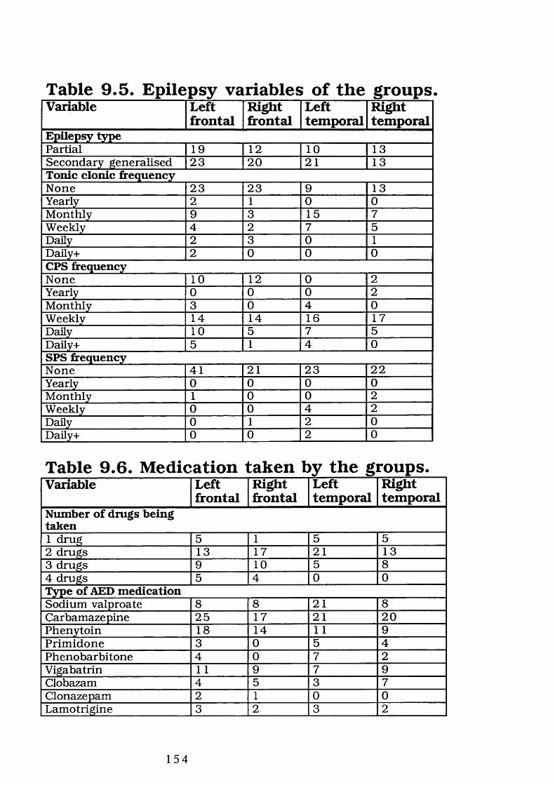
Table 9.5. Epilepsy variables of the groups.Variable Left
frontalRightfrontal
Lefttem poral
Righttem poral
Epilepsy typePartial 19 12 10 13Secondary generalised 23 20 21 13Tonic clonic frequencyNone 23 23 9 13Yearly 2 1 0 0Monthly 9 3 15 7Weekly 4 2 7 5Daily 2 3 0 1Daily+ 2 0 0 0CPS frequencyNone 10 12 0 2Yearly 0 0 0 2Monthly 3 0 4 0Weekly 14 14 16 17Daily 10 5 7 5Daily+ 5 1 4 0SPS frequencyNone 41 21 23 22Yearly 0 0 0 0Monthly 1 0 0 2Weekly 0 0 4 2Daily 0 1 2 0Daily+ 0 0 2 0
Table 9.6. Medication taken by the groups.Variable Left
frontalRightfrontal
Lefttem poral
Righttem poral
Number of drugs being taken1 drug 5 1 5 52 drugs 13 17 21 133 drugs 9 10 5 84 drugs 5 4 0 0Type of AED medicationSodium valproate 8 8 21 8Carbamazepine 25 17 21 20Phenytoin 18 14 11 9Prim idone 3 0 5 4Phenobarbitone 4 0 7 2Vigabatrin 1 1 9 7 9Clobazam 4 5 3 7Clonazepam 2 1 0 0Lamotrigine 3 2 3 2
15 4

9 .2 .4 . Areas o f discrete frontal dysfunction.
The frontal lobe groups were further sub-divided according to the results of high resolution MRI scanning (see table 9.7). There was no significant difference between the left and right frontal groups in the damaged area. The largest group was the dorsolateral area for both the left and right groups. A sizeable proportion of the left frontal group had no apparent underlying identifiable lesion on MRI scanning.
Further medical investigations were undertaken (eg video-EEG and seizure semiology; see appendix 1) and these, with the MRI results, were used to differentiate the whole group (see table 9.8). In this conceptualisation, the motor and premotor areas were collapsed into one U In the categorisation used, 'extensive' refers to theinvolvement of more than one of the regions outlined (but not one that extends outside the frontal regions). This is the classification that will be employed in this investigation. The borders of the regions used in this investigation are given in figure 9.1.
This grouping was based on the results of medical investigations undertaken by Drs Fish and Manford, and Professor Shorvon. For which I acknowledge their assistance. Further details on part of the subject pool can be found in Manford (1993).
155

Table 9.7. Areas of MRI identified frontallobe damage.Area Left frontal Right frontalExtensive 8 5Dorsolateral 17 13Mesial 2 1Orbitofrontal 2 5Motor 0 3Pre-m otor 0 1Unknown 14 1
Table 9.8. Areas of dysfunction according to EEG, MRI and seizure semiology.Area Left frontal Right frontalDorsolateral 17 13Mesial 7 4Orbitofrontal 5 5M otor/pre-m otor 5 5Extensive 8 5
156

Figure 9.1. Borders of frontal regions used in this investigation.
Prem otor/M otor area
Dorsolateral
Orbitofrontal
M otor/Prem otor
Mesial surface
Dorsolateral
Orbitofrontal
157

9.3. TestsThe assessm ent m easures chosen to form the battery, are briefly outlined below. They are grouped into three sections- general background tests, language and memory tests (which were thought to be most sensitive to temporal lobe dysfunction), and frontal lobe tests (which are further sub-divided into either motor tasks or those tapping executive skills).
9.3 .1 . Background T ests.
9.3.1.1. WAIS-R.
A number of reports have suggested that abbreviated versions of the WAIS/WAIS-R may offer an economical measure, in terms of time, without compromising validity. Given the num ber of subjects it was thought appropriate to use an abbreviated version in this study.There are several different short forms available (eg Britton and Savage, 1966; Crawford et al, 1992; CR Reynolds et al, 1983; Süverstein, 1982; Warrington et al, 1986), and American research has suggested that some 80% of clinicians use an abbreviated version (Holmes et al, 1965). Crawford et al (1992) compared the psychometric properties of these shortened forms and found tha t a version described by Warrington et al (1986) was the most valid of those reported upon. In the study presented here, this abbreviated version was used: a verbal IQ is pro-rated from four sub-tests (Arithmetic, Digit Span, Vocabulary, and Similarities); Performance IQ is pro-rated from three sub-tests (Block Design, Picture Completion and Picture Arrangement).
Warrington et al (1986), in a large consecutive series of 656 patients with unilateral cerebral lesions, assessed the validity of the WAIS (the abbreviated version outlined above) as a localising and lateralising instrum ent. Statistical analysis revealed that the WAIS’s ability at detecting precise localised deficits was minimal. Despite the lack of many consistent findings, the authors concluded:
158

"The WAIS provides standardised baseline information as to the individuals level o f functioning and identifies impairments requiring more detailed analysis. The WAIS is undoubtedly a useful screening test that provides a
fir s t approximation to the functional strengths and weaknesses in an individuals cognitive skills. ”
Warrington et al., 1986 p 238
Initially, patients with frontal lobe damage were found not to be deficient on IQ m easures compared to others [eg Hebb, 1945; Mettler, 1949]. Indeed, a common observation was a relative preservation of WAIS IQ, with damage caused by either tum our (eg Smith, 1966) or surgery for epilepsy (Milner, 1975). Such results, however, do not indicate that frontal lobe lesions have no affect on WAIS scores a t all: Milner’s patients showed an average loss of 7.2 IQ points after surgery. There is evidence to suggest tha t orbitofrontal damage produces little impairment, and thus damage to the dorsolateral regions is responsible for the deficits tha t do occur (Girgis, 1971). It has also been suggested that left frontal damage can produce larger IQ falls than right sided damage (Smith, 1966).
9.3.1.2. National Adult Reading Test (NART).
The assessm ent of optimal, or pre-morbid intellectual ability can be considered an essential part of any neuropsychological research with neurologically damaged subjects. The underlying cortical damage may extend to further regions and have a global impact on the intellectual function of the individual; thus the focal damage may not be the only cause of the cognitive deficit noted.
The National Adult Reading Test (NART; Nelson, 1982) is a m easure designed to assess pre-morbid ability. The test consists of 50 irregular words printed in order of increasing difficulty, to be read by the subject. Two studies reported by Nelson (1982) suggested that the NART has reasonable levels of both reliability and validity. Subsequent studies have also confirmed this (BeardsaU and Brayne, 1990; Crawford et al, 1991, 1992; Sharpe and O'CarroU, 1991).
159

9.3.2. Language and memory Tests.9.3.2.1. Graded Naming Test (ONT).
The GNT (McKenna and Warrington, 1983) consists of thirty black and white Une drawings which are presented in ascending order of difficulty. The subject's task is to name each of the drawings in turn. Results have suggested a high degree of reUabfiity and vaUdity. Furthermore, evidence has suggested that the GNT is able to discriminate normal controls from left hemisphere damaged subjects (McKenna and Warrington, 1983).
Q.3.2.2. De Renzi Token Test.
The test used was an abbreviated version of the original token test (De Renzi and Vignola, 1962). The patient is asked to perform operations with a set of twelve shapes (four circles, four squares, four triangles), coloured in red, blue, green or yeUow. The test necessitates memory for verbal sequences and comprehension of verbal commands of increasing complexity. Impairment on this m easure indicates disruptive Unguistic processes commonly associated with left (mainly posterior) temporal lobe dysfunction.
9.3 2.3. Recognition Memory Tests (RMTs).
The Recognition Memory Tests (RMTs) were developed by Warrington (1974) to assess adult global amnesic patients. Subsequently, Warrington (1984) presented data on the reUabfiity and vaUdity of the two tests. The two versions consist of a verbal version in which the subject is presented with words (Recognition Memory Test for Words; RMW), and a non-verbal version in which the subject is presented with photographs of m en's faces (Recognition Memory Test for Faces; RMF). In both versions, after presentation the subject is shown the same 50 stimuU along with distractor items. The subject's task is to detect the originaUy presented item from the distractor item.
Reports in the literature indicate the test is sensitive to unilateral cortical lesions and epileptic disturbance, particularly in the
160

temporal regions (LeFever, 1993; Warrington, 1984), although this is far from conclusive (Hermann et al, 1994).
9.3.2.4. List/Design Learning.
The learning of verbal and non-verbal information was assessed using the list and design learning paradigms drawn from the Adult Memory Information and Processing Battery [AMIPB; Coughlan and Hollows, 1985).
With regard to list learning, the procedure is as follows. A list of fifteen words is read to the subject at the rate of one every second, and the subject is then requested to repeat as many items, in any order, as possible. Subsequently, the list is read a further four times. After each presentation the subject has to attem pt to recall as many items as possible. These are then summed (List A1-5 score). Intrusions are also noted and summed across the five learning trials (List intrusion score). After the final presentation an interference list is read, and recall once again assessed (List B). Finally, the subject is required to recall the originally presented list (List AG).
In the non-verbal analogue a similar procedure is adopted. The subject is shown a 9-line drawing for a ten second period, after which it has to be drawn from memory. Five presentations are given and both the num ber correct (Design A1-5 score) and the intrusions (Design intrusion score) are summed. After the fifth presentation the interference drawing is shown, and the num ber of correct fines recorded (Design B). Finally, recall of the originally presented items is assessed (Design AG).
Given the two forms of presentation, it is thought possible to differentiate between left and right hemisphere lesions. Studies have indicated that the performance on tests of fist/design learning, is mediated by the hippocampus where as the recognition memory test may also involve neocortical structures (Rausch, 1991).
9 3.2.5. Story Recall
Story recall is designed to assess immediate registration of verbal information and retention over time. This version drawn from the AMIPB (Coughlan and Hollows, 1985) assesses immediate recall.
161

delayed recall (after 30 minutes) and consequently, percentage retained. The story consists of 28 /30 items of information that have to be retained. Neuropsychological studies have indicated that damage to both the cortical temporal, and hippocampal regions can impair performance (Rausch, 1991).
9.3 .3 . Frontal Lobe Tests.
The assessm ent of the functions associated with the frontal lobes can be broadly divided into two- those tapping executive skills and those tapping motor skills. Obviously, there may be an overlap between these in certain tests, and a clear-cut distinction is impossible. However, such a division is useful, in as much as simple cortical divisions can subsequently be made.
9.3.3.1. Measures of executive skills.
9.3.3.1.1. Modified Wisconsin Card Sorting Test (MWCST).
The original WCST was devised by Grant and Berg (1948). The test was devised in order to assess abstraction and problem solving abilities. The test does so by providing information on three different aspects of performance. Firstly, the num ber of categories achieved is recorded. Secondly, the num ber of errors made is noted, with emphasis on their nature; perseverative errors occur when the subject fails to change from one set of responses to another, following a cue to do so. Errors other than the perseverative kind are also recorded. These tend to be failure to m aintain the correct response set. Thirdly, the time taken to complete the task can be recorded and used as a measure of general efficiency on the task.
The modified version of the WCST (MWCST; Nelson, 1976) was used in this investigation. This modification aims to reduce some of the ambiguities in the examiners response, thereby simplifying the task for the patient, and clarifying the nature of the errors for the examiner. In particular. Nelson advocates informing the subject when a category change is due.
162

The MWCST uses a 48 card pack (two sets of 24) and four stim ulus cards are shown; one red triangle, two green stars, three yellow crosses and four blue circles. The subject is required to sort the stack of response cards. The set contains cards which are unambiguous, in that it can share only one attribute with three of the stim ulus cards (colour, shape or number). The subject is requested to sort the cards according to a particular category. Whichever category the subject choses first is deemed correct. Once six consecutive correct responses have been made, the subject is told the rule has changed. Provided that the next sorting category chosen is one of the remaining two, then that is accepted by the examiner as the correct rule. The final category is the one remaining. After the attainm ent of three categories they are then repeated again in the original order.
In addition to the procedural changes. Nelson modified the scoring procedure. Besides the num ber of categories achieved. Nelson suggested scoring the total num ber of errors, while considering perseverative errors to be only those of the same category as the immediately preceding response. A third score gives the percentage of errors that are perseverative (perseverative errors/to tal errors* 100). Nelson suggests those with a greater than 50% perseverative error score should be classified as "typically frontal".
The MWCST is thought to assess flexibility, and use of feedback in test performance. Numerous studies have attested to the ability of this task to reveal frontal lobe damage, and it is thought to be affected by left rather than right frontal damage (Milner, 1963; Mountain and Snow, 1993), and by dorsolateral rather than orbital dysfunction (Milner, 1963). However, the test is not without its critics and the possibility that damage to other cortical areas can result in impairment has been mooted (Corcoran and Upton, 1993; Hermann et al, 1987; Robinson et al, 1980; Shoqeirat et al, 1990; van den Broek et al, 1993).
9.3.3.1.2. Twenty Questions.
In comparison to the WCST, this test is a relatively new one in the frontal literature. However, the task on which it is based is
163

extremely old- the parlour game Twenty Questions (Mosher and Hornsby, 1966).
The test requires the subject to guess an animal the examiner has in mind. The examiner can only respond "Yes" or "No" and the subject has twenty questions in order to determine the answer. Particular em phasis is placed upon the type of questions asked and not ju s t the success or failure of the subject. All questions asked by the subject are recorded and classified into one of three types:
(i). C onstraint: Questions of this type are the most effective search question. These types of question narrow the field by as much as half, by eliminating a series of different types of animal. For example- "Does it have four legs ?", "Does it live in water ?".
(ii). P seudoconstra in t Questions of this variety are a less effective search strategy, although they appear to be similar to the constraint- type questions previously described. Thus, whilst appearing to be constraining they only apply to one particular type of animal. For example, "Does it have a trunk ?" or "Does it bark ?".
(fii). H ypothesis scanning: Again, this is a less effective search strategy and basically involves guessing with no previous basis for such a guess. Although a certain am ount of guesses are to be expected, these should occur after the subject has narrowed the search. Thus, hypothesis scanning questions tend to be asked at the end of the procedure and include such items as: "Is it a dog ?", or "Is it an elephant ?".
Apart from the num ber of different types of question asked, two other types of information were recorded. Firstly, the num ber of questions needed in order to arrive at the correct response [20 being the maximum); the total num ber of questions required over two trials was recorded. Secondly, the num ber of questions required until the first guess. This latter variable can be considered a m easure of impulsivity and hence may be deficient in those with frontal lobe dysfunction. For this variable, the mean score over two trials was recorded.
164

There are two means of presentation of the test- open or closed. In the closed version, the subject is given a finite num ber of options from which they have to choose. In the open ended, the subject has to construct the alternatives to be searched for, as well as finding the right one amongst them. In this study, the latter version was used. The instructions given for the test were as follows:
"I am thinking o f an animal and your task is to try to fin d out which one it is I have in mind. To do this you can ask any questions a t all that I can answer 'Yes" or "No"; I cannot give any other answer. You have up to twenty questions to fin d out the animal I am thinking o f but try and fin d out in as fe w questions as possible."
The response given by the subject was recorded verbatim and subsequently categorised into the three "strategy" types.
Mosher and Hornsby (1966), in their work with young children suggested the game taps the cognitive structure of players, indicating the way they represent the cognitive domain involved.The authors proposed that both what players ask, and how they use the answers, depends on how they organise knowledge of the world. The question presupposes an ability to equate things in sub ordinate categories and then to arrange the categories in hierarchies. The efficiency of constraint seeking strategy comes at the expense of considerable cognitive work, the work involved in forming a strategic plan, and in building the necessary conceptual structures.
Klouda and Cooper (1990), in an investigation with only five subjects with a variety of different frontal lobe pathologies, found their subjects to be impaired with respect to the more efficacious strategies. The frontal lobe subjects not only used more specific questions than the controls, but they also asked them earlier. That is, after establishing a prior constraint, frontal lobe patients were less likely th a t controls to narrow the remaining possibilities with further constraints, and more likely to leap immediately to inappropriate specific hypothesis.
There are other reports that have also alluded to poor performance in patients with frontal lobe pathology, for example in patients with
165

motor neurone disease (Neary et al, 1990; Neary and Snowden, 1991).
9.3.3.1.3. Verijal Fluency.
Verbal Fluency has long been used as a measure of cerebral pathology, and has been consistently shown to be depressed in the presence of any cortical lesion (eg Borkowski et al, 1967; MiceU et al, 1981; Pendelton et al, 1982). Some studies have suggested tha t frontal, particularly left frontal lesions, result in greater impairment (Benton, 1968; Crowe, 1992; Milner, 1964; Perret, 1974).
In the current study, subjects were first requested to say as many words in sixty seconds beginning with a particular letter ("S"), and immediately after completion were requested to say as many animals (beginning with any letter), as they could in a further 60 seconds. Generally patients with frontal lesions are impaired on the former.
9.3.3.1.4. Stroop Test.
The Stroop Test has been a widely used test in cognitive psychology since its development (Golden, 1975; Stroop, 1935) and has been modified on many occasions. In this investigation, the version designed by Dodrill (1978) was used. Subjects were requested firstly to read a card containing 88 words (printed in different ink colours) as quickly as possible. Subsequently, subjects were asked to read the card once again, but this time to ignore the word and read the print colour. Time and errors were recorded for both conditions and an interference time calculated (time to read print colour m inus time to read words; Corcoran and Upton, 1993; Dodrill, 1978).
Previous research evidence has suggested that performance can be compromised by both left hemisphere and frontal lobe damage (Macleod, 1991; Perret, 1974). Consequently, maximal impairment is usually reported in subjects with left frontal lesions (Perret,1974).
16 6

9.3.3.1.5. GoUin Incomplete Figures.
A series of ten sets of incomplete pictures was used to assess perceptual function (Gollin, 1960). The test consists of a ten picture series of five line drawings of familiar objects (eg duck, dog and umbrella], ranging in completeness from a barely suggestive sketch to a complete drawing of the figure. The score is the sum of all the set num bers a t which each picture was correctly identified. The test is able to discriminate between left and right hemisphere lesions, with the greatest impairment being with right posterior lesions (Warrington and Jam es, 1967; Warrington and Taylor, 1973).
However, damage to the frontal lobes has also been implicated in the ability to combine into a meaningful whole, all of the elements in a complex visual scene (LansdeU, 1968; Meier and French, 1965; Milner, 1954; Miller and Milner, 1985; Milner, 1964; Zeigarnik, 1968 [quoted in Luria, 1966, 1973]). In this investigation this test was employed in order to examine the ability of those with frontal lobe lesions to make sense of limited information and to synthesize fragmented non-verbal information holisticaUy.
9.3.3.1.6. "DUFFER".
A test designed to be a verbal analogue of the GoUin figures was devised for this study. The details of the development of this test are given in section 9.4. In this test, subjects were read a series of statem ents one after each other and asked to guess the item of which the examiner was thinking (see appendix 2). Each statem ent revealed more information and the final statem ent should have been sufficient for the item to be discovered. The score was the total num ber of questions required to obtain the correct response.
This test is similar to tha t devised by Miller and Milner (1985), and consistent with the literature tha t patients with left hemisphere lesions have difficulty in holding and manipulating verbally expressed ideas (Meyer and Yates, 1955; Miller, 1985; Milner,1954, 1967).
167

9.3.3.1.7. Trail making.
This test, originally taken from the Army Individual Test Battery (1944), is an easily administered test of visual conceptual and visuomotor tracking. It is given in two parts. The patients m ust first draw lines to connect consecutively num bered circles (Part A). Subsequently, the patients m ust connect the same num ber of consecutively numbered and lettered circles on another sheet, by alternating between the two sequences (Part B). Time taken to complete each part, and the num ber of errors made on each part, is recorded. In this study a further measure- "Frontal Lobe Time" was calculated by subtracting the time taken on Part A from the time taken on Part B. In this way, it was hoped to reduce the motor component of the task to a minimum. Obviously this task requires flexibility of thought and action in order to alternate between num bers and letters. Two variables from this m easure were used; "frontal lobe time", and number of errors made on Part B of the task- thought most sensitive to frontal lobe pathology (Reitan,1958).
9.3.3.1.8. Cognitive Estimates.
ShaUice and Evans (1978) presented a task which required the patient to make estimates such as "What is the length of an average m an's spine ?" or "How fast do race horses gallop ?". These questions are not able to be answered by simply consulting information in semantic memory. For some questions, a reasonable estimate can be simply obtained, if an appropriate strategy is selected. For instance, one could answer the spine question by calculating [body-(head+legs)j or by imagining the length of a shirt. The difficulty lies in thinking of a means of using everyday information, rather than in the mental operations required when the Hne of attack has been developed. Other questions stress the ability to assess whether a guess is reasonable. Thus, for the galloping horse question a response of 8 mph would be unreasonable; it is only twice the rate of a person's rapid walk. Similarly, 60mph would be unreasonable; it is as fast as a car travels. ShaUice and Evans (1978) revealed that patients with anterior lesions performed most poorly on this task.
168

The subject is given ten statem ents and their responses are graded on a four point scale (0-4) depending on the accuracy, or more properly, the bizarreness of the response (as described by Shoqeirat et al, 1990). A high score is indicative of more erroneous responses. Parallel versions of this test were established (the details of the development of these are given in section 9.4) for use with postoperative subjects (see appendix 3).
9.3.3.1.9. Cost Estimation.
A similar type of task to the one previously described was also employed. The task, one of price estimation, was first described by Smith and Milner (1984). In the original task the subject was required to estimate the price of a given item presented as a toy, such as a trumpet, a cup and a car- although some objects were rather bizarre (eg a turtle !). Estimating the average price of a particular item (for example a typewriter), requires the development of a strategy that may involve deciding what is a typical typewriter, judging the possible prices and selecting a representative price for a machine of average quality. If patients with frontal lobe lesions faü to develop or use such strategies as systematically as other subjects do, this could result in more errors of price estimation.
A series of steps for each subject was followed in this investigation. Initially, the subject was presented with a line drawing (cf Smith and Milner study in which toys were used), and asked to name it (to establish that the picture was recognisable and that the subject had no naming deficit- if the subject failed to respond then they were informed of the item). Secondly, the subject was requested to estimate the average price of that item, and this was scored for bizarreness of response. This procedure was carried out for all of the twelve items.
Once this part of the task had been completed, the subject was asked to recall as many items as possible, and the total num ber of correctly recalled items, and intrusions noted. Finally, the subject was shown all the items once again, but on this occasion in a different, random order, and asked whether the item had been seen
169

at the start (ie first four items), middle (second four items], or end (ie last four items). In this way the test provided several pertinent variables- a measure of estimation, a measure of recall memory and a measure of recency judgement.
Why use this test when the battery already included an estimation task ? In one way the impairment in price estimation observed after frontal lesions complements the study of ShaUice and Evans. However, Smith and MUner (1984) found that impairment was maximal after right frontal lobectomy. In contrast, ShaUice and Evans found an impairment after both right and left frontal damage. Thus Smith and MUner (1984) argue for a specific role of the right frontal cortex in the performance of this type of estimation task.
DetaUs of the development of this task are given in section 9.4.
9.3.3.1.10. Porteus Maze Test.
Probably the first attem pt to assess planning and problem solving quantatively in patients with frontal lobe lesions was through the use of maze learning. Porteus maze tracing was designed to yield data about the highest levels of mental functioning involving planning and foresight- a skUl thought to be located in the frontal lobes (Porteus, 1965; Porteus and Kepner, 1944). Subsequent studies have indicated that performance on maze tests may be more impaired foUowing right frontal damage (Corkin, 1965; MUner, 1964).
The test involves the subject tracing a pencU fine through a maze, and the time taken being recorded. Number of pencU Ufts and wrong directions are also noted.
9.3.3.1.11. Visual search.
Patients were shown an A4 sheet on which were printed twenty separate numbers; a simpUfied version of that presented by Teuber et al (1949). Subjects were requested to point to ten individual numbers, as they were caUed out as quickly as possible. After completion of this task subjects were asked to count the num ber of num bers on the sheet. On both tasks the time taken to complete was recorded. If subjects pointed to an incorrect number, or
170

counted incorrectly they were informed of this and requested to continue until the correct response had been achieved.
Luria et al (1966) and Karpov et al (1968) documented a patient with a frontal lobe lesion who was impaired on a task of visual searching. Subsequently, others have also testified to the deficit (see section 6.5). The impairment is thought to be a consequence of a deficit in simple strategy formation (McCarthy and Warrington, 1990).
9.3.S .2. Measures o f m otor skills.
9.3.3.2.1. Tapping Task.
A tapping task was used to measure the speed of simple m anual movements (Wyke, 1969). Two 10cm square plates were mounted 7.5cm apart on a rectangular wooden base. Contact of a metal stylus with the metal plates closed an electric circuit and activated a mechanical counter, which recorded the num ber of times contact was made. Subjects were told to tap the two metal plates alternately with the stylus as quickly as possible. Two conditions were investigated: the speed of tapping with firstly, the dominant hand; and subsequently, with the non-dominant hand. In each condition a practise trial was followed by three 20 second trials, with a suitable rest period between them. The mean of these three trials was recorded.
9.3.3.2.2. Bimanual Hand Movements.
In this test, originally described by Luria (1966) the subject is requested to place both hands on the desk, making a fist with one hand and placing the other hand palm down. The subject is then requested to alternate "fist" and "fiat" positions simultaneously. Patients with frontal lesions, particularly of the supplementary motor area, may have specific difficulty in the sim ultaneous coordination of these movements, tending to alternate them or to perform the same action with both hands (Laplane et al, 1977a, b). In this version of the test, subjects were given twenty seconds to perform as many alterations as possible. Both num ber of correctly
171

performed alterations and num ber of alternation errors were recorded.
9.3.3.2 3. Motor Sequences.
Cana van et al (1989) presented results of sequencing ability in a variety of patient groups. Most errors on a test of motor sequence were recorded in the frontal lobe dysfunction group (particularly the left), although a right temporal lobectomy group also performed poorly.
This test was used to examine the sequencing ability of the subjects. In particular, with the more specific localisation available in this study, is was hoped to better document the cortical area within the frontal lobe that results in impairment on this task. In line with previous evidence outlined elsewhere, subjects with m otor/premotor damage were expected to perform most poorly.
The task is simple; the subject is requested to reproduce 10 sequences of three gestures. Each three item sequence was presented individually at a rate of one gesture every second and the num ber of correct sequences reproduced was recorded. The sequences used are shown in appendix 5, as are the parallel versions derived for the post-operative subjects (see section 9.4 for details on the development of these).
9.3.3 2.4. Gesture Span.
Gesture span was assessed by requesting the subject to copy an increasing span of gestures (up to a maximum of six). The first sequence consisted of two gestures (if this was failed one gesture was attempted). Presentation rate was one gesture every second.This procedure was repeated twice and the mean score obtained on two trials recorded. Cana van et al (1989) did not record any significant statistical differences between his groups, although the frontal group did reproduce fewer gestures than the other group (the lack of significance may of been a consequence of small sample size). The gestures used in this procedure are given in appendix 6 (see section 9.4 for details on the development of the parallel versions).
172

9.3.3.2 5. Temporal Order for Gestures.
Frontal lobe lesions, particularly of the dorsolateral region have been reported to disrupt temporal order for events (eg Milner et al,1985). Despite this evidence, little work has been completed looking at the temporal order for gestures- although the importance of the frontal lobes to gesture production is weU known.
In this task, the subject was shown four gestures at the rate of one every second. Subsequently (after minimal delay), the subject was shown, in random order, the gestures individually and asked to say at which order the presented gesture was shown. Total num ber of correct responses over two trials were recorded. The gestures used and the parallel versions derived (see section 9.4 for details on the development of these) are presented in appendix 7.
9.4. Test construction.There are three aspects of test construction tha t wiU be discussed here. Firstly, the collection of data from normal subjects on parallel versions of some tests, in order to evaluate the reliability of these new versions. Secondly, the construction of a novel test, the DUFFER, which was hoped to provide a verbal analogue of the Gollin figures. Finally, the collection of normative data for four versions of the revised cost estimation task.
9 .4 .1 . Normal control subjects.
In order to complete the above aims, normal control subjects were contacted, from one of three sources:
1. Non-medical and non-academic personnel from the hospitals where the studies were undertaken.
2. A large industrial unit located in the West Midlands.
3. A sample of undergraduates undertaking nonpsychology based courses.
173

The inclusion criteria for the normal subject sample was as follows:
1. Aged between 16 years and 65 years of age.
2. No history of any neurological or psychiatricdisturbance.
3. No history of drug or alcohol abuse.
All subjects (basic demographic details are given in table 9.9) were volunteers; none received any financial inducement for completion and no person approached refused to participate. However, four subjects either completed the questionnaires incorrectly or omitted some parts of the protocol and these were therefore excluded from the analysis. This resulted in the total subject pool comprising 86 subjects; 54 from the undergraduate population, 10 from the hospital non-academic personnel and the rem ainder (n=22) from the industrial unit.
Analysis revealed that the normal control group were comparable to the patient group in terms of age (t=0.67, df=158; p>0.2] and sex (^2=4 3 5 df=4; p>0.1]. However, there was a significant difference in term s of educational qualifications (x^=81.89, df=16; p<0.001] in as m uch as the patient sample were less qualified than the normal control group. Obviously, this should be considered when comparing the results especially with regard the cost estimation task where control performance was used to calculate the error score.
174

Table 9.9. Basic characteristics of normal subject sample.
Variable nSexMale 36Female 50AgeMean 25.98SD 11.22Range 18-59Educational qualificationsDegree 0A-Levels 44GCSE/O-Levels 10CSE 14None 18
175

All subjects completed the following tasks at the same testing session in a random order:
DUFFER versions.
Cognitive estimates task versions.
Cost estimation task versions.
Motor sequences versions.
Temporal order versions.
Versions of the individual tests can be found in appendices 2, 3, 4,5 , 7 . Each subject completed the original version (version 1) and two of the newly developed versions (versions 2, 3, 4) of each test. Thirty completed version 2 of each task (and the original), 29 completed version 3 of each task along with the original, and 27 completed version 4 of each task and the original.
For both the cognitive estimation task, and the DUFFER task, twelve items were used. Subsequently, internal consistency checks were completed by applying Cronbach’s alpha to the individual tests, in order to obtain the best ten items in term s of internal consistency. For the cost estimation task 14 items were initially used, which was subsequently reduced to twelve.
9.4.2. Cognitive estim ates.
The mean scores of the normal sample on the four versions can be found in table 9.10. As can be seen the scores obtained on all versions are comparable and this was confirmed when Spearm an's rho correlations were undertaken (table 9.11).
176

Table 9.10. Scores obtained on fourversions of cognitive estim ates.
Version 1 Version 2 Version 3 Version 4Mean 3.09 3.18 3.41 3.78SD 1.27 1.77 1.17 1.26Minimum 0.00 0.00 0.00 0.00Maximum 5.00 5.00 6.00 6.00
Table 9.11. Correlations between four versions of cognitive estim ates.
Version 1 Version 2 Version 3 Version 4Version 1Version 2 0.667 -----
Version 3 0.518 0.508Version 4 0.690 0.684 0.550
Table 9.12. Scores obtained on four versions of DUFFER.
Version 1 Version 2 Version 3 Version 4Mean 28.20 27.17 29.29 31.35SD 6.26 5.83 5.37 5.94Minimum 0 0 0 0Maximum 34.00 35.00 34.00 36.00
Table 9.13. Correlations between four versions of DUFFER.
Version 1 Version 2 Version 3 Version 4Version 1Version 2 0.527Version 3 0.595 0.649Version 4 0.549 0.647 0.609
177

9.4 .3 . DUFFER.
The mean scores, standard deviations, and ranges for the four versions are presented in table 9.12. Again, the four versions appear reliable, given the Spearman's rho correlation obtained (table 9.13).
9 .4 .4 . Cost estim ation.
From the data collected from the normal subjects, the mean and standard deviation were computed for each individual item. From these an error score was derived; if the subject's estimation was more than ±2 SD points away from the mean then this was recorded as one error. The error scores were summed to provide a total error score, with a maximum of 12 (see appendix 4).
From table 9.14 it is evident that few subjects made any errors- given the low mean score. This is to be expected, obviously, as they form the data set from which the error scores are computed. The first aim was to attest to the reliability of the alternate forms and this is certainly confirmed given the correlations in table 9.15. The second aim was to compute normal scores along with their standard deviations so the task could be used with a patient population.
9 .4 .5 . Temporal order.
The temporal order task consists of two presented stimuli from which the total correct responses are summed to give a total score. The versions of the task can be found in appendix 7. Each version is scored, with a maximum score of eight possible. The mean scores are presented in table 9.16 and Spearman's rho correlations given in table 9.17. The latter scores indicate a good association between the four versions.
9 .4 .6 . Motor sequences.
A total of ten correct responses is possible and the mean scores for all four versions, along with standard deviation and range is given in table 9.18. Spearman's rho correlations are given in table 9.19, and these indicate a good level of agreement between the four versions.
178

Table 9.14. Scores obtained on fourversions of cost estimation.
Version 1 Version 2 Version 3 Version 4Mean 1.01 0.84 0.89 0.97SD 1.15 1.09 1.22 1.21Minimum 0.00 0.00 0.00 0.00Maximum 6.00 6.00 6.00 6.00
Table 9.15. Correlations between four versions of cost estimation.
Version 1 Version 2 Version 3 Version 4Version 1Version 2 0.681Version 3 0.601 0.504Version 4 0.610 0.571 0.637
Table 9.16. Scores obtained on four versions of temporal order.
Version 1 Version 2 Version 3 Version 4Mean 5.89 5.07 4.63 4.93SD 1.31 1.28 1.42 1.38Minimum 3 3 3 3Maximum 7 7 7 7
Table 9.17. Correlations between four versions of temporal order.
Version 1 Version 2 Version 3 Version 4Version 1Version 2 0.554Version 3 0.538 0.553Version 4 0.652 0.576 0.550
179

Table 9.18. Scores obtained on fourversions of motor sequences.
Version 1 Version 2 Version 3 Version 4Mean 7.68 8.10 6.32 7.26SD 1.60 1.10 0.86 1.14Minimum 6 5 6 6Maximum 10 9 9 9
Table 9.19. Correlations between four versions of motor sequences.
Version 1 Version 2 Version 3 Version 4Version 1Version 2 0.680Version 3 0.646 0.601Version 4 0.597 0.551 0.529
180

9.4 .7 . Siim m aiy.
From the results of the above Investigations, it is evident that in the normal groups assessed, the four versions of the tasks considered were of acceptable reliability (all Spearm ans rho p<0.01). These parallel versions were used in the assessm ent of those subjects who underwent surgery for the relief of their epilepsy.
In addition, the normal data supplied for the cost estimation task was used to assess and score the patient groups performance on this measure- given the procedure outlined above.
Other m easures employed in the battery also had reliable alternate versions (for example the recognition memory tests, list learning and story recall). Other tests did not (eg WAIS-R, MWCST, GNT), but this was considered acceptable given the nature of these tasks.
9.5. Procedure.All subjects completed the test battery over a two to three hour period, most commonly divided into two sessions in order to overcome possible fatigue and boredom of the subject. The tests forming the battery produced a num ber of indices of performance and these are Hsted in appendix 8.
Those subjects who underwent surgery for the relief of their epilepsy were seen at 3 months, 6 months, 1 year and yearly thereafter. All tests had parallel versions (where necessary). Those tests that had no previously constructed parallel versions were completed by a group of control subjects in order to ensure reliability (as previously outlined).
181

9.6. Statistical Analysis.Data analysis was conducted using the Statistical Package for the Social Sciences PC version (Vers 4- Norussis, 1992). The data was analysed in a variety of ways, outlined more thoroughly in the appropriate results section. Given the num ber of subjects and the num ber of comparisons that are to be made, then a significance level of 0.01 was adopted in most instances to avoid the possibility of a Type 1 error.
Further details on the multivariate procedures used in this study are given below.
9 .6 .1 . Interpretation o f mulHfactorial ANOVAs.
A num ber of methods have been suggested to aid interpretation of significant multifactorial ANOVAs (see for example Heiman, 1995; Howell, 1992; Keppel, 1991; Snodgrass, 1977; Winer et al, 1991). However, although there are num erous methods, as Keppel (1991) suggests when discussing such approaches:
"There is little agreement among methodologists concerning specific courses o f action to be followed."
Keppel, 1991, p. 182.
Prior to outlining the statistical methods that wül be employed in this study, it should be noted that many researchers are content with plotting the data and examination of the resultant graph. This is highlighted by Howell (1992) who states:
"Plotting the data and considering what they mean is an important, i f not the most important, part o f an appropriate analysis o f any set o f datcu "
Howell, 1992, p. 395
In a similar, although characteristically drier fashion, Winer et al(1991) state:
182

"geometric representation o f the profiles corresponding to the means is generally o f considerable value. "
Winer et al, 1991, p. 429
It is therefore im portant that ANOVA graphs are presented for all significant results: a method adopted in this report. However, a num ber of statistical comparisons can be undertaken in order to uncover the meaning of a significant ANOVA. The first distinction tha t should be made is between planned comparisons and post-hoc comparisons. The statistical difference between planned and post- hoc comparisons is that in the latter specific adjustm ents for Type 1 error are made, whereas in the former they are not. There are a num ber of different forms of post-hoc multiple comparison methods (eg Scheffé, Tukey's, Newman-Keuls, D uncan’s) with the major difference being in the severity of the adjustm ents they make for controlling Type 1 error. Similarly, there are a num ber of methods that can either be described as "planned comparisons" or a mixture of both planned and post-hoc methods. Although, an extensive review of aU of these is unwarranted here [see Keppel, 1991 and Winer et al, 1991 for comprehensive reviews), several commonly employed methods will be outlined.
1. Multiple post-hoc comparison methods can be performed directly comparing m eans from individual cells within a row or column (ie pairwise comparisons). Cells that are diagonally positioned are not usually examined (a confounded comparison would result)- hence, only unconfounded comparisons are performed. This is obviously a method tha t employs some of the multiple post-hoc comparison tests previously outlined.
2. Post-hoc multiple comparison techniques (Tukey'Sa) can be performed dealing with both confounded and unconfounded means. However, it has been argued that this says very little about the interaction (Heiman, 1995) and thus is a method that should be avoided.
3. All the cells could be treated individually and a one-way ANOVA performed, with subsequent post-hoc multiple comparison tests. This procedure is obviously akin to the comparison of both
183

unconfounded and confounded means and, hence, the results say little about an interaction and consequently has been deemed of little worth (Heiman, 1995; Howell, 1992).
4. In the case of a significant main effect the scores could be collapsed for one factor and a further one-way ANOVA [with subsequent post-hoc multiple comparison tests) performed. Obviously this is only necessary when there are more than two levels within a factor, for the F-ratio for a main effect with 2 levels tells us if there is a significant difference between the levels within a factor.
5. In the case of an interaction effect a num ber of methods can be adopted. Most of the methods rely on the decomposition of the analysis into smaller components which assist in interpretation. The most appropriate of these methods, it is suggested, is a simple main effect analysis (Heiman, 1995; Howell, 1992; Snodgrass, 1977). Although some have argued this be reserved for planned com parisons (eg Keppel, 1991; Snodgrass, 1977), some consider this position unwarranted (eg Howell, 1992). However, it is clear tha t the investigation of simple effects can aid in interpretation of an interaction effect.
6. In addition to the simple main effect analysis, comparisons between marginal means can be performed. This type of analysis evaluates the differences between the over all m eans corresponding to different levels of one variable, when collapsed over different levels of the other.
7. Interaction comparisons are another forms of analysis that can aid interpretation. These are created whenever particular levels of an independent variable are singled out for analysis. There are two forms of interaction comparisons: analysis of partial interactions (only useful in at least a 3x3 design, and hence will not be considered here), and interaction contrasts. The latter are:
"the most common type o f interaction comparisons in thebehavioural sciences."
Keppel, 1991, p.255.
184

In essence the levels within a factor are reduced, either by "disregarding" one or more (known as pairwise interaction contrasts) or by the averaging of two or more levels (known as complex interaction contrasts; Keppel, 1991).
In this investigation several steps were taken in order to aid interpretation of the results. Primarily, pairwise post-hoc comparisons were undertaken; Duncan's Multiple Range Test (with a significance level set a t p<0.01 for hypothesis 2, and p<0.05 for all other investigations).
Although some have argued that the analysis of simple effects be reserved for planned comparisons, others have argued that this form of analysis can be an important technique for analysing data that contain significant interactions, and that:
"In a very real sense, it allows us to 'tease apart' interactions"
Howell, 1992, p. 392
Hence, this procedure was also performed in order to facilitate interpretation of some of the results obtained. Another general approach is the analysis of interaction comparisons and the analysis of marginal means (Keppel, 1991). Again, these are approaches tha t are commonly thought of in terms of planned comparisons, yet they can greatly assist in the interpretation of any significant results derived from a multifactorial ANOVA and are other methods adopted for this study. Obviously, the choice of analysis wUl vary dependent upon the hypothesis and the data under discussion. Some forms of analysis will, obviously, provide a more valid approach than others in the assistance they offer in the interpretation of the results (all analysis conducted in order to aid interpretation is presented in appendix 9).
However, it should be noted that the possibility of a familywise type 1 error exists with aU these planned comparisons and, although appropriate correction for this possibility do exist, some have argued strongly against such procedures (Keppel, 1991). In this study these forms of analyses were used judiciously and appropriate caveats drawn when making inferences. Furthermore, in this study
185

the analysis was undertaken in order to assist in the interpretation of the ANOVAs performed. The post hoc multiple comparison techniques, that implicitly control for Type I error, are also included in order to guard against this possibility.
However, it should be noted that the statistical procedures were employed in order to aid in interpretation- the psychological interpretation is, however, obviously the most important. As Howell(1992) eloquently suggests:
"Statistical elegance does not necessarily imply scientifically meaningful inferences. Common sense is as least as important as statistical manipulation."
Howell, 1992, p.424
ANOVA summary tables are presented for aU analyses where significant effects (either a main effect for the primary variable under discussion, or an interaction effect involving this variable] were revealed. Although, some have argued tha t the main effects should be ignored if a significant interaction effect is presented (eg Heiman, 1995), not aU are in agreement with this perspective (Howell, 1992; Keppel, 1991). Hence, in this investigation aU significant results are presented. However, it should be noted that interpretation of the main effect should be guarded when a significant interaction is present. AU analysis performed in order to assist in interpretation of the ANOVAs is presented in appendix 9.
If a significant result is revealed, then the ANOVA summary table is included in the appropriate section. If the resultant ANOVA is nonsignificant then the summary table is presented in appendix 10.
9.6 .2 . Logistic Regression.
Logistic analysis was adopted in order to develop a composite measure of frontal dysfunction, that would aUow discrimination between the frontal and temporal lobe groups. Logistic regression is a technique that aUows a constant probability, P, to be determined from a regression equation. It differs from multiple regression in that P is constrained to fie between the values of 0 and 1.
186

Basically, one works with a transformed dependent variable, running a multiple regression on the transformed variable:
Y =ln(P /l-P ) 1
Obtaining a regression equation like:
Y=ln(P/l-P)=a+bis+b2T+b3U 2
Where P is interpreted as the probability. Given the form of this equation, P is constrained to He between 0 and 1, and so has the required properties of a probabiHty. If P is the probabüity of an event then then P/(l-P) is the odds of that event. Hence:
P/(1-P)=ey 3
The In of the odds can be converted back to a probabiHty giving a direct relationship between the area of cerebral dysfunction and the explanatory variables:
P = e y /( l+ e y ) 4
where:
Y=a+bis+b2T+b3U 5
is the predicted value for Y for a given set of values for the explanatory variables.
Thus if the value Y, derived from formula 5 is substituted into equation 4, the value P is found (not to be confused with p- the significance level).
In this study the discriminate variable was the focus of the seizure origin- either frontal (0) or temporal (1). Variables were entered into the logistic regression analysis in order to obtain a regression equation, which provided a probabiHty score ranging from 0 to 1. The nearer to 1 the greater the probabiHty that the subject has a temporal lobe foci. Conversely, the nearer to 0, the greater the probabiHty the subject has a frontal epileptic focus.
187

However, the composite score may provide less information, and be less discriminating, than the results from one individual test.Hence, appropriate comparisons will be undertaken to exclude this possibility. If this is the case then obviously, from a clinical point of view the composite score will be redundant.
9 .6 .3 . D iscrim inant analysis.
Logistic regression deals with binary outcomes- in this study either frontal or temporal lobe cases. In certain situations, however, th is is not enough and the division of the sample into more than one group is necessary. In this investigation, there were four groups- left frontal, left temporal, right frontal and right temporal. Thus a rule was required for discriminating between these groups, so that, for any new individual known to come from one of the groups (the particular group being unknown), the rule could be applied and the individual assigned to the most appropriate group. A technique that is theoretically similar to logistic regression is discriminant analysis (Armitage and Berry, 1994). The one difference is tha t rather than simply dealing with dichotomous variables, it was possible to discrim inate between several.
The purpose of discriminant analysis is to enable individual units to be assigned to one or other of the populations with, in some defined sense, the greatest probability of being correct (or smallest risk of error). This technique of analysis involves the specification of a discrim inant function, in which the relevant variables are assigned a weight, or coefficient. If the discriminant function is linear in form it will "look like" a multiple regression equation. For a particular individual or case, values of the variables are substituted in the discrim inant equation and a value for the function calculated. On the basis of this value the individual is assigned to a particular population.
In this analysis it was possible to assign an individual to one of the four localisation groups outlined by the discriminant functions
188

derived. Furthermore, it was possible to extend this and apply the procedure to different cortical regions within the frontal regions.
1 8 9

9.7. Presentation of results.The analysis presented in the following sections deals with each of the presented hypotheses in turn. The statistical analysis undertaken for each, is outlined in more detail in the appropriate section. Most of the investigations involved parametric measures. The data that failed to fit the criteria for parametric analysis (ie interval or ratio data, normal or near normal distribution and homogeneity of variance), was subjected to appropriate non- parametric analysis.
A large num ber of variables were collected from the battery of m easures (see appendix 8). Although not all the variables collected were used in the analyses (some being used to calculate others- see appendix 8], the chances of obtaining significant results from such a number of comparisons was large. Hence in many of the analyses a significance level of 0.01 was adopted in order to minimise the chances of obtaining a type 1 error.
For the ANOVAs, following commonly adopted procedure ju s t the individual cell means that account for the significant effect dem onstrated are presented. All the appropriate ceU m eans and the standard deviations for the analyses are presented in appendix 9.
190

Chapter 10: Results.
191

10.1 H i There will be a characteristic difference in the neuropsychological performance between those with temporal and those with frontal lobe epileptic foci.10.1.1. Introduction.
Results from the comparison of the frontal and the temporal lobe groups via t-tests, are presented below in tables 10.1 (background tests), 10.2 (motor skills), 10.3 (executive skills), and 10.4 (language and memory tests).
Obviously, this method avoids the latéralisation of both the frontal and temporal lobe groups, and hence ignores hemispheric differences. It was thought appropriate to present such data for two reasons. Firstly, previous studies have generally grouped frontal and temporal lobe groups together, and hence com parisons to such studies would be possible. Secondly, the results allow a general overview of the group’s scores, and hence permit the subsequent analysis of putative intervening variables to be placed in an appropriate context.
10.1 .2 . Background Tests.
The results from this series of analysis reveal limited differences between the frontal and temporal lobe groups on the background m easures (see table 10.1). Only two measures were significant a t the 0.01 level. In both cases the frontal lobe group was more impaired in contrast to the temporal lobe group.
Firstly, the digit span sub-test from the WAIS-R (t=4.55, df=131; p=0.0001) where the frontal lobe group were more impaired (x=12.30, sd=2.62) compared to the temporal lobe group (^1 4 .6 5 , sd=3.29).
On the arithm etic sub-test from the WAIS-R there was also a significant difference (t=3.12, df=131; p=0.002), with the temporal lobe group scoring better [x=10.18, sd=3.30) than the frontal lobe group (x=8.51, sd=2.79).
192

There was a trend for the frontal lobe group to have impaired performance on the block design task from the WAIS-R, although this ju s t failed to reach significance at the 0.01 level (t=2.36, df= 131; p=0.02).
10.1.3. Language and m em ory te sts .
On the measures thought sensitive to temporal lobe dysfunction, three variables significantly differed between frontal and temporal lobe groups (see table 10.2).
These were, recognition for words (t=2.81, df= 131; p=0.006] with the temporal lobe group (x=40.77, sd=4.69] scoring less well than the frontal lobe group (x=43.12, sd=4.78).
On the Graded Naming Test the temporal lobe group (x=13.74, sd=5.61) was more impaired than the frontal lobe group (x= 16.65, sd=5.42; t=3.00, df=131; p=0.003).
Intrusions on the design learning task also differed [t=3.24, df=131; p=0.002) with the frontal lobe group (x= 11.34, sd=8.13) being more impaired than the temporal lobe group (x=7.43, sd=4.63).
10.1.4. Motor skill tests.
On measures of motor skill, only one test discriminated the two groups (see table 10.3). There was a significant difference on the motor sequencing task, with the frontal lobe group (x=4.77, sd=2.12) recording fewer correct responses than the temporal lobe group (x=6.05, sd=1.85; t=3.63, df= 131; p=0.001).
193

Table 10.1. Frontal versus temporal lobegroups: background tests.Test variable Temporal lobe
group.Mean (sd)
Frontal lobe group.Mean (sd)
t-value.(Significance)
Arithmetic 10.18 (3.30) 8.51 (2.79) 3.12 (0.002)Block design 29.23 (8.36) 25.43 (9.70) 2.36 (0.020)Digit Span 14.65 (3.29) 12.30 (2.62) 4.55 (0.0001)Picturearrangem ent
10.02 (4.56) 11.16 (4.45) 1.44 (0.152)
Picture completion 13.40 (2.81) 13.91 (2.86) 1.00 (0.318)Similarities 17.05 (3.85) 17.34 (4.33) 0.39 (0.696)Vocabulary 35.46 (13.16) 37.42 (11.41) 0.91 (0.363)WAIS-R FSIQ 92.81 (11.61) 90.35 (11.87) 1.19 (0.238)WAIS-R PIQ 91.75 (12.66) 92.87 (14.84) 0.46 (0.648)WAIS-R VIQ 90.12 (11.22) 91.08 (10.24) 1.62 (0.108)NART IQ 93.65 (12.65) 94.50 (11.01) 0.41 (0.682)
Table 10.2. Frontal versus temporal lobe groups: Language and memory tests.Test Temporal lobe
group.Mean (sd)
Frontal lobe group.Mean (sd)
t-value.(Significance)
De Renzi 13.77 (1.27) 13.26 (1.43) 2.14 (0.034)Design A1-5 31.63 (6.56) 29.54 (8.33) 1.56 (0.121)Design A6 6.58 (1.70) 6.18 (2.19) 1.15 (0.253)Design B 4.67 (2.05) 4.70 (2.11) 0.10 (0.922)Design intrusions 7.43 (4.63) 11.34 (8.13) 3.24 (0.002)List A1-5 45.19 (7.75) 43.11 (8.34) 1.46 (0.146)List A6 8.55 (2.57) 8.41 (2.52) 0.31 (0.758)ListB 5.55 (1.85) 5.69 (1.82) 0.45 (0.654)List intrusions 1.40 (1.36) 1.57 (1.85) 0.56 (0.575)Naming 13.74 (5.61) 16.65 (5.42) 3.00 (0.003)RMF 39.19 (4.62) 38.34 (4.45) 1.07 (0.285)RMW 40.77 (4.69) 43.12 (4.78) 2.81 (0.006)Story recall- delayed
17.54 (11.89) 20.11 (10.70) 1.13 (0.198)
Story recall- im m ediate
20.23 (11.18) 22.89 (9.53) 1.47 (0.144)
Story recall- retained
79.40 (27.89) 83.97 (27.25) 0.94 (0.248)
194

Table 10.3. Frontal versus temporal lobegroups: Motor skills.
Test variable Temporal lobe. Mean (sd)
Frontal lobe. Mean (sd)
t-value.(Significance)
Bimanual hands 30.56 (9.95) 28.93 (12.35) 0.81 (0.418)Bimanual hands- e rro rs
1.96 (3.16) 2.54 (2.73) 1.12 (0.265)
Gesture Span 2.97 (0.27) 2.82 (0.56) 1.76 (0.082)Motor sequences 6.05 (1.85) 4.77 (2.12) 3.63 (0.0001)Tapping- Left 62.08 (15.49) 60.77 (13.81) 0.51 (0.609)Tapping- Right 69.84 (17.76) 71.50 (12.07) 0.64 (0.526)Temporal order 3.65 (1.68) 3.46 (1.92) 0.59 (0.555)
195

10.1.4. Frontal versus temporal lobe groups: Executive sküls.
There were several signilîcant results from this investigation comparing frontal and temporal lobe groups on m easures of executive skills, all demonstrating poorer performance of the frontal lobe group (see table 10.4).
Firstly, on the Gollin task the frontal group [x=34.19, sd=6.74) was more impaired than the temporal lobe group (x=38.19, sd=4.43; t=3.88, df=131; p=0.001].
The frontal group (x=67.35, sd=57.51) had a greater interference time on the Stroop task than the temporal lobe group (x=37.16, sd=16.31; t=4.30, df=131; p=Q.QQGl).
On the Twenty Questions task, the temporal lobe group required less questions to guess the item correctly (x=22.72, sd=7.4), compared to the frontal lobe group (x=26.45, sd=7.10; t=2.91, df=131; p=0.004].
On the Trail making test, the "frontal lobe" time was greater in the frontal lobe group (x=25.69, sd=20.03), than the temporal lobe group (x=16.19, sd=l 1.88; t=3.17, df=131; p=0.008).
Again, on the Trail making test the frontal lobe group (x=1.66, sd=3.15] made significantly more errors than the temporal lobe group on part B of this measure [x=0.298, sd=0.68; t=3.22, df=131;p=0.001).
On the cost estimation task, the frontal lobe group were more impaired (x=4.02, sd=1.17] than the temporal lobe group (x=2.91, sd=0.95; t=5.86, df=131; p<0.001).
There were three variables that showed a trend towards a significant difference. All of these suggested greater impairment in the frontal lobe group; fluency for words beginning with the letter S (t=2.39, df=131; p=0.018], Stroop errors [t=2.35; p=0.02) and num ber of pseudo-constraint questions asked on the Twenty Questions task (t=2.36, df= 131; p=0.02].
196

Table 10.4. Frontal versus temporal lobegroups: Executive skills.Test variable Temporal lobe.
Mean (sd)Frontal lobe. Mean (sd)
t-value.(Significance)
20 Questions- Constraint seeking
11.42 (4.33) 12.39 (4.08) 1.31 (0.191)
20 Questions- first guess
5.75 (2.44) 5.78 (2.38) 0.06 (0.949)
20 Questions- Hypothesis scanning
8.86 (6.98) 10.12 (5.81) 1.13 (0.261)
20 Questions- Pseudo-constraints
2.21(1.61) 3.20 (2.84) 2.36 (0.020)
20 Questions- Total 22.72 (7.40) 26.45 (7.10) 2.92 (0.004)Cognitive estim ates 8.84 (5.02) 7.32 (4.41) 1.84 (0.068)Costs 2.91 (0.95) 4.02 (1.17) 5.86 (0.0001)Costs- intrusions 0.18 (0.13) 0.00 (0.00) ----
Costs- recall 6.71 (1.40) 6.86 (1.31) 0.69 (0.494)Costs- recency 6.86 (1.36) 7.04 (1.20) 0.81 (0.420)DUFFER 22.87 (5.48) 23.81 (6.51) 0.87 (0.386)Fluency- animals 14.95 (5.82) 15.55 (4.41) 0.68 (0.498)Fluency- S 13.70 (4.31) 12.08 (3.47) 2.39 (0.018)Gollin 38.19 (4.43) 34.19 (6.74) 3.88 (0.0001)Maze- pencil lifts 1.37 (1.53) 1.28 (1.40) 0.33 (0.742)Maze- time 82.53 (47.56) 81.23 (46.41) 0.16 (0.876)Maze- errors 0.98 (1.28) 0.70 (0.92) 1.46 (0.147)Stroop errors 3.98 (3.34) 6.02 (5.85) 2.36 (0.02)Stroop interference tim e
37.16 (16.31) 67.35 (57.51) 3.84 (0.0001)
Trail B- errors 0.298 (0.68) 1.66 (3.15) 3.22 (0.001)Trail making- frontal lobe time
16.19 (11.88) 25.69 (20.03) 3.17 (0.002)
Visual search 34.96 (6.64) 35.49 (10.70) 0.32 (0.747)Visual Search- Counting
12.77 (7.14) 13.92 (9.59) 0.76 (0.451)
WCST- %Perseverative errors
37.41 (25.66) 44.32 (26.10) 1.46 (0.147)
WCST- Categories 5.70 (0.50) 5.47 (0.93) 1.81 (0.072)WCST- errors, category
2.81 (1.68) 3.08 (2.41) 0.73 (0.466)
WCST- errors, perseverative
2.35 (2.51) 3.28 (3.42) 1.73 (0.086)
WCST- time 265.035(124.48)
295.61(122.37)
1.41 (0.162)
197

10.2 H2 There will be a difference in neuropsychological performance dependent upon localisation of epileptic foci. A series of two way ANOVAs were conducted to examine the effect of latéralisation and localisation on test performance. The two factors were localisation (temporal, frontal) and latéralisation (left, right). Given the difference in age a t onset between the four groups, this was used as a covariate. Similarly, in each of the investigations FSIQ was also used as a covariate, being treated as a level of general intellectual ability. There are two main purposes for using covariates. Firstly to correct for bias between groups (as with age at onset). However, as outlined the groups did not significantly differ in their FSIQ, and hence it was mainly for a second reason that FSIQ as a covariate was employed; that is, to reduce random variation. Even if the groups have very similar values of FSIQ, precision can be increased by the use of covariates by reducing the impact of general intellectual level, on test performance (Armitage and Berry, 1994; Keppel, 1991).
The ANCOVA tables and graphs are presented for all significant results, although it should be recognised that the significant difference in performance between the frontal and temporal lobe groups on these measures has been outlined in the previous analysis (hypothesis 1). However, in the case of the ANOVAs reported here two covariates (FSIQ and age at onset) were employed which may obviously influence the results (since adjustm ents are obviously made for the influence of these covariates). AU post-hoc analyses undertaken in order to aid interpretation of the results is presented in appendix 9, as are the individual ceU means and standard deviations. AU ANCOVA summary tables not presented in this section (ie non-significant results) are presented in appendix 10.
198

10.2.1. Background Tests.
Two variables from the investigation of background tests dem onstrated significant results.
10.2.1.1 Arithmetic.
Source of variation
Sum of squares
df Meansquare
F P
Covariate (age at onset)
4.91 1 4.91 1.083 0.300
Covariate(FSIQ)
472.20 1 472.20 104.300 0.0001
Latéralisation 19.45 1 19.45 4.295 0.040Localisation 60.33 1 60.33 13.326 0.0001Interaction 0.93 1 0.934 0.206 0.651Within 565.91 125 4.53
On the measure of arithmetic drawn from the WAIS-R there was a significant effect for localisation (F(l,125)=13.326; p=0.0001) with the frontal group (x=8.57) being more impaired than the temporal lobe group (f=10.00). There was neither a significant main effect for latéralisation nor an interaction effect (see figure 10.1).
10.2.1.2 Digit Span.
Source of variation
Sum of squares
df Meansquare
F P
Covariate (age at onset)
1.19 1 1.19 0.235 0.628
Covariate(FSIQ)
404.80 1 404.80 80.158 0.0001
Latéralisation 1.28 1 1.28 0.254 0.615Localisation 134.27 1 134.27 26.589 0.0001Interaction 8.12 1 8.12 1.608 0.207Within 631.26 125 5.05
On the measure of digit span drawn from the WAIS-R there was a significant effect for localisation (F(l,125]=26.589; p=0.0001] with the frontal group [x= 12.44) being more impaired than the temporal lobe group (x=14.47). There was neither a significant main effect for latéralisation nor an interaction effect (see figure 10.2).
199

Figure 10.1. Arithmetic by localisationand latéralisation.
10.5
1 0 -Iino 9 .5 -
I 9 -
8 .5 -
Left Right
LatéralisationTemporal
......O .... Frontal
Figure 10.2. Digit span by localisation and latéralisation.
1 3 -
O12.5 -
12Left Right
Latéralisation
Temporal
.... o ... Frontal
2 0 0

10.2.2 Language and Memory tests.
Three m easures drawn from the tests of language and memory revealed either a significant main effect, or an interaction effect.
10.2.2.1 Design Learning A1-5.
Source of variation
Sum of squares
df Meansquare
F P
Covariate (age at onset)
293.44 1 293.44 8.066 0.005
Covariate(FSIQ)
1951.15 1 1951.15 53.631 0.0001
Latéralisation 67.15 1 67.15 1.846 0.170Localisation 7.92 1 7.92 0.218 0.642Interaction 410.27 1 410.27 11.277 0.001Within 4547.63 125 36.38
On the design learning task there was neither a m ain effect for latéralisation nor for localisation (see figure 10.3). However, there was a significant interaction effect [F(l,125)=11.277; p=0.001). Interaction contrast analysis, and post-hoc com parisons (Duncan's multiple range test, all p<0.01) revealed the left frontal lobe group (x=27.66) to be more impaired on the task than both the left temporal lobe group (x=31.54) and the right frontal lobe group (if=32.72}. In contrast within the right hem isphere the temporal lobe group (x=29.65) were more impaired than the frontal lobe group (x=33.72). There was no significant difference in performance between the left and right temporal lobe groups.
Hence, it would appear there is an interaction between hemisphere and localisation. That is, within the left hem isphere, frontal lobe dysfunction im pairs performance (compared with a dysfunctional temporal lobe], whereas in the right hemisphere, temporal lobe dysfunction im pairs performance compared to a dysfunctional frontal lobe. Furtherm ore, it would appear that dysfunction within the left frontal lobe impairs performance to a greater extent than epileptic dysfunction within the right frontal lobe.
201

Figure 10.3. Design learning bylocalisation and latéralisation.
Left Right
Latéralisation
- Temporal
.... o ... Frontal
202

10.2.2.2 Graded Naming Test.
On the naming task there was a significant main effect for localisation (F(l,125)=21.255; p=0.0001) with the temporal lobe group (x= 13.40) being more impaired than the frontal lobe group (x= 16.95). There was neither a significant main effect for latéralisation, nor an interaction effect (see figure 10.4).
Source of variation
Sum of squares
df Meansquare
F P
Covariate (age at onset)
5.51 1 5.51 0.280 0.598
Covariate(FSIQ)
972.28 1 972.28 49.377 0.0001
Latéralisation 15.09 1 15.09 0.766 0.383Localisation 418.53 1 418.53 21.255 0.0001Interaction 38.84 1 38.84 1.972 0.163Within 2461.38 125 19.69
10.2.2.3 Recognition Memory for Words.
On the RMW task there was a significant effect for localisation (F(l,125)=8.293; p=0.005) with the temporal lobe group (x=40.75) being more impaired than the frontal lobe group (x=43.21). There was neither a significant main effect for latéralisation nor an interaction effect (see figure 10.5).
Source of variation
Sum of squares
df Meansquare
F P
Covariate (age at onset)
59.73 1 59.73 2.755 0.099
Covariate(FSIQ)
44.15 1 44.15 2.037 0.156
Latéralisation 12.90 1 12.90 0.595 0.442Localisation 179.77 1 179.77 8.293 0.005Interaction 1.63 1 1.63 0.075 0.785Within 2709.50 125 21.67
203

Figure 10.4. Naming by localisation andlatéralisation.
20
10 -
5 -
Left Right
LatéralisationTemporal
......O ..... Frontal
Figure 10.5. Recognition for words by localisation and latéralisation.
44
43 -
So%42 -
41 -
40Left Right
Latéralisation
Temporal
....o ... Frontal
2 0 4

10.2.3 Motor Skills.
There were five significant results (either main effects, or interaction effects) revealed when the tests of motor skills were examined for this hypothesis.
10.2.3.1 Bimanual hand movements.
Source of variation
Sum of squares
df Meansquare
F P
Covariate (age at onset)
0,25 1 0.25 0.002 0.962
Covariate(FSIQ)
1882.18 1 1882.18 17.865 0.0001
Latéralisation 42.09 1 42.09 0.399 0.529Localisation 15.42 1 15.42 0.146 0.703Interaction 1282.34 2 1282.23 12.170 0.001Within 13169.59 125 105.36
On the measure of bim anual hand movements there was neither a main effect for localisation nor for latéralisation (see figure 10.6). However, there was an interaction effect (F( 1,125)= 12.170; p=0.001). Post-hoc D uncan's multiple range test (all p<0.01) revealed the left frontal lobe group (x=26.31) to be more impaired than the left temporal lobe group (x=33.64) and the right frontal lobe group (x=33.46).
Hence, there is an interaction between hemisphere and localisation. That is, within the left hemisphere the presence of a dysfunctional frontal lobe impaired performance to a greater extent compared to a dysfunctional temporal lobe. Furthermore, a dysfunctional left frontal lobe impaired performance to a greater extent than a right frontal epileptic dysfunction.
Finally, it should be noted that although from figure 10.6 there appears to be a difference in performance between the right and left temporal lobe groups this failed to reach significance at the 0.01 level set.
2 0 5

10.2.3.2 Bimanual Hand errors.
Source of variation
Sum of squares
df Meansquare
F P
Covariate (age at onset)
1.48 1 1.48 0.197 0.658
Covariate(FSIQ)
81.13 1 81.13 10.817 0.001
Latéralisation 11.78 1 11.78 1.571 0.212Localisation 5.32 1 5.32 0.710 0.401Interaction 68.41 1 68.41 9.120 0.003Within 937.52 125 7.50
On the measure of bimanual hand movement errors there was neither an effect for localisation or latéralisation (see figure 10.7). However, there was an interaction effect [F(l,125]=9.120; p=0.003). Duncan's multiple range post-hoc analysis [p<0.01] revealed a significant difference between the left temporal lobe group [x=1.06) which was less impaired than either the left frontal lobe group (x=2.78) or the right temporal lobe group [x=3.31].
Hence, there is an interaction between hemisphere and localisation. That is, within the left hemisphere performance is maximally effected when the epilpetic dysfunction is located within the frontal lobe when compared to the temporal lobe. Furthermore, right temporal lobe dysfunction would appear to impair performance to a greater extent compared to left tem poral lobe dysfunction. However, it should also be noted that there was no significant difference between the temporal and frontal lobe groups in the right hem isphere.
2 0 ù

Figure 10.6. Bimanual hand movements bylocalisation and latéralisation.
Left Right
Latéralisation
- Temporal
......O..... Frontal
Figure 10.7. Bimanual hand errors by localisation and latéralisation.
II1
2 -
Left Right
Latéralisation- Temporal
.... o ... ■ Frontal
2 0 7

10.2.3.3 Motor sequences.
Source of variation
Stun of squares
df Meansquare
F P
Covariate (age at onset)
3.98 1 3.98 1.374 0.243
Covariate(FSIQ)
150.27 1 150.27 51.870 0.0001
Latéralisation 1.17 1 1.17 0.405 0.526Localisation 31.77 1 31.77 10.968 0.001Interaction 13.05 1 13.05 4.503 0.036Within 362.14 125 2.90
On the m easure of motor sequences (see figure 10.8) there was a significant effect for localisation (F( 1,125]=10.968; p=0.0001) with the frontal lobe group (x=4.94) being more impaired than the tem poral lobe group (x=5.88). Although there is some evidence of an ordinal interaction effect, this did not reach significance at the 0.01 level (F(l,125)=4.503; p=0.036).
10.2.3.4 Left tapping.
Source of variation
Stun of sqtiares
df Meansqtiare
F P
Covariate (age at onset)
298.23 1 298.23 1.889 0.172
Covariate(FSIQ)
2580.16 1 2580.16 16.347 0.0001
Latéralisation 1633.54 1 1633.54 10.350 0.002Localisation 1.83 1 1.83 0.012 0.914Interaction 3449.29 1 3449.29 21.854 0.0001Within 19729.54 125 157.84
On the m easure of tapping with the left (non-dominant] hand there was a significant latéralisation effect (F (1,125)= 10.35; p=0.002) with the right hemisphere group being more impaired (x=56.93) th an the left hemisphere group (x=65.07). There was also a significant interaction effect (F(l,125)=21.85; p=0.0001). Post-hoc D uncan 's multiple range test (p<0.01) revealed the left temporal lobe group (x=69.79) to make more tapping movements than the right tem poral lobe group (x=50.95), and left frontal lobe group (x=60.34). There was also a significant difference between the right frontal (x=62.91) and right temporal lobe group (x=50.95) with the la tte r being more impaired.
2 0 8

There was no significant difference between the left and right frontal lobe groups. It would appear, therefore, that the latéralisation effect is a consequence of the impaired performance within the right temporal lobe group (see figure 10.9).
Hence, in summary the resu lts suggest that there is an interaction between hemsiphere and localisation. That is, within the right hemisphere a dysfunctional temporal lobe impairs performance to a greater extent than a dysfunctional frontal lobe. However, within the left hemisphere a dysfunctional frontal lobe impairs performance to a greater extent than a dysfunctional temporal lobe. Furthermore, in term s of the temporal lobe, an epileptic dysfunction within the right hemisphere impairs performance to a greater extent than a dysfunction within the left hemisphere.
2 0 9

Figure 10.8. Motor sequences bylocalisation and latéralisation.
w0
1wo
6 -
4 -
I 2 _
Left Right
LatéralisationTemporal
......O ..... Frontal
Figure 10.9. Left tapping by localisation and latéralisation.
70
bi) -
i60 -
50Left Right
LatéralisationTemporal
....o... Frontal
2 1 0

10.2.3.5 Right tapping.
Source of variation
Sum of squares
df Meansquare
F P
Covariate (age at onset)
18.36 1 18.36 0.126 0.723
Covariate(FSIQ)
1423.44 1 1423.44 9.805 0.002
Latéralisation 4496.40 1 4496.40 30.972 0.001Localisation 85.83 1 85.83 0.591 0.443Interaction 3662.93 1 3662.93 25.231 0.0001Within 18147.02 125 145.18
On the measure of tapping with the right (dominant) hand there was a significant latéralisation effect (F (1,125)=30.972; p=0.001) with the right hemisphere group being more impaired [x=63.57] than the left hemisphere group (x=76.52). There was also an interaction effect (F(l,125]=25.231; p=0.0001}. Post-hoc comparisons (Duncan's multiple range test p<0.01] revealed the right temporal lobe group to be more impaired (x=56.44) than the left temporal lobe group (x=80.42) and right frontal lobe group (x=70.70). There was also a significant difference between the left temporal lobe group, which was less impaired, than the left frontal lobe group (x=72.61). There was no significant difference between the left and right frontal lobe groups (p>0.01). The presence of the latéralisation effect can thus be interpreted as a consequence of the greater impairment within the right tem poral lobe group (see figure 10 . 10).
Hence, the results suggest an interaction between hemisphere and localisation. That is, dysfunction within the left hemisphere maximally effects performance when located in the frontal region when compared to the temporal region. However, in the right hemisphere, temporal lobe dysfunction maximally effects performance when compared to frontal lobe dyfunction. It would also appear that with regards the temporal lobes a right hemisphere dysfunction maximally effects performance, compared to a left hemisphere dysfunction. However, there were no differences between the left and right frontal lobe groups.
21 1

Figure 10.10. Right tapping score bylocalisation and latéralisation.
90
60 _
50Left Right
Latéralisation
Temporal
......O ..... Frontal
212

10.2.4 Executive Skills.
There were several variables from the m easures of executive skill tha t revealed either significant interaction effects or main effects.
10.2.4.1 20 Questions- Total number of questions.
Source of variation
Sum of squares
df Meansquare
F P
Covariate (age at onset)
77.00 1 77.00 1.702 0.194
Covariate(FSIQ)
113.92 1 113.92 2.518 0.115
Latéralisation 79.45 1 79.45 1.756 0.188Localisation 455.83 1 455.83 10.075 0.002Interaction 657.96 1 657.96 14.542 0.0001Within 5655.56 125 45.244
The total num ber of questions asked on the Twenty Questions task did not reveal a significant main effect for latéralisation (see figure 10.11). However, there was a significant effect for localisation [F(l,125]=10.075; p=0.002) with the frontal lobe group being more impaired (x=26.07) than the temporal lobe group (x=22.85). There was also an interaction effect [F(l,125]=14.542; p=0.0001] which subsequent post-hoc Duncan's multiple range test (p<0.01) suggested the left frontal lobe group (x=29.01) to be more impaired than both the left temporal lobe group (x=21.11] and the right frontal lobe group (x=23.12).
Thus, the interaciton effect appears to be a consequence of an interaction between left hemisphere and frontal lobe epileptic dysfunction resulting in greater impairment. Within the right hem isphere there was no significant difference between the frontal and temporal lobe groups. Furthermore, there was no difference in performance between the left and right temporal lobe groups. It would thus appear that the main effect for localisation can be explained in term s of the left hemisphere group's performance.
2 1 3

10.2.4.2 Twenty Questions- Pseudo-constraint questions.
Source of variation
Sum of squares
df Meansquare
F P
Covariate (age at onset)
3.56 1 3.56 0.755 0.387
Covariate(FSIQ)
1.99 1 1.99 0.421 0.518
Latéralisation 86.06 1 86.06 18.167 0.0001Localisation 22.41 1 22.41 4.729 0.032Interaction 54.64 1 54.64 11.533 0.001Within 592.18 125 4.74
On the number of pseudo-constraints questions asked on the Twenty Questions task (see figure 10.12) there was a significant effect for latéralisation(F( 1,125)=18.167; p=0.0001) with the left hemisphere group (x=3.39) being more impaired than the right hemisphere group (x=1.84). There was also an interaction effect [F(1,125]=1 1.533; p=0.001] with the left frontal lobe group (x=4.40] being more im paired than the left temporal lobe group (x=2.38), and right frontal lobe group (x=1.50) [all D uncan's multiple range tests p<0.01). There was no significant differences in performance between the right temporal and frontal lobe groups or between the right temporal and left temporal lobe group.
Hence, in summary the resu lts suggest there is an interaction between the left hem isphere and frontal region which results in a greater impairment com pared to the left temporal, or right frontal lobe.
2 1 4

Figure 10.11. 20 Questions- number ofQuestions by localisation andlatéralisation.
30CA
IlO
28 -
2 6 -
22 -
20Left Right
Latéralisation
Temporal
....o... Frontal
Figure 10.12. 20 questions- pseudo constraint questions by localisation and latéralisation.
Left Right
Latéralisation
Temporal
......O ..... Frontal
2 1 5

10.2.4.3 Cognitive estimates.
Source of variation
Sum of squares
df Meansquare
F P
Covariate (age at onset)
91.47 1 91.47 5.265 0.023
Covariate(FSIQ)
148.70 1 148.70 8.559 0.004
Latéralisation 250.88 1 250.88 14.441 0.0001Localisation 93.32 1 93.32 5.372 0.022Interaction 2.29 1 2.29 0.132 0.717Within 2171.61 125 17.37
On the cognitive estim ates task there was a significant main effect for latéralisation (F(l,125)=14.441; p=0.0001) with the left hem isphere groups (x=9.38) being more impaired than the right hemisphere groups (x=6.51). There was neither a significant (at p<0.01) interaction effect, nor a main effect for localisation [see figure 10.13). However, it should be noted that there was a trend for the frontal lobe group to be more impaired on the task than the temporal lobe group (F[l,125]=5.372; p=0.022).
10.2.4.4 Cost estimation task.
Source of variation
Sum of squares
df Meansquare
F P
Covariate (age at onset)
8.06 1 8.06 7.041 0.009
Covariate(FSIQ)
3.39 1 3.39 2.966 0.088
Latéralisation 9.850 1 9.85 8.611 0.004Localisation 31.47 1 31.47 27.514 0.0001Interaction 1.23 1 1.23 1.078 0.301Within 131.55 125 1.14
On the cost estimation task there was no significant interaction effect (see figure 10.14). However, there was a main effect for both localisation (F(l,125)=27.514; p=0.0001) with the frontal lobe group (x=4.04) being more impaired than the temporal lobe group (x=2.97). There was also a main effect for latéralisation (F (1,125)= 8.611; p=0.004) with the right hemisphere group being more impaired (x=3.82) than the left hem isphere group (x=3.19).
2 1 6

Figure 10.13. Cognitive estim ates score bylocalisation and latéralisation.
12.5-|
Iw 1 0 -
7.5-
Ï
I 2.5-
Ü
Left Right
LatéralisationTemporal
......O ..... Frontal
Figure 10.14. Cost estimation score by localisation and latéralisation.
II
4 -
3 -
2 -
Left Right
LatéralisationTemporal
......O"..... Frontal
2 1 7

10.2.4.5 Gollin.
Source of variation
Sum of squares
df Meansquare
F P
Covariate (age at onset)
37.49 1 37.49 1.081 0.300
Covariate(FSIQ)
111.75 1 111.75 3.222 0.075
Latéralisation 8.93 1 8.93 0.258 0.613Localisation 426.21 1 436.21 12.579 0.001Interaction 25.97 1 25.97 0.749 0.389Within 4334.57 125 34.68
On the Gollin task there was a significant main effect for localisation (F(1,125)=12.579; p=0.001) with the frontal group (x=34.38) being more impaired than the temporal lobe group (x=38.05). There was neither an interaction effect nor a main effect for latéralisation (see figure 10.15).
10.2.4.6 Porteus maze time.
Source of variation
Sum of squares
df Meansquare
F P
Covariate (age at onset)
30800.17 1 30800.17 18.053 0.0001
Covariate(FSIQ)
31373.65 1 31373.65 18.389 0.0001
Latéralisation 23308.46 1 23308.46 13.662 0.0001Localisation 2289.14 1 2289.14 1.342 0.249Interaction 148.07 1 148.07 0.087 0.769Within 213263.66 125 1706.11
On the time taken to complete the Porteus maze task there was a significant main effect for latéralisation (F(l,125)=13.662; p=0.0001) with the left hemisphere group (x=70.13) being less impaired than the right hemisphere group (x=97.69). There was neither an interaction effect, nor a main effect for localisation (see figure 10.16).
2 1 8

Figure 10.15. Gollin by localisation andlatéralisation.
39
38 -
37 -
36 -
O 3 5 -
34 -
33Left Right
Latéralisation
Temporal
....o... Frontal
Figure 10.16. Porteus maze time by localisation and latéralisation.
110
S 100-4>%90 -
I80 -
70 -
60Left Right
Latéralisation
Temporal
......O-..... Frontal
2 1 9

10.2.4.7 Stroop Interference time.
Source of variation
Sum of squares
df Meansquare
F P
Covariate (age at onset]
1980.78 1 1980.78 1.006 0.318
Covariate(FSIQ)
15207.12 1 15207.12 7.726 0.006
Latéralisation 272.23 1 272.23 0.138 0.711Localisation 23421.79 1 23421.79 11.899 0.001Interaction 316.26 1 316.26 0.161 0.689Within 246042.90 125 1968.34
On the Stroop interference task there was a main effect for localisation (F(l,125)=l 1.899; p=0.001) with the frontal lobe group (x=65.89) being more impaired than the temporal lobe group (x=38.56). There was neither a significant main effect for latéralisation or a significant interaction effect (see figure 10.17).
10.2.4.8 Trail B errors.
Source of variation
Sum of squares
df Meansquare
F P
Covariate (age at onset)
18.94 1 18.94 3.456 0.065
Covariate(FSIQ)
74.58 1 74.58 13.607 0.0001
Latéralisation 6.17 1 6.17 1.126 0.291Localisation 40.53 1 40.53 7.395 0.007Interaction 3.01 1 3.01 0.549 0.460Within 685.15 125 5.48
The num ber of errors on Part B of the Trail making test revealed a m ain effect for localisation (F(l,125)=7.395; p=0.007) with the frontal lobe group (x=1.62) making more errors than the temporal lobe group (x=0.42). There was neither an interaction effect nor a main effect for latéralisation (see figure 10.18).
2 2 0

Figure 10.17. Stroop interference time bylocalisation and latéralisation.
Oo
B
a
ILeft Right
Latéralisation
Temporal
......O ..... Frontal
Figure 10.18. Trail B errors by localisation and latéralisation.
2 1.5-
Im 1 -
lo,.
Left Right
LatéralisationTemporal
....o... Frontal
221

10.2.4.9 Visual count.
Source of variation
Sum of squares
df Meansquare
F P
Covariate (age at onset)
33.90 1 33.90 0.518 0.473
Covariate(FSIQ)
717.01 1 717.01 10.952 0.001
Latéralisation 619.60 1 619.60 9.464 0.003Localisation 16.87 1 16.87 0.258 0.613Interaction 45.00 1 45.00 0.687 0.409Within 8183.62 125 65.47
On the measure of visual counting there was neither an interaction effect nor a significant main effect for localisation (see figure 10.19). However, there was a main effect for latéralisation (F(l,125}=9.464; p=0.003) with the left hemisphere group being less impaired (x=11.34) than the right hemisphere group (x=15.91).
10.2.4.10 WCST- Category errors.
Source of variation
Sum of squares
df Meansquare
F P
Covariate (age at onset)
8.33 1 8.33 2.117 0.148
Covariate(FSIQ)
46.36 1 46.36 11.778 0.001
Latéralisation 13.29 1 13.29 3.378 0.068Localisation 0.30 1 0.30 0.076 0.783Interaction 33.08 1 33.08 8.406 0.004Within 491.97 125 3.94
The number of category errors drawn from the WCST revealed a significant interaction effect (F(l,125)=8.406)=0.004). There was neither a main effect for localisation or latéralisation (see figure 10.20). Post-hoc Duncan's multiple range test (all p<0.01) revealed the left frontal lobe group (x=2.31) to be less impaired than the right frontal lobe group (x=3.93). No other comparison was significant at the p<0.01 level. Thus there is an interaction effect between right hemisphere and the frontal lobe, impairing performance to a greater degree than a left frontal epileptic dysfunction.
222

Figure 10.19. Visual counting bylocalisation and latéralisation.
16 -
■H
813 -
12 -
10Left Right
Latéralisation
Temporal
......O ..... Frontal
Figure 10.20. WCST Category errors by localisation and latéralisation.
E
II 2 -
ctf0
1Left Right
LatéralisationTemporal
......o .... Frontal
2 2 3

10.2.5 Noii'Signifîcant Results.
The results presented above are those that revealed either a significant main effect or a significant interaction effect. There are a num ber of results that were significant when comparing the temporal and frontal lobe groups by t-test analysis (see hypothesis 1) tha t were not significant when the two-way ANCOVAs were performed (at the p<0.01 level set). Although, there is a direct relationship between the t-test and the F-ratio, the analysis in the investigation used FSIQ and age at onset as covariâtes, and hence this obviously account for the difference (see appendix 10 for the summ ary tables of non-significant results).
2 2 4

10.3. H3 Seizure related variables will influence neuropsychological performance.____________________________The groups did not significantly differ with respect to the epilepsy variables investigated (other than age at onset), and hence the primary criterion for between group analyses has been met (Parsons and Prigatano, 1978). Even so, it may be wise to conduct further analysis which will detail the possible main effects of the variable to be investigated, and more importantly, any interaction effects that may be present. Age of onset being an important variable, is treated separately and the analysis conducted with this variable is detailed a t length in section 10.4.
In the following analyses results are only presented for a significant main effect for the seizure related variable being investigated (latéralisation having been previously addressed), or a significant interaction effect. Obviously, when there is a significant main effect and a significant interaction effect, any main effect m ust be interpreted in light of this interaction. As previously discussed in each of the investigations FSIQ and age at onset were used as covariates.
Given the num ber of groups and hence the reduced num ber of subjects in each, a significance level of 0.05 was adopted to avoid a Type II error. With each significant result the ANCOVA summary table is presented, and a diagrammatic representation of the group m eans given, to assist with interpretation. The appropriate analysis undertaken in order to aid interpretation is presented in appendix 9. Furthermore, non-significant ANCOVA summary tables can be found in appendix 10.
2 2 5

10.3.1. Aetiology of epilepsy.
The aetiology of epilepsy in the subjects has been outlined earlier. Obviously, a difficulty in this analysis is the presence of a large num ber of temporal lobe subjects with hippocampal sclerosis as their aetiology. Thus, in this analysis the frontal lobe epilepsy group was treated alone. Furthermore, viral infections were removed from the analysis, since no member of the frontal lobe group had this as an aetiology. Hence, a series of two way ANOVAs was conducted examining the effect of latéralisation (left and right) and aetiology (head injury, cortical dysplasia, tumour, vascular and unknown). As in previous investigations age at onset and FSIQ were treated as covariates.
The results indicated that there was neither an interaction effect between aetiology, latéralisation of foci, and neuropsychological test performance, nor a main effect for aetiology alone (see appendix 10 ).
10.3.2. Seizure spread.
In this analysis without detailed neurophysiological data, the factor spread of seizures was divided into those with localised and those with generalised seizures. As in the previous investigations the frontal lobe group were treated alone. A series of two way ANOVAs were therefore performed with the factors being latéralisation (left, right) and seizure spread (localised, generalised). As in previous investigations age at onset and FSIQ were treated as covariates.
These analyses revealed neither a main effect for seizure spread, nor an interaction effect between latéralisation and seizure spread (see appendix 10).
2 2 6

10.3.3. Frequency of seizures.
In this analysis frequency of any type of seizure was collapsed into three groups; daily or greater, weekly, or monthly or less. Since the effect of frequency of frontal lobe seizures on cognitive function was the main focus of study, the frontal lobe group were, as in the previous analysis, treated alone. Furthermore, there is evidence to suggest that frontal lobe seizures are more difficult to control (and hence may be more frequent) than temporal lobe seizures and hence the comparisons may not have been appropriate. Consequently, a series of two way ANOVAs were undertaken with the frontal lobe group alone. The factors were latéralisation (left, right) and seizure frequency (daily+, weekly, and monthly). As in previous investigations age at onset and FSIQ were treated as covariates.
The distribution of seizure frequency among the latéralisation groups is given in figure 10.21. There was no significant difference amongst the groups (%^=7.400, df=6; p=0.286) with respect to seizure frequency. However, the limited num ber of individuals with monthly seizures arising from the right frontal region should be noted.
2 2 7

Figure 10.21. Seizure frequency andlatéralisation.
20
u
1%
■
m Daily+
Weekly
Monthly
Left Right
Latéralisation
2 2 8

10.3.3.1. Background tests.
No background m easure showed a significant main effect for seizure frequency, or an interaction effect between the factors.
10.3.3.2. Language and memory tests.
No m easure drawn from this section dem onstrated a significant main effect for seizure frequency, or an interaction effect.
10.3.3.3. Motor tasks.
There were no motor task variables that dem onstrated a significant main effect for seizure frequency or an interaction effect.
10.3.3.4. Executive skills measures.
Only two of the executive skills tasks revealed either a significant main effect for seizure frequency or an interaction effect.
10.3.3.4.1. Cognitive estimates task.
On this measure there was a main effect for latéralisation (F(l,66)=l 1.302; p=0.001) but not for seizure frequency. There was also an interaction effect (F(2,66)=6.395; p=0.003).
Source of variation
Sum of squares
df Meansquare
F P
Covariate (age at onset)
19.04 1 19.04 1.376 0.245
Covariate(FSIQ)
158.56 1 158.56 11.460 0.001
Seizurefrequency
32.23 2 16.11 1.165 0.318
Latéralisation 156.39 1 156.39 11.302 0.001Interaction 177.01 2 88.51 6.395 0.003Within 913.22 66 13.84
The latéralisation effect appears to be a consequence of the left hemisphere group being more impaired than the right hemisphere group (as previously demonstrated, although it is apparent tha t the interaction effect may magnify this difference). It is evident from figure 10.22 (note that the axis has altered in order to foster a
2 2 9

better illustration of performance) that the interaction effect is a consequence of weekly seizures having a different impact on task performance dependent upon latéralisation of damage.
The left frontal group with weekly seizures were more impaired (x=11.18) than those with weekly seizures effecting the right frontal lobe (x=4.78). A simple main effects analysis (see appendix 9) suggested there was no significant difference between those with daily+, weekly, or monthly seizures for either the left or right frontal lobe groups (all p>0.05). Furtherm ore, there was no difference in the performance between the fronted groups when the seizures were either daily + or monthly (p>0.05; see appendix 9). Hence, the only difference was in performance between those with left and right frontal groups when the seizures were weekly. Post- hoc Duncan’s multiple comparison test supported this conclusion (p<0.05).
10.3.3.4.2 Frontal lobe time (trail making test).
On this measure there was a significant m ain effect for seizure frequency (F(2,66)=4.857; p=0.011] but neither an interaction effect, or a main effect for latéralisation (see figure 10.23).
Source of variation
Sum of squares
df Meansquare
F P
Covariate (age at onset)
921.84 1 921.84 2.624 0.110
Covariate(FSIQ)
670.18 1 670.18 1.908 0.172
Seizurefrequency
3412.45 2 1706.23 4.857 0.011
Latéralisation 902.67 1 902.67 2.569 0.114Interaction 590.65 2 295.32 0.841 0.426Within 23186.84 66 351.32
Post-hoc multiple comparisons (all D uncan 's p<0.05) of the seizure frequency groups (collapsed for latéralisation) suggested that the monthly group (x=16.67) were less im paired than those experiencing daily + seizures (x=32.17). However, there was no significant difference between those experiencing weekly seizures (x=20.29) and those experiencing either daily+, or monthly seizures.
2 3 0

Figure 10.22 Cognitive estim ates score byseizure frequency and latéralisation.
12
g)O 10 _ 8S 8 -
1 6- I
}::MonthlyWeeklyDaily+
Seizure Frequency
Figure 10.23 Frontal lobe time by seizure frequency and latéralisation.
40
S 35 -Si
III
30 -
25 -
20 -
10Weekly MonthlyDaily+
- Left
......O..... - Right
Seizure frequency
231

10.4. H4 Neuropsychological performance and age at onset of frontal lobe epilepsy.10.4.1. Correlational Studies.
This investigation was divided into two sections. Firstly a correlation study with the hypothesis: (H4 ^)There will be a relationship betweenage a t onset and neuropsychological performance was undertaken. For this part of the investigation, correlations were computed between age at onset and neuropsychological test score. The use of a non-parametric correlation, Kendall’s tau (t), was thought appropriate given the nature of the age at onset data.
This form of analysis was undertaken for both left and right frontal groups separately, given the significant differences in age at onset between the groups.
10.4.1.1. Background tests: left frontal focus and age at onset.
The correlation of age at onset with background tests for those subjects with a left frontal focus, indicated Performance IQ (Kendall's t=0.282; p=0.006). Full Scale IQ (t=0.335; p=0.002), and the WAIS-R similarities sub-test (t=0.241; p=0.001) to be associated with age at onset; the later the age of onset, the better the score on the measures.
10.4.1.2. Language and memory tests: left frontal focus and age at onset.
On tests of language and memory there were no significant correlations for the left frontal group.
10.4.1.3. Motor tasks: left frontal focus and age at onset.
There were no significant correlations from these analyses.
10.4.1.4. Executive skills tests: left frontal focus and age at onset.
A num ber of results were significant at the 0.01 level, when examining the correlation between age a t onset and m easures of executive skill. Namely, visual search (t=0.308; p=0.004), WCST-
2 3 2

perseverative errors (x=0.323; p=0.003), percentage perseverative errors (t=0.431; p=0.0002), and WCST-time to complete (x=0.370; p=0.005). The results indicating the later the age at onset the greater the impairment exhibited on the measure.
10.4.1.5. Background tests: right frontal focus and age at onset.
In the right frontal lobe group there was a significant association between age at onset and VIQ (x=0.383; p=0.003), digit span sub test of the WAIS-R (x=0.332; p=0.009), vocabulary sub-test (x=0.394; p=0.002), and arithm etic (x=0.455; p=0.0006). These all indicated the later the onset the better the performance on the measure.
10.4.1.6. Language and memory tests: right frontal focus and age at onset.There were no significant correlations at the 0.01 level between these m easures and age at onset for the right frontal lobe group.
10.4.1.7. Motor tasks: right frontal focus and age at onset.
On tests of motor skills there were no significant correlations between the test scores and age a t onset.
10.4.1.8. Executive skills: right frontal focus and age at onset.
A num ber of m easures dem onstrated a significant correlation with age at onset. The recall measure of the cost estimation task (x=0.346; p=0.007), num ber of questions asked on Twenty Questions task (x=-0.338; p=0.007), num ber of hypothesis scanning questions used (x=-0.351; p=0.006), and performance on the GoUin incomplete figures task (x=0.363; p=0.004), aU dem onstrated a correlation suggesting the later the age a t onset the better the performance. In contrast, time taken to complete the Porteus maze task (x=0.286; p=0.002), and the num ber of pencil lifts on this task (x=0.341; p=0.009), suggested the later the onset, the poorer the score.
2 3 3

10.4.2. Age at onset probands and neuropsychological performance.________An alternative investigation to a simple correlational analysis is to divide the age at onset into probands and analyse by m eans of analysis of variance. This latter approach allows the division of age a t onset according to the developmental stages considered im portant (as outlined in detail in chapter 5). Hence the hypothesis for this investigation was: [H4 ^] There will be a difference inneuropsychological performance dependent upon age a t onset
This procedure has advantages over the previous analysis undertaken. Firstly, correlational studies only allow linear relationships to be demonstrated. Secondly, the skewed distribution of age a t onset may have a detrimental effect on revealing any significant relationships. Thirdly, the use of analysis of variance allows other pertinent factors to be taken into account.
Accordingly, three pro-bands were established, 0-6 years, 7-11 years and 12 years and above. These age bands have been considered im portant in frontal lobe development (Chelune and Baer, 1986) . Although, some have argued differently (eg Kolb and Whishaw, 1989; Levin et al, 1991; Passler et al, 1985), the figures suggested are little different, and hence the age bands chosen seemed appropriate. The distribution of these age at onset probands amongst the frontal latéralisation groups is given in figure 10.24. Analysis revealed a significant difference between the groups according to their age at onset proband (% = 15.84, df=2; p=0.0004). From the figure, it can be noted tha t the majority of subjects with right frontal lobe epüepsy had a later age at onset.
A series of two-way ANOVAs were undertaken, with the factors being latéralisation (left or right), and age at onset (0-6 years, 7-11 years, 12 years and above) with FSIQ, once again, being used as a covariate.
In this series of analyses only the frontal lobe group were investigated since different critical periods have been suggested for the tem poral regions (Taylor, 1991).
234

Figure 10.24. Age at onset probands andlatéralisation.
25
20 -
15 - m 0-6 years
7-11 years
m 12+ years
Left Right
Latéralisation
23 5

10.4.2.1. Age at onset, latéralisation and background tests.
The background tests revealed no significant main effect for age at onset proband or any interaction effects.
10.4.2.2. Age at onset, latéralisation and language and memory tests.
On none of the m easures of language and memory administered was there a significant main effect for age a t onset proband, or an interaction effect between this group and latéralisation.
10.4.2.3. Age at onset, latéralisation and motor tasks.
Several significant resu lts were revealed from the motor tasks.
10.4.2.3.1. Bimanual hand movements.
There was both a main effect for latéralisation (F(l,67)=l 1.22; p=0.001) and an interaction effect (F(2,67)=1297.282; p<0.001).
Source of variation
Sum of squares
df MeanSquare
F P
Covariate(FSIQ)
2047.35 1 2047.35 23.925 0.0001
Age at onset 506.76 2 253.38 2.961 0.059Latéralisation 965.47 1 965.47 11.282 0.001Interaction 2131.82 2 1065.91 12.456 0.0001Within 5733.38 67 85.57
The results are presented graphically in figure 10.25. As noted there was a latéralisation difference with the left hemisphere group being more impaired (x=26.04] than the right hemisphere group (x=31.3S). However, there was also an interaction effect. Subsequent interaction contrasts with post-hoc Duncan’s multiple range test revealed that the right early onset group (ie 0-6 years) were less impaired on this task (x=44.92) than the left early onset group (x=22.79). Furthermore, the right early onset group were less impaired than the right 7-11 year onset group (^19.85) and the right 12+years onset group (x=29.38).
2 3 6

In summary it would appear that there is an interaction between right frontal lobe dysfunction and an early onset which appears to spare performance compared to the other groups outlined above.
10.4.2.3.2. Left tapping.
There was both a main effect for age a t onset (F(2,67)=4.413; p=0.01) and an interaction effect (F(2,67)=4.298; p=0.01).
Source of variation
Sum of squares
df MeanSquares
F P
Covariate(FSIQ)
625.30 1 625.30 4.152 0.046
Age a t onset 1974.43 2 987.22 6.555 0.003Latéralisation 174.18 1 174.18 1.157 0.286Interaction 1190.96 2 595.48 3.954 0.024Within 10090.37 67 150.60
Figure 10.26 demonstrates the results of this investigation. A subsequent interaction analysis with post-hoc multiple comparisons (Duncan’s multiple range test, p<0.05) demonstrated the 0-6 years right onset group (x=75.91) to be less impaired in comparison to the left early onset group (x=60.58) and the right 7-11 years (x=52.93) and right 12+ years onset groups (x=57.23).
Although there was also a main effect for age at onset proband with the early onset group being less impaired than those with later onsets this can be attributed to the better performance in the right early onset group.
In essence the results provide a similar pattern to that revealed by the bim anual hand movements investigation: that is, an early onset w ithin the right frontal region does not appear to impair performance to the same extent as an early onset in the left hem isphere or an onset in the right hemisphere later than 7 years of age.
In sum m ary it would appear that there is an interaction between right frontal lobe dysfunction and an early onset which appears to spare performance compared to the other groups outlined above.
2 3 7

Figure 10.25. Bimanual hand movements by latéralisation and age at onset probands.
45
Ig 4 0 -
è 3 5 -S
s
2 5 -
. O
2 0 -m
Left Right
0-6 years
7-11 years
12+ years
Latéralisation
Figure 10.26. Left tapping by latéralisation and age at onset probands
80
7 5 -
7 0 -I§—O
5 5 -
50Left Right
0-6 years
7-11 years
----Q---- 12+ y03.rs
Latéralisation
2 3 8

10.4.2.3.3. Right hand tapping.
There was no main effect for either age a t onset or latéralisation when right hand tapping was examined, however there was an interaction effect (F(2,67)=4.481; p=0.015).
Source of variation
Sum of squares
df MeanSquares
F P
Covariate(FSIQ)
385.04 1 385.04 3.015 0.087
Age at onset 509.98 2 254.99 1.996 0.144Latéralisation 4.841 1 4.84 0.038 0.846Interaction 1144.81 2 572.40 4.481 0.015Within 8557.89 67 127.73
The graph detailing the results of th is investigation is given in figure10.27. Subsequent interaction contrasts with post-hoc analysis (Duncan's multiple range test, p<0.05) revealed tha t the left early onset group were more impaired (x=68.36) than the right early onset group (x=81.11). Furtherm ore, there was a difference between those with a right early onset and the right 12+ years group (x=65.61), with the latter being more impaired. No other comparison was significant.
Hence, it would appear that on th is m easure an early onset in the right hemisphere does not im pair performance compared to an onset within the left hemisphere a t a similar age range. Nor to the extent that an onset within the same hemisphere bu t after the age of 12 years impairs performance.
10.4.2.3.4. Temporal order for gestures.
There was a main effect for both age a t onset (F[2,67)=3.384; p=0.040) and an interaction effect [F(2,67)=3.461; p=0.037).
The results of this investigation are presented graphically in figure10.28. Subsequent interaction contrasts with Duncan’s multiple range test (p<0.05) suggested there to be a difference between the right 7-11 years onset group (x=1.64) which was more impaired than the left 7-11 years onset group (x=4.72). Furthermore, within the left hemisphere an onset early (ie 0-6 years) appears to impair
239

performance (x=2.77) to a greater extent than an onset between the ages of 7 and 11 years (x=4.72). No other comparison undertaken was significant.
Source of variation
Sum of squares
df MeanSquares
F P
Covariate(FSIQ)
3.59 1 3.59 1.181 0.281
Age at onset 20.56 2 10.28 3.384 0.040Latéralisation 9.34 1 9.34 3.076 0.084Interaction 21.02 2 10.51 3.461 0.037Within 203.49 67 3.04
In summary it would appear that for the temporal order task there was an interaction between right frontal dysfunction and onset between the ages of 7 and 11 years of age which resulted in less impairment than those with onset at the same age range, but in the left hemisphere, or in those with onset in the same region but with an onset between the ages of 0 and 6 years.
2 4 0

Figure 10.27. Right tapping bylatéralisation and age at onset probands.
85
8 0 -
II
7 5 -
7 0 -
65Left Right
0-6 years
7-11 years
O 12+ years
Latéralisation
Figure 10.28. Temporal order by latéralisation and age at onset probands
I -cn
I1i 2 -
Left Right
0-6 years
7-11 years
— O— * 12+ years
Latéralisation
241

10.4.2.4. Age at onset, latéralisation and executive skills tasks.
Three of the variables investigated revealed either a significant main effect or an interaction effect.
10.4.2.4.1. Gollin.
On this measure of executive skills there was neither a significant m ain effect for latéralisation, or for age a t onset, although there was an interaction effect (F(2,67)=4.201; p=0.019).
Source of variation
Sum of squares
df MeanSquares
F P
Covariate(FSIQ)
17.24 1 17.24 0.410 0.524
Age at onset 100.05 2 50.02 1.189 0.311Latéralisation 0.01 1 0.01 0.000 0.987Interaction 353.46 2 176.73 4.201 0.019Within 2818.41 67 42.07
The results of this analysis are shown in figure 10.29. Subsequent analysis and post-hoc Duncan's multiple range test (p<0.05) suggested that there was a difference between those with an early onset in the right hemisphere (x=29.26), who were more impaired, compared to those with an onset at a similar age in the left frontal regions (x=34.94), or onset in the right frontal regions after the age of 12 years (x=37.56).
Thus, in summary there is an interaction between an early onset and the right frontal region which impairs performance to a greater extent than either an early onset within the left hemisphere, or w ithin the right frontal region, but after the age of 12 years.
10.4.2.4.2. Porteus Maze time.
On this measure there was a significant main effect for latéralisation (F{1,67)=8.219; p=0.006), age at onset (F(2,67)=3.601; p=0.033), and there was also an interaction effect (F(2,67)=3.648; p=0.031).
See figure 10.30 for the graph detailing the results of this investigation. As previously dem onstrated the main effect for
24 2

latéralisation was a consequence of the right hem isphere group being more impaired than the left hemisphere group. The main effect for age a t onset demonstrated tha t the 12+ years group (x=98.48) were less impaired than the 0-6 year onset group (x=65.65).
Interaction contrast and post-hoc comparisons (Duncan's multiple range test p<0.05) suggested that the left late onset group (x=41.00) was less impaired on the task than the right late onset group (x=95.93). Furthermore, the left late onset group were less impaired than the left early onset group (x=41.00). There was also a difference between the right 7-11 year onset group (x= 149.31), which was more impaired, than the right 0-6 year onset group (x=76.35) and the left 7-11 year onset group (x=80.69).
Source of variation
Sum of squares
df MeanSquares
F P
Covariate(FSIQ)
3998.71 1 3998.71 2.483 0.120
Age at onset 11598.57 2 5799.29 3.601 0.033Latéralisation 13237.32 1 13237.32 8.219 0.006Interaction 11750.75 2 5875.37 3.648 0.031Within 107903.24 67 1610.50
Thus there is an interaction between lateraüsed frontal region and onset, with age at onset having an different impact on performance dependent upon the latéralisation of the frontal dysfunction. The interaction between left frontal region and late onset appears to impair performance to a lesser extent than a late onset in the right frontal or an early onset in the left frontal region. Furtherm ore, there was an interaction between right frontal region and 7-11 year onset which appeared to impair performance to a greater extent than either a right early onset or a left onset in the 7-11 year age range.
243

Figure 10.29. Gollin figures bylatéralisation and age at onset probands.
40
3 7 .5 -
iI
3 5 -
3 0 -
27.5Left Right
0-6 years
7-11 years
“■•■Q”*-” 12+ years
Latéralisation
Figure 10.30. Porteus maze time by latéralisation and age at onset probands
150
gCA
12 5 -
0)
I 1 0 0 -
II
7 5 -
50 -
25Left Right
0-6 years
7-11 years
■ --Q---- 12+ v0Hrs
Latéralisation
2 4 4

10.4.2.4.3. WCST- percentage perseverative errors.
On this measure there was no significant main effect for either variable (see figure 10.31). However, there was a significant interaction effect [F(2,60)=5.505; p=0.006].
Source of variation
Sum of squares
df MeanSquares
F P
Covariate(FSIQ)
272.61 1 272.61 0.456 0.502
Age at onset 1917.21 2 958.60 1.602 0.210Latéralisation 156.86 1 156.86 0.262 0.610Interaction 6587.49 2 3293.75 5.505 0.006Within 35895.94 60 598.27
Interaction contrasts with post-hoc Duncan’s multiple range test revealed a significant difference between the left 0-6 year onset group (x=29.20) which was less impaired than either the left 12+ onset group (x=52.50), or the right 0-6 year onset group (x=60.70).
Hence, there appears to be an interaction effect between the left frontal region, and a 0-6 years onset resulting in less impairment than either a similar age a t onset in the alternate frontal region, or an onset in the left frontal lobe after the age of 12 years.
2 4 5

Figure 10.31. WCST- percentage perseverative errors by latéralisation and age at onset probands.
70
I 6 0 -
5 0 -
%4 0 -
3 0 -
20Left Right
0-6 years
7-11 years
12+ years
Latéralisation
2 4 6

10.5. H5 There will be a characteristic neuropsychological deficit dependent on the localisation of the epileptic foci within the frontal lobe.___________________Given the division of the frontal lobe cases into more discrete areas of dysfunction, it was possible to investigate if there were any significant differences between these groups in term s of neuropsychological performance. Again a series of two way ANOVAs were undertaken with the frontal lobe group alone. The factors were latéralisation (left or right) and area of cortical damage (extensive, orbito-frontal, mesial, motor/pre-motor, and dorsolateral). As in the previous analysis FSIQ and age at onset were treated as covariates and as before only significant results involving the frontal area are shown (since latéralisation has been previously demonstrated).
The latéralisation differences were not significant between the frontal areas of dysfunction (%^=2.160, df=4; p=0.710). The distribution of the cortical regions affected is given in figure 10.32.
Given the limited num bers in each separate group, a significance level of 0.05 was adopted in order to avoid a type II error.
10.5 .1 . Background tests.
Neither a significant main effect for frontal area damaged, nor an interaction effect was dem onstrated on the background tests (see appendix 10).
10.5 .2 . Language and m em ory tests.
None of the language and memory m easures investigated revealed a significant main effect for frontal region, or an interaction effect with latéralisation (see appendix 10).
2 4 7

Figure 10.32. Cortical region affected andlatéralisation.
w 15
Left
[x] Dorsolateral
^ Mesial
Orbitofrontal
Q Motor/Pre-motor
Q Extensive
Right
Latéralisation
2 4 8

10.5.3. Motor skills tests.
On only one measure of motor skills was a significant interaction result revealed. No other measure revealed either an interaction effect or a main effect for frontal region (see appendix 10).
10.5.3.1. Bimanual hand movements errors.
On this m easure (see figure 10.33) there were no significant main effects, but there was an interaction effect (F(4, 62)=2.772; p=0.035).
Source of variation
Stun of squares
df MS F P
Covariate (age a t onset)
5.89 1 5.89 1.013 0.318
Covariate (FSIQ) 68.14 1 68.14 11.712 0.001Frontal region 35.63 4 8.91 1.531 0.204Latéralisation 16.69 1 16.69 2.868 0.095Interaction 64.52 4 16.13 2.772 0.035Within 360.71 62 5.82
Post-hoc multiple comparisons revealed the extensive group was less impaired (x=0.97) in the right hem isphere when compared to the extensive group in the left hem isphere (x=4.05). Furtherm ore, within the right hemisphere the m otor/pre-m otor group (x=4.59) were more impaired than the extensive group (x=0.97) and dorsolateral group (x=1.94). There were no significant differences between the groups in the left hemisphere.
In order to better examine the differences in performance, and help illuminate the complex set of results. In particular, to examine the right m otor/pre-m otor deficit, the cells for the regions other than the m otor/pre-m otor group were collapsed and a complex interaction analysis undertaken (this was chosen rather than a pairwise comparison in an attem pt to dem onstrate specific impairments). Hence, a two way ANCOVA was conducted, with the factors being latéralisation (left, right) and frontal region (motor / pre -motor and rest of frontal region). The results of this investigation (see figure 10.34) revealed a significant interaction effect between latéralisation and localisation within the frontal lobes (F(l,62)=6.295; p<0.05) with the right m otor/pre-m otor group
2 4 9

(x=4.59) being more impaired on the task than either the left pre m otor/m otor region (x=2.00) or other right frontal regions (x=1.83). It would thus appear tha t an epileptic dysfunction within the right m otor/pre-m otor area impairs performance when compared to dysfunction within any other frontal area of the right hem isphere (see appendix 9).
However, the Duncan's multiple comparisons should be noted, in as m uch as the right extensive group were less impaired than the left extensive group. The results (presented in appendix 9) of the complex interaction contrast, with D uncan's multiple range test (p<0.05), did suggest a difference between those with left extensive damage (x=4.05) who were more impaired, than those with right extensive damage (x=0.97). No other comparison was significantly different at the p<0.05 level (this can be noted from figure 10.33). Although it could be argued that an extensive versus rest of frontal region comparison is flawed since both are in essence the same group it should be stressed that, by definition, the extensive group will have larger lesions compared to the rest of frontal group. Furthermore, on an individual subject basis the extensive group had greater than one frontal region affected in comparison to the rest of the other frontal region group. Thus, the comparison was considered valid.
2 5 0

Figure 10.33. Bimanual hand errors byarea of frontal dysfunction andlatéralisation.
4 -
I11
3 -
2-
Left Right
— D — Dorsolateral
.... "O'— Orbitofrontal
• • a a m m m Mesial
---- A----- Motor/pre-motor
— ---- Extensive
Latéralisation
Figure 10.34. Bimanual hand errors by (collapsed) area of frontal dysfunction and latéralisation.
Left Right
Motor/Pre-motor
•■O '..... Rest of frontal region
Latéralisation
251

10.5.4. Executive skills tasks.
There were only two significant effects on the executive skills tasks.
10.5.4.1. DUFFER.
There was neither a main effect for latéralisation, nor an interaction effect (see figure 10.35) on the DUFFER.
Source of variation
Sum of squares
df MS F P
Covariate (Age a t onset)
12.49 1 12.49 0.326 0.570
Covariate (FSIQ) 11.69 1 11.69 0.305 0.583Frontal region 479.12 4 119.78 3.122 0.021Latéralisation 1.53 1 1.53 0.040 0.842Interaction 221.99 4 55.50 1.446 0.229Within 2379.09 62 38.37
There was, however, a significant effect for frontal region (F(4, 62}=3.122; p=0.021]. When the groups were collapsed across latéralisation and a post-hoc Duncan's multiple range test com parison (all p<0.05) undertaken, the results revealed the orbitofrontal group (?=29.64) to be less impaired than any of the o ther groups: dorsolateral (x=23.32), mesial (x=23.51), m otor/pre- motor (x^22.67) and the extensive group (x=21.13).
10.5.4.2. Twenty Questions task- First Guess.
There was no interaction effect (see figure 10.36) on the first guess on the Twenty Questions task, although there was a main effect for latéralisation (F(l,73)=4.385; p=0.040) and for localisation (F(4,73)=3.169; 0=0.019).
Source of variation
Sum of squares
df MS F P
Covariate (Age a t onset)
9.77 1 9.77 2.474 0.121
Covariate (FSIQ) 0.64 1 0.64 0.161 0.689Frontal region 50.00 4 12.50 3.169 0.019Latéralisation 17.30 1 17.30 4.385 0.040Interaction 5.00 4 1.25 0.317 0.866Within 244.90 62 3.95
2 5 2

As far as the main effect for latéralisation it is evident that the right frontal group (x=5.30) were more impaired (that is asked the question earlier) on the task than the left frontal group (x=6.17).
Concerning the main effect for frontal region, again it is apparent tha t the orbitofrontal group were the most impaired group. When the groups were collapsed across latéralisation and post-hoc multiple comparisons (Duncan's multiple range test, p=0.05) undertaken, the results suggested that the orbitofrontal group (x^3.75) to be more impaired than the extensive group (x=7.00), m otor/pre-m otor group (^=6.21) and mesial group (x=5.70), but not the dorsolateral group (x=5.65).
2 5 3

Figure 10.35. DUFFER by area of frontaldysfunction and latéralisation.
35
30 _
§ 25 - CAiI ” ■
15 -
Left Right
— D — Dorsolateral
■— * - Orbitofrontal
— o — Mesial
Motor/pre-motor
- - ~ffl~ - - Extensive
Latéralisation
Figure 10.36. First guess by area of frontal dysfunction and latéralisation.
8
I6 -
h -
I -Left Right
Dorsolateral
O Orbitofrontal
- - - - Q- - - - Mesial
Û Motor/Pre-motor
m Extensive
Latéralisation
2 5 4

10.6. H6 There will be a difference in the discriminatory power of the neuropsychological assessm ents.________A difficulty with any basic statistical analysis of variance, is that although there may be some significant statistical differences, these may not be clinically relevant (Parsons and Prigatano, 1978). Hence the test variable may not be an appropriate m easure for differentiating between groups in a clinical setting and differences may be irrelevant for both clinician and patient (Thompson, 1981, 1991b
10.6.1. Clinical significance and predictive value o f individual te sts.
An approach is thus required that would allow the clinical significance of tests to be evaluated. Such an approach would be to devise a cut-off point tha t would correctly classify the majority of frontal lobe cases as such but would not classify the temporal lobe cases incorrectly as frontal lobe cases. A perfect cut-off score would therefore classify 100% of frontal lobe cases correctly, yet classify none of the temporal lobe cases.
In this study, a certain percentage of frontal lobe subjects will be selected- tha t is the sensitivity of the test will be pre determined. From this value a test score will be calculated (the cut-off point). Then this score will be applied to the temporal lobe group to examine whether it is a good discriminator of the two groups. In this way the specificity and predictive value of each test score can be calculated, along with the false positive and false negative rates.
Obviously, the sensitivity selected is purely arbitrary although it can be guided by clinical and academic sense. In this study the value of 90% was selected. Hence, the test score selected will correctly classify 90% of the frontal lobe cases in the sample chosen (Greenhouse and Mantel, 1950).
In this analysis a series of steps were required. Initially, both the frontal and temporal lobe groups were randomly divided into two
2 5 5

sub-groups- a training set and a validation group (using a random num ber generator). The larger group was chosen as a training set in order to try and improve the predictive power.
Thus, from the first group a score for each test was chosen which would correctly classify 90% of frontal lobe cases. This value was then applied to the alternate half of the subjects and the predictive value and specificity calculated. In this analysis, no account was taken of the latéralisation of the subjects.
The calculations needed for this section are:
Frontal Lobe group Temporal Lobe groupScore Positive 1 A B
Negative C D
1 : Score indicative of frontal lobe dysfunction
Sensitivity = A/(A+C)*100
Specifity= D / [B+D] * 1 GO
Predictive value= A/(A+B)*100
False positive=
False negative=
B/(A+B)*100
C/(C+D)*100
A further index was also calculated; Youden's equivalent index (Youden, 1950). Essentially, this gives a value of between -1 and 1. The closer to 1 the better the test is at predicting frontal lobe dysfunction. If the score is near 0 then the test shows no diagnostic value. If the score is near to -1 the test is negatively associated with the true localisation. Youden’s index is calculated thus:
J = 1 - (a+p)
Where
a = B (expressed as a probability of between 0 and 1)
P = C (expressed as a probability of between 0 and 1)
2 5 6

These calculations were then applied to each of the neuropsychological measures. The results of the investigations are given in table 10.5 (Background tests), table 10.6 (Language and memory tests), table 10.7 (Motor skill tests) and table 10.8 (Executive skill tests), with each table providing the cut-off score, specificity, predictive value, false positive, false negative and Youden's index.
10.6.1.1. Summaiy of analysis of clinical significance.
The scores presented in the tables are indicative of a relatively poor performance of the individual tests in their ability to discriminate between frontal and temporal lobe groups; the majority of tests have a predictive value of less than 60%. The best individual test, in term s of predictive value, was the tapping test with the right (dominant) hand.
2 5 7

Table 10.5. Clinical significancebackground tests.
of
T est Cut-offscore
Specif-icity
PredictiveValue
Falsepositive
Falsenegative
Youden’sindex
VIQ 98.00 20.69 61.67 38.33 33.33 0.28PIQ I I I . 2 10.35 55.17 44.83 72.73 -0.18FSIQ 104.1 17.24 59.33 40.67 50.00 0.09NART IQ 105.7 14.30 49.88 50.12 50.69 0.008Digit span 14.00 13.79 60.94 39.06 20.00 0.41Vocabulary 48.00 10.34 57.63 42.37 60.00 -0.024Arithmetic 11.00 17.24 60.66 39.44 37.50 0.23Similarities 23.00 17.24 54.72 45.28 68.75 -0.14Picturecom pletion
17.10 3.45 55.58 42.42 66.67 -0.091
Picturearrangem ent
17.00 10.34 58.73 41.27 50.00 0.087
Block design 37.00 13.79 57.63 42.37 60.00 -0.024
Table 10.6. Clinical significance of
Test Cut-offscore
Specif-icity
PredictiveValue
Falsepositive
Falsenegative
Youden’sindex
De Renzi Token test
15.00 0 50.00 50.00 50.00 0
Design Al- 5
40.00 6.89 55.74 44.26 75.00 -0.19
Design A6 9.00 0 48.05 51.95 100.00 -0.52Design B 7.00 6.90 44.45 51.55 64.43 -0.16Design- intrusions
24.00 0 56.72 43.28 100.00 -0.44
List A1-5 53.1 3.45 56.25 43.75 80.00 -0.24List A6 11.00 0.00 48.05 51.95 100.00 0.52ListB 8.00 93.1 50.16 49.84 47.92 0.02List-intrusions
4.00 6.89 56.45 43.55 71.43 -0.15
Naming 23.00 7.1 46.08 53.92 74.37 -0.28RMF 45.00 6.89 59.09 40.91 33.33 0.26RMW 48.00 0.00 55.38 44.62 100 -0.45Story- im m ediate
33.30 13.79 59.01 40.98 50.00 0.09
Story-recalldelayed
32.10 10.34 58.06 41.94 57.14 0.009
Story-retained
100.00 3.44 58.82 41.18 0.00 0.59
2 5 8

Table 10.7. Clinical significance of motor skills tests.
Test Cut-offscore
S p ed-ffcity
Predictivevahie
Falsepositive
Falsenegative
Youden'sindex
Bimanualhanderro rs
0.00 10.34 56.67 43.33 66.66 -0.10
Bimanualhands
44.00 3.45 54.10 45.91 50.00 -0.04
Gesturespan
3.00 3.44 58.21 41.79 50.00 0.08
Motorsequence
7.00 6.90 51.79 48.22 0 0.52
Tapping-Left
75.00 41.38 63.83 36.17 45.45 0.18
Tapping-Right
83.00 41.38 67.31 32.69 29.41 0.38
Tem poralorder
6.00 14.3 49.02 50.98 55.17 -0.01
2 5 9

Table 10.8. Clinical significance ofexecutive skills tests.Test Cut-off
scoreSpecifi-city
Predictivevalue
Falsepositive
Falsenegative
Youden'sindex
20Questions- Hypo thesis
3.00 6.89 58.46 41.54 50.00 0.08
20Questions-Constraint
16.00 13.79 60.32 39.68 33.33 0.27
20Questions- First guess
9.00 14 51.14 48.86 33.33 0.18
20Questions- Numb of Questions
17.00 6.89 58.46 41.54 50.00 0.08
20Questions-Pseudo-constraint
0.00 3.44 56.25 43.75 80.00 -0.23
Cognitiveestim ates
2.00 10.34 59.38 40.63 40.00 0.19
Costs 5.00 0 48.48 51.52 100 -0.51Costsintrusions
0.00 0 47.37 52.63 0 0.47
Costs-recall
8.00 0.00 56.06 43.94 100.0 -0.44
Costs-recency
8.00 13.79 60.32 39.68 33.33 0.27
DUFFER 28.00 17.24 61.29 38.71 28.57 0.33Fluency-Animals
21.50 13.79 60.32 39.68 33.33 0.27
Fluency- S 17.00 13.79 56.01 43.98 50.00 0.06GoUin 41.00 3.45 58.21 41.79 50.00 0.08Maze time 50.00 6.89 57.14 42.86 66.67 -0.09Maze-erro rs
2.00 17.24 61.29 38.71 28.57 0.33
Maze- pencil lifts
3.00 17.24 61.29 38.71 28.57 0.33
Stroop In terfe rence time
19.00 3.44 54.84 45.16 85.72 -0.31
VisualCounting
20.00 10.34 58.73 41.26 50.00 0.08
VisualSearch
26.50 3.44 57.57 42.42 66.66 -0.09
2 6 0

Table 10.8 (continued).T est Cut-off
scoreSpecificity
Predictivevalue
Falsepositive
Falsenegative
Youden'sindex
Traü B- erro rs
4.00 0 48.05 51.94 100.0 -0.52
Trailmaking-Frontaltim e
6.00 10.34 58.73 41.26 50.00 0.08
WCST-Categories
4.00 0 47.37 52.63 0 0.48
WCST- % persevera tive
84.29 0 49.21 50.78 100 -0.51
WCST-erro rsperseverative
0.00 3.5 48.25 43.55 71.43 -0.15
WCST-erro rscategory
0.00 3.5 48.25 41.94 57.14 0.009
WCST-Tim e
152.50 55.3 48.73 42.62 62.5 -0.05
261

10.7. H? A composite score of frontal dysfunction will offer greater discriminatory power in the assessm ent of frontal lobe damage than any individual neuropsychological test score.
10.7 .1 . Logistic regression analysis.
In order to overcome the inadequacy in the individual tests, it was thought appropriate to try and combine a num ber of test scores in an attem pt to increase the predictive value. Consequently, a statistical equation from a number of variables was calculated, using a logistic regression procedure (SPSS-PC+ vers 4; Norussis, 1990).
In the analysis presented here, half the temporal lobe group and half the frontal lobe group were used as a training set for the derivation of the logistic regression equation. Once this equation had been built it was applied to the second half of the groups in order to assess its validity. These groups were randomly selected using the procedure previously described. The groups did not significantly differ in their basic demographic or epilepsy details.
The equation was constructed using the logistic regression procedure previously outlined in chapter 9. In essence this method allows for the analysis of the relationship between a quantitative dependent variable, and both qualitative, and quantitative explanatory variables. The ultimate result from the equation provided a score of between 0 and 1. The nearer the score to 0 (ie less than 0.5] the greater the probability of the case being defined as frontal. Conversely, the nearer the score to 1 (ie greater than 0.5] the greater the probability of the case being defined as a temporal lobe case.
There are several difficult decisions that had to be made prior to using the logistic regression procedure. The first is the choice of explanatory variables. On the one hand aU the variables collected could have been included in the analysis. This would certainly have
2 6 2

increased the predictive value of such a procedure and have good validity. However, there are problems with such an approach. Firstly, the resulting equation would be unwieldy and of dubious clinical utility. Secondly, many variables would be inter-correlated and hence would be redundant. On the other hand, a few simple variables could be included; this would have the benefit of reducing the num ber of redundant variables and would increase the clinical viability. However, it would also reduce the predictive power of any resulting equation. The choice of variables was therefore a play-off between excess variables, and too few variables, to produce a viable result.
A second difficulty was in the choice of variables to be included. Obviously, the best discriminators needed to be included, but the possibility of interaction with other variables had to be noted. Thus simply entering the best discriminators from the previous analysis could not be assum ed to provide the "optimum" equation. This may have ultimately resulted in including redundant variables (since two variables may be assessing the same function and hence not adding to the discriminatory power), or not including variables tha t are of importance, even though their discriminatory power alone is minimal.
2 6 3

In th is s tu d y th e choice of v ariab les w as g u id ed by sev era l fea tu res :
1. Inclusion of standard tests usually common to most neuropsychological batteries, but exclusion of variables that are highly correlated.
Thus FSIQ was included but VIQ, PIQ and the individual sub-tests were not.
2. Inclusion of tests that are thought to assess different components of certain skills associated with distinct cortical regions.
Hence both recall and recognition m easures included. Similarly, left and right tapping included, since thought to be associated with different hem ispheres.
3. Exclusion of certain tests that show minimal or no discrimination.
Thus, costs-intrusion score excluded given the observation tha t most subjects scored 0.
4. Inclusion of tests thought to measure the functions associated with the both groups under discussion.
Hence RMF, RMW, list and design learning total, GNT and De Renzi token test, were included as the language and memory tests. M easures of frontal lobe function were included, excluding those tha t were found to be highly correlated.
From this criteria several variables were excluded. The variables selected for entry into the equation are given in table 10.9. The variables are listed in the order of presentation to the SPSS+/PC command.
From the initial training pass a perfect fit was detected, in as much as the derived equation managed to correctly classify all forty frontal lobe subjects, and all temporal lobe cases (see table 10.10). The critical statistical values for the logistic equation are given in table 1 0 . 1 1 .
2 6 4

Table 10.9. Variables entered into logistic regression equation.
Background test FSIQExecutive skills tests Maze pencil lifts
Maze timeMaze wrong directionsFluency- SStroop interference timeTrail B errorsWCST- categories achievedWCST- category errorsWCST- perseverative errorsWCST- time to complete20 questions- constraint questions20 questions- first guess20 questions- hypothesis scanning questions20 questions- pseudo-constraint questionsFrontal lobe timeCosts- scoreGoUinDUFFERCognitive estim ates
Motor skills Tapping - RightTapping- LeftBimanual handsMotor sequenceTemporal order
Language and memory tests Design learning total A1-5List learning total A1-5Story recall % retainedNamingDe RenziRMFRMW
2 6 5

Table 10.10. Initial pass of logisticregression equation.
PredictedObserved Temporal Frontal
Tem poral 29 0
Frontal 0 40
Model x2=93.893, df=32; p<0.00001
2 6 6
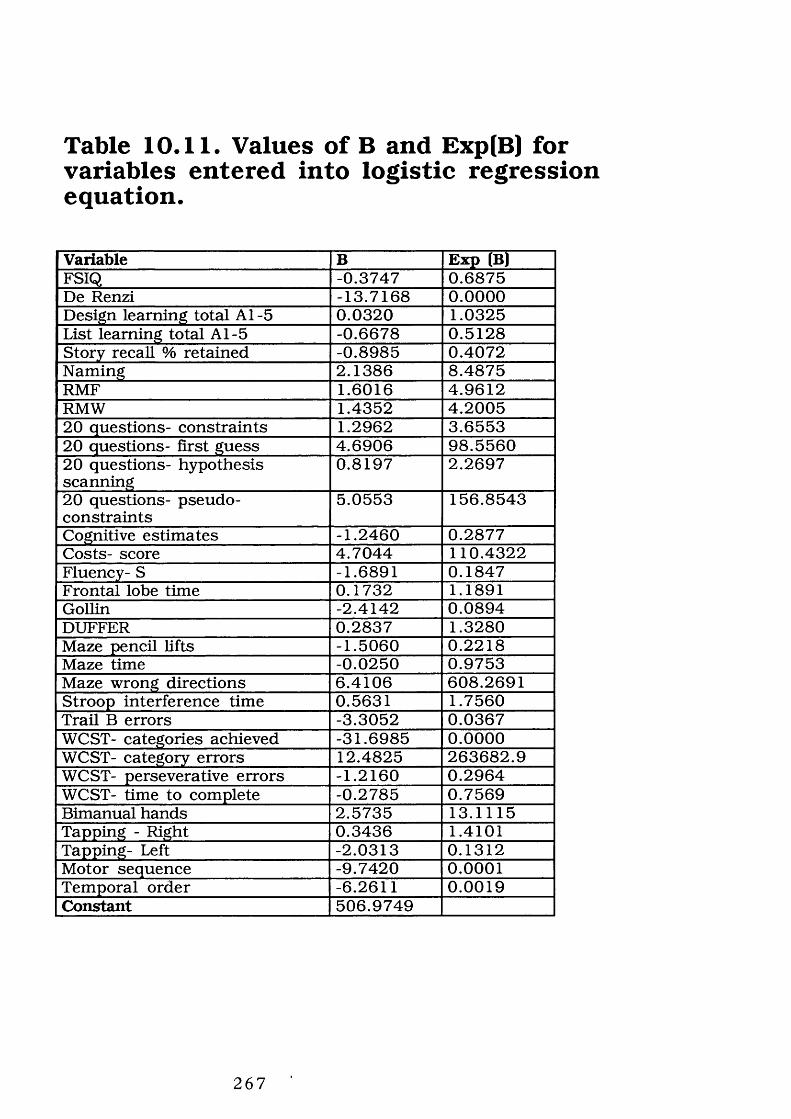
Table 10.11. Values of B and Exp(B) for variables entered into logistic regression equation.
Variable B Exp (B)FSIQ -0.3747 0.6875De Renzi -13.7168 0.0000Design learning total A1-5 0.0320 1.0325List learning total A1-5 -0.6678 0.5128Story recall % retained -0.8985 0.4072Naming 2.1386 8.4875RMF 1.6016 4.9612RMW 1.4352 4.200520 questions- constraints 1.2962 3.655320 questions- first guess 4.6906 98.556020 questions- hypothesis scanning
0.8197 2.2697
20 questions- pseudoconstraints
5.0553 156.8543
Cognitive estim ates -1.2460 0.2877Costs- score 4.7044 110.4322Fluency- S -1.6891 0.1847Frontal lobe time 0.1732 1.1891Gollin -2.4142 0.0894DUFFER 0.2837 1.3280Maze pencil lifts -1.5060 0.2218Maze time -0.0250 0.9753Maze wrong directions 6.4106 608.2691Stroop interference time 0.5631 1.7560Trail B errors -3.3052 0.0367WCST- categories achieved -31.6985 0.0000WCST- category errors 12.4825 263682.9WCST- perseverative errors -1.2160 0.2964WCST- time to complete -0.2785 0.7569Bimanual hands 2.5735 13.1115Tapping - Right 0.3436 1.4101Tapping- Left -2.0313 0.1312Motor sequence -9.7420 0.0001Temporal order -6.2611 0.0019Constant 506.9749
2 6 7

A logistic regression equation takes the form of:
y= constant+B i var i +B2var2+ Bnvarn
Hence the equation was:
y= 506.9764+
(FSIQ*-0.3747)+(De Renzi*-13.7168)+(Design learning total A l- 5*0.0320)+(List learning total Al-5*-0.6678]+(Story recall % retained*Q.8985)+(Naming*2.1386)+(RMF*1.6016]+(RMW* 1.4352) +(20 questions- constraint questions* 1.2962)+(20 questions- first guess*4.6906)+(20 questions- hypothesis scanning questions*0.8197)+(20 questions- pseudo-constraint questions*5.0553)+(Cognitive estim ates*-1.2460)+(Costs- score*4.7044)+(Fluency- S*-1.6891)+(Frontal lobe time*0.1732)+(Gollin*-2.4142)+(DUFFER*0.2837)+(Maze pencil lifts*-1.5060)+(Maze time*-0.0250)+(Maze wrong directions*6.4106)+(Stroop interference tim e*0.563l)+(Trail B errors*-3.3052)+(WCST- categories achieved*-31.6985)+(WCST- category errors* 12.4825)+(WCST- perseverative errors*- 1.2160)+(WCST- time to complete*-0.2785)+(Bimanual hands*2.5735)+(Tapping - Right*0.3436)+(Tapping- Left*- 2.0313)+(Motor sequence*-9.7420)+(Temporal order*-6.2611)
The value of y was then substituted into the following equation:
P= ey 1+ey
If this value was less than 0.5 then the subject was assigned to the frontal group, if greater than 0.5 then it was assigned to the temporal lobe group.
The equation was then applied to the second half of both the frontal and temporal lobe groups, to assess its validity and predictive value (via a specially written computer programme- see appendix 11). A classification table was calculated (see table 10.12) and from this the predictive value, sensitivity, specificity, Youden’s index, false positive, and false negative rates calculated as previously.
2 6 8

Table 10.12. Results of logistic regression equation as applied to the validation set.
Actual frontal Actual temporalTest frontal 29 7Test temporal 5 21
From this the following values were calculated:
Predictive value: 80.55%
Sensitivity: 85.29%
Specificity: 75.00%
False positive : 19.45%
False negative: 19.23%
Youden’s index: 0.6132
2 6 9

The values given indicate the combined equation compares favourably to the individual tests as outlined previously. However, this method has a handicap which may limit its utility. Although it enables the discrimination of frontal and temporal lobe groups, it is impossible for it to discriminate according to latéralisation. This would require a different form of multivariate analysis, namely discrim inant analysis- a procedure theoretically linked to logistic regression (as outlined in chapter 9). The central purpose of the discriminant analysis is to enable individual units to be assigned to one or other of the populations, with in some defined sense the greatest probability of being correct, or smallest risk of error (Daly et al, 1991).
10 .7 .2 . D iscrim inant analysis.
Discrirninant analysis is a statistical technique in which linear combinations of variables are used to distinguish between two or more categories of cases. The variables "discriminate" between groups of cases and predict into which category a case falls, based upon the values of these variables.
In this analysis the groups were the four localisation groups, and the same procedure as in the logistic regression analysis was used. Namely, half the group of frontal and temporal lobe cases from both latéralisation groups were selected as the training set for the discrim inant analysis. The discriminant functions were then applied to the alternate group and the validity of such an approach calculated.
In this analysis a stepwise discriminant analysis was performed, that is, the statistical criteria alone determined order of entry. The same variables used previously were employed in this analysis, presented in the same order as described.
The analysis revealed that several variables were redundant (ie their statistical power was insufficient) within the equation and hence were removed:
2 7 0

FSIQ
DUFFER
Maze- time
Temporal order
Hypothesis scanning
Frontal lobe time
Maze- errors
Trail B errors
WCST- perseverative errors
Thus, the variables in the final equation are as given in table 10.13 (given with the value of Wilks’ lambda, its significance level, and presented in the order of strength).
From this series of variables the classification function co efficients (Fisher's Linear Discriminant Functions] for the four groups were calculated and these are presented in table 10.14 (to four decimal places only).
271

Table 10.13. Wilks' lambda and significance of discriminant analysis variables.Variable Wilks' Lambda SignificanceTapping- right 0.6620 0.0001Costs score 0.47561 0.0001Twenty questions- Pseudoconstraints
0.35290 0.0001
Cognitive estim ates 0.25860 0.0001Fluency- S 0.21505 0.0001Naming 0.17602 0.0001WCST- Category errors 0.14693 0.0001WCST- Categories 0.12537 0.0001Design learning 0.10829 0.0001WCST- time 0.09476 0.0001RMW 0.07834 0.0001Gollin 0.06802 0.0001Twenty questions- First guess 0.06124 0.0001Motor sequences 0.05521 0.0001Maze-pencil lifts 0.05015 0.0001Story recall- retained 0.04650 0.0001Stroop- interference 0.04189 0.0001Bimanual hands 0.03855 0.0001De Renzi 0.03588 0.0001Left tapping 0.03219 0.0001Twenty questions- Constraint questions
0.02996 0.0001
RMF 0.02805 0.0001List learning 0.02568 0.0001
2 7 2

Table 10.14. Fisher's Linear DiscriminantFunctions for the four subject groups.
Variable Function 1 Function 2 Function 3 Function 4Bimanual hands -0.7651 -0.9692 -0.7575 -0.6958Cognitiveestim ates
3.8748 2.7923 2.7660 2.0211
Costs score -1.8507 1.7308 1.9882 4.5534De Renzi 22.2431 24.48017 22.8134 23.1555Design learning 1.2316 1.6049 1.4894 1.9082Fluency- S 1.9488 1.8280 1.4453 1.8731Gollin 1.6368 1.3072 1.2144 0.9723Left tapping 1.5163 1,7529 1.4800 1.6308List learning 0.1542 0.3182 0.1847 0.4478Maze-pencil lifts 8.2910 7.8986 7.4791 8.7023Motor sequences 7.1186 6.8334 5.6380 5.6547Naming -0.5769 -0.7487 -0.5489 -1.0099RMF -0.4666 -0.6520 -0.6310 -1.01889RMW 0.6311 1.11421 1.2809 1.7427Story recall- retained
0.5356 0.5458 0.4917 0.4525
Stroop-in terference
0.3599 0.4459 0.4376 0.5124
Tapping- right 0.4216 -0.2160 0.0980 -0.0797Twentyquestions-Constraintquestions
-0.6736 -1.3115 -1.0735 -1.348981
Twentyquestions- First guess
-4.3506 -3.0692 -2.5845 -2.2928
Twentyquestions-Pseudo-constraints
0.5014 -0.0569 0.9191 0.0345
WCST-Categories
17.2275 14.1278 12.7281 10.6399
WCST- errors, categories
-3.4272 -3.9777 -2.7667 -1.2636
WCST- time 0.1545 0.1244 0.1132 0.083Constant -402.9353 -385.0911 -356.1479 -366.2182
2 7 3

The above co-efficients were then substituted into an equation of the form:
Fi = constanti+ CiVi+CiVi+ CnVn
Where Fi is the discriminant function
Q = Discriminant coefficient for variable
Vi = Variable
Four such discriminant functions were computed- one for each localisation area. The largest of these refers to the group, hence:
Function 1 > Function 2 and Function 3 and Function 4
Left temporal
Function 2 > Function 1 and Function 3 and Function 4
Right temporal
Function 3 > Function 1 and Function 2 and Function 4
Left frontal
Function 4 > Function 1 and Function 2 and Function 3
Right temporal
When these coefficients were applied to the training set the results given in table 10.15 were obtained.
The discriminant functions were then applied to the alternate half of the subjects, and the results of this investigation are given in table10.16.
2 7 4

Table 10.15. Classification tableaccording to discriminant functions of thetraining set.
Group Predicted left temporal
Predictedright
temporal
Predictedleft
frontal
Predicted right frontal
Left temporal 15 1 0 0Righttemporal
0 11 1 1
Left frontal 0 1 20 2Right frontal 0 0 1 16
Giving an overall "success rate" of 89.86% of cases classified correctly.
Individual group success rates:
Group Correct classification %Left temporal 100.00Right temporal 78.57Left frontal 90.90Right frontal 84.21
2 7 5

Table 10.16. Classification tableaccording to discriminant functions of thevalidation set.
Group Predicted left temporal
Predicted right temporal
Predicted left frontal
Predictedrightfrontal
Left temporal 9 0 3 0Righttemporal
1 9 1 3
Left frontal 0 1 12 3Right frontal 5 3 3 9
Thus a total of 62.90% were correctly classified by this procedure. Individual group success rates:
Group Correct classification %Left temporal 60.00Right temporal 69.23Left frontal 63.15Right frontal 60.00
2 7 6

10.8. Hg A composite score will offer greater predictive power of assessing epileptic dysfunction within the frontal lobes than any individual neuropsychological test score.Given the paucity of tests tha t showed any differences between the frontal regions, a further discriminant analysis was undertaken. A similar procedure to tha t adopted with the four localisation groups was adopted (ie a training and validation set were used). Again, the variables were listed for the logistic regression equation SPSS procedure in the order as outlined in table 10.9.
Several variables were removed from this analysis by the initial run through because of their lack of statistical power. The remaining variables are listed with the Wilks’ lambda and significance in table10.17.
From these variables the five classification function coefficients were calculated (Fisher’s Linear Discriminant Functions) and these are presented in table 10.18.
Where:
Group 1 : Dorsolateral
Group 2: Orbitofrontal
Group 3: Mesial
Group 4: M otor/Pre-m otor
Group 5: Extensive
On the original training group the procedure was able to classify 68% of the subjects correctly (see table 10.19).
When the functions were applied to the alternate half of the subjects the discriminant functions were able to correctly classify 58.53% of the subjects (see table 10.20).
2 7 7

Table 10.17. Wilks’ lambda and significance level for discriminant analysis dependent upon frontal region.Variable Wilks' lambda SignificanceTwenty questions- Hypothesis scanning
0.77859 0.0214
Costs score 0.67447 0.0220Twenty questions- First guess
0.54298 0.0400
GoUin 0.45959 0.0252DUFFER 0.38798 0.0163Maze- errors 0.34995 0.0218Design learning 0.30664 0.0222Right tapping 0.29157 0.0137Trail B errors 0.25854 0.0160Fluency S 0.22844 0.0183Motor sequences 0.20260 0.216
Table 10.18. Fisher's Linear Discriminant Functions for frontal region.
Variable Function1
Function2
Function3
Function4
Function5
Designlearning
0.5138 0.3155 0.5108 0.4177 0.5376
Twenty questions- first guess
2.3107 1.7483 2.4293 2.4625 3.151
Twenty questions- Hypothesis scanning
0.7214 0.7281 0.762 1.035 1.622
Costs- score 2.834 2.429 3.009 2.677 1.139Fluency S 1.459 1.318 1.183 1.012 1.009DUFFER 0.976 1.054 0.646 0.724 0.510GoUin 0.875 0.829 1.052 1.153 1.307ATaze errors 6.75 6.05 6.682 4.731 4.718M otorsequences
-0.232 0.288 -0.305 0.667 0.238
Tapping-right
0.793 0.811 0.7393 0.675 0.661
Trail B- e rro rs
1.297 1.492 1.460 2.291 2.828
Constant -94.1368 -85.947 -87.263 -88.247 -95.126
2 7 8

Table 10.19. Classification tableaccording to discriminant analysis forindividual frontal regions (training set).
Group PredictedDorsolateral
PredictedOrbitofrontal
PredictedMesial
Predicted Motor/ Pre motor
PredictedExtensive
Dorsolateral
13 3 4 2 1
Orbitofrontal
1 7 0 0 0
Mesial 0 1 3 0 0Motor/Premotor
1 1 0 5 0
Extensive 0 0 0 2 6
Individual group success rates:
Group % Correctly classifiedDorsolateral 86.67Orbitofrontal 58.33Mesial 42.86M otor/Pre-m otor 55.55Extensive 85.72
2 7 9

Table 10.20. Classification tableaccording to discriminant analysis forindividual frontal regions (validation set).
Group PredictedDorsolateral
PredictedOibitofrontal
PredictedMesial
PredictedMotor/Premotor
PredictedExtensive
Dorsolateral
12 1 2 2 2
Orbitofrontal
0 2 0 0 0
Mesial 1 1 6 0 1Motor/Premotor
1 1 0 2 1
Extensive 1 1 0 2 2
Individual group success rates:
Group % Correctly classifiedDorsolateral 80.00Orbitofrontal 33.33Mesial 75.00M otor/Pre-m otor 33.33Extensive 33.33
2 8 0

10.9. Hg Surgical removal of frontal lobe epileptic foci will result in altered neuropsychological performance. Specifically, there will be a detrimental effect of surgery on those tests purporting to tap the functions of the frontal regions.Four cases were followed who underwent surgery for the relief of their frontal lobe epilepsy, and these are presented below as individual case studies.
10.9.1. Case study 1: MP, Right frontal^.
10.9.1.1. Medical aspects.
MP is a 26 year old male who developed epilepsy at the age of 6 years. Seizures remained resistant to medication and he was having approximately one seizure per week at the time of referral. In tracranial EEG recording revealed a focus in the right frontal pole. A pre-operative MR! scan was inconclusive. Video recordings described his seizure thus:
"He will release a scream, raise his arms above his head, they will jerk. He will recover in a couple o f minutes. This can happen three or four times a day. The seizures may or may not be preceded by his aura o f a butterfly sensation in his stomach rising to the neck."
Subsequently, MP underwent a right frontal lobectomy. The operation was maximal and removal was total back to the motor strip. Post-operative MRI scanning reported:
2 This case has been published: Upton and Corcoran (1995). All the investigation, including all the assessment, was undertaken by myself. I acknowledge the assistance of Dr. Corcoran in her critical discussion that assissted me in the formulation of my ideas.
281

"The resection o f the right frontal lobe is shown. It includes the frontal pole and extends posteriorly to Just in front o f the SMA on the mesial cortex, and involves the anterior parts o f the superior middle and inferior
frontal gyrt Irferiorly, the orbital gyri are involved but most o f the gyrus rectus is spared. "
Following the operation MP was seizure free for approximately two years. He then decided, unilaterally, to discontinue his medication. Unfortunately, this resulted in several seizures. Despite reinstigation of his medication, seizures remained refractory. EEG recordings dem onstrated a right temporal focus, thought to be a consequence of "kindling." Therefore MP underwent a second operation to remove the right temporal lobe. Since this time epileptic seizures have not re appeared; although there has been a reappearance of seizures which are thought to be non-epileptic in origin.
Medication remained stable throughout the assessm ent period, with MP taking phenytoin, vigabatrin and carbamazepine slow-release (all within the therapeutic range).
10.9.1.2. Social aspects.
MP left school at the age of 16 years with no formal qualifications.He was unable to find full time employment and hence helped at a motor repair garage owned by his father. He lives with his parents and has never lived away from home. Following the reappearance of his seizures his mother started to sleep in the same room as MP which led to a degree of family tension. MP's situation was comparable after the second operation.
10.9.1.3. Neuropsychological assessment.
MP was first seen two weeks prior to his first operation. The report suggested a weakness on verbal WAIS-R subtests compared to the performance subtests. This was thought to be a consequence of past vocational and educational background (MP had had a limited academic career). There was no evidence of any intellectual deterioration or any focal neuropsychological features. A sodium
2 8 2

amytal procedure revealed a left hemisphere dominant for language and a relative strength for memory over the right hemisphere.
MP was subsequently seen at three m onths, and one year postopera tively, when the scores obtained on neuropsychological m easures were essentially unchanged. MP was then seen a t two years post-operatively, Just after his seizures had returned, and then three months after his second operation.
10.9.1.3.1. Summary Background tests.
MP's performance on the WAIS-R was comparable throughout the two year period of assessm ent (see table 10.21). There was a moderate deterioration of PIQ at the two year follow up (when his new, temporal lobe seizures were extensive) with a fall of 15 IQ points recorded from his pre-operative level. This was mainly a consequence of a drop in Picture Arrangement and Block Design sub-test scores.
10.9.1.3.2. Summary of performance on language and memory tests.
MP's scores on m easures thought sensitive to temporal lobe dysfunction can be found in table 10.22. On m easures of language functioning, MP's performance was generally adequate. He scored maximally on the De Renzi token test and his performance on the naming task was average throughout the two years follow-up. His performance on the design learning task was extremely good throughout, with one exception- with reoccurrence of his seizures, his scores deteriorated from the maximum 45 to 31 (a score a t the 25th centile). However, after removal of his temporal lobe, his performance returned to maximum. On the list learning paradigm his performance consistently fell below cut-off, showing little change over the two year period. His performance on recognition tasks was generally poor- on the non-verbal version he consistently performed below the 10th centile. On the verbal version his performance was variable- ranging from average to the 5th centile. His performance on story recall was generally poor, although this did improve slightly.
2 8 3

10.9.1.3.3. Summary of motor skills tests.
MP’s performance on the tests tapping motor skills were generally good (see table 10.23). His performance on the bim anual hand task was excellent, making between 51 and 64 fist-pahn alterations in 20 seconds (with no errors). His temporal order and gesture span score remained consistent throughout. On the tapping task his performance was excellent with both dom inant and non-dominant hand.
10.9.1.3.4. Summary of executive skills tests.
MP's performance pre-opera tively was adequate on these m easures, indeed there was little indication of any frontal lobe dysfunction (see table 10.24). For example, on the MWCST his performance was excellent- he made no errors and completed six categories in 216 seconds. MP's performance on this task was also excellent following removal of his right frontal lobe. However, with the recurrence of his seizures with a right temporal lobe onset, his performance deteriorated- completing only four categories and making 10 perseverative errors (83.33%- "typically frontal" [Nelson, 1976]). This poor performance continued following his temporal lobectomy, although there was some sHght improvement. Performance on the Stroop interference task was generally adequate and consistent over the two year period. Similarly, his performance on the fluency tasks was generally competent. In general, on most of these tests his performance remained consistent and generally adequate, especially given the am ount of cerebral tissue resected.
10.9.1.3.5. Stunmaiy of composite scores.
The derived equations from the logistic regression and the discriminant analysis were applied to MP's pre-operative scores.The results were indicative of a frontal lobe dysfunction according to the logistic regression, and right frontal lobe dysfunction from the derived discriminant analysis functions.
2 8 4

Table 10.21. Case MP: Background Tests.Test Pre-op
(2 weeks)Post-op (3 months)
Post-op (1 year)
Post-op (2 years)
Post-op 2 (3 months)
NART IQ 92 92 — 90 --VIQ 85 85 90 91 88PIQ I I I 109 100 96 noFSIQ 103 94 94 92 96Digit span 13 13 12 16 12Vocabulary 20 37 34 36 43A rithm etic 9 7 7 8 7Sim ilarities 19 17 23 22 20Picturecom pletion
IS 17 17 17 18
Picturearrangem ent
16 17 17 . 12 13
Block design 38 39 38 32 42
2 8 5

Table 10.22. Case MP: Language andmemory tests.Test Pre-op Post-op
(3 months)Post-op (1 year)
Post-op (2 years)
Post-op 2 (3 months)
De Renzi 15 15 15 15 15Design A1-5 45 45 45 31 41Design A6 9 9 9 7 9Design B 7 7 6 6 9Design-intrusions
0 0 0 6 0
List A1-5 38 35 38 41 34List A6 7 9 7 5 5List B 5 6 6 4 6List-intrusions
1 0 4 3 2
Naming 17 18 18 16 19RMF 32 38 37 33 33RMW 42 38 40 47 42Story- Delayed
0 0 6 2 20
Story-Im m ediate
10 28 13 12 19
Story-Retained
0 0 46 16 105
Table 10.23. Case MP: Motor skills tests.Test Pre-op Post-op
(3 months)Post-op (1 year)
Post-op (2 years)
Post-op 2 (3 months)
Bimanual hand erro rs
0 0 0 0 0
Bimanualhands
55 57 51 54 64
Gesture span 3 3 3 3 3Motorsequences
8 7 8 4 8
Tapping- Left 80 95 90 85 80Tapping- Right 95 90 87 92 91Tem poralo rder
3 3 3 3 3
2 8 6

Table 10.24. Case MP: Executive skillstests.Test Pre-op Post-op
(3 months)Post-op (1 year)
Post-op (2 years)
Post-op 2 (3 months)
Cognitiveestim ates
2 2 7 5 8
20 questions- Constraints
18 14 16 12 12
20 questions- First guess
4 5 6 8 4
20 questions- Hypothesis
6 4 6 5 5
20 questions- No. of questions
24 18 25 17 17
20 questions-Pseudo-constraints
0 0 0 1 0
Costs 1 1 1 1 1Costs- Recency 6 7 8 8 8Costs-Intrusions 0 0 0 0 0Costs-Recall 8 8 7 7 6Duffer 20 24 16 20 32Fluency- Animals 18 14 15 15 20Fluency- S 13 16 14 17 18Frontal time 18 15 33 10 1GoUin 20 20 23 42 20Maze-PencU Ufts 2 1 0 0 0Maze- errors 2 2 0 2 1Maze-time 84 72 65 97 80StroopInterferencetim e
12 10 26 19 10
TraU B errors 0 0 1 0 0Visual count 11 8 7 9 6Visual search 35 36 48 30 34WCST- %perseverativeerro rs
0 0 0 83.33 52.00
WCST-Categories
6 6 6 4 4
WCST- category erro rs
0 0 0 2 12
WCST-perseverativeerro rs
0 0 0 10 13
WCST- Time 216 154 180 321 351
2 8 7

10.9.2. Case study 2: KW, Right frontal.
10.9.2.1. Medical aspects.
KW is a right handed male bom in 1962. Birth was difficult and he spent the first three days of life in a special care baby unit. He had three febrile convulsions associated with ear infections between the ages of one, and two and a half years of age. However, developmental milestones were normal. Early childhood and adolescence were also normal.
At the age of 18 he had his first witnessed seizure. Whilst sitting in a chair his left leg and arm stiffened and started convulsing. This lasted 15 minutes and was associated with cyanosis and tongue biting. No medical treatm ent was started and over the next four weeks he was experiencing 1-7 episodes per day. At the same time he was also experiencing "absence" seizures approximately five times a day. He was subsequently started on AEDs, which resulted in a reduction in seizure frequency. Throughout the assessm ent period KW remained on phenytoin and carbamazepine. At one stage KW was tried on vigabatrin, but this resulted in an increase in seizures.
At the age of 29 years he was referred to a specialist epilepsy unit where the following was noted:
1. Generalised tonic-clonic seizures; left sided onset.
2. Absence seizures; occurring 5-6 times per day.
3. Episodes of "Paralysis from the neck down".
4. Atonic attacks.
EEG and CT scans were reported as being normal. An MRI was carried out that revealed a small, weU circumscribed area 1.5x2 cm:
"There is a small ovoid well circumscribed lesion lying in the mesial part o f the right frontal lobe. It lies above the cingulum and is anterior to the SMA. It does appear to have slight mass effect displacing cortical sulci"
2 8 8

standard EEG recordings revealed no focal abnormality, although surgical electro-corticography confirmed a right frontal seizure focus. KW underwent surgery for right frontal tum our removal. Postoperative histology of the resected area revealed a low grade (grade 2) astrocytoma. Post-operatively, there were no complications and since the operation there have been no seizures (18 month follow up).
10.9.2.2. Social aspects.
KW was educated at normal schools where he obtained several 'O - level passes. He subsequently completed a vocational training course and currently works as a clock repairer. He is married and has no children.
10.9.2.3. Neuropsychological assessment.
KW was first investigated 1 month prior to his operation. The results suggested a dominant hemisphere dysfunction, given tha t his verbal scores were some 34 points lower than his performance sub test scores. KW was re-assessed at three m onths and twelve months post-operatively.
A sodium amytal indicated left hemisphere dominance for language.
10.9.2.3.1. Summaiy of background tests.
KW’s IQ figures are indicative of dominant hemisphere dysfunction given a 34 point VIQ-PIQ difference (see table 10.25). This discrepancy remained over the 12 m onths follow-up, although this fluctuated and decreased slightly. This reduction was a consequence of a decrease in KW's PIQ, especially at three m onths follow up where there had been a 15 IQ points drop; the sub-test scores reflecting this. Similarly, a t three m onths there also had been a 13 point drop in VIQ. Both of these had largely resolved themselves by the twelve month follow-up.
10.9.2.3.2. Summaiy of language and memory tests.
Table 10.26 outlines KW’s scores on m easures thought sensitive to temporal lobe dysfunction. KW’s performance on a test of expressive
289

language skills was competent, remaining in the bright average range throughout the assessm ent period. However, there did appear to be some weakness on a test of receptive language skill; scoring 12/15 on the De Renzi token test. This did not, however, change following surgery. On tests of recognition memory KW performed at, or above, the 50th centile for the verbal version, bu t below the 5th centile for the non-verbal version. Both showed some minor decline following surgery. List learning was adequate, as was his performance on the non-verbal paradigm. On the m easure of story recall, KW's performance showed a dram atic decrease following surgery from an average performance pre-operatively, to a score below the 10th centile after surgery which was also notable at 12 month follow-up.
10.9.2.3.3. Summaiy of motor skills tests.
Table 10.27 presents KW’s results from motor skill tasks. There was very little change over the 1 year follow-up. However, motor sequences, right hand tapping, and left hand tapping did show some slight decrement at three m onths foUow-up, although these had returned to pre-operative levels at 12 months.
10.9.2.3.4. Summaiy of executive skül tests.
The results from this series of tests are presented in table 10.28. Again, these results are notable for two reasons: firstly, there is little pre-operative indication of frontal lobe dysfunction (except a poor performance on the Trail making task- a long 'frontal lobe time' and several errors on Trail B). Secondly, there appears to be limited change in the scores post-operatively, although there were some minor decrements at 3 m onths post-operatively, these had returned to pre-operative levels a t the twelve month follow-up.
10.9.2.3.5. Summaiy of composite scores.
The derived equations from the logistic regression and the discriminant analysis were applied to KW's pre-operative scores.The results were indicative of a temporal lobe dysfunction according to the logistic regression, and left temporal dysfunction from the discriminant analysis.
2 9 0

Table 10.25. Case KW: Background tests.T est Pre-operative 3 m onths 12 m onthsNART IQ 92 92 -
VIQ 103 90 100PIQ 137 122 130FSIQ 120 102 116Digit span 12 14 13Vocabulary 47 42 44Arithm etic 13 7 10Similarities 26 22 22Picturecom pletion
19 19 19
Picturearrangem ent
19 17 20
Block design 38 31 35
291

Table 10.26. Case KW: Language andmemory tests.T est Pre-operative 3 m onths 12 m onthsDe Renzi 12 12 12Design A1-5 40 40 40Design A6 9 9 9Design B 8 8 8Designintrusions
4 5 3
List A1-5 50 51 50List A6 9 11 10ListB 5 7 6List intrusions 0 5 0Naming 21 23 24RMF 37 35 36RMW 48 46 47Story- delayed 42 21 20Story-im m ediate
44 25 25
Story- retained 95 84 80
Table 10.27. Case KW: Motor skill tests.T est Pre-operative 3 m onths 12 m onthsBimanual hand erro rs
0 0 0
Bimanual hands 42 43 44Gesture span 3 3 3M otorsequences
10 7 10
Tapping- left 65 55 64Tapping- right 70 65 68Tem poralo rder
3 3 3
2 9 2

Table 10.28. Case KW: Executive skillstests.T est Pre-
operative3 m onths 12 m onths
Trail making- ’Frontal time'
40 57 22
20 Questions- Constraints
12 11 10
20 Questions- Hypothesis
4 9 2
20 Questions- No. of questions
16 21 13
20 Questions- Pseudo-constraint
0 1 0
Cognitive estim ates 2 0 0Costs 1 1 2Costs- Intrusions 0 0 0Costs- Recall 8 7 8Costs- Recency 6 5 6Counting 9 10 8Duffer 25 20 1620 questions- first guess
1 3 8
Fluency- Animals 15 16 17Fluency- S 7 14 11GoUin 33 41 33Maze- pencU lift 1 1 0M aze-errors 2 2 0Maze-time 112 107 96Stroopinterference time
35 43 35
TraU B errors 2 3 1Visual search 37 40 30WCST- Categories 6 6 6WCST- Category erro rs
2 2 1
WCST-Perseverative errors %
60 60 50
WCST-Perseverativeerro rs
3 3 1
WCST- Time (sec.) 252 260 243
2 9 3

10.9.3. Case study 3: JS , Left frontal.
10.9.3.1. Medical aspects.
JS is a right handed male who, at the time of his first assessm ent, was 29 years of age. JS had his first witnessed seizure at the age of 12 years. Since this time he has had both complex partial and tonic- clonic seizures which have proved intractable to medical therapy. At the time of his pre-operative assessm ent JS was suffering from approximately ten complex partial seizures per day and several tonic-clonic seizures a year. During the assessm ent period JS remained on primidone and carbamazepine, both within the therapeutic range.
A pre-operative MRI scan was suggestive of a left frontal dysembryoplastic neuro epithelioma. At surgery there was a wide spread removal of the left frontal lobe which involved both medial and lateral surfaces.
Post operative recovery was uneventful, although JS suffered two seizures in the first month after surgery. However, at one year foUow-up JS was seizure free, having had no seizures since those in the initial post-operative period.
10.9.3.2. Social aspects.
JS had attended mainstream schools, bu t had failed to gain any formal qualifications. Preoperatively, he enjoyed a rather bohemian lifestyle and spent most of his day in bed, with no full time occupation. Post-operatively, with his seizures under better control he still tended to continue with his unorthodox life. However, he had started to attend college where he was studying for GCSEs in English and Photography.
10.9.3.3. Neuropsychological assessment.
JS undertook his first assessm ent three weeks before his operation. This revealed a moderate degree of under functioning, from a level in the average range (WAIS-R assessed intellectual level fell in the low average range). This had previously been commented upon in
2 9 4

many previous assessm ents. Indeed, his whole profile was consistent with a degree of under functioning. A sodium amytal procedure suggested left hemisphere language dominance.
10.9.3.3.1. Summaiy of background tests.
The background tests revealed some degree of under functioning- to the level of some 20 IQ points from the NART assessed optimal level (see table 10.29). As mentioned, this had been commented upon for a num ber of years. JS ’s performance on the digit span and arithmetic sub-test appeared to be particularly weak. However, with both the IQ scores and the individual sub-tests, there was little change from pre-operative to post operative assessm ents.
10.9.3.3.2. Summaiy of language and memoiy tests.
JS 's performance on tests of temporal lobe functioning is shown in table 10.30. On tests of language functioning, JS scored well- making no errors on the De Renzi token test and scoring in the bright normal range on a measure of expressive language skills- again perhaps an indication of some general under functioning. On the story recall task at three months there was a remarkable improvement- from a score below the 10th centile to one in the average range. However, JS ’s score a t twelve m onths was similar to his pre-operative level and hence the former may be assum ed to be a spurious result. However, a similar pattern was observed on the list learning task, although to a lesser degree. On the recognition tasks JS ’s performance on the non-verbal version (design learning) was poor (below the 5th centile), although this showed only limited change over time. There was a gradual decrease in JS ’s ability on the verbal version; from a score in the average range to one one below the 5th centile.
10.9.3.3.3. Summaiy of motor skill tests.
Table 10.31 outlines JS ’s performance on m easures of motor skills. His performance on the bimanual hand task was weak, and he only managed 15 alternations in 20 seconds pre-opera tively. This changed very little over the follow-up period and continued to be a poor score. Similarly, the num ber of errors made on th is task were
2 9 5

relatively large. The left tapping scores were adequate and only showed minor change. The right tapping score was again adequate, bu t showed a general decrement over the follow-up period from 60 in twenty seconds at pre-operative assessm ent, to 50 at the twelve month follow up.
10.9.3.3.4. Summaiy of executive skills tasks.
JS ’s pre-operative scores on m easures of executive functioning were indicative of frontal lobe dysfunction (see table 10.32). He only managed to complete 5 categories on the MWCST, making several errors (71% of them perseverative). Similarly, his performance on the Trail making task was impaired, as was the Twenty questions task, cognitive estimates, and the Stroop. Post-operatively there were improvements in some of these tasks, although this was rather variable.
10.9.1.3.5. Summaiy of composite scores.
The derived equations from the logistic regression and the discriminant analysis were applied to JS ’s pre-operative scores. The results were indicative of a frontal lobe dysfunction according to the logistic regression, and a left frontal lobe dysfunction from the discriminant analysis.
296

Table 10.29. Case JS: Background Tests.T ests Pre-operative 3 m onths 12 m onthsNART IQ 113 110 -
VIQ 82 88 84PIQ 84 102 82FSIQ 81 93 82Digit span 7 9 9Vocabnlaiy 48 53 42Arithm etic 9 6 7Sim ilarities 17 21 21Picturecom pletion
14 16 14
Picturearrangem ent
8 15 6
Block design 24 33 24
2 9 7

Table 10.30. Case JS: Language andmemory tests.T ests Pre-operative 3 m onths 12 m onthsDe Renzi 15 15 15Design A1-5 22 31 36Design A6 5 7 7Design B 4 4 7Designintrusions
18 0 7
List A1-5 26 37 30List AG 5 7 5ListB 4 7 3List intrusions 1 7 1Naming 25 23 24RMF 38 37 32RMW 46 44 36Story- delayed 14 31 16Story-im m ediate
18 39 19
Story- retained 77 79 84
Table 10.31. Case JS: Motor skill tests.T ests Pre-operative 3 m onths 12 m onthsBimanual hand erro rs
3 3 5
Bimanual hands 15 22 18Gesture span 3 3 3Motorsequences
4 5 2
Tapping- left 45 55 52Tapping- right 60 55 50Tem poralorder
4 6 6
2 9 8

Table I0.32.Case JS: Executive skills tests .
T ests Pre-operative 3 m onths 12 m onths'Frontal time’ 32 32 38Trail B- errors 3 1 120 questions- Constraints
14 16 18
20 questions- first guess
4 2 4
20 questions- Hypothesis
14 9 5
20 questions- No. of questions
35 30 24
20 questions- Pseudo-constraint
7 5 1
Cognitive estim ates 10 6 10Costs score 3 3 5Costs- Intrusions 0 0 1Costs- Recall 6 7 5Costs- Recency 5 5 10Counting 8 6 10Duffer 26 20 20Fluency- S 13 13 11Fluency- Animals 17 22 19GoUin 39 40 43Maze- errors 6 3 2Maze- pencil Ufts 3 4 2Maze- time 126 139 183StroopIn terference
35 15 7
Visual search 39 39 32WCST- %perseverativeerro rs
71 71 33
WCST- Categories 5 6 6WCST- Category erro rs
2 2 2
WCST-Perseverativeerro rs
5 5 1
WCST- Time (sec.) 264 315 242
2 9 9

10.9.4. Case study 4: SC, Left frontal.
10.9.4.1. Medical aspects.
SC is a right handed female first assessed at the age of twenty-three. Her first seizure was in early childhood, and these had continued until the present with no significant periods of remission. At the time of the first assessm ent she was having approximately one partial seizure every day. The partial seizure consisted of tonic extension of the right arm with no loss of consciousness. In addition to the simple partial seizures, she also experienced tonic-clonic seizures which occurred on approximately a monthly basis. There were, on occasions, some head injuries which required medical intervention.
In 1991 SC was investigated for possible surgical intervention to ameliorate her seizures. Depth EEC video recordings suggested a left frontal focus to:
"Involve the medial frontal gyrus down to approximately the level o f the rostrum, and outside the cingulum. The posterior extent o f this is well anterior to the paracentral lobule. Seizures arose in the mid-anterior parasagittal cortex ju s t anterior to the SMA."
On cortical MRI there was no obvious lesion visible. Because of the detrim ental effect the seizures were having on her life, SC underwent a partial left frontal removal. The resection involved the left frontal lobe for approximately 7.5 cm (antero-posterior diameter), being 4cm at its widest (posterior) and 1.5cm a t the frontal pole.
Following surgery there was an uneventful recovery with no postoperative complications. However, seizure relief was not complete and she subsequently underwent further surgical intervention.
Eight m onths following her first operation, SC had the residual left dorsolateral pre-frontal cortex removed. The dorsolateral prefrontal region was removed to the posterior level of the SMA, and part of the temporal gyrus near the supermedial border was also removed. Pathological examination of both specimens did not reveal any
3 0 0

obvious pathology. Despite these intensive interventions seizures continued, and at the one year post-operative (2) assessm ent she was still experiencing approximately one seizure every week.
There was consideration of the possibility that some of these seizures were non-epüeptic in origin; SC had been treated for depression following her second operation (and was still receiving counselling). However, video EEG was unable to confirm this.
During the assessm ent period, SC remained on primidone and carbamazepine, to which lamotrigine had been added following her first operation.
10.9.4.2. Social aspects.
SC lived alone in her parents house (following their death some years earüer). She had obtained some 'O’-levels at reasonable grades whilst in full time education. Following her schooling, SC was employed as an administrative assistant. However, she lost this job because of the severity and frequency of her seizures. Thus, for her first assessm ent SC was employed, but for aU subsequent neuropsychological assessm ents she was unemployed.
10.9.4.3. Neuropsychological assessment.
SC was first seen one month prior to her first operation. Her performance on the WAIS-R fell in the average range, and was consistent with her optimal level of functioning as assessed by the NART. Her level of performance was compatible with two previous assessm ents she had undertaken. A Wada test suggested a left hemisphere dominant for language.
SC was seen at three months post-operatively for her routine assessm ent. She was then operated on for the second time, and was seen at three and twelve m onths after this second operation.
301

10.9.4.3.1. Summaiy of background tests.
Table 10.33 presents the results obtained by SC on the background tests. Preoperatively there was no indication of under functioning with reference to the NART IQ scores. Post-operatively, there was little change in the VIQ score. There was, however, an increase in the PIQ by some 23 points. This improvement was not maintained a t 1 year post-operatively (2).
10.9.4.3.2. Summaiy of language and memory tests.
SC's performance on the tasks thought sensitive to temporal lobe dysfunction are given in table 10.34. Expressive language skills were competent pre-opera tively (in the bright normal range) and this was m aintained after both operations. In contrast SC's performance on the De Renzi token task was impaired pre-opera tively when she only scored 5 /15 . This improved post-operatively, but was still impaired. Her performance on list learning was excellent (90th centile) preoperatively, but this showed a decrement post-operatively to a figure a t the 10th centile; although there was some slight improvement at post operative 2 (1 year). On design learning, performance was at the 10th centile pre-operatively, rising to the 25th centile post- operatively, and remained at this level throughout the post operative assessm ent period. Recognition for words was generally good throughout, as was the non-verbal version- with one exception three m onths post operatively (1) where her score fell from 50th centile to the 5th centile.
10.9.4.3.3. Summaiy of motor skills tests.
SC's performance on the tapping task (right, dominant hand) showed a gradual decrease as the follow-up period progressed (see table 10.35), although there was some variabüity and the decrease was only slight. In contrast her performance on the left (nondominant) hand tapping task gradually improved over the postoperative period. On the bimanual hand task, there was a drop from pre-operative assessm ent (55) to the post-operative assessm ents (39, 41, 39). Motor sequences remained relatively stable across the
302

assessm ent period, whereas temporal order showed some gradual improvement.
10.9.4.3.4. Summaiy of executive skills tests.
SC's performance on such tasks was rather variable (see table 10.36). For example, pre operatively SC's performance on the cognitive estimation task was extremely poor and this level of performance was sustained after both operations. In contrast, SC's performance on Fluency- S was good preoperatively [compared to Fluency- animals), although this was not maintained post- operatively. On the MWCST, SC scored well on the first two assessm ents, but her performance deteriorated after the second operation completing only 5 categories and making 10 errors (80% perseverative in nature). Performance on both the cost estimation task and the Twenty questions task was generally poor across aU four assessments.
10.9.1.3.5. Summaiy of composite scores.
The derived equations from the logistic regression and the discriminant analysis were applied to SC's pre-operative scores. The results were indicative of a frontal lobe dysfunction according to the logistic regression, and a left temporal lobe dysfunction from the discriminant analysis.
3 0 3

Table 10.33. Case SC: Background tests.
Test Pre-op Post-op 1 (3 months)
Post-op 2 (3 months)
Post op 2 (1 year)
NART IQ 100 100 - -
VIQ 96 94 94 98PIQ 102 125 117 107FSIQ 99 107 103 102D i^t span 14 16 15 19Vocabulary 36 37 43 51Arithm etic 8 9 7 11Similarities 25 22 22 17Picturecom pletion
16 18 18 18
Picturearrangem ent
17 19 16 17
Block design 34 43 45 30
3 0 4

Table 10.34. Case SC: Language andmemory tests.
Test Pre-op Post-op 1 (3 months)
Post-op 2 (3 months)
Post op 2 (1 year)
De Renzi 5 12 12 14Design A1-5 27 33 36 35Design A6 6 7 8 6Design B 7 6 3 8Design-Intrusions
7 5 3 4
List A1-5 65 45 47 54List A6 8 9 10 11List B 9 8 4 5List-In trustons
2 1 1 2
Naming 20 21 22 20RMF 45 37 48 46RMW 45 48 48 44Story-Im m ediate
20 50 23 23
Story-recalldelayed
18 50 18 21
Story- Retained
90 100 78 91
Table 10.35. Case SC: Motor skill tests
Test Pre-op Post-op (3 months)
Post-op 2 (3 months)
Post op 2 (1 year)
Bimanual hand- errors
0 1 0 1
Bimanualhands
55 39 41 39
Gesture span 3 3 3 3Hand errors 0 0 0 2Motorsequences
5 5 4 5
Tapping- left 70 75 74 80Tapping- right 80 81 73 75Tem poralo rder
3 3 4 6
3 0 5

Table 10.36. Case SC: Executive skillstests.
Test Pre-op Post-op (3 months)
Post-op 2 (3 months)
Post op 2 (1 year)
20 questions- Constraints
1 2 1 4
20 questions- First guess
1 1 4 2
20 questions- Hypothesis
11 10 11 12
20 questions- Pseudo-con
7 6 8 7
20 questions- total questions
19 18 20 23
Counting 9 8 7 9Visual search 29 28 27 30Cognitive estimates 14 17 13 15Costs- Intrusions 0 0 0 0Costs- Recall 7 8 8 7Costs- Recency 5 4 6 7Costs- score 3 2 4 5DUFFER 20 12 23 32Fluency animals 11 12 9 8Fluency- S 13 14 11 10Frontal time 8 9 16 16Gollin 25 18 30 36M aze-errors 3 0 1 3Maze-pencil lifts 1 2 1 2Maze-time 107 102 98 83
3 0 6

Table 10.40 (cont).Test Pre-op Post-op
(3 months]Post-op 2 (3 months)
Post op 2 (1 year)
Stroopin terference
48 32 44 29
Trail B errors 0 0 1 1WCST- %perseverativeerro rs
0 0 80 50
WCST- Categories 6 6 5 6WCST- Category erro rs
0 0 2 2
WCST-perseverativeerro rs
0 0 8 2
WCST- Time (sec.) 287 250 332 290
3 0 7

10.9.5. Conclusion.Four cases (two left frontal and two right frontal) have been presented, which dem onstrate some of the difficulties and complexities in attempting to assess frontal dysfunction; all cases show a rather variable pattern of results and there is little commonality amongst them. Each case can be considered to represent a unique study, and provides insight into some of the functions disrupted by frontal lobe epilepsy, and surgical removal of an epileptogenic frontal region. Each individual case's contribution to understanding of the frontal regions will be explored a t length in the discussion.
3 0 8

Chapter 11: Discussion.
3 0 9

11.1. Hi There will be a characteristic difference in the neuropsychological performance between those with temporal and those with frontal lobe epileptic foci.H2 There will be a difference in the neuropsychological performance dependent on latéralisation of the epileptic foci.These two hypotheses are dealt with together, for they can be considered to be examining similar patterns of results. A series of analyses was undertaken to examine the general level of differences between the localisation groups. This is important, since no group study has been conducted with frontal lobe epilepsy subjects and it is worth repeating that:
"No systematic neuropsychological study o f various subtypes o f frontal lobe epilepsy has been reported."
Waterman and Wada, 1990 p.209-210
This is even at the level of left-right hemispheric differences in an epilepsy sample (rather than a post-operative sample):
"Cognitive studies o f post-operative frontal lobe epilepsy patients have contributed greatly to our knowledge o f specific frontal lobe lesion effects. Yet it remains to be seen whether frontal lobe epilepsy subjects show such impairment pre-operatively. "
Swartz et al, 1994 p. 232
This investigation has, for the first time, been able to delineate some of the neuropsychological consequences of lateralised frontal lobe epileptic foci.
A first step in the description of cognitive deficits associated with frontal lobe epilepsy, is to examine the performance of a group of subjects with discrete foci and assess whether they are impaired on tests, compared to a group with equally well defined foci. It is therefore of Limited use from this perspective, to compare
3 1 0

performance with a normative sample (although this does have some uses- see section 11.8.6). The choice of control groups is therefore important. In this study subjects with temporal lobe epilepsy were considered appropriate as a control group for two main reasons.
Firstly, given the close proximity of the frontal and temporal regions, the deficits previously highlighted may simply be a consequence of inaccurate assessm ents of the extent of damage. Thus it is essential for the description of any functional deficit to say with certainty, that the region under discussion is affected, and it is the only one affected. Secondly, given the neuronal connections, many frontal (or for that m atter temporal lobe] impairments previously highlighted may be a consequence of a deficit in the alternate region (Hermann et al, 1987).
In this study priority was placed upon obtaining a "pure" sample of either frontal or temporal lobe damaged subjects. Thus when a deficit is noted, it can be certain to a high degree, that the functional deficit is a consequence of damage to tha t region.
11.1.1. Background tests.
The results dem onstrated minimal differences on most of the m easures undertaken. Some have argued a role for the frontal region in the completion of certain tasks drawn from the WAIS-R, for example similarities (eg McFie, 1975; Newcombe, 1969;Rzechorzek, 1978), and picture arrangem ent (McFie and Thompson, 1972), although this is far from consistent (Lezak, 1983; Warrington et al, 1986). Neither of these tests revealed a specific deficit for any group in the study.
There were two significant results from the individual sub-tests of the WAIS-R. Specifically, the frontal lobe group were more impaired on both the digit span sub-test and the arithmetic sub-test. An impairment on the arithmetic task is not commonly associated with frontal dysfunction, although it is of interest to note that one of the cases (subject 2) described by Penfield and Evans (1935) was reported to be deficient in arithmetic following a left frontal lobectomy. More recently, Lucchelli and De Renzi (1993) have also
311

reported a case study of an individual with a left mesial frontal tum our, who was impaired on arithmetical operations.
The arithmetic sub-test is not simply a test of mathematical ability. Difficulties in immediate memory, concentration, conceptual m anipulation, and tracking can prevent even very mathematically skilled patients from doing weU on this test (Lezak, 1983). Although Long and Brown (1979) reported that patients with temporal lobe lesions (left more than right) were impaired on the task, the abilities necessary for successful completion of the task suggest frontal damage may contribute to performance deficits. For example, impairment in concentration, conceptual manipulation, and tracking, have aU been associated with frontal lesions.
On the digit span sub-test of the WAIS-R there was also an indication of frontal group impairment compared to the temporal lobe group. Most reports have suggested that this test is sensitive to brain pathology in general (Black and Strub, 1978; Costa, 1975; De Renzi and Nichelli, 1975), but that it is unimpaired with frontal lobe pathology alone (Stuss et al, 1978; Teuber, 1964). There are, however, a few exceptions to this view (Hamlin, 1970).
The WAIS-R digit span sub-test consists of two sections; digits forwards and digits backwards. The digits forward assessm ent is probably more a measure of attention rather than memory. The im pairment of frontally damaged subjects on m easures of attention is weU documented (eg Rylander, 1939; Wilkins et al, 1987), and it may be this deficit that accounts for the low digit span score.
Digits backwards is more a measure of memory, as it requires double tracking, in that both the memory and the reversing operations have to be performed simultaneously. It is thus an activity tha t calls upon working memory, commonly associated with the temporal lobes.
However, it is possible that the frontal lobe group was more impaired on the digits backwards component task of the sub-test, than the temporal lobe group. Certainly, some evidence has been presented to support the contention that frontal lobe lesions lead to im pairm ents in recall memory (Volpe and Hirst, 1983) and in
3 1 2

temporal order of event Judgements (Luria, 1973; Milner, 1971). Hence these deficits may account for the lower digit span score.
It would be of interest to investigate the possibility of a difference in performance between the groups on the digits forwards and digits backwards separately. Unfortunately, given the procedure adopted for data collection this was not possible in this study. However, it is an area that certainly warrants future investigation and may help clarify the difference in performance between the frontal and temporal lobe groups.
With both these scores, however, it should be emphasised that although the differences are statistically significant, the actual magnitude of difference is relatively small; approximately 2 points on both the arithmetic and digit span sub-test.
11.1.2. Language and memory tests.
When discussing the 2 way ANOVAs conducted for the m easures of language and memory functioning three significant results were obtained; the Graded Naming task, Recognition for Words and design learning.
On the Graded Naming Task there was a significant difference in performance between the frontal and temporal lobe groups with the latter being more impaired. This is certainly a result tha t was expected, given the evidence supporting the role of the temporal regions in expressive naming skills (McKenna and Warrington,1984). However, it is of interest to note tha t there was no latéralisation difference, nor an interaction between the left hem isphere and the temporal regions.
There was also a localisation effect for the RMW with the temporal regions being more impaired. Again, a result that would be consistent with some of the literature (eg LeFever, 1993) and the validation evidence produced by Warrington (1984), although it should be emphasised that not aU reports have supported this contention (eg Hermann et al, 1994). However, a similar pattern was observed to that obtained on the GNT. Hence, although the temporal lobe groups were impaired, in comparison to the frontal
3 1 3

lobe group, there was no difference in latéralisation or an interaction effect between left hemisphere and temporal regions. For both of these measures, from the results provided here, it would appear that the temporal regions alone are the im portant region, irrespective of latéralisation. Whether this reflects the nature of epileptic dysfunction, or the lack of sensitivity of the m easures is unable to be determined from the results presented here. It is obviously an area that suggests future lines of research.
There was also a significant interaction effect on the design learning task: in the left hemisphere the frontal lobe group was more impaired compared to the temporal lobe groups, whereas in the right hemisphere the temporal lobe group was more impaired in contrast to the frontal lobe. There was also a difference between the left and right frontal lobe groups with the former being more impaired.
The deficit in the right hemisphere is probably to be expected: the design learning task is thought sensitive to right hemisphere, particularly temporal lobe dysfunction (indeed, it was for this reason tha t the m easure was included). However, the deficit recorded within the left hemisphere (ie a frontal deficit in comparison to the temporal lobe] is worthy of further comment.
It is possible that the difference noted was an inability to learn the design (cf Luria, 1973). Although correct completion of the design learning task is presumed to be subsum ed by the temporal regions (hippocampal more than neocortex; Rausch, 1991), the results of Luria (1966) and Stevens (1976 [cited in Shallice et al, 1991]) should be noted. Both describe frontal lobe damage leading to an impairment in list learning. It may be presumed that the lack of improvement on the design learning task may be dem onstrating a similar deficit to that described on the fist learning task.
Alternatively, the left frontal lobe subjects could be impaired because of their tendency to have more intrusion errors than their counterparts (although this would not necessarily account for their lower num ber of correct responses). Numerous studies have attested to the notion of confabulation being associated with frontal lobe lesions. Most commonly this is thought of in verbal terms-
3 1 4

spontaneous or provoked stories consisting of fantastic events. However, it may be that the increase in design intrusions recorded in this analysis, is a consequence of increased confabulation in the non-verbal domain. Similar results have certainly been reported in the literature, with frontal lobe subjects noted to make a greater num ber of intrusion errors on the Benton visual retention task (Vilkki, 1989). Hence the contention is that subjects with left frontal lobe dysfunction produced more intrusions, and this impaired their overall performance on the test. Although not significant at the 0.01 level chosen when undertaking the ANOVAs it can be noted that in the initial analysis investigating frontal versus temporal lobe groups, the temporal lobe group made significantly (p<0.01) less intrusions than the frontal lobe group.
All of these hypotheses, given the results from the other investigations, are in need of further clarification. For example, the left frontal lobe subjects were not impaired on the list learning paradigm. It is im portant not to ignore these findings for they highlight that, although a number of statistically significant results are revealed, these do not appear to be consistent.
Furthermore, some comment should be made on the difference between the left and right frontal lobe groups. Although, a nonverbal task (and presumably mediated by the right hemisphere), it was the left frontal group that were impaired on this task in contrast to the right frontal lobe group. The role of the left frontal lobe in certain memory tasks has been noted previously and hence the observation recorded should not be treated with too much surprise (Canavan et al, 1989; Petrides and Milner, 1982). What is clear, however, that extrapolating previous research findings from other cortical regions to the frontal lobes can prove problematic.
11.1.3. Motor skill tests.
On the motor skills tests, several significant results were uncovered. Firstly, an interaction effect was revealed on the num ber of bimanual hand movements in a twenty second period. Post-hoc analysis revealed that on this measure, the left frontal lobe group were more impaired than either the right frontal lobe group or the left temporal lobe group. Similarly, an interaction effect was revealed on
3 1 5

the num ber of errors on the bimanual hand movement task. Post- hoc analysis suggested tha t the num ber of errors recorded on the task was greater in the left frontal lobe group in comparison to the left temporal lobe group. However, it should be noted that the right temporal lobe group also made more errors on this task in comparison to the left temporal lobe group.
Studies investigating motor skills have implicated both left and right frontal regions- usually suggesting the left frontal region plays an important role in memory for sequences, but not always performance (Jason, 1983). The bimanual hand movement task is a simple one tha t carries minimal memory component- which consequently should reduce the importance of the left frontal region.
However, the bimanual hand task requires flexibility of action and the sim ultaneous coordination of movements. These "control" or executive m echanisms are more often associated with left frontal dysfunction (Milner, 1975). It may be the critical skill necessary for successful completion of the bimanual hands task is not related to motor skill per se, but rather to the preceding step; in the executive programme that enables the translation of intention into action. As McCarthy and Warrington (1990) point out:
"Failure on those tasks which require the internal generation o f strategies and!or control o f motor-executive functions show a greater tendency to be associated with damage to the left frontal lobe rather than to the right"
McCarthy and Warrington, 1990, p. 356
It is noteworthy that in the preliminary analysis there was no significant difference between frontal and temporal lobe groups, in term s of performance, or in num ber of errors made. In the main, this can be considered a consequence of an impairment in the right temporal lobe group, which resulted in the overall level of the temporal lobe group’s performance being comparable to the frontal lobe group.
Motor sequences (Canavan et al, 1989) also significantly differed between the groups with a localisation effect being dem onstrated-
3 1 6

the frontal lobe group being more impaired than the temporal lobe group. This result is consistent with the previous literature (Jason, 1985, 1986; Kolb and Milner, 1981) in as m uch as the frontal lobe group was more impaired compared to the temporal lobe group. It should be noted, once again tha t the actual difference between the temporal lobe group and the frontal lobe group was small, making approximately 6 and 5 correct responses respectively. Again, a difference that is statistically significant but perhaps, not clinically.
On the tapping task a significant interaction effect emerged, both with the dominant (right) hand and the non-dominant (left) hand. The first point to note about these scores is the difference in performance between the dom inant (right) and non-dom inant (left) hand. In agreement with other studies, aU patient groups showed better performance with their right hand compared to their left (Wyke, 1969).
On both the dominant and non-dominant hand task, the right temporal lobe group was the most impaired to the other groups examined when post-hoc analysis was performed. This may suggest a specified role for the right temporal lobe in the performance of simple motor movements. This result is in contrast to much of the Uterature, which has suggested a minimal role for this region in motor production. This study supports, and provides further evidence, however, for the position adopted by Canavan et al (1989). In particular, both the results from the simple tapping task and the bimanual hand movement measure, suggest a role for the right temporal lobe in the production of simple hand movement.
11.1.4. Executive skill tasks.
The Stroop interference time was greater in the frontal lobe group by some 30 seconds- a difference that may be considered both statisticaUy and cUnicaUy significant. This finding is consistent with the report of Perret (1974) who found the Stroop to be impaired in frontal lobe subjects. Ferret discusses this with reference to the inability of frontal lobe subjects to avoid producing the most automatic, or habitual form of response. In contrast, Dodrill (1978) used the Stroop as a measure of attention in his neuropsychological
3 1 7

battery for epilepsy patients. Although, each has a different emphasis, it is probable that both conceptualisations are similar.
Although it is often assum ed and reported tha t the Stroop test is a sensitive m easure of frontal dysfunction, reports have not always confirmed this finding (eg Corcoran and Upton, 1993; Shallice, 1988). Furthermore, the issue of latéralisation is not totally clear. For example Perret (1974) suggests a left hemisphere involvement, as does Golden (1975). In contrast, Pardo et al (1990) showed activity in the right cingulate gyrus on a PET investigation with normal subjects. David (1992) suggests Perret (1974) to be alone in confirming a left frontal lobe impairment. Indeed, Vendrell et al (1995) in a recent study have suggested the converse- that is a specific impairment following right frontal damage. In this study there was neither a localisation effect or an interaction effect between localisation and latéralisation for the Stroop interference time. It is therefore likely, given the results of this investigation and the other research evidence produced, that the Stroop is a good m easure of frontal dysfunction, although no statem ent of latéralisation can be made.
On the Twenty Questions task, the num ber of questions required in order to guess correctly was greater in the frontal lobe group, compared to the temporal lobe group in the initial t-test analysis was conducted. When the differences were examined with regard to latéralisation, the greatest impairment was in the left frontal lobe group in comparison to the right frontal or the left temporal lobe group (ie all post-hoc comparisons p<0.01). Previous studies using the Twenty Questions task are few. Although Klouda and Cooper (1990) report patients with frontal lobe lesions to perform poorly in comparison to a normal control group, their results say nothing regarding the latéralisation of the task (presumably a consequence of the large lesions suffered by their subjects and the small num ber of subjects). The study reported here, is the first to dem onstrate impairment on the Twenty Questions task in a left frontal lobe dysfunctional group.
Klouda and Cooper (1990) suggest frontal lobe dysfunction leads to an ineffective search strategy, which results in the use of less
3 1 8

efficacious question types- hypothesis scanning and pseudoconstraints. In this investigation the left frontal lobe group was also more impaired in the type of questions asked. That is, they tended to use the pseudo-constraint questions more frequently than the left temporal lobe group or the right frontal lobe group. It is evident tha t the more frequent use of these type of questions can account for the greater overall num ber of questions asked. However, it may be worthwhile considering a possible explanation that Klouda and Cooper did not consider. Namely, that the frontal lobe group are impaired not Just in their choice of question, but also in their use of the answers obtained from the examiner.
Although, constraint seeking questions are a more effective search strategy tha t require a great deal of cognitive work, the result still has to be acted upon to formulate future search strategies. Subjects with left frontal lobe damage may ask all types of questions (hence have a higher total number of questions asked), but are unable to act in an effective m anner upon the result. This has parallels with other deficits noted following frontal lobe dysfunction; for example, the deficit recorded in subjects on the WCST has been linked to the inability of patients to use the feedback provided by the examiner to modify behaviour (Goldman et al, 1992). The total num ber of questions asked is probably, therefore, a more accurate measure of dysfunction, than the num ber of single strategy type questions (as suggested by Klouda and Cooper, 1990). The Twenty Questions task provides a great deal of information, and it should be emphasised th a t different functional deficits may account for impairments in strategy choice, use of feedback, and impulsivity in asking questions (which may be related to specific localised cortical regions). It is evident tha t further studies are necessary in order to clarify this fully.
On the cognitive estimation task, there was a significant difference between the left and right hemisphere groups. The results suggests performance is impaired by left hem isphere lesions irrespective of localisation. It therefore cannot be argued tha t the cognitive estimates task is a measure of frontal dysfunction per se. In this sample, rather it appeared to be a m easure of left hemisphere dysfunction. Recent reports have also suggested that the
3 1 9

psychometric properties of this task may be questionable (O'CarroU et al, 1994), in term s of internal reliability, and the study reported here would also question the validity as a measure of frontal lobe functioning.
The results suggest the over-riding skills necessary for successful completion of the cognitive estimate task, are those verbal skills associated with the left hemisphere. The executive skills associated with the frontal region are only part of the skills necessary for completion. This not only raises further doubts regarding the utility of the task for demonstrating frontal deficits, bu t also suggests the original paper describing the task, dem onstrated a deficit because of the methodological practice of grouping frontal and temporal lobe subjects together as "anteriors". If this is correct, then one of the evidential tenets of the SAS can also be questioned.
The other estimation task- the cost estimation task of Smith and Milner (1984)- dem onstrated a significant main effect for both localisation (frontal lobe group most impaired) and latéralisation (right hem isphere most impaired). There was no significant interaction effect, however. The results do suggest that the frontal lobe has a part to play in cost estimation. Similarly, the original task described by Smith and Milner did not dem onstrate a specific deficit for the right frontal lobectomy group. Rather, the right frontal group were more impaired than either temporal lobe groups, but there was no difference between the left and right frontal lobe groups. This study therefore, complements and extends the findings of Smith and Milner, in demonstrating a specific impairment in frontal lobe groups, compared to temporal lobe groups on this task. Furtherm ore, evidence presented suggests a role for the right hemisphere on the task and this warrants further attention.
The impairment in cost estimation cannot be attributed to inattentiveness, lack of interest in the task, or naming deficits, since the patients with frontal lesions were as efficient on the naming and immediate recall of items. This deficit may, therefore, be a consequence of a difficulty in the formation and utilisation of cognitive plans. The deficit may not have been as apparent on the cognitive estimation task because of its poor psychometric qualities.
320

and high loading on a "verbal" skill rather than strategy formation skill. In contrast, the price estimation task, whilst obviously having a verbal component, may load more strongly on executive skills.
The time taken to complete the Porteus maze task was more impaired in the right hemisphere groups. In one sense these results would seem incongruous with the previous results discussed. The Porteus maze task is a measure of planning, and requires the adoption of an adequate strategy and the implementation of this strategy (eg Milner, 1964; Smith, 1960; Smith and Kinder, 1959). Therefore, a decrement in performance following left frontal lobe damage would be expected. Many studies since the development of the Mazes have attested to its sensitivity as a measure of planning and its susceptibility to frontal lobe lesions. Despite the sldlls necessary for the completion of this task (and the similarity of these with other tests tha t show a left frontal deficit), previous research has suggested the right frontal region to be implicated on certain maze tasks (Corkin, 1965; Milner, 1965). However, it should be noted that in this study there was no difference between the localisation groups. Hence, the only comment that can be made is that right hemisphere dysfunction, irrespective of location, impairs performance in contrast to a left hemisphere dysfunction.
It thus appears to be a test of right hemisphere function, rather than specifically frontal lobe dysfunction. Milner (1965) also reported an impairment in a right temporal excision group on a maze task. Indeed, in all of Milner's right hemisphere subjects there was an impairment. The evidence presented here supports the contention that the right hemisphere is implicated in the successful completion of this task. Little comment can be made on the localisation of dysfunction that wül maximally affect performance, however. As with the cognitive estimate task, it may be a num ber of skills are necessary to complete the task successfully and hence the lack of any appreciable localisation differences is probably most easily interpreted as arising from qualitatively different disorders leading to similar quantitative deficits in the two groups.
On the Tran Making Test, the num ber of errors was greatest in the frontal lobe group in comparison to the temporal lobe group. Again,
321

a result which is not totally unexpected. The impairment following frontal lobe damage on this task has been previously documented (Picton et al, 1986; Reitan, 1958), although this is far from consistent with many suggesting the task is a good m easure of cortical dysfunction but not necessarily specific to the frontal regions (eg Anderson, 1994). However, it should be noted that although there was a statistical difference between the frontal and temporal lobe group the difference was approximately 1: a difference which is, obviously, of limited clinical use.
On the MWCST an interaction effect was observed. Post-hoc analysis suggested the number of category errors to be greater in the right frontal, compared to the left frontal group. Again, this is difficult to explain. Firstly, because impairment on the (M)WCST (albeit usually a m easure of perseverative errors) has usually been associated with left frontal lesions. The measure of category errors has tended to be overlooked with regard to frontal dysfunction; these results may suggest that this is a fallacy. Alternatively, these results could certainly be discussed in light of the findings of Corcoran and Upton (1993), who suggest that category errors may be a m easure of the working memory and that the right hemisphere (although in their study, particularly the temporal regions) may be implicated in such deficits (see discussion of case MP for more explicit explanation of this proposal).
The Gollin task was impaired in those subjects with a frontal lobe dysfunction in comparison to those with a temporal lobe dysfunction- no conclusive statem ent can be made about latéralisation of dysfunction that causes maximal impairment. On initial inspection this is a rather unexpected finding. Deficits on the Gollin task are commonly associated with posterior cerebral damage (Warrington and Taylor, 1973) and with right rather than left hem isphere dysfunction. However, Miller (1992) found tha t it was only the left frontal lobe group that demonstrated any difficulty in the synthesizing of fragmented visual information (a task similar to tha t presented here). Thus, the results of this investigation are consistent with these findings, although the findings cannot support the specific deficit for a left frontal impairment on the task.
322

Essentially, both data sets support Luria's (1966) contention that patients with frontal dysfunction are unable to hold various fragments of information in mind, as they manipulate and combine the separate pieces to form a comprehensive whole.
Finally, there was a deficit on the visual counting task with the right hem isphere group being more impaired than the left hemisphere group. Although the measure was included in an attem pt to tap simple strategy formation (McCarthy and Warrington, 1991] this was not observed. However, the deficit in the right hemisphere groups is probably to be expected given the high loading on nonverbal sldlls the task necessitates.
11.1.5 Conclusion
The results considered as a whole are a little disappointing, since few consistent differences between the groups emerged. A num ber of im pairments dependent upon localisation of damage, have been outlined. However, these noted im pairm ents did not appear consistently. That is, tasks supposedly tapping similar skills were not impaired in a common direction. However, in general there appeared to be a greater impairment in the left frontal lobe group compared with the right frontal lobe group.
There are a num ber of reasons that may explain the lack of right-left frontal lobe difference. Firstly, there may be no functional difference between the left and right frontal regions- they may be a homogeneous region. As Walsh (1987) suggests:
"More recently attention has been addressed to the question as to whether the asymmetry o f function o f the two hemispheres is reflected in the frontal lobes as it is in the posterior parts o f the brain."
Walsh, 1987 p. 155
In support of this hypothesis are the num ber of studies, including this one, tha t have been unable to find any consistent differences in performance between the two hemispheric groups. Conversely, there are studies (albeit small in number) tha t have dem onstrated a difference between left and right frontal regions (eg Benton, 1968;
323

Jones-Gotman, 1991; Jones-Gotman and Milner, 1977). Furthermore, surely the fact that latéralisation differences exist in aU the other cortical regions, as expressed by Walsh [1987), makes it unlikely tha t the frontal region is unique in this respect ?
Secondly, and in some ways related, it may be that the right frontal regions are not important in the completion of many of the tasks administered. It was Jackson (1874; Taylor, 1958) who first suggested that the most important structures in the right hemisphere were in the posterior regions, whilst the most im portant areas in the left hemisphere were anterior. The m easures chosen for this study and the majority of those devised so far, have concentrated on those executive functions thought to be associated with the left frontal region. This is an argum ent that has a certain face validity. It is only recently that clinical measures for the possible differentiation of left and right frontal regions has become a focus of attention (Jones-Gotman, 1991).
A third possible explanation for such few differences, concerns firstly, the difficulty in diagnosing frontal lobe epüepsy, and secondly the frequent generalised propagation of seizure activity arising from the frontal regions, suggesting tha t seizure spread diffuses rapidly from the left hemisphere to the right and vice-versa. Similarly, the effects of diachisis (ie the effects of localised brain lesions on more distant parts of the brain) have to be taken into account (Lilja et al, 1992; Smith, 1975). Certainly, given the study design employed here, this is a difficult hypothesis to refute. These points wiU be discussed further in chapter 12.
Finally, there are a number of problems when assessing people with frontal lobe epilepsy, which may limit the extent of conclusions drawn from any neuropsychological comparative study. These should be explicitly acknowledged when attempting to make any firm inferences from an investigation such as that reported here. The following sections outline some of these problems and how they could have affected the results of this study.
3 2 4

11.1.5.1. Transient cognitive impairments.
It is now widely accepted, that transient cognitive impairments (TCI) can accompany sub-clinical impairment, in about 50% of patients whose EEGs show evidence of this generalised sub-clinical or "larval" type of spike-wave activity. Binnie (1991) reports over forty studies that support this conclusion.
Aarts et al (1984) dem onstrated a lateraUsed difference with patients having a right focal discharges being found to be particularly disrupted on spatial tasks, while those having a left focal discharge were more disrupted on verbal memory material. Furtherm ore, Siebelink et al (1988) showed selective effects of TCI on different sub-tests of a children's general intelligence test, and suggested that this might contribute to the abnormal profiles exhibited by many patients with epilepsy. These findings have subsequently been replicated by others (Binnie 1991; PicciriUi et al, 1994; Rugland et al, 1987).
Since these discharges are frequent, the implications should not be under estimated. Furthermore, it has also been suggested that abnormal non-epileptic activity, such as slowing of the background rhythm, can be associated with cognitive dysfunction (Baird et al, 1980; Dodrill and Wilkus, 1978).
The evidence presented above, mainly comes from studies with temporal lobe epilepsy subjects on tests of memory function. The effect of TCI on frontal functions (particularly executive skills) is yet to be conducted. This would prove a difficult study to conduct, necessitating a pure culture of discrete frontal lobe epilepsy subjects, an accurate and sensitive measure of frontal lobe functioning, and concomitant EEC recordings.
In the study reported here, no account was taken of the EEC activity of the individual subjects. Obviously, this would necessitate specialised equipment and would be a major study in its own right. Some control of ongoing clinical seizure activity was attempted; thus no subject was assessed on the battery if they had suffered a recent seizure and were stiU obviously in a post-ictal phase. Similarly, if a
3 2 5

seizure occurred during a testing session this was aborted and rearranged. However, a discrete cut-off period was not thought possible (eg no seizure in the preceding 72 hours as in the Swartz et al, 1990 study) for many of the subjects had regular and frequent seizures. Without the EEG information, it is impossible to say how many of the subjects were suffering from TCI, and what impact this had on their assessed cognitive function.
11.1.5.2. Psychological Êictors.
There has been a recent upsurge in the study of the relationship between epilepsy and psychopathology (Hermann, 1992; Thompson and Upton, 1992). The majority of these studies have found elevated levels of mood disorders in people with epilepsy compared to normal controls (Levin et al, 1988; Robertson and Trimble, 1983).
The relationship between epilepsy and mood is a complex one, since many have suggested that mood may be directly related to biological factors associated with epilepsy, in particular the ictus and the underlying epileptogenic dysfunction (Betts, 1993; Robertson, 1991). This has not gone unchallenged and some have suggested that the psychosocial factors are the most im portant (eg Hermann and Whitman, 1991; Upton, 1993; Upton and Thompson, 1992).
However, whatever the underlying cause, it is evident there is a relationship between mood and epilepsy. What has tended to be overlooked, however, is the relationship between mood disorders and neuropsychological impairment in people with epilepsy. Although a link between mood and poor memory performance has been noted in people with epilepsy (Corcoran, 1992), the results of these investigations are mixed and too few studies have been undertaken for a conclusive statem ent to be articulated.
In other populations, the data strongly suggests that mood can impair neuropsychological performance- for example depression has been dem onstrated to affect certain cognitive skills (Austin et al, 1992; Brown et al, 1994; JW Burgess, 1991; Golinkoff and Sweeny, 1989; Gruzelier et al, 1988; Lang and Frith, 1981; Sweet et al,1992).
326

The relationship between mood and tests of executive skills in people with epilepsy, however, has yet to be fully demonstrated. Some indirect evidence can be gleaned from the examination of depressed patients (with no epilepsy) on tests commonly used to assess frontal dysfunction. For example, Fisher et al (1986) reported a study of 15 depressed patients. They found tha t the depressed patient group was impaired on the Trail making task, tapping task (both dominant and non-dominant hands), and the Stroop interference task. After treatm ent the subjects (now classified as non-depressed) were re-assessed and their performance was found to be comparable to normal controls. Similarly, Frankie et al (1993) report impairment in a group with unipolar major depression, on tests of executive functioning such as the WCST, Trail Making Test and Verbal Fluency.
These results serve to indicate that the mood of a subject can have an impact on the neuropsychological performance on frontal lobe tests. There may be a further problem when assessing subjects with frontal lobe damage; it has been suggested that certain frontal regions may be associated with psychological disorders (Grafman et al, 1986; Robinson et al, 1984; Russell and Roxanas, 1990; Starkstein and Robinson, 1991). Thus the two conditions have to be disentangled- does the neurological damage cause the neuropsychological impairment directly, or does the neurological damage cause depression which in tu rn affects neuropsychological performance ? Again, more appropriately controlled studies are necessary in order to better highlight this.
In this study, limited account was taken of the mood of the individual subject. All of those with a current or previous clinical diagnosis of a psychiatric disorder were excluded (although see case study SC). However, this obviously omits those with mood disorders whose severity was insufficient to warrant a clinical diagnosis. Although minor mood disorders are thought to have less of an impact on neuropsychological functioning (eg Goulet-Fisher et al, 1986; Miller, 1975), not all have supported th is finding (Goughian and Hollows, 1984). It is possible that even minor disorders of mood may impair neuropsychological performance. Further studies.
3 2 7

particularly with epilepsy samples are obviously required to fully clarify this issue.
11.1.5.3. Seizure frequency.
Epilepsy is defined as the tendency to recurrent seizures caused by neuronal discharge. These seizures can be frequent and severe and as such should be taken into account when assessing people with epilepsy. Since the 1920s, it has been suggested that epilepsy can lead to cognitive deterioration (Barnes and Fetterman, 1938; Dawson and Conn, 1929). More recent studies have been less conclusive in their statem ents. For example, Mâdher et al (1990) and Trimble and Corbett (1980) found no relationship between seizure frequency and cognitive skills, whereas EH Reynolds et al (1983) and Girvin (1992) suggested regular and frequent seizures can lead to an ongoing process of cerebral damage, which may lead to cognitive impairment.
Furthermore, recent results have suggested tha t epileptic activity from one region, may impair performance on tests associated with another (eg Helmstaedter et al, 1994; Hermann et al, 1988, 1987).
It is possible that epileptic activity, or neural noise (Hermann et al, 1988), along cortical connections between frontal and temporal regions, leads to impairment in functions associated with one region, as a consequence of epileptic activity in the other. With regular seizures it is possible that the epileptic discharges cause permanent and severe damage to areas other than the one initially affected by the epileptic focus.
This obviously cannot be totally excluded in the study reported here. Indeed, the majority of subjects had chronic epilepsy of long duration (the mean duration of epilepsy was approximately 18 years). As a consequence, seizure induced cortical damage could have been extensive and affected more than the initial focal region.It is impossible to say what effect this had on the results obtained and should be explicitly acknowledged when discussing the findings.
3 2 8

11.1.5.4. Age at onset and duration of epilepsy.
The age at which seizures begin may be critical. The occurrence of abnormal brain activity and possible CNS damage at a young age, may adversely effect the development of cognitive skills. Conversely, the greater plasiticity of the younger CNS may allow a successful transfer of functions damaged to intact hemispheric regions (Strauss et al, 1990). This confusion still remains and is unresolved- "is it better to have your lesion earlier ?" (Schneider, 1979).
It m ust be noted that neither age at onset, nor duration are autonomous variables. Early age at onset and duration mean by definition, that seizures have occurred over a num ber of years and hence the problems outlined above should be noted. Similarly, frequent and chronic seizures typically m eans chronic anti-epileptic medication therapy, which may in itself influence cognitive skills.
In this study, age at onset was investigated thoroughly and the results will be discussed at length in a subsequent section. However, a complication when investigating epilepsy, is that the onset of the seizures may not actually coincide with onset of cortical damage. Hence, there may be considerable difference between the onset of the cortical damage (which may have occurred early in Üfe) and the onset of seizures, which may have been triggered at some later stage (eg puberty), obviously leading to misclassification of the subject groups. Unfortunately, it is difficult to overcome this problem unless the damage can be accurately defined to a given epoch (for example with head injuries), and assurances made about pre-injury status.
11.1.5.5. Demographic variables
Two variables will be discussed in this section. Firstly, sex:
"Given the popularity o f this variable in most dimensions o f our lives, it seems rather strange that the possible differences between males and fem ales in neuropsychological studies has received little attention."
Parsons and Prigatano, 1978 p. 610
329

This quote is now some 16 years old, and m uch of the authors’ criticisms have been addressed. However, this is not so for epilepsy (Upton et al, 1991). The main area of study has been with regard to children, where some differences have emerged (Dodrill, 1988; Kupke et al, 1979). With adults the picture is a little less clear, and a review of the literature concluded the differences in cognitive skills between males and females with epilepsy to be minimal:
'"There are probably more differences within the sexes than between them.' ...this is what we have found fo r cognitive functioning in our epilepsy samples."
Upton et al, 1991 p. 218
It should be noted, however, that the sex of an individual can interact with the age of onset of damage to impact upon cognitive function. There are two separate, although in some ways related, issues in respect of this. Firstly, there is the observation that the sex of an individual can have a differential impact upon the age at which the epilepsy starts (Taylor, 1991). Secondly, it has been noted tha t lesions may have a differential cognitive impact, dependent upon both the sex of the individual and the age a t which the damage was sustained (eg S trauss et al, 1992).
Age is another variable that has attracted minimal attention in the epilepsy literature. Indeed, in many investigations it has often been treated as a variable that has to be controlled. In the general neuropsychological literature, however, there are a num ber of investigations that have documented decline in certain cognitive skills with increasing age (eg Libon et al, 1994; Spirduso, 1975). In essence, this has demonstrated that on tests of problem solving ability the elderly tend to be slower and poorer. This appears to be a function of both the difficulty in generating strategies for solving unfamiliar problems, and an inflexibility in the use of strategies, since there is often a rather rigid perseveration with unsuccessful approaches.
Many studies have demonstrated that frontal functions show greater impairment with ageing than do num erous other non-frontal cortical functions (Albert and Kaplan, 1980; Veroff, 1980; Whelihan
330

and Lesher, 1985). Similarly, rCBF studies have dem onstrated reduced frontal perfusion with age (Shaw et al, 1984). It is apparent tha t age is an important factor in neuropsychological research, although its influence on an epilepsy population has yet to be fuUy detailed. However, it appears that functions associated with the frontal cortex are first to show signs of aging, and these functions may be disproportionately affected.
In the study reported here, the sex and age of an individual were taken into account when investigating performance, in as much as the groups did not differ in either of these variables; thus the prime criteria of comparison was met (Parsons and Prigatano, 1978). However, both of these variables may have an impact on neuropsychological performance and may interact with a num ber of other variables (for example age at onset, localisation of lesion, aetiology and so on). It is evident that more specific studies need to be undertaken to examine explicitly the neuropsychological consequences of these variables in epilepsy, and to identify their relationship with seizure related factors.
11.1.5.6. Medication.
It has long been recognised tha t anti-epileptic medication can have an adverse effect on cognitive functions (Dodrill, 1993). Although many investigations have been undertaken a lack of clarity still remains (Matthews, 1992), mainly as a consequence of methodological problems and inconsistencies in approaches (Dodrill, 1993). However, as outlined previously, it is generally agreed that most anti-epileptic medication can have an impact on cognitive function, although the exact nature of this is yet to be fully detailed.
In this study there was no significant difference between the groups in term s of the type or num ber of medications being taken.However, it was not possible to ensure doses were comparable between groups, and hence one group may have been taking "excessive" medication that could have resulted in impaired performance. This may be particularly im portant when considering frontal lobe epilepsy, as the evidence presented suggests it to be particularly difficult to control, and as a consequence, may result in
331

greater use of increased doses of medication. However, all subjects in this study were on medication within the quoted "therapeutic" range and showed no signs of AED intoxication at the time of assessm ent. It was thus hoped any impairment noted was not a consequence in different levels of AED medication.
11.1.5.7. Aetiology.
It has been suggested that the underlying pathology may have a bearing on the results of neuropsychological investigations. In particular epilepsy studies have focused on symptomatic, versus asymptomatic differences. For example, Matthews and Klove (1968) report tha t patients with symptomatic epilepsy were more impaired than those subjects with idiopathic epilepsy. However, a difficulty with this distinction, is that there may be a difference in seizure control between the two groups- especially with partial seizures arising from the frontal regions [Fish et al, 1993a).
Reitan (1966, 1974) was one of the first to point out that cerebrovascular disease, neoplasms, traum a, and degenerative diseases, give rise to different patterns of neuropsychological deficits. More recently in a study of patients with neurological damage, but not necessarily epilepsy, Anderson et al (1990) reported differential neuropsychological effects dependent upon the neurological impairment. In particular, their findings indicated the neuropsychological profile of subjects with cerebral strokes, to be unequivocally different from people with tum ours similarly situated. The cognitive impgdrments resulting from tum ours, were generally quite mild compared with the deficits resulting from strokes.
Furthermore, evidence has suggested that in subjects with frontal lobe damage, the presence of seizures can actually lead to an increase in neuropsychological impairment (Grafman et al, 1992). Thus to a certain extent, the results drawn from this sample of subjects with frontal lobe epilepsy has to be treated cautiously when referenced to other neurological samples.
In the study reported here, investigations were undertaken with aetiology as a factor, and thus the consequences of specific damage upon cognitive function were assessed. However, as highlighted in
332

the previous paragraph all subjects had epilepsy, which may limit the extent to which the results can be extended to other neurologically damaged groups.
11.1.5.8. Other variables.
Certain variables have been noted to influence neuropsychological performance, and hence should be considered. For example, the education level of the individual may be important (Finlayson et al, 1977; Lezak, 1983; Parsons and Prigatano, 1976). In this study the patient groups did not signiflcantly differ in term s of their educational background. On average this was lower than in the general population, but comparable to other groups of epilepsy subjects drawn from the same population (Thompson and Oxley,1989).
A variable closely related to education is socio-economic level, which may also impact upon neuropsychological performance. In most studies the use of educational level has been considered sufflcient. In this study there was no difference in the employment of the subject groups, although it m ust be noted that this was at a rather simplistic level [employed versus unemployed). However, it is probably a fair summation to say that the groups were similar in this respect. In single case studies, it may be a more important variable, as certain occupations may have over-learned skills which could affect neuropsychological performance, and hence should be taken into account (see with special reference to case MP).
The characteristics of the examiner have also been related to the performance of individuals on neuropsychological measures. Schafer (1954) some years ago, eloquently described the various problems faced by the psychological test examiner. Among them was a "pressure" to obtain a "score", a pressure that can often lead to an inattention to the patient's interpersonal needs during the examination, or even to poor test administration. During this study all assessm ents were undertaken by the author (experienced in neuropsychological testing) and efforts were made during the testing sessions to keep the subject at ease, to have frequent breaks and to instruct the patients in a clear, standardised manner.
333

11.2. H3 Seizure related variables will have an impact upon neuropsychological performance.The aetiology of epilepsy was found to have no impact on neuropsychological performance. On the one hand this is rather unexpected considering the results of previous investigations (Anderson et al, 1990; Reitan, 1966, 1974). However, given the nature of the data collected it is not altogether surprising. It may have been more appropriate to examine the differences between symptomatic versus asymptomatic subjects. However, this is a difference that is difficult to dem onstrate in people with partial seizures. By definition there is a focal deficit, and with increasing technological advances less patients are going to be defined as asymptomatic (Shorvon, 1992). Certainly, in this study the majority of subjects had the underlying aetiology diagnosed.
Moreover, the impact of the epilepsy on cognitive functioning may be greater than the effect of the underlying aetiology per se. This may provide the major explanation for the lack of noted impairment. Whilst the underlying aetiology is obviously important, the consequence of epilepsy on cognitive function may be greater than the underlying pathology itself. Epilepsy can result in distal adverse effects which may impair performance. In support of this hypothesis is the report of Grafman et al (1992) who suggested that cognitive impairment was greater in a group of subjects with frontal lobe damage (missile wounds) and seizures, rather than equally extensive frontal lobe damage alone. This evidence, and the results presented here, would certainly suggest that epileptic seizures have a greater impact on performance than the underlying aetiology itself.
The spread of seizures revealed no significant effects. However, this was only investigated via partial versus generalised seizures; a rather simplistic division. It may be this was not a sensitive measure and further investigations need to be undertaken in order to clarify this issue.
A series of ANOVAs conducted with the frequency of seizures as a factor, revealed two significant effects. Frontal lobe time was noted
334

to be less impaired in those whose seizures occurred monthly or less, in comparison to those whose seizures were daily or greater. This finding was across both frontal groups.
It is possible that the measure of frontal lobe time is a more sensitive m easure of cortical damage per se rather than being specific to impairments in lateralised frontal regions. Thus the deficit is a consequence of global cortical dysfunctioning, rather than being specific to one cortical region. It may be hypothesized tha t added to the underlying cortical damage of the epileptic focus are regular seizures causing greater cognitive impairment, hence the increased frontal lobe time in those with daüy+ seizures and relative lack of impairment in the monthly group. The seizures tha t are weekly, are intermediate and hence no difference between the monthly and daily + seizure groups recorded.
On the cognitive estimation task, there were several significant results that appeared as a result of the analysis undertaken. What is apparent, however, is that firstly, the left hemisphere appears to be greatly implicated in the task, and secondly other cortical damage leads to impairment. It was evident that those with weekly seizures were effected differentially dependent on the latéralisation of their frontal lobe damage. Although, this result may be spurious (ie a Type I error) it may be that other variables other than simply an underlying focus play a part in impairing performance on this task.
A methodological point should be noted about the collection of the data used in this analysis. The frequency of seizures was collected by reference to medical case-note material, which was usually based upon self-report data. Thus, the possibility of inaccurate recording has to be raised. Furthermore, no account was taken of the different type of seizures. Hence, the impact of daily secondary generalised seizures may be greater than daily simple or complex partial seizures. Given the nature of the data analysed it was not possible to investigate this fully.
Similarly, the severity of the seizures was not taken into account. It may be that the severity of the seizures combined with the frequency, had a greater impact upon neuropsychological performance rather than frequency alone. Obviously with the advent
335

of m easures designed to assess seizure severity (eg Baker et al,1991; Duncan and Sander, 1991; O'Donoghue et al, 1994), it may be possible to investigate this in the future.
However, the results of the study undertaken here appear to dem onstrate minimal impact of seizure frequency on cognitive function.
11.3. H4 There will be a difference in the neuropsychological performance dependent upon the age of onset of the frontal lobe epilepsy.This series of analyses attempted to assess the importance of age at onset, as a variable in neuropsychological performance on frontal lobe measures. Two sets of analyses were performed. Initially a correlation between age at onset and test score was provided.
Results from the PIQ and FSIQ correlations indicated that the later the age at onset, the higher the scores on these m easures in the left frontal lobe group. In the right frontal lobe group, there was a significant correlation between age at onset and VIQ- with a later age at onset being associated with better performance. This pattern of results was not demonstrated in the left frontal lobe group on the executive skill measures. On these tasks there was a positive correlation between time taken to complete the MWCST and age at onset, indicating the later the age at onset, the greater the impairment. Similarly, the group was more impaired on the percentage of perseverative errors with later age a t onset.
Thus there appears to be some sort of discrepancy: the later the onset the greater the chance of the IQ scores being unimpaired; compared to the MWCST measures, where a later age at onset appeared to result in greater impairment. It should be noted that this was a correlational study and hence interpretations have to be guarded. The more pertinent investigation was undertaken with analysis of variance studies that examined the performance of the frontal lobe group according to the age a t onset proband (with
336

latéralisation being the other factor). However, the analysis does highlight that age at onset certainly has a differential impact, dependent upon the task.
The first point to note from the series of ANOVAs, is the difference in age a t onset of the latéralisation groups. The initial analysis revealed a significant difference between the three groups in term s of onset proband. Specifically, in the right frontal lobe group the age a t onset was primarily in the later band (12+ years) and there were relatively few subjects in the 7-11 year onset proband. This pattern was reversed in the left hemisphere onset group. Although this would appear to be an unusual finding, it should not be totally unexpected. It is has been previously suggested there may a differential onset of seizures, dependent upon the latéralisation of the dysfunction (Taylor, 1991). Furtherm ore, Smith (1966) examining the age at onset of left and right frontal lobe tum our cases in several studies (eg Halstead, 1947; Rylander, 1939), found consistently younger ages for those with left frontal tumours. Whether this is a consequence of pathophysiology of the tum our and latéralisation differences in the frontal regions (ie the left hem isphere being more susceptible), or the difference in ability to detect left frontal tum ours (ie their effects being more obvious on clinical examination), is unclear. However, in this study the right frontal 7-11 year old group consisted of only 3 subjects. Hence some results from this investigation may be a consequence of the limited num bers in this group.
On both language and memory m easures and background tests there was neither a main effect for age at onset proband, nor an interaction between this factor and latéralisation. However, on the m easures of motor skills, the analyses revealed four out of seven comparisons to provide significant results with three of these providing consistent results. The exception being the temporal order task in which no impairment was noted in the left 7-11 year old group in comparison to the right 7-11 year old onset, or the left 0-6 year onset.
On the bim anual hand movement task, and right and left tapping the results can be summarised as suggesting that a right sided early
3 3 7

onset (ie 0-6 years) did not impair performance compared to a later lesion within the right hemisphere. Furthermore, this sparing of performance was not observed within the left hemisphere. This hemispheric difference should be noted, for it is an im portant finding tha t has not been reliably dem onstrated in the literature.
The literature on age a t onset of lesion and the consequence of this on neuropsychological function are somewhat confused. On the one hand the findings for the early onset within the right frontal lobe (that is the relative lack of impairment on bimanual hands, right tapping and left tapping] are certainly supported by much of the literature. For example, the so called "Kennard doctrine" (Kennard, 1936; Schneider, 1979} would support the interpretation of a lack of impairment with an early onset. Schneider (1979) sum s it up as:
"The belief that the earlier the brain damage, the greater the likelihood o f a reorganisation o f neuronal connections underlying behaviour."
Schneider, 1979 p. 573
Thus, it has been dem onstrated tha t damage to the left hemisphere before 1 year of age allows for significant sparing of language functioning (Strauss et al, 1990; Vargha-Khadem et al, 1985, 1991). This sparing, although significant, is not necessarily complete and selective language deficits may be noted. There are other exceptions to this sparing, and these may include the functions associated with the frontal regions. Hence, Kolb and Whishaw (1989) suggest tha t the earlier the lesion the greater the risk of impairment.
Kolb and Whishaw (1989) also note that in the case of frontal lesions early onset injuries may actually be task specific. They present results demonstrating the earliest lesions to produce the lowest general intelligence (WAIS-R IQ), but allow sparing on the WCST (although the age probands in the study were different; 0-2 years, 2- 14 years, 14+ years).
The results from the correlational study undertaken here, appear to support the contention of Kolb and Whishaw (1989); the lower IQ scores being associated with earlier onset, and impaired MWCST performance being associated with later onset. Kolb and Whishaw do
338

not comment on this apparent dissociation of function other than to say that further investigations are necessary. The results of this investigation would certainly support this contention.
It should also be noted that the presence of an early brain lesion in the frontal regions may influence the development of the remainder of the cortex; resulting in reduced cortical size, and influencing the functions associated with extra-frontal regions (Kolb et al, 1983, 1987). From this perspective it would appear that the earlier the lesion, the greater the chance of impairment. Kolb (1989) expresses it quite forcefully:
"The results are unequivocal frontal lobe injury in the fir s t year o f life leads to lowered IQ and poor performance on all tests sensitive to frontal lobe injury in adulthood."
Kolb, 1989 p. 354-355
Thus, this would support the contention previously discussed: that age at onset has a differential impact dependent on the task. It is im portant to emphasise that the sparing of performance was noted in the right hemisphere for the motor tasks. In contrast this was not noted as consistently with the executive skills assessed. This would certainly be supported by some of the literature (eg Schneider, 1979). Hence, there may be a difference in impact of age a t onset of a frontal lobe dysfunction dependent on whether firstly, the task has a high motor component (in comparison to executive skills), and secondly the type of executive skill that is being investigated. Furthermore, there does appear to be a differential impact dependent on the underlying dysfunctional hemisphere.
For example, on the GoUin task the right hemisphere and 0-6 year onset band were the most most impaired compared to the right 7- 11 year old onset group. These results would suggest, as far as the GoUin task is concerned that early onset in the right hemisphere has the greater impact in contrast to an onset in the 7-11 years epoch. Whether this reflects the different skills tha t are necessary for completion of this task (perceptual, memory and "imagery") and their critical periods of development is unable to be determined.
339

Kolb and Whishaw (1989) suggest differences in impairment, consequent on age at onset, are task specific. Hence, since different tasks are subsum ed by different skills, it is not surprising that early age at onset has resulted in minimal impairment in some studies, whereas in others greater impairment with early age at onset is noted. In summary, the results from this investigation would certainly support this contention and could account for the disparate results obtained. For any conclusive statem ents to be produced, the use of standardised tests (or at least ones that can be assum ed to be measuring the same underlying skills) are obviously necessary. The results of the investigation reported here would also strongly suggest tha t the different hem ispheres be treated independently in any subsequent analysis.
Some methodological criticisms regarding the investigation undertaken should be noted at this stage. Firstly, the age at onset probands chosen were relatively arbitrary and although based on the literature, may have reduced the evidence of any differences between the three groups. In particular, many have argued the first year of Üfe to be the most im portant in cortical development. Hence the investigation, it can be argued, should have been between those with onset less than 1 year compared to the rest. For example, Huttenlocher (1979) has suggested that the frontal regions develop a t their greatest between the ages of 0 and 1, after which there is a gradual decline. Similarly, Rakic et al (1986) have suggested the early post-natal life as the most important for synapse formation, after which critical period regressive events occur, marked by the elimination of about 50% of synaptic contacts. It is reasonable to suppose tha t lesions tha t take place during synapse formation, will be more debilitating than those that occur later in Ufe, during synaptic loss (although others have suggested the converse may be true- Chugani et al, 1987). However, the frontal regions have a more staged progression of development than other cortical regions and limiting the analysis to the 0-1 year and later would certainly obscure some functional differences. As mentioned, other age bands have been suggested that have also minimised the importance of the first year of life, and consequently the choice of probands in this study seems appropriate.
340

Secondly, the onset of seizures does not always equate with onset of damage. Hence in some of the cases, it may have been appropriate to investigate whether any earlier cortical insult had occurred, but had not resulted in seizures and to use this as the age a t onset. However, this is not without its problems; the frontal areas are large, often considered neurologically silent and often the first sign of damage is epilepsy (Stuss and Benson, 1986). Furthermore, with any retrospective recordings the accuracy has to be questioned; at least seizures are a relatively objective sign that can assist in the quantification of onset.
Finally, the analysis took no account of the sex of the subject. Although general morphological changes have not been studied in detail in relation to gender, there is evidence tha t m aturation proceeds more slowly in boys than girls (Conel, 1939). The relative immaturity of the male brain may leave it more vulnerable to marked disruption of cortical architecture. Such gross alterations in morphology may limit severely the capacity of the developing brain to support complex functions. If the sex-related findings reflect differences in m aturational rate, then males who sustain left- hemisphere lesions later in childhood should have a better behavioural outcome. Furthermore, there is growing awareness of the effect of gonadal hormones influencing the development of brain areas. Thus, the impact of cortical injury is probably determined both by the developmental state of the particular group of cells and by its immediate hormonal milieu (Clark and Goldman-Rakic, 1989; Kolb and Stewart, 1991).
Obviously this latter point could have been investigated via a post- hoc analysis using sex as another factor in the ANCOVA analysis. Hence, the factors would have been latéralisation (left, right), age at onset (0-6 years, 7-11 years and 12+ years), with sex (male, female). However, th is was not possible given the limited num bers in the right frontal lobe group (two of whom were female, and hence there was no variance with the male subject). However, it is certainly an area that w arrants further investigation. Indeed, it is only recently tha t attention has been paid to possible differences in executive skills between the sexes (eg Goldberg et al, 1994).
341

11.4. H5 There will be a characteristic neuropsychological deficit dependent on the localisation of the epileptic foci within the frontal lobe.A num ber of methodological problems have to be faced when commenting on this analysis. Firstly, although the division of the cortical regions was based on medical data, neat discrete lesions cannot be assumed in epilepsy- the epileptic activity may spread to involve other regions. Next, the arbitrary nature of the division has to be noted. In other studies regions similar to those described have been classified in a different manner; a consequence of arbitrary boundaries and the confusion that surrounds them. Finally, the num ber of subjects within each group was limited, and hence possible significant results may not have emerged.
With these points in mind, however, the results obtained do provide some pertinent information. Firstly, on neither the background tests nor the m easures of memory and language, was a significant main effect for frontal region, or an interaction between with latéralisation, apparent. On the m easures of motor skills only one result dem onstrated a significant effect; bim anual hand movement errors, where an interaction effect was dem onstrated.
The num ber of errors was greatest in the right m otor/pre-m otor group in comparison to the rest of the right frontal regions or the left m otor/pre-m otor region. It is surprising th a t a deficit was found in this region alone. For some suggest (as discussed previously) that a programming deficit may account for impaired performance on tasks such as the bimanual hand movement task which would, thus, be greatest following dorsolateral damage. For example, Milner (1963) found a deficit in WCST performance in a group with dorsolateral damage, suggesting:
"The results provide clear evidence o f the inability o f patients with dorsolateral frontal lobe lesions to shift from one (sorting) principle to another"
Milner, 1963, p. 99
342

Thus, patients with dorsolateral frontal lobe damage in this study would be impaired on the bimanual hand task because of their inability to shift their motor performance (ie a programming deficit), consequently making a greater num ber of errors. However, in the study reported here this was not observed.
The right hemisphere performance may be deficient for another reason; this may reflect a problem in motor production. Laplane et al [1977a] report upon the inability of subjects with SMA lesions to correctly perform the bimanual hand movement task. In the report, of three cases of SMA corticectomies, it was noted that two subjects (both with right sided lesions) were unable to perform this task, whilst one subject (with a left sided lesion) performed the task normally. Thus, from this report it can tentatively be suggested (although the authors do not comment on this), that right sided SMA lesions alone result in impairment on this task.
Obviously, the lesions noted in the Laplane et al (1977a) report involved the SMA, whilst the subjects in the study reported here performed most poorly consequent to a m otor/pre-m otor dysfunctional area. Laplane et al (1977a) provide a suggestion for this apparent disparity; namely, the SMA is close to the primary motor cortex and hence signs and symptoms of lesions of both areas are usually intermingled. In a similar study Laplane et al (1977b) report, although in less detail, that impairments in such a task can also be found following motor area ablations.
It has previously been suggested that the right frontal region has a minimal part to play in either programming, or motor output. However, both with the m otor/pre-m otor group and the extensive group it was the right hemisphere group tha t was most impaired. It is difficult to square these findings. It may suggest that specific regions within the frontal areas are im portant for completion of the task and simply comparing frontal and temporal regions may be inappropriate; once again emphasising the importance of appropriate comparison groups.
On the m easures of executive skill, two results either had a significant main effect for frontal region or an interaction of this
343

factor with latéralisation: DUFFER score (main effect for frontal lobe region), and the first guess on the Twenty Questions task (frontal region main effect).
On the DUFFER there was only a main effect for the frontal region. Examination of the other groups suggests that in all areas, relative to the orbito-frontal region, there was an im pairm ent (post-hoc multiple com parisons p<0.05). However, perhaps limited weight should be given to this result (it may have been a Type I error). The DUFFER was a test devised by the author in order to try and dem onstrate strategy deficits and the inability of frontal lobe subjects to use partial information in a formative manner. Despite having reasonable psychometric properties on those normal subjects assessed (ie it was reliable), given the results of other analyses, it does not appear to be a valid measure of frontal dysfunction.
Another explanation m ust be proffered for the results from the Twenty Questions- first guess measure. On this m easures the orbito- frontal group, irrespective of latéralisation, was more impaired than extensive group, m otor/pre-m otor group, and the mesial group (but not the dorsolateral group). In general, the orbito-frontal region has been implicated in few neuropsychological impairments, compared to the relatively large am ount of attention given to the dorsolateral area. However, it has been implicated in psychological disturbance (eg Blumer and Benson, 1975; Grafman et al, 1986) and in impaired olfactory disturbance (Potter and Butters, 1980).
The results of Grafman et al (1986) may suggest a reason for the impairment of the orbito-frontal group on the Twenty Questions task. They report that subjects with such lesions were likely to be edgy and anxious compared to various control groups. It may be suggested that the orbito-frontal group had reduced impulse control (Knight, 1991) and had limited consideration for the strategy tha t should be employed; hence being impaired on the first guess measure of this task, but no other variable drawn from this task. It is also of interest to note that this result may have theoretical implications- the 20 questions task is one that is suggested may help confirm certain models of frontal lobe functioning (Grafman,1990). The results of this investigation would certainly support the
344

utility of this task in distinguishing dysfunction in certain frontal regions, and its ability at providing a great deal of information does lend support to this perspective. It is obviously a measure that requires further investigation.
Overall, given the number of analyses undertaken, the results of this investigation suggested few differences between the frontal region sub-groups. It perhaps suggests that the frontal region may be considered a homologue, or a system, and attem pts at differentiating specific deficits associated with the different regions will prove difficult (a point that will be addressed in a subsequent section).
Furthermore, the inadequacy of many m easures to provide quantitatively different results depending upon qualitatively different deficits, has to be highlighted. Again, although different deficits may exist, the current measures available may preclude their measurement.
11.5. He There will be a difference in the discriminatory power of the neuropsychological assessm ents.In the preceding sections the discussion has centred on the analysis of statistically significant differences between the groups. Although a num ber of statistically different results were presented, the results say nothing about the clinical validity of such scores. The assessm ent of clinical significance was undertaken for two reasons. Firstly, to overcome the previously reported tendency to focus on statistical differences, without any reference to whether this is clinically relevant or not. Secondly, to assess the relative validity of the m easures available for assessing frontal lobe dysfunction, and hence either accept or reject the hypothesis under discussion.
Some general points regarding the limitations of statistical significance to evaluate neuropsychological performance should be made at this stage (which can be considered, to an extent, analogous to the importance of clinical change in psychotherapy research-
3 4 5

Jacobson and Truax, 1991). In essence they can be regarded limited in two respects. First, the tests provide no information on the variability within the sample; yet information regarding within group variability is of utmost importance to the clinician. Secondly, whether a group difference exists in a statistical sense, has little to do with clinical significance. Statistical effects refer to real differences as opposed to ones that are illusory, questionable, or unreliable. To the extent that group differences exist, it can be confidently stated tha t the obtained differences in performance of the groups are not simply chance findings. However, the existence of a group statistical difference has no bearing on its size, importance, or clinical significance.
The calculations used in this investigation are those usually employed in the medical diagnostic field. That is, does a test give an accurate response to a diagnostic question; in this study, is the test a valid measure of frontal dysfunction ? The validity of any procedure is only determinable if some "gold standard" of absolute tru th exists, and the results of the test measured against this (Daly et al, 1991).In this study the "gold standard" was set as medical categorisation of frontal or temporal lobe dysfunction. This diagnosis was based upon MRl, CT, EEG and seizure history and could therefore be presumed to be an accurate classification. However, as mentioned earlier the accurate diagnosis of frontal lobe epilepsy is not an easy one. Although there may be evidence of a focus within the frontal region and the seizure behaviours may support this, the actual focus could be some distance away, epileptic activity being propagated by sub- cortical or cortical structures. However, even though care was taken in this study, it could be argued that the accuracy was not perfect (a fuller discussion on the use of the gold standard is given in section 11 .8 .6 ).
A comment on the use of Youden's index should also be made at this stage. The use of Youden's index implicitly assum es that a and (3 are given equal weight. There may be good reasons against this view. First, the two types of error will have very different consequences, and it wül often be reasonable to attach much more weight to a false negative than a false positive; this would suggest more weight be given to (3 than to a. On the other hand, if the condition is rare, the
3 4 6

false negatives will be numerically few, and the num ber of individuals wrongly diagnosed would be minimised by choosing |3>a. These two considerations clearly conflict, and a resolution m ust be achieved in the light of the consequences of diagnostic errors for the condition in question.
Thus in this case, if the patient is being advised on surgery then the lower the false-postive the better; that is it would be advisable not to offer surgery is there was a high chance that the person did not have the predicted focus. Alternatively, if a person if being evaluated for medical treatm ent, then a low false-negative result would be required; that is the test would pick up those that have the condition.
What do the results of these investigations dem onstrate ? The sensitivity of the individual tests were preset a t 90%, thus the test would correctly classify 90% of all cases with frontal lobe epilepsy. This is a high figure and probably represents an over optimistic picture of the neuropsychologist's assessm ent abilities.
The procedure of adopting a 90% sensitivity level should be commented upon. It may have been more appropriate to use normative data to suggest the cut-off point. However, there are problems with this approach. Firstly, on a practical basis, normative scores are not available for all the measures used in this study (a deficit that obviously needs to be corrected), and even those that are may be out dated and incorrectly collected, with little reference to pertinent psychometric details. Secondly, the use of normative data with an epilepsy sample may be inappropriate (Chelune et al, 1993; Dodrill and Troupin, 1975). Specific data sets need to be collected to overcome some of the special difficulties faced in neuropsychological research with epilepsy subjects.
On the background test, the best measure, in term s of differentiating frontal from temporal dysfunction, was the WAIS-R VIQ. As mentioned it classified 90% of aU frontal cases correctly. However, specifity of the test score (ie its ability to distinguish temporal lobe subjects from frontal lobe subjects) was only 20.69%. To put this another way some 80% of temporal lobe subjects would be misclassified by the adoption of such a score. Youden's index
3 4 7

suggested that the digit span sub-test of the WAIS-R had the best predictive power. But again, its specificity was low; 13.79%. Again, indicating that a high proportion (some 86%) of temporal lobe subjects would be misclassified. Other background tests showed little discriminatory power between the frontal and temporal lobe groups.
These scores are obviously entirely inappropriate and as such can be considered a clinical irrelevance regarding the discrimination of frontal from temporal lobe dysfunction. These, it should be emphasised, are the best scores from the background tests.However, one should not be too pessimistic- the background tests were not expected to offer any great possible distinction between the groups.
On the m easures of language and memory functioning the results were also extremely poor. Again the results indicated a limited clinical difference between the frontal and temporal lobe groups.The best measure in terms of predictive value was performance on story recalled (percentage retained] which had a Youden's index of 0.59 and a predictive value 58.82%. However, examination of the other results does indicate that this score may not be as useful as it first appears; the specificity was low (only 3.44%). The results clearly indicate that for successful performance on both the background measures and the language and memory tasks, functions associated with both temporal and frontal regions are required. Alternatively, performance may break down for qualitatively different reasons, but the insensitivity of the m easures results in a quantitatively similar score.
On tests of motor skills, the performance of the tests was slightly better. The best test, in terms of predictive value was tapping with the dominant (right) hand. The predictive value of this test was 67.31% with a specificity of 41.38%; in contrast to the tests thus far discussed, an excellent result. Similarly, the Youden's index of the score was relatively good (0.38). However, the false positive rate is still approaching a third; it would misclassify one third of all patients. The test of motor sequences also appeared to be relatively good at discriminating temporal and frontal lobe groups; the
348

Youden's index was 0.52 and the false negative rate was 0. However, the specificity of the test was poor and this should obviously be taken into consideration. Other m easures appeared to have minimal discriminatory power; for example bim anual hand movements and temporal order for gestures. This serves to emphasise tha t although (as in the case of bimanual hands) there may be a statistical difference, the actual clinical difference may be minimal.
Executive skills tasks were not m uch better at discriminating between frontal and temporal lobe subjects. The performance, in term s of predictive value, was generally weak on all the m easures administered. The specificity of the tests failed to reach 20% in all cases- surely none of these tests are suitable as frontal/temporal lobe discriminators in a clinical setting. The best test score on these m easures (taking into account all the statistical indices), appeared to be the num ber of errors and pencil lifts made on the Porteus maze task.
Tables 11.1 to 11.4 set out a brief summary of the value of each of the m easures used, in assessing frontal lobe epilepsy. These comments reflect the conclusions drawn from all series of analyses.
From the tables it can be seen that many of the tasks were not specifically associated with frontal lobe dysfunction. Thus, although some are good indicators of dysfunction, or appear to be depressed in the presence of some form of cortical damage, few seem to be specifically associated with frontal lobe dysfunction. These results indicate an inability of any single test to satisfactorily differentiate the frontal from the temporal lobe cases.
There are a num ber of reasons why this may be. Firstly, it should be noted that grouping of subjects, irrespective of latéralisation of damage may have been inappropriate (when the analysis of clinical significance was performed). Thus, on some of the tasks there may have been a difference in the performance of the left and right temporal lobe groups (and similarly with the frontal lobe groups) but this was obscured in the calculations undertaken. Furthermore, this can be extended to the different regions within the frontal lobes (and for tha t matter within the temporal lobes). To merely analyse differences according to "frontal" or "temporal", may be falling into
3 4 9

the same trap as researchers who classify subjects into "anterior” or "posterior" groups.
Secondly, it should be pointed out the production of one, single test to distinguish temporals from frontals (or, indeed, frontals from any other group) is unlikely to be successful. As Parsons and Prigatano (1978) eloquently point out when discussing such an approach:
"The search fo r the Holy Grail is not likely to be productive"
Parsons and Prigatano, 1978 p. 614
Both Costa (1988) and Stuss (1992) have reached similar conclusions. The major problem with the frontal regions is the large num ber of functions they subsume, and hence the "quixotic search" (Costa, 1988) for a single test is unlikely to be successful. Thus, another approach has to be adopted (the focus of section 11.6).
3 5 0

Table I I . I. Background tests: summary.NART Useful screening test. No value in discrimination of
frontal and temporal lobe cases.WAIS-R Useful screening tests. Limited discrimination value.
Some evidence of selective deficit on arithmetic and digit span sub-test in frontal lobe subjects.
Table 11.2. Language and memory tests: summary.De Renzi token test Limited value in discrimination.Design learning Limited value in discrimination. Similar to list
learning. Impairment noted in right temporal and left frontal lobe groups. However, performance may be impaired for qualitatively different reasons.
Graded Naming Test
Slight impairment in temporal lobe groups. No value in discrimination.
List learning Limited value in discrimination.RMF No value in discrimination of frontal and
temporal lobe casesRMW Deficits noted in temporal groups, although no
latéralisation difference noted.Story recall Limited value in discrimination. Percentage
retained appears to be the best score drawn from this measure.
351

Table 11.3. Motor tasks: summary.Bimanual hand movements
Appears to be deficient as a consequence of either right hem isphere or frontal lobe damage (especially right m otor/pre-m otor areas). Number of alterations and num ber of errors may provide different information. Doubts about ability to differentiate clinically.
Gesture span Limited discrimination between frontal and temporal lobe subjects.
Motor sequences Appears to be deficient as a consequence of frontal lobe damage but with no clear latéralisation effect.
Tapping, left Appears to be deficient as a consequence of either right temporal or frontal lobe damage. Limited value in discrimination.
Tapping, right Of limited value in discrimination. Some impairment in right temporal lobe and left frontal lobe subjects.
Temporal order Limited value in discrimination.
3 5 2

Table 11.4. Executive skills tasks: summary.Twenty Questions Provides some useful information. Qualitative
analysis should be considered. Appears to be most sensitive to left frontal lobe dysfunction; bar measure of first guess which appears to be selectively impaired with orbito-frontal damage.
Cognitiveestimates
PsychometricaUy weak. Appears a useful indication of left hemisphere dysfunction. Not specifically frontal lobe damage.
Cost estimation Appears to provide distinct information. Deficient following frontal lobe damage and right hemisphere damage.
DUFFER Limited discrimination between groups.Fluency tasks Limited value in discrimination in this
investigation.Gollin Some evidence of weakness in presence of frontal
lobe damage.Porteus mazes Provides useful information and is associated with
right hemisphere dysfunction, irrespective of localisation.
Stroop Specifically impaired in frontal lobe subjects.Trail making task Indicator of frontal lobe damage. Possibility of
Trail B error score and time to complete providing specific information. May be affected by widespread cortical dysfunction.
Visual count Impaired with right hemisphere dysfunction.Visual searching Limited discrimination between frontal and
temporal lobe groups.WCST Limited discrimination between frontal and
temporal lobe groups on any measure derived, although right frontal lobe group impaired in contrast to left frontal lobe group in terms of category errors.
353

11.6. H? A composite score of "frontal dysfunction" will offer greater discriminatory power in the assessm ent of frontal lobe damage than any individual neuropsychological test score.The method adopted in this part of the study was the use of multivariate techniques, in order to provide a composite score which would provide a better measure of frontal dysfunction, and hence help in its localisation. The results from this approach were of a more encouraging nature. The initial pass of the logistic regression equation selected 100% of all cases correctly. However, when the resulting equation was assessed by applying it to the alternate half it only managed to correctly classify some 80% of the sample. This gave a predictive value of 80.55%, and a sensitivity and specificity of 85.29%, and 75%, respectively.
In comparison, the discriminant analysis equation only managed to correctly classify 62.90%. Whilst both these figures could be considered somewhat disappointing, they do allow a greater discrimination amongst the groups than any single test. Obviously this is to be expected given the nature of the procedure outlined, and the important point raised by Wang (1987), that the best test for frontal dysfunction is one that contains the most number of functions.
The use of statistical procedures for the identification of certain conditions has a long history within medical research [Armitage and Berry, 1994), but only recently has it begun to be applied in epilepsy. For example, the MRC Antiepileptic Drug Withdrawal Study Group (1993) has proposed a model attem pting to predict which patients wiU have a reoccurrence of their seizures, following AED withdrawal. In a slightly different statistical analysis, they suggested that their regression analysis (from which they were able to calculate a prognostic index of possible seizure remission or continuation) was a valid, and clinically useful, measure of possible seizure remission.
3 5 4

In the study reported here, a similar multivariate analysis was performed and the logistic regression equation produced a statistical score that would allow to say with some degree of probability, the chances of an individual having a frontal or temporal seizure onset. It is a relatively simple calculation that can be summated by a pocket calculator, or a computer programme (see appendix 11). Obviously it requires a great detail of neuropsychological assessm ent in order to calculate the final values, yet these will probably be undertaken as a m atter of course, and hence the composite scores can be calculated and used as additional weight m the final analysis.
There are some problems with such a statistical approach. Firstly, it is ju s t that- a statistical measure that helps provide an index of performance. It tells us little about the the actual functional performance of the group or the individual. The weighting produced has little value as a concept by itself. Indeed, as Luria (1973) points out, when considering multivariate statistical contribution to neuropsychological research:
"It would be wrong to suppose that the mathematical methods used by these schools are a natural way o f obtaining the solution to the problems which arise."
Luria, 1973, p. 393
Secondly, the problems of such statistical analysis have been well docum ented (Hocking, 1976; Mfiler, 1984; Thompson, 1978). The major problem is tha t the models produced, tend to be highly data dependent. These models are ideal for the data which generate them but perform less well on other, independent sets of data. This is demonstrated, to an extent, with the material produced here. On the initial run through the logisitic regression equation had a 100% accuracy rate. Thus, if the analysis had stopped there, the equation would have been thought flawless. To protect against this over optimistic interpretation, the analysis was performed on the second half of the data, where a success rate of 80% was obtained. From this simple description, is it obvious that the equation is data dependent. Obviously further data sets are required in order to further refine and reformulate the assessm ent procedure. Similarly,
3 5 5

the procedure produced an outcome a t only one point in time, the psychometric properties of some of the m easures included in the analysis are dubious and so the combination of such variables into a larger equation may exaggerate the problem. Hence, the group as a whole may perform very differently on subsequent occasions. It is therefore necessary that the very tests on which the equation is based are refined and their psychometric properties assessed, and in some cases, improved.
The pattern of results obtained from the discriminant analysis revealed a similar pattern of results- an initial excellent classification rate (89.86%), that was ultimately found to classify 62.90% of the subjects within the alternate group. Again, this dem onstrates the difference the data set makes. However, it should be emphasised that the composite score was able to correctly classify more of the individual groups than any one individual neuropsychological measure. These results certainly attest to the validity of such an approach in neuropsychological assessment.
11.7. Hg A composite score will offer greater predictive power of assessing dysfunction within the frontal lobes than any neuropsychological test score.The results of the investigation on intra frontal damage, whilst informative from a theoretical perspective, give little indication on how to clinically assess the differing regions of the frontal lobes. Hence, a discriminant analysis was undertaken which resulted in a 58% success rate overall, although this was highly variable from 33.33% success rate with mesial and extensive lesions, to 80% for dorsolateral lesions. The overall figures, however, do not give m uch support for its utility within a clinical setting. These findings are in contrast to those provided by Swartz et al (1990) whose neuropsychological assessm ent was better than the scan or EEG results in localisation. Indeed, with the "gold standard" set as the same as in this study they found an 85% "hit rate".
3 5 6

One possible explanation for this difference may be in the analysis.In this study a single figure was computed giving an overall score that could classify the individual frontal regions. Thus, as mentioned, the scores are data dependent and in the primary analysis rely on the means and variance of the individual measures in order to describe the discriminant functions. In the Swartz et al (1990) study the analysis was based on patterns of performance. As Costa (1988) suggests:
"What we have are devices which m ust be looked a t with regard to pattern. Clinical neuropsychological formulation always relies on interpretation o f pattern, and pattern in turn, colours the interpretation o f any given result "
Costa, 1988 p. 5
The last statem ent can be considered a double-edged sword; suggesting on the one hand that interpretation of a pattern is useful, but on the other suggesting that it is a subjective experience that may be coloured by prejudices. A step forward may be in the use of appropriate statistical analysis to assess the interpretation of patterns. For example, A Burgess (1991) has suggested that impairment on the WAIS-R can be interpreted in term s of patterns by reference to a statistical procedure, the Mahalaobis distance, which was later confirmed to be an appropriate analysis (Crawford and Allan, 1994). This could perhaps be extended to include other m easures and hence gain a better formulated m easure of "pattern". However, this would require detailed analysis of the psychometric properties of many of the frontal m easures available, including the collection of normative data.
What is clear from this study and from many others, is tha t the single test result that provides a clear and definitive statem ent of frontal dysfunction, of intra-frontal dysfunction will not be possible. Costa (1988) summed it up well:
"Many o f us have engaged in the quixotic search fo r the pure, sensitive, and specific test"
Costa, 1988, p. 5
the conclusion, to paraphrase, was that we will not be successful.
3 5 7

11.8. Hg Surgical removal of epileptic foci in the frontal lobe will result in altered performance on standard neuropsychological tests, specifically there will be a detrimental affect of surgery on those tests purporting to tap the function of the frontal region.11.8.1. Introduction.
Prior to discussing the results of each of the four surgical cases, it is worth commenting on the methodology employed in this section: four case studies were presented detailing the consequences of frontal lobe removal for the treatm ent of epilepsy. It was felt appropriate to use case study material in the description of the surgical cases for a number of reasons.
Although there are a num ber of problems with the approach adopted, the importance of single case studies in any form of research, but particularly neuropsychology, has been recognised (Shallice, 1988). Indeed, some have suggested that the group study approach to cognitive deficit is flawed for a num ber of reasons (eg Caramazza, 1986). Shallice [1979, 1988; see also Shallice et al,1991) outlines several advantages to such an approach:
• The possible heterogenity of any neurological group makes plausible inferences from group means impossible.
• Inferences are not drawn from averaging across patients.
• A large num ber of tests can be carried out with the patient.
• Co-operation of the patient can be assured.
• Baseline IQ can be taken into account.
It has to be noted that the cases described, however, do not necessarily represent a complete, or typical cross-section of those entering surgical programmes for frontal lobe epilepsy.
358

When discussing the results of neuropsychological studies into surgery for epilepsy, several pertinent factors have to be taken into account. Firstly, epilepsy is the result of disordered neurological s ta tus and hence the epileptic brain is not "normal". It is therefore difficult to make conclusive statem ents about the role of the resected region in normal subjects, by examination of p re/post operative change. However, previous investigations have tended to overlook this, perhaps mirroring the methodological problems faced by researchers utilising post-surgical psychiatric subjects (Stuss and Benson, 1984, 1986).
Similarly, chronic epilepsy, which most of those who undergo surgery will have suffered, may have led to both proximal and distal effects which could possibly account for the neuropsychological dysfunction described, irrespective of the site of the original epileptogenic lesion. Secondly, a concomitant of chronic seizures is chronic medication; again, a factor that may possibly impact upon the neuropsychological sta tus of those studied (Dodrill, 1993; Matthews, 1992). Finally, the resection involved may have resulted in greater damage than that described. Previous reports have suggested that operational m easurem ents can be unreliable and the extent of the surgically induced damage may be greater than that described (Awad et al, 1989; Kitchen et al, 1993). Furthermore, the consequence of oedema on neuropsychological s ta tus has to be considered, especially in the short term (up to several months) after surgery .
All these factors are difficult to control for in group studies, and the extraction of pertinent information can therefore be confused. The confusion surrounding the consequences of frontal lobe surgery may thus rest in the methodology employed, given the:
'^general problem o f identifying homogeneous clinical-anatomical groups."
Waterman and Wada, 1990 p. 210
and the associated factors that have to be considered in epilepsy. In single case research these background factors can be acknowledged
359

to a greater extent and the relevant information expressed in light of these.
The presentation of the surgical data in this study allows several factors to be investigated:
• The consequence of epileptogenic cortical tissue removal on cognitive function.
• The validity of the m easures of frontal lobe functioning can be assessed.
• The accuracy of the composite scores can be validated.
The four cases presented reveal some interesting details of frontal lobe functions, which can be discussed in light of both the group studies presented and the previous literature. The discussion of the surgical cases that follows initially treats the individual case alone, discussing pertinent points from each. In order to avoid repetition each case has been chosen to exemplify certain points and hence if two cases dem onstrate similar points they will be noted, but will not be discussed at length on more than one occasion.
11.8.2. Case MP.
The case MP, represents a case of large scale cortical removal that has been followed up for a num ber of years, both preoperatively and after two operations for the relief of his seizures.
This case is interesting for a num ber of reasons. Firstly, the ability of the wide range of m easures adm inistered to detect frontal lobe epileptic damage m ust be questioned. On none of the m easures was MP’s performance particularly impaired preoperatively. There was some degree of verbal deficit on both the learning paradigm and the verbal sub-tests of the WAIS-R, yet this may possibly be explained by MP's lack of academic achievements and the em phasis on his nonverbal skills.
Secondly, case MP dem onstrates tha t surgical removal for frontal lobe epilepsy may have minimal impact on most of the individual neuropsychological m easures administered. There was no evidence
3 6 0

of impairment on any of the m easures of frontal function administered either preoperatively, or after his first operation. This is surprising given the diagnosis of right frontal lobe epilepsy and the consequent large am ount of right frontal cortical material removed.
There are a num ber of hypotheses that may account for this finding. It may be that the lesion was sustained by MP at a developmental stage tha t allowed cortical reorganisation; hence the functions normally associated with this region may have relocated to other cortical areas. Obviously, it is difficult to refute this hypothesis in light of the limited am ount of evidence produced on reorganisation of frontal functions.
The previous discussion on the development of functions thought associated with the frontal lobes (see chapter 5), has emphasised the multi-stage development and the impact of lesions during various age periods. However, the evidence is still far from definitive and any conclusions drawn have to be guarded. Nevertheless, from the results of this case it appears that early onset frontal lobe lesions may have minimal cognitive consequences in later life.
Alternative to this hypothesis, is the view that the m easures thus far devised for the assessm ent of frontal lobe dysfunction, are inadequate for their detection. The battery of m easures employed included those tests thought most sensitive to frontal lobe dysfunction (Anderson, 1994; Benton, 1994). However, a num ber of questions have recently been raised about their validity and general psychometric properties. For example, the MWSCT (Shoqeirat et al,1990), the cognitive estimation task (O’CarroU et al, 1994), and the verbal fiuency task (Huber at al, 1992).
Finally, it may be that the frontal region resected from MP does nothing (cf David, 1992), and the lobectomy was no more than a "cortical appendectomy". Obviously, this may be considered rather naïve, given the large body of evidence suggesting otherwise.
A third point of interest is in MP's performance on the MWCST. MP is a case that clearly and dramatically dem onstrates the role of the non-dominant temporal lobe in the performance of the MWCST.
361

MP's performance on the MWCST declined dramatically when right temporal lobe pathology was superimposed upon prior frontal pathology. This case joins a growing body of evidence in supporting a direct role for the non-dominant temporal lobe in card sorting (Corcoran and Upton, 1993; Hermann et al,1988; S trauss et al,1993). In this case performance on the MWCST was adequate both prior to, and following, frontal lobe removal. Impaired performance was only noted once seizures arising from the hippocampal regions started.
Three points should, however, be emphasised. First, the second surgical procedure involved wholesale removal of the non-dom inant temporal lobe, not a selective hippocampectomy. Obviously it cannot be argued from MP's data, that damage to the right hippocampus alone is sufficient to disrupt card sorting performance. Second, inspite of extensive EEG investigations, the existence from the outset of right medial temporal involvement, or indeed multi focal disorder, cannot be unequivocally ruled out. Finally, MP's adequate performance on all the frontal tests used, when clear frontal pathology was indicated by several investigative procedures, is surprising and should be bourne in mind. Given these points the case of MP dem onstrates the following:
• MWCST performance can be normal in the presence of a dysfunctional right frontal lobe, and following extensive removal of right frontal structures including dorsolateral prefrontal and orbital areas.
• MWCST is severely compromised by dysfunctional or absent right temporal lobe structures, alongside ablation of right frontal cortex.
This case supports the view recently expressed that the MWCST is not sensitive on aU occasions, to the presence of gross prefrontal lesions (eg Esfinger and Damsio, 1985; Heck and Bryer, 1986; ShaUice and Burgess, 1991a; Shoqeirat et al, 1990; van den Broek et al, 1993). Both preoperatively, when there was evidence of cortical epileptic dysfunction, and post-operatively when there was no frontal lobe, MP's performance was more than adequate- indeed on the first two occasions he made no errors on the MWCST and completed the six categories effortlessly. However, with the return
362

of seizures, presumably emanating from the right hippocampus, his performance deteriorated and was marked by num erous errors (mostly perseverative; 83.33%) and the completion of only four categories. Similarly, when the right hippocampus was removed, his performance deteriorated still further and was characterised by num erous errors of both category and perseverative type. There are several hypothesis that may account for this.
Firstly, there is the proposal of Hermann et al (1988), suggesting some form of "neural noise" to account for the deficit. In essence, this hypothesis suggests that it is the neural noise generated from epileptogenic pacemaker cells in the temporal lobe/hippocam pus, that is propagated to the prefrontal cortex and this results in impaired performance. Although, Corcoran and Upton (1993) outlined a num ber of reasons why this may not be a valid hypothesis, case MP adds further to the questionable nature of this proposal. Firstly, MP's performance was not impaired prior to his first operation (when the frontal lobe was epileptogenic). However, following temporal lobe removal, there was an impairment on the MWCST. This finding is contrary to those results presented by Hermann et al (1988) when subjects moderately improved following temporal lobe surgery (although there are some methodological problems associated with this study which makes this finding dubious).
Another hypothesis is equally unlikely- a modern day Flouren view of "mass affect". In essence this suggests tha t there m ust be a critical am ount of cortical tissue removed, before an impairment on neuropsychological m easures is revealed. Chapman and Wolff (1959) have provided convincing evidence of such a relationship; the larger the am ount of tissue removed from the brain, the greater postoperative impairment of the patient. However, this cannot be a valid suggestion in MP's case, since there was no impairment on the other m easures of cognitive function with the large scale removal of cerebral tissue.
Similarly, the view that the epileptic discharges from the right hippocampus some how affected the left frontal region and resulted in impairment, can be dismissed. MP's performance on the other
363

m easures of frontal lobe functioning, including those thought most sensitive to left frontal damage (eg verbal fluency, Stroop interference task), was adequate and showed no evidence of decline throughout the num erous assessm ents undertaken.
Finally, the hypothesis proposed by Corcoran and Upton (1993) m ust be considered. They suggested th a t the MWCST can be considered sensitive to impairments in working memory. This suggestion was based on the notion proposed by Gray (1982). This argued that the hippocampal system provides a monitoring service to on going behaviour, which influences the initiation of future responses. Since the working memory requirem ents of the MWCST are high, then impairment in hippocampal function, caused either by epileptic discharges or by removal, will result in MWCST impairm ent.
It is therefore possible that MP's performance on the MWCST agrees with the proposal put forward by Corcoran and Upton (1993). The one argument against this is that since MP had all of his temporal lobe removed and not simply the hippocampal structures, it cannot be concluded definitively that hippocampal structure removal alone accounts for the observed difficulties on the MWCST. Obviously, this is an area that warrants further investigation.
The composite score derived from the group study proved to be successful in the prediction of MP's seizure focus. Post-hoc application of MP's pre-operative scores accurately detected the seizure focus (as removed). However, the one caveat to this is the fact that the medical investigations were used as the "gold standard" and MP was not seizure free following the surgical removal of the putative dysfunctional area (see later discussion). As mentioned, MP could have been suffering from either a multi-focal disorder, or have had a sub-clinical epileptogenic hippocampus. More certainty over the accuracy of the procedure, would have been possible if MP had remained seizure free following the first operation and the removal of the right frontal lobe focus.
3 6 4

11.8.3. Case KW.
A similar pattern of results was obtained from KW on the MWCST. Prior to the operation, KW’s performance was adequate on the MWCST- completing six categories. However, he did make 60% perseverative errors and would thus be classified as "typically frontal" (Nelson, 1976). Yet, this was the result of making only 5 errors in total, and only 3 of these being perseverative; perhaps further indication of the insensitivity of such a classification. In contrast to MP, on other tests of frontal lobe functioning, KW's performance was selectively impaired. For example his performance on the Trail making test and the Porteus mazes was impaired; reflected in the long time taken to complete these two tasks. These are two tasks which can be supposed to be sensitive to right frontal lobe dysfunction, given the nature of the tasks.
The Porteus maze task has been thought to be associated with right frontal dysfunction in previous investigations, although this finding is far from consistent (Lezak, 1983; Milner, 1964; Walsh, 1987).The research with the Trail making test is also unclear. Research has indicated that it is a good indicator of cerebral dysfunction irrespective of location (Gordon, 1972); in this study there was certainly evidence of a frontal lobe deficit.
The difficulties on some of the m easures a t three month follow-up which KW exhibits are worthy of comment. Firstly, this finding is not unexpected nor unusual- the oedema resulting from the surgical operation could still have been quite severe at three m onths (ie the area of "damage" either permanent, or temporary was extensive) and th is could have resulted in the noted impairment, especially on the frontal tasks (Riddle and Roberts, 1978). Post operative assessm ent a t three m onths usually indicates some form of deficit, which subsequently resolves by six/twelve m onths (Kartsounis et al, 1991). Hence, when examining KW's scores there was an increase in the "frontal lobe time" at three months, but this returned to preoperative levels at twelve months. A similar pattern emerges with the Stoop interference time.
365

However, this observation raises two difficulties. Firstly, why was there an improvement in functioning a t twelve m onths on the Trail making test, in comparison to his pre-operative performance ? One explanation for this is that the removal of an epileptic focus actually improves function. An epileptic focus with on-going seizure activity is more likely to have distal effects, than a relatively small and discrete resected lesion. Hence this can quite neatly explain such a phenomena. Although this has been reported in temporal lobe surgery (eg Chelune, 1991; Rausch and Crandall, 1982), it has not been so in frontal lobe cases.
An interesting comparison can be made with the case report of Boone et al (1988), who suggested tha t with the medical control of frontal lobe epileptic activity, an adolescent with almost continuous frontal EEG activity and concomitant seizures returned to "normal" and no longer had cognitive and behavioural deficits characteristic of frontal lobe dysfunction. In this report neuropsychological impairment was only apparent when there was epileptic activity arising from the frontal regions. When this was suppressed, the functional impairment disappeared. It could, therefore, be argued that the surgical removal of a focus may be demonstrating a similar result- the epileptic activity no longer persists and hence, functionally, KW performs normally. This would suggest that the presence of an epileptogenic region has a greater impact upon cognitive skills, than the removal of such a region.
The pattern was repeated on the Porteus maze time, fiuency for the letter S and the time taken to complete the MWCST (to a slight degree). It should be noted, however, that this pattern was not evident on all of the frontal lobe tasks. On all the other variables assessed there was very little difference between the pre-operative level of performance and his post-operative performance at twelve m onths (a similar pattern of performance is dem onstrated by SC to be discussed later).
What is a little more difficult to explain is why the fall in story recall score following frontal lobe removal- obviously this could have been a consequence of the oedema previously outlined. However, there are two argum ents against this. Firstly, the deficit in story recall was
3 6 6

noted at twelve months, when the oedema should have resolved itself and secondly, surgery should not have encroached upon the area that this measure is supposed to tap (the hippocampal regions and the temporal neocortex). Hence there may be other explanations for this deficit.
It may be that the right frontal lobe has a specific role to play in the successful completion of story recall. However, this is unlikely on two counts; the weight of evidence provided by other studies that suggest a specific role for the temporal structures [Rausch, 1991), and the evidence provided from the group studies, tha t dem onstrate the greatest impairment to lie with the temporal lobe groups. It may thus be attributed to a lack of comprehension, or attention, given his performance on the De Renzi token test, which was, in light of his overall level of functioning, impaired. Although this probably represents a partial explanation it cannot be the full story given his performance on the fist and design learning tasks which were remarkably similar across the assessm ent period.
The composite measure that had been derived from the group study was applied to the pre-operative scores of KW. Unfortunately, neither the logistic regression equation [which suggested a temporal lobe dysfunction), nor the discriminant analysis [suggesting a left temporal lobe dysfunction), was accurate. Hence the composite score was of little use in determining the focus of dysfunction.
The inability of the composite score should not be seen as a total failure. These composite scores cannot predict all of the cases [the "hit rate" for the logistic regression equation was only 80%, and 63% for the discriminant analysis). Therefore it is quite possible that KW falls into the "miss-rate" category [see section 11.8.6 for fuller discussion). Examination of his profile suggests that this may be a plausible explanation as it dem onstrates some unusual scores, given the location of his lesion.
For example, despite having a right hemisphere lesion, his performance was in the superior range for PIQ, compared an average VIQ. On tests of memory his performance was competent on the design learning task [thought a non-dominant hemisphere task)
3 6 7

compared to his performance on the verbal learning task. Case KW dem onstrates that the profile of frontal lobe dysfunction may be bizarre, and not fit the "expected" pattern.
11.8.4. Case JS.
Whereas KW dem onstrates the inability of some frontal m easures to assess frontal dysfunction, JS on the other hand, does show some of the specific impairments expected, following a left frontal lesion.
The first point to note about JS, is the intellectual under functioning tha t was apparent on formal testing. His assessed optimal IQ feU in the high average range (NART IQ=113 preoperatively) compared to an assessed fuU scale IQ in the low average range (FSIQ=82 preoperatively). This impairment had been noted on several occasions [and was hence likely to be long-standing in nature).
There are a num ber of possible explanations for this finding. Firstly, it may be that the frequent seizures and their resultant head injuries caused an organic deterioration; a factor that has been previously implicated in cognitive decline in epilepsy [Besag, 1988; Mâdher et al, 1990).
Alternatively, it may be a result of the lethargy and apathy that JS demonstrated, and hence a consequence of his psychological status. As mentioned JS lead a rather bohemian and relaxed lifestyle- for instance remaining in bed until 12.00 midday; he had forgone the chance of vocational training since it started at 9.30am. His only ambition was to become a drummer in a band (a rather unrealistic option given his musical [in]abüity). Observation of JS on formal testing suggested that JS was not really achieving his full potential. Despite num erous encouragements, JS would give up easily when faced with problems that were no doubt within his intellectual capabilities.
The role of the left frontal lobe in such disorders has been well documented. Blumer and Benson (1975) characterised such a disturbance as "pseudo-depressed" (characterised by slowness, diminished initiative, and indifference). Further evidence collected since this time has supported the role of the left frontal lobe in such
368

a psychological disturbance (Cummings, 1985; Robinson et al, 1984; Starkstein and Robinson, 1991; Trimble, 1990). Hence, it is possible that JS suffered from such a condition, and this pervaded and negatively affected his whole performance (see case SC who dem onstrates a similar phenomena).
This is perhaps given some support by the results of individual tests. For example, JS 's performance on m easures of receptive and expressive language skills was extremely good- tests tha t require minimum of effort. On the list learning test, however, his performance was weak and qualitatively marked by the tendency to give up easily. Obviously, these deficits could be a result of an organic disorder consequent of the left frontal lobe damage.However, what is difficult to explain with this hypothesis, is JS ’s improved performance on the fist learning task at 3 m onths follow up. This was remarkable in that it dem onstrated improved performance over both his pre-operative level, and his assessm ent at twelve months. This was also noted on his performance on other tasks (eg motor sequences, left tapping and bimanual hand movements). This phenomena is therefore, probably a consequence of motivational factors; for some reason, JS was better motivated at his three month follow-up than he was at his pre-operative assessm ent, or his twelve month follow-up.
With these comments in mind, JS 's performance on the m easures of frontal lobe functioning was variable. On tasks of motor performance, JS functioned at a weak level preoperatively and this was maintained at follow-up. For example, on the bimanual hand task he made several errors and only made 15 alternations in a 20 second period (compared to 55 by MP) at the pre-operative assessm ent. Similarly, he managed to only repeat 4 motor sequences out of a possible ten (compared to 8 by MP, and 10 by KW). His tapping performance was also a little weak and this was notable for both left and right hands.
On tests of executive skill, JS 's performance was also impaired. For example, his performance on the Trail making test was slow and he made num erous errors. It is noticeable tha t JS 's performance improved after the operation at twelve months, in term s of errors
369

made on this task. In contrast, his "frontal lobe time" did not- indeed, it appeared to deteriorate. This perhaps raises the possibility that the time taken to complete the task, and the num ber of errors made, are two separate m easures, sensitive to different frontal areas (akin to the dissociation noted on verbal fluency measures; Crowe, 1992 [see also discussion of SC]).
On the Twenty Questions task, JS 's performance improved post- operatively. The difficulty with this m easure (along with many others) that makes interpretation problematic is tha t it may be sensitive to practise effects. There are two problems with repeat assessm ent for post-surgical evaluation. Firstly, m easures have to be altered so that the answers are changed (thus in this case the animal chosen differed between sessions). Secondly, the strategies required for successful completion may be learned, but cannot simply be changed. Despite the fact that the nam es of the animals were changed, it may be that the strategies were learned: hence, in this task using the less efficacious questions less frequently than the more efficacious questions. Although frontal lobe lesions may prevent a person from effectively learning this from previous errors (ie inadequate use of feedback), there is still a probability of this occurring (see section 11.8.6 for fuller discussion).
Finally, when discussing JS it should be noted that the composite score derived from the pre-operative scores, indicated a frontal lesion according to the logistic regression, and a left frontal lobe dysfunction according to the discriminant analysis. Hence it managed to correctly assess JS 's area of dysfunction.
11.8.5. Case SC.
The problem of practise effects can be further exemplified by examination of case SC, who was assessed on four separate occasions for the purposes of this study. She had also been assessed on several other occasions during the span of her epilepsy. However, with this caveat in mind, SC should also provide some specific information regarding the role of certain regions of the frontal lobes, given her two operations, with the second operation removing residual tissue of the frontal lobe. Hence, by careful examination of the results obtained prior to, and after, her second operation, it should be
3 7 0

possible to determine the exact functional role of the tissue resected.
However, there are problems with this approach. Firstly, there were some residual seizures after her first operation and hence the cerebral tissue cannot be assum ed to be normal- the presence of seizures are evidence that dysfunctional, epileptogenic, cortical, tissue remained and hence this adds complication to interpretation.
Secondly, the presence of depression following the second operation, which can have an impact upon the performance of neuropsychological m easures (Sweet et al, 1992). However, the presence of depression (which was formally diagnosed) is interesting and there are two possible explanations for its occurrence. Firstly, it may have been an exogenous depression; a reactive state to the continuation of her seizures after the operation. SC certainly had a great psychological investment in the procedure, expecting a cessation of her seizures and a consequent return to work and an independent lifestyle.
Alternatively, as previously suggested (with case JS), it may have been a consequence of her frontal lobe resection. The anatomical structures removed in the second operation were the remaining anterior prefrontal regions- perhaps this removal underlies a biological explanation for her depression. Grafman et al (1986) however, have suggested the right orbitofrontal regions to be associated with increased edginess and depression.
In contrast to the Grafman et al (1986) findings, other studies have suggested a relationship between left hemisphere anterior lesions and depression. Robinson and Szetela (1981) and Starkstein and Robertson (1991) suggest a positive correlation between the severity of depression and proximity to the frontal pole (ie more anterior lesions were associated with more severe depression). In support of these findings Baxter et al (1989) reported tha t left prefrontal cortical hypometaholism was the most consistent and severe abnormality found in three different forms of affective disorder (ie, unipolar major depression, bipolar depression, and major depression with obsessive compulsive disorder). Similarly, Dolan et al (1994) report an increased rCBF in the left prefrontal
371

cortex (along with the medial cortex) to be associated with depression.
Cummings (1985) suggests there might be three major syndromes: (i) the orbitofrontal syndrome, which features disinhibition, impulsivity, emotional lability, poor insight, poor judgement, and distractibility; (ü) the frontal convexity syndrome, which features apathy, indifference, and psychomotor retardation; and (iii) the medial frontal syndrome which features paucity of spontaneous gesture and movement, and sparse verbal output. Cummings (1985) has called these syndromes, respectively, the disinhibited, apathetic, and akinetic frontal lobe syndromes.
Given all of the evidence cited, it would appear that it was the final removal of the medial and dorsolateral prefrontal cortex from case SC that led to the noted depression. A similar pattern was observable with the damage sustained to JS, but with a lesser degree of localisation accuracy.
Finding neuropsychological evidence to support this suggestion in case SC is difficult. However, careful examination of the results obtained from the executive skills tests, does reveal some supporting evidence.
For example, her performance on the m easures of verbal fluency.The steps necessary for completion of a task can be considered to look something akin to that described in figure 7.1. Thus the dysfunction associated with the pseudodepressed / apathetic conditions (medial and dorsolateral prefrontal cortex) would be associated with the intention and programming deficits.Impairments of this kind are observable with SC. Thus, her verbal fluency performance deteriorated, following the resection of the medial and dorsolateral prefrontal cortex.
However, if the orbital prefrontal cortex had been involved, then the production should be normal, but the errors great (since there would be deficits in the regulation and verification steps of the program). From the scores, it is impossible to assess whether the low score is attributable to a production deficit, or a large num ber of unscorable errors. However, on post-hoc inspection of the raw data
372

it was evident that the low score was a production problem, and not a result of increased errors (SC made no perseverative or rule errors on either fluency task). These findings elucidate a similar pattern to that demonstrated by Crowe (1992), although in his study a large heterogeneous group was employed, and optimistic inferences about localisation made.
A similar process can be assum ed to be responsible for the performance of both JS and SC on the Trail making test; hence, both cases dem onstrated an increase in time taken to complete the task post-operatively, whilst making a limited num ber of errors. Thus, the impairment can be considered a consequence of the medial and dorsolateral prefrontal cortex damage; pseudodepressed/apathetic conditions, associated with intention and programming deficits. Conversely, if the damage had been confined to the orbital prefrontal cortex, then a large num ber of errors would have been expected, but a limited deterioration in time taken to complete.
On the other neuropsychological measures, SC showed a variable pattern of results. On the background tests SC's performance was marked by a VIQ-PIQ discrepancy, with the PIQ being consistently higher throughout the assessm ent period; the operation seemed to exacerbate this difference. This was not a consequence of a greater impairment in the VIQ which may have been expected (ie a consequence of either left hemisphere removal, or of clinical depression), but rather an increase in the PIQ.
From this result, it may be argued that removal of a left frontal lobe results in improved PIQ. Although this initially would appear to be a improbable suggestion, in part, it is probably an accurate one. It is likely tha t the removal of an epileptogenic tissue results in improvement. Hence, it may be suggested tha t on-going seizure activity has a detrimental effect on the contralateral hemisphere and its associated functions. Removal of the on-going seizure activity, or at least its reduction, results in an improvement in performance on these tasks.
This has previously been dem onstrated in temporal lobe cases, where the "release of function", or an improvement in material
373

specific memory capacity, in the hemisphere contralateral to surgery has been noted (Chelune, 1991; Ojemann and Dodrill, 1985; Rausch and Crandall, 1982). However, the impact of frontal lobe epileptogenic lesion removal has not been dem onstrated; this is probably a consequence of the paucity of studies that have explicitly investigated the neuropsychological consequences of surgical intervention for frontal lobe epilepsy. However, the evidence cited above refers to only one m easure (PIQ)- how does SC perform on other measures that may be thought to be associated with right hemisphere, in particular the frontal lobe areas ?
There is definitely some evidence of this form of improvement. On the test of design learning, a non-verbal task thought sensitive to right anterior hemisphere dysfunction, SC's performance improved from a level a t the 10th centile preoperatively, to a level in the 25- 50th centile during her post-operative assessm ent. This is surprising given the group scores collected; the left frontal lobe group were the most impaired on the design learning task (the very region that was removed in case SC). On the Porteus maze task, another measure presumed to be mediated by right frontal regions, her performance in terms of time taken showed a gradual improvement after surgery, although the num ber of errors remained constant.
Similarly, on the tapping task: SC's performance with her right hand (dominant) showed a worsening following frontal lobe removal, this being evident at all stages of post-operative assessm ent; conversely, SC's performance on the left hand (non-dominant) tapping task, showed some improvement with increasing left frontal removal (and simultaneously presumed increasing seizure focus removal). This is an interesting finding, for it suggests differential impact of dysfunctional regions and their removal with the regard to the frontal regions.
In summary, the results would suggest
• An epileptogenic region has a detrim ental effect on thefunctions associated with the contralateral region.
3 7 4

• These functions can be improved following the epileptogenic regions removal.
• However, the removal of this epileptogenic region has a more detrimental effect on its associated functions, than if the region remains in its epileptic state.
Another result of interest, is on the MWCST where despite a dysfunctional left frontal region and its subsequent removal, SC's performance was adequate. Indeed, she completed six categories, other than at post-operatively after the second operation, where she still managed a respectable 5 categories (although she made 8, 80%, perseverative errors). Again, this raises doubts about the validity of this measure.
SC's performance on the cognitive estimates task was poor, and was indicative of some form of dysfunction. The cognitive estimation task has recently been criticised for its poor psychometric properties and consequently, its abüity to detect frontal dysfunction (O'CarroU et al, 1994). However, on this occasion, and in case JS, the scores obtained both pre- and post-operatively were impaired, and hence may be indicative of left frontal lobe dysfunction. Despite the criticisms of the cognitive estimation task, from the surgical cases described here, it would appear that the task does measure some functions associated with the left frontal lobe. However, these may be the verbal skills associated with the left hemisphere (as discussed in relation to the group studies) rather than specifically the left frontal region.
The composite scores had mixed results with case SC. The logistic regression was successful and suggested a frontal lobe dysfunction.In contrast, the discriminant analysis suggested a left temporal lobe dysfunction.
3 7 5

11.8.6. Sum m aiy of surgical cases.
The cases presented reveal some interesting results, with each case highlighting several pertinent points regarding the function of the frontal lobes. On a more general level, the results raise several im portant issues. Firstly, in general, the majority of neuropsychological m easures employed were insensitive to frontal lobe epileptic dysfunction. That is, the results of the assessm ent would give little indication of a frontal lobe dysfunction, and this was particularly apparent preopera tively (although JS can be considered an exception). Similarly, the composite scores had variable success. The logistic regression equation was successful in three out of four cases [KW being misclassified as a temporal lobe case). The discriminant analysis had less success, classifying only two out of the four cases correctly.
A methodological point, with particular regard to the composite score, should be emphasised at this stage, although it is equally applicable to the individual neuropsychological m easures- the provision of a "gold standard". The "gold standard" in neuropsychological research should be an identified cortical region of dysfunction, that is proven beyond doubt. In particular, with surgical intervention it should be possible to accurately delineate epileptic areas of dysfunction; the surgery should be limited to the sm allest region of dysfunction identified, and removal should result in complete cessation of seizures for five years at least (Chang et al, 1991; Laskowitz et al, 1995). Hence, post-operatively, the functional deficits noted preoperatively, can be considered to be a consequence alone of the epileptic region resected. Due to the limited time span of this investigation, it has been impossible to meet the latter criteria fully- although in two of the cases there have been no postoperative seizures for the follow-up period that was possible. Similarly, the former criterion was not met. Due to the poor results of surgery for frontal lobe epilepsy, the tendency has been to remove the maximum am ount of cortical tissue, whilst attem pting to spare motor functions (Fish et al, 1993a; Salanova et al, 1994), with little regard for the other cognitive functions associated with the frontal lobes.
3 7 6

In case JS the operation was a success and hence it can be considered likely that the dysfunctional tissue was removed. It is therefore apparent in this case that the medical investigations can be viewed as the "gold standard"- the case can clearly be considered one of left frontal lobe epilepsy. Thus, the composite scores can be said to have accurately defined the case with respect to the "gold standard".
In contrast case KW also had a successful outcome after his operation and hence again, the dysfunctional, epileptogenic tissue can be presumed to have been removed. KW's composite scores derived from both the discriminant analysis and the logistic regression analysis were unsuccessful. Hence in comparison to this "gold standard", the composite score was inadequate.
In the other two cases it is more difficult to state conclusively the success rate of the composite scores. In both cases, the operation did not result in total cessation of seizures and hence the dysfunctional area identified by medical investigations may not have been the seizure focus, or there may have been a multi focal disturbance. Hence, in these two cases (MP and SC] the term "gold standard" may be a misnomer.
As mentioned, this may also have implications for the individual test scores. Although some impairment was noted on some of the measures, this may not have been the consequence of frontal lobe damage exclusively. Hence, the deficits may have been the result of extra-frontal damage, which may have been a result of epileptic activity or intrinsic damage previously suffered.
The difficulty in obtaining a "pure culture" of subjects with frontal lobe damage is a major one, and has resulted in the use of many different subject samples (see table 1.1). Ironically, this may have added to the inability of investigators to say with certainty, which tests are the most sensitive to frontal lobe damage, and ultimately what the frontal lobes do (Stuss and Benson, 1986).
The choice of a group of people with frontal lobe epilepsy would therefore seem appropriate. The group has a clearly defined lesion
3 7 7

and a common pathology. Unfortunately, this is rather a simplistic viewpoint in frontal lobe epilepsy. Although appearing to form a homogeneous group, there are many variables that have to be taken into account (such as seizure frequency, severity, spread, age at onset) and hence the use of such a group without the control of certain variables, wül ultimately lead to a heterogeneous group that has only one commonality the label epilepsy.
This weakness can also be applied to post operative group studies of people with frontal lobe epilepsy; pre-operative, post-operative and even operative variables, have to be considered before any firm conclusion on the role of normal frontal lobe functions can be made. As outlined, these problems can be compounded in any group study, since the difficulty of adequately controlling for all these factors can be enormous. These problems can, however, be overcome to a large extent by using single case studies.
A further difficulty in any post operative series of patients (whether investigated in a group study or single case study) is in the provision of tasks that are not sensitive to learning (as outlined with case JS and SC). The nature of some (probably the majority) of the executive skfil tasks, is that they involve a certain strategy; once this has been learned the actual performance of the task should not prove problematic. Thus, for example, both the Twenty Questions task and the MWCST require the adoption of certain strategies- once these have been mastered then the task should become easier (although with case SC this does not appear to have been the case- her scores deteriorate throughout the assessm ent period).
Post-epilepsy surgery studies have mainly reported on group studies of test-retest performance (Chelune, 1991; Rausch, 1991; Saykin et al, 1992). However, much of this literature focuses on between- group differences in change scores, with little attention given to meaningful individual variability within groups, and the factors that affect individual variation (Chelune et al, 1993).
The adequacy with which neuropsychological procedures reflect true functional changes in cognition for a given individual, is a function of the reliability and stability of those test procedures over time. To determine whether an individual's cognitive abilities have
3 7 8

been significantly affected by surgical intervention, it is necessary to know whether the observed changes (or their absence) from baseline are reliable, and exceed those expected for comparable non-surgical subjects retested over similar intervals. Unfortunately, there is limited evidence on the effects of test-retest performance on many of the m easures administered. Thus, the possibility arises that repeated administration of certain tests, may elicit performance increments (or no changes in performance where a decrease would be expected) which are not due to improved abilities per se, but to increased familiarity with the tests themselves.
Indeed, Chelune et al (1993) presented data that some neuropsychological variables are quite vulnerable to practise effects and suggest that:
"Meaningful changes subsequent to surgery can be attenuated or obscured if appropriate corrections fo r practise effects are not incorporated in data analysis. "
Chelune et al, 1993, p.50
Unfortunately this was not the case in this study. It is difficult to make appropriate corrections since limited normative data for many of the m easures employed is available. However, even though such data is available for certain measures, the use of normative data within an epilepsy sample is dubious, being based on the questionable assum ption that patients with intractable seizures will show the same relative degree of practice as neurologicaUy intact individuals.
For example, Dodrill and Troupin (1975) noted in their study of serial testing among patients with epilepsy, that some individuals may be so cognitively impaired at baseline testing, that they are not apt to show much improvement in their performance on retesting. Thus, in order to fully evaluate the effects of true change as a consequence of surgical intervention, standardisation data from an epileptic sample would be necessary. This suggests an area of future work; studies are required to dem onstrate the psychometric properties of most of the m easures used, both for a normal sample, and a non-surgical, epileptic one.
3 7 9

Turning to the results of the investigation, there did appear to be some difference between the cases. Both MP and KW showed only the slightest disturbance which could be described as "typically frontal". However, both SC and JS did. One obvious explanation for this difference may be the latéralisation of dysfunction. In both SC and JS the left frontal region was dysfunctional, and in KW and MP the deficit was in the right frontal region. The majority of the previously reported work has suggested the left frontal region to be the one most implicated in the m easures administered.Alternatively, the tests devised have focused on the functions associated with this region, to the detriment of the functions of the right frontal region. Whatever the reason, the difficulty in distinguishing right frontal lobe damage still remains.
Another factor that may be considered important- age at onset- does not appear to be so. Case SC's epilepsy started in early childhood, whilst MP's epilepsy started at the age of six years. JS 's started at 12 years and KW at the age of 18 years. The ages at onset, therefore, appear to span the critical period of development for the frontal lobes and it may be possible to distinguish characteristic neuropsychological deficits associated with each epoch.
However, examination of the case studies provides no conclusive evidence. There are a number of difficulties when examining age a t onset of epilepsy; firstly, the damage that underlies the epilepsy may be sustained at an earlier age, but remains dormant until a critical period. Secondly, the exact age at onset may be earlier than first identified if the seizures are not of the dramatic kind. Hence, partial seizures are more likely to be missed than generalised tonic-clonic seizures.
KW, whose seizures started at the age of 18, can be considered to be least impaired on many of the measures. Thus it may be suggested that the later the lesion the better the outcome. There are however a num ber of problems with the supine acceptance of this proposal. Firstly, there may be a differential impact of age at onset dependent upon the sex of the individual (Taylor, 1991). Thus, there are likely to be critical periods of development for each sex, and onset during
380

one of these periods may have a greater impact on one sex than the other, in term s of cognitive development.
Furthermore, interpretation is difficult given the nature of the underlying epileptogenic lesion, which may have been gradually impinging on the frontal regions for a num ber of years. Thus in case KW, the lesion may not have been of late onset at all, rather the development of seizures was either delayed, or escaped detection for a num ber of years. In addition, as mentioned, KW had a difficult birth, spent his first three days in intensive care, and suffered some febrile convulsions; all possible determ inants of underlying cortical injury.
Case MP also appears to have minimal impairment on the majority of tasks, and in contrast to KW, his seizures started in early childhood. An alternate explanation may be applicable to MP. Given the results of the neuropathological investigation, it was proposed that MP's lesion was embryonic in origin, hence it may be suggested that there had been ample time for the reorganisation of functions within his developing cortex. Hence, removal of the frontal lobe tissue resulted in minimal impairment, since it had always been abnormal and the functions had shifted to other, undamaged regions. When the right temporal lobe was removed this resulted in dysfunction, because reorganisation had not occurred in this area- it had only recently become abnormal.
The greatest impairment was noted in case JS whose seizures had started at the age of twelve. Eslinger et al (1992) suggest that this may be a critical period of development for the frontal regions, for there is a discrepancy between the dem ands of adolescence and the altered m aturation process. Although only a hypothesis, this may explain why JS was the only one of the four cases to display behaviours characteristic of the "frontal lobe syndrome".
Another possible explanation for the presence of "frontal lobe syndrome" in case JS, but absence in the other three cases, can be suggested. Studies of patients with circumscribed frontal lobe lesions have regularly failed to reveal such a syndrome. In those studies that have, there has often been additional cortical dysfunction. Thus, the suggestion being that the syndrome is not a
381

result of a frontal lobe lesion alone, but of a frontal lesion in the setting of a more global cerebral dysfunction (Canavan et al, 1985; Luria, 1966). This would certainly be supported by the case of JS, whose profile was marked by a generalised level of underfunctioning.
These case studies have revealed some interesting points that may not be apparent with group data, and have suggested a number of lines of future research. However, what was not possible on aU tasks was to provide a profile of performance with comparison to the normal distribution of scores (or standardised with other neurological samples). It would appear essential, that in order for the exact nature of the consequences of surgical treatm ent of frontal lobe epilepsy to be better outlined, the following points, highlighted by this present study, should be considered:
• Normative data on a number of tests are required.
• Data on the performance of neurologicaUy damaged groups are required, to dem onstrate the impairment is a consequence of frontal lobe damage alone.
• Data on test-retest performance is required on a num ber of tests, so that the contribution of practise effects can be determ ined.
• The epUepsy characteristics of the individual (seizure frequency, severity, duration of disorder, and age at onset) have to be recorded.
• Pre- and post surgical comparisons have to be undertaken. The current study would suggest that there is limited impairment post-operatively, compared to pre-operative assessment.
• The accuracy of the "gold standard" should be assessed.
• The vaUdity of the composite scores should be further investigated.
382

This will allow more appropriate individual case studies and group studies to be undertaken. The points raised strongly argue for the adoption of both group and case studies- this powerful combination of methodologies should allow the exact consequence of frontal lobe surgery to be better delineated, enabling both better pre-operative counselling of patients, and greater insight into the role of the frontal lobes.
383

Chapter 12: General
Discussion.
384

12.1. Introduction.This chapter will seek to express the contribution of the research project in more general terms, and offer suggestions for future research, again from a general perspective.
This thesis represents the first methodologically sound study to investigate the neuropsychological aspects of frontal lobe epilepsy, and surgery for this condition. In so doing, it aimed to satisfy certain criteria; the collection of appropriate subject groups, the employment of a suitable neuropsychological battery of measures, and the use of appropriate statistical analysis. It aimed to examine frontal lobe epilepsy from a statistical, theoretical and clinical perspective. Furthermore, it was also hoped that the results gathered would provide some insight into the role of the frontal lobes in cognition. Finally, it was also envisaged that the results obtained would enable the validity of some of the m easures of frontal lobe function to be assessed. To a large degree these objectives have been met.
It m ust be noted initially, that interpretation could be considered difficult in Hght of the num erous negative results obtained. These results should be considered, however, and not simply viewed as irrelevant.
Prior to discussing the results as a whole, several im portant points should be outlined at the onset. Firstly, investigators have noted that damage to the frontal lobes can result in an almost ümitless am ount of cognitive deficits, from perceptual to language disorders. To an extent, some of the results can certainly be explained in term s of the SAS, and certainly in terms of an "executive programming unit" model of frontal lobe function; a suggestion with a long historical background (Blanchi, 1922; Duncan, 1986; Luria, 1966; Rylander, 1939; Stuss and Benson, 1986).
In this conceptualisation, the frontal lobes act as the final common pathway (Luria, 1966), integrating and modifying the entering perceptual information and regulating output. Hence, damage to the frontal lobes can result in widespread cognitive problems:
385

"Impairments in behaviour are widespread, manifesting themselves across the range o f information processing domains: perception, memory, language, problem solving, movement, etc. "
Duncan, 1986 p. 283
These deficits may not be as great as the cognitive deficits associated with other cortical regions, or will present in a different format (witness deficits in perceptual skills and memory). Hence, frontal damage will result in memory problems, but these may not be as severe as those experienced by individuals with temporal lobe damage, or they may be qualitatively different (since they have been impaired at a different stage in the cognitive process).
When investigating the deficits associated with frontal lobe damage therefore, it is likely that:
"One might well expect to observe a variety o f functional impairments manifesting themselves in a variety o f tasks. "
Duncan, 1986 p. 280
This suggests that frontal lobe lesions wül lead to impairment which although qualitatively different may be quantitatively simüar. Furthermore, in group studies it may be difficult to clearly dem onstrate the performance deficit between groups, since the averaging of patient’s scores wül result in regression towards the mean.
Not only may the frontal lobes be impHcated in the functions thought to be subsum ed by other cortical regions, but another possibility arises; executive tasks are in fact, not exclusively mediated by the frontal regions, but by a system of cortical regions (Berman et al, 1991; Gray et al, 1991; Robbins, 1990). As Goldman et al (1992) suggest:
"The notion that multiple cognitive processes and cortical sites may be involved in performance o f measures o f executive functioning certainly deserves further examination. "
Goldman et al, 1992 p .l721
386

This notion, of there not being an exclusive cortical "organ" for different functions, is growing in popularity and has been applied not only to the executive skills, but more recently, to the more "elementary" cognitive functions such as memory:
"/ do not believe there is a separate organ fo r memory in the brain structure."
Horel, 1994 p. 277
Next, there are a num ber of problems in the assessm ent of an epilepsy sample, in particular a frontal lobe epilepsy sample. These are conceptualised in figure 12.1. Most of these elements have been discussed previously. However, it is worth noting that these problems are difficult to control fuUy, and that there is a great deal of interaction between many of the elements. As previously outlined, a clear homogeneous sample of subjects with epilepsy is difficult to define.
With these points in mind, the resuits presented do suggest one possible explanation alluded to previously. Specifically, that attem pting to differentiate intra-frontal functions, or even frontal from temporal lobe functions in an epilepsy sample, may be a flawed exercise from the start.
The evidence produced in this study, together with the literature review, suggests that a strict localisation approach to frontal lobe function is impossible. Hence, the anatomical definition of the "frontal lobes" is relatively easy- the anterior third of the brain. However, a functional definition is more difficult. A term such as frontal lobes does not emphasise the brain acting as an integrated unit. As Stuss (1992) suggests:
"The term frontal system s' reflects a more interactive concept There is growing awareness that frontal lobe mental processes may be described as psychological constructs rather than as anatomically localised functions."
Stuss, 1992 p. 9
3 8 7

Figure 12.1. Problems in assessing frontal lobe epilepsy.
Neuronal Variety ofpathways functions
Validity of Limited theoretical Poor methodologymeasures models
Cortical reorganisation
FRONTAL LOBE NEUROPSYCHOLOGICAL ASSESSMENTCompensatory
mechanisms
Medicatio
AetiologyPsychological sex Seizures: aspects ^ ^ - Frequency
- Severity
Ongoing activity
Age at onset Age
388

It is probable that the term s employed (such as "executive control function", "supervisory system", or "dysexecutive syndrome") reflect these underlying psychological concepts. The evidence produced suggests that for successful completion of many tasks, one brain region alone is insufficient, supporting a "systems" model of cortical organisation (Clark et al, 1985; Clark and Stoessl, 1986; Haier et al, 1988; Metter et al, 1984; Parks et al, 1988, 1989; Rezai et al, 1993). Recent rCBF studies have demonstrated that this may be the case (eg Cantor-Graee et al, 1993; Metz et al, 1987; Parks et al, 1989). Hence, impairment on a single test should not be interpreted as demonstrating frontal dysfunction. In summary, the localisation of function on tests typically thought "frontal" is fallacious:
"Given the complexity o f prefrontal circuitry, as well as the diverse cognitive components o f the various frontal lobe tasks"...it is an oversimplification to conceptualise them as specifically 'localising in a single brain region."
Rezai et al, 1993 p. 642
The concept of a systems organisation of cortical function is not a new one. Luria (1973, 1980) suggested tha t complex mental activity is organised in systems of concerted working zones. However, even before this, the thesis of "action commune", or m ass action was applied to much neuropsychological research.
Furthermore, there are a multitude of connections between frontal and other cortical regions (see chapter 4). This evidence offers a framework for the appreciation of the results obtained in this study.
It is suggested that these fronto-temporal systems (whether conceptualised at the anatomical or cognitive level) can be disrupted by epileptic activity. This can occur in either the frontal or temporal regions, and result in disruption of the neuronal pathways, impairing functions associated with both.
A num ber of lines of evidence would appear to support this hypothesis. Firstly, ECoG evidence has suggested that intraoperative recordings of patients undergoing temporal lobe resections for intractable CPS, revealed independent spiking from the third
389

frontal gyrus in 25% of patients (Abou-Kahil et al, 1986). Secondly, Lieb et al (1991) have suggested that the frontal lobes may act as "propagation zones" for seizures of temporal lobe origin:
"These results suggest that the prefrontal region... is strongly influenced by mesial temporal activity. "
Lieb et al, 1991, p. 822
Thus, epileptic activity in the temporal regions rapidly propagates to involve the frontal cortex. The pathways linking the frontal cortex and the temporal lobe structures have been identified (Goldman- Rakic et al, 1984), and hence the concept of epileptic activity propagation from one area to another can be assumed. Although evidence specifically links the dorsolateral and hippocampal structures, there is evidence suggesting the mesial and lateral temporal structures are implicated in nearly all (approximately 80- 85%; Williamson, 1986) temporal lobe seizures. It is therefore possible that epileptic activity arising from the temporal regions, will disrupt and damage these identified neuronal pathways linked to the frontal region, and will consequently impair frontal lobe functions.
Thus, as dem onstrated by Goldberg (1986) the num erous efferent and afferent connections of the prefrontal cortex makes it a:
"likely focus o f cumulative effects o f generalised or neuroanatomically remote cerebral events and disturbances, since its function depends on the input from various anatomically remote areas. "
Goldberg, 1986 p. 722
Finally, the evidence produced by Fish et al (1993b) and Gloor (1990) is also pertinent; electrical activation in one portion of a network can activate its entirety. These networks are, to a large extent, unique and dependent upon the synaptic changes occuring as a result of the individual's epilepsy (thus being influenced by individual characteristics such as those discussed- age at onset, seizure frequency and severity, sex and age). These synaptic changes can be directly influenced by the epileptic activity experienced by the individual (Perez et al, 1993), or the disruption of one area may influence the activation of the system as a whole (eg Eslinger et al.
3 9 0

1992; Grattan and Eslinger, 1991). Similarly, evidence has been produced suggesting that early damage to the frontal cortex can result in distal effects to other cortical regions [Kolb et al, 1983, 1987).
Taken together this evidence suggests cognitive functions disrupted by frontal lobe epilepsy, may be difficult to characterise (and especially difficult to distinguish from those with temporal lobe epilepsy). It may be especially difficult to draw conclusions from large group studies and apply these to individuals. This argument is given further weight by the presentation of the surgical cases, which suggested num erous factors interact to impair, or preserve, cognitive function in frontal lobe epilepsy.
From this perspective it is likely that the description of specific deficits associated with frontal lobe epilepsy will prove problematic. Although some researchers have suggested that:
"We have a growing number o f tests capable o f distinguishing frontal lobe lesions and are working towards differentiating sub-regions o f dysfunction in the
frontal lobes"
Jones-Gotman, 1991 p. S50
The evidence presented both in the literature review and the results provided by this study, would certainly suggest that this may be an over optimistic viewpoint. This study has dem onstrated the neuropsychological consequences of frontal lobe epilepsy both in general, and the various sub-types of frontal lobe epilepsy. The results strongly suggest minimal measurable differences exist between frontal and temporal lobe epilepsy subjects and furthermore, there are minimal intra-frontal measurable deficits. Given the nature of the frontal lobes and the many functions they subsum e, then it may never be possible to devise a simple, easily administered test, that gives pure localisation value. Indeed Roland (1984) states quite clearly:
"It is absurd now to speak about the function o f the prefrontal cortex."
Roland, 1984 p.435
391

The statistical differences noted, although minimal, are worth summarising. Firstly, it is possible to dem onstrate certain neuropsychological differences between subjects with frontal and temporal lobe epilepsy (although these were not consistently demonstrated). Secondly, the use of the composite score should provide a m easure that wiU aid in the localisation of epileptic dysfunction. Thirdly, only minor differences between left and right frontal lobe dysfunction were apparent. In particular there appeared to be greater dysfunction with left frontal lobe epilepsy, compared to right frontal dysfunction.
Finally, the surgery cases certainly suggest that resection of large am ounts of frontal cortical tissue will not necessarily result in increased cognitive impairment, rather there may be a "release of function" in certain areas. The surgical cases would suggest that if there is minimal functional impairment prior to surgery, then this will be reflected with minimal post operative impairment.
In summary, the results suggest that one single neuropsychological m easure of frontal dysfunction can never be produced. Again, this is not a novel suggestion, and it is worth reiterating a
"quixotic search fo r the pure, sensitive, and specific test"
Costa, 1988 p. 5
of frontal lobe function may be a flawed one.
Indeed, the evidence presented previously, suggests that even the most popular m easures of frontal lobe dysfunction are of limited clinical validity. This thesis has suggested a possible way forward; the derivation of a composite score from a num ber of measures. This may enable the clinician to better evaluate patients, and hence better suggest possible localisation of epileptic dysfunction.However, it should be noted that any multivariate analysis is highly data dependent and hence further studies, with larger num bers, should be undertaken in order to further refine and develop the composite scores derived.
392

Obviously, as commented upon, the mathematical model enables the localisation of the lesion to be predicted, but says little about the functions associated with the region under discussion. The procedure is a statistical approach which is presented as a suggestion to aid in the localisation of frontal lobe epileptic dysfunction. It does not, however, enable much comment to be made on the specific functions of the frontal lobes.
12.2. Future research.Throughout this thesis suggestions have been made on possible areas of future research. It is worth re-iterating and summarising these.
1. There still remain a num ber of unresolved issues surrounding the frontal regions and the impact of epilepsy. In particular, work investigating the "cortical systems" suggestion, as outlined, is warranted. There is a need for both "traditional" neuropsychological and rCBF studies to be undertaken.
2. The m easures of assessing frontal lobe dysfunction need to be better validated and more attention paid to psychometric properties.
3. Attention should be paid to the clinical relevance of neuropsychological m easures and their utility in the discrimination of intra-frontal lesions and frontal-temporal dysfunction.
4. The influence of many factors outlined in previous discussions need to be considered. For example, the sex and age of an individual should be further investigated in relation to an epilepsy sample. There is also a need for a clarification of the impact of the seizure related variables (eg frequency, type).
5. The impact of these variables should also be investigated with relation to the frontal lobe functions. For example, there have been few studies that have investigated sex differences in executive skfils.
393

6. The age at onset of the epilepsy should also be further investigated. This should also be related to the sex of the individual and the seizure related variables discussed.
7. The composite score needs to be further refined and assessed, and its utility validated.
8. It is suggested that these points cannot be addressed adequately by the use of either group studies, or single case studies alone. A combination of both methodologies is required.
394

Chapter 13:Summary andconclusions.
395

This thesis sought to explore frontal lobe epilepsy from a theoretical, clinical, and statistical perspective. It also aimed to extend the findings and contribute to existing evidence on frontal lobe functioning. Finally, it also hoped to comment on the adequacy of the currently available m easures of frontal lobe function. A num ber of conclusions can be drawn from the results of the investigations carried out.
1. There appear to be minimal differences between frontal and temporal lobe epileptic dysfunction. Although some statistical differences emerged, these were not consistently dem onstrated.
2. There appear to be minimal differences between left and right frontal lobe epileptic dysfunction. Again, although some statistical differences emerged, these were not consistently dem onstrated.
3. Differences in the intra-frontal regions exist, but cannot be consistently dem onstrated.
4. In part this may be due to the poor psychometric properties of the currently available methods for frontal lobe assessm ent.
5. The clinical utility of many of the currently available m easures of frontal lobe function appears to be limited.
6. These need to be improved before a comprehensive picture of the frontal lobe function can be presented, both from a clinical and theoretical perspective.
7. The composite score dem onstrates that multivariate approaches towards neuropsychological localisation, can be considered an appropriate method of localising epileptic dysfunction.
8. There are certain unresolved questions surrounding the role of demographic and seizure related variables, on the performance on certain cognitive tasks in an epilepsy sample.
9. Furthermore, the role of some of these factors in frontal lobe functioning has yet to be adequately addressed.
396

10. The age at onset of damage appears to be an im portant variable, although its effects may be modified according to demographic variables (eg sex). This is an area that warrants further attention from both an epilepsy, and frontal lobe perspective.
11. The results can be interpreted in light of the notion of a "cortical system" underlying many functions, which is disrupted by epileptic activity and results in impairment irrespective of foci.
12. From this perspective the functional disturbance resulting from frontal and temporal lobe epileptic activity may be similar and difficult to distinguish.
13. Surgical removal of an epileptic frontal region appears to result in minimal post operative changes, especially in the long-term.
397

Chapter 14: References.
398

Aarts JHP Binnie CD Smith AM Wilkins AJ (1984) Selective cognitive impairment during focal and generalised EEG activity Brain 107 293-308.
Abou-Khalil LF Quesney LF Stefan H Olivier A (1986) Electrocorticography in temporal lobe epilepsy: Anatomical distribution of epileptiform activity Epilepsia 27 635.
Ackerly S (1964) A case of paranatal bilateral frontal lobe defect observed for thirty years. In: Warren JM Akert K (eds) The Frontal Granular Cortex and Behaviour New York: McGraw-Hül.
Ackerly S Benton AL (1947) Report of a case of bilateral frontal lobe defect Research Publications for the Association of Research into Nervous and Mental Disease 27.
Adams RD Victor M (1977) Principles o f Neurology New York: McGraw-Hill.
Aguglia V Tinisper P (1983) Dating seizures Revue of Neurology 22 227-231.
Albert MS Kaplan E (1980) Organic implications of neuropsychological deficits in the elderly. In: Poon LW Fozard JL (eds) New Directions in Memory and Aging Hillside NJ: Lawrence Erlbaum.
Alderman N Evans J Burgess P Wilson BA (1983) Behavioural assessm ent of the dysexecutive syndrome Journal of Clinical and Experimental Neuropsychology 15 69-70.
Alexander MP (1982) Traumatic brain injury. In: Bensen DF Blumer D (eds) Psychiatric Aspects of Neurologic Disease Vol 1 New York: Grune and Stratton.
Alvisatos B Milner B (1989) Effects of frontal or temporal lobectomy on the use of advanced information in a choice reaction time task Neuropsychologia 27 495-504.
399

Ames D Cummings JL Wirshing WC et al (1994) Repetitive and compulsive behaviour in frontal lobe degenerations The Journal o f Neuropsychiatry and Clinical Neurosciences 6 100-113.
Anderson AL Heaton RK Lehman RAW Stilson DW (1980) The utility of the Wisconsin Card Sorting Test in detecting and localising frontal lobe lesions Journal of Consulting and Clinical Psychology 48 605-614.
Anderson RM J r (1994) Practioner’s Guide to Clinical Neuropsychology New York: Plenum Press.
Anderson SW Damasio H Jones RD Tran el D (1991) Wisconsin Card Sorting Test performance as a measure of frontal lobe damage Journal of Clinical and Experimental Neuropsychology 13 909-922.
Anderson SW Damasio H Tranel D (1990) Neuropsychological impairments associated with lesions caused by tumor or stroke Archives of Neurology 47 397-405.
Anderson SW Damasio H Tranel D Damasio AR (1988) Neuropsychological correlates of bilateral frontal lobe lesions in hum ans Society for Neuroscience 14 1288.
Andreasen N Rezai K AUiger R et al (1992) Hypofrontality in neuroleptic naïve patients and in patients with chronic schizophrenia Archives of General Psychiatry 49 943-958.
Anzai Y Simon H (1979) The theory of learning by doing Psychological Review 86 124-140.
Armitage P Berry G (1994) Statistical Methods in Medical Research, 3rd edition Oxford: Blackwell.
Army Individual Test Battery (1944) Manual of Directions and Scoring Washington DC: War Department, Adjunct General’s Office.
Amett PA Rao SM Bernardin MS et al (1994) Relationship between frontal lobe lesions and Wisconsin Card Sorting performance in patients with multiple sclerosis Neurology 44 420-435.
4 0 0

Austin MP Ross M Murray C et al (1992) Cognitive function in major depression Journal of Affective Disorders 25 30-31.
Awad LA Katz A Hahn JF et al (1989) Extent of resection in temporal lobectomy for epilepsy: I. Inter-observer analysis and correlation with seizure outcome Epilepsia 30 756-762.
Baddeley AD (1982) Implications of neuropsychological evidence for theories of normal memory. In: Broadbent DE Weiskrantz L (eds)The Neuropsychology of Cognitive Function London: The Royal Society.
Baddeley AD (1990) Human Memory. Theory and Practice Hove: Lawrence Erlbaum Associates.
Baddeley AD Wilson B (1988) Frontal amnesia and the dysexecutive syndrome Brain and Cognition 7 212-230.
Baird HW John ER Ahn JH Maisel E (1980) Neurometric evaluation of epileptic children who do well and poorly at school Electroencephalography and Clinical Neurophysiology 48 609-732.
Baker GA Smith DF Dewey M et al (1991) The development of a seizure severity scale as an outcome m easure in epilepsy Epilepsy Research 8 245-251.
Bancaud J (1969) Physiopathogenesis of generalised epilepsies of organic nature (SEEG study). In: Gastaut H Jasper HH Bancuad J Waltregny A (eds) The Physiopathogenesis of the Epilepsies Springfield, II: CC Thomas.
Bancaud J Bonis A Munair C et al (1984) Localising value of the clinical manifestations of the partial seizures Acta Neurochir Suppl 33 7-15.
Bancaud J Talairach J (1992) Clinical Semiology of the frontal lobes. In: Chauvel P Delgado-Escueta AV et al (eds) Advances in Neurology: Frontal Lobe Epilepsy New York: Raven Press.
Bancaud J Talairach J Bonis A et al (1965) La Stereoelectroencephalographie dans VEpilepsie Paris: Masson.
40 1

Bancaud J Talairach J Geler S et al (1977) Manifestations comportamentales induites par la stimulation electrique du gyrus cingulaire antérieur chez Vhomme Rev Neurol 132 705-724.
Bam es GW Lucas GL (1974) Cerebral dysfunction vs. psychogenesis in Halstead-Reitan Tests Journal of Nervous and Mental Disease 14960-69.
Barnes MR Fetterm an JN (1938) Mentality of dispensary epileptic patients Archives of Neurology and Psychiatry 40 903-910.
Basso A Lecours AR Moraschini S Vanier M (1985)Anatomicoclinical correlations of the aphasias as defined through computerised tomography: Exceptions Brain and Language 26 201- 229.
Baxter LP Phelps ME Mazziotta JC et al (1987) Local cerebral glucose metabolism rates in obsessive-compulsive disorder Archives of General Psychiatry 44 211-218.
Baxter LR Schwartz JM Phelps ME et al (1991) Reduction of prefrontal cortex glucose metabolism common to three types of depression Archives of General Psychiatry 46 243-250.
Bayless JD Varney NR Roberts RJ (1989) Tinker Toy Test performance and vocational outcome in patients with closed head injuries Journal of Clinical and Experimental Neuropsychology 11 913-917.
BeardsaU L Brayne C (1990) Estimating verbal intelligence in an eldery community: A prediction analysis using a shortened NART British Journal Of Clinical Psychology 29 83-90.
Beardsworth ED Adams CBT (1988) Modified hemispherectomy for epilepsy: Early results in 10 cases British Journal of Neurosurgery 2 73-84.
Becker MG Isaac W Hynd GW (1987) Neuropsychological development of nonverbal behaviours attributed to "frontal lobe" functioning Developmental Neuropsychology 3 275-298.
4 0 2

Ben-Menachem E (1992) Vagal stim ulation for treatm ent of refractory partial epilepsy: an overview of cHnical results Seizure 1 (suppl A) 821/4.
Benson DF (1985) Aphasia. In: Heilman KM Valenstein E (eds) Clinical Neuropsychology New York: Oxford University Press.
Benson DF S tuss DT (1982) Motor abilities after frontal leucotomy Neurology 32 1353-1357.
Benson DF Stuss DT Naeser MA et al (1981) The long term effects of prefrontal leukotomy Archives of Neurology 38 165-169.
Benton AL (1968) Differential behavioural effects in frontal lobe disease Neuropsychologia 6 53-60.
Benton AL (1976) Historical development of the concept of hemispheric cerebral dominance. In: Spicker SF Engelhardt HT (eds) Philosophical Dimensions of the Neuromedical SciencesDordrecht, Th Netherlands: Reidel.
Benton AL (1981) Focal brain damage and the concept of localisation. In: Loeb C (ed) Studies in Cerebrovascular DiseaseMilano: Masson Italia Editore.
Benton AL (1991) Prefrontal injury and behaviour: Early findings Developmental Neuropsychology 7 275-282.
Benton AL (1994) Neuropsychological assessm ent Annual Review of Psychology 45 1-23.
Benton AL EH thorn A Fogel M Kerr M (1963) A perceptual maze test sensitive to brain damage Journal of Neurology, Neurosurgery and Psychiatry 26 540-544.
Benton AL van AUen MW Fogel ML (1964) Temporal orientation in cerebral disease Journal o f Nervous and Mental Disease 139 110- 119.
Berg EA (1948) A simple, objective technique for m easuring flexibüity in thinking Journal of General Psychology 39 15-22.
4 0 3

Bergen D Morrell F Bleck TP Whisler WW (1984) Prediction of success in surgical treatm ent of intractable epilepsy Epilepsia 25 665.
Berlyne N (1972) Confabulation British Journal of Psychiatry 12031-39.
Berman KF Randolph C Gold J et al (1991) PET studies of frontal lobe function during cognition Schizophrenia Research 4 399.
Besag FMC (1988) Cogntive deterioration in children with epilepsy. In: Trimble MR Reynolds EH (eds) Epilepsy, Behaviour and Cognitive Function Chichester: Wiley.
Betts TA (1993) Neuropsychiatry. In: Laidlaw J Richens A Chadwick D (eds) A Textbook of Epilepsy, 3rd edition London: Churchill Livingstone.
Bianchi L (1895) The functions of the frontal lobes Brain 18 497- 522.
Bianchi L (1922) The Mechanisms of the Brain and the Function of the Frontal Lobes Edinburgh: Livingstone (Trans JH McDonald).
Binnie CD (1991) Methods of detecting transitory cognitive impairment during epileptiform EEC discharges. In: Dodson WE Minsboume M Hiltbrunner B (eds) The Assessment of Cognitive Function in Epilepsy New York: Demos.
Bishop DVM (1993) Annotation: Autism, executive function and theory of mind: A neuropsychological perspective Journal o f Child Psychology and Psychiatry 34 279-293.
Black RB Strub RL (1978) Digit repitition performance in patients with focal brain damage Cortex 14 12-21.
Blasczynski A Hyde J Sandanam J (1991) Pathological gambling secondary to brain traum a Journal of Gambling Studies 7 65-71.
Blinkov SM Glazer II (1968) The Human Brain: A QuantitativeHandbook New York: Basic books/ Plenum Press.
4 0 4

Blumer D Benson DF (1975) Personality changes with frontal and temporal lobe lesions. In: Benson DF Blumer D (eds) Psychiatric Aspects of Neurological Disease. London: Grune and Stratton.
Bogen JE DeZure R Tenhouten WD March JF (1972) The other side of the brain IV: The A/P ratio Bulletin of the Los Angeles Neurological Societies 37 49-61.
Boone KB Miller BL Rosenberg L et al (1988) Neuropsychological and behavioural abnormalities in an adolescent with frontal lobe seizures Neurology 38 583-586.
Borkowski JG Benton AL Spreen O (1967) Word fluency and brain damage Neuropsychologia 5 135-140.
Bomstein RA (1986) Contribution of various neuropsychological m easures to detection of frontal lobe impairment The International Journal of Clinical Neuropsychology 8 18-21.
Botez MI (1974) Frontal lobe tumours. In: Vinken PJ Bruyen GW (eds) Handbook of Clinical Neurology, Vol 17: Tumours of the Brain and Skull. Amsterdam: North Holland.
Boudouresques J Khalil P Poncet M et al (1977) Néologisme de Vaphasue motrice (pathologie de Vqfference) Rev Neurol 1 23-30.
Brandt J (1985) Access to knowledge in the dementia of Huntington's disease Developmental Neuropsychology 1 335-348.
Brazelli M Colombo N Delia-Sala S Spinnler H (1994) Spared and impaired cognitive abilities after bilateral frontal damage Cortex 30 27-51.
Brickner RM (1936) Intellectual Functions of the Frontal LobesMacMillan: New York.
Brinkman C (1981) Lesions in supplem entary motor area interfere with a monkey’s performance of a bimanual coordination task Neuroscience Letters 27 267-270.
4 0 5

Brinkman C (1984) Supplementary motor area of the monkey’s cerebral cortex: short- and long- term deficits after unilateral ablation and the effects of subsequent callosal section Journal o f Neurosciences 4 918-929.
Britton PG Savage RD (1966) A short form of the WAIS for use with the aged British Journal of Psychiatry 112 417-418.
Broca P (1861) Perte de la parole: Ramollissement chronique et destruction partelle du lobe antérieur gauche du cerveau Paris Bull. Soc. Anthropol 2 235-262.
Brodie MJ McPhail EM McPhee GJA et al (1987) Psychomotor impairment and anticonvulsant therapy in adult epileptic patients European Journal of Clinical Pharmacology 31 655-660.
Brodmann K (1909) Vergleichende Lokalisationslehre der Grosshirrinde Leipzig: Barth.
Brodmann K (1914) Physiologie des gerh im In: Die Allgemaine Chirurgie der Gehirhkrankheiten, New Deutsche ChirgurgieStuttgart: Enke.
Brown RG Marsden CD (1987) Neuropsychology and cognitive function in Parkinson’s disease: An overview. In: Marsden CD Fahn S (eds) Movement Disorders London: Butterworths.
Brown RG Scott LC Bench CJ Dolan RJ (1994) Cognitive function in depression: its relationship tO the presence and severity of intellectual decline Psychological Medicine 24 829-847.
Browne TR Dreifuss FE Dyken PR et al (1975) Ethosuximide (zarontin) in the treatm ent of absence (petit mal) seizures Neurology 25 515-525.
Burgess A (1991) Profile analysis of the Wecshler Intelligence Scales: A new index of sub-test scatter British Journal of Clinical Psychology 30 257-263.
4 0 6

Burgess JW (1991) Neurocognition in acute and chronic depression, personality disorder, major depression and schizophrenia Biological Psychiatry 30 305-309.
Burgess PW (1992) Frontal Lobe Memory Unpublished PhD London: University of London.
Burgess PW Alderman N (1990) Rehabilitation of dyscontrol syndromes following brain injury: A cognitive neuropsychological approach. In: Wood R LI and Fussey I (eds) Cognitive Rehabilitation in Perspective London: Taylor and Francis.
Butler M Retzlaff P Vanderploeg R (1991) Neuropsychological test usage Professional Psychology: Research and Practice 22 510-512.
Butler RW Rorsman I Hill JM Tuma R (1993) The effects of frontal brain impairment on fluency: simple and complex paradigms Neuropsychology 7 519-529.
Butters MA Kaszniak AW Glisky EL et al (1994) Recency discrimination deficits in frontal lobe patients Neuropsychology 8 343-353.
Caine ED H unt RD Weingartner H Ebert MH (1978) Huntington's dementia: Clinical and neuropsychological features Archives of General Psychiatry 35 377-384.
Camefield CS Chaplin S Doyle AB et al (1979) Side effects of phenobarbitol in toddlers Journal o f Pediatrics 95 361-365.
Campbell W (1905) Histological Studies on the Localisation of Cerebral Function Cambridge: Cambridge University Press.
Cana van AGM (1983) Stylus maze performance in patients with frontal lobe lesions: effects of signal valency and relationship to verbal and spatial abilities Neuropsychologia 21 375-382.
Cana van AGM Janota I Schurr PH (1985) Luria's frontal lobe syndrome: psychological and anatomical considerations Journal of Neurology, Neurosurgery and Psychiatry 48 1049-1053.
4 0 7

Cana van AGM Passingham RE Marsden CD et al (1989) Sequencing ability in Parkonsonians, patients with frontal lobe lesions and patients who have undergone unilateral temporal lobectomies Neuropsychologia 27 787-798.
Canter-Graae E Warkentin S Franzen G Risberg J (1993) Frontal lobe challenge: A comparison of activation procedures during rCBF measurem ents in normal subjects Neuropsychiatry,Neuropsychology and Behavioural Neurology 6 83-92.
Caramazza A (1986) On drawing inferences about the structure of normal cognitive systems from the analysis of patterns of impaired performance: The case for single-patient studies Brain and Cognition 5 41-66.
Carpenter MB Sutin J (1983) Human Neuroanatomy Baltimore: Williams and Wilkins.
Cascino GD Sharbrough FW Trenerry MR et al (1994) Extratemporal cortical resections and lesionectomies for partial epilepsy: Complications of surgical treatm ent Epilepsia 35 1085-1090.
Chang C-N Ojemann LM Ojemannm GA Lettich E (1991) Seizures of fronto-orbital origin: A proven case Epilepsia 32 487-491.
Chapmann LF Wolff HG (1959) The cerebral hem ispheres and the highest integrative functions of man Archives of Neurology 1 357- 424.
Chavez EL Trautt GM Brandon A Steyaert J (1983) Effects of test anxiety and sex of subject on neuropsychological test performance: finger tapping, Trail making, digit span and digit symbol tests Perceptual and Motor Skills 56 923-929.
Chelune GJ (1991) Using neuropsychological data to forecast postsurgical cognitive outcome. In: Luders H (ed) Epilepsy Surgery New York: Raven Press.
Chelune GJ Baer RA (1986) Developmental norms for the Wisconsin Card Sorting Test Journal of Clinical and Experimental Neuropsychology 8 219-228.
4 0 8

Chelune GJ Naugle RI Luders H et al (1993) Individual change after epilepsy surgery: Practise effects and base-rate information Neuropsychology 7 41-52.
Chelune GJ Thompson LL (1987) Evaluation of the general sensitivity of the WCST among younger and older children Journal of Clinical and Experimental Neuropsychology 8 219-228.
Chi JG DooÜng EC Güles FH (1977) Gyral development of the hum an brain Annals of Neurology 1 86-93.
Chugani HT Phelps ME Mazziotta JC (1987) Positron Emission Tomography study of hum an brain functional development Annals o f Neurology 22 287-497.
Chusid JG (1970) Correlative Neuroanatomy and Functional Neurology California: Lange.
Cicereone KD Lazar RM Shapiro WR (1983) Effects of frontal lobe lesions on hypothesis sampling during concept formation Neuropsychologia 21 513-524.
Clark AS Goldman-Rakic PS (1989) Gonadal hormones influence the emergence of cortical function in nonhum an primates Behavioural Neurosciences 103 1287-1295.
Clark CM Carson R Kessler R et al (1985) Alternative statistical models for the examination of clinical positron emmission tomography/flurodeoxyglucose data Journal of Cerebral Blood Flow and Metabolism 5 142-150.
Clark CM Stoessl AJ (1986) Glucose use correlations: A m atter of inference Journal of Cerebral Blood Flow and Metabolism 6 511- 513.
Clarke E Jacyna LS (1987) Nineteenth-century Origins ofNeuroscioentific Concepts Berkeley: University of Calafomia Press.
Cohen L. (1959) Perception of reversible figures after brain injury American Medical Association Archives of Neurology and Psychiatry81 765-775.
4 0 9

Conel J (1939) The Postnatal Development of the Human Cerebral Cortex Cambridge, MA: Harvard University Press.
Conrad R (1971) The chronology of the development of covert speech in children Development Psychology 5 398-405.
Corcoran R (1992) The Assessment, Meaning and Amelioration of Everyday Memory Difficulties in People with Epilepsy Unpublished Phd London: University of London.
Corcoran R Upton D (1993) A role for the hippocampus in card sorting ? Cortex 29 293-304.
Corkin S (1965) TactuaUy guided maze learning in man: Effects of unilateral cortical excisions and bilateral hippocampal lesions Neuropsychologia 3 339-351.
Corkin S (1968) Acquisition of motor skill after bilateral medial temporal lobe excision Neuopsychologia 6 225-265.
Corrigan JD Agresti AA Hikeldey NS (1987) Psychometric characteristics of the Category Test: Replication and extension Journal of Clinical Psychology 43 368-376.
Costa LD (1975) The relation of visuospatial dysfunction to digit span performance in patients with cerebral lesions Cortex 11 31-36.
Costa LD (1988) Clinical neuropsychology: prospects and problems The Clinical Neuropsychologist 2 3-11.
Coughlan A Hollows S (1984) Use of memory tests in differentiating organic disorder from depression British Journal of Psychiatry 145 164-167.
Coughlan AK Hollows SE (1985) The Adult Memory and Information Processing Battery Manual Leeds: University of Leeds.
Coughlan AK Warrington EK (1978) Word comprehension and word retrieval in patients with localised cerebral lesions Brain 101 163- 185.
4 1 0

Crawford JR Allan KM (1994) The Mahalanobis distance index of WAIS-R subtest scatter: psychometric properties in a healthy UK sample British Journal of Clinical Psychology 33 65-69.
Crawford JR Allan KM Jack AM (1992) Short forms of the UK WAIS- R: Regression equations and their predictive validity in a general population sample British Journal of Clinical Psychology 31 191- 202 .
Crawford JR Parker DM Allan KM et al (1991) The short NART: Cross-validation, relationship to IQ and some practical considerations British Journal of Clinical Psychology 30 223-230.
Crockett D Bilsker D Hurwitz T Kozak J (1986) Clinical utility of three m easures of frontal lobe dysfunction in neuropsychiatrie samples International Journal of Neurosciences 30 241-248.
Cronin A Golomb B (1990) Abstract thought in aging and age-related neurological disease. In: Boiler F Grafman J (eds) Handbook of Neuropsychology, IV Amsterdam: Elseiver.
Crowe SF (1992) Dissociation of two frontal lobe syndromes by a test of verbal fluency Journal of Clinical and Experimental Neuropsychology 14 327-339.
Cummings JL (1985) Clinical Neuropsychiatry Orlando: Grune and Stratton.
Daigneault S Braun CMJ Whitaker HA (1992) An empirical test of two opposing theoretical models of prefrontal function Brain and Cognition 19 48-71.
Daly LE Bourke GJ McGüvray J (1991) Interpretation and Uses of Medical Statistics, 4th edition Oxford: Blackwell Scientific Publications.
Damasio AR (1985) The frontal lobes. In: Heilman KM Valenstein E (eds) Clinical Neuropsychology, 2nd edition New York: Oxford University Press.
411

Damasio AR Tranel D Damasio H (1990) Individuals with sociopathic behaviour caused by frontal damage fail to respond autonomically to social stimuli Behavioural Brain Research 41 81-94.
Damasio AR Tranel D Damasio H (1991) Somatic m arkers and the guidance of behaviour: Theory and preliminary testing. In: Levin HS Eisenberg HM Benton AL (eds) Frontal Lobe Function and Dysfunction New York: Oxford University Press.
Damasio AR Van Hoesen GW (1983) Emotional disturbances associated with focal lesions of the limbic frontal lobe. In: Heilman KM Satz P (eds) Neuropsychology of Human Emotion New York: Guilford Press.
Damsio HC (1991) Neuroanatomy of frontal lobe in vivo: A comment on methodology. In: Levin HS Eisenberg HM Benton AL (eds) Frontal Lobe Function and Dysfunction New York: Oxford University Press.
David AS (1992) Frontal lobology- Psychiatry's new pseudoscience. British Journal of Psychiatry 161 244-248.
Dawson S Conn JCM (1929) The intelligence of epileptic children Archives of Disorders of Childhood 4 142-151.
De Renzi E FagUoni P Lodesani M Vecchi A (1983) Performance of left brain damaged patients on imitation of simple movements and movement sequences frontal and parietal injured patients compared Cortex 19 333-343.
De Renzi E Motti F NichelH P (1980) Imitating gestures: A quantative approach to ideomotor apraxia Archives of Neurology 37 6 - 10 .
De Ren2d E Nichelli P (1975) Verbal and non-verbal memory impairment following hemispheric damage Cortex 11 341-354.
De Renzi E Vignolo LA (1962) The Token Test: a sensitive test to detect disturbances in aphasies Brain 85 665-678.
De Zubicary G Ashton R (submit) Nelson's (1976) Modified Card Sorting Test: A Review The Clinical Neuropsychologist
4 1 2

Delbecq-Derouesne J Beauvois MF Shallice T (1990) Preserved recall versus impaired recognition Brain 113 1045-1074.
DeUa-Sala S Laiacona M Spinnler H TriveUi C (1993) Autobiographical recollection and frontal damage Neuropsychologia 31 823-839.
Deutsch G (1991) The nonspecificity of frontal dysfunction in disease and altered states: cortical blood flow evidence Neuropsychiatry, Neuropsychology and Behavioural Neurology 5 301-307.
Diamond MC Scheibel AB Elson LM (1985) The Human Brain Colouring Book New York: Harper CoUins.
Dodrill CB (1978) A neuropsychological battery for epilepsy Epilepsia 19 611-623.
Dodrill CB (1979) Sex differences on the Halstead-Reitan neuropsychological battery and on other neuropsychological measures Journal of Clinical Psychology 35 236-241.
Dodrill CB (1988) Neuropsychology. In: Laidlaw J Richens A Oxley J (eds) Textbook of Epilepsy, 2nd edition Edinburgh: ChurchiU- Livingstone.
Dodrill CB (1993) Neuropsychology. In: Laidlaw J Richens A Chadwick D (eds) A Textbook of Epilepsy, 3rd edition Edinburgh: ChurchiU-Livingstone.
Dodrill CB Troupin AS (1975) Effects of repeated adm inistration of a comprehensive neuropsychological battery among chronic epileptics Journal of Nervous and Mental Disease 161 185-190.
Dodrill CB Troupin AS (1991) Neuropsychological effects of carbamazepine and phenytoin: A reanalysis Neurology 41 141-143.
Dodrill CB Wilkus RJ (1978) Neuropsychological correlates of the EEC in epileptics: III Generalised non-epiptiform abnormalities Epilepsia 19 453-462.
4 1 3

Dolan RJ Bench CJ Brown RG et al (1994) Neuropsychological dysfunction in depression: the relationship to regional cerebral blood flow Psychological Medicine 24 849-857.
Bonders J Kirsch N (1991) Nature and implications of selective impairment on the Booklet Category Test and Wisconsin Card Sorting Test The Clinical Nenropsychologist 5 78-82.
Dreifuss FE Martinez-Lage M Roger J et al (1985) Proposal for classiflcation of epilepsies and epileptic syndromes Epilepsia 26 268-278.
Drewe EA (1974) The effect of type and area of brain lesion on Wisconsin Card Sorting Test performance Cortex 10 159-170.
Drewe EA (1975) An experimental investigation of Luria's theory on the effects of frontal lobe lesions in man Neuropsychologia 13 421- 429.
Duffy JD Campbell J J (1994) The regional prefrontal syndromes: A theoretical and clinical overview The Journal of Neuropsychiatry and Clinical Neurosciences 6 379-387.
Duncan J (1986) Disorganisation of behaviour after frontal lobe damage Cognitive Neuropsychology 3 271-290.
Duncan JS (1988) Strategies of antiepileptic drug treatm ent in patients with chronic epilepsy. In: Trimble MR (ed) Chronic Epilepsy: Its Prognosis and Management New York: Wiley.
Duncan JS Sander JWAS (1991) The Chalfont seizure severity scale Journal of Neurology, Neurosurgery and Psychiatry 54 873-876.
Eisenberg HM Weiner RL (1987) Input variables: How information from the acute injury can be used to characterise groups of patients for study of outcome. In: Levin HS Grafman J Eisenberg HM (eds) Neurobehavioural Recovery from Head Injiuy New York: Oxford University Press.
4 1 4

Elithorn A (1955) A preliminary report on a perceptual maze test sensitive to brain damage Journal of Neurology, Neurosurgery and Psychiatry 18 287-292.
Eli thorn A Kerr M Jones D (1963) A binary perceptual maze American Journal of Psychology 76 506-508.
EUiot FA (1978) Neurological aspects of antisocial behaviour. In:Reid WH (ed) The Psychopath Springfield, 11: C C Thomas.
Engel J (1987) Surgical Treatment of the Epilepsies New York: Raven Press.
Engel J Jann-Brown W Kuhl DE et al (1982) Pathological findings underlying focal temporal lobe hypometabolism in partial epilepsy Annals of Neurology 12 518-528.
Engel J J r Cahan L (1986) Potential relevance of kindling to hum an partial epilepsy. In: Wada J (ed) Kindling 3 New York: Raven Press.
Engel J J r Van Ness PC Rasmussen TB Ojemann LM (1993) Outcome with respect to epileptic seizures. In: Engel J J r (ed) Surgical Treatment of the Epilepsies New York: Raven Press.
Epstein HT Epstein EB (1978) The relationship between brain weight and head circumfrence from birth to 18 years American Journal of Physical Anthropology 48 471-474.
Eslinger PJ Damasio AR (1985) Severe disturbance of higher cognition after bilateral frontal lobe ablation Neurology 35 1731- 1741.
Eslinger PJ Damasio AR Damasio H Grattan L (1989) Developmental consequences of early frontal lobe damage Journal of Clinical and Experimental Neuropsychology 11 51.
Eslinger PJ Grattan LM Damasio AR Damasio H (1990) Childhood frontal lobe lesion and psychosocial development: Patient DT Journal of Clinical and Experimental Neuropsychology 12 94-95.
4 1 5

Eslinger PJ Gratton LM Damasio H Damsio AR (1992)Developmental consequences of childhood frontal lobe damage Archives of Neurology 49 764-769.
Faust C (1960) Die psychischen Storungen nach Hirntraumen: Akute taumatische Psychosen und psychische Spatfolgen nach Himverletzungen, In: Gruhle HW Jung BR Mayer-Gross W and Muller M (eds) Psychiatrie der Gegenwart Vol II. Berlin: Springer.
Fegersten L Roger A (1961) Frontal epileptogenic foci and their clinical correlations Electorencephelography and Clinical Neurophysiology 13 905-913.
Ferrier D (1875) Experiments on the brains of monkeys No 1 Proceedings of the Royal Society of London 23 409-430.
Feuchtwanger E (1923) Die F unktionen des S t im h im s Berlin: Springer.
Fiducia D O'Leary DS (1990) Development of a behaviour attributed to the frontal lobes and the relationship to other cognitive functions Developmental Neuropsychology 6 85-94.
Finlay son MAJ Johnson KA Reitan RM (1977) Relationship of level of education to neuropsychological m easures in brain damaged and non-brain damaged adults Journal of Consulting and Clinical Psychology 45 536-542.
Fish DR Gloor P Quesney FL Olivier A (1993b) Clinical responses to electrical brain stimulation of the temporal and frontal lobes in patients with epilepsy Brain 116 397-414.
Fish DR Smith SJ Quesney LF et al (1993a) Surgical treatm ent of children with medically intractable epilepsy: Results and highlights of 40 years’ experience Epilepsia 34 244-247.
Fish F (1976) Schizophrenia. In: Hamilton M (ed) Bristol: John Wright.
4 1 6

Fisher DG Sweet J J Pfaelzer-Smith EA (1986) Influence of depression on repeated neuropsychological testing The International Journal o f Clinical Neuropsychology VUI 14-18.
Fisher RS Uematsu S Krauss GL et al (1992) Placebo-controlled pilot study of centromedian thalamic stimulation in treatm ent of intractable seizures Epilepsia 33 841-851.
Fogel ML (1965) The proverbs test in the appraisal of cerebral disease Journal of General Psychology 72 269-275.
Forsythe I Butler R Berg I McGuire R (1991) Cognitive impairment in new cases of epilepsy randomly assigned to carbamazepine, phenytoin and sodium valproate Developmental Medicine and Child Neurology 33 524-534.
Frankie P Maier W Hardt J et al (1993) Assessment of frontal lobe functioning in schizophrenia and unipolar major depression Psychopathology 26 76-84.
Freedman M Cermak LS (1986) Semantic encoding deficits in frontal lobe disease and amnesia Brain and Cognition 5 108-114.
Freeman W Watts JW (1948) Frontal lobe function as revealed by psychosurgery Digest of Neurology and Psychiatry 16 62-68.
Fritsch GT Hitzig E (1870) Uber die electrische Erregbarkeit des Grosshims Arch Anat Physiol Physiol Abt 37 300-332.
Fusco L lani C Faedda MT et al (1990) Mesial frontal lobe epilepsy: A clinical entity not sufficiently described Journal of Epilepsy 3 123- 135.
Fuster JM (1980/1989) The Prefrontal Cortex New York: Raven Press.
Fuster JM (1991) Role of the prefrontal cortex in delay tasks: Evidence from reversible lesion and unit recording in the monkey. In: Levin HS Eisenberg HM Benton AL (eds) Frontal Lobe Function and Dysfunction New York: Oxford University Press.
4 1 7

Gall FJ (1835) On the Functions of the Brain and E)ach of its Parts(W Lewis Trans) Boston: Margh, Capen and Lyon.
Gall FJ Spurzheim G (1809) Recherches su r le systèm e nerveux en general, e t sur celui de cerveau en particu lar Paris: F SchoeU.
Gedye A (1989) Extreme self injury attributed to frontal lobe seizures American Journal of Mental Retardation 94 20-26.
Gedye A (1991) The self-injury hypothesis: Addressing a neurologist's concerns American Journal of Mental Retadation 9685-94.
Geier S Bancaud J Talairach J et al (1977) The seizures of frontal lobe epüepsy A study of cünical manifestations Neurology 27 951- 958.
Güham RA Blacklaw J McKee PJW Brodie MJ (1993) Effect of vigabatrin on sedation and cognitive function in patients with refractory epilepsy Journal of Neurology, Neurosurgery and Psychiatry 56 1271-1275.
Gilham RA Williams N Weidmann K et al (1988) Concentration- effect relationships with carbamazepine and its epoxide on psychomotor and cognitive function in epileptic patients Journal of Neurology, Neurosurgery and Psychiatry 51 929-933.
Girgis M (1971) The orbital surface of the frontal lobe of the brain and mental disorders Acta Psychiatrica Scandavica (Suppl) 222 1 -58.
Girvin JP (1992) Is epilepsy a progressive disorder ? Journal of Epilepsy 5 94-104.
Gloor P (1990) Experimental phenomena of temporal lobe epilepsy. Brain 113 1673-1694.
Gold JM Goldberg TE Weinberger DR (1992) Prefrontal function and schizophrenic symptoms Neuropsychiatry, Neuropsychology and Behavioural Neurology 5 253-261.
4 1 8

Goldberg E (1986) Varieties of perseveration: A comparison of two taxonomies Journal of Clinical and Experimental Neuropsychology 8710-726.
Goldberg E Büder RM (1987) The frontal lobes and hierarchical organisation of cognitive control. In: Fireman E (ed) The Frontal Lobes Revisited New York: IRBN Press.
Goldberg E Costa L (1985) Qualitative indices in neuropsychological assessm ent: Executive deficit following prefrontal lesions. In: Adams K Grant I (eds). Neuropsychological Assessment of Neuropsychiatrie Disorders Oxford University Press: London and New York.
Goldberg E Ham er R Lovell M et al (1994) Cognitive bias, functional cortical geometry, and the frontal lobes: lateraüty, sex and handedness Journal of Cognitive Neuroscience 6 276-296.
Goldberg ER Tucker D (1979) Motor perseveration and long term memory for visual forms Journal of Clinical Neuropsychology 1 273- 288.
Goldberg TE Weinberg DR Berman KF et al (1987) Further evidence for dementia of the prefrontal type in schizophrenia Archives of General Psychiatry 44 1008-1014.
Golden CJ (1975) A group version of the Stroop colour and word test Perceptual and Motor Skills 39 1067-1070.
Golden CJ (1981) A standardised version of Luria's neuropsychological tests: A quantitative and qualitative approach to neuropsychological evaluation. In: Füskov SB Boll TJ (eds) Handbook of Clinical Neuropsychology New York: Wüey-Interscience.
Goldensohn ES Gold AP (1960) Prolonged behavioural disturbances as ictal phenomena Neurology 10 1-9.
Goldman PS (1971) Functional development of the prefrontal cortex in early life and the problem of neuronal plasticity Experimental Neurology 32 366-387.
4 1 9

Goldman PS Galkin TW (1978) Prenatal removal of frontal association cortex in the foetal rhesus monkey: Anatomical and functional consequences in post natal life Brain Research 152 451- 485.
Goldman PS Nauta WSH (1977) Columnar distribution of cortico- cortical fibres in the frontal associates, limbic, and motor cortex of the developing rhesus monkey Brain Research 122 393-413.
Goldman RS Axelrod BN Tompkins LM (1992) Effect of instructional cues on schizophrenic patients' performance on the Wisconsin Card Sorting Test American Journal of Psychiatry 149 1718-1722.
Goldman-Rakic PS (1984) The frontal lobes: Uncharted provinces of the brain Trends in Neurosciences 7 425-429.
Goldman-Rakic PS (1987) Circuitry of primate prefrontal cortex and regulation of behaviour by representational memory. In: Plum F. (ed) Handbook of Physiology: The Nervous System, Vol 5 New York: Oxford University Press.
Goldman-Rakic PS Selemon LD Schwartz ML (1984) Dual pathways connecting the dorsolateral prefrontal cortex with the hippocampal formation and parahippocampal cortex in the rhesus monkey Neuroscience 12 719-743.
Goldstein K (1936a) The significance of the frontal lobes for mental performance Journal of Neurology and Psychopathology 17 27-40.
Goldstein K (1936b) The modifications of behaviour consequent to cerebral lesions Psychiatric Quarterly 10 586-610.
Goldstein K (1939a) Clinical and theoretical apsects of lesions of the frontal lobes Archives of Neurology and Psychiatry 41 865-867.
Goldstein K (1939b) The Organism New York: American Books.
Goldstein K (1944) The mental changes due to frontal lobe damage Journal of Psychology 17 187-208.
4 2 0

Goldstein K Scheerer M (1941) Abstract and concrete behaviour: An experimental study with special tests Psychological Monographs 43 1-151.
Goldstein LH Bernard S Fenwick PCB et al (1993) Unilateral frontal lobectomy can produce strategy application disorder Journal o f Neurology, Neurosurgery and Psychiatry 56 274-276.
Golinkoff M Sweeny JA (1989) Cognitive impairment in depression Journal of Affective Disorders 17 105-112.
GoUin ES (1960) Developmental studies of visual recognition of incomplete objects Perceptual and Motor Skills 11 289-298.
Goodridge DMG Shorvon SD (1983) Epileptic seizures in a population of 6000 British Medical Journal 287 641-647.
Gordon NG (1972) The trail making test in neuropsychological diagnosis Journal of Clinical Psychology 28 167-169.
Gorenstein EE (1982) Frontal lobe function in psychopaths Journal of Abnormal Psychology 91 368-379.
Gorham DR (1956) A proverbs test for clinical and experimental use Psychological reports 1 1-12.
Gorman DG Unutzer J (1993) Brodmann's missing num bers Neurology 43 226-227.
Gotham AM Brown RG Marsden CD (1988) 'Frontal' cognitive function in patients with Parkinson's disease 'on' and 'off levodopa Brain 111 299-321.
Goulet-Fisher D Sweet J Pfaelzer-Smith E (1986) Influence of depression on repeated neuropsychological assessm ent International Journal of Clinical Neuropsychology 8 14-18.
Grafman J (1985) Preserved memory processes following frontal lobe lesions. In: Grafman J Schacter D (eds) Preserved learning in patients with organic memory disorders Symposium conducted at
421

the Intemationsal Neuropsychological Society Meeting February 6-9 San Diego, California.
Grafman J (1990) Plans, actions and mental sets: Managerial Units in the frontal lobes. Intergrating Theory and Practice in Neuropsychology. In: Pircbaum E (ed) The Frontal lobes Revisted New York: IRBN Press.
Grafman J Jonas B Salazar A (1992) Epilepsy following penetrating head injury to the frontal lobes: Effects on cognition. In: Chauvel P Delgardo-Escueta AV et al (eds) Advances in Neurology Vol 57 Frontal Lobe Epilepsy New York: Raven Press.
Grafman J Vance SC Weingartner H et al (1986) The effects of laterahsed frontal lesions on mood regulation Brain 109 1127-1148.
Grant DA Berg EA ( 1948) A behavioural analysis of degree of reinforcement and ease of shifting to new responses in a Weigl-type card-sorting problem Journal of Experimental Psychology 38 404- 411.
Grattan LM Eslinger PJ (1991) Frontal lobe damage in children and adults: A comparative review Developmental Neuropsychology 7283-326.
G rattan LM Eslinger PJ Damasio A Damasio H (1989) Cognitive developmental study of childhood frontal lobe lesions The Clinical Neuropsychologist 3 287.
Gray JA (1982) The Neuropsychology of Anxiety Oxford: Oxford University Press.
Gray JA Feldon J Rawlins JNP et al (1991) The neuropsychology of schizophrenia Behavioural Brain Sciences 14 1-84.
Greenblatt M Solomon HC ( 1966) Studies of lobotomy Proceedings of the Association of Research in Nervous and Mental Diseases 3619-34.
Greenfield JG (1958) Infectious diseases of the central nervous system. In: Greenfield JG Blackwood W Meyer A et al (eds) Neuropathology London: Edward Arnold.
422

Greenhouse SW Mantel N (1950) The evaluation of diagnostic tests Biometrics 6 399-412
Grünewald RA Thompson PJ Corcoran R et al (1994) Effects of vigabatrin on partial seizures and cognitive function Journal of Neurology, Neurosurgery and Psychiatry 57 1057-1063.
Gruzelier JH Seymour K Wilson L et al (1988) Impairments on neuropsychologic tests of temporohippocampal and frontohippocampal functions and word fluency in remitting schizophrenia and affective disorders Archives of General Psychiatry 45 623-629.
Guey J Charles C Coquery C et al (1967) Studies of psychological effects of ethosuximide (Zarontin) on 25 children suffering from petit mal epilepsy Epilepsia 8 129-141.
Gustafson L Risberg J Johanson M Brun A (1984) Evaluation of organic dementia by regional cerebral blood flow m easurem ents and clinical and psychometric methods. In: Cohen MM (ed) Monographs in Neural Science, Vol 11 Basel: Karger.
Haier RJ Siegel BV Nuechterlein KH et al (1988) Cortical glucose metabolic rate correlates of abstract reasoning and attention studied with Positron Emission Tomography Intelligence 12 199-217.
Hajek M Wieser HG (1988) Extratemporal, mainly frontal lobe, epilepsies; surgical results Journal of Epilepsy 1 103-119.
Halaand KY Cleeland CS Carr D (1977) Motor performance after unilateral hemisphere damage in patients with tum our Archives of Neurology 34 556-559.
Halstead WC (1947) Brain and Intelligence Chicago: University of Chicago Press.
Hamlin RM (1970) Intellectual function 14 years after frontal lobe surgery Cortex 6 299-307.
423

Hare RD (1984) Performance of psychopaths on cognitive tasks related to frontal lobe function Journal of Abnormal Psychology 93 133-140.
Harlow JM (1848) Passage of an iron bar through the head Boston Medical and Surgical Journal 39 389-393.
Harlow JM (1868) Recovery from the passage of an iron bar through the head Publication of the Massachusetts Medical Society 2 327- 347.
narrow er MR (1939) Changes in figure-ground perception in patients with cortical lesions British Journal of Psychology 30 47- 51.
Hart RP Kwentus JA Wade JB Taylor JR (1988) Modified Wisconsin Sorting Test in elderly normal, depressed and demented patients The Clinical Neuropsychologist 2 49-56.
Hart YM Sander JWAS (1994) The range of treatm ents for epilepsy Reviews in Contemporary Pharmacotherapy 5 67-85.
H auser WA Annergers JF (1993) Epidemiology of epilepsy. In: Laidlaw J Richens A Chadwick D (eds) A Textbook of Epilepsy, 3rd edition Edinburgh: Churchfil-Livingstone.
Hauser WA Hesdorffer DH (1990) Epilepsy: Frequency, Causes andConsequences New York: Demos Press.
Heap M Wyke M (1972) Learning of a unim anual motor skfil by patients with brain lesions: An experimental study Cortex 8 1-18.
Hebb DO (1945) Man’s frontal lobes: A critical review Archives of Neurology and Psychiatry 54 10-24.
Hebb DO Penfield W (1940) Human behaviour after extensive bilateral removal from the frontal lobes Archives of Neurology and Psychiatry 44 421-438.
Hécaen H Albert ML (1978) Human Neuropsychology New York: Wiley.
4 2 4

Hécaen H Angelergues R (1964) Localisation of symptoms in aphasia. In: de Rueck AVS O'Connor M (eds) Disorders of Language London: Churchill.
Heck ET Bryer JB (1986) Superior sorting and categorizing ability in a case of bilateral frontal lobe atrophy: An exception to the rule Journal of Clinical and Experimental Neuropsychology 8 313-316.
Heiman GA (1995) Research Methods in Psychology Boston: Houghton MifUn
Helmstaedter C Eiger CE Lendt M (1994) Postictal courses of cognitive deficits in focal epilepsy Epilepsia 35 1073-1078.
Henneman E (1980) Motor functions of the cerebral cortex. In: Moutcastle VB (ed) Medical Physiology, Vol 1 (14th ed) St Louis: Mosby.
Hermann BP (1992) Quality of life in epilepsy Journal of Epilepsy 5 153-165.
Hermann BP Somes G Wyler A (1994) Diagnostic efficiency statistics for the Warrington Recognition Memory Test Epilepsia 35 (Suppl 8) 82.
Hermann BP Whitman S (1991) Neurobiological, psychosocial and pharmalogical factors underlying interictal psychopathology in epilepsy. In: Smith DB Treiman DM Trimble MR (eds) Neurobiological Problems in Epilepsy New York: Raven Press.
Hermann BP Wyler AR Richey ET (1987) Epilepsy, frontal lobe and personality Biological Psychiatry 22 1055-1057.
Hermann BP Wyler AR Richey ET (1988) Wisconsin Card Sorting Test performance in patients with complex partial seizures of temporal lobe origin Journal of Clinical and Experimental Neuropsychology 10 467-476.
Hirst W (1985) Use of mnemonics in patients with frontal lobe damage Journal of Clinical and Experimental Neuropsychology 7 175
4 2 5

Hirst W Volpe BT (1988) Memory strategies with brain damage Brain and Cognition 8 379-408.
Hocking RR (1976) The analysis and selection of variables in linear regression Biometrics 32 1 -49.
Holmes DS Armstrong HE Johnson MH Reis HA (1965) Further evaluation of an abbreviated form of the WAIS Psychological Reports 16 1163-1164.
Hopkins A (1987) Definition and epidemiology of epilepsy. In: Hopkins A (ed) Epilepsy London: Chapman and HaU.
Horel JA (1994) Some comments on the special cognitive functions claimed for the hippocampus Cortex 30 269-280.
Howell DC (1992) Statistical Methods for Psychology, 3rd editionDuxbury Press: Belmont, California
Huber SJ Bornstein RA Rammohan KW et al (1992) Magnetic Resonance Imaging correlates of executive function impairments in multiple sclerosis Neuropsychiatry, Neuropsychology, and Behavioural Neurology 5 33-36.
Hudspeth WJ Pribram KH (1990) Stages of brain and cognitive maturation Journal of Educational Psychology 82 881-884.
Hutt SJ Jackson PM Belsham BA Higgins GA (1968) Perceptual- motor behaviour in relation to blood phenobaribitone level Developmental Medicine and Child Neurology 16 626-632.
Huttenlocher PR (1979) Synaptic density in hum an frontal cortex- developmental changes and effects of aging Brain Research 163 195-205.
International League Against Epilepsy (1970) Classification of seizures Epilepsia 11 95-119.
International League Against Epilepsy (1981) Proposal for revised clinical and electroencephalographic classification of epileptic seizures Epilepsia 22 489-501.
4 2 6

International League Against Epilepsy (1985) Proposal for classification of epilepsies and epileptic syndromes Epilepsia 26 268-273.
Ishii N Nishihara Y Inam ura T (1986) Why do frontal lobe symptoms predominate in vascular dementia with lacunes ? Neurology 36 340- 345.
Jacobson NS Traux P (1991) Clinical significance: A statistical approach to defining meaningful change in psychotherapy research Journal of Consulting and Clinical Psychology 59 12-19.
Janowsky JS Shimamura AP Squire LR (1989) Source memory impairment in patients with frontal lobe lesions Neuropsychologia 27 1043-1056.
Jason GW (1983a) Hemispheric asymmetries in motor function: 1. Left-hemisphere specialisation for memory but not performance Neuropsychologia 21 35-45.
Jason GW (1983b) Hemispheric asymmetries in motor function: 11. Ordering does not contribute to left hemisphere specialisation Neuropsychologia 21 47-58.
Jason GW (1985) Gesture fluency after focal cortical lesions Neuropsychologia 23 463-481.
Jason GW (1986) Performance of manual copying tasks after focal cortical lesions Neuropsychologia 24 181-191.
Jasper HH (1941) Electroencephalography. In: Penfield W Erickson T (eds) Epilepsy and Cerebral Localisation CO Thomas: Springfield, 11.
Jenkins RL Parsons GA (1978) Cognitive deficts in male alcoholics as measured by a modified Wisconsin card sorting test Alcohol Technical Reports 7 76-83.
Jensen 1 (1976) Temporal lobe surgery around the world. Results, complications, mortality Acta Neurologica Scandanvica 52 374-380.
4 2 7

Johnson RN (1972) Aggression in Man and Animals Philadelphia: Saunders.
Jones-Gotm an M (1991) Localization of lesions by neuropsychological testing Epilepsia 32 (suppl 5) 841-852.
Jones-Gotm an M Milner B (1977) Design fluency: The invention of nonsense drawings after focal cortical lesions Neuropsychologia 15 653-674.
Jouandet M Gazzaniga M8 (1979) The frontal lobes. In: Gazzaniga MS (ed) Handbook of Behavioural Neurobiology Vol 2: Neuropsychology New York: Plenum Press.
Jovanovic UJ (1974) Psychomotor Epilepsy: A Polydimensional Study SpringfUed, H: Charles C Thomas.
Jurgens V (1976) Reinforcing concomitants of electrically elicited vocalisations Experimental Brain Research 26 203-214.
Kanner AM Morris HH Luders H et al (1990) Supplementary motor seizures mimicking pseudoseizures: some clinical differences Neurology 40 1404-1407.
Kapur N Goughian AK (1980) Confabulation and frontal lobe dysfunction Journal of Neurology, Neurosurgery and Psychiatry 43461-463.
Karnarth HO WaUesch CW Zimmermann P (1991) Mental planning and anticpatory processes with acute and chronic frontal lobe lesions: A comparison of maze performance in routine and nonroutine situations Neuropsychologia 29 271-290.
Karpov BA Luria AR Yarbus AL (1968) Disturbances of the structure of the posterior and anterior regions of the brain Neuropsychologia 6 124-132.
Kartsounis LD Poynton A Bridges PK Bartlett JR (1991) Neuropsychological correlates of stereotactic subcaudate tractotomy Brain 114 2657-2673.
428

Kemper B Helmstaedter C Eiger CE (1993) Neuropsychological assessm ent in patients with frontal lobe epilepsy Epilepsia 34 (Suppl 2) 170.
Kennard MA (1936) Age and other factors in motor recovery from precentral lesions in monkeys American Journal of Physiology 115138-146.
Kennard MA (1942) Cortical reorganisation of motor function Archives of Neurology 48 227-240.
Kennard MA (1944) Reactions of monkeys at various ages to partial and complete decortication Journal of Neuropathology and Experimental Neurology 3 289-310.
Kennard MA Fulton JF (1942) Age and reorganisation of central nervous system Mount Sinai Journal of Medicine 9 594-606.
Keppel G (1991) Design and Analysis: A Researcher's Handbook, 3rd edition New Jersey: Prentice HaU.
Kiernan JA Hudson AJ (1994) Frontal lobe atrophy in motor neuron diseases Brain 117 747-757.
Kimura D (1977) Acquisition of a motor skiU after left hemisphere damage Brain 100 527-542.
Kimura D (1982) Left hemisphere control of oral and brachial movements and their relationship to communication Philosophical Transactions of the Royal Society of London B 298 135-149.
Kimura D (1983) Sex differences in cerebral organisation for speech and praxic functions Canadian Journal of Psychology 37 19-35.
Kimura D Archibald Y (1974) Motor functions of the left hemisphere Brain 97 337-350.
Kimura D Davidson W (1975) Right arm superiority for tapping with distal and proximal joints Journal of Human Movement Studies 1 199-202.
429

King GD Hannay HJ Masek BJ B um s JW (1978) Effects of anxiety and sex on neuropsychological tests Journal of Consulting and Clinical Psychology 46 375-376.
Kirkorian R Bartok J Gay N (1994) Tower of London procedure: A standard method and developmental data Journal of Clinical and Experimental Neuropsychology 16 840-850.
Kitchen ND Thomas DGT Shorvon SD et al (1993) Volumetric analysis of epilepsy surgery resections using high resolution magnetic imaging: technical report British Journal of Neurosurgery 7 651-656.
Kleist K (1934) Kriegverletzungen des Gehim s in ihrer Bedeutung f a r H imlokalisation and Himpathologie Leipzig: Barth.
Klouda GV Cooper WE (1990) Information search following damage to the frontal lobes Psychological Reports 67 411-416.
Knight RT (1984) Decreased response to novel stimuli after prefrontal lesions in man Electroencephelography and Clinical Neurophysiology 59 9-20.
Knight RT (1991) Evoked potential studies of attention capacity in hum an frontal lobe lesions. In: Levin HS Eisenberg HM Benton AL (eds) Frontal Lobe Function and Dysfunction New York: Oxford University Press.
Knight RT HiUyard SA Woods DL Neville HJ (1981) The effects of frontal and temporal-parital lesions on the auditory evoked potential in man Electroencephelography and Clinical Neurophysiology 52571-582.
Kohlberg L (1976) Moral stages and moraüsation: The cognitive- developmental approach. In: Lickona T (ed) Moral Development and Behaviour: Theoiy, Research and Social Issues New York: Rinehart and Winston.
Kolb B (1989) Brain development, plasticity, and behaviour American Psychologist 44 1203-1212.
4 3 0

Kolb B Gibb R ( 1990) Anatomical correlates of behavioural change after neonatal prefrontal lesions in rats. In: UyÜngs HBM van Eden De Bruin JPC et al (eds) Progress in Brain Research. Amsterdam: Elsevier.
Kolb B Holmes C Whishaw IQ (1987) Recovery from early cortical lesions in rats: 111. Neonatal removal of posterior parietal cortex has greater behavioural and anatomical effects than similar removals in adulthood Behavioural Brain Research 26 119-137.
Kolb B Milner B (1981) Performance of complex arm and facial movements after focal brain lesions Neuropsychologia 19 491-503.
Kolb B Stewart J (1991) Sex-related differences in dendritic branching of cells in the prefrontal cortex of rats Journal of Neuroendrocrinology 3 95-99.
Kolb B Sutherland RJ Whishaw IQ (1983) Abnormalities in cortical and subcortical morphology after neonatal neocortical lesions in rats Experimental Neurology 79 223-244.
Kolb B Whishaw IQ (1989) Plasticity in the neocortex: Mechanisms underlying recovery from early brain damage Progress in Neurobiology 32 235-276.
Kopelman MD Wilson BA Baddley AD (1989) The autobiographical memory interview: A new assessm ent of autobiographical and personal semantic memory in amnesic patients Journal of Clinical and Experimental Neuropsychology 11 724-744.
Kupke T Lewis R Rennick P (1979) Sex differences in the neuropsychological functioning of epileptics Journal o f Consulting and Clinical Psychology 47 1128-1130.
Lang RJ Frith CD (1981) Learning and reminiscence in the pursuit- motor performance of normal and depressed subjects Personality and Individual differences 2 207-213.
Lange K Robbins T Marsden C et al (1992) L-Dopa withdrawal in Parkinson's disease selectively impairs cognitive performance in
431

tests sensitive to frontal lobe dysfunction Psychopharamcology 107 394-404.
Lansdell H (1968) The use of factor scores in the Weschler-BeUevue Scale of Intelligence in assessing patients with temporal lobe removal Cortex 4 257-268
Lapierre D Braun CMJ Hodgins S (1995) Ventral frontal deficit in psychopathy: Neuropsychological test findings Neuropsychologia 33139-151
Laplane D Talairach J Meininger V et al (1977a) Clinical consequences of corticectomies involving the supplem entary motor area in man Journal of Neurological Science 34 301-314.
Laplane D Talairach J Meininger V et al (1977b) Motor consequences of ablation of motor area in man Journal of Neurological Science 34 29-29.
Laskowitz DT Sperling MR French JA O'Connor MJ (1995) The syndrome of frontal lobe epilepsy: Characteristics and surgical management Neurology 45 780-787
LeFever FF (1993) Material Specific memory and lateralised focus in epilepsy: Possible utility of the Warrington Recognition Memory Test for identifying right temporal foci Journal of Clinical and Experimental Neuropsychology 15 411-412.
Leonard G Milner B Jones L (1988) Performance on unim anual and bim anual tapping tasks by patients with lesions of the frontal or temporal lobes Neuropsychologia 26 79-91.
Lesser R (1976) Verbal and non-verbal memory components in the token test Neuropsychologia 14 79-85.
Levin H Kraus MF (1994) The frontal lobes and traum atic brain injury Journal of Neuropsychiatry 6 443-454.
Levin HS Culhane KA Hartmann J et al (1991) Developmental changes in performance on tests of purported frontal lobe functioning Developmental Neuropsychology 7 377-396.
432

Levin HS Mendelsohn D Lüly MA et al (1994) Tower of London performance in relation to magnetic resonance imaging following closed head injury in children Neuropsychology 8 171-179.
Levin R Banks S Berg B (1988) Psychosocial dimensions of epilepsy: a review of the literature Epilepsia 29 805-816.
Lewis DO Pincus JH Bard B et al (1988) Neuropsychiatrie, psychoeducational and family characteristics of 14 juveniles condemned to death in the United States Amercian Journal of Psychiatry 145 584-589.
Lezak M (1983) Neuropsychological Assessment, 2nd edition NewYork: Oxford University Press.
Lezak MD (1982) The problem of assessing executive functions International Journal of Psychology 17 281-297.
Lhermitte F (1983) ’Utilisation behaviour' and its relation to lesions of the frontal lobe Brain 106 237-255.
Lhermitte F (1986) Human autonomy and the frontal lobes Part II: Patient behaviour in complex and social situations: the 'environmental dependency syndrome' Annals o f Neurology 19 335- 343.
Lhermitte F Pillon B Serdaru M (1986) Human autonomy and the frontal lobes. Part I: Imitation and utilisation behavior: A neuropsychological study of 75 patients Annals o f Neurology 19 326- 334.
Libon DJ Glosser G Malamut BL et al (1994) Age, executive functions, and visuospatial functioning in healthy older adults Neuropsychology 8 38-43.
Lieb JP Dasheiff RM Engel J (1991) Role of frontal lobes in the propagation of mesial temporal lobe seizures Epilepsia 32 822-837.
Lüja A Salford LG Smith GJW et al (1992) Neuropsychological indexes of a partial frontal syndrome in patients with nonfrontal gliomas Neuropsychology 6 315-326.
433

Lishman WA (1966) Psychiatric disability after head injury: The significance of brain damage Proceedings of the Royal Society o f Medicine 59 261-266.
Logdberg B Brun A [1993] Prefrontal neocortical disturbances in mental retardation Journal of Intellectual Disability Research 37459-468.
Long CJ Brown DA (1979) Analysis of temporal cortex dysfunction by neuropsychological techniques Paper presented at the American Psychological Association Convention New York.
LucchelH F De Renzi E (1993) Primary dyscalculia after a medial frontal lesion on the left hemisphere Journal of Neurology, Neurosurgeiy and Psychiatry 56 304-307.
Ludwig B Ajmone-Marsan C Van-Buren JM (1975) Cerebral seizures of probable orbito-frontal origin Epilepsia 16 141-158.
Ludwig B Ajmone-Marsan C Van-Buren JM (1976) Depth and direct cortical recordings in seizure disorders of extratemporal origin Neurology 26 1085-1099.
Lugaresi E Cirignotta F Montagna P (1986) Nocturnal paraoxysmal dystonia Journal of Neurology, Neurosurgery and Psychiatiy 49 375- 380.
Luria AR (1959) The directive role of speech in development and dissolution Word 15 341-352.
Luria AR (1966/1980) Higher cortical functions of man (Haigh B trans) New York: Basic Books.
Luria AR (1969) The frontal lobe syndrome. In: Vinken PJ Bruyn GW (eds) The Handbook of Clinical Neurology, Vol II Amsterdam: North Holland.
Luria AR (1973) The Working Brain (Haigh B trans) London: Penguin Books.
4 3 4

Luria AR Karpov BA Yarbus AL (1966) Disturbances of active visual perception with lesions of the frontal lobes Cortex 2 202-212.
Luria AR Pribram KH Homskaya ED (1964) An experimental analysis of the behavioural disturbance produced by a left frontal arachnoidal endothelioma (mengingioma) Neuropsychologia 2 257-280.
MacLeod CM (1991) Half a century of research on the Stroop effect: An integrative review Psychological Bulletin 109 163-203.
MacMillan MB (1986) A wonderful journey through the skull and brains: The travels of Mr Gage’s tamping iron Brain and Cognition 5 67-107.
Mâdher MJ Thompson PJ Dyskin S et al (1990) Declino intelectual e perjil de deterioracao cognitiva em pacientes portadores de epilpsia cronica Jom al da Liga Brasileria de Epilepsia 3 (Suppl 1)23.
Malmo RB (1948) Psychological aspects of frontal gyrectomy and frontal lobectomy in mental patients Research Publications. Association for Research in Nervous and Mental Disease 27 231- 237.
Manford MRA (1993) The Clinical Patterns of Frontal Lobe EpilepsyUnpublished MD thesis, University of London.
Manford M Hart YM Sander JWAS Shorvon SD (1992) National General Practice Study of Epilepsy (NGPSE): Partial seizure patterns in a general population Neurology 42 1911-1917.
Marlowe W (1989) Consequences of frontal lobe injury in the developing child Journal of Clinical and Experimental Neuropsychology 12 105.
Marlowe WB (1992) The impact of a right prefrontal lesion on the developing brain Brain and Cognition 20 205-213.
Mateer CA Williams D (1991) Effects of frontal lobe injury in childhood Developmental Neuropsychology 7 359-376.
4 3 5

Matthews CG (1992) The Neuropsychology of epilepsy: An overview Journal of Clinical and Experimental Neuropsychology 14 133-143.
Matthews CG Klove H (1968) MMPI performance in major motor, psychomotor and mixed seizure classifications of known and unknown aetiology Epilepsia 9 43-53.
Mattson AJ Levin HS (1990) Frontal lobe dysfunction following closed head injury: A review of the literature Journal o f Nervous and Mental Disease 178 282-291.
Maurice-Williams RS Dunwoody G (1988) Late diagnosis of frontal meningiomas presenting with psychiatric symptoms British Medical Journal 296 1785-1786.
Mazziotta JC Phelps ME (1986) Positron Emission Tomography Studies of the brain. In: Phelps ME Mazzioatta Schelbert HR (eds) Positron Emmision Tomography and Autoradiology: Principles and Applications for the Brain and Heart New York: Raven Press.
Mazzocco MMM Hagerman RJ Cronister-Silverman A Pennington BF (1992) Specific frontal lobe deficits among women with the fragile X syndrome Journal of the American Academy of Child and Adolscent Psychiatry 31 1141-1148.
McAndrews MP Milner B (1991) The frontal cortex and memory for temporal order Neuropsychologia 29 849-859.
McCarthy RA Warrington EK (1990) Cognitive Neuropsychology: AnIntroduction London: Academic Press.
McCullough MW (1950) Wechsler-BeUevue changes following prefrontal lobotomy Journal of Clinical Psychology 6 270-273.
McFie J (1975) Assessment of Organic Intellectual ImpairmentLondon: Academic Press.
McFie J Piercy MF (1952) The relation of laterality of lesion to performance on Weigl's sorting Journal of Mental Science 98 299- 305.
4 3 6

McFie J Thompson JA (1972) Picture arrangement: A m easure of frontal lobe function ? British Journal of Psychiatry 121 547-552.
McKenna P Warrington EK (1980) Testing for nominal dysphasia Journal of Neurology, Neurosurgeiy and Psychiatry 43 781-788 .
McKenna P Warrington EK (1983) Graded Naming Test- Manual Windsor: NFER-Nelson.
McNeil J (1991) Recall and Recognition Memory in SchizophrenicsUnpublished MSc London: Univeristy of London.
Meader KJ Loring DW Huh K et al (1990) Comparative effects of anticonvulsants Neurology 40 391 -394.
Medical Research Council Antiepüeptic Drug Withdrawal Study Group (1993) Prognostic index for recurrence of seizures after remission of epilepsy British Medical Journal 306 1374-1378.
Meenan JP Miller A (1994) Perceptual flexibility after frontal or temporal lobectomy Neuropsychologica 32 1145-1149.
Meier MJ French LA (1965) Some personality correlates of unilateral and bilateral EEC abnormalities in psychomotor epilepsy Journal of Clinical Psychology 21 3-9.
Meier MJ French LA (1966) Longitudinal assessm ent of intellectual functioning following unilateral temporal lobectomy Journal of Clinical Psychology 22 23-27.
Merritt HH (1955) A Textbook of Neurology Philadelphia: Lea and Febiger.
Mesulam MM (1987) Patterns in Behavioural Neuroanatomy: Association Areas, the Limbic System, and Hemispheric Specialisation. Principles of Behavioural Neurology Philadelphia: FA Davis.
Metter EJ Riege WH Kuhl DE Phelps ME (1984) Cerebral metabolic relationships for selected brain regions in healthy adults Journal o f Cerebral Blood Flow and Metabolism 4 1-7.
4 3 7

Mettier FA (ed) (1949) Selective Partial Ablation of the Frontal Cortex: A Correlative Study of its Effects on Human Psychotic Subjects New York: Hoeber.
Metz JT Yasillo NJ Cooper M (1987) Relationship between cognitive functioning and cerebral metabolism Journal of Cerebral Blood Flow and Metabolism 7 (Suppl 1) 305.
Meyer A (1971) Historical Aspects of Cerebral Anatomy New York: Oxford University Press.
Meyer V Yates AJ (1955) Intellectual changes following temporal lobectomy for psychomotor epilepsy: preliminary communications Journal of Neurology, Neurosurgeiy and Psychiatiy 103 44-52.
Miceli G Caltagirone C Gainotti G et al (1981) Neuropsychological correlates of localised cerebral lesions in non-aphasic brain damaged patients Journal of Clinical Neuropsychology 3 53-63.
Müler AJ (1984) Selection of subsets of regression variables Journal of the Royal Statistical Society, Series A 147 389-425.
Müler L (1985) Cognitive risk taking after frontal or temporal lobectomy- I the synthesis of fragmented visual information Neuropsychologia 23 359-369.
Müler L Müner B (1985) Cognitive risk taking after frontal or temporal lobectomy- II the synthesis of phonemic and semantic information Neuropsychologia 23 371-379.
Müler LA (1992) Impulsivity, risk taking and the abüity to synthesise fragmented information after frontal lobectomy Neuropsychologia 30 69-79.
Müler WR (1975) Psychological deficit in depression Psychological Bulletin 82 238-260.
Müner B (1954) InteUectual functions of the temporal lobes Psychological Bulletin 51 42-62.
4 3 8

Müner B (1958) Psychological defects produced by temporal lobe excision Research Publication of the Association for Research into Nervous and Mental Disease 36 244-257.
Müner B (1963) Effects of different brain lesions on card sorting Archives of Neurology 9 100-110.
Müner B (1964) Some effects of frontal lobectomy in man. In:Warren JM Akert K (eds) The Frontal Granular Cortex and BehaviourNew York: McGraw HiU.
Müner B (1965) VisuaUy guided maze learning in man: effects of büateral hippocampal, büateral frontal and unüateral cerebral lesions Neuropsychologia 3 317-338.
Müner B (1967) Brain mechanisms suggested by studies of temporal lobes. In: Darley FL (ed) Brain Mechanisms Underlying Speech and Language New York: Grune and Stratton.
Müner B (1968a) Amnesia foUowing operation on the temporal lobes In: Whitty CWM ZangweU OL (eds) Amnesia London: Butterworths.
Müner B (1968b) Visual recognition and recaU after right temporal lobe excision in man Neuropsychologia 6 191-209.
Milner B (1971) Interhemispheric differences in the localisation of psychological processes in man British Medical Bulletin 27 272- 277.
Müner B (1975) Psychological aspects of focal epüepsy and its management Advances in Neurology 8 299-314.
Müner B (1975a) Psychological aspects of focal epüepsy and its neuropsychological management. In: Purpura DP (ed) Neurosurgical Management of the Epilepsies New York: Raven Press.
Müner B (1975b) Report on section of the ’Frontal lobes’ at the 17th International Symposium on neuropsychology Neuropsychologia 13 129-133.
4 3 9

Müner B (1982) Some cognitive effects of frontal lobe lesions in man Philosophical Transactions of the Royal Society of London, Series B 298 211-226.
Müner B (1982) Some cognitive effects of frontal lobe lesions in man. In: Broadbent DE Weiskrantz L (eds) The Neuropsychology of Cognitive Function London: The Royal Society.
Müner B (1988) Patterns of neuropsychological deficit in frontal lobe epüepsies Epilepsia 29 221.
Müner B Corsi P Leonard G (1991) Frontal lobe contribution to recency judgem ents Neuropsychologia 29 601-618.
Müner B Petrides M (1984) Behavioural effects of frontal lobe lesions in man Trends in Neurosciences 7 403-407.
Müner B Petrides M Smith ML (1985) Frontal lobes and the temporal organisation of memory Human Neurobiology 4 137-142.
Müner B Teuber H-L (1968) alteration of perception and memory in man: Reflections on methods. In: Weiskrantz L (ed) Analysis of Behavioural Changes New York: Harper and Row.
Mimura M (1992) Deficits in problem-solving abüity in patients with focal brain damage: Neuropsychological investigation of prediction and hypothesis behaviour Keio Journal o f Medicine 41 87-98.
Mittenberg W Seidenberg M O'Leary DS DiGiuHo DV (1989) Changes in cerebral functioning associated with normal aging Journal o f Clinical and Experimental Neuropsychology 11 918-932.
MoU L Kuypers HGJM (1977) Premotor cortical ablations in monkeys: contralateral changes in visuaUy guided reaching behaviour Science 198 317-319.
MorreU F (1960) Secondary epüeptogenesis Epilepsia 1 538-560.
Morreü F (1985) Secondary epüeptogenesis in man Archives of Neurology 42 318-335.
4 4 0

Morrell F Rasmussen T Gloor P et al (1985) Secondary epüeptogenesis foci in patients with verified temporal lobe tum ours Electroencephelography and Clinical Neurophysiology 56 26.
MorreU F Whisler WW Bleck TP (1989) Multiple subpial transection: A new approach to the surgical treatm ent of focal epüepsy Journal o f Neurosurgery 70 231-239.
Morris H (1991) Frontal Lobe Epüepsies. In: Luders HO (ed)Epilepsy Surgery. New York: Raven Press.
Morris HH Dinner DS Luders H et al (1988) Supplementary motor seizures: Clinical and EEC findings Neurology 38 1075-1082.
Morris RG Ahmed S Syed GM Toone BK (1993) Neural correlates of planning abüity: Frontal lobe activation during the tower of London test Neuropsychologia 31 1367-1378.
Morris RG Downes J J Sahakian BJ et al (1988) Planning and spatial working memory in Parkinson’s disease Journal of Neurology, Neurosurgery and Psychiatry 51 757-766.
Moscovich M (1982) Multiple dissociations of function in amnesia.In: Cermak L (ed) Human Memory and Amnesia Hülside, NJ: Lawrence Erlbaum.
Moscovitch B (1989) Confabulation and frontal systems: Strategic versus associative retrieval in neuropsychological theories of memory. In: Roedinger L Craik FIM (eds) Varieties of Memory and Consciousness Hillsdale: Lawrence-Erlbaum.
Mosher FA Hornsby JR (1966) On asking questions. In: JS Brunet et al (eds) Studies in Cognitive Growth. A Collaboration at the Centre for Cognitive Studies New York: Wüey.
Mountain MA Snow WG (1993) Wisconsin Card Sorting Test as a measure of frontal pathology: A review The Clinical Neuropsychologist 7 108-118.
441

Mufson EJ Mesulam MM Pandya DN (1981) Insular interconnections with amygdala in the rhesus monkey Neuroscience 6 1231-1248.
Mumford JP Beaumont D Gisselbrecht D (1990) Cognitive function, mood and behaviour in vigabatrin treated patients Acta Neurologica Scandinavica 82 (Suppl 133) 15.
Naeser MA Levine HL Bensen DF et al (1981) Frontal leukotomy size and hemispheric asymmetries on computerised tomography scans of schizophrenics with variable recovery Archives of Neurology 38 30-37.
Neary D Snowden JS (1991) Dementia of the frontal lobe type. In: Levin HS Eisenberg HM Benton AL (eds) Frontal Lobe Function and Dysfunction New York: Oxford University Press.
Neary D Snowden JS Mann DMA et al (1990) Frontal lobe dementia and motor neuron disease Journal of Neurology, Neurosurgery and Psychiatry 53 23-32.
Nelson HE (1976) A modified card sorting test sensitive to frontal lobe defects Cortex 12 313-324.
Nelson HE (1982) The National Adult Reading Test (NART) ManualWindsor: NFER-Nelson.
Newcombe F (1969) Missile Wounds o f the Brain London: Oxford University Press.
Newcombe F (1982) The psychological consequences of closed head injury: Assessment and rehabilitation Injury 14 111-136.
Norman DA ShaUice T (1980) Attention to Action: Willed and Automatic Control of Behaviour Centre for Human Information Processing (Technical Report No. 99).
Norman DA ShaUice T (1986) Attention to action: WiUed and autom atic control of behaviour. In: Davidson RJ Schwartz GE Shapiro D (eds) Consciousness and Self-regulation (vol 4) New York: Plenum Press.
4 4 2

Norman RM (1945) Thalamic degeneration following bilateral premotor frontal lobe atrophy of the StrumpbeU type Journal of Neurology, Neurosurgery and Psychiatry 8 52-56.
Norussis MJ (1990) SPSS Advanced Statistics Users Guide Chicago: SPPS inc.
O’Carroll R Egan V MacKenzie DM (1994) Assessing cognitive estimation British Journal of Clinical Psychology 33 193-197.
O'Donoghue MF Duncan JS Sander JWAS (1994) National Hospital Seizure Severity Scale (NHS3): Validation Epilepsia 35 (Suppl 7) 94.
O'Shea MF SaHng MM Bladin PF (1994) Can metamemory be localised ? Journal of Clinical and Experimental Neuropsychology 16640-646.
Ojemann GA DodriU CB (1985) Verbal memory deficits after left temporal lobectomy for epilepsy Journal o f Neurosurgery 62 101- 107.
Orbaek P Rosen I Svensson K (1988) Power spectrum analysis of EEC at diagnosis and follow up of patients with solvent induced chronic toxic encephalopathy British Journal of Industrial Medicine 45 476-482.
Owen AM Downes J J Sahakian BJ et al (1990) Planning and spatial working memory following frontal lobe lesions in man Neuropsychologia 29 993-1006.
Pardo JV Pardo PJ Janer KW et al (1990) The anterior cingulate gyrus mediates processing selection in the Stroop attentional conflict paradigm Proceedings of the National Academy of Science 67 256-259.
Parker DM Crawford JR (1992) Assessment of frontal lobe dysfunction. In: Crawford JR Parker DM McKinlay WW (ed) A Handbook of Neuropsychological Assessment New York: Lawrence Erlbaum Associates:.
4 4 3

Parkin A Yeomans J Bindschaedler C (1994) Further characteristics of the executive memory impairment following frontal lobe lesions Brian and Cognition 26 23-42.
Parkin AJ Leng NRC Stanhope NAP Smith AP (1988) Memory impairment following ruptured aneurysm of the anterior communicating artery Brain and Cognition 7 231-243.
Parks RW Crockett DJ McGeer PL (1989) Systems model of cortical organisation: Positron Emission Tomography and neuropsychological test performance Archives of Clinical Neuropsychology 4 335-349.
Parks RW Lowenstien DA Dodrill KL et al (1988) Cerebral metabolic effects of a verbal fluency test: A PET study Journal of Clinical and Experimental Neuropsychology 10 565-575.
Parsons OA Prigatano GP (1978) Methodological considerations in clinical neuropsychological research Journal o f Consulting and Clinical Psychology 46 608-619.
Passingham RE (1985a) Memory of monkeys [Macca mulatta] with lesions of the prefrontal cortex Behavioural Neurology 99 3-21.
Passingham RE (1985b) Prefrontal cortex and the sequencing of movement in monkeys [Macara mulatta] Neuropsychologia 23 453- 462.
Passingham RE (1993) The Frontal Lobes and Voluntary ActionOxford: Oxford University Press.
Passler MA Isaac W Hynd GW (1985) Neuropsychological development of behaviour attributed to frontal lobe functioning in children Developmental Neuropsychology 1 349-370.
Pendelton MG Heaton RK Lehman RAW HulLhan D (1982) Diagnostic utflity of the Thum stone Word Fluency Test in neuropsychological evaluation Journal of Clinical Neuropsychology 4 307-317.
Penfield W Evans J (1935) The frontal lobes in man: A clinical study of maximum removals Brain 58 115-133.
4 4 4

Penfield W Jasper H (1954) Epilepsy and the Functional Anatomy of the Human Brain Boston: Little Brown.
Penfield W Perot E (1963) The brain's record of auditory and visual experience. A final summary and discussion Brain 86 595-596.
Penfield W Rasmussen T (1950) The Cerebral Cortex o f Man. A Clinical Study of Localisation of Function New York: McMillan.
Penfield W Welch K (1951) The supplem entary motor area of the cerebral cortex Archives of Neurology and Psychiatry 66 289-317.
Perez ER Davidoff V Despland PA Deonna T (1993) Mental and behavioural deterioration of children with epilepsy and CSWS: Acquired epileptic frontal syndrome Developmental Medicine and Child Neurology 35 661-674.
Perret E (1974) The left frontal lobe of man and the supression of habitual responses in verbal categorical behaviour Neuropsychologia 12 323-330.
Peters M (1980) Why the preferred hand taps more quickly than the non-preferred hand: Three experiments on handedness Canadian Journal of Psychology 34 62-71.
Peters M (1981) Handedness: Effect of prolonged practise on between hand performance differences Neuropsychologia 19 587- 590.
Petrides M (1985) Deficits on conditional associative learning tasks after frontal and temporal lobe lesions in man Neuropsychologia 23 601-614.
Petrides M (1989) Frontal lobes and memory. In: Boiler F Grafman J (eds) Handbook of Neuropsychology Elseiver: Netherlands.
Petrides M Milner B (1982) Deficits on subject ordered tasks after frontal and temporal lobe lesions in man Neuropsychologia 20 249- 262.
4 4 5

Pew R (1966) Acquisition of hierarchichal control over the temporal organisation of skill Journal of Experimental Psychology 71 764- 771.
Pfeifer B (1910) Les troubles psychiques dans les tumeurs cerebrales Archives de psychologie 47 558-591.
PMQips JL (1969) The Origins of Intellect: Piaget's Theory SanFransisco: Freeman.
PiccirilH M D'Alessandro P Sciarma T et al (1994) Attentional problems in epilepsy: Possible significance of the epileptogenic focus Epilepsia 35 1091-1096.
Picton TW Stuss DT Marshall KC (1986) Attention and the brain. In: Friedman SL Klivington KA Peterson RW (eds) The Brain, Cognition, and Education New York: Academic Press.
Pieczuro AC Vignolo LA (1967) Studio sperimentale sulla aprassia ideomotoria S istem a Nervosa 29 131-143.
Polkey CE (1989) Surgical treatm ents. In: Trimble MR (ed) Chronic Epilepsy: Its Prognosis and Management New York: John Wiley and sons
Polkey CE (1993) Neurosurgery. In: Laidlaw J Richens A Chadwick D (eds) A Textbook of Epilepsy, 3rd edition Edinburgh: Churchill Livingstone.
Pontius AA (1972) Neurological aspects in some type of delinquency, especially among juveniles Adolescence 7 289-308.
Pontius AA Yudowitz BS (1980) Frontal lobe system dysfunction in some criminal actions as shown in narrative tests Journal o f Nervous and Mental Disease 168 111-117.
Porter RJ MorseUi PL (1985) The Epilepsies London: Buttersworth.
Porteus SD (1965) Porteus Maze Test. Fifty years' Applications NewYork: Psychological Corporation.
4 4 6

Porteus SD Kepner R DeM (1944) Mental changes after bilatéral prefrontal lobotomy Genetic Psychology Monographs 29 4.
Potter H Butters N (1980) An assessm ent of olfactory deficits in patients with damage to prefrontal cortex Neuropsychologia 18 621- 628.
Price BH Daffner KR Stowe RM Mesulam MM (1990) The comportmental learning disabilities of early frontal lobe damage Brain 113 1383-1393.
Prior M Hoffmann W (1990) Brief report: Neuropsychological testing of autistic children through an exploration with frontal lobe tests Journal of Autism and Developmental Disorders 20 581-590.
Rakic P (1988) Specification of cerebral cortical areas Science 241 170-176.
Rakic P Bourgeois JP Zecevic N et al (1986) Concurrent overproduction of synapses in diverse regions of the primate cerebral cortex Science 232 232-235.
Ramier A-M Hécaen H (1970) Role respectif des atteintes frontales et de la latéralisation lesionnelle dans les déficits de la ’fluence verbale" Revue Neurologique 123 17-22.
Rao SM (1986) Neuropsychology of multiple sclerosis: A critical review Journal of Clinical and Experimental Psychology 8 263-265.
Rasm ussen T (1975) Surgery for frontal lobe epilepsy Advances in Neurology 8 197-205.
Rasm ussen T (1987a) Focal epilepsies of nontemporal and nonfrontal origin. In: Wieser HG Eiger CE (eds) Presurgical Evaluation of Epileptics Berlin: Springer-Verlag.
Rasm ussen T (1987b) Cortical resection for multilobe epileptogenic lesions. In: Weiser HG Eiger CE (eds) Presurgical E)valuation of Epileptics Berlin: Springer-Verlag.
4 4 7

Rasmussen T (1987c) Commentary: extra temporal cortical excisions and hemispherectomy. In: Engel J Jr(ed) Surgical Treatment o f the Epilepsies New York: Raven Press.
Rasmussen T Milner B (1975) CHnical and surgical studies of the cerebral speech areas in man. In: Zuelch KJ Creutzfeldt O Galbraith GC (eds) Cerebral Localisation New York: Springer-Verlag.
Rausch R (1991) Effects of temporal lobe surgery on behaviour. In: Smith D Treiman D Trimble MR (eds) Advances in Neurology, Vol 55 New York: Raven Press.
Rausch R Crandell PH (1982) Psychological sta tus related to surgical control of temporal lobe seizures Epilepsia 23 191-202.
Rausch R Risinger M (1990) Intracarotid sodium amobarbital technique. In: Boulton AA Baker G (eds) Neuromethods Clifton: Humana Press.
Rausch R Walsh GO (1984) Right hemisphere language dominance in right handed epileptic patients Archives of Neurology 41 1077- 1080.
Reines S Goldman JM (1980) The Development of the BrainSpringfield H: Thomas.
Reitan R (1966) Problems and prospects in studying the psychological correlates of brain lesions Cortex 2 127-154.
Reitan R (1974) Methodological problems in clinical neuropsychology. In: Reitan R Davison LA (eds) Clinical Neuropsychology: Current Status and Applications New York: Wiley.
Reitan RM (1958) Correlation between the Trail Making Test and the Wechsler-BeUevue Scale Perceptual and Motor Skills 8 271-276.
Reitan RM Davison LA (1974) Clinical Neuropsychology: CurrentStatus and Applications Washington, DC: VH Winston.
4 4 8

Reitman RM (1959) The Effects of Brain Lesions on Adaptive Abilities in Human Beings Indianapolis: Indiana University University Medical School.
Reutens DC Bye AM Hopkins IJ et al (1993) Corpus callosotomy for intractable epilepsy: Seizure outcome and prognostic factors Epilepsia 34 904-909.
Reynolds CR Wilson VL Clark PL (1983) A four test short-form of the WAIS-R for clinical screening Clinical Neuropsychology 5 111-116.
Reynolds EH Elwes RDC Shorvon SD (1983) Why does epilepsy become intractable ? Lancet 328 952-954.
Reynolds EH Trimble MR (1976) Editorial: Anticonvulsant drugs and mental symptoms: a review Psychological Medicine 6 169-178.
Rezai K Andreasen NC AUiger R et al (1993) The neuropsychology of the prefrontal cortex Archives of Neurology 50 636-642.
Ricci C Blundo C (1990) Perception of ambiguous figures after focal brain injury Neuropsychologia 28 1163-1173.
Riddle M Roberts AH (1978) Psychosurgery and the Portues maze tests: Reviews and reanalysis Archives of General Psychiatry 35 493- 497.
Riegele L (1931) De Cytoarchitektonic der /e lder der Brocaschen region Journal of Psychology and Neurology 42 496-514.
Risberg J Gustafson L (1984) i^Sxe xerebral blood flow in dementia and in neuropsychiatry research. In: Magisterri PL (ed) Functional Radionuclide Imaging of the Brain New York: Raven Press.
Risberg J Hagstadius S (1986) Effects on the regional cerebral blood flow of long term exposure to organic solvents Acta Psychiatrica Scandinavica 67 92-99.
Robbins TW (1990) The case for frontastriatal dysfunction in shizophrenia Schizophrenia Bulletin 16 391-402.
4 4 9

Robertson MM (1991) Depression in epilepsy. In: Trimble MR (ed) Women and Epilepsy Chichester: John Wiley and sons.
Robertson MM Trimble MR (1983) Depressive illness in patients with epilepsy: A review Epilepsia 24 (Suppl 2] S109-S116.
Robillard A Saint-Hilaire JM Mercier M Bouvier G (1983) The lateralising and localising value of adversion in epileptic seizures Neurology 33 1241-1242.
Robinson AL Heaton RK Lehman RA Stüson DW (1980) The utility of the Wisconsin Card So ting Test in detecting and localising frontal lobe lesions Journal of Consulting and Clinical Psychology 48 605- 614.
Robinson RG Kubos KL Rao K Price TR (1984) Mood disorders in stroke patients: importance of location of lesion Brain 107 81-93.
Robinson RG Szetela B (1981) Mood changes following left hemispheric brain injury Annals of Neurology 9 447-453.
Roland PE (1984) Metabolic m easurem ents of the working frontal cortex in man Trends in Neuroscience 7 430-435.
Rosen JA (1968) Dilantin dementia Transactions of the Amercian Neurological Association 93 273.
Rouke BP Bakker DJ Fisk JL Strang JD (1983) Child Neuropsychology: An Introduction to Theory, Research and Clinical Practise New York: Guilford.
Roy EA (1981) Action sequencing and lateralised cerebral damage: Evidence for asymmetries in control. In: Long J Baddeley A (eds) Attention and Performance, Vol 9 New Jersey: Lawrence Erlbaum.
Ruesch JR (1943) Intellectual impairments in head injuries American Journal of Psychiatry 100 480-496.
Rugland AL Bjooes H Henrikson O Loyning A (1987) The development of computerised tests as a routine procedure in clinical EEG practice for the evaluation of cognitive changes in
4 5 0

patients with epüepsy 17th Epilepsy International Congress:Abstracts.
Russell JD Roxanas MG (1990) Review: Psychiatry and the frontal lobes Australian and New Zealand Journal of Psychiatry 24 113-132.
Russell WR (1948) Functions of frontal lobes Lancet 254 356-360.
Russell WR Whitty CWM (1955) Studies in traum atic epilepsy: 3 Visual fits Journal of Neurology, Neurosurgery and Psychiatry 18 79-96.
Rylander G (1939) Personality changes after operations on the frontal lobes: a clinical study of 32 cases Acta Psychiatrica et Neurologica Scandinavica Suppl 20 1-327.
Rzechorzek A (1978) Cognitve dysfunctioning resulting from unilateral frontal lobe lesions in man. In: Molby M Stanley GV Walsh KW (eds) Brain Impairment: Proceedings of the 1978 Brain Impairment Workshop Melbourne: University of Melbourne.
Saint-Cyr JA Taylor AE Lang AE (1988) Procedural learning and neostratial dysfunction in man Brain 111 941-959.
Salamaso D Denes G (1982) Role of the frontal lobes on an attention task: A signal detection analysis Perceptual and Motor Skills 54 1147-1150.
Salanova V Quesney LF Rasmussed T et al (1994) Réévaluation of surgical failures and the role of reoperation in 39 patients with frontal lobe epilepsy Epilepsia 35 70-80.
Sander JWAS Hart Y (1990) Vigabatrin and behavioural disorders. Lancet 335 57.
Sander JWAS Hart YM Shorvon SD et al (1990) National General Practise Study of Epilepsy: Newly diagnosed epileptic seizures in a general population Lancet 335 1267-1274.
Sander JWAS Shorvon SD (1987) Incidence and prevelance studies in epilepsy and their methodological problems: A review Journal of Neurology, Neurosurgery and Psychiatry 50 829-839.
451

Satz P S trauss E Wada J Orsini DL (1988) Some correlates of intra- and interhemispheric speech organisation after left focal brain injury Neuropsychologia 26 345-350.
Saygi S Katz A Marks D et al (1992) Frontal lobe partial seizures and psychogenic seizures Neurology 42 1274-1277.
Saykin AJ Robinson LJ StafLniak P et al (1992) Neuropsychological changes after anterior temporal lobectomy: Acute effects on memory, language, and music. In: Bennett T (ed) The Neuropsychology of Epilepsy New York: Plenum Press.
Schacter DL (1987) Implicit memory: History and current sta tus Journal of Experimental Psychology 13 501-518.
Schafer R (1954) Psychoanalytical Interpretation in Rorschach Testing New York: Grune and Stratton.
Schiff HE Alexander MP Naeser MA Galaburda AM (1983) Aphemia: Clinical-anatomic correlations Archives of Neurology 40 720-727.
Schneider GE (1979) Is it really better to have your lesion early ? A revision of the "Kennard Principle" Neuropsychologia 17 557-583.
Semmes J Weinstein S Ghent L Teuber HL (1963) Correlates of impaired orientation in personal and extra-personal space Brain 86 747-772.
ShaUice T (1979) Case study approach in neuropsychological research Journal of Clinical Neuropsychology 1 183-212.
ShaUice T (1982) Specific impairments of planning Philosophical Transactions o f the Royal Society of London (Biology) 298 199-209.
ShaUice T (1988) From Neuropsychology to Mental StructureCambridge: Cambridge University Press.
ShaUice T Burgess P Frith C (1991) Can the neuropsychological case-study approach be appUed to schizophrenia ? Psychological Medicine 21 661-673.
4 5 2

ShaUice T Burgess PW (1991a) Deficits in strategy appUcation foUowing frontal lobe damage in man Brain 114 727-741.
ShaUice T Burgess PW (1991b) Higher-order cognitive im pairments and frontal lobe lesions in man. In: Levin HS Eisenberg HM Benton AL (eds) Frontal Lobe Function and Dysfunction. New York: Oxford University Press.
ShaUice T Burgess PW Baxter DM Schon F (1989) The origins of utUisation behaviour Brain 112 1587-1598.
ShaUice T Evans ME (1978) The involvement of the frontal lobes in cognitive estimation Cortex 14 294-303.
Sharpe K O'CarroU R (1991) Estimating premorbid inteUectual level in dementia using National Adult Reading Test: A Canadian study British Journal of Clinical Psychology 30 381-384.
Shaw TG Mortel KF Meyer JS et al (1984) Cerebral blood flow changes in benign ageing and cerebrovascular disease Neurology 34 855-862.
Sheer DE (1956) Psychometric studies. In: Lewis C Landis NDC King HE (eds) Studies in Topectomy New York: Grune and Stratton.
Sheer DE Shuttleworth M (1952) Psychometric studies. In: Mettler FA (ed) Psychosurgical Problems London: Routledge.
Sherm an M Nass R Shapiro T (1984) Brief report: Regional cerebral blood flow in autism Journal of Autism and Developmental Disorders14 439-446.
Shim am ura AP Squire LR (1986) Memory and metamemory: A study of the feeling-of-knowing phenomenon in amnesic patients Journal of Experimental Psychology: Learning, Memory and Cognition 12452-460.
Shoqeirat MA Mayes A MacDonald C et al (1990) Performance on tests of sensitive to frontal lobe lesions by patients with organic amnesia: Leng and Parkin revisited British Journal of Clinical Psychology 29 401-408.
4 5 3

Shorvon SD (1991) Medical assessm ent and treatm ent of chronic epilepsy British Medical Journal 302 363-366.
Shorvon SD (1992) Recent Advances in Magnetic Resonance Imaging Seizure 1 (suppl A) S L l/4 .
SiebeHnk BM Bakkar DJ Binnie CD et al (1988) Psychological effects of subcUnical epileptiform EEG discharges in children: General intelligence tests Epilepsy Research 2 117-121.
Signoret JL Lhermitte F (1976) The amnesic syndrome and encoding processes. In: Rosenzweig MR Bennett EL (eds) Neural Mechanisms of Learning and Memory MIT press: Cambridge M assachsetts.
Silverstein AB (1982) Two- and four-subtest short forms of the Wechsler Adult Intelligence Scale- Revised Journal o f Consulting and Clinical Psychology 50 415-418.
Smith A (1960) Changes in Porteus maze score of brain-operated schizophrenics after an eight year interval Journal o f Mental Science 106 967- 978.
Smith A (1966) InteUectual functions in patients with lateraUsed frontal tumours Journal of Neurology, Neurosurgery, and Psychiatry29 52-59.
Smith A (1975) Neuropsychological testing in neurological disorders. In: Friedlander WJ (ed) Advances in Neurology, Vol 7; Current Reviews of Higher Nervous System Dysfunction New York: Raven Press.
Smith A Kinder EF (1959) Changes in psychological test performances of brain-operated schizophrenics after eight years Science 129 149-150.
Smith D Baker G Davies G et al (1993) Outcomes of add-on treatm ent with lamotrigine in partial epüepsy Epilepsia 34 312- 322.
4 5 4

Smith DB (1988] Anticonvulsants, seizures and performance: The Vetrans adm inistration experience. In: Trimble MR Reynolds EH (eds] Epilepsy, Behaviour and Cognitive function Chichester: John Wiley and sons.
Smith ML Milner B (1984] Differential effects of frontal lobe lesions on cognitive estiamtion and spatial memory Neuropsychologia 22 697-705.
Smith ML Milner B (1988] The role of the right hippocam pus in the recall of spatial location Neuropsychologia 19 781-793.
Smith PK Cowie H (1991] Understanding Children's Development, 2nd edition Cambridge: Blackwell.
Smith WL Phillips MJ Guard HL (1968] Psychometric study of children with learning problems and 14-16 positive spike EEG pattens treated with ethosuxLmide Archives of Diseases o f Childhood 43 616-619.
Snodgrass JG (1977] The Numbers Game: Statistics for PsychologyNew York: Oxford University Press.
Spillane JD (1981] The Doctrine of the Nerves: Chapters in the History of Neurology New York: Oxford University Press.
Spirduso WW (1975] Replication and movement time as a function of age and physical activity level Journal of Gerontology 30 435-440.
Starkstein SE Robinson RG (1991] The role of the frontal lobes in affective disorders following stroke. In: Levin HS Eisenberg HM Benton AL (eds] Frontal Lobe Function and Dysfunction New York: Oxford University Press.
StelHng MW McKay SW Carr A et al (1986] Frontal lobe lesions and cognitive development in Craniopharyngioma Survivors American Journal of Disease of Children 14 710-714.
S trauss E Hunter M Wada J (1993] Wisconsin Card Sorting performance: Effects of age of onset of damage and lateraüty of
4 5 5

dysfunction Journal of Clinical and Experimental Neuropsychology15 896-902.
S trauss E Satz P Wada J (1990) An examination of the crowding hypothesis in epileptic patients who have undergone the carotid amytal test Neuropsychologia 28 1221-1227.
S trauss E Wada J H unter M (1992) Sex-related differences in the cognitive consequences of early left-hemisphere lesions Journal o f Clinical and Experimental Neuropsychology 14 738-748.
Stroop JR (1935) Studies of interference in serial verbal reactions Journal of Experimental Psychology 18 643-662.
Stuss DT (1987) Contribution of frontal lobe injury to cognitive impairment after closed head injury: methods of assessm ent and recent findings. In: Levin HS Grafman J Eisenberg DM (eds) Neurobehavioural Recovery from Head Injury New York: Oxford University Press.
S tuss DT (1992) Biological and Psychological development of executive function Brain and Cognition 20 8-23.
S tuss DT Alexender MP Lieberman A Levine H (1978) An extraordinary form of confabulation Neurology 28 1166-1172.
S tuss DT Benson DF (1984) Neuropsychology of the frontal lobes Psychological Bulletin 95 3-28.
S tuss DT Benson DF (1986) The Frontal Lobes New York: Raven Press.
S tuss DT Benson DF (1987) The frontal Lobes and control of cognition and memory. In: Pircbaum E (ed) The Frontal lobes Revisted New York: Raven Press.
S tuss DT Benson DF Kaplan EF et al (1981) Leucotmised and nonleucotomised schizophrenics: Comparison on tests of attention Biological Psychiatry 16 1085-1100.
4 5 6

Stuss DT Benson DF Kaplan EF et al (1983) The involvement of the orbitofrontal cerebrum in cognitive tasks Neuropsychologia 21 235- 248.
Stuss DT Eskes GA Foster JK (1994) Experimental neuropsychological studies of frontal lobe function. In: Boiler F Grafman J (eds) Handbook of Neuropsychology, Vol 9 Amsterdam: Elsevier.
S tuss DT Gow CA (1992) "Frontal dysfunction" after traum atic brain injury Neuropsychiatiy, Neuropsychology and Behavioural Neurology5 272-282.
Stuss DT Gow CA Hetherington CR (1992) "No longer Gage": Frontal lobe dysfunction and emotional changes Journal of Consulting and Clinical Psychology 60 349-359.
Stuss DT Kaplan EF Benson DF (1982) Long term effects of prefrontal leucotomy: cognitive functions. In: Malatesha RN Hartlage LC (eds) Neuropsychology and Cognition, Volume III The Hague: Martinus Nijhoff.
Sveinbjornsdottir S Duncan JS (1993) Parietal and Occipital lobe epilepsy: A review Epilepsia 34 493-521
Swartz BE Delgardo-Escueta AV (1987) Complex partial seizures of extratemporal origin: "The evidence for". In: Weiser HG Speckmann EJ Engel J (eds) Current Problems in Epilepsy London: John Libbey.
Swartz BE Halgren E Daims R et al (1993) Neurocognitive function in frontal lobe epilepsy. Paper presented to the American Epilepsy Society, Florida.
Swartz BE Halgren E Delgado-Escueta AV et al (1990) Multidisciplinary analysis of patients with extra-temporal complex partial seizures 1. Inter-test agreement Epilepsy R esearch 5 61-73.
Swartz BE Halgren E Simpkins F Syndulko K (1994) Primary memory in patients with frontal and primary generalised epilepsy Journal of Epilepsy 7 232-241.
4 5 7

Sweet J J Newman P Nell B (1992) Significance of depression in clinical neuropsychological assessm ent Clinical Psychology Review 12 21-45.
Swirsky-Sacchetti T Mitchell DR Seward J et al (1992) Neuropsychological and structural brain lesions in multiple sclerosis: A regional analysis Neurology 42 1291-1295.
Talairach J Bancaud J Bonis A et al (1992) Surgical therapy for frontal epilepsies. In: Chauvel P Delgado-Escueta AV et al Advances in Neurology, Vol 57 New York: Raven Press.
Taylor DC (1991) Developmental and behavioural differences between males and females. In: Trimble MR (ed) Women and Epilepsy Chichester: Wiley.
Taylor J (ed) (1958) Selected Writings of John Hughlings JacksonNew York: Basic Books.
Temkin O (1972) The Falling Sickness Baltimore: John Hopkins Press.
Terzuolo CA Adey WR (1960) Sensorimotor cortical activities. In: Fiedl J Magoun HW Hall VE (eds) Handbook of Physiology Vol II American Physiological Society: Washington DC.
Teuber HL (1964) "The riddle of the frontal lobes in man". In: Warren JM Akert K (eds) The Frontal Granular Cortex and BehaviourNew York: McGraw-HiU.
Teuber HL Battersby WS Bender MB (1949) Changes in visual searching performances following cerebral lesions American Journal of Physiology 159 192.
Tharp BR (1972) Orbital frontal seizure A unique EEG and clinical syndrome Epilepsia 13 627-642.
Thatcher RW (1991) M aturation of the hum an frontal lobe: Physiological evidence for staging Developmental Neuropsychology 7 397-419.
4 5 8

Thatcher RW Walker RA Giudice S (1987) Human cerebral hemispheres develop at different rates and ages Science 236 1110- 1113.
Thompson ML (1978) Selection of variables in multiple regression parts I and II International Statistical Review 46 1-49.
Thompson PJ (1981) The effects of anticonvulsant drugs on healthy volunteers and patients with epilepsy. Unpublished Phd Thesis London: University of London.
Thompson PJ (1991) Integrating computerised and traditional neuropsychological assessm ent techniques. In: Dodson WE Kinsboume M Hiltbrunner B (eds) The Assessment of Cognitive Function in Epilepsy New York: Demos.
Thompson PJ Huppert F Trimble MR (1981) Phenytoin and cognitive functions: effects on normal volunteers and implications for epilepsy British Journal of Clinical Psychology 20 155-162.
Thompson PJ Oxley J (1989) Social difficulties and severe epilepsy: survey results and recommendations. In: Trimble MR (ed) Chronic Epilepsy, its Prognosis and Management Chichester: Wiley.
Thompson PJ Trimble MR (1982) Anticonvulsant drugs and cognitive functions Epilepsia 23 531-544.
Thompson PJ Upton D (1992) Anxiety and depression in people with epilepsy: the role of psychosocial factors B oletim da E pilepsia 1 66-71.
Tinsley VS Waters HS (1982) The development of verbal control over motor behaviour: A replication of Luria's findings Child Development 53 746-753.
Tinuper P Cerullo A Cirignotta F et al (1990) Nocturnal paroxysmal dystonia with short-lasting attacks: Three cases with evidence for an epileptic frontal lobe origin of seizures Epilepsia 31 549-556.
Tow PM (1955) Personality Changes Following Frontal LeucotomyLondon: Oxford University Press.
4 5 9

Trimble MR (1988) Anticonvulsant drugs: mood and cognitve function. In: Trimble MR Reynolds EH (eds) Epilepsy, Behaviour and Cognitive Function Chichester: John Wiley and sons.
Trimble MR (1990) Psychopathology of frontal lobe syndromes Seminars in Neurology 10 287-294.
Trimble MR Corbett JA (1980) Behavioural and cognitive disturbances in epileptic children Irish Medical Journal 73 (Suppl) 21-28.
Trimble MR Thompson PJ (1984) Sodium valproate and cognitve function Epilepsia 25 S60-S64.
Tyler HR (1969) Disorders of visual scanning with frontal lobe lesions. In: Locke S (ed) Modem Neurology: Papers in Tribute to Derick Denny-Brown Boston: Little Brown.
Upton D (1993) Social support and emotional adjustm ent in people with chronic epilepsy Journal of Epilepsy 6 105-111.
Upton D Corcoran R (1995) The role of right temporal structures in card sorting: A case study Cortex 31 405-409.
Upton D Corcoran R Fowler A Thompson PJ (1992)Autobiographical memory and epilepsy Seizure 1 P I4/10 .
Upton D Thompson PJ (1992) Coping and emotional adjstm ent in people with chronic epilepsy Journal o f Epilepsy 5 119-128
Upton D Thompson PJ Corcoran R (1991) Cognitive differences between males and females with epilepsy. In: Trimble MR (ed) Women and Epilepsy Chichester: Wiley.
Valenstein ES (1977) The practice of psychosurgery. In: National Commission for the Protection of Human Subjects of Biomedical and Behavioural Research, publication No. OS 77-0002 Washington: US Dept of Health, Education and Welfare.
4 6 0

Van Ness PC Estes ML Salanova V et al (1989) Seizure reduction and pathologic findings in 27 operated patients with suspected neocortical epilepsy Epilepsia 30 728.
van den Broek MD Bradshaw CM Szabadi E (1993) Utility of the Modified Wisconsin Card Sorting Test in neuropsychological assessm ent British Journal of Clinical Psychology 32 333-344.
Vargha-Khadem F Isaacs EB Papaleloudi H et al (1991) Development of language in six hemispherectomised patients Brain 114 473-495.
Vargha-Khadem F O'Gorman AM Watters GV (1985) Aphasia and handedness to hemispheric side, age at injury and severity of cerebral lesion during childhood Brain 108 677-696.
Vamey NR Menefee L (1993) Psychosocial and executive deficits following closed head injury: Implications for orbital frontal cortex Journal of Head Trauma Rehabilitation 8 32-44.
Vendrell P Junqué C Pujol J et al (1995) The role of prefrontal regions in the Stroop task Neuropsychologia 33 341-752.
Veroff AE (1980) The neuropsychology of aging: Qualitative analysis of visual reproductions Psychological Research 41 259-268.
Vilkki J (1989) Perseveration in memory fior figures after frontal lobe lesion Neuropsychologia 27 1101-1104.
Volpe BT Hirst W (1983) Amnesia following the rupture and repair of an anterior communicating artery aneurysm Journal o f Neurology, Neurosurgery and Psychiatry 46 704-709.
Von Bonin G Baily P (1947) The Neocortex of Macaca m u la ttaUrbana: University of Illinois Press.
Von Economo C (1929) The Cytoarchitectonies of the Human Cerebral Cortex New York: Oxford University Press.
Von Wagenen WP Herren RY (1940) Surgical divsion of commissural pathways in the corpus callosum Archives of Neurology and Psychiatry 44 740-759.
461

Walsh KW (1976) Frontal lobe problems. In: Stanley GV Walsh KW (eds) Brain Impairment: Proceedings of the 1976 Brain Impairment Workshop Parkville, Victoria: University of Melbourne.
Walsh KW (1978) Neuropsychology: A Clinical Approach Edinburgh: Churchill-Livingstone.
Walsh KW (1978) Neuropsychology: A Clinical Approach, 2nd editionEdinburgh: Churchill-Livingstone.
Wang PL (1984) Modified Vygotsky Concept Formation TestChicago, H: Stoelting Co.
Wang PL (1987) Concept formation and frontal lobe function The search for a clinical frontal lobe test. In: Pribram E (ed) The Frontal Lobes Revisited New York: IRBN Press
Warrington EK (1974) Deficient recognition memory in organic amnesia Cortex 10 289-291.
Warrington EK (1975) The selective impairment of semantic memory Quarterly Journal of Experimental Psychology 27 187-199.
Warrington EK (1984) Recognition Memory Test- Manual Windsor: NFER-Nelson.
Warrington EK Jam es M (1967) An experimental investigation of facial recognition in patients with unilateral cerebral lesions Cortex 3 317-326.
Warrington EK Jam es M Maciejewski C (1986) The WAIS as a lateralising instrum ent: A study of 656 patients with unilateral cerebral lesions Neuropsychologia 24 223-239.
Warrington EK Taylor AM (1973) The contribution of the right parietal lobe to object recognition Cortex 9 152-164.
Waterman K Wada J (1990) Frontal lobe epilepsy. In: Dam M Gram L (eds) Comprehensive Epileptology New York: Raven Press.
Weigl E (1927) Zur psychologie sogenannter abstraktionsprozesse Zeitschrift fu r psychologie 103 2-45 [Translated by Rioch M and
4 6 2

reprinted (1948) On the psychology of so calUed processes of abstraction Journal of Abnormal and Social Psychology 36 3-33]
Weinberger DR Aloia MS Goldberg TE Berman KF (1994) The frontal lobes and shcizophrenia Journal of Neuropsychiatry and Clinical Neurosciences 6 419-427.
Weinberger DR Berman KF Suddath R Torrey EF (1992) Evidence of dysfunction of a prefrontal-limbic network in schizophrenia: A MRI and regional cerebral blood flow study of discordant monozygotic twins American Journal of Psychiatry 149 890-897.
Weinberger DR Berman KF Zee RF (1986) Physiologic dysfunction of dorsolateral prefrontal cortex in schizophrenia Archives of General Psychiatry 43 114-124.
Weintraub S Mesulam MM (1985) Mental state assessm ent of young and elderly adults in behavioural neurology. In: Mesulam MM (ed) Principles of Behavioural Neurology Philadelphia: FA Davis Co.
Weiser HG (1983) Electroclinical Features of the Psychomotor Seizure (a StereoEEG Study of Ictal Symptoms and Chronotopographical Seizure Patterns Including E jects of Intercerebral Stimulation) New York: Gustav Fischer Verlag.
Welsh MG Pennington BF (1988) Assessing frontal lobe functioning in children Developmental Neuropsychology 4 199-230.
WheHhan WM Lesher EL (1985) Neuropsychological changes in frontal functions with aging Developmental Neuropsychology 1371- 380.
Wükins AJ ShaUice T McCarthy RA (1987) Frontal lesions and sustained attention Neuropsychologia 25 359-365.
WUliamson PD (1986) Intensive monitoring of complex partial seizures: Diagnosis and subclassification Advances in Neurology 46 69-84.
WUUamson PD Spencer DD Spenser SS et al (1985) Complex seizures of frontal lobe origin Annals of Neurology 18 497-504.
4 6 3

Williamson PD Spencer SS (1986) Clinical and EEG features of complex partial seizures of extra temporal origin Epilepsia 2 7 (Suppl 2) 46-63.
Wilson BA (1993) Ecological validity of neuropsychological assessm ent: Do neuropsychological indexes predict performance in everyday activities ? Applied and Preventative Psychology 2 209- 215.
Wilson DH Reeves A Gazzaniga M (1980) Corpus caUosotomy for the control of intractable seizures. In: Wada JA Pendry JK (eds)Advances in Epileptology: The Xth Epilepsy International Symposium New York: Raven Press.
Winer BJ Brown DR Michels KM (1991) Statistical principles in experimental design, 3rd edition New York: McGraw-HiU.
Wright CDS McLellan DL Brice JG (1985) A double blind trial of chronic cerebellar stimulation in 12 patients with severe epüepsy Journal of Neurology, Neurosurgery and Psychiatry 47 769-774.
Wyke M (1967) Effect of brain lesions on the rapidity of arm movement Neurology 17 1113-1120.
Wyke M (1969) Influence of direction on the rapidity of büateral arm movements Neuropsychologia 7 189-194.
WyUie E Luders H Morris HH et al (1988) Subdural electrodes in the evaluation for epüepsy surgery in chüdren and adults Neuropediatrics 19 80-86.
Yacorzynski GK Davis L (1945) An experimental study of the frontal lobes in m an Psychosomatic medicine 7 97-107.
Yakovlev PI (1962) Morphological criteria of growth and m aturation of the nervous system in man Research Publications of the Association of Nervous and Mental Disease 39 3-46.
Yakovlev PI Lecours AR (1967) The myelogenetic cycle of regional m aturation of the brain. In: Minkowski A (ed) Regional Developments of the Brain in Early Life Oxford: BlackweU.
4 6 4

Youden WJ (1950) Index for rating diagnostic tests Cancer 3 32-35.
Zuelch KJ Creutzfeld O GaUbraith S (1975) Cerebral Localisation New York: Springer.
4 6 5

Chapter 15: Appendices.
4 6 6

Index of appendices.
Appendix Title Page1 Clinical investigations performed for
study.468
2 DUFFER versions. 4713 Cognitive estimate versions. 4804 Cost estimation versions and scoring
procedure.489
5 Motor sequences versions. 4936 Gesture span versions. 5027 Temporal order versions. 5118 Neuropsychological variables collected. 5149 Data and analysis for ANOVA
investigations.519
10 Summary ANOVA tables for hypotheses 2-5.
559
11 Computer programme for calculation of composite scores.
624
4 6 7

Appendix 1: Clinical investigations
performed for study.
4 6 8

This appendix outlines the investigations conducted in order to ensure the group met the inclusion criteria for the study.
The subjects were collected as either outpatients or inpatients a t a National Centre for Epilepsy. The subjects were investigated for one of three reasons:
(i). For possible surgical intervention for their epilepsy (although not all necessarily underwent surgery).
(ii). For a research project into the clinical characteristics and epidemiology of frontal lobe epilepsy (Manford, 1993).
(üi). For possible rationalisation of medical therapy, this included a comprehensive medical, psychological and social assessm ent.
The total subject pool was comprised of subjects investigated for the following reason:
Surgicalintervention
Researchproject
Medicalassessment
Left temporal 14 2 15Right temporal 16 3 13Left frontal 3 20 19Right frontal 4 13 15
As can be noted the majority of the subjects classified as temporal lobe subjects were being investigated for possible surgical intervention, whereas the num ber of frontal lobe subject being investigated for this possibility were relatively few. It is likely tha t this is a reflection of the bias in surgical protocol of the institution where the study was undertaken rather than any difference in subject selection.
A num ber of investigations were performed in order to classify the subjects. The num ber of subjects from each group having each investigative procedure is given below.
4 6 9

Subject groupInvestigation Left Right Left Right
temporal temporal frontal frontalRoutine EEG 31 26 42 32> 2 routine EEGs 29 24 36 27Ictal EEGs 16 17 5 3Video-EEG telemetry 16 17 10 6Clinical history 31 26 42 32MRI 31 26 40 29CT 31 26 42 32Functional imaging 3 3 4 2Depth electrode studies
4 2 2 1
In terms of intra-frontal dysfunction the different left frontal lobe groups had the following investigations:
Subject groupInvestigation Dorso
lateralMesial Orbito
frontalMotor/pre-motor
Extensive
Routine EEG 17 7 5 5 8> 2 routine EEGs 15 6 5 4 6Ictal EEGs 2 1 1 1 1Video-EEG telemetry 6 1 1 1 1Clinical history 17 7 5 5 8MRI 17 7 5 5 6CT 17 7 5 5 8Functional imaging 2 1 1 0 0Depth electrode studies
1 1 0 0 0
In terms of intra-frontal dysfunction the different right frontal lobe groups had the following investigations:
Subject groupInvestigation Dorso
lateralMesial Orbito
frontalMotor/pre-motor
Extensive
Routine EEG 13 4 5 5 5> 2 routine EEGs 11 4 5 4 3Ictal EEGs 1 1 1 0 0Video-EEG telemetry 2 2 1 1 0Clinical history 13 4 5 5 5MRI 12 4 5 5 3CT 13 4 5 5 5Functional imaging 1 1 0 0 0Depth electrode studies
1 0 0 0 0
4 7 0

Appendix 2: DUFFERversions.
47 1

Version 1.Instructions.I am going to read you a statem ent and I want you to guess what I am talking about. If you do not get the correct answer I will read more statem ents to give you more information.
Those in italics were ommitted from the final version.Item Score1. CactusIt's got spikes 4It's green 3You find it in a desert 2It's a plant 12. CoffeeIt's a drink 4It comes from Brazil 3It's made from beans 2Nescafe make it 13. TrainsThey are usually diesel or electric 4You travel in them 3They have a guard on them 2They run on rails 14. SpaghettiIt's long and thin 4It comes from Italy 3It's made from Pasta 2You have it with a Bolognase sauce 15. StampYou lick it 4The Queen's head is on it 3You buy them at Post Offices 2You put them on letters 16. ClockIt hangs on the wall 4It's got hands 3It's usually round 2It tells the time 1
4 7 2

7. CurtainsYou open or close them 4They can be lacy or net 3They hang at the window 2They keep out the light 18. ApplesThey are red or green 4They grow on trees 3You eat them 2They are a kind of fruit 19. RosesIt smells nice 4Sweet-hearts give them to one another on Valentine's day
3
It's a type of flower 2It has thorns 110. GuitarYou play it 4It has strings 3It can be acoustic or electric 2You strum it I*11. S ca rfIt mau be square or rectangular 4You wear it 3Usuallq made o f silk or wool 2Goes on gour head or around gour neck 1*12. Winston ChurchillA famous politician 4Famous during World War II 3A Prime-minister 2His first name was Winston 1
4 7 3

Version 2.
1. CakeYou eat it 4You have it a t tea-time 3You have it on special occasions 2Sometimes it has candles on it 12. DogIt's got four legs 4It's a pet 3It goes for walks 2It barks 13. RockFound at the seaside 4You eat it 3Usually pink or striped 2Has writing through the middle 14. BedA large object 4Found in the upstairs floor of a house 3Use sheets with it 2Sleep on it 15. DeckchairIt's got legs 4Usually found on the beach 3Folds up and down 2You sit on it 16. BellA musical instrum ent 4Sometimes attached to ropes 3Found in churches 2You ring them 17. BoatYou travel in them 4Carry oil, coal and passengers 3Can be powered by the wind 2Travel on water 1
474

8. ShoesYou wear them 4They come in pairs 3You wear them on your feet 2They have laces 19. RugbyIt is a sport 4Played by a team of 13 or 15 3Played at Twickemham or Cardiff Arms Park 2The ball is a funny shape 110. Records/CDThey are round 4They have a hole in the middle 3You play them 2You listen to music with them 1*11. FishIt’s an animal 4It’s got no legs 3It lives in water 2It has fins and gills 1*12. SnorkelIt’s long and bent 4You use it when swimming 3You breathe through it 2It allows gou to go underwater swimming 1
4 7 5

Version 3.
1. DustbinIt’s a household object 4Usually made of black plastic 3Usually kept outside 2You put rubbish in it 12. BicycleHas got wheels 4Use it for travel 3You ride on it 2You have to pedal it 13. Teddy bearUsually furry 4Has got arm s and legs 3A type of toy 2Little children cuddle them 14. TeethThey are white 4You eat with them 3Can be natural or false 2Found in the mouth 15. KnifeAre usually made of metal 4Are sharp 3Used at meal times 2Used for cutting food 16. Hot water bottleAre made of rubber 4Usually a rectangular shape 3Fül it with hot water 2Keeps you warm at night I7. BananaIt's yellow 4Long and curved 3Have to be peeled 2A type of fruit 1
4 7 6

8. ToastA type of food 4Has to be cooked 3Sometimes have with marmalade or baked beans 2It's warmed up bread 19. Buckingham PalaceA building 4A large house 3Is in the centre of London 2The Queen lives there 110. Easter eggGet them once a year 4Made of chocolate 3Wrapped in silver foil 2Oval in shape 1*11. SpoonAre made of metal 4Have a bowl 3Used at meal times 2Used with soups and puddings 1*12. Pot p la n tIs green 4Lives in a pot 3Has to be watered 2Has leaves 1
4 7 7

Version 4.
1. CarHas got wheels 4Usually powered by petrol 3Usually go on roads 2You travel in them 12. ForkAre made of metal 4Are used for moving food 3Are used at meal times 2Have prongs 13. KettleUsually made of plastic or metal 4Gets very hot 3Filled with water 2Used for making tea or coffee 14. PostcardAre rectangular 4Usually have pictures on them 3Buy them when on holiday 2You send them through the post 15. CigaretteAre long and thin 4Buy them from shops 3You light them 2They cause cancer 16. TelephoneMost offices and homes have them 4Use it to communicate 3It rings 2Speak through them 17. TelevisionAre usually square or rectangular 4Found in the home 3A form of entertainm ent 2You watch them 1
4 7 8

8. GarageSometimes has windows 4Usually part of the house 3Has a big door 2Keep your car in it 19. BeardSome men have them 4Father Christmas has one 3Hair 2Found on the face 110. FenceUsually made of wood 4Can be high or low 3Keeps things in or out 2Usually surrounds a garden 1*11, GnomeMade o f plaster or stone 4Usuallp found in the garden 3Usuallg different colours 2A garden ornament 1*12, PenLong and thin 4Has a point at one end 3Used to write with 2Have to use ink with them 1
4 7 9

Appendix 3: Cognitive estimates versions.
4 8 0

Version 1.I am going to read you some questions to which you will probably not know the correct answer. You have to make a sensible guess at the correct response (scores given in bold).Those in italics were ommitted from the final version.
1. What is the height of the post office tower (feet) ?
>1500 3 <60= 1500 2 =60>800 1 <100
2. How fast do race horses gallop (miles per hour] ?
>50 3 <9=50 2 <15>40 1
3. What is the best paid job or occupation in Britain ?
Manual workers 3Car workers/blue collar 2Professional 1
4. What is the age of the eldest person in Britain today (years) ?
>115 3 <103= 115 2= 114 1 = 103
5. What is the length of an average man's spine (feet/inches) ?
>5' 3 <1’6>4' 2=4' 1 = 1'6"
6. How tall is the average English woman (feet/inches) ?
>6' 3 <5’2"=5'11/6’ 2=5’9"/5'10" 1 =5’2"
48 1

7. What is the population of Great Britain (number) ?
>1000 000 000 3 <2 000 000>500 000 000 2 <3 000 000=500 000 000 1 <10 000 000
8. How heavy is a full pint bottle of milk (pounds) ?
>3 3 <1= 3 1 = 1
9. What is the largest object found in a house (item) ?
<Carpet 3Carpet 2Piano, etc 1
10. How may camels are there in Holland (number) ?
Very large num ber 3None 1
482

Version 2.1. How tall is a block of flats (feet) ?
>1500 3 <60= 1500 2 =60>800 1 <100
2. How fast do greyhounds run (miles per hour) ?
>50 3 <9=50 2 <15>40 1
3. How many hairs on my head (number) ?
>100 0000 3 <100>500 000 2 <500>250 000 1 <1000
4. What is the length of an average man's leg (feet/inches) ?
>6' 3 < r>5' 2 = r=5' 1 = 1’6"
5. How many giraffes are there in Belgium (number) ?
Very large number 3None 1
6. How tall is a lamp post (feet) ?
>100' 3 <6'>75’ 2 =6’>50 1
7. How tall is the average English man (feet/inches) ?
>6’4" 3 <5'4"=6'4" 2=6'3""/6'2" 1 =5’4"
483

8. How much does a new Rolls Royce car cost (£)?
>1000 000 3 <10 000=1000 000 2 =10 000>750 000 1 <12 000
9. How many bricks in an average three bedroom house (number) ?
>1000 000 3 <1000=1000 000 2 = 1000>500 000 1 <500
10. How far is it from here to America (miles) ?
>1000 000 3 <100=1000 000 2 = 100>500 000 1 <500
11. How many leaves on an OaJc tree [number) ?
<100 3 >100 000= 100 2 =100 000<500 I >50 000
12, How f a r is it from here to Australia (miles) ?
>10000 3 <100=10000 2 = 100
3 <500
4 8 4

Version 3.1. How many currants in an average fruit scone (number) ?
>100 3 <3= 100 2 = 3>30 1
2. How old is the oldest person in the world (years) ?
>115 3 <103= 115 2= 114 1 = 103
3. How much does a mug of coffee weigh (pounds/ounces)?
>3 3 <0.5= 3 1 =0.5
4. How many lions are there in Ireland (number) ?
Very large number 3None 1
5. How fast do racing pigeons fly (miles per hour) ?
>60 3 <15=60 2 <25>50 1
6. How many cornflakes in a packet (number) ?
>100 000 3 <100=100 000 2 = 100>50 000 1 <200
7. How tall is a telegraph pole (feet) ?
>100' 3 <6'>75' 2 =6'>50 1
8. How many letters on an average page of a novel (number) ?
>2000 3 <50=2000 2 <100>1500 1 = 100
4 8 5

9. How heavy is a cow (tonnes) ?
>50 tonnes 3 <0.5 tonnes=50 tonnes 2 =0.5 tonnes>20 tonnes 1
10. How long is an elephant's trunk (feet) ?
<3 3 >20= 3 2 =20
*11. How many stripes on a zebra (number) ?
<10 3 >800= 10 2 >600
1 >300
*12. How many peta ls on a carnation [number) ?
<10 3 >100= 10 2 = 100
1 >50
486

Version 4.1. How many leopards in France (munber) ?
Very large num ber 3None 1
2. How many spots on a dalmation (number) ?
<10 3 >1000= 10 2 = 1000<20 1 >700
3. How tall is a giraffe (feet) ?
<10 3 >100= 10 2 = 100
1 >50
4. How long is the QE2 (feet) ?
>100 000 3 <60=100 000 2 =60>50 000 1 <100
5. How many crisps in a packet (number) ?
<5 3 >500= 5 2 =500
1 >200
6. How many books in a Ubraiy (number) ?
<100 3 >1000 000<200 2 >500 000=200 1
7. How fast does a jumbo jet fly (miles per hour) ?
<70 3 >2000<100 2 >1000= 100 1
8. How many baked beans in a tin (number) ?
<20 3 >10 000<50 2 >5000= 100 1 >1000
4 8 7

9. How long is concorde (feet) ?
<100 3 >10 000= 100 2 >5000
10. How heavy is an elephant (tonnes) ?
<1 3 >100= 1 2 >50
1 >25
11, How tall is Canary w h a if tower (feet) ?
>1500 3 <60= 1500 2 =60>800 1 <100
12, How m any lines on a page (number) ?
>1000 3 <10= 1000 2 = 10>500 1
488

Appendix 4: Cost estimation versions
and scoring procedure.
4 8 9

Version 1.If value given for each individual item is either higher than the maximum or lower than the minimum then one error scored. Score is the total of these error scores.Those in italics were ommitted from the final version.
Item Minimum MaximumClock 0 25Dustbin 6 23Golf club 4 34Kettle 12 25Ladder 18 52Mug 1 4Pram 10 131Saucepan 5 16Saw 5 24Shaver 9 32Sofa 129 371Spade 6 25Toaster 11 32Typew riter 43 200
Version 2.Item Minimum MaximumBicycle 48 136Computer 398 1273Deckchair 11 42Dog kennel 12 37Double bed 307 684Electric fire 52 138Fishing rod 2 28Iron 9 18M irror 4 25Sewing m achine 76 192Slide 13 26Table lamp 8 17Television 62 342Umbrella 0 7
4 9 0

Version 3.Item Minimum MaximumCamera 10 164Cot 67 102Electric shower 47 89Guitar 37 71Hair dryer 8 22Hat stand 7 19Hoover 38 64Lawn mower 28 72Suitcase 27 50Tea pot 15 22Telephone 2 121Tennis racket 8 21Video camera 348 1276Wheel barrow 40 68
Version 4.Item Minimum MaximumBowler hat 32 67Aquarium 28 63CD player 71 139Radiator 8 42Dining room table 54 147Candle stick 2 42Garden fork 14 33Wardrobe 60 121Rocking chair 34 87Food mixer 18 79Electric drill 15 62H am m er 3 66Doll 6 18Set of dumb bells 14 46
491

For each of the cost estimation items a black and white line drawing of the object was given. Examples of the drawings are given below.
3 .
0
492

Appendix 5:Motor sequence versions.
493

Version 1.
/
4 9 4

Version 1 (cent).
1).
495

Version 2./
I
I'%
I
u
496

Version 2 (cent)
.(fo
V
4 9 7

Version 3.
4 9 8

Version 3 (cent).
499

Version 4.
5 0 0

Version 4 (cent).
501

Appendix 6: Gesture span versions.
5 0 2

Version 1./
5 0 3

Version 1 (cent)
5 0 4

Version 2.
i5 0 5

Version 2 (cent).
50 6

Version 3.
5 0 7

Version 3 (cent).
i
50 8

Version 4.
509

Version 4 (cent).
5 1 0

Appendix 7: Temporal order versions.
5 1 1

Version 1.
Version 2.
5 1 2

Version 3.
Version 4.
5 1 3

Appendix 8: Neuropsychological variables collected.
5 1 4

Background Tests.Variable Brief descriptionArithm etic Raw score obtainedBlock design Raw score obtainedDigit span Raw score obtainedPicture arrangem ent Raw score obtainedPicture completion Raw score obtainedSimilarities Raw score obtainedVocabulary Raw score obtainedStory recall, delayed Number of recalled items after 30
m inutes delayStory recall, im m ediate
Number of recalled items remembered
Story recall, retained Delayed recall/im m ediate recall* 100WAIS-R Full scale IQ Pro-rated from 7 sub-testsWAIS-R PerformanceIQ
Pro-rated from 3 sub-tests
WAIS-R Verbal IQ score
Pro-rated from 4 sub-tests
NART IQ Pre-morbid IQ
5 1 5

Language and memory tests,Variable Brief descriptionRecognition for words
Raw score, max 50
Recognition for faces Raw score, max 50List A1-5 total Total of lists Al-5List 1 Words remembered trial 1List 2 Words remembered trial 2List 3 Words remembered trial 3List 4 Words remembered trial 4List 5 Words remembered trial 5List 6 Words remembered after list (B)ListB Intrusion list, num ber recalledList intrusions Words stated but not on listDesign A1-5 total Lines recalled trials 1-5Design 1 Lines recalled trial 1Design 2 Lines recalled trial 2Design 3 Lines recalled trial 3Design 4 Lines recalled trial 4Design 5 Lines recalled trial 5Design 6 Lines recalled after design (B)Design B Lines recalled on intrusion trialDesign intrusions Lines drawn but not on original
stimulusDe Renzi score Total score, max 12Naming Test Total correct, max 30
Motor skills tasks.Variable Brief descriptionBimanual hand errors Number of errors made during
20s periodBimanual hand movements total Number correctly completed,
20sGesture span Total recalled over two trialsMotor sequence Number correctly recalled, max
10Tapping- Left Number in 20 seconds (mean of
three trials)Tapping- Right Number in 20 seconds (mean of
three trials)Temporal order Number correctly positioned
(over two trials)
5 1 6

Executive skills tasks.Variable Brief description20 questions first question
Question first guessed at animal Mean recorded over two trials
20 questions hypothesis scannin/^
Number of hypothesis questions Total recorded over two trials
20 questions pseudoconstraints
Number of pseudo-constraints. Total recorded over two trials
20 questions- constraints
Number of constraint questions. Total recorded over two trials
20 questions- Total Total before getting correct answer (max 20). Total recorded over two trials
Cognitive estim ates Raw score of errorsCosts- intrusions Intrusion errors on cost estimation
taskCosts- naming Ability to name items, max 12Costs- recency Number of items correctly
classified according to recency judgements, max 12
Costs- score Total extreme score, max 12Costs- total recalled Number of items recalled, max 12DUFFER raw score Points scored, max 40Fluency- Animals Number of animals stated in 60sFluency- S Number of words stated in 60s
starting with letter SGoUin raw score Points scored, max 50Porteus maze- pencil lifts
Number of pencil lifts
Porteus maze- time Time taken to complete (secs)Porteus maze- wrong directions
Number of wrong directions
Stroop, colour- corrected errors
Number of errors- self corrected. Totalled with uncorrected errors
Stroop, colour- time taken
Time to read print colour. Used to calculate interference time
Stroop, colour- uncorrected errors
Number of errors not corrected. Totalled with corrected errors
Stroop, word- time taken
Time to read words. Used to calculate interference time
Stroop- interference tim e
Colour time-Word time (seconds)
5 1 7

Executive skills tasks (cont).
Variable Brief descriptionTrail A errors Errors recorded during part A. Not
usedTrail A time Time taken for Part A. Used to
calculate Frontal lobe timeTrail B errors Errors recorded during part BTrail B time Time taken for Part B. Used to
calculate Frontal lobe timeTrail making, Frontal lobe time
Trail B time-Trail A time (seconds)
Visual count Time taken to count all ten items correctly [seconds]
Visual search Time taken to complete (seconds)WCST- categories achieved
Categories (max 6)
WCST- category errors Category errorsWCST- perseverative % Perseverative errors /
(persévérative+category errors)* 100
WCST- perseverative erro rs
Perseverative errors according to Nelson's (1976) classification
WCST- time Time taken for either aU categories to be achieved or all cards used (seconds)
5 1 8

Appendix 9: Data and analysis for the
ANOVA investigations.
5 1 9

15.9.1 Introduction.
In this appendix all the further analyses conducted in order to aid interpretation of the ANOVAs are presented. This took one of several forms, which have been previously outlined. However, several points should be made at the outset of the data presented here:
1. Duncan's post-hoc multiple comparisons were employed in many of the investigations. This allows pairwise comparisons between cell means, correcting for the possibility of a Type 1 error. In the following pages, the critical value and the actual value obtained for the significant results a t the a level set are presented. Most researchers suggest that the presentation of such information is of most value, rather than the computed q figure (eg Keppel, 1991; Winer et al, 1991). Thus this is the format adopted for this set of results. Only the results where the critical value < actual value are presented. All post-hoc comparisons undertaken for the interaction contrasts are presented.
2. In terms of the simple effects analysis and interaction contrast analysis undertaken the summary tables are presented in the m anner suggested by most com mentators (eg Keppel, 1991; Snodgrass, 1977; Winer et al, 1991). Similarly, in order to improve accuracy in line with the recommendation of Keppel (1991) and Snodgrass (1977) the MSerror term is taken from the original ANOVA. Thus, for the interaction contrasts, the two-way ANOVA tables are presented for information with the F-ratio being formed by:
"dividing the mean square fo r the contrast by the error term from the overall analysis o f variance"
Keppel, 1991, p. 257.
3. In term s of the interaction analysis undertaken there are a number of comparisons that can be undertaken but obviously this increases the possibility of a family wise Type 1 error. In this appendix the full range of analyses are presented. If the resultant a
5 2 0

level is low, but significant for the level set, then any interpretation should be treated with caution,
4. The cell means and standard deviations from the individual analysis are presented for information. Obviously, on occasions these are able to be determined from previous sets of analysis presented for other investigations and hence, in order to avoid repetition, some are omitted.
5. The analysis is presented in the order followed within the main body of the report.
15.9.2. H2 There will be a difference in neuropsychological performance dependent on latéralisation o f the epileptic foci.
15.9 .2 .1 . Background tests.
15.9.2.1.1. Arithmetic.
Cell meansL ocalisation
Temporal FrontalL atéralisation Left 10.30 (3.05) 9.04 (2.72)
Right 9.70 (3.55) 8.10 (2.93)
On this m easure there was a significant effect for localisation (F( 1,125)=13.326; p=0.0001] with the temporal group being less impaired than the frontal group.
15.9.2.1.2. Digit span.
Cell meansLocalisation
Temporal FrontalL atéralisation Left 14.85 (3.63) 12.30 (1.92)
Right 14.08 (2.77) 12.57 (3.28)
On this m easure there was a significant effect for localisation (F(l,125)=26.589; p=0.0001] with the temporal group being less impaired than the frontal group.
521

15.9 .2 .2 . Language and m em oiy tests.
15.9.2.2.1. Design Learning Al-5.
Cell meansLocalisation
Temporal FrontalL atéralisation Left 31.54 (6.86) 27.66 (7.57)
Right 29.65 (6.02) 33.72 (8.35)
On this m easure there was a significant interaction effect (F( 1,125)=11.277; p=0.001). D uncan's post-hoc multiple comparisons were undertaken (calculated with qct=0.01):
Comparison Critical Actualdifference difference
Left frontal-Right frontal 4.98 6.06Left frontal-Left temporal 3.73 3.88Right temporal - Right frontal 3.98 4.07
Furthermore, in support of this contention a simple effects analysis revealed the following:
Source Sum of squares
df Meansquares
F P
L ocalisationLocalisation at left 241.50 1 241.50 6.638 0.01Localisation at right 355.82 1 355.82 9.781 0.01L atéralisationLatéralisation at temporal
65.26 1 65.26 1.794 ns
Latéralisation at frontal
537.71 1 537.71 14.78 0.001
Error 4547.63 125 36.38
522

15.9.2.2.2. Graded Naming Test.
Cell meansL ocalisation
Temporal FrontalL atéralisation Left 12.43 (5.31) 17.12 (5.55)
Right 14.36 (5.98) 16.78 (5.32)
On this measure there was a significant effect for localisation (F(l,125)=21.255; p=0.0001) with the temporal group being more impaired than the frontal group.
15.9.2.2.3. Recognition Memory for Words.
Cell meansL ocalisation
Temporal FrontalL atéralisation Left 40.55 (5.20) 42.78 (5.16)
Right 40.94 (4.10) 43.64 (4.17)
On this m easure there was a significant effect for localisation (F(l,125)=21.255; p=0.0001) with the temporal group being more impaired than the frontal group.
15.9.2.3. Motor skills.
15.9.2.3.1. Bimanual hands movements.
Cell means
L ocalisationTemporal Frontal
L atéralisation Left 33.64 (8.35) 26.31 (9.94)Right 26.73 (10.62) 33.46 (13.95)
On this m easure there was a significant interaction effect (F( 1,125)= 12.17; p=0.001). Duncan's post-hoc multiple comparisons were undertaken (calculated with qd=0.01):
Comparison Criticaldifference
Actualdifference
Left frontal-Right frontal 7.09 7.15Left frontal-Left temporal 7.21 7.33
523

Sim ple effects an a ly s is for B im an u a l h a n d m ovem ents:
Source Sum of squares
df Meansquares
F P
L ocalisationLocalisation at left 818.94 1 818.94 7.773 0.01Localisation at right 25.44 1 25.44 0.241 nsL atéralisationLatéralisation at temporal
496.08 1 496.08 4.708 ns
Latéralisation at frontal
959.83 1 959.83 9.110 0.01
Error 13169.59 125 105.36
15.9.2.3.2. Bimanual hand errors.
Cell meansL ocalisation
Temporal FrontalL atéralisation Left 1.06 (1.15) 2.78 (2.76)
Right 3.31 (4.18) 2.01 (2.65)
On this m easure there was a significant interaction effect (F( 1,125)= 9.120; p=0.003). Duncan's post-hoc multiple comparisons were undertaken (calculated with qct=0.01):
Comparison Criticaldifference
Actualdifference
Left temporal-Right temporal 2.10 2.25Left frontal-Left temporal 1.61 1.72
Simple main effects table for bimanual hand errors:
Source Sum of squares
df Meansquares
F P
L ocalisationLocalisation at left 59.76 1 59.76 7.968 0.01Localisation at right 25.44 1 25.44 3.392 nsL atéralisationLatéralisation at temporal
70.75 1 70.75 9.433 0.01
Latéralisation at frontal
11.99 1 11.99 1.599 ns
Error 937.52 125 7.50
5 2 4

15.9.2.3.3. Motor sequences.
Cell meansL ocalisation
Temporal FrontalL atéralisation Left 6.14 (1.76) 4.55 (1.50)
Right 5.62 (1.90) 5.32 (2.66)
On this measure there was a significant effect for localisation (F( 1,125)=10.968; p=0.0001] with the frontal group being more impaired than the temporal group.
15.9.2.3.4. Left tapping
Cell meansL ocalisation
Temporal FrontalL atéralisation Left 69.79 (13.82) 60.34 (13.22)
Right 50.95 (9.62) 62.91 (14.66)
On this m easure there was a significant interaction effect (F( 1,125)= 21.854; p=0.001). Duncan's post-hoc multiple comparisons were undertaken (calculated with qa=0.01):
Comparison Criticaldifference
Actualdifference
Left temporal- Right temporal 9.77 18.84Left temporal- Left frontal 8.46 9.45Right frontal- Right temporal 9.44 11.96
Simple main effects table for left tapping:
Source Sum of squares
df Meansquares
F P
L ocalisationLocalisation at left 1551.22 1 1551.22 9.828 0.01Localisation at right 2172.86 1 2172.86 13.766 0.001L atéralisationLatéralisation at temporal
4555.27 1 4555.27 28.860 0.001
Latéralisation at frontal
282.00 1 282.00 1.787 ns
Error 19729.54 125 157.84
5 2 5

15.9.2.3.5. Right tapping
Cell meansL ocalisation
Temporal FrontalL atéralisation Left 80.42 (13.09) 72.61 (10.15)
Right 56.44 (12.43) 70.70 (14.37)
On this m easure there was a significant interaction effect (F( 1,125)= 25.231; p=0.0001). Duncan's post-hoc multiple comparisons were undertaken (calculated with qa=0.01):
Comparison Critical Actualdifference difference
Right temporal- Left temporal 9.26 23.98Right temporal- Right frontal 8.94 14.26Left temporal- Left frontal 6.83 7.81
Simple main effects table for right tapping:
Source Sum of squares
df Meansquares
F P
L ocalisationLocalisation at left 964.55 1 964.55 6.644 0.01Localisation at right 3096.63 1 3096.63 21.330 0.001L atéralisationLatéralisation at temporal
7488.31 1 7488.31 51.579 0.001
Latéralisation at frontal
2.55 1 2.55 0.018 ns
Error 18147.02 125 145.18
526

15.9.2.4. Executive skills.
15.9.2.4.1. 20 Questions- total number of questions asked
Cell meansLocalisation
Temporal FrontalL atéralisation Left 21.11 (7.71) 29.01 (6.89)
Right 24.58 (6.61) 23.12 (5.58)
On this m easure there was a significant interaction effect (F(l,125)= 14.542; p=0.0001). Duncan’s post-hoc multiple comparisons were undertaken (calculated with qa=0.01]:
Comparison Criticaldifference
Actualdifference
Left frontal- Right frontal 4.35 5.89Left frontal- Left temporal 4.47 7.90
Simple main effects for number of questions on 20 Questions task:
Source Sum of squares
df Meansquares
F P
L ocalisationLocalisation at left 955.41 1 955.41 21.117 0.001Localisation at right 4.29 1 4.29 0.095 nsL atéralisationLatéralisation at temporal
169.28 1 169.28 3.741 ns
Latéralisation at frontal
546.67 1 546.67 12.083 0.001
Error 5655.56 125 45.244
15.9.2.4.2. 20 Questions- number of pseudo-constraint questions
On this m easure there was a significant interaction effect (F( 1,125)=11.533; p=0.001).
Cell meansLocalisation
Temporal FrontalL atéralisation Left 2.38 (1.72) 4.40 (3.20)
Right 2.17 (1.49) 1.50 (1.20)
5 2 7

Duncan's post-hoc multiple comparisons were undertaken (calculated with qct=0.01):
Comparison Criticaldifference
Actualdifference
Left frontal-Right frontal 1.44 2.90Left frontal-Left temporal 1.36 2.02
Simple main effects for num ber of pseudo-constraint questions on 20 Questions tasks:
Source Sum of squares
df Meansquares
F P
L ocalisationLocalisation at left 73.98 1 73.98 15.608 0.001Localisation at right 1.59 1 1.59 0.335 nsL atéralisationLatéralisation at temporal
0.423 1 0.423 0.089 ns
Latéralisation at frontal
135.85 1 135.85 28.660 0.001
Error 592.18 125 4.74
15.9.2.4.3. Cognitive estimates
Cell meansL ocalisation
Temporal FrontalL atéralisation Left 10.37 (4.56) 8.38 (4.78)
Right 7.23 (5.21) 5.79 (3.03)
On this measure there was a significant effect for latéralisation with the left hemisphere group being more impaired than the right hem isphere group (F[l,125)=14.441; p=0.0001).
528

15.9.2.4.4. Cost estimation task.
Cell meansL ocalisation
Temporal FrontalL atéralisation Left 2.57 (0.81) 3.81 (1.19)
Right 3.37 (0.94) 4.26 (1.09)
There was a main effect for latéralisation (F(l,125]=8.611; p=0.004) with the right hemisphere group being more impaired than the left hemisphere group. There was also a main effect for localisation (F(1,125)=27.514; p=0.0001) with the frontal lobe group being more impaired than the temporal lobe group.
15.9.2.4.5. GoUin
Cell meansL ocalisation
Temporal FrontalL atéralisation Left 38.28 (4.03) 33.69 (7.24)
Right 37.81 (4.93) 35.07 (6.04)
On this m easure there was a significant effect for localisation (F(1,125)=12.579; p=0.001) with the frontal group being more impaired than the temporal group.
15.9.2.4.6. Porteus maze time
Cell meansL ocalisation
Temporal FrontalL atéralisation Left 73.52 (22.93) 66.74 (25.49)
Right 103.29 (62.31) 92.08 (59.94)
On this m easure there was a significant effect for latéralisation with the right hemisphere group being more impaired than the left hem isphere group (F(l,125)=13.662; p=0.0001).
529

15.9.2.4.7. Stroop interference time
Cell meansLocalisation
Temporal FrontalL atéralisation Left 38.28 (14.25) 68.85 (67.39)
Right 38.83 (18.69) 62.92 (41.88)
On this m easure there was a significant effect for localisation with the frontal group being more impaired than the temporal lobe group (F(1,125)=11.899; p=0.001).
15.9.2.4.8. Trail B errors
Cell meansL ocalisation
Temporal FrontalL atéralisation Left 0.37 (0.60) 1.25 (1.50)
Right 0.47 (0.76) 1.99 (4.48)
On this measure there was a significant effect for localisation (F(l,125)=7.395; p=0.007) with the frontal group being more impaired than the temporal group.
15.9.2.4.9. Visual count
Cell meansL ocalisation
Temporal FrontalL atéralisation Left 10.44 (3.33) 12.24 (9.55)
Right 16.23 (8.85) 15.58 (9.55)
On this measure there was a significant effect for latéralisation (F(1,125)=9.464; p=0.003) with the right hem isphere group being more impaired than the left hemisphere group.
5 3 0

15.9.2.4.10. WCST- Category errors
Cell meansL ocalisation
Temporal FrontalL atéralisation Left 3.11 (1.80) 2.31(1.91)
Right 2.63 (1.55) 3.93 (2.73)
On this m easure there was a significant interaction effect (F( 1,125)= 8.406; p=0.004). Duncan's post-hoc multiple comparisons were undertaken (calculated with qct=0.01]:
Comparison Criticaldifference
Actualdifference
Left frontal- Right frontal 1.35 1.62
Simple main effects table for WCST category errors:
Source Sum of squares
df Meansquares
F P
L ocalisationLocalisation at left 8.42 1 8.42 2.137 nsLocalisation at right 15.68 1 15.68 3.980 nsL atéralisationLatéralisation at temporal
2.08 1 2.08 0.528 ns
Latéralisation at frontal
46.92 1 46.92 11.909 0.001
Error 491.97 125 3.94
531

15.9.3. Hg Seizure related variables will influence neuropsychological perform ance.
15.9.3.1. Cognitive estimatesSeizure frequency
Daily+ Weekly Monthly
LatéralisationLeftfrontal
8.06 (3.31) 11.18 (6.77) 7.45 (4.36)
Rightfrontal
6.39 (2.31) 4.78 (2.84) 5.22 (4.24)
On this m easure there was a significant interaction effect (F(2, 66)=6.395; p=0.003). D uncan’s post-hoc multiple comparisons were undertaken (with qct=0.05):
Comparison Criticaldifference
Actualdifrerence
Left weekly - Right Weekly 5.15 6.40
Similarly, this was supported when a simple main effects for the task was undertaken:
Source Sum of squares
df Meansquares
F Prob
Seizure frequencySeizure frequency at left
68.51 2 34.25 2.470 ns
Seizure frequency at right
21.74 2 10.87 0.785 ns
L atéralisationLatéralisation at daily+
25.85 1 25.85 1.868 ns
Latéralisation at weekly
245.69 1 245.69 17.75 0.001
Latéralisation at monthly
2.40 1 2.40 0.173 ns
Error 913.22 66 13.84
5 3 2

15.9.3.2. Frontal lobe time and seizure frequencySeizure frequency
Daily+ Weekly Monthly
LocalisationLeftfrontal
37.95(27.52)
26.55(21.87)
16.08(10.34)
Rightfrontal
26.39(10.36)
14.04(16.07)
17.26(13.47)
On this m easure there was a significant effect for seizure frequency (F(2,66)=4.857; p=0.011). The m eans of frontal lobe time were collapsed for latéralisation:
Daüy+ Weekly Monthly32.17 (21.38) 20.30 (20.65) 16.67 (10.65)
Duncan's post-hoc multiple comparisons were undertaken (calculated with qd=0.05), which revealed the only significant difference to lie between daily + and monthly:
Comparison Criticaldifference
Actualdifterence
Daily+ - Monthly 11.57 15.50
533

15.9.4. There will be a difference in the neuropsychological
performance dependent upon the age of onset of ffontal lobe epilepsy.
15.9.4.1. Motor skills.
15.9.4.1.1. Bimanual hand movements.AOO proband
0-6 7-11 12+L atéralisation Left 22.79
(11.53)28.82(7.42)
26.52(8.99)
Right 44.92(13.62)
19.85(17.51)
29.38(9.80)
On this m easure there was a significant interaction effect (F2,67)=12.456; p=0.0001). In order to further investigate this result a pairwise interaction contrast was conducted.
Summary Interaction contrast tal îleContrast Sum of
Squaresdf Mean
squareF P
0-6 years - 7-11 years 1607.43 1 1607.43 18.785 0.01
0-6 years - 12+ years 1198.98 1 1198.98 14.012 0.017-11 years - 12+ years 249.91 1 249.91 2.921 nsE rror 5733.38 67 85.57
The two way ANCOVA tables that were required in order to produce the interaction contrast table are presented below:
0-6 years - 7-11 yearsSource of variation Sum of
squaresdf Mean
SquareF P
Covariate (FSIQ) 1472.90 1 1472.90 15.003 0.0001Age at onset 16.211 1 16.211 0.165 0.687Latéralisation 1303.21 1 1303.21 13.274 0.001Interaction 1607.43 1 1607.43 16.373 0.0001Within 3927.03 40 98.176
5 3 4

0 6 years - 12+ yearsSource of variation Sum of
squaresdf Mean
SquareF P
Covariate (FSIQ) 1562.45 1 1562.45 16.598 0.0001Age at onset 581.58 1 581.58 6.178 0.017Latéralisation 1726.97 1 1726.97 18.346 0.0001Interaction 1198.98 1 1198.98 12.737 0.0001Within 4424.33 47 94.135
7-11 years - 12+ yearsSource of variation Stun of
squaresdf Mean
SquareF P
Covariate (FSIQ) 1261.97 1 1261.97 18.804 0.0001Age at onset 0.601 1 0.601 0.009 0.925Latéralisation 5.443 1 5.44 0.081 0.777Interaction 249.91 1 249.91 3.724 0.060Within 3087.08 46 67.11
From this analysis it was evident that there were two results that were deserving of further investigation 0-6 years versus 7 - 11 years, and 0-6 years versus 12+ years.
(i). Latéralisation (left, right) x age at onset (0-6 years, 7-11 years).
A simple effects analysis suggested:
Soturce Stun of sqtiares
df Meansqtiares
F P
Age at onsetAge at onset at left 485.74 1 485.74 4.950 0.05Age at onset at right 1103.89 1 1103.89 11.244 0.01LatéralisationLatéralisation at 0- 6 years
2775.88 1 2775.88 28.27 0.001
Latéralisation at 7- 11 years
138.22 1 138.22 1.408 ns
Error 3927.03 40 98.176
5 3 5

D uncan's post-hoc multiple (qa=0.05) comparisons:
Comparison Criticaldifference
Actualdifference
Right 0-6 years - Left 0-6 years 9.82 22.13Right 0-6 years - Right 7-11 years 15.84 25.07Left 0-6 years - Left 7-11 years 5.67 6.03
(ii). Latéralisation (left, right) x age at onset (0-6 years, 12+ years).
A simple effects analysis suggested:
Source Sum of squares
df Meansquares
F P
Age at onsetAge at onset at left 124.71 1 124.71 1.325 nsAge at onset at right 1852.22 1 1852.22 19.976 0.001L atéralisationLatéralisation at 0- 6 years
826.74 1 826.74 8.783 0.01
Latéralisation at 12+ years
17.18 1 17.18 0.183 ns
Error 4424.33 47 94.135
D uncan’s post-hoc multiple comparisons (qa=0.05):
Comparison Criticaldifference
Actualdifference
Right 0-6 years - Right 12+ years 4.21 15.54Left 0-6 years - Right 0-6 years 4.88 22.13
15.9.4.1.2. Left tappingAOO proband
0-6 7-11 12+L atéralisation Left 60.58
(13.25)59.36(14.40)
59.37(11.35)
Right 75.91(12.48)
52.93(14.03)
57.23(8.85)
A significant interaction effect was dem onstrated (F(2, 67)=3.954; p=0.024). A pairwise interaction contrast was conducted with each of the age at onset probands:
536

Summary Interaction contrast tableContrast Sum of
Squaresdf Mean
squareF P
0-6 years - 7-11 years 1187.49 1 1187.49 7.885 0.010-6 years - 12+ years 865.41 1 865.41 5.746 0.057-11 years - 12+ years 249.91 1 249.91 1.659 nsE rro r 10090.37 67 150.60
The two way ANCOVA tables that were required in order to produce the interaction contrast table are presented below:
0 6 years - 7-11 yearsSource of variation Sum of
squaresdf Mean
SquareF P
Covariate (FSIQ) 1187.49 1 1187.4 6.511 0.015Age at onset 664.68 1 664.68 3.644 0.063Latéralisation 527.74 1 527.74 2.893 0.097Interaction 593.15 1 593.15 3.252 0.079Within 7925.50 40 182.39
0-6 years -12+ yearsSource of variation Sum of
squaresdf Mean
SquareF P
Covariate [FSIQ] 134.37 1 134.37 1.092 0.301Age at onset 1832.15 1 1832.15 14.89 0.0001Latéralisation 450.69 1 450.69 3.663 0.062Interaction 865.41 1 865.41 7.033 0.011Within 5783.04 47 123.04
7-11 years - 12+ yearsSource of variation Sum of
squaresdf Mean
SquareF P
Covariate (FSIQ) 1261.97 1 1261.97 18.804 0.0001Age at onset 0.601 1 0.601 0.009 0.925Latéralisation 5.443 1 5.443 0.081 0.777Interaction 249.91 1 249.91 3.724 0.060Within 3087.08 46 67.110
From this analysis it was evident that there were two results that were deserving of further investigation 0-6 years versus 7-11 years, and 0-6 years versus 12+ years.
5 3 7

(i). Latéralisation (left, right) x age at onset (0-6 years, 7-11 years).
A simple effects analysis suggested:
Source Sum of squares
df Meansquares
F P
Age at onsetAge at onset at left 44.94 1 44.94 0.246 nsAge at onset at right 984.56 1 984.56 5.398 0.025L atéralisationLatéralisation at 0- 6 years
1214.76 1 1214.76 6.660 0.025
Latéralisation at 7- 11 years
0.550 1 0.550 0.003 ns
Error 7925.50 40 182.39
D uncan's post-hoc multiple comparisons (qa=0.05):
Comparison Criticaldifference
Actualdifference
Right 0-6years - Left 0-6years 12.38 15.33Right 0-6years - Right 7-11 years 20.94 22.93
(ii). Latéralisation (left, right) x age at onset (0-6 years, 12+ years).
A simple effects analysis suggested:
Source Sum of squares
df Meansquares
F P
Age at onsetAge at onset at left 14.93 1 14.93 0.121 nsAge at onset at right 2931.84 1 2931.84 23.828 0.001L atéralisationLatéralisation at 0- 6 years
1473.81 1 1473.81 11.978 0.001
Latéralisation at 12+ years
39.32 1 39.32 0.320 ns
Error 5783.04 47 123.04
538

D uncan’s post-hoc multiple comparisons (qct=0.05):
Comparison Criticaldifference
Actualdifference
Right 0-6 years - Right 12+ years 9.91 18.68Right 0-6 years - Left 0-6 years 4.14 15.33
15.9.4.1.3. Right tappingAOO proband
0-6 7-11 12+Latéralisation Left 69.86
(15.12)73.75(5.89)
72.90(7.32)
Right 81.70(11.37)
69.32(23.80)
65.61(12.46)
A significant interaction effect was dem onstrated (F(2,67)=4.481; p=0.015). Consequently, a pairwise interaction contrast was conducted with each of the age at onset probands:
Summary Interaction contrast talbleContrast Sum of
Squaresdf Mean
squareF P
0-6 years - 7-11 years 334.51 1 334.51 2.619 ns0-6 years - 12+ years 1058.98 1 1058.98 8.291 0.017-11 years - 12+ years 11.10 1 11.10 0.087 nsE rror 8557.89 67 127.73
The two way ANCOVA tables that were required in order to produce the interaction contrast table are presented below:
0-6 years - 7-11 yearsSource of variation Sum of
squaresdf Mean
SquareF P
Covariate (FSIQ) 920.19 1 920.19 7.317 0.010Age at onset 2.524 1 2.524 0.020 0.888Latéralisation 356.05 1 356.04 2.831 0.100Interaction 334.51 1 334.51 2.660 0.111Within 5030.24 40 125.76
5 3 9

0-6 years - 12+ yearsSource of variation Sum of
squaresdf Mean
SquareF P
Covariate (FSIQ) 133.90 1 133.90 0.882 0.352Age at onset 437.93 1 437.93 2.885 0.096Latéralisation 46.11 1 46.11 0.304 0.584In teraction 1058.98 1 1058.98 6.975 0.011Within 7135.68 47 151.82
7-11 years - 12+ yearsSource of variation Sum of
squaresdf Mean
SquareF P
Covariate (FSIQ) 191.64 1 191.64 1.843 0.181Age at onset 14.76 1 14.76 0.142 0.708Latéralisation 358.09 1 358.09 3.444 0.070In teraction 11.10 1 11.10 0.107 0.745Within 4782.24 46 103.96
From this analysis it was evident that there was one result that was deserving of further investigation namely, 0-6 years versus 12+ years.
(i). Latéralisation (left, right) x age at onset (0-6 years, 12+ years).
A simple effects analysis suggested:
Source Sum of squares
df Meansquares
F P
Age at onsetAge at onset at left 76.29 1 76.29 0.503 nsAge at onset at right 1380.12 1 1380.12 9.09 0.01LatéralisationLatéralisation at 0- 6 years
2214.54 1 2214.54 14.587 0.001
Latéralisation at 12+ years
44.50 1 44.50 0.293 ns
Error 7135.68 47 151.82
5 4 0

Duncan's post-hoc multiple comparisons (qct=0.05):
Comparison Criticaldifference
Actualdifference
Right 0-6 years - Right 12+ years 10.88 16.09Left 0-6 years - Right 0-6 years 11.20 11.84
15.9.4.1.4. Temporal order for gesturesAOO proband
0-6 7-11 12+L atéralisation Left 2.32
(2.78)4.72(1.86)
4.18(2.03)
Right 2.77(1.22)
1.64(1.53)
3.33(1.15)
On this task there was a significant main effect for age at onset (F(2,67)=10.28; p=0.040) with the 7-11 year old onset group being less impaired than the 0-6 year old onset group (Duncan's multiple range test p<0.05).
Age at onset proband
0-6 years 7-11 years 12+ years
Mean (SD) 2.74 (1.91) 4.41 (2.11) 3.31 (1.49)
However, there was also an significant interaction effect (F(2,67)=3.461; p=0.037). In order to better investigate the interaction effect, a pairwise interaction contrast was conducted with each of the age at onset probands:
Summary Interaction contrast tableContrast Sum of
Squaresdf Mean
squareF P
0-6 years - 7-11 years 17.29 1 17.29 5.688 0.050-6 years - 12+ years 4.80 1 4.80 1.579 ns7-11 years -12+ years 10.93 1 10.93 3.595 0.05E rror 203.49 67 3.04
The two way ANCOVA tables that were required in order to produce the interaction contrast table are presented below:
541

0-6 years - 7-11 yearsSource of variation Sum of
squaresdf Mean
SquareF P
Covariate (FSIQ) 17.37 1 17.37 4.877 0.033Age at onset 16.63 1 16.63 4.670 0.037Latéralisation 4.30 1 4.30 1.208 0.278Interaction 17.29 1 17.29 4.854 0.033Within 142.48 40 3.56
0-6 years - 12+ yearsSource of variation Sum of
squaresdf Mean
SquareF P
Covariate (FSIQ) 3.83 1 3.83 1.337 0.253Age at onset 3.26 1 3.26 1.138 0.291Latéralisation 1.16 1 1.16 0.405 0.528Interaction 4.80 1 4.80 1.677 0.202Within 134.53 47 2.86
7-11 years - 12+ yearsSource of variation Sum of
squaresdf Mean
SquareF P
Covariate (FSIQ) 1.18 1 1.18 0.439 0.511Age at onset 0.50 1 0.50 0.186 0.668Latéralisation 20.36 1 20.36 7.557 0.009Interaction 10.93 1 10.93 4.056 0.050Within 123.93 46 2.69
From this analysis it was evident that there were two results that were deserving of further investigation 0-6 years versus 7 - 11 years, and 7-11 years versus 12+ years.
5 4 2

(i). Latéralisation (left, right) x age at onset (0-6 years, 7-11 years).
A simple effects analysis suggested:
Source Sum of squares
df Meansquares
F P
Age at onsetAge at onset at left 27.75 1 27.75 7.795 0.01Age at onset at right 3.40 1 3.40 0.955 nsLatéralisationLatéralisation at 0- 6 years
0.188 1 0.188 0.053 ns
Latéralisation at 7- 11 years
29.11 1 29.11 8.177 0.01
Error 142.48 40 3.56
D uncan's post-hoc multiple comparisons (qa=0.05):
Comparison Criticaldifference
Actualdifference
Left 0-6 years - Left 7-11 years 1.41 2.40Left 7-11 years - Right 7-11 years 2.58 3.08
(ii). Latéralisation (left, right) x age at onset (7-11 years, 12+ years).
A simple effects analysis suggested:
Source Sum of squares
df Meansquares
F P
Age at onsetAge at onset at left 6.04 1 6.04 2.240 nsAge at onset at right 4.45 1 4.45 1.653 nsLatéralisationLatéralisation at 7- 11 years
20.77 1 20.77 7.721 0.01
Latéralisation at 12+ years
1.41 1 1.41 0.524 ns
Error 123.93 46 2.69
5 4 3

Duncan's post-hoc multiple comparisons (qa=0.05):
Comparison Criticaldifference
Actualdifference
Left 7-11 years - Right 7-11 years 2.68 3.08
15.9.4.2. Executive skills.
15.9.4.2.1. GollinAOO proband
0-6 7-11 12+L atéralisation Left 34.94
(6.69)31.22(7.39)
35.28(7.43)
Right 29.26(8.85)
38.59(3.21)
37.56(3.09)
There was a significant interaction effect (F(2,67)=4.201; p=0.019) which was subsequently investigated via a pairwise interaction contrast.
Summary Interaction contrast talbleContrast Sum of
Squaresdf Mean
squareF P
0-6 years - 7-11 years 295.98 1 295.98 7.035 0.010-6 years - 12+ years 185.46 1 185.46 4.408 0.057-11 years - 12+ years 32.74 1 32.74 0.778 nsE rror 2818.41 67 42.07
The two way ANCOVA tables tha t were required in order to produce the interaction contrast table are presented below:
0 6 years - 7-11 yearsSource of variation Sum of
squaresdf Meap
SquareF P
Covariate (FSIQ) 1.86 1 1.86 0.034 0.855Age at onset 18.13 1 18.13 0.329 0.569Latéralisation 22.20 1 22.20 0.403 0.529Interaction 295.98 1 295.98 5.377 0.026Within 2201.85 40 55.05
5 4 4

0-6 years - 12+ yearsSource of variation Sum of
squaresdf Mean
SquareF P
Covariate (FSIQ) 94.64 1 94.64 2.530 0.118Age at onset 48.71 1 48.71 1.302 0.260Latéralisation 39.46 1 39.46 1.055 0.310Interaction 185.46 1 185.46 4.958 0.031Within 1758.26 47 37.41
7-11 years - 12+ yearsSource of variation Sum of
squaresdf Mean
SquareF P
Covariate (FSIQ) 2.09 1 2.09 0.059 0.809Age at onset 13.63 1 13.63 0.386 0.537Latéralisation 122.92 1 122.92 3.485 0.068Interaction 32.74 1 32.74 0.928 0.340Within 1622.41 46 35.27
From this analysis it was evident that there were two results that were deserving of further investigation 0-6 years versus 7 - 11 years, and 0-6 years versus 12+ years.
(i). Latéralisation (left, right) x age at onset (0-6 years, 7-11 years).
A simple effects analysis suggested:
Source Sum of squares
df Meansquares
F P
Age at onsetAge at onset at left 148.85 1 148.85 2.704 nsAge at onset at right 166.07 1 166.07 3.017 nsLatéralisationLatéralisation at 0- 6 years
227.10 1 227.10 4.125 0.05
Latéralisation at 7- 11 years
39.04 1 39.04 0.709 ns
Error 2201.85 40 55.05
D uncan's post-hoc multiple comparisons (qa=0.05) were undertaken:
Comparison Criticaldifference
Actualdifference
Left 0-6 years - Right 0- 6 years 4.67 5.68
5 4 5

(ii). Latéralisation (left, right) x age at onset (0-6 years, 12+ years).
A simple effects analysis suggested:
Source Sum of squares
df Meansquares
F P
Age at onsetAge at onset at left 38.09 1 38.09 1.018 nsAge at onset at right 229.39 1 229.39 6.132 0.025L atéralisationLatéralisation at 0- 6 years
205.24 1 205.24 5.486 0.025
Latéralisation at 12+ years
9.83 1 9.83 0.263 ns
Error 1758.26 47 37.41
D uncan’s post-hoc multiple comparisons (qd=0.05):
Comparison Criticaldifference
Actualdifference
Right 0-6 years - Right 12+ years 5.42 8.30Left 0-6 years - Right 0-6 years 5.58 5.68
15.9.4.2.2. Porteus maze timeAOO proband
0-6 7-11 12+L atéralisation Left 91.23
(21.71)80.69(26.84)
41.00(30.23)
Right 76.35(36.61)
149.31(72.11)
95.93(60.89)
A main effect for age at onset proband (F(2,67)=3.601; p=0.033), was uncovered (as was a main effect for latéralisation- previous discussed).
Age at onset proband
0-6 years 7-11 years 12+ years
Mean (SD) 65.65 (28.05) 74.77 (38.09) 98.48 (58.03)
Post hoc D uncan's Multiple Range Test (p<0.05) indicated that the late onset group were the most impaired compared to the early onset group.
54 6

However, an interaction effect was also observed (F(2,67)=3.648; p=0.031). Consequently a pairwise interaction contrast was conducted:
Siimmaiy Interaction contrast tableContrast Sum of
Squaresdf Mean
squareF P
0-6 years - 7-11 years 7858.04 1 7858.04 4.879 0.050-6 years - 12+ years 10698.87 1 10698.87 6.643 0.057-11 years - 12+ years 610.04 1 610.04 0.379 nsE rro r 107903.24 67 1610.50
The two way ANCOVA tables tha t were required in order to produce the interaction contrast table are presented below:
0-6 years - 7-11 yearsSource of variation Sum of
squaresdf Mean
SquareF P
Covariate (FSIQ) 7620.00 1 7620.00 10.005 0.003Age at onset 4437.67 1 4437.67 5.827 0.021Latéralisation 1018.92 1 1018.92 1.338 0.255Interaction 7858.04 1 7858.04 10.317 0.003Within 25895.22 34 761.62
0-6 years - 12+ yearsSource of variation Sum of
squaresdf Mean
SquareF P
Covariate [FSIQ] 250.06 1 250.06 0.125 0.726Age at onset 7623.80 1 7623.80 3.804 0.058Latéralisation 8925.07 1 8925.07 4.453 0.041Interaction 10698.87 1 10698.87 5.338 0.026Within 88193.12 44 2004.39
7-11 years - 12+ yearsSource of variation Sum of
squaresdf Mean
SquareF P
Covariate (FSIQ) 3501.31 1 3501.31 1.664 0.204Age at onset 966.91 1 966.91 0.460 0.502Latéralisation 30240.48 1 30240.48 14.373 0.0001Interaction 610.04 1 610.04 0.290 0.593Within 86260.44 41 2103.91
From this analysis it was evident that there were two results that were deserving of further investigation 0-6 years versus 7 - 11 years, and 0-6 years versus 12+ years.
5 4 7

(i). Latéralisation (left, right) x age at onset [0-6 years, 7-11 years).
A simple effects analysis suggested:
Source Sum of squares
df Meansquares
F P
Age at onsetAge at onset at left 157.23 1 157.23 0.206 nsAge at onset at right 8493.79 1 8493.79 11.152 0.01LatéralisationLatéralisation at 0- 6 years
246.53 1 246.53 0.324 ns
Latéralisation at 7- 11 years
5318.99 1 5318.99 6.984 0.025
Error 25895.22 34 761.62
D uncan's post-hoc multiple comparisons (qa=0.05) were undertaken:
Comparison Criticaldifference
Actualdifference
Right 0-6 years - Right 7-11 years 45.77 72.96Left 7-11 years - Right 7-11 years 44.29 68.62
(ii). Latéralisation (left, right) x age at onset (0-6 years, 12+ years).
A simple effects analysis suggested:
Source Sum of squares
df Meansquares
F P
Age at onsetAge at onset at left 18926.49 1 18926.49 9.443 0.01Age at onset at right 71.26 1 71.26 0.036 nsLatéralisationLatéralisation at 0- 6 years
3908.03 1 3908.03 1.950 ns
Latéralisation at 12+ years
17383.07 1 17383.07 8.672 0.01
Error 88193.12 44 2004.39
5 4 8

Duncan's post-hoc multiple comparisons (qct=0.05):
Comparison Criticaldifference
Actualdifference
Left 0-6 years - Left 12+ years 38.09 50.23Left 12+ years - Right 12+ years 37.80 54.93
15.9.4.2.3. WCST- percentage perseverative errorsAOO proband
0-6 7-11 12+L atéralisation Left 29.20
(32.97)51.75(22.79)
52.50(25.42)
Right 60.70(21.08)
26.53(1.61)
41.20(18.47)
A significant interaction effect was observed (F(2,60)=5.505; p=0.006). Consequently, a pairwise interaction contrast was conducted with each of the age at onset probands:
Stimmaiy Interaction contrast tableContrast Sum of
Squaresdf Mean
squareF P
0-6 years - 7-11 years 2536.96 1 2536.96 4.240 0.050-6 years - 12+ years 4685.99 1 4685.99 7.833 0.017-11 years - 12+ years 259.99 1 259.99 0.435 nsE rror 35895.94 60 598.27
The two way ANCOVA tables that were required in order to produce the interaction contrast table are presented below:
0-6 years - 7-11 yearsSource of variation Sum of
squaresdf Mean
SquareF P
Covariate (FSIQ) 3014.14 1 3014.14 4.487 0.042Age at onset 1167.59 1 1167.59 1.738 0.196Latéralisation 3008.24 1 3008.24 4.478 0.042Interaction 2536.96 1 2536.96 3.776 0.060Within 22840.58 34 671.78
5 4 9

0-6 years - 12+ yearsSource of variation Sum of
squaresdf Mean
SquareF P
Covariate (FSIQ) 34.71 1 34.71 0.055 0.816Age at onset 1611.30 1 1611.30 2.551 0.117Latéralisation 827.08 1 827.08 1.309 0.259Interaction 4685.99 1 4685.99 7.418 0.009Within 27796.31 44 631.73
7-11 years - 12+ yearsSource of variation Sum of
squaresdf Mean
SquareF P
Covariate (FSIQ) 1.27 1 1.27 0.003 0.959Age at onset 659.80 1 659.80 1.418 0.241Latéralisation 1492.23 1 1492.23 3.207 0.081Interaction 259.99 1 259.99 0.559 0.459Within 19075.36 41 465.25
From this analysis it was evident that there were two results that were deserving of further investigation 0-6 years versus 7-11 years, and 0-6 years versus 12+ years.
(i). Latéralisation (left, right) x age at onset (0-6 years, 7-11 years).
A simple effects analysis suggested:
Source Sum of squares
df Meansquares
F P
Age at onsetAge at onset at left 157.23 1 157.23 0.234 nsAge at onset at right 916.13 1 916.13 1.364 nsLatéralisationLatéralisation at 0- 6 years
5085.52 1 5085.52 7.570 0.01
Latéralisation at 7- 11 years
226.43 1 226.43 0.337 ns
Error 22840.58 34 671.78
D uncan's post-hoc multiple comparisons (qa=0.05):
Comparison Criticaldifference
Actualdifference
Left 0-6 years - Right 0-6 years 22.90 31.50
5 5 0

(ii). Latéralisation (left, right) x age at onset (7-11 years, 12+ years).
A simple effects analysis suggested:
Source Sum of squares
df Meansquares
F P
Age at onsetAge at onset at left 5872.95 1 5872.95 9.300 0.01Age at onset at right 552.78 1 552.78 0.875 nsL atéralisationLatéralisation at 0- 6 years
5823.14 1 5823.14 9.217 0.01
Latéralisation at 12+ years
911.63 1 911.63 1.443 ns
Error 27796.31 41 631.73
D uncan's post-hoc multiple comparisons (qa=0.05):
Comparison Criticaldifference
Actualdifference
Left 0-6 years - Left 12+ years 22.57 23.30Left 0-6 years - Right 0-6 years 23.33 31.50
551

15.9.5. Hg There will be a characteristic neuropsychological deficit
dependent on the localisation of the epileptic foci within the frontal lobe.
15.9.5.1 Motor skills.
15.9.5.1.1. Bimanual hand movement errors
On this m easure there was a significant interaction effect between frontal region and latéralisation (F(4,62)=2.772; p=0.035).
Cell means.L atéralisation
Left Right
FrontalRegion
Dorsolateral 2.26 (1.73) 1.94 (1.72)Orbitofrontal 1.29 (2.46) 2.12 (1.00)Mesial 3.68 (4.12) 2.30 (1.57)Motor/Premotor 2.00 (1.21) 4.59 (4.26)Extensive 4.05 (3.89) 0.97 (1.51)
D uncan's post-hoc multiple comparisons were undertaken (with qa=0.05):
Comparison Criticaldifference
Actualdifference
Right extensive - Left extensive 2.76 3.08Right m otor/pre-m otor - Right extensive
3.12 3.62
Right m otor/pre-m otor - Right dorsolateral
2.55 2.65
A simple main effects for bimanual hand movement errors revealed the following:
5 5 2

Source Sum of squares
df Meansquares
F Prob
L atéralisationLatéralisation at dorsolateral
928.74 1 1.27 0.218 ns
Latéralisation at Orbitofrontal
215.76 1 3.67 0.632 ns
Latéralisation mesial 68.56 1 4.57 0.785 nsLatéralisationmotor/pre-motor
14.85 1 22.22 3.818 ns
Latéralisation at extensive
197.77 1 34.08 5.856 0.05
Frontal regionFrontal region at left 34.42 4 11.76 2.02 nsFrontal region at right
64.66 4 16.17 2.73 0.05
Error 422.18 62 5.82
When the cells were collapsed for an interaction analysis with the factors being latéralisation (left, right) and frontal region (individual frontal region, rest of frontal regions) the following results were obtained:
Summaiy Interaction contrast tableContrast Sum of
Squaresdf Mean
squareF P
Dorsolateral - rest 0.407 1 0.407 0.069 nsOrbitofrontal - rest 4.563 1 4.563 0.784 nsMesial - rest 2.979 1 2.979 0.512 nsMotor - rest 36.636 1 36.636 6.295 0.05Extensive - rest 34.762 1 34.762 5.973 0.05E rro r 360.71 62 5.820
The two way ANCOVA tables tha t were required in order to produce the interaction contrast table are presented below:
Dorsolateral - restSource of variation Sum of
squaresdf Mean
SquareF P
Covariate (age at onset)
5.896 1 5.896 0.891 0.349
Covariate (FSIQ) 68.136 1 68.136 10.292 0.002Frontal region 10.25 1 10.25 1.548 0.218Latéralisation 12.90 1 12.90 1.948 0.167In teraction 0.407 1 0.407 0.061 0.805Within 450.20 68 6.621
5 5 3

Orbitofrontal - restSource of variation Stun of
squaresdf Mean
SquareF P
Covariate (age at onset)
5.896 1 5.896 0.896 0.347
Covariate (FSIQ) 68.136 1 68.136 10.350 0.002Frontal region 8.615 1 8.615 1.309 0.257Latéralisation 13.637 1 13.637 2.071 0.155Interaction 4.563 1 4.563 0.693 0.408Within 447.67 68 6.583
Mesial - restSotuce of variation Stun of
squaresdf Mean
SquareF P
Covariate (age at onset)
5.896 1 5.896 0.878 0.352
Covariate (FSIQ) 68.136 1 68.136 10.146 0.002Frontal region 1.215 1 1.215 0.181 0.672Latéralisation 12.489 1 12.489 1.860 0.177Interaction 2.979 1 2.979 0.444 0.508Within 456.66 68 6.716
Motor - restSotuce of variation Stun of
squaresdf Mean
SquareF P
Covariate (age at onset)
5.896 1 5.896 0.996 0.322
Covariate (FSIQ) 68.136 1 68.136 11.504 0.001Frontal region 21.467 1 21.467 3.624 0.061Latéralisation 14.435 1 14.435 2.437 0.123Interaction 36.636 1 36.636 6.186 0.015Within 402.75 68 5.923
Extensive - restSotuce of variation Stun of
sqtiaresdf Mean
SqtiareF P
Covariate (age at onset)
5.896 1 5.896 0.947 0.334
Covariate (FSIQ) 68.136 1 68.136 10.940 0.002Frontal region 2.556 1 2.556 0.410 0.524Latéralisation 11.929 1 11.929 1.915 0.171Interaction 34.762 1 34.762 5.581 0.021Within 423.53 68 6.228
5 5 4

It is evident from this that the main area of interest is in the motor X rest, and extensive x rest analysis.
(i). Motor region x rest of frontal regions.
Cell meansFrontal region
Motor/Pre-motor
Not pre-motor
L atéralisation Left 2.00 (1.21) 2.97 (2.92)Right 4.59 (4.26) 1.37 (1.47)
A simple effects analysis suggested:
Source Sum of squares
df Meansquares
F P
Frontal regionFrontal region at left 1.25 1 1.25 0.211 nsFrontal region at right
59.99 1 59.99 10.128 0.01
L atéralisationLatéralisation at motor
10.52 1 10.52 1.776 ns
Latéralisation at rest of frontal region
30.74 1 30.74 5.190 0.05
Error 402.75 68 5.923
Duncan's post-hoc multiple comparisons (qa=0.05):
Comparison Criticaldifference
Actualdifference
Right m otor/pre-m otor - Right non m otor/pre-m otor
1.86 3.22
Left non motor/pre-m otor- Right non m otor/pre-m otor
1.36 1.60
(ii). Extensive versus rest of frontal regions.
Cell meansFrontal region
Extensive Not extensiveL atéralisation Left 4.05 (3.89) 2.53 (2.31)
Right 0.97 (1.51) 2.26 (2.80)
5 5 5

A sim ple effects a n a ly s is suggested :
Source Sum of squares
df Meansquares
F P
Frontal regionFrontal region at left 20.63 1 20.63 3.311 nsFrontal region at right
11.48 1 11.48 1.843 ns
L atéralisationLatéralisation at extensive
32.59 1 32.59 5.231 0.05
Latéralisation at rest of frontal region
1.17 1 1.17 0.188 ns
Error 423.53 68 6.23
Duncan's post-hoc multiple comparisons (pa=0.05):
Comparison Criticaldifference
Actualdifference
Left extensive - Right extensive 2.54 3.08
15.9.5.2. Executive skills.
15.9.5.2.1. DUFFERL atéralisation
Left Right
FrontalRegion
Dorsolateral 22.51(3.26) 24.39 (3.48)Orbitofrontal 32.41 (14.16) 24.44 (1.16)Mesial 21.63 (5.81) 25.51 (7.16)Motor/Premotor 22.23 (4.34) 23.12 (2.49)Extensive 21.65 (6.29) 20.38 (6.14)
A main effect for frontal region emerged and this was analysed via post-hoc Duncan's multiple range test. The m eans collapsed for latéralisation:
Dorsolateral Orbitofrontal Mesial Motor/Premotor
Extensive
23.40(3.43)
29.36(11.78)
23.60(6.50)
22.60(3.34)
21.15(6.03)
5 5 6

Post-hoc Duncan's revealed that the orbitofrontal group was less impaired on the task than the other four frontal region groups (all Duncan's p<0.05).
Duncan's post-hoc multiple comparisons were undertaken (with qa set a t 0.05):
Comparison Criticaldifference
Actualdifference
Orbitofrontal - Dorsolateral 4.58 4.98Orbitofrontal - Mesial 4.44 4.86Orbitofrontal - Motor/ pre-motor 5.68 5.75Orbitofrontal - Extensive 5.62 7.41
15.9.5.2.2. Twenty Questions- First guessL atéralisation
Left Right
FrontalRegion
Dorsolateral 6.29 (1.25) 4.89 (1.72)Orbitofrontal 4.00 (3.15) 3.50 (1.00)Mesial 5.70 (0.76) 5.59 (2.36)Motor/Premotor 6.30 (2.21) 5.70 (2.19)Extensive 7.00 (3.43) 6.88 (2.89)
In this investigation there was a significant effect for latéralisation (F(l,62)=4.385; p=0.040).
Cell meansLeft frontal group Right frontal group6.17 (2.40) 5.30 (2.29)
There was also a main effect for frontal region (F(4,62)=3.169; p=0.019). When these were collapsed for latéralisation, and multiple post-hoc comparisons undertaken the results suggested the orbitofrontal group to be impaired in contrast to the extensive group, m otor/pre-m otor group, and mesial group.
Cell meansDorsolateral Orbitofrontal Motor/
pre-motorMesial Extensive
5.65 (1.26) 3.75 (0.87) 5.70 (0.76) 6.21 (2.59) 7.00 (3.02)
5 5 7

D uncan's post-hoc multiple comparisons were undertake:
Comparison Critical Actualdifference difference
Orbitofrontal - extensive 2.47 3.25Orbitofrontal - m otor/pre-m otor 1.65 1.95Orbitofrontal - mesial 1.98 2.46
5 5 8

Appendix 10: Summary ANOVA
tables for hypotheses2-5.
5 5 9

The summary ANOVA tables for all the investigations are presented This is for the non-significant results only- the summ ary tables of the significant results have been presented in the main body of the report. The tables are grouped by the hypothesis to which they refer and are presented in the order of the hypotheses. Within each hypothesis the summary tables are presented in the following order:
Background tests Executive skills tasksArithmetic 20 Questions- Constraint
seeking questionsBlock design 20 Questions- first guessDigit Span 20 Questions- Hypothesis
scanning questionsPicture arrangem ent 20 Questions- Pseudo-constraint
questionsPicture completion 20 Questions- Total questionsSimilarities Cognitive estim atesVocabulary Costs
Costs- intrusionsLanguage and memory tasks Costs- recallDe Renzi Costs- recencyDesign A1-5 DUFFERDesign A6 Fluency- animalsDesign B Fluency- SDesign intrusions GollinList A1-5 Porteus maze- pencil liftsList A6 Porteus maze- timeListB Porteus maze- errorsList intrusions Stroop errorsNaming Stroop interference timeRMF Trail B- errorsRMW Trail making- frontal lobe timeStory recall- delayed Visual searchStory recall- immediate Visual search- CountingStory recall-retained WCST- % Perseverative errors
WCST- CategoriesMotor tasks WCST- errors, categoryBimanual hands WCST- errors, perseverativeBimanual hands- errors WCST- timeGesture SpanMotor sequencesTapping- LeftTapping- RightTemporal order
5 6 0

H ypothesis 2: There will be a difference in neuropsychological perform ance dependent upon
localisation o f epileptic foci.Block DesignSource of variation
Sum of squares
df Meansquare
F P
Covariate (age at onset)
507.68 1 507.68 12.774 0.001
Covariate (FSIQ) 5936.64 1 5936.64 149.375 0.0001Latéralisation 118.84 1 118.84 2.990 0.086Localisation 100.75 1 100.75 2.535 0.114Interaction 24.00 1 24.00 0.604 0.439Within 4967.90 125 39.73Picture arrangementSource of variation
Sum of squares
df Meansquare
F P
Covariate (age at onset)
10.45 1 10.45 4.010 0.047
Covariate (FSIQ) 83.90 1 83.90 32.200 0.0001Latéralisation 3.39 1 3.39 1.301 0.256Localisation 0.659 1 0.659 0.253 0.616Interaction 13.60 1 13.60 5.219 0.024Within 325.70 125 2.61Picture completionSource of variation
Sum of squares
df Meansquare
F P
Covariate (age at onset)
10.50 1 10.50 4.010 0.047
Covariate (FSIQ) 83.99 1 83.99 32.30 0.0001Latéralisation 3.39 1 3.39 1.301 0.256Localisation 0.66 1 0.66 0.253 0.616Interaction 13.60 1 13.60 5.219 0.024Within 325.70 125 2.61SimilaritiesSource of variation
Sum of squares
df Meansquare
F P
Covariate (age at onset)
0.797 1 0.797 0.110 0.741
Covariate (FSIQ) 1054.59 1 1054.59 145.26 0.0001Latéralisation 6.082 1 6.082 0.838 0.362Localisation 33.191 1 33.191 4.572 0.034Interaction 8.40 1 8.40 1.157 0.284Within 907.52 125 907.52VocabularySource of variation
Sum of squares
df Meansquare
F P
Covariate (age at onset)
1186.20 1 1186.20 16.08 0.0001
Covariate (FSIQ) 4723.04 1 4723.04 64.03 0.0001Latéralisation 247.04 1 247.04 3.349 0.070Localisation 218.09 1 218.09 2.96 0.088Interaction 153.37 1 153.37 2.08 0.152Within 9219.82 125 73.76
561

De RenziSource of variation
Sum of squares
df Meansquare
F P
Covariate (age at onset)
5.46 1 5.46 3.077 0.082
Covariate (FSIQ) 0.34 1 0.34 0.191 0.663Latéralisation 11.26 1 11.26 6.343 0.02Localisation 5.48 1 5.48 3.088 0.081Interaction 3.34 1 3.34 1.885 0.172Within 221.94 125 1.78Design learning- D6Source of variation
Sum of squares
df Meansquare
F P
Covariate (age at onset)
7.44 1 7.44 2.226 0.138
Covariate (FSIQ) 71.95 1 71.95 21.523 0.0001Latéralisation 16.77 1 16.77 5.015 0.027Localisation 0.43 1 0.43 0.128 0.721Interaction 9.65 1 9.65 2.887 0.092Within 417.89 125 3.343Design learning- BSource of variation
Sum of squares
df Meansquare
F P
Covariate (age at onset)
3.22 1 3.22 2.085 0.151
Covariate (FSIQ) 5.79 1 5.79 3.750 0.05Latéralisation 1.99 1 1.99 1.287 0.259Localisation 0.003 1 0.003 0.002 0.963Interaction 5.20 1 5.20 3.364 0.070Within 193.04 125 1.54Design learning- intrusionsSource of variation
Sum of squares
df Meansquare
F P
Covariate (age at onset)
248.63 1 248.63 6.190 0.014
Covariate (FSIQ) 1194.07 1 1194.07 29.73 0.0001Latéralisation 30.92 1 30.92 0.770 0.382Localisation 247.22 1 247.22 6.150 0.02Interaction 0.415 1 0.415 0.010 0.919Within 5021.33 125 40.17List learning A l-5Source of variation
Sum of squares
df Meansquare
F P
Covariate (age at onset)
18.38 1 18.38 0.317 0.575
Covariate (FSIQ) 672.91 1 672.91 11.59 0.001Latéralisation 26.01 1 26.01 0.448 0.505Localisation 36.41 1 36.41 0.627 0.430Interaction 130.40 1 130.40 2.247 0.137Within 6674.54 115 58.04
562

List learning- L6Source of variation
Sum of squares
df Meansquare
F P
Covariate (age at onset)
6.01 1 6.01 0.938 0.335
Covariate (FSIQ) 30.23 1 30.23 4.729 0.032Latéralisation 0.56 1 0.56 0.088 0.767Localisation 0.04 1 0.04 0.006 0.938Interaction 2.07 1 2.07 0.324 0.571Within 801.54 125 6.412List Learning- BSource of variation
Sum of squares
df Meansquare
F P
Covariate (age at onset)
1.12 1 1.12 0.354 0.553
Covariate (FSIQ) 20.53 1 20.53 6.495 0.012Latéralisation 0.85 1 0.85 0.268 0.606Localisation 1.59 1 1.59 0.501 0.480Interaction 6.31 1 6.31 1.997 0.160Within 395.09 125 3.16List leaning- intrusionsSource of variation
Sum of squares
df Meansquare
F P
Covariate (age at onset)
0.13 1 0.13 0.049 0.825
Covariate (FSIQ) 8.24 1 8.24 3.09 0.081Latéralisation 9.87 1 9.87 3.696 0.057Localisation 0.57 1 0.57 0.212 0.646Interaction 1.81 1 1.81 0.678 0.412Within 333.69 125 2.67Recognition for facesSource of variation
Sum of squares
df Meansquare
F P
Covariate (age at onset)
0.736 1 0.736 0.038 0.845
Covariate (FSIQ) 136.66 1 136.66 7.100 0.009Latéralisation 16.25 1 16.25 0.840 0.360Localisation 15.99 1 15.99 0.830 0.364Interaction 47.77 1 47.77 2.480 0.118Within 2408.70 125 19.27Delayed story recaJ11Source of variation
Sum of squares
df Meansquare
F P
Covariate (age at onset)
283.74 1 283.74 2.367 0.126
Covariate (FSIQ) 452.792 1 452.79 3.777 0.054Latéralisation 37.056 1 37.056 0.309 0.579Localisation 203.855 1 203.855 1.700 0.195Interaction 7.455 1 7.455 0.062 0.803Within 14984.96 125 119.88
563

Immediate story recallSource of variation
Sum of squares
df Meansquare
F P
Covariate (age at onset)
158.91 1 157.91 1.69 0.196
Covariate (FSIQ) 1046.76 1 1046.76 11.21 0.001Latéralisation 13.174 1 13.174 0.141 0.193Localisation 289.312 1 289.312 3.097 0.081Interaction 3.231 1 3.321 0.035 0.853Within 11676.98 125 93.416Story recall- retainedSource of variation
Sum of squares
df Meansquare
F P
Covariate (age at onset)
3553.29 1 3553.29 4.773 0.031
Covariate (FSIQ) 2244.35 1 2244.35 3.015 0.085Latéralisation 1027.97 1 1027.97 1.381 0.242Localisation 86.23 1 86.23 0.116 0.734Interaction 50.91 1 50.91 0.068 0.794Within 93062.94 125 744.50Gesture spanSource of variation
Sum of squares
df Meansquare
F P
Covariate (age at onset)
0.060 1 0.06 0.294 0.589
Covariate (FSIQ) 0.92 1 0.92 4.494 0.036Latéralisation 0.07 1 0.07 0.361 0.549Localisation 0.42 1 0.42 2.069 0.153Interaction 0.32 1 0.32 1.567 0.213Within 25.50 125 0.20Temporal OrderSource of variation
Sum of squares
df Meansquare
F P
Covariate (age at onset)
0.24 1 0.24 0.210 0.647
Covariate (FSIQ) 2.47 1 2.47 2.170 0.143Latéralisation 3.78 1 3.78 3.313 0.071Localisation 0.28 1 0.28 0.242 0.624Interaction 0.004 1 0.004 0.004 0.951Within 142.44 125 1.13920 Questions- ConstraintsSource of variation
Sum of squares
df Meansquare
F P
Covariate (age at onset)
2.71 1 2.71 0.161 0.689
Covariate (FSIQ) 43.69 1 43.69 2.591 0.110Latéralisation 65.92 1 65.92 3.910 0.050Localisation 38.46 1 38.46 2.281 0.133Interaction 32.20 1 32.20 1.910 0.169Within 2107.57 125 16.86
5 6 4

20 Questions- First guessSource of variation
Sum of squares
df Meansquare
F P
Covariate (age at onset)
15.43 1 15.43 2.95 0.089
Covariate (FSIQ) 39.85 1 39.85 7.612 0.007Latéralisation 21.28 1 21.28 4.066 0.046Localisation 0.22 1 0.22 0.042 0.838Interaction 0.03 1 0.03 0.006 0.939Within 601.98 115 5.23520 Questions- Hypothesis scanningSource of variation
Sum of squares
df Meansquare
F P
Covariate (age at onset)
58.85 1 58.85 1.581 0.211
Covariate (FSIQ) 207.13 1 207.13 5.57 0.020Latéralisation 31.30 1 31.30 0.841 0.361Localisation 52.83 1 52.83 1.419 0.236Interaction 79.88 1 79.88 2.146 0.145Within 4652.41 125 37.22Cost estimation- intrusionsSource of variation
Sum of squares
df Meansquare
F P
Covariate (age at onset)
0.001 1 0.001 0.094 0.760
Covariate (FSIQ) 0.002 1 0.002 0.209 0.648Latéralisation 0.009 1 0.009 1.104 0.295Localisation 0.012 1 0.012 1.499 0.223Interaction 0.006 1 0.006 0.717 0.399Within 0.964 125 0.008Cost estimation- recallSource of variation
Sum of squares
df Meansquare
F P
Covariate (age at onset)
4.84 1 4.84 2.740 0.100
Covariate (FSIQ) 0.06 1 0.06 0.033 0.855Latéralisation 0.45 1 0.45 0.254 0.615Localisation 0.36 1 0.36 0.202 0.654Interaction 7.32 1 7.32 4.122 0.044Within 221.94 125 1.78Costs- recencySource of variation
Sum of squares
df Meansquare
F P
Covariate (age at onset)
2.15 1 2.15 1.340 0.249
Covariate (FSIQ) 0.61 1 0.61 0.382 0.538Latéralisation 0.05 1 0.05 0.031 0.861Localisation 0.79 1 0.79 9.496 0.483Interaction 3.65 1 3.65 2.28 0.133Within 199.81 125 1.60
5 6 5

DUFFERSource of variation
Sum of squares
df Meansquare
F P
Covariate (age at onset)
8.81 1 8.81 0.249 0.619
Covariate (FSIQ) 274.59 1 274.59 7.760 0.006Latéralisation 5.08 1 5.08 0.144 0.705Localisation 64.05 1 64.05 1.810 0.181Interaction 25.85 I 25.85 0.730 0.394Within 4423.40 125 35.387Fluency- animalsSource of variation
Sum of squares
df Meansquare
F P
Covariate (age at onset)
10.93 1 10.93 0.585 0.446
Covariate (FSIQ) 694.78 1 694.78 37.213 0.0001Latéralisation 5.37 1 5.37 0.288 0.593Localisation 38.65 1 38.65 2.070 0.153Interaction 10.35 1 10.35 0.554 0.458Within 2333.80 125 18.67Fluency- SSource of variation
Sum of squares
df Meansquare
F P
Covariate (age at onset)
18.30 1 18.30 1.468 0.228
Covariate (FSIQ) 140.43 1 140.43 10.894 0.001Latéralisation 31.85 1 31.85 2.471 0.118Localisation 70.91 1 70.91 5.501 0.021Interaction 35.38 1 35.38 2.745 0.100Within 1611.29 125 12.89Porteus maze- pencil liftsSource of variation
Sum of squares
df Meansquare
F P
Covariate (age at onset)
9.30 1 9.30 4.557 0.035
Covariate (FSIQ) 14.43 1 14.43 7.071 0.009Latéralisation 0.33 1 0.33 0.162 0.688Localisation 1.97 1 1.97 0.967 0.327Interaction 0.05 1 0.05 0.023 0.879Within 255.03 125 2.04Porteus maze- wrong directionsSource of variation
Sum of squares
df Meansquare
F P
Covariate (age at onset)
0.48 1 0.48 0.408 0.524
Covariate (FSIQ) 3.37 1 3.37 2.853 0.094Latéralisation 0.12 1 0.12 0.101 0.751Localisation 3.92 1 3.92 3.32 0.071Interaction 0.001 1 0.001 0.001 0.974Within 147.62 125 1.18
5 6 6

stroop- ErrorsSource of variation
Sum of squares
df Meansquare
F P
Covariate (age at onset)
37.70 1 37.70 1.597 0.209
Covariate (FSIQ) 116.48 1 116.48 4.935 0.028Latéralisation 0.11 1 0.11 0.005 0.946Localisation 160.38 1 160.38 6.795 0.02Interaction 34.71 1 34.71 1.471 0.228Within 2714.12 115 23.601Trail making- Frontal lobe timeSource of variation
Sum of squares
df Meansquare
F P
Covariate (age at onset)
1438.34 1 1438.34 5.259 0.024
Covariate (FSIQ) 3080.01 1 3080.01 11.26 0.001Latéralisation 454.252 1 454.252 1.66 0.200Localisation 1701.63 1 1701.63 6.221 0.02Interaction 280.64 1 280.64 1.026 0.313Within 34190.50 125 273.52Visual searchSource of variation
Sum of squares
df Meansquare
F P
Covariate (age at onset)
334.84 1 334.84 4.50 0.036
Covariate (FSIQ) 1218.14 1 1218.14 16.35 0.0001Latéralisation 235.02 1 235.02 3.155 0.078Localisation 25.00 1 25.00 0.336 0.563Interaction 39.41 1 39.14 0.529 0.468Within 9310.60 125 74.49WCST- % perseverative errorsSource of variation
Sum of squares
df Meansquare
F P
Covariate (age at onset)
4292.15 1 4292.15 6.491 0.012
Covariate (FSIQ) 588.15 1 588.15 0.889 0.348Latéralisation 192.44 1 192.44 0.291 0.591Localisation 585.80 1 585.80 0.885 0.349Interaction 205.65 1 205.65 0.311 0.578Within 76044.10 115 661.253WCST- categoriesSource of variation
Sum of squares
df Meansquare
F P
Covariate (age at onset)
3.35 1 3.35 6.06 0.015
Covariate (FSIQ) 7.67 1 7.67 13.869 0.0001Latéralisation 0.14 1 0.14 0.253 0.616Localisation 0.47 1 0.47 0.845 0.360Interaction 0.07 1 0.07 0.134 0.715Within 69.12 125 0.553
5 6 7

WCST- perseverative errorsSource of variation
Sum of squares
df Meansquare
F P
Covariate (age at onset)
91.13 1 91.13 10.34 0.002
Covariate (FSIQ) 67.95 1 67.95 7.712 0.006Latéralisation 5.41 1 5.41 0.614 0.435Localisation 9.23 1 9.23 1.047 0.308Interaction 7.65 1 7.65 0.868 0.353Within 1101.34 125 8.811WCST- time to completeSource of variation
Sum of squares df Meansquare
F P
Covariate (age at onset)
27451.95 1 27451.95 1.839 0.178
Covariate (FSIQ) 96871.47 1 96871.47 6.488 0.012Latéralisation 2670.83 1 2670.83 0.179 0.673Localisation 13798.54 1 13798.54 0.924 0.338Interaction 10493.25 1 10493.25 0.703 0.403Within 1866299.27 125 14930.39
568

H ypothesis 3: Seizure related variables will influence neuropsychological performance: Aetiology o f epilepsy.
ArithmeticSource of variation
Sum of squares
df Meansquare
F P
Covariate (age at onset)
7.46 1 7.46 1.619 0.208
Covariate (FSIQ) 169.71 1 169.71 36.834 0.0001Aetiology 13.55 4 3.39 0.735 0.572Latéralisation 11.97 1 11.97 2.597 0.112Interaction 4.85 4 1.21 0.263 0.900Within 285.67 62 4.61Block designSource of variation
Sum of squares
df Meansquare
F P
Covariate (age at onset)
357.54 1 357.54 9.488 0.003
Covariate (FSIQ) 4036.49 1 4036.49 107.118 0.0001Aetiology 281.77 4 70.44 1.869 0.127Latéralisation 0.18 1 0.18 0.005 0.944Interaction 101.04 4 25.26 0.670 0.615Within 2336.33 62 37.68Digit spanSource of variation
Sum of squares
df Meansquare
F P
Covariate (age at onset)
15.81 1 15.81 4.361 0.041
Covariate (FSIQ) 140.08 1 140.08 38.642 0.0001Aetiology 20.72 4 5.18 1.429 0.235Latéralisation 2.10 1 2.10 0.580 0.449Interaction 16.22 4 4.06 1.119 0.356Within 224.75 62 3.63Picture arrangementSource of variation
Sum of squares
df Meansquare
F P
Covariate (age at onset)
57.24 1 57.24 5.038 0.028
Covariate (FSIQ) 645.92 1 645.92 56.852 0.0001Aetiology 47.99 4 12.00 1.056 0.386Latéralisation 16.18 1 16.18 1.424 0.237Interaction 21.43 4 5.36 0.472 0.756Within 704.41 62 11.36Picture completionSource of variation
Sum of squares
df Meansquare
F P
Covariate (age at onset)
7.49 1 7.49 1.674 0.201
Covariate (FSIQ) 250.23 1 250.23 55.88 0.0001Aetiology 25.05 4 6.26 1.398 0.245Latéralisation 14.24 1 14.24 3.180 0.079Interaction 9.31 4 2.33 0.520 0.721Within 277.65 62 4.48
5 6 9

SimilaritiesSource of variation
Sum of squares
df Meansquare
F P
Covariate (age at onset)
8.20 1 8.20 1.039 0.312
Covariate (FSIQ) 687.44 1 687.44 87.133 0.0001Aetiology 60.69 4 15.17 1.923 0.118Latéralisation 3.112 1 3.22 0.394 0.532Interaction 29.52 4 7.38 0.935 0.449Within 489.15 62 7.89VocabularySource of variation
Sum of squares
df Meansquare
F P
Covariate (age at onset)
600.95 1 600.95 7.525 0.008
Covariate (FSIQ) 1829.28 1 1829.28 22.905 0.0001Aetiology 288.59 4 72.15 0.903 0.468Latéralisation 10.93 1 10.93 0.137 0.713Interaction 177.30 4 44.33 0.555 0.696Within 4951.59 62 79.86De Renzi token testSource of variation
Sum of squares
df Meansquare
F P
Covariate (age at onset)
2.50 1 2.50 1.231 0.272
Covariate (FSIQ) 0.04 1 0.04 0.022 0.883Aetiology 15.42 4 3.86 1.895 0.122Latéralisation 0.001 1 0.001 0.001 0.980Interaction 3.34 4 0.84 0.410 0.801Within 126.14 62 2.04Design Learning A1-5Source of variation
Sum of squares
df Meansquare
F P
Covariate (age at onset)
163.94 1 163.94 4.253 0.043
Covariate (FSIQ) 1780.29 1 1780.29 46.190 0.0001Aetiology 273.95 4 68.50 1.777 0.145Latéralisation 305.57 1 305.57 7.928 0.007Interaction 47.13 4 11.78 0.306 0.875Within 2389.67 62 38.54Design learning- A6Source of variation
Sum of squares
df Meansquare
F P
Covariate (age at onset)
6.33 1 6.33 1.804 0.184
Covariate (FSIQ) 80.05 1 80.05 22.803 0.0001Aetiology 22.55 4 5.64 1.606 0.184Latéralisation 12.19 1 12.19 3.471 0.067Interaction 4.46 4 1.12 0.318 0.865Within 217.65 62 3.51
5 7 0

Design learning- BSource of variation
Sum of squares
df Meansquare
F P
Covariate (age at onset)
15.10 1 15.10 4.394 0.040
Covariate (FSIQ) 42.76 1 42.76 12.445 0.001Aetiology 34.48 4 8.62 2.500 0.059Latéralisation 7.89 1 7.89 2.296 0.135Interaction 16.87 4 4.22 1.228 0.308Within 213.03 62 3.44Design leaning- interactionSource of variation
Sum of squares
df Meansquare
F P
Covariate (age at onset)
240.19 1 240.19 4.525 0.037
Covariate (FSIQ) 1232.76 1 1232.76 23.222 0.0001Aetiology 161.72 4 40.43 0.762 0.554Latéralisation 12.11 1 12.11 0.228 0.635Interaction 110.08 4 27.52 0.518 0.722Within 3291.27 62 53.09List learning A l-5Source of variation
Sum of squares
df Meansquare
F P
Covariate (age at onset)
1.99 1 1.99 0.400 0.529
Covariate (FSIQ) 7.38 1 7.38 1.482 0.228Aetiology 27.16 4 6.79 1.363 0.257Latéralisation 10.34 1 10.34 2.075 0.155Interaction 30.42 4 7.60 1.526 0.206Within 308.86 62 4.98List learning- L6Source of variation
Sum of squares
df Meansquare
F P
Covariate (age at onset)
10.43 1 10.43 1.912 0.172
Covariate (FSIQ) 9.30 1 9.30 1.705 0.196Aetiology 40.42 4 10.11 1.85 0.130Latéralisation 3.38 1 3.38 0.620 0.434Interaction 33.75 4 8.44 1.547 0.200Within 338.14 62 5.45List learning- LBSource of variation
Sum of squares
df Meansquare
F P
Covariate (age at onset)
0.54 1 0.54 0.154 0.696
Covariate (FSIQ) 15.57 1 15.57 4.414 0.040Aetiology 21.77 4 5.44 1.543 0.201Latéralisation 0.40 1 0.40 0.116 0.735Interaction 8.56 4 2.14 0.607 0.659Within 218.70 62 3.53
571

List learning- intrusionsSource of variation
Sum of squares
df Meansquare
F P
Covariate (age at onset)
9.39 1 9.39 4.403 0.040
Covariate (FSIQ) 58.46 1 58.46 27.42 0.0001Aetiology 16.55 4 4.14 1.941 0.115Latéralisation 4.75 1 4.75 2.230 0.140Interaction 4.50 4 1.124 0.527 0.716Within 132.19 62 2.13NamingSource of variation
Sum of squares
df Meansquare
F P
Covariate (age at onset)
1.64 1 1.64 0.073 0.788
Covariate (FSIQ) 488.45 1 488.45 21.674 0.0001Aetiology 164.71 4 41.18 1.827 0.135Latéralisation 28.44 1 28.44 1.262 0.266Interaction 15.90 4 3.98 0.176 0.950Within 1397.23 62 22.54Recognition memory for facesSource of variation
Sum of squares
df Meansquare
F P
Covariate (age at onset)
0.53 1 0.53 0.027 0.870
Covariate (FSIQ) 58.93 1 58.93 3.024 0.087Aetiology 94.16 4 23.54 1.208 0.316Latéralisation 50.26 1 50.26 2.580 0.113Interaction 31.22 4 7.80 0.401 0.808Within 1208.00 62 19.48Recognition memory for wordsSource of variation
Sum of squares
df Meansquare
F P
Covariate (age at onset)
21.62 1 21.62 0.976 0.327
Covariate (FSIQ) 77.68 1 77.68 3.51 0.066Aetiology 71.01 4 17.75 0.802 0.529Latéralisation 23.47 1 23.47 1.060 0.307Interaction 48.73 4 12.18 0.550 0.700Within 1373.07 62 22.15Story recall- delayedSource of variation
Sum of squares
df Meansquare
F P
Covariate (age at onset)
33.52 1 33.52 0.273 0.603
Covariate (FSIQ) 256.12 1 256.12 2.083 0.154Aetiology 68.41 4 17.10 0.139 0.967Latéralisation 14.86 1 14.86 0.121 0.729Interaction 206.52 4 51.63 0.420 0.794Within 7624.81 62 122.98
5 7 2

story recall- immediateSource of variation
Sum of squares
df Meansquare
F P
Covariate [age at onset)
0.72 1 0.72 0.008 0.929
Covariate (FSIQ) 626.49 1 626.49 6.943 0.011Aetiology 118.31 4 29.58 0.328 0.858Latéralisation 5.88 1 5.88 0.065 0.799Interaction 173.54 4 43.38 0.481 0.750Within 5594.22 62 90.23Story recalled- retainedSource of variation
Sum of squares
df Meansquare
F P
Covariate (age at onset)
2427.01 1 2427.01 3.222 0.078
Covariate (FSIQ) 961.42 1 961.42 1.276 0.263Aetiology 3182.81 4 795.70 1.056 0.386Latéralisation 49.10 1 49.10 0.065 0.799Interaction 1394.30 4 348.58 0.463 0.763Within 46702.97 62 753.27Bimanual hand movementsSource of variation
Sum of squares
df Meansquare
F P
Covariate (age at onset)
0.042 1 0.042 0.014 0.906
Covariate (FSIQ) 2.16 1 7.16 2.381 0.128Aetiology 25.61 4 6.40 2.127 0.088Latéralisation 9.84 1 9.84 3.269 0.075Interaction 13.95 4 3.49 1.159 0.338Within 186.57 62 3.01Bimanual hand movements errorsSource of variation
Sum of squares
df Meansquare
F P
Covariate (age at onset)
5.90 1 5.90 0.877 0.353
Covariate (FSIQ) 68.13 1 68.13 10.13 0.002Aetiology 35.84 4 8.96 1.332 0.268Latéralisation 10.25 1 10.25 1.523 0.222Interaction 8.00 4 2.00 0.298 0.878Within 417.00 62 6.73Gesture spanSource of variation
Sum of squares
df Meansquare
F P
Covariate (age at onset)
0.004 1 0.004 0.12 0.914
Covariate (FSIQ) 0.24 1 0.24 0.723 0.398Aetiology 0.51 4 0.13 0.387 0.817Latéralisation 0.32 1 0.32 0.973 0.328Interaction 1.17 4 0.29 0.888 0.476Within 20.41 62 0.329
57 3

Motor sequencesSource of variation
Sum of squares
df Meansquare
F P
Covariate (age at onset)
0.42 1 0.42 0.128 0.721
Covariate (FSIQ) 82.76 1 82.76 25.312 0.0001Aetiology 9.84 4 2.46 0.753 0.560Latéralisation 6.60 I 6.60 2.018 0.160Interaction 9.22 4 2.31 0.705 0.592Within 202.71 62 3.27Tapping- leftSource of variation
Sum of squares
df Meansquare
F P
Covariate (age at onset)
1090.76 1 1090.76 7.179 0.009
Covariate (FSIQ) 1343.49 1 1343.49 8.84 0.004Aetiology 1338.54 4 334.64 2.203 0.079Latéralisation 320.76 1 320.76 2.111 0.151Interaction 1160.83 4 290.21 1.910 0.120Within 9419.66 62 151.93Tapping rightSource of variation
Sum of squares
df Meansquare
F P
Covariate (age at onset)
258.00 1 258.00 1.958 0.167
Covariate (FSIQ) 604.32 1 604.32 4.586 0.036Aetiology 586.76 4 146.69 1.113 0.357Latéralisation 0.36 1 0.36 0.003 0.958Interaction 1236.37 4 309.09 2.35 0.064Within 8169.77 62 131.77Temporal orderSource of variation
Sum of squares
df Meansquare
F P
Covariate (age at onset)
1.66 1 1.66 0.443 0.508
Covariate (FSIQ) 5.11 1 5.11 1.368 0.247Aetiology 8.49 4 2.12 0.568 0.687Latéralisation 15.05 1 15.05 4.027 0.049Interaction 4.88 4 1.22 0.327 0.859Within 231.69 62 3.74Questions- Constraint seeking
Source of variation
Sum of squares
df Meansquare
F P
Covariate (age at onset)
12.39 1 12.39 0.757 0.388
Covariate (FSIQ) 7.96 1 7.96 0.486 0.488Aetiology 85.15 4 21.29 1.300 0.280Latéralisation 121.46 1 121.46 7.419 0.008Interaction 17.12 4 4.28 0.262 0.902Within 1015.02 62 16.37
5 7 4

Questions- First guessSource of variation
Sum of squares
df Meansquare
F P
Covariate (age at onset)
13.93 1 13.93 2.504 0.119
Covariate (FSIQJ 5.39 1 5.39 0.970 0.329Aetiology 32.36 4 8.09 1.454 0.227Latéralisation 3.82 1 3.82 0.687 0.410Interaction 12.27 4 3.07 0.551 0.699Within 344.89 62 5.56Questions- Hypothesis scanning
Source of variation
Sum of squares
df Meansquare
F P
Covariate (age at onset)
80.82 1 80.82 2.417 0.125
Covariate (FSIQ) 23.27 1 23.27 0.696 0.407Aetiology 52.51 4 13.13 0.393 0.813Latéralisation 2.72 1 2.72 0.081 0.777Interaction 159.72 4 39.93 1.194 0.322Within 2072.86 62 33.43Questions- Pseudo constraints
Source of variation
Sum of squares
df Meansquare
F P
Covariate (age at onset)
0.08 1 0.08 0.012 0.911
Covariate (FSIQ) 2.68 1 2.68 0.384 0.538Aetiology 5.17 4 1.29 0.185 0.945Latéralisation 109.19 1 109.19 15.66 0.0001Interaction 11.71 4 2.93 0.420 0.794Within 432.37 62 6.97Questions- total number of questions asked
Source of variation
Sum of squares
df Meansquare
F P
Covariate (age at onset)
197.68 1 197.68 4.965 0.029
Covariate (FSIQ) 15.77 1 15.77 0.396 0.531Aetiology 63.24 4 15.81 0.397 0.810Latéralisation 550.25 1 550.25 13.82 0.0001Interaction 286.07 4 71.52 1.796 0.141Within 2468.28 62 39.81Cognitive estimatesSource of variation
Sum of squares
df Meansquare
F P
Covariate (age at onset)
19.04 1 19.04 1.309 0.257
Covariate (FSIQ) 158.56 1 158.56 10.902 0.002Aetiology 44.62 4 11.16 0.767 0.551Latéralisation 70.83 1 70.83 4.870 0.031Interaction 72.70 4 18.17 1.250 0.299Within 901.71 62 14.54
5 7 5

Cost estimation scoreSource of variation
Sum of squares
df Meansquare
F P
Covariate (age at onset)
3.74 1 3.74 0.2864 0.096
Covariate (FSIQJ 0.19 1 0.19 0.150 0.700Aetiology 4.99 4 1.25 0.956 0.438Latéralisation 0.76 1 0.76 0.582 0.448Interaction 7.52 4 1.88 1.44 0.231Within 80.97 62 1.31Cost estimation- intrusionsSource of variation
Sum of squares
df Meansquare
F p
Covariate (age at onset)
0.00 1 0.00
Covariate (FSIQ) 0.00 1 0.00Aetiology 0.00 4 0.00Latéralisation 0.00 1 0.00Interaction 0.00 4 0.00Within 0.00 62 0.00Cost estimation- total recalledSource of variation
Sum of squares
df Meansquare
F P
Covariate (age at onset)
7.62 1 7.62 4.821 0.032
Covariate (FSIQ) 1.97 1 1.97 1.248 0.268Aetiology 5.55 4 1.39 0.878 0.482Latéralisation 6.38 1 6.38 4.035 0.049Interaction 1.57 4 0.39 0.248 0.910Within 98.05 62 1.58Cost estimation- recency scoreSource of variation
Sum of squares
df Meansquare
F P
Covariate (age at onset)
3.96 1 3.96 2.813 0.099
Covariate (FSIQ) 0.90 1 0.90 0.641 0.427Aetiology 2.37 4 0.59 0.421 0.793Latéralisation 0.18 1 0.18 0.126 0.724Interaction 6.69 4 1.67 1.188 0.325Within 87.35 62 1.41DUFFERSource of variation
Sum of squares
df Meansquare
F P
Covariate (age at onset)
12.49 1 12.49 0.262 0.610
Covariate (FSIQ) 11.68 1 11.68 0.245 0.622Aetiology 50.23 4 12.56 0.263 0.900Latéralisation 0.63 1 0.63 0.013 0.909Interaction 74.93 4 18.73 0.393 0.813Within 2955.05 62 47.66
5 7 6

Fluency- animalsSource of variation
Sum of squares
df Meansquare
F P
Covariate (age at onset)
6.65 1 6.65 2.731 0.103
Covariate (FSIQJ 61.62 1 61.62 25.29 0.0001Aetiology 9.33 4 2.33 0.957 0.437Latéralisation 14.85 1 14.85 6.10 0.016Interaction 6.63 4 1.66 0.680 0.608Within 151.07 62 2.44Fluency- SSource of variation
Sum of squares
df Meansquare
F P
Covariate (age at onset)
6.59 1 6.59 2.131 0.149
Covariate (FSIQ) 59.08 1 59.08 19.08 0.0001Aetiology 14.64 4 3.66 1.183 0.327Latéralisation 7.49 1 7.49 2.418 0.125Interaction 3.90 4 0.98 0.315 0.867Within 191.93 62 3.10GollinSource of variation
Sum of squares
df Meansquare
F P
Covariate (age at onset)
5.11 1 5.11 0.113 0.738
Covariate (FSIQ) 7.85 1 7.85 0.173 0.679Aetiology 164.96 4 41.24 0.911 0.463Latéralisation 8.14 1 8.14 0.180 0.673Interaction 300.33 4 75.08 1.659 0.171Within 2806.07 62 45.26Porteus maze- Pencil liftsSource of variation
Sum of squares
df Meansquare
F P
Covariate (age at onset)
16.76 1 16.76 10.41 0.002
Covariate (FSIQ) 8.73 1 8.73 5.423 0.023Aetiology 14.59 4 3.65 2.266 0.072Latéralisation 0.05 1 0.05 0.032 0.860Interaction 10.10 4 2.53 1.570 0.194Within 99.77 62 1.61Porteus maze- timeSource of variation
Sum of squares
df Meansquare
F P
Covariate (age at onset)
45853.69 1 45853.69 37.31 0.0001
Covariate (FSIQ) 21931.05 1 21931.05 17.846 0.0001Aetiology 11602.49 4 2900.62 2.360 0.063Latéralisation 10035.86 1 10035.86 8.167 0.006Interaction 11918.08 4 2979.52 2.425 0.057Within 76191.12 62 1228.89
5 7 7

Porteus maze- wrong directionsSource of variation
Sum of squares
df Meansquare
F P
Covariate (age at onset)
3.14 1 3.14 3.678 0.060
Covariate (FSIQ) 1.57 1 1.57 1.841 0.180Aetiology 4.32 4 1.08 1.266 0.293Latéralisation 0.20 1 0.20 0.242 0.625Interaction 0.42 4 0.11 0.124 0.973Within 52.90 62 0.85Stroop- total errorsSource of variation
Sum of squares
df Meansquare
F P
Covariate (age at onset)
31.71 1 31.71 1.030 0.314
Covariate (FSIQ) 173.53 1 173.53 5.636 0.021Aetiology 41.06 4 10.27 0.333 0.854Latéralisation 29.27 1 29.27 0.951 0.333Interaction 192.87 4 48.22 1.571 0.195Within 1909.04 62 30.79Stroop- interference timeSource of variation
Sum of squares
df Meansquare
F P
Covariate (age at onset)
102.74 1 102.74 0.032 0.858
Covariate (FSIQ) 17944.85 1 17944.85 5.64 0.021Aetiology 15880.06 4 3970.01 1.247 0.300Latéralisation 17.92 1 17.92 0.006 0.940Interaction 7407.01 4 1851.75 0.582 0.677Within 197348.91 62 3183.05Trail making- Part B errorsSource of variation
Sum of squares
df Meansquare
F P
Covariate (age at onset)
10.41 1 10.41 1.075 0.304
Covariate (FSIQ) 64.94 1 64.94 6.706 0.012Aetiology 25.23 4 6.31 0.651 0.628Latéralisation 3.19 1 3.19 0.330 0.568Interaction 21.80 4 5.50 0.563 0.691Within 600.41 62 6.68Trail making- frontal lobe timeSource of variation
Sum of squares
df Meansquare
F P
Covariate (age at onset)
921.84 1 921.84 2.357 0.130
Covariate (FSIQ) 670.18 1 670.18 1.713 0.195Aetiology 2259.75 4 564.94 1.444 0.230Latéralisation 1340.50 1 1340.50 3.427 0.069Interaction 677.08 4 169.27 0.433 0.784Within 24253.11 62 391.18
5 7 8

Visual searchSource of variation
Sum of squares
df Meansquare
F P
Covariate (age at onset)
632.42 1 632.42 5.796 0.019
Covariate (FSIQJ 939.53 1 939.53 8.611 0.005Aetiology 449.91 4 112.48 1.031 0.400Latéralisation 273.66 1 273.66 2.508 0.119Interaction 91.28 4 22.82 0.209 0.932Within 6001.11 55 109.11Visual countingSource of variation
Sum of squares
df Meansquare
F P
Covariate (age at onset)
5.78 1 5.78 2.358 0.130
Covariate (FSIQ) 109.03 1 109.03 44.44 0.0001Aetiology 8.68 4 2.17 0.885 0.478Latéralisation 23.61 1 23.61 9.624 0.003Interaction 4.39 4 1.10 0.448 0.774Within 152.12 62 2.45WCST- % persévérative errorsSource of variation
Sum of squares
df Meansquare
F P
Covariate (age at onset)
2184.40 1 2184.40 3.042 0.087
Covariate (FSIQ) 71.63 1 71.63 0.100 0.753Aetiology 733.81 4 183.45 0.255 0.905Latéralisation 15.19 1 15.19 0.0210.885Interaction 2282.93 4 570.73 0.795 0.534Within 39393.89 55 718.09WCST- categoriesSource of variation
Sum of squares
df Meansquare
F P
Covariate (age at onset)
1.85 1 1.85 2.128 0.150
Covariate (FSIQ) 4.22 1 4.22 4.861 0.32Aetiology 1.93 4 0.48 0.555 0.697Latéralisation 0.00 1 0.00 0.00 0.984Interaction 6.00 4 1.50 1.726 0.157Within 47.82 55 0.87WCST- category errorsSource of variation
Sum of squares
df Meansquare
F P
Covariate (age at onset)
3.95 1 3.95 1.008 0.320
Covariate (FSIQ) 33.52 1 33.52 8.541 0.005Aetiology 26.33 4 6.58 1.677 0.168Latéralisation 54.40 1 54.40 13.86 0.0001Interaction 7.88 4 1.97 0.502 0.734Within 215.83 55 3.92
5 7 9

WCST- perseverative errorsSource of variation
Sum of squares
df Meansquare
F P
Covariate (age at onset)
59.13 1 59.13 5.53 0.022
Covariate (FSIQJ 82.89 1 82.89 7.756 0.007Aetiology 21.25 4 5.31 0.497 0.738Latéralisation 17.74 1 17.74 1.660 0.202Interaction 53.19 4 13.30 1.244 0.302Within 662.65 62 10.69WCST- timeSource of variation
Sum of squares
df Meansquare
F P
Covariate (age at onset)
33964.51 1 33964.51 2.27 0.137
Covariate (FSIQ) 114601.31 1 114601.31 7.648 0.007Aetiology 16134.62 4 4033.66 0.269 0.897Latéralisation 1889.28 1 1889.18 0.126 0.724Interaction 30090.23 4 7522.56 0.502 0.734Within 928977.48 62 14983.51
5 8 0

H ypothesis 3: Seizure related variables will influence neuropsychological performance: Seizure spread.
ArithmeticSource of variation
Sum of squares
df Meansquare
F P
Covariate [age at onset)
7.08 1 7.08 1.091 0.299
Covariate (FSIQ) 418.22 1 418.22 64.439 0.0001Seizure spread 0.040 1 0.040 0.006 0.938Latéralisation 8.07 1 8.07 1.243 0.268Interaction 18.38 1 18.38 2.832 0.096Within 434.83 67 6.49Block designSource of variation
Sum of squares
df Meansquare
F P
Covariate (age at onset)
472.44 1 472.44 8.75 0.004
Covariate (FSIQ) 5263.85 1 5263.85 97.49 0.0001Seizure spread 147.76 1 147.76 2.737 0.102Latéralisation 28.06 1 28.06 0.520 0.473Interaction 152.16 1 152.16 2.818 0.097Within 3617.33 67 53.99
Source of variation
Sum of squares
df Meansquare
F P
Covariate (age at onset)
6.50 1 6.50 0.822 0.367
Covariate (FSIQ) 305.98 1 305.98 38.67 0.0001Seizure spread 5.46 1 5.46 0.690 0.408Latéralisation 3.87 1 3.87 0.489 0.486Interaction 0.436 1 0.436 0.055 0.815Within 529.97 67 7.91Picture arrangementSource of variation
Sum of squares
df Meansquare
F P
Covariate (age at onset)
58.73 1 58.73 4.351 0.040
Covariate (FSIQ) 1175.69 1 1175.69 87.106 0.0001Seizure spread 2.43 1 2.43 0.180 0.672Latéralisation 0.85 1 0.85 0.063 0.802Interaction 7.66 1 7.66 0.567 0.453Within 904.50 67 13.50Picture completionSource of variation
Sum of squares
df Meansquare
F P
Covariate (FSIQ) 0.44 1 0.44 0.072 0.789Covariate (age at onset)
297.44 1 297.44 49.041 0.0001
Seizure spread 0.06 1 0.06 0.009 0.923Latéralisation 26.90 1 26.90 4.435 0.038Interaction 15.36 1 15.36 2.533 0.115Within 406.70 67 6.07
581

SimilaritiesSource of variation
Sum of squares
df Meansquare
F P
Covariate (age at onset)
4.31 1 4.31 0.465 0.497
Covariate (FSIQJ 1004.60 1 1004.60 108.437 0.0001Seizure spread 13.57 1 13.57 1.465 0.229Latéralisation 19.00 1 19.00 2.051 0.156Interaction 28.10 1 28.10 3.033 0.085Within 620.42 67 9.26VocabularySource of variation
Sum of squares
df Meansquare
F P
Covariate (age at onset)
961.30 1 961.30 9.113 0.003
Covariate (FSIQ) 4672.26 1 4672.26 44.29 0.0001Seizure spread 0.27 1 0.27 0.003 0.960Latéralisation 282.40 1 282.40 2.677 0.105Interaction 89.13 1 89.13 0.845 0.361Within 7067.16 67 105.48De Renzi token testSource of variation
Sum of squares
df Meansquare
F P
Covariate (age at onset)
2.39 1 2.39 0.963 0.329
Covariate (FSIQ) 0.02 1 0.02 0.010 0.922Seizure spread 3.39 1 3.39 1.368 0.245Latéralisation 7.51 1 7.51 3.025 0.086Interaction 0.04 1 0.04 0.015 0.904Within 166.16 67 2.48Design Learning Al-5Source of variation
Sum of squares
df Meansquare
F P
Covariate (age at onset)
432.02 1 432.02 8.294 0.005
Covariate (FSIQ) 2360.27 1 2360.27 45.311 0.0001Seizure spread 19.46 1 19.46 0.374 0.543Latéralisation 175.71 1 175.71 3.373 0.070Interaction 81.92 1 81.92 1.573 0.213Within 3490.03 67 52.09Design learning- A6Source of variation
Sum of squares
df Meansquare
F P
Covariate (age at onset)
14.84 1 14.84 3.390 0.069
Covariate (FSIQ) 88.92 1 88.92 20.309 0.0001Seizure spread 0.65 1 0.65 0.148 0.701Latéralisation 19.04 1 19.04 4.348 0.040Interaction 10.01 1 10.01 2.287 0.134Within 293.46 67 4.38
582

Design learning- BSource of variation
Stun of squares
df Meansquare
F P
Covariate [âge at onset)
14.97 1 14.97 2.995 0.087
Covariate (FSIQJ 83.17 1 83.17 16.642 0.0001Seizure spread 7.18 1 7.18 1.437 0.234Latéralisation 0.44 1 0.44 0.087 0.769Interaction 8.33 1 8.33 1.666 0.200Within 334.33 67 4.99Design leaning- interactionSource of variation
Stun of squares
df Meansquare
F P
Covariate (age at onset)
269.58 1 269.58 4.694 0.033
Covariate (FSIQ) 1152.76 1 1152.76 20.07 0.0001Seizure spread 45.53 1 45.53 0.793 0.376Latéralisation 25.76 1 25.76 0.449 0.505Interaction 33.03 1 33.03 0.575 0.450Within 3847.81 67 57.43List learning Al-5Source of variation
Stun of squares
df Meansquare
F P
Covariate (age at onset)
102.94 1 102.94 1.259 0.265
Covariate (FSIQ) 934.84 1 934.84 11.434 0.001Seizure spread 5.13 1 5.13 0.063 0.803Latéralisation 22.68 1 22.68 0.277 0.600Interaction 37.79 1 37.79 0.462 0.498Within 5477.92 67 81.76List learning- L6Soturce of variation
Stun of squares
df Meansquare
F P
Covariate (age at onset)
17.05 1 17.05 1.993 0.162
Covariate (FSIQ) 51.89 1 51.89 6.066 0.016Seizure spread 1.12 1 1.12 0.131 0.718Latéralisation 0.31 1 0.31 0.036 0.850Interaction 0.10 1 0.10 0.011 0.916Within 572.85 67 8.55List learning- LBSoturce of variation
Stun of squares
df Meansquare
F P
Covariate (age at onset)
0.09 1 0.09 0.022 0.882
Covariate (FSIQ) 22.89 1 22.89 5.63 0.020Seiztire spread 1.31 1 1.31 0.323 0.571Latéralisation 5.76 1 5.76 1.415 0.237Interaction 1.24 1 1.24 0.306 0.582Within 272.69 67 4.07
583

List learning- intrusionsSource of variation
Sum of squares
df Meansquare
F P
Covariate [age at onset)
2.26 1 2.26 0.656 0.420
Covariate IFSIQJ 15.81 1 15.81 4.59 0.035Seizure spread 4.16 1 4.16 1.209 0.275Latéralisation 3.49 1 3.49 1.015 0.317Interaction 0.59 1 0.59 0.172 0.679Within 230.48 67 3.44NamingSource of variation
Sum of squares
df Meansquare
F P
Covariate [age at onset)
0.01 1 0.01 0.001 0.982
Covariate (FSIQ) 1044.27 1 1044.27 36.61 0.0001Seizure spread 0.33 1 0.33 0.012 0.914Latéralisation 26.07 1 26.07 0.914 0.342Interaction 50.94 1 50.94 1.786 0.185Within 1910.84 67 28.52Recognition memory for facesSource of variation
Sum of squares
df Meansquare
F P
Covariate (age at onset)
0.001 1 0.001 0.00 0.996
Covariate (FSIQ) 170.73 1 170.73 6.329 0.014Seizure spread 21.95 1 21.95 0.814 0.369Latéralisation 4.11 1 4.11 0.152 0.697Interaction 20.72 1 20.72 0.768 0.383Within 1807.66 67 26.98Recognition memory for wordsSource of variation
Sum of squares
df Meansquare
F P
Covariate (age at onset)
14.01 1 14.01 0.475 0.493
Covariate (FSIQ) 107.13 1 107.13 3.632 0.060Seizure spread 12.71 1 12.71 0.431 0.513Latéralisation 21.08 1 21.08 0.715 0.400Interaction 63.85 1 63.85 2.17 0.145Within 1976.50 67 29.50Story recall- delayedSource of variation
Sum of squares
df Meansquare
F P
Covariate (age at onset)
55.14 1 55.14 0.356 0.552
Covariate (FSIQ) 781.51 1 781.51 5.04 0.027Seizure spread 317.43 1 317.43 2.048 0.156Latéralisation 58.17 1 58.17 0.375 0.542Interaction 2.41 1 2.41 0.016 0.901Within 10385.67 67 155.01
5 8 4

story recall- immediateSource of variation
Sum of squares
df Meansquare
F P
Covariate (age at onset)
13.38 1 13.38 0.112 0.739
Covariate (FSIQJ 1476.61 1 1476.61 12.307 0.001Seizure spread 222.24 1 222.24 1.852 0.177Latéralisation 71.41 1 71.41 0.595 0.443Interaction 1.15 1 1.15 0.010 0.922Within 8038.66 67 119.98Story recalled- retainedSource of variation
Sum of squares
df Meansquare
F P
Covariate (age at onset)
1392.26 1 1392.26 1.436 0.234
Covariate (FSIQJ 603.88 1 603.88 0.623 0.432Seizure spread 1631.87 1 1631.87 1.683 0.198Latéralisation 16.56 1 16.56 0.017 0.896Interaction 10.71 1 10.71 0.011 0.917Within 64973.25 67 969.75Bimanual hand movementsSource of variation
Sum of squares
df Meansquare
F P
Covariate (age at onset)
7.37 1 7.37 0.048 0.828
Covariate (FSIQJ 1872.01 1 1872.01 12.068 0.001Seizure spread 13.80 1 13.80 0.089 0.766Latéralisation 198.66 1 198.66 1.281 0.261Interaction 243.48 1 243.48 1.570 0.214Within 10397.06 67 155.13Bimanual hand movements errorsSource of variation
Sum of squares
df Meansquare
F P
Covariate (age at onset)
5.48 1 5.48 0.516 0.474
Covariate (FSIQJ 95.57 1 95.57 8.999 0.004Seizure spread 0.15 1 0.15 0.014 0.906Latéralisation 0.37 1 0.37 0.035 0.852Interaction 15.69 1 15.69 1.477 0.227Within 711.54 67 10.62Gesture spanSource of variation
Sum of squares
df Meansquare
F P
Covariate (age at onset)
16.26 1 16.26 0.538 0.467
Covariate (FSIQ) 79.95 1 79.95 2.645 0.110Seizure spread 1.30 1 1.30 0.043 0.837Latéralisation 5.17 1 5.17 0.171 0.681Interaction 8.09 1 8.09 0.268 0.607Within 2025.41 67 30.23
5 8 5

Motor sequencesSource of variation
Sum of squares
df Meansquare
F P
Covariate (age at onset)
0.01 1 0.01 0.000 0.985
Covariate (FSIQ) 55.38 1 55.38 2.058 0.158Seizure spread 0.01 1 0.01 0.000 0.985Latéralisation 42.82 1 42.82 1.591 0.213Interaction 6.53 1 6.53 0.243 0.625Within 1802.97 67 26.91Tapping- leftSource of variation
Sum of squares
df Meansquare
F P
Covariate (age at onset)
1090.76 1 1090.76 6.350 0.014
Covariate (FSIQ) 1343.49 1 1343.49 7.821 0.007Seizure spread 1.20 1 1.20 0.007 0.934Latéralisation 275.81 1 275.81 1.606 0.209Interaction 236.86 1 236.86 1.379 0.244Within 11680.97 68 171.78Tapping rightSource of variation
Sum of squares
df Meansquare
F P
Covariate (age at onset)
8.29 1 8.29 0.039 0.844
Covariate (FSIQ) 1502.60 1 1502.50 7.07 0.009Seizure spread 233.91 1 233.91 1.101 0.297Latéralisation 2053.17 1 2053.17 9.67 0.003Interaction 714.77 1 714.77 3.36 0.070Within 14232.81 67 212.43Temporal orderSource of variation
Sum of squares
df Meansquare
F P
Covariate (age at onset)
2.96 1 2.96 0.670 0.415
Covariate (FSIQ) 13.55 1 13.55 3.071 0.083Seizure spread 2.11 1 2.11 0.479 0.491Latéralisation 8.47 1 8.47 1.920 0.169Interaction 0.31 1 0.31 0.069 0.793Within 295.47 67 4.4120 Questions- Constraint seekingSource of variation
Sum of squares
df Meansquare
F P
Covariate (age at onset)
0.007 1 0.007 0.000 0.986
Covariate (FSIQ) 23.62 1 23.62 1.006 0.319Seizure spread 21.70 1 21.70 0.924 0.339Latéralisation 60.37 1 60.37 2.570 0.112Interaction 21.26 1 21.26 0.906 0.344Within 1572.49 67 23.47
5 8 6

20 Questions- First guessSource of variation
Sum of squares
df Meansquare
F P
Covariate (age at onset)
3.24 1 3.24 0.459 0.500
Covariate (FSIQ) 25.79 1 25.79 3.652 0.059Seizure spread 1.51 1 1.51 0.214 0.645Latéralisation 21.21 1 21.21 3.002 0.087Interaction 5.38 1 5.38 0.762 0.385Within 473.69 67 7.0720 Questions- Hypothesis scaimingSource of variation
Sum of squares
df Meansquare
F P
Covariate (age at onset)
74.60 1 74.60 1.422 0.236
Covariate (FSIQ) 137.11 1 137.11 2.613 0.110Seizure spread 74.48 1 74.48 1.419 0.237Latéralisation 16.09 1 16.09 0.307 0.581Interaction 58.70 1 58.70 1.119 0.293Within 3515.49 67 52.4720 Questions- Pseudo constraintsSource of variation
Sum of squares
df Meansquare
F P
Covariate (age at onset)
7.51 1 7.51 1.042 0.310
Covariate (FSIQ) 3.91 1 3.91 0.543 0.463Seizure spread 2.99 1 2.99 0.415 0.521Latéralisation 68.38 1 68.38 9.483 0.003Interaction 0.807 1 0.807 0.112 0.739Within 483.07 67 7.2120 Questions- total number of questions askedSource of variation
Sum of squares
df Meansquare
F P
Covariate (age at onset)
51.28 1 51.28 0.796 0.375
Covariate (FSIQ) 93.90 1 93.90 1.457 0.231Seizure spread 61.82 1 61.82 0.959 0.330Latéralisation 163.52 1 163.52 2.538 0.115Interaction 0.42 1 0.42 0.007 0.936Within 4317.48 67 64.44Cognitive estimatesSource of variation
Sum of squares
df Meansquare
F P
Covariate (age at onset)
51.08 1 51.08 2.178 0.144
Covariate (FSIQ) 206.16 1 206.16 8.788 0.004Seizure spread 24.11 1 24.11 1.028 0.313Latéralisation 226.27 1 226.27 9.65 0.003Interaction 25.57 1 25.57 1.090 0.299Within 1571.82 67 23.46
5 8 7

Cost estimation scoreSource of variation
Sum of squares
df Meansquare
F P
Covariate (age at onset)
4.19 1 4.19 2.59 0.111
Covariate (FSIQ) 2.28 1 2.28 1.408 0.239Seizure spread 0.75 1 0.75 0.462 0.498Latéralisation 4.11 1 4.11 2.530 0.115Interaction 0.002 1 0.002 0.001 0.970Within 108.54 67 1.62Cost estimation- intrusionsSource of variation
Sum of squares
df Meansquare
F p
Covariate (age at onset)Covariate (FSIQ) 0.00 1 0.00Seizure spread 0.00 1 0.00Latéralisation 0.00 1 0.00Interaction 0.00 2 0.00Within 0.00 67 0.00Cost estimation- total recalledSource of variation
Sum of squares
df Meansquare
F P
Covariate (age at onset)
5.24 1 5.24 2.255 0.137
Covariate (FSIQ) 0.007 1 0.007 0.003 0.957Seizure spread 5.69 1 5.69 2.446 0.121Latéralisation 0.99 1 0.99 0.427 0.515Interaction 1.59 1 1.59 0.684 0.411Within 155.44 67 2.32Cost estimation- recency scoreSource of variation
Sum of squares
df Meansquare
F P
Covariate (age at onset)
3.96 1 3.96 2.882 0.094
Covariate (FSIQ) 0.90 1 0.90 0.656 0.421Seizure spread 2.76 1 2.76 2.008 0.161Latéralisation 0.35 1 0.35 0.255 0.615Interaction 0.15 1 0.15 0.109 0.742Within 93.51 68 1.38DUFFERSource of variation
Sum of squares
df Meansquare
F P
Covariate (age at onset)
18.80 1 18.80 0.376 0.541
Covariate (FSIQ) 298.50 1 298.50 5.976 0.017Seizure spread 33.78 1 33.78 0.676 0.413Latéralisation 8.06 1 8.06 0.161 0.689Interaction 0.28 1 0.28 0.006 0.940Within 3346.65 67 49.95
588

Fluency- animalsSource of variation
Sum of squares
df Meansquare
F P
Covariate (age at onset)
12.62 1 12.62 0.521 0.473
Covariate (FSIQJ 513.13 1 513.13 21.16 0.0001Seizure spread 23.06 1 23.06 0.951 0.332Latéralisation 2.11 1 2.11 0.087 0.769Interaction 5.76 1 5.76 . 0.238 0.627Within 1624.75 67 24.25Fluency- SSource of variation
Sum of squares
df Meansquare
F P
Covariate (age at onset)
27.02 1 27.02 1.510 0.223
Covariate (FSIQ) 105.61 1 105.61 5.900 0.017Seizure spread 51.11 1 51.11 2.855 0.095Latéralisation 11.66 1 11.66 0.651 0.422Interaction 1.58 1 1.58 0.088 0.767Within 1199.30 67 17.90GollinSource of variation
Sum of squares
df Meansquare
F P
Covariate (age at onset)
29.66 1 29.66 0.572 0.451
Covariate (FSIQ) 83.91 1 83.91 1.618 0.207Seizure spread 89.07 1 89.07 1.717 0.193Latéralisation 1.26 1 1.26 0.024 0.876Interaction 3.47 1 3.47 0.067 0.796Within 3474.62 67 51.86Porteus maze- Pencil liftsSource of variation
Sum of squares
df Meansquare
F P
Covariate (age at onset)
9.37 1 9.37 4.828 0.031
Covariate (FSIQ) 16.56 1 16.56 8.529 0.004Seizure spread 0.13 1 0.13 0.068 0.795Latéralisation 0.08 1 0.08 0.042 0.837Interaction 1.32 1 1.32 0.683 0.411Within 129.98 67 1.94Porteus maze- timeSource of variation
Sum of squares
df Meansquare
F P
Covariate (age at onset)
29108.90 1 29108.90 18.270 0.0001
Covariate (FSIQ) 31451.59 1 31451.59 19.74 0.0001Seizure spread 2264.60 1 2264.60 1.421 0.236Latéralisation 17716.20 1 17716.20 11.120 0.001Interaction 1661.02 1 1661.02 1.043 0.310Within 106747.75 67 1593.25
5 8 9

Porteus maze- wrong directionsSource of variation
Sum of squares
df Meansquare
F P
Covariate (age at onset)
3.14 1 3.14 3.704 0.058
Covariate (FSIQ) 1.57 1 1.57 1.855 0.178Seizure spread 0.001 1 0.001 0.001 0.969Latéralisation 0.37 1 0.37 0.435 0.512Interaction 0.03 1 0.03 0.037 0.849Within 57.61 68 0.85Stroop- total errorsSource of variation
Sum of squares
df Meansquare
F P
Covariate (age at onset)
21.29 1 21.29 0.654 0.421
Covariate (FSIQ) 102.08 1 102.08 3.133 0.080Seizure spread 11.73 1 11.73 0.360 0.550Latéralisation 17.65 1 17.65 0.542 0.464Interaction 19.89 1 19.89 0.611 0.437Within 2182.86 67 32.58Stroop- interference timeSource of variation
Sum of squares
df Meansquare
F P
Covariate (age at onset)
793.66 1 793.66 0.271 0.604
Covariate (FSIQ) 11695.30 1 11695.30 3.992 0.049Seizure spread 676.90 1 676.90 0.231 0.632Latéralisation 919.92 1 919.92 0.314 0.577Interaction 6742.29 1 6742.29 2.301 0.133Within 196282.53 67 2929.59Trail making- Part B errorsSource of variation
Sum of squares
df Meansquare
F P
Covariate (age at onset)
17.84 1 17.84 2.250 0.137
Covariate (FSIQ) 68.55 1 68.55 8.645 0.004Seizure spread 16.61 1 16.61 2.095 0.151Latéralisation 7.01 1 7.01 0.884 0.350Interaction 3.36 1 3.36 0.424 0.517Within 531.31 67 7.93Trail making- frontal lobe timeSource of variation
Sum of squares
df Meansquare
F P
Covariate (age at onset)
1120.31 1 1120.31 2.871 0.094
Covariate (FSIQ) 2458.13 1 2458.13 6.300 0.014Seizure spread 1076.98 1 1076.98 2.760 0.100Latéralisation 705.45 1 705.45 1.808 0.182Interaction 183.93 1 183.93 0.471 0.494Within 26144.07 87 390.21
5 9 0

Visual searchSource of variation
Sum of squares
df Meansquare
F P
Covariate [age at onset)
540.95 1 540.95 4.87 0.030
Covariate (FSIQ) 1366.52 1 1366.52 12.311 0.001Seizure spread 44.82 1 44.82 0.404 0.527Latéralisation 323.59 1 323.59 2.915 0.092Interaction 40.65 1 40.65 0.366 0.547Within 7437.00 67 111.00Visual countingSource of variation
Sum of squares
df Meansquare
F P
Covariate (age at onset)
14.44 1 14.44 0.143 0.706
Covariate (FSIQ) 467.71 1 467.71 4.640 0.034Seizure spread 193.31 1 193.31 1.918 0.170Latéralisation 374.85 1 374.85 3.719 0.057Interaction 50.18 1 50.18 0.498 0.483Within 6752.93 67 100.79WCST- % perseverative errorsSource of variation
Sum of squares
df Meansquare
F P
Covariate (age at onset)
5561.47 1 5561.47 6.412 0.013
Covariate (FSIQ) 1704.42 1 1704.42 1.965 0.165Seizure spread 1043.55 1 1043.55 1.203 0.276Latéralisation 248.24 1 248.24 0.286 0.594Interaction 754.51 1 754.51 0.870 0.354Within 58112.45 67 867.35WCST- categoriesSource of variation
Sum of squares
df Meansquare
F P
Covariate (age at onset)
4.12 1 4.12 5.769 0.019
Covariate (FSIQ) 8.00 1 8.00 11.21 0.001Seizure spread 2.84 1 2.84 3.980 0.050Latéralisation 0.000 1 0.000 0.000 0.992Interaction 0.28 1 0.28 0.390 0.534Within 47.57 67 0.71WCST- category errorsSource of variation
Sum of squares
df Meansquare
F P
Covariate (age at onset)
1.78 1 1.78 0.350 0.556
Covariate (FSIQ) 21.65 1 21.65 4.266 0.042Seizure spread 15.04 1 15.04 2.964 0.089Latéralisation 11.21 1 11.21 2.209 0.141Interaction 7.82 1 7.82 1.540 0.218Within 340.36 67 5.08
591

WCST- perseverative errorsSource of variation
Sum of squares
df Meansquare
F P
Covariate (age at onset)
59.13 1 59.13 5.575 0.021
Covariate (FSIQ) 82.90 1 82.90 7.816 0.001Seizure spread 0.08 1 0.08 0.008 0.931Latéralisation 17.20 1 17.20 1.622 0.207Interaction 15.81 1 15.81 1.49 0.226Within 721.20 68 10.61WCST- timeSource of variation
Sum of squares
df Meansquare
F P
Covariate (age at onset)
31022.42 1 31022.42 1.521 0.221
Covariate (FSIQ) 98449.27 1 98499.27 4.830 0.031Seizure spread 49358.03 1 49358.03 2.420 0.123Latéralisation 80.85 1 80.85 0.004 0.950Interaction 2199.64 1 2199.64 0.108 0.743Within 1366397.33 67 20393.99
592

H ypothesis 3: Seiztire related variables will influence neuropsychological performance: Seizure frequency.
ArithmeticSource of variation
Sum of squares
df Meansquare
F P
Covariate (age at onset)
7.46 1 7.46 1.707 0.196
Covariate (FSIQ) 169.71 1 169.71 38.817 0.0001Seizure frequency 3.58 2 1.779 0.409 0.666Latéralisation 12.44 1 12.44 2.85 0.096Interaction 11.93 2 6.97 1.364 0.263Within 288.56 66 4.37Block designSource of variation
Sum of squares
df Meansquare
F P
Covariate (age at onset)
357.54 1 357.54 9.078 0.004
Covariate (FSIQ) 4036.49 1 4036.49 102.489 0.0001Seizure frequency 100.10 2 50.05 1.271 0.287Latéralisation 7.47 1 7.47 0.190 0.665Interaction 19.66 2 9.83 0.250 0.780Within 2599.38 66 39.38Digit spanSource of variation
Sum ol squares
df Meansquare
F P
Covariate (age at onset)
15.81 1 15.81 4.589 0.036
Covariate (FSIQ) 140.08 1 140.08 40.667 0.0001Seizure frequency 27.09 2 13.54 3.932 0.024Latéralisation 0.19 1 0.19 0.054 0.817Interaction 7.27 2 3.64 1.055 0.354Within 227.34 66 3.45Picture arrangementSource of variation
Sum of squares
df Meansquare
F P
Covariate (age at onset)
57.24 1 57.24 4.96 0.029
Covariate (FSIQ) 645.92 1 645.92 56.018 0.0001Seizure frequency 7.42 2 3.71 0.322 0.726Latéralisation 9.83 1 9.83 0.852 0.359Interaction 5.41 2 2.70 0.234 0.792Within 761.01 66 11.53Picture completionSource of variation
Sum of squares
df Meansquare
F P
Covariate (age at onset)
7.50 1 7.50 1.654 0.203
Covariate (FSIQ) 250.24 1 250.24 55.226 0.0001Seizure frequency 9.50 1 4.75 1.049 0.356Latéralisation 9.87 1 9.87 2.177 0.145Interaction 3.46 2 1.73 0.382 0.684Within 299.05 66 4.531
5 9 3

SimilaritiesSource of variation
Sum of squares
df Meansquare
F P
Covariate (age at onset)
8.20 1 8.20 0.970 0.328
Covariate (FSIQ) 687.44 1 687.44 81.371 0.0001Seizure frequency 18.86 2 9.43 1.116 0.334Latéralisation 14.05 1 14.05 1.662 0.202Interaction 2.92 2 1.46 0.173 0.842Within 557.59 66 8.45VocabularySource of variation
Sum of squares
df Meansquare
F P
Covariate (age at onset)
600.95 1 600.95 7.930 0.006
Covariate (FSIQ) 1829.28 1 1829.28 24.138 0.0001Seizure frequency 255.68 2 127.84 1.687 0.193Latéralisation 106.97 1 106.97 1.412 0.239Interaction 160.12 2 80.06 1.056 0.353Within 5001.67 66 75.78De Renzi token testSource of variation
Sum of squares
df Meansquare
F P
Covariate (age at onset)
2.50 1 2.50 1.176 0.282
Covariate (FSIQ) 0.04 1 0.04 0.021 0.886Seizure frequency 1.44 2 0.72 0.339 0.714Latéralisation 2.33 1 2.33 1.093 0.300Interaction 2.88 2 1.44 0.676 0.512Within 140.58 66 2.12Design Learning Al-5Source of variation
Sum of squares
df Meansquare
F P
Covariate (age at onset)
163.94 1 163.94 4.794 0.032
Covariate (FSIQ) 1780.29 1 1780.29 52.064 0.001Seizure frequency 82.31 2 41.16 1.204 0.307Latéralisation 547.07 1 547.07 15.999 0.0001Interaction 371.64 2 185.82 5.434 0.007Within 2256.80 66 34.19Design learning- A6Source of variation
Sum of squares
df Meansquare
F P
Covariate (age at onset)
6.33 1 6.33 1.944 0.168
Covariate (FSIQ) 80.05 1 80.05 24.581 0.001Seizure frequency 1.21 2 0.60 0.185 0.831Latéralisation 19.40 1 19.40 5.954 0.017Interaction 28.53 2 14.26 4.38 0.016Within 214.931 66 3.257
5 9 4

Design learning- BSource of variation
Sum of squares
df Meansquare
F P
Covariate (age at onset)
0.04 1 0.04 0.013 0.908
Covariate (FSIQJ 7.17 1 7.17 2.304 0.134Seizure frequency 9.67 2 4.83 1.554 0.219Latéralisation 1.92 1 1.92 0.618. 0.435Interaction 11.24 2 5.62 1.806 0.172Within 205.23 66 3.11Design leaning- intrusionsSource of variation
Sum of squares
df Meansquare
F P
Covariate (age at onset)
1.99 1 1.99 0.393 0.533
Covariate (FSIQ) 7.38 1 7.38 1.453 0.232Seizure frequency 16.98 2 8.49 1.672 0.196Latéralisation 4.67 1 4.67 0.919 0.341Interaction 14.20 2 7.10 1.398 0.254Within 335.25 66 5.08List learning Al-5Source of variation
Sum of squares
df Meansquare
F P
Covariate (age at onset)
30.88 1 30.88 0.488 0.487
Covariate (FSIQJ 279.77 1 279.77 4.424 0.039Seizure frequency 347.51 2 173.76 2.747 0.071Latéralisation 0.337 1 0.337 0.005 0.942Interaction 269.21 2 134.60 2.128 0.127Within 4174.04 66 63.24List learning- L6Source of variation
Sum of squares
df Meansquare
F P
Covariate (age at onset)
12.40 1 12.40 2.003 0.162
Covariate (FSIQJ 19.67 1 19.67 3.178 0.079Seizure frequency 30.34 2 15.17 2.451 0.094Latéralisation 0.81 1 0.81 0.131 0.719Interaction 0.92 2 0.46 0.074 0.929Within 408.55 66 6.190List learning- LBSource of variation
Sum of squares
df Meansquare
F P
Covariate (age at onset)
0.15 1 0.15 0.046 0.831
Covariate (FSIQ) 7.96 1 7.96 2.508 0.118Seizure frequency 9.77 2 4.89 1.540 0.222Latéralisation 3.54 1 3.54 1.114 0.295Interaction 4.75 2 2.37 0.748 0.477Within 209.39 66 3.17
5 9 5

List learning- intrusionsSource of variation
Sum of squares
df Meansquare
F P
Covariate (age at onset)
1.70 1 1.70 0.492 0.486
Covariate (FSIQJ 8.20 1 8.20 2.376 0.128Seizure frequency 11.01 2 5.50 1.595 0.211Latéralisation 3.54 1 3.54 1.025 0.315Interaction 1.93 1 0.97 0.280 0.757Within 227.71 66 3.45NamingSource of variation
Sum of squares
df Meansquare
F P
Covariate (age at onset)
1.64 1 1.64 0.075 0.786
Covariate (FSIQ) 488.45 1 488.45 22.181 0.0001Seizure frequency 10.82 2 5.41 0.246 0.783Latéralisation 0.10 1 0.10 0.005 0.945Interaction 113.62 2 56.81 2.580 0.083Within 1453.39 66 22.02Recognition memory for facesSource of variation
Sum of squares
df Meansquare
F P
Covariate (age at onset)
0.53 1 0.53 0.027 0.871
Covariate (FSIQJ 58.93 1 58.93 2.984 0.089Seizure frequency 22.96 2 11.48 0.581 0.562Latéralisation 36.52 1 36.52 1.849 0.178Interaction 7.09 2 3.55 0.180 0.836Within 1303.33 66 19.75Recognition memory for wordsSource of variation
Sum of squares
df Meansquare
F P
Covariate (age at onset)
21.62 1 21.62 0.976 0.327
Covariate (FSIQ) 77.68 1 77.68 3.507 0.066Seizure frequency 27.11 2 13.56 0.612 0.545Latéralisation 15.48 1 15.48 0.699 0.406Interaction 3.83 2 1.92 0.086 0.917Within 1461.87 66 22.15Story recall- delayedSource of variation
Sum of squares
df Meansquare
F P
Covariate (age at onset)
33.52 1 33.52 0.310 0.580
Covariate (FSIQJ 256.12 1 256.12 2.366 0.129Seizure frequency 182.97 2 91.48 0.845 0.434Latéralisation 3.42 1 3.42 0.032 0.859Interaction 573.54 2 286.77 2.65 0.078Within 7143.23 66 108.56
5 9 6

story recall- immediateSource of variation
Sum of squares
df Meansquare
F P
Covariate (age at onset)
0.716 1 0.716 0.008 0.927
Covariate (FSIQ) 626.49 1 626.49 7.432 0.008Seizure frequency 132.41 2 66.20 0.785 0.460Latéralisation 1.14 1 1.14 0.014 0.908Interaction 189.76 2 94.88 1.125 0.331Within 5563.90 66 84.30Story recalled- retainedSource of variation
Sum of squares
df Meansquare
F P
Covariate (age at onset)
2427.01 1 2427.01 3.387 0.070
Covariate (FSIQ) 961.42 1 961.42 1.342 0.251Seizure frequency 745.66 2 372.83 0.520 0.597Latéralisation 369.53 1 369.53 0.516 0.475Interaction 3240.07 2 1620.04 2.261 0.0112Within 47294.34 66 716.58Bimanual hand movementsSource of variation
Sum of squares
df Meansquare
F P
Covariate (age at onset)
86.53 1 86.53 0.779 0.381
Covariate (FSIQ) 2017.94 1 2017.94 2.685 0.076Seizure frequency 596.27 2 298.14 2.685 0.076Latéralisation 836.99 1 836.99 7.538 0.008Interaction 113.75 2 56.88 0.512 0.602Within 7328.93 66 111.04Bimanual hand movement errorsSource of variation
Sum of squares
df Meansquare
F P
Covariate (age at onset)
5.89 1 5.89 0.985 0.325
Covariate (FSIQ) 68.14 1 68.14 11.381 0.001Seizure frequency 28.35 2 14.17 2.367 0.102Latéralisation 21.16 1 21.16 3.533 0.065Interaction 37.32 2 18.66 3.12 0.052Within 395.19 66 5.99Gesture spanSource of variation
Sum of squares
df Meansquare
F P
Covariate (age at onset)
0.004 1 0.004 0.13 0.910
Covariate (FSIQ) 0.238 1 0.238 0.801 0.374Seizure frequency 1.79 2 0.89 3.011 0.087Latéralisation 1.07 1 1.07 3.61 0.035Interaction 0.35 2 0.17 0.584 0.560Within 19.60 66 0.297
5 9 7

Motor sequencesSource of variation
Sum of squares
df Meansquare
F P
Covariate (age at onset)
0.42 1 0.42 0.149 0.701
Covariate (FSIQJ 82.76 1 82.76 29.32 0-0001Seizure frequency 17.14 2 8.57 3.04 0.055Latéralisation 5.12 1 5.12 1.815 0.182Interaction 17.35 2 8.68 3.07 0.060Within 186.29 66 2.82Tapping- leftSource of variation
Sum of squares
df Meansquare
F P
Covariate (age at onset)
1090.76 1 1090.76 6.227 0.015
Covariate (FSIQ) 1343.49 1 1343.49 7.67 0.007Seizure frequency 147.86 2 73.93 0.422 0.657Latéralisation 364.78 1 364.78 2.083 0.154Interaction 210.66 2 105.33 0.601 0.551Within 11560.51 66 175.16Tapping rightSource of variation
Sum of squares
df Meansquare
F P
Covariate (age at onset)
258.01 1 258.01 1.739 0.192
Covariate (FSIQ) 604.32 1 604.32 4.073 0.048Seizure frequency 123.51 2 61.76 0.416 0.661Latéralisation 17.56 1 17.56 0.118 0.732Interaction 76.11 2 38.05 0.256 0.775Within 9793.29 66 148.38Temporal orderSource of variation
Sum of squares
df Meansquare
F P
Covariate (age at onset)
1.66 1 1.66 0.453 0.503
Covariate (FSIQ) 5.11 1 5.11 1.396 0.242Seizure frequency 3.33 2 1.66 0.454 0.637Latéralisation 14.38 1 14.38 3.926 0.052Interaction 0.062 2 0.03 0.008 0.992Within 241.68 66 3.6620 Questions- Constraint seekingSource of variation
Sum of squares
df Meansquare
F P
Covariate (age at onset)
12.39 1 12.39 0.742 0.392
Covariate (FSIQ) 7.96 1 7.96 0.477 0.492Seizure frequency 13.66 2 6.83 0.409 0.666Latéralisation 60.34 1 60.34 3.612 0.062Interaction 1.01 2 0.50 0.030 0.970Within 1102.63 66 16.71
5 9 8

20 Questions- First guessSource of variation
Sum of squares
df Meansquare
F P
Covariate (age at onset)
13.93 1 13.93 2.714 0.104
Covariate (FSIQ) 5.39 1 5.39 1.050 0.309Seizure frequency 24.28 2 12.14 2.367 0.102Latéralisation 12.67 1 12.67 2.469 0.121Interaction 7.79 2 3.90 0.759 0.472Within 338.70 66 5.1320 Questions- Hypothesis scanningSource of variation
Sum of squares
df Meansquare
F P
Covariate (age at onset)
80.82 1 80.82 2.416 0.125
Covariate (FSIQ) 23.27 1 23.27 0.696 0.407Seizure frequency 71.24 2 35.62 1.065 0.351Latéralisation 20.70 1 20.70 0.619 0.434Interaction 6.22 2 3.11 0.093 0.911Within 2207.63 66 33.4520 Questions- Pseudo constraintsSource of variation
Sum of squares
df Meansquare
F P
Covariate (age at onset)
0.087 1 0.087 0.013 0.909
Covariate (FSIQ) 2.68 1 2.68 0.409 0.525Seizure frequency 15.17 2 7.59 1.158 0.320Latéralisation 107.14 1 107.14 16.36 0.001Interaction 1.75 2 0.87 0.133 0.876Within 432.32 66 6.5520 Questions- total number of questions askedSource of variation
Sum of squares
df Meansquare
F P
Covariate (age at onset)
197.68 1 197.68 4.650 0.035
Covariate (FSIQ) 15.77 1 15.77 0.371 0.545Seizure frequency 8.34 2 4.47 0.105 0.900Latéralisation 540.74 1 540.74 12.72 0.001Interaction 3.15 2 1.57 0.037 0.964Within 2805.50 66 42.51Cost estimation scoreSource of variation
Sum of squares
df Meansquare
F P
Covariate (age at onset)
3.74 1 3.74 2.81 0.098
Covariate (FSIQ) 0.20 1 0.20 0.147 0.702Seizure frequency 4.47 2 2.24 1.682 0.194Latéralisation 1.37 1 1.37 1.030 0.314Interaction 1.28 2 0.64 0.480 0.621Within 87.74 66 1.33
5 9 9

Cost estimation- intrusionsSource of variation
Sum of squares
df Meansquare
F p
Covariate (age at onset)
0.00 1 0.00
Covariate (FSIQJ 0.00 1 0.00Seizure frequency 0.00 2 0.00Latéralisation 0.00 1 0.00Interaction 0.00 2 0.00Within 0.00 66 0.00Cost estimation- total recalledSource of variation
Sum of squares
df Meansquare
F P
Covariate (age at onset)
7.62 1 7.62 5.21 0.026
Covariate (FSIQ) 1.97 1 1.97 1.348 0.250Seizure frequency 7.24 2 3.62 2.473 0.092Latéralisation 3.44 1 3.44 2.349 0.130Interaction 1.30 2 0.65 0.442 0.644Within 96.64 66 1.46Cost estimation- recency scoreSource of variation
Sum of squares
df Meansquare
F P
Covariate (age at onset)
3.74 1 3.74 2.777 0.100
Covariate (FSIQ) 0.19 1 0.19 0.145 0.704Seizure frequency 2.40 2 1.20 0.891 0.415Latéralisation 1.89 1 1.89 1.403 0.241Interaction 2.20 2 1.10 0.818 0.446Within 88.88 66 1.347DUFFERSource of variation
Sum of squares
àf Meansquare
F P
Covariate (age at onset)
12.49 1 12.49 0.276 0.601
Covariate (FSIQ) 11.69 1 11.69 0.258 0.613Seizure frequency 85.78 2 42.89 0.949 0.392Latéralisation 4.33 1 4.33 0.096 0.758Interaction 10.84 2 5.42 0.120 0.887Within 2983.59 66 45.21Fluency- animalsSource of variation
Sum of squares
df Meansquare
F P
Covariate (age at onset)
14.92 1 14.92 0.861 0.357
Covariate (FSIQ) 150.57 1 150.57 8.688 0.004Seizure frequency 3.71 2 1.85 0.107 0.899Latéralisation 13.71 1 13.71 0.791 0.377Interaction 0.551 2 0.275 0.016 0.984Within 1143.82 66 17.33
6 0 0

Fluency- SSource of variation
Sum of squares
df Meansquare
F P
Covariate (age at onset)
26.55 1 26.55 2.657 0.108
Covariate (FSIQJ 29.49 1 29.49 2.951 0.091Seizure frequency 7.91 2 3.96 0.396 0.675Latéralisation 69.05 1 69.05 6.91 0.011Interaction 36.73 2 18.37 1.838 0.167Within 659.57 66 9.99GollinSource of variation
Sum of squares
df Meansquare
F P
Covariate (age at onset)
5.11 1 5.11 0.112 0.739
Covariate (FSIQ) 7.85 1 7.85 0.172 0.679Seizure frequency 160.99 2 80.50 1.769 0.178Latéralisation 44.62 1 44.62 0.981 0.326Interaction 107.37 2 53.68 1.18 0.314Within 3003.01 66 45.50Porteus maze- Pencil liftsSource of variation
Sum of squares
df Meansquare
F P
Covariate (age at onset)
16.76 1 16.76 9.243 0.003
Covariate (FSIQ) 8.73 1 8.73 4.813 0.032Seizure frequency 3.74 2 1.87 1.032 0.362Latéralisation 0.22 1 0.22 0.123 0.727Interaction 1.06 2 0.53 0.291 0.748Within 119.67 66 1.813Porteus maze- timeSource of variation
Sum of squares
df Meansquare
F P
Covariate (age at onset)
45853.69 1 45853.69 31.37 0.0001
Covariate (FSIQ) 21931.05 1 21931.05 15.002 0.0001Seizure frequency 371.67 2 185.83 0.127 0.881Latéralisation 7165.77 1 7165.77 4.902 0.030Interaction 2854.75 2 1427.37 0.976 0.382Within 96485.27 66 1461.90Porteus maze- wrong directionsSource of variation
Sum of squares
df Meansquare
F P
Covariate (age at onset)
3.14 1 3.14 3.727 0.058
Covariate (FSIQ) 1.57 1 1.57 1.867 0.176Seizure frequency 0.37 2 0.19 0.220 0.803Latéralisation 0.17 1 0.17 0.203 0.654Interaction 1.71 2 0.86 1.017 0.367Within 55.56 66 0.84
601

Stroop- total errorsSource of variation
Sum of squares
df Meansquare
F P
Covariate (age at onset)
31.71 1 31.71 1.064 0.306
Covariate (FSIQ) 173.53 1 173.53 5.821 0.019Seizure frequency 50.09 2 25.05 0.840 0.436Latéralisation 16.76 1 16.76 0.562 0.456Interaction 125.31 2 62.65 2.102 0.130Within 1967.58 66 29.81Stroop- interference timeSource of variation
Sum of squares
df Meansquare
F P
Covariate (age at onset)
102.74 1 102.74 0.033 0.856
Covariate (FSIQ) 17944.85 1 17944.85 5.82 0.019Seizure frequency 1110.13 2 555.07 0.180 0.836Latéralisation 491.74 1 491.74 0.159 0.691Interaction 16039.69 2 8019.85 2.601 0.082Within 203486.15 66 3083.12Trail making- Part B errorsSource of variation
Sum of squares
df Meansquare
F P
Covariate (age at onset)
10.41 1 10.41 1.078 0.303
Covariate (FSIQ) 64.94 1 64.94 6.723 0.012Seizure frequency 3.59 2 1.80 0.186 0.831Latéralisation 9.61 1 9.61 0.995 0.322Interaction 6.31 2 3.16 0.327 0.722Within 637.53 66 9.66Visual searchSource of variation
Sum of squares
df Meansquare
F P
Covariate (age at onset)
632.42 1 632.42 6.16 0.016
Covariate (FSIQ) 939.53 1 939.53 9.147 0.004Seizure frequency 307.27 2 153.63 1.496 0.232Latéralisation 298.30 1 298.30 2.904 0.094Interaction 175.21 2 87.61 0.853 0.431Within 6059.82 59 102.71Visual countingSource of variation
Sum of squares
df Meansquare
F P
Covariate (age at onset)
127.86 1 127.86 1.367 0.247
Covariate (FSIQ) 578.21 1 578.21 6.181 0.016Seizure frequency 209.02 2 104.51 1.117 0.334Latéralisation 113.62 1 113.62 1.215 0.275Interaction 50.98 2 25.49 0.273 0.762Within 5518.88 59 93.64
6 0 2

WCST- % perseverative errorsSource of variation
Sum of squares
df Meansquare
F P
Covariate (age at onset)
2184.40 1 2184.40 3.120 0.083
Covariate (FSIQJ 71.63 1 71.63 0.102 0.750Seizure frequency 1141.28 2 570.64 0.815 0.448Latéralisation 73.02 1 73.02 0.104 0.748Interaction 63.36 2 31.68 0.045 0.956Within 41306.99 59 700.12WCST- categoriesSource of variation
Sum of squares
df Meansquare
F P
Covariate (age at onset)
1.85 1 1.85 1.980 0.165
Covariate (FSIQJ 4.23 1 4.23 4.52 0.038Seizure frequency 0.49 2 0.25 0.265 0.768Latéralisation 0.05 1 0.05 0.057 0.812Interaction 0.138 2 0.069 0.074 0.929Within 55.12 59 0.93WCST- category errorsSource of variation
Sum of squares
df Meansquare
F P
Covariate (age at onset)
3.95 1 3.95 1.008 0.319
Covariate (FSIQ) 33.52 1 33.52 8.547 0.005Seizure frequency 7.737 2 3.87 0.987 0.379Latéralisation 58.97 1 58.97 15.038 0.0001Interaction 10.94 2 5.47 1.395 0.256Within 231.36 59 3.92WCST- perseverative errorsSource of variation
Sum of squares
df Meansquare
F P
Covariate (age at onset)
59.13 1 59.13 5.477 0.022
Covariate (FSIQ) 82.89 1 82.89 7.679 0.001Seizure frequency 8.686 2 4.34 0.402 0.671Latéralisation 14.91 1 14.91 1.381 0.244Interaction 15.93 2 7.96 0.738 0.482Within 712.49 66 10.80WCST- timeSource of variation
Sum of squares
df Meansquare
F P
Covariate (age at onset)
33964.51 1 33964.51 2.385 0.127
Covariate (FSIQ) 114601.31 1 114601.31 8.048 0.006Seizure frequency 12811.87 2 6405.94 0.450 0.640Latéralisation 446.72 1 446.72 0.031 0.865Interaction 22622.15 2 11311.07 0.794 0.456Within 939768.32 66 14238.91
6 0 3

Hypothesis 4a: There will be a difference in neuropsychological performance dependent upon age at
onset.ArithmeticSource of variation
Sum of squares
df Meansquare
F P
Covariate [FSIQJ 245.39 1 245.39 55.576 0.0001Age at onset 17.53 2 8.76 1.985 0.145Latéralisation 16.07 1 16.07 3.640 0.061Interaction 3.91 2 1.96 0.443 0.644Within 295.83 67 4.42
Source of variation
Sum of squares
df Meansquare
F P
Covariate (FSIQJ 3757.76 1 3757.61 91.59 0.0001Age at onset 150.63 2 75.32 1.836 0.167Latéralisation 18.12 1 18.12 0.442 0.509Interaction 87.53 2 43.76 1.067 0.350Within 2748.94 67 41.03Digit spanSource of variation
Sum of squares
df Meansquare
F P
Covariate (FSIQJ 222.95 1 222.95 58.96 0.0001Age at onset 1.98 2 0.98 0.260 0.772Latéralisation 2.19 1 2.19 0.579 0.449Interaction 18.52 2 9.26 2.448 0.094Within 253.36 67 3.78Picture arrangementSource of variation
Sum of squares
df Meansquare
F P
Covariate (FSIQJ 601.28 1 601.28 55.511 0.0001Age at onset 55.55 2 27.77 2.564 0.074Latéralisation 20.37 1 20.37 1.881 0.175Interaction 38.90 2 19.45 1.796 0.174Within 725.74 67 10.83Picture completionSource of variation
Sum of squares
df Meansquare
F P
Covariate (FSIQJ 261.98 1 261.98 57.69 0.0001Age at onset 2.85 2 1.42 0.314 0.732Latéralisation 13.28 1 13.28 2.92 0.092Interaction 6.79 2 3.39 0.747 0.478Within 304.25 67 4.54SimilaritiesSource of variation
Sum of squares
df Meansquare
F P
Covariate (FSIQJ 762.40 1 762.40 95.50 0.0001Age at onset 2.60 2 1.30 0.163 0.850Latéralisation 5.26 1 5.26 0.659 0.420Interaction 43.12 2 21.56 2.702 0.055Within 534.86 67 7.98
6 0 4

VocabularySource of variation
Sum of squares
df Meansquare
F P
Covariate (FSIQJ 3446.95 1 3446.95 44.06 0.0001Age at onset 272.83 2 136.42 1.744 0.183Latéralisation 139.44 1 139.44 1.782 0.186Interaction 366.07 2 183.03 2.339 0.104Within 5242.12 67 78.24De Itenzi token testSource of variation
Stun of squares
df Meansquare
F P
Covariate (FSIQJ 0.95 1 0.95 0.432 0.513Age at onset 0.63 2 0.31 0.143 0.867Latéralisation 0.91 1 0.91 0.414 0.522Interaction 1.22 2 0.61 0.279 0.757Within 146.71 67 2.19Design Learning A1-5Soiuce of variation
Stun of squares
df Meansquare
F P
Covariate (FSIQJ 1647.98 1 1647.98 38.18 0.0001Age at onset 133.64 2 66.82 1.548 0.220Latéralisation 240.90 1 240.90 5.581 0.021Interaction 11.49 2 5.75 0.133 0.840Within 2892.39 67 43.17Design learning- A6Soiuce of variation
Stun of squares
df Meansquare
F P
Covariate (FSIQJ 75.72 1 75.72 21.44 0.0001Age at onset 7.67 2 3.84 1.090 0.343Latéralisation 20.73 1 20.73 5.870 0.018Interaction 15.42 2 7.71 2.182 0.121Within 236.65 67 3.53Design learning- BSotuce of variation
Sum of squares
df Meansquare
F P
Covariate (FSIQJ 29.17 1 29.17 7.104 0.010Age at onset 8.45 2 4.23 1.029 0.363Latéralisation 8.88 1 8.88 2.163 0.146Interaction 6.00 2 3.00 0.730 0.485Within 275.10 67 4.11
Sotuce of variation
Stun of squares
d^ Meansquare
F P
Covariate (FSIQJ 993.02 1 993.02 18.970 0.0001Age at onset 92.42 2 46.21 0.883 0.418Latéralisation 17.37 1 17.37 0.332 0.567Interaction 225.88 2 112.94 2.158 0.124Within 3507.20 67 52.35
6 0 5

List learning A l-5Source of variation
Sum of squares
df Meansquare
F P
Covariate (FSIQJ 251.78 1 251.78 3.710 0.058Age at onset 41.16 2 20.58 0.303 0.739Latéralisation 1.84 1 1.835 0.027 0.870Interaction 236.66 2 118.33 1.743 0.183Within 4547.52 67 67.87List learning- LGSource of variation
Sum of squares
df Meansquare
F P
Covariate (FSIQJ 10.55 1 10.55 1.689 0.198Age at onset 11.95 2 5.97 0.956 0.390Latéralisation 6.85 1 6.85 1.097 0.299Interaction 18.44 2 9.22 1.476 0.236Within 418.57 67 6.247List learning- LBSource of variation
Sum of squares
df Meansquare
F P
Covariate (FSIQJ 10.85 1 10.85 3.320 0.073Age at onset 4.91 2 2.46 0.752 0.476Latéralisation 9.99 1 9.99 3.057 0.085Interaction 0.18 2 0.09 0.027 0.973Within 219.00 67 3.27List learning- intrusionsSource of variation
Sum of squares
df Meansquare
F P
Covariate (FSIQJ 6.51 1 6.51 1.864 0.177Age at onset 3.61 2 1.81 0.517 0.599Latéralisation 2.19 1 2.19 0.628 0.431Interaction 3.88 2 1.94 0.556 0.576Within 233.96 67 3.49NamingSource of variation
Sum of squares
df Meansquare
F P
Covariate (FSIQ) 566.66 1 566.66 24.74 0.0001Age at onset 27.55 2 13.77 0.602 0.551Latéralisation 0.316 1 0.32 0.014 0.907Interaction 16.75 2 8.38 0.366 0.695Within 1534.45 67 22.90
Source of variation
Sum of squares
df Meansquare
F P
Covariate (FSIQJ 66.21 1 66.21 3.459 0.067Age at onset 13.36 2 6.68 0.349 0.707Latéralisation 57.99 1 57.99 3.029 0.086Interaction 39.26 2 19.63 1.025 0.364Within 1282.65 67 19.14Recognition memory for wordsSource of variation
Sum of squares
df Meansquare
F P
Covariate (FSIQJ 141.85 1 141.85 6.617 0.012Age at onset 44.08 2 22.04 1.028 0.363Latéralisation 14.18 1 14.18 0.661 0.419Interaction 24.70 2 12.35 0.576 0.565Within 1436.23 67 21.44
6 0 6

story recall- delayedSource of variation
Sum of squares
df Meansquare
F P
Covariate IFSIQJ 415.49 1 415.49 3.770 0.056Age at onset 230.96 2 115.48 1.048 0.356Latéralisation 23.67 1 23.67 0.215 0.645Interaction 328.57 2 164.29 1.491 0.233Within 7384.64 67 110.22Story recall- immediateSource of variation
Sum of squares
df Meansquare
F P
Covariate (FSIQ) 742.17 1 742.17 8.87 0.004Age at onset 37.77 2 18.88 0.226 0.799Latéralisation 5.42 1 5.42 0.065 0.800Interaction 245.19 2 122.60 1.466 0.238Within 5603.57 67 83.64Story recalled- retainedSource of variation
Sum of squares
df Meansquare
F P
Covariate (FSIQJ 124.66 1 124.66 0.164 0.687Age at onset 850.92 2 425.46 0.560 0.574Latéralisation 38.98 1 38.98 0.051 0.821Interaction 2341.53 2 1170.77 1.542 0.222Within 50884.69 67 759.47Bimanual hand movementsSource of variation
Sum of squares
df Meansquare
F P
Covariate (FSIQ) 2047.38 1 2048.38 18.21 0.0001Age at onset 662.66 2 331.33 2.947 0.059Latéralisation 634.84 1 634.84 5.646 0.020Interaction 176.42 2 88.21 0.785 0.460Within 7532.88 67 112.43Bimanual hand movements errorsSource of variation
Sum of squares
df Meansquare
F P
Covariate (FSIQ) 63.64 1 63.64 10.08 0.002Age at onset 10.94 2 5.47 0.867 0.425Latéralisation 14.32 1 14.32 2.268 0.137Interaction 38.50 2 19.17 3.028 0.060Within 422.87 67 0.63Gesture spanSource of variation
Sum of squares
df Meansquare
F P
Covariate (FSIQ) 0.32 1 0.32 1.052 0.309Age at onset 0.94 2 0.472 1.541 0.222Latéralisation 0.003 1 0.003 0.010 0.919Interaction 0.64 2 0.32 1.040 0.359Within 20.52 67 0.31Motor sequencesSource of variation
Sum of squares
df Meansquare
F P
Covariate (FSIQ) 94.91 1 94.91 28.838 0.0001Age at onset 0.10 2 0.05 0.016 0.984Latéralisation 5.72 1 5.72 1.737 0.192Interaction 3.65 2 1.83 0.555 0.577Within 220.50 67 3.29
6 0 7

Tapping- leftSource of variation
Sum of squares
df Meansquare
F P
Covariate (FSIQ) 163.95 1 163.95 1.248 0.268Age at onset 334.19 2 167.10 1.272 0.288Latéralisation 15.29 1 15.29 0.116 0.734Interaction 116.06 2 58.03 0.883 0.351Within 8013.48 61 131.37Tapping rightSource of variation
Sum of squares
df Meansquare
F P
Covariate IFSIQJ 244.82 1 244.82 1.828 0.181Age at onset 109.33 2 54.67 0.408 0.667Latéralisation 107.06 1 107.06 0.799 0.375Interaction 460.16 2 230.08 1.715 0.132Within 8168.77 61 133.91Temporal orderSource of variation
Sum of squares
df Meansquare
F P
Covariate (FSIQ) 3.59 1 3.59 1.032 0.313Age at onset 6.50 2 3.25 0.935 0.397Latéralisation 15.41 1 15.41 4.432 0.039Interaction 2.17 2 1.09 0.313 0.676Within 233.16 67 3.4820 Questions- Constraint seekingSource of variation
Sum of squares
df Meansquare
F P
Covariate (FSIQJ 2.15 1 2.15 0.132 0.717Age at onset 5.40 2 2.69 0.166 0.848Latéralisation 89.61 1 89.61 5.51 0.022Interaction 23.05 2 11.53 0.709 0.496Within 1089.19 67 16.2620 Questions- First guessSource of variation
Sum of squares
df Meansquare
F P
Covariate (FSIQJ 0.67 1 0.67 0.117 0.733Age at onset 7.89 2 3.95 0.695 0.503Latéralisation 17.83 1 17.83 3.139 0.081Interaction 8.20 2 4.10 0.722 0.490Within 380.46 67 5.68
Source of variation
Sum of squares
df Meansquare
F P
Covariate (FSIQJ 90.95 1 90.95 2.86 0.096Age at onset 191.36 2 95.68 3.006 0.056Latéralisation 6.58 1 6.58 0.207 0.651Interaction 21.22 2 11.61 0.365 0.696Within 2132.72 67 31.8320 Questions- Pseudo constraintsSource of variation
Sum of squares
df Meansquare
F P
Covariate (FSIQJ 2.79 1 2.79 0.431 0.514Age at onset 17.44 2 8.72 1.349 0.267Latéralisation 116.61 1 116.61 18.04 0.0001Interaction 13.25 2 6.62 1.025 0.364Within 433.08 67 6.46
6 0 8

20 Questions- total number of questions askedSource of variation
Sum of squares
df Meansquare
F P
Covariate (FSIQ) 120.36 1 120.36 2.971 0.089Age at onset 107.62 2 53.871 1.328 0.272Latéralisation 535.50 1 535.50 13.218 0.001Interaction 35.49 2 17.75 0.438 0.647Within 2714.39 67 40.51Cognitive estimatesSource of variation
Sum of squares
df Meansquare
F P
Covariate (FSIQJ 254.36 1 254.36 18.33 0.0001Age at onset 8.40 2 4.20 0.303 0.740Latéralisation 91.76 1 91.76 6.61 0.012Interaction 81.32 2 40.66 2.93 0.060Within 929.82 67 13.88Cost estimation scoreSource of variation
Sum of squares
df Meansquare
F P
Covariate (FSIQ) 0.17 1 0.17 0.125 0.725Age at onset 1.23 2 0.62 0.440 0.646Latéralisation 2.81 1 2.81 2.01 0.161Interaction 0.50 2 0.25 0.177 0.838Within 93.61 67 1.40Cost estimation- intrusionsSource of variation
Sum of squares
df Meansquare
F p
Covariate (FSIQJ 0.00 1 0.00Age at onset 0.00 2 0.00Latéralisation 0.00 1 0.00Interaction 0.00 2 0.00Within 0.00 67 0.00Cost estimation- total recalledSource of variation
Sum of squares
df Meansquare
F P
Covariate (FSIQJ 8.09 1 8.09 5.121 0.027Age at onset 4.66 2 2.33 1.474 0.236Latéralisation 3.53 1 3.53 2.232 0.140Interaction 4.98 2 2.49 1.58 0.214Within 105.89 67 1.58
Source of variation
Sum of squares
df Meansquare
F P
Covariate (FSIQJ 3.93 1 3.93 2.82 0.098Age at onset 3.76 2 1.88 1.352 0.266Latéralisation 0.38 1 0.38 0.275 0.602Interaction 2.29 2 1.14 0.823 0.443Within 93.15 67 1.39DUFFERSource of variation
Sum of squares
df Meansquare
F P
Covariate (FSIQJ 4.49 1 4.49 0.101 0.751Age at onset 52.50 2 26.25 0.593 0.556Latéralisation 0.93 1 0.93 0.021 0.885Interaction 70.31 2 35.16 0.794 0.456Within 2967.94 67 44.30
6 0 9

Fluency- animalsSource of variation
Sum of squares
df Meansquare
F P
Covariate (FSIQ) 235.89 1 235.89 14.056 0.0001Age at onset 0.17 2 0.08 0.005 0.995Latéralisation 22.28 1 22.28 1.33 0.253Interaction 29.05 2 14.52 0.865 0.426Within 1124.37 67 16.78Fluency- SSource of variation
Sum of squares
df Meansquare
F P
Covariate (FSIQJ 70.73 1 70.73 7.016 0.010Age at onset 11.58 2 5.79 0.575 0.566Latéralisation 66.43 1 66.43 6.589 0.012Interaction 22.40 2 11.20 1.111 0.335Within 675.39 67 10.08GollinSource of variation
Sum of squares
df Meansquare
F P
Covariate (FSIQJ 17.24 1 17.24 0.378 0.541Age at onset 190.80 2 95.40 2.092 0.131Latéralisation 44.82 1 44.82 0.983 0.325Interaction 25.27 2 12.63 0.277 0.759Within 3055.85 67 45.61Porteus maze- Pencil liftsSource of variation
Sum of squares
df Meansquare
F P
Covariate (FSIQ) 1.81 1 1.81 0.959 0.331Age at onset 8.43 2 4.22 2.237 0.115Latéralisation 0.032 1 0.032 . 0.017 0.897Interaction 4.72 2 2.36 1.252 0.293Within 126.28 67 1.89Porteus maze- timeSource of variation
Sum of squares
df Meansquare
F P
Covariate (FSIQJ 3998.71 1 3998.71 2.067 0.155Age at onset 1555.68 2 777.84 0.402 0.671Latéralisation 20301.71 1 20301.71 10.494 0.002Interaction 81.82 2 40.91 0.021 0.979Within 129615.06 67 1934.55Porteus maze- wrong directionsSource of variation
Sum of squares
df Meansquare
F P
Covariate (FSIQJ 0.31 1 0.31 0.358 0.552Age at onset 1.25 2 0.63 0.728 0.487Latéralisation 0.21 1 0.21 0.240 0.626Interaction 2.24 2 1.12 1.304 0.278Within 57.65 67 0.86Stroop- total errorsSource of variation
Sum of squares
df Meansquare
F P
Covariate (FSIQJ 295.29 1 295.29 10.10 0.002Age at onset 74.47 2 37.23 1.274 0.286Latéralisation 10.45 1 10.45 0.358 0.552Interaction 124.26 2 62.13 2.13 0.127Within 1958.01 67 29.22
6 1 0

stroop- interference timeSource of variation
Sum of squares
df Meansquare
F P
Covariate (FSIQ) 20499.47 1 20499.47 6.522 0.013Age at onset 6190.03 2 3095.02 0.985 0.379Latéralisation 666.17 1 666.17 0.212 0.647Interaction 4064.31 2 2032.16 0.647 0.527Within 210582.68 67 3143.03Trail making- Part B errorsSource of variation
Sum of squares
df Meansquare
F P
Covariate (FSIQ) 54.58 1 54.58 5.95 0.017Age at onset 7.01 2 3.51 0.382 0.684Latéralisation 8.89 1 8.89 0.970 0.328Interaction 30.75 2 15.38 1.677 0.195Within 614.13 67 9.17Trail making- frontal lobe timeSource of variation
Sum of squares
df Meansquare
F P
Covariate (FSIQ) 206.38 1 206.38 0.534 0.468Age at onset 2385.00 2 1192.50 3.083 0.052Latéralisation 1620.62 1 1620.62 4.189 0.045Interaction 355.53 2 177.77 0.460 0.634Within 25918.43 67 386.84Visual searchSource of variation
Sum of squares
df Meansquare
F P
Covariate (FSIQ) 442.63 1 442.63 3.97 0.051Age at onset 625.17 2 312.59 2.807 0.068Latéralisation 363.18 1 363.18 3.261 0.076Interaction 140.64 2 70.32 0.631 0.535Within 6682.34 60 111.37Visual countingSource of variation
Sum of squares
df Meansquare
F P
Covariate (FSIQ) 450.62 1 450.62 5.230 0.026Age at onset 369.10 2 184.55 2.142 0.126Latéralisation 37.70 1 37.70 0.438 0.511Interaction 272.08 2 136.04 1.579 0.215Within 5170.08 60 86.17WCST- % perseverative errorsSource of variation
Sum of squares
df Meansquare
F P
Covariate (FSIQ) 419.26 1 419.26 0.601 0.441Age at onset 119.02 2 59.51 0.085 0.918Latéralisation 537.94 1 537.94 0.771 0.384Interaction 90.60 1 90.60 0.130 0.720Within 41191.16 59 698.16WCST- categoriesSource of variation
Sum of squares
df Meansquare
F P
Covariate (FSIQ) 2.54 1 2.54 2.685 0.107Age at onset 0.25 2 0.12 0.132 0.876Latéralisation 0.02 1 0.02 0.019 0.890Interaction 0.70 2 0.35 0.370 0.692Within 56.80 60 0.95
611

WCST- category errorsSource of variation
Sum of squares
df Meansquare
F P
Covariate IFSIQJ 44.97 1 44.97 9.616 0.003Age at onset 2.06 2 1.03 0.220 0.803Latéralisation 52.76 1 52.76 11.282 0.001Interaction 8.73 2 4.37 0.933 0.398Within 313.36 67 4.68WCST- perseverative errorsSource of variation
Sum of squares
df Meansquare
F P
Covariate [FSIQJ 41.64 1 41.64 3.88 0.053Age at onset 20.87 2 10.44 0.972 0.384Latéralisation 12.78 1 12.78 1.191 0.279Interaction 33.93 2 16.97 1.580 0.213Within 719.17 67 10.73WCST- timeSource of variation
Sum of squares
df Meansquare
F P
Covariate (FSIQJ 82657.34 1 82657.34 6.018 0.017Age at onset 19554.24 2 9777.12 0.712 0.494Latéralisation 1938.44 1 1938.44 0.141 0.708Interaction 70177.13 2 35088.57 2.555 0.085Within 920308.73 67 13735.95
6 1 2

H ypothesis 5: There will be a characteristic neurop^chological deficit dependent on th e localisation o f
the epileptic foci within the frontal lobes.ArithmeticSource of variation
Sum of squares
df Meansquare
F P
Covariate (age at onset)
7.46 1 7.46 1.607 0.210
Covariate (FSIQJ 169.71 1 169.71 36.55 0.0001Frontal region 10.29 4 2.57 0.554 0.697Latéralisation 12.48 1 12.48 2.688 0.106Interaction 5.89 4 5.89 1.473 0.865Within 287.88 62 4.64Block designSource of variation
Sum of squares
df Meansquare
F P
Covariate (age at onset)
357.54 1 357.54 9.96 0.002
Covariate (FSIQ) 4036.49 1 4036.49 112.42 0.0001Frontal region 363.89 4 90.97 2.534 0.052Latéralisation 14.43 1 14.43 0.402 0.529Interaction 129.08 4 32.27 0.899 0.470Within 2226.17 62 35.91Digit spanSource of variation
Sum of squares
df Meansquare
F P
Covariate (age at onset)
15.81 1 15.81 4.15 0.046
Covariate (FSIQ) 140.08 1 140.08 36.77 0.0001Frontal region 6.04 4 1.51 0.396 0.810Latéralisation 1.27 1 1.27 0.335 0.565Interaction 19.43 4 4.86 1.28 0.289Within 236.22 62 3.81Picture arrangementSource of variation
Sum of squares
df Meansquare
F P
Covariate (age at onset)
57.24 1 57.24 5.162 0.027
Covariate (FSIQ) 645.92 1 645.92 58.25 0.0001Frontal region 58.14 4 14.54 1.311 0.276Latéralisation 14.00 1 14.00 1.262 0.266Interaction 28.18 4 7.04 0.635 0.639Within 687.53 62 11.09Picture completionSource of variation
Sum of squares
df Meansquare
F P
Covariate (age at onset)
7.50 1 7.50 1.580 0.213
Covariate (FSIQ) 250.24 1 250.24 52.75 0.001Frontal region 13.25 4 3.31 0.698 0.596Latéralisation 17.47 1 17.47 3.683 0.060Interaction 4.65 4 1.16 0.245 0.912Within 294.12 62 4.74
6 1 3

SimilaritiesSource of variation
Sum of squares
df Meansquare
F P
Covariate (age at onset)
8.20 1 8.20 0.957 0.332
Covariate (FSIQJ 687.44 1 687.44 80.309 0.0001Frontal region 22.25 4 5.56 0.650 0.629Latéralisation 12.84 1 12.84 1.499 0.225Interaction 26.40 4 6.60 0.771 0.548Within 530.72 62 8.56VocabularySource of variation
Sum of squares
df Meansquare
F P
Covariate (age at onset)
600.95 1 600.95 7.468 0.008
Covariate (FSIQ) 1829.28 1 1829.28 22.733 0.0001Frontal region 232.12 4 58.03 0.721 0.581Latéralisation 22.44 1 22.44 0.279 0.599Interaction 196.41 4 49.10 0.610 0.657Within 4988.94 62 80.47De Renzi token testSource of variation
Sum of squares
df Meansquare
F P
Covariate (age at onset)
2.50 1 2.50 1.194 0.279
Covariate (FSIQ) 0.04 1 0.04 0.021 0.885Frontal region 5.11 4 1.28 0.656 0.658Latéralisation 1.81 1 1.81 0.864 0.356Interaction 9.81 4 2.45 1.169 0.333Within 129.99 62 2.10Design Learning A1-5Source of variation
Sum of squares
df Meansquare
F P
Covariate (age at onset)
163.94 1 163.94 4.522 0.037
Covariate (FSIQ) 1780.29 1 1780.29 49.109 0.0001Frontal region 303.82 4 75.96 2.095 0.092Latéralisation 490.72 1 490.72 13.536 0.0001Interaction 159.31 4 39.82 1.099 0.365Within 2247.62 62 36.25Design learning- A6Source of variation
Sum of squares
df Meansquare
F P
Covariate (age at onset)
6.33 1 6.33 1.961 0.166
Covariate (FSIQ) 80.05 1 80.05 24.79 0.0001Frontal region 31.92 4 7.98 2.471 0.060Latéralisation 19.84 1 19.84 6.143 0.020Interaction 12.54 4 3.14 0.971 0.430Within 200.20 62 3.23
6 1 4

Design learning- BSource of variation
Sum of squares
df Meansquare
F P
Covariate (age at onset)
15.10 1 15.10 3.832 0.055
Covariate (FSIQJ 42.76 1 42.76 10.852 0.002Frontal region 5.709 4 1.43 0.362 0.835Latéralisation 19.09 1 19.09 4.845 0.031Interaction 14.38 4 3.60 0.912 0.462Within 244.29 62 3.94Design leaning- interactionSource of variation
Sum of squares
df Meansquare
F P
Covariate (age at onset)
240.19 1 240.19 4.834 0.032
Covariate (FSIQ) 1232.76 1 1232.76 24.810 0.0001Frontal region 305.86 4 26.47 1.539 0.202Latéralisation 7.84 1 7.84 0.158 0.692Interaction 126.61 4 44.15 0.889 0.476Within 3080.60 62 49.69List learning A1-5Source of variation
Sum of squares
df Meansquare
F P
Covariate (age at onset)
30.88 1 30.88 0.447 0.506
Covariate (FSIQ) 279.77 1 279.77 4.054 0.048Frontal region 433.44 4 108.36 1.570 0.193Latéralisation 3.37 1 3.37 0.049 0.826Interaction 78.63 4 19.66 0.285 0.887Within 4278.69 62 69.01List learning- L6Source of variation
Sum of squares
df Meansquare
F P
Covariate (age at onset)
12.40 1 12.40 1.976 0.165
Covariate (FSIQ) 19.67 1 19.67 3.136 0.081Frontal region 37.11 4 37.11 1.479 0.219Latéralisation 1.89 1 1.89 0.301 0.586Interaction 13.74 4 3.438 0.548 0.701Within 388.90 62 6.27List learning- LBSource of variation
Sum of squares
df Meansquare
F P
Covariate (age at onset)
0.15 1 0.15 0.041 0.839
Covariate (FSIQ) 7.96 1 7.96 2.254 0.138Frontal region 2.59 4 0.65 0.183 0.946Latéralisation 6.94 1 6.94 1.966 0.166Interaction 2.38 4 0.60 0.169 0.954Within 218.94 62 3.53
6 1 5

List learning- intrusionsSource of variation
Sum of squares
df Meansquare
F P
Covariate (age at onset)
1.70 1 1.70 0.537 0.466
Covariate (FSIQ) 8.20 1 8.20 2.594 0.112Frontal region 32.118 4 8.03 2.530 0.060Latéralisation 2.71 1 2.71 0.858 0.358Interaction 12.63 4 3.18 0.999 0.415Within 195.90 62 3.16NamingSource of variation
Sum of squares
df Meansquare
F P
Covariate (age at onset)
1.64 1 1.64 0.072 0.789
Covariate (FSIQ) 488.45 1 488.45 21.39 0.0001Frontal region 73.38 4 18.35 0.803 0.528Latéralisation 2.29 1 2.29 0.100 0.753Interaction 88.56 4 22.14 0.969 0.431Within 1415.89 62 22.84Recognition memory for facesSource of variation
Sum of squares
df Meansquare
F P
Covariate (age at onset)
0.53 1 0.53 0.025 0.875
Covariate (FSIQ) 58.93 1 58.93 2.818 0.098Frontal region 30.42 4 7.60 0.364 0.834Latéralisation 52.31 1 52.31 2.502 0.119Interaction 6.67 4 1.67 0.080 0.988Within 1296.30 62 20.91Recognition memory for wordsSource of variation
Sum of squares
df Meansquare
F P
Covariate (age at onset)
21.62 1 21.62 1.04 0.312
Covariate (FSIQ) 77.68 1 77.68 3.731 0.058Frontal region 87.75 4 21.94 1.054 0.387Latéralisation 8.42 1 8.42 0.404 0.527Interaction 114.38 4 28.60 1.374 0.253Within 1290.68 62 20.82Story recall- delayedSource of variation
Sum of squares
df Meansquare
F P
Covariate (age at onset)
35.52 1 33.52 0.308 0.581
Covariate (FSIQ) 256.12 1 256.12 2.36 0.130Frontal region 488.64 4 122.16 1.124 0.353Latéralisation 27.58 1 27.58 0.254 0.616Interaction 672.75 4 168.19 1.548 0.200Within 6738.34 62 108.68
6 1 6

story recall- immediateSource of variation
Sum of squares
df Meansquare
F P
Covariate (age at onset)
0.72 1 0.72 0.009 0.925
Covariate (FSIQ) 626.49 1 626.49 7.752 0.007Frontal region 383.82 4 95.95 1.187 0.325Latéralisation 3.03 1 3.03 0.037 0.847Interaction 491.80 4 122.95 1.521 0.207Within 5010.45 62 80.81Story recalled- retainedSource of variation
Sum of squares
df Meansquare
F P
Covariate (age at onset)
2427.01 1 2427.01 3.418 0.069
Covariate (FSIQ) 961.42 1 961.42 1.354 0.249Frontal region 5307.79 4 1326.95 1.869 0.127Latéralisation 462.78 1 462.78 0.652 0.423Interaction 1953.46 4 488.36 0.688 0.603Within 44018.83 62 709.98Gesture spanSource of variation
Sum of squares
df Meansquare
F P
Covariate (age at onset)
0.004 1 0.004 0.012 0.913
Covariate (FSIQ) 0.24 1 0.24 0.749 0.390Frontal region 1.12 4 0.28 0.883 0.479Latéralisation 0.37 1 0.37 1.160 0.286Interaction 1.28 4 0.319 1.006 0.411Within 19.69 62 0.318Motor sequencesSource of variation
Sum of squares
df Meansquare
F P
Covariate (age at onset)
0.42 1 0.42 0.129 0.721
Covariate (FSIQ) 82.76 1 82.76 25.338 0.0001Frontal region 8.54 4 2.14 0.654 0.626Latéralisation 8.95 1 8.95 2.739 0.103Interaction 10.73 4 2.68 0.821 0.517Within 202.51 62 3.27Tapping- leftSource of variation
Sum of squares
df Meansquare
F P
Covariate (age at onset)
1090.76 1 1090.76 6.276 0.015
Covariate (FSIQ) 1343.49 1 1343.49 7.731 0.007Frontal region 402.51 4 100.63 0.579 0.679Latéralisation 245.18 1 245.18 1.411 0.239Interaction 741.61 4 185.40 1.067 0.381Within 10774.92 62 173.79
6 1 7

Tapping rightSource of variation
Sum of squares
df Meansquare
F P
Covariate (age at onset)
258.01 1 258.01 1.707 0.196
Covariate (FSIQ) 604.32 1 604.32 3.999 0.050Frontal region 145.40 4 36.35 0.241 0.914Latéralisation 4.88 1 4.88 0.032 0.858Interaction 477.39 4 119.35 0.790 0.536Within 9370.11 62 151.13Temporal orderSource of variation
Sum of squares
df Meansquare
F P
Covariate (age at onset)
1.66 1 1.66 0.471 0.495
Covariate (FSIQ) 5.11 1 5.11 1.45 0.233Frontal region 19.60 4 4.90 1.390 0.247Latéralisation 18.78 1 18.78 5.334 0.024Interaction 7.19 4 1.80 0.510 0.728Within 218.76 62 3.5220 Questions- Constraint seekingSource of variation
Sum of squares
df Meansquare
F P
Covariate (age at onset)
12.39 1 12.39 0.763 0.386
Covariate (FSIQ) 7.96 1 7.96 0.490 0.486Frontal region 11.91 4 2.98 0.183 0.946Latéralisation 85.36 1 85.36 5.255 0.025Interaction 98.36 4 24.59 1.514 0.209Within 1007.02 62 16.24220 Questions- Hypothesis scanningSource of variation
Sum of squares
df Meansquare
F P
Covariate (age at onset)
80.82 1 80.82 2.503 0.119
Covariate (FSIQ) 23.27 1 23.27 0.720 0.399Frontal region 137.97 4 34.49 1.068 0.380Latéralisation 3.57 1 3.57 0.111 0.741Interaction 145.01 4 26.25 1.123 0.354Within 2002.10 62 32.2920 Questions- FSeudo constraintsSource of variation
Sum of squares
df Meansquare
F P
Covariate (age at onset)
0.09 1 0.09 0.013 0.908
Covariate (FSIQ) 2.68 1 2.68 0.411 0.524Frontal region 34.96 4 8.52 1.306 0.277Latéralisation 146.72 1 146.72 22.51 0.0001Interaction 11.10 4 2.78 0.426 0.789Within 404.07 62 6.52
6 1 8

20 Questions- total number of questions askedSource of variation
Sum of squares
df Meansquare
F P
Covariate (age at onset)
197.68 1 197.68 5.020 0.029
Covariate (FSIQ) 15.77 1 15.77 0.401 0.529Frontal region 192.88 4 48.22 1.225 0.309Latéralisation 547.30 1 547.30 13.90 0.001Interaction 184.34 4 46.09 1.171 0.332Within 2440.36 62 39.36Cognitive estimatesSource of variation
Sum of squares
df Meansquare
F P
Covariate (age at onset)
19.04 1 19.04 1.315 0.256
Covariate (FSIQ) 158.56 1 158.56 10.953 0.002Frontal region 87.57 4 21.89 1.512 0.210Latéralisation 104.00 1 104.00 7.184 0.009Interaction 33.92 4 8.48 0.586 0.674Within 897.55 62 14.48Cost estimation scoreSource of variation
Sum of squares
df Meansquare
F P
Covariate (age at onset)
3.74 1 3.74 2.993 0.089
Covariate (FSIQ) 0.20 1 0.20 0.157 0.694Frontal region 10.55 4 2.64 2.16 0.146Latéralisation 2.70 1 2.70 2.163 0.146Interaction 5.47 4 1.37 1.095 0.367Within 77.47 62 1.249Cost estimation- intrusionsSource of variation
Sum of squares
df Meansquare
F p
Covariate (age at onset)
0.00 1 0.00 0.000 1.000
Covariate (FSIQ) 0.00 1 0.00 0.000 1.000Frontal region 0.00 1 0.00 0.000 1.000Latéralisation 0.00 4 0.00 0.000 1.000Interaction 0.00 4 0.00 0.000 1.000Within 0.00 62 0.00 0.000Cost estimation- total recalledSource of variation
Sum of squares
df Meansquare
F P
Covariate (age at onset)
7.62 1 7.62 4.834 0.032
Covariate (FSIQ) 1.97 1 1.97 1.252 0.267Frontal region 5.68 4 1.42 0.900 0.470Latéralisation 4.39 1 4.39 2.784 0.100Interaction 1.72 4 0.43 0.273 0.894Within 97.78 62 1.58
6 1 9

Cost estimation- recency scoreSource of variation
Sum of squares
df Meansquare
F P
Covariate (age at onset)
3.96 1 3.96 2.884 0.094
Covariate (FSIQ) 0.902 1 0.902 0.657 0.421Frontal region 8.54 4 2.13 1.553 0.198Latéralisation 0.909 1 0.91 0.661 0.419Interaction 2.68 4 0.67 0.488 0.745Within 85.20 62 1.37Fluency- animalsSource of variation
Sum of squares
df Meansquare
F P
Covariate (age at onset)
14.92 1 14.92 0.8809 0.352
Covariate (FSIQ) 150.57 1 150.57 8.89 0.004Frontal region 31.76 4 7.94 0.469 0.759Latéralisation 25.58 1 25.58 1.510 0.224Interaction 65.72 4 16.43 0.970 0.431Within 1050.60 62 16.95Fluency- SSource of variation
Sum of squares
df Meansquare
F P
Covariate (age at onset)
26.55 1 26.55 2.680 0.107
Covariate (FSIQ) 29.49 1 29.49 2.977 0.089Frontal region 34.93 4 8.73 0.881 0.480Latéralisation 79.80 1 79.80 8.056 0.006Interaction 55.06 4 13.76 1.389 0.248Within 614.23 62 9.91GollinSource of variation
Sum of squares
df Meansquare
F P
Covariate (age at onset)
5.11 1 5.11 0.119 0.732
Covariate (FSIQ) 7.85 1 7.85 0.182 0.671Frontal region 173.86 4 43.47 1.009 0.410Latéralisation 12.86 1 12.86 0.298 0.587Interaction 425.52 4 106.38 2.458 0.060Within 2671.97 62 42.10Porteus maze- timeSource of variation
Sum of squares
df Meansquare
F P
Covariate (age at onset)
45853.69 1 45853.69 31.94 0.0001
Covariate (FSIQ) 21931.05 1 21931.05 15.274 0.0001Frontal region 4983.04 4 1245.76 0.868 0.489Latéralisation 8456.66 1 8456.66 5.890 0.018Interaction 5705.71 4 1426.43 0.993 0.418Within 89022.94 62 1435.85
620

Porteus maze- Pencil liftsSource of variation
Sum of squares
df Meansquare
F P
Covariate (age at onset)
16.76 1 16.76 9.22 0.003
Covariate (FSIQJ 8.73 1 8.73 4.80 0.032Frontal region 4.34 4 1.09 0.598 0.666Latéralisation 0.09 1 0.09 0.050 0.824Interaction 7.45 4 1.86 1.025 0.401Within 112.67 62 1.82Porteus maze- wrong directionsSource of variation
Sum of squares
df Meansquare
F P
Covariate (age at onset)
3.14 1 3.14 3.718 0.058
Covariate (FSIQ) 1.57 1 1.57 1.863 0.177Frontal region 1.46 4 0.37 0.434 0.784Latéralisation 0.31 1 0.31 0.369 0.546Interaction 3.86 4 3.86 0.965 1.143Within 52.32 62 0.84Stroop- total errorsSource of variation
Sum of squares
df Meansquare
F P
Covariate (age at onset)
45853.69 1 45853.69 31.935 0.0001
Covariate (FSIQJ 21931.05 1 21931.05 15.274 0.0001Frontal region 4983.04 4 1245.76 0.868 0.489Latéralisation 8456.66 1 8456.66 5.89 0.018Interaction 5705.71 4 1426.43 0.993 0.418Within 89022.94 62 1435.85Stroop- interference timeSource of variation
Sum of squares
df Meansquare
F P
Covariate (age at onset)
102.74 1 102.74 0.033 0.857
Covariate (FSIQ) 17944.85 1 17944.85 5.755 0.019Frontal region 11877.85 4 4969.46 0.952 0.440Latéralisation 182.98 1 182.98 0.059 0.809Interaction 15444.30 1 15444.30 1.238 0.304Within 193313.82 62 3117.97Trail making- Part B errorsSource of variation
Sum of squares
df Meansquare
F P
Covariate (age at onset)
10.41 1 10.41 1.228 0.272
Covariate (FSIQ) 64.94 1 64.94 7.66 0.007Frontal region 37.87 4 9.48 1.117 0.357Latéralisation 7.30 1 7.30 0.861 0.357Interaction 83.85 4 20.96 2.470 0.060Within 525.71 62 8.48
621

\Trail making- frontal lobe timeSource of variation
Sum of squares
df Meansquare
F P
Covariate (age at onset)
921.84 1 921.84 2.230 0.140
Covariate (FSIQJ 670.18 1 670.18 1.621 0.208Frontal region 664.51 4 166.13 0.402 0.807Latéralisation 977.08 1 977.08 2.364 0.129Interaction 898.92 4 224.73 0.544 0.704Within 25626.52 62 413.33Visual searchSource of variation
Sum of squares
df Meansquare
F P
Covariate (age at onset)
632.42 1 632.42 5.896 0.018
Covariate (FSIQ) 939.53 1 939.53 8.759 0.005Frontal region 335.24 4 83.81 0.781 0.542Latéralisation 312.46 1 312.46 2.913 0.094Interaction 307.69 4 76.92 0.717 0.584Within 5899.38 55 107.26Visual countingSource of variation
Sum of squares
df Meansquare
F P
Covariate (age at onset)
127.86 1 127.86 1.436 0.236
Covariate (FSIQ) 578.21 1 578.21 6.495 0.014Frontal region 334.91 4 83.73 0.941 0.448Latéralisation 167.81 1 167.81 1.885 0.175Interaction 547.65 4 136.91 1.538 0.204Within 4896.32 55 89.02WCST- % perseverative errorsSource of variation
Sum of squares
df Meansquare
F P
Covariate (age at onset)
2184.40 1 2184.40 3.021 0.088
Covariate (FSIQ) 71.63 1 71.63 0.099 0.754Frontal region 1047.84 4 261.96 0.362 0.834Latéralisation 0.815 1 0.815 0.001 0.973Interaction 1697.47 4 424.37 0.587 0.673Within 39766.31 62 723.02WCST- categoriesSource of variation
Sum of squares
df Meansquare
F P
Covariate (age at onset)
1.85 1 1.85 2.466 0.131
Covariate (FSIQ) 4.23 1 4.23 4.972 0.030Frontal region 3.86 4 0.97 1.135 0.350Latéralisation 0.059 1 0.059 0.069 0.794Interaction 5.15 4 1.29 1.514 0.211Within 46.75 62 0.75
622

WCST- category errorsSource of variation
Sum of squares
df Meansquare
F P
Covariate (age at onset)
3.95 1 3.95 0.967 0.330
Covariate (FSIQ) 33.52 1 33.52 8.198 0.006Frontal region 19.55 4 4.89 1.196 0.323Latéralisation 57.92 1 57.92 14.17 0.0001Interaction 5.64 4 1.41 0.345 0.847Within 224.85 62 4.09WCST- perseverative errorsSource of variation
Sum of squares
df Meansquare
F P
Covariate (age at onset)
59.13 1 59.13 5.792 0.019
Covariate (FSIQ) 82.90 1 82.90 8.120 0.006Frontal region 35.48 4 8.87 0.869 0.488Latéralisation 20.94 1 20.94 2.051 0.157Interaction 68.64 4 17.16 1.681 0.166Within 632.98 62 10.21WCST- timeSource of variation
Sum of squares
df Meansquare
F P
Covariate (age at onset)
33964.51 1 33964.51 2.362 0.129
Covariate (FSIQ) 114601.31 1 114601.31 7.97 0.006Frontal region 46128.95 4 11532.24 0.802 0.529Latéralisation 3776.84 1 3776.84 0.263 0.610Interaction 37526.60 4 9381.65 0.652 0.627Within 891546.79 62 14974.60
623

Appendix 11: Computer programme
for calculation of composite scores.
6 2 4

REM *** ZERO TERM S U SED IN CALCULATION ***
1 LET LOGI=0: REM LOGISTIC REGRESSION VALUE LET FUNCI=0: REM DISCRIMINANT FUNCTION 1 LET FUNC2=0: REM DISCRIMINANT FUNCTION 2 LET FUNC3=0: REM DISCRIMINANT FUNCTION 3 LET FUNC4=0: REM DISCRIMINANT FUNCTION 4 INPUT "Enter subject name NAME$
REM *** LOOP FOR CALCULATING LOGISTIC AND DISCRIMINANT FUNCTIONS***
FOR L=I TO 32READ VAR$,LI,FI,F2,F3,F4PRINT "Enter values for variable ";VAR$;INPUT VALLET LOGI=LOGI+(VAL*LI)LET FUNC1=FUNCI+(F1*VAL)LET FUNC2=FUNC2+(F2*VAL)LET FUNC3=FUNC3+(F3*VAL)LET FUNC4=FUNC4+(F4*VAL)NEXT L
REM *** FINAL CALCULATIONS FOR LOGISTIC REGRESSION ***
LET LOGI=LOGI+506.9764LET P=((EXP(LOGI))/(I+EXP(LOGI)))IF P>0.5 THEN LET X$="FrontaI"IF P<0.5 THEN LET X$="Temporal"
625

REM *** FINAL CALCULATIONS FO R DISCRIMINANT ANALYSIS ***
LET FUNCI =FUNC1 -402.9353 LET FUNC2=FUNC2-385.09II LET FUNC3=FUNC3-356.I479 LET FUNC4=FUNC4-366.2I82IF FUNC I > FUNC2 AND FUNC3 AND FUNC4 THEN LOCAL$="Left temporal"IF FUNC2 > FUNC I AND FUNC3 AND FUNC4 THEN LOCAL$="Right temporal"IF FUNC3 > FUNCl AND FUNC2 AND FUNC4 THEN LOCAL$="Left frontal"IF FUNC4 > FUNC I AND FUNC2 AND FUNC3 THEN LOCAL$="Right temporal"PRINT "According to the composite scores subject "; NAME$;" has a "; X$;" focus according to the logistic regression equation and a "; LOCAL$; " according to the discriminant analysis equation."INPUT "Do you want to calculate another (Y/N)"; ANSWER$IF ANSWER$="Y" OR ANSWER$="y" THEN GOTO I END
REM *** DATA LIST OF VALUES FOR DISCRIMINANT AND LOGISTIC REGRESSION ***
DATA LIST 20 questions- constraint,1 .2962,-0.6736,-1.3115,- 1.0735, -1.348981DATA LIST 20 questions- first guess,4.6906,-4.3506,-3.0692,- 2.5845,-2.2928DATA LIST 20 questions- hypothesis scanningquestions,0 .8197,0,0,0,0DATA LIST 20 questions- pseudo-constraintquestions,5.0553,0.5014,-0.0569,0.9191, 0.0345DATA LIST Bimanual hands,2 .5735,-0 .765I,-0.9692,-0.7575,-0.6958DATA LIST Cognitive estim ates,-1.2460,3.8748, 2.7923, 2.7660, 2 .0211
6 2 6

DATA LIST Costs- score,4.7044,-1.8507, 1.7308, 1.9882, 4.5534DATA LIST De Renzi,-13.7168,22.2431, 24.48017, 22.8134, 23.1555DATA LIST Design learning total A l-5,0.0320,1.2316, 1.6049, 1.4894, 1.9082DATA LIST DUFFER,0.2837,0,0,0,0DATA LIST Fluency- S,-1.6891,1.9488, 1.8280, 1.4453,1.8731DATA LIST Frontal lobe time,0.1732,0,0,0,0 DATA LIST FSlQ,-0.3747,0,0,0,0DATA LIST Gollin,-2.4142,1.6368, 1.3072, 1.2144, 0.9723 DATA LIST List learning total A1-5,-0.6678,0.1542, 0.3182, 0.1847, 0.4478DATA LIST Maze pencü lifts ,-l.5060,8.2910, 7.8986, 7.4791, 8.7023DATA LIST Maze tim e,-0.0250,0,0,0,0DATA LIST Maze wrong directions,6.4106,0,0,0,0DATA LIST Motor sequence,-9.7420,7.1186, 6.8334, 5.6380,5.6547DATA LIST Naming,2.1386,-0.5769, -0.7487, -0.5489, - 1.0099DATA LIST RMF,1.6016,-0.4666, -0.6520, -0.6310, -1.01889 DATA LIST RMW, 1.4352,0.6311, 1.11421, 1.2809, 1.7427 DATA LIST Story recall % retained,0.8985,0.5356, 0.5458, 0.4917, 0.4525DATA LIST Stroop interference tim e,0.5631,0.3599, 0.4459, 0.4376, 0.5124DATA LIST Tapping - Right,0.3436,0.4216, -0.2160, 0.0980, - 0.0797DATA LIST Tapping- Left,-2.0313, 1.5163, 1,7529, 1.4800, 1.6308DATA LIST Temporal order,-6.2611,0,0,0,0DATA LIST Trail B errors,-3.3052,0,0,0,0DATA LIST WCST- categories achieved,-31.6985,17.2275,14.1278, 12.7281, 10.6399DATA LIST WCST- category errors, 12.4825, -3.4272, -3.9777, -2.7667, -1.2636
6 2 7

DATA LIST WCST- perseverative errors,-1 .2 1 6 0 ,0 ,0 ,0 ,0 DATA LIST WCST- time to com p lete ,-0 .2785 ,0 .1545 , 0 .1244 , 0 .1 1 3 2 , 0 .083
6 2 8