Prefrontal Cortical GABA Modulation of Spatial Reference and Working Memory
Upload
independentCategory
view
1download
0
Excitatory GABA in Rodent Developing Neocortex In Vitro
Sylvain Rheims,1 Marat Minlebaev,1 Anton Ivanov,1 Alfonso Represa,1 Rustem Khazipov,1 Gregory L. Holmes,2
Yehezkel Ben-Ari,1 and Yuri Zilberter1
1Institut de Neurobiologie de la Mediterranee, Institut National de la Sante et de la Recherche Medicale U901, Universite de laMediterranee, Marseille, France; and 2Department of Neurology, Dartmouth Medical School, Lebanon, New Hampshire
Submitted 26 March 2008; accepted in final form 3 May 2008
Rheims S, Minlebaev M, Ivanov A, Represa A, Khazipov R,Holmes GL, Ben-Ari Y, Zilberter Y. Excitatory GABA in rodentdeveloping neocortex in vitro. J Neurophysiol 100: 609–619, 2008.First published May 21, 2008; doi:10.1152/jn.90402.2008. GABAdepolarizes immature cortical neurons. However, whether GABAexcites immature neocortical neurons and drives network oscillationsas in other brain structures remains controversial. Excitatory actionsof GABA depend on three fundamental parameters: the resting mem-brane potential (Em), reversal potential of GABA (EGABA), andthreshold of action potential generation (Vthr). We have shown re-cently that conventional invasive recording techniques provide anerroneous estimation of these parameters in immature neurons. In thisstudy, we used noninvasive single N-methyl-D-aspartate and GABAchannel recordings in rodent brain slices to measure both Em and EGABA
in the same neuron. We show that GABA strongly depolarizes pyramidalneurons and interneurons in both deep and superficial layers of theimmature neocortex (P2–P10). However, GABA generates action poten-tials in layer 5/6 (L5/6) but not L2/3 pyramidal cells, since L5/6 pyra-midal cells have more depolarized resting potentials and more hyperpo-larized Vthr. The excitatory GABA transiently drives oscillations gener-ated by L5/6 pyramidal cells and interneurons during development(P5–P12). The NKCC1 co-transporter antagonist bumetanide stronglyreduces [Cl�]i, GABA-induced depolarization, and network oscillations,confirming the importance of GABA signaling. Thus a strong GABAexcitatory drive coupled with high intrinsic excitability of L5/6 pyramidalneurons and interneurons provide a powerful mechanism of synapse-driven oscillatory activity in the rodent neocortex in vitro. In the com-panion paper, we show that the excitatory GABA drives layer-specificseizures in the immature neocortex.
I N T R O D U C T I O N
Immature neurons readily generate network driven patternsin a wide range of brain structures and animal species (Adels-berger et al. 2005; Ben-Ari et al. 1989; Corlew et al. 2004;Galli and Maffei 1988; Garaschuk et al. 2000; Khazipov et al.2001; Meister et al. 1991; Owens and Kriegstein 1998; Yusteet al. 1992). Several factors converge to faciltiate the genera-tion of early oscillations including the depolarizing and exci-tatory actions of GABA (Ben-Ari 2002; Ben-Ari et al. 1989,2007), high-input resistance of immature neuron and, possibly,high intrinsic excitability of immature neurons (Ben-Ari 2002;Ben-Ari et al. 2007; Moody and Bosma 2005). Understandinghow immature networks generate oscillations is conditionnedby a detailled description of the developmental shifts of intrin-sic and synapse driven properties and identification of theneuronal generators. Since the developing brain has a higher
incidence to seizures and synchronized activity patterns (Ben-Ari 2006), this issue also directly related to epilepsies.
Like other brain regions, immature neocortical neurons gen-erate oscillations that modulate the construction of functionalmaps (Cang et al. 2005; Kandler and Gillespie 2005; Katz andShatz 1996; Khazipov et al. 2004b). These patterns are intrinsicsince they occur in vitro (Aguilo et al. 1999; Corlew et al.2004; Dupont et al. 2006; Garaschuk et al. 2000; Yuste et al.1992) and are generated when neocortex is deprived of itssensory inputs (Hanganu et al. 2006; Khazipov et al. 2004b;Minlebaev et al. 2006). Although GABA depolarizes neocor-tical neurons (Daw et al. 2007b; Luhmann and Prince 1991;Owens et al. 1996, 1999; Yamada et al. 2004), cortical patternsreported to date (Aguilo et al. 1999; Corlew et al. 2004;Garaschuk et al. 2000; Yuste et al. 1992) are not generated byGABA (Garaschuk et al. 2000; McCabe et al. 2006). Thereasons for this discrepancy and a possible contribution ofintrinsic excitability properties of neurons (Burgard andHablitz 1993; Daw et al. 2007b; Franceschetti et al. 1998;McCormick and Prince 1987; Picken Bahrey and Moody 2003;Reboreda et al. 2007; Zhang 2004; Zhou and Hablitz 1996) areyet unknown.
In this study, we describe the intrinsic and synapse drivenproperties of pyramidal neurons and interneurons in deep andsuperficial cortical layers during maturation and show thatthese properties underlie the generation of a layer specificpattern equivalent to the giant depolarizing potentials (GDPs)reported in many cortical and subcortical structures (Ben-Ariet al. 2007). We exclusively used the noninvasive techniques(single channel recordings) for measuring neuronal propertiessince, as we have shown previously (Tyzio et al. 2003), theinvasive recording techniques (including perforated patch) re-sult in significant errors in the estimation of Em and EGABA ofimmature neurons. We therefore used cell-attached recordingsof N-methyl-D-aspartate (NMDA) and GABA channels todetermine Em and driving force of GABA (DFGABA), respec-tively, and, importantly, measured both parameters in the sameneuron thus identifying the genuine EGABA. We report thelaminar and neuron type-specific distribution of excitatoryaction of GABA and intrinsic excitability that results in thegeneration of layer-specific network-driven oscillations. Theimpact of these properties on seizure generation is described ina companion paper (Rheims et al. 2008).
Address for reprint requests and other correspondence: S. Rheims, Institutde Neurobiologie de la Mediterranee, INSERM U901, Universite de la Medi-terranee, Route de Luminy, Marseille, France (E-mail: [email protected]).
The costs of publication of this article were defrayed in part by the paymentof page charges. The article must therefore be hereby marked “advertisement”in accordance with 18 U.S.C. Section 1734 solely to indicate this fact.
J Neurophysiol 100: 609–619, 2008.First published May 21, 2008; doi:10.1152/jn.90402.2008.
6090022-3077/08 $8.00 Copyright © 2008 The American Physiological Societywww.jn.org
by 10.220.33.2 on Septem
ber 13, 2016http://jn.physiology.org/
Dow
nloaded from

M E T H O D S
Slices preparation
Brain slices were prepared from postnatal day (P)1 to P15 Swissmice of both sexes. P0 was the day of birth. All animal protocolsconformed to the French Public Health Service policy and theINSERM guidelines on the use of laboratory animals. Animals weredecapitated, and brains were removed. Sagittal slices (300–400 �m)were cut using a Microm tissue slicer (International) with ice-coldoxygenated modified artificial cerebrospinal fluid (mACSF), with 0.5mM CaCl2 and 7 mM MgSO4, in which Na� was replaced by anequimolar concentration of choline. Slices were transferred into oxy-genated normal ACSF containing (in mM) 126 NaCl, 3.5 KCl, 1.2NaH2PO4, 26 NaHCO3, 1.3 MgCl2, 2.0 CaCl2, and 10 D-glucose, pH7.4, at room temperature (20–22°C) for �1 h before use.
Electrophysiology
For recordings, slices were placed into a conventional fully sub-merged chamber superfused with ACSF (32–34°C) at a rate of 2–3ml/min. Cell types were identified by IR-DIC video microscopy.Patch-clamp recordings were performed using dual EPC-9 or EPC-10amplifiers (HEKA Elektronik). Pipettes (resistance of 3.5–8 MOhm)were pulled from borosilicate glass capillaries.
Patch-clamp recordings in cell-attached configuration were per-formed using the following pipette solutions (in mM): for recordingsof single GABA channels, 140 NaCl, 2.5 KCl, 2 CaCl2, 1 MgCl2, 10HEPES, and 0.01 GABA, pH adjusted to 7.3 by NaOH; and forrecordings of single NMDA channels: 140 NaCl, 2.5 KCl, 2 CaCl2, 10HEPES, 0.01 NMDA, 0.01 and glycine, pH adjusted to 7.3 by NaOH.Analysis of currents through single channels and current–voltagerelationships were performed using Clampfit 9.2 (Axon Instruments,Union City, CA) as described previously (Tyzio et al. 2003, 2006).
To estimate reliability of this technique for Em measurements, weperformed an experiment using dissociated cultures of rat hippocam-pus at 16–21 days in vitro. We first estimated Em in the intact cellusing cell-attached recordings of NMDA channels that gave a value of�81 � 3 mV (n � 3 for dual recordings; n � 8 for single cell–attached recordings). Keeping cell-attached recordings, the cell waspatched with a second pipette and recorded in the whole cell mode.Under these recording conditions, Em deduced from cell-attachedrecordings of NMDA channels shifted to a more depolarized value of�54 � 4 mV (n � 3) that was close to the Em measured directly inwhole cell recordings (�56 � 3 mV; n � 72), and the differencebetween these two values was within �3 mV (0.9 � 1.3 mV; n � 3)(Tyzio et al. 2008).
In addition, because the composition of solution for cell-attachedrecordings differs from ACSF, it may introduce a systematic error inmeasurements of DFGABA. This error was estimated experimentally intwo experimental sets. First, we determined the reversal potential ofcurrents via GABA channels in outside-out patches. To preventrundown of GABA currents, 2 mM Mg-ATP and 5 mM EGTA wereadded to the pipette solution. Brief pressure application of isoguvacine(100 �M, 100 ms, 2–8 psi) to the outside-out or nucleated patchesevoked GABAA receptor (GABAAR)-mediated currents that werecompletely suppressed by the GABAAR antagonist bicuculline (20�M; n � 2, data not shown). Current–voltage relationships showedthat these responses have a reversal potential of 2.1 � 0.4 mV (n �7 for outside-out and n � 1 for nucleated patches). In a second seriesof experiments, we measured DFGABA in cell-attached configuration(1-month-old rats) using the pipette solution containing ACSF andGABA (2 �M). To maintain pH at a physiological level, the pipetteACSF was continuously oxygenated with 95% O2-5% CO2 via thepatch pipette holder. The value of DFGABA with the ACSF-basedpipette solution was of 4.0 � 0.6 mV (n � 9) compared with 5.4 �1.3 mV (n � 4) obtained using the pipette solution for GABA channel
recordings. The difference of 1.6 mV was close to that obtained withoutside-out patches (2.1 mV) (Tyzio et al. 2008).
Analyzing GABA-activated single channel recordings, we used alinear fit since a function theoretically appropriate for fitting current–voltage relationships is not known. We compared reversal potentialsobtained with the linear or polynomial fit in 14 randomly selectedcells (P2–P15). With the linear fit, DFGABA was more depolarizing by0.6 � 0.78 mV.
Patch-clamp recordings in the whole cell configuration were per-formed using the following pipette solution (in mM): 115 potassiumgluconate, 20 KCl, 4 ATP-Mg, 10 Na-phosphocreatine, 0.3 GTP-Na,10 HEPES, and 0.5% biocytin, pH 7.3, adjusted by NaOH. A similarsolution was used for recordings of action potentials (APs).
Field potentials were recorded using electrodes made from boro-silicate glass capillaries filled with ACSF. Signals were amplifiedusing DAM8A (World Precision Instruments).
Continuous recordings were digitized (10 kHz) on-line using aDigidata 1322 (Axon Instruments) and analyzed off-line with Clamfit9.0 (Axon Instruments). Noncontinuous recordings were digitized (10or 50 KHz) on-line and analyzed off-line (Igor Wavemetrics, LakeOswego, OR).
Measurements of Vthr
Cells were excited by random synaptic stimulation (L2/3 and L4) orspontaneous oscillations (L5/6). During random synaptic stimulation,nerve fibers were stimulated each 10 s via a bipolar metal extracellularelectrode (50 �m) positioned in L1, L4, or white matter. Eachstimulus consisted of a random sequence of 0.2-ms stimuli (minimalinterpulse interval, 10 ms) for 200 ms. Detection of the AP activationthreshold was performed off-line using the custom made software(IGOR Pro, WaveMetrics). Only the first AP in each sequence wasanalyzed. Vthr was detected by measuring the maximum of a secondderivative of membrane potential. The average potential slope wasmeasured by averaging the first derivative of membrane potential inthe time range starting from the set deviation from Em and up to Vthr.
Since the input resistance of neurons was large [522 � 159 (SD)M� for L2/3 pyramids; 734 � 324 M� for L2/3 interneurons; 560 �96 M� for L4 stellate cells; 430 � 149 M� for L5/6 pyramids; 491 �128 M� for L5/6 interneurons at P7-P8], it was necessary to correctthe measured potentials (Tyzio et al. 2003) using the followingformula
V � V* � Rseal/�Rseal � Rin�
where V* is the measured potential; Rseal is the seal resistance; and Rin
is the input resistance.
Pharmacology
Drugs used were purchased from Tocris (gabazine, NBQX,D-APV), Sigma (bumetanide, biocytin, picrotoxin), and Roche Phar-maceutical Division (diazepam).
Histological processing
To show biocytin-injected cells, slices were immerged in a fixativesolution of paraformaldehyde (4%) and glutaraldehyde (0.2%) over-night at 4°C after electrophysiological recordings. To increase pene-tration of the reagents used for biocytin detection, slices were quicklyfrozen on dry ice and thawed in a phosphate buffer. Slices were rinsedin 0.05 M Tris-buffered saline (TBS), pH 7.4, containing 0.3% TritonX-100 for 30 min and incubated overnight at 4°C in an avidin–biotin–peroxidase solution prepared in TBS according to the manufacturerrecommendation (Vectastain Elite ABC, Vector Laboratories, Burlin-game, CA). After a 30-min wash in TBS and a 10-min rinse in Trisbuffer (TB), pH 7.6, slices were processed for 15 min in 0.06%
610 RHEIMS ET AL.
J Neurophysiol • VOL 100 • AUGUST 2008 • www.jn.org
by 10.220.33.2 on Septem
ber 13, 2016http://jn.physiology.org/
Dow
nloaded from

3-3-diaminobenzidine tetrahydrochloride and 0.01% hydrogen perox-ide diluted in TB. The slices were rinsed in TB for 30 min, mountedon gelatin-coated slides, dehydrated, and coverslipped with permount.Stained cells were reconstructed for morphometric analysis using theNeurolucida system (MicroBrightField, Colchester, VT).
Statistical analysis
Group measures are expressed as means � SE; error bars alsoindicate SE. The statistical significance was assessed with the Stu-dent’s t-test or ANOVA. The level of significance was set at P � 0.05.
R E S U L T S
Strongly depolarizing GABA signaling over the firstpostnatal week
To determine the effects of GABA, it is essential to measurecorrectly the basic electrical properties of immature neurons, inparticular, Em and EGABA. We relied exclusively on noninva-sive recordings and measured Em and EGABA in the same targetneurons by analyzing I-V relations for NMDA- and GABA-activated single channels with a dual patch recording protocol(Fig. 1A). Recordings in the same neuron enabled measuringboth values in absolute potentials. At P7–P8 (Fig. 1B), Em ofneurons in L2/3 and L4 was not statistically different (P � 0.38by ANOVA) for L2/3 pyramidal cells (�84.5 � 1.2 mV; n �35), for L2/3 interneurons (�82 � 1.5 mV; n � 18), and for L4spiny stellate cells (�82 � 1 mV; n � 13). In contrast, Em ofboth pyramidal and interneurons of L5/6 were significantlymore depolarized than L2/3 neurons [�73.9 � 0.7 mV (n �23) for pyramidal cells; and �69.2 � 3 mV for interneurons(n � 9), P � 0.001 by ANOVA]. Therefore deep layer neuronshave 10 mV more depolarized Em than superficial layerneurons and are potentially more liable for excitation.
Figure 1C shows EGABA values in different cell types andlayers at P7–P8. The mean EGABA values were �53.8 � 2 mVfor L2/3 pyramidal cells (n � 10); �47.5 � 2.1 mV for L2/3interneurons (n � 9); �52.7 � 3.3 mV for L4 stellate cells(n � 13); �49.6 � 2.1 mV for L5/6 pyramidal cells (n � 18);and �46.5 � 2 mV for L5/6 interneurons (n � 9). EGABAvalues deviated in cells of the same type, presumably reflectingvariability of [Cl�]i. Nevertheless, a considerable difference inthe EGABA and Em values (Fig. 1D) allows to conclude that, forall layers and cell types, GABAergic transmission provide astrong depolarizing drive.
Figure 2A shows the developmental profile of Em and EGABAin pyramidal cells of L2/3 and L5/6. The profiles suggest thata switch of GABA actions occurs after P10. Before this age,EGABA gradually increases in all layers. Interestingly, Emgradually hyperpolarizes with age reaching a steady-state levelafter P5 (P � 0.001 by ANOVA for Em at �P5 and P5 forboth L2/3 and L5/6 pyramidal cells). Em of pyramidal cells inL2/3 and L5/6 do not differ significantly at P9–P14 [�80.4 �1.1 mV (n � 31) and �78.7 � 1.4 (n � 25), respectively; P �0.34, by ANOVA].
To verify that the positive values of EGABA are related to ahigh [Cl�]i, we blocked activity of the NKCC1 transporter by10 �M bumetanide (Yamada et al. 2004). Single channel I-Vrelations were measured in control and after application ofbumetanide in the same L5/6 pyramidal cells (Fig. 2B). AtP7–P8, bumetanide induced a small but significant hyperpo-
larization (Em � �73.9 � 1 mV in control and �78.6 � 1.3mV after bumetanide; n � 15; P � 0.01, paired t-test).Bumetanide also induced a strong reduction in [Cl�]i andcorresponding EGABA (EGABA � �52.8 � 3.5 mV in controland �76.4 � 3.5 mV after bumetanide; n � 6; P � 0.01,paired t-test). Interestingly, the effect of bumetanide on Em wasmore pronounced at P4 (Fig. 2B): �68.2 � 2 mV in controland �78.7 � 3.2 mV after bumetanide (n � 7). In presence ofbumetanide, the Em values did not differ significantly at P4 andP7–P8 (P � 0.97), suggesting that the age-dependent Emdepolarization directly relates to [Cl�]i. The mechanism un-derlying this effect was not investigated further.
Layer 5/6 pyramidal neurons have a more negative actionpotential threshold than L2/3 pyramidal neurons
Another major factor that underlies neuronal excitability isthe AP threshold (Vthr). Since Vthr is defined by co-activation ofvoltage-sensitive channels during synaptic input, we measuredVthr in spontaneously generated APs or in APs induced byrandom extracellular synaptic stimulation (see METHODS).
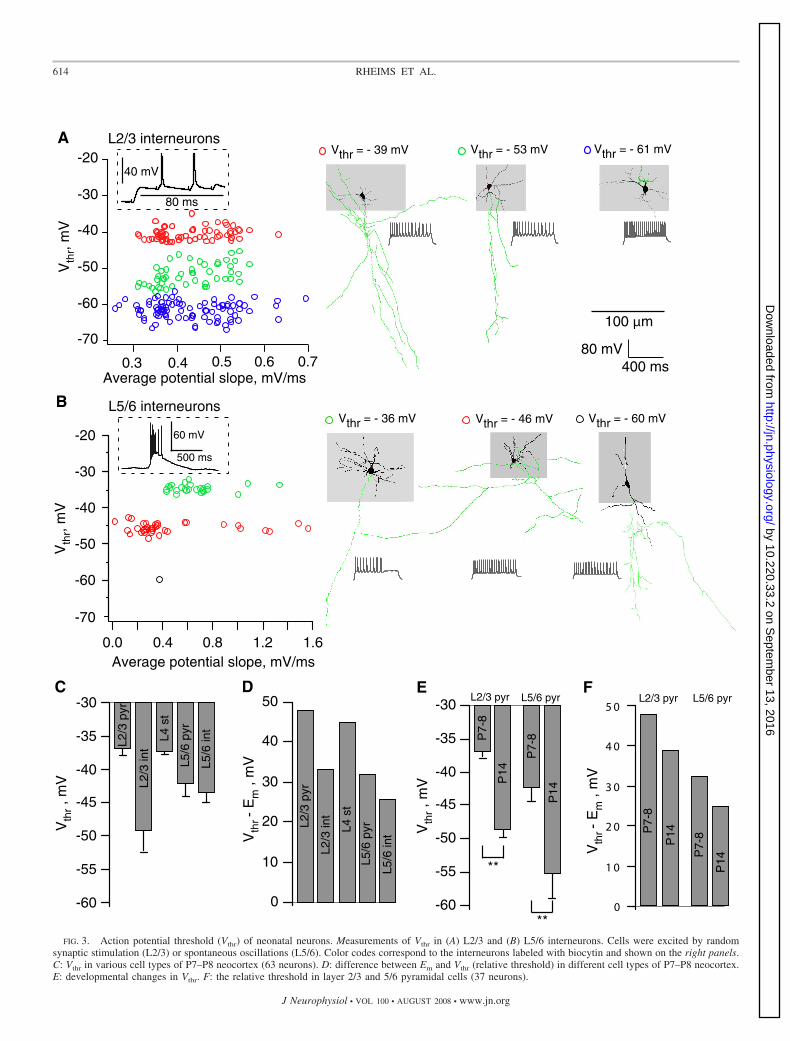
Figure 3A shows the dependence of Vthr on the averagepotential slope in APs induced in three L2/3 interneurons(P7–P8) by random synaptic stimulation (see inset). All inter-neurons (coded by different colors) were filled out with bio-cytin and reconstructed afterward. Representative reconstruc-tions of interneurons are shown in Fig. 3A. Scattering of theVthr values reflects the heterogeneity of interneuron population.The mean Vthr was �47.6 � 4 mV (n � 9). Note, however, thatsome interneurons subtypes (e.g., in blue on Fig. 3A) have aVthr at about �60 mV, indicating that the correspondinginterneurons are highly excitable. Measurements in L5/6 inter-neurons (Fig. 3B) also showed pronounced variability of theVthr values and corresponding heterogeneity of interneuronsubtypes. Primary morphological characterization of interneu-rons is provided in Supplementary Materials.1
Similar measurements in L2/3 pyramidal and L4 stellatecells at P7–P8 showed much smaller variability of the Vthrvalues within each neuron type (Fig. 3C). The mean Vthr valueswere �36.8 � 0.46 mV at neocortical pyramids (12 cells) and�37.4 � 0.4 mV at stellate cells (11 cells). Thus Vthr in bothneuron types are very positive, indicating that voltage devia-tions from Em of 40 mV are required for activating theseneurons (Fig. 3D). L5/6 pyramidal cells at P7–P8 had morenegative Vthr (P � 0.001 for L2/3 and L5/6 pyramids), �42 �1.9 mV (16 cells), with large variability between neurons (Fig.3C). Importantly, plotting the mean Vthr values relative to thecorresponding mean Em values (Fig. 3D) clearly shows thatL5/6 neurons are potentially more excitable than neurons inother layers. For pyramidal cells, this conclusion remains validat P14 animals (Fig. 3, E and F), although Vthr is gettingsignificantly more negative for both L2/3 (�48.3 � 1.2 mV,P � 0.001) and L5/6 (�54.7 � 3.7 mV, P � 0.001) pyramids.Neuron spiking could be induced by synaptic stimulation atearlier (�P5) ages. Unfortunately, a very high membraneresistance (1 GOhm) of neurons prevented the accurateestimation of Vthr at these ages.
Therefore deep layer pyramidal neurons are more excitablethan superficial ones because of a lower Vthr.
1 The online version of this article contains supplemental data.
611EXCITATORY GABA IN DEVELOPING NEOCORTEX IN VITRO
J Neurophysiol • VOL 100 • AUGUST 2008 • www.jn.org
by 10.220.33.2 on Septem
ber 13, 2016http://jn.physiology.org/
Dow
nloaded from

GABAergic transmission generates APs in L5/6 but not L2/3pyramidal neurons
The results above suggest that, at P7–P8, GABAergic trans-mission is depolarizing and presumably strong enough toinitiate cell firing. To directly test this hypothesis, we blockedglutamatergic transmission (5 �M NBQX � 50 �M APV) andstimulated slice by the extracellular electrode (Davies et al.1990). In most interneurons tested in L2/3 (10 of 11), extra-
cellular stimulation induced APs (Fig. 4A, cell-attached record-ings). This was prevented by application of 10 �M gabazine(data not shown). In contrast, such stimulation failed to induceAPs in L2/3 pyramidal cells (n � 5; Fig. 4A). In L5/6,extracellular stimulation reliably induced APs in both pyrami-dal cells and interneurons (Fig. 4A). Figure 4B represents asummary of these experiments, and Fig. 4C shows deviationsbetween the mean values of EGABA and Vthr in each cell type.
FIG. 1. Em and EGABA in neonatal neurons measured by cell-attached recordings of single N-methyl-D-aspartate (NMDA)- and GABA-activated channels. A: current–voltagerelationships of single NMDA- and GABA-activated channels. Inset: examples of single NMDA (left) and GABA (right) channel activity at various pipette potentials. Recordingsfrom the same P2 cortical plate pyramidal cell (left) and from the same P9 layer 2/3 pyramidal cell (right). B: Em in various cell types and layers of mouse P7–P8 neocorticalslices (97 neurons). C: values of EGABA. D: GABA driving force (DFGABA) in different cell types and layers of mouse neocortical slices at P7–P8 (59 neurons).
612 RHEIMS ET AL.
J Neurophysiol • VOL 100 • AUGUST 2008 • www.jn.org
by 10.220.33.2 on Septem
ber 13, 2016http://jn.physiology.org/
Dow
nloaded from

Note, that APs are generated in a fraction of L5/6 neurons,although their mean EGABA is more negative than mean Vthr.This can be explained by the large deviations of Vthr in neuronpopulations (from �35 to �60 mV for interneurons and from�31 to �58 mV for pyramids). It is also because of theactivation of Na channels that boost postsynaptic potentials(PSPs), thereby facilitating the generation of APs (Hausseret al. 2001; Williams and Stuart 2003). Indeed, Fig. 4D showswhole cell recordings in an L2/3 pyramidal cell and an inter-neuron. The pipette solution contained 20 mM Cl� that corre-sponded to an EGABA of about �49 mV (shown by dash lineson the figure). Nevertheless, GABAergic PSP values recordedat the soma exceeded EGABA and readily triggered APs.
Thus GABAergic synapses are able to initiate APs in theintact L2/3 interneurons but not in pyramidal cells. L5/6pyramidal cells seem to be more excitable than the L2/3 onessince they have been readily activated by GABAergic trans-mission.
Recurrent GABA driven oscillations in deep layer neurons
At P6–P8, field and cell-attached recordings showed recur-rent oscillations (each 20 s in average; �1-s duration) in themajority of slices (68/76; see Fig. 5A). These oscillations aregenerated by deep layer (L5/6) neurons since 1) simultaneousfield recordings show that they are most powerful in L5/6,much smaller in L4 and nearly absent in L2/3 (Fig. 5B); (4similar experiments; 13 experiments with simultaneous L2/3and L5/6 recordings); 2) combined field and dual cell-attachedrecordings showed that pyramidal cells and interneurons ofL2/3 are quiescent during oscillations (Fig. 5, C and D; 12/14interneurons were silent and 21/21 pyramids); field oscillationsin L4 are not associated with firing of stellate or pyramidalcells (Fig. 5, C and D; 7 cells); oscillations are always syn-chronous with firing of most pyramidal cells and interneuronsin L5/6 (Fig. 5, A and D; 23 pyramidal cells and 23 interneu-rons); 3) oscillations always sustain after resection of allsubcortical regions (n � 4; not shown); and 4) a mini-slice,prepared from a L5/6 slice region originally showing oscilla-tions (Supplementary Fig. S2), is still highly active (6 similar
experiments). In contrast, surgically isolated superficial layersdid not show any activity (3 experiments; data not shown).These results are summarized in Fig. 5D. Therefore the dom-inant activity pattern at that developmental stage is generatedby deep cortical layer neurons.
The following observations suggest that these oscillations,referenced as cortical GDPs (cGDPs), resemble the classicalGDPs recorded in hippocampus and other developing brainstructures (Ben-Ari et al. 1989; for review, see Ben-Ari 2002;Feller 1999). First the oscillations are controlled by GABAergicsignaling: 1) the cGDP frequency is significantly reduced bylow doses of gabazine (100 nM, n � 3; Fig. 6, A and B), aselective GABAAR antagonist; and by low doses of picrotoxin(3 �M, n � 4; Fig. 6B), a specific GABA channel blocker;2) the cGDP frequency is highly increased by diazepam(5 �M, n � 3; Fig. 6, A and B), a benzodiazepine efficientlyenhancing GABAergic currents; and 3) bumetanide (10 �M,n � 9; Fig. 6, A and B), which reduces the intracellularconcentration of chloride ([Cl�]i) by blocking activity ofNKCC1 (Dzhala et al. 2005; Hannaert et al. 2002; Yamadaet al. 2004), significantly suppresses cGDPs. Second, cGDPsrequire operative glutamatergic synapses since they are sup-pressed by APV (50 �M, n � 3; Fig. 6B) and abolished byNBQX (5 �M, n � 2; Fig. 6B). Third, like GDPs of thehippocampus (Ben-Ari et al. 1989; Khazipov et al. 2004a),cGDPs are present only transiently during maturation. Asshown in Fig. 6C, the peak occurrence of cGDPs is withinP6–P10 and they disappear by the end of second postnatalweek.
To ensure that the network oscillations are not animal strainspecific, we also tested the presence of cGDPs in P5–P8 ratslices and observed oscillations in all experiments (n � 7).Therefore cGDPs are generated by a mutual excitatory activityof pyramidal cells and interneurons in deep cortical layers.
D I S C U S S I O N
Using noninvasive single channel recordings, we showedthat GABA robustly depolarizes immature neocortical neurons.However, although strong GABAergic depolarizing drive was
FIG. 2. Developmental profile of Em and EGABA in pyramidal cells. A: developmental changes in Em and EGABA in layer 2/3 and 5/6 pyramidal cells (pooleddata from 166 cells). B: bumetanide, a NKCC1 chloride co-transporter antagonist, induces hyperpolarization of Em in P4 (7 cells) and P7/8 layer 5/6 pyramidalcells (15 cells) and strongly decreases EGABA in P7/8 layer 5/6 pyramidal cells (6 cells).
613EXCITATORY GABA IN DEVELOPING NEOCORTEX IN VITRO
J Neurophysiol • VOL 100 • AUGUST 2008 • www.jn.org
by 10.220.33.2 on Septem
ber 13, 2016http://jn.physiology.org/
Dow
nloaded from
Vthr = - 36 mV Vthr = - 46 mV Vthr = - 60 mV
Vthr = - 61 mVVthr = - 53 mVVthr = - 39 mV
100 µm
-70
-60
-50
-40
-30
-20
1.61.20.80.40.0Average potential slope, mV/ms
60 mV
500 ms
L5/6 interneurons
Vth
r,m
V
B
Average potential slope, mV/ms
Vth
r,m
V
L2/3 interneurons
-70
-60
-50
-40
-30
-20
0.70.60.50.40.3
40 mV
80 ms
A
80 mV400 ms
-60
-55
-50
-45
-40
-35
-30
Vth
r,m
V L2/3
int
L4st
L5/6
p yr
L2/3
pyr
L5/6
int
-60
-55
-50
-45
-40
-35
-30 L5/6 pyrL2/3 pyr
P7-
8
P14
P7-
8
P14
Vth
r,m
V
E50
40
30
20
10
0
P7-
8
P14
P7-
8
P14
L5/6 pyrL2/3 pyr
Vth
r-
Em
,mV
FD
Vth
r-
Em
,mV
50
40
30
20
10
0
L2/3
pyr
L2/3
int
L4st
L5/6
pyr
L5/6
int
C
**
**FIG. 3. Action potential threshold (Vthr) of neonatal neurons. Measurements of Vthr in (A) L2/3 and (B) L5/6 interneurons. Cells were excited by random
synaptic stimulation (L2/3) or spontaneous oscillations (L5/6). Color codes correspond to the interneurons labeled with biocytin and shown on the right panels.C: Vthr in various cell types of P7–P8 neocortex (63 neurons). D: difference between Em and Vthr (relative threshold) in different cell types of P7–P8 neocortex.E: developmental changes in Vthr. F: the relative threshold in layer 2/3 and 5/6 pyramidal cells (37 neurons).
614 RHEIMS ET AL.
J Neurophysiol • VOL 100 • AUGUST 2008 • www.jn.org
by 10.220.33.2 on Septem
ber 13, 2016http://jn.physiology.org/
Dow
nloaded from

observed in all cortical layers, spontaneous activity of neuronsin deep layers was generally associated with oscillations. Thisis because of important intrinsic differences between Em andVthr in neuronal populations. Em and Vthr are cell type specific,and the membrane voltage deviations required for neuronactivation by synaptic input vary in different neuron types—from 20 to 50 mV—suggesting major differences in intrin-sic excitability. Strong excitatory GABA signaling in combi-nation with high intrinsic excitability of deep layer neuronsgenerates a coherent network-driven activity in the immatureneocortex. This convergence seems to be the main condition-ing factor for selective generation of cGDPs by layer 5/6neurons. Oscillations typically remain confined to deep neo-cortical layers, requiring recurrent excitatory loops of pyrami-dal neurons and interneurons.
GABAergic transmission is strongly depolarizing overthe first postnatal week
To directly determine the action of GABA, we recordedNMDA- and GABA-activated channels in the same neuron(Fig. 1) that provide EGABA in absolute values. With thistechnique, we can compare alterations in DFGABA acrossneuronal populations and different developmental stages. Wefound that EGABA is 20 mV more depolarized than Em atP7–P8 in neurons of all cortical layers. Thus the GABAergictransmission provides depolarizing drive to quiescent neurons.For pyramidal neurons, the developmental profile of EGABA
showed gradual depolarization at earlier ages (Fig. 2A), andGABA was still depolarizing at the end of second postnatalweek. The shift in the mode of GABA action occurs mostlikely within the second week of life.
After the first postnatal week, Em of principal cells in alllayers did not differ significantly and stabilized at a negativelevel of about �80 mV (see Fig. 2A). In contrast to hippocam-pal neurons recorded with the same techniques (Tyzio et al.2003, 2006), Em of cortical neurons showed a developmentallyregulated trend to more hyperpolarized potentials in both L5/6and L2/3 pyramidal cells (Fig. 2A). This shift is chloridedependent, since bumetanide, which reduces [Cl�]i by block-ing activity of NKCC1 (Dzhala et al. 2005; Hannaert et al.2002), mimicked the age-dependent hyperpolarization: Em washyperpolarized by 10 mV at P4 and by 5 mV at P7/8 (Fig.2B). Although further experiments are required to explain theunderlying mechanism, it is possible that GABA-induced toniccurrent modulates Em in immature neurons as it regulatespyramidal cell excitability in adult somatosensory cortex (Salinand Prince 1996; Yamada et al. 2007).
Earlier studies have measured DFGABA using the gramicidinperforated-patch technique and reported values of �15 mV(Owens et al. 1996; Yamada et al. 2004) or even hyperpolar-izing (Daw et al. 2007b; Yamada et al. 2004) before the end offirst postnatal week. The differences with these observationsmay be explained by significant errors introduced by invasiverecording techniques. Indeed, the correct estimation of Em is ofa primary importance for showing the mode of GABA action,
L5 pyramidalL2/3 pyramidal
20ms40 pA
L2/3 interneuron L5 interneuron
NBQX + APVP7
A
C
100
80
60
40
20
0Cel
lsex
cit a
t ed
b yG
AB
A,%
L2/3
int
L2/3
pyr
L5/6
int
L5/6
pyr
-8
-6
-4
-2
0
L2/3
int
L2/3
pyr
L5/6
int
L5/6
pyr
EG
AB
A-
Vth
r,m
V
2
B
-60
-40
-20
0
mV
200 ms
L2/3 pyramidal cell
-60
-40
-20
0
mV
200 ms
40 ms
L2/3 interneuron
D
FIG. 4. Cell type and layer specificity of the excitatory GABA action in the immature neocortex. A: examples of responses to synaptic activation of GABAA
receptors (GABAARs; stimulation in the presence of NBQX and APV) in different cell types recorded in a cell-attached mode. B: summary plot of the proportionof cells excited by GABA in different cell types at P7 (36 cells). C: summary plot of the excitatory force for GABA (i.e., the difference between EGABA andVthr) in different cell types at P7 (54 cells). D: whole cell recordings in a L2/3 pyramidal cell and an interneuron. The pipette solution contained 20 mM Cl�
that corresponded to EGABA of about �49 mV (shown by dash lines on the figure). Nevertheless, GABAergic postsynaptic potential (PSP) values recorded atthe soma considerably exceeded EGABA and readily induced action potentials (APs).
615EXCITATORY GABA IN DEVELOPING NEOCORTEX IN VITRO
J Neurophysiol • VOL 100 • AUGUST 2008 • www.jn.org
by 10.220.33.2 on Septem
ber 13, 2016http://jn.physiology.org/
Dow
nloaded from

be it inhibitory or excitatory. The high-input resistance ofimmature neurons introduces significant errors in measure-ments of Em in whole cell or gramicidin perforated-patchrecordings (Tyzio et al. 2003). We tested these effects directlyin neocortical neurons by performing a dual recording from thesame neuron with cell-attached single channel and whole cellpipettes. Em measured with single NMDA channels was about�80 mV, but once an additional whole cell recording electrodewas added, Em depolarized by 20 mV and reached a value of56 mV (Tyzio et al. 2008). Clearly, Em of immature neocorticalneurons differs when measured by noninvasive or invasivetechniques. This also confirms the validity of single channelrecordings that provides a reliable estimation of Em. An alter-native invasive recording technique—the gramicidin perfo-rated patch—avoids the direct dialysis of internal chloride butmay indirectly change [Cl�]i via modification of [Na�]i and[K�]i. It is worth noting that [Cl�]i depends on the activity ofNKCC1 and KCC2 co-transporters, which in turn are sensitiveto alterations in [Na�]i or [K�]i (Brumback and Staley 2008;for review, see Adragna et al. 2004; Payne et al. 2003).
Deep layer pyramidal neurons are more excitablethan superficial ones
Despite its generally strong depolarizing action, the GABAergictransmission generates APs in some interneuron subtypes andin deep but not superficial layer pyramidal neurons (Fig. 4). Weobserved significant variations of Em and Vthr across differentcell types. At P7–P8, concomitant with the maximum intensityof cGDPs, the voltage deviation required for activating L2/3pyramidal or L4 stellate cells is 15 mV larger than that for L5/6pyramidal cells (Fig. 3D). Also, the Vthr values for somesubtypes of interneurons in L2/3 and L5/6 are very negative(Fig. 3, A and B) making these cells highly excitable. This isalso the case for most L5/6 pyramidal cells compared with
those in L2/3. Therefore although L5/6 interneurons are able tocreate excitatory loops with pyramidal cells, in L2/3, thisseems not to be possible. It is important to stress that, althoughEGABA is often not more negative than the spike threshold, itsdriving force is sufficient to generate action potentials becauseof activation of voltage-gated currents (Fig. 4D).
Results also indicated that Vthr is progressively shifted dur-ing maturation to more hyperpolarized values. Similar devel-opmental shift has been reported previously in L5 pyramidalneurons of the medial entorhinal cortex (Reboreda et al. 2007)and prefrontal cortex (Zhang 2004). The underlying mecha-nisms are not known but are consistent with the progressivematuration of Na� and K� channels (Picken Bahrey andMoody 2003; for review, see Moody and Bosma 2005). Inter-estingly, despite the large driving force of GABA in the veryimmature neurons (P3), the excitatory action of GABA was notobserved. Indeed, synaptic stimulation failed to initiate APs inpyramidal cells at P3 (n � 5) and activated only one of fourinterneurons tested. This is most likely because of a smalldensity of sodium channels and corresponding low excitabilityof these neurons. In fact, we could not determine the thresholdof action potential generation in these neurons since the inputresistance is very high (1 Gohm).
Therefore the developmental and cell type variations of Emand Vthr underlie higher excitability of deep layer neurons.
Deep cortical layers generate GABA-driven cGDPs
A variety of spontaneous patterns have been observed inneocortical slices (Aguilo et al. 1999; Corlew et al. 2004;Dupont et al. 2006; Garaschuk et al. 2000; Yuste et al. 1992).,However, cGDPs differ from activity patterns reported in thedeveloping neocortex previously, in their temporal/spatial pa-rameters and generating mechanisms. Thus cGDPs dominateneocortical activity around the end of first postnatal week,
FIG. 5. Cortical giant depolarizing potentials (GDPs)—the principal pattern of activity in the neonatal neocortex in vitro is generated by deep cortical layers.A: L5 local field potential (top) and cell-attached recordings from an L5 pyramidal cell (middle) and an interneuron (bottom) in a P8 mouse neocortical slice.cortical GDP (cGDP) outlined with a dashed line is shown on the right panel at expanded time scale. B: simultaneous field recordings at different layers indicatethat oscillations are most powerful in L5/6 (top), much smaller in L4 (middle), and nearly absent in L2/3 (bottom). C: dual cell-attached recordings in combinationwith field ones in L2/3 (left) and L4 (right). Cells in L2/3 and L4 are quiescent during cGDPs. Field recordings in B and C are low-pass filtered at 100 Hz.D: proportion of active cells during cGDPs in different layers at P7–P8.
616 RHEIMS ET AL.
J Neurophysiol • VOL 100 • AUGUST 2008 • www.jn.org
by 10.220.33.2 on Septem
ber 13, 2016http://jn.physiology.org/
Dow
nloaded from

contrasting with earlier patterns that peak at birth and disappearshortly after, like the synapse-independent coherent activity(Corlew et al. 2004; Yuste et al. 1992) or the layer 1–correlatedactivity (Aguilo et al. 1999).
In addition, cGDPs differ from the early cortical networksoscillations (ENOs, Adelsberger et al. 2005; Garaschuk et al.2000) since (1) ENOs peak at P1–P2 and disappear at P5–P6,whereas cGDPs are not observed before P4, peak at P7–P9, anddisappear at about P13; (2) ENOs occur at very low rates (ap-proximately once per 1–12 min), whereas cGDPs are observedonce per 6–40 s at P7–P9; (3) ENOs involve the entire networkof neurons whereas cGDPs are generated by deep cortical layers;and (4) GABAARs are not involved in the ENOs initiation; incontrast, cGDPs are inhibited by selective GABAAR antagonistsand bumetanide and are enhanced by diazepam (Fig. 6). CGDPsalso differ from the recently described carbachol-induced oscilla-tions (Dupont et al. 2006) that have a much longer duration, apropagation to the entire cortical mantle, and, across all layers, aninitial dependence on gap junctions and subsequently on NMDAbut not GABA receptors.
The sequential maturation of central oscillations has beensystematically studied recently in the developing hippocampusby fast dynamic imaging techniques (Crepel et al. 2007).Around delivery, the gap junction interconnected small cellassemblies generate nonsynaptically induced calcium signals
[synchronous plateau assemblies (SPA)] followed, at the endof first postnatal week, by synapse-driven GDPs (Crepel et al.2007). SPAs coexist with GDPs during a transient develop-mental stage. However, an increase in occurrence of GDPscorrelates with the decrease in SPA activity. Therefore theinitial synapse-driven oscillations in hippocampus are precededby nonsynapse driven patterns. Further studies are required tofind out whether a similar developmentally regulated sequenceexists in the immature neocortex as well.
The relevance of cGDPs to in vivo patterns remains to bedetermined. In the neonatal rat neocortex in vivo, the dominantpattern of electrical activity is spindle bursts. This transient coor-dinated activity controls the organization and shaping of sensoryand motor systems (Dupont et al. 2006; Khazipov et al. 2004b;Minlebaev et al. 2006). Delta-brushes, observed in human pretermneonates, may represent the alike pattern in humans playing asimilar role during formation of sensorimotor maps (Milh et al.2007). Although cGDPs and spindle bursts share certain commonfeatures, it is difficult to directly test this hypothesis in the motorcortex since functional thalamocortical connections are needed.Thalamocortical slices are available for the barrel, visual, audi-tory, and anterior cingulate cotices (Agmon and Connors 1991;Cruikshank et al. 2002; Daw et al. 2007a,b; Lee et al. 2007;MacLean et al. 2006) but not the motor cortex. Interestingly, wefound that glutamate puffs in thalamocortical slices of the barrel
30 µV
200 sec100 nM gabazine
15 µV
200 sec
controlField L5, P7control
1 µM diazepam
140
120
100
80
60
40
20
0
CT
R
BM
T
DZ
P
PT
X
GB
Z
AP
V
NB
QX
cGD
Pam
plitu
de,%
300
250
200
150
100
50
0
cGD
Pra
te,%
CT
R
BM
T
DZ
P
PT
X
GB
Z
AP
V
NB
QX
0
10
20
30
40
cGD
Pam
plitu
de,µ
V
P1-3
P4-5
P6-8
P9-10
P11-1
2
P13-1
5
cGD
Pfr
eque
ncy,
Hz
P1-3
P4-5
P6-8
P9-10
P11-1
2
P13-1
50.00
0.02
0.04
0.06
0.08
0.10
B
A
C
10 µV
100 sec
control
10 µM bumetanide
FIG. 6. Excitatory GABA is instrumental for generation of cGDPs. A: L5 local field potential recordings in control (top trace) and in the presence ofGABAergic transmission modulators: the GABAAR antagonist gabazine (100 nM), the positive GABAA channel effector diazepam (5 �M), and the NKCC1co-transporter antagonist bumetanide (10 �M). B: effects of GABAAR and GluR acting drugs on the occurrence (left) and amplitude (right) of cGDPs (pooleddata from 26 P7–P8 neocortical slices). C: frequency (left) and amplitude (right, 100-Hz low pass) of cGDPs as a function of age (pooled data from 64 slices).
617EXCITATORY GABA IN DEVELOPING NEOCORTEX IN VITRO
J Neurophysiol • VOL 100 • AUGUST 2008 • www.jn.org
by 10.220.33.2 on Septem
ber 13, 2016http://jn.physiology.org/
Dow
nloaded from

cortex (Agmon and Connors 1991) generate spindle-burst-likeoscillations (P7 mice; data not shown). Thus thalamic input caninitiate oscillations in deep neocortical layers, at least in the barrelcortex.
Thus our results indicate that GABA depolarizes and excitesneocortical neurons in an age-, layer-, and type-specific mannerand that excitatory GABA plays an important role in genera-tion of endogenous pattern of cGDPs in the developing neo-cortical networks. This is in agreement with our more generalhypothesis that, early in development, neuronal networks gen-erate specific patterns of activity driven by depolarizing GABA(Ben-Ari 2002; Ben-Ari et al. 2007). We suggest that cGDPs,which provide coordinated activation of neocortical neuronsand thus stimulate synaptic plasticity, participate in the activ-ity-dependent formation of neocortical circuitry.
A C K N O W L E D G M E N T S
We thank I. Jorquera for technical assistance in histological processing.
G R A N T S
This work was supported by Institut National de la Sante et de la RechercheMedicale and grants from Fondation pour la Recherche Medicale, AgenceNationale de la Recherche 2005 Neurosciences, neurologie et psychiatrie and Euro-pean Council LSH-CT-2006-037315 (EPICURE) FP6-Thematic priority LIFESCI-HEALTH.
R E F E R E N C E S
Adelsberger H, Garaschuk O, Konnerth A. Cortical calcium waves inresting newborn mice. Nat Neurosci 8: 988–990, 2005.
Adragna NC, Di Fulvio M, Lauf PK. Regulation of K-Cl cotransport: fromfunction to genes. J Membr Biol 201: 109–137, 2004.
Agmon A, Connors BW. Thalamocortical responses of mouse somatosensory(barrel) cortex in vitro. Neuroscience 41: 365–380, 1991.
Aguilo A, Schwartz TH, Kumar VS, Peterlin ZA, Tsiola A, Soriano E,Yuste R. Involvement of cajal-retzius neurons in spontaneous correlatedactivity of embryonic and postnatal layer 1 from wild-type and reeler mice.J Neurosci 19: 10856–10868, 1999.
Ben-Ari Y. Excitatory actions of gaba during development: the nature of thenurture. Nat Rev Neurosci 3: 728–739, 2002.
Ben-Ari Y. Basic developmental rules and their implications for epilepsy inthe immature brain. Epileptic Disord 8: 91–102, 2006.
Ben-Ari Y, Cherubini E, Corradetti R, Gaiarsa JL. Giant synaptic potentials inimmature rat CA3 hippocampal neurones. J Physiol 416: 303–325, 1989.
Ben-Ari Y, Gaiarsa JL, Tyzio R, Khazipov R. GABA: a pioneer transmitterthat excites immature neurons and generates primitive oscillations. PhysiolRev 87: 1215–1284, 2007.
Brumback AC, Staley KJ. Thermodynamic regulation of NKCC1-mediatedCl- cotransport underlies plasticity of GABA(A) signaling in neonatalneurons. J Neurosci 28: 1301–1312, 2008.
Burgard EC, Hablitz JJ. Developmental changes in NMDA and non-NMDAreceptor-mediated synaptic potentials in rat neocortex. J Neurophysiol 69:230–240, 1993.
Cang J, Renteria RC, Kaneko M, Liu X, Copenhagen DR, Stryker MP.Development of precise maps in visual cortex requires patterned spontane-ous activity in the retina. Neuron 48: 797–809, 2005.
Corlew R, Bosma MM, Moody WJ. Spontaneous, synchronous electricalactivity in neonatal mouse cortical neurones. J Physiol 560: 377–390, 2004.
Crepel V, Aronov D, Jorquera I, Represa A, Ben-Ari Y, Cossart R. Aparturition-associated nonsynaptic coherent activity pattern in the develop-ing hippocampus. Neuron 54: 105–120, 2007.
Cruikshank SJ, Rose HJ, Metherate R. Auditory thalamocortical synaptictransmission in vitro. J Neurophysiol 87: 361–384, 2002.
Davies CH, Davies SN, Collingridge GL. Paired-pulse depression of mono-synaptic GABA-mediated inhibitory postsynaptic responses in rat hip-pocampus. J Physiol 424: 513–531, 1990.
Daw MI, Scott HL, Isaac JT. Developmental synaptic plasticity at thethalamocortical input to barrel cortex: mechanisms and roles. Mol CellNeurosci 34: 493–502, 2007a.
Daw NW, Ashby MC, Isaac JTR. Coordinated developmental recruitment oflatent fast spiking interneurons in layer IV barrel cortex. Nat Neurosci 10:453–461, 2007b.
Dupont E, Hanganu IL, Kilb W, Hirsch S, Luhmann HJ. Rapid develop-mental switch in the mechanisms driving early cortical columnar networks.Nature 439: 79–83, 2006.
Dzhala VI, Talos DM, Sdrulla DA, Brumback AC, Mathews GC, BenkeTA, Delpire E, Jensen FE, Staley KJ. NKCC1 transporter facilitatesseizures in the developing brain. Nat Med 11: 1205–1213, 2005.
Feller MB. Spontaneous correlated activity in developing neural circuits.Neuron 22: 653–656, 1999.
Franceschetti S, Sancini G, Panzica F, Radici C, Avanzini G. Postnataldifferentiation of firing properties and morphological characteristics in layerV pyramidal neurons of the sensorimotor cortex. Neuroscience 83: 1013–1024, 1998.
Galli L, Maffei L. Spontaneous impulse activity of rat retinal ganglion cells inprenatal life. Science 242: 90–91, 1988.
Garaschuk O, Linn J, Eilers J, Konnerth A. Large-scale oscillatory calciumwaves in the immature cortex. Nat Neurosci 3: 452–459, 2000.
Hanganu IL, Ben-Ari Y, Khazipov R. Retinal waves trigger spindle bursts inthe neonatal rat visual cortex. J Neurosci 26: 6728–6736, 2006.
Hannaert P, Alvarez-Guerra M, Pirot D, Nazaret C, Garay RP. RatNKCC2/NKCC1 cotransporter selectivity for loop diuretic drugs. NaunynSchmiedebergs Arch Pharmacol 365: 193–199, 2002.
Hausser M, Major G, Stuart G. Differential shunting of EPSPs by actionpotentials. Science 291: 138–141, 2001.
Kandler K, Gillespie DC. Developmental refinement of inhibitory sound-localization circuits. Trends Neurosci 28: 290–296, 2005.
Katz LC, Shatz CJ. Synaptic activity and the construction of cortical circuits.Science 274: 1133–1138, 1996.
Khazipov R, Esclapez M, Caillard O, Bernard C, Khalilov I, Tyzio R, HirschJ, Dzhala V, Berger B, Ben-Ari Y. Early development of neuronal activity inthe primate hippocampus in utero. J Neurosci 21: 9770–9781, 2001.
Khazipov R, Khalilov I, Tyzio R, Morozova E, Ben-Ari Y, Holmes GL.Developmental changes in GABAergic actions and seizure susceptibility inthe rat hippocampus. Eur J Neurosci 19: 590–600, 2004a.
Khazipov R, Sirote A, Leinekugel X, Holmes GL, Ben-Ari Y, Buzsaki G.Early motor activity drives spindle bursts in the developing somatosensorycortex. Nature 432: 758–761, 2004b.
Lee CM, Chang WC, Chang KB, Shyu BC. Synaptic organization andinput-specific short-term plasticity in anterior cingulate cortical neuronswith intact thalamic inputs. Eur J Neurosci 25: 2847–2861, 2007.
Luhmann HJ, Prince DA. Postnatal maturation of the GABAergic system inrat neocortex. J Neurophysiol 65: 247–263, 1991.
MacLean JN, Fenstermaker V, Watson BO, Yuste R. A visual thalamo-cortical slice. Nat Methods 3: 129–134, 2006.
McCabe AK, Chisholm SL, Picken-Bahrey HL, Moody WJ. The self-regulating nature of spontaneous synchronized activity in developing mousecortical neurones. J Physiol 577: 155–167, 2006.
McCormick DA, Prince DA. Post-natal development of electrophysiologicalproperties of rat cerebral cortical pyramidal neurones. J Physiol 393:743–762, 1987.
Meister M, Wong RO, Baylor DA, Shatz CJ. Synchronous bursts of actionpotentials in ganglion cells of the developing mammalian retina. Science252: 939–943, 1991.
Milh M, Kaminska A, Huon C, Lapillonne A, Ben-Ari Y, Khazipov R.Rapid cortical oscillations and early motor activity in premature humanneonate. Cereb Cortex 17: 1582–1594, 2007.
Minlebaev M, Ben-Ari Y, Khazipov R. Network mechanisms of spindle-burst oscillations in the neonatal rat barrel cortex in vivo. J Neurophysiol 97:692–700, 2006.
Moody WJ, Bosma MM. Ion channel development, spontaneous activity, andactivity-dependent development in nerve and muscle cells. Physiol Rev 85:883–941, 2005.
Owens DF, Boyce LH, Davies CH, Kriegstein AR. Excitatory GABAresponses in embryonic and neonatal cortical slices demonstrated by gram-icidin perforated-patch recordings and calcium imaging. J Neurosci 16:6414–6423, 1996.
Owens DF, Kriegstein AR. Patterns of intracellular calcium fluctuation inprecursor cells of the neocortical ventricular zone. J Neurosci 18: 5374–5388, 1998.
Owens DF, Liu X, Kriegstein AR. Changing properties of GABAA receptor–mediated signaling during early neocortical development. J Neurophysiol82: 570–583, 1999.
618 RHEIMS ET AL.
J Neurophysiol • VOL 100 • AUGUST 2008 • www.jn.org
by 10.220.33.2 on Septem
ber 13, 2016http://jn.physiology.org/
Dow
nloaded from

Payne JA, Rivera C, Voipio J, Kaila K. Cation-chloride co-transporters inneuronal communication, development and trauma. Trends Neurosci 26:199–206, 2003.
Picken Bahrey HL, Moody WJ. Early development of voltage-gated ioncurrents and firing properties in neurons of the mouse cerebral cortex.J Neurophysiol 89: 1761–1773, 2003.
Reboreda A, Raouf R, Alonso A, Seguela P. Development of cholinergicmodulation and graded persistent activity in layer v of medial entorhinalcortex. J Neurophysiol 97: 3937–3947, 2007.
Rheims S, Represa A, Ben-Ari Y, Zilberter Y. Layer-specific generation andpropagation of seizures in slices of developing neocortex: role of excitatoryGABAergic synapses. J Neurophysiol doi:10.1152/jn.90403.2008.
Salin PA, Prince DA. Spontaneous GABAA receptor-mediated inhibitory cur-rents in adult rat somatosensory cortex. J Neurophysiol 75: 1573–1588, 1996.
Tyzio R, Cossart R, Khalilov I, Minlebaev M, Hubner CA, Represa A,Ben-Ari Y, Khazipov R. Maternal oxytocin triggers a transient inhibitoryswitch in GABA signaling in the fetal brain during delivery. Science 314:1788–1792, 2006.
Tyzio R, Ivanov A, Bernard C, Holmes GL, Ben-Ari Y, Khazipov R.Membrane potential of CA3 hippocampal pyramidal cells during postnataldevelopment. J Neurophysiol 90: 2964–2972, 2003.
Tyzio R, Minlebaev M, Rheims S, Ivanov A, Jorquera I, Holmes GL,Zilberter Y, Ben-Ari Y, Khazipov R. Postnatal change in somaticGABA signaling in the rat hippocampus. Eur J Neurosci 27: 2515–2528,2008.
Williams SR, Stuart GJ. Voltage- and site-dependent control of the somaticimpact of dendritic IPSPs. J Neurosci 23: 7358–7367, 2003.
Yamada J, Furukawa T, Ueno S, Yamamoto S, Fukuda A. Molecular basisfor the GABAA receptor-mediated tonic inhibition in rat somatosensorycortex. Cereb Cortex 17: 1782–1787, 2007.
Yamada KA, Okabe A, Toyoda H, Kilb W, Luhmann HJ, Fukuda A.Cl- uptake promoting depolarizing GABA actions inimmature rat neo-cortical neurones is mediated by NKCC1. J Physiol (Lond) 557: 829 –841, 2004.
Yuste R, Peinado A, Katz LC. Neuronal domains in developing neocortex.Science 257: 665–669, 1992.
Zhang ZW. Maturation of layer V pyramidal neurons in the rat prefrontalcortex: intrinsic properties and synaptic function. J Neurophysiol 91: 1171–1182, 2004.
Zhou FM, Hablitz JJ. Postnatal development of membrane properties of layerI neurons in rat neocortex. J Neurosci 16: 1131–1139, 1996.
619EXCITATORY GABA IN DEVELOPING NEOCORTEX IN VITRO
J Neurophysiol • VOL 100 • AUGUST 2008 • www.jn.org
by 10.220.33.2 on Septem
ber 13, 2016http://jn.physiology.org/
Dow
nloaded from
Copyright © 2022 FDOKUMEN