Differential Compartmentalization and Distinct Functions of GABA B Receptor Variants
13
Neuron 50, 589–601, May 18, 2006 ª2006 Elsevier Inc. DOI 10.1016/j.neuron.2006.04.014 Differential Compartmentalization and Distinct Functions of GABA B Receptor Variants Re ´ jan Vigot, 1,10 Samuel Barbieri, 1,10 Hans Bra ¨ uner-Osborne, 1,2,10 Rostislav Turecek, 1,3 Ryuichi Shigemoto, 4,5 Yan-Ping Zhang, 6 Rafael Luja ´n, 4,5,7 Laura H. Jacobson, 8 Barbara Biermann, 1 Jean-Marc Fritschy, 9 Claire-Marie Vacher, 1,11 Matthias Mu ¨ ller, 8 Gilles Sansig, 8 Nicole Guetg, 1 John F. Cryan, 8,12 Klemens Kaupmann, 8 Martin Gassmann, 1 Thomas G. Oertner, 6 and Bernhard Bettler 1, * 1 Department of Clinical-Biological Sciences Institute of Physiology Pharmazentrum University of Basel CH-4056 Basel Switzerland 2 Department of Medicinal Chemistry Danish University of Pharmaceutical Sciences DK-2100 Copenhagen Denmark 3 Institute of Experimental Medicine Academy of Sciences 142 20 Prague Czech Republic 4 Division of Cerebral Structure National Institute for Physiological Sciences Myodaiji, Okazaki 444-8585 Japan 5 CREST Japan Science and Technology Corporation Kawaguchi 332-0012 Japan 6 Friedrich Miescher Institute CH-4058 Basel Switzerland 7 Department Ciencias Me ´ dicas Facultad de Medicina-CRIB Universidad de Castilla-La Mancha 02006 Albacete Spain 8 Novartis Institutes for BioMedical Research Novartis Pharma AG CH-4002 Basel Switzerland 9 Institute of Pharmacology and Toxicology University of Zurich CH-8057 Zurich Switzerland Summary GABA B receptors are the G protein-coupled receptors for the main inhibitory neurotransmitter in the brain, g-aminobutyric acid (GABA). Molecular diversity in the GABA B system arises from the GABA B1a and GABA B1b subunit isoforms that solely differ in their ectodomains by a pair of sushi repeats that is unique to GABA B1a . Using a combined genetic, physiological, and morphological approach, we now demonstrate that GABA B1 isoforms localize to distinct synaptic sites and convey separate functions in vivo. At hippocampal CA3-to-CA1 synapses, GABA B1a assembles heterore- ceptors inhibiting glutamate release, while predomi- nantly GABA B1b mediates postsynaptic inhibition. Electron microscopy reveals a synaptic distribution of GABA B1 isoforms that agrees with the observed functional differences. Transfected CA3 neurons se- lectively express GABA B1a in distal axons, suggesting that the sushi repeats, a conserved protein interaction motif, specify heteroreceptor localization. The consti- tutive absence of GABA B1a but not GABA B1b results in impaired synaptic plasticity and hippocampus- dependent memory, emphasizing molecular differen- ces in synaptic GABA B functions. Introduction GABA B receptors are considered promising drug targets for the treatment of neurological and mental health disor- ders (Bettler et al., 2004; Cryan and Kaupmann, 2005). Presynaptic GABA B receptors are subdivided into auto- and heteroreceptors that control the release of GABA and other neurotransmitters, respectively. They restrict neu- rotransmitter release either by inhibiting voltage-sensi- tive Ca 2+ channels or through a direct modulation of syn- aptic vesicle priming (Mintz and Bean, 1993; Poncer et al., 1997; Sakaba and Neher, 2003). Postsynaptic GABA B receptors induce slow inhibitory potentials by gating Kir3-type K + channels (Lu ¨ scher et al., 1997). Consider- able evidence has accumulated over the years, using a variety of preparations and techniques, to support the notion that multiple subtypes of GABA B receptors exist (Bonanno and Raiteri, 1993; Bowery et al., 2002; Cunning- ham and Enna, 1996; Deisz et al., 1997; Gemignani et al., 1994; Lei and McBain, 2003; Mohler and Fritschy, 1999; Pozza et al., 1999; Yamada et al., 1999). The predicted re- ceptor heterogeneity is not readily supported by molecu- lar studies (Bettler et al., 2004). GABA B receptors are het- erodimers composed of GABA B1 and GABA B2 subunits, which are both required for normal receptor functioning (Marshall et al., 1999; Mohler and Fritschy, 1999). Accord- ingly, mice lacking GABA B1 (referred to as 1 2/2 mice) or GABA B2 subunits show a complete absence of typical GABA B responses (Gassmann et al., 2004; Prosser et al., 2001; Schuler et al., 2001). The only firmly estab- lished molecular diversity in the GABA B system arises from the GABA B1a and GABA B1b subunit isoforms (Kaup- mann et al., 1997). However, no unique pharmacological or functional properties could be assigned to GABA B1a or GABA B1b . Most, if not all neurons coexpress GABA B1a and GABA B1b , which are generated by differential pro- moter usage from the GABA B1 gene (Bischoff et al., 1999; Steiger et al., 2004). GABA B1a and GABA B1b *Correspondence: [email protected] 10 These authors contributed equally to this work. 11 Present address: Neurobiologie de l’Olfaction et de la Prise Alimentaire, University of Paris-Sud 11, 91405 Orsay cedex, France. 12 Present address: Department of Pharmacology and Therapeutics, School of Pharmacy, University College Cork, Cork, Ireland.
-
Upload
aitthailand -
Category
Documents
-
view
0 -
download
0
Transcript of Differential Compartmentalization and Distinct Functions of GABA B Receptor Variants
Neuron 50, 589–601, May 18, 2006 ª2006 Elsevier Inc. DOI 10.1016/j.neuron.2006.04.014
Differential Compartmentalizationand Distinct Functions of GABAB Receptor Variants
Rejan Vigot,1,10 Samuel Barbieri,1,10
Hans Brauner-Osborne,1,2,10 Rostislav Turecek,1,3
Ryuichi Shigemoto,4,5 Yan-Ping Zhang,6
Rafael Lujan,4,5,7 Laura H. Jacobson,8
Barbara Biermann,1 Jean-Marc Fritschy,9
Claire-Marie Vacher,1,11 Matthias Muller,8
Gilles Sansig,8 Nicole Guetg,1 John F. Cryan,8,12
Klemens Kaupmann,8 Martin Gassmann,1
Thomas G. Oertner,6 and Bernhard Bettler1,*1Department of Clinical-Biological SciencesInstitute of PhysiologyPharmazentrumUniversity of BaselCH-4056 BaselSwitzerland2Department of Medicinal ChemistryDanish University of Pharmaceutical SciencesDK-2100 CopenhagenDenmark3 Institute of Experimental MedicineAcademy of Sciences142 20 PragueCzech Republic4Division of Cerebral StructureNational Institute for Physiological SciencesMyodaiji, Okazaki 444-8585Japan5CRESTJapan Science and Technology CorporationKawaguchi 332-0012Japan6Friedrich Miescher InstituteCH-4058 BaselSwitzerland7Department Ciencias MedicasFacultad de Medicina-CRIBUniversidad de Castilla-La Mancha02006 AlbaceteSpain8Novartis Institutes for BioMedical ResearchNovartis Pharma AGCH-4002 BaselSwitzerland9 Institute of Pharmacology and ToxicologyUniversity of ZurichCH-8057 ZurichSwitzerland
Summary
GABAB receptors are the G protein-coupled receptors
for the main inhibitory neurotransmitter in the brain,
*Correspondence: [email protected] These authors contributed equally to this work.11 Present address: Neurobiologie de l’Olfaction et de la Prise
Alimentaire, University of Paris-Sud 11, 91405 Orsay cedex, France.12 Present address: Department of Pharmacology and Therapeutics,
School of Pharmacy, University College Cork, Cork, Ireland.
g-aminobutyric acid (GABA). Molecular diversity inthe GABAB system arises from the GABAB1a and
GABAB1b subunit isoforms that solely differ in theirectodomains by a pair of sushi repeats that is unique
to GABAB1a. Using a combined genetic, physiological,andmorphologicalapproach,wenowdemonstrate that
GABAB1 isoforms localize to distinct synaptic sites andconvey separate functions in vivo. At hippocampal
CA3-to-CA1 synapses, GABAB1a assembles heterore-
ceptors inhibiting glutamate release, while predomi-nantly GABAB1b mediates postsynaptic inhibition.
Electron microscopy reveals a synaptic distributionof GABAB1 isoforms that agrees with the observed
functional differences. Transfected CA3 neurons se-lectively express GABAB1a in distal axons, suggesting
that the sushi repeats, a conserved protein interactionmotif, specify heteroreceptor localization. The consti-
tutive absence of GABAB1a but not GABAB1b resultsin impaired synaptic plasticity and hippocampus-
dependent memory, emphasizing molecular differen-ces in synaptic GABAB functions.
Introduction
GABAB receptors are considered promising drug targetsfor the treatment of neurological and mental health disor-ders (Bettler et al., 2004; Cryan and Kaupmann, 2005).Presynaptic GABAB receptors are subdivided into auto-and heteroreceptors that control the releaseof GABAandother neurotransmitters, respectively. They restrict neu-rotransmitter release either by inhibiting voltage-sensi-tive Ca2+ channels or through a direct modulation of syn-aptic vesicle priming (Mintz and Bean, 1993; Poncer et al.,1997; Sakaba and Neher, 2003). Postsynaptic GABAB
receptors induce slow inhibitory potentials by gatingKir3-type K+ channels (Luscher et al., 1997). Consider-able evidence has accumulated over the years, usinga variety of preparations and techniques, to support thenotion that multiple subtypes of GABAB receptors exist(Bonanno and Raiteri, 1993; Boweryet al., 2002; Cunning-ham and Enna, 1996; Deisz et al., 1997; Gemignani et al.,1994; Lei and McBain, 2003; Mohler and Fritschy, 1999;Pozza et al., 1999; Yamada et al., 1999). The predicted re-ceptor heterogeneity is not readily supported by molecu-lar studies (Bettler et al., 2004). GABAB receptors are het-erodimers composed of GABAB1 and GABAB2 subunits,which are both required for normal receptor functioning(Marshall et al., 1999; Mohler and Fritschy, 1999). Accord-ingly, mice lacking GABAB1 (referred to as 12/2 mice) orGABAB2 subunits show a complete absence of typicalGABAB responses (Gassmann et al., 2004; Prosseret al., 2001; Schuler et al., 2001). The only firmly estab-lished molecular diversity in the GABAB system arisesfrom the GABAB1a and GABAB1b subunit isoforms (Kaup-mann et al., 1997). However, no unique pharmacologicalor functional properties could be assigned to GABAB1a orGABAB1b. Most, if not all neurons coexpress GABAB1a
and GABAB1b, which are generated by differential pro-moter usage from the GABAB1 gene (Bischoff et al.,1999; Steiger et al., 2004). GABAB1a and GABAB1b
Neuron590
expression levels vary during development and acrossindividual cells, suggestive of a functional specialization.Structurally, the isoforms differ in their N-terminal ecto-domain by a pair of sushi repeats that is present inGABAB1a but not in GABAB1b (Blein et al., 2004). Sushi re-peats, also known as complement control protein mod-ules, or short consensus repeats, are found in other Gprotein-coupled receptors as well (Grace et al., 2004)and mediate protein interactions in a wide variety of ad-hesion proteins (Lehtinen et al., 2004). The presence ofsushi repeats in GABAB1a, together with the absence offunctional or pharmacological differences in vitro, sug-gested the existence of auxiliary proteins that modify re-ceptor activity, pharmacology, and localization (Marshallet al., 1999; Mohler and Fritschy, 1999), precedence forwhich is found with other G protein-coupled receptors(McLatchie et al., 1998). So far, the lack of selective re-agents has not allowed addressing the individual contri-butions of GABAB1a and GABAB1b to native GABAB func-tions. In the light of the proposed heterogeneity of nativeGABAB receptors, it therefore remains a key questionwhether GABAB1 isoforms exhibit pharmacological and/or functional differences in vivo. Here, we have taken agenetic approach to dissociate the native functions ofGABAB1a and GABAB1b.
Results
Generation of Mice Selectively Expressing GABAB1a
or GABAB1b SubunitsTo selectively prevent translation of the GABAB1a andGABAB1b proteins, we converted their initiation codonsin the GABAB1 gene into stop codons (Figure 1). Balb/cgene targeting constructs with mutated initiation codons(Figure 1A) were electroporated into Balb/c embryonicstem cells (Dinkel et al., 1999) and homologous recombi-nation events diagnosed with short-arm PCR and South-ern blots (data not shown). Targeted embryonic stemcells were injected into C57BL/6 blastocysts. Foundermice were crossed with Balb/c mice expressing Cre-recombinase under control of the cytomegalus virus pro-moter to excise the neomycin cassette. Pups born fromthese matings were scored for Cre-mediated loss ofthe neomycin cassette and bred to homozygosity. Con-sequently, all mutant mice were on a pure inbred Balb/cgenetic background, which was maintained throughoutthe experiments. Homozygous mice with mutations inthe GABAB1a (referred to as 1a2/2 mice) or GABAB1b
(1b2/2 mice) initiation codon were viable, reproducednormally, and exhibited no overt phenotypic abnormali-ties. Mutant mice showed normal levels of GABAB1a
and GABAB1b mRNA, indicating that the genetic manip-ulations do not influence mRNA expression or stability(Figure 1B). Immunoblot analysis revealed the total ab-sence of GABAB1a and GABAB1b protein in 1a2/2 and1b2/2 mice, respectively, confirming that mutation ofthe initiation codons prevents translation of the individ-ual subunits (Figure 1C). GABAB1a and GABAB1b proteinsappeared upregulated in total brain extracts of knockoutmice (Figure 1C), possibly because of increased avail-ability of complementary GABAB2 protein, which is re-quired for cross-stabilization (Gassmann et al., 2004).We analyzed whether GABAB protein is also upregulatedin the CA1 region of the hippocampus, where the electro-
physiological and morphological studies described be-low were carried out. Similar to those seen in total brainextracts, GABAB1a and GABAB1b protein levels in CA1extracts were increased in the 1b2/2 (129% of wild-type) and 1a2/2 mice (115% of wild-type), respectively(Figure S1).
Immunohistochemical, Pharmacological, and
Biochemical Characterization of 1a2/2 and 1b2/2
Mice
Immunohistochemistry in the CA1 and CA3 region of thehippocampus revealed completely overlapping expres-sion patterns for the GABAB1a and GABAB1b proteins(Figure 2), consistent with an ubiquitous expression ofthe two proteins in brain neurons (Bischoff et al., 1999).The regional immunostaining in 1a2/2 and wild-typemice was similar, while the staining in 1b2/2 mice wasmore diffuse and lacked distinct laminar boundaries. Im-munohistochemistry therefore suggests differences inthe relative abundance of the two isoform proteins at dif-ferent subcellular sites. For example, intense immunore-activity is evident in CA3 stratum lucidum of 1b2/2 mice,which may hint at a preferential expression of GABAB1a
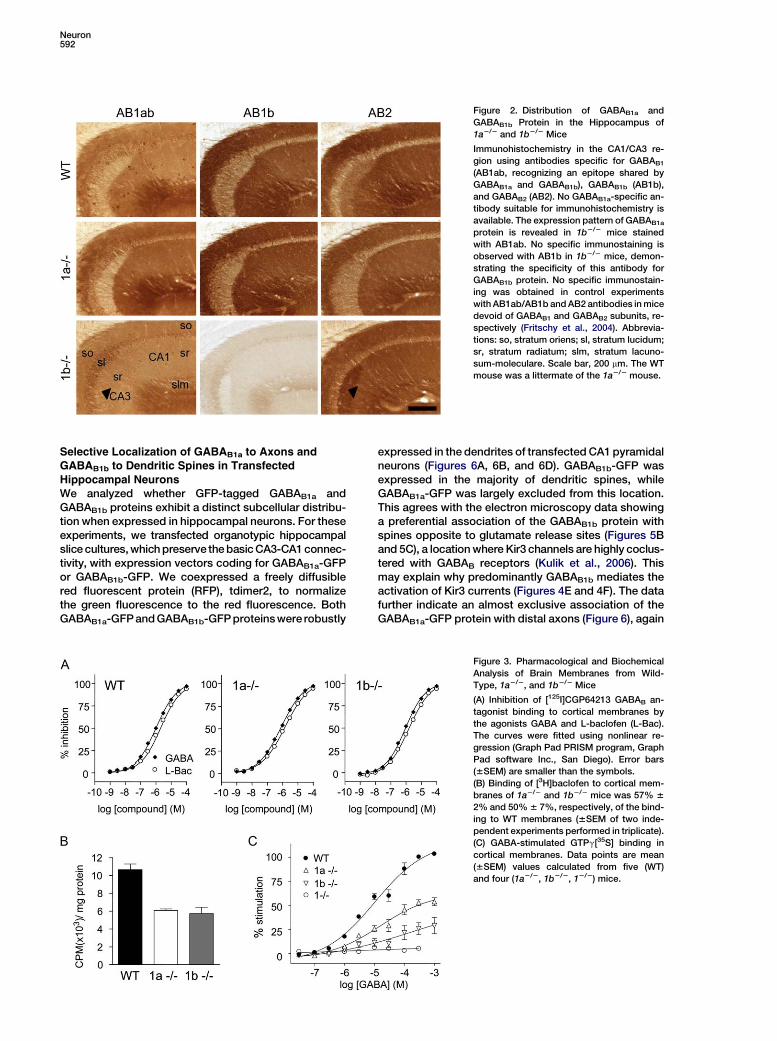
protein at presynaptic sites (arrowhead in Figure 2).The immunostainings obtained with antibodies directedat the GABAB1 and GABAB2 proteins are similar in the dif-ferent strains of mice, suggesting that most of theGABAB2 and GABAB1 protein assembles into heterodi-meric receptors. To compare the pharmacology ofGABAB1a and GABAB1b in native tissue, we analyzed theinhibition of [125I]CGP64213 antagonist binding (Kaup-mann et al., 1997) by GABA and L-baclofen in corticalmembranes (Figure 3A). In agreement with recombinantdata (Kaupmann et al., 1998), the inhibition curves forwild-type, 1a2/2, and 1b2/2 mice were almost identical(IC50 values for wild-type, 1a2/2, and 1b2/2 mice in mMare as follows: GABA: 0.7 6 0.2, 0.4 6 0.2, 0.6 6 0.2; ba-clofen: 1.2 6 0.3, 0.8 6 0.3, 0.9 6 0.3; n = 3 per genotype).[3H]baclofen binding in 1a2/2 and 1b2/2 cortical mem-branes was similarly reduced compared to wild-typemembranes (Figure 3B), in agreement with the relativeabundance of the two isoform proteins in the cortex(Kaupmann et al., 1997). To determine functional GABAB
receptor levels, we measured GTPg[35S] binding, whichassesses the activation of Gai/o-type G proteins, themain effectors of GABAB receptors (Figure 3C). Corticalmembranes of 1a2/2 and 1b2/2 mice showed 52% 64% and 28% 6 8% of the maximal GTPg[35S] bind-ing seen with wild-type mice. The sum of the maximalGTPg[35S] responses in knockout membranes is there-fore 20% lower than expected. This suggests the ab-sence of a compensatory upregulation of functionalreceptor levels, despite the upregulation of GABAB1 iso-forms seen at the protein level (Figure 1C and Figure S1 inthe Supplemental Data available with this article online).Presumably, most of the extra GABAB1 isoform proteinis retained intracellularly and does not participate infunctional responses.
Distinct Contributions of GABAB1a and GABAB1b
to Pre- and Postsynaptic GABAB FunctionsUsing whole-cell patch-clamp recording in slice prepa-rations, we examined whether wild-type and knockoutmice differ in their hippocampal GABAB responses. We
Pre- and Postsynaptic Functions of GABAB Variants591
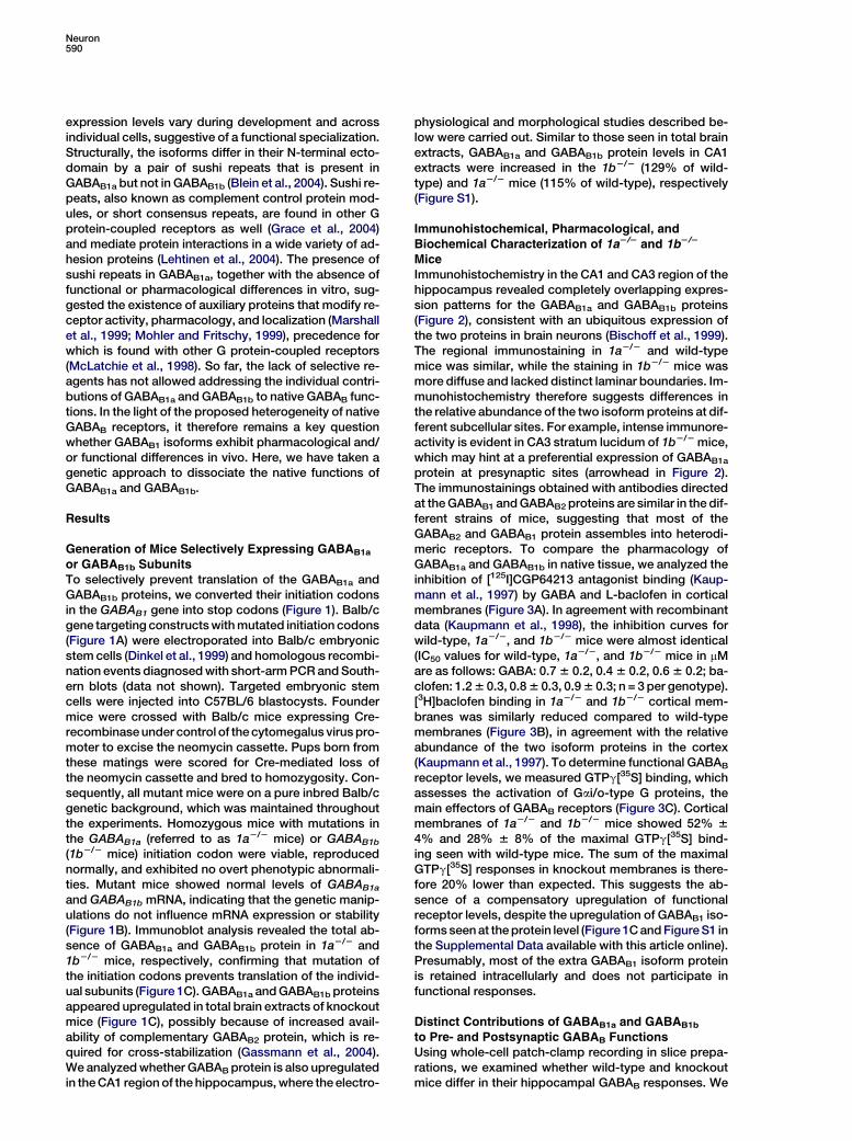
Figure 1. Generation of 1a2/2 and 1b2/2 Mice
(A) 50 region of wild-type (WT) (Martin et al.,
2001) and mutated GABAB1 alleles. Exons en-
coding the N terminus of GABAB1a are repre-
sented by white boxes and specify the signal
peptide (exon 2a), a pair of sushi repeats of
75 amino acids each (exons 3a, 4a), and a
linker of six amino acids (exon 5a). The exon
specifying the N terminus of GABAB1b is rep-
resented by a gray box. All exons downstream
of exon 1b are shared between the two iso-
forms (only exon 6 is shown; hatched box).
Start codons for GABAB1a (Ma) and GABAB1b
(Mb) transcripts were converted into stop
codons (S) using a knockin approach. A puta-
tive alternative start site (Ma*) in GABAB1a
transcripts was mutated in addition. The
floxed neomycin cassette (black bar) for
selection of transfected embryonic stem cells
was introduced in the introns between exons
2a/3a (1a2/2neo) or exons 5a/1b (1b2/2neo).
A loxP site (arrow) is left behind after Cre-
mediated excision of the neomycin cassette
(1a2/2, 1b2/2).
(B) Northern blot analysis of GABAB1a and GABAB1b mRNA expression in the brain of WT, heterozygous (+/2), and homozygous (2/2) knockout
mice. The 1a hybridization probe (1a probe) corresponds to nucleotides 1–405 of the GABAB1a cDNA (Kaupmann et al., 1997) and detects GABAB1a
as well as a truncated GABAB1j transcript (M.G., unpublished data) of w1.6 kb (upper panel). The 1b probe corresponds to nucleotides 16–259 of
the GABAB1b cDNA (Kaupmann et al., 1997) and detects 1b transcripts (lower panel).
(C) Immunoblot analysis of total brain lysates using antibodies recognizing the common C terminus of GABAB1a and GABAB1b (AB1ab) (Gassmann
et al., 2004). Anti-syntaxin (ABstx) antibodies control for sample loading.
first checked for the presence of heteroreceptors on ex-citatory terminals. Stimulation of the Schaffer collateral-commissural fibers induces excitatory postsynapticcurrents (EPSCs) in CA1 pyramidal neurons, which arereduced by blocking glutamate release through activa-tion of GABAB heteroreceptors (Schuler et al., 2001).Baclofen, a GABAB agonist, was effective in reducingthe EPSC amplitude in wild-type and 1b2/2 mice butnot in 1a2/2 mice (Figures 4A and 4B). As a control, aden-osine inhibited glutamate release in all three genotypes.This indicates that 1a2/2 mice, in contrast to 1b2/2 mice,lack GABAB heteroreceptors on Schaffer collateral ter-minals. Small residual heteroreceptor activity in 1a2/2
mice suggests that minute amounts of GABAB receptorsassembled with GABAB1b are localized at glutamatergicterminals. We next looked for the presence of autorecep-tors on GABAergic terminals and recorded inhibitorypostsynaptic currents (IPSCs) in the presence of theionotropic glutamate receptor antagonist kynurenate.Baclofen reduced the amplitude of IPSCs in CA1 pyrami-dal neurons of all genotypes, suggesting that bothGABAB1a and GABAB1b can efficiently participate in au-toreceptor function (Figures 4C and 4D). PostsynapticGABAB receptors induce a late IPSC by activating Kir3-type K+ channels (Luscher et al., 1997). At a holding po-tential of 250 mV and in physiological extracellular[K+], baclofen elicited similar outward currents in CA1pyramidal cells of 1a2/2 and wild-type mice (Figures 4Eand 4F). However, in CA1 pyramidal cells of 1b2/2 mice,the baclofen-induced outward current was reducedby w60% compared to wild-type or 1a2/2 mice. This in-dicates that predominantly GABAB1b mediates postsyn-aptic inhibition. As a control, adenosine receptors, whichconverge on the same Kir3 channels (Luscher et al.,1997), induced similar outward currents in all genotypes.It is formally possible that the upregulation of GABAB1a
protein observed in the 1b2/2 mice (Figure 1C andFigure S1) compensates to some extent for the missingGABAB1b protein. We consider this unlikely becausefunctional receptor levels in the 1b2/2 mice are lowerthan expected (Figure 3C). Moreover, GFP-taggedGABAB1a protein clearly distributes to the dendritic com-partment of CA1 neurons when expressed in organo-typic slice culture (Figure 6A). Likely, therefore, bothGABAB1a- and GABAB1b-containing receptors addressKir3 channels under normal conditions.
Distinct Subcellular Compartmentalizationof the GABAB1a and GABAB1b Proteins
The lack of suitable antibodies thus far prevented study-ing the distribution of GABAB1 isoforms using electronmicroscopy. We now used the 1a2/2 and 1b2/2 miceto determine the subcellular localization of GABAB1b
and GABAB1a protein, respectively. Preembeddingimmunogold labeling experiments in the CA1 stratum ra-diatum of wild-type mice confirmed that GABAB1 proteinis present in pre- and postsynaptic elements (Figure 5A),as reported for rat brain (Kulik et al., 2003). In 1a2/2 mice,GABAB1b was mostly found in spines opposite gluta-mate release sites (Figures 5B and 5C). In 1b2/2 mice,GABAB1a predominantly localized to glutamatergicterminals (Figures 5D and 5E). Quantitative analysisof GABAB1 labeling showed that the ratio of pre- topostsynaptic immunoparticles in wild-type, 1a2/2, and1b2/2 mice was 0.31, 0.17, and 1.61, respectively(Figure 5F). Thus, the electron microscopy data supportthe electrophysiological data (Figure 4) and confirmthat GABAB1a preferentially localizes to glutamatergicterminals. Consistent with residual heteroreceptoractivity (Figures 4A and 4B), some presynaptic im-munogold labeling persisted at glutamatergic 1a2/2
synapses.
Neuron592
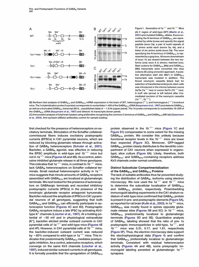
Figure 2. Distribution of GABAB1a and
GABAB1b Protein in the Hippocampus of
1a2/2 and 1b2/2 Mice
Immunohistochemistry in the CA1/CA3 re-
gion using antibodies specific for GABAB1
(AB1ab, recognizing an epitope shared by
GABAB1a and GABAB1b), GABAB1b (AB1b),
and GABAB2 (AB2). No GABAB1a-specific an-
tibody suitable for immunohistochemistry is
available. The expression pattern of GABAB1a
protein is revealed in 1b2/2 mice stained
with AB1ab. No specific immunostaining is
observed with AB1b in 1b2/2 mice, demon-
strating the specificity of this antibody for
GABAB1b protein. No specific immunostain-
ing was obtained in control experiments
with AB1ab/AB1b and AB2 antibodies in mice
devoid of GABAB1 and GABAB2 subunits, re-
spectively (Fritschy et al., 2004). Abbrevia-
tions: so, stratum oriens; sl, stratum lucidum;
sr, stratum radiatum; slm, stratum lacuno-
sum-moleculare. Scale bar, 200 mm. The WT
mouse was a littermate of the 1a2/2 mouse.
Selective Localization of GABAB1a to Axons andGABAB1b to Dendritic Spines in Transfected
Hippocampal NeuronsWe analyzed whether GFP-tagged GABAB1a andGABAB1b proteins exhibit a distinct subcellular distribu-tion when expressed in hippocampal neurons. For theseexperiments, we transfected organotypic hippocampalslice cultures, which preserve the basic CA3-CA1 connec-tivity, with expression vectors coding for GABAB1a-GFPor GABAB1b-GFP. We coexpressed a freely diffusiblered fluorescent protein (RFP), tdimer2, to normalizethe green fluorescence to the red fluorescence. BothGABAB1a-GFP and GABAB1b-GFP proteins were robustly
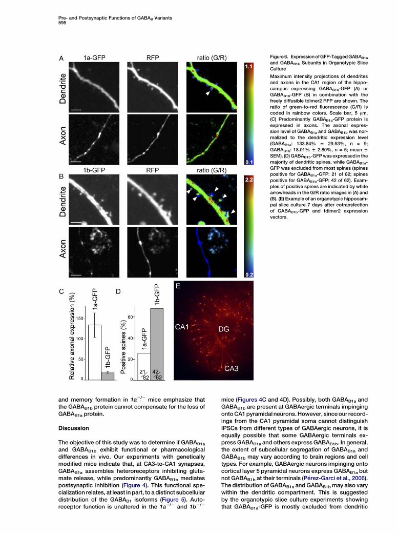
expressed in the dendrites of transfected CA1 pyramidalneurons (Figures 6A, 6B, and 6D). GABAB1b-GFP wasexpressed in the majority of dendritic spines, whileGABAB1a-GFP was largely excluded from this location.This agrees with the electron microscopy data showinga preferential association of the GABAB1b protein withspines opposite to glutamate release sites (Figures 5Band 5C), a location where Kir3 channels are highly coclus-tered with GABAB receptors (Kulik et al., 2006). Thismay explain why predominantly GABAB1b mediates theactivation of Kir3 currents (Figures 4E and 4F). The datafurther indicate an almost exclusive association of theGABAB1a-GFP protein with distal axons (Figure 6), again
Figure 3. Pharmacological and Biochemical
Analysis of Brain Membranes from Wild-
Type, 1a2/2, and 1b2/2 Mice
(A) Inhibition of [125I]CGP64213 GABAB an-
tagonist binding to cortical membranes by
the agonists GABA and L-baclofen (L-Bac).
The curves were fitted using nonlinear re-
gression (Graph Pad PRISM program, Graph
Pad software Inc., San Diego). Error bars
(6SEM) are smaller than the symbols.
(B) Binding of [3H]baclofen to cortical mem-
branes of 1a2/2 and 1b2/2 mice was 57% 6
2% and 50% 6 7%, respectively, of the bind-
ing to WT membranes (6SEM of two inde-
pendent experiments performed in triplicate).
(C) GABA-stimulated GTPg[35S] binding in
cortical membranes. Data points are mean
(6SEM) values calculated from five (WT)
and four (1a2/2, 1b2/2, 12/2) mice.

Pre- and Postsynaptic Functions of GABAB Variants593
Figure 4. GABAB Responses in Wild-Type, 1a2/2, and 1b2/2 CA1 Pyramidal Neurons
(A and B) Peak amplitudes and representative traces (A) and summary histogram (B) of monosynaptic EPSC inhibition by baclofen and adeno-
sine. Baclofen (50 mM) depresses the amplitude of EPSCs in WT (76.5% 6 3.1% inhibition; n = 8) and 1b2/2 (83.4% 6 2.9% inhibition; n = 5) but
not in 1a2/2 (15.9% 6 5.3% inhibition; n = 13; p < 0.001, ANOVA/Scheffe post hoc test) mice. Adenosine (100 mM) depresses EPSCs in all geno-
types (WT: 89.1% 6 1.6% inhibition, n = 6; 1a2/2: 85.3% 6 1.8% inhibition, n = 13; 1b2/2: 85.6% 6 6.6% inhibition, n = 4).
(C and D) Peak amplitudes and representative traces (C) and summary histogram (D) of IPSC inhibition by baclofen. Baclofen significantly de-
presses the IPSC amplitude in all genotypes (WT: 82.7% 6 4.8% inhibition, n = 12; 1a2/2: 71.8% 6 2.3% inhibition, n = 9; 1b2/2 mice: 85.7% 6
2.4% inhibition, n = 7).
(E and F) Representative changes in the holding current of CA1 neurons following application of baclofen and adenosine (E) and summary histo-
gram of the amplitude of baclofen- and adenosine-induced K+ currents (F). The amplitude of the outward K+ current induced by baclofen appli-
cation is similar in 1a2/2 (99.3 6 8.8 pA; n = 14) and WT (89.8 6 7.7 pA; n = 16) neurons. In 1b2/2 cells, the amplitude of the baclofen-induced
current is strongly reduced (37.4 6 2.7 pA; n = 10; p < 0.001, ANOVA/Scheffe post hoc test). Control adenosine-induced K+ currents are similar
in all genotypes. (Vclamp: 250 mV, TTX 1 mM, ***p < 0.001, ANOVA/Scheffe post hoc test). All baclofen-induced responses (inhibition of PSCs and
activation of K+ currents) were fully blocked by the GABAB antagonist CGP54626 (1 mM). Values are expressed as mean 6 SEM.
in agreement with the electrophysiological (Figures 4Aand 4B) and morphological data (Figures 5D and 5E).
Impaired Synaptic Plasticity in 1a2/2 MiceActivation of postsynaptic GABAB receptors was shownto restrict long-term potentiation (LTP), whereas activa-tion of autoreceptors promotes LTP (Davies and Colling-ridge, 1996; Davies et al., 1991). A role for GABAB heter-oreceptors in synaptic plasticity is not known. Wetherefore addressed whether the genetic loss of hetero-receptors in 1a2/2 mice affects LTP at CA3-to-CA1 syn-apses. In wild-type CA1 neurons, the pairing protocolinduced a marked potentiation of the EPSC amplitude(268.9% 6 58.3%; n = 7) (Figures 7A and 7B). No LTPdeveloped when NMDA receptors were antagonized(CPP + 7-Cl-kynurenic acid) or when the cell under re-cording was kept at 270 mV (Figure 7B). 12/2 mice,which completely lack functional GABAB receptors(Schuler et al., 2001), did not exhibit notable LTP (9.4%6 20.3%; n = 5), nor did 1a2/2 mice (28.9% 6 9.3%;n = 9) (Figures 7A and 7B). In contrast, 1b2/2 mice ex-hibited normal LTP (228.7% 6 43.3%; n = 5). Wild-typeneurons developed LTP even after acute pharmacologi-cal blockade of GABAB receptors with the antagonist
CGP54626 (182.8% 6 54%; n = 6), showing that acuteblockade of GABAB receptors does not prevent the in-duction of LTP. Adaptive changes (see below) due tothe constitutive absence of heteroreceptors are there-fore expected to underlie the lack of LTP in 1a2/2 neurons(Figure 7B).
Synaptic Modifications in 1a2/2 MiceThe amplitude ratio of evoked EPSCs in response topaired-pulse stimulation was similar in wild-type, 1a2/2,and 1b2/2 CA1 pyramidal neurons (Figure 7C). A changein the paired-pulse ratio is generally believed to reflect anunderlying change in the presynaptic probability of re-lease. The absence of differences in the paired-pulse ra-tio between the three genotypes therefore provides noevidence for a change in the release probability in 1a2/2
mice. Moreover, these results indicate that the paired-pulse protocol does not result in the activation of GABAB
heteroreceptors in wild-type neurons, nor do hetero-receptors appear to be tonically activated by ambientGABA, in agreement with earlier studies (Morrisett et al.,1991; Scanziani, 2000). CGP54626 had no effect onthe miniature EPSC (mEPSC) frequency or amplitude inwild-type CA1 neurons, further supporting that ambient

Neuron594
Figure 5. Preembedding Electron Micrographs Showing GABAB1
Immunogold Labeling at Asymmetrical, i.e. Glutamatergic, Synap-
ses in CA1 Stratum Radiatum
(A) Pre- and postsynaptic immunogold labeling in WT mice.
(B and C) Predominant postsynaptic (B) and rare presynaptic (C) la-
beling (arrowhead) in 1a2/2 mice.
(D and E) Predominant presynaptic (D) and less frequent postsynap-
tic (E) labeling in 1b2/2 mice.
(F) Percentage of pre- and postsynaptic immunogold particles in
WT, 1a2/2, and 1b2/2 mice (presynaptic: WT, 24% 6 1%; 1a2/2,
14% 6 3%; 1b2/2, 62% 6 4%; n = 3 for each genotype; mean 6
SEM). Immunogold labeling was less frequent in 1b2/2 compared
GABA does not tonically activate heteroreceptors underbaseline conditions (Figure 7D). The frequency of spon-taneous mEPSCs was significantly increased in 1a2/2
mice, while the mEPSC amplitude remained similar asin wild-type or 1b2/2 mice (Figure 7E). The observed in-crease in the baseline mEPSC frequency in 1a2/2 micewould normally argue for an increase in the probabilityof glutamate release. However, since CA3-to-CA1 syn-apses in 1a2/2 mice exhibit no change in the paired-pulse ratio and heteroreceptors remain inactive underbaseline conditions, we favor that the increase in mEPSCfrequency is indicative of an increased number of func-tional synapses. An increase in mEPSC frequency, witha concomitant modest increase in mEPSC amplitude,has been observed in cultured hippocampal neuronsas a consequence of the unmasking of ‘‘silent’’ synapses(Liao et al., 1999). The insertion of AMPA receptors intosynapses that only contain NMDA receptors is expectedto result in a decrease in the coefficient of variation (CV)of the AMPA receptor-mediated component of the EPSC(CVAMPA), with no change in the CV of the NMDA compo-nent (CVNMDA) (Kullmann, 1994). We measured the vari-ability of the AMPA and NMDA receptor-mediatedEPSC amplitudes recorded in the same cells at 270 mVand +30 mV, respectively (Figure 7F). We found thatthe CVAMPA was significantly higher for the wild-type(0.37 6 0.04; n = 12) than for the 1a2/2 mice (0.24 60.03; n = 10; p < 0.02), while the CVNMDA for wild-type(0.23 6 0.02; n = 12) and 1a2/2 mice (0.22 6 0.03; n =10) was similar. Comparison of the CVAMPA and CVNMDA
between 1a2/2 and wild-type mice is therefore consis-tent with a decreased proportion of silent synapses in1a2/2 mice (Figure 7G).
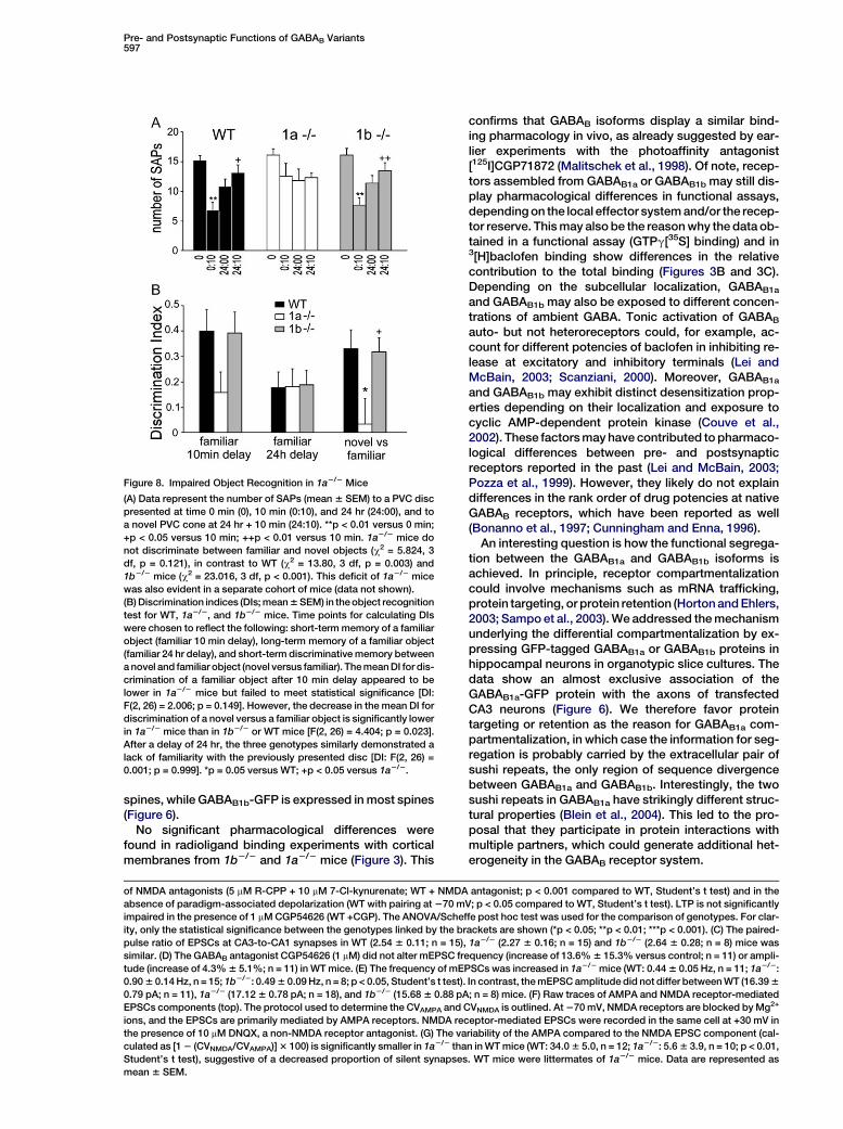
Impaired Object Recognition in 1a2/2 Mice
Changes in hippocampal LTP accompany certain alter-ations in cognitive function (Barnes et al., 1994). Weused an object recognition task that depends on hippo-campal function (Broadbent et al., 2004) to analyzewhether impaired presynaptic heteroreceptor inhibitionand lack of LTP in 1a2/2 mice is paralleled by memorydeficits. The task relies upon the tendency of rodentsto attend to a novel object more than to a familiar one.For each mouse, we scored the number of stretch attendpostures (SAP, defined as head and shoulders extendedtoward the object) in response to a PVC disc presented attimes 10 min and 24 hr following initial presentation attime 0, as well as to a novel PVC cone at 24 hr + 10 min.Wild-type and 1b2/2 mice, in contrast to 1a2/2 mice,showed a significantly reduced number of SAPs towardthe familiar object (time 10 min) and subsequently an in-creased number of SAPs toward the novel versus famil-iar object (time 24 hr + 10 min) (Figure 8A). Calculation ofdiscrimination indices (DIs) showed that 1a2/2 mice didnot discriminate between familiar or novel objects(Figure 8B). Therefore, in 1a2/2 mice, the lack of LTP iscorrelated with an impairment of nonspatial hippocam-pal memory formation. The effects on synaptic plasticity
to 1a2/2 mice, which is reflected in the number of immunogold par-
ticles that were analyzed. Arrow: examples of immunogold particles
in spines and dendritic shafts; arrowhead: examples of immunogold
particles in presynaptic terminals. t, terminal; s, spine; d, dendrite;
scale bars, 200 nm.
Pre- and Postsynaptic Functions of GABAB Variants595
Figure6. ExpressionofGFP-TaggedGABAB1a
and GABAB1b Subunits in Organotypic Slice
Culture
Maximum intensity projections of dendrites
and axons in the CA1 region of the hippo-
campus expressing GABAB1a-GFP (A) or
GABAB1b-GFP (B) in combination with the
freely diffusible tdimer2 RFP are shown. The
ratio of green-to-red fluorescence (G/R) is
coded in rainbow colors. Scale bar, 5 mm.
(C) Predominantly GABAB1a-GFP protein is
expressed in axons. The axonal expres-
sion level of GABAB1a and GABAB1b was nor-
malized to the dendritic expression level
(GABAB1a: 133.84% 6 29.53%, n = 9;
GABAB1b: 18.01% 6 2.80%, n = 5; mean 6
SEM). (D) GABAB1b-GFP was expressed in the
majority of dendritic spines, while GABAB1a-
GFP was excluded from most spines (spines
positive for GABAB1a-GFP: 21 of 82; spines
positive for GABAB1b-GFP: 42 of 62). Exam-
ples of positive spines are indicated by white
arrowheads in the G/R ratio images in (A) and
(B). (E) Example of an organotypic hippocam-
pal slice culture 7 days after cotransfection
of GABAB1b-GFP and tdimer2 expression
vectors.
and memory formation in 1a2/2 mice emphasize thatthe GABAB1b protein cannot compensate for the loss ofGABAB1a protein.
Discussion
The objective of this study was to determine if GABAB1a
and GABAB1b exhibit functional or pharmacologicaldifferences in vivo. Our experiments with geneticallymodified mice indicate that, at CA3-to-CA1 synapses,GABAB1a assembles heteroreceptors inhibiting gluta-mate release, while predominantly GABAB1b mediatespostsynaptic inhibition (Figure 4). This functional spe-cialization relates, at least in part, to a distinct subcellulardistribution of the GABAB1 isoforms (Figure 5). Auto-receptor function is unaltered in the 1a2/2 and 1b2/2
mice (Figures 4C and 4D). Possibly, both GABAB1a andGABAB1b are present at GABAergic terminals impingingonto CA1 pyramidal neurons. However, since our record-ings from the CA1 pyramidal soma cannot distinguishIPSCs from different types of GABAergic neurons, it isequally possible that some GABAergic terminals ex-press GABAB1a and others express GABAB1b. In general,the extent of subcellular segregation of GABAB1a andGABAB1b may vary according to brain regions and celltypes. For example, GABAergic neurons impinging ontocortical layer 5 pyramidal neurons express GABAB1a butnot GABAB1b at their terminals (Perez-Garci et al., 2006).The distribution of GABAB1a and GABAB1b may also varywithin the dendritic compartment. This is suggestedby the organotypic slice culture experiments showingthat GABAB1a-GFP is mostly excluded from dendritic

Neuron596
Figure 7. Lack of LTP and Reduction in the Proportion of Silent Synapses in 1a2/2 Mice
(A) The pairing protocol fails to induce LTP in 12/2 and 1a2/2 mice but induces a clear potentiation of the EPSC amplitude in WT and 1b2/2 mice.
Averages of the maximal EPSC amplitudes (6SEM) are shown. Pairing induction is indicated with an arrow. (Insets) Mean of 10 to 15 successive
EPSCs recorded before and after pairing. Scale, 20 ms, 50 pA. (B) Summary histogram of LTP experiments. The percent increase in EPSC am-
plitude after pairing is shown. Values for EPSC potentiation were assessed 25 min after induction. No LTP is induced in WT mice in the presence
Pre- and Postsynaptic Functions of GABAB Variants597
spines, while GABAB1b-GFP is expressed in most spines(Figure 6).
No significant pharmacological differences werefound in radioligand binding experiments with corticalmembranes from 1b2/2 and 1a2/2 mice (Figure 3). This
Figure 8. Impaired Object Recognition in 1a2/2 Mice
(A) Data represent the number of SAPs (mean 6 SEM) to a PVC disc
presented at time 0 min (0), 10 min (0:10), and 24 hr (24:00), and to
a novel PVC cone at 24 hr + 10 min (24:10). **p < 0.01 versus 0 min;
+p < 0.05 versus 10 min; ++p < 0.01 versus 10 min. 1a2/2 mice do
not discriminate between familiar and novel objects (c2 = 5.824, 3
df, p = 0.121), in contrast to WT (c2 = 13.80, 3 df, p = 0.003) and
1b2/2 mice (c2 = 23.016, 3 df, p < 0.001). This deficit of 1a2/2 mice
was also evident in a separate cohort of mice (data not shown).
(B) Discrimination indices (DIs; mean 6 SEM) in the object recognition
test for WT, 1a2/2, and 1b2/2 mice. Time points for calculating DIs
were chosen to reflect the following: short-term memory of a familiar
object (familiar 10 min delay), long-term memory of a familiar object
(familiar 24 hr delay), and short-term discriminative memory between
a novel and familiar object (novel versus familiar). The mean DI for dis-
crimination of a familiar object after 10 min delay appeared to be
lower in 1a2/2 mice but failed to meet statistical significance [DI:
F(2, 26) = 2.006; p = 0.149]. However, the decrease in the mean DI for
discrimination of a novel versus a familiar object is significantly lower
in 1a2/2 mice than in 1b2/2 or WT mice [F(2, 26) = 4.404; p = 0.023].
After a delay of 24 hr, the three genotypes similarly demonstrated a
lack of familiarity with the previously presented disc [DI: F(2, 26) =
0.001; p = 0.999]. *p = 0.05 versus WT; +p < 0.05 versus 1a2/2.
confirms that GABAB isoforms display a similar bind-ing pharmacology in vivo, as already suggested by ear-lier experiments with the photoaffinity antagonist[125I]CGP71872 (Malitschek et al., 1998). Of note, recep-tors assembled from GABAB1a or GABAB1b may still dis-play pharmacological differences in functional assays,depending on the local effector system and/or the recep-tor reserve. This may also be the reason why the data ob-tained in a functional assay (GTPg[35S] binding) and in3[H]baclofen binding show differences in the relativecontribution to the total binding (Figures 3B and 3C).Depending on the subcellular localization, GABAB1a
and GABAB1b may also be exposed to different concen-trations of ambient GABA. Tonic activation of GABAB
auto- but not heteroreceptors could, for example, ac-count for different potencies of baclofen in inhibiting re-lease at excitatory and inhibitory terminals (Lei andMcBain, 2003; Scanziani, 2000). Moreover, GABAB1a
and GABAB1b may exhibit distinct desensitization prop-erties depending on their localization and exposure tocyclic AMP-dependent protein kinase (Couve et al.,2002). These factors may have contributed to pharmaco-logical differences between pre- and postsynapticreceptors reported in the past (Lei and McBain, 2003;Pozza et al., 1999). However, they likely do not explaindifferences in the rank order of drug potencies at nativeGABAB receptors, which have been reported as well(Bonanno et al., 1997; Cunningham and Enna, 1996).
An interesting question is how the functional segrega-tion between the GABAB1a and GABAB1b isoforms isachieved. In principle, receptor compartmentalizationcould involve mechanisms such as mRNA trafficking,protein targeting, or protein retention (Horton and Ehlers,2003; Sampo et al., 2003). We addressed the mechanismunderlying the differential compartmentalization by ex-pressing GFP-tagged GABAB1a or GABAB1b proteins inhippocampal neurons in organotypic slice cultures. Thedata show an almost exclusive association of theGABAB1a-GFP protein with the axons of transfectedCA3 neurons (Figure 6). We therefore favor proteintargeting or retention as the reason for GABAB1a com-partmentalization, in which case the information for seg-regation is probably carried by the extracellular pair ofsushi repeats, the only region of sequence divergencebetween GABAB1a and GABAB1b. Interestingly, the twosushi repeats in GABAB1a have strikingly different struc-tural properties (Blein et al., 2004). This led to the pro-posal that they participate in protein interactions withmultiple partners, which could generate additional het-erogeneity in the GABAB receptor system.
of NMDA antagonists (5 mM R-CPP + 10 mM 7-Cl-kynurenate; WT + NMDA antagonist; p < 0.001 compared to WT, Student’s t test) and in the
absence of paradigm-associated depolarization (WT with pairing at 270 mV; p < 0.05 compared to WT, Student’s t test). LTP is not significantly
impaired in the presence of 1 mM CGP54626 (WT +CGP). The ANOVA/Scheffe post hoc test was used for the comparison of genotypes. For clar-
ity, only the statistical significance between the genotypes linked by the brackets are shown (*p < 0.05; **p < 0.01; ***p < 0.001). (C) The paired-
pulse ratio of EPSCs at CA3-to-CA1 synapses in WT (2.54 6 0.11; n = 15), 1a2/2 (2.27 6 0.16; n = 15) and 1b2/2 (2.64 6 0.28; n = 8) mice was
similar. (D) The GABAB antagonist CGP54626 (1 mM) did not alter mEPSC frequency (increase of 13.6% 6 15.3% versus control; n = 11) or ampli-
tude (increase of 4.3% 6 5.1%; n = 11) in WT mice. (E) The frequency of mEPSCs was increased in 1a2/2 mice (WT: 0.44 6 0.05 Hz, n = 11; 1a2/2:
0.90 6 0.14 Hz, n = 15; 1b2/2: 0.49 6 0.09 Hz, n = 8; p < 0.05, Student’s t test). In contrast, the mEPSC amplitude did not differ between WT (16.39 6
0.79 pA; n = 11), 1a2/2 (17.12 6 0.78 pA; n = 18), and 1b2/2 (15.68 6 0.88 pA; n = 8) mice. (F) Raw traces of AMPA and NMDA receptor-mediated
EPSCs components (top). The protocol used to determine the CVAMPA and CVNMDA is outlined. At 270 mV, NMDA receptors are blocked by Mg2+
ions, and the EPSCs are primarily mediated by AMPA receptors. NMDA receptor-mediated EPSCs were recorded in the same cell at +30 mV in
the presence of 10 mM DNQX, a non-NMDA receptor antagonist. (G) The variability of the AMPA compared to the NMDA EPSC component (cal-
culated as [1 2 (CVNMDA/CVAMPA)] 3 100) is significantly smaller in 1a2/2 than in WT mice (WT: 34.0 6 5.0, n = 12; 1a2/2: 5.6 6 3.9, n = 10; p < 0.01,
Student’s t test), suggestive of a decreased proportion of silent synapses. WT mice were littermates of 1a2/2 mice. Data are represented as
mean 6 SEM.
Neuron598
An open question remains why LTP at CA3-to-CA1synapses is impaired in 1a2/2 mice. Since autoreceptorand postsynaptic GABAB functions are preserved in1a2/2 mice (Figure 4), the impairment of LTP must relateto the constitutive absence of heteroreceptors. Hetero-receptors do not appear to be activated by ambient or re-leased GABA under baseline conditions (Figures 7C and7D), as already suggested in earlier experiments (Morri-sett et al., 1991; Scanziani, 2000). Presumably, heterore-ceptors are only activated during periods of intense neu-ronal activity, when GABA released from interneuronsspills over to the glutamatergic terminals. Uncontrolledrelease of glutamate during such periods is likely to trig-ger adaptive changes (Burrone and Murthy, 2003). Forexample, excess released glutamate may spill over tosynapses at which glutamate release has not occurred(Scimemi et al., 2004), which could convert silent synap-ses to a functional state (Isaac et al., 1995; Liao et al.,1995). The observed increase in the mEPSC frequencyin 1a2/2 mice (Figure 7E) could reflect such an increasein the rate of activation of previously silent synapses(Liao et al., 1999). We addressed this possibility andcompared the CVs of AMPA and NMDA receptor-medi-ated EPSCs in wild-type and 1a2/2 mice. The CVAMPA
was significantly reduced in 1a2/2 mice, while theCVNMDA was unaffected by genotype, consistent witha decreased number of silent synapses in 1a2/2 mice(Kullmann, 1994). Since silent synapses provide an idealsubstrate for LTP (Durand et al., 1996; Isaac et al., 1995;Kullmann, 1994; Liao et al., 1995; Malinow and Malenka,2002), the observed impairment of LTP in 1a2/2 micecould be explained by the decrease in the proportion ofsilent synapses. A plausible physiological role for heter-oreceptors could therefore be to limit the loss of silentsynapses and to ensure that plasticity processes aremaintained in the dynamic range. However, the constitu-tive absence of heteroreceptors in 1a2/2 mice may haveallowed time for other compensatory adaptations. Forexample, disinhibition of adenylate cyclase activity(Pineda et al., 2004), transcriptional (West et al., 2002)and/or morphological changes (Luthi et al., 2001) maycontribute to the impairment of LTP and hippocampus-dependent memory. Importantly, however, the LTP andbehavioral data reinforce that GABAB1a and GABAB1b
convey separate functions in vivo.In summary, our combined physiological, morpholog-
ical, and behavioral analysis of 1a2/2 and 1b2/2 miceclearly establishes that GABAB1a and GABAB1b are dif-ferentially compartmentalized and fulfill distinct func-tions. We hypothesize that interactions with the sushi re-peats are responsible for retaining GABAB1a at itsspecific location. From a pharmaceutical perspective,the existence of functionally distinct receptor subtypesopens up new opportunities for therapeutic interference.
Experimental Procedures
Generation and Pharmacological and Biochemical
Characterization of 1a2/2 and 1b2/2 Mice
Knockin mice were generated as outlined in Figure 1. All animal ex-
periments were subjected to institutional review and conducted in
accordance with Swiss guidelines and approved by the veterinary
Office of Basel-Stadt. [125I]CGP64213 was synthesized at Novartis
and labeled to a specific radioactivity of >2000 Ci/mmol (ANAWA,
Wangen, Switzerland). The probes used for Northern blot analysis,
the [125I]CGP64213 displacement experiments, immunoblot, and
GTPg[35S] analysis were as described (Bischoff et al., 1999; Gass-
mann et al., 2004; Kaupmann et al., 1997). Since we did not observe
significant differences in any of our assays between wild-type litter-
mates derived from 1a+/2 or 1b+/2 heterozygous breeding pairs, the
data from wild-type mice were pooled.
Electrophysiology
Standard procedures were used to prepare 300 mm thick parasagittal
hippocampal slices from P18–P28 mice. Slices were incubated for 45
min at 35ºC in an interface chamber containing saline (124 mM NaCl,
2.7 mM KCl, 1.3 mM MgCl2, 2 mM CaCl2, 1.24 mM NaH2PO4, 26 mM
NaHCO3, 18 mM glucose, 2.25 mM ascorbate) equilibrated with 95%
O2/5% CO2. Slices were then kept at room temperature for at least 45
min before starting recordings at 30ºC–32ºC. Whole-cell patch-
clamp recordings were performed from the somata of CA1 pyramidal
neurons to measure holding currents and synaptic responses; neu-
rons were visualized using infrared and differential interference con-
trast optics. Drugs, applied by superfusion into the recording cham-
ber, were kept as aliquots, and solutions were freshly prepared on
the day of the experiment. K+ currents induced by baclofen (50 mM)
or adenosine (100 mM) were elicited at 250 mV in the presence of
TTX (1 mM). Patch electrodes (w5 MU) were filled with a solution con-
taining the following: 140 mM K-gluconate, 5 mM HEPES, 2 mM
MgCl2, 1.1 mM EGTA, 2 mM Na2ATP, 5 mM phosphocreatine, 0.6
mM Tris-GTP, at pH 7.25 with KOH and 285 mOsm). EPSCs and
IPSCs were elicited by voltage pulses (100 ms, 2–5 V stimuli) or by cur-
rent pulses (100 ms, 0.1–0.3 mA stimuli) delivered through a bipolar
Pt-Ir electrode (25 mm in diameter) placed in the stratum radiatum
at a distance of 150–200 mm from the soma of the recorded cell.
The recording electrode was filled with a solution containing the fol-
lowing: 140 mM Cs-gluconate, 10 mM HEPES, 10 mM phosphocrea-
tine, 5 mM QX-314, 4 mM Mg-ATP, 0.3 mM Na-GTP, at pH 7.25 with
CsOH and 285 mOsm. EPSCs were measured at 270 mV in the pres-
ence of 100 mM picrotoxin. In some cells, stimuli were delivered in
pairs (interpulse interval 70 ms) (Palmer et al., 2004), and the
paired-pulse ratio (PPR) was calculated as the ratio of the 2nd
EPSC amplitude/1st EPSC amplitude. IPSCs were measured at
0 mV in the presence of 2 mM kynurenic acid. For the LTP experi-
ments, the baseline stimulus frequency was set to 0.05 Hz to mini-
mize ‘‘rundown’’ of the EPSC amplitudes (Gasparini et al., 2000;
Xiao et al., 2004). Cells were voltage clamped at 270 mV during base-
line and recovery periods. LTP was induced by depolarizing the cell
to 0 mV while delivering 40 stimuli at 0.5 Hz at baseline stimulus inten-
sity (pairing paradigm) (Palmer et al., 2004). When the potentiation of
EPSC amplitudes lasted for >30 min, we considered this as LTP.
mEPSCs were recorded at 270 mV in the presence of 0.5 mM TTX
and 10 mM bicuculline. Detection and analysis of mEPSCs were
done using the MiniAnalysis software (Synaptosoft, Decatur, GA).
For analysis of the CVAMPA (Kullmann, 1994), AMPA receptor-medi-
ated EPSCs were recorded in the presence of 100 mM picrotoxin
and 5 mM bicuculline while neurons were clamped at 270 mV (0.05
Hz stimulation). Following 15–20 min recording, non-NMDA gluta-
mate receptors were blocked with 10 mM DNQX, and the holding volt-
age was changed to +30 mV. For analysis of the CVNMDA, NMDA re-
ceptor-mediated EPSCs were recorded for >10 min and, as
a control, eventually blocked by adding CPP (5 mM) and 7-chloroky-
nurenate (10 mM) to the superfusion. CVAMPA and CVNMDA were calcu-
lated as SD/mean of AMPA and NMDA receptor-mediated EPSC
peak amplitudes, respectively. Data were obtained with an Axopatch
200B (Axon Instruments, Union City, CA), filtered at 2 kHz and digi-
tized at 10 kHz, and acquired and analyzed with pClamp9 (Axon In-
struments, Union City, CA). Values are expressed as mean 6 SEM.
The experimenter was blind to the genotype of the mice.
Immunohistochemistry and Preembedding Electron
Microscopy
Hippocampal sections were treated for light and electron micros-
copy immunolabeling as described (Gassmann et al., 2004; Kulik
et al., 2002). For ultrastructural analysis, only immunogold particles
inside the plasma membrane (closer than 30 nm) of morphologically
identifiable terminals (with presynaptic active zone or clear vesicles)
and dendrites/spines were counted. Unassigned particles represent
background labeling and labeling in axonal fibers (Kulik et al., 2002).
Pre- and Postsynaptic Functions of GABAB Variants599
The immunogold particle density in 12/2 mice, which completely lack
GABAB1 protein (Schuler et al., 2001), was 7% of that seen in wild-
type mice. The presynaptic percentage of particles allocated to the
plasma membrane in 12/2 mice was 52%, thus showing that back-
ground labeling equally distributes over pre- and postsynaptic mem-
branes. Only asymmetrical (glutamatergic) synapses were analyzed.
An ultrastructural analysis of GABAergic (symmetrical) synapses in
1a2/2 and 1b2/2 mice was impossible due to infrequent GABAB1 im-
munogold labeling (Kulik et al., 2002, 2003). The experimenter was
aware of the genotype of the mice.
Transfection of Organotypic Slice Cultures and Two-Photon
Laser Scanning Microscopy
Organotypic hippocampal slices were prepared from Wistar rats at
postnatal day 5 as described (Stoppini et al., 1991). After 7 days in vi-
tro, cultures were biolistically transfected with a Helios Gene Gun
(Bio-Rad, CA) with GABAB1a-GFP or GABAB1b-GFP expression vec-
tors in combination with a tdimer2 expression vector (gift from R.
Tsien). Expression of GABAB1a-GFP and GABAB1b-GFP was under
control of the neuron-specific synapsin-1 promoter (gift from K. Svo-
boda) for 7–8 days. For imaging, we used a custom-built two-photon
laser scanning microscope based on a BX51WI microscope (Olym-
pus, Japan) and a pulsed Ti:Sapphire laser (Chameleon XR, Coher-
ent, Scotland) tuned to l = 870 nm, controlled by an open source soft-
ware package (ScanImage) written in Matlab (Pologruto et al., 2003).
Fluorescence was detected in epifluorescence (LUMPlan W-IR2 60 3
0.9 NA, Olympus) and transfluorescence mode (achromatic aplanatic
condenser, 1.4 NA, Olympus) using four photomultiplier tubes
(R2896, Hamamatsu, Japan). We used 725DCXR dichroic mirrors
and E700SP blocking filters to reflect emitted photons into a second-
ary beamsplitter, containing a 560DCXR dichroic, 525/50 (green) and
610/75 (red) band-pass filters (AHF Analysentechnik AG, Tubingen,
Germany). The slice was placed into a perfusion chamber and super-
fused continuously (2 ml/min) with ACSF (119 mM NaCl, 2.5 mM KCl,
4 mM CaCl2, 4 mM MgCl2, 26.2 mM NaHCO3, 1 mM NaH2PO4, 11 mM
glucose, gassed with 95% O2 and 5% CO2 at room temperature).
Stacks of images (256 3 256 pixels) from secondary dendritic
branches and thin axons were obtained from transfected CA3 and
CA1 pyramidal neurons (Z step: 0.5 mm). Maximum intensity projec-
tions of green and red stacks were constructed. For the ratio images,
we used a hue/saturation/brightness model, where hue was deter-
mined by the green/red ratio (using a rainbow color table), and the in-
tensity in the red channel was used to set the brightness. For quan-
titative analysis, we calculated the green-to-red ratio in a region of
interest (dendrite or axon) after subtracting the background fluores-
cence. To compensate for differences in laser power and expression
level, we normalized the ratio in the axon by the average dendritic
ratio. Spines were identified by anatomy from tdimer2 images.
GABAB1a-GFP- and GABAB1b-GFP-positive spines were defined as
having intensity in the green channel at least three standard devia-
tions above background.
Object Recognition Test
The test was designed according to the principles of Ennaceur and
Delacour (1988), which rely upon the natural tendency of rodents to
attend to a novel object more than a familiar one. Male wild-type
(n = 7), 1a2/2 (n = 8), and 1b2/2 (n = 13) single-housed mice, aged
21 (6 0.7) weeks, were used. Mice were habituated overnight (17–
21 hr) to a novel enclosure [22 3 37 3 15 (h)cm], with w2 cm sawdust
and standard food and water provided ad libitum until testing began.
All sessions were recorded on video while the experimenter was out
of the testing room. The number of SAPs for each mouse in each
3 min period was scored by a trained observer blinded to the animals’
genotypes. All videos were scored twice by the same observer, and
duplicate SAP scores were averaged within animal and time point.
The intrascorer correlation (Pearson Product Moment correlation)
was 0.91. A DI, the difference in SAP numbers at a pair of time points
divided by the total number of SAPs at those time points, was calcu-
lated for each animal (Figure 8B). DIs reflect (1) short-term memory of
a familiar object ([Number of SAPs at Time 0 min 2 Number of SAPs at
Time 10 min]/[Total number of SAPs at both time points]); (2) long-
term memory of a familiar object ([Number of SAPs at Time 0 min 2
Number of SAPs at Time 24 hr]/[Total number of SAPs at both time
points]); and (3) short-term discriminative memory between a novel
and a familiar object ([Number of SAPs at Time 24 hr + 10 min 2 Num-
ber of SAPs at Time 10 min]/[Total number of SAPs at both time
points]). A DI of 1 reflects perfect discrimination, while a DI of 0 indi-
cates complete loss of discrimination. SAP data were analyzed
within a given genotype for the factor ‘‘Time’’ using the nonparamet-
ric Friedman repeated measures ANOVA on ranks test followed by
Dunn’s method for post hoc analysis. DIs were analyzed within
a given time for the factor ‘‘Genotype’’ using one-way ANOVA fol-
lowed by Fisher’s LSD post hoc analysis.
Reagents
Tetrodotoxin (TTX) was from Latoxan (Valence, France). (R)-CPP [3-
((R)-2-carboxypiperazin-4-yl)-1-phosphonic acid] and 7-chloroky-
nurenic acid (7-chloro-4-hydroxyquinoline-2-carboxylic acid) were
from Tocris Cookson Ltd. (Bristol, UK). Baclofen and CGP54626
were from Novartis Pharma AG (Basel, Switzerland). All other re-
agents were from Fluka/Sigma (Buchs, Switzerland).
Supplemental Data
The Supplemental Data for this article are available online at http://
www.neuron.org/cgi/content/full/50/4/589/DC1/.
Acknowledgments
This study was supported by the Swiss Science Foundation (3100-
067100.01; B. Bettler), the Desiree and Nils Yde Foundation (B. Bet-
tler), the Danish Medical Research Council (H.B.-O.), the Human
Frontier Science Program (R.T), the Grant Agency of the Czech Re-
public (309/03/1158; R.T.), the Wellcome Trust (International Senior
Research Fellowship; R.T.), and the National Institutes of Mental
Health/National Institute of Drug Abuse (U01 MH69062; J.F.C.,
K.K., L.H.J.). We thank A. Luthi, M. Larkum, and M. Ruegg for critical
reading of the manuscript. Some of the authors of this manuscript
work for Novartis Pharma AG, which has an interest in GABAB recep-
tors as drug targets. The authors declare no competing financial in-
terest.
Received: January 17, 2006
Revised: March 24, 2006
Accepted: April 3, 2006
Published: May 17, 2006
References
Barnes, C.A., Jung, M.W., McNaughton, B.L., Korol, D.L., Andreas-
son, K., and Worley, P.F. (1994). LTP saturation and spatial learning
disruption: effects of task variables and saturation levels. J. Neuro-
sci. 14, 5793–5806.
Bettler, B., Kaupmann, K., Mosbacher, J., and Gassmann, M. (2004).
Molecular structure and physiological functions of GABA(B) recep-
tors. Physiol. Rev. 84, 835–867.
Bischoff, S., Leonhard, S., Reymann, N., Schuler, V., Shigemoto, R.,
Kaupmann, K., and Bettler, B. (1999). Spatial distribution of
GABABR1 receptor mRNA and binding sites in the rat brain.
J. Comp. Neurol. 412, 1–16.
Blein, S., Ginham, R., Uhrin, D., Smith, B.O., Soares, D.C., Veltel, S.,
McIlhinney, R.A., White, J.H., and Barlow, P.N. (2004). Structural
analysis of the complement control protein (CCP) modules of
GABA(B) receptor 1a: only one of the two CCP modules is compactly
folded. J. Biol. Chem. 279, 48292–48306.
Bonanno, G., and Raiteri, M. (1993). Multiple GABAB receptors.
Trends Pharmacol. Sci. 14, 259–261.
Bonanno, G., Fassio, A., Schmid, G., Severi, P., Sala, R., and Raiteri,
M. (1997). Pharmacologically distinct GABAB receptors that mediate
inhibition of GABA and glutamate release in human neocortex. Br. J.
Pharmacol. 120, 60–64.
Bowery, N.G., Bettler, B., Froestl, W., Gallagher, J.P., Marshall, F.,
Raiteri, M., Bonner, T.I., and Enna, S.J. (2002). International union
of pharmacology. XXXIII. Mammalian g-aminobutyric acidB recep-
tors: Structure and function. Pharmacol. Rev. 54, 247–264.
Neuron600
Broadbent, N.J., Squire, L.R., and Clark, R.E. (2004). Spatial mem-
ory, recognition memory, and the hippocampus. Proc. Natl. Acad.
Sci. USA 101, 14515–14520.
Burrone, J., and Murthy, V.N. (2003). Synaptic gain control and ho-
meostasis. Curr. Opin. Neurobiol. 13, 560–567.
Couve, A., Thomas, P., Calver, A.R., Hirst, W.D., Pangalos, M.N.,
Walsh, F.S., Smart, T.G., and Moss, S.J. (2002). Cyclic AMP-depen-
dent protein kinase phosphorylation facilitates GABAB receptor-
effector coupling. Nat. Neurosci. 5, 415–424.
Cryan, J.F., and Kaupmann, K. (2005). Don’t worry ‘B’ happy!: a role
for GABA(B) receptors in anxiety and depression. Trends Pharma-
col. Sci. 26, 36–43.
Cunningham, M.D., and Enna, S.J. (1996). Evidence for pharmaco-
logically distinct GABAB receptors associated with cAMP produc-
tion in rat brain. Brain Res. 720, 220–224.
Davies, C.H., and Collingridge, G.L. (1996). Regulation of EPSPs by
the synaptic activation of GABAB autoreceptors in rat hippocampus.
J. Physiol. 496, 451–470.
Davies, C.H., Starkey, S.J., Pozza, M.F., and Collingridge, G.L.
(1991). GABA autoreceptors regulate the induction of LTP. Nature
349, 609–611.
Deisz, R.A., Billard, J.M., and Zieglgansberger, W. (1997). Presynap-
tic and postsynaptic GABAB receptors of neocortical neurons of the
rat in vitro: differences in pharmacology and ionic mechanisms. Syn-
apse 25, 62–72.
Dinkel, A., Aicher, W.K., Warnatz, K., Burki, K., Eibel, H., and Leder-
mann, B. (1999). Efficient generation of transgenic BALB/c mice
using BALB/c embryonic stem cells. J. Immunol. Methods 223,
255–260.
Durand, G.M., Kovalchuk, Y., and Konnerth, A. (1996). Long-term
potentiation and functional synapse induction in developing hippo-
campus. Nature 381, 71–75.
Ennaceur, A., and Delacour, J. (1988). A new one-trial test for neuro-
biological studies of memory in rats. 1: Behavioral data. Behav. Brain
Res. 31, 47–59.
Fritschy, J.M., Sidler, C., Parpan, F., Gassmann, M., Kaupmann, K.,
Bettler, B., and Benke, D. (2004). Independent maturation of the
GABA(B) receptor subunits GABA(B1) and GABA(B2) during post-
natal development in rodent brain. J. Comp. Neurol. 477, 235–252.
Gasparini, S., Saviane, C., Voronin, L.L., and Cherubini, E. (2000).
Silent synapses in the developing hippocampus: lack of functional
AMPA receptors or low probability of glutamate release? Proc.
Natl. Acad. Sci. USA 97, 9741–9746.
Gassmann, M., Shaban, H., Vigot, R., Sansig, G., Haller, C., Barbieri,
S., Humeau, Y., Schuler, V., Muller, M., Kinzel, B., et al. (2004). Redis-
tribution of GABAB(1) protein and atypical GABAB responses in
GABAB(2)-deficient mice. J. Neurosci. 24, 6086–6097.
Gemignani, A., Paudice, P., Bonanno, G., and Raiteri, M. (1994).
Pharmacological discrimination between gamma-aminobutyric
acid type B receptors regulating cholecystokinin and somatostatin
release from rat neocortex synaptosomes. Mol. Pharmacol. 46,
558–562.
Grace, C.R., Perrin, M.H., DiGruccio, M.R., Miller, C.L., Rivier, J.E.,
Vale, W.W., and Riek, R. (2004). NMR structure and peptide hormone
binding site of the first extracellular domain of a type B1 G protein-
coupled receptor. Proc. Natl. Acad. Sci. USA 101, 12836–12841.
Horton, A.C., and Ehlers, M.D. (2003). Neuronal polarity and traffick-
ing. Neuron 40, 277–295.
Isaac, J.T., Nicoll, R.A., and Malenka, R.C. (1995). Evidence for silent
synapses: implications for the expression of LTP. Neuron 15, 427–
434.
Kaupmann, K., Huggel, K., Heid, J., Flor, P.J., Bischoff, S., Mickel,
S.J., McMaster, G., Angst, C., Bittiger, H., Froestl, W., and Bettler,
B. (1997). Expression cloning of GABAB receptors uncovers similar-
ity to metabotropic glutamate receptors. Nature 386, 239–246.
Kaupmann, K., Malitschek, B., Schuler, V., Heid, J., Froestl, W.,
Beck, P., Mosbacher, J., Bischoff, S., Kulik, A., Shigemoto, R.,
et al. (1998). GABAB-receptor subtypes assemble into functional
heteromeric complexes. Nature 396, 683–687.
Kulik, A., Nakadate, K., Nyiri, G., Notomi, T., Malitschek, B., Bettler,
B., and Shigemoto, R. (2002). Distinct localization of GABAB recep-
tors relative to synaptic sites in the rat cerebellum and ventrobasal
thalamus. Eur. J. Neurosci. 15, 291–307.
Kulik, A., Vida, I., Lujan, R., Haas, C.A., Lopez-Bendito, G., Shige-
moto, R., and Frotscher, M. (2003). Subcellular localization of metab-
otropic GABAB receptor subunits GABAB(1a/b) and GABAB(2) in the rat
hippocampus. J. Neurosci. 23, 11026–11035.
Kulik, A., Vida, I., Fukuzawa, Y., Guetg, N., Kasugai, Y., Marker, C.L.,
Rigato, F., Bettler, B., Wickman, K., Frotscher, M., and Shigemoto,
R. (2006). Compartment-dependent colocalization of Kir3.2-contain-
ing K+ channels and GABAB receptors in hippocampal pyramidal
cells. J. Neurosci. 26, 4289–4297.
Kullmann, D.M. (1994). Amplitude fluctuations of dual-component
EPSCs in hippocampal pyramidal cells: implications for long-term
potentiation. Neuron 12, 1111–1120.
Lehtinen, M.J., Meri, S., and Jokiranta, T.S. (2004). Interdomain con-
tact regions and angles between adjacent short consensus repeat
domains. J. Mol. Biol. 344, 1385–1396.
Lei, S., and McBain, C.J. (2003). GABA B receptor modulation of ex-
citatory and inhibitory synaptic transmission onto rat CA3 hippo-
campal interneurons. J. Physiol. 546, 439–453.
Liao, D., Hessler, N.A., and Malinow, R. (1995). Activation of postsyn-
aptically silent synapses during pairing-induced LTP in CA1 region
of hippocampal slice. Nature 375, 400–404.
Liao, D., Zhang, X., O’Brien, R., Ehlers, M.D., and Huganir, R.L.
(1999). Regulation of morphological postsynaptic silent synapses
in developing hippocampal neurons. Nat. Neurosci. 2, 37–43.
Luscher, C., Jan, L.Y., Stoffel, M., Malenka, R.C., and Nicoll, R.A.
(1997). G protein-coupled inwardly rectifying K+ channels (GIRKs)
mediate postsynaptic but not presynaptic transmitter actions in hip-
pocampal neurons. Neuron 19, 687–695.
Luthi, A., Schwyzer, L., Mateos, J.M., Gahwiler, B.H., and McKinney,
R.A. (2001). NMDA receptor activation limits the number of synaptic
connections during hippocampal development. Nat. Neurosci. 4,
1102–1107.
Malinow, R., and Malenka, R.C. (2002). AMPA receptor trafficking
and synaptic plasticity. Annu. Rev. Neurosci. 25, 103–126.
Malitschek, B., Ruegg, D., Heid, J., Kaupmann, K., Bittiger, H., Frostl,
W., Bettler, B., and Kuhn, R. (1998). Developmental changes in ago-
nist affinity at GABAB(1) receptor variants in rat brain. Mol. Cell. Neu-
rosci. 12, 56–64.
Marshall, F.H., Jones, K.A., Kaupmann, K., and Bettler, B. (1999).
GABAB receptors—the first 7TM heterodimers. Trends Pharmacol.
Sci. 20, 396–399.
Martin, S.C., Russek, S.J., and Farb, D.H. (2001). Human GABAB(R)
genomic structure: evidence for splice variants in GABAB(R1) but
not GABAB(R2). Gene 278, 63–79.
McLatchie, L.M., Fraser, N.J., Main, M.J., Wise, A., Brown, J.,
Thompson, N., Solari, R., Lee, M.G., and Foord, S.M. (1998). RAMPs
regulate the transport and ligand specificity of the calcitonin-recep-
tor-like receptor. Nature 393, 333–339.
Mintz, I.M., and Bean, B.P. (1993). GABAB receptor inhibition of
P-type Ca2+ channels in central neurons. Neuron 10, 889–898.
Mohler, H., and Fritschy, J.M. (1999). GABAB receptors make it to
the top—as dimers. Trends Pharmacol. Sci. 20, 87–89.
Morrisett, R.A., Mott, D.D., Lewis, D.V., Swartzwelder, H.S., and
Wilson, W.A. (1991). GABAB-receptor-mediated inhibition of the
N-methyl-D-aspartate component of synaptic transmission in the
rat hippocampus. J. Neurosci. 11, 203–209.
Palmer, M.J., Isaac, J.T., and Collingridge, G.L. (2004). Multiple,
developmentally regulated expression mechanisms of long-term
potentiation at CA1 synapses. J. Neurosci. 24, 4903–4911.
Perez-Garci, E., Gassmann, M., Bettler, B., and Larkum, M.E. (2006).
The GABAB1b isoform mediates long-lasting inhibition of dendritic
Ca2+ spikes in layer 5 somatosensory pyramidal neurons. Neuron
50, this issue, 603–616.
Pineda, V.V., Athos, J.I., Wang, H., Celver, J., Ippolito, D., Boulay, G.,
Birnbaumer, L., and Storm, D.R. (2004). Removal of G(ia1)
Pre- and Postsynaptic Functions of GABAB Variants601
constraints on adenylyl cyclase in the hippocampus enhances LTP
and impairs memory formation. Neuron 41, 153–163.
Pologruto, T.A., Sabatini, B.L., and Svoboda, K. (2003). ScanImage:
flexible software for operating laser scanning microscopes. Biomed.
Eng. Online 2, 13.
Poncer, J.C., McKinney, R.A., Gahwiler, B.H., and Thompson, S.M.
(1997). Either N- or P-type calcium channels mediate GABA release
at distinct hippocampal inhibitory synapses. Neuron 18, 463–472.
Pozza, M.F., Manuel, N.A., Steinmann, M., Froestl, W., and Davies,
C.H. (1999). Comparison of antagonist potencies at pre- and post-
synaptic GABAB receptors at inhibitory synapses in the CA1 region
of the rat hippocampus. Br. J. Pharmacol. 127, 211–219.
Prosser, H.M., Gill, C.H., Hirst, W.D., Grau, E., Robbins, M., Calver,
A., Soffin, E.M., Farmer, C.E., Lanneau, C., Gray, J., et al. (2001).
Epileptogenesis and enhanced prepulse inhibition in GABAB(1)-
deficient mice. Mol. Cell. Neurosci. 17, 1059–1070.
Sakaba, T., and Neher, E. (2003). Direct modulation of synaptic ves-
icle priming by GABAB receptor activation at a glutamatergic syn-
apse. Nature 424, 775–778.
Sampo, B., Kaech, S., Kunz, S., and Banker, G. (2003). Two distinct
mechanisms target membrane proteins to the axonal surface. Neu-
ron 37, 611–624.
Scanziani, M. (2000). GABA spillover activates postsynaptic
GABA(B) receptors to control rhythmic hippocampal activity. Neu-
ron 25, 673–681.
Schuler, V., Luscher, C., Blanchet, C., Klix, N., Sansig, G., Klebs, K.,
Schmutz, M., Heid, J., Gentry, C., Urban, L., et al. (2001). Epilepsy,
hyperalgesia, impaired memory, and loss of pre- and postsynaptic
GABAB responses in mice lacking GABAB(1). Neuron 31, 47–58.
Scimemi, A., Fine, A., Kullmann, D.M., and Rusakov, D.A. (2004).
NR2B-containing receptors mediate cross talk among hippocampal
synapses. J. Neurosci. 24, 4767–4777.
Steiger, J.L., Bandyopadhyay, S., Farb, D.H., and Russek, S.J.
(2004). cAMP response element-binding protein, activating tran-
scription factor-4, and upstream stimulatory factor differentially
control hippocampal GABABR1a and GABABR1b subunit gene ex-
pression through alternative promoters. J. Neurosci. 24, 6115–6126.
Stoppini, L., Buchs, P.A., and Muller, D. (1991). A simple method for
organotypic cultures of nervous tissue. J. Neurosci. Methods 37,
173–182.
West, A.E., Griffith, E.C., and Greenberg, M.E. (2002). Regulation of
transcription factors by neuronal activity. Nat. Rev. Neurosci. 3,
921–931.
Xiao, M.Y., Wasling, P., Hanse, E., and Gustafsson, B. (2004). Crea-
tion of AMPA-silent synapses in the neonatal hippocampus. Nat.
Neurosci. 7, 236–243.
Yamada, K., Yu, B., and Gallagher, J.P. (1999). Different subtypes of
GABAB receptors are present at pre- and postsynaptic sites within
the rat dorsolateral septal nucleus. J. Neurophysiol. 81, 2875–2883.