Characterization of ACE2 naturally occurring missense variants
15
Page 1/15 Characterization of ACE2 naturally occurring missense variants: impact on subcellular localization and tracking Sally Badawi Department of Genetics and Genomics, College of Medicine and Health Sciences, United Arab Emirates University, United Arab Emirates Feda E. Mohamed Department of Genetics and Genomics, College of Medicine and Health Sciences, United Arab Emirates University, United Arab Emirates Nesreen R. Alkhofash Department of Genetics and Genomics, College of Medicine and Health Sciences, United Arab Emirates University, United Arab Emirates Anne John Department of Genetics and Genomics, College of Medicine and Health Sciences, United Arab Emirates University, United Arab Emirates Amanat Ali Department of Genetics and Genomics, College of Medicine and Health Sciences, United Arab Emirates University, United Arab Emirates Bassam R. Ali ( [email protected] ) Department of Genetics and Genomics, College of Medicine and Health Sciences, United Arab Emirates University, United Arab Emirates Research Article Keywords: Angiotensin Converting Enzyme 2 (ACE2), Subcellular tracking, SARS-CoV-2, Polymorphism Posted Date: February 16th, 2022 DOI: https://doi.org/10.21203/rs.3.rs-1345864/v1 License: This work is licensed under a Creative Commons Attribution 4.0 International License. Read Full License
-
Upload
khangminh22 -
Category
Documents
-
view
0 -
download
0
Transcript of Characterization of ACE2 naturally occurring missense variants

Page 1/15
Characterization of ACE2 naturally occurring missense variants: impact onsubcellular localization and tra�ckingSally Badawi
Department of Genetics and Genomics, College of Medicine and Health Sciences, United Arab Emirates University, United Arab EmiratesFeda E. Mohamed
Department of Genetics and Genomics, College of Medicine and Health Sciences, United Arab Emirates University, United Arab EmiratesNesreen R. Alkhofash
Department of Genetics and Genomics, College of Medicine and Health Sciences, United Arab Emirates University, United Arab EmiratesAnne John
Department of Genetics and Genomics, College of Medicine and Health Sciences, United Arab Emirates University, United Arab EmiratesAmanat Ali
Department of Genetics and Genomics, College of Medicine and Health Sciences, United Arab Emirates University, United Arab EmiratesBassam R. Ali ( [email protected] )
Department of Genetics and Genomics, College of Medicine and Health Sciences, United Arab Emirates University, United Arab Emirates
Research Article
Keywords: Angiotensin Converting Enzyme 2 (ACE2), Subcellular tra�cking, SARS-CoV-2, Polymorphism
Posted Date: February 16th, 2022
DOI: https://doi.org/10.21203/rs.3.rs-1345864/v1
License: This work is licensed under a Creative Commons Attribution 4.0 International License. Read Full License

Page 2/15
AbstractBackground:
Human angiotensin converting enzyme 2 (ACE2), a type I transmembrane receptor physiologically acting as a carboxypeptidase enzyme within the renin-angiotensin system (RAS), is a critical mediator of infection by several severe acute respiratory syndrome (SARS) corona viruses. For instance, it has beendemonstrated that ACE2 is the primary receptor for the SARS-CoV-2 entry to many human cells through binding to the viral spike S protein. Consequently,genetic variability in ACE2 gene has been suggested to contribute to the variable clinical manifestations in COVID-19. Many of those genetic variations resultin missense variants within the amino acid sequence of ACE2. The potential effects of those variations on binding to the spike protein has been speculatedand, in some case, demonstrated experimentally. However, their effects on ACE2 protein folding, tra�cking and subcellular targeting have not beenestablished.
Results:
In this study we aimed to examine the potential effects of 28 missense variants (V801G, D785N, R768W, I753T, L731F, L731I, I727V, N720D, R710H, R708W,S692P, E668K, V658I, N638S, A627V, F592L, G575V, A501T, I468V, M383I, G173S, N159S, N149S, D38E, N33D, K26R, I21T, S19P) distributed across the ACE2receptor domains on its subcellular tra�cking and targeting through combinatorial approach involving in silico analysis and experimental subcellularlocalization analysis. Our data show that none of the studied missense variants (including 3 variants predicted to be deleterious R768W, G575V and G173S)has a signi�cant effect on ACE2 intracellular tra�cking and subcellular targeting to the plasma membrane.
Conclusion:
Loss of proper tra�cking and targeting of ACE2 is expected to lead to loss of the protein function and therefore our results support the notion that loss of thisprotein function is not tolerated in humans due to its many essential physiological functions.
IntroductionThe global infection and mortality rates of the Coronavirus Disease 2019 (COVID-19), caused by the novel severe acute respiratory syndrome coronavirus 2(SARS-CoV-2), continue to increase and thus in�ecting unprecedented economic and health burden worldwide (1). The highly contagious SARS-CoV-2 has awide spectrum of clinical presentations ranging from asymptomatic infection to severe cardiorespiratory failure that acquire hospitalization and mechanicalrespiratory and cardiac support for some cases (2). Understanding the basis of the extreme interindividual clinical variability may aid clinicians andresearchers to assign the most suitable and personalized supportive treatments to patients, improve the e�cacy of the available vaccines and potentially aidin the development of novel effective therapies. Unhealthy habits like smoking or individuals affected by chronic conditions such as obesity, hypertension,cardiovascular diseases and diabetes are more prone to severe illness and worse prognosis (3,4). In addition to the viral genome variability, other risk factorsmay in�uence disease clinical severity and presentation including patient’s gender, race, age, and his/her genetic make-up (5).
Multiple studies have linked genetic variation in angiotensin-converting enzyme 2 (ACE2) to the clinical heterogeneity in infected patients as the receptor isprimarily utilized via SARS-CoV-2 for cellular entry to initiate the infection process (6). Various levels of ACE2 expression as well as natural sequence variantshave been reported to directly affect its binding a�nity to the viral S-protein, hence the ongoing extensive research targeting ACE2 for COVID-19 therapy (6,7).For example, it has been speculated that the East Asian populations would be more susceptible to the severe form of the infection due to certain high allelefrequency variants that may lead to higher ACE2 expression (8). However, several other ACE2 genetic variants detected in the Italian population have beenshown to be protective against COVID-19 as it was predicted to impair ACE2 expression and/or function (9,10). ACE2 is a metalloproteinase type 1transmembrane protein made of 805 amino acids and mainly plays a role in balancing the renin-angiotensin system through the conversion of angiotensin II(Ang II) to angiotensin 1–7 (1). ACE2 receptor is tra�cked to the cell surface through the secretory pathway where it is initially synthesized in the endoplasmicreticulum (ER). Once properly folded and post-translationally modi�ed, ACE2 is transported to the Golgi apparatus for further complex posttranslationalmodi�cations and folding and then transported to the plasma membrane by vesicular transport (1). Misfolded, unassembled or unstable proteins in the ER areusually directed for proteasomal degradation via the ER protein quality control machinery, named endoplasmic reticulum associated degradation (ERAD) (11).
By the beginning of the spread of COVID-19 infection in 2019, Cao et al. and other groups have demonstrated that ACE2 expression levels and geneticvariation may in�uence its interaction with the SARS-CoV-2 spike S-protein (8,12,13). Therefore, the highlighted variable outcomes may explain some of theinterindividual variability of the infection onset, susceptibility, and severity (9). Different genetic variants were accounted to mainly affect ACE2 binding a�nityto the viral S-protein, its expression level, or internalization but little is known regarding its effect on the receptor processing and tra�cking through thesecretory pathway to the plasma membrane (6). Some missense variants in secretory proteins like plasma membrane proteins and receptors are known fortheir deleterious effect and disease causation at various levels (14,15). Amino acid substitutions lying away from critically and functionally important proteindomains may indirectly result in a loss of function effect due to total or partial retention of the protein in the ER and thus mis-tra�cking (14,16–18). Despitetheir possible intact biological function, mis-localized membranous proteins lose their function due to their quantitative or partial loss from their distinctfunctional cellular location. In fact, the Q1069R missense mutation in the ACE2 homologue; angiotensin-converting enzyme (ACE), was found to besequestered by the ER quality control machinery and prevented from tra�cking to the cell surface (19). Therefore, we hypothesize that some missensevariants in ACE2 receptor might exert tra�cking defects on this receptor and its levels at the plasma membrane. Moreover, considering ACE2 critical biologicalfunctions, partial ER retention or delay might potentially explain some of the interindividual COVID-19 clinical variability and may provide a drug target forSARS-CoV-2 and other coronaviruses infections (1,19).

Page 3/15
In their comparative genetic analysis at the time of initiating this study, Cao et al. have pointed out 30 missense variants out of total 62 genetic variations inthe ACE2 coding region for their potential effect on the protein amino acid sequence (Fig. 1A) (8). We decided then to generate all those variants and evaluatetheir effects on the subcellular localization and N-glycosylation pro�le of the ACE2 receptor. Our �ndings indicate very limited or no detectable effects of thesevariations on the subcellular localization of ACE2 which may augment the notion that the biological functions of this receptor are essential, and its partial losscannot be tolerated.
MethodologyIn silico prediction of the structural effects of ACE2 variants
ACE2 rsIDs with their global allele frequency were retrieved from Ensembl database(https://asia.ensembl.org/Homo_sapiens/Transcript/Variation_Transcript/Table?db=core;g=ENSG00000130234;r=X:15494566-15607236;t=ENST00000427411). To assess the effect of 29 ACE2 nonsynonymous variants on protein function, different in silico prediction tools have beenused. SIFT (Sorting Intolerant from Tolerant) algorithm (https://sift.bii.a-star.edu.sg) was utilized to predict whether the studied ACE2 variants affect ACE2protein function. If SIFT’s score <0.05 the variant is considered tolerated and if score > 0.05 the variant is considered to affect protein function (20). PolyPhen-2 (Polymorphism Phenotyping v2) algorithm (http://genetics.bwh.harvard.edu/pph2/index.shtml) was also used to predict the impact of ACE2 missensevariants on its structure and function using different sequence and structure-based predictive features. Two pairs of datasets, HumanDiv and HumanVar, wereused to evaluate the consequent damage. PolyPhen-2 scores range between 0 and 1, Benign variants have scores in the range of 0 and 0.15, possiblydamaging variants have a score in the range of 0.15 and 0.9 and con�dently damaging variants have a score between 0.9 and 1 (21). PROVEAN (ProteinVariation Effect Analyzer) software (http://provean.jcvi.org/seq_submit.php) was also used to predict whether ACE2 amino acid substitutions in�uence itsbiological function (22), where a score less than -2.5 correspond to a deleterious variant. Finally, to evaluate whether these variants might be disease causing,Mutation Taster has been applied (https://www.mutationtaster.org/MutationTaster69/index.html) (23). If a variant was found to affect protein function in atleast one of the prediction tools, it is classi�ed as possibly deleterious, otherwise it is benign.
Analysis of the effect of missense Variants on the protein stability and their impact on the protein structure using protein modeling
I-Mutant tool was employed to predict the effect of selected missense variants on the protein stability (24). For this, the protein sequence of ACE2 andmissense variants data was submitted in FASTA format. The primary amino acid sequence of ACE2 obtained from the UniProt (Accession No. Q9BYF1) and28 missense variants generated manually were used as input. Moreover, the coordinates of ACE2 crystal structure were obtained from the protein data bank(PDB), PDB ID 6M17, excluding the coordinates of receptor binding domain (RBD) of SARS-CoV2 from the co-complexed BoAT1 dimer. I-TASSER was used tomodel the missing C terminal residues (769 to 805) and N terminal residues (1-19) of the ACE2 transmembrane helices (25). SWISS-MODEL was used togenerate the three-dimensional homology models of mutant proteins using modeled full-length structure of ACE2 as a template (26).
Mutagenesis primers design and the of ACE2 missense variants by site-directed mutagenesis
FLAG-tagged Human ACE2 wild type plasmid (NM_021804) was purchased from OriGene Inc. (RC208442). Twenty-eight of the reported missense variants inACE2 were generated, using the wild-type construct as a template, by Quick-Change site directed mutagenesis kit with the Pfu Ultra High-Fidelity DNApolymerase (Stratagene). The primers used for the mutagenesis were designed (additional �le 1, Table S1) using PrimerX software(https://www.bioinformatics.org/primerx/) and purchased from Metabion International AG (https://www.metabion.com/). The generation of the desiredvariants was con�rmed by the dideoxy Sanger DNA sequencing using the ABI 3130xl automated �uorescent Genetic Analyzer (Applied Biosystems). ClustalOmega softaware (https://www.ebi.ac.uk/Tools/msa/clustalo/) was used for sequence alignments.
Cell culture
HeLa cells and HEK293 cells were cultured in Dulbecco’s Modi�ed Eagle Medium (Gibco) supplemented with 10% fetal bovine serum (Gibco), antibiotic-antimycotic (Gibco) at 37 °C and 5% CO2 as previously described (27).
Immuno�uorescence and confocal microscopy
HeLa cells were seeded on sterilized cover slips for imaging. Cells were co-transfected with WT or mutated ACE2 plasmid and GFP-tagged HRas, a plasmamembrane marker. The methodology has been described previously (27). Twenty-four hours later, cells were washed three times with phosphate-bufferedsaline (PBS) and then �xed with methanol at -20 °C for 5 minutes. Fixed cells were then blocked with 3% bovine serum albumin (Sigma-Aldrich) for 30 minutesat room temperature. Fixed cells were co-stained with Anti-Flag primary antibody (1:100 Cell Signaling) and anti-Calnexin (1:50 Santa Cruz Biotechnology) for1 hour in the dark at room temperature. Cells were then washed three times with PBS and then incubated with the respective secondary antibody (ThermoFischer Scienti�c) for 45 minutes in the dark at room temperature. Afterwards, cells were then washed and mounted with immmuno�uor medium (ICNBiomedicals) and images were acquired using the 100x objective Nikon confocal Eclipse 80I microscope (Nikon Instruments Inc.) Images were furtheranalyzed and merged using ImageJ software (28).
SDS-PAGE immunoblotting
Forty-eight hours post transfection, HEK293 cells seeded in 6 well-plates were lysed according to manufacturers’ instructions in RIPA lysis buffer (Pierce Inc.)along with protease inhibitor cocktail (Pierce). Total proteins were quanti�ed by the colorimetric bicinchoninic acid protein assay (BCA kit, Pierce). 20 ug totalprotein lysate were resolved on 4-12% SDS-PAGE precast gradient gel (GeneScript) followed by transfer into PVDF membrane. Membrane was then probedwith anti-ACE2 (1:1000 Santa Cruz, cat# sc-390851) anti-Flag antibody (1:1000 Cell Signaling, cat # 8146S), anti-GFP (1:1000 Cell Signaling, cat# 2955S) and

Page 4/15
anti-actin (1:1000 Santa Cruz Biotechnology, cat# sc-47778) and their corresponding secondary antibodies (Sigma-Aldrich). Membranes were then incubatedwith Enhanced Chemiluminescence Plus reagent (Pierce) and developed using the Typhoon FLA 9500 imager (GE Healthcare Biosciences, Piscataway, NJ,USA). Blot analysis quanti�cation were then performed using ImageJ software.
Glycosylation sensitivity and resistance assays
HEK293 cells seeded in 6 well-plates were co-transfected with wild type and mutant ACE2 and GFP plasmids. Forty-eight hours later, cells were harvested, andlysates were then denatured in denaturation buffer at 100 °C for 10 minutes. Equal amounts of the proteins were incubated at 37 °C for 3 hours in presence orabsence of 10U of endoglycosidase H (Sigma-Aldrich). Samples were then resolved on 4-12% SDS-PAGE gel and processed for western blotting as previouslydescribed.
N-linked oligosaccharides were removed by PNGase F treatment (New England Biolabs). Cell lysates were denatured at 100 °C for 10 minutes and equalamounts of glycoproteins were then incubated at 37 °C for 1 hour in presence and absence of PNGase F enzyme. Samples were then resolved on 4-12% SDS-PAGE gradient gels and proceeded for western blotting.
Protein stability analysis and half-life determination:
Twenty-four hours post transfection, HEK293 cells seeded in 6 well-plates were treated with 100 mg/ml cycloheximide (CHX) (Sigma-Aldrich) to stop newprotein translation for different time intervals (4, 8, 12, 18 and 24 hours). DMSO treated cells at the same time intervals were taken as control. Cells were thenharvested and proceeded by western blotting.
ResultsIn silico analysis of ACE2 naturally occurring missense variants:
ACE2 variants were tested using different bioinformatic predictive tools to identify the possible functional effects of these variants on ACE2. Altogether, only 3variants (R768W, G575V and G173S) were predicted to be deleterious by all the evaluated algorithms (Table 1). Twelve variants were detected to affect proteinfunction by SIFT algorithm. Only 4 variants were found to be deleterious by PROVEAN analysis and 13 were predicted to be damaging by PolyPhen-2HumanVar, and PolyPhen-2 HumanDiv. Predictions by Mutation Taster show that 10 of the studied substitutions might be disease causing where all othershave no phenotypic effect and are considered polymorphic. In total, 10 variants (V801G, N720D, E668K, V658I, F592L, N159S, N149S, D38E, K26R, and I21T)were found to have no effect on the function of ACE2 by all the tested algorithms, classi�ed as benign (additional �le 1, Table S2).
Analysis of the structural stability and effect of missense variants on ACE2 protein:
I-mutant Suite was performed to evaluate the effect of missense variants on the overall protein stability. Results demonstrated that all studied variantsdecreased the ACE2 stability, compared to wild type ACE2 except for S19P, D38Q and S692P variants (Table 2). Among the 28 studied variants, I21T, N33D,F592L, D785N and V801G were found to introduce the highest instability in ACE2 with a Gibbs free energy change value (ΔΔG) of -2.16, -2.10, -2.97, -2.16, and-3.20, respectively.
Moreover, protein modeling was performed to determine the effect of missense variants on ACE2 structure and function. The full length ACE2 protein wasmodelled, and missense variants were generated on the protein structure (Fig. 1B). Among 28 studied variants, 13 are located in peptidase domain of ACE2which is a potential binding site for RBD of the spike protein (S) of SARS-CoV2. Substitutions of ACE2 residues involved in SARS-CoV-2 binding have beenshown to alter the binding a�nity of SARS-CoV-2 S protein (6,29). Although some variants are present distant from the ACE2 interface and do not form directinteractions with the RBD of SARS-SoV-2, they might exert structural deformities on ACE2 protein. The evaluation of S19P indicated that the substitution ofconserved serine into a hydrophobic proline is likely to disturb the interactions with other ACE2 residues important for protein structure (Fig. 2A1 and 2A2).Experimental studies have also shown that S19P variant increased the binding a�nity of SARS-CoV2 (29). The substitution of non-polar isoleucine to a polarthreonine at 21 amino acid position is likely to produce additional hydrogen bonds with A25 and E87 residues of ACE2 which could disrupt proteinconformation (Fig. 2B1 and 2B2). K26, N33, and D38 are essential residues present at the interface of ACE2 and formed direct interactions with RBD of SARS-CoV-2 (29). The physiochemical changes of K26R and N33D variants are signi�cant and have been shown to enhance the binding a�nity of SARS-CoV-2 (Fig.2C1-2D2). However, the substitution of charged residues at amino acid position 38 is less likely to cause signi�cant changes in protein conformation andSARS-CoV-2 binding a�nity (Fig. 2E1 and 2E2). Asparagine for serine substitutions at positions 149 and 159 are physiochemically insigni�cant and may haveno effect on protein structure or function (Fig. 2F1-2G2). G173 is buried in the core of ACE2 and is also important in forming the catalytic site. G173S variant islikely to disrupt the catalytic site formation as well as catalysis and substrate speci�city (Fig. 2H1 and 2H2). Although the substitution of methionine forisoleucine at ACE2 position 383 is physiochemically signi�cant (Fig. 2I1 and 2I2), it has not been found to alter SARS-CoV-2 binding a�nity (29). Thephysiochemical properties of I468V and G575l variants are insigni�cant and are unlikely to disrupt protein structure and function (Fig. 2J1-2L2). A501Tvariant is expected to disturb the polar interaction of alanine with Glu181 (Fig 2K1 and 2K2).
Out of 11 variants present in the collectrin domain, �ve variants (A627V, N638S, V658I, I727V, and L731I) are physiochemically insigni�cant and are notobserved to affect protein structure and function (Fig. 2N1-2W2). E668K variant is likely to disrupt the polar interaction of wild type glutamic acid with E667which could be important for protein conformation (Fig. 2Q1 and 2Q2). The substitution of polar serine with a hydrophobic proline residue at 692 position islikely to disrupt the hydrogen bonds of serine with N159 and N690 (Fig. 2R1 and 2R2). The substitution of charged arginine with neutral tryptophan at 708position is expected to disrupt the hydrogen bonds and salt bridges formed by R708 with D719, L722, E723, and I727 residues of ACE2 (Fig. 2S1 and 2S2).Such changes of S692P and R708W could lead to improper protein folding. ACE2 exists as a homodimer, and the PD, TM, and neck domains all contribute todimerization, but it is the neck residues that primarily mediate ACE2 dimerization (30). Residues 636-658 and 708-717 present at the neck region are observed

Page 5/15
to form stable polar interactions. Experimental studies have indicated that R710 formed cation-π interactions with Y641 and Y633 as well as hydrogen bondwith E639, which also clashes with Q653, as does N636 (30). These interactions are important for stable dimer formation. The possible loss of charge in caseof R710H variant is likely to disturb these interactions, which could result in improper dimer formation (Fig. 2T1 and 2T2). I753T, R763W, D785N and V801Gvariants are located at the cytoplasmic end of ACE2 (Fig. 2Y1-2Z6). The substitutions are physiochemically signi�cant, particularly R763W, but given theirlocation and the lack of obvious intramolecular interactions, these variants are unlikely to affect protein structure or function.
Exogenously expressed FLAG-tagged wild type ACE2 localizes to the plasma membrane and has a half-life of about 12 hours
ACE2 endogenous protein expression in the human HEK293 and HeLa cell lines was assessed by immunoblotting assay using anti-ACE2 monoclonalantibody (Santa Cruz; 1:1000 dilution, cat# sc-390851). Our blot shows that ACE2 protein was not detectable in either HEK293 or HeLa cell lines even whenhigh amounts of cell lysates (50 mg) were analyzed (additional �le 2, Fig. S1A). However, overexpressed Flag-tagged ACE2 in HEK293 cells was detected withthe same anti-ACE2 speci�c antibody at ~120 KDa molecular weight similar to endogenous ACE2 detected in other cell lines like HuH7, Caco-2, Calu-3 andHepG2 cells (31).
To establish the intracellular localization of wild-type overexpressed ACE2, HeLa cells were co-transfected with Flag-tagged ACE2 and GFP-tagged HRasplasmids. Visualization of stained cells by confocal microscopy displayed a plasma membranal pro�le for WT ACE2 that overlapped with GFP-tagged HRasas shown in Fig. 3A, where red staining of WT ACE2 by anti-Flag antibody, colocalizes with the green GFP-tagged HRas, staining the plasma membrane anddid not show any colocalization with the blue stained ER marker, Calnexin. Similarly, overexpressed ACE2 displays a similar pattern in HEK293 (additional �le2, Fig. S1B). To con�rm the tra�cking of ACE2 across the secretory pathway, the N-glycosylation pro�le of WT ACE2 was evaluated. Glycoprotein N-glycosylation is an enzyme directed process that occurs at speci�c asparagine residue in N-X-T/S sequence motif, in the ER that is then further modi�ed in theGolgi apparatus. ACE2 is reported to have 7 N-glycosylation sites as shown in Fig.1A (32,33). Digestion of protein lysates from HEK293 cells overexpressingWT ACE2 with Endoglycosidase H (Endo H) enzyme, which cleaves off the immature N-glycans only, showed that around 2.8% ± 0.36 of the protein isdigestible to a lower molecular weight band (~100 KDa) suggestion high level of maturation of the WT protein. The remaining 97.2% did not change theirmolecular weight protein band of ~120KDa suggesting that it has acquired the complex N-glycans that usually take place in the Golgi complex beforetra�cking to the plasma membrane (Fig. 3B). On the other hand, treatment of N-glycans of ACE2 WT protein by the PNGase F enzyme, a peptide N-glycosidase F that cleaves off N-glycans regardless of their glycans maturation stage, resulted in shift from a high molecular weight band (120 KDa) to alower molecular weight protein band (~100KDa). These results demonstrate that WT ACE2 acquires fully mature N-glycans quantitatively and presumably hashigh maturation rate.
To gain insight on the half-life, stability, and turnover of the overexpressed WT ACE2, we overexpressed the protein for 24 hours then added the proteinsynthesis inhibitor cycloheximide at 100 mg/ml and quanti�ed the remaining ACE2 by Western blotting at several subsequent time points up to 18 hours. Asshown in Fig. 3C and 3D, overexpressed WT ACE2 has an approximate half-life of 12 hours suggesting a relatively slow turn over.
All studied ACE2 variants tra�c normally and localize to the plasma membrane resembling the WT protein:
To assess the effects of the studied ACE2 missense variants, we expressed them individually in HeLa and HEK293 cell lines. HEK293 cells overexpressingACE2 mutants display normal protein expression pattern by western blotting compared to WT ACE2 (additional �le 2, Fig. S2). HeLa cells were co-transfectedwith an ACE2 construct (WT or mutants) and GFP-tagged HRas plasmids then the subcellular localization of the proteins was evaluated by confocalmicroscopy. Interestingly, as shown in Fig. 4 and additional �le 2, Fig. S3, none of these variants (including the possibly deleterious variants) affected theapparent subcellular localization of the ACE2 protein. For all the studied variants, ACE2 appeared at the plasma membrane and co-localized with GFP-taggedHRas.
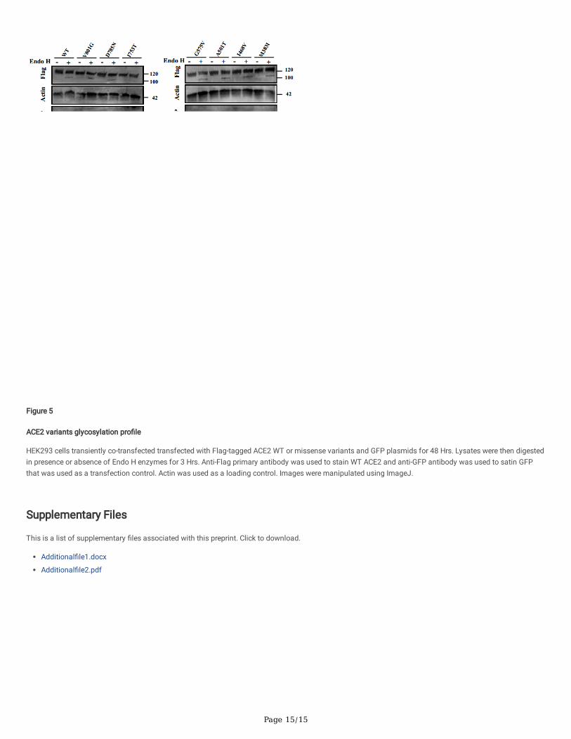
To con�rm these results, the N- glycosylation pro�les of all the studied variants were assessed by Endo H digestion sensitivity and resistance assay andcompared to WT ACE2. Similarly, all mutants have shown no signi�cant change in the electrophoretic behaviors compared to WT ACE2, where the vastmajority of the expressed ACE2 appears to be resistant to Endo H treatment suggesting that they are fully glycosylated and have normal maturation levels.The Endo H resistant band at ~120 KDa accounts for over 96% of the expressed protein for all studied variants. Whereas the lower molecular weight bandaccount to less than ~2.8% of total expressed ACE2 as shown in Fig. 5.
DiscussionThe current ongoing COVID-19 pandemic created an urgent need for deciphering the interlink between SARS-CoV-2 and its primary cellular receptor, ACE2.Several studies have extensively investigated ACE2 polymorphic footprint and its associated effects on its structure, binding and stability (8,9,34,35). Geneticvariations in ACE2 gene are regarded as a potential risk factor in COVID-19 patients (36). In this context, different predictive studies based on bioinformaticsand simulation tools have generated a bulk of data that helped identify major residues in ACE2 and their consequent effect on SARS-CoV-2 binding (37–39).
In the current study, we have evaluated the impact of several ACE2 coding missense variants using different predictive algorithms and investigated their effecton ACE2 protein subcellular localization, tra�cking and membrane availability. ACE2 gene displays a unique polymorphic pro�le in the human population inwhich 332 missense variants were reported in Ensembl database. Interestingly, in comparison to its homologue the Angiotensin Converting Enzyme (ACE),ACE2 displays a lower probability of losing its function by genetic mutations, where probability of being loss-of-function intolerant of ACE2 is pLI=0.998 bygnomAD database, noting that if pLI > 0.9 gene is considered extremely intolerant (34,40). Unlikely, a missense variant in ACE protein (Q1069R) was reportedto be associated with renal tubular disease resulting in premature death caused by improper localization of ACE and loss of its function (19). Althoughdifferent global or conditioned knockout mouse models have been generated for ACE2, all have reported serious associated phenotypes includingdevelopmental, cardiovascular, renal and respiratory clinical manifestations (41).
Page 6/15
All studied genetic variants in this paper had a low population distribution with minor allele frequency less than 1%. Although eight of the variants (K26R,I468V, A627V, N638S, S692P, N720D and L731F/L731I) were considered major hotspot by Cao et al, and were distributed in different populations, two of them,K26R and N720D, were shown to be benign by the different evaluated tools. Stability analysis of K26R and N720D variants was decreased by -0.34 Kcal/moland -1.38 Kcal/mol, respectively with no signi�cant change in the cell surface availability of these two variants and no effect on their cellular tra�ckingdetected by glycosylation analysis and immuno�uorescence assays. K26 residue is present in the binding domain with SARS-CoV-2 receptor binding domain(RBD), several studies suggest that residual substitution of lysine with arginine at this site enhances ACE2-SARS-CoV-2 binding and could contribute to highersusceptibility to SARS-CoV-2 (37,42–44). Unlikely, N720 amino acid residue is located in the peptidase domain in a proximity to type II transmembrane serineprotease (TMPRSS2) cleavage site. Aside from having lower stable structure compared to WT ACE2, substituting asparagine with aspartic acid at this residueis found to weaken TMPRSS2-ACE2 complex and consequently augmenting SARS-CoV-2 viral entry (34,45). Our analysis has also revealed three deleteriousvariants (R768W, G575V and G173S) that were found to affect ACE2 functionality by all the prediction tools in our study. Similarly, intracellular localizationand cell surface availability of these variants were not affected in our studied model. Knowing that G575 and G173 are located in the peptidase domain ofACE2 protein, G173S variant is predicted to stabilize ACE2-SARS-CoV-2 complex (46), noting that this substitution might affect ACE2 catalytic activity, whilethe effect of G575V on the binding a�nity to SARS-CoV-2 RBD is not investigated yet. Our results come in line with another in silico analysis showing thatR768W is a high risk ACE2 variant and might exert a deleterious effect on ACE2 structure (47). Additionally, previous studies have shown that the 43 longamino acid topological domain of ACE2 doesn’t affect signi�cantly the cell surface expression and SARS-CoVs mediated viral entry (48,49). These data �tswith our reported results showing that all mutants present in the cytoplasmic tail of ACE2 (V801G, D785N and R768W) display no change in ACE2 tra�ckingand transmembrane localization.
In this context, little has been reported in literature about ACE2 biosynthesis and intracellular tra�cking. Evident role of N-linked glycosylation on membraneproteins stability, compartmental tra�cking, and cell surface expression has been largely reported (50–52). Among the seven glycosylation sites of ACE2, N90,N322 and N546 form glycan-mediated interactions between ACE2 and SARS-CoV-2 RBD complex (53), noting that N90 and N322 have opposing effects onSARS-CoV-2 binding (33). ACE2 deprived of all N-glycans was found to accumulate in the endoplasmic reticulum with no signi�cant effect on its enzymaticcarboxypeptidase activity (32). N-linked glycans are usually attached to an asparagine residue of N-X-S/T motif, where X is any amino acid except proline andS/T are serine/threonine amino acids. Among the variants we were interested in studying is S692P, two amino acids proximal to N690 glycosylation site.Substituting serine for proline in the S/T site would consequently lead to the loss of N-glycosylation at N690. We show in our analysis that this substitutionincreases ACE2 stability (ΔΔG=1.41 Kcal/mol), however, immuno�uorescence data show no change in the tra�cking of this variant compared to the WTACE2.
ConclusionIn summary, throughout this study, we show that none of the coding variants included in our work display an effect on ACE2 protein subcellular tra�cking andits cell surface availability. This might be due to the high importance of this gene and therefore its intolerance to the loss of its function. Noting that thedeleterious effect reported by the computational tools might be affecting the carboxypeptidase activity and binding a�nity of ACE2 that were not evaluated inour paper, rather than its tra�cking and SARS-CoVs related effect. Apart from ACE2 tra�cking modulation, the receptor membranous expression andavailability might be in�uenced at different levels (54). Considering that ACE2 gene lies on the X chromosome, it is strongly evident that females have anadvantage due to the greater chance to form structurally variable ACE2 dimers compared to males who are hemizygous for the ACE2 gene (55). ACE2expression is highly affected by sex hormones where estrogen strongly elevates its expression explaining the variable effect of COVID-19 in both sexes (56).Moreover, ACE2 expression might also be affected by different epigenetic changes providing some evidence to explain the difference in the interindividualsusceptibility and clinical variability in infected patients (57,58). Collectively, these data have important implication on COVID-19 progression and to face theseoutcomes more detailed knowledge is needed to understand the normal mechanisms controlling ACE2 expression and tra�cking at the cellular level as wellas its role in the pathogenesis of SARS-CoVs infection.
Abbreviations

Page 7/15
3D Three dimensional
AA Amino acid
ACE Angiotensin converting enzyme
ACE2 Angiotensin converting enzyme 2
Ang II Angiotensin II
CHX Cycloheximide
COVID-19 Corona Virus Disease - 2019
DMSO Dimethyl sulfoxide
Endo H Endoglycosidase H
ER Endoplasmic reticulum
ERAD Endoplasmic reticulum associated degradation
Hrs Hours
PBS Phosphate buffered serum
pLI Probability of being loss-of-function intolerant
PNGase F Peptide N-glycosidase F
RBD Receptor binding domain
SARS-CoVs Severe acute respiratory syndrome corona viruses
SNP Single nucleotide polymorphism
SP Signal Peptide
TMPRSS2 Type II Transmembrane Serine Protease
WT Wild type
DeclarationsEthics Approval and Consent to participate
Not applicable
Consent for Publication
Not applicable
Availability of Data and Materials
All data generated or analyzed during the study are included in this article and its additional �les. Any further requirements are available from thecorresponding author upon request.
Competing interest
The authors declare that the research was conducted in the absence of any commercial or �nancial relationships that could be constructed as a potentialcon�ict of interest.
Funding
This work was funded by the Abu Dhabi ASPIRE Award for Research Excellence (AARE) (grant AARE19-086, 21M136).
Authors Contribution
SB and FM equally contributed to conducting the experiments and writing the manuscript draft. NK, SB, and FM contributed to generating ACE2 missensevariants and �gures of the manuscript. AJ con�rmed the generated variants by sequencing. AA performed the protein modeling, analyzed the data, and wrotethis part of the manuscript. BA conceived the idea of the study, re�ned, and edited manuscript drafts, and approved the �nal version of the manuscript.
Con�ict of interest statement
The authors declare that the research was conducted in the absence of any commercial or �nancial relationships that could be constructed as a potentialcon�ict of interest.
Page 8/15
Acknowledgement
We would like to thank Dr Saeed Tariq for his assistance in confocal microscopy imaging.
References1. Badawi S, Ali BR. ACE2 Nascence, tra�cking, and SARS-CoV-2 pathogenesis: the saga continues. Hum Genomics. 2021 Jan 29;15(1):8.
2. Bai Y, Yao L, Wei T, Tian F, Jin D-Y, Chen L, et al. Presumed Asymptomatic Carrier Transmission of COVID-19. JAMA. 2020 Apr 14;323(14):1406–7.
3. Zhang J, Wang X, Jia X, Li J, Hu K, Chen G, et al. Risk factors for disease severity, unimprovement, and mortality in COVID-19 patients in Wuhan, China. ClinMicrobiol Infect. 2020 Jun 1;26(6):767–72.
4. Richardson S, Hirsch JS, Narasimhan M, Crawford JM, McGinn T, Davidson KW, et al. Presenting Characteristics, Comorbidities, and Outcomes Among 5700Patients Hospitalized With COVID-19 in the New York City Area. JAMA. 2020 May 26;323(20):2052–9.
5. Pereira NL, Ahmad F, Byku M, Cummins NW, Morris AA, Owens A, et al. COVID-19: Understanding Inter-Individual Variability and Implications for PrecisionMedicine. Mayo Clin Proc. 2021 Feb;96(2):446–63.
6. Suryamohan K, Diwanji D, Stawiski EW, Gupta R, Miersch S, Liu J, et al. Human ACE2 receptor polymorphisms and altered susceptibility to SARS-CoV-2.Commun Biol. 2021 Apr 12;4(1):1–11.
7. Jia H, Neptune E, Cui H. Targeting ACE2 for COVID-19 Therapy: Opportunities and Challenges. Am J Respir Cell Mol Biol. 2021 Apr;64(4):416–25.
8. Cao Y, Li L, Feng Z, Wan S, Huang P, Sun X, et al. Comparative genetic analysis of the novel coronavirus (2019-nCoV/SARS-CoV-2) receptor ACE2 in differentpopulations. Cell Discov. 2020 Feb 24;6(1):1–4.
9. Benetti E, Tita R, Spiga O, Ciol� A, Birolo G, Bruselles A, et al. ACE2 gene variants may underlie interindividual variability and susceptibility to COVID-19 in theItalian population. Eur J Hum Genet. 2020 Nov;28(11):1602–14.
10. Novelli A, Biancolella M, Borgiani P, Cocciadiferro D, Colona VL, D’Apice MR, et al. Analysis of ACE2 genetic variants in 131 Italian SARS-CoV-2-positivepatients. Hum Genomics. 2020 Sep 11;14(1):29.
11. Liu X, Yang N, Tang J, Liu S, Luo D, Duan Q, et al. Downregulation of angiotensin-converting enzyme 2 by the neuraminidase protein of in�uenza A (H1N1)virus. Virus Res. 2014 Jun 24;185:64–71.
12. Saih A, Baba H, Bouqdayr M, Ghazal H, Hamdi S, Kettani A, et al. In Silico Analysis of High-Risk Missense Variants in Human ACE2 Gene and Susceptibilityto SARS-CoV-2 Infection. BioMed Res Int. 2021 Apr 9;2021:e6685840.
13. Chen F, Zhang Y, Li X, Li W, Liu X, Xue X. The Impact of ACE2 Polymorphisms on COVID-19 Disease: Susceptibility, Severity, and Therapy. Front Cell InfectMicrobiol [Internet]. 2021 [cited 2022 Jan 20];11. Available from: https://www.frontiersin.org/article/10.3389/fcimb.2021.753721
14. Kizhakkedath P, John A, Al-Sawa� BK, Al-Gazali L, Ali BR. Endoplasmic reticulum quality control of LDLR variants associated with familialhypercholesterolemia. FEBS Open Bio. 2019 Nov;9(11):1994–2005.
15. Fukuda R, Okiyoneda T. Cystic Fibrosis Transmembrane Conductance Regulator (CFTR) Ubiquitylation as a Novel Pharmaceutical Target for CysticFibrosis. Pharmaceuticals. 2020 Apr 22;13(4):75.
16. Mohamed FE, Al Sorkhy M, Ghattas MA, Al-Gazali L, Al-Dirbashi O, Al-Jasmi F, et al. The pharmacological chaperone N-n-butyl-deoxygalactonojirimycinenhances β-galactosidase processing and activity in �broblasts of a patient with infantile GM1-gangliosidosis. Hum Genet. 2020 May;139(5):657–73.
17. Needham PG, Guerriero CJ, Brodsky JL. Chaperoning Endoplasmic Reticulum–Associated Degradation (ERAD) and Protein Conformational Diseases. ColdSpring Harb Perspect Biol. 2019 Aug;11(8):a033928.
18. Gariballa N, Ali BR. Endoplasmic Reticulum Associated Protein Degradation (ERAD) in the Pathology of Diseases Related to TGFβ Signaling Pathway:Future Therapeutic Perspectives. Front Mol Biosci. 2020;7:575608.
19. Oliveira RM de, Marijanovic Z, Carvalho F, Miltényi GM, Matos JE, Tenreiro S, et al. Impaired Proteostasis Contributes to Renal Tubular Dysgenesis. PLOSONE. 2011 Jun 9;6(6):e20854.
20. Vaser R, Adusumalli S, Leng SN, Sikic M, Ng PC. SIFT missense predictions for genomes. Nat Protoc. 2016 Jan;11(1):1–9.
21. Adzhubei IA, Schmidt S, Peshkin L, Ramensky VE, Gerasimova A, Bork P, et al. A method and server for predicting damaging missense mutations. NatMethods. 2010 Apr;7(4):248–9.
22. Choi Y, Sims GE, Murphy S, Miller JR, Chan AP. Predicting the Functional Effect of Amino Acid Substitutions and Indels. PLOS ONE. 2012 Oct8;7(10):e46688.
Page 9/15
23. Schwarz JM, Cooper DN, Schuelke M, Seelow D. MutationTaster2: mutation prediction for the deep-sequencing age. Nat Methods. 2014 Apr;11(4):361–2.
24. Capriotti E, Fariselli P, Casadio R. I-Mutant2.0: predicting stability changes upon mutation from the protein sequence or structure. Nucleic Acids Res. 2005Jul 1;33(Web Server issue):W306-310.
25. Zhang Y. I-TASSER server for protein 3D structure prediction. BMC Bioinformatics. 2008 Jan 23;9:40.
26. Waterhouse A, Bertoni M, Bienert S, Studer G, Tauriello G, Gumienny R, et al. SWISS-MODEL: homology modelling of protein structures and complexes.Nucleic Acids Res. 2018 Jul 2;46(W1):W296–303.
27. Hume AN, Buttgereit J, Al-Awadhi AM, Al-Suwaidi SS, John A, Bader M, et al. Defective cellular tra�cking of missense NPR-B mutants is the majormechanism underlying acromesomelic dysplasia-type Maroteaux. Hum Mol Genet. 2009 Jan 15;18(2):267–77.
28. Schneider CA, Rasband WS, Eliceiri KW. NIH Image to ImageJ: 25 years of Image Analysis. Nat Methods. 2012 Jul;9(7):671–5.
29. Chan KK, Dorosky D, Sharma P, Abbasi SA, Dye JM, Kranz DM, et al. Engineering human ACE2 to optimize binding to the spike protein of SARS coronavirus2. Science. 2020 Sep 4;369(6508):1261–5.
30. Yan R, Zhang Y, Li Y, Xia L, Guo Y, Zhou Q. Structural basis for the recognition of SARS-CoV-2 by full-length human ACE2. Science. 2020 Mar27;367(6485):1444–8.
31. Sherman EJ, Emmer BT. ACE2 protein expression within isogenic cell lines is heterogeneous and associated with distinct transcriptomes. Sci Rep. 2021Aug 5;11(1):15900.
32. Rowland R, Brandariz-Nuñez A. Analysis of the Role of N-Linked Glycosylation in Cell Surface Expression, Function, and Binding Properties of SARS-CoV-2Receptor ACE2. Microbiol Spectr. 9(2):e01199-21.
33. Mehdipour AR, Hummer G. Dual nature of human ACE2 glycosylation in binding to SARS-CoV-2 spike. Proc Natl Acad Sci U S A. 2021 May11;118(19):e2100425118.
34. Al-Mulla F, Mohammad A, Al Madhoun A, Haddad D, Ali H, Eaaswarkhanth M, et al. ACE2 and FURIN variants are potential predictors of SARS-CoV-2outcome: A time to implement precision medicine against COVID-19. Heliyon. 2021 Feb 1;7(2):e06133.
35. Ortiz-Fernández L, Sawalha AH. Genetic variability in the expression of the SARS-CoV-2 host cell entry factors across populations. Genes Immun. 2020Aug;21(4):269–72.
36. Hou Y, Zhao J, Martin W, Kallianpur A, Chung MK, Jehi L, et al. New insights into genetic susceptibility of COVID-19: an ACE2 and TMPRSS2 polymorphismanalysis. BMC Med. 2020 Jul 15;18:216.
37. Ren W, Zhu Y, Lan J, Chen H, Wang Y, Shi H, et al. Susceptibilities of Human ACE2 Genetic Variants in Coronavirus Infection. J Virol. 2022 Jan12;96(1):e0149221.
38. Sorokina M, M. C. Teixeira J, Barrera-Vilarmau S, Paschke R, Papasotiriou I, Rodrigues JPGLM, et al. Structural models of human ACE2 variants with SARS-CoV-2 Spike protein for structure-based drug design. Sci Data. 2020 Sep 16;7(1):309.
39. Heinzelman P, Romero PA. Discovery of human ACE2 variants with altered recognition by the SARS-CoV-2 spike protein. PLOS ONE. 2021 May12;16(5):e0251585.
40. Karczewski KJ, Francioli LC, Tiao G, Cummings BB, Alföldi J, Wang Q, et al. The mutational constraint spectrum quanti�ed from variation in 141,456humans. Nature. 2020 May;581(7809):434–43.
41. Jia H, Yue X, Lazartigues E. ACE2 mouse models: a toolbox for cardiovascular and pulmonary research. Nat Commun. 2020 Oct 14;11(1):5165.
42. Suryamohan K, Diwanji D, Stawiski EW, Gupta R, Miersch S, Liu J, et al. Human ACE2 receptor polymorphisms and altered susceptibility to SARS-CoV-2.Commun Biol. 2021 Apr 12;4(1):1–11.
43. Barton MI, MacGowan SA, Kutuzov MA, Dushek O, Barton GJ, van der Merwe PA. Effects of common mutations in the SARS-CoV-2 Spike RBD and itsligand, the human ACE2 receptor on binding a�nity and kinetics. eLife. 10:e70658.
44. Shukla N, Roelle SM, Suzart VG, Bruchez AM, Matreyek KA. Mutants of human ACE2 differentially promote SARS-CoV and SARS-CoV-2 spike mediatedinfection. PLOS Pathog. 2021 Jul 16;17(7):e1009715.
45. Mohammad A, Mara�e SK, Alshawaf E, Abu-Farha M, Abubaker J, Al-Mulla F. Structural analysis of ACE2 variant N720D demonstrates a higher bindinga�nity to TMPRSS2. Life Sci. 2020 Oct 15;259:118219.
46. Hadi-Alijanvand H, Rouhani M. Studying the Effects of ACE2 Mutations on the Stability, Dynamics, and Dissociation Process of SARS-CoV-2 S1/hACE2Complexes. J Proteome Res. 2020 06;19(11):4609–23.

Page 10/15
47. Saih A, Baba H, Bouqdayr M, Ghazal H, Hamdi S, Kettani A, et al. In Silico Analysis of High-Risk Missense Variants in Human ACE2 Gene and Susceptibilityto SARS-CoV-2 Infection. BioMed Res Int. 2021 Apr 9;2021:e6685840.
48. Inoue Y, Tanaka N, Tanaka Y, Inoue S, Morita K, Zhuang M, et al. Clathrin-Dependent Entry of Severe Acute Respiratory Syndrome Coronavirus into TargetCells Expressing ACE2 with the Cytoplasmic Tail Deleted. J Virol. 2007 Aug;81(16):8722–9.
49. Karthika T, Joseph J, Das VRA, Nair N, Charulekha P, Roji MD, et al. SARS-CoV-2 Cellular Entry Is Independent of the ACE2 Cytoplasmic Domain Signaling.Cells. 2021 Jul 17;10(7):1814.
50. Singh R, Almutairi MM, Pacheco-Andrade R, Almiahuob MYM, Di Fulvio M. Impact of Hybrid and Complex N-Glycans on Cell Surface Targeting of theEndogenous Chloride Cotransporter Slc12a2. Int J Cell Biol. 2015;2015:505294.
51. Weng T-Y, Chiu W-T, Liu H-S, Cheng H-C, Shen M-R, Mount DB, et al. Glycosylation regulates the function and membrane localization of KCC4. BiochimBiophys Acta. 2013 May;1833(5):1133–46.
52. Wang T, Nakagawa S, Miyake T, Setsu G, Kunisue S, Goto K, et al. Identi�cation and functional characterisation of N-linked glycosylation of the orphan Gprotein-coupled receptor Gpr176. Sci Rep. 2020 Mar 10;10(1):4429.
53. Zhao P, Praissman JL, Grant OC, Cai Y, Xiao T, Rosenbalm KE, et al. Virus-Receptor Interactions of Glycosylated SARS-CoV-2 Spike and Human ACE2Receptor. Cell Host Microbe. 2020 Oct 7;28(4):586-601.e6.
54. Beyerstedt S, Casaro EB, Rangel ÉB. COVID-19: angiotensin-converting enzyme 2 (ACE2) expression and tissue susceptibility to SARS-CoV-2 infection. EurJ Clin Microbiol Infect Dis. 2021 Jan 3;1–15.
55. Gemmati D, Bramanti B, Serino ML, Secchiero P, Zauli G, Tisato V. COVID-19 and Individual Genetic Susceptibility/Receptivity: Role of ACE1/ACE2 Genes,Immunity, In�ammation and Coagulation. Might the Double X-Chromosome in Females Be Protective against SARS-CoV-2 Compared to the Single X-Chromosome in Males? Int J Mol Sci. 2020 May 14;21(10):3474.
56. Chen J, Jiang Q, Xia X, Liu K, Yu Z, Tao W, et al. Individual variation of the SARS‐CoV‐2 receptor ACE2 gene expression and regulation. Aging Cell. 2020Jul;19(7):e13168.
57. Lima RS, Rocha LPC, Moreira PR. Genetic and epigenetic control of ACE2 expression and its possible role in COVID‐19. Cell Biochem Funct. 2021 Jun1;10.1002/cbf.3648.
58. Novelli G, Biancolella M, Mehrian-Shai R, Colona VL, Brito AF, Grubaugh ND, et al. COVID-19 one year into the pandemic: from genetics and genomics totherapy, vaccination, and policy. Hum Genomics. 2021 May 10;15(1):27.
TablesTable 1: ACE2 missense coding variants predictions by different computational tools.

Page 11/15
SIFT PROVEAN HumanDivPolyPhen.2
HumanVarPolyPhen.2
Mutationtaster
ACE2mutant
SNP AAsubstitution
gnomADallelefrequency
Effect Score Prediction Score Prediction Score Prediction Score Predictio
c.2353G>A rs373153165 D785N 3.867e-05 APF 0.00 Neu -0.416 Ben 0.102 Ben 0.001 Pol
c.2302C>T rs140016715 R768W 1.385e-05 APF 0.00 Del -2.822 Dam 1 Pro Dam 0.996 DC
c.2258T>C NA I753T NA APF 0.01 Neu -0.763 Dam 0.887 Dam 0.62 Pol
c.2191C>T rs147311723 L731F 0.001 APF 0.00 Neu -1.124 Pro Dam 0.975 Dam 0.695 DC
c.2191C>A NA L731I NA APF 0.00 Neu -0.669 Ben 0.443 Dam 0.45 Pol
c.2179A>G NA I727V NA APF 0.00 Neu -0.421 Ben 0.011 Ben 0.044 Pol
c.2129G>A rs370187012 R710H 3.912e-05 APF 0.00 Neu -1.788 Pro Dam 1 Pro Dam 1 DC
c.2122C>T rs776995986 R708W 6.918e-06 APF 0.00 Del -3.105 Pro Dam 1 Pro Dam 0.998 Pol
c.2074T>C rs149039346 S692P 0.0003776 APF 0.02 Neu -1.260 Dam 0.678 Dam 0.578 Pol
c.1913A>G rs183135788 N638S 0.0002628 APF 0.03 Neu -1.242 Ben 0.226 Ben 0.041 Pol
c.1880C>T rs748163894 A627V 1.095e-05 Tol 0.09 Neu -1.532 Dam 0.888 Ben 0.279 DC
c.1724G>T NA G575V NA APF 0.00 Del -8.358 Dam 1 Dam 0.984 DC
c.1501G>A rs140473595 A501T 1.259e-05 Tol 0.09 Neu -2.025 Dam 0.858 Ben 0.235 DC
c.1402A>G rs191860450 I468V 0.001 Tol 0.44 Neu -0.339 Pro Dam 0.966 Dam 0.793 DC
c.1149G>A NA M383I NA Tol 0.11 Neu -2.431 Pro Dam 0.992 Pro Dam 0.957 DC
c.517G>A rs754511501 G173S 2.182e-05 APF 0.02 Del -5.891 Pro Dam 0.962 Dam 0.82 DC
c.97A>G NA N33D NA Tol 0.34 Neu -1.225 Ben 0.054 Ben 0.059 DC
c.55T>C rs73635825 S19P 0.0002518 Tol 0.18 Neu -0.757 Dam 0.767 Dam 0.74 Pol
Abbreviations: Tol, Tolerated; APF, Affect protein function; Neu, Neutral; Del, Deleterious; Ben, Benign; Dam, Possibly damaging; Pro Dam, Probably damagingPol, Polymorphism; DC, Disease Causing; NA: Not available.
Table 2: ACE2 variants stability pro�le and energy calculations.

Page 12/15
ACE2 mutant SNP AA substitution Stability RI (0-10) ΔΔG (Kcal/mol)
c.2402T>G rs1464340051 V801G Decrease 9 -3.20
c.2353G>A rs373153165 D785N Decrease 8 -2.16
c.2302C>T rs140016715 R768W Decrease 6 -1.01
c.2258T>C rs931448406 I753T Decrease 4 -1.24
c.2191C>T rs147311723 L731F Decrease 6 -0.35
c.2191C>A NA L731I Decrease 5 -0.55
c.2179A>G NA I727V Decrease 6 -0.66
c.2158A>G rs41303171 N720D Decrease 7 -1.38
c.2129G>A rs370187012 R710H Decrease 8 -1.85
c.2122C>T rs776995986 R708W Decrease 7 -1.21
c.2074T>C rs149039346 S692P Increase 1 1.41
c.2002G>A rs200180615 E668K Decrease 8 -1.10
c.1972G>A rs1295899858 V658I Decrease 5 -0.37
c.1913A>G rs183135788 N638S Decrease 8 -1.11
c.1880C>T rs748163894 A627V Decrease 5 -0.86
c.1774T>C NA F592L Decrease 8 -2.97
c.1724G>T NA G575V Decrease 3 -0.13
c.1501G>A rs140473595 A501T Decrease 5 -0.96
c.1402A>G rs191860450 I468V Decrease 8 -0.70
c.1149G>A NA M383I Decrease 2 -0.02
c.517G>A rs754511501 G173S Decrease 7 -0.48
c.476A>G rs746034076 N159S Decrease 6 -0.47
c.446A>G rs373252182 N149S Decrease 4 -0.40
c.114C>G NA D38E Increase 3 0.02
c.97A>G NA N33D Decrease 4 -2.10
c.77A>G rs4646116 K26R Decrease 5 -0.34
c.62T>C rs1244687367 I21T Decrease 8 -2.16
c.55T>C rs73635825 S19P Increase 6 0.39
SNP, Single nucleotide polymorphism; AA, Amino acid; RI, reference index; ΔΔG, Gibbs free energy
Figures
Page 13/15
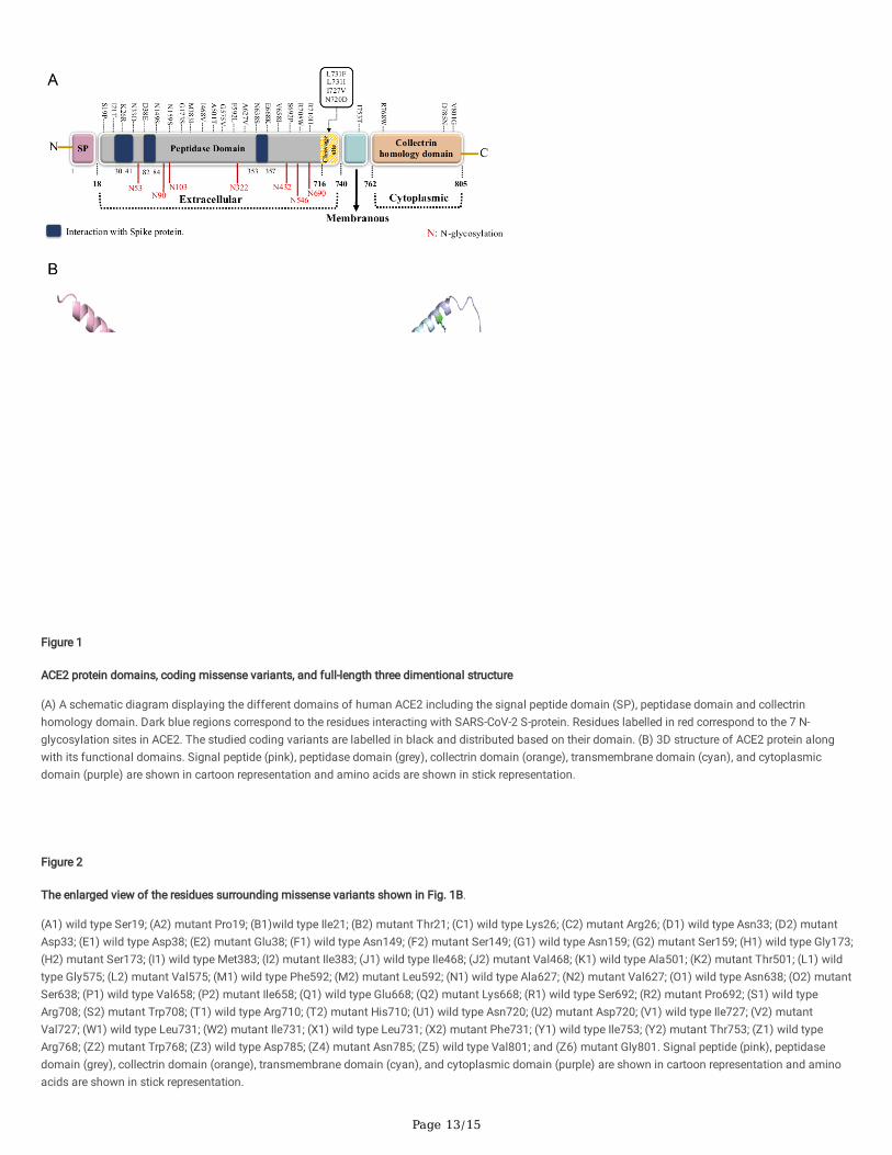
Figure 1
ACE2 protein domains, coding missense variants, and full-length three dimentional structure
(A) A schematic diagram displaying the different domains of human ACE2 including the signal peptide domain (SP), peptidase domain and collectrinhomology domain. Dark blue regions correspond to the residues interacting with SARS-CoV-2 S-protein. Residues labelled in red correspond to the 7 N-glycosylation sites in ACE2. The studied coding variants are labelled in black and distributed based on their domain. (B) 3D structure of ACE2 protein alongwith its functional domains. Signal peptide (pink), peptidase domain (grey), collectrin domain (orange), transmembrane domain (cyan), and cytoplasmicdomain (purple) are shown in cartoon representation and amino acids are shown in stick representation.
Figure 2
The enlarged view of the residues surrounding missense variants shown in Fig. 1B.
(A1) wild type Ser19; (A2) mutant Pro19; (B1)wild type Ile21; (B2) mutant Thr21; (C1) wild type Lys26; (C2) mutant Arg26; (D1) wild type Asn33; (D2) mutantAsp33; (E1) wild type Asp38; (E2) mutant Glu38; (F1) wild type Asn149; (F2) mutant Ser149; (G1) wild type Asn159; (G2) mutant Ser159; (H1) wild type Gly173;(H2) mutant Ser173; (I1) wild type Met383; (I2) mutant Ile383; (J1) wild type Ile468; (J2) mutant Val468; (K1) wild type Ala501; (K2) mutant Thr501; (L1) wildtype Gly575; (L2) mutant Val575; (M1) wild type Phe592; (M2) mutant Leu592; (N1) wild type Ala627; (N2) mutant Val627; (O1) wild type Asn638; (O2) mutantSer638; (P1) wild type Val658; (P2) mutant Ile658; (Q1) wild type Glu668; (Q2) mutant Lys668; (R1) wild type Ser692; (R2) mutant Pro692; (S1) wild typeArg708; (S2) mutant Trp708; (T1) wild type Arg710; (T2) mutant His710; (U1) wild type Asn720; (U2) mutant Asp720; (V1) wild type Ile727; (V2) mutantVal727; (W1) wild type Leu731; (W2) mutant Ile731; (X1) wild type Leu731; (X2) mutant Phe731; (Y1) wild type Ile753; (Y2) mutant Thr753; (Z1) wild typeArg768; (Z2) mutant Trp768; (Z3) wild type Asp785; (Z4) mutant Asn785; (Z5) wild type Val801; and (Z6) mutant Gly801. Signal peptide (pink), peptidasedomain (grey), collectrin domain (orange), transmembrane domain (cyan), and cytoplasmic domain (purple) are shown in cartoon representation and aminoacids are shown in stick representation.

Page 14/15
Figure 3
Overexpressed WT ACE2 subcellular localization and stability
(A) Immuno�uorescence confocal imaging of permeabilized HeLa cells transfected with Flag-tagged ACE2 (red), GFP-tagged HRas (plasma membrane markerin green) and Calnexin (ER marker in blue). Images were acquired using 100X magni�cation and manipulated by ImageJ. Scale bar = 50 mm. (B) HEK293 cellstransiently transfected with GFP and Flag-tagged WT ACE2 plasmids for 48 Hrs. Lysates were then digested in presence or absence of Endo H and PNGase Fenzymes for 3 Hrs and 1 Hr, respectively. Anti-Flag primary antibody was used to stain WT ACE2 and anti-GFP antibody was used to satin GFP that was usedas a transfection control. (C) Flag-tagged WT ACE2 transfected cells were treated with DMSO and 100 mg/ml cycloheximide for a period of 18 Hrs. Cells werelysed at the indicated time points (0, 4, 8, 12 and 18 Hrs) and lysates were analyzed by western blotting. Anti-Flag antibody was used to stain WT ACE2. Mocksample represent un-transfected cells. In all the experiments actin was used as a loading control. (D) Graph representing the relative expression of WT ACE2compared to DMSO treated cells at the indicated time points. Error bars represent ± SEM of three independent experiments. Band quanti�cation wasperformed using ImageJ.
Figure 4
ACE2 variants subcellular localization
HeLa cells were grown on coverslips and transiently co-transfected with Flag-tagged ACE2 WT or missense variants and GFP-tagged HRas (plasma membranemarker). 24 Hrs post transfection, cells were �xed and stained with anti-Flag and anti-Calnexin (ER marker) antibodies. Images were acquired using 100Xmagni�cation and manipulated by ImageJ. Scale bar = 50 mm.
Page 15/15
Figure 5
ACE2 variants glycosylation pro�le
HEK293 cells transiently co-transfected transfected with Flag-tagged ACE2 WT or missense variants and GFP plasmids for 48 Hrs. Lysates were then digestedin presence or absence of Endo H enzymes for 3 Hrs. Anti-Flag primary antibody was used to stain WT ACE2 and anti-GFP antibody was used to satin GFPthat was used as a transfection control. Actin was used as a loading control. Images were manipulated using ImageJ.
Supplementary Files
This is a list of supplementary �les associated with this preprint. Click to download.
Additional�le1.docx
Additional�le2.pdf