New Teratosphaeria species occurring on eucalypts in Australia
13
This article was published in the above mentioned Springer issue. The material, including all portions thereof, is protected by copyright; all rights are held exclusively by Springer Science + Business Media. The material is for personal use only; commercial use is not permitted. Unauthorized reproduction, transfer and/or use may be a violation of criminal as well as civil law. ISSN 1560-2745, Volume 43, Number 1
Transcript of New Teratosphaeria species occurring on eucalypts in Australia
This article was published in the above mentioned Springer issue.The material, including all portions thereof, is protected by copyright;all rights are held exclusively by Springer Science + Business Media.
The material is for personal use only;commercial use is not permitted.
Unauthorized reproduction, transfer and/or usemay be a violation of criminal as well as civil law.
ISSN 1560-2745, Volume 43, Number 1
New Teratosphaeria species occurring on eucalyptsin Australia
Vera Andjic & Gilbert Whyte & Giles Hardy &
Treena Burgess
Received: 23 August 2009 /Accepted: 21 January 2010 /Published online: 11 February 2010# Kevin D. Hyde 2010
Abstract Although the first Teratosphaeria spp. withcolletogloeopsis-like anamorphs were described outside ofAustralia, recently many new species have been describedfrom Australia. In the present study, several new Teratos-phaeria spp. were collected from infected eucalypt leavesin eastern Australia. Phylogenetic and morphologicalstudies revealed five new taxa described here as Teratos-phaeria aurantia, T. biformis, T. foliensis, T. micromaculataand T. tinara.
Keywords Teratosphaeria anamorphs . Colletogloeopsis-like . Eucalypts . Leaf disease . Taxonomy .Molecularphylogeny
Introduction
Teratosphaeria was established in 1912 and is typifiedwith T. fibrillosa Syd and P. Syd (Crous et al. 2004a). In2003, Taylor et al. (2003) showed that the DNA sequenceof the ITS region of the type species of Teratosphaeriaclustered within Mycosphaerella and thus synonymised itunder Mycosphaerella. Later, the data based on the LSUsequence showed that Mycosphaerella is polyphyletic(Crous et al. 2007) contradicting earlier findings ofmonophyly based on ITS data (Crous et al. 2000; Goodwinet al. 2001) thus, the Mycosphaerellaceae was split to twofamilies; Mycosphaerellaceae and Teratosphaeriaceae. Asa result some Mycosphaerella spp. and anamorphs weretransferred to Teratosphaeriaceae. Amongst them the most
important are diseases of Eucalyptus caused by M. cryptica,M. nubilosa, Kirramyces destructans (= Readerielladestructans), K. eucalypti (= R. eucalypti), K. gauchensis(= Colletogloeopsis gauchensis, = R. gauchensis), and K.zuluensis (= C. zuluensis = R. zuluensis).
Although single-celled conidia of Colletogloeopsis speciesare morphologically different to multiseptate conidia ofKirramyces spp., Andjic et al. (2007) showed that theanamorphs of Mycosphaerella from eucalypt leaves andstems, currently residing in Colletogloeopsis, occur in asingle monophyletic assemblage together with species ofKirramyces and thus transferred all Colletogloeopsis spp. toKirramyces. However, Crous et al. (2007) argued that themorphological features associated with Kirramyces andColletogloeopsis from other hosts have evolved more thanonce within the family Teratosphaeriaceae and proposed theoldest generic name Readeriella. As result all Kirramycesspp. from eucalypts were transferred to the genus Readeriella.Recently, in order to introduce generic names for separatelineages and not continue with dual nomenclature, severalnovel taxa in the Mycosphaerellaceae and Teratosphaeria-ceae have been proposed to try and achieve a more naturalclassification among the genera (Crous 2009; Crous et al.2009b). As a result, many Mycosphaerella spp. and theiranamorphs (including former Kirramyces and Colletogloeop-sis spp.) have been transferred to the genus Teratosphaeria.
There have been many Teratosphaeria spp. described foreucalypts with colletogloeopsis-like anamorphs. The firsttwo species described were Colletogloeum nubilosum = T.cryptica from New Zealand (Ganapathi and Corbin 1979)and Coniothyrium ovatum (Swart 1988) = T. ovata (Crouset al. 2009a). Subsequently, several species withcolletogloeopsis-like anamorphs were described and includ-ed Coniothyrium zuluense (Wingfield et al. 1997) = T.zuluensis from South Africa; Colletogloeopsis molleriana
V. Andjic :G. Whyte :G. Hardy : T. Burgess (*)Biological Science, Murdoch University,South St,Murdoch 6150, Australiae-mail: [email protected]
Fungal Diversity (2010) 43:27–38DOI 10.1007/s13225-010-0033-5
Author's personal copy

(Crous and Wingfield 1997) = T. molleriana from Portugal;Phaeophleospora toledana (Crous et al. 2004b) = T.toledana from Spain; Colletogloeopsis gauchensis (Corti-nas et al. 2006) = T. gauchensis from Uruguay; C.stellenboshiana (Crous et al. 2006) = T. stellenboshianafrom South Africa; Colletogloeopsis sp. (anamorph of T.pseudocryptica) from New Zealand (Crous et al. 2006); andT. verrucosa and T. juvenalis from South Africa (Crous etal. 2009a), T. hortaea (Crous et al. 2009e) and T.xenocryptica from Chile (Crous et al. 2009c). AlthoughTeratosphaeria spp. with colletogloeopsis-like anamorphswere first described outside Australia, recently many havebeen discovered in Australia such as T. blakelyi, T.considenianae, T. dimorpha (Summerell et al. 2006), T.angophorae, T. corymbiae (Andjic et al. 2007), T. brun-neotingens (Crous et al. 2007), T. ovata, T. veloci (Crous etal. 2009a), T. alboconidia, T. complicata, T. majorizuluen-sis, T. miniata and T. profusa (Crous et al. 2009c).
During surveillance of eucalypt species trials in easternAustralia between 2005–2007, we observed leaves exhibit-ing symptoms associated with Mycosphaerella leaf disease(MLD). Samples were collected across several sites andpreliminary microscopy examination revealed several fungiwith a conidial morphology similar to that of Teratosphae-ria spp. (colletogloeopsis-like). In the present study wedescribe five new Teratosphaeria species.
Materials and methods
Isolates
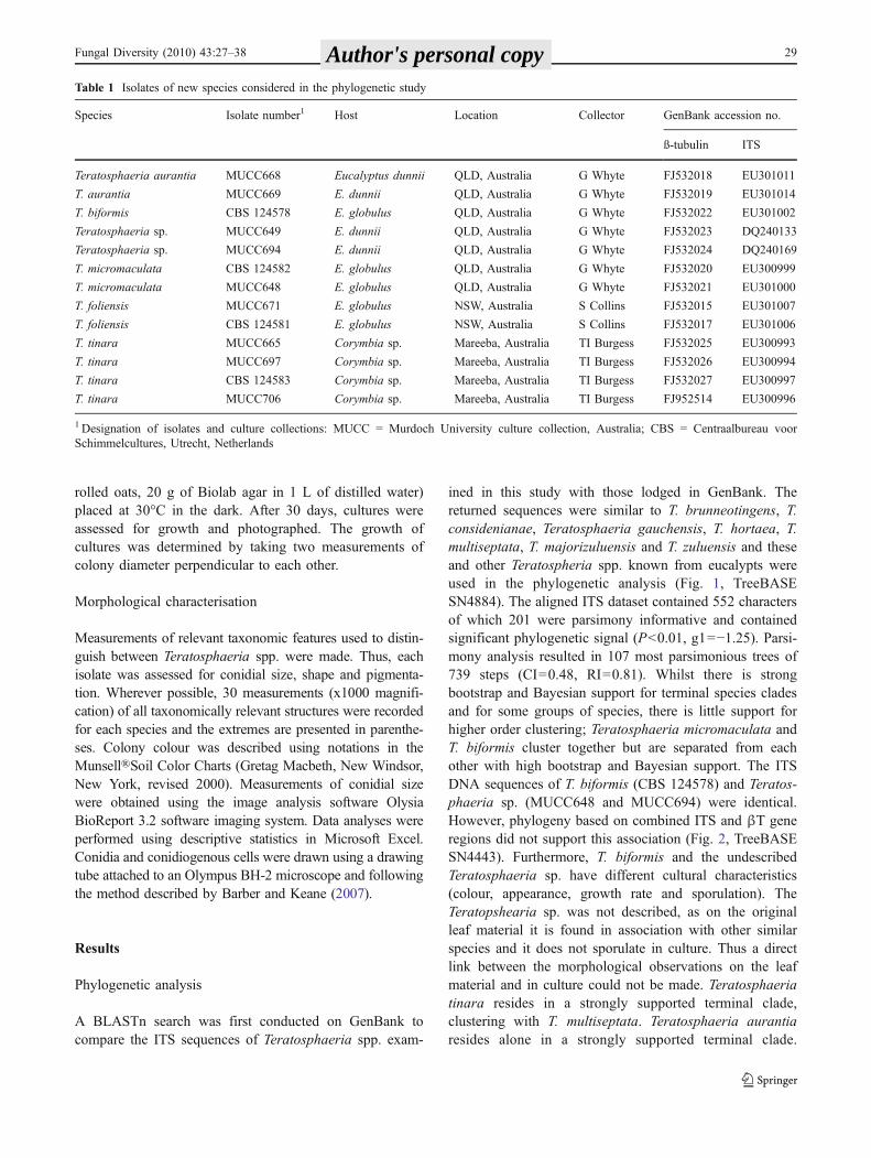
Eucalypt leaves with symptoms of MLD were collected fromEucalyptus and Corymbia spp. in southern Queensland,northern Queensland and central New South Wales. Isolateswere obtained by collecting conidia exuding from singlepycnidia using the tip of a sterile needle. These weretransferred onto 2% Malt Extract Agar (MEA; 20 g/LBiolab malt extract, 15 g/L Biolab agar) containingStreptomycin 150μg/ml (Sigma-Aldrich, Australia) in asingle spot and allowing it to hydrate for 5 min. Under adissecting microscope, spores were streaked across the agarusing a sterile needle and single spores immediatelytransferred to MEA plates. Cultures were grown in the darkat 30°C for 2 weeks and then transferred to fresh MEAplates. All cultures were maintained on 2% MEA in tubes at20°C. All isolates are maintained in the culture collection atMurdoch University (MUCC) (Table 1). Reference isolateshave been deposited in the collection of the Centraalbureauvoor Schimmelcultures (CBS), Utrecht. Herbarium speci-mens of new collections have been lodged in the MurdochUniversity herbarium (MURU). Descriptions were depositedin MycoBank.
DNA Extraction and PCR amplification
The isolates were grown on 2% MEA at 20°C for 4 weeksand the mycelium harvested and placed in a 1.5 ml sterileEppendorf® tube. Harvested mycelium was frozen in liquidnitrogen, ground to a fine powder and genomic DNA wasextracted as described previously (Andjic et al. 2007). Apart of the internal transcribed spacer (ITS) region of theribosomal DNA operon was amplified using the primersITS-1F (5′ CTT GGT CAT TTA GAG GAA GTA A)Gardes and Bruns (1993) and ITS-4 (5′TCC TCC GCTTAT TGA TAT GC 3′) White et al. (1990). Part of the β-tubulin (βT) gene region was amplified with the primersβT2a (5′GGTAAC CAA ATC GGT GCT GCT TTC 3′) andβT2b (5′ACC CTC AGT GTA GTG ACC CTT GGC 3′)Glass and Donaldson (1995). The PCR reaction mixture,PCR conditions, the clean- up of products and sequencingwere as described previously by Andjic et al. (2007).
Phylogenetic analysis
In order to compare Teratosphaeria isolates generated fromthis study with other closely related species, additional ITSsequences were obtained from GenBank. Sequence datawere assembled using Sequence Navigator version 1.01(Perkin Elmer) and aligned in Clustal X (Thompson et al.1997). Manual adjustments were made visually by insertinggaps where necessary. All sequences derived in this studywere deposited in GenBank and accession numbers areshown in Table 1.
Parsimony analysis with heuristic search was performedusing PAUP (Phylogenetic Analysis Using Parsimony)(Swofford 2000) as described previously (Andjic et al.2007). ITS trees were rooted to Readeriella spp., andcombined trees were rooted to Dothistroma septospora.Bayesian analysis was conducted on the same datasets asthe one used in the distance analysis. MrModeltest v. 3.5(Nylander 2004) was used to determine the best nucleotidesubstitution model. Phylogenetic analyses were performedwith MrBayes v. 3.1 (Ronquist and Huelsenbeck 2003)applying a general time reversible (GTR) substitutionmodel with gamma (G) and proportion of invariable site(I) parameters to accommodate variable rates across sites asdescribed previously (Andjic et al. 2007). The newsequences were deposited in GenBank and the alignmentsand phylogenetic trees in TreeBASE (www.treebase.org).
Culture characteristics
Plugs (2 mm diam) were cut from actively growing culturesand placed at the centre of Petri dishes (55 mm) containingone of two nutrient media. Three replicates of each isolatewere grown on 2% MEA; and oatmeal agar (OMA; 30 g/L
28 Fungal Diversity (2010) 43:27–38 Author's personal copy
rolled oats, 20 g of Biolab agar in 1 L of distilled water)placed at 30°C in the dark. After 30 days, cultures wereassessed for growth and photographed. The growth ofcultures was determined by taking two measurements ofcolony diameter perpendicular to each other.
Morphological characterisation
Measurements of relevant taxonomic features used to distin-guish between Teratosphaeria spp. were made. Thus, eachisolate was assessed for conidial size, shape and pigmenta-tion. Wherever possible, 30 measurements (x1000 magnifi-cation) of all taxonomically relevant structures were recordedfor each species and the extremes are presented in parenthe-ses. Colony colour was described using notations in theMunsell®Soil Color Charts (Gretag Macbeth, New Windsor,New York, revised 2000). Measurements of conidial sizewere obtained using the image analysis software OlysiaBioReport 3.2 software imaging system. Data analyses wereperformed using descriptive statistics in Microsoft Excel.Conidia and conidiogenous cells were drawn using a drawingtube attached to an Olympus BH-2 microscope and followingthe method described by Barber and Keane (2007).
Results
Phylogenetic analysis
A BLASTn search was first conducted on GenBank tocompare the ITS sequences of Teratosphaeria spp. exam-
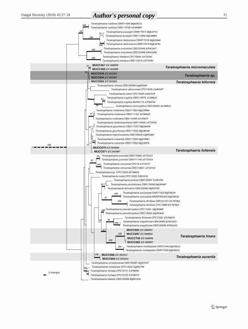
ined in this study with those lodged in GenBank. Thereturned sequences were similar to T. brunneotingens, T.considenianae, Teratosphaeria gauchensis, T. hortaea, T.multiseptata, T. majorizuluensis and T. zuluensis and theseand other Teratospheria spp. known from eucalypts wereused in the phylogenetic analysis (Fig. 1, TreeBASESN4884). The aligned ITS dataset contained 552 charactersof which 201 were parsimony informative and containedsignificant phylogenetic signal (P<0.01, g1=−1.25). Parsi-mony analysis resulted in 107 most parsimonious trees of739 steps (CI=0.48, RI=0.81). Whilst there is strongbootstrap and Bayesian support for terminal species cladesand for some groups of species, there is little support forhigher order clustering; Teratosphaeria micromaculata andT. biformis cluster together but are separated from eachother with high bootstrap and Bayesian support. The ITSDNA sequences of T. biformis (CBS 124578) and Teratos-phaeria sp. (MUCC648 and MUCC694) were identical.However, phylogeny based on combined ITS and βT generegions did not support this association (Fig. 2, TreeBASESN4443). Furthermore, T. biformis and the undescribedTeratosphaeria sp. have different cultural characteristics(colour, appearance, growth rate and sporulation). TheTeratopshearia sp. was not described, as on the originalleaf material it is found in association with other similarspecies and it does not sporulate in culture. Thus a directlink between the morphological observations on the leafmaterial and in culture could not be made. Teratosphaeriatinara resides in a strongly supported terminal clade,clustering with T. multiseptata. Teratosphaeria aurantiaresides alone in a strongly supported terminal clade.
Table 1 Isolates of new species considered in the phylogenetic study
Species Isolate number1 Host Location Collector GenBank accession no.
ß-tubulin ITS
Teratosphaeria aurantia MUCC668 Eucalyptus dunnii QLD, Australia G Whyte FJ532018 EU301011
T. aurantia MUCC669 E. dunnii QLD, Australia G Whyte FJ532019 EU301014
T. biformis CBS 124578 E. globulus QLD, Australia G Whyte FJ532022 EU301002
Teratosphaeria sp. MUCC649 E. dunnii QLD, Australia G Whyte FJ532023 DQ240133
Teratosphaeria sp. MUCC694 E. dunnii QLD, Australia G Whyte FJ532024 DQ240169
T. micromaculata CBS 124582 E. globulus QLD, Australia G Whyte FJ532020 EU300999
T. micromaculata MUCC648 E. globulus QLD, Australia G Whyte FJ532021 EU301000
T. foliensis MUCC671 E. globulus NSW, Australia S Collins FJ532015 EU301007
T. foliensis CBS 124581 E. globulus NSW, Australia S Collins FJ532017 EU301006
T. tinara MUCC665 Corymbia sp. Mareeba, Australia TI Burgess FJ532025 EU300993
T. tinara MUCC697 Corymbia sp. Mareeba, Australia TI Burgess FJ532026 EU300994
T. tinara CBS 124583 Corymbia sp. Mareeba, Australia TI Burgess FJ532027 EU300997
T. tinara MUCC706 Corymbia sp. Mareeba, Australia TI Burgess FJ952514 EU300996
1 Designation of isolates and culture collections: MUCC = Murdoch University culture collection, Australia; CBS = Centraalbureau voorSchimmelcultures, Utrecht, Netherlands
Fungal Diversity (2010) 43:27–38 29 Author's personal copy

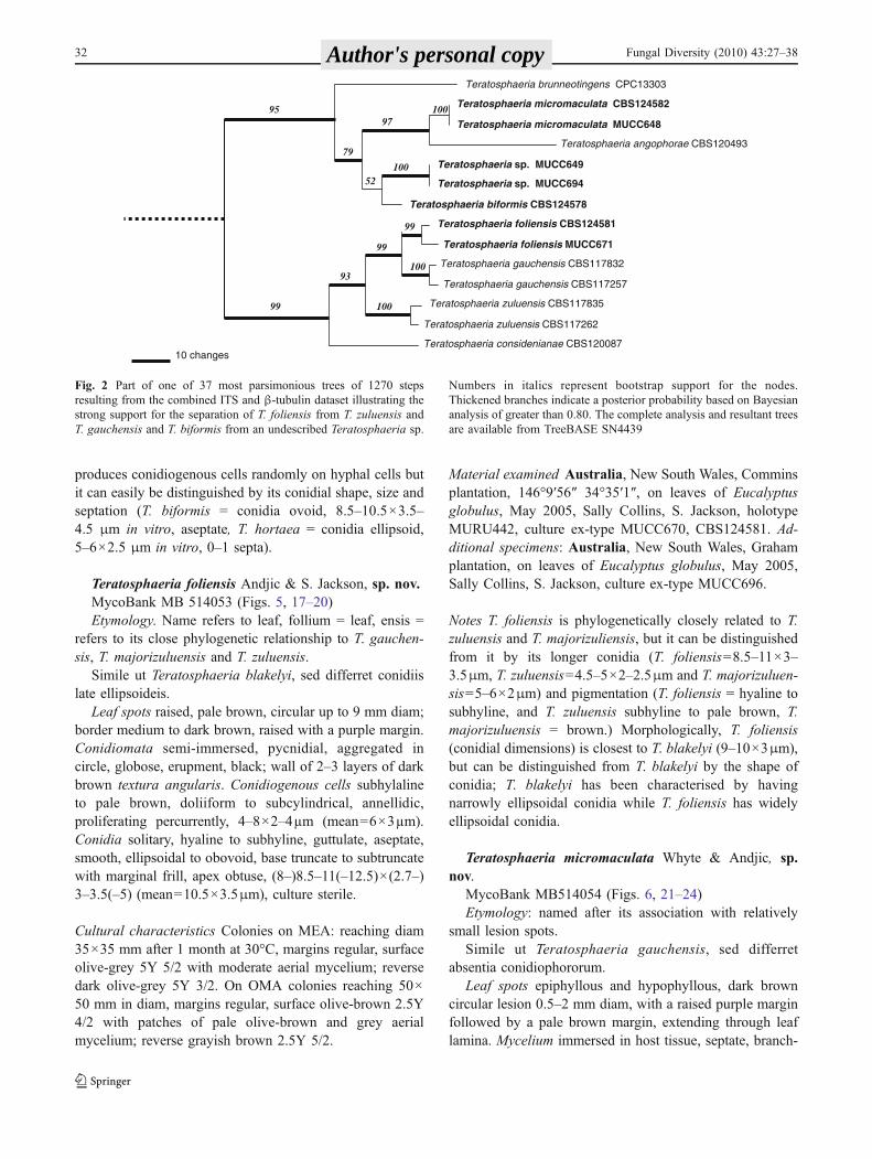
Teratosphaeria foliensis grouped in a clade containing T.considenianae, T. stellenboschiana, T. gauchensis, T.majorizuluensis and T. zuluensis. This clade could not beclearly resolved on ITS data alone, but in the combinedanalysis with βT each species was clearly resolved withbootstrap values of 100% and posterior probabilities of 1.00(Fig. 2, TreeBASE SN4443).
Taxonomy
Morphological comparison of Teratosphaeria spp. withcolletogloeopsis-like anamorphs found on eucalypts arepresented in Table 2.
Teratosphaeria aurantia Whyte & Andjic, sp. nov.MycoBank MB 514050 (Figs. 3, 25–28)Etymology: named after the golden yellow stain of the agar.Simile ut T. pseudocryptica, sed differret conidiis
tenuioris et discoloratione aureo-flava agaris.Leaf spots epiphyllous and hypophyllous, extending
through leaf lamina, pale brown, conspicuously circular0.5–5 mm diam with corky-brown margins. Myceliumimmersed in host tissue, septate, branched, melanised.Conidiophores reduced to conidiogenous cells. Conidio-mata pycnidial, embeded, sub-epidermal, separate, globose,wall consisting of 4–5 layers of dark brown texturaangularis. Conidiogenous cells subcylindrical to doliiform,subhyaline to medium brown, smooth, proliferating percur-rently with 1–2 annulations, formed from the inner cells ofthe pycnidial wall, 5.5×4.0 μm. Conidia ellipsoidal, 0–1septate, subhyaline to medium brown, smooth, guttulate,gradually tapering toward apex, truncate to subtruncate atbase with marginal frill, (9.5–)11–14(–16.0)×(2.5–)2.5–3.5(–4.0) (mean=12.5×3.0 μm).
Cultural characteristics Colonies on MEA reaching a diam4×5 mm after 1 month at 30°C, globular aggregating orseparate masses with white to cream 2Y 8/3 short aerialhyphae on the surface; dark brown 10YR 3/3 in reverse. OnOMA colonies reaching 7×8 mm diam, after 1 month,globular aggregating or separate masses with white tocream 2Y 8/3 short aerial hyphae on surface; dark brown10YR 3/3 in reverse.
Material examined Australia, Queensland, Rosedale, onleaves of Eucalyptus grandis, G. Whyte 2007, holotypeMURU440, culture ex-type MUCC668, CBS125243. Ad-ditional specimens: Australia, Queensland, Rosedale, onleaves of Eucalyptus dunnii, G. Whyte 2007, (MURU439)(culture ex-type MUCC669).
Notes Teratosphaeria aurantia is phylogenetically andmorphologically similar to the anamorph of T. pseudocryp-
tica (12–14×4 μm). However, it can be distinguished fromthe latter species by the golden-yellow pigment in the agar,and slightly thinner conidia (11–14×2.5–3.5 μm). Inaddition, the lesions with which T. aurantia is associatedare distinct in appearance from other described Teratos-phaeria species with distinctly circular and raised marginswith an aggregation of fruiting structures in the centre(Fig. 9).
Teratosphaeria biformis Whyte &Andjic, sp. nov.MycoBank MB 514051 (Figs. 4, 12–16)Etymology: named after its ability to produce conidia
both as a coelomycete and hyphomycete on the leaf and asa hyphomycete on agar.
Simile ut Teratosphaeria hortaea, quaternus cellulaeconidiogenae producentur de cellulis hypharum, sed con-idiae differrent in forma et magnitudini et absentiaseptarum.
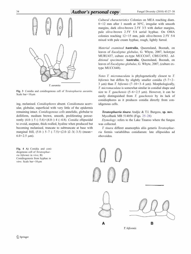
Leaf spots epiphyllous and hypophyllous, pale brown,conspicuously circular, 0.5–5 mm diam, extending throughleaf lamina. Mycelium immersed in host tissue, septate,branching, melanised. Conidiophores absent. Conidiomatapycnidial, dark brown, amphigenous, aggregated, globose.Conidiogenous cells globular, brown, smooth. Conidiaholoblastic, subhyaline but becoming melanised whenmature, aseptate, ovoid, thick-walled, truncate at base witha minute marginal frill, (6.0–)7–10(–11.0)×(2.5–)3–4(–4.0)(mean=8.5×3.5 μm).
Cultural characteristics Colonies on MEA reaching diam45×45 mm after 1 month at 30°C, regular with smoothmargins, pale gray 2.5Y 7/3 with pale yellowish brownmycelium 2.5Y 6/3 on surface; reverse olive-brown 2.5Y 4/4 with white margins. On OMA colonies reaching 25×25 mm diam, regular with smooth margins distributed inthree concentric zones of different colours, out zone palegrey 2.5Y 7/1, middle pale brown-grey 6/2 and inner zonepale grey 7/1 on top; reverse side is the same as surface.
Material examined Australia, Queensland, Rosedale, onleaves of Eucalyptus globulus, G. Whyte, 2007,MURU438, culture ex-type MUCC693, CBS124578.
Notes T. biformis is phylogenetically closest to T. micro-maculata from which it differs by slightly longer and widerconidia (7–10×3–4 μm), T. micromaculata (5–7×2–3 μm).T. biformis is morphologically closest to T. hortaea as it
Fig. 1 One of the 107 most parsimonious trees of 739 steps resultingfrom analysis of the ITS dataset. The complete tree can be viewed atTreeBASE (SN4884). Numbers in italics represent bootstrap supportfor the nodes. Thickened branches indicate a posterior probabilitybased on Bayesian analysis of greater than 0.80. The new Teratos-phaeria species described in this study are highlighted
b
30 Fungal Diversity (2010) 43:27–38 Author's personal copy
Teratosphaeria foliensis
Teratosphaeria micromaculata
Teratosphaeria sp.
Teratosphaeria aurantia
Teratosphaeria tinara
Teratosphaeria biformis
Teratosphaeria nubilosa CMW11560 DQ658232
Teratosphaeria nubilosa CBS114708 AF449099
Teratosphaeria eucalypti CMW17917 DQ632711
Teratosphaeria eucalypti CBS113992 DQ240001
Teratosphaeria destructans CMW17918 DQ632666
Teratosphaeria destructans CMW17919 DQ632701
Teratosphaeria corymbiae CBS120495 EF011657
Teratosphaeria corymbiae CBS120496 EF011656
Teratosphaeria toledana CPC10840 AY725581
Teratosphaeria toledana CBS113313 AY725581
MUCC467 EU300999MUCC468 EU301000
MUCC649 EU301001MUCC694 EU301003MUCC693 EU301002
Teratosphaeria miniata CBS125006 GQ852803
Teratosphaeria alboconidia CPC14535 FJ493197
Teratosphaeria veloci CPC14600 FJ023539
Teratosphaeria cryptica CBS110975 AY309623
Teratosphaeria cryptica MURU115 AY509754
Teratosphaeria xenocryptica CBS122905 AF309622
Teratosphaeria molleriana CBS117924 DQ239968
Teratosphaeria molleriana CBS111164 AF309620
Teratosphaeria molleriana CBS110499 AY150675
Teratosphaeria stellenboschiana CBS116428 AY725518
Teratosphaeria gauchensis CBS117257 DQ240198
Teratosphaeria gauchensis CBS117832 DQ240188
Teratosphaeria majorizuluensis CBS120040 GQ852802
Teratosphaeria zuluensis CBS117835 DQ239987
Teratosphaeria zuluensis CBS117262 DQ239976
MUCC670 EU301006MUCC671 EU301007
Teratosphaeria juvenalis CBS110906 AY725513
Teratosphaeria juvenalis CBS111149 AY725514
Teratosphaeria verrucosa CPC18 AY725517
Teratosphaeria verrucosa CBS113621 AY725515
Teratosphaeria sp. CPC13303 EF394853
Teratosphaeria ovata CPC14632 FJ023538
Teratosphaeria profusa CBS125007 FJ493196
Teratosphaeria pluritubularis CBS118508 DQ303007
Teratosphaeria dimorpha CBS120085 DQ923529
Teratosphaeria syncarpiae DAR77433 DQ530219
Teratosphaeria syncarpiae NSWF005320 DQ530220
Teratosphaeria fibrillosa CBS121707 EU707862
Teratosphaeria fibrillosa CPC13969 EU707863
Teratosphaeria pseudocryptica CPC11264 DQ303009
Teratosphaeria pseudocryptica CBS118504 DQ303010
Teratosphaeria fimbriata CPC13321 EF394835
Teratosphaeria angophorae CBS120493 EF011653
Teratosphaeria angophorae CBS120496 EF011652
MUCC665 EU300993MUCC697 EU300094
MUCC706 EU300096MUCC665 EU300997
Teratosphaeria multiseptata DAR77440 DQ530223
Teratosphaeria multiseptata DAR77439 DQ530225
MUCC668 EU301011MUCC669 EU301014
Teratosphaeria considenianae CBS120087 DQ923527
Teratosphaeria complicata CPC13032 GQ852790
Teratosphaeria hortaea CPC15741 FJ790282
Teratosphaeria hortaea CPC15723 FJ790279
Teratosphaeria blakelyi CBS120089 DQ923526
5 changes
100
58
100
100
79
100
100
100
98
95
98
95
97
53
67
95
92
98
100
83
87
100
100
54
100
63
99100
100
100
100
100
8883
Fungal Diversity (2010) 43:27–38 31 Author's personal copy
produces conidiogenous cells randomly on hyphal cells butit can easily be distinguished by its conidial shape, size andseptation (T. biformis = conidia ovoid, 8.5–10.5×3.5–4.5 μm in vitro, aseptate, T. hortaea = conidia ellipsoid,5–6×2.5 μm in vitro, 0–1 septa).
Teratosphaeria foliensis Andjic & S. Jackson, sp. nov.MycoBank MB 514053 (Figs. 5, 17–20)Etymology. Name refers to leaf, follium = leaf, ensis =
refers to its close phylogenetic relationship to T. gauchen-sis, T. majorizuluensis and T. zuluensis.
Simile ut Teratosphaeria blakelyi, sed differret conidiislate ellipsoideis.
Leaf spots raised, pale brown, circular up to 9 mm diam;border medium to dark brown, raised with a purple margin.Conidiomata semi-immersed, pycnidial, aggregated incircle, globose, erupment, black; wall of 2–3 layers of darkbrown textura angularis. Conidiogenous cells subhylalineto pale brown, doliiform to subcylindrical, annellidic,proliferating percurrently, 4–8×2–4µm (mean=6×3µm).Conidia solitary, hyaline to subhyline, guttulate, aseptate,smooth, ellipsoidal to obovoid, base truncate to subtruncatewith marginal frill, apex obtuse, (8–)8.5–11(–12.5)×(2.7–)3–3.5(–5) (mean=10.5×3.5μm), culture sterile.
Cultural characteristics Colonies on MEA: reaching diam35×35 mm after 1 month at 30°C, margins regular, surfaceolive-grey 5Y 5/2 with moderate aerial mycelium; reversedark olive-grey 5Y 3/2. On OMA colonies reaching 50×50 mm in diam, margins regular, surface olive-brown 2.5Y4/2 with patches of pale olive-brown and grey aerialmycelium; reverse grayish brown 2.5Y 5/2.
Material examined Australia, New South Wales, Comminsplantation, 146°9′56″ 34°35′1″, on leaves of Eucalyptusglobulus, May 2005, Sally Collins, S. Jackson, holotypeMURU442, culture ex-type MUCC670, CBS124581. Ad-ditional specimens: Australia, New South Wales, Grahamplantation, on leaves of Eucalyptus globulus, May 2005,Sally Collins, S. Jackson, culture ex-type MUCC696.
Notes T. foliensis is phylogenetically closely related to T.zuluensis and T. majorizuliensis, but it can be distinguishedfrom it by its longer conidia (T. foliensis=8.5–11×3–3.5μm, T. zuluensis=4.5–5×2–2.5μm and T. majorizuluen-sis=5–6×2μm) and pigmentation (T. foliensis = hyaline tosubhyline, and T. zuluensis subhyline to pale brown, T.majorizuluensis = brown.) Morphologically, T. foliensis(conidial dimensions) is closest to T. blakelyi (9–10×3μm),but can be distinguished from T. blakelyi by the shape ofconidia; T. blakelyi has been characterised by havingnarrowly ellipsoidal conidia while T. foliensis has widelyellipsoidal conidia.
Teratosphaeria micromaculata Whyte & Andjic, sp.nov.
MycoBank MB514054 (Figs. 6, 21–24)Etymology: named after its association with relatively
small lesion spots.Simile ut Teratosphaeria gauchensis, sed differret
absentia conidiophororum.Leaf spots epiphyllous and hypophyllous, dark brown
circular lesion 0.5–2 mm diam, with a raised purple marginfollowed by a pale brown margin, extending through leaflamina. Mycelium immersed in host tissue, septate, branch-
10 changes
95
99
Teratosphaeria brunneotingens CPC13303
Teratosphaeria micromaculata CBS124582
Teratosphaeria micromaculata MUCC648
Teratosphaeria angophorae CBS120493
Teratosphaeria sp. MUCC649
Teratosphaeria sp. MUCC694
Teratosphaeria biformis CBS124578
Teratosphaeria foliensis CBS124581
Teratosphaeria foliensis MUCC671
Teratosphaeria gauchensis CBS117832
Teratosphaeria gauchensis CBS117257
Teratosphaeria zuluensis CBS117835
Teratosphaeria zuluensis CBS117262
Teratosphaeria considenianae CBS120087
79
97100
52100
93
99
99
100
100
Fig. 2 Part of one of 37 most parsimonious trees of 1270 stepsresulting from the combined ITS and β-tubulin dataset illustrating thestrong support for the separation of T. foliensis from T. zuluensis andT. gauchensis and T. biformis from an undescribed Teratosphaeria sp.
Numbers in italics represent bootstrap support for the nodes.Thickened branches indicate a posterior probability based on Bayesiananalysis of greater than 0.80. The complete analysis and resultant treesare available from TreeBASE SN4439
32 Fungal Diversity (2010) 43:27–38 Author's personal copy
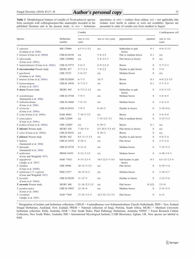
Table 2 Morphological features of conidia of Teratosphaeria species from eucalypts with colletogloeopsis-like anamorphs recorded in thepublished literature and in the present study; in vivo = herbarium specimens, in vitro = isolates from culture, n/a = not applicable (the isolates weresterile in culture or were not available). Species are presented in order of conidia size from smallest to largest
Conidia Conidiogenous cell
Species Herbariumnumber
size in vivo(μm)
size in vitro (μm) pigmentation septation size in vivo(μm)
T. zuluensis(Cortinas et al. 2006)
IMI 370886 4.5–5×2–2.5 n/a Subhyaline to palebrown
0–1 4–8×2–3.5
T. hortaea (Crous et al. 2009d) CBS-H-20194 n/a 5–6×2.5 Pale to medium brown 0–1 n/a
T. alboconidia(Crous et al. 2009c)
CBS 1250004 n/a 5–6×2.5–3 Pale brown to brown 0 n/a
T. majorizuluensis (Crous et al. 2009c) CBS H-19773 5–6×2 6–9×2.5–4 Brown 0 5–7×2–3
T. micromaculata Present study MURU 437 5–7×2–3 7–8×2.2 Hyaline to pale brown 0 5×4
T. gauchensis(Cortinas et al. 2006)
CBS 19722 5–6×2.5 n/a Medium brown 0 n/a
T. miniata (Crous et al. 2009c) CBS H-20269 6–7×3 10×5 Brown 0–1 4–8×2.5–3.5
T. brunneotingens(Crous et al. 2007)
CBS-H 19838 6–7×2–3 n/a Brown 0–1 5–7×3–5
T. tinara Present study MURU 445 6–7.5×3–3.5 n/a Subhyaline to palebrown
0 5–6×3–3.5
T. considenianae(Summerell et al. 2006)
CBS H-19744 7–9×3 n/a Medium brown 0 3–6×4–5
T. stellenboschiana(Crous et al. 2006)
CBS H-19688 7–9×3.5 n/a Medium brown 0 3–6×3–4
T. verrucosa(Crous et al. 2009a)
CBS H-20183 7–9×5 8–10×5 Hyaline to brown 0 5–10×4–6
T. ovata (Crous et al. 2009a) DAR 49461 7–10×3–3.5 n/a Brown 0 3–6×4–6
T. xenocryptica(Crous et al. 2009c)
CBS 122905 n/a 7–15×2.5–3.5 Pale to medium brown 0 5–15×3–5
T. profusa (Crous et al. 2009a) CBS 125007 n/a 8–10×3 Brown 0–1 n/a
T. biformis Present study MURU 438 7–10×3–4 8.5–10.5×3.5–4.5 Pale brown to brown 0 n/a
T. veloci (Crous et al. 2009a) CBS H-20182 n/a 8–10×3 Brown 0 n/a
T. foliensis Present study MURU 442 8.5–11×3–3.5 n/a Hyaline to pale brown 0 4–8×2–4
T. blakelyi(Summerell et al. 2006)
CBS H-19743 9–10×3 n/a Pale brown 0 5–7×3–4
T. dimorpha(Summerell et al. 2006)
CBS H-19739 9–11×4 n/a Medium brown 0 7–15×3–5
T. molleriana(Crous and Wingfield 1997)
PREM 54395 9–12×3–3.5 n/a Medium brown 0 4–20×3.5–5
T. angophorae(Andjic et al. 2007)
DAR 77452 9–15×2.5–4 10.5–22.5×3–4.5 Sub-hyaline to palebrown
0–3 6.5–12×2.5–4
T. toledana(Crous et al. 2004b)
CBS 59896 10–12×3–3.5 n/a Pale brown 0 6–10×3–4
T. nubilosum (=T. cryptica)(Crous and Wingfield 1997)
PDD 37677 10–15×4–5 n/a Medium brown 0 5–10×4–7
T. juvenalis(Crous et al. 2009a)
CBS H-20180 11–13×5 n/a Hyaline to brown 0 3–12×5–6
T. aurantia Present study MURU 440 11–14×2.5–3.5 n/a Pale brown 0–1(2) 5.5×4
T. pseudocryptica(Crous et al. 2006)
CBS H-19693 12–14×4 n/a Medium brown 0 5–15×3–5
T. corymbiae(Andjic et al. 2007)
DAR 77445 17–23×3.5–5 16.5–22×2.5–3.5 Pale brown 0 6–13
1 Designation of isolates and herbarium collections: CBS-H = Centraalbureau voor Schimmelcultures Utrecht Netherlands; PDD = New ZealandFungal Herbarium, Auckland, New Zealand, PREM = National collection of fungi, Pretoria, South Africa, MURU = Murdoch Universityherbarium collection, Perth; Australia; DAR = New South Wales, Plant Pathology Herbarium, Australia; NSWF = Forest Research CultureCollection, New South Wales, Australia; IMI = International Mycological Institute, CABI Bioscience, Egham, UK. New species are labeled inbold.
Table 2 Morphological features of conidia of Teratosphaeria speciesfrom eucalypts with colletogloeopsis-like anamorphs recorded in thepublished literature and in the present study; in vivo = herbarium
specimens, in vitro = isolates from culture, n/a = not applicable (theisolates were sterile in culture or were not available). Species arepresented in order of conidia size from smallest to largest
Fungal Diversity (2010) 43:27–38 33 Author's personal copy
ing, melanised. Conidiophores absent. Conidiomata acerv-ular, globular, superficial with very little of the epidermisremaining intact. Conidiogenous cells annelidic, globular todolliform, medium brown, smooth, proliferating percur-rently (4.0–) 5 (–5.6)×(4.0–) 4 (–4.8). Conidia ellipsoidalto ovoid, aseptate, thick-walled, hyaline when produced butbecoming melanised, truncate to subtruncate at base withmarginal frill, (5.0–) 5–7 (–7.5)×(2.0–)2–3(–3.5) (mean=6.0×2.5 μm).
Cultural characteristics Colonies on MEA reaching diam.8×12 mm after 1 month at 30°C, irregular with smoothmargins, dark olive-brown 2.5Y 3/3 with darker margins,pale olive-brown 2.5Y 5/4 aerial hyphae. On OMAcolonies reaching 12×15 mm, pale olive-brown 2.5Y 5/4mixed with pale cream hyphae, rough, lightly furred.
Material examined Australia, Queensland, Boonah, onleaves of Eucalyptus globulus, G. Whyte, 2007, holotypeMURU437, culture ex-type MUCC647, CBS124582. Ad-ditional specimens: Australia, Queensland, Boonah, onleaves of Eucalyptus globulus, G. Whyte, 2007, (culture ex-type MUCC648).
Notes T. micromaculata is phylogenetically closest to T.biformis but differs by slightly smaller conidia (5–7×2–3 μm) than T. biformis (7–10×3–4 μm). Morphologically,T. micromaculata is somewhat similar in conidial shape andsize to T. gauchensis (5–6×2.5 μm). However, it can beeasily distinguished from T. gauchensis by its lack ofconidiophores as it produces conidia directly from con-idigenous cells.
Teratosphaeria tinara Andjic & T.I. Burgess, sp. nov.MycoBank MB 514056 (Figs. 25–28)Etymology: refers to the Lake Tinaroo where the fungus
was collected.T. tinara differet anamorphis aliis generis Teratosphae-
ria formis variabilibus conidiarum: late ellipsoidea adobovoidea.
Fig. 3 Conidia and conidiogenous cell of Teratosphaeria aurantia.Scale bar=10μm
Fig. 4 A) Conidia and coni-diogenous cell of Teratosphae-ria biformis in vivo; B)Conidiogenesis from hyphae invitro. Scale bar=10μm
34 Fungal Diversity (2010) 43:27–38 Author's personal copy
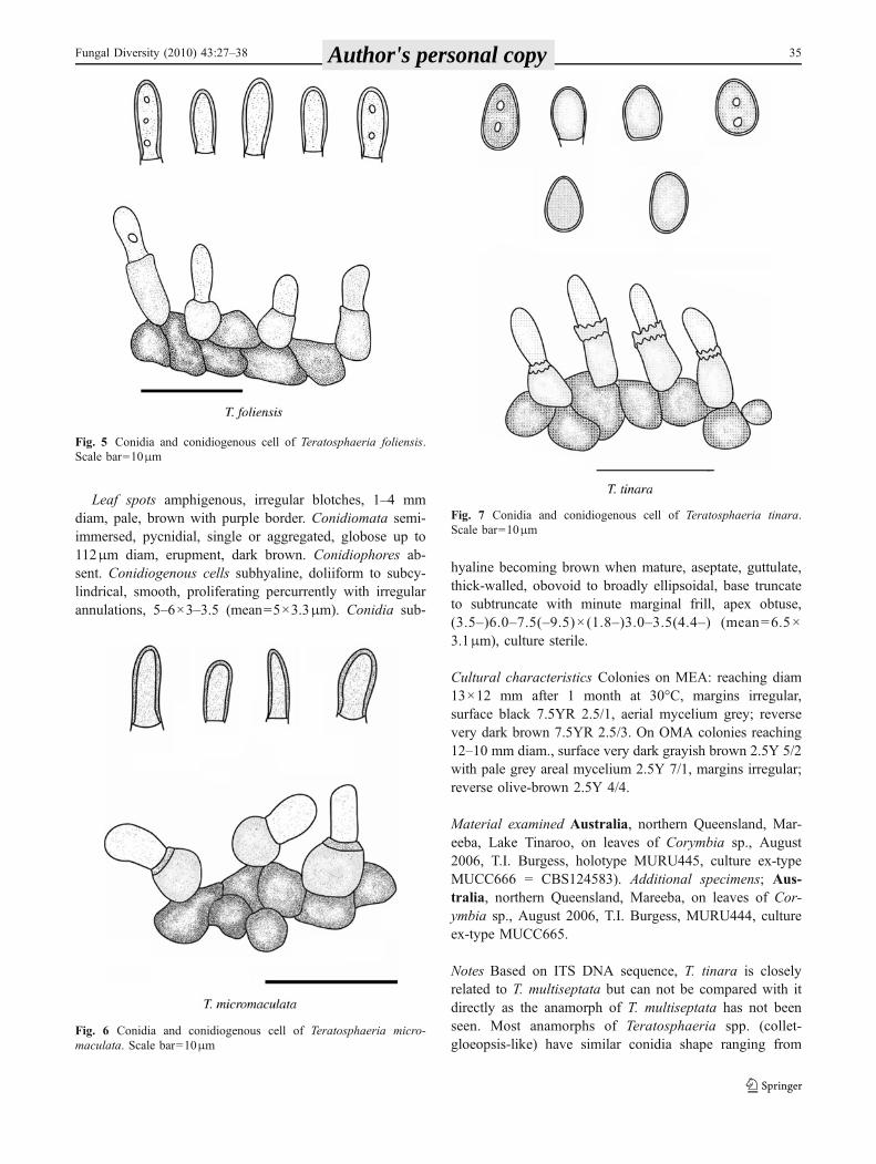
Leaf spots amphigenous, irregular blotches, 1–4 mmdiam, pale, brown with purple border. Conidiomata semi-immersed, pycnidial, single or aggregated, globose up to112μm diam, erupment, dark brown. Conidiophores ab-sent. Conidiogenous cells subhyaline, doliiform to subcy-lindrical, smooth, proliferating percurrently with irregularannulations, 5–6×3–3.5 (mean=5×3.3μm). Conidia sub-
hyaline becoming brown when mature, aseptate, guttulate,thick-walled, obovoid to broadly ellipsoidal, base truncateto subtruncate with minute marginal frill, apex obtuse,(3.5–)6.0–7.5(–9.5)× (1.8–)3.0–3.5(4.4–) (mean=6.5×3.1μm), culture sterile.
Cultural characteristics Colonies on MEA: reaching diam13×12 mm after 1 month at 30°C, margins irregular,surface black 7.5YR 2.5/1, aerial mycelium grey; reversevery dark brown 7.5YR 2.5/3. On OMA colonies reaching12–10 mm diam., surface very dark grayish brown 2.5Y 5/2with pale grey areal mycelium 2.5Y 7/1, margins irregular;reverse olive-brown 2.5Y 4/4.
Material examined Australia, northern Queensland, Mar-eeba, Lake Tinaroo, on leaves of Corymbia sp., August2006, T.I. Burgess, holotype MURU445, culture ex-typeMUCC666 = CBS124583). Additional specimens; Aus-tralia, northern Queensland, Mareeba, on leaves of Cor-ymbia sp., August 2006, T.I. Burgess, MURU444, cultureex-type MUCC665.
Notes Based on ITS DNA sequence, T. tinara is closelyrelated to T. multiseptata but can not be compared with itdirectly as the anamorph of T. multiseptata has not beenseen. Most anamorphs of Teratosphaeria spp. (collet-gloeopsis-like) have similar conidia shape ranging from
Fig. 7 Conidia and conidiogenous cell of Teratosphaeria tinara.Scale bar=10μm
Fig. 5 Conidia and conidiogenous cell of Teratosphaeria foliensis.Scale bar=10μm
Fig. 6 Conidia and conidiogenous cell of Teratosphaeria micro-maculata. Scale bar=10μm
Fungal Diversity (2010) 43:27–38 35 Author's personal copy
ellipsoidal to subcylindrical and fusiform shape. However,T. tinara differs by producing both ellipsoidal and obovoidconidia shape.
Discussion
Teratosphaeria spp. and their anamorphs include some ofthe most important pathogens of eucalypts (Crous 2009).
Many of them have been moved around the world throughthe establishment of eucalypt plantations. In recent yearsnumerous new Teratosphaeria spp. have been described(Crous et al. 2006, 2007, 2009a, b, c, d; Summerell et al.2006; Andjic et al. 2007) including the five new species inthis study. Many species of Teratosphaeeria are morpho-logically indistinguishable and their taxonomy relies heavi-ly on DNA sequence comparison (Cortinas et al. 2006;Hunter et al. 2006; Andjic et al. 2007; Crous 2009, Crous etal. 2009b).
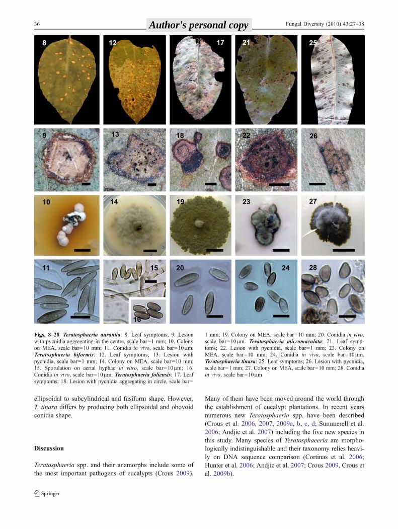
Figs. 8–28 Teratosphaeria aurantia: 8. Leaf symptoms; 9. Lesionwith pycnidia aggregating in the centre, scale bar=1 mm; 10. Colonyon MEA, scale bar=10 mm; 11. Conidia in vivo, scale bar=10μm.Teratosphaeria biformis: 12. Leaf symptoms; 13. Lesion withpycnidia, scale bar=1 mm; 14. Colony on MEA, scale bar=10 mm;15. Sporulation on aerial hyphae in vitro, scale bar=10μm; 16.Conidia in vivo, scale bar=10μm. Teratosphaeria foliensis: 17. Leafsymptoms; 18. Lesion with pycnidia aggregating in circle, scale bar=
1 mm; 19. Colony on MEA, scale bar=10 mm; 20. Conidia in vivo,scale bar=10μm. Teratosphaeria micromaculata: 21. Leaf symp-toms; 22. Lesion with pycnidia, scale bar=1 mm; 23. Colony onMEA, scale bar=10 mm; 24. Conidia in vivo, scale bar=10μm.Teratosphaeria tinara: 25. Leaf symptoms; 26. Lesion with pycnidia,scale bar=1 mm; 27. Colony on MEA, scale bar=10 mm; 28. Conidiain vivo, scale bar=10μm
36 Fungal Diversity (2010) 43:27–38 Author's personal copy

T. aurantia and T. biformis were isolated from E. dunniiand E. grandis plantations in southern Queensland but theincidence and severity they pose to eucalypt plantations isuncertain. T. micromaculata was found on E. globulus insouthern Queensland with incidence and severity ranked aslow. T. foliensis was found in a plantation on Eucalyptusglobulus in New South Wales. Although, very closelyrelated to the serious pathogen T. zuluensis and T.gauchensis which cause a stem canker disease, T. foliensiswas symptomatic rather than damaging. T. tinara wasisolated from native Corymbia sp. from north Queenslandand it is probably native to the region.
To date, there is sequence data and type cultures for 28Teratosphaeria spp. described from eucalypts for which acolletogloeopsis-like anamorph is known. Conidia range insize from the smallest T. zuluensis (4.5–5×2–2.5µm) to T.corymbiae (17–23×3.5–5µm), pigmentation and septationalso varies, but generally condia are aseptate. With theexception of stem canker pathogens T. zuluensis and T.gauchensis, the rest of these species produce lesions onleaves with various symptoms. All except T. gauchensis, T.juvenalis, T. pseudocryptica, T. zuluensis, T. stellenbochi-ana, T. xenocryptica and T. verrucosa have been reported inAustralia.
The Teratopshaeria spp. with colletogloeopsis-like ana-morphs are related to the most damaging leaf diseasespecies (kirramyces-like anamorphs) found on Eucalyptus;T. destructans, T. eucalypti, and T. viscidus. Unlike thelatter species, no species with a colletogloeopsis-likeanamorphs has been found to cause major leaf diseases inAustralia with the exception of T. cryptica (=T. nubilosum).The expansion of eucalypt plantation forestry into sub-tropics of Australia has led to the discovery of many newTeratopshaeria spp. and they now seem to be the dominantgenus on sub-tropical eucalypts. Currently, the speciesdescribed in this study are not causing any significantdamage to Australian eucalypt plantations and incidenceand threat they may pose to forest industry is unknown.
Acknowledgements This work was supported financially by anARC APAI linkage project LP0349190. The authors acknowledgeSally Collins, ITC Ltd, Australia for collecting samples from NewSouth Wales. We also thank Ewald Groenewald CBS Fungal DiversityCentre, Uppsalalaan, Utrecht, the Netherlands for BT sequences ofCPC13303, CPC11264 and CPC11267. We thank Kate Taylor andPaul Barber for assistance with the drawings and Thomas Jung forLatin translations.
References
Andjic V, Barber PA, Carnegie AJ, Hardy GEStJ, Wingfield MJ,Burgess TI (2007) A morphological and phylogenetic reassess-ment of the genus Phaeophleospora and the resurrection of thegenus Kirramyces. Mycol Res 111:1184–1198
Barber PA, Keane PJ (2007) A novel method of illustrating micro-fungi. Fungal Divers 27:1–10
Cortinas MN, Crous PW, Wingfield BD, Wingfield MJ (2006) Multi-gene phylogenies and phenotypic characters distinguish twospecies within the Colletogloeopsis zuluensis complex associatedwith Eucalyptus stem cankers. Stud Mycol 55:135–149
Crous PW (2009) Taxonomy and phylogeny of the genus Mycos-phaerella and its anamorphs. Fungal Divers 38:1–24
Crous PW, Wingfield MJ (1997) Colletogloeopsis a new coelomycetegenus to accommodate anamorphs of two species of Mycos-phaerella on Eucalyptus. Can J Bot 75:667–674
Crous PW, Aptroot A, Kang JC, Braun U, Wingfield MJ (2000) Thegenus Mycosphaerella and its anamorphs. Stud Mycol 45:107–121
Crous PW, Denman S, Taylor JE, Swart L, Palm ME (2004a)Cultivation and diseases of Proteaceae: Leucadendron, Leuco-spermum and Protea. CBS Biodivers Series 2:1–228
Crous PW, Groenewald JZ, Mansilla JP, Hunter GC, Wingfield MJ(2004b) Phylogenetic reassessment of Mycosphaerella spp andtheir anamorphs occurring on Eucalyptus. Stud Mycol 50:195–214
Crous PW, Wingfield MJ, Mansilla JP, Alfenas AC, Groenewald JZ(2006) Phylogenetic reassessment of Mycosphaerella spp. andtheir anamorphs occurring on Eucalyptus II. Stud Mycol 55:99–131
Crous PW, Braun U, Groenewald JZ (2007) Mycosphaerella ispolyphyletic. Stud Mycol 58:1–32
Crous PW, Groenewald JZ, Summerell BA, Wingfield BD, WingfieldMJ (2009a) Co-occuring species of Teratosphaeria on Eucalyp-tus. Persoonia 22:38–48
Crous PW, Summerell BA, Carnegie AJ, Wingfield MJ, Hunter GC,Burgess TI, Andjic V, Barber PA, Groenewald JZ (2009b)Unravelling Mycosphaerella: do you believe in genera? Persoo-nia 23:99–118
Crous PW, Summerell BA, Carnegie AJ, Wingfield MJ, GroenewaldJZ (2009c) Novel species of Mycosphaerellaceae and Teratos-phaeriaceae. Persoonia 23:119–146
Crous PW, Wingfield MJ, Groenewald JZ (2009d) Niche sharingreflects a poorly understood biodiversity phenomenon. Persoonia22:83–94
Ganapathi A, Corbin JB (1979) Colletogloeum nubilosum sp. nov., theimperfect state of Mycosphaerella nubilosa on Eucalyptus inNew Zealand. Trans Br Mycol Soc 72:237–244
Gardes M, Bruns T (1993) ITS primers with enhanced specificity forbasidiomycetes—application to the identification of mycorrhizaeand rusts. Mol Ecol 2:113–118
Glass NL, Donaldson GC (1995) Development of primer setsdesignated for use with the PCR to amplify conserved regionsfrom filamentous Ascomycetes. Appl Environ Microbiol61:1323–1330
Goodwin SB, Dunkley LD, Zismann VL (2001) Phylogenetic analysisof Cercospora and Mycosphaerella based on the internaltranscribed spacer region of ribosomal DNA. Phytopathology91:648–658
Hunter GC, Wingfield BD, Crous PW, Wingfield MJ (2006) A multi-gene phylogeny for species of Mycosphaerella occurring onEucalyptus leaves. Stud Mycol 55:147–161
Nylander JAA (2004) MrModeltest v2.2. Program distributed by theauthor. Evolutionary Biology Centre, Uppsala University
Ronquist F, Huelsenbeck JP (2003) MrBayes 3.1: Bayesian phyloge-netic inference under mixed models. Bioinformatics 19:1572–1574
Summerell BA, Groenewald JZ, Carnegie AJ, Summerbell RC, CrousPW (2006) Eucalyptus microfungi known from culture. 2. Alysi-diella, Fusculina and Phlogicylindrium genera nova, with notes onsome other poorly known taxa. Fungal Divers 23:323–350
Fungal Diversity (2010) 43:27–38 37 Author's personal copy

Swart HJ (1988) Australian leaf-inhabiting fungi XXVI. Some notewor-thy coelomycetes on Eucalyptus. Trans Br Mycol Soc 90:279–291
Swofford DL (2000) PAUP*, Phylogenetic analysis using parsimony(*and other methods). Version 4.0b10, in: (Sinauer Associates:Sunderland, Massachusetts, USA)
Taylor JE, Groenewald JE, Crous PW (2003) A phylogenetic analysisof Mycosphaerellaceae leaf spot pathogens of Proteaceae. MycolRes 107:653–658
Thompson JD, Gibson TJ, Plewniak F, Jeanmougin F, Higgins DG(1997) The ClustalX windows interface: flexible strategies for
multiple sequence alignment aided by quality analysis tools.Nucleic Acids Res 24:4876–4882
White TJ, Bruns T, Lee S, Taylor J (1990) Amplification and directsequencing of fungal ribosomal RNA genes for phylogenetics.In: Innes MA, Gelfand DH, Sninsky JJ, White TJ (eds) PCRprotocols: A guide to methods and applications. Academic, SanDiego, pp 315–322
Wingfield MJ, Crous PW, Couthinho TA (1997) A serious new cankerdisease of Eucalyptus in South Africa caused by a new species ofConiothyrium. Mycopathologia 136:139–145
38 Fungal Diversity (2010) 43:27–38 Author's personal copy