pharma - Optometry Australia
36
March 2011 pharma AUSTRALIAN Supplement to Ocular injuries Meibomian gland dysfunction Herpes simplex virus Anaesthesia drugs in ocular surgery Allergic conjunctivitis Fuchs’s uveitis
-
Upload
khangminh22 -
Category
Documents
-
view
3 -
download
0
Transcript of pharma - Optometry Australia

March 2011
pharmaAUSTRALIAN
Supplement to
��Ocular injuries ��Meibomian gland dysfunction��Herpes simplex virus ��Anaesthesia drugs in ocular surgery��Allergic conjunctivitis ��Fuchs’s uveitis

Turn the page on DRY EYE misery...
...and transform your dry eye
experience
Alcon Laboratories (Australia) Pty Ltd.Allambie Grove Business Park.10/25 Frenchs Forest Road East, Frenchs Forest NSW 2086.
® Registered Trademark. POPH.2088
Lasts6 months
onceopened!
1. Data on file. Alcon Laboratories, Inc. 2. Ketelson HA, Davis J, Meadows DL. Characterization of a novelpolymeric artificial tear delivery system. Invest Ophthamol Vis Sci; 2008; 49: E-Abstract 112.
Systane® ULTRA Lubricating Eye Drops goes further to lubricate and protect the ocular surface, providing
immediate comfort and extended protection.1,2
Breakthrough relief is finally here.
2 Ocular injuries Early treatment can be crucial
4 There is no substitute for a dilated fundus examination Optic disc photography should be used as an adjunct
7 Masquerade A systemic disease may be the underlying cause of allergic conjunctivitis
8 The search for the perfect solution Improvements in lens care systems
9 PHOTO CLINIC: Suffocated by hybrid
10 Meibomian gland dysfunction Hygiene, heat and nutrition play a part in treatment
13 TGA review unveils a flawed system Preliminary results reveal the need for change
14 CASE REPORT: Spot the difference Acute onset of floaters is often the first sign of Fuchs’s uveitis
16 Lower IOP without eye-drops A laser procedure may offer an alternative to glaucoma drugs
18 ABSTRACTS
19 CASE REPORT: Drainage problem
20 Traditional treatment is still the mainstay Handling herpes simplex virus
22 It’s mainstream now Growing need for therapeutic CPD
24 Anaesthesia drugs have come a long way New designer drugs are safer and more predictable
26 PHOTO CLINICS
28 With responsibility comes reward A therapeutic endorsement can change the way you practise
30 PBS list of medicines for optometrists February 2011
32 Scheduled drugs used or prescribed by optometrists February 2011
Editor: GARY OSHRYNational Publications Manager:
SANDRA SHAWClinical editor: MARK ROTHAdvertising: GARY OSHRY
Optometrists Association AustraliaABN 17 004 622 431204 Drummond Street
Carlton VIC 3053Tel (03) 9668 8500Fax (03) 9663 7478
Copyright © 2011
COVER Acute corneal hydrops resulting in
scar affecting vision
Comments made in OPTOMETRY PHARMA are of a general nature and intended for guidance only. Op-tometrists Association Australia and the individual contributors expressly disclaim all liability and responsibility to any person in respect of, and for the consequences of, anything done or omitted to be done in reliance wholly or partly on anything in this publication.
Published by
All references to pharmaceutical preparations in Pharma are applicable within Australia
AUSTRALIAN
pharma March 2011

OPTOMETRY PHARMA MARCH 2011
2
the eye may be perplexing. Bee and wasp stings to the cornea can produce a toxic inflammatory reaction in the eye and may require analgesia, specialist assessment or surgical treatment, depending on the sever-ity of the reaction and whether the stinger is amenable to removal.3
Most small superficial corneal foreign bodies can be removed with a foreign body spud after instilling topical anaes-thetic. Anaesthetic drops should never be dispensed to the patient as they can lead to corneal epithelial loss and significantly interfere with corneal immunity.4
Grinding or projectile injuries often result in a small piece of metal embedded in the cornea that will produce a rust ring, which will necessitate removal with an Alger brush. If there is suspicion that the foreign body has penetrated the eye, the patient will require an orbital X-ray.5
Corneal abrasionsA corneal abrasion can be recognised by instilling sodium fluorescein onto the eye. The dye stains the damaged epithelial cells and outlines the abraded area. While once common, patching the abraded eye is felt to be counter-productive to healing, as it reduces oxygen supply to the cornea. It also increases the corneal temperature and the micro-organism replication rate, leading to an increased risk of infection.6 Treatment includes a broad spectrum an-tibiotic, making sure to provide coverage for pseudomonas in a contact lens wearing patient—in these cases a fluorquinolone is a good choice. Cycloplege the affected eye with cyclopentolate 1% or 2% for discomfort from traumatic iritis, and consider topical non-steroidal anti-inflammatories for pain
control.7 Some practitioners choose to use a bandage contact lens for corneal abrasions.
Recurrent corneal erosionA patient with recurrent corneal erosion (RCE) presents with recurrent attacks of ocular pain, photophobia and tearing, often on awakening or when the eyelids are rubbed or opened. There is often a history of prior corneal abrasion in the involved eye. Localised roughing of the corneal epithe-lium may be seen on slitlamp examination, although epithelial changes may resolve within hours so that no abnormality is seen on examination.
Corneal epithelial dots or microcysts and subepithelial lines (map-dot-fingerprints) may be observed bilaterally if there is an underlying epithelial basement dystrophy.4 Treatment for an initial RCE is the same as that for a corneal abrasion. Erosions not responding to initial therapy may require an extended wear bandage contact lens for prolonged wear or anterior corneal stromal puncture. Alternatively, treatment with oral doxycline and a mild steroid have been postulated.8
Traumatic iritisTraumatic iritis or iridocyclitis, a generally mild inflammatory reaction of the iris or ciliary body, is commonly seen after blunt trauma to the globe.
Symptoms include pain, photophobia and occasionally epiphora. Signs are best seen on slitlamp examination and consist of cell and flare in the anterior chamber and perilimbal injection. Treatment includes topi-cal cycloplegic agents (2% cyclopentolate or 5% homatropine) and topical steroids (1% prednisolone acetate qid).
Ocular injuries
Ernie BowlingOD MS FAAO Dipl
It is very likely that optometrists will encounter patients suffering ocular trauma with some frequency. In the United States in 2001, about two million (6.98 per 1,000 population) people experienced an eye injury requiring treatment in an emer-gency room, inpatient or out-patient facility, or at private practice.1
Eye injuries are the leading cause of monocular blindness in the United States and are second only to cataract as the most common cause of visual impairment.2
Corneal and conjunctival foreign bodiesThe superior tarsal plate is a common lodg-ing spot for conjunctival foreign bodies. Sub-tarsal foreign bodies can be removed under topical anaesthesia by gently everting the upper lid with a cotton tip applicator. While asking the patient to look down, place pressure on the tarsal plate and remove the object with a foreign body spud. Insects on
early treatment can be

OPTOMETRY PHARMA MARCH 2011
3
Chemical injuriesChemical injuries are commonly caused by household disinfectants, detergents, sol-vents, cosmetics, drain cleaners, ammonia and bleach. Agriculturally related chemicals such as fertilisers and pesticides, and in-dustrial chemicals such as caustic solutions and solvents can cause complete ocular destruction.9 Early treatment is crucial. The immediate management is to irrigate the eye with copious amounts of normal saline or sterile water. Irrigation removes the offend-ing agent and neutralises the surface pH. Irrigation should commence immediately at the time of injury and continue until the patient reaches the optometrist’s practice and be continued thereafter. It is impossible to overirrigate a chemically-burned eye.
Irrigation should be performed for at least 10 minutes, the pH checked and irrigation continued if necessary until a neutral pH is obtained. Topical anaesthetic drops can be
used to diminish eye pain. Following irriga-tion, urgent care should be initiated and often includes topical steroids, antibiotics and cycloplegic agents to minimise inflam-mation and the risk of infection. Elevated IOP if present must be treated. Patching and sometimes the application of a bandage contact lens are necessary to facilitate the re-epithelisation of the cornea. Once the ocular surface has been stabilised for an extended period, penetrating keratoplasty may be required.
Thermal injuriesEye injuries caused by a spark from a match or fire, boiling water or a curling iron can vary in severity. A thermal burn will usually
When a patient walks in the door with an eye injury, you
need to know what to do there and then
result in a corneal abrasion and sometimes is associated with a conjunctival abrasion and some debris will accumulate in the fornices of the eye. A boiling water burn may be mild or severe, depending on the circumstances surrounding the injury.
Concurrent skin and scalp burns are common and require immediate attention.
1. McGwin G Jr, Xie A, Owsley C. Rate of eye injury in the United States. Arch Ophthalmol 2005; 123: 7: 970-976.
2. National Eye Institute. Eye and vision statistics 2002. Available at http://optometry.berkley.edu/~library/stats.htm=General.
3. Kirk RW, Andrew JW et al. External disease and cornea. Basic and Clinical Science Course. American Academy of Ophthalmology 1998; 361-362.
4. Kuhn F, Pieramici DJ. Ocular Trauma: Principles and Practice. New York, NY: Thieme Medical Publishers, 2002.
5. Naidu K. The injured eye: practical management guidelines and referral criteria for the rural doctor. SA Fam Pract 2006; 48: 7: 39-45.
6. Janda AM. Ocular trauma. Triage and treatment. Postgrad Med 1991; 90: 7: 55-60.
7. Scheufele TA, Blomquist PH. Spectrum of ocular trauma at an urban county hospital. Tex Med 2004; 100: 12: 60-63.
8. Wang L, Tsang H and Coroneo M. Treatment of recurrent corneal erosion syndrome using the combination of oral doxycycline and topical cor t icosteroid. Clinical and Experimental Ophthalmology 2008; 36: 1: 8-12.
9. Wagoner MD, Kenyon KR. Chemical injuries of the eye. In: Shingleton BJ, Hersh PS, Kenyon KR, eds. Eye Trauma. St. Louis: C.V. Mosby, 1991: 79-94.
crucial
Blunt trauma to the globe

OPTOMETRY PHARMA MARCH 2011
4
The diagnosis of diabetes was once aided by practitioners dipping their fingers in the patient’s urine and tasting it for glucose. Physicians would diagnose cardiac failure by looking for raised jugular venous pressure and listening for crackles at the lung base. Surgeons would determine whether the patient with abdominal pain had appendi-citis by looking for tenderness over McBurney’s point along with re-bound tenderness of the abdomen.
Fast forward to the 21st century and we have glucometers that can measure blood sugar from a finger prick, echocardiograms to diagnose cardiac failure and CT scans to diagnose appendicitis. We have such advanced technologies that virtually any disease can be diagnosed by some test or other.
While no-one mourns the passing of the urine taste test for diabetes, physicians still listen to their patient’s chest, surgeons still palpate patient’s abdomens, and oph-thalmologists and optometrists should still examine the optic disc. A patient history and examination are the cornerstone of clinical practice and the most fundamental requirement for good patient care. Investiga-tions, if performed, are done to confirm the diagnosis or exclude other pathologies. If the diagnosis is clear, investigations may not necessarily be required.
The day when patients have a CT scan and a laboratory sends the blood test to a doctor sitting at home in front of a laptop is
still a long way off.Has technology in the ophthalmic world
advanced sufficiently to allow patients to be assessed without the need for a direct examination? Can a fundus photograph, OCT or Heidelberg retina tomograph (HRT) substitute for a dilated fundus examination of the optic nerve? Can a frequency doubling technology (FDT) or a field test provide enough information to allow us to forgo a full examination?
These complex questions have been the subject of many studies.1 With glaucoma, consideration needs to be given to questions such as how we define glaucoma and what criteria we use to judge whether an optic nerve is abnormal. This is beyond the scope of this article and we limit our discussion to consideration of optic disc photography.
Role of optic photographyWhat is the role of optic disc photography and is there evidence that imaging alone with a fundus camera can replace an
Optic disc photography should be used to confirm
your diagnosis, rather than make it for you
There is no substitute for a
Cataract distorts resultsThis is a disc photograph taken of a patient pre-cataract surgery. The patient was a 62-year-old female with no past ocular or medical history. Vision was recorded as 6/12 R and 6/15 L and intraocular pres-sures were 18 R and 20 L. Field testing was normal. Pachymetry readings were 553 R and 544 L.
Based on the disc photograph and the above results, does the patient have glaucoma?
This cannot be determined from examin-
ing the disc photograph due to the presence of cataract and the lack of depth perception. A dilated examination showed that there is inferior rim thinning and notching along with nerve fibre layer drop-out.
Subsequent follow-up revealed an elevated intraocular pressure to 25 OU.
The diagnosis of glaucoma would have been missed if a disc photograph alone had been taken because a disc photo in isola-tion does not afford the same examination opportunity as a direct exam.
Case study

OPTOMETRY PHARMA MARCH 2011
5
examination? There are few studies in this area. Most of the published works on fundus photography relate to fundus photography for screening in diabetic retinopathy and here the findings are conflicting. There are just as many studies that show that it is suit-able as there are that show that it not.
With the proliferation of non-mydriatic cameras, optic disc photography has be-come widespread practice. It is convenient and efficient, and there is a perception that patients do not like to be dilated.
Fundus photography is of great value as a means of objective documentation and while the arguments for not dilating and photographing instead are compelling, the question arises of how useful this is.
Qualitative examination of sterodisc photography can be used to assess cup-disc ratio, rim notching and thickness, disc colour and contour, vessel position and baring, and rim haemorrhages. A major drawback of this method is that it is very operator dependent and relies on quality photographs.
Conducting planimetric measurements of the optic nerve is another approach, which consists of computer assisted analyses of disc photographs. This approach requires the operator to mark the optic nerve boundaries and cup on a computer screen. Software then calculates cup-disc ratio, rim area and cup area. This technique is also very operator dependent, has a sensitivity of only about 62 per cent and a specificity of about 67 per cent.2
The ability of expert observers to correctly differentiate normal from glaucomatous optic nerves by examining stereodisc pho-tographs has been examined2-4 and so too has the ability of HRT to correctly identify abnormal discs.2 The findings of these stud-ies were surprising because the participants were able to correctly identify the abnormal
discs with a sensitivity of about 70 per cent and a specificity of about 85 per cent.
Looking at this another way, stereo disc examination by skilled observers missed 30 per cent of the abnormal discs. Furthermore, the quality of the photographs was judged to be poor in as many as 45 per cent of all photographs taken. The other surprising finding of this study was that HRT was more accurate with a sensitivity of about 75 per cent, a finding that has been confirmed by other studies.5,6
Observers assessing stereodisc photo-graphs are more likely to judge the disc as glaucomatous if there is more advanced global disc damage than if there is only localised or focal damage. Glaucomatous optic disc damage tends to occur more com-monly in the inferior neuroretinal retinal rim and particularly in the inferotemporal sector. This is closely followed by temporal rim loss. Nasal disc damage was particularly difficult
dilated fundus examination Joseph San Laureano
MB BS MMed BSc FRACOMelbourne Eye Centre
Epworth Private Hospital
to detect with sterodisc photographs with only 30 per cent of practitioners correctly making the diagnosis.2
The European Optic Disc study3 pub-lished in 2010 came to similar conclusions with stereodisc photography having a sensitivity of about 75 per cent and speci-ficity of 87 per cent. Interestingly, this study also found that assessments made by older practitioners were less accurate.
How do optometrists fare in optic nerve examination? This has been the subject of a number of studies including one by the American Society of Optometrists in 2000.4,7,8 In essence, monocular examina-tion of the optic disc (direct ophthalmoscope or monocular fundus photography) tended to underestimate disc damage, due primar-ily to pallor being used to assess the disc rather than the true neuroretinal rim. Not
Continued page 6
Epiretinal membrane This is a fundus photograph of a pre-cataract surgery patient who also has higher risk factors for glaucoma. Vision was recorded as 6/15 R and 6/7.5 L. Intraocular pressures were within the nor-mal range and a visual field test showed a repeatable superior field defect.
Does the photograph give a true repre-sentation of the clinical picture?
Further examination confirmed the pres-ence of cataract and of disc changes con-sistent with glaucoma, which importantly were not discernible on the photograph. A right epiretinal membrane was noted, which was also not readily seen on the photograph. Epiretinal membrane peel with vitrectomy and gas was performed,
followed by cataract surgery, which was required following vitrectomy. Glaucoma lowering medication was commenced for management of the glaucoma.
Case study

OPTOMETRY PHARMA MARCH 2011
6
Peripheral tear This image is of a patient with a fam-ily history of glaucoma along with the finding of a rim haemorrhage. Disc examination was undertaken with difficulty due to a small pupil. Intraocular pressure was normal as were visual fields.
Vision was recorded as 6/9 R and 6/7.5 L. The patient was a moderate myope and had no past medical or ocular history. The fundus photograph reveals no other positive findings apart from the haemorrhage but a dilated examination revealed rim thinning and notching along with nerve fibre layer drop-out. In addition, vitreous blood was noted, which a peripheral examination uncovered was from a horseshoe tear.
Barrier laser was applied with eventual clearing of the vitreous blood. Vision returned to 6/7.5+. HRT was abnormal in keeping with the clinical disc findings. While this case would probably not have been due to the presence of the haemorrhage, it required a dilated examination to reveal fully the disc changes. One must also bear in mind the other causes of disc haemorrhage such as hyper-tension, diabetes, vein occlusion and idiopathic. The peripheral tear would not have been seen on disc photography, which usually images only around 30 degrees. This case highlights the importance of routine dilation to exclude any peripheral retinal pathology.
surprisingly, the study also found that the more experienced the optometrist, the more accurate the interpretation.
This study supports the trend of Australian optometrists using binocular indirect oph-thalmoscopy as well as slitlamp funduscopy to gain a highly magnified and stereoscopic view of the disc.
Stereodisc photographyIn general, stereodisc photographs are dif-ficult to take, require skilled technicians with a steroviewer and most importantly, require a well-dilated pupil. Few fundus cameras have this facility.
There has not been a study looking at the sensitivity of monocular disc photography but we can draw some inferences based on those looking at stereodisc photography.
If skilled observers did not fare well with stereodisc photographs, how would they have performed looking at a monocular disc photograph?
The quality of the photograph will profoundly impact the ability to judge the optic disc. Miotic pupils, media opacities and patient movement will result in a poor image in which the subtle features of disc damage may not be visible. The absence of depth in a flat monocular image will mean greater reliance on other clues such as colour to judge cup-disc ratios. Rim and nerve fibre layer will be difficult to judge, undermining and bayonetting will be dif-ficult to assess, although disc haemorrhage will not be missed.
If monocular disc photography has draw-backs in the assessment of disc pathology and may also miss other pathologies, what role does it play in patient management?
There is no question that monocular fundus photography has an important role to play in patient management as long as you acknowledge its limitations. As a general baseline and supplement to a full examination, most practitioners would find it invaluable. For diabetic retinopathy it has become the standard of care. In glaucoma serial disc photography has been in use for many years for monitoring progression and plays an important role.
Glaucoma progresses slowly and disc changes are subtle and often easy to miss. In moderate to advanced glaucoma there is significant disc rim atrophy already present, making the detection of progression even more difficult.
In glaucoma management, stereo disc photography is still the gold standard. The role of monocular disc photography is not entirely clear, yet it is preferable to a draw-ing or a textual description. Whether it can be used to monitor progression is unknown but it is probably useful.
ConclusionThe standard of care for monitoring glau-coma progression requires serial, stereo disc photographs. This is not available in most practices, is difficult to perform and requires a skilled operator, and is not a substitute for disc examination. Serial disc chronoscopy can be used with non-stereo photography but it too has its limitations.
Dilated fundus examination along with intraocular pressure estimation and visual fields remains the gold standard. Sup-plementary investigations such as disc photography and the emergence of OCT and HRT cannot replace these fundamentals and monocular disc photography cannot substitute for a dilated clinical examination.
1. Pan YZ. [Effect of fundus image devices for early diagnosis of primary open angle glaucoma]. Zhonghua Yan Ke Za Zhi 2009; 45: 10: 871-874.
2. Wollstein G, Garway-Heath DF, Fontana L, Hitchings RA. Identifying early glaucomatous changes: comparison between expert clinical assessment of optic disc photographs and con-focal scanning ophthalmoscopy. Ophthalmology 2000; 107: 2272–2277.
3. Nicolaas J Reus, Hans G Lemij, David F Garway-Heath. Clinical assessment of stereoscopic optic disc photographs for glaucoma: the European Optic Disc Assessment Trial. Ophthalmology 2010; 117: 4, 717-723.
4. Spalding JM, Litwak AB, Shufelt CL. Optic nerve evaluation among optometrists. Optom Vis Sci 2000; 77: 9: 446-452.
5. Greaney MJ, Hoffman DC, Garway-Heath DF et al. Comparison of optic nerve imaging methods to distinguish normal eyes from those with glaucoma. Invest Ophthalmol Vis Sci 2002; 43: 140-145.
6. Alencar MA, Bowd C, Weinreb RN, Zangwill LM, Sample PA, Medeiros FA. Comparison of HRT-3 glaucoma probability score and subjective stereophotograph assessment for prediction of progression in glaucoma. Invest Ophthalmol Vis Sci 2008; 49: 1898-1906.
7. Abrams LS, Scott IU, Spaeth GL et al. Agreement among optometrists, ophthalmologists, and residents in evaluating the optic disc for glaucoma. Ophthalmology 1994; 101: 1662–1667.
8. Varma R, Steinmann WC, Scott IU. Exper t agreement in evaluating the optic disc for glaucoma. Ophthalmology 1992; 99: 215–221.
From page 5
No substitute for dilated fundus examCase study

OPTOMETRY PHARMA MARCH 2011
7
It is a hot Friday afternoon and you are looking forward to enjoying the sunshine when a patient with a pair of miserable, red, itchy, watery eyeballs walks into your practice.
Automatically, you think, ‘Ah-ha, I know exactly what can cure this problem.’ You reach for the magic pad and write out a prescription for the latest and greatest antihistamine eye-drop. A week later, the same pair of eyeballs is back, the patient still unhappy. The antihistamine you prescribed has not provided any relief. Now what?
As optometrists, we often attempt to treat a patient’s symptoms with bandage therapy, which can be unsuccessful in cer-tain circumstances. After ruling out atopic keratoconjunctivitis, and seasonal and per-ennial allergic conjunctivitis, it is important to explore the underlying aetiology of the apparently allergic response. This requires us to expand our view of the patient and investigate all systemic avenues. The patient is no longer just a set of eyeballs.
Many systemic diseases that cause a con-junctival reaction can easily be mistaken for
Ujjwal Bagga BSJennifer Keller BSLeonid Skorin Jr
OD DO FAAO FAOCO
� Erythema multiforme (Stevens-Johnson syndrome)
� Wegener’s granulomatosis� Newcastle disease� Sjögren’s syndrome� Lyme disease� Reiter’s syndrome� Acne rosacea� Measles� Relapsing polychondritis� Folic acid deficiency� Cicatricial pemphigoid� Herpes simplex infection� Sarcoidosis� Bell’s palsy and other CN VII
disorders� Pulmonary oedema
Rosacea a catalystA 62-year-old female presented with puffy and erythematous eyelids, along with intense itchiness of the eyes. The patient had a history of acne rosacea for which she was being treated with minocycline and metronidazole. She also had diabetes.
Slitlamp examination showed a poor tear film with negative corneal staining, 1+ follicles and slight conjunctival injec-tion bilaterally. All other ocular structures were normal. Previous treatment with ketotifen fumarate ophthalmic solution (an antihistamine) and non-preserved artificial tear lubricants were unsuccessful in treat-ing her symptoms.
At her follow-up appointment the pa-tient noticed that her rosacea symptoms seemed to be worsening. Her treatment regimen was changed to azithromycin ophthalmic solution, which mildly reduced her symptoms. Bepotastine besilate 1.5% ophthalmic solution bid was added, which eliminated her conjunctival injection and itchiness. It was concluded that the patient had allergic conjunctivitis exacerbated by the ocular rosacea.
Swelling extremitiesA 74-year-old female presented with decreased visual acuity, bilateral conjunc-tival chemosis, erythema and epiphora for the previous three months.
Previous treatments, which included 50 mg oral doxycycline bid, olopatadine ophthalmic solution bid and prednisolone acetate 1% ophthalmic suspension qid, had been unsuccessful at relieving her signs or symptoms.
The patient was then referred to her primary care physician to explore pos-sible systemic causes of the oedema. At her medical examination she reported swelling of both lower extremities. The patient was diagnosed with mild pulmo-nary hypertension and treated with 50 mg isosorbide mononitrate (Imdur) daily. Four weeks later, the patient returned to the optometric practice reporting improve-ment in her vision, and showed reduced conjunctival chemosis and minimal epi-phora. It was concluded that the patient had conjunctival chemosis secondary to systemic fluid retention from her pulmo-nary hypertension.
allergic conjunctivitis. The Table (below) lists some of the underlying systemic conditions that may cause conjunctivitis.
Persistent conjunctivitis of any aetiology is an ocular condition that can cause vary-ing degrees of irritation for the patient. If traditional therapy is unsuccessful in treating symptoms, it is important for the optometrist to explore further systemic conditions and the likelihood that they may be causing the allergic conjunctivitis—like signs and symptoms. Our role is to reduce symptoms and treat the disease whenever possible. It is equally important to acknowledge that sometimes it is best to comanage patients with their primary care physician for man-agement of the systemic disease with the overall goal of reducing ocular symptoms.
Further readingwww.wrongdiagnosis.com/c/conjunctivi-
tis/tests.htmwww.aoa.org/documents/CPG-11.pdf
Systemic conditions that may cause conjunctivitis
A systemic disease may be the underlying
cause of allergic conjunctivitis
Masquerade
Case study

OPTOMETRY PHARMA MARCH 2011
8
Increased convenience balanced with safety and efficacy is an ever-present challenge in the ongoing development of contact lens mul-tipurpose solutions (MPS) to meet consumer demand and improve patient compliance.
Rubbing and rinsing mechanical clean-ing of contact lenses reduces the presence of micro-organisms by greater than 90 per cent. Some newer MPS products rely solely on improved antimicrobial activity in efforts to reduce the burden of contact lens care. Choice of disinfecting solution is a major fac-tor in maintaining optimum patient comfort and satisfaction, and reducing drop-out from contact lens wear. Between 1997 and 2007 in the United Kingdom, Efron and Morgan reported an increase in MPS prescribed from 56 to 93 per cent, with a concomitant decrease in one-step hydrogen peroxide systems from 20 to seven per cent.1 With patients’ obvious desire for ease of use products, development of efficacious MPS is imperative.
Multipurpose solutionsMultipurpose solutions offer convenient, low-cost contact lens care. They comprise an antimicrobial agent acting as a preservative and disinfectant, a surfactant for increased wettability and to remove deposits, a chela-tor that can act antimicrobially and a buffer-ing agent. MPS must be bacteriocidal yet not cytotoxic to cells, as traces of the solution make contact with the ocular surface when lenses are applied.
To reduce cytotoxic effects, high mo-lecular weight compounds with reduced
penetrance into the contact lens matrix are employed. Unfortunately, these simplified care systems represent an unavoidable compromise of cleaning and disinfecting functions. Manufacturers must determine appropriate preservative concentrations that sufficiently disinfect without causing any adverse effects.
First generation MPS contained antimicro-bial agents such as polyhexamethylene bi-guanide (PHMB) and polidronium chloride (Polyquad). Development led to increased efficacy and comfort, with the addition of antimicrobial agents such as myristami-dopropyl dimethylamine (MAPDA, trade name Aldox), cleaners, and sequestering and wetting agents.
Preservatives in MPS initiate microbial death by destabilising and disrupting the cytoplasmic membrane, resulting in leak-age of macromolecular components. This response is irreversible and the microbe cannot adapt or become resistant to the preservative. Antibacterial effectiveness of preservatives varies with pH and concen-tration. Buffering agent additives work to stabilise pH on opening of the solution to preserve both efficacy and comfort.
MPS differ in their surfactant and buffer-ing systems potentiating varied biological effects with use of different solution brands. Clinical trials have reported significantly higher levels of corneal staining with some PHMB-based regimens, compared with polyquad-based care regimens when used with hydrogel lenses,2 while multipurpose solutions with identical concentrations of PHMB behave differently depending on the solution formulation.
PreservativesPreservatives span a range of chemical classes including quaternary ammoniums, mercurials, alcohols, carboxylic acids, phenols and amidines.
The following minimal requirements are
required in a preservative:� low irritation potential� pH range for maximal antimicrobial
activity� compatibility with other ingredients� synergism or antagonism in antimicrobial
activity.Preservatives should be non-cytotoxic
and provide broad antimicrobial activity, chemical or thermal stability, compatibility with the container and other compounds present, as well as ocular comfort.
Benzalkonium chloride (BAC; a quater-nary ammonium) has been used in contact lens solutions since the 1940s due to its efficacy, stability and low cost. It is found in almost 70 per cent of ocular medications that are stored for extended periods and is active against bacteria, fungi and protozoa. Its microbiocidal action lies in disruption of the plasma membrane, which increases cel-lular permeability and induces cell death.
Use of BAC in contact lens care regimens is associated with toxic side-effects such as allergic reactions and dry-eye due to its tendency to bind with contact lens materi-als, thus increasing in concentration over successive days of lens wear and care.3
Improved high-molecular weight quaternary ammoniums have been developed such as polidronium chloride (Polyquad), which is bacteriocidal at lower concentrations than BAC.
SiHy lenses and solutionsMPS, which was originally developed for conventional hydrogel lenses, may be incompatible with silicone hydrogel (SiHy) lenses due to the bulk material and unique surfaces of some silicone hydrogels. Report-ed incompatibilities such as unacceptable parameter changes in galyfilcon A lenses (ACUVUE Advance; Johnson & Johnson Vision Care, Jacksonville, FL) and significant levels of corneal fluorescein staining with balafilcon A lenses (PureVision; Bausch +
The search for the perfect Robert Terry MSc
Judith Flanagan PhDBrien Holden Vision Institute
Improvements in lens care systems are providing enhanced
antimicrobial efficacy with increased comfort and ease of use

OPTOMETRY PHARMA MARCH 2011
9
Lomb, Rochester, NY)4 support this notion of clinically important differences related to lens care products. Formulation of lens care systems is complex. Unanticipated adverse events have been reported arising from reformulation of certain MPS in efforts to improve SiHy compatibility, resulting in the withdrawal of several products from the market.
Toxic reactionsAntimicrobial agents are potentially toxic substances. Toxic effects induced by contact lens care systems include corneal staining, increased limbal and conjunctival hyper-aemia, chemosis, oedema, superior limbic kerato-conjunctivitis, stromal infiltrates and papillary conjunctivitis. The diagnosis and management of ocular hypersensitivity can present a challenge to eye-care practition-
solutioners, and an understanding of the mecha-nisms underlying the signs and symptoms of such conditions is necessary for their appropriate management.
Cytotoxicity and decreased corneal epi-thelial barrier function may be caused by MPS formulation as a whole and not attribut-able to any single component as proprietary MPS containing identical concentrations of the same preservative behave differently in identical cytotoxicity assays. As a further complication, MPS formulation components can interact with contact lenses. SiHy lenses absorb more PHMB than polyquaternium-1. Variability in signs and symptoms occurs between long-term users of the two pre-servative systems used in many contact lens MPS. In some studies, the degree of corneal staining relates to lens uptake.
Fewer corneal inflammatory events have been reported with use of hydrogen peroxide systems compared to MPS use.5 While preserved MPS are commonly used due to convenience and low cost, hydrogen peroxide lens-care systems result in minimal degrees of corneal staining associated with SiHy lens materials.
SummaryThe search for the ‘perfect’ lens care system continues. Enhancing patient compliance should reduce the incidence of adverse reactions. The extent to which these issues are resolved will determine the future share of lens care systems in the marketplace. The overall size of the contact lens solutions market is declining. The industry is moving towards daily disposable contact lenses, ob-viating any need for lens care products such as MPS. In the short term, new products will continue to provide enhanced antimicrobial efficacy with increased comfort and ease of use to assure user compliance.
1. Efron N, Morgan PB. Soft contact lens care regimens in the UK. Contact Lens Ant Eye 2008; 31: 283-284.
2. Jones L, MacDougall N, Sorbara LG. Asymptomatic corneal staining associated with the use of balafilcon silicone-hydrogel contact lenses disinfected with a polyaminopropyl biguanide-preserved care regimen. Optom Vis Sci 2002; 79: 12: 753-761.
3. Chapman JM, Cheeks L, Green K. Interactions of benzalkonium chloride with soft and hard contact lenses. Arch Ophthalmol 1990; 108: 2: 244-246.
4. Zigler L, Cedrone R, Evans D, Helbert-Green C, Shah T. Clinical evaluation of silicone hydrogel lens wear with a new multipurpose disinfection care product. Eye Contact Lens 2007; 33: 5: 236-243.
5. Carnt N, Jalbert I, Stretton S et al. Solution toxicity in soft contact lens daily wear is associated with corneal inflammation. Optom Vis Sci 2007; 84: 309-315.
Lachlan Scott-Hoy BAppSc(Optom) PGCert
Photo clinic
Suffocated by hybrid
Upper-lid eversion reveals GPC
Note the indentation at the margin of the rigid centre, the central
epithelial disease and the vascular ingrowth at the limbus
Mrs EA, a 52-year-old nurse presented with an uncomfortable red eye. EA has keratoconus and wears hybrid (Soft-Perm) contact lenses prescribed by a colleague.Diagnosis: CLARE, corneal hypoxia, giant papillary conjunctivitis (GPC).Management: Discontinue contact lens wear. Flarex 0.1% qid. Blink Intensive
Tears q1h. The patient was refitted with intracorneal RGPs at one week review.Result: There was corneal re-epithe-lialisation after two weeks and GPC resolution after one month. Patient now happily wears intracorneal RGPs.
Images taken using the Takagi SM-70N slitlamp with a Canon EOS 450D, supplied through Optimed.

OPTOMETRY PHARMA MARCH 2011
10
Diagnosing the diseaseA careful inspection of the lid margin is a key factor in diagnosing this condition. Examina-tion of the eyelashes allows us to determine the presence of an anterior blepharitis component. These patients have bacteria that have overpopulated the anterior lid margins, and are likely to have debris at the base of their lashes representing bacterial exotoxins and other byproducts.9 Often this will irritate the eyelid margin and cause a diffuse inflammation leading to a red-rimmed appearance of the eyelids. These patients may have meibomian gland issues as many of the bacteria that produce the signs described may also produce lipases and esterases, which will enzymatically alter the oils composing the meibum, giving the secretions a thicker consistency.
The quality of the meibum should be tested on every patient. This can be done by manually applying pressure to the outside of the eyelid towards the eyelid margin. Viewing the quality of the meibum that is expressed is telling of meibomian gland dysfunction. In a healthy patient, the meibum secreted has fluid consistency resembling that of vegetable oil while viewed during slitlamp examination. Altered meibomian
gland secretions vary in their appearance but may have a white purulent appearance (Figure 1), a thick more yellow turbid ap-pearance (Figure 2), or may not be able to be expressed at all, demonstrating either significant stagnation or complete dysfunc-tion of the gland, resulting in no meibum production.
There are many ways that the meibomian glands may be expressed. The most com-mon way clinically is to simply apply digital pressure close to the eyelid margin. Some practitioners may use a cotton tip applicator to apply the pressure. The limitation with both direct digital pressure and using a cot-ton tip applicator is that the amount of lever-age that you can use is restricted because of pressure being placed against the eye.
When additional pressure is needed because of difficulty viewing the secretions, two cotton tip applicators may be used to sandwich the meibomian glands and express the meibum. One of the applicators will be placed on the outer lid near the lid margin while the other is placed on the palpebral conjuctiva. Then in a rolling motion, meibum is attempted to be expressed from the gland. Usually a topical anesthetic will be used during this procedure to minimise discomfort.
Meibomian gland
Dry eye is an increasingly frustrat-ing condition that affects many patients. Studies have shown vary-ing prevalence of dry eye ranging from 3.5 to 33 per cent depending on the criteria used to identify those patients and also the populations studied.1-5
As we learn more about the intricacies of the tear film, we gain a new appreciation for the complexities that exist between the lipid, aqueous and mucin layers in their ability to synergistically promote an even, well-lubricated ocular surface.
Much attention has been given to the lipid layer of the tear film. This layer is produced by the meibomian glands, which are sebaceous glands located at the eyelid. These glands gently express small amounts of meibum as the anterior layer of the tear film with every blink.6 The fluid consistency with which it leaves the orifice of the gland will dictate much of the effectiveness of the lipid layer to evenly form an anti-evaporative layer of the tear film. The more fluid the meibum, the more likely these oils will be to evenly spread over the underlying tear layers.
Unfortunately, when the consistency of the meibum is altered and becomes more stagnant, lipids are less easily expressed from the gland orifices. In a normal per-son with properly functioning meibomian glands, the meibum has a melting point of between 18.8O C and 31.7O C, which means that it exists in a fluid state at the normal body temperature of 37O C.7 In patients with stagnation of the meibum, the melting point of the oils is increased so that they exist in a more solid state at normal body temperature. In a healthy person, these lipids have a melting point three degrees lower than those with meibomian gland dysfunction.8
Understanding the underlying mechanism behind meibomian gland dysfunction helps us to devise strategies to diagnose and treat these patients, which effectively alleviate many of their symptoms while rehabilitating the glands to better produce a healthier lipid layer.
Mile Brujic OD
Figure 1. Thick meibum with a white appearance being expressed from a patient with active lid disease
Hygiene, heat and nutrition

OPTOMETRY PHARMA MARCH 2011
11
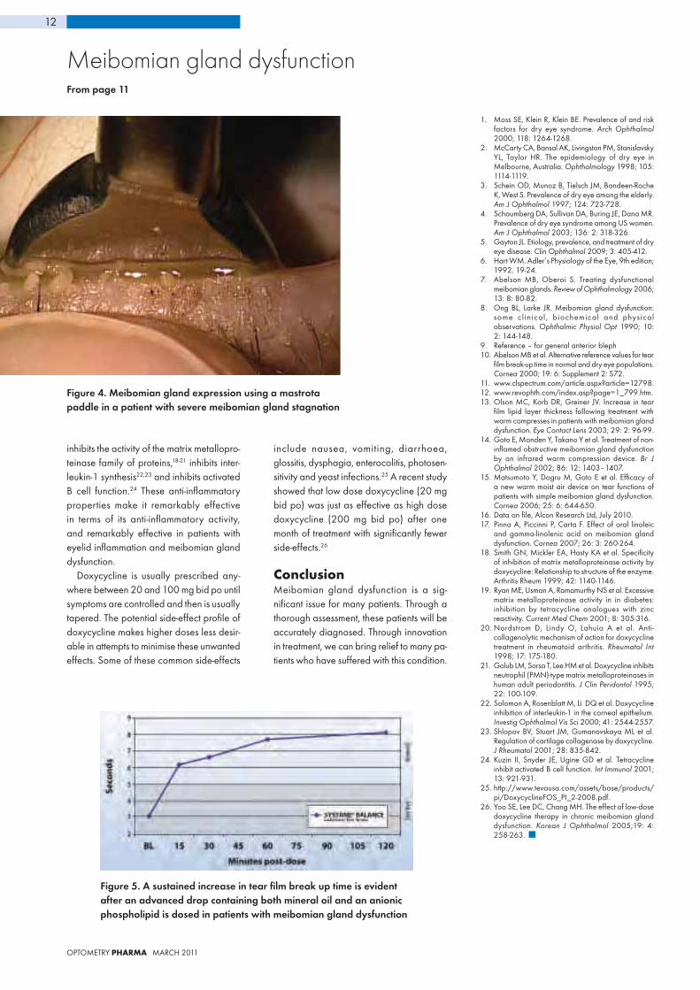
A mastrota paddle may also be used for meibomian gland expression. This is a small titanium metal rod with a paddle on each of its ends (Figure 3). Either end of the paddle is placed directly behind the eyelid margin so that it rests against the palpebral conjunctiva. The examiner then applies pressure to the front of the eyelid close to the eyelid margin directly over the area where the paddle is located (Figure 4). The paddle in this instance provides leverage for the pressure that is placed on the anterior eyelid margin. I do not use a topical anesthetic for this procedure if there is no visible eyelid inflammation, and most of the time patients feel little discomfort. If there is visible eyelid inflammation, I usually place one or two drops of anesthetic in the eyes as these patients are more likely to experience discomfort during the procedure.
Fluorescein assessment of the ocular surface is critical in gaining a greater appre-ciation of the health of the anterior segment. Patients with meibomian gland dysfunction often have a reduced tear film break up time (TBUT). Simply assess the tear film with a cobalt blue light and a Wratten #12 barrier filter after fluorescein is placed on the eye and assess for dark areas that form in the
tear film after the blink is complete. Usually a TBUT less than six seconds is considered abnormal and maybe seen with patients who have MGD.10 Significantly reduced TBUT over long periods may also lead to corneal staining, which is easily viewed with a cobalt blue light and a written filter.11,12
TreatmentFor patients with visible debris in the eye-lashes along with inflammation of the eyelid margins, lid hygiene is a first step to improv-ing outcomes. This can be done by digitally rubbing the area with a warm wash cloth to remove the debris, which is the substances that cause much of the inflammation. In more severe cases, a one part baby shampoo, fours parts water mixture can be applied to the end of a cotton swab and then rubbed along the eyelid margin to remove much of the debris in the lashes and thus remove the inciting irritants from the ocular surface.
Packaged commercially available lid scrubs are also available and work remarkably well to clean the eyelid margins and de-crease the bacterial microflora.
Heat applied to the eyelids increases the temperature of the meibomian glands and when done regularly, liquefies the meibum.13,14,15 The analogy that I use with patients is that butter is a solid at room temperature but by placing it in a frying pan you can convert it into a liquid. The lipids produced by the meibomian glands also become more liquid in consistency when the temperature is raised above the melt-ing point. Usually it is recommended that you apply heat to the eyes bid to tid for 10 minutes at a time.
Symptomatic relief for patients with MGD often requires a long course of therapy. With the addition of a drop that contains a lipid component, patients can feel comfort immediately as the layer that is deficient is replenished. Systane Balance is a unique drop that contains both mineral oil and an anionic phospholipid, which replaces the lipid layer of patients with MGD who suffer from lipid deficient dry eye. Its unique and sustained delivery system offers long-lasting lipid layer protection.
In a recent study, patients were recruited who wanted to use lubricant eye-drops at least ‘some of the time’, had a TFBUT of less than five seconds and were MGD patients (either drop-out or poor quality secretion). Baseline TBUT measurements were taken and at baseline were three seconds. Systane Balance was instilled and within 15 minutes the TBUT increased to six seconds. What is interesting is that even two hours after instil-lation, patients experience a TBUT greater than six seconds (Figure 5).16
Much research has been done in the field of ocular nutrition and its effect on patient with dry eye. A study randomised 57 patients to either taking tablets that contain 28.5 mg of linoleic acid (LA) and 15.1 mg of gamma-linolenic acid (GLA) once a day, those who performed eyelid hygiene once a day and those who received both treatments. The group receiving both LA/GLA and eyelid hygiene improved both symptoms and signs of eyelid margin inflam-mation more than groups receiving either treatment alone.17
Doxycycline works remarkably well for patients with meibomian gland dysfunction who are unresponsive to other therapies. It
dysfunction
Figure 2. Thick, yellow appearing meibum on the eyelid of a patient after being expressed
Figure 3. Mastrota paddle
play a part in treatment
Continued page 12
OPTOMETRY PHARMA MARCH 2011
12
Figure 5. A sustained increase in tear film break up time is evident after an advanced drop containing both mineral oil and an anionic phospholipid is dosed in patients with meibomian gland dysfunction
inhibits the activity of the matrix metallopro-teinase family of proteins,18-21 inhibits inter-leukin-1 synthesis22,23 and inhibits activated B cell function.24 These anti-inflammatory properties make it remarkably effective in terms of its anti-inflammatory activity, and remarkably effective in patients with eyelid inflammation and meibomian gland dysfunction.
Doxycycline is usually prescribed any-where between 20 and 100 mg bid po until symptoms are controlled and then is usually tapered. The potential side-effect profile of doxycycline makes higher doses less desir-able in attempts to minimise these unwanted effects. Some of these common side-effects
include nausea, vomiting, diarrhoea, glossitis, dysphagia, enterocolitis, photosen-sitivity and yeast infections.25 A recent study showed that low dose doxycycline (20 mg bid po) was just as effective as high dose doxycycline (200 mg bid po) after one month of treatment with significantly fewer side-effects.26
ConclusionMeibomian gland dysfunction is a sig-nificant issue for many patients. Through a thorough assessment, these patients will be accurately diagnosed. Through innovation in treatment, we can bring relief to many pa-tients who have suffered with this condition.
1. Moss SE, Klein R, Klein BE. Prevalence of and risk factors for dry eye syndrome. Arch Ophthalmol 2000; 118: 1264-1268.
2. McCarty CA, Bansal AK, Livingston PM, Stanislavsky YL, Taylor HR. The epidemiology of dry eye in Melbourne, Australia. Ophthalmology 1998; 105: 1114-1119.
3. Schein OD, Munoz B, Tielsch JM, Bandeen-Roche K, West S. Prevalence of dry eye among the elderly. Am J Ophthalmol 1997; 124: 723-728.
4. Schaumberg DA, Sullivan DA, Buring JE, Dana MR. Prevalence of dry eye syndrome among US women. Am J Ophthalmol 2003; 136: 2: 318-326.
5. Gayton JL. Etiology, prevalence, and treatment of dry eye disease. Clin Ophthalmol 2009; 3: 405-412.
6. Hart WM. Adler’s Physiology of the Eye, 9th edition; 1992. 19-24.
7. Abelson MB, Oberoi S. Treating dysfunctional meibomian glands. Review of Ophthalmology 2006; 13: 8: 80-82.
8. Ong BL, Larke JR. Meibomian gland dysfunction: some cl in ical , biochemical and physical observations. Ophthalmic Physiol Opt 1990; 10: 2: 144-148.
9. Reference – for general anterior bleph10. Abelson MB et al. Alternative reference values for tear
film break-up time in normal and dry eye populations. Cornea 2000; 19: 6: Supplement 2: S72.
11. www.clspectrum.com/article.aspx?article=12798.12. www.revophth.com/index.asp?page=1_799.htm.13. Olson MC, Korb DR, Greiner JV. Increase in tear
film lipid layer thickness following treatment with warm compresses in patients with meibomian gland dysfunction. Eye Contact Lens 2003; 29: 2: 96-99.
14. Goto E, Monden Y, Takano Y et al. Treatment of non-inflamed obstructive meibomian gland dysfunction by an infrared warm compression device. Br J Ophthalmol 2002; 86: 12: 1403–1407.
15. Matsumoto Y, Dogru M, Goto E et al. Efficacy of a new warm moist air device on tear functions of patients with simple meibomian gland dysfunction. Cornea 2006; 25: 6: 644-650.
16. Data on file, Alcon Research Ltd, July 2010.17. Pinna A, Piccinni P, Carta F. Effect of oral linoleic
and gamma-linolenic acid on meibomian gland dysfunction. Cornea 2007; 26: 3: 260-264.
18. Smith GN, Mickler EA, Hasty KA et al. Specificity of inhibition of matrix metalloproteinase activity by doxycycline: Relationship to structure of the enzyme. Arthritis Rheum 1999; 42: 1140-1146.
19. Ryan ME, Usman A, Ramamurthy NS et al. Excessive matrix metalloproteinase activity in in diabetes: inhibition by tetracycline analogues with zinc reactivity. Current Med Chem 2001; 8: 305-316.
20. Nordstrom D, Lindy O, Lahuio A et al. Anti-collagenolytic mechanism of action for doxycycline treatment in rheumatoid arthritis. Rheumatol Int 1998; 17: 175-180.
21. Golub LM, Sorsa T, Lee HM et al. Doxycycline inhibits neutrophil (PMN)-type matrix metalloproteinases in human adult periodontitis. J Clin Peridontol 1995; 22: 100-109.
22. Solomon A, Rosenblatt M, Li DQ et al. Doxycycline inhibition of interleukin-1 in the corneal epithelium. Investig Ophthalmol Vis Sci 2000; 41: 2544-2557.
23. Shlopov BV, Stuart JM, Gumanovskaya ML et al. Regulation of cartilage collagenase by doxycycline. J Rheumatol 2001; 28: 835-842.
24. Kuzin II, Snyder JE, Ugine GD et al. Tetracycline inhibit activated B cell function. Int Immunol 2001; 13: 921-931.
25. http://www.tevausa.com/assets/base/products/pi/DoxycyclineFOS_PI_2-2008.pdf.
26. Yoo SE, Lee DC, Chang MH. The effect of low-dose doxycycline therapy in chronic meibomian gland dysfunction. Korean J Ophthalmol 2005;19: 4: 258-263.
Figure 4. Meibomian gland expression using a mastrota paddle in a patient with severe meibomian gland stagnation
From page 11
Meibomian gland dysfunction

OPTOMETRY PHARMA MARCH 2011
13
A review is examining the way the Therapeutic Goods Administration (TGA) communicates its regulatory processes and decisions. The public was invited to submit proposals dur-ing the three-month period ending 11 February 2011, for consideration by a review panel over the coming months. Judging by the fervent sub-missions, the panel will have much to consider.
The objective of the review is to address community concerns about the lack of information made available by the TGA on the benefits and risks of therapeutic goods, including prescription and over-the-counter medicines, complementary medicines and medical devices.
Consumer Health Forum of Australia was particularly scathing in its proposal with chief executive officer Carol Bennett stating that there was a lack of engagement by the TGA in public discussions about safety and regulation. ‘The TGA seems to operate on the basis that their decision-making requires a confidential expertise approach. As a con-sequence, the clinical basis for decisions is not often made clear to the public,’ she said.
Targeting the TGA website, she said it ‘is an example of what is often described as an inhospitable public face—too complex and bureaucratic, contains too much information and is hard to navigate.’
The Choice submission stated that the TGA level of transparency was trailing that of the USA Food and Drug Administration
and the European Medicines Agency. ‘There is a great discrepancy between the pre- and post-market assessment of medical devices (and the information made publi-cally available) compared to prescription medicines,’ it said.
In its report to be submitted by the end of April 2011, the review panel will look at strategies on how the TGA can provide more information about new and existing products on the market, improve transparency on how products are assessed and monitored, and disseminate effectively education material to the public.
Health providers, predominantly doctors, nurses and pharmacists, are represented on the review panel, as well as consumer representatives who are concerned with the listing of medicines on the PBS. The National Rural Health Alliance was invited to join the panel as much of the information about safety and quality of medicines is even less likely to reach people in rural areas.
The issue of the adverse reactions to medications is set to worsen with an ageing population, say Optometrists Association professional services manager Shirley Loh. In terms of optometry, she says that there is not as much risk to the public of not receiv-ing up to date information from the TGA because within the profession there is a bias towards providing one-on-one patient education on drug health and safety. The exception is the problem of novelty contact lenses, where there have been concerns of patients wearing lenses without being fully aware of the risks involved.
Loh offers as an example that in relation to optometry-related eye-drops, transpar-
TGA review unveils a flawed system
ency of TGA information is not a major issue for patients. ‘Most of the eye-drops we pre-scribe are for acute conditions and their use can be fairly easily explained to patients,’ she said. ‘With management of long-term eye conditions such as glaucoma, gener-ally patients accept the advice given by their practitioner. For more general chronic systemic diseases such as obesity and high cholesterol, there is wider scope for the TGA to work with related health bodies to improve the way information is given.
‘The product recalls I have seen have been well-handled by the pharmaceutical companies, which want to ensure that their brands are not sullied. Usually it is the com-panies, rather than the TGA, which inform the association and involve it in their actions and communications to optometrists.’
Details of the review panel’s report will be available to the public early in 2012.
To read submissions, visit www.tga.gov.au/consult/tga-transparency-review-panel-01.htm.
Preliminary results from a review of the TGA reveal the
need for sweeping changes in the way it communicates
with the public. Gary Oshry reports

OPTOMETRY PHARMA MARCH 2011
14
A 35-year-old male was referred to me by an optometrist, who had diagnosed him with acute unilateral uveitis. The patient originally noted sudden floaters, which led him to seek help from an optometrist. The referral letter mentioned visual acu-ity of 6/6 and the patient had little, if any, symptoms of photophobia, pain or irritation.
This history was common and characteristic, and made me look for certain features in the examination, which confirmed my suspicion.
The affected eye showed multiple small keratic precipitates (KP) with a +2 cellular reaction. There were several pupillary and iris nodules, but no posterior synechiae. The lens showed a mild posterior sub-capsular cataract, and the vitreous was mobile and hazy with 2-3+ cell. Closer examination of the irides showed the tell-tale flattening of the iris surface, which was featureless and smooth, compared to the normal, contralat-eral iris. Despite the significant vitritis, there was no evidence of cystoid macular oedema.
DiscussionThere are not many conditions which would present with this combination of positive and negative findings. The most common one is Fuchs’s heterochromic uveitis, which this patient had. Unlike popular belief, heterochromia is not a universal finding in this disorder, and the more important and
universal finding is, in fact, iris flattening and loss of features. In some patients this causes heterochromia and in others, espe-cially those with brown eyes, it does not. In patients with heterochromia, the affected eye may be either darker or lighter in colour than the healthy eye.
Commonly, patients with Fuchs’s uveitis are unaware of it for many years. The acute onset of floaters in this setting is usually due to the occurrence of secondary posterior vitreous detachment (PVD). Another symp-tom that may declare the presence of uveitis is blurry vision due to cataract, which may sometimes progress to maturity before the patient notices the problem.
The diagnosis of Fuchs’s uveitis relies on a combination of positive and negative clini-cal features. The most important of these are:
Positive features� unilateral or bilateral chronic iridocyclitis� KP: typically but not exclusively diffuse
and stellate
Spot the difference
Ehud ZamirMD FRANZCO
Ocular Immunology Clinic, Royal Victorian Eye and Ear
Hospital
Case report
A clinical trial of laser assisted in situ ker-atomileusis (LASIK) procedures at the Naval Medical Center in California will serve as a template for a national study looking at the safety and effectiveness of the vision-enhancing procedure, the US Food and Drug Administration (FDA) has announced.
The pilot study, entitled Patient-Reported Outcomes with LASIK (PROWL-1), is in its second phase and comes in the wake of a warning issued by the FDA to 17 LASIK ambulatory facilities about their inadequate
LASIK reporting inadequate The consumption of fish and shellfish rich in omega-3 may protect against advanced age-related macular degeneration (AMD).1 Researchers studied data from a random sample of about 2,400 people aged from 65 to 84 years, for whom fish and shellfish were a usual part of the diet.
The results indicate that participants who consumed one or more servings per week of fish or shellfish high in omega-3 fatty acids were less likely to have advanced AMD than those who consumed fish or shellfish less frequently.
1. Ophthalmology 2010; 117: 12: 2395-2401.
Include fish on the menu
News briefs
adverse event reporting systems.In the study, US military personnel will
undergo elective LASIK and then complete questionnaires related to the procedures that were developed during the f irs t phase of the project, and to outcomes. The questionnaires will be administered pre-operatively and at one, three, and six months post-operatively.
More information on the LASIK Quality of Life Project is available on the FDA’s website.
Acute onset of floaters is often the first sign of Fuchs’s uveitis, a
condition that can go unnoticed for many years

OPTOMETRY PHARMA MARCH 2011
15
A patient with Fuchs’s uveitis in left eye with obvious heterochromia. Note that in many cases no heterochromia may be detected.
A more comprehensive understanding of risk factors can assist practitioners in identification and appropriate referral of patients at risk of age-related macular degeneration (AMD).
Numerous risk factors for AMD have been reported but the evidence and strength of association are variable. The objective of one study was to identify risk factors with strong levels of evidence that could be easily assessed by practitioners so that preventive interventions could be implemented or current behaviours addressed.
A systematic review identified 18 prospec-tive and cross-sectional studies and six case control studies involving 113,780 people with
Assess AMD risk profilePatients receiving androgen deprivation therapy (ADT) to treat prostate cancer may be at a higher risk of developing cataracts.1 After examining more than 65,000 prostate cancer patients who had undergone the therapy, researchers concluded that side-effects asso-ciated with ADT such as weight gain, dyslipi-daemia and insulin resistance may increase the risks of the patient having cataracts. In the study ADT treatment was defined as at least one dose of a gonadotropin-releasing hormone agonist or orchiectomy within six months after prostate cancer diagnosis.
1. Ann Epidemiol published online ahead of print November 26, 2010.
Dose side-effect
� flattened, atrophic iris surface: requires conscious comparison to the other iris under high magnification
� vitritis: common but not universal, may be very visually-significant
Negative features� lack of history of photophobia or pain� lack of posterior synechiae� lack of cystoid macular oedema or other
gross fundus abnormalities� lack of focal iris transillumination, which
would make herpetic uveitis likely.The aetiology of this condition is un-
known. It could represent a ‘final common pathway’ to several different insults includ-ing infections such as toxoplasmosis, and hereditary diseases such as retinitis pigmen-tosa. The vast majority of patients with this condition are otherwise completely healthy.
You should ensure that your Fuchs’s uveitis patients understand a few important points.� The disease is relatively mild in most
patients but cannot be cured.� The disease may be life-long.� Very symptomatic patients can be
helped only surgically, by cataract surgery and sometimes by pars plana vitrectomy; medical treatment is of little benefit for most patients.
� Severe secondary open angle glaucoma is a serious, life-long risk; it requires regular IOP monitoring.
I typically see patients with this condition after they have been started on topical ster-oid treatment. In such cases, I gradually stop their drops while ensuring their symptoms or signs remain consistent with the diagnosis. Occasionally, there would be an increase
in the amount of KP and iris nodules once steroids are stopped. In young patients with tolerable visual symptoms I try to avoid pars plana vitrectomy, given the inevitable cata-ract formation, leading to pseudophakia and loss of accommodation.
Vitrectomy is often the only effective treat-ment when severe visual disturbance results from vitreous opacification.
Diagnostic tips� As always, ensure you take a moment to
compare the two eyes in every patient. Under the slitlamp compare the iris colour and its texture.
� Consider the alternatives: both herpetic uveitis and Posner-Schlossman syndrome
may look similar to Fuchs’s uveitis. In herpetic uveitis there is often focal iris atrophy and transillumination, and in equivocal cases, aqueous humour analysis is useful. Posner-Schlossman syndrome usually presents with acute IOP rise, corneal oedema, very few KP and iris nodules, and rapid reduction of intraocular pressure with topical steroids.
� Do not confuse acute visual symptoms due to PVD with acute uveitis.
Further readingMohamed Q, Zamir E. Update on Fuchs’ uveitis
syndrome. Curr Opin Ophthalmol 2005; 16: 356-363.
www.uveitissociety.org/pages/diseases/fhu.pdf (patient information leaflet).
17,236 cases of late AMD.Increasing age, current cigarette smoking,
previous cataract surgery and a family his-tory of AMD showed strong and consistent associations with late AMD. Risk factors with moderate and consistent associations were higher body mass index, history of cardio-vascular disease, hypertension and higher plasma fibrinogen.
Risk factors with weaker and inconsistent associations were gender, ethnicity, diabetes, iris colour, history of cerebrovascular disease, and serum total and HDL cholesterol and triglyceride levels.BMC Ophthalmology 2010, 10: 31.

OPTOMETRY PHARMA MARCH 2011
16
continuous wave energy at 810 nm, a 175 W xenon light source, a helium-neon laser aiming beam and video camera imaging.3 All four elements are transmitted via fibre optics to a 19-gauge probe that is inserted intraocularly3 (Figure 1). The optimum focus for the laser is 0.75 mm from the probe tip and the endoscope provides a 70-degree field of view3 (Figure 2). The re-sults of ECP surgery in refractory glaucoma cases have shown that treatment of at least 180 degrees of ciliary processes is required to achieve significant reductions in IOP.3 We prefer to treat at least 270 degrees to achieve target IOPs in adults.
NSAIDs and antibioticsAs with cataract extraction, pre- and post-operative topical medications are also required with ECP. The medications used for the combined ECP/cataract extraction procedure are the same as those required for cataract extraction alone. The only dif-ference is an increase in steroid medication application post-operatively when the two procedures are combined. As with any intraocular surgery, the risk, although rare, of endopthalmitis does exist. In attempt to decrease risk and avoid this serious compli-cation, topical antibiotics, commonly fourth
generation fluroquinolones, should be used three days prior to surgery and continued seven days post-operatively. In the USA it is common to prescribe Vigamox (moxifloxa-cin 0.5%) or Zymar (gatifloxacin 0.3%) and instruct patients to use one drop four times daily in the eye prior to undergoing surgery.
Both Vigamox and Zymar are fourth generation fluroquinolones, which have a dual-binding mechanism of action, inhibit-ing both DNA gyrase and topoisomerase in gram-positive species.4 The dual-binding mechanism allows superior gram-positive and gram-negative coverage. Vigamox is occasionally preferred to Zymar because of its 0.5% concentration compared to Zymar’s 0.3%, it has pH of 6.8 and it is preserva-tive free.4 Both antibiotic agents are good options for significantly decreasing the risk of endophthalmitis in intraocular surgery.
Non-steroidal anti-inflammatory drugs (NSAIDs) also play a key role in preparing patients for cataract surgery and ECP.2 Not only do these drugs act as a pain reliever, when used several days pre-operatively they also help maintain dilation during surgery.2 NSAIDs also play a useful role in their additivity to steroids in preventing post-operative cystoid macular oedema.2 NSAIDs limit inflammation by blocking cyclo-oxygenase (COX) and halting prosta-glandin production.2 Commonly prescribed NSAIDs include Nevanac (nepafenac 0.1%), Xibrom (bromfenac 0.9%), Acular (ketorolac tromethamine 0.5%), Acular LS (ketorolac tromethamine 0.4%), and Acuvail (ketorolac tromethamine 0.45%).5 We most commonly prescribe Acular LS, one drop four times daily in the given eye three days prior to surgery and seven days post-operatively.
Acular and Acular LS have the greatest number of prescriptions written due in part to their long-term safety record.5 Voltaren (diclofenac sodium 0.1%), a once common-ly prescribed NSAID, lost popularity when the generic version of the drug was found to cause corneal erosions and corneal melts.5
A laser procedure may free your patients from the life-long
chore of taking multiple glaucoma medications
Lower IOP without
For many patients diagnosed with glaucoma, pressure-lowering eye-drops can be burdensome. Often more than one medication is re-quired to get or keep the intraocular pressure (IOP) at the desired level to prevent damage to the optic nerve. Endoscopic cyclophotocoagulation (ECP) is a glaucoma laser procedure that provides a safe and effective alternative to multiple glaucoma medications.
ECPECP can effectively reduce IOP in several categories of glaucoma including neovas-cular, pseudoexfoliative and difficult to treat paediatric glaucomas, and patients who have undergone multiple, unsuccessful transscleral cyclophotocoagulation (TCP) applications.1 ECP can also be combined safely with cataract surgery. Active uveitic glaucoma and IOPs greater than 40 mmHg are relative contraindications for ECP.1
ECP works by selectively ablating ciliary body tissue (ciliary processes). The selectiv-ity of ablation with ECP results in a relatively low incidence of vision-threatening compli-cations such as hypotony.2 The ECP pro-cedure uses a diode laser emitting pulsed
Figure 1. Probe placed intraocularly
Ceara Steiner BVScLeonid Skorin Jr
DO OD FAAO FAOCO

OPTOMETRY PHARMA MARCH 2011
17
Nevanac is a pro-drug that is hydrolysed into its active form (amfenac) within the eye and this may reduce surface toxicity related to the active drug.5 The relative potencies of the drugs have been measured based on their abilities to inhibit the COX-1 and COX-2 enzymes.5 The inhibition of COX-2 is the basis for the anti-inflammatory effect of the drug and is thought to be primarily responsible for a drug’s efficacy against ocular disorders.5
The newer NSAIDs, Xibrom, Nevanac and Acuvail, are dosed less frequently due in part to their greater apparent potency for inhibiting COX-25. Less frequent dosing is known to improve compliance. Nevanac is recommended at three times a day dosing; Xibrom and Acuvail are dosed at twice a day and when used alone, would provide the best compliance. Most NSAIDs are used in conjunction with an antibiotic dur-ing the pre-operative and post-operative period. The majority of prescriptions for post-operative topical antibiotics are dosed at four times daily. This may account for the persistent popularity of Acular and Acular LS, which are also dosed at four times daily.5
Steroids post operationSteroids, in combination with NSAIDs and fluroquinolones, are the final piece to the tri-ad of post-operative ocular medications. As one of many anti-inflammatory mechanisms of action, steroids block the production of the arachidonic acid from the phospholipids found in the cell wall.6 These phospholipids are broken down by the phospholipase into the precursor arachidonic acid.6 Steroids inhibit the activity of the phospholipase and also block the production of the leukot-rienes—very potent inflammatory chemical mediators that also have arachidonic acid as its precursor.6
NSAIDs do not block the enzyme, lipoxy-genase, which produces the leukotrienes from arachidonic acid.6 Steroids not only work biochemically as in the inhibition of phospholipase, but also inside the cell nucleus to modulate gene expression and disrupt protein synthesis.6 They also work at a cellular level by stabilising both intracellu-lar and extracellular membranes to prevent the release of most of the inflammatory chemical mediators.6
By these properties and other mecha-nisms of action, steroids have an effect on almost all aspects of the inflammatory cascade as opposed to the NSAIDs, which work effectively on only one branch of the inflammatory cascade.6 Because these drugs, steroids and NSAIDs, work by dif-ferent mechanisms, a maximum inhibition of inflammation is achieved when they are used in combination.2
We commonly prescribe the steroid Pred Forte (prednisolone acetate 1%) and instruct the patient to begin using the medication immediately following combined ECP/cataract surgery. We recommend instilling the drop every two hours while awake for the first week post-operatively, and then begin tapering according to the amount of inflammation remaining. In patients who respond as expected, our standard taper regimen allows the patient to decrease us-age to one drop four times daily at week two. Four times a day dosage will continue for seven days and decrease by one drop each week until the patient is down to a once daily dosage. At this point, the patient will continue one drop daily until the bottle is empty. The majority of patients are on Pred Forte for about one month following surgery.
Glaucoma drug classesGlaucoma medications lower intraocular pressure by either decreasing the amount of aqueous production or by increasing uveoscleral outflow. There are several classes of IOP-lowering medications that are often used in combination when intraocular pressure cannot be controlled with the use of only one class of drug. Alpha-adrenergic agonists inhibit aqueous production and may enhance uveoscleral outflow. Prosta-glandins and cholinergic agonists work by
eye-drops
Figure 2. View of ciliary processes, displayed on monitor. The two processes on the left are still untreated. All treated processes on right side of image show whitening.
increasing outflow, while beta-blockers and carbonic anhydrase inhibitors (CAIs) work by decreasing aqueous production.7 Also commonly used are combination medica-tions, which combine a beta-blocker with a CAI or a beta-blocker with an alpha-adrenergic receptor agonist.
Less commonly used medications to de-crease IOP are hyperosmotic agents that work by lowering fluid volume in the eye. These are generally given on a one-time, emergency basis. They include glycerin and isosorbide that are given orally, and man-nitol and urea administered intravenously.7
With the extensive list of pressure-low-ering medications, it is easy to understand glaucoma patients’ desire to free them-selves from a lifelong obligation to these drugs, especially when the patient requires several classes of medications to reach desired IOPs. In the short term, pre- and post-operative medication requirements for patients undergoing combined ECP and cataract extraction or ECP alone may seem daunting. The possibility of a future free from eye-drops seems very appealing.
1. Skorin L. Consider ECP for glaucoma. Rev Optom 2008; 145: 11: 43-48.
2. Skorin L, Kuntz M. Combination intraocular surgery: cataract extraction and endoscopic cyclophotocoagulation. Optom Today 2006; 46: 9: 48-49.
3. Lin S. Endoscopic cyclophotocoagulation. Br J Ophthalmol 2002; 86: 12: 1434–1438.
4. Rhee DJ, Rapuano CJ, Papaliodis GN, Fraunfelder FL. PDR for Ophthalmic Medicines, 39th ed. PDR network, 2010; 214-236.
5. Gallemore R. NSAIDs in treatment of retinal disorders. Rev Ophthal 2006; 13: 11.
6. Slonim CB. Effective therapeutics post cataract surgery. Wolters Kluwer Pharma Solutions, Inc 2002: accessed online 9 January 2011.
7. Kaiser PK, Friedman NJ, Pineda II R. The Massachusetts Eye and Ear Infirmary Illustrated Manual of Ophthalmology, 2nd ed. Elsevier, 2004; 495-496.

OPTOMETRY PHARMA MARCH 2011
18
Macular thickness a sign to neurological disorderA study conducted at Cornell Medical College (USA) has identified an associa-tion between Parkinson’s disease (PD) and altered macular thickness.
Using high-resolution spectral-domain optical coherence tomography (SD-OCT), eyes were compared between patients with PD (n = 18) and age-matched controls (n = 16). Retinal nerve fibre layer (RNFL), inner retinal layer (IRL) and macular thickness were analysed.
In Parkinson’s disease, macular thickness was significantly altered in three of nine sub-fields assessed by SD-OCT; RNFL and IRL thicknesses were not significantly different. The authors concluded that macular thick-ness may potentially be an objective, non-invasive and quantifiable in vivo biomarker in Parkinson’s disease.Clin Ophthalmol 2010; 6: 4: 1427-1432
Aquariums can be toxicA recent article reported two cases of coral toxic keratoconjunctivitis incidental to con-tact with aquarium biological toxins.
Zoanthids are fast-growing coral com-monly used in aquaria; some produce palytoxin (PTX), a deadly marine toxin with reported dermal and ocular side-effects.
In both cases, the patients presented with acute ocular pain, redness, photophobia and blurred vision. A corneal stromal ring infiltrate was observed in one patient.
Aquarium keratoconjunctivitis should be suspected in patients who experience ocular surface symptoms and a metallic taste in their mouth following exposure to aquaria. Following exclusion of potential infective causes, aggressive topical corticosteroid and lubricant treatment is indicated to mini-mise visual morbidity.Arch Ophthalmol 2010; 128: 10: 1360-1361
Keep your macula fitLiving a healthy lifestyle has been shown to significantly decrease the subsequent prevalence of early age-related macular degeneration in women.
More than 1,300 female participants between the ages of 55 and 74 years from the Carotenoids in Age-Related Eye Disease Study were investigated to establish relation-ships between lifestyle behaviours and the subsequent development of AMD after six years. Multivariate analysis indicated that a combination of three behaviours—healthy diet, higher physical activity and not smok-ing—was associated with a 71 per cent lower risk of AMD. It was concluded that
lifestyle modification may reduce risk for early AMD in women as much as three-fold.Arch Ophthalmol 2010; Dec 13 [Epub ahead of print]
Rub and rinse previalsMechanical rubbing and physical wiping of contact lens cases with tissue has been demonstrated to be effective in reducing bacterial biofilm.
This in vitro study compared the efficacy of common contact lens cleaning practices. Bacterial biofilm formation involved Pseu-domonas aeruginosa, Serratia marcescens and Staphylococcus aureus. The level of re-sidual biofilm was quantified after an unused case was subjected to one of six cleaning regimes: rinsed, rubbed and rinsed, air-dried, soaked in multipurpose contact lens solution, tissue-wiped and lids recapped.
Air-drying and recapping the lens case lids were least efficient in removing biofilm. Digital rubbing with tissue wiping was concluded to be the most effective cleaning procedure.Invest Ophthalmol Vis Sci 2010; 51: 12: 6329-6333
Another tool for uveitis managementA dexamethasone (DEX) intra-vitreal implant has been shown to be safe and effective for non-infectious intermediate and posterior uveitis.
In this 26-week trial, eyes with noninfec-tious intermediate or posterior uveitis were randomly assigned to a single treatment with a 0.7-mg DEX implant (n = 77), 0.35-mg DEX implant (n = 76) or a sham procedure (n = 76). The main outcome was the propor-tion of eyes with a vitreous haze score of zero at eight weeks.
A single DEX implant significantly re-duced intra-ocular inflammation (p < 0.001) and improved visual acuity (p < 0.05); this effect persisted at six months. There was no significant association between DEX implants and raised intraocular pressure or the incidence of cataract surgery compared with shams (p > 0.05). Arch Ophthalmol 2010; Jan 10 (Epub ahead of print)
Treat amblyopia with acupunctureA single-centre, randomised controlled trial conducted in China has demonstrated the potential value of acupuncture treatment in improving acuity in anisometropic amblyo-pia. Children with established amblyopia were randomly assigned to receive two hours of daily patching of the sound eye or weekly acupuncture. The primary outcome measure was BCSVA at 15 weeks.
Acupuncture produced similar treat-ment effects for anisometropic amblyopia compared with patching (mean BCSVA improvement from baseline being 1.83 and 2.27 lens in the patching and acupuncture groups, respectively, p = 0.03). It was con-cluded that further studies were warranted to investigate the value of acupuncture in amblyopia treatment.Arch Ophthalmol 2010; 128: 2: 1510-1517
Risky businessA higher risk-taking personality style has been shown to be associated with reduced compliance in contact lens wear.
Australian optometrists were asked to randomly approach their current contact lens wearers to complete a questionnaire as-sessing risk-taking propensity, non-compliant behaviour and demographics. A total of 73 patients were recruited.
Reduced compliance was independently associated with risk-taking behaviour, young-er age and male gender. Of these factors, risk-taking was shown to be a more reliable predictor of compliance than age, gender or practitioner perception of compliance.Cont Lens Anterior Eye 2010: Nov 4 (Epub ahead of print)
Fight CIE with mucin ballsThe presence of contact lens mucin balls has been linked to reduced incidence of corneal infiltrative events (CIEs). This effect is greatest when mucin deposits are repeat-edly present over time.
Subjects wore Lotrafilcon A silicone hy-drogel lenses on an extended wear basis for 12 months. Over half (54.2 per cent) of subjects displayed mucin balls during at least one visit and about one-third (32.8 per cent) displayed repeated episodes. Relative hazard for CIEs was 0.35 with a single occurrence of mucin balls and 0.17 if repeated episodes were detected.
The authors hypothesised that mucin balls indicate a more concentrated or viscous mu-cus layer, which prevents the upregulation of the immune response against bacterial ligands.Cornea 2010; Dec 15 (Epub ahead of print)
Dr Laura DowniePhD BOptom PGCertOcTher
DipMus(Prac) AMusA
Abstracts

OPTOMETRY PHARMA MARCH 2011
19
Dr Suzanne Cochrane MBBS(Hons) FRANCO
Case report
An 80-year-old generally well man presented with left visual distor-tion and left visual acuity of 6/7.5, N6. On monthly Lucentis injec-tions, his macular remained dry with a visual acuity of 6/6, N5. He subsequently developed an episode of acute dacryocystitis, which delayed one of his treatments by two weeks. This delay was associ-ated with worsening of his vision and return of his subfoveal fluid. He responded well to a further injection with complete resolution of fluid and return to normal visual acuity. He remains on monthly Lucentis treatment. DCR surgery was required to decrease the risk of endophthalmitis associated with his poor nasolacrimal drainage.
Figure 1. Colour fundus photograph of the left eye at pres-entation (visual acuity 6/7.5); he presented with a three-week history of left visual distortion and difficulty reading
Figure 2. Spectralis OCT scan of the left macula at presen-tation, showing subretinal fluid (visual acuity 6/7.5, N6)
Figure 3. Spectralis OCT scan of the left macula after two Lucentis treatments (visual acuity 6/6, N5), showing resolu-tion of the subretinal fluid
Figure 4. Spectralis OCT of left macula after one of his injections had to be delayed by two weeks due to an episode of acute dacryocystitis (visual acuity 6/7.5, N6). With a six week gap in treatment, the subretinal fluid had reaccumulated. He was treated at this visit, and the fluid had completely resolved at his four week visit. He continued to have four-weekly injections.
Figure 5. Spectralis OCT scan of the left macular, four weeks after the previous OCT scan and intravitreal Lucentis treatment (visual acuity 6/6, N5).
Drainage problem

OPTOMETRY PHARMA MARCH 2011
20
The Herpetic Eye Disease Studies (HEDS) has established the ground-work for guiding treatment with topical antivirals, topical steroids and oral antivirals in ocular herpes simplex.1,2 For patients with whom oral antivirals are not an option or who are seeking to prevent future outbreaks with a more holistic ap-proach, there is scientific evidence that the amino acid lysine can help to decrease the recurrence of the herpes simplex virus (HSV).
Traditional medication Topical antivirals are used for epithelial herpetic keratitis. This form of HSV classi-cally presents with a dendritic ulcer on the corneal epithelium, or geographic ulcer if the dendritic ulcer becomes enlarged. To treat this type of HSV, Zovirax ophthalmic ointment (acyclovir ophthalmic ointment 3%, GlaxoSmithKline) is the standard of care in Australia. A one centimetre ribbon of Zovirax is applied in the lower cul-de-sac of the infected eye five times a day, spaced out about every four hours. Adverse reactions are minimal, stinging on instillation being the most common and possible inflammation such as a blepharitis or conjunctivitis.3,4
Traditionally, topical Viroptic (trifluridine 1%, Monarch Pharmaceuticals) or topical Zirgan (ganciclovir 0.15% ophthalmic gel, Bausch + Lomb) are used as the first line of care in the USA. Viroptic dosing schedule is one drop in the affected eye every two hours or a maximum of nine times a day until the corneal ulcer has healed.5 It is then usually tapered to one drop every four hours for one week. Zirgan is dosed less than Viroptic at one drop in the affected eye every three hours while awake or a maximum of five times a day.5
Both these drugs target HSV very effec-tively but Zirgan claims to have benefit over Viroptic. Viroptic is preserved with thimero-sal 0.001%. This preservative can take a toll on the healthy corneal epithelium, causing medicamentosa. Zirgan is preserved with benzalkonium chloride.7 This preservative is less toxic to the healthy corneal epithelium, reducing the risk of medicamentosa.5
Stromal keratitis, also known as interstitial keratitis, manifests as stromal infiltration, neovascularisation and scarring.6 Treating stromal keratitis includes the use of a topical steroid. Topical antivirals alone are of no help in cases where there is only stromal involvement of HSV.
In addition to topical antivirals and topi-
cal steroids, oral antivirals can be benefi-cial. The HEDS 2 study has demonstrated that oral antivirals such as acyclovir can decrease recurrences of ocular HSV mani-festations.7 This trial demonstrated that 400 mg of acyclovir twice a day decreases the number of ocular HSV recurrences during a 12-month period.1,2 The drawback to oral antivirals is that their side-effects may include renal failure, hepatitis or anaphylaxis.8
Lysine’s role in HSVLysine is an essential amino acid (Figure 1), meaning that it is not produced by the body but must be ingested. Like most amino acids, lysine is used to assemble the body’s proteins and is crucial for the production of collagen, bone, skin and elastin. Meats, poultry and milk have a high content of lysine and are key foods for obtaining this essential amino acid in your diet.9
To understand the benefits of lysine in herpes simplex treatment, another amino acid will be reviewed, which is known as ar-ginine. Arginine is crucial to the replication of HSV.9 Lysine has the ability to antagonise arginine and in doing so weakens the virus. It antagonises arginine through multiple mechanisms of action. The first is that lysine is an antimetabolite of arginine. It is able to block the metabolic pathway of arginine by competitively inhibiting the use of its metabolites. Lysine also competes with argi-nine for intestinal absorption. It induces the enzyme arginase thus decreasing arginine in the body, and it competes with arginine for transport into cells.9
The benefits of lysine in the management of herpes simplex have been known for some time. One study published in 1987 followed 45 patients who had recurrent episodes of herpes simplex.10 The study found that a dosing of 312 to 1200 mg of lysine per day, ranging from two months to three years, resulted in a decrease in recur-rence of infection.10
A double-blind placebo controlled cross-
Figure 1. Molecular drawing of lysine
Traditional treatment is
H
H2N - C - COOH
CH2
CH2
CH2
CH2
NH2
Optometrists find topical antivirals and topical steroids are
often the best way to handle herpes simplex virus

OPTOMETRY PHARMA MARCH 2011
21
over trial analysed the effect of dosing 1000 mg of lysine, 500 mg of lysine and a placebo.11 Forty-one participants in the trial were given lysine (either a high or low dose) for 24 weeks and then a placebo for another 24 weeks. The results demonstrated that those on the higher dosage of lysine (1000 mg) had decreased recurrences of outbreaks compared to when on the placebo. Individuals receiving the lower dosage of lysine (500 mg) exhibited no significant decrease in outbreaks compared to the placebo.11 Arginine was also restricted in the test patients’ diets.
Another study enquiring about the subjec-tive feelings of 1,543 patients using lysine for their herpes simplex showed benefits as well.12 About 88 per cent of the individuals in the study perceived lysine to be an effec-tive treatment.12
These published studies support that supplemental lysine may be effective in de-creasing the recurrence of HSV outbreaks. There is no consensus yet on the dosing of lysine in the treatment of HSV, although a range of 500 to 3,000 mg daily has been reported as effective.9 Furthermore, the type of lysine studied has not always been de-fined. Pure lysine and lysine hydrochloride (80 per cent lysine) appear to be the two most common forms reported. In either form, the side-effects of lysine and its long-term ramifications are minimal. The most com-mon side-effects are seen with high doses of lysine (greater than 10 to 15 grams daily) and those include nausea and diarrhoea.13 Contraindications for lysine consumption include but are not limited to hyperlysi-naemia/hyperlysinuria, a rare metabolic disease.13 Individuals with hepatic problems should consult their health-care provider if considering taking lysine because it is degraded primarily in the liver.
Supplemental lysine is not the only way to obtain lysine. As mentioned before, there are many foods that are rich in lysine (Table 1). Specifically, tuna, turkey, chicken,
still the mainstay
halibut, salmon and cheese contain high amounts of this amino acid.9 In contrast, a diet high in arginine-rich foods such as nuts and seeds may increase HSV outbreaks but there is no specific evidence to confirm this assumption.
ConclusionTopical antivirals and topical steroids as described in the HEDS study should remain the mainstay in the treatment of ocular HSV. Oral acyclovir should also be considered as a preventative measure to decrease recurrent episodes in those where it is not contraindicated. For practitioners looking to achieve a more holistic approach in pre-venting recurrent episodes of HSV, consider adding lysine as a supplement or modifying the patient’s diet to boost lysine intake.
Lysine has been shown to have few side-effects and early scientific data sup-port its ability to decrease HSV replication resulting in fewer outbreaks. There are also other dietary modifications such as vitamin C, vitamin E, lemon balm and adenosine monophosphate. All have been studied in hopes of reducing the recurrence of herpes simplex outbreaks.
Food Amount mg/ mg/ Ratio of Lysine Arginine Arg to Lys
Tuna 85 gms 2400 1450 0.60Turkey (baked light meat) 85 gms 2400 1770 0.74Chicken (baked light meat) 85 gms 2232 1584 0.71Halibut (baked) 85 gms 2083 1357 0.65Salmon 85 gms 2014 1311 0.65Liver, beef 85 gms 1671 1363 0.82Cheese 85 gms 1650 600 0.40Pork 85 gms 1586 1470 0.83Granola 1 cup 500 900 1.85Peanuts ¼ cup 363 1080 3.00Cashews 10 nuts 185 490 2.60Almonds 18 nuts 145 683 4.70Egg 1 medium 400 400 1.00
Modified from: Marz R. Medical Nutrition from Marz. Portland, Oregon: Omni-Press; 1999: 422. Reproduced with permission Dr R Marz.
Common foods and their lysine contentSarah RodgersLeonid Skorin Jr
DO OD FAAO FAOCO
1. Herpetic Eye Disease Study Group. Oral acyclovir for herpes simplex virus eye disease: effect on prevention of epithelial keratitis and stromal keratitis. Arch Ophthalmol 2000; 118: 1030-1036.
2. Uchoa U, Rezende R, Carrasco M, Rapuano C, Laibson P, Cohen E. Long term acyclovir use to prevent recurrent ocular herpes simplex virus infection. Arch Ophthalmol 2003; 121: 12: 1702-1704.
3. Zovirax ophthalmic (eye) ointment: consumer medicine information. GlaxoSmithKline Australia Pty Ltd. June 2005. Accessed 8 Feb 2011. <http://www.racgp.org.au/cmi/gwczxoph.pdf>.
4. Grant DM. Acyclovir (Zovirax) ophthalmic ointment: a review of clinical tolerance. Curr Eye Res. 1987; 6: 1: 231-235
5. Advances in the treatment of acute herpetic keratitis (dendritic ulcers). Primary Care Optometry News Monograph. Oct 2010. Accessed 7 Jan 2011 <PCONSuperSite.com>.
6. Dalton M. New treatments for HSV keratitis. EyeWorld 2010; 15: 12: 39-41.
7. Wilhelmus K, Beck R, Moke P et al. Herpetic Eye Disease Study Group. Acyclovir for the prevention of recurrent herpes simplex virus eye disease. N Engl J Med 1998; 339: 300-306.
8. Rajalakshmi R, Kumari R, Thappa D. Acyclovir versus valacyclovir. Indian J Dermatol, Venereology and Leprology 2010; 76: 4: 439-444.
9. Gaby A. Natural remedies for herpes simplex. Alternative Medicine Rev 2006; 11: 2: 93-101.
10. Griffith RS, Norins AL, Kagan C. A multicentered study of lysine therapy in herpes simplex infection. Dermatologica 1978; 156: 257-267.
11. McCune MA, Perry HO, Muller SA, O’Fallon WM. Treatment of recurrent herpes simplex infections with L-lysine monohydrochloride. Curtis 1984; 34: 366-375.
12. Walsh DE, Griffith RS, Behforooz A. Subjective response to lysine in therapy of Herpes simplex. J Antimicrobial Chemotherapy 1983; 12: 489-496.
13. L-lysine. Monograph. Alternative Medicine Rev 2007; 12: 2: 169-172.

OPTOMETRY PHARMA MARCH 2011
22
Under the newly established Op-tometry Board of Australia (OBA) guidelines, there are differing requirements for optometrists to remain eligible for registration re-newal, depending on whether they are therapeutically endorsed.
All optometrists in Australia must com-plete a minimum of 40 CPD points per year to maintain their registration, yet for therapeutically endorsed optometrists, 20 of these points must be related to therapeutic education—a minimum of 10 hours, depend-ing on the type of activities chosen. Is this creating a two-tiered assessment of what it means to be a qualified optometrist?
Chairman of the OBA Colin Waldron says that in terms of patient safety, it does not. A key component of all therapeutic-related CPD events is diagnosis, he says. It is imperative to maintain your diagnostic skills regardless of whether you have the endorse-ment; the only difference is whether the patient is treated in the practice or referred to a specialist. The cost to the government and convenience for the patient may be affected by the practitioner’s endorsement status but public safety is not.
Association professional services man-ager and CPD accreditation committee member Shirley Loh says that there has been universal agreement that endorsed optom-etrists should be subjected to more rigorous CPD requirements than their non-endorsed counterparts. Questions have been raised about the reason for half of the required points needing to be therapeutics-related when this area of practice does not take up half of an optometrist’s time, she says.
The answer is that there is a proportion-ally higher risk associated with therapeutic prescribing than other areas of optometry. Therapeutically endorsed optometrists face the added responsibility of dealing with safety in prescribing of medications, such as consideration of side-effects and poly-pharmacy.
‘One of the board’s main objectives is to provide protection to the public by ensuring that only optometrists who are suitably trained and qualified to practise in a competent and ethical manner are registered,’ Loh said.
‘The association supports a fair and reasonable therapeutic CPD requirement, as this also provides practitioners and the profession long-term security, reassurance and protection against liability claims. Many of the association’s successful bids to further expand optometry’s therapeutic scope have drawn on the fact that there has never been a professional indemnity insurance claim re-garding inappropriate therapeutic prescrib-ing. It is in the best interests of optometrists and the public that this tradition continues.’
Over the past two months, the OBA has been garnering feedback on how to ad-dress this two-tiered classification that has emerged within the profession, an issue that is set to deepen.
About 20 per cent of optometrists in Australia are therapeutically endorsed, although over the next three years this num-ber will increase substantially. The training necessary for therapeutic endorsement has been incorporated into existing optometry courses so that, from 2014, all Australian and New Zealand graduates will be eli-gible for registration with endorsement to prescribe or supply scheduled medicines.
Waldron says that to maintain consistency and equity in registration requirements, it will be the public’s and government’s expecta-tion that all newly registered optometrists be therapeutically endorsed, and therefore it is reasonable to expect that overseas trained optometrists have the therapeutic endorse-
ment before being eligible for general reg-istration. Some form of limited registration conditional on obtaining endorsement may be considered.
According to Waldron, the OBA is con-sidering a number of options. He says that it would not be feasible to provide therapeutic training for all non-endorsed optometrists in a short timeframe due to the limited capac-ity of universities and limited availability of clinical placements.
‘Optometrists are encouraged to com-plete a therapeutic endorsement but there would be no compulsion to do so,’ he said. ‘In time the majority of practitioners would be endorsed and therapeutic practice would become the norm.’
Optometrists, education providers and professional associations have been invited to offer comment on what they believe is a viable option, and all submissions will be posted on the OBA website.
In the meantime, CPD providers have been working towards increasing the level of therapeutic-related education events in 2011 (Table right). Even if you are not therapeutically endorsed, you should try and attend some of these events.
Monitoring and planning your CPD is now easier with the new CPD classifications included on the Optometrists Association online CPD calendar. Look for the ‘T’ to find events that attract therapeutic CPD points.
For a full definition of therapeutic CPD, visit the OBA website at www.optometry-board.gov.au.
It’s mainstream nowDedicated CPD is meeting the needs of the growing number
of therapeutically endorsed optometrists who must update their
knowledge to maintain registration. Gary Oshry reports
THERAPEUTICS

OPTOMETRY PHARMA MARCH 2011
23
Title Type Location Provider Dates Max credit Therapeutic points
Major CPD eventAustralian Vision Convention Conference Gold Coast OAA QLD/
NT28 Apr-1 May
40 At least 20
Macular Degeneration & Therapeutics conference
Conference Coffs Harbour OAA NSW/ACT
9-10 Apr 20 (30 with questions)
10 (15 with questions)
Southern Regional Congress Conference Melbourne OAA VIC 14-16 May TBA At least 20 (extra points with keypad assessment)
North Queensland Vision Conference Cairns OAA QLD/NT
10-12 June 36 6 to 8
WAVE Conference Perth OAA WA 20-21 Aug TBA TBA
Tasmanian Lifestyle Conference Conference Hobart OAA TAS 26-28 Aug 31 (45 with online assessment)
At least 20 (extra point with online assessment)
ACO National Conference Conference Melbourne ACO 22-23 Oct 48 (extra point with assessment)
At least 20
Blue Sky Conference Adelaide OAA SA 25-26 Nov TBA At least 20 (extra points with assessment)
Lectures and seminarsGlaucoma in 2011: Evidence based diagnosis, management and treatment
Lecture Various Luxottica Feb-April 6 6
OCT: Clinical and practice building pearls
Workshop Sydney Carl Zeiss 1 Mar-6 Aug 3 3
Therapeutics refresher Lecture AGSM, UNSW (COE)
OAA NSW/ACT
14 Mar 4 (6 with questions) 4 (6 with questions)
Optic nerve evaluation Seminar Series Carlton ACO 19 Apr 6 6
Pathogenesis of glaucoma Seminar Series Carlton ACO 19 Apr 6 6
Gonioscopy assessment techniques Clinical workshop Carlton ACO 13 May 9 9
OCT diagnostic imagery Clinical workshop Carlton ACO 13 May 9 9
Therapeutics two-hour refresher Lecture Canberra OAA NSW/ACT
25 May 4 (6 with questions) 4 (6 with questions)
Therapeutics Day CE Day Sydney OAA NSW/ACT
19 June 10 (15 with questions)
10 (15 with questions)
Therapeutics two-hour refresher Lecture Newcastle OAA NSW/ACT
27 June 4 (6 with questions) 4 (6 with questions)
Drug response in children: a topical issue with systemic consequences
Seminar Series Carlton ACO 18 Oct 6 3
Clinical photography Clinical workshop (pre-conference)
Carlton ACO 21Oct TBA TBA
Double-bill lecture series Lectures Various OAA SA TBA TBA TBA
Topic 1. Urgent referral conditionsTopic 2. Laser refractive surgery
Lectures Various Vision Eye Institute
TBA TBA TBA
Self-directed clinical learningOnline learning modules Webinars Correspondence OAA SA TBA TBA TBA
Five DVDs each with two lectures from the Seminar Series
DVDs Correspondence ACO 10 (20 with assessment)
6
2011 Modules OptomCPD Remote learning with assessment
Correspondence OptomCPD/Optician
8 each 4 issues x 2 points each
Co-regulation of IOP & vascular risk factors in glaucoma
Remote learning with assessment
Correspondence Luxottica 4 4
Assessment and management of bacterial keratits in contact lens wear
Remote learning with assessment
Correspondence Luxottica 4 4
Topical steriods for treatment of anterior segment disease
Remote learning with assessment
Correspondence Luxottica 4 4
Review of antibacterial drugs treatment for ocular infections
Remote learning with assessment
Correspondence Luxottica 4 4
Management of ocular allergies Remote learning with assessment
Correspondence Luxottica 4 4
Therapeutic-related events in 2011

OPTOMETRY PHARMA MARCH 2011
24
Drugs used in anaesthesia have undergone a revolution over the past 20 years.
A new generation of ‘designer’ drugs has been developed resulting from advances in our understanding of their molecular mecha-nism of action and receptor structure. These new drugs are safer and have more specific, intense and predictable actions. Not only has there been a quantum improvement in the available drugs, the way we use drugs is more sophisticated. Local anaesthetic techniques are now safer and more effec-tive. In addition, distressing side-effects such as nausea and vomiting are now managed more effectively.
Developments in anaesthesia have gone hand-in-hand with developments in surgi-
cal techniques. Small incision, minimally invasive cataract surgery enables minimalist anaesthetic techniques to be used. Aside from receiving topical local anaesthesia drops, the surgery is done without intrave-nous cannulation and drugs.
Many patients still prefer to receive some sedation to allay their anxiety and assist their co-operation.
Sedative agents��MidazolamMidazolam is a commonly used benzodiaz-epine, the same class of drug as diazepam (Valium). As well as causing sedation, it also causes amnesia and has mild relaxant effects. It has a rapid onset of less than five minutes, and relatively short duration of action of between two and four hours. It is most effective in patients who are highly anxious. Unfortunately, its side-effects can be unpredictable resulting in excessive sedation, snoring, arousal and confusion that may jeopardise the surgery. It may also contribute to postoperative confusion in the elderly.� PropofolPropofol has gained notoriety through its contribution to Michael Jackson’s death. It is a hypnotic drug inducing general anesthe-sia and respiratory depression when used in higher doses. In lower doses, propofol is a useful sedative that is cleared by the body quickly with minimal hangover. It may be used before a local anaesthetic block is performed. It can also be used by continuous infusion for general anaesthesia, known as Total Intravenous Anaesthesia (TIVA), conferring advantages such as rapid awakening with a very low level of nausea.
General anaesthetic agentsTraditionally, general anaesthesia has been maintained using a combination of nitrous oxide (laughing gas) and a vapour (ether, halothane or isoflurane). The use of nitrous oxide is in decline as it is associated
Anaesthesia drugs have
Subtenon injection of local anaesthetic
Dr Lindy Cass MBBS FANZCA
Visiting anaesthetist and former director Eye and Ear
Hospital Melbourne

OPTOMETRY PHARMA MARCH 2011
25
with nausea and vomiting. The traditional vapours have been made obsolete by the introduction of sevoflurane and desflurane. The high cost of these new agents is more than compensated by the advantages that include ease of use, the ability for the practi-tioner to rapidly control the depth of anaes-thesia, and quick recovery for the patient.
Analgesic drugsPain relieving drugs are a cornerstone in anaesthesia. The most commonly used analgesics are opiates alfentanil and fentanyl. They have been developed from morphine but are preferred as they have a faster action and are more potent. They may be used before an eye block is performed or to treat intraoperative dis-comfort. Often used in combination with a sedative, the combined effect ensures even the most anxious patient will have a pleasant painless experience.
The latest drug released in this group is remifentanil. This drug has a remarkably ephemeral, potent and predictable action of only one to two minutes. It is usually used by infusion for long procedures under general anaesthesia where there will be minimal post-operative pain.
Local anaesthetic drugsLocal anaesthetic agents are used in every eye operation. The simplest application is a topical drop of amethocaine or oxybu-procaine (BNX) from a Minim. This gives remarkably effective analgesia to the sur-face of the eye for a relatively short duration (20 minutes). If more extensive anaesthesia is needed, local anaesthetic agents are instilled by injection within the orbit (Figure).
The choice of local anaesthetic agents is determined by factors such as speed of onset, duration of action, the ability to paralyse muscles and toxicity. Lignocaine is one of the oldest agents available and has a fast onset of action although it is relatively
short duration of action of about one hour limits its usefulness.
Bupivacaine (Marcain) has a slower on-set and longer duration of action, and may be used in combination with lignocaine as it has complementary actions. Bupivacaine can cause life threatening heart arrhythmias in higher dosage or if inadvertently injected
come a long way
‘ ’Developments in anaesthesia have gone hand-in-hand with developments in surgical techniques. Small incision, minimally invasive cataract surgery enables minimalist anaesthetic techniques to be used.
into a blood vessel. This toxicity is very rarely seen in eye surgery as the doses of local anaesthetics used are relatively low. Nevertheless, concern about toxicity has spawned a new generation of local anaes-thetic agents. Levobupivacaine, a purified stereoisomer of bupivacaine, has the same clinical advantages as bupivacaine, with significantly lower cardiac toxicity. This means it can be used safely in higher doses. Ropivacaine is a related drug that also has reduced toxicity, prolonged action and reduced motor block. It is often used in eye blocks for vitreoretinal surgery.
New designer drugs are safer and have more specific,
intense and predictable actions
ConclusionThere have been exciting advances in an-aesthesia drugs that confer real benefits to patients. In particular, the drugs available to ensure comfort during procedures under local anaesthesia and rapid recovery from general anaesthesia offer immense benefits to patients.

OPTOMETRY PHARMA MARCH 2011
26
Lachlan Scott-Hoy BAppSc(Optom) PGCert
Images taken with a Takagi SM-70N slitlamp and a Canon EOS 450D, supplied through Optimed
Photo clinics
Mr SB, a 26-year-old physicist, presented with a painful, blurry, watery, photophobic red eye. SB has bilateral keratoconus and has successfully worn RGP contact lenses for more than five years.Diagnosis: Acute corneal hydrops.Management: Homatropine dosage 2% qid to ease pain and reduce potential for anterior cham-ber reaction, Flarex 0.1% qid to reduce inflamma-tion and potentially reduce corneal scarring and
The fall of Descemet’s
Figure 1. Eye at presentation Figure 2. Eye two months after presentation
Figure 3. Indirect illumination of corneal scarring one month later
Figure 4. Direct illumination of corneal
scarring one month later
Hyperosmotic Saline 5% (homemade according to instructions) to reduce epithelial oedema (this has very little effect on stromal oedema).Results: The patient was reviewed regularly in the first month. It took two months for the stromal oedema to resolve and the endothelium to cover the break. Due to the location of the scar affecting vision, the patient is destined for a corneal graft.

27
Two consecutive patients younger than 30 years presented with dry, irritated, occasionally red eyes. Symptoms were worse in air-conditioned environ-ments, while performing near tasks for prolonged periods and at the end of the day.Diagnosis: Dry eyes secondary to meibomian gland dysfunction.Management: In-office expression of meibomian gland, fish oil supplements, lubricants prn, lid
Life without lipidshygiene bd (warm compresses, lid massage and sterilid). Possible communication to GP for Oral Doxycycline 100 mg bd for 28 days, tapered to 50 mg bd for 14 days. Patients were reviewed at two weeks.Results: At two weeks both of these patients showed significant improvements in tear break up time, tear film osomolarity and quality of themeibom oil.
Phone 1300 657 720 Email [email protected] Web www.optimed.com.au
For quality diagnostic instruments and pharmaceuticals, professional service and support,
connect with OptiMed and make your practice perfect.
BM
A O
PT0
101
DIAGNOSTIC INSTRUMENTS DIAGNOSTIC PHARMACEUTICALS
Connect with OptiMed for the finest diagnostic equipment and pharmaceuticals.
Digital slit lamps and retinal cameras.To document your patient management.
Extensive range, fast delivery,no minimum order quantity.
NEW
FROM
CANON

OPTOMETRY PHARMA MARCH 2011
28
Australia has moved to a smarter, more responsive health-care system in which therapeutics is less likely to be the domain of a very few health providers. The new health landscape has widened the field for optometry, with many optometrists signing up to become therapeuti-cally endorsed. What changes can you expect once you have this en-dorsement and is it worth the invest-ment of time, money and energy? Members share their experiences.
Helen Summers is a Northern Territory-based optometrist who has been prescrib-ing medications for three years. She took a therapeutics course 12 years ago and although it took a decade for legislation to catch up, her daily work ensured that thera-peutic knowledge gained was not forgotten.
‘It’s a must for everyone,’ Summers said. ‘The big advantage of taking the course is that you gain the knowledge and con-fidence to deal with everything from your simple red eye to far more complicated issues. Although we cannot prescribe for some conditions, the expansion of knowl-edge that comes with the course widens your scope of practice for all issues.’
Her experience with therapeutics led her to purchase an OCT, which has helped Summers further her experience in macular and glaucoma management. ‘The ability to prescribe is minute compared to the rest of
the knowledge you gain from the course,’ she said.
Challenges can arise when a patient questions the need for therapeutic interven-tion. How do you deal with a patient who believes they know best? ‘You need to give the patient full knowledge of what you are doing, Summers said. ‘It is certainly a big change from our parents’ generation when patients never questioned what their doctor was giving them.
‘Poor compliance comes from poor un-derstanding. That’s when your professional standing comes into question,’ she said.
‘Some people are against putting any sort of substance in their eyes. Let them know what the symptoms will be if they don’t take a medicated drop and encourage them to seek a second opinion if they feel they need it. Luckily, the eye is a difficult area for the patient to self-diagnose.’
Concerns about therapeutic prescribing costing practices more through indemnity insurance are ill-founded. Since therapeu-tic qualifications were introduced, there have been no complaints of optometrists prescribing misadventures and insurance agreements are set to remain the same.
Mark Hind is a contact lens specialist operating in Brunswick, Queensland. After receiving his therapeutic qualification in mid-2010, he continues to improve his skills by undertaking further therapeutic related courses.
‘It takes the worry out of many situations,’ Hind said. ‘We had a corneal infiltrative event come into our practice today. Now, I can treat it prophylactically and monitor it in case the situation evolves.
‘There is no doubt that the change has been beneficial. It gives optometry more credibility as a health profession. When a patient comes to us with a condition, we can
With responsibility
The ability to prescribe is minute compared to the other knowledge you gain from the courseHelen Summers
When a patient comes to us with a condition, we can diagnose it, treat it and monitor it Mark Hind
THERAPEUTICS

OPTOMETRY PHARMA MARCH 2011
29
comes reward
diagnose it, treat it and monitor it.’Hind sees this as far better than the old
days of phoning a busy GP or ophthalmolo-gist and adding to their workload. ‘Doctors in our area are identifying eye-related health complaints and referring to us instead,’ he said.
Hind markets his jointly-owned practice as being on the vanguard of eye health, so for him a therapeutic qualification was essential. He argues that keeping abreast of therapeutics does not conflict with contact lens education when it comes to selecting CPD events. The beauty of therapeutics is its complementary nature, he says. An optometrist can now quickly give relief to a patient who has trialled a lens that might not be right, expanding the contact lens treatment options.
‘CPD requirements seem a little heavily weighted towards therapeutics at the mo-ment; some may argue a little too much so,’ Hind said. ‘Education is good for you and good for your practice, and I am happy to do it.’
In Rosny, Tasmania, Karen Hurtado has been practising with therapeutic qualifica-tions for seven years. Although many of her patients are children and her speciality is behavioural optometry, she has found the expanded scope of practice keeps her prepared for any patient. She especially enjoys being able to treat the foreign body removal accidents that pop up from patients doing home improvements.
After working with therapeutics for years, Hurtado is not concerned about the chal-lenging cases that may present urgently. ‘It is a matter of gaining experience with different conditions and having more knowledge about the medications you are using,’ she said.
To improve your confidence in prescrib-
ing, Hurtado recommends that optometrists meet monthly with their colleagues in the practice to discuss interesting cases or challenges. ‘All of the optometrists at my practice are keen to share their knowledge,’ she said. ‘We have meetings every couple of weeks where we discuss cases and share the lessons we have learned.’
A therapeutic endorsement can change how you practise
in ways you may least expect. Andrei Buters reports
We have meetings every couple of weeks where we discuss cases and share the lessons we have learned
Karen Hurtado
Western Australian practitioner Hui-Lin Chan graduated with a therapeutic endorsement from The University of Melbourne four years ago. Although she cannot begin prescribing until March 2011 when her registration is cleared with the Australian Health Practition-ers Registration Authority, Chan is helping to co-ordinate therapeutic refresher courses in her state.
‘At times there have been situations where my ability to treat the patient has been limited due to waiting for prescribing rights,’ she said. ‘When it does happen, it is frustrating. Most of that frustration stems from the inconvenience for the patient.’
Chan prefers therapeutic courses when attending CPD events. She thinks that many optometrists in Western Australia would be surprised at just how widely therapeutic knowledge can apply in every practice.

OPTOMETRY PHARMA MARCH 2011
30
PBS list of medicines for optometrists 11 February 2011
Product Max Repeats qty
Antiglaucoma preparations Betaxolol eye-drops, suspension, 2.5 mg (as hydrochloride)/mL, 5 mL Betoptic S 1 5Betaxolol eye-drops, solution, 5 mg (as hydrochloride)/mL, 5 mL Betoptic BetoQuin 1 5Bimatoprost eye-drops 300 micrograms/mL, 3 mL Lumigan 1 5Bimatoprost with timolol eye-drops containing 300 micrograms bimatoprost Ganfort 0.3/5 1 5 with timolol 5 mg (as maleate)/mL, 3 mLBrimonidine eye-drops containing brimonidine tartrate 2 mg/mL, 5 mL Alphagan Enidin 1 5Brimonidine with timolol eye-drops containing brimonidine tartrate 2 mg with timolol 5 mg (as maleate)/mL, 5 mL Combigan 1 5Brinzolamide eye-drops 10 mg/mL, 5 mL Azopt BrinzoQuin 1 5Brinzolamide with timolol eye-drops containing brinzolamide 10mg/mL with timolol 5mg (as maleate)/mL, 5mL Azarga 1 5Dorzolamide eye-drops 20 mg (as hydrochloride)/mL, 5 mL Trusopt 1 5Dorzolamide with timolol eye-drops containing dorzolamide 20 mg Cosopt (as hydrochloride) with timolol 5 mg (as maleate)/mL, 5 mL 1 5Latanoprost eye-drops 50 micrograms/mL, 2.5 mL Xalatan 1 5Latanoprost with timolol eye-drops 50 micrograms latanoprost with timolol 5 mg (as maleate)/mL, 2.5 mL Xalacom 1 5Pilocarpine eye-drops containing pilocarpine hydrochloride 10 mg/mL, 15 mL Isopto Carpine Pilopt 1 5 PV CarpinePilocarpine eye-drops containing pilocarpine hydrochloride 20 mg/mL, 15 mL Isopto Carpine Pilopt 1 5 PV CarpinePilocarpine eye-drops containing pilocarpine hydrochloride 40 mg/mL, 15 mL Isopto Carpine Pilopt 1 5 PV CarpinePilocarpine eye-drops containing pilocarpine hydrochloride 60 mg/mL, 15 mL Pilopt 1 5 PV CarpineTimolol eye-drops 2.5 mg (as maleate)/mL, 5 mL Tenopt Timoptol 1 5Timolol eye-drops 5 mg (as maleate)/mL, 5 mL Tenopt Timoptol 1 5Timolol eye-drops (gellan gum solution) 2.5 mg (as maleate)/mL, 2.5 mL Timoptol XE 1 5Timolol eye-drops (gellan gum solution) 5 mg (as maleate)/mL, 2.5 mL Timoptol XE 1 5Timolol eye gel 1 mg (as maleate)/g, 5 g Nyogel 1 5Travoprost eye-drops 40 micrograms/mL, 2.5 mL Travatan 1 5Travoprost with timolol eye-drops 40 micrograms travoprost with timolol 5 mg (as maleate)/mL, 2.5 mL Duotrav 1 5
By writing a PBS prescription for an antiglaucoma agent, the prescriber is certifying that the criteria set out in the PBS guidelines are satisfied
Product Restriction Max Repeats qty
Anti-viral eye preparations Restricted: Aciclovir eye ointment 30 mg per g (3%), 4.5 g Zovirax Herpes simplex keratitis 1 0
Antibiotics UnrestrictedChloramphenicol eye-drops 5 mg/mL (0.5%), 10 mL Chlorsig 1 2 Chloromycetin 1 2Chloramphenicol eye ointment 10 mg/g (1%), 4 g Chlorsig 1 0 Chloromycetin 1 0Framycetin sulfate eye-drops 5 mg/mL (0.5%), 8 mL Soframycin 1 2Sulfacetamide sodium eye-drops 100 mg per mL (10%), 15 mL Bleph-10 1 2
Anti-inflammatory agents UnrestrictedFluorometholone eye-drops 1mg/mL (0.1%), 5 mL Flucon 1 0 FML Liquifilm 1 0Fluorometholone acetate eye-drops 1 mg/mL (0.1%), 5 mL Flarex 1 0Flurbiprofen sodium eye-drops 300 μg/mL (0.03%) Ocufen 1 0 single dose units 0.4 mL, 5Hydrocortisone acetate eye ointment 10 mg/g (1%), 5 g Hycor 1 0
Anti-allergy agents Restricted: Sodium cromoglycate eye-drops 20 mg/mL (2%), 10 mL Cromolux Vernal keratoconjunctivitis 1 5 Opticrom 1 5

OPTOMETRY PHARMA MARCH 2011
31
Product Restriction Max Repeats qty
Tear supplements Restricted: Carbomer eye gel 2 mg/g (0.2%), 10 g Geltears Severe dry eye including 1 5 PAA Sjögren’s syndrome 1 5 Viscotears Liquid Gel 1 5Carmellose sodium with glycerin eye-drops 5 mg-9 mg per mL (0.5%-0.9%), 15 mL Optive 1 3Carmellose sodium eye-drops 10 mg/mL (1%), 15 mL Refresh Liquigel 1 5Carmellose sodium eye-drops 5 mg/mL (0.5%), 15 ml Refresh Tears plus 1 5Hypromellose eye-drops 3 mg/mL (0.3%), 15 mL In a Wink Moist’ing 1 5 (contains sodium perborate) Genteal 1 5Hypromellose eye-drops 5 mg/mL (0.5%), 15 mL Methopt 1 5Hypromellose with carbomer 980 ocular lubricating gel HPMC PAA 1 5 3 mg-2 mg/g (0.3-0.2%), 10 g Genteal gel 1 5Hypromellose with dextran eye-drops 3 mg-1 mg/mL Poly-Tears 1 5 (0.3%-0.1%), 15 mL Tears Naturale 1 5Polyethylene glycol 400 with propylene glycol drops Systane 1 5 4 mg-3 mg/mL (0.4-0.3%); 15 mLPolyethylene glycol 400 eye drops 2.5 mg per mL (0.25%), 15 mL Blink Intensive Tears 1 5Polyvinyl alcohol eye-drops 14 mg/mL (1.4%), 15 mL PVA Tears 1 5Polyvinyl alcohol eye-drops 30 mg/mL (3%), 15 mL PVA Forte 1 5Polyvinyl alcohol eye-drops 14 mg/mL (1.4%), 15 mL Liquifilm Tears 1 5Polyvinyl alcohol eye-drops 30 mg/mL (3%), 15 mL Liquifilm Forte 1 5Polyvinyl alcohol eye-drops 14 mg/mL (1.4%), 15 mL Vistil 1 5 (contains sodium chorite/hydrogen peroxide as preservative)Polyvinyl alcohol eye-drops 30 mg/mL (3%), 15 mL Vistil Forte 1 5 (contains sodium chorite/hydrogen peroxide as preservative)
Unpreserved tear supplements Authority required: Carbomer 974 ocular lubricating gel 3 mg/g (0.3%), Poly Gel Severe dry eye syndrome 3 5 single dose units 0.5 g, 30 in patients sensitive toCarbomer eye-gel 2 mg per (0.2%) , Viscotears preservatives in multi-dose 3 5 single dose units 0.6 mL, 30 eye-dropsCarmellose sodium eye-drops 5 mg/mL (0.5%), Cellufresh 3 5 single dose units 0.4 mL, 30Carmellose sodium eye-drops 10 mg/mL (1%) , Celluvisc 3 5 single dose unit 0.4 mL, 30Carmellose sodium eye-drops 2.5 mg/mL (0.25%), TheraTears 4 5 single dose units, 0.6 mL, 24Carmellose sodium ocular lubricating gel 10 mg/mL TheraTears 3 5 (1%), single dose 0.6 mL, 28Hypromellose with dextran eye-drops 3-1 mg/mL Bion Tears 3 5 (0.3-0.1%), single 0.4 mL, 28Polyethylene glycol 400 with propylene glycol drops Systane 2 5 4 mg-3 mg/mL (0.4-0.3%); single dose units 0.8 mL, 28Polyethylene glycol 400 eye drops 2.5 mg per mL (0.25%), single dose units 0.4 mL, 20 Blink Intensive Tears 5 5Soy lecithin eye spray 10 mg/mL (1%), 10 mL Tears again 2 5
Topical ocular lubricant ointments UnrestrictedParaffin compound eye ointment 3.5 g Polyvisc 2 5 Duratears 2 5Paraffin pack containing 2 tubes compound eye ointment 3.5 g Polyvisc (2 pack) 1 5Paraffin pack containing 2 tubes compound eye ointment 3.5 g Ircal (2 pack) 1 5 Lacri-Lube (2 pack) 1 5
Continued
PBS-listed medicines available to medical practitioners only
Anti-infectives Anti-inflammatories Anti-glaucoma preparations Mydriatics and cycloplegicsCiprofloxacin Dexamethasone Apraclonidine AtropineGentamycin Prednisolone HomatropineOfloxacin PilocarpineTobramycin

OPTOMETRY PHARMA MARCH 2011
32
Scheduled drugs that may be used or prescribed by optometrists
11 February 2011
Commercially available drugs
Anti-infectivesChloramphenicolCiprofloxacin*FramycetinGentamicin sulfate*Ofloxacin*SulfacetamideTetracyclineTobramycin*Aciclovir* Anti-inflammatoriesDexamethasoneFluorometholoneFluorometholone acetateHydrocortisonePrednisoloneDiclofenacLFlurbiprofenKetorolac Decongestants, anti-allergics and astringentsAntazolineKetotifenLevocabastineLodoxamideNaphazolineOlopatadinePheniramineSodium cromoglycateTetrahydrozoline
Anti-glaucoma preparationsApraclonidineBetaxololBimatoprostBrimonidineBrinzolamideDorzolamideLatanoprostPilocarpineTimololTravoprost
Mydriatics and cycloplegicsAtropineCyclopentolateHomatropinePilocarpine*PhenylephrineTropicamide
Local anaestheticsAmethocaineLignocaine*OxybuprocaineProxymetacaine
Anti-infectives Azithromycin*†§
Bacitracin*Cephazolin*†§
Gramicidin§
NeomycinPolymixinVidarabine*†
Anti-inflammatoriesCyclosporin*§
Anti-glaucoma preparationsCarbacholDipivefrinLevobunolol
* Not available in ACT† Not available in South Australia§ Not available in Tasmania
Drugs which are not commercially available

Make potential glaucoma sufferers easier to target.
S.T.A.R. II, Pfi zer Ophthalmics’ Scoring Tool for Assessing RiskTM calculator is now available as aniPhone® App and USB App. Early detection of glaucoma is crucial,1 and this 5 year risk assessment tool is a fl exible, easy way to help determine risk. So why wait? Contact your Pfi zer Ophthalmics’ representative for your S.T.A.R. II USB App or download the iPhone® App from the Apple® store online.
Pfizer Australia Pty Limited. ABN 50 008 442 348. 38-42 Wharf Road, West Ryde NSW 2114. Pfizer Medical Information1800 675 229. Reference 1. Weinreb RN et al. Am J Ophthalmol 2004; 138: 458–467. XAL0189/OP 11/10 P2974

Now there’s a better way to g ive today’s contact lenses 16 hours of moisture.1
Alcon Laboratories (Australia) Pty Ltd. 25 Frenchs Forest Rd. Frenchs Forest NSW 2086.® Registered Trademark. ™ Trademark. POPH2120
Reference: 1. Data on file. Alcon Laboratories, Inc.
Introducing OPTI-FREE® EverMoist® Multi-Purpose Disinfecting Solution with HydraGlyde®
Moisture Matrix, a breakthrough wetting technology engineered to hydrate today’s advanced lenses all day and well into the night1 for long-lasting comfort. After all, shouldn’t their contacts last as long as they do?
More moisture by design™