systemic med - Review of Optometry
110
Many ocular adverse effects may be hiding inside your patient’s pill bottle. Here’s a new way to think about them. Page 34 Using Antibiotics in Anterior Segment Care Page 50 Managing Miotics and Mydriatics Page 56 Make Glaucoma Therapy More Patient-Friendly Page 62 ALSO INSIDE May 15, 2021 • reviewofoptometry.com Earn 2 CE Credits: Get to Know HZO, p. 78 • Comanagement Series: Neuro, p. 70 14th Annual Pharmaceuticals Report • Systemic meds • Antibiotics • Mydriatics and miotics • Glaucoma care Leadership in clinical care stay alert for systemic med surprises Artificial tears for today’s digital device users. NEED REFRESH ® SAMPLES AND RESOURCES? Call at 833-REF-SMPL today! CRACK THE CODE: RELIEVE EYE DRYNESS
-
Upload
khangminh22 -
Category
Documents
-
view
0 -
download
0
Transcript of systemic med - Review of Optometry

Many ocular adverse effects may be hiding inside your patient’s pill bottle. Here’s a new way to think about them.
Page 34
Using Antibiotics in Anterior Segment CarePage 50
Managing Miotics and MydriaticsPage 56
Make Glaucoma Therapy More Patient-FriendlyPage 62
ALSO INSIDE
May 15, 2021 • reviewofoptometry.com
Earn 2 CE Credits: Get to Know HZO, p. 78 • Comanagement Series: Neuro, p. 70
14th AnnualPharmaceuticals
Report
• Systemic meds • Antibiotics • Mydriatics and miotics
• Glaucoma care
Leadership in clinical care
stay alert forsystemic med
surprises
May 15, 2021 reviewofoptometry.comreviewofoptometry.comreviewofoptometry.comreviewofoptometry.comreviewofoptometry.com Leadership in clinical careLeadership in clinical careLeadership in clinical careLeadership in clinical careLeadership in clinical careLeadership in clinical careLeadership in clinical careMay 15, 2021 May 15, 2021
Artifi cial tears for today’s digital device users.
NEED REFRESH® SAMPLES AND RESOURCES?
Call at 833-REF-SMPL today!
CRACK THE CODE: RELIEVE EYE DRYNESS

refreshbrand.com/doc
© 2021 AbbVie. All rights reserved. All trademarks are the property of their respective owners. REF141368 10/20
Low-viscosity, lipid-enhanced formulas act fast to relieve eye dryness from screen time
Supports all three tear fi lm layers
Features HydroCell™ technology–our proprietaryNaCl-free, glycerin-based solution that enables hydration and maintains the volume of cells on the ocular surface
Prevents tear evaporation due to lipid layer defi ciencies
Reboot. Relieve. REFRESH .®

Many ocular adverse effects may be hiding inside your patient’s pill bottle. Here’s a new way to think about them.
Page 34
Using Antibiotics in Anterior Segment CarePage 50
Managing Miotics and MydriaticsPage 56
Make Glaucoma Therapy More Patient-FriendlyPage 62
ALSO INSIDE
May 15, 2021 • reviewofoptometry.com
Earn 2 CE Credits: Get to Know HZO, p. 78 • Comanagement Series: Neuro, p. 70
14th AnnualPharmaceuticals
Report
• Systemic meds • Antibiotics • Mydriatics and miotics
• Glaucoma care
Leadership in clinical care
stay alert forsystemic med
surprises
May 15, 2021 reviewofoptometry.comreviewofoptometry.comreviewofoptometry.comreviewofoptometry.comreviewofoptometry.com Leadership in clinical careLeadership in clinical careLeadership in clinical careLeadership in clinical careLeadership in clinical careLeadership in clinical careLeadership in clinical careMay 15, 2021 May 15, 2021

INDICATIONUpneeq® (oxymetazoline hydrochloride ophthalmic solution), 0.1% is indicated for the treatment of acquired blepharoptosis in adults.
IMPORTANT SAFETY INFORMATIONWARNINGS AND PRECAUTIONS
• Alpha-adrenergic agonists as a class may impact blood pressure. Advise Upneeq patients with cardiovascular disease, orthostatic hypotension, and/or uncontrolled hypertension or hypotension to seek medical care if their condition worsens.
• Use Upneeq with caution in patients with cerebral or coronary insuffi ciency or Sjögren’s syndrome. Advise patients to seek medical care if signs and symptoms of potentiation of vascular insuffi ciency develop.
• Upneeq may increase the risk of angle closure glaucoma in patients with untreated narrow-angle glaucoma. Advise patients to seek immediate medical care if signs and symptoms of acute narrow-angle glaucoma develop.
• Patients should not touch the tip of the single patient-use container to their eye or to any surface, in order to avoid eye injury or contamination of the solution.
Distributed by: RVL Pharmaceuticals, Inc.Bridgewater, NJ 08807Customer Service 1-866-600-4799Upneeq is a registered trademark of RVL Pharmaceuticals, Inc.©2021 RVL Pharmaceuticals, Inc. PM-US-UPN-0197 01/21
PMS 137 CPMS 541 CPMS 908 C
PMS 541 C PMS 908 C
FONTSRVL: Verdigris MVB Pro TextPHARMACEUTICALS, INC.: Forma DJR Text

Learn more at Upneeq.com
ADVERSE REACTIONS
Adverse reactions that occurred in 1-5% of subjects treated with Upneeq were punctate keratitis, conjunctival hyperemia, dry eye, blurred vision, instillation site pain, eye irritation, and headache.
DRUG INTERACTIONS
• Alpha-adrenergic agonists, as a class, may impact blood pressure. Caution in using drugs such as beta blockers, anti-hypertensives, and/or cardiac glycosides is advised. Caution should also be exercised in patients receiving alpha adrenergic receptor antagonists such as in the treatment of cardiovascular disease, or benign prostatic hypertrophy.
• Caution is advised in patients taking monoamine oxidase inhibitors which can affect the metabolism and uptake of circulating amines.
To report SUSPECTED ADVERSE REACTIONS or product complaints, contact RVL Pharmaceuticals at 1-877-482-3788. You may also report SUSPECTED ADVERSE REACTIONS to the FDA at 1-800-FDA-1088 or www.fda.gov/medwatch.Please see next page for Brief Summary of full Prescribing Information.
(oxymetazoline hydrochlorideophthalmic solution), 0.1%*
*Each mL of Upneeq contains 1 mg of oxymetazoline hydrochloride, equivalent to 0.9 mg (0.09%) of oxymetazoline free base.
UPNEEQU ®
Eye-Opening Possibilities
Reference: 1. Upneeq® (oxymetazoline hydrochloride ophthalmic solution), 0.1%. [Prescribing Information].
UUTM
Give Ptosis Patients an EYE-OPENING Lift With a Daily Drop of Upneeq® (oxymetazoline hydrochloride ophthalmic solution), 0.1% 1
The only FDA-approved prescription eyedrop proven to lift upper eyelids in adults with acquired blepharoptosis (low-lying lids)1
Learn more at Upneeq.com.

UPNEEQ® (oxymetazoline hydrochloride ophthalmic solution), 0.1%,* for topical ophthalmic use* Each mL of UPNEEQ contains 1 mg of oxymetazoline hydrochloride, equivalent to 0.9 mg (0.09%) of oxymetazoline free base.
BRIEF SUMMARY: The following is a brief summary only; see full Prescribing Information at https://www.upneeq.com/Upneeq-PI.pdf for complete information.
1 INDICATIONS AND USAGE UPNEEQ is indicated for the treatment of acquired blepharoptosis
in adults.
2 DOSAGE AND ADMINISTRATION Contact lenses should be removed prior to instillation of UPNEEQ
and may be reinserted 15 minutes following its administration. If more than one topical ophthalmic drug is being used, the drugs
should be administered at least 15 minutes between applications.
4 CONTRAINDICATIONSNone.
5 WARNINGS AND PRECAUTIONS5.1 Potential Impacts on Cardiovascular Disease
Alpha-adrenergic agonists may impact blood pressure. UPNEEQ should be used with caution in patients with severe or unstable cardiovascular disease, orthostatic hypotension, and uncontrolled hypertension or hypotension. Advise patients with cardiovascular disease, orthostatic hypotension, and/or uncontrolled hypertension/hypotension to seek immediate medical care if their condition worsens.5.2 Potentiation of Vascular Insufficiency
UPNEEQ should be used with caution in patients with cerebral or coronary insufficiency, or Sjögren’s syndrome. Advise patients to seek immediate medical care if signs and symptoms of potentiation of vascular insufficiency develop.5.3 Risk of Angle Closure Glaucoma
UPNEEQ may increase the risk of angle closure glaucoma in patients with untreated narrow-angle glaucoma. Advise patients to seek immediate medical care if signs and symptoms of acute angle closure glaucoma develop.5.4 Risk of Contamination
Patients should not touch the tip of the single patient-use container to their eye or to any surface, in order to avoid eye injury or contamination of the solution.
6 ADVERSE REACTIONS6.1 Clinical Trials Experience
Because clinical trials are conducted under widely varying conditions, adverse reaction rates observed in the clinical trials of a drug cannot be directly compared to rates in the clinical trials of another drug and may not reflect the rates observed in practice.
A total of 360 subjects with acquired blepharoptosis were treated with UPNEEQ once daily in each eye for at least 6 weeks in three controlled Phase 3 clinical trials, including 203 subjects treated with UPNEEQ for 6 weeks and 157 subjects treated with UPNEEQ for 12 weeks. Adverse reactions that occurred in 1-5% of subjects treated with UPNEEQ were punctate keratitis, conjunctival hyperemia, dry eye, blurred vision, instillation site pain, eye irritation, and headache.
7 DRUG INTERACTIONS7.1 Anti-hypertensives/Cardiac Glycosides
Alpha-adrenergic agonists, as a class, may impact blood pressure. Caution in using drugs such as beta-blockers, anti-hypertensives, and/or cardiac glycosides is advised.
Caution should also be exercised in patients receiving alpha adrenergic receptor antagonists such as in the treatment of cardiovascular disease, or benign prostatic hypertrophy.7.2 Monoamine Oxidase Inhibitors
Caution is advised in patients taking MAO inhibitors which can affect the metabolism and uptake of circulating amines.
8 USE IN SPECIFIC POPULATIONS8.1 PregnancyRisk Summary
There are no available data on UPNEEQ use in pregnant women to inform a drug-associated risk for major birth defects and miscarriage. In animal reproduction studies, there were no adverse developmental effects observed after oral administration of oxymetazoline hydrochloride in pregnant rats and rabbits at systemic exposures up to 7 and 278 times the maximum recommended human ophthalmic dose (MRHOD), respectively, based on dose comparison. [see Data]. The estimated background risks of major birth defects and miscarriage for the indicated population are unknown. All pregnancies have a background risk of birth defect, loss, or other adverse outcomes. In the U.S. general population, the estimated background risk of major birth defects and miscarriage in clinically recognized pregnancies is 2-4% and 15-20%, respectively.DataAnimal Data Effects on embryo-fetal development were evaluated in rats and rabbits following oral administration of oxymetazoline hydrochloride during the period of organogenesis. Oxymetazoline hydrochloride did not cause adverse effects to the fetus at oral doses up to 0.2 mg/kg/day in pregnant rats during the period of organogenesis (28 times the MRHOD, on a dose comparison basis). Oxymetazoline hydrochloride did not cause adverse effects to the fetus at oral doses up to 1 mg/kg/day in pregnant rabbits during the period of organogenesis (278 times the MRHOD, on a dose comparison basis). Maternal toxicity, including decreased maternal body weight, was produced at the high dose of 1 mg/kg/day in pregnant rabbits and was associated with findings of delayed skeletal ossification.
In a rat prenatal and postnatal development study, oxymetazoline hydrochloride was orally administered to pregnant rats once daily from gestation day 6 through lactation day 20. Maternal toxicity was produced at the high dose of 0.2 mg/kg/day (28 times the MRHOD, on a dose comparison basis) in pregnant rats and was associated with an increase in pup mortality and reduced pup body weights. Delayed sexual maturation was noted at 0.1 mg/kg/day (14 times the MRHOD, on a dose comparison basis). Oxymetazoline hydrochloride did not have any adverse effects on fetal development at a dose of 0.05 mg/kg/day (7 times the MRHOD, on a dose comparison basis).8.2 LactationRisk Summary
No clinical data are available to assess the effects of oxymetazoline on the quantity or rate of breast milk production, or to establish the level of oxymetazoline present in human breast milk post-dose. Oxymetazoline was detected in the milk of lactating rats. The developmental and health benefits of breastfeeding should be considered along with the mother’s clinical need for UPNEEQ and any potential adverse effects on the breastfed child from UPNEEQ.
8.4 Pediatric Use Safety and effectiveness of UPNEEQ have not been established in
pediatric patients under 13 years of age.8.5 Geriatric Use
Three hundred and fifteen subjects aged 65 years and older received treatment with UPNEEQ (n = 216) or vehicle (n = 99) in clinical trials. No overall differences in safety or effectiveness were observed between subjects 65 years of age and older and younger subjects.
10 OVERDOSAGE Accidental oral ingestion of topical intended solutions (including
ophthalmic solutions and nasal sprays) containing imidazoline derivatives (e.g., oxymetazoline) in children has resulted in serious adverse events requiring hospitalization, including nausea, vomiting, lethargy, tachycardia, decreased respiration, bradycardia, hypotension, hypertension, sedation, somnolence, mydriasis, stupor, hypothermia, drooling, and coma. Keep UPNEEQ out of reach of children.
PATIENT COUNSELING INFORMATION Advise the patient to read the FDA-approved patient labeling
(Instructions for Use).
Manufactured for: RVL Pharmaceuticals, Inc. Bridgewater, New Jersey 08807 ©2021 RVL Pharmaceuticals, Inc. UPNEEQ is a registered trademark of RVL Pharmaceuticals, Inc. PM-US-UPN-0203 01/21

MAY 15, 2021 | REVIEW OF OPTOMETRY 5
Femtosecond laser–assisted cataract surgery (FLACS) has received much buzz since it
was introduced over a decade ago, but there is uncertainty about whether this high-tech, high-price approach offers any clinical benefits over conventional surgery using manual techniques. For this reason, researchers from London decided to study and report 12-month outcomes of a randomized controlled trial comparing FLACS with conven-tional methods.
Visual acuity, refraction, central corneal thickness, endothelial cell loss, adverse events, quality of life out-comes and patient-reported outcome measures (PROMs) were recorded for 400 patients (single eye surgery for each). There were no differences in corrected or uncorrected visual acuity, corneal thickness (compared to pre-op), endothelial cell loss (compared to pre-op and one month post-op), residual refractive cylinder, spherical equivalent refractive error from target refractions or changes in PROM indi-ces. Mean uncorrected distance visual acuity was 0.12 logMAR for FLACS
and 0.13 logMAR for conventional phaco. Rates of spherical equivalent refraction within 0.5D of target were 78% for FLACS and 81% for conven-tional phaco.
The authors concluded that FLACS was not superior to conventional pha-co, as it did not provide any benefits in outcomes and it incurred higher costs. A little more than half (58.5%) of the 400 patients attended a 12-month follow-up exam.
“Our results indicated that bothgroups had excellent visual andrefractive outcomes at 12 monthswith no statistically significant dif-ference in any of the tested param-eters,” the authors explained in theirstudy. “Although validated cataractsurgery-specific PROMs at one monthpostoperatively have been reported,this is the first time the same PROMsat 12 months postoperatively havebecome available” for a study of femtocataract vs. conventional approaches.“In contrast with other studies, we didnot find that FLACS resulted in morepredictable refractive outcomes.”
Visual outcomes remain unchangedand, moreover, the femto approachwasn’t any safer, they found. “Therate of onset of new visually significantcomorbidities in the fellow eye be-tween four weeks and 12 months wasnot significantly different between theFLACS group and the [conventionalphaco] group.”
Stanojcic N, Roberts HW, Wagh VK, et al. A randomised con-trolled trial comparing femtosecond laser-assisted cataract surgery versus conventional phacoemulsification surgery: 12-month results. Br J Ophthalmol. 2021;105(5):631-8.
No Clinical Advantage to Femto Cataract Surgery
news reviewClinical, legislative and practice development updates for ODs.
SmartPhone Apps and Vision Testing, p. 6 >> POAG Conversion RAte, p. 8 >> Tanning Beds Cause Ocular Harm, p. 10 >> SPEEDY DEQ-5 vs. OSDI, p. 10 >> Screen Time aFfects MG, p. 13
Get the latest atwww.reviewofoptometry.com/news
Stories post every weekday
Study finds equal levels of vision, safety and patient-perceived quality vs. traditional methods.
The authors concluded that FLACS was not superior to conventional phaco, as it did not provide any benefits in outcomes and it incurred higher costs.
Photo: Derek Cunningham, OD, and W
alter Whitley, OD
IN BRIEF g Patients who underwent intra-vitreal therapy have an increased risk of intraoperative complications during cataract surgery. A new model can predict the risk of com-plications such as posterior capsule rupture and zonular dehiscence. This large-scale study included 900,000 eyes from the Swedish Na-tional Cataract Register. Overall, the rate of intra-op complications was 0.86%. Patients were more likely to
encounter complications if they had BCVA of 1logMAR or greater, were older than 90, male or diabetic or had pseudoexfoliation, glaucoma or previous intravitreal therapy. Other predictors included surgeon experi-ence (fewer than 600 surgeries), use of rhexis hooks, blue staining and mechanical pupil dilation.
Af Segerstad PH. Risk model for intraoperative complication during cataract surgery based on data from 900,000 eyes: previous intravitreal injection is a risk factor. Br J Ophthalmol. April 22, 2021. [Epub ahead of print].
g New research suggests greater BMI may be linked with higher acute optic neuritis severity in males with multiple sclerosis. The investigation also reports that the hormones estrogen and leptin appeared to influence the ocular condition in men with MS.
The study enrolled 61 MS patients whose acute optic neuritis severity and recovery (based on VA) was evaluated before, during and after the relapse. Males with moderate/severe acute optic
neuritis had higher BMI (31.26 vs. 25.73), greater serum estrogen lev-els (32.24nmol/L vs. 23.06nmol/L) and enhanced serum leptin rates (12.29ng/mL vs. 4.1ng/mL) com-pared with male subjects with mild acute optic neuritis.
Of note: the researchers didn’t observe these same findings in female patients.
Chu DT, Rosso M, Gonzalez CT, et al. Obesity is association with the optic neuritis severity in male patients with multiple sclerosis. Mult scler relat disord. March 21, 2021. [Epub ahead of print].

REVIEW OF OPTOMETRY | MAY 15, 20216
NEWS REVIEW | Get the latest at www.reviewofoptometry.com/news
Visual acuity apps aren’t in theballpark of comparability toin-person refractions, but new
research that analyzed 24 currentlyavailable platforms suggests three thatseem to perform the best if an officevisit isn’t feasible, such as when offer-ing remote screenings for telehealthconsults.
“A growing number of ophthalmichealth tool apps are available for bothpatients and clinicians, which mayaddress the increasing demand foreye care in the future. As they are arelatively new form of technology, theyare not without disadvantages,” theresearch team from the UK wrote intheir paper.
Compared with traditional VA test-ing that relies on printed optotypes,smartphone apps suffer from a rangeof variables that can influence the ac-curacy of results. This includes screensize, aspect ratio, pixel density, contrastand screen brightness, the study noted.
With these limitations in mind, hereare the three apps the authors saidwould be suitable for clinical practiceunder appropriate circumstances:
Peek Acuity (Peek Vision)The standalone app measures VA usingthe tumbling E test and includes aninteractive guide on proper usage. Theapp begins by measuring VA monocu-larly at a 2m distance. The optotypedecreases in size as the patient cor-rectly identifies its direction until thefinal VA is reached. If the patient isunable to identify the direction of theoptotype at 2m, the user is prompted todecrease the test distance to 1m. If thepatient fails to identify the optotype at1m, the user is instructed to decreasethe test distance to 30cm.
The app offers two additionalprompts corresponding to decrease VA,including a moving target, and abil-ity to perceive the phone’s torchlight.Final VA is expressed as logMAR (0.0),
and this can be switched to Snellenmetric (6/6) or Snellen imperial (20/20).Peek Acuity has been clinically vali-dated and shown to produce accurateand repeatable acuity measurementcompared with conventional acuitycharts in peer-reviewed research, theresearchers noted.
Peek Acuity Pro (Peek Vision)The Pro version of the app is a CE-registered, class 1, medical deviceavailable in certain countries, andboth versions (Peek Acuity and PeekAcuity Pro) are available for free onthe Google Store. The two apps havemethods for calibrating both optotypesize and brightness.
The limitation of these two apps isthe inability for users to self-test, sincethe test requires a second person to actas the device operator. In the contextof ophthalmic telehealth consults, thiswould limit the suitability of the appto patients living with friends or familymembers who can accurately operatethe device, the authors noted.
Looc-Mobile Eye Test (Looc GmbH)When testing near vision, the calibra-tion stage of this app involves usinga mirror and the phone’s front facingcamera to estimate the user’s interpu-pillary distance, which takes around 30seconds to complete. The app uses aphone’s front-facing camera and facedetection to determine the distance
the device is being held from the facewhen measuring near VA, using theLandolt C or tumbling E optotype.The testing process involves the useridentifying the direction of the opto-type of increasing or decreasing sizedepending on the user response. Thisis a test of monocular VA, and acuity ispresented as a Snellen imperial.
This novel technique of calibra-tion has not been employed by anyof the other apps tested; however,there are some inherent limitations,the investigators said. The methodrelies on detecting facial landmarking,which varies greatly depending on theoptics of the camera being used, mostnotably the focal length. Additionally,the LooC–Mobile eye test hasn’t beenclinically validated. Still, the app hasbeen created according to the Interna-tional Organization for Standardizationcriteria for VA testing and has beenimplemented in individual ophthal-mology clinics in Berlin with goodresults, the researchers said.
The team started by conducting asystemic search for VA testing apps onthe Google Play and Apple App stores.They narrowed it down to 16 (67%)that tested near vision, five (21%) thatmeasured distance and three (13%)that offered both. Out of the 24 apps,only five (21%) offered a method ofcalibration of optotype size, while thethree previously mentioned (13%)demonstrated evidence of clinicalvalidation.
More work is needed in visiontesting smartphone applications,including the clinical validation ofindividual apps, improved governanceof health apps and cohort managementsystems for the integration of theseprograms into existing care pathways,the study concluded.
Kawamoto K, Stanojcic N, Olivia JP, Thomas PBM. Visual acuity apps for rapid integration in teleconsultation services in all resource settings: a review. Asia Pac J Ophthalmol. February 9, 2021. [Epub ahead of print].
Smartphone Apps Fare Poorly at Vision Testing
Variables can impact smartphone apps’ accuracy compared with printed optotypes.
Photo: Bisant Labib, OD
Researchers identified three as the best of the bunch, yet stressed these too had limitations.

Journey to a worldW H E R E A L O S S O F T E A R F I L M
H O M E O S T A S I S L E A D S T O D R Y E Y E 1
Allow us to be your guide to dry eye—visit DryEyeland.com to see the sights.
Because a whole world awaits beyond the ocular surface.
When it comes to dry eye disease and the loss of tear film homeostasis, there’s a broader integrated system that needs exploring. We call it Dry Eyeland.2,3
Come travel this anatomical landscape, where:
• Loss of tear film homeostasis is the unifying characteristic of all dry eye1
• The parasympathetic nervous system plays a major role in tear film homeostasis2
• The lacrimal functional unit (LFU) is far more than just the lacrimal gland3
References: 1. Craig JP, Nelson JD, Azar DT, et al. Ocul Surf. 2017;15(4):802-812. 2. Efron N, Jones L, Bron AJ, et al. Invest Ophthalmol Vis Sci. 2013;54(11):TFOS98-TFOS122. 3. Ocul Surf. 2007;5(2):75-92.
© 2021 Oyster Point Pharma, Inc. Oyster Point™ and the Oyster Point logo are trademarks of Oyster Point Pharma, Inc. OP-COM-000083

REVIEW OF OPTOMETRY | MAY 15, 20218
NEWS REVIEW | Get the latest at www.reviewofoptometry.com/news
Recombinant Herpes Vaccine Effective But Not PopularWhile prevention can be the best medicine, sometimes the battle begins even earlier; namely, in helping those who need it to take the first step. The relatively recent recombinant vaccine for herpes zoster virus has been proven to reduce the incidence rate of herpes zoster ophthalmicus (HZO), but researchers have uncovered a low vaccination rate and highlight the public health need to increase herpes zoster vaccination in eligible patients.
To assess HZO incidence in the United States in vaccinated vs. unvaccinated individuals, the study included 4.8 million people who were age-eligible for herpes zoster vaccination (≥50 years old). Those with a diagnosis of herpes zoster or an immunocompromising condition within one year prior to study inclusion were excluded. The researchers found that 177,289 (3.7%) received two valid doses of the recombinant vaccine. The second dose was considered valid only if it oc-curred 30 to 210 days after the first dose.
The incidence rate of HZO was 25.5 cases per 100,000 person-years in the vaccinated group compared with 76.7 in the unvaccinated group. The overall adjusted effectiveness of the vaccine against HZO was 89.1%. The vaccinated group in the study was also older than the unvaccinated group, highlighting the need to improve vaccination efforts in eligible younger patients.
“By focusing on the ocular benefits of vaccination, clinicians could play a bigger role in the public health sphere by championing vaccination efforts to prevent a debilitating eye disease,” the researchers noted.
Nevertheless, the study also could not assess waning of the recombinant vaccine’s effectiveness, since it was only introduced in late 2017, and individuals had relatively short post-vaccination fol-low-up time. The researchers suggested a future study with a longer study period could confirm long-term effectiveness.
Lu A, Sun Y, Porco TC, et al. Effectiveness of the recombinant zoster vaccine for herpes zoster ophthal-micus in the United States. Ophthalmology. April 20, 2021. [Epub ahead of print].
Optometrists have long knownthat ocular hypertension is arisk factor for development of
primary open-angle glaucoma (POAG),but anticipating disease manifestationis a perennial problem for doctors moni-toring such cases. In a recent study,researchers determined the cumula-tive incidence and severity of POAGafter 20 years of follow-up amongparticipants in the Ocular Hyperten-sion Treatment Study (OHTS). Thedata from this long-term follow-up canbe used to inform ocular hypertensionmanagement.
In phase one of the seminal study,participants were randomized toreceive either topical ocular hypoten-sive therapy (medication group) orclose observation (observation group).In phase two, both groups receivedmedication. In phase three, participantsreceived eye exams and visual functionassessments.
Over 1,600 individuals were random-ized in phase one of the trial. Of those,483 participants (29.5%) developedPOAG in one or both eyes (unadjustedincidence). After adjusting for exposuretime, the 20-year cumulative incidence
of POAG in one or both eyes was45.6% among all participants, 49.3%among participants in the observationgroup and 41.9% among participantsin the medication group. The 20-year cumulative incidence of POAGwas 55.2% among African Americanparticipants and 42.7% among partici-pants of other races, but the authorsnoted “when patients are stratified bybaseline risk, Black/African Americanindividuals and others in the same riskcategory have similar outcomes.”
“In total, 199 participants devel-oped optic disc POAG deterioration inone or both eyes without visual fieldabnormality,” the authors noted in theirstudy. “As a group, these participantshad few differences from those who didnot develop POAG when comparingthe last-assessed visual field mean de-viation or pattern standard deviation vi-sual acuity, contrast sensitivity or fovealsensitivity. Greater functional differenc-es might have been detected with morerigorous psychophysical, performanceor electrophysiologic tests that were notperformed in the OHTS.”
Over the course of the study, 515participants died, which alarmed theauthors. “It is concerning that the num-ber of participants who died and thenumber who developed POAG wereapproximately the same,” they noted.“The incidence of POAG appeared tobe generally linear over 20 years, with apossible modest increase in the rate ofconversion after 15 years. ”
The authors noted some factors thatmay have implicated the results. Forexample, participants may have a lowerrisk of developing POAG becausevolunteers in most studies may be morelikely to return for follow-up visits andadhere to medication.
Kass MA, Keuer DK, Higginbotham, EJ, et al. Assessment of cumulative incidence and severity of primary open-angle glaucoma among participants in the ocular hypertension treatment study after 20 years of follow-up. JAMA Ophthal-mol. April 15, 2021. [Epub ahead of print].
Ocular Hypertension to POAG Conversion Rate Low, Study Finds
Only one-fourth of participants developed visual field loss in either eye over a 20-year follow-up.
Photo: Justin Cole, OD, and Jarett Mazzarella, OD

The Vantage BIO is great for ROP screening! It’s lightweight, has settings for different pupil sizes, a cool, white LED light and the longest battery ever!!”
I’m a big fan of the All Pupil BIO. I had issues with other models so when I started[my practice], I knew theAll Pupil would be my go-to BIO...I greatly appreciatethe new custom fit KeelerBIO shields as an addedsafety layer.”
I chose my [Vantage Plus]for the optics and value...with other brands, I had difficulty focusing up close during my dilated fundus exams. [The oculars] made my eyes feel more relaxed, and I felt likemy view was better.”
[I’ve] been seeingemergent and urgentcases every day duringthe COVID19 pandemic.I really like [the Vantage BIO] because [it’s a] very good quality and providesa super clear view.”
Dra. Paulina Ramirez Neria
Dr. Annie BaconDr. Michelle Hammond Dr. Reza Moradi
Helping Heroes See Clear And Stay Safe
A world without vision loss
www.keelerusa.com • 3222 Phoenixville Pike - Bldg. #50 • Malvern, PA 19355Tel No: 1-610-353-4350 • Toll Free: 1-800-523-5620 • Fax: 1-610-353-7814
Choose one of the programs below when you purchase a BIO*(Expires June 30, 2021)
Contact us at 800-523-5620 or [email protected] to learn more or place your order. This promo cannot be combined with any other Keeler offers.
RECEIVE A 24-MONTH RECEIVE
lease as low as $128/month*bottles of phenylephrine
2.5%, 15mLcredit towards any PPE
$850 0% 10 FREE
*this program is valid for our wireless indirects: All Pupil II and Vantage Plus
*All Pupil II: $127.92/month; Vantage Plus: $155/month(shipping and taxes not included).
*Leasing may also be combined with the PPE credit OR the phenylephrine option.

REVIEW OF OPTOMETRY | MAY 15, 202110
NEWS REVIEW | Get the latest at www.reviewofoptometry.com/news
There are a number of clinicalstudies demonstrating ocularsurface damage from natural
UV exposure, but there’s limitedevidence of the experience of indoorsun tanning sessions. In a new study,researchers evaluated the ocularsurface clinically and at a microstruc-tural level using in vivo confocalmicroscopy.
Female participants between ages20 and 29 were enrolled into a studygroup with a history of UV indoor tan-ning and a control group with no priorhistory. The study subjects partici-pated in voluntary tanning sessionsperformed with standard equipmentand maintained their usual routine foreye protection. Slit lamp biomicros-copy and in vivo confocal microscopywere performed at baseline beforeundertaking a series of sun tanningsessions (10 sessions of 10-minuteduration over a 15-day period), withinthree days and four weeks after thelast session. Control group partici-pants were examined at baseline andeight weeks later and did not partici-
pate in tanning sessions.No clinically significant changes
were observed in either group overtime using slit lamp biomicroscopy;however, statistically significant dif-ferences were observed between thestudy and the control group for allcorneal layers imaged using confo-cal microscopy. Characteristic cysticconjunctival lesions with dark centersand bright borders were observed in95% of the study group before and in100% after the sun tanning sessions.
This study found that the corneasof sunbed users had lower keratocyte
and endothelial cell counts whencompared with age-matched popula-tions, but exposure to UV light alsodamages notably the epithelium ofthe cornea immediately after severaltanning sessions.
“Although the overall cornealdamage cannot be directly correlatedto the UV damage, the short-term,reversible epithelial damage has noother common causative agent in ourstudy group,” the authors explainedin their paper. “The number of con-junctival cystic lesions was greatest inthe superior and inferior conjunctiva.”
The study concluded that sun tan-ning leads to clinically undetectable,microstructural changes affecting thecornea and the bulbar conjunctiva.The long-term effect of those changeswould lead to “UV aging of the an-terior ocular surface,” which appearsto be similar to microstructural skindamage from UV exposure.
Grupcheva CN, Radeva MN, Grupchev DI. Damage of the ocular surface from indoor suntanning—Insights from in vivo confocal microscopy. Cont Lens Anterior Eye. April 8, 2021. [Epub ahead of print].
Tanning Beds Induce Ocular Surface Changes
Given the chronic nature of dryeye, it’s often helpful to tracksymptoms over time using
standardized questionnaires, but theexperience can be time-consumingfor patients and practice alike. Themost frequently used question-naire—the Ocular Surface DiseaseIndex (OSDI)—contains 12 items anduses three subscales to assess dry eyesymptoms.
The Dry Eye Questionnaire (DEQ-5) is a simplified version of the originalDEQ, but compared to the OSDI, ithas not been well studied. In a cross-sectional study conducted in Ghana,researchers compared the performanceof both questionnaires.
Various statistical measures of reli-ability, sensitivity and specificity were
used to evaluate the OSDI and DEQ-5questionnaires. The reliability of theoverall OSDI and DEQ-5 scores were0.919 and 0.819, respectively. The au-thors noted the DEQ-5 questionnairehad good concurrent validity, basedon the relatively strong correlation be-tween its overall score and the overallscore of the OSDI questionnaire.
“However, there wasn’t a perfectcorrelation between the two question-naires, indicative of the fact that eitherquestionnaire might capture unique as-pects of dry eye disease that the othermight not,” the authors explained intheir study. “An imperfect correlationbetween the two questionnaires wasexpected, owing to the difference inthe structure and content of the twoquestionnaires. The OSDI question-
naire only measures frequency of dryeye symptoms and their effects onvision-related functioning. The DEQ-5 questionnaire, in addition to assess-ing frequency of dry eye symptoms,is also sensitive to dry eye symptomintensity.”
Despite this, the authors concludedthe performance of the DEQ-5 ques-tionnaire in discriminating symptomsof dry eye is comparable to the OSDI.The DEQ-5 questionnaire is a validmeasure of dry eye symptoms andcan be used as a dry eye symptomassessment tool in both studies and inpractice, they argued.
Akowuah PK, Adjei-Anang J, Nkansah, EK, et al. Com-parison of the performance of the dry eye questionnaire (DEQ-5) to the ocular surface disease index in a non-clinical population. Cont Lens Anterior Eye. April 6, 2021. [Epub ahead of print].
Speedy DEQ-5 Comparable to OSDI
Sun tanning leads to clinically undetectable, microstructural changes affecting the cornea and the bulbar conjunctiva.

NO PAZEO*? NO PROBLEM!
Choose the topical prescription treatment that delivers the proven power of cetirizine (active ingredient in ZYRTEC*)1
• Provides fast-acting, long-lasting relief that lubricates with every drop2,3
• Covered on most commercial and Medicare Part D plans
© 2021 Eyevance Pharmaceuticals LLC. All rights reserved.ZERVIATE® is a registered trademark and HYDRELLA™ is a trademark of Eyevance Pharmaceuticals LLC.*All other trademarks are the property of their respective owners. ZER-21-AD-106-02
INDICATIONS AND USAGE ZERVIATE® (cetirizine ophthalmic solution) 0.24% is a histamine-1 (H1) receptor antagonist indicated for treatment of ocular itching associated with allergic conjunctivitis. IMPORTANT SAFETY INFORMATION ADVERSE REACTIONS The most commonly reported adverse reactions occurred in approximately 1%-7% of patients treated with either ZERVIATE or vehicle. These reactions were ocular hyperemia, instillation site pain, and visual acuity reduced.Please see brief summary of Full Prescribing Information on the adjacent page.
References: 1. ZERVIATE [package insert]. Fort Worth, TX: Eyevance Pharmaceuticals LLC; 2018. 2. Malhotra RP, Meier E, Torkildsen G, et al. Safety of cetirizine ophthalmic solution 0.24% for the treatment of allergic conjunctivitis in adult and pediatric subjects. Clin Ophthalmol. 2019;13:403-413. 3. Meier EJ, Torkildsen GL, Gomes PJ, et al. Phase III trials examining the eff icacy of cetirizine ophthalmic solution 0.24% compared to vehicle for the treatment of allergic conjunctivitis in the conjunctival allergen challenge model. Clin Ophthalmol. 2018;12:2617-2628.
that lubricates with every drop2,3
• Covered on most commercial and Medicare Part D plans
Available by prescription only
Formulated with HYDRELLA™ for comfort.1Visit MyZERVIATE.com for more information.
For ocular itch associated with allergic conjunctivitis
INITIATE ZERVIATE®

ZERVIATE™ (cetirizine ophthalmic solution) 0.24%Brief SummaryINDICATIONS AND USAGE ZERVIATE (cetirizine ophthalmic solution) 0.24% is a histamine-1 (H1) receptor antagonist indicated for treatment of ocular itching associated with allergic conjunctivitis.DOSAGE AND ADMINISTRATION Recommended Dosing: Instill one drop of ZERVIATE in each a�ected eye twice daily (approximately 8 hours apart). The single-use containers are to be used immediately after opening and can be used to dose both eyes. Discard the single-use container and any remaining contents after administration. The single-use containers should be stored in the original foil pouch until ready to use. CONTRAINDICATIONS None.WARNINGS AND PRECAUTIONS Contamination of Tip and Solution: As with any eye drop, care should be taken not to touch the eyelids or surrounding areas with the dropper tip of the bottle or tip of the single-use container to avoid injury to the eye and to prevent contaminating the tip and solution. Keep the multi-dose bottle closed when not in use. Discard the single-use container after using in each eye.Contact Lens Wear: Patients should be advised not to wear a contact lens if their eye is red.ZERVIATE should not be instilled while wearing contact lenses. Remove contact lenses prior to instillation of ZERVIATE. The preservative in ZERVIATE, benzalkonium chloride, may be absorbed by soft contact lenses. Lenses may be reinserted 10 minutes following administration of ZERVIATE.ADVERSE REACTIONS Clinical Trials Experience: Because clinical trials are conducted under widely varying conditions, adverse reaction rates observed in the clinical trial of a drug cannot be directly compared to rates in the clinical trials of another drug and may not reflect the rates observed in practice. In 7 clinical trials, patients with allergic conjunctivitis or those at risk of developing allergic conjunctivitis received one drop of either cetirizine (N=511) or vehicle (N=329) in one or both eyes. The most commonly reported adverse reactions occurred in approximately 1%–7% of patients treated with either ZERVIATE or vehicle. These reactions were ocular hyperemia, instillation site pain, and visual acuity reduced.USE IN SPECIFIC POPULATIONS Pregnancy Risk Summary There were no adequate or well-controlled studies with ZERVIATE in pregnant women. Cetirizine should be used in pregnancy only if the potential benefit justifies the potential risk to the fetus.Data Animal Data Cetirizine was not teratogenic in mice, rats, or rabbits at oral doses up to 96, 225, and 135 mg/kg, respectively (approximately 1300, 4930, and 7400 times the maximum recommended human ophthalmic dose (MRHOD), on a mg/m2 basis).Lactation Risk Summary Cetirizine has been reported to be excreted in human breast milk following oral administration. Multiple doses of oral dose cetirizine (10 mg tablets once daily for 10 days) resulted in systemic levels (Mean Cmax = 311 ng/mL) that were 100 times higher than the observed human exposure (Mean Cmax = 3.1 ng/mL) following twice daily administration of cetirizine ophthalmic solution 0.24% to both eyes for 1 week. Comparable bioavailability has been found between the tablet and syrup dosage forms. However, it is not known whether the systemic absorption resulting from topical ocular administration of ZERVIATE could produce detectable quantities in human breast milk.
There is no adequate information regarding the e�ects of cetirizine on breastfed infants, or the e�ects on milk production to inform risk of ZERVIATE to an infant during lactation. The developmental and health benefits of breastfeeding should be considered along with the mother’s clinical need for ZERVIATE and any potential adverse e�ects on the breastfed child from ZERVIATE.Pediatric Use: The safety and e�ectiveness of ZERVIATE has been established in pediatric patients two years of age and older. Use of ZERVIATE in these pediatric patients is supported by evidence from adequate and well-controlled studies of ZERVIATE in pediatric and adult patients.Geriatric Use: No overall di�erences in safety or e�ectiveness have been observed between elderly and younger patients.NONCLINICAL TOXICOLOGY Carcinogenesis, Mutagenesis, Impairment of Fertility Carcinogenicity In a 2-year carcinogenicity study in rats, orally administered cetirizine was not carcinogenic at dietary doses up to 20 mg/kg (approximately 550 times the MRHOD, on a mg/m2 basis). In a 2-year carcinogenicity study in mice, cetirizine caused an increased incidence of benign liver tumors in males at a dietary dose of 16 mg/kg (approximately 220 times the MRHOD, on a mg/m2 basis). No increase in the incidence of liver tumors was observed in mice at a dietary dose of 4 mg/kg (approximately 55 times the MRHOD, on a mg/m2 basis). The clinical significance of these findings during long-term use of cetirizine is not known.Mutagenesis Cetirizine was not mutagenic in the Ames test or in an in vivo micronucleus test in rats. Cetirizine was not clastogenic in the human lymphocyte assay or the mouse lymphoma assay.Impairment of Fertility In a fertility and general reproductive performance study in mice, cetirizine did not impair fertility at an oral dose of 64 mg/kg (approximately 875 times the MRHOD, on a mg/m2 basis).PATIENT COUNSELING INFORMATION Risk of Contamination: Advise patients not to touch dropper tip to eyelids or surrounding areas, as this may contaminate the dropper tip and ophthalmic solution. Advise patients to keep the bottle closed when not in use. Advise patients to discard single-use containers after each use.Concomitant Use of Contact Lenses: Advise patients not to wear contact lenses if their eyes are red. Advise patients that ZERVIATE should not be used to treat contact lens–related irritation. Advise patients to remove contact lenses prior to instillation of ZERVIATE. The preservative in ZERVIATE solution, benzalkonium chloride, may be absorbed by soft contact lenses. Lenses may be reinserted 10 minutes following administration of ZERVIATE.Administration: Advise patients that the solution from one single-use container is to be used immediately after opening. Advise patients that the single-use container can be used to dose both eyes. Discard the single-use container and remaining contents immediately after administration.Storage of Single-use Containers: Instruct patients to store single-use containers in the original foil pouch until ready to use.Rx OnlyDistributed by: Eyevance Pharmaceuticals LLC. Fort Worth, TX 76102
© 2020 Eyevance Pharmaceuticals LLC. All rights reserved. ZERVIATE™ is a trademark of Eyevance Pharmaceuticals LLC.ZER-04-20-MS-44


†Compared to prior JJV multifocal design; technology optimized for both the parameters of refractive error and add power for a multitude of viewing distances and light levels.*Compared to leading competitors’ designs; technology optimized for both the parameters of refractive error and ADD power.**www.clinicaltrials.gov is a website maintained by the NIH. The 25 clinical studies evaluated subjective comfort as a primary or secondary endpoint for ACUVUE® OASYS Brand 2-weekly and ACUVUE® OASYS with Transitions™ Light Intelligent Technology™. Review conducted as of November 12, 2020.References: 1. JJV Data on File 2020. ACUVUE® PUPIL OPTIMIZED DESIGN TECHNOLOGY: JJVC Contact Lenses, Design Features, and Associated Benefits. 2. JJV Data on File 2014. 1-DAY ACUVUE® MOIST MULTIFOCAL Designed for the Aging Eye.
Important Safety Information: ACUVUE® Contact Lenses are indicated for vision correction. As with any contact lens, eye problems, including corneal ulcers, can develop. Some wearers may experience mildirritation, itching or discomfort. Lenses should not be prescribed if patients have any eye infection, or experience eye discomfort, excessive tearing, vision changes, redness or other eye problems. Consult the package insert for complete information. Complete information is also available from Johnson & Johnson Vision Care, Inc. by calling 1-800-843-2020, or by visiting JNJVISIONPRO.com.© Johnson & Johnson Vision Care, Inc. 2021 | PP2021AVO4115v2 | MB-00551B
Designed for Superior Performance†
PRECISE FIT
Hybrid Back Curve includes an aspheric center to help keep the
optics in the right shape, and a spherical periphery to help
keep optics in the right place2
NEVER BEATEN IN COMFORT
The ACUVUE® OASYS 2-week Brand Family has never
been beaten in comfort in 25 clinical studies**
CRISP, CLEARRELIABLE VISION
Pupil Optimized Design: 100% of parameters tailored to
pupil size variations acrossage and refraction vs
<2% for the leading competitor*1
INTRODUCING
MULTIFOCAL PUPIL OPTIMIZED DESIGNWITH
OASYS
NEW
Talk to your sales representative today.
Same design, parameters and simple fit process as 1-DAY ACUVUE® MOIST Brand Multifocal Contact Lenses.

MAY 15, 2021 | REVIEW OF OPTOMETRY 15
featuresREVIEW OF OPTOMETRY • Vol. 158, No. 5 • MAY 15, 2021
CATCH UP ON THE LATEST NEWSStories post online every weekdayWeekly recap emailed every Sunday
70Take the Fear Out of Comanaging Neuro CasesMany patients are best served through a partnership between primary care optometrists and appropiate subspecialists.By Catlin Nalley, Contributing Writer
56Managing Miotics and MydriaticsTake a closer look at old, new and future uses of these drugs.By Paymaun Asnaashari, OD
78Get to Know HZOGain a comprehensive understanding of the various treatment options to effectively manage this condition.By Aaron Bronner, OD
50Using Antibiotics in Anterior Segment CareBacterial ocular and periocular infections are common in adults and children, but choosing an effective treatment strategy relies on more than an accurate clinical diagnosis.By Jessica Steen, OD
62Make Glaucoma Therapy More Patient-FriendlyHere’s how to better ensure patient adherence to treatment.By Michael Dorkowski, OD, Shaleen Ragha, OD, and Jennifer Sanderson, OD
EARN 2 CE CREDITS
34Stay Alert For Systemic Med SurprisesMany ocular adverse effects may be hiding inside your patient’s pill bottle. Here’s a new way to think about them.By Aaron Case, OD, Joy Kerns, OD, and Sara Weidmayer, OD
14th AnnualPharmaceuticals
Report

REVIEW OF OPTOMETRY | MAY 15, 202116
departments5NEWS REVIEW
20OUTLOOKThe Force AwakensIn-person conferences are coming back, slowly and fitfully, but with momentum and goodwill on their side.Jack Persico, Editor-in-Chief
24CHAIRSIDELooking to Be a Trendsetter? Yeah, Me TooI may not have a million followers on Instagram—or even have an account—but I do know a thing or two about things.Montgomery Vickers, OD
VISIT US ON SOCIAL MEDIA Facebook: www.facebook.com/revoptom
Twitter: twitter.com/revoptomInstagram: www.instagram.com/revoptom
REVIEW OF OPTOMETRY • MAY 15, 2021
100CLASSIFIED ADVERTISING
26CLINICAL QUANDARIESBlindsidedA thorough, dilated examination of the retina is necessary to diagnose retinitis pigmentosa.Paul C. Ajamian, OD
28THE ESSENTIALSSurveying the CavernTake a closer look at how the features that make up this sinus function—and can lead to dysfunction.Bisant A. Labib, OD
88CORNEA AND CONTACT LENS Q+ASidestep Side EffectsHere’s how to minimize the potential complications of a KPro.Joseph P. Shovlin, OD
90URGENT CAREFrom Dislocation to RestorationThis patient had to undergo IOL replacement surgery to finally achieving satisfactory vision.Leonid Skorin, Jr., DO, OD, MSRichard Mangan, OD 106
DIAGNOSTIC QUIZGame OverAn anxious patient presents complaining of recent-onset blurred vision at distance. Are his fears warranted?Andrew S. Gurwood, OD
105ADVERTISERS INDEX
104PRODUCT REVIEW
22THROUGH MY EYESDose EscalationA number of new and exciting pharmaceuticals to help manage ocular disease are on the horizon. Get to know them here.Paul M. Karpecki, OD
32CODING CONNECTIONSpring RenewalWith the change of the seasons comes opportunity.John Rumpakis, OD, MBA
94RETINA QUIZPersistent ProliferationSolve this patient’s cloudy vision with fundus photography.Rami Aboumourad, ODMark Dunbar, OD

XOMA SMART LANE™
Set your practice apart with heightened innovation.
The exam lane of the future.
866.769.1197 © 2021 Haag-Streit USA. All Rights Reserved.
What happens when you focus on people, not products? You reshape the future of the exam lane. Making it easier on doctors and patients.
With XOMA Smart Lane, traditional thinking gives way to visionary ergonomic engineering, elevating the comfort, ease, style and usability of the exam lane through integrated technology and design. The more research we did, the smarter we became. And the smarter the exam lane became.
Reserve yours today at xomasmartlane.com.
Visit us at
Vision Expo East
Booth TF715

REVIEW OF OPTOMETRY | MAY 15, 202118
CONTRIBUTING EDITORS
Leadership in clinical care
EDITORIAL ADVISORY BOARD
REVIEW OF OPTOMETRY (ISSN 0147-7633) IS PUBLISHED MONTHLY, 12 TIMES A YEAR BY JOBSON MEDICAL INFORMATION LLC, 395 HUDSON STREET, 3RD FLOOR FLOOR, NEW YORK, NY 10014. PERIODICALS POSTAGE PAID AT NEW YORK, NY AND ADDITIONAL MAILING OFFICES. POSTMASTER: SEND ADDRESS CHANGES TO REVIEW OF OPTOMETRY, PO BOX 81, CONGERS, NY 10920-0081. SUBSCRIPTION PRICES: US: ONE YEAR $56; TWO YEARS $97, CANADA: ONE YEAR $88, TWO YEARS $160, INT’L: ONE YEAR $209, TWO YEARS $299. FOR SUBSCRIPTION INFORMATION CALL TOLL-FREE (877) 529-1746 (USA); OUTSIDE USA, CALL (845) 267-3065. OR EMAIL US AT [email protected]. PUBLICATIONS MAIL AGREEMENT NO: 40612608. CANADA RETURNS TO BE SENT TO BLEUCHIP INTERNATIONAL, P.O. BOX 25542, LONDON, ON N6C 6B2.
Business Offices19 Campus Boulevard, Suite 101
Newtown Square, PA 19073Subscription inquiries (877) 529-1746 (USA only)
outside USA, call (847) 763-9630
PUBLISHERMICHAEL HOSTER
(610) 492-1028 [email protected]
EXECUTIVE DIRECTORJAMES HENNE(610) 492-1017
REGIONAL SALES MANAGER MICHELE BARRETT
(610) 492-1014 [email protected]
REGIONAL SALES MANAGER JONATHAN DARDINE
(610) 492-1030 [email protected]
PRODUCTION MANAGER FARRAH APONTE
212-274-7057 [email protected]
PRODUCTION MANAGER KAREN LALLONE
(610) 492-1010 [email protected]
CLASSIFIED ADVERTISING(888)-498-1460
SUBSCRIPTIONS$63 PER YEAR, $99 (US) IN CANADA, $158 (US) IN ALL OTHER COUNTRIES
CIRCULATIONPO BOX 71, CONGERS, NY 10920-0071
(877) 529-1746OUTSIDE USA: (845) 267-3065
SENIOR CIRCULATION MANAGERHAMILTON MAHER
(212) 219-7870 [email protected]
CEO, INFORMATION GROUP SERVICES MARC FERRARA
SENIOR VICE PRESIDENT, OPERATIONSJEFF LEVITZ
VICE PRESIDENT, HUMAN RESOURCES TAMMY GARCIA
VICE PRESIDENT, CREATIVE SERVICES & PRODUCTION MONICA TETTAMANZI
CORPORATE PRODUCTION DIRECTORJOHN ANTHONY CAGGIANO
VICE PRESIDENT, CIRCULATIONJARED SONNERS
Jobson Health Information/WebMD395 Hudson Street, 3rd Floor, New York, NY 10014
PAUL C. AJAMIAN, OD, ATLANTA
DEREK N. CUNNINGHAM, OD, AUSTIN, TEXAS
MARK T. DUNBAR, OD, MIAMI
JAMES L. FANELLI, OD, WILMINGTON, NC
ANDREW S. GURWOOD, OD, PHILADELPHIA
PAUL HARRIS, OD, MEMPHIS, TENN.
PAUL M. KARPECKI, OD, LEXINGTON, KY.
BISANT LABIB, OD, ELKINS PARK, PA.
RICHARD B. MANGAN, OD, BOULDER, COLO.
JOHN RUMPAKIS, OD, MBA, PORTLAND, ORE.
JOSEPH P. SHOVLIN, OD, SCRANTON, PA.
JOSEPH W. SOWKA, OD, FORT LAUDERDALE, FLA.
MARC TAUB, OD, MEMPHIS, TENN.
MONTGOMERY VICKERS, OD, DALLAS, TEXAS
WALTER O. WHITLEY, OD, MBA, VIRGINIA BEACH, VA.
JEFFREY R. ANSHEL, OD, ENCINITAS, CALIF.
JILL AUTRY, OD, RPH, HOUSTON
SHERRY J. BASS, OD, NEW YORK
EDWARD S. BENNETT, OD, ST. LOUIS
MARC R. BLOOMENSTEIN, OD, SCOTTSDALE, ARIZ.
AARON BRONNER, OD, KENNEWICK, WASH.
MILE BRUJIC, OD, BOWLING GREEN, OHIO
CHRIS J. CAKANAC, OD, MURRYSVILLE, PA
JERRY CAVALLERANO, OD, PhD, BOSTON
WALTER L. CHOATE, OD, MADISON, TENN.
BRIAN CHOU, OD, SAN DIEGO
MICHAEL CHAGLASIAN, OD, CHICAGO
A. PAUL CHOUS, MA, OD, TACOMA, WASH.
GLENN S. CORBIN, OD, WYOMISSING, PA
MICHAEL DELGIODICE, OD, CLIFTON, NJ
ANTHONY S. DIECIDUE, OD, STROUDSBURG, PA
S. BARRY EIDEN, OD, DEERFIELD, ILL.
ARTHUR B. EPSTEIN, OD, PHOENIX
STEVEN FERRUCCI, OD, SEPULVEDA, CALIF.
MURRAY FINGERET, OD, HEWLETT, NY
IAN BEN GADDIE, OD, LOUISVILLE, KY
GARY S. GERBER, OD, HAWTHORNE, NJ
PAUL HARRIS, OD, MEMPHIS, TENN.
MILTON HOM, OD, AZUSA, CALIF.
DAVID KADING, OD, SEATTLE
JEROME A. LEGERTON, OD, MBA, SAN DIEGO
THOMAS L. LEWIS, OD, PhD, PHILADELPHIA
BLAIR B. LONSBERRY, MS, OD, MED, PORTLAND, ORE.
DOMINICK MAINO, OD, MED, CHICAGO
KELLY A. MALLOY, OD, PHILADELPHIA
RICHARD B. MANGAN, OD, BOULDER, COLO.
DANICA MARRELLI, OD, HOUSTON, TEX.
RON MELTON, OD, CHARLOTTE, NC
PAMELA J. MILLER, OD, JD, HIGHLAND, CALIF.
BRUCE MUCHNICK, OD, COATESVILLE, PA
MARC MYERS, OD, COATESVILLE, PA
CARLO J. PELINO, OD, JENKINTOWN, PA
JOSEPH PIZZIMENTI, OD, FORT LAUDERDALE, FLA.
CHRISTOPHER J. QUINN, OD, ISELIN, NJ
MICHAEL C. RADOIU, OD, STAUNTON, VA
MOHAMMAD RAFIEETARY, OD, MEMPHIS, TENN.
JOHN L. SCHACHET, OD, ENGLEWOOD, COLO.
JACK SCHAEFFER, OD, BIRMINGHAM, ALA.
LEO P. SEMES, OD, BIRMINGHAM, ALA.
DIANA L. SHECHTMAN, OD, FORT LAUDERDALE, FLA.
JEROME SHERMAN, OD, NEW YORK, NY
LEONID SKORIN, JR., OD, DO, ROCHESTER, MINN.
JOSEPH W. SOWKA, OD, FORT LAUDERDALE, FLA.
BRAD M. SUTTON, OD, INDIANAPOLIS
LORETTA B. SZCZOTKA, OD, PhD, CLEVELAND
MARC TAUB, OD, MEMPHIS, TENN.
TAMMY P. THAN, MS, OD, BIRMINGHAM, ALA.
RANDALL THOMAS, OD, CONCORD, NC
SARA WEIDMAYER, OD, ANN ARBOR, MICH.
KAREN YEUNG, OD, LOS ANGELES
CLINICAL EDITORSCHIEF CLINICAL EDITOR ~ PAUL M. KARPECKI, OD
ASSOCIATE CLINICAL EDITORS ~ JOSEPH P. SHOVLIN, OD, CHRISTINE SINDT, OD

*Only available on new prescriptions. Cannot be combined with other discounts. Automatic refill set up is required. Months supply can vary based on the dosing regimen prescribed by the doctor. Full details of program eligibility are available by contacting ImprimisRx**For professional use only. ImprimisRx Pharmacy specializes in customizing medications to meet unique patient and practitioner needs. ImprimisRx dispenses only to individually identified patients with valid prescriptions. No compounded medication is reviewed by the FDA for safety or efficacy. ImprimisRx does not compound essential copies of commercially available products. References available upon request.Total Tears, Klarity-C Drops and ImprimisRx are registered trademarks of Harrow Health, Inc. ©2021 ImprimisRx. All Rights Reserved. IMPO0528 Rev1 4/21
Start Your Patients Today for Just $1*
NO PRIOR AUTHORIZATIONS
Klarity-C Drops®
Visit: imprimisrx.com/klarityc�����������������������������������
First 30-Day Supply*First 30-Day Supply*First 30-Day Supply*First 30-Day Supply*First 30-Day Supply*First 30-Day Supply*First 30-Day Supply*First 30-Day Supply*First 30-Day Supply*First 30-Day Supply*First 30-Day Supply*First 30-Day Supply*First 30-Day Supply*First 30-Day Supply*First 30-Day Supply*First 30-Day Supply*First 30-Day Supply*First 30-Day Supply*First 30-Day Supply*First 30-Day Supply*First 30-Day Supply*First 30-Day Supply*
Klarity-C Drops (Preservative-Free) Cyclosporine 0.1% Ophthalmic Emulsion**
$59.00 per month
Start Your Patients Today for Just $1*Start Your Patients Today for Just $1*Start Your Patients Today for Just $1*
Klarity-C DKlarity-C DKlarity-C DKlarity-C DKlarity-C DKlarity-C DKlarity-C DKlarity-C DKlarity-C DCyclosporine 0.1% Ophthalmic Emulsion**Cyclosporine 0.1% Ophthalmic Emulsion**Cyclosporine 0.1% Ophthalmic Emulsion**
$59.00 per month$59.00 per month$59.00 per month

REVIEW OF OPTOMETRY | MAY 15, 202120
Founded 1891Founding Editor, Frederick Boger
Editorial Offices19 Campus Blvd., Suite 101Newtown Square, PA 19073
EDITOR-IN-CHIEFJACK PERSICO
(610) 492-1006 • [email protected]
SENIOR EDITORJULIE SHANNON
(610) 492-1005 • [email protected]
ASSOCIATE EDITORCATHERINE MANTHORP
(610) 492-1043 • [email protected]
ASSOCIATE EDITORMARK DE LEON
(610) 492-1021 • [email protected]
SPECIAL PROJECTS MANAGERJILL GALLAGHER
(610) 492-1037 • [email protected]
SENIOR ART DIRECTORJARED ARAUJO
(610) 492-1032 • [email protected]
DIRECTOR OF CE ADMINISTRATIONREGINA COMBS
(212) 274-7160 • [email protected]
Clinical Editors
Chief Clinical Editor • Paul M. Karpecki, OD
Associate Clinical EditorsJoseph P. Shovlin, OD, Christine W. Sindt, OD
Clinical & Education Conference Advisor Paul M. Karpecki, OD
Case Reports Coordinator • Andrew S. Gurwood, OD
Clinical Coding Editor • John Rumpakis, OD, MBA
Columnists
Chairside – Montgomery Vickers, OD
Clinical Quandaries – Paul C. Ajamian, OD
Coding Connection – John Rumpakis, OD
Cornea and Contact Lens Q+A – Joseph P. Shovlin, OD
Diagnostic Quiz – Andrew S. Gurwood, OD
The Essentials – Bisant A. Labib, OD
Focus on Refraction – Marc Taub, OD, Paul Harris, OD
Glaucoma Grand Rounds – James L. Fanelli, OD
Ocular Surface Review – Paul M. Karpecki, OD
Retina Quiz – Mark T. Dunbar, OD
Surgical Minute – Derek Cunningham, OD, Walter Whitley, OD
Therapeutic Review – Joseph W. Sowka, OD
Through My Eyes – Paul M. Karpecki, OD
Urgent Care – Richard B. Mangan, OD
Jobson Medical Information, LLC395 Hudson Street, 3rd Floor, New York, NY 10014
Subscription inquiries: (877) 529-1746Continuing Education inquiries: (800) 825-4696
Printed in USA
Those of us who are regular travel-ers on the educational conferencecircuit will probably always thinkof SECO 2020 when we reflect on
the COVID pandemic lockdown. Thatwas the last big meeting to happenbefore the profession, and pretty muchthe entire world, retreated from societyfor a year or so.
This magazine hosts a big recep-tion at SECO each year, where over100 people gather to talk and eat andlaugh together, elbow-to-elbow, forthree hours. You couldn’t ask for amore convivial atmosphere. Less thantwo weeks after last year’s event, I wasliterally wearing surgical gloves and amask in the grocery store. Quite a 180from SECO!
So, like many others, I was encour-aged to see SECO’s 2021 meeting hap-pen live in-person. Kudos to them forputting in the tremendous additionaleffort it took to pull it off (on top of thealready heavy lift of running a large,complex meeting). Organizers heededall the necessary protocols to ensure asafe event. Attendees dutifully woremasks and abided by the social distanc-ing requirements. It all looked veryfunctional—but, without the personalconnection, more somber this year.The evident desire to be back togetherran smack into the reality that thepandemic circumstances, while muchimproved, are not yet resolved.
Our editorial staff produced thedaily newspaper for SECO 2021, as wehave for many years. Like the confer-ence itself, we used a hybrid approach.Our publisher and sales team were inAtlanta; the editors were working fromhome, viewing video feeds of the pre-sentations and reporting remotely.
I have to admit that streaming thelectures to my living room was aw-fully convenient, but it wasn’t alwaysengrossing. In fact, it felt a lot likewatching TV: comfortable, passive andprone to distraction. Emails and texts,social media feeds and a boisterousfive-year-old son who knows no work/home boundaries all competed withSECO for my attention. The contentwas as great as always; my couch potatoexperience, not so much.
TV binge-watching exploded duringthe pandemic as people found them-selves with few options for their leisuretime. Turn on the TV or flip open yourlaptop and hundreds of choices areinstantly at your fingertips. But so areall the others if your attention drifts.
The day SECO ended, the ARVOconference began—all virtual this year.Just another TV show to watch.
Contrast TV with the experience ofseeing a movie—critically, in a theater.That’s an immersive, compelling ex-perience that you actively must createfor yourself by traveling to the locationand making other logistical arrange-ments. A night out at the movies usedto sometimes feel like a chore. (Child-care hassles! Overpriced popcorn!) Andyet, that’s what people seem to reallycrave after 14 months of isolation.
All year, it’s been an open question ofwhether or not in-person meetings willrebound once safety concerns abate.Streaming CE’s convenience, some say,demands inclusion regardless. Maybeso. It got us by when we needed it. Butin-person shows feel like the movie-going experience now, and I thinkmany post-pandemic events will beblockbusters. As Siskel and Ebert usedto sign off: I’ll see you at the movies. g
In-person conferences are coming back, slowly and fitfully, but with momentum and goodwill on their side.
The Force Awakens
By Jack Persico Editor-in-Chief
OUTLOOK

NEW!
The FIRST and ONLYFDA-approved OTCpreservative-freeantihistamine eye drops
EYE ITCH RELIEFPRESERVATIVE-FREE
ONE-OF-A-KIND
RELIEVES SYMPTOMS AT THE SOURCE, WITH LESS IRRITATION1
WORKS IN MINUTES, LASTS UP TO 12 HOURS1,2
ORIGINAL PRESCRIPTION STRENGTH
Now available at Amazon, CVS, Walgreens, & Walmart.
Visit alaway.com for more information. | Call 800-871-1103 for patient coupons.
Recommend ALAWAY Preservative Free!
References: 1. Torkildsen GL, et al. Clin Ther. 2008;30(7):1272-1282. doi:10.1016/s0149-2918(08)80051-3 2. Data on fi le.Alaway is a trademark of Bausch & Lomb Incorporated or its a� liates. © 2021 Bausch & Lomb Incorporated or its a� liates. PN09876 APF.0015.USA.21

REVIEW OF OPTOMETRY | MAY 15, 202122
we’re currently in a growthphase for pharmaceuticalmanagement of eye disease. Inthe last month, two companies
announced positive Phase III resultson novel drugs for dry eye/MGD andallergic conjunctivitis. The newestFDA-approved drugs include a dropfor ptosis and dry eye flares, an infusiontherapy for thyroid eye disease and aglaucoma implant. Getting to knowthese new meds can greatly improveyour care of ocular disease patients.
Two Future TherapiesBausch + Lomb, with partner Novaliq,announced top-line results from thefirst Phase III trial of NOV03. Thisperfluorohexyloctane agent works tosolubilize lipids for the treatment ofMGD and evaporative dry eye disease(DED). It achieved a statistically sig-nificant improvement over vehicle forboth signs (total corneal staining) andsymptoms (eye dryness score). Mostimpressive: improvement on bothmeasures was noted as soon as day 15.
Even more recently, and closer toFDA approval, Aldeyra Therapeuticsannounced a novel allergy medicationcalled reproxalap. Reactive aldehydespecies levels are highly elevated inallergic conjunctivitis and dry eyedisease. Positive, statistically signifi-cant top-line results from the PhaseIII clinical trial were observed in theprimary pre-specified endpoint andall secondary endpoints. In the last20 years, only the steroid Alrex hasachieved a sign-and-symptom labelfor allergic conjunctivitis.
Eysuvis for Dry Eye FlaresHaving an FDA approval for dry eyeflares using this new agent from Kalain is not only significant in helpingDED patients but also validates ourlong-standing approach to the man-agement of this disease. The approvalprovides us an on-label loteprednol0.25% eye drop for short-term therapythat improves dry eye signs andsymptoms. In fact, 91% of patients onchronic immunomodulators admit toexperiencing flares and the averagedry eye patient has between four andsix of such incidents per year.
Blepharoplasty in a BottleShy of surgical options, there waslittle that could be done to improvethe appearance or functioning of aptosis. Upneeq (oxymetazoline 0.1%)was recently FDA-approved as a oncedaily drop for the treatment of ac-quired blepharoptosis. It is an α1 andpartial α2 adrenergic agonist capableof contracting Müller’s muscle. Thetreatment elevated the upper eyelidabout 2mm and statistically improvedthe superior visual field, making thistherapy an effective nonsurgical treat-ment for upper lid ptosis.
Thyroid Eye DiseaseAnother 2020 approval was Tepezza(teprotumumab-trbw) from HorizonTherapeutics for the treatment ofthyroid eye disease (TED). The treat-ment involves a series of eight infu-sions of a human monoclonal antibodyand a specific growth factor receptorinhibitor. It is the only therapeuticfor this indication. Prior to Tepezza,patients experienced the severe reper-cussions of Graves’ ophthalmopathy,including eye dryness, proptosis andeventual nerve compression, alongwith the negative cosmesis associatedwith TED. Decompression surgeryis painful and difficult, so having ahighly effective (though pricey) alter-native treatment has done wonders forpatients with TED.
Sustained-release BimatoprostThe FDA also approved Allergan’sDurysta (bimatoprost) implant in 2020.An inserter placed at the limbus allowsthe eyecare provider to easily delivera biodegradable, sustained-releaseglaucoma implant into the anteriorchamber. The device sits in the lowerangle and is evident for about threemonths before dissolving. What hasbeen interesting is that a lowered IOPequivalent to using topical drops re-mains for between 18 and 24 months.This treatment is ideal for many pa-tients, particularly those with dexterityissues or ocular surface disease.
There are numerous exciting and ef-fective pharmaceuticals at our disposal,including new mechanisms of actionand first-in-class therapeutics. Theycan greatly help our patients withocular surface disease, glaucoma, TEDand even ptosis. It’s time to get famil-iar with these new medications that gofar in helping us effectively manageour patients. ■
A number of new and exciting pharmaceuticals to help manage ocular disease are on the horizon. Get to know them here.
Dose Escalation
Dr. Karpecki is medical director for Keplr Vision and the Dry Eye Institutes of Kentucky and Indiana. He is the Chief Clinical Editor for Review of Optometry and chair of the New Technologies & Treatments conferences. A fixture in optometric clinical education, he consults for a wide array of ophthalmic clients, including ones discussed in this article. Dr. Karpecki’s full disclosure list can be found in the online version of this article at www.reviewofoptometry.com.
About Dr. Karpecki
By Paul M. Karpecki, OD Chief Clinical Editor
Through my eyes
Decompression surgery is painful and difficult, so having a highly effective (though pricey) alternative treatment has done wonders for patients with TED.

2021. MacuLogix, Inc. All rights reserved. MM-487
AMD is one of optometry’s biggest opportunities to impact patient lives.
With the AdaptDx Pro® guided by Theia™, you can leverage the science of dark adaptation and the power of artificial intelligence to help detect and manage AMD in your practice!
Discover how our AMD Excellence Program® gives you the hands-on training and best practices to change the future of AMD care for your patients and your practice.
CHANGE THE FUTURE
SPECIAL SUBSCRIPTION OFFER$99/month for 1st six months
Learn more at maculogix.com/6months

REVIEW OF OPTOMETRY | MAY 15, 202124
Anyone who knows me knowsI am not at all trendy. Perhapsthat’s what has stifled every oneof my attempts to become a social
influencer, which I’m told is a thingthese days. Even though all you have todo to be a social influencer is say you area social influencer, I still cannot succeedas one. That tells you something.
So, it should come at no surprise at allthat no one comes to me to learn aboutthe latest trends in optometry (or in anyother area of interest for that matter,although I do know a lot about sneakingcookies past my wife).
Still, I am undeterred. My crack teamof investigators assures me the list oftrends I’ve put together is “in,” so hereare “our” findings:
1. There is a growing glut in IT.Every freakin’ kid in the universe is go-ing to school to study IT. The more ITprofessionals there are, the less they’llbe paid. Perhaps your kid should be aplumber. That’s the next trend becausewho wants to fix toilets? People whowant to get rich, that’s who.
2. In spite of our every attemptto keep patients from making graveerrors, giant glasses are coming back.It’s a trend. What’s next? Crown glass?Executive trifocals in a +7 hyperope?Say goodbye to each of your patient’snose and ears.
3. ODs share a similar mindset bythinking the only thing we have to doto stay in the game is have a full-servicewebsite. This trend was started by folks
who want you to pay them to developand maintain your full-service website.Smart people. Trendy people. Now,websites are better than phone books,but they still need to hold a millennial’sattention for longer than the time ittakes them to check a text while drivingon (and sometimes off) the interstate.But this whole trend of scheduling,buying and sometimes even having aneye exam through a website is just plaincreepy. I can’t practice in Florida, butsomeone in Florida can check my Texaspatient’s eyes and sell them glasses andcontacts! Where’s the Board?
4. Leasing cool, overpriced luxuryvehicles is definitely a trend,even for ODs. It’s okay. Youshould get to decide whereto spend whatever little bitof cash our benevolentgovernment decidesyou get to keep (fornow). But I alwaysthought living in ahut to give off theillusion that you canafford to drive an unaf-fordable car was overrated.
5. Wearing more masksthan anyone else is trendy. It’shard for me to believe some80-year-old guy who sat quietlyfor 50 years in a little officein Washington, DC, andwas paid to work crosswordpuzzles is now trendingbecause he says you should
wear a whole bunch of masks. You haveto admire his patience. So, load up.Cover up. All the cool kids are doing it.
6. Speaking of pandemics, I got mytwo vaccines. They may be protectingme, but more important is that they’realso a trend. “You haven’t had your vac-cines? Ewwwww!” Feeling superior toothers is the first step to feeling trendy.
7. I’ve noticed if a patient opts outof widefield fundus photography andchooses dilation instead, they tend tonot buy from your optical. Then theyhave to see you again before they leavethe office, taking up more valuable timeand resources and offering next to noth-ing in return. It’s your call, but I wouldurge you to strongly encourage them tocome in yearly. And I personally wouldnever be so presumptuous as to pre-appoint them. They’ll call, right? Bythe way, three or four drops of atropinereally dilate well.
8. I guess the use of scribes is a trend.You see, we all have our charts on com-puters in the exam rooms. Marketing
companies have convinced us thathaving our records on comput-ers is just wonderful. It is kinda
nice, but it takes us twice aslong to complete
the chart so weneed scribes toget it right. I likehaving scribes for
scribe purposes andto blame dilationissues on (see #7).I also like hav-
ing scribes to see if I can finddiagnoses that aren’t foundanywhere in the EHRchecklists they use. Makes
me feel superior/trendy.Don’t follow every trend. Be
yourself. And don’t post stupid stuff.Nobody will stop following Dua Lipa tomake room for you anyway. g
I may not have a million followers on Instagram—or even have an account—but I do know a thing or two about things.
Looking to Be a Trendsetter? Yeah, Me Too
Dr. Vickers received his optometry degree from the Pennsylvania College of Optometry in 1979 and was clinical director at Vision Associates in St. Albans, WV, for 36 years. He is now in private practice in Dallas, where he continues to practice full-scope optometry. He has no financial interests to disclose.
About Dr. Vickers
By Montgomery Vickers, OD
ChairSide

Now you can help slow the progression of myopia in your age-appropriate patients.1
Introducing the Brilliant Futures™ Myopia Management Program with MiSight® 1 day contact lenses.MiSight® 1 day is the fi rst and only FDA-approved* so� contact lens to slow the progression of myopia
in children aged 8-12 at the initiation of treatment.1†
Ask your CooperVision sales representative about Brilliant Futures™ with MiSight® 1 day lenses
*Indications for use: MiSight® 1 day (omafi lcon A) so� (hydrophilic) contact lenses for daily wear are indicated for the correction of myopic ametropia and for slowing the progression of myopia in children with non-diseased eyes, who at the initiation of treatment are 8-12 years of age and have a refraction of -0.75 to -4.00 diopters(spherical equivalent) with ≤ 0.75 diopters of astigmatism. The lens is to be discarded a� er each removal.
†Compared to a single vision 1 day lens over a 3 year period.1Chamberlain P, et al. A 3-year randomized clinical trial of MiSight® lenses for myopia control. Optom Vis Sci. 2019; 96(8):556-567.
KIDSSHOULD GROW
STRONGERTheir myopia shouldn’t.
Now you can help slow the progression of myopia in your age-appropriate patients.
Their myopia shouldn’t.
PROVOVO EN
CLINICALLYLYL
Axial LengthElongation Reduction
on average1†
52%Child Friendly1
1 day lensSlows Myopia Progression
on average1†
59%
©2021 CooperVision, Inc. 10307RCCL 02/21

REVIEW OF OPTOMETRY | MAY 15, 202126
A 32-year-old mother of threecame in for a routine exam with
no complaints. Vision was 20/20, butconfrontation fields were dramatically constricted. The fundus exam confirmed retinitis pigmentosa (RP). How do Ideliver the news, and what are the nextsteps?
“Breaking this news to patientsis often more of a challenge than
reaching the proper diagnosis,” saysChelsea Miller, OD, of Athens FamilyVision Clinic in Athens, GA. In manyRP patients there is a known history,and family members have a clearunderstanding of the condition andits inheritance pattern. However, in apatient like ours, who presented for aroutine eye exam, this conversation canbe a lot more difficult.
After discussing a potentially devas-tating diagnosis like RP with a patient,Dr. Miller believes ongoing support isimportant. They will have more ques-tions after they do their own research.“In my experience, let the patientknow that you are available to answerany further questions via phone, emailor future appointments,” she recom-mends. “Patients who know their eyecare provider is reliable and accessiblewill be very appreciative.”
RP RefresherRetinitis pigmentosa is a progressivecondition that causes retinal degen-eration at the photoreceptor level. Itaffects rods initially and more severelythan cones. Vision loss will begin inthe periphery and gradually progressinto the central vision, where rods are
less dense. Because rods are more af-fected, complaints of difficulty seeingin dim/dark lighting are common. Inits end stages, RP can also causes aloss of cones and affect central visionand best-corrected visual acuity. Theinheritance pattern can be autosomaldominant (30% to 40%), autosomalrecessive (50% to 60%) or X-linkedrecessive (5% to 15%). RP can alsobe associated with Leber’s congenitalamaurosis and Usher syndrome.1
A thorough, dilated retinalexamination can determine if there isany alteration in pigmentation and alsodetect the presence of bone spicules,a common finding in RP patients.Considering the degree of fieldloss (Figure 1), there were few bone
spicules noted in this case (Figure 2).Comanaging with a retina specialist
is not always necessary. However,documenting the degree of visionand visual field loss as a baselineis critical to monitor progression.Fundus photos, visual field testing andelectrodiagnostic testing if availableare other essential components ofdiagnosis, according to Dr. Miller.
Electroretinograms (ERGs) measurethe electrical response of the photo-receptors and can help detect earlydisease as well as distinguish betweenretinal disease and optic nerve disease.The multifocal ERG can measureresponses in the fovea, which is morebeneficial in later stages of RP.
Testing ResponsibilitiesReferring a patient and their familymembers for genetic testing is impor-tant. “The genetics of RP can vary, andtesting by a trained counselor may offerinformation in terms of inheritancepatterns and gene mutations as well asa prognosis,” Dr. Miller says.
There are risks and benefits to pre-dicting the future of RP in asymptom-atic family members without clinicalsigns—either a false sense of securitythat they will never get it or a lifetimeof fear that they will. One pro to ge-netic testing in children of RP patientsis that it may qualify them for clinicaltrials. Gene therapy research for certaintypes of RP is currently underway, giv-ing hope to patients in the future.2
In this specific case, genetic testingwas performed using the patient’ssaliva, which confirmed RP and willalso help determine the risk factors forher children. g
1. Natarajan S. Retinitis pigmentosa: a brief overview. Indian J Ophthalmol. 2011;59(5):343-6.2. Huang H, Chen Y, Chen H, et al. Systematic evaluation of a targeted gene capture sequencing panel for molecular diagnosis of retinitis pigmentosa. PLoS One. 2018;13(4):e0185237.
A thorough, dilated examination of the retina is necessary to diagnose retinitis pigmentosa.
Blindsided
Dr. Ajamian is the center director of Omni Eye Services of Atlanta. He currently serves as general chairman of the education committee for SECO International. He has no financial interests to disclose.
About Dr. Ajamian
Q
A
Edited by Paul C. Ajamian, OD
CLINICAL QUANDARIES
Fig. 2. Bone spicules are a common finding of most RP patients.
Fig. 1. VF reveals peripheral vision loss.

WIDELY AVAILABLE
E Y E C A R E
AzaSite® and DuraSite® are registered trademarks of Sun Pharma and used under license. ©2021 Akorn, Inc. All rights reserved. JA019 Rev 01/21
Sustained Delivery with DuraSite® Technology
MyAkornEyeCare.comfor office resources

REVIEW OF OPTOMETRY | MAY 15, 202128
Dr. Labib graduated from Pennsylvania College of Optometry, where she now works as an associate professor. She completed her residency in primary care/ocular disease and is a fellow of the American Academy of Optometry and a diplomate in the Comprehensive Eye Care section. She has no financial interests to disclose.
AboutDr. Labib
The eye is an extension of thebrain and central nervous system.Through a simple ocular exami-nation, we can glean significant
insight into complex neurological pro-cesses. Ophthalmologic examinationsthus serve as conduits through whichwe can detect and diagnose manyunderlying neurologic conditions.1
One of the anatomic conduits thatconnects the eye to the brain is thecavernous sinus. This structure is oneof two paired intracranial dural venoussinuses located posterior to the orbitand on either side of the sella turcicaof the sphenoid bone. Anteriorly, thecavernous sinus communicates directlywith the superior and inferior orbitalfissures and indirectly with the ptery-gopalatine fossa.
Its primary function is the transportof deoxygenated blood. As such, itcommunicates with numerous venousstructures, including the superior andinferior ophthalmic veins, basal venousplexus, superficial middle cerebralvein and superior and inferior petrosalsinuses.
The cavernous sinus is so muchmore than a simple vein. Many im-portant neurovascular structures travelthrough it due to its location betweenthe orbit and brain proper. These in-clude the internal carotid artery (ICA),cranial nerve (CN) III, CN IV, twodivisions of CN V (1 and 2) and CNVI. All the constituents of the cavern-ous sinus are related to and have aneffect on ocular function.2 As such, it isimportant we understand them.
Deep DiveOften, observable ocular abnormalitieson clinical exam aid in identifying thesource, if not also the etiology, of a pa-tient’s condition. Here’s what disrup-tions to the features of the cavernoussinus look like and tell us.
ICA. The common carotid artery(CCA) arises directly from the aortaon the left and indirectly on the rightthrough the brachiocephalic artery.The CCA bifurcates within the neck,giving rise to the ICA, which contin-ues intracranially and gives off severalsmaller branches.3 Surrounding theICA are postganglionic, sympatheticfibers. The sympathetic system affectsmany ocular structures and is respon-sible for innervating the superior tarsalmuscle of the upper eyelid and thepupil dilator. Sympathetic disruptionmay occur in cavernous sinus disease,
allowing the parasympathetic systemto dominate, resulting in a miotic pupiland ptosis. These features, with theaddition of anhidrosis on the affectedside, constitute Horner’s syndrome.4
Oculomotor nerve (CN III). Runningalong the superior-lateral wall of thecavernous sinus is CN III. In conjunc-tion with the aforementioned sympa-thetic stimulation of the upper eyelid,CN III innervates the levator palpe-brae superioris, contributing to themajority of eyelid elevation. Addition-ally, it innervates many ocular musclebranches, such as the superior rectus,inferior rectus, medial rectus and infe-rior oblique. Fibers also extend to thepupillary sphincter. As such, a lesion inthe cavernous sinus affecting the thirdnerve causes ptosis that is more signifi-cant than in sympathetic dysfunction.
Additionally, patients may have aresting position where their eye is sit-ting “down and out” due to the lack ofmuscular innervation. If pupil involve-ment is present, dilation would be evi-dent. A patient with CN III palsy mayreport symptoms of diagonal, binoculardiplopia and a drooping eyelid, amongother side effects.5
Take a closer look at how the features that make up this sinus function—and can lead to dysfunction.
Surveying the Cavern
B
By Bisant A. Labib, OD
THE ESSENTIALS
MRI imaging of the brain through the cavernous sinus shows the lateral border of the right cavernous sinus (blue), cavernous segment of the right ICA (red), left CN III (orange), left CN IV (yellow) and left CN VI (green). The left CN V-1 and left CN V-2 are not well resolved (purple).

iCare IC200200 degrees of tonometry
+ Supine, elevated & seated operation
+ No corneal disruptions
Keeping the world visible for all. Together.
For more information, scan, call 888.422.7313, or [email protected]/USA
COMPASS, DRS, DRSplus, EIDON, EIDON AF, EIDON FA, MAIA are devices manufactured by Centervue Spa. IC200, IC100, HOME, TA01i are devices manufactured by iCare. iCare is a registered trademark of ICARE FINLAND OY. CENTERVUE S.P.A., ICARE USA INC. and ICARE FINLAND OY are parts of REVENIO GROUP and represent the brand iCare.
Trade-in available
for any Visual Field!
iCareCOMPASSFundus automatedperimetry+ Real-time retinal tracking+ Imaging & Perimetry in one device+ TrueColor confocal retinal imaging+ No trial lens required+ Significantly less variable than
Standard Perimetry
+ Suitable for every patient
+ Single use probesto exceed infectioncontrol guidelines

REVIEW OF OPTOMETRY | MAY 15, 202130
Trochlear nerve (CN IV).Subadjacent to CN IIIwithin the lateral wall of thecavernous sinus is CN IV.This nerve, after exiting thecavernous sinus, continueson its course to eventually in-nervate the superior oblique(SO) muscle. The SO isthe sole extraocular musclethat is innervated by CN IVand functions primarily asa depressor. Secondary andtertiary functions includeabduction and intorsion ofthe eye. Patients with SOpalsies may report verticalor diagonal diplopia that isworse on downgaze. Theymay also have a compensa-tory head tilt to balance outthe torsional effect.6
Trigeminal nerve (CNV). CN V consists of threebranches on either side toprovide sensory and motorfunction to the entire face.Only the first two of thesebranches, the ophthalmic(V-1) and maxillary (V-2), arecontained within the inferioraspect of the cavernous sinus.These two divisions areprimarily sensory in func-tion. The ophthalmic division providessensory innervation to the orbital andsupraorbital portion of the face. It alsosupplies various ocular structures,such as the lacrimal gland, ciliary body,cornea and conjunctiva. The maxil-lary division innervates the infraorbitalportion of the face above the mouth, aswell as the maxillary teeth and sinuses.
Cavernous sinus pathology that af-fects the area housing these branchesreduces sensation on the ipsilateralside of the face.7 This can be testedclinically using the end of a tissueor cotton wisp to stroke the area ofdistribution on either side while thepatient has their eyes closed, to checkfor symmetry.
Abducens nerve (CN VI). Cranialnerve VI is responsible for the abduc-tion of the eye. It is the most medially
positioned cranial nerve within thecavernous sinus, located just slightlyinferior to the cavernous ICA. Afterexiting the cavernous sinus, it even-tually innervates the lateral rectusmuscle within the orbit.
Patients with CN VI dysfunctionmay report horizontal, binocular diplo-pia due to their inability to abduct theeye.8
Take-home MessageWith a clear understanding of cavern-ous sinus anatomy, we can see howpathology in and around the structureleads to a myriad of ocular manifesta-tions. These pathologic conditions ofthe cavernous sinus typically fall intoone of four categories: vascular, tumor,trauma or inflammation.2 Vascularconditions include aneurysms aris-
ing from the cavernous ICAor venous thrombosis ofthe cavernous sinus itself.Numerous tumors may arisefrom or invade the cavernoussinus, including schwan-noma, meningioma, extra-medullary plasmacytoma,pituitary macroadenoma andmetastasis.9 Trauma may leadto the formation of a cavern-ous-carotid fistula. Finally,inflammation may affect thecavernous sinus in the formof idiopathic orbital inflam-mation, also called Tolosa-Hunt syndrome.2
Pathology within thecavernous sinus can leadto many vision-threateningscenarios. But more thanthat, these ocular signs andsymptoms may often bethe initial heralds of a life-threatening cavernous sinussyndrome. It is imperativeto identify and appropriatelywork up suspected cavernoussinus pathology with timelydiagnostic neuroimaging andreferrals.
In the event that multiplecranial nerves are implicatedon clinical examination, it is
necessary to keep in mind this special-ized intracranial compartment. g
1. Yap TE, Balendra SI, Almonte MT, et al. Retinal cor-relates of neurological disorders. Ther Adv Chronic Dis.2019;10:2040622319882205.2. Kuybu O, Dossani RH. Cavernous Sinus Syndromes. In: StatPearls [Internet]. Treasure Island, FL: StatPearls Publishing; 2020.3. von Arx T, Tamura K, Yukiya O, et al. The face—a vascular perspective. A literature review. Swiss Dent J. 2018;128(5):382-92.4. Khan Z, Bollu PC. Horner Syndrome. In: StatPearls [Inter-net]. Treasure Island, FL: StatPearls Publishing; 2020.5. Margolin E, Freund P. Third nerve palsies: review. Int Ophthalmol Clin. 2019;59(3):99-112.6. Kim SY, Motlagh M, Naqvi IA. Neuroanatomy, Cranial Nerve 4 (Trochlear). In: StatPearls [Internet]. Treasure Island, FL: StatPearls Publishing; 2020.7. Huff T, Daly DT. Neuroanatomy, Cranial Nerve 5 (Trigemi-nal). In: StatPearls [Internet]. Treasure Island, FL: StatPearls Publishing; 2020.8. Graham C, Mohseni M. Abducens Nerve Palsy. In: Stat-Pearls [Internet]. Treasure Island, FL: StatPearls Publishing; 2020.9. Labib ML, Som PM. Unusual extramedullary plasmacy-toma of the head and neck: a case series. Neurographics. 2017;7(2):115-20.
The Function and Dysfunction of Cavernous Sinus Structures
Structure Ocular Function Ocular Dysfunction
ICA • Superior tarsal muscle of upper eyelid
• Pupil dilator
• Horner’s syndrome (miosis, small ptosis, anhidrosis)
CN III • Levator palpebrae superioris
• Superior rectus• Inferior rectus• Medial rectus• Inferior oblique• Pupillary sphincter
• Larger ptosis• Diagonal binocular
diplopia• Mydriasis
CN IV • Superior oblique • Diagonal binocular diplopia worse on downgaze
• Head tilt
CN V-1 • Sensory input orbital and supraorbital portion
• Ciliary muscle• Lacrimal gland• Conjunctiva• Cornea
• Decreased sensation of the forehead and above the eye
CN V-2 • Sensory input to infraorbital portion extending above the mouth
• Decreased sensation below the eye to above the mouth
CN VI • Lateral rectus • Horizontal binocular diplopia
THE ESSENTIALS | Surveying the Cavern

IRIS/
Space Planning & Design Experts.
Ophthalmic Space Planning, Interior Design, Optical Furniture, Exam Consoles & So Much More!
CUSTOM INTERIORS + FURNITUREwww.eyedesigns.com
800-346-8890
Come see us at Vision Expo East June 3-5 in Orlando, FL
Contact us today for your Complimentary Space Plan

REVIEW OF OPTOMETRY | MAY 15, 202132
Spring hits different this year—one can sense the feeling ofrenewal and optimism that comeswith the rollout of the COVID
vaccines and communities reopen-ing. How different from last year. Onething, however, isn’t different. Spring-time in the world of eye care bringsanother feeling: the itch and discomfortof ocular surface disease (OSD).
Beware Allergy SeasonOSD is a broad and common presen-tation in our offices. We often thinkabout dry eye when we hear OSD, butdon’t forget that OSD incorporates liddisease as well as other conjunctivaland corneal issues. OSD also oftenoccurs with other disease states, suchas ocular allergy. When we think aboutproper diagnosis and treatment of theentire ocular surface, it is critical thatour medical record keeping maintainsproper detail to support our billings.
Understanding the fundamentals ofdetermining the proper type of medicaloffice visit (920XX or 992XX) is essen-tial for proper medical record compli-ance and patient management. Whilesome conditions that we treat, such asseasonal allergies, are acute, many arechronic and require continuing care.Set expectations with these patients,as this is not a one- or two-visit processbut ongoing management. By mostOSD protocols, you may be seeing thispatient two to four times per year in ad-dition to their general refractive care.
Proper ProtocolThe chief complaint becomes theepicenter of our medical record when
coding for ocular surface disease. Keepin mind that the CC must be stated interms of “complaints or symptoms ofan eye disease or injury” to meet thestandard of invoking a patient’s medi-cal insurance benefits, and we wantto be specific about these signs andsymptoms particularly since we maybe diagnosing and treating multipleconcurrent conditions. If providing aphysician-directed return office visit,the CC should be implicit; e.g., “patientreturning for physician directed visitsecondary to findings consistent with(insert signs and symptoms here).”
Once the CC requirement has beenmet, then you can proceed with yourevaluation. Be mindful that the level ofthe office visit must be proportional tothe level of the disease state. Never as-sume or code an office visit based uponthe patient diagnosis but rather on themedically necessary individual compo-nents that you performed. Don’t forget:with the change in 2021 E&M codingrules, you can choose to use either totaltime or medical decision making todetermine the office visit level.
When differentiating between a920XX code and a 992XX code, a92012 requires that the patient presentwith a new condition or an existingcondition complicated with a newdiagnostic or management problem
not necessarily relating to the primarydiagnosis. If that isn’t the case, a992XX code is the way to go. The newE&M code definitions make codingmuch easier since we have to providea “medically appropriate history andexamination,” but they can complicatethe medical necessity requirementssince it is now more subjective.
Personally, I prefer the 992XX codesfor medically related eye care visits.Think of them as structure- and func-tion-based coding hierarchy rather thanthe general overall evaluation a 920XXcode provides. Always code each pa-tient encounter by the individual casepresentation, as well as the individualpatient you are examining and treating,using the choices that you have usingeither total time or medical decisionmaking as your coding criterion.
When doing ancillary testing, mostof the additional tests that you performwill be incidental to the office visitand not separately billable. Measur-ing tear volume whether by evaluat-ing the meniscus or by Schirmer arenot separately billable procedures.Standing orders for clinical lab testsfor inflammation (MMP-9) and tearosmolarity are common if the patienthas documented signs and symptomsfrom a qualified questionnaire. How-ever, adjunctive items like photos, liddebridement and meibography musthave a clear path of medical necessityestablished in the record in order forthem to be legitimately performed—and don’t forget the interpretation andreport requirement with any specialophthalmic testing.
Diseases that affect the ocularsurface can be seasonally acute orchronic, but pollen isn’t the only thingin the air this spring—it’s optimism andopportunity. ■
Send your coding questions [email protected].
With the change of the seasons comes opportunity.Spring Renewal
Dr. Rumpakis is president and CEO of Practice Resource Management, a firm that provides consulting, appraisal and management services for healthcare professionals and industry partners. As a full-time consultant, he provides services to a wide array of ophthalmic clients. Dr. Rumpakis’s full disclosure list can be found in the online version of this article at www.reviewofoptometry.com.
About Dr. Rumpakis
By John Rumpakis, OD, MBA Clinical Coding EditoR
CODING CONNECTION
While some conditions, such as seasonal allergies, are acute, many are chronic and require continuing care.

Atlantis™SCLERAL
© 2021 X-Cel Specialty Contacts

34 REVIEW OF OPTOMETRY | MAY 15, 2021
Stay Alert for Systemic Med Surprises
Many ocular adverse effects may be hiding inside your patient’s pill bottle. Here’s a new way to think about them.
the list of systemic drugs that mayhave ocular effects is enormous.While perusing a patient’s medi-cation list and considering all the
possible effects can be daunting, it isimportant to be watchful and educatethe patient about any concerning ocularcomplications that could potentiallyarise. However, rather than the typi-cal “this medication can cause theseeffects” mentality, it’s often more clini-cally relevant to think of it the otherway around: “With this clinical finding,could one of the patient’s medicationsbe causing the problem?” This sortof effectgcause rather than causegeffectthinking can help you when you’reconfronting a clinical finding in yourchair and need to think backwards todetermine the source.
Here, we’ll touch on some of themore commonly encountered ocularside effects related to systemic meds,
grouping the findings by ocular locationor function to help allow that “retraceyour steps” approach to connectingpresentations to medications.
CorneaSome of the most readily apparenteffects of systemic meds appear in thecornea, given the structure’s ease ofobservation to patient and doctor alike.Many drugs allow drug penetrance intocells with the potential for phospho-lipid accumulation intracellularly. Thismanifests in the eye as vortex (or whorl)keratopathy (Figure 1). The finding
starts at approximately three weeks andis generally reversible within months ofdiscontinuation of the medicine. Vortexkeratopathies rarely have any effect onacuity but can cause blurry vision or, onrare occasions, even corneal ulceration.Drug-related keratopathies generallyresolve with discontinuation of the of-fending agent.
Drugs causing vortex epithelialdeposits include amiodarone, hydroxy-chloroquine, chlorpromazine, tamoxi-fen, nonsteroidal anti-inflammatorydrugs, atovaquone, suramin, perhexi-line maleate and aminoglycosides.1
PEER REVIEWED
Dr. Case is an attending and residency director at the Fort Wayne VA NIHCS Eye Clinic, an adjunct assistant professor with Indian University and a clinical associate professor with the Michigan College of Optometry. Dr. Kerns practices at the Wyoming VA Community Based Outpatient Clinic in Wyoming, Michigan, and is on clinical faculty for the Michigan College of Optometry, Illinois College of Optometry and Pennsylvania College of Optometry. Dr. Weidmayer practices at the VA Ann Arbor Healthcare System and is a clinical assistant professor for the University of Michigan’s Department of Ophthalmology & Visual Sciences. None of the authors have any financial interests to disclose.
Aboutthe authors
Fig. 2. Early subepithelial refractile deposits in a patient on macrolide therapy for chronic obstructive pulmonary disease.
S Y S T E M I C M E D I C AT I O N SFeature
AARON case, odfort wayne, in
joy kerns, odwyoming, mi
sara weidmayer, odgrand rapids, mi
Photo: Jay Pepose, MD, PhD
Fig. 1. Amiodarone causes corneal whorl keratopathy, or corneal verticillata, visible at the level of the basal epithelium.
Photo: Sara Weidm
ayer, OD

35MAY 15, 2021 | REVIEW OF OPTOMETRY
Similarly, many drugs are subject tolysosomal sequestration or drug precipi-tation, which can lead to a crystallinekeratopathy. These often begin as adiffuse punctate subepithelial deposit,which may become more linear. Incontrast to whorl forms, crystalline-likekeratopathies are more likely to elicita decrease in acuity, especially whencaused by antineoplastic agents.
Inciting agents include benoquine,tilorone, macrolide antibiotics, clofazi-mine, gold salts and multiple antineo-plastic agents, including vandetanib,osimertinib, cytarabine and emtansine(Figure 2).1 While discontinuing theoffending agent may be impossible, it’simportant to identify the correlationand communicate with the prescribingphysician to consider titrating to a toler-able dose.
While any topical ophthalmic drugcontaining the common preservativebenzalkonium chloride (BAK) canindependently elicit corneal edema,as can several topical antiseptics, it isa very rare complication of systemicmedications. Most notably, amanta-dine—an anti-Parkinson’s drug—hasbeen demonstrated to reversibly pro-voke corneal edema in rare cases.
LensEarly cataract formation is the mostlikely drug-related effect we commonlysee in practice.
Phenothiazines (chlorpromazine, thiorid-azine). This is a group of psychotropicmedications used to treat serious men-tal disorders including schizophreniaand psychoses. These drugs, specifi-cally chlorpromazine and thioridazine,are associated with cataract formation inthe anterior subcapsular layers and arenearly always associated with pigmentaccumulation.2 Over time, this pigmentcan become arranged in a stellate pat-tern, but is rarely associated with visualimpairment.3 These opacities are rarelybilateral but are typically asymmetricwhen they are, and are not likely to re-verse with cessation of the medication.4
Glucocorticoids. These are a group ofcorticosteroid hormones used to reduceinflammation and suppress the immune
system. Whether systemic, topical, in-halant, nasal spray or injected, they canproduce posterior subcapsular cataract(PSC) in both adults and children, anddo appear to be dose- and time-depen-dent. While the lenticular changes mayprogress or remain stationary, they willrarely reverse with discontinuation ofthe medication. The cataract typicallybegins as a PSC but can gradually prog-ress to appear similar to other types,including senile cataract.2 Patients oncorticosteroids should be educated onthe possibility of cataract formation andhave routine eye evaluations.
RetinaAccess to the vasculature makes theretina fertile ground for complicationsof systemic med use.
Interferon (Intron A, Roferon-A). Theinterferons (IFNs) are glycoproteinsclassified as cytokines and used primar-ily for hepatitis C virus (HCV). Specifi-cally, INF-α used to treat HCV that isassociated with ocular manifestations.
The most common finding is INF-αassociated retinopathy, predominantlywith cotton wool spots and flame-shaped hemorrhages in the posteriorpole radiating from the optic nerve(Figure 3). These findings rarely causesignificant visual disturbance and maygo undetected due to lack of patientsymptoms. Risk factors include type2 diabetes, systemic hypertension, in-creasing age and duration of treatment.
Retinal changes usually occur byweek 12 of therapy but can take aslong as 28 weeks to manifest. Typi-cally, the retinopathy will resolve on itsown, even with the continued use of
the medication. Patients should have abaseline dilated fundus exam prior toinitiating INF-α therapy, then follow-up every three to six months through-out the course of treatment. Thefinding of retinopathy is not typically areason to discontinue treatment unlessthere’s associated acute vision loss, andthe retinopathy most often resolves lessthan one month after completion oftreatment.5
Tamoxifen (Soltamox). This anti-estrogen medication is used to treatestrogen receptor–positive breast can-cer, ovarian cancer, pancreatic cancerand malignant melanoma.6 The retinalfindings most often associated with itinclude small refractive bodies in theinner retina, which appear like dot-likeyellowish deposits surrounding themacula.2 Patients may also be at risk fordevelopment of pseudocystic cavitaryspaces—without macular edema—which may only be visible on SD-OCTimaging.
The OCT appearance of the condi-tion appears very similar to that ofidiopathic macular telangiectasia type2 and has been found to occur early intreatment, at low doses of the drug; itis irreversible.7 There is also a moresevere form of retinopathy that caninclude cystoid macular edema, retinalhemorrhages and optic nerve edema,and is associated with vision loss. Thismore acute form of toxicity is mostlikely to occur only after a few weeks ofstarting therapy whereas the less severeform usually occurs after more than oneyear of treatment, when a total of morethan 100 grams of the drug has beendosed.6
Fig. 3. Interferon retinopathy with cotton wool spots.
Photo: Sara Weidm
ayer, OD
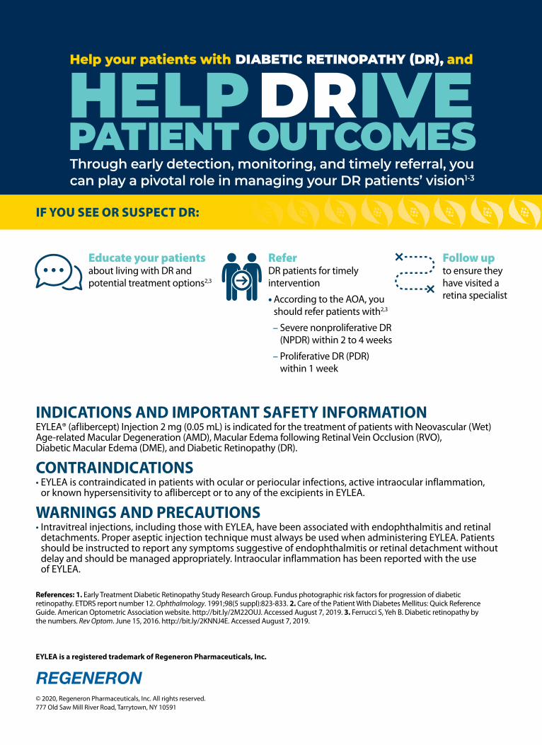
INDICATIONS AND IMPORTANT SAFETY INFORMATIONEYLEA® (aflibercept) Injection 2 mg (0.05 mL) is indicated for the treatment of patients with Neovascular (Wet) Age-related Macular Degeneration (AMD), Macular Edema following Retinal Vein Occlusion (RVO), Diabetic Macular Edema (DME), and Diabetic Retinopathy (DR).
CONTRAINDICATIONS• EYLEA is contraindicated in patients with ocular or periocular infections, active intraocular inflammation,
or known hypersensitivity to aflibercept or to any of the excipients in EYLEA.
WARNINGS AND PRECAUTIONS• Intravitreal injections, including those with EYLEA, have been associated with endophthalmitis and retinal
detachments. Proper aseptic injection technique must always be used when administering EYLEA. Patients should be instructed to report any symptoms suggestive of endophthalmitis or retinal detachment without delay and should be managed appropriately. Intraocular inflammation has been reported with the use of EYLEA.
Through early detection, monitoring, and timely referral, you can play a pivotal role in managing your DR patients’ vision1-3
HELP DRIVEPATIENT OUTCOMES
Help your patients with DIABETIC RETINOPATHY (DR), and
IF YOU SEE OR SUSPECT DR:
EYLEA is a registered trademark of Regeneron Pharmaceuticals, Inc.
References: 1. Early Treatment Diabetic Retinopathy Study Research Group. Fundus photographic risk factors for progression of diabetic retinopathy. ETDRS report number 12. Ophthalmology. 1991;98(5 suppl):823-833. 2. Care of the Patient With Diabetes Mellitus: Quick Reference Guide. American Optometric Association website. http://bit.ly/2M22OUJ. Accessed August 7, 2019. 3. Ferrucci S, Yeh B. Diabetic retinopathy by the numbers. Rev Optom. June 15, 2016. http://bit.ly/2KNNJ4E. Accessed August 7, 2019.
© 2020, Regeneron Pharmaceuticals, Inc. All rights reserved. 777 Old Saw Mill River Road, Tarrytown, NY 10591
Educate your patientsabout living with DR and potential treatment options2,3
ReferDR patients for timely intervention
• According to the AOA, you should refer patients with2,3
– Severe nonproliferative DR (NPDR) within 2 to 4 weeks
– Proliferative DR (PDR) within 1 week
Follow upto ensure they have visited a retina specialist

anti-VEGF = anti–vascular endothelial growth factor; AOA = American Optometric Association.
WARNINGS AND PRECAUTIONS (cont’d)• Acute increases in intraocular pressure have been seen within 60 minutes of intravitreal injection, including
with EYLEA. Sustained increases in intraocular pressure have also been reported after repeated intravitreal dosing with VEGF inhibitors. Intraocular pressure and the perfusion of the optic nerve head should be monitored and managed appropriately.
• There is a potential risk of arterial thromboembolic events (ATEs) following intravitreal use of VEGF inhibitors, including EYLEA. ATEs are defined as nonfatal stroke, nonfatal myocardial infarction, or vascular death (including deaths of unknown cause). The incidence of reported thromboembolic events in wet AMD studies during the first year was 1.8% (32 out of 1824) in the combined group of patients treated with EYLEA compared with 1.5% (9 out of 595) in patients treated with ranibizumab; through 96 weeks, the incidence was 3.3% (60 out of 1824) in the EYLEA group compared with 3.2% (19 out of 595) in the ranibizumab group. The incidence in the DME studies from baseline to week 52 was 3.3% (19 out of 578) in the combined group of patients treated with EYLEA compared with 2.8% (8 out of 287) in the control group; from baseline to week 100, the incidence was 6.4% (37 out of 578) in the combined group of patients treated with EYLEA compared with 4.2% (12 out of 287) in the control group. There were no reported thromboembolic events in the patients treated with EYLEA in the first six months of the RVO studies.
ADVERSE REACTIONS• Serious adverse reactions related to the injection procedure have occurred in <0.1% of intravitreal injections
with EYLEA including endophthalmitis and retinal detachment.• The most common adverse reactions (≥5%) reported in patients receiving EYLEA were conjunctival
hemorrhage, eye pain, cataract, vitreous detachment, vitreous floaters, and intraocular pressure increased.
Please see Brief Summary of Prescribing Information on the following pages.
Brought to you by REGENERON
Visit diabeticretinaldisease.com for additional information and useful patient resources
03/2020EYL.20.02.0082
The more you know about emerging clinical science about anti-VEGF and other potential therapies for DR, the better you can help inform your patients about how treatment may be able to help
Refer patients to a retina specialist who can treat DR2,3
Continue to monitoryour patients with DR2,3
• The AOA recommends frequent monitoring of patients2
– At least every 6 to 8 months in patients with moderate NPDR and more frequently for patients with greater disease severity2

1 INDICATIONS AND USAGE EYLEA is a vascular endothelial growth factor (VEGF) inhibitor indicated for the treatment of:Neovascular (Wet) Age-Related Macular Degeneration (AMD); Macular Edema Following Retinal Vein Occlusion (RVO); Diabetic Macular Edema (DME); Diabetic Retinopathy (DR).4 CONTRAINDICATIONS4.1 Ocular or Periocular Infections EYLEA is contraindicated in patients with ocular or periocular infections. 4.2 Active Intraocular Inflammation EYLEA is contraindicated in patients with active intraocular inflammation. 4.3 Hypersensitivity EYLEA is contraindicated in patients with known hypersensitivity to aflibercept or any of the excipients in EYLEA. Hypersensitivity reactions may manifest as rash, pruritus, urticaria, severe anaphylactic/anaphylactoid reactions, or severe intraocular inflammation.5 WARNINGS AND PRECAUTIONS 5.1 Endophthalmitis and Retinal Detachments. Intravitreal injections, including those with EYLEA, have been associated with endophthalmitis and retinal detachments [see Adverse Reactions (6.1)]. Proper aseptic injection technique must always be used when administering EYLEA. Patients should be instructed to report any symptoms suggestive of endophthalmitis or retinal detachment without delay and should be managed appropriately [see Patient Counseling Information (17)].5.2 Increase in Intraocular Pressure. Acute increases in intraocular pressure have been seen within 60 minutes of intravitreal injection, including with EYLEA [see Adverse Reactions (6.1)]. Sustained increases in intraocular pressure have also been reported after repeated intravitreal dosing with vascular endothelial growth factor (VEGF) inhibitors. Intraocular pressure and the perfusion of the optic nerve head should be monitored and managed appropriately.5.3 Thromboembolic Events. There is a potential risk of arterial thromboembolic events (ATEs) following intravitreal use of VEGF inhibitors, including EYLEA. ATEs are defined as nonfatal stroke, nonfatal myocardial infarction, or vascular death (including deaths of unknown cause). The incidence of reported thromboembolic events in wet AMD studies during the first year was 1.8% (32 out of 1824) in the combined group of patients treated with EYLEA compared with 1.5% (9 out of 595) in patients treated with ranibizumab; through 96 weeks, the incidence was 3.3% (60 out of 1824) in the EYLEA group compared with 3.2% (19 out of 595) in the ranibizumab group. The incidence in the DME studies from baseline to week 52 was 3.3% (19 out of 578) in the combined group of patients treated with EYLEA compared with 2.8% (8 out of 287) in the control group; from baseline to week 100, the incidence was 6.4% (37 out of 578) in the combined group of patients treated with EYLEA compared with 4.2% (12 out of 287) in the control group. There were no reported thromboembolic events in the patients treated with EYLEA in the first six months of the RVO studies.6 ADVERSE REACTIONS The following potentially serious adverse reactions are described elsewhere in the labeling: • Hypersensitivity [see Contraindications (4.3)] • Endophthalmitis and retinal detachments [see Warnings and Precautions (5.1)] • Increase in intraocular pressure [see Warnings and Precautions (5.2)] • Thromboembolic events [see Warnings and Precautions (5.3)]6.1 Clinical Trials Experience. Because clinical trials are conducted under widely varying conditions, adverse reaction rates observed in the clinical trials of a drug cannot be directly compared to rates in other clinical trials of the same or another drug and may not reflect the rates observed in practice.A total of 2980 patients treated with EYLEA constituted the safety population in eight phase 3 studies. Among those, 2379 patients were treated with the recommended dose of 2 mg. Serious adverse reactions related to the injection procedure have occurred in <0.1% of intravitreal injections with EYLEA including endophthalmitis and retinal detachment. The most common adverse reactions (≥5%) reported in patients receiving EYLEA were conjunctival hemorrhage, eye pain, cataract, vitreous detachment, vitreous floaters, and intraocular pressure increased.
Neovascular (Wet) Age-Related Macular Degeneration (AMD). The data described below reflect exposure to EYLEA in 1824 patients with wet AMD, including 1223 patients treated with the 2-mg dose, in 2 double-masked, controlled clinical studies (VIEW1 and VIEW2) for 24 months (with active control in year 1).Safety data observed in the EYLEA group in a 52-week, double-masked, Phase 2 study were consistent with these results.
Table 1: Most Common Adverse Reactions (≥1%) in Wet AMD StudiesBaseline to Week 52 Baseline to Week 96
Adverse ReactionsEYLEA
(N=1824)
Active Control (ranibizumab)
(N=595)EYLEA
(N=1824)
Control (ranibizumab)
(N=595)Conjunctival hemorrhage 25% 28% 27% 30%Eye pain 9% 9% 10% 10%Cataract 7% 7% 13% 10%Vitreous detachment 6% 6% 8% 8%Vitreous floaters 6% 7% 8% 10%Intraocular pressure increased 5% 7% 7% 11%Ocular hyperemia 4% 8% 5% 10%Corneal epithelium defect 4% 5% 5% 6%Detachment of the retinal pigment epithelium 3% 3% 5% 5%Injection site pain 3% 3% 3% 4%Foreign body sensation in eyes 3% 4% 4% 4%Lacrimation increased 3% 1% 4% 2%Vision blurred 2% 2% 4% 3%Intraocular inflammation 2% 3% 3% 4%Retinal pigment epithelium tear 2% 1% 2% 2%Injection site hemorrhage 1% 2% 2% 2%Eyelid edema 1% 2% 2% 3%Corneal edema 1% 1% 1% 1%Retinal detachment <1% <1% 1% 1%
Less common serious adverse reactions reported in <1% of the patients treated with EYLEA were hypersensitivity, retinal tear, and endophthalmitis.
Macular Edema Following Retinal Vein Occlusion (RVO). The data described below reflect 6 months exposure to EYLEA with a monthly 2 mg dose in 218 patients following CRVO in 2 clinical studies (COPERNICUS and GALILEO) and 91 patients following BRVO in one clinical study (VIBRANT).
Table 2: Most Common Adverse Reactions (≥1%) in RVO StudiesCRVO BRVO
Adverse ReactionsEYLEA
(N=218)Control (N=142)
EYLEA (N=91)
Control (N=92)
Eye pain 13% 5% 4% 5%Conjunctival hemorrhage 12% 11% 20% 4%Intraocular pressure increased 8% 6% 2% 0%Corneal epithelium defect 5% 4% 2% 0%Vitreous floaters 5% 1% 1% 0%Ocular hyperemia 5% 3% 2% 2%Foreign body sensation in eyes 3% 5% 3% 0%Vitreous detachment 3% 4% 2% 0%Lacrimation increased 3% 4% 3% 0%Injection site pain 3% 1% 1% 0%Vision blurred 1% <1% 1% 1%Intraocular inflammation 1% 1% 0% 0%Cataract <1% 1% 5% 0%Eyelid edema <1% 1% 1% 0% Less common adverse reactions reported in <1% of the patients treated with EYLEA in the CRVO studies were corneal edema, retinal tear, hypersensitivity, and endophthalmitis.
Diabetic Macular Edema (DME) and Diabetic Retinopathy (DR). The data described below reflect exposure to EYLEA in 578 patients with DME treated with the 2-mg dose in 2 double-masked, controlled clinical studies (VIVID and VISTA) from baseline to week 52 and from baseline to week 100.
Table 3: Most Common Adverse Reactions (≥1%) in DME StudiesBaseline to Week 52 Baseline to Week 100
Adverse ReactionsEYLEA
(N=578)Control
(N=287)EYLEA
(N=578)Control
(N=287)Conjunctival hemorrhage 28% 17% 31% 21%Eye pain 9% 6% 11% 9%Cataract 8% 9% 19% 17%Vitreous floaters 6% 3% 8% 6%Corneal epithelium defect 5% 3% 7% 5%Intraocular pressure increased 5% 3% 9% 5%Ocular hyperemia 5% 6% 5% 6%Vitreous detachment 3% 3% 8% 6%Foreign body sensation in eyes 3% 3% 3% 3%Lacrimation increased 3% 2% 4% 2%Vision blurred 2% 2% 3% 4%Intraocular inflammation 2% <1% 3% 1%Injection site pain 2% <1% 2% <1%Eyelid edema <1% 1% 2% 1% Less common adverse reactions reported in <1% of the patients treated with EYLEA were hypersensitivity, retinal detachment, retinal tear, corneal edema, and injection site hemorrhage. Safety data observed in 269 patients with nonproliferative diabetic retinopathy (NPDR) through week 52 in the PANORAMA trial were consistent with those seen in the phase 3 VIVID and VISTA trials (see Table 3 above).6.2 Immunogenicity. As with all therapeutic proteins, there is a potential for an immune response in patients treated with EYLEA. The immunogenicity of EYLEA was evaluated in serum samples. The immunogenicity data reflect the percentage of patients whose test results were considered positive for antibodies to EYLEA in immunoassays. The detection of an immune response is highly dependent on the sensitivity and specificity of the assays used, sample handling, timing of sample collection, concomitant medications, and underlying disease. For these reasons, comparison of the incidence of antibodies to EYLEA with the incidence of antibodies to other products may be misleading. In the wet AMD, RVO, and DME studies, the pre-treatment incidence of immunoreactivity to EYLEA was approximately 1% to 3% across treatment groups. After dosing with EYLEA for 24-100 weeks, antibodies to EYLEA were detected in a similar percentage range of patients. There were no differences in efficacy or safety between patients with or without immunoreactivity.
8 USE IN SPECIFIC POPULATIONS.8.1 Pregnancy Risk Summary Adequate and well-controlled studies with EYLEA have not been conducted in pregnant women. Aflibercept produced adverse embryofetal effects in rabbits, including external, visceral, and skeletal malformations. A fetal No Observed Adverse Effect Level (NOAEL) was not identified. At the lowest dose shown to produce adverse embryofetal effects, systemic exposures (based on AUC for free aflibercept) were approximately 6 times higher than AUC values observed in humans after a single intravitreal treatment at the recommended clinical dose [see Animal Data].Animal reproduction studies are not always predictive of human response, and it is not known whether EYLEA can cause fetal harm when administered to a pregnant woman. Based on the anti-VEGF mechanism of action for aflibercept, treatment with EYLEA may pose a risk to human embryofetal development. EYLEA should be used during pregnancy only if the potential benefit justifies the potential risk to the fetus.All pregnancies have a background risk of birth defect, loss, or other adverse outcomes. The background risk of major birth defects and miscarriage for the indicated population is unknown. In the U.S. general population, the estimated background risk of major birth defects and miscarriage in clinically recognized pregnancies is 2-4% and 15-20%, respectively.DataAnimal Data In two embryofetal development studies, aflibercept produced adverse embryofetal effects when administered every three days during organogenesis to pregnant rabbits at intravenous doses ≥3 mg per kg, or every six days during organogenesis at subcutaneous doses ≥0.1 mg per kg. Adverse embryofetal effects included increased incidences of postimplantation loss and fetal malformations, including anasarca, umbilical hernia, diaphragmatic hernia, gastroschisis, cleft palate, ectrodactyly, intestinal atresia, spina bifida, encephalomeningocele, heart and major vessel defects, and skeletal malformations (fused vertebrae, sternebrae, and ribs; supernumerary vertebral arches and ribs; and incomplete ossification). The maternal No Observed Adverse Effect Level (NOAEL) in these studies was 3 mg per kg. Aflibercept produced fetal malformations at all doses assessed in rabbits and the fetal NOAEL was not identified. At the lowest dose shown to produce adverse embryofetal effects in rabbits (0.1 mg per kg), systemic exposure (AUC) of free aflibercept was approximately 6 times higher than systemic exposure (AUC) observed in humans after a single intravitreal dose of 2 mg.8.2 Lactation Risk Summary There is no information regarding the presence of aflibercept in human milk, the effects of the drug on the breastfed infant, or the effects of the drug on milk production/excretion. Because many drugs are excreted in human milk, and because the potential for absorption and harm to infant growth and development exists, EYLEA is not recommended during breastfeeding. The developmental and health benefits of breastfeeding should be considered along with the mother’s clinical need for EYLEA and any potential adverse effects on the breastfed child from EYLEA.8.3 Females and Males of Reproductive Potential Contraception Females of reproductive potential are advised to use effective contraception prior to the initial dose, during treatment, and for at least 3 months after the last intravitreal injection of EYLEA.
Infertility There are no data regarding the effects of EYLEA on human fertility. Aflibercept adversely affected female and male reproductive systems in cynomolgus monkeys when administered by intravenous injection at a dose approximately 1500 times higher than the systemic level observed humans with an intravitreal dose of 2 mg. A No Observed Adverse Effect Level (NOAEL) was not identified. These findings were reversible within 20 weeks after cessation of treatment.8.4 Pediatric Use. The safety and effectiveness of EYLEA in pediatric patients have not been established.8.5 Geriatric Use. In the clinical studies, approximately 76% (2049/2701) of patients randomized to treatment with EYLEA were ≥65 years of age and approximately 46% (1250/2701) were ≥75 years of age. No significant differences in efficacy or safety were seen with increasing age in these studies.17 PATIENT COUNSELING INFORMATION In the days following EYLEA administration, patients are at risk of developing endophthalmitis or retinal detachment. If the eye becomes red, sensitive to light, painful, or develops a change in vision, advise patients to seek immediate care from an ophthalmologist [see Warnings and Precautions (5.1)]. Patients may experience temporary visual disturbances after an intravitreal injection with EYLEA and the associated eye examinations [see Adverse Reactions (6)]. Advise patients not to drive or use machinery until visual function has recovered sufficiently.
BRIEF SUMMARY—Please see the EYLEA full Prescribing Information available on HCP.EYLEA.US for additional product information.
Manufactured by: Regeneron Pharmaceuticals, Inc. 777 Old Saw Mill River Road Tarrytown, NY 10591
EYLEA is a registered trademark of Regeneron Pharmaceuticals, Inc. © 2019, Regeneron Pharmaceuticals, Inc. All rights reserved.
Issue Date: 08/2019 Initial U.S. Approval: 2011
Based on the August 2019 EYLEA® (aflibercept) Injection full Prescribing Information.
EYL.19.07.0306

39MAY 15, 2021 | REVIEW OF OPTOMETRY
Patients should have a baseline eyeexam and yearly screening with a dilat-ed fundus examination and SD- OCTimaging. If ocular manifestations arepresent, it is recommended to consultwith the patient’s oncologist as a changein dosage or discontinuation of the drugmay be indicated.7
Canthaxanthine (Orobronze). Anaturally occurring carotenoid whoseoral consumption may cause bronzingof the skin, canthaxanthine is sold overthe counter as a tanning agent. Thissubstance has been FDA approved foruse as an artificial food coloring agentbut not for any other use.
Orobronze deposits in all layers of theretina with a preference for the moresuperficial layers and will appear asvery small, gold-like particles near themacula.6 The associated retinopathydoes appear to be dose-dependent, andis found in 50% of patients who havetaken at least 37 grams and nearly 100%of those who have consumed at least 60grams of the drug. Most patients will beasymptomatic; however, occasionallyit can cause abnormal dark adaptation.The retinal deposits do resolve with thediscontinuation of the drug.8
MaculaHere we encounter some of the heavy-hitters most familiar to optometrists,especially Plaquenil, with which wehave a long history.
Hydroxychloroquine (Plaquenil). Thecholoroquines are a class of chemo-therapeutic agents and a mainstay asboth treatment and prophylaxis againstmalarial disease and Q fever. Hydroxy-chloroquine is effective in managingvarious autoimmune conditions, includ-ing rheumatoid arthritis, Sjögren’s,lupus and porphyria cutanea tarda.
The ophthalmic complicationsprimarily lie in its ability to produce atoxic maculopathy in a dose-dependentmanner. Findings include a bull’s-eyemaculopathy in the late stages, as-sociated with more subtle findings onOCT, fundus autofluorescence (FAF),multifocal electroretinogram (mfERG)and threshold perimetry findings in theearly stages (Figure 4).
Monitoring should include a baselineexam within one year of initiating thera-py, then biannual exams after five yearsof use including OCT and 10-2 thresh-old visual fields. FAF and mfERG mayhave utility in early detection. Earlyfindings in OCT may show disruptionof the outer ellipsoid zone and parafo-veal thinning of the outer nuclear layer,which may progress to “flying saucer”sign with advanced toxicity.
FAF may demonstrate early patchyhyperautofluorescence (hyperAF) cir-cumferentially around the fovea, whichmay coalesce to a ring in advanced dis-ease. mfERG studies are sensitive butwidely variable and are of limited valuein isolation. Threshold visual fields willsimilarly show patchy shallow depres-sions circumferentially about the foveawhich will coalesce into continuous,deeper scotomas in advanced disease.
Of note, Asian patients tend to havedefects more peripherally in the maculaand should be evaluated with a 24-2visual field. The chance of retinal toxic-ity increases with time and has as highas a 20% risk at 20 years.9 Concurrentuse of tamoxifen increases the risk ofmaculopathy fivefold.
Pentosan polysulfate sodium (Elmiron).PPS is an oral medication approved forthe relief of bladder pain or discomfortassociated with interstitial cystitis/pain-ful bladder syndrome.
A unique pigmentary maculopathywas only recently elucidated in 2018
and has subsequently been bolsteredby larger population studies. Currentevidence suggests a dose-dependentpattern of damage with caution inanyone with a cumulative exposureof greater than 500g. Color fundusphotos, OCT, FAF and near-infraredreflectance (nearIR) imaging are keyto detection. Fundus evaluation willdemonstrate yellow deposits at thelevel of the retinal pigmented epithe-lium (RPE), which correlate to hyper-reflective focal RPE thickening viaOCT. Similarly, these deposits correlateto hyperAF on FAF and hyperreflec-tive patches on nearIR imaging, whichshow highly disorganized patterns andirregularity in excess of what might bepredicted by fundus evaluation.
A hyperAF halo may be evident inthe peripapillary region (Figure 5).9
The clinical course and endpoints areunknown, but maculopathy can evolveafter discontinuation and patientsshould be followed at regular intervals.
Phenothiazines (multiple trade names).This drug class encompasses a largergroup of antipsychotic agents in whichthioridazine and chlorpromazine arethe most prescribed. They have beenlargely supplanted by newer medica-tions— especially in chronic therapy—but are still commonly used.
In contrast to most toxic maculopa-thies, the macular toxicity seen here ismore dependent on daily doses ratherthan a cumulative one. The mechanism
S Y S T E M I C M E D I C AT I O N SFeature
Fig. 4. Classic advanced hydroxychloroquine-induced toxic maculopathy showing parafoveal RPE/outer retinal loss (yellow arrows on OCT), hypoAF on FAF imaging and corresponding paracentral visual field defects.
Photo: Sara Weidm
ayer, OD

40 REVIEW OF OPTOMETRY | MAY 15, 2021
of damage is not completely under-stood, but they are thought to disruptrhodopsin synthesis.11
Onset of visual symptoms typi-cally occurs within weeks of initiatingtherapy in doses greater than 800mgdaily of thioridazine and 1200mg dailyof chlorpromazine. In the early stages,course granular pigmentation occurs,followed by patchy RPE loss with acharacteristic ovoid shape extending tothe midperipheral retina. In late stages,widespread areas of depigmentationand hyperpigmented plaques are vis-ible, followed by geographic atrophy.
At most doses, benign pigmentedcorneal and anterior lens opacities canalso occur. OCT can reveal photore-ceptor loss, which may span the foveaextending far into the arcades. There istypically some measure of visual recov-ery upon discontinuation.12 In standarddoses, routine exams are sufficientfor monitoring, while symptomaticindividuals should be examined withinthe week. Alternate meds should bediscussed with the prescribing practitio-ner at the first sign of toxicity.
Sertraline (Zoloft, Celexa, Lexapro, Pro-zac, Paxil). This a commonly prescribedselective serotonin reuptake inhibitor(SSRI) for depressive and anxiety disor-ders and more infrequently for bulimianervosa and anorexia nervosa.
While the link between sertralineand maculopathy has yet to be firmly
established, there are multiple casereports describing acute-onset bilateralmaculopathies in otherwise healthyeyes following initiation of therapy atstandard doses, whose findings closelymimic those in advanced hydroxychlo-roquine toxicity.13 While maculopathiesappear infrequently, the reported casesare profound, so be aware of this poten-tial adverse effect given the widespreaduse of this medication.
Optic NerveAs we encounter neuro-ophthalmicinvolvement, the potential for seriousvision loss escalates.
Phosphodiesterase Type 5 (PDE5)Inhibitors (Viagra, Levitra, Cialis). Thisclass of drugs is used to treat erectiledysfunction (ED). There has been anassociation between PED5 inhibitorsand non-arteritic anterior ischemicoptic neuropathy (NAION); however,there has been no evidence confirminga cause-effect relationship. The clinicalpicture of NAION in patients takingthese medications is the same as onethat occurs spontaneously: edematousand hyperemic optic disc, relative af-ferent pupil defect and painless loss ofvision.
It’s important to note that mostpatients who require the use of EDmedications have the same risk factors(older age, vascular risk factors such ashypertension and diabetes) for NAION,
which makes it very difficult to estab-lish a definite association between thetwo.2 It is recommended that thesedrugs be avoided in patients who haveexperienced NAION in one eye, asthey may be more likely to develop thecondition in the same or fellow eye.6
Amiodarone (Pacerone). An anti-ar-rhythmic drug, amiodarone is one of themost effective medications available totreat various cardiac arrhythmias.
While the exact mechanism isunknown, there is a probable associa-tion between amiodarone use and opticneuropathy. However, note that theneuropathy secondary to amiodaroneuse differs from typical NAION in thatthe condition is bilateral, associated vi-sion loss progresses slowly over severalmonths and the degree of vision losstends to be less severe. The disc edemais also much slower to improve whencompared to NAION, which can helpdistinguish between the two.
Patients on amiodarone should have abaseline exam with follow-up every 12months, or immediately should visualsymptoms occur.
If optic neuropathy is suspected, it’srecommended that the decision to dis-continue the medication be discussedwith that patient’s prescribing cardiolo-gist. Once the drug has been discontin-ued, it may take several months for theedema to resolve, ultimately leaving thepatient with optic atrophy (Figure 6).6
Vigabatrin (Sabril). An anticonvul-sant used in the treatment of seizures,vigabatrin is most often used to managedrug-resistant partial seizures and infan-tile spasms.
It has been associated with vi-sual field loss and optic nerve atrophy,although the exact mechanism is un-known. These complications are typi-cally irreversible and occur in approxi-mately 45% of patients treated with thismedication. Exam shows a bilateral,symmetric, concentric peripheral fielddefect with no central involvement.Retinal nerve fiber layer (RNFL) analy-sis with OCT shows thinning, with cor-responding pallor of the optic nerves.
Even in severe case of visual fieldloss and optic atrophy, visual acuity can
S Y S T E M I C M E D I C AT I O N SFeature
Fig. 5. Multimodal imaging demonstrating collocated yellow deposits, RPE thickening, near infrared hyper-reflectance and hyperAF on FAF imaging in advanced Elmiron maculopathy.
Photo: Sara Weidm
ayer, OD

TREAT OCULAR INFLAMMATION AND INFECTION, AND...
PRESCRIBE TOBRADEX® ST to control ocular infl ammation with risk of bacterial infection
Rapid relief from blepharitis/blepharoconjunctivitis symptoms1,a
XanGen™ suspension technology provides increased viscosity for improved ocular bioavailability of drug and consistent delivery2
TOBRADEX ST contains half the dexamethasone as TobraDex®, yet similar ocular tissue exposure2,b
TURN ONRELIEF
FORMULATED WITH ™
Eligible patients could pay as little as $45 for TOBRADEX STLEARN MORE AT MYTOBRADEXST.COM
Indications and Usage For steroid responsive infl ammatory ocular conditions of the palpebral and bulbar conjunctiva, cornea, and anterior segment of the globe and chronic anterior uveitis, corneal injury from chemical, radiation or thermal burns, or penetration of foreign bodies for which a corticosteroid is indicated and where the risk of superfi cial bacterial ocular infection is high or where there is an expectation that potentially dangerous numbers of bacteria will be present in the eye.
Important Safety Information CONTRAINDICATIONS:Most viral disease of the cornea and conjunctiva including epithelial herpes simplex keratitis (dendritic keratitis), vaccinia, and varicella, and also in mycobacterial infection of the eye and fungal disease of ocular structures. Hypersensitivity to any components of the medication.
© 2020 Eyevance Pharmaceuticals LLC. All rights reserved.TOBRADEX® ST and XanGen™ are trademarks of Eyevance Pharmaceuticals LLC. All other trademarks are the property of their respective owners. TST-01-20-AD-04
WARNINGS & PRECAUTIONS: • IOP increase – Prolonged use may result
in glaucoma with damage to the optic nerve, defects in visual acuity and fi elds of vision. IOP should be monitored.
• Aminoglycoside sensitivity – Sensitivity to topically applied aminoglycosides may occur.
• Cataracts – Posterior subcapsular cataract formation may occur.
• Delayed healing – May delay healing and increase the incidence of bleb formation. Perforations of the cornea or sclera have occurred. Slit lamp biomicroscopy, and fl uorescein staining should be conducted.
• Bacterial infections – May suppress host response and increase secondary ocular infections. In acute purulent conditions, steroids may mask infection or enhance existing infection. If signs and symptoms fail to improve after 2 days, the patient should be re-evaluated.
• Viral infections – Use with history of herpes simplex requires great caution. The course and severity of many viral infections of the eye (including herpes simplex) may be exacerbated.
• Fungal infections – Fungal infections of the cornea may occur and should be considered in any persistent corneal ulceration.
• Use with systemic aminoglycosides –Total serum concentration of tobramycin should be monitored.
ADVERSE REACTIONS:The most frequent adverse reactions (<4%) to topical ocular tobramycin are hypersensitivity and localized ocular toxicity, including eye pain, eyelid pruritus, eyelid edema, and conjunctival hyperemia.
The reactions due to the steroid component are increased intraocular pressure with possible development of glaucoma, and infrequent optic nerve disorder; subcapsular cataract; and impaired healing.
The development of secondary infection has occurred. Fungal infections of the cornea may occur. Secondary bacterial ocular infection following suppression of host responses also occurs.
Non-ocular adverse events (0.5% to 1%) included headache and increased blood pressure.
Please see Brief Summary of Full Prescribing Information on the adjacent page.
a Randomized, investigator-masked, active-controlled, parallel-group trial conducted at 7 private practice clinical sites in the United States with 122 adult patients who had moderate to severe blepharitis/blepharoconjunctivitis.1
b Multicenter, double-blind, parallel-group, single-dose study of 987 patients receiving a single dose of TOBRADEX ST or TobraDex ophthalmic suspension.2
References: 1. Torkildsen GL, Cockrum P, Meier E, et al. Curr Med Res Opin. 2011;27(1):171-178. 2. Scoper SV, Kabat AG, Owen GR, et al. Adv Ther. 2008;25(2):77-88.

TOBRADEX® ST (tobramycin/dexamethasone ophthalmic suspension) 0.3%/0.05%
Brief Summary
This Brief Summary does not include all the information needed to use TOBRADEX ST safely and effectively. Please see Full Prescribing Information for TOBRADEX ST at MyTobraDexST.com.
INDICATIONS AND USAGE TOBRADEX ST is a topical antibiotic and corticosteroid combination for steroid-responsive inflammatory ocular conditions for which a corticosteroid is indicated and where superficial bacterial ocular infection or a risk of bacterial ocular infection exists.
Ocular steroids are indicated in inflammatory conditions of the palpebral and bulbar conjunctiva, cornea and anterior segment of the globe where the inherent risk of steroid use in certain infective conjunctivitides is accepted to obtain a diminution in edema and inflammation. They are also indicated in chronic anterior uveitis and corneal injury from chemical, radiation or thermal burns, or penetration of foreign bodies.
The use of a combination drug with an anti-infective component is indicated where the risk of superficial ocular infection is high or where there is an expectation that potentially dangerous numbers of bacteria will be present in the eye.
DOSAGE AND ADMINISTRATION Recommended Dosing: Instill one drop into the conjunctival sac(s) every four to six hours. During the initial 24 to 48 hours, dosage may be increased to one drop every 2 hours. Frequency should be decreased gradually as warranted by improvement in clinical signs. Care should be taken not to discontinue therapy prematurely.
CONTRAINDICATIONS Nonbacterial Etiology: TOBRADEX ST is contraindicated in most viral diseases of the cornea and conjunctiva including epithelial herpes simplex keratitis (dendritic keratitis), vaccinia, and varicella, and also in mycobacterial infection of the eye and fungal diseases of ocular structures.
Hypersensitivity: Hypersensitivity to any component of the medication.
WARNINGS AND PRECAUTIONS IOP increase: Prolonged use of corticosteroids may result in glaucoma with damage to the optic nerve, defects in visual acuity and fields of vision. IOP should be monitored.
Aminoglycoside sensitivity: Sensitivity to topically applied aminoglycosides may occur.
Cataracts: May result in posterior subcapsular cataract formation.
Delayed healing: May delay healing and increase the incidence of bleb formation after cataract surgery. In those diseases causing thinning of the cornea or sclera, perforations have been known to occur with the use of topical steroids.
Bacterial infections: May suppress the host response and thus increase the hazard of secondary ocular infections. In acute purulent conditions, steroids may mask infection or enhance existing infection. If signs and symptoms fail to improve after 2 days, the patient should be re-evaluated.
Viral infections: Treatment in patients with a history of herpes simplex requires great caution. Use of ocular steroids may prolong the course and may exacerbate the severity of many viral infections of the eye (including herpes simplex).
Fungal infections: Fungal infections of the cornea are particularly prone to develop with long-term use. Fungal invasion must be considered in any persistent corneal ulceration.
Use with systemic aminoglycosides: Use with systemic aminoglycoside antibiotics requires monitoring for total serum concentration of tobramycin.
ADVERSE REACTIONS The most frequent adverse reactions to topical ocular tobramycin (TOBREX®) are hypersensitivity and localized ocular toxicity, including eye pain, eyelids pruritis, eyelid edema, and conjunctival hyperemia. These reactions occur in less than 4% of patients. Similar reactions may occur with the topical use of other aminoglycoside antibiotics.
Non-ocular adverse events occurring at an incidence of 0.5% to 1% included headache and increased blood pressure.
The reactions due to the steroid component are: increased intraocular pressure (IOP) with possible development of glaucoma, and infrequent optic nerve disorder; subcapsular cataract; and impaired healing.
Secondary Infection. The development of secondary infection has occurred. Fungal infections of the cornea are particularly prone to develop with long-term use. Fungal invasion must be considered in any persistent corneal ulceration. Secondary bacterial ocular infection following suppression of host responses also occurs.
USE IN SPECIFIC POPULATIONS Pregnancy and Nursing Mothers There are no adequate and well controlled studies in pregnant women. TOBRADEX® ST ophthalmic suspension should be used during pregnancy only if the potential benefit justifies the potential risk to the fetus. Caution should be exercised when TOBRADEX® ST is administered to a nursing woman.
Pediatric Use: Safety and effectiveness in pediatric patients below the age of 2 years have not been established.
Geriatric Use: No overall differences in safety or effectiveness have been observed between elderly and younger patients.
Rx Only Distributed by: Eyevance Pharmaceuticals LLC. Fort Worth, TX 76102
© 2020 Eyevance Pharmaceuticals LLC. All rights reserved. TOBRADEX® ST is a trademark of Eyevance Pharmaceuticals LLC.All other trademarks are the property of their respective owners.TST-01-20-MS-05

43MAY 15, 2021 | REVIEW OF OPTOMETRY
be normal and the patient asymptom-atic. The damage does appear to bedose-dependent and irreversible withthe first signs of RNFL thinning show-ing around 1000 grams cumulative doseand worsening as that dose increases.14
Patients should have a comprehen-sive eye exam within four weeks ofstarting the medication, every threemonths during treatment and three tosix months once treatment has beendiscontinued. If vision loss is confirmedor suspected, the prescribing physicianshould be consulted and the medica-tion should be discontinued.15
Ethambutol (Myambutol). This drugtreats pulmonary tuberculosis and thereis significant evidence of optic neuropa-thy associated with its use (Figure 7).The condition is typically bilateral butcan be asymmetric or unilateral.
The most common clinical findingsinclude a decrease in visual acuity, colorvision loss or central scotoma on visualfield testing. Ethambutol may also af-fect the optic chiasm and cause bitem-poral visual field defects. The adverseocular effects appear to be related todose, with those taking 60-100 mg/kg/day at the highest risk of 50%. At therecommended dose of 15mg/kg/day orless, the risk goes down to 1%.
Patients prescribed this medicationshould be made aware of the poten-tial for severe, irreversible vision lossand a baseline exam, including visualfield and color vision testing should beperformed. RNFL OCT analysis is alsorecommended, as it could possibly de-tect early stages of toxicity. If patients
are taking greater than 15mg/kg/day,it is recommended they have monthlyfollow-ups. Those taking lower dosesmay need to be followed closely if theyhave any conditions that may put themat higher risk for toxicity, such as diabe-tes, chronic renal failure or alcoholism.
The medication should be discon-tinued, at the discretion of the treatingphysician, if there is any evidence ofvision loss, visual field or color visiondefect.6
Isoniazid (Nydrazid). Another pul-monary tuberculosis drug, this one iscommonly prescribed in combinationwith other meds due to concern of drugresistance to monotherapy. Isoniazidhas also been associated with opticneuropathy; therefore, when used withethambutol, it can be difficult to deter-mine the causative agent.
Optic neuropathy from isoniazidtends to occur less frequently, is lesssevere and is reversible with medica-tion discontinuation.6 Rarely, the drug
has been associated with optic neuritis.Visual symptoms are like those seenwith other optic neuropathies andinclude decreased vision, loss of colorvision and either a bitemporal or cen-trocecal visual field defect. The opticnerves appear hyperemic with blurredmargins, though rarely may appearnormal. The condition is reversible,and improvement can begin as early asfour days after discontinuing the medi-cation, with complete recovery takinganywhere from four to six months.Continuation of treatment may lead tooptic atrophy.16
Tetracyclines. Part of a group of com-monly prescribed antibiotics that alsoincludes doxycycline, minocycline andoxytetracycline, these meds are usedto treat conditions like acne, rosacea,blepharitis and dry eye syndrome.
While there is currently no evidencethat they cause intracranial hyperten-sion, they have long been associatedwith it. Patients with this conditionmost often present with headache,pulsatile tinnitus, transient visualobscurations or diplopia.
Exam findings include bilateral op-tic disc edema and possibly decreasedvisual acuity. While obese women intheir childbearing years are at higherrisk in general for the development ofidiopathic intracranial hypertension(IIH), when associated with tetracy-cline use the condition tends to occurin even younger age groups (teens)and patients are less likely to be obese.
Average duration of antibiotic usebefore symptom onset can range from
S Y S T E M I C M E D I C AT I O N SFeature
Fig. 6. Late stage, severe optic atrophy OS in a patient on chronic amiodarone therapy.
Fig. 7. Optic neuropathy with pallor OD>OS due to ethambutol.
Photo: Sara Weidm
ayer, OD
Photo: Sara Weidm
ayer, OD

44 REVIEW OF OPTOMETRY | MAY 15, 2021
14.4 to 18.9 weeks.17 IIH is a diagnosisof exclusion and additional testing,neuroimaging and cerebrospinal fluidevaluation must be performed for con-firmation.18 The condition does tendto improve with discontinuation of themedication, and recurrence—whilerare—is typically less severe than theoriginal presentation.17
It is recommended that patientstaking tetracyclines be educated aboutthe possible side effects and have aneye exam after one month of treat-ment. Patients who experience any ofthe common IIH symptoms should beinstructed to seek prompt care with aneye care provider.19
Inflammatory ChangesFor the remainder of our discussion,we’ll shift emphasis from anatomicalchanges to those affecting multiplestructures and/or functions, beginningwith the effects of inflammation.
Alendronate (Fosamax, Actonel,Boniva, Reclast, Prolia, Evista, Duavee).Bisphosphonates inhibit bone resorp-tion and are most used to treat or pre-vent osteoporosis in post-menopausalwomen.
The most common ocular sideeffect is uveitis or scleritis. Patientswith uveitis will typically present withsevere pain, redness, sensitivity tolight and decreased vision. There willbe cell in the anterior chamber and oc-casionally keratic precipitates. Scleritiswill cause a severe deep pain, oftenthat awakens the patient at night. Thesclera can have a deep purple huewith vessel engorgement, and scleraledema and thinning can occur.
These conditions can be unilateralor bilateral and the onset of symptomsis two days to two weeks after start-ing treatment. In almost all cases ofinflammation, the alendronate must bediscontinued for the condition to fullyresolve and reinitiating any bispho-sphate prompts recurrence of theinflammation.6,20
Pembrolizumab (Keytruda). This is acancer immunotherapy drug that worksby increasing the natural immune sys-tem’s ability to detect and fight cancercells. Due to the increased immuneresponse, this drug has been associatedwith ocular inflammation, specificallyanterior uveitis or panuveitis (Figure 8).
Classic uveitis symptoms includepain, redness, photophobia and de-creased vision present, typically withmild to moderate cells in the anteriorchamber. It is possible, however, tohave a more severe reaction with vitre-ous involvement. The condition isalmost always bilateral and will occurwithin the first six months of therapy.Most patients respond well to tradi-tional topical uveitis treatment; in rarecases, long-term inflammation and/orsevere vision loss have been reported.
Patients on pembrolizumab shouldhave a baseline exam and be educatedon ocular side effects. Routine monitor-ing once treatment has been started isacceptable but may not be necessaryunless patients become symptomatic.21
Mechanical ChangesOcular effects in this category cancomplicate surgical procedures and,in severe presentations, expose thepatient to risk of angle closure.
Topiramate (Topamax). An oralsulfamate originally approved as ananticonvulsant, topiramate has morerecently been expanded for use formigraines, weight loss, depression andethanol dependence.
The drug may induce uveal ef-fusions, which result in ciliary bodyedema and forward rotation of thelens-iris diaphragm and angle closureglaucoma. Myopic shifts average underone diopter but have been reported atgreater than eight.
Adverse events are rare, but typi-cally occur within two weeks of start-ing the medication. Much rarer casesof ciliary body effusion have been re-ported with other sulfa drugs includingacetazolamide, sulfamethoxazole, pro-methazine, spironolactone, isosorbidedinitrate and bromocriptine.22 Isolatedcases have also been reported withtetracyclines, bupropion, oseltamivir,duloxetine and aspirin.
Recognition of the inciting medica-tion is critical, as the treatment differsfrom a primary angle closure andincludes the use of cycloplegics and atopical steroid with discontinuation ofthe inciting medication.
Alpha-1 blockers (Flomax, Uroxatral,Rapaflo). These treat symptoms ofbenign prostatic hyperplasia (BPH).
The presence of alpha-adrenocep-tors in mediating the contraction ofthe dilator pupillae muscle leads to ageneralized miosis and the potentialof intraoperative floppy iris syndrome(IFIS). This condition shows a billow-ing iris, rapid progressive intraopera-tive miosis and protrusion of iris tissuethrough surgical incisions. The effecttends to occur in as little as two weeksafter initiation of therapy and remainseven when discontinued. Severe IFISoccurs in roughly one-third of thosetaking Flomax and Rapaflo, while theeffect is less pronounced with Uroxa-tral.23
Although the challenges surround-ing IFIS cannot be eliminated withdiscontinuation of the drug, the intra-operative risks can be mitigated withintracameral epinephrine, preoperativeatropine, intraoperative trypan blue
Fig. 8. Findings due to chronic anterior uveitis associated with Keytruda use.
S Y S T E M I C M E D I C AT I O N SFeature
Photo: Sara Weidm
ayer, OD

Now more than ever, it’s a great time for eye care professionals (ECPs) to get inspired to explore new educational and professional development opportunities, enhancing their practices by increasing efficiency and expanding treatment options, such as multifocal contact lenses for presbyopic patients. Alcon is committed to providing high-quality edu-cational resources and training to ECPs around the world through the Alcon Experience Academy (AEA), which offers both online and in-person training.
Alcon Experience Academy Online—One-Stop Shop for ECP EducationThe Alcon Experience Academy website (https://www.alconexperienceacademy.com/) currently hosts over 800 educa-tional videos and other resources such as webcasts, podcasts, and literature aimed at a broad range of ECPs, including optometrists, ophthalmologists, nurses, and technicians.
For ECPs looking to better serve their presbyopic patients, Alcon offers the Multifocal Contact Lens Core Curriculum. This guided educational program features practical advice from experts in the field and covers a range of topics, including lens fitting, patient identification, practice opportunities, and misconceptions about multifocal lenses. Users are presented with a selection of brief e-learning modules based on individual areas of interest, practice characteristics, and educational needs as identified by a short survey.
Alcon Experience Academy IRL—Practitioners Visiting AlconAlthough online learning is hard to beat for convenience, it cannot fully replace inperson education, especially hands-on instruction for clinical techniques, such as fitting multifocal contact lenses. For this reason, the Alcon Experience Center at Alcon’s
US Headquarters in Fort Worth, Texas, provides onsite training of ECPs through the Practitioners Visiting Alcon (PVA) Program. The center offers a multifaceted learning experience, including educational courses, experiential learning, and cutting-edge immersive virtual reality technology.
The PVA program is also an opportunity for participants to build new relationships with other ECPs, says Dr. Elin Wu: “About a year ago, I visited Alcon Experience Academy in Fort Worth, Texas. It was a fun weekend of networking, learning, and discussing how to streamline soft multifocal fits through their workshop and seminars. I met a lot of wonderful talented and smart ODs there. As a result of this program, the DAILIES TOTAL1® Multifocal has been one of my go-to lenses for fitting soft multifocals for presbyopes because of the ease of fitting. Hoping for more programs like these once it’s safe to gather.”
Multifocal Fit Training Road Shows—Bringing Training Directly to
Your PracticeAlcon’s Multifocal Fit Training Road Shows bring peer-to-peer, hands-on fit training and other guidance on the use of multifocal contact lenses directly to you. Ask your Alcon representative about in-person and virtual programs to help maximize the potential of multifocal lenses in your practice.
Alcon’s Commitment to ECP Education—
The Bottom LineThese programs and others offered
through the AEA reflect Alcon’s on-going commitment to providing
quality education and training for ECPs. By staying up to date on the optimal use of available clinical tools, such as multifocal contactlenses, ECPs gain the knowledge and skills they need to help their patients and practices thrive.
IT’S TIME TO EXPERIENCE
ALCON EXPERIENCE ACADEMY!
SPONSORED CONTENT
Multifocal Core Curriculum
On-demand training
Register today
Virtual Multifocal Roadshows
Use the QR codes above to start your experience
© 2021 Alcon Inc. 4/21 US-DTM-2100023
See product instructions for complete wear, care, and safety information.

46 REVIEW OF OPTOMETRY | MAY 15, 2021
and iris retractors. As such, simply recognizing the risk andappropriate preoperative planning should minimize IFIS.
Perceptual ChangesThese adverse effects may be particularly troubling topatients without necessarily representing high-risk eventsthat jeopardize eye health.
Color Changes (multiple classes). PDE5 inhibitors dem-onstrate mild cross-reactivity with PDE6 receptors in theretina. Changes in color perception are a common sideeffect, with a blue tint being the most common. The effectoccurs in a dose-dependent manner with a 5% chance ofside effects at a standard dose. Voriconazole—a commonantifungal—has a similar cross reactivity and may elicitsimilar changes in perception.11 Fosamax and digoxin arecommonly associated with a yellow or gold color distur-bance.
Palinopsia (multiple classes). Defined as isochromaticafterimages which last following the removal of a stimulus,palinopsia can be caused by a wide variety of medications.Razadone, clomiphene, risperidone, topiramate and mir-tazapine have been associated with these manifestations.12
The mechanism of action is unclear, but effects on poste-rior cortical matter are implicated.
Blurred vision/diplopia (multiple classes). Blurred visionand diplopia can occur with a wide variety of oral meds.The most common classes include alpha-blockers, secondgeneration fluoroquinolones, statins, PDE5 inhibitors andbisphosphonates. The practitioner should investigate eachcase to rule out the more common etiologies and medica-tions cross-referenced with known side effects.
Functional ChangesAnother patient-forward category of effects, these changesare often amenable to mitigation with discontinuation ofthe drug.
Phenytoin (Dilantin). Phenytoin is a commonly prescribedantiseizure medication and has been associated with ocu-lomotor disturbances including gaze-evoked nystagmus,downbeat nystagmus, periodic alternating nystagmus andpartial or complete ophthalmoplegia. These findings cantake place in the normal therapeutic range, but are muchmore common in overdosed/ and toxic levels. Consider-ing the oculomotor findings could suggest a structuralbrainstem lesion rather than a drug-induced side effect, itis important to review all medications to determine if a fullneurologic work-up, including neuroimaging, is indicated.The clinical findings do improve and resolve once the doseis decreased or the drug is discontinued.24
Selective serotonin reuptake inhibitors (Prozac, Zoloft, Paxil,Lexapro, Celexa). SSRIs are psychiatric medications used totreat anxiety disorders.
These drugs are known to cause changes to the accom-modative status due to their anticholinergic effects. Theycause both mydriasis by stimulating the alpha receptors
Finding/Association Medication or Drug Class
Cornea Vortex/whorl keratopathy • Aminoglycosides• Amiodarone• Atovaquone• Chlorpromazine• Hydroxychloroquine• NSAIDs• Perhexiline maleate• Suramin• Tamoxifen
Crystalline keratopathy • Antineoplastic agents (cytarabine, emtansine, osimertinib, vandetanib)• Benoquine• Clofazimine• Gold salts• Macrolide antibiotics• Tilorone
Pigmented keratopathy • Phenothiazines
Corneal Edema • Amantadine• Antiseptics• BAK
Sclera Scleritis • Alendronate
Lens Anterior subcapsular cataract • Phenothiazines
Posterior subcapsular cataract • Glucocorticoids
Uvea Intraoperative floppy iris syndrome (IFIS) • α-1 blockers
Mydriasis and/or cycloplegia • SSRIs
Uveitis • Alendronate• Pembrolizumab
Uveal effusion • Topiramate• Sulfa-based medications
Retina Retinopathy • Interferon-α• Tamoxifen
Intraretinal deposits • Tamoxifen• Canthaxanthine
Macula Macular edema • Tamoxifen
Toxic maculopathy • Chloroquines• Phenothiazines• Setraline
Pigmentary maculopathy • Pentosan polysulfate sodium• Phenothiazines
Optic Nerve
Edema • Tamoxifen
Intracranial hypertension • Tetracyclines
NAION • PDE5 inhibitors
Optic neuritis • Isoniazid
Optic neuropathy/Atrophy • Amiodarone• Ethambutol• Isoniazid• Vigabatrin
Functional Abnormal dark adaptation • Canthaxanthine
Perceptual Color perception changes • PDE5 inhibitors• Voriconazole• Alendronate• Digoxin
Oculomotor dysfunction • Phenytoin
Palinopsia • Clomiphene• Mirtazapine• Risperidone• Topiramate• Trazadone
S Y S T E M I C M E D I C AT I O N SFeature
Table 1. Ocular Adverse Effects of Systemic Medications

located on the radial dilator muscle of the iris, and cyclople-gia due to the paretic effect on the ciliary muscle. Patientsexperiencing these effects will likely complain of blur—mostly at near—and occasionally glare. Eye care providersshould consider prescribing appropriate spectacle correctionto alleviate symptoms. The conditions are reversible oncethe medication is discontinued.25
Keep a Watchful EyeAs we’re reminded all the time in clinical practice, the eyesare not an island and are very susceptible to medication-induced side effects. Being aware of these possible effectscan help providers educate their patients, quickly recognizeand mitigate unwanted side effects, and avoid costly andtime-consuming workups. ■
1. Raizman MB, Hamrah P, Holland EJ, Kim T, et al. Drug-induced corneal epithelial changes. Surv Ophthalmol. 2017-05-01;62(3):286-301. 2. Li J, Tripathi RC, Tripathi BJ. Drug-induced ocular disorders. Drug Saf. 2008;31(2):127-41.3. McCarty CA, Wood CA, Fu CL, Livingston PM, et al. Schizophrenia, psychotropic medication, and cataract. Ophthalmology. 1999 Apr;106(4):683-7.4. Minhas B. The mind’s eye: ocular complications of psychotropic medications. Review of Optometry. 2016 Jan 15; 153(1): 42-49.5. Rentiya ZS, Wells M, Bae J, Chen KJ, et al. Interferon-α-induced retinopathy in chronic hepatitis C treatment: summary, considerations, and recommendations. Graefes Arch Clin Exp Ophthalmol. 2019 Mar;257(3):447-52.6. Santaella RM, Fraunfelder FW. Ocular adverse effects associated with systemic medications: recognition and management. Drugs. 2007;67(1):75-93.7. Doshi RR, Fortun JA, Kim BT, Dubovy SR, Rosenfeld PJ. Pseudocystic foveal cavitation in tamoxifen retinopathy. Am J Ophthalmol. 2014 Jun;157(6):1291-1298.8. Espaillat A, Aiello LP, Arrigg PG, Villalobos R, et al. Canthaxanthine retinopathy. Arch Ophthal-mol. 1999 Mar;117(3):412-3.9. Melles RB, Marmor MF. The risk of toxic retinopathy in patients on long-term hydroxychloro-quine therapy. JAMA Ophthalmol. 2014 Dec;132(12):1453-60. 10. Barnes AC, Hanif AM, Pentosan NJ. Polysulfate Maculopathy versus inherited macular dystrophies: comparative assessment with multimodal imaging. Ophthalmol Retina. 2020 Dec;4(12):1196-1201.11. Garg P, Yadav S. Ocular side effects of systemic drugs. Era J Med Res. 2019;6(1):1-9.12. Blomquist PH. Ocular complications of systemic medications. Am J Med Sci. 2011 Jul;342(1):62-9.13. Ewe S, Abell R, Vote B. Bilateral maculopathy associated with sertraline. Australas Psychia-try. 2014 Dec;22(6):573-5.14. Clayton LM, Devile M, Punte T, de Haan GJ, et al. Patterns of peripapillary retinal nerve fi ber layer thinning in vigabatrin-exposed individuals. Ophthalmology. 2012 Oct;119(10):2152-60.15. Vigabatrin: Medline Plus Drug Information. US National Library of Medicine. medlineplus.gov/druginfo/meds/a610016.html. Accessed 23 March 2021. 16. Kulkarni HS, Keskar VS, Bavdekar SB, Gabhale Y. Bilateral optic neuritis due to isoniazid (INH). Indian Pediatr. 2010 Jun;47(6):533-5.17. Orme D, Vegunta S, Miller M, Warner J, et al. A comparison between the clinical features of pseudotumor cerebri secondary to tetracyclines and idiopathic intracranial hypertension. Am J Ophthalmol. 2020; 220:177-182.18. Thurtell M, Wall M. Idiopathic intracranial hypertension (pseudotumor cerebri): recognition, treatment and ongoing management. Curr Treat Options Neurol. 2013 Feb;15(1):1-12.19. Kesler A, Goldhammer Y, Hadayer A, Pianka P. The outcome of pseudotumor cerebri induced by tetracycline therapy. Acta Neurol Scand. 2004;110:408-411.20. Samalia P, Sims J, Niederer R. Drug-induced ocular infl ammation. N Z Med J. 2020 Dec 18;133(1527):83-94.21. Sun M, Levinson R, Filipowicz A, Anesi S, et al. Uveitis in patients treated with CTLA-4 and PD-1 checkpoint blockade inhibition. Ocul Immunol Infl amm. 2020;28(2):217-27.22. Chen TC, Chao CW, Sorkin JA. Topiramate induced myopic shift and angle closure glau-coma. Br J Ophthalmol. 2003;87(5):648-649.23. Handzel DM, Briesen S, Rausch S, Kälble T. Cataract surgery in patients taking alpha-1 antagonists: know the risks, avoid the complications. Dtsch Arztebl Int. 2012;109(21):379-384. 24. Praveen-kumar S, Desai M. Ocular motor abnormalities in a patient with phenytoin toxicity--case report and minireview. Clin Neurol Neurosurg. 2014 Dec;127:116-7. 25. Minhas B. The Mind’s Eye: Ocular complications of psychotropic medications. Review of Optometry. 2016 Jan 15; 153(1): 42-49.
ELEVATE PATIENT EXPERIENCE
BOOTHTF233

Remote Refraction
Evolving Refraction Capabilities
New remote diagnostic technology allows refractions from anywhere
By H. Jay Wisnicki, MD
Even before COVID was an issue, our New York City-based eye care practice of optometrists and ophthal-mologists had been doing an increasing amount of
virtual work. This has been the case for many of us in eye-care; we have been outsourcing our billing operations, front desk services, human resources, and a variety of administra-tive and clerical functions for many years.
At the same time, the introduction of electronic medical records (EMRs) has increased the level of medical device integration and the exchange of clinical information across the spectrum of eyecare. For example, image management software has made it possible for us to view fundus and OCT images, in addition to fi ndings from corneal topogra-phy, biometry, ultrasound, and other digital devices be-tween our o� ces and networks.
In the clinical realm, while medical specialties such as radiology, pathology and behavioral health have been con-ducting some form of telemedicine for years, other areas of medicine such as optometry and ophthalmology requiring a close-up view of the patient, have had a slower time making the same leap. A few areas of eyecare have successfully used telemedicine to some degree to monitor patients with, or at risk of retinal pathologies such as diabetic retinopathy or age-related macular degeneration. However, eyecare in general has not utilized remote capabilities to perform portions of the comprehensive eye examination or conduct diagnostic evaluations and tests.
That is all changing with the introduction of new software technology enabling clinicians to perform remote diag-nostics, refractions and eye examinations without being physically in front of the patient.
Advancing Refraction CapabilitiesThere has been an evolution of refraction technology over the years, from conventional subjective refraction to com-puterized or digital phoropters and now remote operation.
In December 2020, Topcon Healthcare (Tokyo, Japan) introduced Topcon RDx®, an ocular telehealth platform enabling eye care providers to remotely connect to their o� ces to conduct portions of the comprehensive eye exam. RDx integrates with Topcon’s CV-5000S digital phorop-
ter so practitioners can virtually perform refractions, view supplementary diagnostic information through Topcon’s Harmony data management software, and video consult with the patient in real time.
This new capability o� ers the patient and provider more exam fl exibility, e� ciency and an increased level of safety. The doctor and patient are able to virtually meet for the patient’s eye exam, which helps to ensure the patient is presented with timely, high quality eye care while reducing the number of people and touch points encountered during an exam. Imagine how much more comfortable the patient might feel in a room with just one technician, while the per-son performing the exam is in a di� erent location.
From a practice standpoint, this technology has trans-formed our busy urban clinics. Our remote optometrists are able to quickly move from patient to patient, serving patients in both of our o� ce locations from essentially anywhere.
H. Jay Wisnicki, MD, is a professor of ophthalmology at the Icahn School of Medicine at Mount Sinai, and founder and medical director of Union Square Eye Care, all in New York City.
Topcon RDx® and the CV-5000S Digital Phoropter Benefi ts the Patient and the PracticeTopcon RDx®, an ocular telehealth platform enables eye care providers to connect to their o� ces remotely and conduct portions of the comprehensive eye exam. Topcon’s CV-5000S automated phoropter integrates with RDx remotely, so practitioners can perform refractions from anywhere.
Sponsored by48 • REVIEW OF OPTOMETRY

Benefi ts of Remote Refraction By Ryan Ngo, OD
As an optometrist who has spent years manually and digitally refracting patients, heading into a virtual realm has required a slightly di� erent approach to
the way I practice; however, the remote refraction process is not that di� erent from using the CV-5000S digital pho-ropter in the o� ce. The main di� erence is that rather than using a manual control panel in the examination room, the clinician is instead using a computer interface and mouse at a remote location to control the phoropter head.
I like that the Topcon RDx platform is intuitive and easy to start using. A technician is still needed to set up and align the patient before the refraction starts, but once the program is up and running, the sequence of steps is the same as an in-person refraction. The more we use the technology, the more seamless and integrated it’s becom-ing in our day-to-day clinical examinations.
Patient & Provider Benefi ts of RDx TechnologyIn practice, we have found that the patients’ early ex-perience with this new way of refracting has been over-whelmingly positive. After a recent remote refraction on a patient, the young woman gave us her impression of the process: “I was told prior to my exam that I would be getting a remote refraction from a clinician that would be o� site. This new examination process was so easy and was as if the doctor was still in the room with me!”
She liked the e� ciency that remote refraction o� ered, noting: “It’s nice just to be able to walk in, have a seat, video connect with the doctor and start the refraction process. I really liked that I didn’t have to wait. Sometimes in busy practices the doctors are running behind schedule, but with the remote refraction, I was able to get in front of the video screen with Dr. Ngo right away and he quickly refracted me and provided a new prescription.”
This patient also appreciated the additional fl exibility and convenience that remote refraction o� ered. She said, “You have more choices now based on your schedule or what doctor you prefer to see, because the doctor does not have to physically be in the o� ce to see you. For people who are super busy and have fast-paced lifestyles, I think the convenience and time savings with remote re-fraction is going to be a big value moving forward.”
As an eye care provider, I also value fl exibility in my professional life. Remote eye exams and refractions are es-pecially helpful if I am travelling, unable to make it into one of our o� ces or need to work from home. In the past, one
of the main drawbacks of being an eyecare pro-fessional was that you couldn’t provide care to patients and work from home at the same time. Now that we have the ability to remotely conduct eye exams, including refractions, that opens up options for doctors who may mix in-person and remote work, switching between the two to suit their personal needs and allowing a better quality of life.
Utilization TodayIn addition to its potential use for many of our routine evaluations, I think remote refraction can help improve access to vision care and fi ll in service gaps for large and small practices. For example, a patient who has an urgent need for glasses but can’t schedule a live visit with a doc-tor nearby, could schedule an appointment at a practice o� ering remote refractions, and quickly obtain a prescrip-tion through this virtual service. In addition, high-volume specialty practices that don’t do refractions might consid-er adding remote capabilities to o� er a more comprehen-sive eye exam for their patients. Another application could be for small optical shops that can’t a� ord a full-time optometrist but could hire someone part-time to manage remote refractions.
While remote refraction is an excellent fi t for many patients, certain patients might not be ideal candidates. Those individuals could include patients with large de-grees of astigmatism, or those who have conditions such as keratoconus or more complicated pathologies that require in-person or follow-up evaluations. Young children or elderly patients may not be as well-suited for remote refraction.
I estimate that 60% to 70% of my patient population might enjoy utilizing this new technology, in particular, “computer-age” patients like Millennials, Generations X and Z who have a comfort level with innovation and virtual tech-nology. In all cases, it will be exciting to see where remote refraction will take eye care now and into the future.
Ryan Ngo, OD, is an optometrist at Denny Eye & Laser Center in San Francisco; he formerly practiced at Union Square Eye Care in New York City.
REVIEW OF OPTOMETRY • 49
50 REVIEW OF OPTOMETRY | MAY 15, 2021
Using Antibiotics in Anterior Segment Care
Bacterial ocular and periocular infections are common in adults and children, but choosing an effective treatment strategy relies on more than an accurate clinical diagnosis.
Ocular and periocular infectionspresent in a variety of locationsfrom a variety of causes. The ap-propriate treatment course varies
from observation to non-pharmacologicmeasures to the prescription of topicalor oral anti-infective agents.1-3 In theabsence of the need for a bacterialculture, which is generally reserved forvision-threatening conditions, a care-ful history and thorough examinationguides the clinician to determine themost likely underlying cause that caninform treatment.
To muddy the waters further, nar-rowing down the diagnosis to suspect-ing a bacterial etiology leaves theclinician with a broad range of treat-ment strategies. Known patterns ofresistance, allergy history and systemichistory complicate the choice betweenantibiotics in clinical practice. Whiletreatment failure may be caused byinaccurate diagnosis, the cause mayalso be inadequate dosing, organismresistance, development of toxicity orallergy or nonadherence.
The chosen route of treatment typi-cally depends on the tissue involved:soft tissue infections such as preseptalcellulitis and dacyrocystitis requireantimicrobial drug levels at the siteof infection, which typically requiresoral administration. Conjunctival andcorneal disease is most commonlytreated efficiently with topical agents,while externally pointed hordeola mayrespond well to topical ointments andhot compresses.1-3
The Dangers of OvertreatmentWhile most anti-infective agents usedin periocular infection managementare considered to be broad-spectrum,emerging trends in pathogen resistancehave resulted in the need for under-standing most common underlyingpathogens likely to cause infection andtheir susceptibility to commonly pre-scribed antibiotics. However, overtreat-ment through unnecessary prescribingas well as undertreatment of antibioticspersists.
Up to 80% of uncomplicated acuteconjunctivitis in adults are viral innature. In those where bacterial infec-tion is the underlying cause, cases aretypically self-limiting within seven
days. Considering their self-limitingnature, topical antibiotics may not beprescribed; however, they modestlyimprove (by approximately two days)the time to resolution compared withsupportive therapy alone.1 Interest-ingly, nearly 60% of patients with acuteconjunctivitis in the United States fillprescriptions for topical antibiotics.4
The gap between the number ofcases of conjunctivitis that may benefitfrom topical antibiotic therapy and thenumber of prescriptions filled high-lights the concern of over-prescribingantibiotics when not clinically indicat-ed, a driver of antibiotic resistance.
The concept of a pre-determinedset course of antibiotic therapy as abroad prescribing strategy aims toensure complete eradication of disease-causing bacteria.5 But a one-size-fits-alltreatment approach ignores a centralcomponent to patient care: the patient.The fear of undertreatment has ledprescribers to generally prescribe thesame duration of treatment regardlessof the patient’s response to therapy.5
If our goal of appropriate antibioticprescribing is centered on limitingantibiotic use based on necessity andminimizing side effects to reach clini-
PEER REVIEWED
By Jessica Steen, ODFort Lauderdale, FL
Dr. Steen is an attending optometrist and assistant professor of ocular pharmacology at Nova Southeastern University College of Optometry. She has no financial interests to disclose.
Aboutthe author
A N T I B I OT I C SFeature

51MAY 15, 2021 | REVIEW OF OPTOMETRY
cal efficacy, antibiotic therapy discon-tinuation should differ based on thepatient’s response and how follow-upcare goes. One should only continuetreatment with topical ocular antibiot-ics for the prophylaxis of infection—forexample, in cases of corneal abrasion,while the increased risk is present—while epithelial damage exists ratherthan for a pre-determined course.
The minimum inhibitory concen-tration (MIC) value describes the invitro susceptibility or resistance levelsof bacteria to an antibiotic and servesas the basis for assessing whether anantibiotic may be effective in a clinicalsetting. Undertreatment of an infectioncan cause failure to reach the MIC,resulting in bacteria survival, mutationand replication during treatment, in-creasing the likelihood of developmentof a drug-resistant population.6 Nevertaper antibiotic agents below theirequivalent MIC, which is often clini-cally represented by FDA-approveddosing. In order to taper a steroid in atopical antibiotic and steroid combina-tion, first switch the patient to a steroidagent alone, rather than taper thecombination agent.
A Better UnderstandingThe main source of bacteria causingperiorbital soft tissue infection orconjunctivitis in adults is the natural
flora of the eyelids and conjunctiva.Common pathogens include gram-positive cocci, such as Staphylococcusepidermidis, Staphylococcus aureus andstreptococcus species as well as Proteusmiribalis, Serratia marcenas and gram-negative Pseudomonas species.2.3,7,8
In children, Haemophilus influenzaeand Streptococcus pneumoniae are themost common ocular isolates inconjunctivitis cases.8,9
A basic understanding of the trendsof resistance of common ocular isolatesforms the basis for antibiotic selec-tion for treatment with topical ocularantibiotics and oral medications to treatperiocular infections. The Antibi-otic Resistance Monitoring in OcularMicroorganisms (ARMOR) studysurveyed antibiotic resistance patternsof common ocular isolates.8 Its 10-yearresults showed that while there was nosignificant increase in the rates of re-sistance during the study period, therewas generally increased resistance withage.8 A significant proportion of S. au-reus isolates were resistant to oxacillin/methicillin (MRSA) and at least threeclasses of antibiotics, termed to be“multi-drug resistant.” Many believeresistance and susceptibility patterns toclosely mirror patterns in systemic in-fections, so we can apply these findingsto treatment of ocular infections withsystemic and topical antibiotics.8
The study also found that morethan half of all S. aureus isolates wereresistant to azithromycin, and 33.3%were resistant to ciprofloxacin. MRSAisolates were nearly all resistant toazithromycin. Importantly, MRSAisolates were determined to be highlysusceptible to trimethoprim and tet-racycline, but a high percentage ofisolates displayed resistance to fluoro-quinolones other than besifloxacin.8 S.pneumoniae isolates were most likelyto be resistant to azithromycin (36.3%)and penicillin (32.2%). P. aureginosaisolates displayed the lowest MIC withciprofloxacin.8
In general, azithromycin showswidespread resistance to the commonocular isolates implicated in ocular andperiocular disease, including MRSA,which is likely a result of the wide-spread use of the medication. Specificresistance strategies of macrolides maybe compounded by the medication’slong half-life and make azithromycin anon-preferred choice for the treatmentof soft tissue infections or bacterialconjunctivitis.8,10
Pertinent to infections in children,nearly all H. influenzae isolates weresusceptible to all antibiotics evalu-ated including ofloxacin, ciprofloxacin,moxifloxacin, azithromycin and tetracy-cline.8
The ARMOR trial also determinedresistance to besifloxacin to be negli-gible. Besifloxacin has no systemic for-mulation, and due to its mechanism ofaction, the development of resistancerequires mutation in both DNA gyraseand DNA topoisomerase IV.8
While community-acquired rates ofMRSA have increased over time,individuals in healthcare settings,those recently released from thehospital, incarcerated patients andpatients with a history of MRSA carry agreater possibility of harboring MRSAand therefore developing a bacterialinfection due to MRSA.3 Based onthe ARMOR data on treating bacte-rial infections in patients who may beat increased risk of MRSA, a topicalagent containing trimethoprim, such astrimethoprim sulfate and polymyxin BBacterial keratitis in a soft contact lens wearer.

52 REVIEW OF OPTOMETRY | MAY 15, 2021
sulfate or besifloxacin, is preferred.8 Forthe treatment of infections requiringoral medication, consider trimethoprim160mg and sulfamethaxazole 800mg,or for sulfonamide allergic patients,doxycycline 100mg.
For the treatment of bacterialconjunctivitis in children, where thegram-negative bacteria H. influenzae isa common underlying cause, considerfluoroquinolones such as ciprofloxacin0.3%, moxifloxacin 0.5% and ofloxacin0.3% ophthalmic solution. The safetyand efficacy of these medications havebeen evaluated in children one year ofage and older.11-13
For children younger than oneyear old, researchers have evaluatedpolymyxin B sulfate and trimethoprimsulfate in children as young as twomonths. While trimethoprim is not ef-fective against H. influenzae, polymyxinB can provide coverage against thisspecific organism.14
Pregnancy ConsiderationsEvery prescribing decision is based ona careful understanding and discussionwith the patient of the risks, benefitsand expected result of treatment. Thelack of data regarding prescriptionmedication safety in pregnant individu-als stems from protections implement-ed to limit the study of pregnant indi-
viduals, which resulted in the difficultyof inclusion of pregnant individuals inclinical trials. This resulting lack of datamay make both the prescriber and thepatient reluctant to treat the underlyingcondition.15
Taking medications during pregnan-cy or during lactation is common, withan estimated 90% of individuals takingan over-the-counter or prescriptionmedication during pregnancy, whichmeans that 5.4 million pregnanciesare exposed to medications annually.16
When treating pregnant individuals,make note of alcohol use, drug use andsmoking as well as inappropriately pre-scribed medications. Consider the risksand benefits of therapy for the patientand fetus.17
In general, penicillins and cephalo-sporins are safe to use during pregnan-cy.18 Of the macrolides, azithromycin isgenerally safe to use, while one shoulduse erythromycin and clarithromycinwith caution and only when the benefitof treatment outweighs the risk.18 Tet-racyclines have proven teratogenicityin humans, and fluoroquinolones haveconcerns of fetal malformation in ani-mals and therefore should be avoided.Avoid trimethoprim and sulfamethoxa-zole in the first trimester of pregnancyand after 32 weeks of gestation.18 Whilesystemic absorption with concur-
rent punctal occlusion of any topicalophthalmic agent is low, many considertopical ophthalmic antibiotic agents tobe safe for use in pregnant individuals.Still, take additional clinical risk-bene-fit consideration for topical ophthalmicfluoroquinolones due to the risk thatsystemically applied agents pose.
Adverse Effects ReviewWhen discussing the risks, benefitsand expected effects of a prescribedoral antibiotic with patients, do devotea part of the conversation to likely andpossible side effects. Due to orallyadministered antibiotics’ disruptionof disease-causing flora and naturalgastrointestinal flora, antibiotic-associated diarrhea is common and canbe expected in approximately 30% ofindividuals—one of the most commonreasons for nonadherence to antibiotictreatment.19
Probiotics are ingested microorgan-isms, including Lactobacillus, Bifido-bacterium and others, that may help tomaintain or restore the natural gastroin-testinal flora and ecosystem during andafter antibiotic treatment; however, notall patients will respond favorably, witha determined number of 13 individualspecies that need to be treated in orderto improve one case of antibiotic-asso-ciated diarrhea.20
While reconstitution of the naturalgut flora is often slow followingantibiotic treatment, probiotics mayfurther delay the reconstitution to anatural gastrointestinal microbiome.Notably, gastrointestinal flora is uniqueto individuals. By ingesting a largeamount of specific probiotic organisms,which are often not an exact reflectionof the composition of an individuals’sflora, the introduced species maydominate and delay recolonizationof natural flora by up to five months,compared with individuals who did notuse probiotics.21
While warnings about taking anti-biotics with alcohol are often added toprescription bottles, the evidence forreduced efficacy for most antibiotics islacking. Erythromycin, which may beprescribed in the context of periocular
This internally pointed hordeolum of the upper eyelid may respond well to topical ointments and hot compresses.
Photo: Greg Caldwell, OD
A N T I B I OT I C SFeature

A VISION THATINSPIRESYOURS
a team by your side
we care for our community
Satisfaction guaranteed >>>>>>>>>> from one independent owner to another
Ezer opHthalmic lineEzer opHthalmic lineThe Ezer Way of LifeThe Ezer Way of Life
impress your patient with an emerald
Light Source Options
LED & Halogen
emerald-26
High-End Camera SONY CCD
Contact us for informationContact us for information
9990 NW 14th Street, Unit #105, Doral, FL 33172 | USA
Follow Our Social Media Channels @USOPHTHALMIC
WWW.USOPHTHALMIC.COM | PH: 1.888.881.1122 [email protected]
SCAN TO SEE MORE!Read all the features and benefits this item has to o�er by connecting to our page.
While you’re there, don’t forget toRequest A Virtual Demo Online!
EASY FINANCE AVAILABLE$
Fundus CameraPerimeter
Ultrasound
Digital refractor
Digital lensmeter
Slit lamp line
refraction units
auto refractor
Digital chart
EASY FINANCE AVAILABLE
Non-contact tonometer
refraction units

54 REVIEW OF OPTOMETRY | MAY 15, 2021
disease treatment, should not be takenwith alcohol due to the potential forreduced efficacy and potential systemictoxicity.22
Also, cephalosporins and sulfon-amide antibiotics carry a specificadverse effect that patients shouldbe advised of. A “disulfiram-like”reaction describes specific symptomsthat mimic the effect of disulfiram, amedication used for the treatment ofalcohol dependency disorder. Patientsmay experience dizziness, sweating,headache, nausea, weakness, vomitingand hypotension—symptoms often de-scribed to be similar to the exacerbatedeffects of a hangover. While disulfiram-like reactions are well-documentedwith cephalosporin and alcohol use,only isolated case reports have beendescribed following the administrationof trimethoprim-sulfamethaxazole.23,24
While azithromycin is a commonlyprescribed oral antibiotic, recent re-ports note an increased risk of suddencardiac death due to prolongation ofthe QT interval in individuals with car-diovascular event risk factors that laterprompted the FDA to include a blackbox warning for the drug.25 Individualsmost at risk are those with underlyingarrhythmia termed torsades de pointes.25
Allergy PerilsPrior to prescribing any medication,perform a careful review of history ofadverse drug reactions and allergies.
Differentiating between a true type-1hypersensitivity reaction or an IgE-mediated response and another typeof adverse effect requires a carefuldiscussion of symptoms the patientexperienced with each reportedadverse event.
Urticaria, anaphylaxis,bronchospasm, laryngospasm andangioedema are all signs of true allergy,while gastrointestinal upset is a signof an expected adverse effect of orallyadministered antibiotics.26,27 Whileapproximately 10% of the populationhave reported an allergy to penicillin,less than 5% have a true allergy.26
The allergic potential of sulfonamideantibiotics carries a similar risk, withapproximately 3% of the populationhaving experienced a true allergicreaction.28
The practice of avoiding cephalospo-rins in penicillin-allergic patients stemsfrom the potential for cross-reactivitydue to their structurally similar R1side-chain of first- (i.e., cephalexin)and second-generation cephalosporins.The risk of allergy to a cephalosporinin a penicillin-allergic patient due tocross-reactivity is much less than thehistorically understood 10%.29 How-ever, avoid cephalexin in patients withknown penicillin hypersensitivity dueto the approximate 1% cross-reactivitywith first-generation cephalosporins.29
When patients have a true allergyto one type of drug—for instance, toa sulfonamide non-antibiotic such asa thiazide diuretic—is there potentialfor allergy to a structurally similar drugclass, such as a sulfonamide antibiotic?While the potential exists for allergicreaction in both classes, it appears tonot be due to structural similarity anddevelopment of cross-reactivity butrather increased propensity to allergicreaction in general.
Pay particular attention to patientswith an atopic history or pre-existingasthma diagnosis, especially whenthey have a history of penicillin orsulfonamide antibiotic allergy. Eitherof these can suggest that multipleallergies may exist across non-structurally similar drug categories.27
TakeawaysOptimal prescribing for the treatmentof bacterial infections requires carefulclinical examination, a thorough
Hypersensitivity reaction of the left eye secondary to topical brimonidine use. Differentiating between a true type-1 hypersensitivity reaction or an IgE-mediated response and another type of adverse effect requires a careful discussion of symptoms the patient experienced with each reported adverse event.
Mucopurulent discharge characteristic of bacterial conjunctivitis.
Photo: Greg Caldwell, OD
A N T I B I OT I C SFeature

history and an understanding of themicrobiome of periocular tissues, resistance and susceptibility patterns and risk of potential adverse effects. Using the right antibiotic at the right time, dose and duration will ensure that your patient is appropriately managed while minimizing the risk of adverse effect to your patient and the greater community. ■
1. Azari AA, Barney NP. Conjunctivitis: a systematic review of diagnosis and treatment. JAMA. 2013; 310(16):1721-9.2. Mannis MJ, Plotnick RD. Bacterial conjunctivitis. In: Tas-man W, Jaeger EA, ed. Duane’s Clinical Ophthalmology. Vol. 4. Philadelphia: Lipencott-Raven; 2004:1-7.3. Charalampidou S, Connell P, Fennell J, et al. Preseptal cellulitis caused by community acquired methicillin resis-tant Staphylococcus aureus (CAMRSA). Br J Ophthalmol. 2007;91(12):1723-4.4. Shekhawat NS, Shtein RM, Blachley TS, et al. Antibiotic prescription fills for acute conjunctivitis along enrollees in a large United States managed care network. Ophthalmology. 2017;124(8):1099-1107.5. Llewelyn MJ, Fitzpatrick JM, Darwin E, et al. The antibi-otic course has had its day. BMJ. 2017;358;j3418.6. Kowalska-Krochmal B, Dudek-Wicher R. The minimum inhibitory concentration of antibiotics: methods, interpreta-tion, clinical relevance. Pathogens. 2021;10(2):165.7. Ratnumnoi R, Keorochana N, Sontisomabat C. Normal flora of conjunctiva and lid margin, as well as its antibiotic sensitivity, in patients undergoing cataract surgery at Phra-mongkutklao Hospital. Clin Ophthalmol. 2017;11:237-41.
8. Asbell PA, Sanfilippo CM, Sahm DF, et al. Trends in antibiotic resistance among ocular microorganisms in the United States from 2009 to 2018. JAMA Ophthalmol. 2020;138(5):439-50.9. Cavuoto K, Zutshi D, Karp CL, et al. Update on bacterial conjunctivitis in South Florida. Ophthalmology. 2008;115(1):51-6.10. Food and Drug Administration. Highlights of prescribing information: Zithromax. 2019. www.accessdata.fda.gov/drugsatfda_docs/label/2019/050710s049,050711s047,050784s034lbl.pdf. Accessed April 9, 2021. 11. Novartis Pharma AG. Prescribing information: Ciloxan. 2013. www.novartis.com.sg/system/files/product-info/CILOXAN%20STERILE%20OPHTHALMIC%20AND%20OTIC%20SOLUTION%200.3%25.pdf. Accessed April 9, 2021. 12. Food and Drug Administration. Highlights of prescribing information: Vigamox. 2020. www.accessdata.fda.gov/drugsatfda_docs/label/2020/21598s024lbl.pdf. Accessed April 9, 2021. 13. Allergan. Prescribing Information: Ocuflox. 2016. www.accessdata.fda.gov/drugsatfda_docs/label/2016/019921s021lbl.pdf. Accessed April 9, 2021. 14. Allergan. Prescribing Information: Polytrim. 2018. media.allergan.com/actavis/actavis/media/allergan-pdf-documents/product-prescribing/20180622-POLYTRIM-USPI-1-0USPI7824.pdf. Accessed April 9, 2021.15. van der Graaf R, van der Zande ISE, den Ruijter HM, et al. Fair inclusion of pregnant women in clinical trials: an inte-grated scientific and ethical approach. Trials. 2018;19(1):78.16. Mitchell AA, Gilboa SM, Werler MM, et al. National birth defects prevention study. Medication use during pregnancy, with particular focus on prescription drugs: 1976-2008. Am J Obstet Gynecol. 2011;205(1):51.e1-8.17. Genetic Alliance; District of Columbia Department of Health. Understanding genetics: a District of Columbia guide for patients and health professionals. Washington (DC): Genetic Alliance; 2010 Feb 17. Appendix D, Terato-gens/Prenatal Substance Abuse.
18. Lamont HF, Blogg HJ, Lamont RF. Safety of antimicrobial treatment during pregnancy: a current review of resistance, immunomodulation and teratogenicity. Expert Opin Drug Saf. 2014;13(12):1569-81.19. Szajewska H, Mrukowicz JZ. Probiotics in prevention of antibiotic-associated diarrhea:meta-analysis. J Pediatr. 2003;142(1):85. 20. Hempel S, Newberry SJ, Maher AR, et al. Probiotics for the prevention and treatment of antibiotic-associated diarrhea: a systematic review and meta-analysis. JAMA. 2012;307(18):1959-69. 21. Suez J, Zmora N, Zilberman-Schapira G, et al. Post-antibiotic gut mucosal microbiome reconstitution is impaired by probiotics and improved by autologous FMT. Cell. 2018;174(6):1406-23.e16.22. Mergenhagen KA, Wattengel BA, Skelly MK, et al. Fact versus fiction: a review of the evidence behind alcohol and antibiotic reactions. Antimicrob Agents Chemother. 2020;64(3):e02167-19.23. Barth KS, Malcolm RJ. Disulfiram: an old therapeutic with new applications. CNS Neurol Disord Drug Targets. 2010;9(1):5-12. 24. Heelon MW, White M. Disulfiram-cotrimoxazole reaction. Pharmacotherapy. 1998;18(4):869-70. 25. Ray WA, Murray KT, Hall K, et al. Azithromycin and the risk of cardiovascular death. N Engl J Med. 2012; 366(20):1881-90.26. Shenoy ES, Macy E, Rowe T, et al. Evaluation and manage-ment of penicillin allergy: a review. JAMA. 2019;321(2):188-99. 27. Strom BJ, Schinnar R, Apter AJ, et al. Absence of cross-reactivity between sulfonamide antibiotics and sulfonamide nonantibiotics. N Engl J Med. 2003;349(17):1628-35. 28. Lieberman P, Anderson JA, eds. Allergic diseases: diagno-sis and treatment. Totowa, NJ:Humana Press; 1997:289.29. Campagna JD, Bond MC, Schabelman E, et al. The use of cephalosporins in penicillin-allergic patients: a literature review. J Emerg Med. 2012;42(5):612-20.
"Increased digital device use caused a dramatic rise in ocular surface disease symptoms in our clinic, making hygiene more important than ever. This comprehensive kit makes it easy to satisfy patient needs at a time they are more motivated to follow a recommended regimen."
Wipe to remove build up, oil, dirt, pollen, and debris that may cause eye irritation and infection
Spray pure 0.02% HOCI solution to help reduce bacteria and inflammation
Warm to naturally unclog meibomian glands and relieve dry eye symptoms
t-lEALTHy EYES START HERE
Bruder. Hygienic E'YELJD CARE KIT'" Dliii 4 , r !'f!D
Request a complimentary sample kit at bruder.com/rokit21 Have a question or want to learn more? Give us a call at 888-827-8337 or email [email protected] c. .. � 1 Jdltl :.areCJmp y >.p ett C:A�
Review of Optometry_Bruder Kit_Karpecki_May 2021

56 REVIEW OF OPTOMETRY | MAY 15, 2021
ManagingMiotics and Mydriatics
Take a closer look at old, new and future uses of these drugs.
Optometrists are well-acquaintedwith the two opposing musclesin the iris, the sphincter andthe dilator, as we witness their
effects daily in clinical practice. Pupilconstriction (miosis) can either bestimulated by contraction of the irissphincter or by relaxation of the irisdilator. On the other hand, pupil dila-tion (mydriasis) can either be stimu-lated by contraction of the iris dilatoror by relaxation of the iris sphincter.
Miotic and mydriatic drops work byacting on these different muscles ofthe iris. The drops are able to controlpupil size by targeting two parts ofthe autonomic nervous system: thesympathetic and parasympathetic sys-tems. Let’s review their function andclinical role to better understand theirpresent uses and why some of theseagents are undergoing re-evaulationfor potential new ones.
Behind the ScenesThe sympathetic pathway, mainly re-sponsible for pupil mydriasis, involvesa three-neuron pathway.1,2 The first
neuron begins in the hypothalamusand descends through the midbrainto synapse onto a specific area of thespinal cord, known as the ciliospi-nal center of Budge. This synapseis located between the C8 and T2vertebrae. The second neuron, whichis the preganglionic neuron, exitsthe spinal cord, ascends through thethorax and synapses near the apexof the lung into the superior cervicalganglion. The third postganglionicneuron travels to the cavernous sinusand enters the orbit through the shortand long ciliary nerves, synapsing tothe iris dilator.1,2
Contrarily, the parasympatheticpathway is mainly responsible forpupil miosis.1,3 Pupil constrictionstarts when light enters the retinaand activates the retinal ganglioncells—the beginning of the affer-ent arm—which then transmit theirimpulses into the optic nerve. Thisstimulus travels to the optic chiasm,through the optic tract and eventuallyreaches the pretectal nucleus. Theimpulses from the pretectal nucleusbegin the efferent arm, which projectsto the Edinger-Westphal nucleus. TheEdinger-Westphal nucleus gives riseto preganglionic fibers, which then
synapse with postganglionic neuronsin the ciliary ganglion. Postganglionicneurons leave the ciliary ganglion toinnervate the iris sphincter.1,3
Mydriatic drops work by either in-hibiting the parasympathetic pathwayto the iris sphincter or by promot-ing the sympathetic pathway to theiris dilator. Drops that antagonizethe parasympathetic pathway blockacetylcholine, a neurotransmitter ofthe autonomic nervous system, fromreaching muscarinic receptors, whichare located within the iris sphincter.1,4
This leads to relaxation of the irissphincter and promotes mydriasis.
On the other hand, mydriatic dropsthat promote the sympathetic path-way prevent the reuptake of norepi-nephrine, another neurotransmitterof the autonomic nervous system,from the synapse.1 This allows morenorepinephrine to bind to alpha-oneadrenergic receptors on the iris dilator,stimulating the activation of the irisdilator and leading to mydriasis.
Miotic drops work by either pro-moting the parasympathetic pathwayto the iris sphincter or by inhibitingthe sympathetic pathway to the irisdilator.1,5 Taking the former route,these drops mimic the activity of
P U P I L C O N T R O LFeature PEER REVIEWED
By Paymaun Asnaashari, ODsacramento
Dr. Asnaashari graduated from the UC Berkeley School of Optometry in 2018. He is a therapeutic- and glaucoma-certified optometrist in Sacramento, CA, and a Fellow of the American Academy of Optometry. One of his areas of expertise is specialty contact lenses. He has no financial interests to disclose.
Aboutthe author

57MAY 15, 2021 | REVIEW OF OPTOMETRY
acetylcholine, and when they bind tomuscarinic receptors, the iris sphinctercontracts and pupil size decreases.
Drops that inhibit the sympatheticpathway can also stimulate pupil mio-sis. These drops selectively bind toalpha-two adrenergic receptors, whichare responsible for decreasing thesympathetic tone. These receptors arelocated on the iris dilator, which whenactivated leads to a decrease in theamount of norepinephrine releasedinto the synapse. This reduces thesympathetic tone on the iris dilatorand encourages pupil miosis.
Miotic DropsThese drops can be used for a numberof things, including diagnosis of pupilabnormalities (e.g., Adie’s tonic pupil)and treatment of ocular hypertension,acute angle closure, dry eye and post-surgical glare.3 Exercise caution oravoid use altogether in patients whoare pregnant, breastfeeding or at riskof retinal detachment. The followingare several established drops and as-sociated clinical pearls of each:
Parasympathomimetics. Pilocarpineis a miotic drop that is used primarilyto treat ocular hypertension.6 It caneffectively lower intraocular pressure(IOP) and is easily accessible. How-
ever, it can also induce myopic shiftsand precipitate retinal detachments.For these reasons, pilocarpine is notcommonly used in general practice.
Pilocarpine is a direct muscarinicacetylcholine agonist, or a parasympa-thomimetic. It binds to the M3 mus-carinic receptor, an excitatory receptorin the iris sphincter and ciliary body.This leads to an upregulation of cal-cium and smooth muscle contractionof the iris sphincter and ciliary body.The contraction of the iris sphinctercauses the pupil to decrease in size,and the contraction of the ciliarybody opens the trabecular meshwork,decreasing IOP by enhancing aqueousoutfl ow.
Sympathomimetics. Brimonidine andapraclonidine are miotic drops thatare typically used to lower IOP andassist with post-surgical glare, ocularredness and diagnosis of pupil abnor-malities (e.g., Horner’s syndrome).7
These drops are able to rapidly lowerIOP and are readily available. Theyare contraindicated in patients withunderlying asthma and those takingMAO inhibitors. They are also morestrongly associated with allergic reac-tions.
Both of these drops exhibit selec-tive alpha-two adrenergic agonistactivity. They bind directly to alpha-two adrenergic receptors, which areinhibitory receptors of the sympa-thetic system. These receptors arepresent on the iris dilator, ciliary bodyand conjunctival blood vessels. Theycause a downregulation of cAMPand a decrease in the sympathetictone, leading to iris dilator relaxation,increasing uveoscleral outfl ow anddecreasing aqueous production andvasoconstriction.
Mydriatic DropsThese drops can be used to performdilated fundus examination, diagnosepupil abnormalities (e.g., Horner’ssyndrome), treat anterior uveitis andengage in pharmacological penaliza-tion for amblyopia.2 Exercise cautionor completely avoid use in patientswho are pregnant or breastfeeding and
in those who have anatomically nar-row angles. The following are severalestablished drops and associated clini-cal pearls of each:
Parasympatholytics. Tropicamide,cyclopentolate and atropine are allmydriatic drops that are traditionallyused for dilated fundus examination,paralysis of accommodation, anterioruveitis treatment, myopia controland pharmacological penalization foramblyopia treatment.8-10
Tropicamide is typically used fordilated fundus exam, paralysis ofaccommodation and anterior uveitistreatment. It has a strong mydriatic ef-fect, rapid onset and shorter durationof action in comparison with cyclo-pentolate and atropine. Its disadvan-tages include its ineffectiveness atinhibiting accommodation and its sideeffects of light sensitivity and blurredvision.
Cyclopentolate is commonly usedto inhibit accommodation and treatamblyopia and anterior uveitis. It iseffective at accommodation paralysis.On the other hand, it has a long onsetof action, long duration of action and
Tropicamide has a strong mydriatic effect.
Cyclopentolate can effectively inhibit accommodation to help treat amblyopia and anterior uveitis.

58 REVIEW OF OPTOMETRY | MAY 15, 2021
side effect profi le that includes lightsensitivity and blurred vision. Thelong duration of action makes thisa viable drop of choice for anterioruveitis treatment to prevent anteriorsynechiae.
Similar to cyclopentolate, atropineis traditionally used to inhibit accom-modation, manage amblyopia, treatanterior uveitis and provide myopiacontrol. It has a strong mydriatic effectand is effective at inhibiting accom-modation and slowing the progressionof myopia. Its disadvantages mirrorthose of cyclopentolate.
The mechanism of action of each ofthese drops is identical: they all havedirect antimuscarinic activity. Theycompetitively bind to muscarinic re-ceptors on the iris sphincter and ciliarybody and inhibit their activity. Thisresults in a downregulation of calciumand induces smooth muscle relaxationof the iris sphincter and ciliary body.
Sympathomimetics. Phenylephrineis a mydriatic drop that is traditionallyused for dilated fundus examination,ocular redness relief and diagnosis ofpupil abnormalities (e.g., Horner’s syn-drome).11 It has a rapid onset of actionand is easily accessible. However, it
has a weak mydriatic effect and shouldnot be used alone for dilated fundusexamination.
Phenylephrine promotes the sympa-thetic pathway as a selective alpha-oneadrenergic receptor agonist. Alpha-onereceptors are excitatory receptors ofthe sympathetic system and reside inthe iris dilator and conjunctival bloodvessels. Activation of these recep-tors causes an upregulation of cAMP,which leads to iris dilator contractionand vasoconstriction.
Sympathomimetics and parasympatho-lytics. Paremyd (hydroxyamphetamine1% and tropicamide 0.25%, Akorn) isa mydriatic that is traditionally usedfor dilated fundus examination.12 Ithas a rapid onset of action, reducedeffect on accommodation and shorterduration of action. Its disadvantagesinclude a reduced mydriatic effect ondark irides, decreased ability to inhibitaccommodation and a side effect pro-fi le that includes light sensitivity andblurred vision.
Hydroxyamphetamine acts as anadrenergic agonist and blocks thebreakdown of norepinephrine. Thisincreases the sympathetic tone and in-directly initiates contraction of the irisdilator. Tropicamide has antimuscarin-ic activity and inhibits the muscarinicreceptors on the iris sphincter, leadingto pupil dilation.
In the PipelineThe exploration of pharmaceuticaladvancements in the areas of myopiacontrol, presbyopia and dilation is onthe rise.
Myopia. The increasing prevalenceof myopia worldwide and its associ-ated risk factors have led to a publichealth crisis. Historically, orthokera-tology has been a leading option forslowing myopic progression, butophthalmic drops are catching up andmay potentially surpass this modal-ity. Treatment with low-dose atropinehas been increasing in popularity asmore evidence continues to show itseffectiveness. The Atropine for theTreatment of Myopia studies (ATOM1 and ATOM 2) demonstrated the ef-
fectiveness of low-dose atropine. Theinvestigators found that if the con-centration of atropine is low enough,it is possible to avoid side effects ofphotophobia and blurred vision whilestill controlling myopic progression.13,14
The Low-Concentration Atropine forMyopia Progression (LAMP) studyshowed that 0.05% atropine had themost profound effect on minimiz-ing myopic progression and axialelongation compared with 0.01% and0.025%.15
Furthermore, new clinical studiesare looking into the effectiveness ofcombination treatments for myopiacontrol. The Bifocal and Atropinein Myopia (BAM) study is assessingthe pharmacological effect of 0.01%atropine with the optical effect of softbifocal contact lenses.16 Studies likethese could lead to additional novelstrategies to either optimize or providegreater slowing of myopia progressionthan single-treatment strategies.
Presbyopia. Another area of focus forpharmaceutical companies has beendeveloping noninvasive treatments forpresbyopia. The race to develop ef-fective treatments for the underlyingcause of symptoms is underway.
One of the most interesting avenuesof presbyopia treatment involves theuse of topical drops to alter the pupilsize to aid near vision. The challengewith miotic drops is avoiding too muchpupil constriction, as this can have anegative impact on distance vision,night vision and even visual fi eld. Thekey to a productive presbyopia drop isfi nding the miosis “sweet spot.”
Allergan is in the process of devel-oping a presbyopic drop known asAGN-190584, which consists of anoptimized formulation of pilocarpine,a muscarinic receptor agonist, theactivation of which causes the irissphincter and ciliary body to contract.This can enhance depth of focus andpromote accommodation.17
Another company, Orasis, is also de-veloping a miotic drop to constrict thepupil, known as CSF-1. Its completecomposition has not yet been released,but it appears to be a combination of
P U P I L C O N T R O LFeature
Phenylephrine should not be the sole drop used for dilated fundus examination, as it has a weaker mydriatic effect.

Change the outlook for dry eye disease
INDICATIONS AND USAGECEQUA™ (cyclosporine ophthalmic solution) 0.09% is a calcineurin inhibitor immunosuppressant indicated to increase tear production in patients with keratoconjunctivitis sicca (dry eye).
IMPORTANT SAFETY INFORMATION
WARNINGS AND PRECAUTIONSPotential for Eye Injury and Contamination: To avoid the potential for eye injury and contamination, advise patients not to touch the vial tip to the eye or other surfaces.
Use with Contact Lenses: CEQUA should not be administered while wearing contact lenses. If contact lenses are worn, they should be removed prior to administration of the solution. Lenses may be reinserted 15 minutes following administration of CEQUA ophthalmic solution.
ADVERSE REACTIONSThe most common adverse reactions reported in greater than 5% of patients were pain on instillation of drops (22%) and conjunctival hyperemia (6%). Other adverse reactions reported in 1% to 5% of patients were blepharitis, eye irritation, headache, and urinary tract infection.
Please see brief summary of Full Prescribing Information on the adjacent page.
• NCELL helps improve the delivery of cyclosporine to where it is needed2,3
• Signi� cant improvement in tear production at 3 months1
• Signi� cant improvement in corneal staining as early as 1 month2,4
• In a comfort assessment at 3 minutes post instillation, 90% (Day 0) and 85% (Day 84) of patients had no or mild ocular discomfort4
© 2019 Sun Ophthalmics, a division of Sun Pharmaceutical Industries, Inc. All rights reserved.CEQUA and NCELL are trademarks of Sun Pharma Global FZE.PM-US-CQA-0327 10/2019
References: 1. CEQUA [package insert]. Cranbury, NJ: Sun Pharmaceutical Industries, Inc.; 2018. 2. Data on � le. Cranbury, NJ: Sun Pharmaceutical Industries, Inc. 3. US Patent 9,937,225 B2. 4. Tauber J, Schechter BA, Bacharach J, et al. A Phase II/III, randomized, double-masked, vehicle-controlled, dose-ranging study of the safety and ef� cacy of OTX-101 in the treatment of dry eye disease. Clin Ophthalmol. 2018;12:1921-1929.
Only CEQUA™ features NCELL™, an innovative technologythat helps improve the ocular penetration of cyclosporine1-3
Visit GetCequa.com to learn more.

Brief Summary of Prescribing Information for CEQUA™ (cyclosporine ophthalmic solution) 0.09%, for topical ophthalmic use
CEQUA™ (cyclosporine ophthalmic solution) 0.09%See package insert for Full Prescribing Information.
INDICATIONS AND USAGECEQUA ophthalmic solution is a calcineurin inhibitor immunosuppressant indicated to increase tear production in patients with keratoconjunctivitis sicca (dry eye).
CONTRAINDICATIONSNone.
WARNINGS AND PRECAUTIONSPotential for Eye Injury and Contamination To avoid the potential for eye injury and contamination, advise patients not to touch the vial tip to the eye or other surfaces.
Use with Contact Lenses CEQUA should not be administered while wearing contact lenses. If contact lenses are worn, they should be removed prior to administration of the solution. Lenses may be reinserted 15 minutes following administration of CEQUA ophthalmic solution.
ADVERSE REACTIONSClinical Trials Experience Because clinical trials are conducted under widely varying conditions, adverse reaction rates observed in the clinical trials of a drug cannot be directly compared to rates in the clinical trials of another drug and may not re�ect the rates observed in practice.
In clinical trials, 769 patients received at least 1 dose of cyclosporine ophthalmic solution. The majority of the treated patients were female (83%).
The most common adverse reactions reported in greater than 5% of patients were pain on instillation of drops (22%) and conjunctival hyperemia (6%). Other adverse reactions reported in 1% to 5% of patients were blepharitis, eye irritation, headache, and urinary tract infection.
USE IN SPECIFIC POPULATIONSPregnancyRisk SummaryThere are no adequate and well-controlled studies of CEQUA administration in pregnant women to inform a drug-associated risk. Oral administration of cyclosporine to pregnant rats or rabbits did not produce teratogenicity at clinically relevant doses.
DataAnimal DataOral administration of cyclosporine oral solution (USP) to pregnant rats or rabbits was teratogenic at maternally toxic doses of 30 mg/kg/day in rats and 100 mg/kg/day in rabbits, as indicated by increased pre- and postnatal mortality, reduced fetal weight, and skeletal retardations. These doses (normalized to body weight) were approximately 3200 and 21,000 times higher than the maximum recommended human ophthalmic dose (MRHOD) of 1.5 mcg/kg/day, respectively. No adverse embryofetal effects were observed in rats or rabbits receiving cyclosporine during organogenesis at oral doses up to 17 mg/kg/day or 30 mg/kg/day, respectively (approximately 1800 and 6400 times higher than the MRHOD, respectively).
An oral dose of 45 mg/kg/day cyclosporine (approximately 4800 times higher than MRHOD) administered to rats from Day 15 of pregnancy until Day 21 postpartum produced maternal toxicity and an increase in postnatal mortality in offspring. No adverse effects in dams or offspring were observed at oral doses up to 15 mg/kg/day (approximately 1600 times greater than the MRHOD).
LactationRisk SummaryCyclosporine blood concentrations are low following topical ocular administration of CEQUA. There is no information regarding the presence of cyclosporine in human milk following topical administration or on the effects of CEQUA on breastfed infants and milk production. Administration of oral cyclosporine to rats during lactation did not produce adverse effects in offspring at clinically relevant doses. The developmental and health bene�ts of breastfeeding should be considered along with the mother’s clinical need for CEQUA and any potential adverse effects on the breastfed child from cyclosporine.
Pediatric UseThe safety and ef�cacy of CEQUA ophthalmic solution have not been established in pediatric patients below the age of 18.
Geriatric UseNo overall differences in safety or effectiveness have been observed between elderly and younger adult patients.
PATIENT COUNSELING INFORMATIONHandling the Vial Advise patients to not allow the tip of the vial to touch the eye or any surface, as this may contaminate the solution. Advise patients also not to touch the vial tip to their eye to avoid the potential for injury to the eye.
Use with Contact Lenses CEQUA should not be administered while wearing contact lenses. Patients with decreased tear production typically should not wear contact lenses. Advise patients that if contact lenses are worn, they should be removed prior to the administration of the solution. Lenses may be reinserted 15 minutes following administration of CEQUA ophthalmic solution.
Administration Advise patients that the solution from one individual single-use vial is to be used immediately after opening for administration to one or both eyes, and the remaining contents should be discarded immediately after administration.
Rx OnlyDistributed by: Sun Pharmaceutical Industries, Inc. Cranbury, NJ 08512
© 2018 Sun Ophthalmics, a division of Sun Pharmaceutical Industries, Inc. All rights reserved. CEQUA is a trademark of Sun Pharma Global FZE.PLR-00020 2018

61MAY 15, 2021 | REVIEW OF OPTOMETRY
a parasympathomimetic agent and anonsteroidal anti-infl ammatory agentin an oil-based vehicle.18
In addition, Presbyopia Therapies isdeveloping a presbyopic drop, calledLiquid Vision, that produces miosiswithout stimulating accommodation.Rather than using pilocarpine, the ac-tive ingredient is aceclidine, which hasa different mechanism of action thatmay provide a greater depth of fi eldthan pilocarpine.19
Visus Therapeutics is using acombination of ingredients to achievea similar effect. Brimochol is a once-daily drop that combines carbacholand brimonidine tartrate to providefunctional near vision for at least eighthours.20
While recent pharmaceutical strate-gies for presbyopia focus on alteringpupil size and the ciliary body, othersare investigating targeted approachesto alter the crystalline lens. Studieshave suggested that the formationof disulfi de bonds within the crystal-line lens restricts the ability of thelens to change shape in response tociliary muscle contraction.21 Due to itsdecline in fl exibility with age, alter-ing the lens shape could be a futureavenue for novel treatment strategies.
Routine dilation. Newer mydriaticdrops for routine dilation did not ex-ist until recently. Eyenovia is near-ing FDA approval for its new drop,MydCombi, which contains a fi xedcombination of phenylephrine andtropicamide. The drop would be thefi rst microdosed ocular therapeuticthat contains a smart delivery system.According to the company, Eyeno-via’s Optejet dispenser is designed toprovide a consistent, effi cient and easytouchless application of two mydriaticmedications. The bottle design ofMydCombi contains no protrudingparts, which can help prevent ac-cidental interaction with the ocularsurface.22
Reverse dilation. Currently, there areno FDA-approved drops to reversepharmacological dilation. OcuphirePharma is currently developing anoption that counteracts the side ef-
fects of routine pupil dilation. Thedrop, Nyxol (phentolamine 0.75%), is currently undergoing clinical trials to determine its safety and effi cacy.
With a drop like this, subjects may be able to return to near baseline pupil diameter and accommodation within two hours, independent of the myd-riatic agent used. Patients commonly defer routine dilation due to the long-lasting and disruptive side effects. If Nyxol shows promise, it could offer a novel solution to signifi cantly improve the patient experience when it comes to routine dilation.23
Take-home MessageThe autonomic nervous system plays an important role in managing pupil size. Parasympathetic and sympathetic innervation compete for control over the pupil. The sympathetic system dilates it, while the parasympathetic system constricts it. The diameter of the pupil at any time refl ects the balance of these two forces acting simultaneously.
The pupil response to miotic and mydriatic drops is directly dependent on which of these two autonomic sys-tems is activated or inhibited. These drops continue to play a pivotal role in clinical practice, with new alternatives paving the way for treatments that were previously unavailable. ■
1. McDougal DH, Gamlin PD. Autonomic control of the eye. Compr Physiol. 2015;5(1):439-73.2. Lykstad J, Reddy V, Hannah A. Neuroanatomy, Pupillary Dilation Pathway. In: StatPearls [Internet]. Treasure Island, FL: StatPearls Publishing; 2020.3. Belliveau AP, Somani AN, Dossani RH. Pupillary Light Re-fl ex. In: StatPearls [Internet]. Treasure Island, FL: StatPearls Publishing; 2020.4. Smith SV, Dake BA, Panneerselvam S. Pharmacologic dilation of pupil. EyeWiki. eyewiki.aao.org/Pharmacologic_di-lation_of_pupil. December 29, 2020. Accessed March 17, 2021.5. Harvey RD. Muscarinic receptor agonists and antagonists: effects on cardiovascular function. Handb Exp Pharmacol. 2012;(208):299-316.6. Panarese V, Moshirfar M. Pilocarpine. In: StatPearls [Inter-net]. Treasure Island, FL: StatPearls Publishing; 2020.7. Yüksel N, Karabaş L, Altintaş O, et al. A comparison of the short-term hypotensive effects and side effects of unilateral brimonidine and apraclonidine in patients with elevated intra-ocular pressure. Ophthalmologica. 2002;216:45-9.8. Alimgil ML, Erda N. The cycloplegic effect of atropine in comparison with the cyclopentolate-tropicamide-phenyleph-rine combination. Klin Monbl Augenheilkd. 1992;201(1):9-11.9. Wallace DK, Repka MX, Lee KA, et al. Amblyopia preferred practice pattern. Ophthalmology. 2018;125(1):P105-42.
10. Li FF, Yam JC. Low-concentration atropine eye drops for myopia progression. Asia Pac J Ophthalmol (Phila). 2019;8(5):360-5.11. Matsumoto S, Tsuru T, Araie M, et al. Pharmacokinetics of topical phenylephrine hydrochloride in the normal human eye. Jap J Ophthalmol. 1981;26(3):338-44.12. Bartlett JD, Janus SD. Clinical Ocular Pharmacology. 5th edition. St. Louis, MO: Butterworth-Heinemann; 2008.13. Chua WH, Balakrishnan V, Chan YH, et al. Atropine for the treatment of childhood myopia. Ophthalmology. 2006;113:2285-91.14. Chia A, Chua WH, Cheung YB, et al. Atropine for the treatment of childhood myopia: safety and effi cacy of 0.5%, 0.1%, and 0.01% doses (atropine for the treatment of myopia 2). Ophthalmology. 2012;119:347-54.15. Yam JC, Jiang Y, Tang SM, et al. Low-Concentration Atropine for Myopia Progression (LAMP) study: a random-ized, double-blinded, placebo-controlled trial of 0.05%, 0.025%, and 0.01% atropine eye drops in myopia control. Ophthalmology. 2018;126(1):113-24.16. Huang J, Mutti DO, Jones-Jordan LA, et al. Bifocal & atro-pine in myopia study: baseline data and methods. Optom Vis Sci. 2019;96(5):335-44.17. Phase 3 effi cacy study of AGN-190584 in participants with presbyopia. Allergan. www.allerganclinicaltrials.com/en/trial-details/?id=1883-301-013. Accessed March 23, 2021.18. CSF-1 overview. Orasis. www.orasis-pharma.com/our-solution/csf-1-overview/. Accessed March 23, 2021.19. Lipner M. A unique drop. EyeWorld. October 2014. www.eyeworld.org/article-a-unique-drop. Accessed March 23, 2021.20. Visus Therapeutics announces FDA acceptance of IND to proceed with clinical development of presbyopia-correct-ing eye drop. Eyewire News. March 16, 2021. eyewire.news/articles/visus-therapeutics-announces-fda-acceptance-of-ind-for-presbyopia-correcting-eye-drop/. Accessed March 23, 2021.21. Garner WH, Garner MH. Protein disulfi de levels and lens elasticity modulation: applications for presbyopia. Invest Ophthalmol Vis Sci. 2016;57(6):2851-63.22. Eyenovia announces FDA acceptance of the MydCombi NDA. Business Wire. March 2, 2021. www.businesswire.com/news/home/20210302005329/en/. Accessed March 23, 2021.23. Ocuphire announces publication of MIRA-1 Phase 2b results demonstrating reduction of pharmacologically induced mydriasis by Nyxol. Eyewire News. March 5, 2021. eyewire.news/articles/ocuphire-announces-publication-of-mira-1-phase-2b-results-demonstrating-reduction-of-pharmacologically-induced-mydriasis-by-nyxol/. Accessed March 23, 2021.
Paremyd is commonly the drop of choice for pupil dilation.
P U P I L C O N T R O LFeature

62 REVIEW OF OPTOMETRY | MAY 15, 2021
Make Glaucoma Therapy More Patient-Friendly
Here’s how to better ensure patient adherence to treatment.
It is estimated that glaucoma affectsover three million Americans andover 76 million adults over the age of40 worldwide.1,2 By 2050, glaucoma
is expected to impact nearly eightmillion adults in the United States andover 110 million around the globe.3
Glaucoma is a leading cause of blind-ness, and the only proven therapy thathas been shown to decrease the riskof progression is reducing intraocularpressure (IOP).4,5
Historically, topical medicationswere the mainstay therapy; now themost effective management involvesa continuum of both medical andsurgical treatments.6,7 Multiple factorsdetermine which treatment paradigmbests suits each patient. Details suchas the disease severity at the time ofdiagnosis, rate of disease progression,age of the patient, ocular and sys-temic comorbidities, allergies, previoustherapy, cost of treatment, target IOPand other patient-specifi c issues andpreferences must be considered.8
Helping pa-tients understandthe disease, setlong-term expec-tations, embracevarious thera-peutic optionsand anticipatepotential hurdlesplays an essentialrole in success-ful outcomes;namely, slowingthe progressionof the diseaseand reducingvision loss. Thisarticle focuses on the specifi c factorsoptometrists should address with theirpatients for the best results with topi-cal therapy.
Price and AcquisitionCost is one of the most importantthings to keep in mind to encourageadherence to therapy and patientcompliance.9,10 Financial constraintsare a real issue many patients face intoday’s healthcare system. Data fromthe National Center for Health Statis-
tics indicates roughly 10% of the USpopulation was uninsured in the fi rstquarter of 2020.11 With the COVID-19pandemic, these statistics are ex-pected to worsen as loss of jobs meansloss of employer-provided insurance.12
Even having insurance or a Medicareplan does not necessarily guaranteean associated drug benefi t. Samplinga new drop is only effective if thepatient can afford it, let alone locate itat a nearby pharmacy and tolerate itsside effects.
G L A U C O M A T R E AT M E N TFeature PEER REVIEWED
By Michael Dorkowski, OD, Shaleen Ragha, OD, and Jennifer Sanderson, ODmemphis
Dr. Dorkowski is an associate professor at the Southern College of Optometry (SCO). Clinically, he works in the advanced care ocular disease and the adult primary care clinics and is coordinator of the nursing home and assisted living programs at the Eye Center at SCO. Dr. Ragha is a clinical instructor at SCO. She sees patients and supervises students in a clinical setting at the Eye Center, in the primary care, ocular disease and fl uorescein angiography departments, and at the Focal Point at Crosstown Concourse in Memphis. Dr. Sanderson is an associate professor and the assistant director of residency programs at SCO. The authors have no fi nancial interests to disclose.
Aboutthe authors
Glaucomatous optic nerve.
Photo: The Eye Center at SCO

63MAY 15, 2021 | REVIEW OF OPTOMETRY
Lifetime medication use is ex-pensive, especially if the diseaseprogresses and the patient requiresadditional therapy. This factor mustbe weighed against surgical interven-tion and the associated set of fees andadverse effects. After taking all costsinto consideration, one study foundthat selective laser trabeculoplasty(SLT) is typically less expensive thanmost long-term brand name medica-tions.13 Another study reported thatSLT effectively maintained IOP innewly diagnosed glaucoma and ocularhypertensive patients over a 36-monthperiod.14 Patients who had SLT wereat or below target IOP readings formore follow-ups than their counter-parts on topical therapy.14
Prescribing generic medicationswhen possible can also be a lesscostly alternative. However, the costof generic medications is set by eachindividual pharmacy, so price mayvary widely, even within the samecommunity. Two optometrists startedwerx.org to help patients determinethe cost analysis of medications acrosslocal pharmacies for both name brandand generic medications. A similarfeature is offered on www.goodrx.com.Many pharmacies and other organiza-tions (including ScriptSave WellRx)have membership plans that offeropportunities for reduced medica-tion cost. It would be benefi cial forprescribers to review local providers toevaluate what ophthalmic drugs maybe available and at what cost.
Additional web-based resourcessuch as www.needymeds.org andwww.blinkhealth.com offer ways toassist patients in navigating the cost oftheir medications, including one-stop
access to manufacturer-sponsoredpatient assistance programs, coupons,rebate programs and even drug de-livery. Discussing all of these optionswith patients can help them under-stand their choices and how to bestafford their care.15
To that end, online formulary searchtools can ensure the recommendedtherapy is cost effective for each pa-tient. Many insurance providers posttheir medication formularies onlinethrough easily accessible platforms.There are also several search enginesand associated cell phone apps thatoffer information on medicationcoverage for a multitude of insuranceproviders in a region. Such resourcesinclude www.formularylookup.com,lookup.decisionresourcesgroup.com,www.epocrates.com and www.healthcare.gov.16
Insurance considerations and fi nan-cial concerns can be a limiting factorto medication compliance, yet optom-etrists spend little time discussing thecost of therapy with their patients—abarrier that we can easily over-come.17,18 Even if the patient is ableto afford and obtain the prescribedmedication, non-adherence is one ofthe most important and costly world-wide healthcare problems.19 A recentstudy demonstrated that roughly half
of patients who start topical glaucomatherapy are noncomplaint or havecompletely discontinued their pre-scribed medications by six months.20
An additional study analyzed dispens-ing records to determine the degreeof adherence for glaucoma medicationrefi lls at community pharmacies andfound that over a 12-month period,only about 57% of patients had theproper amount of medication to lastthe year.19 In addition to cost andaccessibility, several other factors canhelp us avoid these outcomes.
Routine and RegimenPatients often have diffi culty fi ttingglaucoma drops into their establishedroutine due to forgetfulness.21-23 Ask-ing patients about the details of theirdaily routine, such as when they wakeup, work, eat and sleep, can assist indrop integration. When a patient’slifestyle needs dictate an atypicaldosing schedule, optometrists mustunderstand how to best accommodatethe patient. For example, patientswho have evening or overnight workschedules may benefi t from alteringthe typical nightly dosing regimen andinstead take prostaglandin analogs inthe morning in conjunction with theirsleep schedule to maintain IOP. Onestudy showed that mean 24-hour IOPcontrol was equivalent when compar-ing travoprost dosed in the morningvs. in the evening.24
If a patient is already compliantwith other regimens such as oral med-ications and self-care routines such asbrushing teeth, placing the eye dropsnear their medication or toothbrushmay improve compliance with ther-apy. To optimize the 12- or 24-houreffect of the bulk of glaucoma drops,
Table 1. Combination Medications Commercially Available in the United States
Name (Manufacturer) Ingredients
CoSopt/CoSopt PF (Akorn) timolol maleate 0.5% + dorzolamide hydrochloride 2%
Combigan (Allergan) timolol maleate 0.5% + brimonidine tartate 0.2%
Simbrinza (Novartis) brinzolamide 1% + brimonidine tartate 0.2%
Rocklatan (Aerie) netarsudil 0.02% + latanoprost 0.005%
Table 2. Compounded Combination Medications Available Through ImprimisRx
brimonidine (0.15%) + dorzolamide (2%)
timolol (0.5%) + brimonidine (0.15%) + dorzolamide (2%) + latanoprost (0.005%)
timolol (0.5%) + brimonidine (0.15%) + dorzolamide (2%)
timolol (0.5%) + dorzolamide (2%) + latanoprost (0.005%)
timolol (0.5%) + latanoprost (0.005%)

64 REVIEW OF OPTOMETRY | MAY 15, 2021
setting reminders can be beneficial.Cell phone alarms, calendar alertsand app reminders are all options. Astudy showed that 72.9% of glaucomapatients expressed interest in using anapp, one of the more popular routesthat we can take advantage of.25 Alongwith these recommendations, phar-macy auto-refills or mail orders are alsoadvised so that patients do not run outof their medications.
The complexity of the dosing regi-men can also affect adherence. Simplyadding a second eye drop to theregimen can increase dosing errors bynearly 400%.15,26 There are a multitudeof combination agents available com-mercially and through compoundingpharmacies that can make treatmentcoordination and adherence easier forpatients (Tables 1 and 2).27
Adherence to follow-up appoint-ments is still necessary to complete di-agnostic testing to ensure the patient’sdaily routine and glaucoma medicationare sufficient in disease control, andpre-appointing follow-up exams helpsensure patients return for their on-going assessments.
InstillationAdministering the dropitself can be exceedinglydifficult for some pa-tients with limited mo-bility or arthritis. Pooraim, inability to extendtheir neck and dexter-ity issues can all add upto cause an undesirableresult. In fact, one studyfound that nearly nineout of every 10 glau-coma patients were un-able to instill eye dropscorrectly.28 Anotherfound that patientsadminister an averageof seven drops beforethey feel confident inthe instillation process.29
The author of the latterstudy published a videotitled “Glaucoma DropOperator Error” that
can be found at eyetube.net/videos/glaucoma-drop-operator-error.
Finally, other researchers concludedthat despite excellent confidence intheir ability, patients rarely adminis-ter topical ophthalmic drops withouttouching the bottle to their eye, whichincreases risk of infection.30 Punctalocclusion or eyelid closure for threeto five minutes post-instillation is animportant final step of the process thatmany patients miss, as it can have theintended effect of increasing ocularpenetrance and decreasing systemicabsorption.31
In-office training, including watch-ing the patient administer a dropto their eye, can help mitigate andcorrect many instillation mistakes. Toaid in the instillation process, thereare several different types of autodrop mechanisms and guides on themarket.32 Some are pharmaceuticalcompany-specific, and others can beobtained online for use with a varietyof different-sized bottles. One studyproposed using an alternate instillationtechnique into the medial canthal areaover a closed eyelid.33 The investiga-
tors observed similar reductions inIOP with this method compared withdirect instillation onto the cornea.33
Directing your patient to resourceson drop administration such as www.glaucoma.org and www.aoa.org can bebeneficial in giving them a referencepoint and tips on how to apply themedication to the eye.34,35
ComplicationsSide effects of medications are anothercommon barrier to drug adherenceencountered by patients with glau-coma. These patients overcome theobstacles of obtaining and instill-ing the drop only to be thwarted byintolerable adverse events, which canbe temporary or persistent. Stinging,redness and burning are commonsequelae of drop instillation. Storingthe drop in the refrigerator can helpalleviate these complaints and helpsthe patient with proper administrationonto the ocular surface as the drop iseasier to detect.34
Another consideration is the effectthese medications have on pre-existing ocular conditions or diseases,especially when it comes to dry eye.36
Controlling and treating the underly-ing disease concurrently with glau-coma treatment can help preventocular discomfort. Preservativesthat are inherent in topical therapycan exacerbate discomfort, specifi-cally benzalkonium chloride (BAK).37
To alleviate this dilemma, use ofpreservative-free medication—Zi-optan (tafluprost, Akorn), CoSopt PF(dorzolamide hydrochloride-timololmaleate, Akorn), Timoptic in Ocudose(timolol maleate, Bausch + Lomb)—or alternatives to BAK-preservedmedication—Travatan Z (travoprost,Novartis), Alphagan P (brimonidine,Allergan), Xelpros (latanoprost, SunPharmaceutical)—are recommended,especially for those who require ad-ditional therapy.38
Requesting the patient bring theirmedication bottle with them to subse-quent appointments may be necessarywhen attempting to decipher patient
G L A U C O M A T R E AT M E N TFeature
The Autodrop Eye Drop Guide (Maddak) can help patients with drop instillation.


67MAY 15, 2021 | REVIEW OF OPTOMETRY
complaints and confirm they are takingthe correct drug in the first place.
Patient EducationUpon prescribing a new medication,effective education can help guidepatient expectations moving forward.39
Patients may notice mild momentaryblurring or clearing of their vision fromthe effect of the drop on the ocularsurface and assume it is a problem withthe efficacy of the medication ratherthan a temporary adverse effect.21 Wecan resolve this popular misconceptionwith clear communication prior to thepatient beginning their regimen. Ifside effects persist and are intolerable,however, patients must understand theimportance of calling the office, as themedication may be substituted.
Glaucoma management can befraught with miscommunication, andhealth literacy plays an important rolein patient compliance and diseasetreatment.22 A study found that 44% ofpatients in an urban American set-ting did not have an acceptable ideaof what the term “glaucoma” meansand that 30% were unsure as to whythey were taking medications in thefirst place.40 Without appropriateunderstanding of their disease or itsmanagement, patients lose motivationand can progress to advanced stagesof glaucoma due to poor adherence totreatment and follow-up.41
Many individuals may not even wantto pursue treatment until symptomspresent; hence, doctors must advisethat the associated vision loss is irre-versible. Patients should be remindedthat treatment is not geared towardprevention of glaucoma, but ratherprevention of vision loss. Along theselines, patients should not get into thehabit of assuming a lower IOP read-ing equates to adequate control andstop using their drops or only use themprior to an appointment.
Patients should also be informed oftheir test findings and rate of progres-sion, when applicable. Perceptual visu-al field loss can be vastly different fromwhat doctors observe on automatedvisual field testing. One study found
45% of patients were completely un-aware of the true nature of their visionloss.42 The survey showed that patientsdo not have tunnel vision or perceiveblack spots in their vision; rather, theysee blurred patches. Patients shouldbe informed that glaucoma is slowlyprogressive and they will likely beasymptomatic until advanced visualfield loss occurs. Hence the importanceof treatment continuation despite per-ceived subjective improvements.
Some patients rarely participatein preventive care or routine screen-ings or physicals to begin with. Thesepatients may only seek medical careif any problems or symptoms that doarise fail to improve. On the otherhand, some patients are too discour-aged to seek out medical care, whetherdue to trust issues with the physicianor unfavorable practice conditions.22,43
If one optometrist discovers glauco-
matous risks or signs when a previousprovider did not, the patient mayquestion their judgment. Patients mayalso assume their glaucoma is similar toan acquaintance’s, but glaucoma statusvaries based on type of glaucoma, rateof progression and treatment history.
Written instructions as a follow-upto verbal education can help alleviatemisunderstandings the patient mayencounter. A study showed that evenafter a patient has been successfulwith their drug regimen for over a year,continued education and positive rein-forcement have ongoing benefits.44 An-other confirmed positive reinforcementincreases short-term improvements inmeasures regarding adherence.15 As op-tometrists, it is our job to continuouslyeducate our glaucoma patients on thepurpose of their therapy and follow-ups, and the risks of abstinence andprogressive visual loss.23,45
Glaucomatous visual defects tend to appear as negative scotomas, but rather than presenting as dark or blurred, the brain creates an inaccurate but believable image in the defective visual field using surrounding retinal images. By late stages of the disease, the brain no longer receives sufficient visual input to compose an image.

68 REVIEW OF OPTOMETRY | MAY 15, 2021
G L A U C O M A T R E AT M E N TFeature
TakeawaysGlaucoma patients are in it for the long haul. Place an emphasis on clear, direct instructions to aid in manageable, patient-friendly care. Be supportive and empathetic while actively listening and providing solutions to patient con-cerns regarding possible barriers to ef-fective treatment. Advocating for your patient and helping them overcome the challenges they may face will assist in ensuring a successful partnership in their long-term care.
1. Vajaranant TS, Wu S, Torres M, et al. The changing face of primary open-angle glaucoma in the United States: demo-graphic and geographic changes from 2011 to 2050. Am J Ophthalmol. 2012;154(2):303-14.2. Tham YC, Li X, Wong TY, et al. Global prevalence of glaucoma and projections of glaucoma burden through 2040: a systematic review and meta-analysis. Ophthalmology. 2014;121(11):2081-90.3. Vajaranant TS, Wu S, Torres M, et al. A 40-year forecast of the demographic shift in primary open-angle glaucoma in the united states. Invest Ophthalmol Vis Sci. 2012;53(5):2464-6.4. Weinreb RN, Aung T, Medeiros FA. The pathophysiology and treatment of glaucoma: a review. JAMA. 2014;311(18):1901-11.5. Flaxman SR, Bourne RRA, Resnikoff S, et al. Global causes of blindness and distance vision impairment 1990–2020: a systematic review and meta-analysis. Lancet Glob Health. 2017;5(12):e1221-34.6. European Glaucoma Society Terminology and Guidelines for Glaucoma, 4th Edition - Part 1. Br J Ophthalmol. 2017;101(4):1-72.7. Preferred practice patterns committee GP. Chicago, IL: American Academy of Ophthalmology; 2010.8. Fingeret M. Optometric clinical practice guideline, care of the patient with open angle glaucoma. St. Louis, MO: American Optometric Association; 2010.9. Tsai JC, McClure CA, Ramos SE, et al. Compliance barri-ers in glaucoma: a systematic classifi cation. J Glaucoma. 2003;12(5):393-8.10. Feldman RM, Cioffi GA, Liebmann JM, et al. Current knowl-edge and attitudes concerning cost-effectiveness in glaucoma pharmacotherapy: a glaucoma specialists focus group study. Clin Ophthalmol. 2020;14:729-39.11. Cohen RA, Terlizzi EP, Cha AE, et al. Health insurance coverage: early release of estimates from the National Health Interview Survey, January-June 2020. National Center for
Health Statistics. February 2021. www.cdc.gov/nchs/data/nhis/earlyrelease/insur202102-508.pdf. Accessed March 11, 2021.12. McDermott D, Cox C, Rudowitz R, et al. How has the pandemic affected health coverage in the U.S.? KFF. December 9, 2020. www.kff.org/policy-watch/how-has-the-pandemic-affected-health-coverage-in-the-u-s/. Accessed March 11, 2021.13. Seider MI, Keenan JD, Han Y. Cost of selective laser trabeculoplasty vs topical medications for glaucoma. Arch Ophthalmol. 2012;130(4):529-30.14. Gazzard G, Konstantakopoulou E, Garway-Heath D, et al. Selective laser trabeculoplasty versus eye drops for fi rst-line treatment of ocular hypertension and glaucoma (LiGHT): a multicentre randomised controlled trial. Lancet. 2019;393(10180):1505-16.15. Robin AL, Muir KW. Medication adherence in patients with ocular hypertension or glaucoma. Expert Rev Ophthalmol. 2019;14(4-5):199-210.16. Davison R. Making drug formulary search tools better for patients. The Catalyst. September 7, 2016. catalyst.phrma.org/making-drug-formulary-search-tools-better-for-patients. Accessed March 11, 2021.17. Slota C, Davis SA, Blalock SJ, et al. Patient-physician com-munication on medication cost during glaucoma visits. Optom Vis Sci. 2018;95(6):554.18. Rylander NR, Vold SD. Cost analysis of glaucoma medica-tions. Am J Ophthalmol. 2008;145(1):106-13.19. Feehan M, Munger MA, Durante R, et al. Adherence to glaucoma medications over 12 months in two US community pharmacy chains. J Clin Med. 2016;5(9):79.20. Nordstrom BL, Friedman DS, Mozaffari E, et al. Persistence and adherence with topical glaucoma therapy. Am J Ophthal-mol. 2005;140(4):598-606.21. Taylor SA, Galbraith SM, Mills RP. Causes of non-compliance with drug regimens in glaucoma patients: a qualitative study. J Ocul Pharmacol Ther. 2002;18(5):401-9.22. Newman-Casey PA, Robin AL, Blachley T, et al. The most common barriers to glaucoma medication adherence: a cross-sectional survey. Ophthalmology. 2015;122(7):1308-16.23. MacKean JM, Elkington AR. Compliance with treatment of patients with chronic open-angle glaucoma. Br J Ophthalmol. 1983;67(1):46-9.24. Konstas AGP, Mikropoulos D, Kaltsos K, et al. 24-Hour intraocular pressure control obtained with evening- versus morning-dosed travoprost in primary open-angle glaucoma. Ophthalmology. 2006;113(3):446-50.25. Waisbourd M, Dhami H, Zhou C, et al. The Wills Eye glaucoma app: interest of patients and their caregivers in a smartphone-based and tablet-based glaucoma application. J Glaucoma. 2016;25(9):e787-91.26. Robin AL, Novack GD, Covert DW, et al. Adherence in glau-coma: objective measurements of once-daily and adjunctive medication use. Am J Ophthalmol. 2007;144(4):533-40.27. ImprimisRx. www.imprimisrx.com/search/ophthalmology. Accessed March 11, 2021.
28. Gupta R, Patil B, Shah BM, et al. Evaluating eye drop instillation technique in glaucoma patients. J Glaucoma. 2012;21(3):189-92.29. Robin AL. Instilling drops. Glaucoma Today. September 2010. [Epub ahead of print].30. Stone JL, Robin AL, Novack GD, et al. An objective evalu-ation of eyedrop instillation in patients with glaucoma. Arch Ophthalmol. 2009;127(6):732-6.31. Flach AJ. The importance of eyelid closure and naso-lacrimal occlusion following the ocular instillation of topical glaucoma medications, and the need for the universal inclusion of one of these techniques in all patient treatments and clinical studies. Trans Am Ophthalmol Soc. 2008;106:138-45.32. Davies I, Williams AM, Muir KW. Aids for eye drop adminis-tration. Surv Ophthalmol. 2017;62(3):332-45.33. Freddo TF, Ho DY, Steenbakkers M, et al. Validation of a more reliable method of eye drop self-administration. Optom Vis Sci. 2020;97(7):496-502.34. Gudgel DT. How to put in eye drops. American Academy of Ophthalmology. March 10, 2021. www.aao.org/eye-health/treat-ments/how-to-put-in-eye-drops. Accessed March 11, 2021.35. Robin AL. Glaucoma eye drops: suggestions on use. Glau-coma Research Foundation. October 29, 2017. www.glaucoma.org/treatment/glaucoma-eye-drops-suggestions-on-use.php. Accessed March 11, 2021.36. Zhang X, Vadoothker S, Munir WM, et al. Ocular surface disease and glaucoma medications: a clinical approach. Eye Contact Lens. 2019;45(1):11-8.37. Rosin LM, Bell NP. Preservative toxicity in glaucoma medica-tion: clinical evaluation of benzalkonium chloride-free 0.5% timolol eye drops. Clin Ophthalmol. 2013;7:2131-5.38. Yadgarov A, Garg RA. Preservative-free alternatives. Glau-coma Today. November/December 2016. [Epub ahead of print].39. Budenz DL. A clinician’s guide to the assessment and management of nonadherence in glaucoma. Ophthalmology. 2009;116(11):S43-7.40. Costa VP, Spaeth GL, Smith M, et al. Patient education in glaucoma: what do patients know about glaucoma? Arq Bras Oftalmol. 2006;69(6):923-7.41. Miller NH. Compliance with treatment regimens in chronic asymptomatic diseases. Am J Med. 1997;102(2A):43-9.42. Crabb DP, Smith ND, Glen FC, et al. How does glaucoma look?: patient perception of visual fi eld loss. Ophthalmology. 2013;120(6):1120-6.43. Taber JM, Leyva B, Persoskie A. Why do people avoid medi-cal care? A qualitative study using national data. J Gen Intern Med. 2015;30(3):290-7.44. Curtis C, Lo E, Ooi L, et al. Factors affecting compliance with eye drop therapy for glaucoma in a multicultural outpatient setting. Contemp Nurse. 2009;31(2):121-8.45. McDonald JE, Dickinson JK. A novel approach to helping people with glaucoma use their drops routinely. Optom Vis Sci. 2019;96(5):331-4.
The PanoMap feature on the Zeiss Cirrus OCT (left) exhibits thinning of both the retinal nerve fi ber layer and ganglion cell layer-inner plexiform layer complex with a corresponding visual fi eld result (right) showing a superior nasal step defect.
Photos: The Eye Center at SCO

NEW! CATS® TONOMETER PRISM*
OCULAR RESPONSE ANALYZER® G3
· © 2021 AMETEK, Inc. & Reichert, Inc. (04-2021) · Ocular Response Analyzer is a registered trademark of Reichert, Inc. · Designed & assembled in USA · References: 1. Medeiros FA, Meira-Freitas D, Lisboa R, Kuang TM, Zangwill LM, Weinreb RN. Corneal hysteresis as a risk factor for glaucoma progression: a prospective longitudinal study. Ophthalmology. 2013 Aug;120(8):1533-40. 2. De Moraes CV, Hill V, Tello C, Liebmann JM, Ritch R. Lower corneal hysteresis is associated with more rapid glaucomatous visual fi eld progression. J Glaucoma. 2012 Apr-May;21(4):209-13. 3. Susanna CN, Diniz-Filho A, Daga FB, Susanna BN, Zhu F, Ogata NG, Medeiros FA. Am J Ophthalmol. A Prospective Longitudinal Study to Investigate Corneal Hysteresis as a Risk Factor for Predicting Development of Glaucoma. 2018 Mar;187:148-152. doi: 10.1016/j.ajo.2017.12.018. 4. McCa¥ erty S, Lim G, Duncan W, et al. Goldmann tonometer prism with an optimized error correcting applanation surface. Transl Vis Sci Technol 2016; 5:4–5. 5. McCa¥ erty S, Tetrault K, McColgin A, Chue W, Levine J, Muller M. Modifi ed Goldmann prism intraocular pressure measurement accuracy and correlation to corneal biomechanical metrics: multicentre randomised clinical trial. Br J Ophthalmol. 2019 Dec;103(12):1840-1844. doi: 10.1136/bjophthalmol-2018-313470. 6. McCa¥ erty S, Lim G, Duncan W, et al. Goldmann tonometer error correcting prism: clinical evaluation. Clin Ophthalmol 2017;11:835–40. 7. McCa¥ erty S, Enikov E, Schwiegerling J, et al. Goldmann tonometry tear-fi lm error and partial correction with a shaped applanation surface. Clin Ophthal 2018;12:71–8.*CATS® Tonometer Prism is distributed by Reichert Techonologies · CATS and CATS logo are trademarks of CATS Tonometer, LLC.
Only Ocular Response Analyzer® G3 provides a better pressure measurement with Corneal Compensated IOP (IOPcc) and measures beyond pressure with Corneal Hysteresis (CH) using patented technology to assesscorneal biomechanical properties. Using biomechanics, IOPcc is less infl uenced by corneal properties than
Goldmann applanation tonometry.4 Corneal Hysteresis has shown to be an independent risk factor and more predictive of glaucoma development and progression than CCT or IOP.1-3
CATS® Tonometer Prism is the new shape of IOP. The patented, CATS Tonometer Prism’s dual-curved design successfully reduces measurement errors caused by corneal biomechanics, corneal thickness, and tear fi lm, which a� ect a large percentage of patients.4-7 Modernize your Goldmann tonometer today with CATS Tonometer Prism.5-7
FROM BETTER TO BEYOND.
WATCH THE VIDEOS: PASSIONATEABOUTEYECARE.COM/GLAUCOMA

70 REVIEW OF OPTOMETRY | MAY 15, 2021
Take the Fear out of comanaging neuro casesMany patients are best served through a partnership between primary care optometrists
and appropiate subspecialists.
Comanagement, a key aspect ofoptometric practice, can takemany forms and this is especiallytrue for neuro-ophthalmic cases.
These complex conditions, which canbe vision- or life-threatening, requiretimely collaboration across variousspecialties.
While challenging, these conditionscan be comanaged successfully andoptometrists are in the perfect positionto take a leadership role in the care ofthis patient population.
“It’s important to recognize thatthere are too few neuro-ophthal-mologists to effectively manage thenumber of patients that present withneuro-ophthalmic disorders,” notesLeonard Messner, OD, an optometricexpert in care of neuro-ophthalmicdisease and vice president for pa-tient care services at Illinois Collegeof Optometry. “Therefore, it is veryimportant that the general optometristhas a fundamental knowledge andunderstanding of these disorders toensure comprehensive patient care.”
Recent research highlights theimportant role ODs can play in themanagement of these patients and theneed for a comprehensive understand-
ing of these conditions. A prospective,cross-sectional study of cases sent toneuro-ophthalmology subspecialists(from both optometrists and generalophthalmologists) found that the re-ferral diagnosis was incorrect in 49% ofcases.1 The researchers reported that26% of misdiagnosed patients sufferedharm, which could have been prevent-ed by earlier referral. In 23% of cases,patients experienced inappropriatelaboratory testing, diagnostic imagingor treatment prior to referral.
Clearly, there’s plenty of headroomfor optometry to grow its capabilitiesin triaging neuro cases.
Given the seriousness of theseconditions, tackling the managementof neuro-ophthalmic patients canbe overwhelming. As a part of ourcomanagement series, this article willhelp lay the framework for a strongreferral relationship as well as offerclinical pearls and practical advice togive ODs the confidence to take onthese patients.
Determining the Referral PathwayUnlike other areas of optometry, suchas cataract or cornea care, the referralpathway for neuro-ophthalmic cases isnot always straightforward. Therefore,it is important for ODs to have a clearplan on when and to whom they refer.
Michael DelGiodice, OD, PhD,who practices in New Jersey, hasaccess to neuro-ophthalmologists;however, it can still be challeng-ing to get appointments for patientsquickly. “I typically always comanagewith neurology,” he explains, notingthat neuro-ophthalmologists usuallywork with neurology as well. “Asoptometrists, we have the knowledgeand skills to work directly with thesespecialists.”
Comfort level plays a key role inthe referral pathway an optometristchooses to follow, notes Kelly Malloy,OD, professor and chief of neuro-ophthalmic disease at PennsylvaniaCollege of Optometry. “If an optom-etrist has the confidence to do so, theycan directly handle imaging and refer-ral to neurology and neurosurgery,”she says. “Since timely intervention iscritical for many neurologic conditions,it is important to take the most directreferral route whenever possible.”
That isn’t to say that neuro-oph-thalmology has no role to play. Fornon-urgent cases, they can serve as asecond opinion or additional resource,notes Dr. DelGiodice. In emergencysituations, waiting for an appointmentwith a specialist—whether that’s theneuro-ophthalmologist or neurolo-gist—is not always an option. Those
By catlin nalleycontributing writer
C O M A N A G E M E N T C O N N E CT I O N S
Series
Part 3 of 6

71MAY 15, 2021 | REVIEW OF OPTOMETRY
cases require the ODs to make a judg-ment call and send their patient to theemergency department for immediatecare. Once admitted to the hospital,the patient will then be able to moreacutely benefit from the knowledgeand expertise of neuro-ophthalmolo-gists, neurologists and other specialistsas needed, according to Dr. Malloy.
In some cases, the best specialistmay not be neurology related. Forinstance, Dr. DelGiodice will refer apatient with unilateral disc edema orsigns suggestive of incipient disc ede-ma to a retina specialist for immediate,same-day fluorescein angiography(FA). “These cases obviate the needfor neuro-ophthalmic referral becausethe FA will elucidate the seat of thepathology,” he notes.
Fostering Strong RelationshipsLike any comanagement relation-ship, building a partnership based onmutual trust and respect is vital to suc-cess. This begins by finding special-ists who not only have strong clinicalskills but also value the importantrole optometry plays. Without this, atrue comanagement relationship thatbenefits both patients and providers isnot possible.
“Effective comanagement requiresa high level of coordination be-tween providers, whether that is theneuro-ophthalmologist, neurologistor primary care provider,” notes Dr.Messner. “Building and maintaining astrong relationship depends on com-munication.”
Direct, consistent communicationhelps take the guesswork out of casemanagement. “Nothing can replacepicking up the phone and communi-cating peer-to-peer. This ensures therelationship is a two-way street and ev-eryone is on the same page,” empha-sizes Dr. Messner. “That is the art andscience of medicine. It is the essenceof taking care of people properly.”
Given the complex, and oftensubtle, nature of neuro-ophthalmicconditions, ODs must feel comfort-able working collaboratively, butfinding the right specialist can be achallenge. Visiting physicians at theiroffice, whether by appointment ordropping off your resume and makinga quick introduction, can be a goodway to build connections, according toDr. DelGiodice.
“Writing comprehensive letters isanother way to enhance the referralrelationship,” he says, noting that thisis an effective approach to showcaseyour own skills and knowledge of neu-rologic conditions. Dr. DelGiodice alsosuggests taking it a step further andcalling the office to discuss the case aswell. “Comanagement becomes easieras you build that trust and both pro-viders are confident in one another’sabilities.”
Optimizing the Approach toNeuro-Ophthalmic CareUnderstandably, many neuro-ophthal-mic conditions can pose a challengefor optometrists. Neuro abnormali-ties can present within any of the
three main components of the eyeexamination (afferent assessment,efferent assessment and ocular healthassessment) and these conditionsoften have features that overlap witha variety of other ocular and systemicconditions. Determining when andwhen not to refer can be a difficulttask.
Common neurologic complaintsoptometrists may encounter includediplopia, vision loss or visual distur-bances, and headaches. Facial and lidabnormalities, such as ptosis, propto-sis, facial weakness, head turn or tilt,and blepharospasm, can also indicateneurologic eye disease.2
As with any type of condition, adetailed history is a vital first step, fol-lowed by a comprehensive eye exam.This should include blood pressuremeasurement, relative afferent pupil-lary defect assessment, measuringpupils and eyelids, visual field testingand OCT.3 In addition, a cursoryneurologic examination can also helpsignificantly with localization of theabnormality.
“Optometrists should always con-sider that every patient might have aneuro-ophthalmic disease process,”suggests Dr. Malloy, who notes thatwhile it may be obvious amongpatients who present urgently, ODsalso need to pay attention for signs ofneuro-ophthalmic disease in patientswho present for routine care withoutany specific complaints. “It is impor-tant to comprehensively assess theafferent and efferent visual systems
As many as 60% of myasthenia gravis patients, such as this one, present with ptosis and diplopia.
Photo: Christopher Suhr, OD
The appearance of optic neuritis on MRI. Optometrists should be comfortable knowing when and why to order neuroimaging work.
Photo: Kelly Malloy, OD

72 REVIEW OF OPTOMETRY | MAY 15, 2021
as well as the ocular health status ofevery patient.”
Navigating when a referral is neces-sary and when it isn’t depends on afundamental understanding of theseconditions as well as the necessarytesting. “ODs must be able to recog-nize the signs and symptoms of under-lying neurologic disease and conductappropriate tests,” Dr. Messner says.“And, when needed, work hand inhand with either neuro-ophthalmologyor neurology for the betterment of thepatient.”
For example, there are number ofvision and eye movement problemsassociated with multiple sclerosis,including optic neuritis and brainstemmotility abnormalities. The generaloptometrist should be able to recog-nize these issues and work with neu-rology to evaluate and manage theseindividuals effectively.
Once patients have been diagnosedwith multiple sclerosis, the OD con-tinues to play a role in their long-termcomanagement. For example, com-municating follow-up OCT and visualfield findings to neurology can helpthem determine stability and adjusttreatment plans as needed.
A condition that Dr. Messner seesfrequently is idiopathic intracranialhypertension. “A key finding associ-ated with this disease is papilledema,”he says. “So, these are individualsthat invariably are initially seen byoptometrists with comanagement byneurology.”
However, this does not mean the
optometrist’s role ends with the refer-ral. Someone still needs to be fol-lowing the patient’s vision and doingregular visual fields and OCTs, notesDr. Messner. “Neurologists, to a largeextent, do not have the fundamen-tal knowledge of the visual field orOCT interpretation, and this is whereoptometry must take the lead. This iscomanagement in its truest sense.”
When a condition necessitates areferral, optometrists should maintainrelationships with both the specialistand patient to ensure they continue totake a leadership role in care. Com-munication with the neurologist orother specialists is crucial throughoutfollow up.
“I make a point to schedule an ap-pointment with my patient after theysee the neurologist and I will continueto manage their visual challenges,”explains Dr. DelGiodice. “This caninclude, for example, running OCTsor visual field tests. The results of allof those images and testing are thensent to the neurologist. While theyare taking care of the chronic man-agement of a patient’s disease, I amfollowing the patient and continuingto manage their visual needs.”
One of the most challenging—andpotentially daunting—aspects ofneuro-ophthalmic comanagement isthe life-threatening implications someconditions may have. Optometristsmust be able to recognize when a situ-ation is an emergency and when it isnot. Conditions that require immedi-ate referral to the emergency depart-
ment include aneurysm, giant cellarteritis, acute stroke, papilledema,pituitary apoplexy and carotid arterydissection.3
In these cases, timing is critical;ODs need to be ready to take imme-diate action and send their patients tothe emergency department. Referringto their primary care provider or an-other specialist could cost the patientvaluable time. “Optometrists have asignificant role to play in recognizingemergency cases and acting according-ly,” explains Dr. Malloy, who suggeststhat ODs stay current on the latestrecommendations as well as partici-pate in ongoing continuing educationto ensure they have the confidence tomake these calls.
Recommendations and standardsof care can change from the time anOD graduates from optometry school,notes Dr. Malloy. “More recently,we have seen this with how acutetransient vision loss and symptomaticretinal emboli are managed,” shesays. “We now understand that theseconditions should be treated the sameas acute stroke, requiring the patientbe sent immediately to an emergencydepartment of a hospital with a dedi-cated stroke center.”
Effective Patient CommunicationA sometimes difficult but importantaspect of comanagement is patientcommunication. This involves notonly explaining a diagnosis or reasonfor referral, but helping them throughthe process of seeing a specialist.
C O M A N A G E M E N T C O N N E CT I O N SSeries
CONDUCTING A COMPREHENSIVE NEURO EXAM4
The neurologic exam is a vital part of comanaging neuro-ophthalmic conditions and can easily be conducted in the optometry office. Key components of the exam, include:
1. Mental status2. Cranial nerve testing3. Motor/reflex examination 4. Coordination/gait5. General sensory exam
Idiopathic intracranial hypertension is fairly commonly diagnosed by optometrists—and it’s incumbent on them to coordinate care, says Dr. Messner.
Photo: Joseph Sowka, OD

Have you been looking for a custom contact lens replacement for Hydrasoft® lenses since they were discontinued?
Look no further.
As a full service, custom contact lens lab, we can custom make the lenses your patients need. Our custom soft lenses are available in virtually any
parameter, and even better, our product ships within 24 to 48 hour hours!
No need to worry...We have a risk-free, easy-to-use warranty featuring unlimited exchanges within 120 days of original invoice, and with our flexible Paperless Credits option, you are not required to return the lens, saving you
both time and money.
Learn more at xcelspecialtycontacts.com or call and speak to one of our NCLE consultants at 800.241.9312.
CUSTOM LENSES ARE IN OUR DNA...
...and we’re here to help.
© 2021 X-Cel Specialty Contacts
800.241.9213 | [email protected]

74 REVIEW OF OPTOMETRY | MAY 15, 2021
When discussing neurologicalconcerns, ODs must strike a balancebetween conveying the seriousnessof a condition without unduly alarm-ing their patient. “You don’t want tooverstate the diagnosis,” suggests Dr.DelGiodice, “because the majority ofthe time you’re not going to have adefinitive answer yet.”
Using words like proactive or preven-tive while describing your recom-mendations and actions can be a goodapproach with patients. “The key is tocommunicate,” says Dr. DelGiodice.“Explain the process and let themknow that you will be following upwith them after they see the special-ist.” When patients hear you taking
the lead in coordinating their care,they tacitly acknowledge that yourrole in their care will continue even asother doctors become involved.
“There’s a tendency to think thatif you scare someone [with a dramaticdescription of the condition], they’reactually going to follow through” andurgently see a specialist, Dr. Del-Giodice continues. “However, in myexperience, it has the opposite effect.Often, the patient will shut down andwon’t process what you are saying.”
Handling emergency cases requiresa nuanced approach, he notes. Anemergent situation still doesn’t meanyou should scare your patients, butyou do need to emphasize that mov-
ing quickly is in the best interest oftheir health and overall well-being.
Dr. DelGiodice goes the extra mileto make sure his patients have sup-port, especially when they have tocontend with a visit to the emergencydepartment. He provides them witha note for the doctor as well as hisphone number and tells them to call ifthey have any questions or need help.
The optometrist should also callthe hospital and, if possible, speakwith either the neurologist on call,emergency department doctor or atleast the triage nurse to explain thepatient’s presentation and why it war-rants emergency evaluation, suggestsDr. Malloy.
C O M A N A G E M E N T C O N N E CT I O N SSeries
TABLE 1. COMMUNICATION GOALS AND TECHNIQUES FOR NEURO-OPHTHALMIC REFERRALSWHAT TO COMMUNICATE HOW TO COMMUNICATE THIS INFORMATION
COMMUNICATE WITH PATIENT/FAMILY
Why referring to emergency department or specialist
• Explain reasoning to both patient and family/support system (in person or via phone) that they need more evaluation to rule out or prevent a particular problem, or to find out the etiology of their signs/symptoms.
• Ask what questions they have to help them better understand the reason for referral.
• Give them a way to contact you if they think of additional questions later.
• Emphasize that keeping this appointment is in their best interest.
How to prepare for the referred visit/referral
• Write out details of upcoming appointment (date, time, exact location).
• Write out doctor/facility NPI number and tell the patient to call their PCP for a referral if necessary.
• Write out instructions on what to bring with them to the appointment (test results, MRI disc, medications).
• If they need to have outpatient testing prior to referred appointment, give them any necessary scripts and write out timeline of when testing should be done/is scheduled.
• Explain what to expect at upcoming appointment or emergency department visit.
• Give them an appointment card for a follow-up visit with you.
COMMUNICATE WITH PROVIDER TO WHOM YOU ARE REFERRING
Key history and/or in-office clinical findings
• Phone call preferred, especially for emergent or urgent referrals, or when sending to ED.
• Comprehensive letter, including clinical findings to back up phone conversation should be faxed or sent via secure email to avoid delay.
• VF, OCT and/or optic disc photos sent to highlight significant clinical findings and demonstrate interval change.
• Be sure the provider has a way to reach you to further discuss he patient during the visit, if needed.
External testing/work-up • Phone call and/or letter indicating testing OD deems necessary for provider to perform/order.
• Phone call and/or letter indicating outpatient or inpatient testing that has been previously obtained.
• Send copies of all reports to provider via fax or secure email to avoid delay.
Conditions that need to be ruled out
• Phone call and/or letter indicating conditions of concern and any noted signs or symptoms consistent with each.
When patient should return to see the OD
• Make it clear that you plan to continue to comanage the patient, and include the date of follow-up appointment with you.
• Each time the patient does see you in follow-up, communicate (via a comprehensive letter) with all of their providers to keep them updated regarding patient progress. This will prompt the other providers to regularly communicate with you as well as you continue to comanage the patient.

���������������������������������������������������
���������� ����� ��������������������������������������������������������� ���������������������
Become a Dry Eye Center of Excellence with innovative technology from Marco. Effectively diagnose and treat dry eye in your patients and generate additional revenue streams for your practice.
Equinox Low Level Light TherapyEffectively treat the root cause of MGD using red light to stimulate ATP production, leading to increased cellular function and enhanced cell vitality.
Phoenix Dry Eye Workstation with TopographyThe Phoenix Meibography Workstation with Topography gives a dynamic view of non-invasive tear film break-up time with tear meniscus height imaging and measurement.
Me-Check Meibomian Gland ImagingCapture high-quality meibomian gland images in your exam room with ME-CHECK. Simply mount it on your slit lamp, align, click, and easily save to your EMR or imaging database.

76 REVIEW OF OPTOMETRY | MAY 15, 2021
When a patient adamantly refusesto go to the emergency room, Dr.DelGiodice will follow up with thepatient as well as connect with one oftheir other providers. This may not betheir PCP but rather the person theysee most frequently, such as their car-diologist. “In these situations, send-ing your patient back to a providerthey haven’t seen in two years is notan effective approach,” he notes.
Table 1, developed by Dr. Malloy,lists many specific points to com-municate to patients and comanagingproviders.
Ongoing Education & GrowthGiven the wide range of neuro-ophthalmic conditions as well as theircomplex nature, continuing educationand professional growth is necessaryfor the ongoing enhancement of yourpractice.
One way to build your confidencewhen assessing these cases is throughmentorship. “Fostering a mutualrelationship with someone who doesneuro work, ideally a fellow optom-etrist, is an excellent way to grow yourskills,” notes Dr. DelGiodice. “Thiswill help you become more comfort-able with timing and diagnosis ofthese conditions.”
An area where mentorship could bevery useful is double vision, accord-ing to Dr. DelGiodice, who notes thatgiven the subtlety of this issue, build-ing up skills on your own can be verychallenging. Optometrists should alsocontinue to develop their ability to in-
terpret visual field and OCT findingsfor patients with neuro-ophthalmicdisorders, notes Dr. Messner, whoemphasizes the importance of keep-ing up with the latest technologies.
“It’s fascinating to see how technol-ogy is evolving, particularly in thearea of OCT,” he says. “We’re ableto look at the structure of the opticnerve and retina almost down to a cel-lular level. It’s been said now that wereally need to be looking at OCTs thesame way that neuroradiology looks atthe MRI and I think that is absolutelytrue.”
Another key component related tothe care and comanagement of thesepatients is knowing which neuroim-aging studies to perform. Any ODwho wants to take this aspect of theirpractice to the next level must devotesignificant time to honing these skills.
As mentioned above, optometristsshould play a central role in the man-agement of these patients, includingordering imaging. With a shortage ofneuro-ophthalmologists, especiallyin more rural areas, ODs should beprepared to take charge of thesepatients. “The optometrist should befundamentally involved in orderingneuroimaging studies, MRI and CTscans,” explains Dr. Messner.
“We don’t want to abrogate thisresponsibility to the primary carephysician or the general ophthalmolo-gist,” he continues. “As optometrists,we are the ones seeing the patientand we have the greatest understand-ing of their clinical manifestations;therefore, it is our responsibility totake the lead.”
Optometrists need to know whichimaging studies correlate with whichconditions. Who needs a CT? Doesthe patient require an MRI with orwithout contrast? Once you haveidentified the condition, ODs mustfeel comfortable directing the imag-ing center. “Proving localization anddirections for the proper imagingstudy is crucial,” says Dr. DelGiodice.“The final part, which can be morechallenging, is reviewing the scans.”
Over time, the OD’s experience
will grow, and they will become morecomfortable reading the scans. Somemay ask, is this a necessary step? “Ithink it is important because it isour field,” notes Dr. DelGiodice. “Ifyou’re comanaging with a neurologist,you want to know what you are look-ing at. This step may be less neces-sary if you are working with a neuro-ophthalmologist; however, I believeimproving your skills in this area willonly enhance your practice.”
Dr. DelGiodice makes a point torequest the discs and review all ofthe scans. If questions arise, he willconnect with the neuroradiologist.He acknowledges that not everyonehas direct access to this specialty.However, the OD should alwayshave the ability to call and speak withthe reading radiologist regardless ofwhere they work and/or where thestudy was performed. In addition,comanaging with neurology or neuro-ophthalmology can allow for anotheropinion regarding neuroimaging find-ings when needed.
With limited access to neuro-ophthalmologists as well as cases thatoften require timely intervention,it is critical that ODs be positionedas leaders in care for patients withneuro-ophthalmic conditions. Effec-tive comanagement begins with ODsand their desire to explore what areoften complex and challenging cases.
“Most patients present with subtleclinical signs, so you have to be ableto carve out the time to carefullyevaluate these cases and you haveto be curious,” concludes Dr. Del-Giodice. “Curiosity is paramountwhen it comes to managing theseconditions.” ■
1. Stunkel L, Sharma RA, Mackay DD, et al. Patient harmdue to diagnostic error of neuro-ophthalmologic condi-tions. Ophthalmology. March 10, 2021. [Epub ahead of print].2. Trottini M, DelGiodice M. An Intro to Neuro. Review of Optometry. 2015. www.reviewofoptometry.com/article/an-intro-to-neuro. 3. Malloy K. 10 Tips and Helpful Hints for Neuro-ophthalmic Disease. Review of Optometry. 2018. www.reviewofoptometry.com/article/10-tips-and-helpful-hints-for-neuroophthalmic-disease. 4. Maglione AK, Seidler K. The Neurologic Exam, Step-by-step. Review of Optometry. 2019. www.reviewofoptom-etry.com/article/the-neurologic-exam-stepbystep.
KEY TAKEAWAYS• Establish strong referral pathways
depending on the condition.• Determine the urgency of the clinical
presentation.• Maintain open lines of communication
with comanaging specialists.• Take the lead on neuroimaging and
patient management.• Support patients through clear,
consistent communication.• Take advantage of CE and mentors to
hone your skills.
C O M A N A G E M E N T C O N N E CT I O N SSeries

Follow us on Instagram at @revoptom for striking clinical images, daily news headlines, issue previews and great content from the magazine—all formatted for mobile
A NEW WAY TO EXPERIENCE REVIEW OF OPTOMETRY

78
Determining the right avenue oftreatment for herpes zoster oph-thalmicus (HZO), which occurswhen the varicella-zoster virus
(VZV) is reactivated in the ophthalmicsection of the trigeminal nerve, can bea challenge. While antiviral use duringthe acute, infectious stage is common,treatment in later, post-infectiousstages can vary. With the potential forchronic and recurrent disease, it can bedifficult for clinicians to identify theright management approach.
The purpose of this article is toreview what we do know about HZO,including its clinical manifestations.We will also delve into its treatmentand where these concepts divergefrom its cousins herpes simplex virus(HSV) 1 and 2, with which HZO issometimes mistakenly blended in oureducation.
Clinical ManifestationsWhile shingles lesions occur mostcommonly on the trunk and abdo-men—which is where the disease
gets its name—the most frequentlyinvolved cranial nerve is the trigeminalnerve (CN V). When the ophthal-mic branch of the CN V is affected,the condition may affect the ocularstructures, at which point it may be de-scribed as HZO, an entity accountingfor 10% to 20% of all cases of zoster.1,2
The cornea is among the mostfrequently involved ocular tissues inHZO. Keratitis occurs in up to 65%of HZO cases and has varied mani-festations.3,4 In seeking to distinguishthis from its cousin HSV keratitis, it’suseful to point out that while HSVcorneal disease may vacillate from onemanifestation to the next without anyparticular chronology, HZO seems tofollow a sequential chronology withsome manifestations possible in thefirst few days and weeks after theinitial infection and others only mani-festing months to years following theepisode.1,5
Infectious epithelial keratitis. As withHSV, true infection of the corneal epi-thelium sets the stage for other cornealmanifestations, and with VZV thisinitial infection begins as focal patchesof coarse vesicular epithelial kerati-tis. There are often multiple lesionsscattered across the cornea. Within
one to three days’ time, these lesionseither regress or coalesce to formpseudodendrites.1,5 Despite similarnames and gross appearance, pseu-dodendrites, which appear in 50% to75% of cases of HZO corneal disease,are readily distinguished from theHSV dendritic equivalents primarilythough lack of ulceration and second-arily through lack of terminal end-bulbs.3,4,6,7 Because of the absence ofulceration in either forms of varicellazoster ophthalmicus (VZO) epithelialkeratitis, neither pseudodendrites northeir vesicular precursors stain withfluorescein, though staining with rosebengal is reported variably.3-5,7
Stromal/endothelial keratitis. Ingeneral, it’s thought that HZO stromalkeratitis represents an inflammatorymanifestation of the disease ratherthan a true infection, though there issome controversy surrounding this.A form of deep keratitis follows theinfectious episode in around 40%to 50% of cases.3,4 The early form ofthis with HZO, known as nummularcorneal infiltrates, occurs immediatelybelow the previous pseudodendritewithin the anterior corneal stroma andfollows the infectious episode by a pe-riod of seven to 14 days.5-7 More rarely,
Get to Know HZOGain a comprehensive understanding of the various treatment options to effectively manage this condition.
Dr. Bronner is an attending optometrist at Pacific Cataract and Laser Institute in Kennewick, WA. He has no financial interests to disclose.About the author
Earn 2 CE Credits(COPE APPROVED)
PEER REVIEWED
78 REVIEW OF OPTOMETRY | MAY 15, 2021
H E R P E S ZO S T E R O P HT H A L M I C U SOptometric Study Center
By AARON BRONNER, odKennewick, Wa

79
an acute form of endotheliitis may alsodevelop within this same timeframeand may cause signifi cant cornealedema and subsequent disruptionto vision. Depending on the severityof the attack, this endotheliitis couldresult in corneal decompensation andthe need for endothelial transplanta-tion.3,5 Both nummular and endothe-lial keratitis are frequently paired withanterior uveitis.5 While stromal diseasetypically follows infectious epitheli-opathy, it may occur without history ofa previous epithelial episode in 10% ofpatients.7
Corneal mucous plaques. A latecorneal manifestation of HZO ismucous plaque keratitis (MPK). Theincidence varies but is estimated at4% to 13% and typically develops oneto three months after an infectiousepisode, though occasionally its ap-pearance may be delayed by years.5,8,9
The lesions of MPK are coarse,grayish fi lamentous elevations weaklyadherent to epithelium, and may belinear or dendriform in appearance.8
As these are actually mucous depositsand not epithelial ulcerations, theystain negatively with fl uorescein butvigorously with rose bengal.
Some sources report the lesions ashaving a predilection for prior fociof epithelial or stromal disease, andothers report them as freely migra-tory.4,5,8 Patients will be symptomaticwith red, irritated eyes as a result of
the mechanicalshearing forceson the cornealepithelium. Cul-ture and cytologyshow no viralactivity.10,11 Asthese are not in-fectious, steroidsmay be con-sidered. Theireffi cacy, however,is variable fromstudy to study.4,7,9
The lesions canbe manuallyremoved, leavingbehind intactepithelium, buttend to recur.The develop-ment of MPKmay be linkedto more seriousocular events,but their ownpresence is transitory, with a naturalhistory characterized by gradual reso-lution leaving behind a mild underly-ing stromal haze.11,12
Marginal keratitis. Sclerokeratitisand serpiginous keratitis are uncom-mon, late manifestations of HZO,occurring one to four months after theinfectious episode.5 These are eachpresumed to be infl ammatory respons-es secondary to a stromal infl amma-
tion or limbal vasculitis. Serpiginouskeratitis manifests as a peripheralarcuate-shaped area of corneal ulcer-ation and infi ltration. It is often pairedwith localized corneal edema. As thecondition progresses, thinning andvascularization take place, which mayultimately lead to perforation. Thelesions have been described as similarto a Mooren’s-type ulceration. Sclero-keratitis results as an infl ammatory
Release Date: May 15, 2021Expiration Date: May 15, 2024Estimated Time to Complete Activity: 2 hoursJointly provided by Postgraduate Institute for Medicine (PIM) and Review Education GroupEducational Objectives: After completing this activity, the participant should be better able to:• Understand the current standard of care for herpes zoster ophthalmicus.• Understand the importance of early detection to initiate timely treatment.• Determine when to use different treatment modalities.• Prescribe topical steroids/antivirals appropriately.• Recognize the role of oral antivirals in herpes zoster ophthalmicus treat-
ment.Target Audience: This activity is intended for optometrists engaged in routine eye care and primary care of ocular disease.
Accreditation Statement: In support of improving patient care, this activity has been planned and implemented by the Postgraduate Institute for Medicine and Review Education Group. Postgraduate Institute for Medicine is jointly accredited by the Accreditation Council for Continuing Medical Education, the Accreditation Council for Pharmacy Education, and the American Nurses Credentialing Center, to provide continuing education for the healthcare team. Postgraduate Institute for Medicine is accredited by COPE to provide continuing education to optometrists.Reviewed by: Salus University, Elkins Park, PAFaculty/Editorial Board: Aaron Bronner, OD, Pacific Cataract and Laser Institute, Kennewick, WACredit Statement: This course is COPE approved for 2 hours of CE credit. Activity #121689 and course ID 72517-SD. Check with your local state licens-ing board to see if this counts toward your CE requirement for relicensure.Disclosure Statements: Author: Dr. Bronner has no financial interests to disclose. Managers and Editorial Staff: The PIM planners and managers have nothing to disclose. The Review Education Group planners, managers and edi-torial staff have nothing to disclose.
79MAY 15, 2021 | REVIEW OF OPTOMETRY
Fig. 1. A severe episode of HZO affecting the nasociliary branch of the fi fth cranial nerve. The patient has profound ocular involvement and already has a total corneal epithelial defect. The severity increases the likelihood of complex and chronic ocular involvement.
Photo used with perm
ission of Brian Johnson, OD

80
spillover from a preceding episodeof HZO-linked scleritis. It may beseen following cases of HZO scleritiswhen corticosteroid therapy has beentoo rapidly tapered. It also may causestromal thinning and guttering of theperipheral cornea.4,5,7
Disciform keratitis/corneal immunerings. These two forms of late stromalkeratitis, which can occur withinHZO, typically manifest months afterthe infectious episode. Though oftendiscussed part and parcel with eachother, they probably result from quitedifferent immunologic scenarios.5,10,13
The etiology of disciform keratitis isdebatable and not completely definedas infectious or inflammatory, but thetarget is most likely the endotheliumand would probably be more accurate-ly described as disciform endotheliitis.Clinically, disciform disease is char-acterized by a suddenly developingcircular form of corneal edema withan intact epithelium and no clearlydefinable infiltrate, which may haveunderlying keratic precipitates.5,9,10
Less common than disciform kera-titis are corneal ring infiltrates.5,13 Ringinfiltrates are most often described inAcanthamoeba keratitis as one of thelate stromal manifestations of that dis-ease, but they may also be part of theclinical presentation of other sources ofmicrobial keratitis as well as with bothHSV and HZO keratitis. With viralforms, corneal rings are most typicallythought to be composed of antigen-antibody complexes. When fronts ofantigen migrating outward from thenidus of involvement meet a front ofantibody migrating in from the limbus,the result is precipitate of an Ab-Agcomplex.11-13 Unlike microbial rings,which are generally ulcerated andapical, classic viral rings are usually notulcerated, and their location varies.
It’s important to note that the clini-cal appearance of many forms of HSVand HZO anterior segment diseaseare indistinguishable. Therefore, it isimportant to establish a prior history ofHZO in these cases, the timing fromwhich will aid in the diagnosis.
Interstitial keratitis. The develop-ment of significant corneal haze,vascularization and lipid depositionmay follow any of the intense preced-ing forms for HZO keratitis. Cornealneovascularization following or as partof herpetic eye disease seems to bemediated by T-cells and, as with otherforms of deep HZO keratitis, may bepurely inflammatory or may be causedby a latent viral infection within thecorneal stroma and may perpetuate foryears.5
Neurotrophic disease. Neurotrophyof the cornea is a relatively commonmanifestation of HZO, with an esti-mated 20% to 45% of patients exhib-iting signs of neurotrophic diseasewithin the first year after infection, andwith risk increasing following more se-vere episodes of HZO.5,7,9 The clinicalmanifestation of corneal hypoesthesiais neurotrophic keratitis (NK), whichmay be mild or very severe. Classifica-tion of hypoesthesia can be performedwith an esthesiometer or, as in ourclinic, dental floss or cotton wisps.
The clinical picture of HZO neuro-trophic disease is similar to that seenwith herpes simplex, but is reportedas being more severe.5,7 Severe casesmanifest as oval, “boggy” ulcerationsmost commonly found in the centralof paracentral inferior cornea. Withoutsuccessful treatment, these ulcerationsmay develop scarring, secondarysuperinfection or progressive melting,thinning and perforation.7,9,10
Zoster-associated uveitis. This is thesecond-most common manifestationof HZO, developing in up to 40% ofpatients.5,9 A history of HZO increasesthe risk for development of uveitis by13 times compared with the patientswith shingles alone.14 And thoughkeratouveitis may develop twice asfrequently as uveitis alone, HZOremains an important cause of isolateduveitis, particularly in an elderly popu-lation, where it may account for a highpercentage of isolated uveitis cases.14-16
When seen as an association withHZO keratitis, the uveitis follows thetimeline for keratitis described above.As an isolated finding, however, it can
80 REVIEW OF OPTOMETRY | MAY 15, 2021
Natural History of HZOVaricella-zoster virus is a member of the alpha herpesviradae family of human herpes viruses, which includes HSV 1 and 2, cytomegalovirus (CMV) and Epstein Barr virus (EBV). Like all families, the viruses that make up this group share many core similarities; in the case of alpha herpesviradae, the ability to create latency, the inability of body to fully clear the virus once initial infection has oc-curred and periodic reactivation are shared.
VZV causes disease during one of two states: the primary infection, known as varicella (or, more commonly, chickenpox) and secondly, an endogenous reactivation of latent VZV in the form of herpes zoster or shingles. Primary infection occurs as a result of direct contact with or via respiratory droplets from an infected individual. It manifests as chickenpox, the widely recognized itchy vesicular rash that spreads across the body.5 Though ocular involvement here is unusual, lid and conjunctival vessels as well as dendriform corneal lesions are all occasionally encountered.9
Upon either vaccination or resolution of a varicella infection, the virus (the wild type or the attenuated vaccine type, depending on the source of exposure) is transported to a dorsal root ganglion of sensory nerves where, in combination with host cellular immunity, it establishes latency. About 10% to 30% of infected individuals will have latency broken and the virus will migrate from one of the ganglion reservoirs, typically resulting in the classic shingles vesicular rash respecting a dermatome.1,5 The switch from latency to active disease seems to require some stress to host cell-mediated immunity, and the disease’s increasing rate with advancing age may be tied directly to the immunosenescence, explain-ing the elevated risk with age.1,2,5,43
H E R P E S ZO S T E R O P HT H A L M I C U SOptometric Study Center

JANUARY 2021 | REVEW OF OPHTHALMOLOGY 81
lag behind the acute HZO episode, insome cases by years.16
When seen in combination withkeratitis, as a keratouveitis or endo-theliitis plus uveitis, the diagnosis isgenerally straightforward. In the caseof delayed manifestation, its clinical di-agnosis is more difficult. Any unilateraluveitis in a patient over the age of 60should prompt questioning for a recentor remote history of HZO or periocularshingles rash, among whom it accountsfor a high percentage of uveitis cases.Helpful ophthalmic clues may besectoral iris atrophy and subsequentlymild corectopia, and poor direct pupil-lary response (with intact consensualresponse on fellow eye), diffuselydistributed, medium-sized keratitisprecipitates and ocular hypertension,though studies vary on their incidencewith HZO uveitis. Ocular hyperten-sion is also associated with a morechronic course of disease.16,17
Rarely, HZO can result in severepanuveitis: acute retinal necrosis(ARN) or progressive outer retinalnecrosis (PORN). Each of these enti-ties can progress to severe vision loss.18
Among other sources of vision loss,untreated cases of ARN or PORNwill go on to have profound vision lossfrom rhegmatogenous retinal detach-ment in up to 75% of cases, whichhighlights the importance of dilatinguveitis patients to make sure there isno posterior involvement.17,18
Treatment ApproachesEfforts to manage HZO can be brokendown into prevention of disease, treat-ment of active infection, treatment ofpost-infectious inflammatory eventsand treatment of corneal neurotrophy.
Vaccination. Prevention of varicellazoster in its two forms has been accom-plished with two related vaccinations.The first, Varivax (Merck), is givento infants to reduce primary infec-tion, with good results. This vaccinehas seen significant success and hasreduced the incidence of varicella inat-risk populations by 70%.5,19,20
However, there are possibleconsequences of this vaccination
program, primarilythe unknown impacton the epidemiologyof the shingles. It’sspeculated by somethat widespreadvaccination mayactually increase therate of herpes zosterat least over the shortterm.19 Theoretically,people who havelatent, wild-type VZVinfection will see areduction in theirnormal environmen-tal exposures to VZVas a wider percent-age of children arevaccinated. This thenresults in a reductionin immune boost-ing to the virus thataccompanies theseexposures and maymanifest as increasingrates of shingles andat younger ages.5,9,20,21
While the jury isout on the role ofVarivax in increas-ing the prevalenceof shingles in anat-risk population,two things are not indebate: (1) the aver-age age of patientsdeveloping shinglesis decreasing and (2) the incidence ofshingles is going up.22-24 Detractors ofVarivax point this out as a cause-and-effect relationship while supportersnote these trends may have precededwidespread use of Varivax and can alsobe seen in countries without varicellavaccination programs.22-24
For patients at risk for shingles,Zostavax (Merck) or Shingrix (Glaxo-SmithKline) should be used to reducerisk. Zostavax is a smaller dose of thesame attenuated virus used in Varivax.This vaccine reduces the develop-ment of shingles by around 50%, andlowers the chances of developingsevere disease and the potential for
post-herpetic neuralgia as well.25-27
Zostavax’s effectiveness, however,seems to vary with patient age. Thosebetween 60 and 69 saw a reduction indisease of 60%, but only 40% whendosed beyond the age of 70. Further,as a live virus, there is the slight riskof inducing zoster with the vaccine.Though the CDC recommended thisvaccine for all people over the age of60, its use has lagged way behind thatof Varivax, with only 31% of eligibleadults receiving the vaccine.27
The more recently developed Shin-grix vaccine substitutes the live at-tenuated virus of Varivax and Zostavaxfor a viral subunit. Though efficacy of
81MAY 15, 2021 | REVIEW OF OPTOMETRY
Fig. 2. Pseudodendrite seen with HZO. This occurred two weeks after the beginning of a shingles rash involving the brow of this patient. Note that the lesion is not ulcerated and only stains with rose bengal in a patchy manner.
Fig. 3. Mucous plaque keratitis with HZO. This patient had a shingles episode four months prior to being referred in for eye pain. The patient did not feel the eye was involved with the initial episode. Note that these lesions, unlike the pseudodendrite in Figure 2, stain vigorously with rose bengal.

REVEW OF OPHTHALMOLOGY | JANUARY 202182
Shringrix seems superior to Zostavaxwith 91% efficacy even beyond the ageof 70—and, theoretically, using a viralsubunit reduces potential for causing areactivation—it is a two-dose scheduleand, perhaps partially as a result ofthis, has failed to gain wide penetranceinto the at-risk population.26,27
Thus far, it seems logical thatshingles vaccines are perhaps bestsuited for patients with elevated risk ofshingles but not an immediate historyof it. That said, recent history of shin-gles does not contraindicate a booster,which may still be beneficial to thepatient; in fact, the CDC recommendsZostavax in all people over the age of60 regardless of previous episodes.25
Exactly how beneficial a vaccinemay be after a previous episode prob-ably relates to patient-specific factorssuch as immune status and the timingof vaccination relative to the episode.One author makes the case that theimmune boost following an episode ofshingles lasts for around three years,so recommending the vaccine threeyears after a flare seems reasonable.28
Further, neither vaccine has a perma-nent effect and each can be repeatedfive years after a previous dose.
The lack of high utilization of thesevaccines should be a concern withinoptometry and, though we generallyplay little role in recommending vac-cinations, strongly advocating for shin-
gles vaccination to ourpatients is within ourpurview. Keeping inmind that up to 20% ofall shingles cases mayinvolve ocular struc-tures and each casecarries the potential fordebilitating, chronicor recurrent pathologyto the eye, it stands toreason that as provid-ers on the forefront ofthe nation’s eye care,we should be activelytaking a role in recom-mending our patientsget this vaccine. Cur-rently, the American
Academy of Ophthalmology recom-mends that all patients 50 and olderreceive one or the other vaccine.27
Antivirals. In the late 1970s, acyclo-vir was developed as a highly selective,minimally toxic antiviral nucleosideanalog effective in the treatment ofboth HSV 1 and 2 as well as VZV. Themechanism of acyclovir is to block gua-nosine base pairing, thereby haltingDNA synthesis.29 Acyclovir’s selectiv-ity and subsequent safety is derivedfrom it being phosphorylated into itsactive form by viral thymidine kinase,and thus it only blocks DNA synthesisin virally infected cells. Acyclovir’sdisadvantage is its poor bioavailability;possibly only 10% is absorbed follow-ing an 800mg dose, the standard dosefor VZV treatment.30
From acyclovir, the prodrug valacy-clovir, which shows improved bio-availability compared with its parentdrug, was developed.29,30 Penciclovir,a later-developed antiviral drug, hadeven poorer bioavailability than acyclo-vir and possibly a weaker mechanismof action, but from penciclovir theprodrug famciclovir was developed,which shows a 77% bioavailability.29,30
Ganciclovir is a related guanosineanalog that has greater potential fortoxicity than the other antivirals whendosed systemically and so remains asecondary option in all cases exceptthe treatment of cytomegalovirus.30,31
Though these groups of medicationsare found to be effective in both HSVand VZV, the inhibitory concentration(IC50) of acyclovir is 3µg/mL for VZVand somewhere between 0.5µl/ml and2µl/ml for HSV 1 and 2.30 Due to thisgreater concentration demand for VZV,all antivirals used in its treatment aredosed at higher levels than HSV-baseddisease. For example, the standarddose of acyclovir or valacyclovir forHSV is half that used for HZO (400mgfive times per day for acyclovir or500mg TID with valacyclovir for HSVkeratitis) compared with that used inHZO (800mg five times per day withacyclovir/1000mg TID with valacyclo-vir).
The use of these medications in thetreatment of HZO is established basedon research with acyclovir in the 1980s,which showed benefit in reducing theduration of viral shedding and symp-toms, limiting the severity of diseaseand reduction in late manifestations ofthe disease, and later studies show-ing a reduction in PHN.32,33 Finally,although there is good rationale toexpect guanosine analogs to be usefulfor VZO, the actual effectiveness oftheir use has been called into question.A large retrospective review foundno evidence of reduction in severityof the acute episode or in the reduc-tion of complications of the diseasein patients treated with oral acyclovir(800mg five times per day) vs. thosewho were not treated.34
Theoretically, it’s possible to expectthat suppression dosing, found effec-tive for HSV via the HEDS study,may also be helpful in cases of VZV,though it might be anticipated tobe needed at higher dosages thanthose given with HSV.35 However, nogeneral consensus or recommendationon this matter exists and communitypractice is divided.36 In the literature,I identified a couple of small reviewsthat support the use of low-dosechronic acyclovir. Both of these studiessuggest that, in select populations theuse of prophylactic therapy may resultin substantial reduction of recurrence,but both studies are too limited in
82 REVIEW OF OPTOMETRY | MAY 15, 2021
H E R P E S ZO S T E R O P HT H A L M I C U SOptometric Study Center
Fig. 4. A double corneal ring occurring four months following a shingles episode in a 42-year-old patient. The lesion does not stain with fluorescein or rose bengal. This issue resolved with treatment and the patient has had one further flare of HZO but has been quiet for three years.

JANUARY 2021 | REVEW OF OPHTHALMOLOGY 83
design to broadly apply.22,37 The ongo-ing Zoster Eye Disease Study seeks toclarify some uncertainty with the useof prophylactic antivirals but, to date,no information has been publishedand enrollment has been significantlybelow expectation.38
At this time, despite the lack ofgood data on the topic, over half of thecornea specialists polled in one reviewuse long-term suppression therapy forchronic or recurrent HZO.36
Topical antivirals. Topical acyclovirmay have some effect on the cornealinfectious manifestations of VZV;however, the topical form of this drugis not available in the United States.5,29
Trifluridine, an older antiviral, iseffective in the treatment of infec-tious ocular HSV, but is not effec-tive against HZO and so should notbe used.5 Therefore, until recentlythere were no available products tobe used topically against HZO. Thismay have changed with the release ofZirgan (ganciclovir 0.15% gel, Bausch+ Lomb). A small case series suggeststhat the use of topical ganciclovir mayhave shortened the duration of thedisease.39 While this study does notprovide extremely strong evidenceof effect, we know that ganciclovirdoes have efficacy against VZV, whichmakes its topical use a reasonableadjunct in some cases of HZO, par-ticularly during the actively infectiousstage manifesting with pseudoden-drites.30
Corticosteroids. The use of cortico-steroids in cases of corneal HZO isan area of debate and is even morecontroversial than in HSV, where atleast in stromal and endothelial diseaseit is felt to be a required componentof treatment as determined by theHEDS study.40 In McGill’s article from1987 on the uncertain mechanismsof both simplex and varicella zosteretiologies of stromal keratitis, the sug-gestion is made that injudicious treat-ment with corticosteroids may prolongepisodes and promote recurrence.6
This advice is echoed in Holland’sCornea; Dr. Lee acknowledges theeffectiveness of corticosteroids to treat
these manifestations on one hand, buton the other recommends they shouldbe avoided, when possible, given thepotential for extending the duration ofdisease beyond its normal course.5
Despite the mini controversysurrounding their use, these recom-mendations against their use do notappear to be explicitly followed inclinical practice. The most widelyused treatment modality for HZO is acombination of topical corticosteroidsand oral antivirals, according to onereview on practice patterns amongcornea specialists.36 At our clinic, wetreat cases of HZO with steroids whenreduction in vision is likely to occur intheir absence, which would be cases ofsignificant stromal/interstitial disease,corneal endothelial disease and uveitisbut generally not epithelial disease.The dosage of steroid varies on a case-by-case basis, but generally we willseek to use the lowest dose of steroidthat allows total elimination of clinicalinflammation.
Treatment of neurotrophic disease. NKmay be one of the most troublesomeand recalcitrant forms of zoster-relatedkeratitis to manage. While ulcers andepithelial disturbances associatedwith neurotrophy can wax and waneover the disease course, unless theunderlying level of corneal hypoes-thesia remits, the problems will recur.Treatment varies andcan include lubrication,punctal occlusion witheither plugs or cauter-ization, bandage con-tact lenses—often usedchronically—amnioticmembrane, autolo-gous serum eye drops,scleral contact lenses,Oxervate (cenegermin,Dompé), tarsorrha-phy and conjunctival/Gunderson flaps.41,42
While conservativetherapy is helpful inmild and acute mani-festations, long-termmanagement of NK inunremitting recurrent
cases should use long-term solutions,either a scleral contact lens, a conjunc-tival flap (where the visual potential islimited), a partial or full tarsorrhaphy orperhaps Oxervate. When consideringOxervate for long-term managementof NK, it’s important to recognize thatit has not been shown to increase cor-neal sensitivity, the underlying mecha-nism of NK, but only to speed thehealing of the ulcer itself.42 Our clinichas used the medication in recalcitrantcases of NK to good effect with posi-tive responses in both the acute phase(healing the ulcer) and the chronicphase (preventing re-ulceration).
ConclusionHerpes zoster is a systemic infectiousdisease with a moderately high predi-lection for the ocular and periocular tis-sue. When the trigeminal nerve is af-fected, ophthalmic findings frequentlymanifest. These manifestations areextremely variable, though they seemto follow a predictable timeline. Early-stage findings such as epitheliopathymay resolve and give way to mid-stagefindings such as nummular keratitis.These in turn may become disciformkeratitis, or in more serious cases ser-piginous disease. Within the eye, iritisand panuveitis may develop. Essen-tially, any unusual unilateral red eyein a patient with a history of ipsilateral
83MAY 15, 2021 | REVIEW OF OPTOMETRY
Fig. 5. Iritis in HZO. The iritis in this eye developed about two months after the episode of MPK seen in Figure 3. The patient had modest AC reaction, sectoral iris atrophy and a distorted, poorly reactive pupil (this photo is in a non-dilated eye). Two years later, the patient is still unable to come off of steroids without a flare-up of their disease.

REVEW OF OPHTHALMOLOGY | JANUARY 202184
shingles in the preceding six monthsshould have HZO on the differential.
Though use of antivirals in theacute, infectious stage is widespread,treatment approaches in the later, post-infectious stages are more varied. Thepotential for chronic and recurrent dis-ease are particularly troublesome andcan leave the clinician stumbling alonga poorly defined treatment algorithm.Further, for one reason or another,shingles is becoming more commonand occurring at a younger age.
This highlights perhaps the mostimportant thing optometrists can bedoing in the management of HZO—start helping to prevent the disease. Asa profession, we need to be vocal withour patients in promoting the use ofone of the two available shingles vac-cines. An ounce of prevention is wortha pound of cure after all, particularlywhen the “pound of cure” is not welldefined or even particularly effective,as is the case with much of our man-agement options with HZO once thedisease has taken hold. g
1. Liesegang TJ. Herpes Zoster Ophthalmicus: Natural History, Risk Factors, Clinical Presentation and Morbidity. Ophthalmology. 2008;115:S3-S12.2. Borkar DS, Tham VM, Esterberg E, et al. Incidence of herpes zoster ophthalmicus: results from the pacific ocular inflammation study. Ophthalmology. 2013;120(3):451-6.3. Kaufman SC. Anterior segment complications of herpes
zoster ophthalmicus. Ophthalmology. 2008;115:S24-32.
4. Cobo LM. Corneal complications of herpes zoster ophthalmicus. Cornea. 1988;7(1):50-6.
5. Barry Lee W, Liesegang TJ. Herpes Zoster Keratitis. In: Krachmer JH, Mannis MJ, Holland EJ eds. Cornea. 4th ed. St Louis: Mosby; 2004:1075-90.
6. McGill J. The enigma of herpes stromal disease. Br J Ophthalmol. 1987;71(2):118-25.
7. Liesegang TJ. Corneal complications from herpes zos-ter ophthalmicus. Ophthalmology. 1985;92(3):316-24.
8. Marsh RJ, Cooper M. Ophthalmic zoster: mucous plaque keratitis. Br J Ophthalmol. 1987;71(10):725-8.
9. Rapuano C, Luchs JI, Kim T. Corneal Infections, Inflam-mations, and Surface Disorders. In: Anterior Segment; The Requisites in Ophthalmology. Mosby; 2000:115-8.
10. Holland EJ, Brilakis HS, Schwartz GS. Herpes Simplex Keratitis. In: Krachmer JH, Mannis MJ, Holland EJ, eds. Cornea. 2nd ed. St. Louis: Mosby; 2004:1043-74.
11. Mondion BJ, Rabin BS, Kessler E, et al. Corneal rings with gram negative bacteria. Arch Opthalmol. 1977;95(12):2222-5.
12. Sery TW, Pinkes AH, Nagy RM. Immune corneal rings: I. Evaluation of reaction to equine albumin. Invest Ophthal-mol. 1962;1:672-85.
13. Sery TW, Pinkes AH, Nagy RM. Immune corneal rings: III. Mechanism of local corneal ring formation. Invest Ophthalmol. 1962;1:762-72.
14. Wang TJ, Hu CC, Lin HC. Increased risk of anterior uveitis following herpes zoster: a nationwide population-based study. Arch Ophthalmol. 2012;130(4):451-5.
15. Chatzistefanou K, Markomichelakis NN, Christen W, et al. Characteristics of uveitis presenting for the first time in the elderly. Ophthalmology. 1998;105(2):347-52.
16. Tugal-Tutkum I, Cimino L, Aydin Akova Y. Review for the disease of the year: varicella zoster virus-induced ante-rior uveitis. Ocul Immunol Inflamm. 2018;26(2):171-7.
17. Jap A, Chee SP. Viral anterior uveitis. Curr Opin Oph-thalmol. 2011;22(6):483-8.
18. Duker JS, Blumenkranz MS. Diagnosis and manage-ment of the acute retinal necrosis syndrome. Surv Ophthalmol. 1991;35(5):327-43.
19. Brisson M, Edmunds WJ, Gay NJ, et al. Modelling the impact of immunization on the epidemiology of varicella zoster virus. Epidemiol Infection. 2000;125(3):651-69.
20. Seward JF, Watson BM, Peterson CL, et al. Varicella disease after introduction of varicella vaccine in the United States. JAMA. 2002;287(5):606-11.21. Quirk M. Varicella vaccination reduces risk of herpes zoster. Lancet Infect Dis. 2002;2(8):454.22. Miserocchi E, Fogliato G, Bianchi I, et al. Clinical features of ocular herpetic infection in an Italian referral center. Cornea. 2014;33(6):565-70.23. Davies EC, Pavan-Langston D, Chodosh J. Herpes zoster ophthalmicus: declining age at presentation. Br J Ophthalmol. 2016;100(3):312-4.24. Jeng B. Herpes zoster eye disease: new ways to combat an old foe. Ophthalmology. 2018;125(11):1671-4.25. Centers for Disease Control and Prevention. Shingles vaccination: what you need to know: http://www.cdc.gov/vaccines/vpd-vac/shingles/vacc-need-know.htm#get-vaccine. 26. Gelb LD. Preventing herpes zoster through vaccina-tion. Ophthalmology. 2008;115:S35-8.27. AOA Policy Statement: Recommendations for Herpes Zoster Vaccine for Patients 50 Years of Age and Older. Ophthalmology. 2018;125(11):1813-6.28. Cohen J. Herpes zoster. New Engl J Med. 2013;369(18):1766-7.29. De Clercq ED, Field HJ. Antiviral prodrugs – the de-velopment of successful prodrug strategies for antiviral chemotherapy. Br J Pharmacol. 2006;147(1):1-11.30. Gnann JW. Antiviral therapy of varicella-zoster virus infections. In: Arvin A, et al Eds. Human Herpesviruses: Biology Therapy and Immunoprophylaxis. Cambridge University Press; 2007.31. Koyano S, Suzutani T, Yoshida I, et al. Analysis of phosphorylation pathways of antiherpesvirus nucleosides by varicella-zoster virus specific enzymes. Antimicrob Agents Chemother. 1996;40(4):920-3.32. Cobo LM, Foulks GN, Liesegang T, et al. Oral acyclo-vir in the treatment of acute herpes zoster ophthal-micus. Ophthalmology. 1986;93(6):763-70.33. Hoang-Xuan T, Buchi ER, Herbort CP, et al. Oral acy-clovir for herpes zoster ophthalmicus. Ophthalmology. 1992;99(7):1062-71.34. Aylward GW, Claoue CM, Marsh RJ, et al. Influence of oral acyclovir on ocular complications of herpes zoster ophthalmicus. Eye. 1994;8:70-4.35. Acyclovir for the prevention of herpes simplex virus eye disease. Herpetic Eye Disease Study Group. N Engl J Med. 1998;339(5):300-6.36. Sy A, McLeod SD, Cohen EJ, et al. Practice patterns and opinions in the management of recurrent or chronic herpes zoster ophthalmicus. Cornea. 2012;31(7):786-90.37. Seok JK, Kim K, Rok Do Y, et al. Low-dose acyclovir is effective for prevention of herpes zoster in myeloma patients treated with bortezomib: a report from the Korean Multiple Myeloma Working Party (KMMWP) Retrospective Study. Jpn J Clin Oncol. 2011;41(3):353-7.38. Cohen E, Jeng BH, Troxel AB, et al. Enrollment in The Zoster Eye Disease Study. Cornea. 2020;39(12):1480-4.39. Aggarwal S, Cavalcanti BM, Pavan-Langston DP. Treatment of pseudodendrites in herpes zoster ophthalmicus with topical ganciclovir 0.15% gel. Cornea. 2014;33(2):109-13.40. Wilhelmus KR, Gee L, Hauck WW, et al. Herpetic Eye Disease Study. A controlled trial of topical corticoste-roids for herpes simplex stromal keratitis. Ophthalmol-ogy. 1994;101(12):1883-95.41. Dua HS, Gomes J, King AJ, et al. The amni-otic membrane in ophthalmology. Surv Ophthalmol. 2004;49(1):51-77.42. Bonini S, Lambiase A, Rama P, et al. Phase II ran-domized, double-masked vehicle-controlled trial of re-combinant human nerve growth factor for neurotrophic keratitis. Ophthalmology. 2018;125(9):1332-43.43. Miller AE. Selective decline in cellular immune response to varicella-zoster in the elderly. Neurology. 1980;30(6):582-7.
84 REVIEW OF OPTOMETRY | MAY 15, 2021
Fig. 6. This is the patient from Figure 1. After years of dealing with unremitting zoster-related inflammation, iritis and neurotrophy, the patient’s cornea opacified and vascularized, and the pupil became irregular and non-responsive. His case illustrates the potentially catastrophic impact of HZO on the visual system.
H E R P E S ZO S T E R O P HT H A L M I C U SOptometric Study Center
Photo used with perm
ission of Brian Johnson, OD

JANUARY 2021 | REVEW OF OPHTHALMOLOGY 85
O P TO M E T R I C S T U D Y C E N T E R Q U I Z
T o obtain continuing education credit through the Optometric Study Center, com plete the test form on the following page and return it with the $35 fee to: Jobson Healthcare Information, LLC, Attn.: CE Processing, 395 Hudson Street, 3rd Floor New York, New York 10014. To be eligible, please return the card within three years of publication. You can also access the test form and submit your answers and payment via credit card
online at revieweducationgroup.com. You must achieve a score of 70 or higher to receive credit. Allow four weeks for processing. For each Optomet-ric Study Center course you pass, you earn 2 hours of credit from Pennsyl vania College of Optometry. Please check with your state licensing board to see if this approval counts toward your CE requirement for relicensure.
85MAY 15, 2021 | REVIEW OF OPTOMETRY
1. Which is NOT true of alpha herpes viruses? a. They target humans. b. The body is able to effectively clear the virus. c. Initial infection is followed by a period of latency. d. Broken latency results in recurrent disease.
2. By what percent has Varivax reduced chickenpox in the US among the at-risk population?
a. 30%. b. 60%. c. 70%. d. 100%.
3. In the US over the last 25 years, what trends have been seen with shingles epidemiology?
a. Less frequent and occurring later in life than in previous generations.
b. Less frequent but occurring earlier in life than in previous generations.
c. More frequent and occurring earlier in life than in previous generations.
d. More frequent but occurring later in life than in previous generations.
4. Which is NOT true regarding shingles? a. The most frequently involved area is along the
trunk. b. The most frequently involved cranial nerve is the
facial nerve. c. Involvement of the nasociliary nerve increases the
likelihood of ocular involvement. d. The incidence of shingles increases with
increasing age.
5. Which statement most accurately conveys chronology-based differences between HSV and VZV ocular disease?
a. The varied ophthalmic manifestations of VZV follow a specific chronology relative to the time from the original flare-up, while HSV does not.
b. Neither VZV nor HSV follows a specific chronology with their ophthalmic effects.
c. The varied ophthalmic manifestations of both VZV and HSV follow a specific chronology relative to the time from the original flare-up.
d. The varied ophthalmic manifestations of HSV follow a specific chronology relative to time from the original flare-up, while those of VZV does not.
6. Which of the following is FALSE in describing the timing of the various manifestations of HZO?
a. Epithelial disease is an early finding of the disease.
b. Mucous plaque keratitis follows the initial infection days later.
c. Stromal disease may immediately follow epithelial disease.
d. Interstitial keratitis may simmer for years following the initial episode.
7. Which is TRUE of pseudodendrites with HZO? a. They are a late manifestation of the disease. b. They will not clear without antiviral therapy. c. They lead to geographic ulceration if untreated. d. Unlike HSV dendrites, pseudodendrites are not
true ulcerations.
8. Which is TRUE of stromal keratitis with HZO? a. It is universally accepted to be an inflammatory
manifestation. b. It is universally accepted to be an infectious
manifestation. c. It is frequently paired with anterior uveitis. d. It occurs in only 10% of patients with preceding
epithelial disease.
9. Mucous plaque keratitis in HZO _________. a. Often follows the original episode by months. b. Can be permanently removed at the slit lamp. c. Is a reactivated viral process. d. Generally causes no symptoms.
10. Which is FALSE regarding HZO-associated marginal keratitis?
a. It is a late manifestation of HZO and typically follows the initial episode by months.
b. It is due to an incompletely treated infection and represents reactivation of a perpetuating infection.
c. It may lead to perforation. d. It is a severe manifestation and can behave
somewhat like a Mooren’s ulcer.
11. HZO disciform keratitis ____________. a. Clinically looks like HSV-linked disciform disease. b. Generally, immediately follows epithelial keratitis. c. Will be paired with significant stromal infiltration. d. Is caused by reduction in corneal sensitivity.
12. Corneal rings from HZO ____________. a. Are generally ulcerated and apical. b. Are typically composed of infiltrating T-cells and
virally infected cells. c. Usually occur within a few days of the initial
episode of HZO. d. Are a precipitate of antigen-antibody complex.
13. Which is TRUE regarding corneal neurotrophy following HZO?
a. 90% of patients will develop clinical signs of neurotrophy.
b. Is a mild form of the disease to treat. c. Seems related to the initial episode’s intensity. d. Is less severe than HSV-induced neurotrophy.
14. Which is FALSE about HZO-linked uveitis? a. May lead to a retinal detachment. b. May account for a significant percentage of all
uveitis cases among the elderly. c. Corectopia may be a clue that an iritis is viral in
origin. d. Typically is both bilateral and granulomatous.
15. Which best describes the available shingles vaccination options?
a. Both effectively reduce the incidence of shingles. b. Shingrix has a higher rate of inducing an episode
of shingles than does Zostavax. c. Zostavax is a two-step vaccine. d. Shingrix does not perform as well as Zostavax in
patients over age 70.
16. Which is FALSE regarding the shingles vaccines? a. The CDC recommends for all patients over age 60. b. The American Academy of Ophthalmology
recommends for all patients over age 50. c. The vaccines have been widely used by the
susceptible population. d. The vaccines may reduce incidence of shingles by
up to 90%.
17. Which is true regarding antivirals for HZO? a. Topical trifluridine is a front-line treatment. b. There is at least some evidence that oral
acyclovir is NOT effective in reducing severity and complication rates.
c. There is no clinical or even theoretic support for the use of topical ganciclovir.
d. Acyclovir is a pro-drug of valacyclovir and improves its bioavailability.
18. Which statement is FALSE regarding the role of acyclovir in prophylaxis of HZO?
a. Acyclovir may have a role in the management, but strong prospective evidence does not exist.
b. The HEDS study showed that low-dose oral acyclovir reduced the risk of recurrence of HZO.
c. Small retrospective reviews have shown some benefit of prophylactic acyclovir.
d. The ZEDS study is seeking to answer questions surrounding the role of oral acyclovir in the prophylaxis of HZO but is ongoing.
19. Which best describes the role of steroids in management of HZO?
a. Always recommended for HZO corneal disease. b. Sometimes used in treatment of HZO corneal
disease. c. Never recommended in treatment of HZO corneal
disease. d. There is a consensus of expert opinion on the role
of steroids in HZO.
20. Which statement regarding the treatment options for neurotrophic keratitis following HZO is TRUE?
a. Amniotic membrane may be useful in healing resultant corneal ulcers.
b. The nerve growth factor Oxervate has been proven to re-establish normal corneal sensitivity.
c. A conjunctival flap is considered front-line therapy.
d. Priming the ocular surface with preservative-free tears, punctal occlusion and ointment is outdated and no longer has a role in neurotrophic keratitis.

REVEW OF OPHTHALMOLOGY | JANUARY 20218686 REVIEW OF OPTOMETRY | MAY 15, 2021
Please retain a copy for your records. Please print clearly.
First Name
Last Name
The following is your: Home Address Business Address
Business Name
Address
City State
ZIP
Telephone # - -
Fax # - -
OE Tracker Number
Answers to CE exam: 1. A B C D 2. A B C D 3. A B C D 4. A B C D 5. A B C D 6. A B C D 7. A B C D 8. A B C D 9. A B C D 10. A B C D 11. A B C D 12. A B C D 13. A B C D 14. A B C D 15. A B C D 16. A B C D 17. A B C D 18. A B C D 19. A B C D 20. A B C D
Examination Answer Sheet Get to Know HZOValid for credit through May 15, 2024
Online: This exam can be taken online at revieweducationgroup.com. Upon passing the exam, you can view your results immediately and download a real-time CE certificate. You can also view your test history at any time from the website.
Directions: Select one answer for each question in the exam and completely darken the appropriate circle. A minimum score of 70% is required to earn credit.
Mail to: Jobson Healthcare Information, LLC, Attn.: CE Processing, 395 Hudson Street, 3rd Floor New York, New York 10014
Payment: Remit $35 with this exam. Make check payable to Jobson Healthcare Information, LLC.
Credit: This course is COPE approved for 2 hours of CE credit. Course ID is 72517-SD.
Processing: There is a four-week processing time for this exam.
Jointly provided by Postgraduate Institute for Medicine and Review Education Group.Salus University has sponsored the review and approval of this activity.
31. The content was evidence-based.
32. The content was balanced and free of bias.
33. The presentation was clear and effective.
Rate the quality of the material provided:
1=Strongly disagree, 2=Somewhat disagree, 3=Neutral, 4=Somewhat agree, 5=Strongly agree
1 2 3 4 5
1 2 3 4 5
1 2 3 4 5
By submitting this answer sheet, I certify that I have read the lesson in its entirety and completed the self-assessment exam personally based on the material presented. I have not obtained the answers to this exam by any fraudulent or improper means.
Signature Date Lesson 121183 RO-OSC-0521
H E R P E S ZO S T E R O P HT H A L M I C U SOptometric Study Center
Post-activity evaluation questions:
Rate how well the activity supported your achievement of these learning objectives. 1=Poor, 2=Fair, 3=Neutral, 4=Good, 5=Excellent
21. Understand the current standard of care for herpes zoster ophthalmicus.
22. Understand the importance of early detection to initiate timely treatment.
23. Determine when to use different treatment modalities.
24. Prescribe topical steroids/antivirals appropriately.
25. Recognize the role of oral antivirals in herpes zoster ophthalmicus treatment.
26. Based upon your participation in this activity, do you intend to change your practice behavior? (Choose only one of the following options.) A I do plan to implement changes in my practice based on the information presented. B My current practice has been reinforced by the information presented. C I need more information before I will change my practice.
27. Thinking about how your participation in this activity will influence your patient care, how many of your patients are likely to benefit? (please use a number): 28. If you plan to change your practice behavior, what type of changes do you plan to implement? (Check all that apply.)
29. How confident are you that you will be able to make your intended changes? A Very confident B Somewhat confident C Unsure D Not confident
30. Which of the following do you anticipate will be the primary barrier to implementing these changes?
31. Additional comments on this course: _______________________________________________________________________________________________________
1 2 3 4 5
1 2 3 4 5
1 2 3 4 5
1 2 3 4 5
A Formulary restrictions B Time constraints C System constraints
D Insurance/financial issues E Lack of interprofessional team support F Treatment related adverse events
G Patient adherence/compliance H Other, please specify: ____________________________________________
A Apply latest guidelines B Change in diagnostic methods C Choice of management approach
D Change in current practice for referral E Change in vision correction offerings F Change in differential diagnosis
G More active monitoring and counseling H Other, please specify: _______________________________________________________________
1 2 3 4 5

Optometric Glaucoma Symposiums SPRING | MAY 7–8, 2021
WINTER | DECEMBER 10–11, 2021
*Earn up to 24 LIVE COPE Credits
INTERACTIVE VIRTUAL CONFERENCESThe Optometric Glaucoma Symposiums, brought to you by Review Education Group and MedscapeLIVE!, come to you virtually in 2021! The OGS conferences are long-running and trusted programs for optometrists managing patients with glaucoma.
REGISTRATIONEarn up to 12 LIVE COPE credits* when you register for a single conference, or bundle together to save $50 o� the registration fee and double your credit! Flat rate bundled discount: $248 Super early single conference: $149
PROGRAM CO-CHAIRS
Murray Fingeret, OD, FAAO Chief of the Optometry Section Brooklyn/St. Albans Campus Department of Veterans Administration New York Harbor Health Care System Clinical Professor SUNY, College of Optometry
Robert N. Weinreb, MD Chair and Distinguished Professor of Ophthalmology Director, Shiley Eye Institute Director, Hamilton Glaucoma Center Morris Gleich, M.D. Chair in Glaucoma Board Certification in Ophthalmology
*Approval pending
Review Education Group partners with Salus University for those ODs who are licensed in states that require university credit.
To register visit: www.ReviewEdu.com/OGS2021
e-mail: [email protected]
LIVE COPE*

REVIEW OF OPTOMETRY | MAY 15, 202188
I have a few patients with a kerato-prosthesis (KPro). What does this
procedure involve? What form of prophy-laxis is recommended to reduce risk ofcomplications?
“The most common artifi cialcornea used today is the Boston
KPro,” says Larae Zimprich, OD, ofVance Thompson Vision. A KPro is in-dicated in poor candidates for PKP dueto reasons such as repeated graft failure,aniridia, Stevens-Johnson syndrome,chemical burn and autoimmune dis-ease, in which there is often completeor nearly complete stem cell defi ciency.
Since FDA approval in 1992, morethan 12,000 patients have been implant-ed with a KPro.1 The most widely used(Type I) is for patients with adequateor normal tear function.2 Type II is re-served for end-stage corneal disease andrequires a permanent tarsorrhaphy.2
The KPro Type I consists of a clearPMMA front plate, a back plate oftitanium or PMMA that contains 16 fen-estrations to allow passage of nutrients,and a locking ring behind the plates tosecure the system.2 The corneal graft issituated between the two plates.2
The ProcedureKPro insertion typically takes 1.5 hours.Patients are monitored under generalanesthesia or a retrobulbar block.3 First,a surgeon marks the center of the hostcornea by using a Sinskey hook andmeasures the cornea to determine theappropriate transplant size. Then, theKPro is assembled. A donor corneawith a 3mm central hole is sandwichedbetween the front and back plates. Thelocking ring binds the system together.
Next, the surgeon trephines the
recipient’s cornea. A paracentesis is cre-ated for injection of viscoelastic into theanterior chamber to maintain structureand stability. The KPro is then suturedinto place with at least 12 uninterruptedor running 9-0 nylon sutures. Once thesutures are buried and Seidel-negative,a bandage contact lens is placed onthe eye. Following surgery, an antibi-otic and a steroid are injected into theconjunctiva.2
Post-surgical complications such asretroprosthetic membranes, glaucoma,cataracts, infection, retinal detachment,device extrusion and stromal necro-sis can occur. For this reason, otherprocedures can be combined with theKPro, such as cataract removal or IOP-lowering device implantation. Educatepatients on the risk of adverse effectsand the associated prognosis.
Follow-upLong-term care by different special-ists, including cornea, glaucoma andretina, is required for patients with aKPro, according to Dr. Zimprich. Oneof the most common complications isendophthalmitis. To date, the overallincidence of endophthalmitis is 2.7%,
which is 67.5 times greater than the rateassociated with cataract surgery.2 To de-crease endophthalmitis risk, a cocktailof medications is typically administeredduring surgery and continued prophy-lactically over a patient’s lifetime.
While there’s controversy over whichantibiotic should be taken long-term, abroad-spectrum, fourth-gen fl uoroqui-nolone (moxifl oxacin or gatifl oxacin) ismost commonly used, says Dr. Zim-prich. In autoimmune or monocularpatients, fortifi ed vancomycin is pre-scribed. These should be taken QD.4
Following surgery, patients requirelong-term bandage contact lens wearto prevent surface dehydration andenhance graft retention, notes Dr. Zim-prich. Consequently, fungal keratitis ismore likely to occur. Prescribe patientsamphotericin B (0.15%) or natamycin(5%) to use as needed.4 Lens removaland replacement every three months orso is crucial.4 Betadine is recommendedbefore lens reinsertion.
Lifelong topical steroids (e.g., prednis-olone acetate) are required in all KProeyes, says Dr. Zimprich. After the initialpost-op period, taper down to oncedaily.4 Close monitoring is necessary,as steroids can exacerbate infection. Ifirritation occurs, patients should stopsteroid use and see an eye specialistimmediately.
Repeat all glaucoma tests every sixmonths, suggests Dr. Zimprich. Thecentral cornea is no longer intact; there-fore, measure IOP every three monthsusing a tactile approach. This is whymany surgeons implant an IOP-lower-ing device in glaucoma suspects.4
1. KPro Study Group. kpro.org. Accessed March 19, 2021.2. Boston keratoprosthesis (KPro). EyeWiki. eyewiki.aao.org/Boston_Keratoprosthesis_(KPro). Accessed March 19, 2021.3. Keratoprosthesis patient management. Mass Eye and Ear. www.masseyeandear.org/medical-professionals/keratoprosthesis. Accessed March 19, 2021.4. Recommendations: Boston KPro follow-up. Harvard Medical School. eye.hms.harvard.edu/eyeinsights/2015-september/recom-mendations-for-boston-kpro-follow-up. Accessed March 19, 2021.
Here’s how to minimize the potential complications of a KPro.Sidestep Side Effects
Dr. Shovlin, a senior optometrist at Northeastern Eye Institute in Scranton, PA, is a fellow and past president of the American Academy of Optometry and a clinical editor of Review of Optometry and Review of Cornea & Contact Lenses. He consults for Kala, Aerie, AbbVie, Novartis, Hubble and Bausch + Lomb and is on the medical advisory panel for Lentechs.
About Dr. Shovlin
Edited by Joseph P. Shovlin, OD
CORNEA and CONTACT LENS Q+A
Q
A
Retroillumination view of the fenestrated back plate on the Boston KPro.
Photo: Stephen Orlin, MD

When unexpected challenges led you to pivot over the last year, you found ways to adapt and persevere. It’s time
to join forces and see how the practice of eye care can continue to evolve to meet this unparalleled
moment. To create a vision of what optometry can be. And start looking toward the future.
REGISTER TODAY
LEARN. LEAD. [RE]CONNECT WITH YOUR COMMUNITY
OPTOMETRYSMEETING.ORG/REGISTER

90 REVIEW OF OPTOMETRY | MAY 15, 2021
edited by richard mangan, oD
URGENT CARE
By Leonid Skorin, jr., do, od, msMinneapolis
A75-year-old male presented withsudden vision loss in his left eyethat had occurred earlier thatsame day. He reported that his
vision was somewhat clearer in the pe-riphery than centrally. There were noaccompanying flashes and, although hehad a history of floaters, they did notincrease in incidence with the loss ofvision. He denied eye pain, new-onsetheadache and jaw claudication. He hadnot experienced any trauma.
CaseThe patient’s ocular history includedopen-angle glaucoma, for which he wasusing latanoprost 0.005% in each eye atbedtime. He was also pseudophakic inboth eyes. He had phacoemulsificationcataract surgery with posterior chamber(PC) intraocular lens (IOL) implanta-tion 27 years earlier. At that time, hewas corrected for monovision, with hisright eye set for distance and his lefteye set for near. He noted that he hadnever been satisfied with his monovi-sion prescription. He also had YAG la-ser capsulotomies in both eyes 16 yearsago. His medical history was positivefor benign prostatic hypertrophy, forwhich he was taking tamsulosin.
The patient’s entrance acuities withhis current glasses were 20/20-1 ODand count-fingers at six feet OS. Hisvision improved to 20/175 OS with
pinhole, but he was still unable to seethe near-point chart with his near-corrected left eye. Manifest refractionof the left eye yielded 20/20-2 with a+6.00D sphere lens. Pupils, extraocularmotility and confrontation visual fieldswere normal. Intraocular pressureswere 13mm Hg OD and 14mm HgOS.
The slit lamp exam showed a stableposterior chamber IOL in the righteye. There was an apparent void withno PC-IOL seen behind the pupil ofthe left eye. Otherwise, the anteriorsegments of both eyes were unremark-able. The fundus exam found an opticnerve cup-to-disc ratio of 0.75 but wasotherwise unremarkable. The posteriorchamber IOL was not visible in thevitreal cavity of the left eye; only whenthe patient was placed in Trendelen-burg’s position did it finally emerge.The PC-IOL was still within thecapsular bag and attached by zonulesat the six o’clock position, causing ahinge-like phenomenon. It positioneditself inferiorly when the patient satup. This was confirmed with ante-rior segment B-scan ultrasonography(Figure 1).
We were able to review the patient’seye records from 32 years prior. He wasa moderate myope in both eyes. Hispre-cataract surgery glasses correctionwas -5.00+1.25x082 OS, and his kera-tometry measurement was 40.25x41.12OS. We found that he had significantaxial myopia OS, with an A-scan mea-
surement of 28.48mm (axial myopiadefined as axial length >26.00mm).Nowhere was there any indication ofpseudoexfoliation (PEX) or uveitishistory. The patient did not have anyconnective tissue disorders.
DiscussionIOL dislocation is an uncommoncomplication following cataract sur-gery. With respect to time, posteriorchamber IOL dislocation is dividedinto early and late cases. Early casesoccur within the first three monthsof cataract surgery, while late caseshappen after.1,2 In general, early casesof PC-IOL dislocation are the resultof inadequate posterior chamber IOLfixation within the capsular bag butcan also result from a zonular ruptureduring a traumatic cataract surgery.3,4 Late cases result from progressive zonular weakness and capsular bag contraction years after even uncompli-cated cataract surgery.5
More specifically, posterior chamber IOL dislocation can be divided into five catagories:6
• A lens that is decentered within an intact capsular bag. This is most often seen in patients with PEX.
• A lens that is partially subluxed out of the capsular bag, with one haptic in the bag and one haptic out or one haptic in and the optic and other haptic out. This can also result in uveitis-glaucoma-hyphema (UGH) syndrome.
• A lens that is in the ciliary sulcus. An IOL is surgically positioned in the sulcus if there is a compromised capsular bag, meaning there is a tear in the posterior capsule or rupture of the equatorial capsule. Posterior chamber IOL dislocation in this case is referred to as sunset (inferior displacement) or sunrise (superior displacement) syndrome.2
This patient had to undergo IOL replacement surgery to finally achieve satisfactory vision.
From Dislocation to Restoration
Dr. Mangan is a board-certified consultative optometrist from Boulder, CO, and a fellow of the American Academy of Optometry. He is an assistant professor in the depart-ment of ophthalmology at the University of Colorado School of Medicine. His focus is on ocular disease and surgical comanagement. He has no financial interests to disclose.
About Dr. Mangan

To register visit:www.ReviewEdu.com/NTT2021
e-mail: [email protected]
*Approval pendingReview Education Group partners with Salus University for those ODs who are licensed in states that require university credit.
Partially supported by unrestricted educational grants from Aerie, Alcon, Kala Pharmaceuticals, Oyster Point, and Sun Pharmaceuticals.
OVERVIEWNew Technologies & Treatments in Eye Care, brought to you by Review Education Group and MedscapeLIVE, comes to you virtually in 2021!NT&T’s long-running and trusted program for optometrists serves to keep you up-to-date with the latest diagnosis, treatment and management in the areas of dry eye, glaucoma, and retinal disease.
JUNE 11–12, 2021
New Technologies &Treatments in Eye Care
*Earn up to 12 LIVE COPE Credits
PLANNING PANEL
Aaron Bronner, OD, FAAODanica Marrelli, OD, FAAOWalter Whitley, OD, FAAO
CHAIR
Paul M. Karpecki, OD, FAAODirector of CorneaKentucky Eye Institute
LIVE COPE*

REVIEW OF OPTOMETRY | JANUARY 15, 202192
• A PC-IOL that’s in the capsularbag, both of which are dislocated. Thisis what our patient presented with.
• A PC-IOL dislocated into the vit-real cavity or sitting on the retina.
The first three categories are usuallyseen within the first three months aftercataract surgery (early cases), while thelast two usually occur in late cases.
Late, spontaneous PC-IOL disloca-tion occurs with a cumulative rate of0.1% after 10 years and 1.7% after 25years.2 One study found late-case PC-IOL dislocation occurred with virtuallyall posterior chamber IOL materialsand lens designs.7 The same seriesfound that PEX accounted for 50% ofall PC-IOL dislocations.7
Other causes included high myopia,trauma, vitreoretinal surgery, retinitispigmentosa, connective tissue dis-orders, diabetes, atopic dermatitis,previous angle-closure attacks andeven older age, as zonules becomemore friable with age.2,7 Any condition,including uveitis, that results in thebreakdown of the blood-aqueous bar-rier is also likely to be associated withprogressive zonulysis and potentiallylate-case PC-IOL dislocation.8
ManagementSurgery for secondary IOL implanta-tion in the absence of capsular support
may include an anterior chamber IOL,an iris-fixed IOL, a transscleral, suturedPC-IOL or a flanged, intrascleral PC-IOL fixation technique.9 In one study,dislocated, in-the-bag PC-IOLs werereplaced with anterior chamber IOLsin 60% of patients or repositioned orexchanged and then fixed to the sclerain 40% of patients.1 Surgeons usuallytry to reuse the current PC-IOL if it isnot damaged and if the patient reactedwell up until the dislocation.8
Our patient was never satisfied withhis monovision correction, so he chosea lens exchange with the new posteriorchamber IOL to be set for optimaldistance correction.
The corrective surgery was per-formed with the assistance of a retinasurgeon. The surgeon performed avitrectomy and prolapsed the dislo-cated posterior chamber IOL/capsularbag complex into the anterior cham-ber. This lens/bag complex was thenremoved through a temporally placedscleral tunnel incision. A three-pieceacrylic monofocal posterior chamberIOL was passed through this samesurgical incision and into the anteriorchamber. It was then maneuvered intothe posterior chamber with its two hap-tics externalized through the sclera andsecured using the flanged, intrascleralfixation technique.9
At the three-month post-op visit, thepatient was no longer on any post-opmedications. His uncorrected distancevision was 20/30+2 OS. With a refrac-tion of -0.50 +0.50x174 OS, he was ableto see 20/20-1 OS. A +2.50 add yielded20/20 OS at near. He was happy he didnot need distance glasses and satisfiedthat he only had to use simple readingglasses for near work. g
1. Gross JG, Kokame GT, Weinberg DV. In-the-bag intraocularlens dislocation. Am J Ophthalmol. 2004;137(4):630-5.2. Ascaso FJ, Huerva V, Grzybowski A. Epidemiology, etiology and prevention of late IOL-capsular bag complex dislocation: review of the literature. J Ophthalmol. 2015;2015:805706.3. Matsumoto M, Yamada K, Uematsu M, et al. Spontaneous dislocation of in-the-bag intraocular lens primarily in cases with prior vitrectomy. Eur J Ophthalmol. 2012;22(3):363-7.4. Wilson DJ, Jaeger MJ, Green WR. Effects of extracapsular cataract extraction on the lens zonules. Ophthalmology. 1987;94(5):467-70.5. Krėpštė L, Kuzmiene L, Miliauskas A, et al. Possible predisposing factors for late intraocular lens dislocation after routine cataract surgery. Medicina. 2013;49(5):229-34.6. Stephenson M. How to manage dislocated IOLs. Rev Ophthalmol. 2018;25(10):54-60.7. Davis D, Brubaker J, Espandar L, et al. Late in-the-bag spontaneous intraocular lens dislocation. Ophthalmology. 2009;116(4):664-70.8. Stephenson M. How to handle dislocated IOLs. Rev Oph-thalmol. 2020;27(11):76-8.9. Yamane S, Sato S, Maruyama-Inoue M, et al. Flanged intra-scleral intraocular lens fixation with double-needle technique. Ophthalmology. 2017;124(8):1136-42.
Fig. 1. Reverberations of sound waves can be seen from the dislocated posterior chamber IOL/capsular bag complex behind the iris.
Dr. Skorin is a consultant in the Department of Surgery, Community Division of Ophthalmology at the Mayo Clinic Health System in Albert Lea and Austin, MN. He has no financial interests to disclose.
ABOUT THE AUTHOR
URGENT CARE | From Dislocation To Restoration

LIVE COPE*
Earn 1 LIVE COPE Credit for $19.99*
*Approval pending
Review Education Group partners with Salus University for those ODs who are licensed in states that require university credit.
For additional information visit
www.ReviewEdu.com/Eventse-mail: [email protected]
Genetic Testing A New Frontier in Management of Inherited Retinal DiseasesTUESDAY, MAY 18, 2021 | 8:00 PM ET SPEAKER: Mohammad Rafieetary, OD, FAAORegister: www.ReviewEdu.com/GeneticTesting
What’s Next in Glaucoma Diagnostics?WEDNESDAY, JUNE 9, 2021 | 8:00 PM ET SPEAKERS: Anu Laul, OD, FAAO, Dipl. ABO and Daniel Epshtein, OD, FAAORegister: www.ReviewEdu.com/GlaucomaDiagnostics
Register for an upcoming CE webinar from Review Education Group!

94 REVIEW OF OPTOMETRY | MAY 15, 2021
edited by Mark Dunbar, OD
reTINA QUIZ
By Rami Aboumourad, ODHouston
A56-year-old Hispanic male pre-sented with a two-week historyof “blurry vision” in his left eye.He described a cloudiness and
fogginess to his vision and a left-sideddull, achy eye pain. Upon furtherquestioning, he endorsed a three-weekhistory of fl ashes and fl oaters in the lefteye. The right eye was asymptomaticand he denied any similar previousepisodes in either eye. His past ocular,medical and social histories wereunremarkable. He had no known drugallergies and did not remember his lasteye exam.
On examination, his best-correctedvisual acuity (BCVA) was 20/25-1 ODand hand motions OS. There was arelative afferent pupillary defect inthe left eye. His extraocular motilitieswere full OU, and there was a signifi -cant constriction to his confrontationvisual fi elds OS. His intraocular pres-sure (IOP) measured 18mm Hg ODand 4mm Hg OS. Anterior segment
examination revealed a deep anteriorchamber (AC) OU with rare cells and1+ fl are OS. Imaging of the posteriorsegment is shown below (Figure 1).
Take the Retina Quiz1. Which is seen in the fundus photo?a. A retinal tear or retinal detachment
(RD).b. A choroidal detachment.c. Pre-retinal membranes.d. All of the above.
2. Which of the following is true of the mem-branes in the fundus photos?a. They are secondary to retinal pig-
ment epithelial cell proliferation.b. They are secondary to chronic retinal
ischemia.c. They are fi brovascular in origin.d. They can be treated with intravitreal
anti-VEGF therapy.
3. What is the strongest preoperative riskfactor for surgical failure of RD repair?a. Acute macular involvement.b. Proliferative vitreoretinopathy
(PVR).
c. Vitreous hemorrhage.d. None of the above; there is a 100%
success rate of RD repair.
4. Which of the following is true ofrhegmatogenous retinal detachment (RRD)repair?a. Smaller-gauge surgical instrumenta-
tion yields a better prognosis.b. Intravitreal steroid use yields a better
prognosis.c. Cigarette smoking yields a poorer
prognosis.d. Perfl uoropropane (C3F8) gas always
yields a poorer prognosis thansilicone oil.
5. What level of VA will most patientsachieve in the setting of postperative ana-tomic success?a. Approximately 20/50 BCVA.b. Ambulatory vision (approximately
20/800).c. Light perception.d. No light perception.
For answers, see page 106.
DiagnosisSlit lamp examination of the patientrevealed positive Shafer sign in theleft eye with pigment clumping in theinferior vitreous and moderate vitreoushaze. Fundus exam revealed a largeradial superotemporal retinal tear withan adjacent smaller break at 12:30 inthe left eye, with bullous surroundingsubretinal fl uid (Figure 1). The maculawas detached, and there were dif-fuse preretinal tractional membranesanterior and posterior to the equator.There remained only a small sector ofattached retina inferotemporally (Fig-ures 1B and 2). Additionally, there wereserous choroidal effusions nasally andtemporally (Figure 1).
We diagnosed an RRD with PVROS. The size and chronicity of the largeretinal tear led to secondary hypotony
Dr. Dunbar is the director of optometric services and optometry residency supervisor at the Bascom Palmer Eye Institute at the University of Miami. He is a founding member of the Optometric Glaucoma Society and the Optometric Retina Society. Dr. Dunbar is a consultant for Carl Zeiss Meditec, Allergan, Regeneron and Genentech.
About Dr. Dunbar
Solve this patient’s cloudy vision with fundus photography.Persistent Proliferation
Fig. 1.Optos ultra-widefi eld (A) and Topcon (B) montage fundus images of the left eye.

Find the best talentfor your practiceAccelerate your hiring process with Eyes On Eyecare’s industry-leading solutions. We’ve helped hundreds of private practices and large organizations find qualified optometrists, techs, opticians, and ophthalmologists.
Post a job oneyecare’s #1 job board
Leverage ourskilled recruiters
Build your brand withrecruitment marketing
The largest eyecarepartner networkWe’re pleased to partner with JobsonOptical Group to extend the reach of yourjob listings. Jobs are automatically sharedacross 12+ eyecare and hiring websites.
POST
Find your next hire at eyesoneyecare.com/hire-now

REVIEW OF OPTOMETRY | MAY 15, 202196
with choroidal effusions, AC inflam-mation and ciliary body shutdown—resulting in the patient’s eye pain.
DiscussionPVR is defined as the avascular pro-liferation and contraction of fibrocel-lular retinal membranes associatedwith a rhegmatogenous retinal de-tachment.1,2 Despite surgical advanc-es, PVR remains the leading cause ofrhegmatogenous retinal detachment(RRD) surgical failure at a rate of 5%to 10% (mean of 7%).1-4 Risk factorsfor PVR development include retinaltear size, presence of vitreous hemor-rhage, chronic intraocular inflammation,longstanding untreated RRD, unsuc-cessful RRD repair and penetratingocular trauma.1,3-6 While its role is notcompletely understood, smoking is theonly identified modifiable risk factor forPVR development.7
Disruption of the blood-retinalbarrier—as in the case of a retinal breakor detachment, uveitis, cryotherapyand trauma—allows for the influx ofinflammatory and retinal pigmentepithelial (RPE) cells into the vitreouscavity.5,6 Once liberated, the prolifera-tion of these cells leads to formation ofpreretinal and subretinal membranes.2
Proliferative membranes, once formed,contract and induce retinal traction.2
The tractional forces exerted fromthese membranes can create secondaryretinal breaks as well as reopen breaksthat have already been treated.2 Thesesecondary breaks in the retina serve asa conduit for further influx of catalyticRPE and inflammatory cells.2 As dem-onstrated, the pathogenesis of PVR iscyclic and self-propagating.
Clinically, the retina takes on anopaque appearance with wrinkles andfixed folds.2 Due to gravity, PVR tendsto begin inferiorly with fine mem-branes bridging the retinal folds as theycontract. Often, one can appreciateproliferative membranes at the leadingedge of a retinal break, which causesstiffening of the torn retina and a rolledposterior edge. Dynamic ultrasonog-raphy remains a powerful tool foridentifying advanced PVR by evaluat-
ing the mobility and rigidity of theretina, which can aid prognosticationand surgical planning.5 Retinal rigid-ity can also be assessed during scleraldepression, and by evaluating retinalmobility during saccades on indirectophthalmoscopy.1,5
Early PVR is characterized by vitre-ous haze, pigment clumping and retinalfolds, and advanced PVR becamefurther subdivided in a more recentclassification after surgical success rateswere better for posterior than anteriorPVR.3,4 Many clinicians still simplygrade PVR as mild, moderate or severeand acknowledge anterior/posteriorextent of starfolds and traction.
Treatment OptionsSurgical intervention remains theprimary treatment modalities for thesecomplex eyes.8 Pars plana vitrectomy(PPV) has been performed using 20-,23-, 25- and 27-gauge instrumentationwith equivalent outcomes.3 A thoroughvitrectomy with complete removal ofthe vitreous and any preretinal mem-branes is necessary to achieve optimaloutcomes.3 Comparison of intraoculartamponades revealed that silicone oiland perfluoropropane (C3F8) gas areboth equivalent to each other but supe-rior to sulfur hexafluoride (SF6) gas.9,10
Subgroup analysis, however, suggestedbetter visual prognosis with silicone oilfor eyes with anterior PVR.10
Unfortunately, the prognosis forthese complex RRDs remains poordespite best surgical efforts. The retinalredetachment rate can be as high as75% depending on preoperative sever-
ity.8 Reported anatomic success ratein these cases is 60% to 80%.8 Ofthose that are anatomically success-ful, only 40% to 80% will recoverambulatory vision (5/200) or better.6,8
The patient in question wasreferred to our retina service. Un-derstanding the risks, benefits andalternatives to treatment, the patientunderwent 25-gauge PPV, endoscop-ic laser photocoagulation, membranepeel, fluid-air exchange, cryotherapyand insertion of 5000-centistokesilicone oil.On the first day of post-op, the
patient’s vision was counting fingersat three feet and IOP was 4mm Hg.There was good silicone oil fill, and theretina was grossly flat under the oil withfresh laser surrounding the superotem-poral retinal breaks. The patient wasthen lost to follow-up until month twoof post-op, at which his vision remainedat counting fingers and IOP was 17mmHg. The retina remained attachedunder oil with an epiretinal membranealong the inferior arcade inducing trac-tion (Figure 2). ■
1. Yanoff M, Duker JS. Ophthalmology. 5th ed. Elsevier; 2018.2. Pastor JC. Proliferative vitreoretinopathy: an overview. Surv Ophthalmol. 1998;43(1):3-18.3. Idrees S, Sridhar J, Kuriyan AE. Proliferative vitreoretinopa-thy: a review. Int Ophthalmol Clin. 2019;59(1):221-40.4. Coffee RE, Jiang L, Rahman SA. Proliferative vitreoretinopa-thy: advances in surgical management. Int Ophthalmol Clin. 2014;54(2):91-109.5. Schachat AP, Wilkinson CP, Hinton D, et al. Ryan’s Retina. 6th ed. Elsevier;2018.6. Pastor JC, de la Rúa ER, Martín F. Proliferative vitreoreti-nopathy: risk factors and pathobiology. Prog Retin Eye Res. 2002;21(1):127-44.7. Xu K, Chin EK, Bennett SR, et al. Predictive factors for prolif-erative vitreoretinopathy formation after uncomplicated primary retinal detachment repair. Retina. 2019;39(5):1488-95.8. Sadaka A, Giuliari GP. Proliferative vitreoretinopathy: current and emerging treatments. Clin Ophthalmol. 2012;6:1325-33.9. Abrams GW, Azen SP, McCuen BW, et al. Vitrectomy with silicone oil or long-acting gas in eyes with severe proliferative vitreoretinopathy: results of additional and long-term follow-up: Silicone Study Report 11. Arch Ophthalmol. 1997;115(3):335-44.10. Diddie KR, Azen SP, Freeman HM, et al. Anterior proliferative vitreoretinopathy in the Silicone Study: Silicone Study Report number 10. Ophthalmology. 1996;103(7):1092-9.
Fig. 2. Ultra-widefield fundus image of the left eye at post-op month two.
Dr. Aboumourad is an assistant professor of ophthalmology at Baylor College of Medicine in Houston. He also practices at the Jamail Specialty Care Center of the Alkek Eye Center. He has no financial interests to disclose.
ABOUT THE AUTHOR
RETINA QUIZ | Persistent Proliferation

New Technologies & Treatments in Eye CareSUMMER | JUNE 11–12, 2021EARN UP TO 12* LIVE COPE CREDITS Register: www.ReviewEdu.com/NTT2021
Optometric Glaucoma SymposiumWINTER | DECEMBER 10–11, 2021EARN UP TO 12* LIVE COPE CREDITS Register: www.ReviewEdu.com/OGS2021
*Approval pending
Review Education Group partners with Salus University for those ODs who are licensed in states that require university credit.
For additional information visit
www.ReviewEdu.com/Eventse-mail: [email protected]
Register for an upcoming CE conference from Review Education Group!
LIVE COPE*

www.rickbayfoundation.org
The Rick Bay Foundationfor Excellence in Eyecare Education
Scholarships are awarded to advance the education
of students in both Optometry and Ophthalmology,
and are chosen by their school based on qualities that
embody Rick’s commitment to the profession, including
integrity, compassion, partnership and dedication to the
greater good.
Support the Education of Future Healthcare & Eyecare Professionals
About RickRick Bay served as the publisher of The Review Group for more than 20 years.
To those who worked for him, he was a leader whose essence was based in a fi erce and
boundless loyalty.
To those in the industry and the professions he served, he will be remembered for his unique array of skills and for his dedication to exceeding the expectations of his customers, making many of them fast friends.
Interested in being a partner with us?Visit www.rickbayfoundation.org
(Contributions are tax-deductible in accordance with section 170 of the Internal Revenue Code.)
(The Rick Bay Foundation for Excellence in Eyecare Education is a nonprofi t, tax-exempt organization under section 501(c)(3) of the Internal Revenue Code.)
creo

Optometry CE Study Center
Buy just one 2-hour course for $35 or buy as many as you need
Choose from over 50 individual CE courses
Includes the hard to fi nd topics that fulfi ll your state requirements
All available in a convenient online format
The CE YouNeed in 2021
As low as$17.50per Credit Hour
The CE Study Center courses have been developed in conjunction with expert faculty from leading schools of optom-etry and are oriented toward clinical practice issues that are designed to assist eye care professionals in serving their
patient’s healthcare needs. CE Courses also appear in print monthly in Review of Optometry.
1-800-825-4696 • [email protected]
These COPE-Accredited CE courses are administered in partnership with an accredited school of optometry.
Visit www.ReviewEdu.com for additional information and purchasing options.

REVIEW OF OPTOMETRY MAY 15, 2021
Contact Lenses
Practice For Sale
www.PracticeConsultants.com
Practice Sales • Appraisals • Consulting
www.Pract iceConsultants .com
PRACTICES FOR SALENATIONWIDE
Visit us on the Web or call us to learnmore about our company and the
practices we have available.
Do you have Productsand Services for sale?
CONTACT US TODAY FOR CLASSIFIED ADVERTISING
Toll free: 888-498-1460E-mail: [email protected]

REVIEW OF OPTOMETRY MAY 15, 2021
Continuing Education
MEDICAL OPTOMETRISTSThe American Board of Certification in
Medical Optometry (ABCMO) is recognized atJoint Commission (JC) accredited medicalfacilities as issuing board certification in thespecialty of medical optometry and thoseABCMO certifies are eligible for credentialingat these facilities as specialists rather thangeneral optometry practitioners.^
The Joint Commission, the acceptednational Gold Standard, reviews and accreditsover 21,000 federal, state and local-charteredmedical facilities.
To Be Eligible for ABCMO board certification:
1. Complete an accredited residency in medical optometry
2. Pass the national Advanced Competencein Medical Optometry Examination
3. Practice in a medical setting for a minimum of two years.#
www.abcmo.org
Visit www.abcmo.org to understand how JC accredited medical facilities credential specialists and why specialty certification canenhance the careers of optometrists who complete residencies in medical optometry.
For Application procedures seewww.abcmo.org
or contact [email protected]^ At this time, 127 JC accredited hospitals, clinics and teaching institutions
recognize ABCMO specialist certification.* www.jointcommission.org# Waived for two years after residency
Opportunities currently available in:CA, CT, DC, DE, FL, MD, MO, NJ, NY, OH, PA, RI, TN, VA.
To find out more, or submit your resume go to: sightrite.com/careersOr email: [email protected]
Or scan the QR code
We will be exhibiting at
VISION EXPO EASTJune 3-5, 2021
Come visit us at BOOTH #TF133 and enter our raffle for a fabulous prize!
Set your own schedule
Never work evenings or weekends again. We have plenty of flexible, part-time, and full-time positions available.
Work less, earn more
The average SightRite optometrist works approximately 5 hours a day while earning more than most doctors make in an 8-10 hour day. Looking to earn even more? Simply increase your patient load to increase salary.
25
FLEXIBILITY. FREEDOM.
CAREER GROWTH.With SightRite, there's a better way
to practice optometry.
Career Opportunities
Equipment & Supplies
creo

Thank you for your support!
These school children in El Salvador needed an eye exam and a pair of glasses and Optometry Giving Sight donors made that possible. Working with our partner in country, many children will now be better able to learn and live their lives. Give sight and hope today!
WE THAN K OU R MAJOR COR PORATE DONOR S FOR TH E I R SU PPORT
CooperVision
Bausch+Lomb
Vision Source
Essilor
PECAA
Eye Recommend
ABB Optical Group
Carl Zeiss Vision
Healthy Vision Association
Alcon Foundation
WestGroupe
Johnson & Johnson Vision
Allergan Foundation
Hoya
LensFerry
CLMA
Texas State Optical
Invision
Jobson Optical Group
PentaVision
OSI Group
Canadian Association of Optometrists
To get started, go to givingsight.org/donate/
or contact us at:
USA 303 526 0430 CANADA 403 670 2619 [email protected] [email protected]
WE delivered!
they needed glasses.

FightingBlindness.orgHelp accelerate our mission by donating at ECPs4Cures.org.
WE’RE SEEING AMAZING RESULTS.
AND SO ARE THEY.At the Foundation Fighting Blindness our mission is everybody’s vision.
Our work shines a light on the darkness of inherited retinal diseases (IRDs).
We’re the world’s leading organization searching for treatments and cures. We need your help to fuel the discovery of innovations that will illuminate the future
for so many. We have robust disease information, a national network of local chapters and support groups, local educational events, and our My Retina Tracker® Registry
to help keep your patients connected with clinical and research advancements.
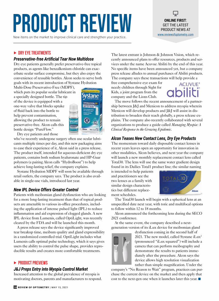
REVIEW OF OPTOMETRY | MAY 15, 2021104
dry eye treatmentsPreservative-free Artificial Tear Now MultidoseDry eye patients generally prefer preservative-free topical products, as agents like benzalkonium chloride can exac-erbate ocular surface compromise, but they also enjoy the convenience of reusable bottles. Alcon seeks to serve both goals with its recent introduction of Systane Hydration Multi-Dose Preservative-Free (MDPF), which puts its popular ocular lubricant in a specially designed bottle. The tip of the device is equipped with a one-way valve that blocks uptake of fluid back into the bottle to help prevent contamination, allowing the product to remain preservative-free. Alcon calls this bottle design “PureFlow.”
Dry eye patients and those who’ve recently undergone surgery often use ocular lubri-cants multiple times per day, and this new packaging aims to ease their experience of it, Alcon said in a press release. The product itself, intended for aqueous-deficient dry eye patients, contains both sodium hyaluronate and HP-Guar polymers (a pairing Alcon calls “HydroBoost”) to help achieve long-lasting relief, the company explains.
Systane Hydration MDPF will soon be available through retail outlets, the company says. The product is also avail-able in single-use vials, introduced last year.
New IPL Device Offers Greater ControlPatients with meibomian gland dysfunction who are looking for a more long-lasting treatment than that of topical prod-ucts are amenable to various in-office procedures, includ-ing the application of intense pulsed light (IPL) to reduce inflammation and aid expression of clogged glands. A new IPL device from Lumenis, called OptiLight, was recently cleared by the FDA and will be launched this month.
A press release says the device significantly improved tear breakup time, meibum quality and gland expressibility in a randomized controlled trial. OptiLight includes what Lumenis calls optimal pulse technology, which it says gives users the ability to control the pulse shape, provides repro-ducible results and creates more comfortable treatments.
product previewsJ&J Preps Entry into Myopia Control MarketIncreased attention to the global prevalence of myopia is motivating doctors, parents and manufacturers to respond.
The latest entrant is Johnson & Johnson Vision, which re-cently announced plans to offer resources, products and ser-vices under the name Acuvue Abiliti by the end of this year. No specific items have been announced yet, but a company press release alludes to annual purchases of Abiliti products. The company says these transactions will help provide a free comprehensive eye exam for needy children through Sight for Kids, a joint program from the company and the Lions Club.
The move follows the recent announcement of a partner-ship between J&J and Menicon to address myopia wherein Menicon will develop products and J&J will assist in dis-tribution to broaden their reach globally, a press release ex-plains. The company also recently collaborated with several organizations to produce a guide called Managing Myopia: A Clinical Response to the Growing Epidemic.
Alcon Teases New Contact Lens, Dry Eye ProductsThe momentum toward daily disposable contact lenses in recent years leaves open an opportunity for innovation in other modalities, Alcon believes, and next year the company will launch a new monthly replacement contact lens called Total30. The lens will use the same water gradient design found in its Dailies Total1 product line; the similar naming is intended to help patients and practitioners see the two lenses as a family with similar design characteris-tics but different replace-ment schedules.
The Total30 launch will begin with a spherical lens at an unspecified date next year, with toric and multifocal options to follow within 12 to 18 months.
Alcon announced the forthcoming lens during the SECO 2021 conference.
At the same event, the company described a next-generation version of its iLux device for meibomian gland
dysfunction coming in the second half of 2021. The new model, called Systane iLux2 (pronounced “iLux squared”) will include a camera that can perform meibography and demonstrate the results to patients imme-diately after the procedure. Alcon says the device allows high resolution visualization rather than simple magnification. Under the
company’s “No Reason to Wait” program, practices can pur-chase the current device on the market and then apply that cost to the next-gen one when it launches later this year. g
product review ONLINE FIRST: GET THE LATEST
PRODUCT NEWS AT www.reviewofoptometry.com
New items on the market to improve clinical care and strengthen your practice.

MAY 15, 2021 | REVIEW OF OPTOMETRY 105
Advancing Eyecare ..............................................................................75........................................................................................................................(904) 997-4131...................................................................................................................www.marco.com
Akorn Pharmaceuticals .......................................................................27.............................................................................................www.MyAkornEyeCare.com
Alcon Laboratories ............................................................................ 108.......................................................................................................................(800) 451-3937.....................................................................................................................www.alcon.com
Alcon Laboratories Inc. ........................................................................45.......................................................................................................................(800) 451-3937
Allergan .................................................................................... Cover Tip...............................................................................................................www.allergan.com
Bausch + Lomb .................................................................................... 21.........................................................................................................................(800) 871-1103................................................................................................................. www.alaway.com
Bruder Ophthalmic Products ...............................................................55.......................................................................................................................(888) 827-8337........................................................................................eyes@bruderophthalmic.com...........................................................................................www.bruderophthalmic.com
Coburn Technologies ...........................................................................47.......................................................................................................................(800) 262-8761.........................................................................................www.coburntechnlogies.com
CooperVision .......................................................................................25.......................................................................................................................(800) 341-2020..................................................................................................... www.coopervision.com
CooperVision ..................................................................................... 107.......................................................................................................................(800) 341-2020..................................................................................................... www.coopervision.com
Eye Designs ......................................................................................... 31..................................................................................................................... (800) 346-8890........................................................................................................ www.eyedesigns.com
Eyevance ...................................................................................... 11 & 12.........................................................................................................................(817) 677-6120.........................................................................................................www.MyZerviate.com
Eyevance ......................................................................................41 & 42.........................................................................................................................(817) 677-6120...................................................................................................www.mytobradexst.com
Eyevance ..................................................................................... 65 & 66.........................................................................................................................(817) 677-6120............................................................................................................www.eyevance.com
Haag-Streit USA ................................................................................... 17........................................................................................................................(866) 769-1197...............................................................................................www.xomasmartlane.com
Icare USA .............................................................................................29......................................................................................................................(888) 389-4022............................................................................................................ www.icare-usa.com
Imprimis Pharmaceuticals, Inc. ........................................................... 19..................................................................................................................... (858) 704-4040......................................................................................................... www.imprimisrx.com
Johnson & Johnson .............................................................................. 14........................................................................................................www.jnjvisionpro.com
Keeler Instruments ...............................................................................9......................................................................................................................(800) 523-5620........................................................................................................... www.keelerusa.com
Lacrimedics, Inc. ................................................................................. 13.......................................................................................................................(800) 367-8327...................................................................................................... info@lacrimedicsl.com.......................................................................................................www.lacrimedicsl.com
MacuLogix ...........................................................................................23.......................................................................................................................(888) 392-6801.......................................................................................................... www.maculogix.com
Oyster Point Pharma ..............................................................................7.........................................................................................................www.dryeyeland.com
Regeneron Pharmaceuticals, Inc. ..........................................36 , 37 & 38....................................................................................................................... (914) 847-7000.......................................................................................................... www.regeneron.com
Reichert Technologies .........................................................................69......................................................................................................................(888) 849-8955........................................................ www.passionateabouteyecare.co /glaucoma
RVL Pharmaceuticals, Inc. ...........................................................2 , 3 & 4......................................................................................................................(866) 600-4799................................................................................................................ www.Upneeq.com
Sun Pharma .................................................................................59 & 60............................................................................................................www.GetCequa.com
Topcon ........................................................................................48 & 49...........................................................................................www.topconhealthcare.com
US Ophthalmic .....................................................................................53....................................................................................................www.usophthalmic.com
X-cel Specialty ....................................................................................33........................................................................................................................(800) 241-9312.........................................................................xcelinfo@xcelspecialtycontacts.com..................................................................................www.xcelspecialtycontacts.com
X-cel Specialty ....................................................................................73........................................................................................................................(800) 241-9312.........................................................................xcelinfo@xcelspecialtycontacts.com..................................................................................www.xcelspecialtycontacts.com
This index is published as a convenience and not as part of the advertising contract. Every care will be taken to index correctly. No allowance will be made for errors due to spelling, incorrect page number, or failure to insert.
ADVERTISER INDEX
REVIEW OF OPHTHALMOLOGY | MAY 202170
DISCLOSURES
fail, surgical options include shrink-age of redundant tissue, removal of redundant tissue and increased adhesion between conjunctiva and sclera.
1. Elschnig A. Beitrag zur aetiologie und therapie der chronischen konjunktivitis. Dtsch Med Wochenschr 1908;34:1133-1155.2. Hughes WL. Conjunctivochalasis. Am J Ophthalmol 1942;25:48-51.3. Di Pascuale MA, Espana EM, Kawakita T, Tseng SCG. Clinical characteristics of conjunctivochala-sis with or without aqueous tear deficiency. Br J Ophthalmol 2004;88:388-391.4. Balci O. Clinical characteristics of patients with conjunctivochalasis. Clinical Ophthalmology 2014;8:1655-1660.5. Meller, D, Tseng SC. Conjunctivochalasis: Literature review and possible pathophysiology. Survey of Ophthalmology 1998;43:3:225-232.6. Marmalidou A, Kheirkhah A, Dana R. Conjunctivochalasis: A systematic review. Survey of Ophthalmology 2018;3:554-564.7. Mimura T, Yamagami S, Usui T, et al. Changes of conjunctivochalasis with age in a hospital-based study. Am J Ophthalmol 2009;147:1:171-77.8. Mimura T, Mori M, Obata H, et al. Conjunctivochalasis: Associations with pinguecula in a hospital-based study. Acta Ophthalmol 90:8:773-82.9. Tong L, Lan W, Sim HS. Conjunctivochalasis is the precursor to pterygium. Med Hypotheses 2013;81:5:927-30.10. Watanabe A, Yokoi N, Kinoshita S, Hino Y,
Tsuchihashi Y. Clinicopathologic study of conjunc-tivochalasis. Cornea 2004;23:3:294-298.11. Francis IC, Chan DG, Kim P, Wilcsek G, Filipic M, Yong J, Coroneo MT. Case-controlled clinical and histopathologic study of conjunctivochalasis. Br J Ophthalmol 2005;89:302-305. 12. Tong L, Lan W, Sim HS, Hou A. Conjunctivochalasis is the precursor to pterygium. Medical Hypotheses 2013;81:927-930.13. Li D, Meller D, Liu Y, Tseng SCG. Overexpression of MMP-1 and MMP-3 by cultured conjunctivochala-sis fibroblasts. IOVS 2000;41:2:404-410.14. Acera A, Vecino E, Duran JA. Tear MMP-9 levels as a marker of ocular surface inflammation in con-junctivochalasis. IOVs 2013;54:13:8285-8291.15. Guo P, Xhang S, He H, Zhu Y, Tseng SCG. TSG-6 controls transcription and activation of matrix metalloproteinase 1 in conjunctivochalasis. IOVS 2012;53:3:1372-1380.16. Ward SK, Wakamatsu TH, Dogru M, Ibrahim OMA, Kaido M, Ogawa Y, Matsumoto Y, Igarashi A, Ishida R, Shimazaki J, Schnider C, Negishi K, Katakami C, Tsubota K. The role of oxidative stress and inflam-mation in conjunctivochalasis. IOVS 2010;51:4:1994-2002.17. Mamalidou A, Palioura A, Dana R, Kheirkhah A. Medical and surgical management of conjunctivocha-lasis. The Ocular Surface 2019;17:393-399. 18. Haefliger IO, Vysniauskiene I, Figueiredo AR, Piffaretti JM. Superficial conjunctiva cauterization to reduce moderate conjunctivochalasis. Klin Monatsbl Augenheilkd 2007;224:237-239.19. Trivle A, Dalianias G, Terzidou C. A quick surgical treatment of conjunctivochalasis using radiofrequen-cies. Healthcare 2018;6:14:1-5.20. Petris CK, Holds JB. Medial conjunctival resec-tion for tearing associated with conjunctivochalasis.
Ophthalmic Plast Reconstr Surg 2013;29:304–7. 21. Liu D. Conjunctivochalasis: A cause of tear-ing and its management. Ophthalmic Plastic and Reconstructive Surgery 1986;21:25-28. 22. Brodbaker E, Bahar I, Slomovic AR. Novel use of fibrin glue in the treatment of conjunctivochalasis. Cornea 2008;27:950-2. 23. Georgiadia NS, Terzidou CD. Epiphora caused by conjunctivochalasis: Treatment with transplantation of preserved human amniotic membrane. Cornea 2001;20:6:619-621.24. Meller D, Maskin SL, Pired RTF, Tseng SCG. Amniotic membrane transplantation for symptomatic conjunctivochalasis refractory to medical treatments. Cornea 2000;19:6:796-803.25. Nakasato H, Uemoto R, Meguro A, et al. Treatment of symptomatic inferior conjunctivochalasis by liga-tion. Acta Ophthalmol 2014;92:e411-2. 26. Otaka I, Kyu N. A new surgical technique for management of conjunctivochalasis. Brief Reports 1999;129:3:385-387.
CORNEA/ANTERIOR SEGMENT | Conjunctivochalasis Surgical Techniques
Samantha Marek, MD, is a resident at the University of Pennsylvania Department of Ophthalmology. Stephen Orlin, MD, is the director of the Cornea Department, chair of the Cornea Service, co-director of the Refractive Surgery Service and an associate professor of ophthalmology at the Penn Presbyterian Medical Center of Philadelphia. They report no related financial disclosures.
REVIEW OF OPHTHALMOLOGY | MARCH 202130
phacoemulsifi cation. Int Ophthalmol. 2021;41:1:273-282.2. Mencucci R, Cennamo M, Venturi D, et al. Visual out-come, optical quality, and patient satisfaction with a new monofocal IOL, enhanced for intermediate vision: Prelimi-nary results. J Cataract Refract Surg. 2020;46:3:378-387.3. Eguileor B, Martinez-Indart L, Alday N, et al. Differences in intermediate vision: Monofocal intraocular lenses vs. monofocal extended depth of focus intraocular lenses. Arch Soc Esp Oftalmol. 2020;95:11:523-527.4. Yangzes S, Kamble N, Grewal S, et al. Comparison of an aspheric monofocal intraocular lens with the new generation monofocal lens using defocus curve. Indian J Ophthalmol. 2020;68:12:3025-3029.5. Auffarth GU, Gerl M, Tsai L, et al. Clinical evaluation of a new monofocal intraocular lens with enhanced inter-mediate function in cataract patients. J Cataract Refract Surg 2021;47:2:184-91.
Bright Light May Help Glaucoma Patients SleepGlaucoma patients who are exposed to bright light during the day may have improved sleep and an increased melanopsin-dependent pupil re-sponse after just four weeks, a pilot
study in Frontiers in Neurology reports.1
The multinational research team composed of researchers from Swit-zerland, the United Kingdom and New Zealand suggests the change in pupil response might indicate that melanopsin activity in viable intrinsi-cally photosensitive retinal ganglion cells can adapt to different light lev-els, if sustained over a period of time.
Their study enrolled 20 glaucoma patients without severe vision loss and considered how 30 minutes of daily bright light exposure from a table-based light box placed in their homes affected pupil constriction, circadian rest-activity cycles, sleep, relaxation, alertness and mood.
Participants continuously wore an activity monitor and self-assessed sleep quality, well-being and visual comfort for several days before and during the four weeks of daily 10,000 lux polychromatic bright white light exposure.
Individuals underwent pupillom-etry at baseline and on the last day of
the study.Following light exposure, partici-
pants showed a much greater post-illumination pupil response and had better quality of sleep. Additionally, researchers report that they found no signifi cant changes in the participants’ 24-hour rhythms or sleep parameters.
The study demonstrated that even a relatively short duration of added light exposure in a room is benefi cial and also supports the general advice that the elderly should go outside for half an hour each morning, the researchers suggest.
While glaucoma patients can never recover the vision lost from damaged retinal ganglion cells, they may be able to maintain a robust day–night cycle and concomitant good circadian entrainment which helps maintain high sleep quality, the investigators explain.
1. Kawasaki A, Udry M, Wardani ME, Münch M. Can extra daytime light exposure improve well-being and sleep? A pilot study of patients with glaucoma. Front Neurol 2020;11:584479.
Eyhance Intraocular Lenses Approved by FDA (Continued from p. 16)
Review newsReview news
The Rick Bay Foundationfor Excellence in Eyecare Education
Support the Education of Future Healthcare & Eyecare Professionals
Scholarships are awarded to advance the education of students in both Optometry and Ophthalmology, and are chosen by their school based on
qualities that embody Rick’s commitment to the profession, including integrity, compassion, partnership and dedication to the greater good.
Interested in being a partner with us?Visit www.rickbayfoundation.org
(Contributions are tax-deductible in accordance with section 170 of the Internal Revenue Code.)
(The Rick Bay Foundation for Excellence in Eyecare Education is a nonprofi t, tax-exempt organization under section 501(c)(3) of the Internal Revenue Code.)

REVIEW OF OPTOMETRY | MAY 15, 2021106
A55-year-old man reported tothe office emergently with achief complaint of blurry vi-sion at distance in both eyes.
He explained in a panic that he hadwoken up early that morning seeingwell, then played a game on his cellphone and noticed that, after hestopped, he no longer needed hisreading glasses to read.
He explained further that every-thing in the distance was blurry. Thepatient was properly medicated forhypertension and high cholesteroland denied trauma or allergies of anykind.
Diagnostic DataHis best-corrected entering visualacuities were 20/70 at distance and
20/20 at near with a working distanceof 10 inches. There was substantialimprovement upon the pinhole atdistance. External examination wasnormal for color, brightness andextraocular motilities. Confrontationfields were normal and facial Amslergrid was normal with no afferentpupillary defect.
Refraction uncovered -0.75Dsphere, resulting in 20/20 vision atdistance. Biomicroscopy showednormal anterior segment structures,and Goldmann applanation tonom-etry recorded pressures of 16mm Hgin each eye. Dilated exam uncoverednormal nerves with no evidence ofmaculopathy or disc edema.
Additional TestingThe first important additional testis lensometry. This will permit anunderstanding of the previous refrac-tive error. It can then be matchedagainst the refraction to understandhow things have changed. A detailedhistory should include whether vi-sion is absent, distorted or blurry, andin which eye and when.
Your DiagnosisWhat would be your diagnosis inthis case? What is the patient’s likelyprognosis? To find out, please readthe online version of this article atwww.reviewofoptometry.com. g
An anxious patient presents complaining of recent-onset blurred vision at distance. Are his fears warranted?
Game Over
By Andrew S. Gurwood, OD
diagnostic quiz
Next Month in the MagIn June, we present our annual retina report. Articles will include:
• The Retina Research Pipeline: Upcoming Advances to Look Forward to
• How to Handle Central Serous Chorioretinopathy
• Be Ready for Acute Retinal Ischemia
• Comanagement Series: Be a Retina Referral Rock Star
• Assessing Macular Disorders and Dystrophies
• Optometric Retina Society Annual Larry Alexander Case Report Contest Winner
Also included in June:
• PAL Primer: How to Use New and Specialty Lens Designs
The patient’s description of events: iPhone video play, followed by ability to read without reading glasses but distance vision blur.
Retina Quiz Answers (from page 94)—Q1: d, Q2: a, Q3: b, Q4: c, Q5: b
Dr. Gurwood is a professor of clinical sciences at The Eye Institute of the Pennsylvania College of Optometry at Salus University. He is a co-chief of Primary Care Suite 3. He is attending medical staff in the department of ophthalmology at Albert Einstein Medical Center, Philadelphia. He has no financial interests to disclose.
About Dr. Gurwood

Certainly. Totally.Mhmm.
Say “Yes” to 99.9%1of your astigmats.The most prescribed2and complete toric portfolio is growing.
1.CooperVision data on file 2021. Rx coverage database; 14 to 70 years. 2.CooperVision data on file, 2017-2019. Based on number of US softcontact lens fits. Includes CooperVision branded and customer-branded equivalent lenses. US industry reports and internal estimates. 3. Around theclock axis in 10° from Plano to -6.00DS in -0.75DC, 1.25DC and -1.75DC. 4. CVI data on file 2021. Based on number of prescription options available in the USA across all soft 1-day toric lenses as reported by the 4 main manufacturers and the SKU expansion for MyDay® toric (2H’21).5. CooperVision data on file 2020. Kubic masked online survey; n=404 US ODs who prescribe toric soft CLs.
©2021 CooperVision 11006 04/21
Biofinity® XR toric MyDay® toric
NEW SKUSNOW AVAILABLE
Biofinit ® XR i
NNNOO
Sphere powers expanded to
±20.00D, bringing the total
number of unique prescription
options to nearly 33,000.
Adding around-the-clock axes3and
0.25D steps in plus powers, giving
MyDay® toric the most prescription
options of any 1-day lens4.
Learn more about how CooperVision® torics fit virtually any astigmat5.
Visit coopervision.com/toric
Now more than ever, CooperVision® fits your practice perfectly.
NEW SKUSCOMING JULY 2021

Recommend long-lasting, preservative-free dry eye relief1*
Introducing HydroBoost Technology
An exclusive combination that helps retain the lubricants, for relief to
patients who need it most1
The Relief Is Real®
*Vs SYSTANE ULTRA Lubricant Eye Drops.References: 1. Mrukwa-Kominek E, Baranska K, Jadczyk K. First clinical reports on the application of the modern dual-polymer formula in aqueous defi ciency dry eye syndrome - Polish observations. Presented at the 20th European Society of Cataract & Refractive Surgery Winter Meeting; February 26-28, 2016; Athens, Greece. 2. Davitt WF, Bloomenstein M, Christensen M, Martin AE. E� cacy in patients with dry eye after treatment with a new lubricant eye drop formulation. J Ocul Pharmacol Ther. 2010;26:347-353. 3. Rah MJ. A review of hyaluronan and its ophthalmic applications. Optometry. 2011;82:38-43.
© 2020 Alcon Inc. 09/20 US-SYH-2000026
HP-GUARForms a cross-linked meshwork
on the surface of the eye to hold lubricants in place2
1
SODIUM HYALURONATENaturally occurring moisture substance found in tears that helps the lubricants to provide longer-lasting hydration1,3*
2