ro1213i.pdf - Review of Optometry
88
Office Design Contest: Putting Art to Practice, p. 29 • Evolving Therapies for Macular Edema: Options Abound, p. 52 • Diabetic Eye Disease—Lessons from a Telemedicine Reader, p. 46 15th Annual Diabetes Report www.revoptom.com December 15, 2013 www.revoptom.com Can We Influence the Course of Diabetes? Moving the Needle: Evidence suggests nutritional supplements really can make a difference in diabetic retinopathy. Page 36
-
Upload
khangminh22 -
Category
Documents
-
view
6 -
download
0
Transcript of ro1213i.pdf - Review of Optometry

Office Design Contest: Putting Art to Practice, p. 29
RE
VIE
W O
F O
PT
OM
ET
RY
■ V
OL
. 15
0 N
O. 1
2 ■
DE
CE
MB
ER
15
, 20
13
■ N
UT
RIT
ION
& D
IAB
ET
ES ■
DM
E T
HE
RA
PIE
S ■ T
EL
EM
ED
ICIN
E ■
OF
FIC
E D
ESIG
N
• Evolving Therapies for Macular Edema: Options Abound, p. 52
• Diabetic Eye Disease—Lessons from a Telemedicine Reader, p. 46
15th Annual Diabetes Reportwww.revoptom.com
December 15, 2013
www.revoptom.com
Can We Infl uence theCourse of Diabetes?
Moving the Needle:
Evidence suggests nutritional supplements really can make a
difference in diabetic retinopathy. Page 36
fc_ro1213_final.indd 1 11/27/13 10:52 AM

For more information visit acuvuepro.com
HEALTH YOU CAN SEE™
RO1213_Vistakon TruEye.indd 2 11/20/13 10:35 AM

Well beyond comfortable...The only lens shown to be clinically comparable to wearing
NO LENS AT ALLON 5 OF 6 KEY MEASURES OF OCULAR HEALTH*†1:
• Limbal hyperemia • Corneal staining • Corneal vascularization
• Bulbar conjunctival hyperemia
• Papillary conjunctivitis
* A 1-year, randomized, investigator-masked, parallel-group study of 74 subjects (aged 18-51 years) who had never worn soft contact lenses previously. Subjects were randomized to 1-DAY ACUVUE® TruEye® Brand or spectacles. Ocular physiology was assessed at 2 weeks, 1 month, 3 months, 6 months, 9 months, and 1 year using the Efron Grading Scale (a 0- to 4-point scale, with fi ndings recorded to the nearest 0.1). P<0.05 based on 2-sided 95% confi dence intervals.
† The sixth measure was conjunctival staining. ACUVUE® Brand Contact Lenses are indicated for vision correction. As with any contact lens, eye problems, including corneal ulcers, can develop. Some wearers may experience mild irritation, itching or discomfort. Lenses should not be prescribed if patients have any eye infection, or experience eye discomfort, excessive tearing, vision changes, redness or other eye problems. Consult the package insert for complete information. Complete information is also available from VISTAKON® Division of Johnson & Johnson Vision Care, Inc., by calling 1-800-843-2020 or by visiting acuvueprofessional.com.
Reference: 1. Morgan P, Chamberlain P, Moody K, et al. Ocular physiology and comfort in neophyte subjects fi tted with daily disposable silicone hydrogel contact lenses. Cont Lens Anterior Eye. 2013;36(3):118-125.
ACUVUE®, 1-DAY ACUVUE® TruEye®, HEALTH YOU CAN SEE™, and VISTAKON® are trademarks of Johnson & Johnson Vision Care, Inc.
© Johnson & Johnson Vision Care, Inc. 2013 ACU-32440G July 2013
Go beyond ocular comfort and help maintain ocular health.
The sixth measure was conjunctival staining which diff ered between the lens wearers and spectacle wearers; however, both measures were very low and of limited clinical relevance.
RO1213_Vistakon TruEye.indd 3 11/20/13 10:35 AM

N e w s R e v i e w
4 REVIEW OF OPTOMETRY DECEMBER 15, 2013
Baby Photos Can Detect Retinoblastoma Early
Baby pictures are perhaps the best method for early detec-tion of retinoblastoma, a new
study found. “In a majority of retinoblastoma
cases, it is the parents that initi-ate the diagnosis based on seeing leukocoria or ‘white eye’ in photos of their children,” says the study’s lead author Bryan F. Shaw, PhD, who is not an eye researcher but an assistant professor of chemistry and biochemistry at Baylor College.
Dr. Shaw initiated the study after his own son was diagnosed and treated for retinoblastoma.
Researchers already knew that children with retinoblastoma often display persistent leukocoria in photographs. Despite this, digital photography hasn’t been intention-ally used to screen for retinoblasto-ma because “white eye” is assumed to be a symptom of advanced reti-noblastoma, not early stage retino-blastoma.
Dr. Shaw’s study dispelled this
assumption. It found that leukoco-ria can be a sign of retinoblastoma in its earliest stages—even in an infant as young as 12 days old, as in the case of Dr. Shaw’s son. Early detection and treatment would increase the chances of survival and reduce loss of vision.
The study, published in the online journal PLoS One, also determined that the brightness and the color saturation of the leukocoria eye can indicate the retinoblastoma’s sever-ity. The brighter the white eye, the larger the tumor, Dr. Shaw says.
The researchers’ next step: “If we can create software that can detect leukocoria and alert a parent when it begins to occur persistently, then I believe digital photography can eradicate metastatic retinoblastoma from this world and prevent most of the deaths that occur,” Dr. Shaw says. Abdolvahabi A, Taylor BW, Holden RL, et al. Colorimetric and longitudinal analysis of leukocoria in recreational photo-graphs of children with retinoblastoma. PLoS One. 2013 Oct 30;8(10):e76677.
IN THE NEWS
Optometry mourns the loss of Paul E. Berman, OD, a sports vision special-ist and the founder and global clinical director of the Special Olympics’ Lions Club International Opening Eyes and Healthy Athletes programs. He was also the team optometrist for the New Jersey Devils hockey team. Dr. Berman unexpectedly passed away on Novem-ber 10, 2013, at age 62.
Cataract surgery is highly cost effec-tive, according to a cost-utility study published in Ophthalmology. Cataract surgeries performed over one year eventually save $123.4 billion over 13 years in direct health care costs and lost productivity, and deliver a 4,567% return on investment to society. The study also found that the overall cost of cataract surgery in 2012 was 34.4% less than in 2000 and 85% less than in 1985, after adjusting for infl ation.
The American Board of Optometry announced its newly elected offi cers: James M. Vaught, OD as chairman; David A. Heath, OD, EdM, as vice-chairman; Chelsea L. Miller, OD, as secretary; and John P. McGuire, CPA, as treasurer. Also, Jackson Lau, OD, was elected as the new director represent-ing the AOA, and Michael D. Gerstner, OD, was elected as a new member-at-large.
There is no association between AMD and Alzheimer’s disease or dementia, contrary to prior research indicating a connection, a new study suggests. Pub-lished online in JAMA Ophthalmology, the study reported that the genetic risk factors for the two types of diseases show no evidence of linkage.
V O L . 1 5 0 N O . 1 2 ■ D E C E M B E R 1 5 , 2 0 1 3
This study also found that a brighter ‘white eye’ signals a larger tumor. By John Murphy, Executive Editor
Leukocoria or “white eye” has been assumed to be a sign of advanced retinoblastoma. But a new study shows that it can appear in infants as young as a few days old.
Photos: Brian F. Shaw, PhD, Baylor University
004_ro1213_news.indd 4 11/27/13 2:14 PM

RO1213_Freedom.indd 1 11/25/13 1:26 PM

6 REVIEW OF OPTOMETRY DECEMBER 15, 2013
News Review
Women who have taken oral contraceptives may be twice as likely to be
diagnosed with glaucoma. But whether contraceptives actually cause glaucoma remains unclear.
Researchers with the University of California, San Diego, Duke University School of Medicine and Third Affiliated Hospital of Nanchang University in Nan-chang, China, gleaned three-year data from the Center for Disease Control’s National Health and Nutrition Examination Survey (NHANES) and found that women age 40 and older who had used oral contraceptives for three years or longer are twice as likely to be
diagnosed with glaucoma. The findings were presented at the annual meeting of the American Academy of Ophthalmology in November.
“The message is that long-term use of oral contraceptives may be considered an additional risk factor associated with increased incidence of primary open-angle glaucoma in women,” says Sherry Bass, OD, of SUNY College of Optometry.
Dr. Bass notes that the current study reflects similar results from a 2011 Nurses’ Health Study, which found a 25% higher glaucoma risk among women who used birth con-trol pills.
However, more study is needed,
“since the information is prelimi-nary at best,” says Kathy Yang-Wil-liams, OD, who practices in Seattle with an emphasis on glaucoma.
“This study shows an associa-tive, but not necessarily causal, relationship between oral contra-ceptives and glaucoma,” she says. “These findings should not affect ODs in their practice with regards to the diagnosis of glaucoma. If a patient takes oral contraceptives, then this should be noted as part of the review of systems and this fac-tor added to the basic risk profile.”
The study’s researchers say they hope it will serve as an impetus for further research to prove potential causative effects.
Does ‘the Pill’ Cause Glaucoma?
Infants born extremely prema-turely are up to 19 times more likely to experience a retinal
detachment later in life than babies who are carried to full term, ac-cording to a study in the November issue of Ophthalmology.
A Swedish research team evaluat-ed more than three million individu-als born between 1973 and 2008. The subjects were separated into two groups: those born in 1973 to 1986 (before the mandate of Swe-den’s national screening program for retinopathy of prematurity), and those born in 1987 to 2008.
Within these groups, the researchers further classified sub-jects born at less than 28 weeks ges-tation as “extremely premature,” those born between 28 and 31 weeks gestation as “very prema-
ture” and those born between 32 and 36 weeks gestation as “moder-ately preterm.”
The researchers determined that extremely premature infants born from 1973 to 1986 were 19 times more likely to experience a retinal detachment during childhood, ado-lescence or young adult life than their full-term peers. Additionally, those born between 1987 and 2008 were nine times more likely to expe-rience a retinal detachment.
The overall risk of retinal detach-ment in very premature infants fell significantly between the two groups—four times more likely for those in the older group (born 1973 to 1986) compared with three times more likely for those in the younger group (born 1987 to 2008). Inter-estingly, the researchers noted that
moderately preterm infants in both groups were no more likely to expe-rience retinal detachment than full-term infants.
“We may just be seeing the tip of the iceberg of late ophthalmic com-plications after preterm birth,” says lead author Anna-Karin Edstedt Bonamy, MD, PhD, pediatrician at Karolinska Institutet in Stockholm. “Not only does the risk of retinal detachment increase with age, but there has also been an increase in survival among people born prematurely since the 1970s. This provides opportunities for future research to address if the increased risk persists among those born pre-maturely as they age.” Bonamy AK, Holmström G, Stephansson O, et al. Preterm birth and later retinal detachment: a population-based cohort study of more than 3 million children and young adults. Ophthalmology. 2013 Nov;120(11):2278-85.
Premature Birth Linked to Higher Chance of Retinal Detachment
004_ro1213_news.indd 6 11/27/13 2:15 PM

Hassle-free returns! No return of lenses for exchange or credit
Satisfaction guaranteed! All lenses carry a 90-day warranty
Easy to get started! Fit Duette lenses empirically…no diagnostic sets or fluorescein needed
Introducing the New SynergEyes®: the leading global contact lens company for the independent eye care professional.
Call Customer Care to place your order: 877.733.2012, option 1.
To Learn More, visit www.SynergEyes.com/NewSynergEyes
“We are determined to become a company that is 100% ECP-centric by delivering products and
services that will provide foundational value to your practice.”James K. Kirchner, O.D.; President & CEO
Vision Correction
Irregular Cornea
Exciting changes make it easy to do
business with SynergEyes!
A Two Division Company Delivering Exceptional Vision!
RO1113_Synergeyes.indd 1 10/22/13 11:16 AM

8 REVIEW OF OPTOMETRY DECEMBER 15, 2013
News Review
Anew study has found the earliest sign of au-tism ever observed—a
steady decline in eye contact within a child’s first months of life.
Autism is usually diagnosed after age two, when delays in a child’s social behavior and language skills become appar-ent. This new study found that babies as young as two to six months old can start to show a decrease in eye con-tact—a clear sign of autism.
This research, published online in Nature, also sug-gests that there’s a window of opportunity to possibly pre-vent some disabilities associ-ated with the disorder.
Lack of eye contact has been a hallmark diagnostic sign of autism since the con-dition was initially identified. But no one knew how early this deficit began. To find out, researchers at the Marcus Autism Center in Atlanta used eye-tracking equip-ment to measure eye movements in babies who watched video scenes of a caregiver.
Followed from birth to three years, the infants were divided into two groups based on their risk for developing an autism spectrum disorder. Those in the high-risk group had an older sibling already diagnosed with autism; the low-risk group did not. The researchers cal-culated the percentage of time each child fixated on the caregiver’s eyes, mouth and body, as well as the non-human spaces in the images.
By age three, nearly all the chil-dren in the high-risk group had received a clinical diagnosis of an autism spectrum disorder. The
researchers then reviewed the eye-tracking data to determine which factors differed between children who received an autism diagnosis and those who did not.
“In infants later diagnosed with autism, we see a steady decline in how much they look at mom’s eyes,” says Warren Jones, PhD, lead author of the study.
This drop in eye fixation began between two and six months and continued throughout the course of the study. By 24 months, the chil-dren who were later diagnosed with autism focused on the caregiver’s eyes only about half as long as did their typically developing counter-parts. Also, those infants whose lev-els of eye contact diminished most rapidly were those who were most disabled later in life.
Importantly, these results suggest that social engagement skills are
intact after birth in children with autism. This finding challenges a long-standing theory—that social behaviors are entirely absent in children with autism. In other words, the observation of a decline in eye fixation—rather than an outright absence—offers a promising opportunity for early intervention.
“This insight—the preserva-tion of some early eye-look-ing—is important,” Dr. Jones says. “In the future, if we were able to use similar technolo-gies to identify early signs of social disability, we could then consider interventions to build on that early eye-looking and help reduce some of the asso-ciated disabilities that often accompany autism.”
In the meantime, the inves-tigators’ next step is to translate this research into a viable tool for use in the clinic.
“Most kids on the autism spec-trum are not identified until later, so looking early is helpful,” says Glen T. Steele, OD, professor of pediatric optometry at Southern College of Optometry and chair of the AOA’s InfantSEE Committee.
Optometrists who examine babies and very young children aren’t expected to make the diagno-sis of autism, Dr. Steele says. “But we might be able to notice these issues much earlier in life, and a caution flag should go up.” If you suspect autism, refer the child to a developmental specialist who regularly works with children with special needs, he says.
Jones W, Klin A. Attention to eyes is present but in decline in 2-6-month-old infants later diagnosed with autism. Nature. 2013 Nov 6. [Epub ahead of print.]
Lack of Eye Contact Signals Autism
Using eye-tracking technology, researchers found that infants later diagnosed with autism showed a decline in attention to others’ eyes by two to six months of age.
Photo: Kay Hinton, Emory University
004_ro1213_news.indd 8 11/27/13 2:15 PM

www.eyepromise.com | [email protected]
*These statements have not been evaluated by the FDA. This product is not intended to diagnose, treat, cure, or prevent any disease.
highest quality
EyePromise® DVS Nutritional Support for Retinal Health
EyePromise DVS is designed to support healthy blood vessels – essential in preserving healthy vision – through a unique combination of ingredients with documented effects for eye health. EyePromise DVS offers the most comprehensive, science-based retinal health formula designed to:*
Support retinal metabolism, structure, and function
Promote blood vessel health
Combat oxidative stress
Increase Macular Pigment Optical Density (MPOD)
Download the free education guide and receive a special offer!
Visit eyepromise.com/odbooklet to download, “Nutritional Support for Diabetes and Visual Health,” and receive a special limited-time offer email on EyePromise DVS.
RO1213_Zeavision.indd 1 11/20/13 10:29 AM

10 REVIEW OF OPTOMETRY DECEMBER 15, 2013
News Review
BUSINESS OFFICES11 CAMPUS BLVD., SUITE 100NEWTOWN SQUARE, PA 19073
CEO, INFORMATION SERVICES GROUP MARC FERRARA
(212) 274-7062 • [email protected]
SALES MANAGER, NORTHEAST, OHIOJAMES HENNE
(610) 492-1017 • [email protected]
SALES MANAGER, SOUTHEAST, WESTMICHELE BARRETT
(610) 492-1014 • [email protected]
VICE PRESIDENT, OPERATIONS CASEY FOSTER
(610) 492-1007 • [email protected]
VICE PRESIDENT, CLINICAL CONTENT PAUL M. KARPECKI, OD, FAAO
EDUCATION/CONFERENCE MANAGERMEG MCDONALD
(610) 492-1045 • [email protected]
PRODUCTION MANAGERSCOTT TOBIN
(610) 492-1011 • [email protected]
SENIOR CIRCULATION MANAGERANTHONY GUADAGNINO
(212) 219-7870 • [email protected]
CLASSIFIED ADVERTISING(888) 498-1460
SUBSCRIPTIONS$56 A YEAR, $88 (US) IN CANADA,
$209 (US) IN ALL OTHER COUNTRIES.
SUBSCRIPTION INQUIRIES(877) 529-1746 (US ONLY);
OUTSIDE US, CALL (847) 763-9630
CIRCULATIONPO BOX 2025
SKOKIE, IL 60076TEL: (TOLL FREE) 1-877-529-1746
OUTSIDE US: (847)763-9630FAX: (847)763-9631
CEO, INFORMATION SERVICES GROUP MARC FERRARA
SENIOR VICE PRESIDENT, OPERATIONS JEFF LEVITZ
SENIOR VICE PRESIDENT, HUMAN RESOURCES LORRAINE ORLANDO
VICE PRESIDENT, CREATIVE SERVICES & PRODUCTION MONICA TETTAMANZI
VICE PRESIDENT, CIRCULATION EMELDA BAREA
A small group of patients with Fuchs’ corneal dystrophy were successfully treated
with a Rho-associated kinase (ROCK) inhibitor topical medica-tion—a technique that could have far-reaching clinical implications, according to researchers from Japan who presented their findings at the annual meeting of the American Academy of Ophthalmology in November.
The preliminary study, conducted by investigators at Kyoto Prefectur-al University of Medicine, reported that a one-week treatment of the ROCK inhibitor Y-27632 stimu-lated the proliferation of corneal endothelial cells in four test patients with Fuchs’ corneal dystrophy.
Patients showed corneal healing and restored visual acuity, as well as reduced corneal thickness from 700 cells/mm2 to 563 cells/mm2 by three months after treatment.
“Overall, it’s intriguing and, if it proves to be viable, may reduce the number of surgeries required for Fuchs’ and other endothelial
diseases,” says Joseph Shovlin, OD, of Scranton, Pa. “ROCK inhibitors would represent the first successful medical treatment using a topical agent for certain forms of endothe-lial disease.”
The technique involves transcor-neal freezing to remove damaged endothelial cells, and then applying the eye drops to promote prolifera-tion in the remaining functional cells. This small study treated eight patients—four with Fuchs’ and four with pseudophakic bullous keratophy. Although the technique was not effective for pseudophakic bullous keratophy, it could pave the way for minimally invasive surgery for Fuchs’ and other forms of cor-neal problems, the researchers say.
While more research must be done before such a topical drop becomes available, “it’s crucial that ODs are aware of such potential non-surgical treatments in order to render the very best care possible, and also field questions that may arise in the course of patient care,” Dr. Shovlin says.
Novel Topical Compound Restores Endothelial Cells
Retinal Thickness May Help Diagnose Alzheimer’sThe loss of cells in the inner nuclear layer of the retina may be a predictor of Alzheimer’s disease, according to fi ndings presented in November at the annual meeting of the Soci-ety for Neuroscience.
A team of researchers from Georgetown University Medical Center and the University of Hong Kong studied the retinal thickness of mice genetically engineered to develop Alzheimer’s and compared them to a group of healthy, age-matched mice.
During the study, the researchers observed a signifi cant loss of thickness in both the retinal ganglion cell layer and the inner nuclear layer of the retina. When compared with control mice, inner nuclear layer in the mice with Alzheimer’s exhibited a 37% loss of neurons and the retinal ganglion cell layer had a 49% loss of neurons.
The researchers say that eye care providers may be able to diagnose or predict Al-zheimer’s by simply examining the thickness of these retinal layers with OCT. Additionally, new Alzheimer’s treatments may even prove useful for future glaucoma management. ■
004_ro1213_news.indd 10 11/27/13 2:15 PM

1-800-756-2020©2011 Three Rivers Optical Inc. www.3riversoptical.com
COMMITMENT
PERSONAL SERVICE
INDEPENDENCE
PRODUCTQUALITYThree Rivers Optical produces
the best lab quality, period.
We know that your reputation
rides on how well we do our
job, and we won’t disappoint
you. Across all lens designs,
materials and designs, we are
dedicated to optimizing the
optics and cosmetics of the
final product that you
dispense to your patient.
And our See-More line of
products gives you the option
of the very best value in each
major lens category.
Does this sound like the
product quality you want?
Call Steve Seibert,
Three Rivers Optical CEO,
for more information.
And stop worrying.
Now is not the time to worry about product quality.
MORE
RO1112_Three Rivers.indd 1 11/8/12 10:38 AM

Know who’s sitting in your chair in less than 1 minute:• Harvest more accurate data in less time• Understand each patient’s full optical pathway• Determine refractions needing minimal correction to achieve 20/20• Save 5-7 minutes per patient• See an additional 3+ patients each day• Give every patient more quality time• Increase optical revenue up to 30+%• Validate Rx changes and increase patient satisfaction• Optimize cataract and refractive outcomes to 20/happy• Enjoy more patient loyaltyNow that’s outstanding return on investment. It’s about time!
XFraction: WaVEFront oPtiMiZED rEFraXion
FractionXX sm
FractionFractionWavefront Optimized RefraXionBecause it’s Time for Better
800.874.5274www.marco.com
*Data based on national averages.
We all know that ‘time is money’, but do you know how to ‘buy more time’?
oPD-Scan iii wavefront system measures >20 diagnostics – and how cleanly light passes through the optical path. in less than 1 minute, Wavefront optimized refraction can discern which patients will/will not require a full refraction to achieve 20/20.
trS-5100 rapidly completes the required refraction with digital speed and accuracy.
you do.you do.
Take
Advantage of the Section 179 Tax Code Deduction
by December 31, 2013 and Save Thousands of Dollars! *
*Please consult your tax advisor.
FractionXXFractionWavefront Optimized RefraFractionWavefront Optimized RefraFractionWavefront Optimized Refra Year-End Savings!
350-XF now-better-RO-CRST-AOC.indd 1 11/8/13 10:04 AM

REVIEW OF OPTOMETRY DECEMBER 15, 2013 13
50 Why Dry Eye TrialsOften Fail
From disease variability to confounding underlying conditions, there are countless reasons why new dry eye drugs have come up short in FDA testing. By Paul M. Karpecki, OD, Co-Chief Clinical Editor
61 A Lifetime ofDry Eye
Dry eye can strike patients of any age. Do you know the subtle signs and symptoms to look for, and the particular treatments to provide, among patients of different ages?By Cheryl G. Murphy, OD, Contributing Editor
ContentsReview of Optometry December 2013
Take an inside look at winning designs that transformed these practices. By Erin Kelly, Senior Associate Editor
Office Design Contest: Putting Art to Practice29
Here’s a primer on the great strides eye care has made in fighting this common cause of visual disability. By Marta C. Fabrykowski, OD
Evolving Therapies for Macular Edema: Options Abound52
A remote reader sees dozens of retinas in a day. Here’s how to put a reader’s knowledge to practical use for your patients. By Erin Giles, OD
Diabetic Eye Disease—Lessons from a Telemedicine Reader46
Evidence suggests nutritional supplements really can make a difference in diabetic retinopathy. By A. Paul Chous, MA, OD
Moving the Needle: Can We Influence the Course of Diabetes?
3615th Annual Diabetes Report
013_ro1213_toc.indd 13 11/27/13 3:06 PM

PRINTED IN U.S.A.
FOUNDING EDITOR
FREDERICK BOGER
1891-1913
EDITORIAL OFFICES
11 CAMPUS BLVD., SUITE 100NEWTOWN SQUARE, PA 19073
EMAIL • [email protected]
WEBSITE • WWW.REVOPTOM.COM
SUBSCRIPTION INQUIRIES
1-877-529-1746CONTINUING EDUCATION INQUIRIES
1-800-825-4696
EDITOR-IN-CHIEF • JACK PERSICO
(610) 492-1006 • [email protected]
EXECUTIVE EDITOR • JOHN MURPHY
(610) 492-1021 • [email protected]
MANAGING EDITOR • MICHAEL HOSTER
(610) 492-1028 • [email protected]
SENIOR ASSOCIATE EDITOR/WEB EDITOR • ERIN KELLY
(610) 492-1005 • [email protected]
ASSOCIATE EDITOR • FRANK AULETTO
(610) 492-1043 • [email protected]
DIRECTOR ART/PRODUCTION • JOE MORRIS
(610) 492-1027 • [email protected]
ART DIRECTOR • JARED ARAUJO
(610) 492-1032 • [email protected]
DIRECTOR OF CE ADMINISTRATION • REGINA COMBS
(212) 274-7160 • [email protected]
SPECIAL PROJECTS • JANE COLE
(610) 492-1043 • [email protected]
EDITORIAL BOARD
CHIEF CLINICAL EDITORS • PAUL M. KARPECKI, OD;CHRISTINE W. SINDT, OD
ASSOCIATE CLINICAL EDITORS • JOSEPH P. SHOVLIN, OD;ALAN G. KABAT, OD
DIRECTOR OPTOMETRIC PROGRAMS • ARTHUR EPSTEIN, ODCLINICAL & EDUCATION CONFERENCE ADVISOR •
PAUL M. KARPECKI, ODCASE REPORTS COORDINATOR • ANDREW S. GURWOOD, OD
CLINICAL CODING EDITOR • JOHN RUMPAKIS, OD, MBACONSULTING EDITOR • FRANK FONTANA, OD
EMERITUS CLINICAL EDITOR • ROBERT M. COLE, III, OD
COLUMNISTS
CHAIRSIDE • MONTGOMERY VICKERS, ODCOMANAGEMENT Q+A • PAUL C. AJAMIAN, OD
CORNEA & CONTACT LENS Q+A • JOSEPH P. SHOVLIN, ODDIAGNOSTIC QUIZ • ANDREW S. GURWOOD, OD
GLAUCOMA GRAND ROUNDS • JAMES L. FANELLI, ODRESEARCH REVIEW • PAUL M. KARPECKI, OD;
DIANA L. SHECHTMAN, ODRETINA QUIZ • MARK T. DUNBAR, OD
REVIEW OF SYSTEMS • CARLO J. PELINO, OD;JOSEPH J. PIZZIMENTI, OD
SURGICAL MINUTE • DEREK N. CUNNINGHAM, OD;WALTER O. WHITLEY, OD, MBA
THERAPEUTIC REVIEW • JOSEPH W. SOWKA, OD; ALAN G. KABAT, OD
JOBSON MEDICAL INFORMATION LLC
14 REVIEW OF OPTOMETRY DECEMBER 15, 2013
Departments
On The Web ››Check out our multimedia and continuing education @ www.revoptom.com
Digital EditionLeft your Review of Optometry at the office? No problem!Access Review on your computer or
mobile device!Go to www.revoptom.com
and click on the digimag link to for the current issue.
Facebook and TwitterFor daily updates, “Like” our page on Facebook or “Follow” us on Twitter!
• www.facebook.com/revoptom• http://twitter.com/#!/revoptom
Review of Optometry December 2013
4 News Review
16 Outlook Destined for Diabetes PAUL M. KARPECKI, OD
20 Chairside Complaint is a Pain in the Brain MONTGOMERY VICKERS, OD
22 Coding Abstract Revolution or Evolution JOHN RUMPAKIS, OD, MBA 58 Comanagement Q+A Looks Like Glaucoma, But Is It? PAUL C. AJAMIAN, OD
60 Cornea + Contact Lens Q+A Under Pressure JOSEPH P. SHOVLIN, OD
62 Glaucoma Grand Rounds How to Tame a Steroid Response JAMES L. FANELLI, OD
66 Retina Quiz Two Isn’t Better Than One MARK T. DUNBAR, OD
70 Therapeutic Review Sowka Down Under, Part III JOSEPH W. SOWKA, OD
72 Research Review The Lens: Window to Diagnosis? DIANA L. SHECHTMAN, OD PAUL M. KARPECKI, OD
74 Product Review
76 Meetings + Conferences
77 Advertisers Index
78 Classifieds
84 Surgical Minute RK: The Unkindest Cut DEREK N. CUNNINGHAM, OD WALTER O. WHITLEY, OD, MBA
86 Diagnostic Quiz Worst Trip Ever! ANDREW S. GURWOOD, OD
86
60
013_ro1213_toc.indd 14 11/27/13 3:06 PM

1. Alcon data on fi le. 2. SOFTWEAR™ Saline package insert. 3. Paugh J, Brennan N, Efron N. Ocular response to hydrogen peroxide. Am J of Opt & Physical Optics: 1988; 65:2,91-98.
© 2012 Novartis 11/12 CCS12017JAD-B
CLEAR CARE® Cleaning & Disinfecting SolutionFor more information, contact your sales representative today.
The gold standard in peroxide cleaners, CLEAR CARE® Solution is proven to give your patients more by leaving them with less residual H2O2 than they can feel.
WHEN IT COMES TO RESIDUAL PEROXIDE,
LESS IS MORE.
CLEAR CARE® Solution1
RESIDUAL H2O2 IN PARTS PER MILLION (PPM)
SOFTWEAR™ Saline2
OCULAR AWARENESS THRESHOLD3
2050 40 60 80 100
Trust the unique formula of CLEAR CARE® Solution to leave even your most sensitive patients with pristine clean and irritant-free comfort.
RO0913_Alcon Clear Care.indd 1 8/12/13 2:31 PM

Outlook
16 REVIEW OF OPTOMETRY DECEMBER 15, 2013
America’s diabetes epidemic, like its citizens’ waistlines, is growing. Data from the
2011 National Diabetes Fact Sheetshow that nearly 26 million Ameri-cans—8.3% of the population—have been diagnosed with diabetes. The prevalence increases by 6.5% annually, according to some esti-mates. By the year 2020, one out of three people over age 65 will have diabetes; 27% already do.
Worse still is the sobering news that more than seven million people suffer from the disease but remain undiagnosed, and that another 79 million people are considered pre-diabetic. Among adults, either manifest or incipient diabetes will ultimately become more common than its absence.
This is borne out in our clinics. Diabetes is the leading cause of new cases of blindness in US adults. A little over four million, or 28.5%, of people with diabetes age 40 or older had a diagnosis of diabetic retinopathy, and 4.4% have the proliferative form.
Literally millions more are wait-ing for appointments right now.
There are over 100 million eye exams performed annually (most commonly by optometrists) but only about 40 million visits to primary care providers for annual check-ups. Unfortunately, many patients—even those at high risk—are not compliant with annual physicals. So, there are at least twice as many opportunities for us to diagnose diabetes as there are for GPs or FPs in the course of one
year. Furthermore, type 2 diabe-tes occurs when patients are most likely to seek an eye examination, which is in the early 40s, around the age of presbyopia.
It is within our power to dramat-ically alter the course of diabetic eye disease, and diabetes itself—a systemic disease with multi-organ morbidity that may be first discov-ered in your chair.
We Set the PaceThis crisis highlights the pro-
found importance of optometry’s role as the health care providers who initiate and coordinate dia-betes care in collaboration with endocrinologists, primary care physicians, retina specialists and diabetes educators. As we all know, the only anatomical site where one can directly examine the human vasculature is the eye. This gives us unprecedented opportunity to initi-ate care for many patients.
Of course, changes in blood glucose might be identified prior to any sort of retinopathy, but improved diagnostic capabilities are helping optometrists to identify diabetes even prior to any retinopa-thy findings and now even prior to changes in A1C.
New technology that can measure autofluorescence of the lens (see this month’s ‘Research Review,’ page 72) could enhance our ability to diagnose diabetes as much as six to seven years in advance of other clinical manifesta-tions. Furthermore, detecting and timely treatment of diabetic reti-
nopathy with laser can reduce the development of neovascularization by an estimated 50% to 60%. And the Diabetes Control and Com-plication Trial showed that earlier diagnosis with well controlled blood glucose in patients with type 1 diabetes reduced the incidence of diabetic retinopathy by more than 75%.
Diabetes patients require an annual dilated examination; those diagnosed with nonproliferative diabetic retinopathy may be seen every six months to ensure that it is stable. Progression to prolifera-tive diabetic retinopathy would then warrant involving the retina specialist.
In short, we set the pace of patient care.
Something Old, Something New
This patient population’s care requirements also dovetail nicely with traditional optometry.
Diabetes patients have a higher risk of glaucoma and greater pro-pensity to develop cataracts pre-maturely; they also have dry eye in about 50% of cases, and over two-thirds of adults with diabetes and poor vision have a refractive error that can be improved with correc-tive lenses.
Let’s rely on our strengths in vision and eye health to serve exist-ing diabetes patients’ needs while embracing our new responsibilities in not merely primary eye care but primary care, period, as we remain vigilant for those at risk. ■
With the disease’s prevalence at epidemic levels, now is our moment to shine. By Paul M. Karpecki, OD, Chief Clinical Editor
Destined for Diabetes
016_ro1213_editorpg.indd 16 11/27/13 3:11 PM

REVIEW OF OPTOMETRY AUGUST 15, 2011 17
CONTRIBUTING EDITORSPAUL C. AJAMIAN, OD, ATLANTA
JEFFREY R. ANSHEL, OD, CARLSBAD, CALIF.JILL AUTRY, OD, RPH, HOUSTON
SHERRY J. BASS, OD, NEW YORK
MILE BRUJIC, OD, BOWLING GREEN, OHIO
WALTER L. CHOATE, OD, MADISON, TENN. ROBERT M. COLE, III, OD, BRIDGETON, NJ
DEREK N. CUNNINGHAM, OD, AUSTIN, TEXAS
ANTHONY S. DIECIDUE, OD, STROUDSBURG, PA.MARK T. DUNBAR, OD, MIAMI
S. BARRY EIDEN, OD, DEERFIELD, ILL.ARTHUR B. EPSTEIN, OD, PHOENIX
JAMES L. FANELLI, OD, WILMINGTON, NCFRANK FONTANA, OD, ST. LOUIS
GARY S. GERBER, OD, HAWTHORNE, NJANDREW S. GURWOOD, OD, PHILADELPHIA
MILTON HOM, OD, AZUSA, CALIF.ALAN G. KABAT, OD, MEMPHIS, TENN.
PAUL M. KARPECKI, OD, LEXINGTON, KY.JEROME A. LEGERTON, OD, MBA, SAN DIEGO
THOMAS L. LEWIS, OD, PHD, PHILADELPHIA
DOMINICK MAINO, OD, MED, CHICAGO
JASON R. MILLER, OD, MBA, POWELL, OHIO
PAMELA J. MILLER, OD, JD, HIGHLAND, CALIF.CHERYL G. MURPHY, OD, HOLBROOK, NY
JOHN W. POTTER, OD, MA, DALLAS
CHRISTOPHER J. QUINN, OD, ISELIN, NJJOHN L. SCHACHET, OD, ENGLEWOOD, COLO.
JACK SCHAEFFER, OD, BIRMINGHAM, ALA.CAROL SCHWARTZ, OD, MBA, SAN JOSE DEL CABO, MEXICO
JEROME SHERMAN, OD, NEW YORK
JOSEPH P. SHOVLIN, OD, SCRANTON, PA.JOSEPH W. SOWKA, OD, FORT LAUDERDALE, FLA.
LORETTA B. SZCZOTKA, OD, PHD, CLEVELAND
MONTGOMERY VICKERS, OD, ST. ALBANS, W.VA.KATHY C. WILLIAMS, OD, SEATTLE
EDITORIAL REVIEW BOARDEDWARD S. BENNETT, OD, ST. LOUIS
MARC R. BLOOMENSTEIN, OD, SCOTTSDALE, ARIZ.CHRIS J. CAKANAC, OD, MURRYSVILLE, PA.
JERRY CAVALLERANO, OD, PHD, BOSTON
BRIAN CHOU, OD, SAN DIEGO
A. PAUL CHOUS, MA, OD, TACOMA, WASH.GLENN S. CORBIN, OD, WYOMISSING, PA.
STEVEN FERRUCCI, OD, SEPULVEDA, CALIF.MURRAY FINGERET, OD, HEWLETT, NYIAN BEN GADDIE, OD, LOUISVILLE, KY.
MATTHEW J. GARSTON, OD, BOSTON ROBERT M. GROHE, OD, HOMEWOOD, ILL.ANDREW S. GURWOOD, OD, PHILADELPHIA
NICKY HOLDEMAN, OD, MD, HOUSTON
MILTON HOM, OD, AZUSA, CALIF.WILLIAM L. JONES, OD, ALBUQUERQUE, NM
ALAN G. KABAT, OD, MEMPHIS, TENN.PAUL M. KARPECKI, OD, LEXINGTON, KY.
RICHARD B. MANGAN, OD, RICHMOND, IND.RON MELTON, OD, CHARLOTTE, NC
BRUCE MUCHNICK, OD, COATESVILLE, PA.MARC MYERS, OD, COATESVILLE, PA.
CARLO J. PELINO, OD, JENKINTOWN, PA.JOSEPH PIZZIMENTI, OD, FORT LAUDERDALE, FLA.
WILLIAM B. POTTER, OD, FREEHOLD, NJJOHN RUMPAKIS, OD, MBA, PORTLAND, ORE.
MICHAEL C. RADOIU, OD, STAUNTON, VA.LEO P. SEMES, OD, BIRMINGHAM, ALA.
DIANA L. SHECHTMAN, OD, FORT LAUDERDALE, FLA.LEONID SKORIN, JR., OD, DO, ROCHESTER, MINN.JOSEPH W. SOWKA, OD, FORT LAUDERDALE, FLA.
RANDALL THOMAS, OD, CONCORD, NC WALTER O. WHITLEY, OD, MBA, VIRGINIA BEACH, VA.
016_ro1213_editorpg.indd 17 11/27/13 3:11 PM

www.mstech-eyes.com1-877-225-6101
ColorCheck is a fully randomizable protocol using LEA® symbols and numbersthat accurately screens for all types of color deficiencies regardless of age. Easy to use,
this versatile software can be used on the Smart System® at distance or on the Tablet at near.The LEA optotype is exclusive to M&S Smart System.
Experience the versatility of the Tablet as acustomizable controller for the Smart System®
AND for conducting stand-alone testing!
Purchase before year’s end and take advantage of the Section 179 Tax Deduction!Talk to your tax professional and verify that you are leveraging the deduction to the full extent.
M&S holds US Patents 7,354,155; 7,926,948; 8,425,040; 8,167,429; 8,419,184 and 8,550,631. Other Patents Pending.©2013 M&S Technologies, Inc. Smart System and M&S are registered trademarks of M&S Technologies, Inc. All Rights Reserved.
RO1213_MS Technology.indd 1 11/25/13 1:27 PM

*Approval pending
SAVE THESE DATES:
ARUBA January 16-19 SAN DIEGO, CA May 2-4ORLANDO, FL June 19-22MINNEAPOLIS, MN July 17-20TYSONS CORNER, VA September 12-14(Washington DC area)
®
…REVIEW MEETINGS OF CLINICAL EXCELLENCE
2013_newtechflyer.indd 1 8/29/13 2:06 PM

Cha i r Side
REVIEW OF OPTOMETRY DECEMBER 15, 201320
Jingle Bells, Christmas SellsIt’s that time of year when everyone has visions of sugarplums! Too bad, because our scope of practice doesn’t include that, and we can’t bill for it. By Montgomery Vickers, OD
Iam thrilled that: (a) I have survived my first foray into Obamacare; and (b) Christmas
is right around the corner. I look at both issues with a 60-year-old jaded eye (ICD-10 has a code for this).
Christmas, like health care, has evolved. It’s now perhaps the big-gest driver of our economy. If retail-ers are successful at Christmas, it keeps our country afloat for another year. We do this by bor-rowing money from China and spending it on stuff made in China. Did you notice that Christmas and China both start with “ch”? That might not be an accident. By the way, so does Chia Pet and chala-zion, either of which you can order for your loved ones with free ship-ping from Amazon this season.
And that brings us back to eyes. For Christmas, amid the glitz and glamour of politically correct Barbies and Canadian mayoral bobbleheads, we should sit back and remember the real reason for the season: selling second pairs of glasses.
Seriously, I love when my opti-cal lab rep comes in all full of Christmas cheer (or could it be one hot toddy over the line?) and tells me how we can improve this/his holiday season by convincing our patients that they should give glasses as a Christmas gift. But, I have a hard time believing that any eight-year-old (or even any 80-year-old) would be thrilled with spectacles in the stocking.
Instead, here are some gift ideas
that are sure to please:• Easy-Bake Contact Lens
Sterilizer (ah, the good ol’ days of contact lens care)
• Magic Melting Spectacles (guaranteed to melt within two years)
• See Your Soul Contact Lenses (when you peer inward, you should be 20/20)
• Vodka Eye Drops (heard about this from a frat brother)
• My Little Glaucoma Treatment Kit (contents should be planted only in Colorado or California)
• Genuine Pope Hat (has noth-ing to do with eye care—I just want one)
• Play Money (as minted by the US Treasury Department—has no real value but you have to send back a third anyway)
• Doctor Income Multiplier (it’s a copy of How to Sell Old Sweaters on eBay)
• Box O’ Glasses (you already have one stuck away and the grand-children will be thrilled!)
• Dr. Dandy’s Subconjunctival Hemorrhage Maker (a rubber band and a 12-year-old boy)
• LASIK Mommy Doll (comes with arm extend-ers for reading after sur-gery)
• Super Politician (includes lifetime supply of baloney—bull manure sold separately, but avail-able in all states)
I hope that helps. Now, if Christmas is not your
thing, that’s OK. I’m not here to judge. You can always fall back on Obamacare. That’s what I call a reason to celebrate! ■
Personal note: Thanks to my readers, friends and colleagues for the outpouring of prayer and sup-port for my new little granddaugh-ter, Grace Annette Vickers, who had heart surgery at two weeks of age. She is recovering, but has a long way to go and more surgeries to come.
Please visit www.facebook.com/Gifts4Grace for updates and infor-mation about how you can assist her and her family.
020_ro1213_chairside.indd 20 11/27/13 2:34 PM

water at their core, but over 80% water in the 6 microns between the core and the surface.4* The result is that DAILIES TOTAL1® contact lenses combine outstanding
surface lubricity for comfort throughout the day with high oxygen transmissibility (Dk/t of 156 at –3.00 D), and essentially no silicone at the surface.
Thanks to the water gradient, the remarkable surface of DAILIES TOTAL1® contact lenses is exceptionally lubricious, offering a smooth, wet surface for the lids to slide over during blink. Indeed, DAILIES TOTAL1® contact lenses have the lowest coeffi cient of friction of any daily disposable contact lenses tested.5 The result is outstanding comfort from beginning to end of day.
In an ongoing multicenter European clinical study (n = 280),patients preferred DAILIES TOTAL1®
contact lenses to their habitual lenses by a ratio of 13 to 1.6** That startlingly high level of preference was replicated in my own patients’ enthusiastic reactions to these lenses.
A High-performance Product When I introduce DAILIES TOTAL1® water gradient
contact lenses, patients are naturally curious about what makes them different from the ones they currently wear. I describe the revolutionary water gradient concept, emphasizing that the low water content core makes the lenses highly breathable, while the highly lubricious surface makes them exceptionally comfortable.
* In vitro measurement of unworn lenses. ** Percentage of wearers agreeing with statement “I prefer these lenses to my
previous contact lenses.”
Mile Brujic, OD, is a partner of Premier Vision Group, a four location optometric practice in Northwest Ohio.
REFERENCES 1. Stapleton F, Stretton S, et al. Silicone hydrogel contact lenses and the ocular
surface. Ocul Surf. 2006;4(1):24-43. 2. Sweeney DF. Have silicone hydrogel lenses eliminated hypoxia? Eye Contact Lens.
2013;39:53-60. 3. Rumpakis J. New data on contact lens dropouts: an international perspective. Rev
Optom. 2010;147(1):37-42. 4. Alcon Data on File, 2011. 5. Based on critical coeffi cient of friction measured by inclined plate method:
signifi cance demonstrated at the 0.05 level. Alcon Data on File, 2011. 6. Alcon Data on File, 2012.
New DAILIES TOTAL1® Water Gradient Contact Lenses:Comfort Redefi ned
A new era in contact lenses for a new era in comfort. — Mile Brujic, OD
Sponsored by Alcon ©2013 Novartis 4/13 DAL13155AD
When I graduated from optometry school in 2002, silicone hydrogel lenses had been available for several years, but most of the lenses we fi t were still hydrogels. Over the last decade we have seen a major transition in soft contact lens prescribing, motivated by the hope that increasing oxygen fl ow to the cornea would enhance ocular health and comfort.
Oxygen PermeabilityUnique among tissues, the
avascular cornea gets much of its oxygen directly from the air, and, to varying degrees, contact lenses can impede that process. Over time, diminished corneal oxygen fl ow can result in physiological changes, including edema, epithelial microcysts, limbal hyperemia, and neovascularization.1
The demand for greater oxygen transmissibility led to the addition of silicone, an extremely oxygen permeable material, to the hydrogel lens matrix. Silicone hydrogel solved the oxygen transmissibility problem, and the incidence of serious hypoxia-related complications was reduced to almost zero.1,2
Silicone and ComfortUnfortunately, while silicone is highly oxygen
permeable, it is also extremely hydrophobic. Even embedded in a hydrogel matrix, hydrophobic silicone moieties can migrate to the lens–air interface. At the lens surface, tiny hydrophobic areas can form and coalesce, reducing surface lubrication and potentially creating discomfort during blink.
To address this challenge, material scientists tried surface treatments to encapsulate the silicone and added wetting agents to the lens matrix to improve surface moisture. These strategies have worked well, but a subset of patients continues to remain uncomfortable.
A New Approach: The Water GradientThe novel material (delefi lcon A), from which DAILIES
TOTAL1® contact lenses are made, has brought a new era in contact lens comfort. The fi rst and only water gradient contact lenses, DAILIES TOTAL1® contact lenses are 33%
COMFORT COUNTSComfort issues are surprisingly common
among contact lens wearers—even ones who never complain about discomfort. Industry studies show us that the overwhelmingmajority of patients with discomfort symptomssimply don’t tell us about them—even when we ask. I have found that when I ask patients about their lens-wearing experience, they typically answer “good” or “fi ne.” But when I ask them to rank it on a scale of zero to 10, they will rate it as a 6 or 7.
To me, 6 or 7 out of 10 is an opportunity to improve a patient’s wearing experience. We know that lack of comfort is the number one reason patients give up on contact lens wear.3 I want my patients to truly enjoy contact lens wear and to be happy with the lenses I prescribe. Their health, and that of my practice, depends on it.
RO0613_Alcon Dailies Total.indd 1 5/21/13 3:28 PM

REVIEW OF OPTOMETRY DECEMBER 15, 201322
Coding Abstract
As you’re certainly aware, health care is undergoing a massive upheaval. Change
is occurring in the medical fee-for-service system, in the upcoming (ongoing) adoption and rollout of accountable care organizations, in refractive carrier policies, in revised rules and regulations, and more—and all are valid reasons to seriously question how we’re doing what we’re doing.
So, how are you going to address these stresses, these changes, these cur-rent and nonstop issues that you now must deal with on a daily basis?
Here’s What You ThinkSome say that this is
just a natural evolution of America’s capitalistic health care system. Some would have you believe that it is unsustainable, while others tout our financially incentivized system as being the only sustainable model for delivering high-quality, professional services. Still others say that the Affordable Care Act is a revolution—a complete tipping of the cart toward socialized medicine.
No matter what camp you’re in, as health care providers, change is upon us. More importantly, though, is how you plan on responding to this change that is thrust upon you—because if you don’t respond to changes within the marketplace,
you may not have the luxury of being around in future years.
Today’s system isn’t as flexible. And policy makers are taking a harder line, both in what they allow us to do professionally and in how we are compensated for it. So, pre-tending that change doesn’t affect your ability to provide appropriate clinical care to your patients is not a sound strategy in today’s world.
In eye care, we’re seeing change in separate areas. We have the area of prepaid managed vision care plan benefits (which include opti-cal materials), and we have the world of medical eye care (which is limited to professional encounters, special ophthalmic procedures and surgical services). For the average
optometrist’s practice, the bulk of patient encounters, and income, comes through managed vision care plans (MVCP).
Within the last year, these plans have proposed and implemented a significant number of changes, causing disruption within the aver-age practice. To find out about the average practitioner’s percep-tion of these changes, we recently
conducted a brief survey to gauge optometrists’ perspectives on the recent changes that managed vision care plans have implemented within their networks. The survey went out to Review of Optometry’s entire read-ership, and some 350 optometrists responded.
The results found: • 72% of the ODs
who responded to the survey indicate that they are aware of the changes in the MVCP models and channeling of products.
• 90% of all respon-dents say that they are concerned—either “very concerned” (67%) or “somewhat concerned”
(23%)—about these MVCP devel-opments.
• 82% of ODs report that the changes would have a somewhat (45%) or very negative impact (37%) on their practices.
• 51% say that VSP is the MVCP that has the most impact on their practice, followed by EyeMed
Changes in health care delivery affect everyone. Now, what are you going to do about it? By John Rumpakis, OD, MBA, Clinical Coding Editor
Revolution or Evolution?
Somewhat negative impact45%
What kind of impact will managed vision care plans’ consolidation/‘product restriction’ have on your practice?
Very negative impact37%
Very positive impact1%
No impact14%
Somewhat positive impact
4%
n = 306
022_ro1213_coding.indd 22 11/27/13 1:33 PM

INDICATIONS AND USAGE: DUREZOL® Emulsion is a topical corticosteroid that is indicated for:
• The treatment of inflammation and pain associated with ocular surgery.
• The treatment of endogenous anterior uveitis.
Dosage and Administration
• For the treatment of inflammation and pain associated with ocular surgery instill one drop into the conjunctival sac of the affected eye 4 times daily beginning 24 hours after surgery and continuing throughout the first 2 weeks of the postoperative period, followed by 2 times daily for a week and then a taper based on the response.
• For the treatment of endogenous anterior uveitis, instill one drop into the conjunctival sac of the affected eye 4 times daily for 14 days followed by tapering as clinically indicated.
IMPORTANT SAFETY INFORMATION
Contraindications: DUREZOL® Emulsion, as with other ophthalmic corticosteroids, is contraindicated in most active viral diseases of the cornea and conjunctiva including epithelial herpes simplex keratitis (dendritic keratitis), vaccinia, and varicella, and also in mycobacterial infection of the eye and fungal diseases of ocular structures.
Warnings and Precautions
• Intraocular pressure (IOP) increase – Prolonged use of corticosteroids may result in glaucoma with damage to the optic nerve, defects in visual acuity and fields of vision. If this product is used for 10 days or longer, IOP should be monitored.
• Cataracts – Use of corticosteroids may result in posterior subcapsular cataract formation.
• Delayed healing – The use of steroids after cataract surgery may delay healing and increase the incidence of bleb formation. In those diseases causing thinning of the cornea or sclera, perforations have been known to occur with the use of topical steroids. The initial prescription and renewal of the medication order beyond 28 days should be made by a physician only after examination of the patient with the aid of magnification such as slit lamp biomicroscopy and, where appropriate, fluorescein staining.
• Bacterial infections – Prolonged use of corticosteroids may suppress the host response and thus increase the hazard of secondary ocular infections. In acute purulent conditions, steroids may mask infection or enhance existing infection. If signs and symptoms fail to improve after 2 days, the patient should be re-evaluated.
• Viral infections – Employment of a corticosteroid medication in the treatment of patients with a history of herpes simplex requires great caution. Use of ocular steroids may prolong the course and may exacerbate the severity of many viral infections of the eye (including herpes simplex).
• Fungal infections – Fungal infections of the cornea are particularly prone to develop coincidentally with long-term local steroid application. Fungus invasion must be considered in any persistent corneal ulceration where a steroid has been used or is in use.
• Contact lens wear – DUREZOL® Emulsion should not be instilled while wearing contact lenses. Remove contact lenses prior to instillation of DUREZOL® Emulsion. The preservative in
DUREZOL® Emulsion may be absorbed by soft contact lenses. Lenses may be reinserted after 10 minutes following administration of DUREZOL® Emulsion.
Most Common Adverse Reactions
• Post Operative Ocular Inflammation and Pain – Ocular adverse reactions occurring in 5-15% of subjects included corneal edema, ciliary and conjunctival hyperemia, eye pain, photophobia, posterior capsule opacification, anterior chamber cells, anterior chamber flare, conjunctival edema, and blepharitis.
• In the endogenous anterior uveitis studies, the most common adverse reactions occurring in 5-10% of subjects included blurred vision, eye irritation, eye pain, headache, increased IOP, iritis, limbal and conjunctival hyperemia, punctate keratitis, and uveitis.
For additional information about DUREZOL® Emulsion, please refer to the brief summary of prescribing information on adjacent page.
Scan the QR code with your smartphone or log
on to www.infl ammationhappens.com
to see the results for yourself.
© 2013 Novartis 8/13 DUR13148JAD
Reference: 1. DUREZOL® Emulsion package insert.
The results you want. The relief they need.
DUREZOL® Emulsion has head-to-head data vs prednisolone acetate in patients with endogenous anterior uveitis.1
RO1113_Alcon Durezol.indd 1 10/25/13 11:08 AM

BRIEF SUMMARY OF PRESCRIBING INFORMATION
INDICATIONS AND USAGEOcular SurgeryDUREZOL®
a topical corticosteroid, is indicated for the treatment
surgery.Endogenous Anterior UveitisDUREZOL® Emulsion is also indicated for the treatment of endogenous anterior uveitis.
DOSAGE AND ADMINISTRATIONOcular Surgery
eye 4 times daily beginning 24 hours after surgery
postoperative period, followed by 2 times daily for a week and then a taper based on the response.
Endogenous Anterior Uveitis
eye 4 times daily for 14 days followed by tapering as clinically indicated.
DOSAGE FORMS AND STRENGTHSDUREZOL®
a sterile preserved emulsion for topical ophthalmic administration.
CONTRAINDICATIONSThe use of DUREZOL® Emulsion, as with other ophthalmic corticosteroids, is contraindicated in most active viral diseases of the cornea and conjunctiva including epithelial herpes simplex keratitis (dendritic keratitis), vaccinia, and varicella, and also in mycobacterial infection of the eye and fungal disease of ocular structures.
WARNINGS AND PRECAUTIONSIOP IncreaseProlonged use of corticosteroids may result in glaucoma with damage to the optic nerve, defects
be used with caution in the presence of glaucoma. If this product is used for 10 days or longer, intraocular pressure should be monitored.
CataractsUse of corticosteroids may result in posterior subcapsular cataract formation.
Delayed HealingThe use of steroids after cataract surgery may delay healing and increase the incidence of bleb formation. In those diseases causing thinning of the cornea or sclera, perforations have been known to occur with the use of topical steroids. The initial prescription and renewal of the medication order beyond 28 days should be made by a physician only after examination
slit lamp biomicroscopy and, where appropriate,
Bacterial InfectionsProlonged use of corticosteroids may suppress the host response and thus increase the hazard of secondary ocular infections. In acute purulent conditions, steroids may mask infection or enhance existing infection. If signs and symptoms fail to improve after 2 days, the patient should be re-evaluated.
Viral InfectionsEmployment of a corticosteroid medication in the treatment of patients with a history of herpes simplex requires great caution. Use of ocular steroids may prolong the course and may exacerbate the severity of many viral infections of the eye (including herpes simplex).
Fungal InfectionsFungal infections of the cornea are particularly prone to develop coincidentally with long-term local steroid application. Fungus invasion must be considered in
any persistent corneal ulceration where a steroid has been used or is in use. Fungal culture should be taken when appropriate.
Topical Ophthalmic Use OnlyDUREZOL® Emulsion is not indicated for intraocular administration.
Contact Lens WearDUREZOL® Emulsion should not be instilled while wearing contact lenses. Remove contact lenses prior to instillation of DUREZOL® Emulsion. The preservative in DUREZOL® Emulsion may be absorbed by soft contact lenses. Lenses may be reinserted after 10 minutes following administration of DUREZOL® Emulsion.
ADVERSE REACTIONSAdverse reactions associated with ophthalmic steroids include elevated intraocular pressure, which may be associated with optic nerve damage, visual acuity and
secondary ocular infection from pathogens including herpes simplex, and perforation of the globe where there is thinning of the cornea or sclera.
Ocular SurgeryOcular adverse reactions occurring in 5-15% of subjects in clinical studies with DUREZOL® Emulsion included corneal edema, ciliary and conjunctival hyperemia, eye pain, photophobia, posterior capsule
ocular adverse reactions occurring in 1-5% of subjects included reduced visual acuity, punctate keratitis,
occurring in < 1% of subjects included application site discomfort or irritation, corneal pigmentation and striae, episcleritis, eye pruritis, eyelid irritation and crusting, foreign body sensation, increased lacrimation, macular edema, sclera hyperemia, and uveitis. Most of these reactions may have been the consequence of the surgical procedure.
Endogenous Anterior UveitisA total of 200 subjects participated in the clinical trials for endogenous anterior uveitis, of which 106 were exposed to DUREZOL® Emulsion. The most common adverse reactions of those exposed to DUREZOL®
Emulsion occurring in 5-10% of subjects included blurred vision, eye irritation, eye pain, headache, increased IOP, iritis, limbal and conjunctival hyperemia, punctate keratitis, and uveitis. Adverse reactions occurring in 2-5% of subjects included anterior
photophobia, and reduced visual acuity.
USE IN SPECIFIC POPULATIONSPregnancyTeratogenic E
shown to be embryotoxic (decrease in embryonic
and teratogenic (cleft palate and skeletal) anomalies when administered subcutaneously to rabbits during organogenesis at a dose of 1–10 mcg/kg/day. The
to be a teratogenic dose that was concurrently found in the toxic dose range for fetuses and pregnant females. Treatment of rats with 10 mcg/kg/day subcutaneously during organogenesis did not result in any reproductive toxicity, nor was it maternally toxic. At 100 mcg/kg/day after subcutaneous administration in rats, there was a decrease in fetal weights and
human doses of DUREZOL® Emulsion, since DUREZOL®
Emulsion is administered topically with minimal
were not measured in the reproductive animal studies.
pregnancy has not been evaluated and cannot rule out the possibility of harm, DUREZOL® Emulsion should
Nursing MothersIt is not known whether topical ophthalmic administration of corticosteroids could result in
quantities in breast milk. Systemically administered corticosteroids appear in human milk and could suppress growth, interfere with endogenous corticosteroid production, or cause other untoward
®
Emulsion is administered to a nursing woman.
Pediatric Use
Geriatric Use
been observed between elderly and younger patients.
NONCLINICAL TOXICOLOGYCarcinogenesis, Mutagenesis, and Impairment of Fertility
in vitro in the Ames test, and in cultured mammalian cells CHL/IU (a
female Chinese hamsters). An in vivo micronucleus
Treatment of male and female rats with subcutaneous
mating did not impair fertility in either gender. Long term studies have not been conducted to evaluate the
Animal Toxicology and/or Pharmacology In multiple studies performed in rodents and non-rodents, subchronic and chronic toxicity tests
as suppression of body weight gain; a decrease in lymphocyte count; atrophy of the lymphatic
thinning of the skin; all of which were due to the pharmacologic action of the molecule and are well
The NOEL for the subchronic and chronic toxicity tests were consistent between species and ranged from 1–1.25 mcg/kg/day.
PATIENT COUNSELING INFORMATIONRisk of Contamination This product is sterile when packaged. Patients should be advised not to allow the dropper tip to touch any surface, as this may contaminate the emulsion. Use of the same bottle for both eyes is not recommended with topical eye drops that are used in association with surgery.
Risk of Secondary Infection
becomes aggravated, the patient should be advised to consult a physician.
Contact Lens WearDUREZOL® Emulsion should not be instilled while wearing contact lenses. Patients should be advised to remove contact lenses prior to instillation of DUREZOL®
Emulsion. The preservative in DUREZOL® Emulsion may be absorbed by soft contact lenses. Lenses may be reinserted after 10 minutes following administration of DUREZOL® Emulsion.
Revised: May 2013U.S. Patent 6,114,319
DUREZOL® Emulsion was evaluated in a 3-month, multicenter, double-masked, trial in 79 pediatric patients (39 DUREZOL® Emulsion; 40 prednisolone acetate) 0 to 3 years of age for the treatment of inflammation following cataract surgery. A similar safety profile was observed in pediatric patients comparing DUREZOL® Emulsion to prednisolone acetate ophthalmic suspension, 1%.
© 2013 Novartis 8/13 DUR13148JAD
Manufactured For:
Alcon Laboratories, Inc.6201 South Freeway Fort Worth, Texas 76134 USA1-800-757‐9195Manufactured By:Alcon Laboratories, Inc.6201 South FreewayFort Worth, Texas 76134 USAorCatalent Pharma SolutionsWoodstock, IL 60098
RO1113_Alcon Durezol PI.indd 1 10/25/13 11:17 AM

REVIEW OF OPTOMETRY DECEMBER 15, 2013 25
Coding Abstract
(31%), Davis Vision (9%) and Spectera (5%).
• 78% of ODs say that they would be changing their practice strategy either somewhat (51%) or significantly (27%) in response to MVCPs’ channeling of products.
• 89% of respondents indicated that they are looking for resources that would help to reduce their practices’ dependency on MVCPs.
How Will You Respond?
So, most of you indicated that you’re not happy with MVCPs’ changes, and that these changes affect your ability to provide the quality of care that you want to provide to your patients.
Additionally, we also know that reimbursements from managed vision care plans are on the decline, and the premium dollars for those plans continue to get challenged in the consumer marketplace. The question is, of course, how are you going to respond to those decreased reimbursements?
Let’s take a look at a classic profit triangle. If reimbursement per exam is decreasing and patient volume stays the same, then the only way that your profitability can move is down. Because you don’t have the power to increase your reimbursement, the only other variable that you can change in this system is the volume of patients per hour.
That means that the days of the 30-minute exam are over. If you want to keep your profitability intact, you’re going to have to look at alternative ways that you can still deliver quality care in 15 minutes or less per exam.
Of course, if this isn’t what you want to do, then you have another choice: quit the plan. While on the surface that may seem scary, our survey shows that many of you are contemplating this very action. Personally, I’ve been first party to many practitioners who have done this, and done it successfully, by replacing their low-paying refrac-tive examinations with higher-paying refractive and medical eye care visits.
Prepare to AdaptOptometrists have long enjoyed
a significant portion of their prac-tices’ profitability from sales of retail products. While being able to sell and profit from what you prescribe is generally not allowed in medicine due to self-referral rules (the Stark laws), optometry and ophthalmology are currently exempt from these restrictions. That said, market forces today are dictating change in our retail strate-gies as well.
Refractive carriers that restrict practitioner choice in exchange for better pricing, or to simply participate in the plan, as well as greater challenges from other retail
models (e.g., online sell-ers such as Warby Parker, Zenni Optical, etc.) are putting greater pressure on our traditional profit model.
And to top it off, our responsibility to provide and maintain the standard of care to our patients is increasing, which is putting our survival as an independent practice model in the crosshairs of sustainability.
As dire as all of this sounds, remember that these are all potential con-
sequences if you ignore the changes in the health care landscape around you or if you delay taking action to these changes until it is far too late to respond.
Many practitioners are very suc-cessful in being proactive and keep-ing themselves and their staff on top of both federal and local issues that are affecting their chosen way of practice. These practitioners are the models for us to all follow; we know who they are in our commu-nities, we wonder at their continued success and are often stumped at why they excel at something that we may struggle with. Like them, we have to be perpetual students, always learning, always adapting to the challenges that lie before us.
I have no doubt that 2014 will be a pivotal year for the profession of optometry and how eye care is delivered. I also have no doubt that we are and will continue to be the primary eye care providers in any health care system of the future. Is it a health care revolution or simply evolution? Adapting to change is required to be part of either. ■
Please send your questions and comments to [email protected].
The profit triangle: If reimbursements go down, then a practice’s patient volume must go up to maintain the same profit.
022_ro1213_coding.indd 25 11/27/13 1:33 PM

When it comes to managing bacterial conjunctivitis, this fl uoroquinolone antibiotic has proven effi cacy. Find out why.
ADVERTORIAL
More than four million Americans suffer from bacterial conjunctivitis each year,1
with patients most often seeking consultation for complaints of secretions and red, infl amed eyes. The initial infection usu-ally manifests itself unilaterally and typically spreads to the fellow eye within a 48-hour time span. Patients often complain of mucous discharge with lid crusting, tearing and foreign body sensation.
Microorganisms associated with bacterial conjunctivitis include Staphylococcus aureus, Streptococcus pneumonia, Pseudomonas aeruginosa and Haemophilus infl uenza.
From 2000 to 2005, there has been an increasing incidence of methicillin-resistant S. aureus (MRSA) in serious ocular infec-tions in the United States.2 It is unquestion-able that bacteria develop various degrees of tolerance and susceptibility to the agents designed to control them. This fact has been well documented in the medical literature.2,3 Not surprisingly, cases of MRSA and other resistant organisms, such as methicillin-re-sistant Staphylococcus epidermidis (MRSE), have become a serious potential complication and a concern for optometrists who manage bacterial conjunctivitis.
BESIVANCE® INDICATIONBESIVANCE® is a quinolone antimicrobial
indicated for the treatment of bacterial con-junctivitis caused by susceptible isolates of the following bacteria: Aerococcus viridans*, CDC coryneform group G, Corynebacterium pseudodiphtheriticum*, Corynebacterium striatum*, Haemophilus infl uenzae, Moraxella catarrhalis*, Moraxella lacunata*, Pseudo-monas aeruginosa*, Staphylococcus aureus, Staphylococcus epidermidis, Staphylococ-cus hominis*, Staphylococcus lugdunensis*, Staphylococcus warneri*, Streptococcus mi-tis group, Streptococcus oralis, Streptococ-cus pneumoniae, Streptococcus salivarius*.
*Effi cacy for this organism was studied in fewer than 10 infections.
THE CONTACT LENS CONNECTION P. aeruginosa is a Gram-negative bacteria
that is a habitual contaminant of aqueous solutions. Because of the structure of themicroorganism itself, it is particularly virulent. The bacterial cell surface is hydrophobic and contains pili, which aid the bacteria in adher-ing to host cells. The presence of fl agella on the cell surface increases the mobility of the bacteria, and extracellular proteases promote the ability of the bacteria to digest its sub-strate. All of these factors make contact lens-related P. aeruginosa bacterial conjunctivitis a challenging concern that is diffi cultto eradicate.4–6
As with any bacterial infection, antibiotics are helpful for eliminating the bacteria. Most often, treatment of bacterial conjunctivitis is accomplished with the use of topical antibi-otic eye drops and/or eye ointments and can take from one to two weeks, depending on infection severity.7,8
ANTIBIOTIC POTENCY & FORMULATIONThe potency of an antibiotic is an im-
portant metric for antibiotics and is used to evaluate potential bacterial resistance. When talking about antimicrobial effi cacy, the minimum inhibitory concentration (MIC)
is the metric by which antibiotic activity and potency is evaluated. It is a descriptive measurement of the concentration at which a specifi c molecule inhibits in vitro growth of a specifi c bacterial culture.9
MIC90 and MIC50 are the metrics used to describe the lowest amount of drug concen-tration that inhibits the growth of 90% or 50% of in vivo isolates of cultured microor-ganism. A lower MIC value indicates a more powerful drug and, in turn, a lower chance for the development of resistance (the higher the amount of microorganisms killed, the lower the risk of resistance to the molecule).9
We can use MIC values to help deter-mine the ability of an antibiotic to eradicate organisms. Besivance has a very low MIC against ocular pathogens. Because bacterial conjunctivitis is often treated in an empiri-cal manner, a low MIC value is important. BESIVANCE® has demonstrated success in the elimination of common ocular pathogens, including MRSA, in preliminary microbiologi-cal studies.8,10 One in vitro study of 2,690 clinical isolates from 40 species of bacteria showed that the MICs for besifl oxacin were at least two to four times lower than the other antibiotics tested.8 Most notably, besifl oxacin had demonstrated in vitro activity against
REVIEW OF OPTOMETRY DECEMBER 201326
By Agustín L. González, OD
Important Risk Information for BESIVANCE®
• BESIVANCE® is for topical ophthalmic use only, and should not be injected subconjunctivally, nor should it be introduced directly into the anterior chamber of the eye.
• As with other anti-infectives, prolonged use of BESIVANCE® may result in overgrowth of non-susceptible organisms, including fungi. If superinfection occurs, discontinue use and institute alternative therapy.
• Patients should not wear contact lenses if they have signs or symptoms of bacterial conjunctivitis or during the course of therapy with BESIVANCE®.
• The most common adverse event reported in 2% of patients treated with BESIVANCE® was conjunctival redness. Other adverse events reported in patients receiving BESIVANCE® occurring in approximately 1% to 2% of patients included: blurred vision, eye pain, eye irritation, eye pruritus and headache.
• BESIVANCE® is not intended to be administered systemically. Quinolones administered systemically have been associated with hypersensitivity reactions, even following a single dose. Patients should be advised to discontinue use immediately and contact their physician at the fi rst sign of a rash or allergic reaction.
• Safety and effectiveness in infants below one year of age have not been established.
Please see the full prescribing information for BESIVANCE® on page 28.
BESIVANCE is a registered trademark of Bausch & Lomb Incorporated. All other product/brand names are trademarks of their respective owners.
A Good Choice for Treating Bacterial Conjunctivitis
026_1213B+L_ODbrand.indd 26 11/26/13 10:56 AM

REVIEW OF OPTOMETRY DECEMBER 2013 27Supported by
both Gram-positive and Gram-negative bacterial isolates deemed widely resistant to fl uoroquinolones, including MRSA.11
Halogenation has long been used in antibiotic drug design to improve penetra-tion and activity. Besides the fl uorine atom common to all fl uoroquinolones, a second halogen substitution in the form of a chlorine atom is added to the besifl oxacin molecule. The additional 7-azepinyl ring distinguishes it from other fl uoroquinolones and is believed to contribute to its potency. The clinical signifi cance of in vitro data has not been established. The unique molecular design of the 7-azepinyl ring and the double chloro-fl uoro halogenation contributes to potency by increasing affi nity for topoisomerase IV.12
Fluoroquinolones work by inhibiting two critical bacterial enzymes: DNA gyrase and topoisomerase IV. Quinolones effi ciently bind to DNA gyrase, but generally have less effect on topoisomerase IV, which explains why early quinolone antibiotics are more active against Gram-negative microbes.8,12–14 BESIVANCE® is unique because in addition to having a C6-fl uoro-substituent, which is common to all fl uoroquinolones, it also has a distinct chloro-substituent at the C8 position, which has demonstrated potency against Gram-positive activity. Its dual halogenation derives from its combination of these two substituents.
BESIVANCE® is only available as an ophthalmic suspension and is formulated with the DuraSite vehicle (InSite Vision Inc.). DuraSite is a biocompatible polymer used to deliver therapeutic drug dosages by suspending the drug molecule in the polymer matrix. The polymer matrix is designed to increase contact time on the ocular surface after instillation. Even 12 hours following instillation, BESIVANCE® tear concentrations were greater than the MIC90s for Staphylo-coccus epidermidis, S. aureus, Streptococ-cus pneumoniae, Haemophilus infl uenzae, P. aeruginosa and ciprofl oxacin-resistant MRSA/MRSE.15, 16
In the ARMOR study, besifl oxacin, the only dual-halogenated chloro-fl uoroquinolone, was shown to be the most potent fl uoroqui-nolone against staphylococci, specifi cally the ciprofl oxacin resistant staphylococci.14,15 There are two forms of resistant Staphylo-coccus: MRSA/MRSE and QRSA/QRSE (quinolone-resistant S. aureus and S. epidermidis). However, quinolone-resistant Staph is really ciprofl oxacin-resistant, since that is what is tested. This dual-halogenated chloro-fl uoroqinolone has balanced abilityto inhibit topoisomerase IV and DNAgyrase with nearly equal potency. In vitro studies demonstrated cross-resistance between BESIVANCE® and some fl uoro-quinolones.10,17,18
Research has also proven besifl oxacin to be effective in the treatment of the most common bacteria that cause bacterial con-junctivitis such as Staphylococci, Strepto-
cocci and Corynebacterium.14,19 Similarly, it has been shown to be effective in contact lens-related bacterial conjunctivitis caused by P. aeruginosa.14,19
Antimicrobial effi cacy in the treatment of bacterial conjunctivitis is important. The standard of care has been to provide patients with a seven-day treatment with a broad-spectrum antibiotic. The quick initia-tion of treatment helps reduce the time of contagion, thus helping to reduce the spread of the bacterial conjunctivitis. Since its release in 2009, I’ve been making a decision to choose BESIVANCE® to treat bacterial conjunctivitis.
CONCLUSIONThe reported rise of drug-resistant organ-
isms and the spectrum and virulence of offending microorganisms are factors that pose increased concern for clinicians who treat bacterial conjunctivitis. In these cases, ocular anti-infective strategy is aimed to deliver higher drug levels early in the course of infection when the bacterial burden is likely highest.
Because empirical treatment is the norm, and cultures are not routinely performed in most clinical settings, clinicians must choose an antimicrobial therapy with a broad spec-trum of activity. With empirical treatment of bacterial conjunctivitis, a potent and broad-spectrum antibiotic is an appropriate weapon. BESIVANCE® is a molecule designed with dual halogenation for potency and balanced activity against bacterial DNA gyrase and topoisomerase IV.
Bacteria can easily and rapidly mutateand form bactericidal-resistant strains; therefore, effective agents are always in high demand. BESIVANCE® is a good choice for the treatment of bacterial conjunctivitis.
Dr. González is a licensed optometric glaucoma specialist and therapeutic optom-etrist in Richardson, Texas, specializing in
the treatment of eye diseases, conditions and infections. He is a nationally recog-nized expert in optometric public health and serves as an industry expert on dry eye and ophthalmic medications. Additionally, Dr. González lectures extensively to both optometry and ophthalmology groups and is a consultant for various pharmaceutical and contact lens companies.
1. Smith AF, Waycaster C. Estimate of the direct and indirect annual cost of bacte-rial conjunctivitis in the United States. BMC Ophthalmol. 2009;9:13.2. Asbell PA, Sahm DF, Shaw M, Draghi DC, Brown NP. Increasing prevalence of methicillin resistance in serious ocular infections caused by Staphylococcus au-reus in the United States: 2000 to 2005. J Cataract Refract Surg. 2008;34:814-818.3. Adebayo A, Parikh JG. McCormick SA, et al. Shifting trends in in vivo antibiotic susceptibilities for common bacteria conjunctival isolates in the last decade at the New York Eye and Era infi rmary. Graefes Arch Clin Exp Ophthalmol. 2011;249:111-119.4. Pinna A, Usai D, Sechi LA, Molicotti P, Zanetti S carta A. detection and virulence factors in Pseudomonas aeruginosa strains isolated from contact lens–associated corneal ulcers. Cornea. 2008;27:320-326.5. Klotz SA, Brutus SI, Misra RP, Osato MS. The contribution of bacterial surface hydrophilicity to the process of adherence of Pseudomonas aeruginosa to hydro-philic contact lenses. Curr Eye Res. 1989;8(2):195-202.6. Kreger AS. Pathogenesis of Pseudononas aeruginosa ocular disease. Rev Infect Dis. 1983 Nov-Dec;5(5)(suppl):S931-5.7. American OptometricAssociation. Optometric Clinical Practice Guidelines-Care of the patient with Conjunctivitis. 2nd ed. St Louis, MO, American Optometric Association: 2002.8. Haas W, Pillar CM, Zurenko GE, Lee JC,Brunner LS, Morris TW. Besifl ozacin, a novel fl uoroquinolone, has broad spectrum in vitro activity against aerobic and anaerobic bacteria. Antimicrob Agents Chemother. 2009;53(8):3552-3560.9. Kowalski RP, Yates KA, Romanaski EG, et al. An ophthalmologists’s guide to understanding antibiotic suucetibility and minimum inhibitory concentration data. Ophthalmology. 2005;112(11):1987-91.10. Tepedino ME, Heller WH, Usner DW, et al. Phase III effi cacy and safety study of besifl oxacin ophthalmic suspension 0.6% in the treatment of bacterial conjunctivi-tis. Curr Med Res Opin. 2009;25:1159-69.11. McDonald MB, Protzko EE, Bunner LS, et al. Effi cacy and safety of besifl oxacin ophthalmic suspension 0.6% compared with moxifl oxacin ophthalmic solution .5% for treating bacterial conjunctivitis. Ophthalmology. 2009;116:1615-1623.12. Ward KW, Lepage JF, Driot JY. Nonclinical pharmacodynamics, pharmacokinet-ics, and safety of BOL-303224-A, a novel fl uoroquinolone antimicrobial agent for topical ophthalmic use. J Ocular Pharmacol Ther. 2007;23(3):243-56.13. Cambau E, Matrat S, Pan X-S, et al. Target specifi city of the new fl uoro-quinolone besifl oxain in Streptococus pneumonia, Staphylococcus aureus and Escherichia coli. J Antimicrob Chemother. 2009;63:443-450.14. Haas W, Pillar CM, Torres M, et al. Monitoring antibiotic resistance in ocular microorganisms: results from the Antibiotic Resistance Monitoring in Ocular microrganisms (ARMOR) 2009 surveillance study. Am J Ophthalmol. 2011;152(4):567–574. 15. Haas W, Gearinger LS, Usner DW, et al. Integrated analysis of three bacterial conjunctivitis trials of besifl oxacin ophthalmic suspension, 0.6%: etiology of bacterial conjunctivitis and antibacterial susceptibility profi le. Clin Ophthalmol. 2011 Sept;5:1369-79.16. Proksch JW, Granvil CP, Siou-Mermet R, Comstock TL, Paterno MR, Ward KW. Ocular pharmacokinetics of besifl oxacin following topical administration to rabbits, monkeys and humans. J Ocul Pharmacol Ther. 2009;25:335-343.17. Karpecki P, DePaolis M, Hundter JA, et al. Besifl oxacin ophthalmic suspension 0.6% in patients with bacterial conjunctivitis: a multicenter, prospective, random-ized, double-masked, vehicle-controlled, 5-day effi cacy and safety study. Clin Ther. 2009;31:514-526.18. Haas W, Pilar C, Morris TW, Sahm D. Antiobiotic resistance trends in ocular pathogens—an update from the ARMOR 2009 and ARMOR 2010 Surveillance Studies. Presented at: the 2011 Annual Meeting of the Association for Research in Vision and Ophthalmology; May 1-5, 2011; Ft. Lauderdale, FL. Poster D1129.19. Comstock TL, DeCory HH, Gearinger LS, et al. Clinical effi cacy of besifl oxacin ophthalmic suspension, 0.6% against less common conjunctivitis pathogens of oph-thalmic interest. Presented at the Annual Meeting of the Association for Research in Vision and Ophthalmology; May 6-12, 2012, Fort Lauderdale, FL. Poster #D995.
PATIENT CASE STUDY
A 27-year-old white female showed up in our offi ce with complaints of profuse mucous secretions with crusting and “sticky, glued” eyelids of the right eye when she woke up in the morning. She claimed to have a mild, tender foreign-bodyfeeling the previous day with no light sensitivity or other relevant history.
Exam Findings: Physical examination revealed a unilateral red eye and mild lid edema, + 2 bulbar conjunctiva injection, mild papillary reaction and evident mucous discharge. No notable meibomian gland dysfunction, cells/fl are or anterior infl ammatory entity was observed. Nicox RPS testing was negative for adenoviral etiology and there was no evidence of preauricular lymphadenopathy. History was negative for pharyngitis, fever or any respiratory infection.
Diagnosis: Bacterial conjunctivitis; pathogen identity and resistance profi le unknown.Treatment: Considering the lack of immediate information about the identity and susceptibility of the causative pathogen,
I immediately prescribed BESIVANCE® (besifl oxacin ophthalmic suspension 0.6%, Bausch + Lomb). With prolonged contact time, a t.i.d.-dosing regimen and potency against both Gram-positive and Gram-negative pathogens, it is a good choice.
Proper antibiotic therapy will lead to quicker remission and decreased transmission. Because bacterial conjunctivitis is a contagious condition and contagion time is unpredictable, I suggested the patient stay home for the next two days and follow proper hygiene, including hand washing multiple times a day. I also advised the patient to call the offi ce if no signs of resolu-tion where seen by the third day.
Outcome: The patient reported improvement in signs and symptoms after initiating topical antimicrobial therapy. She alsonoted that both conjunctival injection and secretions resolved quickly after the second day. I instructed her to continue the use of BESIVANCE® t.i.d. (four to 12 hours apart) for the complete seven-day treatment cycle.
© 2013 Bausch & Lomb Incorporated. US/BES/13/0059
026_1213B+L_ODbrand.indd 27 11/26/13 10:57 AM

REVIEW OF OPTOMETRY DECEMBER 201328
026_1213B+L_ODbrand.indd 28 11/26/13 10:57 AM

REVIEW OF OPTOMETRY DECEMBER 15, 2013 29
Art in Practice
When you commit to building a new practice or improving your current one, you need persever-ance, vision and a plan of attack. A blank canvas offers you limitless possibility, but it also means starting from scratch—a potentially overwhelming proposition when faced with unexpected costs, logistical snags, late deliveries and failed design concepts. The weight and responsibility can be daunting, but the rewards are ultimately vast. The three winners of this year’s annual Office
Design Contest all attest to that.These featured practices were chosen from numerous entries as reflecting the best overall approach based
on design, efficiency, style and technological integration. Using feedback from our trio of qualified judges, we crowned one winner and two runners-up, all of which have enjoyed the rewards of a new build.
We called on last year’s winners to judge the entries for 2013. Using their own field experience and eye for design, they scored entries based on pictures and detailed entry forms in which applicants explained how each new build improved functionality, incorporated new optometric equipment, considered ergonomics in the layout and achieved an aesthetically pleasing new look.
So, let’s meet our winners!
Turning a blank canvas into a winning look.By Erin Kelly, Senior Associate Editor
Dr. Golemba is the owner of Valley Vision Optometry in Port Alberni, British Columbia.
Dr. Franklin is the owner of Modern Eyes in Austin, Texas.
Dr. Wilson is the owner of Pine Creek Vision Clinic in Colorado Springs, Colo.
Meet the Judges
Shaun Golemba, OD2012 Office Design Contest Winner
Sonja Franklin, OD2012 Office Design Contest Winner
Tom Wilson, OD2012 Office Design Contest Winner
029_ro1213_f1.indd 29 11/26/13 2:30 PM

REVIEW OF OPTOMETRY DECEMBER 15, 201330
When you have a creative project on your hands, inspiration can come from anywhere. And looking beyond optometry sometimes offers a fresh approach. For Teri Hung, OD, owner of Eye Elements in Dunwoody, Ga., inspiration came from the luxury, efficiency and creative beauty of the restaurant industry. She searched for interior design photos from restaurants all over the world and pinned her
favorites to a Pinterest board. Atlanta designer James Beaty brought that board to life in a new office space of more than 3,500 usable square feet.
Patients who walk into this L-shaped office hear ambient music from the integrated in-ceiling speaker system and can easily browse the optical area without creating a logistical logjam with people waiting for eye exams. A centralized pre-testing area and contact lens area allows multiple doctors and multiple patients to be examined and escorted to any of the exam rooms in this large practice.
“This is a very nice ‘open concept’ office with good use of an accent color,” contest judge Sonja Franklin, OD, says. “The office has great curb appeal and the use of window displays create interest to pedestrians.”
Indeed, Dr. Hung says some people walk into the new office just to enjoy the aesthetics.
Eye Elements
WINNER
Michael Hung, OD and
Teri Hung, ODDunwoody, Ga.
029_ro1213_f1.indd 30 11/26/13 2:32 PM

REVIEW OF OPTOMETRY DECEMBER 15, 2013 31
Getting in Good ShapeAt first, the owners were concerned that their L-shaped office would pose a design challenge.
“Many office spaces are linear, but our office space was more unique,” Dr. Hung says. Turns out, the L shape was ideal: “It works perfectly for our needs. Our L-shaped space allows us to differentiate the optometric clinic from the optical.”
With a centralized pre-testing and contact lens area, patients can move eas-ily throughout the practice.
“Placing the optical near the front entry allows walk-in shoppers to browse products, and the front desk is conveniently positioned to serve both patients in the clinic and buyer in the optical, with an open view to the entire front office,” she says.
“Any time you put a lot of time, money and effort into something, you hope that it will be success-ful. We created a practice of our dreams and are thrilled that we have a space for all of our patients and staff to enjoy,” Dr. Hung says. “New patients are intrigued by the space, while established patients are pleasantly surprised. The design of the office represents who we are and that we pay care-ful attention to details.”
The office expansion includes upgraded computer and clinical equipment, a new exam lane and server integration that connects Dunwoody to another location in Roswell.
“Nice open concept office with good use of color.”
029_ro1213_f1.indd 31 11/26/13 2:32 PM

REVIEW OF OPTOMETRY DECEMBER 15, 201332
1st RUNNER-UP
Sharokh Kapadia, ODPonte Vedra, Fla.
029_ro1213_f1.indd 32 11/26/13 2:33 PM

REVIEW OF OPTOMETRY DECEMBER 15, 2013 33
St. John’s Eye Associates
Better, Brighter RefractionsOne of Dr. Kapadia’s goals was to offer the best technology possible, so he invested in two automated refracting lanes that allow for quick and more accurate refrac-tions.
“Patients also benefit because they don’t have to be asked the same, ‘What’s better, one or two?’ as much,” he says.
The refractions are done in a well-lit room, rather than a dark-ened one, which makes for a bet-ter atmosphere.
Every inch of the 1,900 square feet of St. John’s Eye Associates was designed with patients in mind, from the pleasant office environment to the flat-screen exam room TVs that allow
for easy patient viewing, according to owner Sharokh Kapadia, OD. The office layout includes a walk-through between the inner business office and contact lens room, with ergonomically designed seating in the exam rooms for better comfort and posture.
“Patients really like the clean, neat look to the prac-tice,” says Dr. Kapadia, who also owns a practice in St. Augustine, Fla. “The LED lighting in the optical really enhances the way the eyewear looks when it’s on the frame boards.”
For contest judge Shaun Golemba, OD, the remark-able lighting was one of the most notable features of Dr. Kapadia’s entry.
“I like the high-end feel,” Dr. Golemba says. “I think all of the elements coordinate. Everything is cohesive. I like the contrast of the dark cabinets from the white walls and light floor, and the big-screen TV in the exam room would be very practical for viewing the various imaging devices we use these days.”
Dr. Kapadia also invested in dry-air technology that releases fragrance without sprays, aerosols or heated oils, so one of the first things his patients notice is the smell.
He says he spent more than a year planning and build-ing the practice, but watching it grow over the past 12 months has been its own reward, especially as the first eye care provider at his second location.
When you’re the first ECP, “it’s slower at first, but patients appreciate the fact that you have spent your time and resources to open a practice to take care of their needs.”
“All of the elements coordinate! High-end feel.”
029_ro1213_f1.indd 33 11/26/13 2:33 PM

REVIEW OF OPTOMETRY DECEMBER 15, 201334
Eye ColumbusS
ome patients of Eye Columbus have visited the practice ever since it was originally founded in 1967. Craig Miller, OD, felt it was time for a
substantial redesign that rewarded the loyalty of older patients and met the expectations of the newer ones. He delivered on that vision with the recent overhaul of Eye Columbus’s 3,760-square-foot location.
Dr. Miller drew inspiration from eye-catch-ing retail spaces and input from his staff to create an atmosphere that seamlessly married the optical retail experience with the optomet-ric clinical experience.
“Although it was a great challenge, I feel we accomplished that completely,” Dr. Miller says.
The use of dark wood and shades of orange accents create the foundation for the warm, welcoming environment of Eye Columbus.
Contest judge Tom Wilson, OD, said Eye Columbus was notable for its “creative look and open feeling.”
The practice moved from a single exam
2nd RUNNER-UP
Craig Miller, ODColumbus, Ohio
029_ro1213_f1.indd 34 11/26/13 2:33 PM

REVIEW OF OPTOMETRY DECEMBER 15, 2013 35
room office with two dispensing desks to a three exam room office with four dispensing desks, which dramatically improved quality of care. Dr. Miller also incorporated an open design lab with multiple entrance points to improve the flow for opticians.
During this process, Dr. Miller also kept the future in mind, try-
ing to predict how the practice will change over the next 10 years so he can continue offering updated service.
“Patients and guests are truly wowed when they walk into the front door, which is great, but the best compliment is when patients acknowledge that the level of care is top-notch,” he says.
Ergonomically Sound PracticeIn the exam rooms, a single monitor is attached to a wall-mounted swivel so that the doctor or scribe can use it. A single computer controls the letter chart, EHR, interactive patient education software and Internet.
“Dual keyboards enable the doctor and the scribe to input material, allowing the doctor to face the patient most of the exam and verbalize what needs to be written down,” Dr. Golemba says.
New drawers in the exam rooms improve ease of access to equipment and make it easier to keep the space tidy:
• The first drawer holds pretest tools.• The second contains the doctor’s tools.• Trial frames are found in the third
drawer, which is positioned at arm’s length when standing, so the doctor doesn’t have to bend to access them.
• Brochures and other information mate-rials are located in the fourth drawer.
The business office comfortably holds five workstations and an overhead projec-tor for business meetings and distance-education webinars.
“Creative look and open feeling.”
029_ro1213_f1.indd 35 11/26/13 2:33 PM

REVIEW OF OPTOMETRY DECEMBER 15, 201336
Cover Story
As most readers probably know, the landmark Age-Related Eye Disease Study (AREDS) demonstrated
that a nutritional supplement could positively influence progression of AMD.1 Since that 2001 study appeared, a big question has been: might nutritional supplements also have a similar effect on diabetic retinopathy (DR)? Because vitamins, minerals and other micronutrients serve a variety of biological func-tions potentially beneficial in diabe-tes, there has been renewed interest in the possibility of these supple-ments treating a host of diabetes complications.
Long-term administration of AREDS antioxidants has yielded exciting results in preventing the pathogenesis of DR in rodent mod-els. “These results suggest the merit of testing the AREDS antioxidants in a clinical trial to prevent the development and/or progression of diabetic retinopathy, with the pos-sibility of reducing the impact of this common vision-threatening disease,” wrote prominent retinal biologists in a 2011 Investigative Ophthalmology & Visual Science paper.2
We can safely say there is a plau-sible epidemiological basis for think-ing that dietary supplementation might correct nutritional deficiencies common in diabetes. Studies suggest that patients with diabetes are more likely to suffer from deficiencies of potassium, calcium, magnesium, zinc, manganese, chromium, vitamin D and serum carotenoids.3-8 Such deficiencies may negatively affect glucose control, cell repair and sur-vival, and may ultimately contribute to diabetes complications. Moreover, long-term use of the most widely prescribed oral anti-diabetic agent, metformin, has recently been linked
with vitamin B12 deficiency, a risk factor for diabetic retinopathy.9,10
But will merely correcting micro-nutrient deficiencies make a mean-ingful difference in terms of the development and progression of DR in humans? Another, related ques-tion: might additional preventative micronutrients be helpful?
Of course, only a large, well-designed clinical trial would give us the answer. In the meantime, how-ever, we combine our knowledge of the complex biological pathways that lead to diabetic retinopathy with evidence that shows how tar-geted micronutrients block these pathways to make scientifically rational recommendations to our patients with diabetes.
Current clinical algorithms for diabetic eye disease call for earlier diagnosis of diabetes, tighter meta-bolic control, annual dilated retinal examinations and treatment (e.g., laser, anti-VEGF drugs, steroids) if DR threatens vision. There is no doubt this “reactive” strategy has protected the vision of millions of diabetes patients, but we also know there is room for improve-ment. For example, despite the
Evidence suggests nutritional supplements really can make a difference in diabetic retinopathy. By A. Paul Chous, MA, OD
Moving the Needle: Can We Influence the Course of Diabetes?
15th Annual Diabetes Report
Factors Contributing to Diabetic Retinopathy
• Hyperglycemia• Hypertension• Inflammatory dyslipidemia• Oxidative stress• Release and suppression of growth
factors• Hormonal influences• Apoptosis• Upregulation of inflammatory
cytokines• BRB breakdown and hypoxia
036_ro1213_f2.indd 36 11/27/13 1:16 PM

miru.meniconamerica.com ©2013 Menicon America, Inc. Miru is a trademark of Menicon Company Ltd. US-MI004
Introducing Miru, the revolutionary daily disposable contact lensesfrom Menicon. Named after the Japanese word for “seeing”, they come inan ultra-slim package barely 1mm thick. To find out more about whatmakes Miru so special, and to be among the first to introduce Miru,visit our website today.
RO1013_Menicon.indd 1 9/24/13 2:19 PM

REVIEW OF OPTOMETRY DECEMBER 15, 201338
enormous effort put into education, many patients with diabetes do not undergo dilated exams or achieve their metabolic targets—and, fur-thermore, even those who strictly follow these guidelines may still develop sight-threatening diabetic retinopathy (STR).
A major strength of the targeted nutritional supplements discussed in this article is their ability to offer a proactive, rather than reactive, strategy in fighting this disease—an ounce of prevention that might ultimately be worth the proverbial pound of cure.
A Continuous ClimbDiabetic retinopathy remains
the leading cause of new cases of blindness in Americans under age 74. This may be surprising to many practitioners who have noticed a long-term trend towards reduced rates of severe DR over the last several decades. Despite evidence that tighter control of blood glucose and blood pressure reduces the risk of microvascular diabetes com-plications, as well as tremendous advances in the clinical management of diabetic eye disease, rates of DR in the US have increased by 89%
over the last decade (probably due to better detection and increasing rates of diabetes).11
Importantly, significant visual impairment associated with diabetes remains high, and recent estimates show that nearly 5% of US adults with diabetes have STR––with significantly higher rates amongst African, Latino and Native Ameri-can populationss.12 Moreover, global estimates of DR and STR based on pooled analysis of population-based studies show 93 million cases of DR and 28 million cases of STR, which suggests that nearly 2.8 million
The biology of diabetic retinopathy. A number of factors cause the small blood vessel damage that leads to diabetic retinopathy, including high blood sugar, high blood pressure, high and abnormal cholesterol, production of harmful free-radical molecules and inflammation. Complex interactions among metabolic abnormalities characteristic of diabetes lead to activation of abnormal bio-chemical pathways (hexosamine, AGE, PKC, Polyol), oxidative stress, apoptosis and inflammation, in addition to breakdown of the blood/retinal barrier, hypoxia and neovascularization.
DIABETES
DIABETIC RETINOPATHYBRB breakdown
Hypoxia/Neovascularization
GROWTH FACTORSElevated: TGF, PDGF,
IGF, VEGFReduced: BDNF, PEDF
HYPERTENSIONRAAS, AGEs, ET-1
HYPERGLYCEMIAElevated mitochondrial ROS
HORMONESCortisol, GH
DYSLIPIDEMIAAtherogenic LDL
HEXOSAMINE AGEs PKC POLYOL
OXIDATIVESTRESS
APOPTOSISRGCs/Inner Retina
Capillary Endothelium
INFLAMMATIONNFkB, TNF-a, IL-6, MMP,
kallikrein, ICAM
Cover Story
036_ro1213_f2.indd 38 11/27/13 1:17 PM

REVIEW OF OPTOMETRY DECEMBER 15, 2013 39
Americans may have sight-threaten-ing diabetic retinopathy today.13
Increasing the rates of dilated eye exams among patients with diabetes reduces blindness, yet nearly 40% of patients failed to have one per-formed in 2010.14 We also know that improving blood glucose and blood pressure control significantly reduces rates of DR and STR. None-theless, 12% of diabetes patients had HbA1c levels >9% in 2004, while more than 30% had HbA1c levels >8%.15 Although improving blood glucose control lowers risk, evidence shows that there is no level of aver-age blood glucose that fully protects against DR.
Perhaps the single most important step we can take to combat DR is to reduce the incidence of diabetes (especially type 2) by addressing complex environmental factors like obesity, sedentary lifestyle and even sleep deprivation.16,17 Rates of dia-betes continue to climb, and projec-tions are that one in three American adults will have it by 2050.18
Working Model of DRTo better understand how and
why nutritional supplements might benefit diabetic retinopathy, it is helpful to have a working model of the biology underlying the disease.
Diabetes causes a number of metabolic abnormalities, includ-ing hyperglycemia, dyslipidemia, hypertension and oxidative stress. These factors interact in a variety of ways to cause increased production of singlet oxygen (also known as reactive oxygen species [ROS]) by mitochondria, especially within the retina and other insulin-independent tissues (small-caliber neurons, renal glomeruli and aorta). These mito-chondrial ROS activate specific and well-established biochemical path-ways within retinal cells that directly lead to vascular and retinal ganglion
cell damage (polyol, PKC, hexos-amine flux and advanced glycation endproduct pathways).19,20
Release of inflammatory proteins and programmed destruction (apop-tosis) of capillary endothelial cells and retinal ganglion cells may lead to breakdown of the blood/retinal barrier, resulting in vascular leakage, hypoxia and retinal neovascular-ization—the hallmarks of diabetic retinopathy. Accompanying deficits in visual function, including contrast sensitivity, visual field sensitivity and color vision, are common.
Elevated blood glucose plays a central role in DR, worsening dyslip-idemia and hypertension, and driv-ing oxidative stress that damages the retina. This is why control of blood sugar (as well as blood pressure and blood lipids) constitutes the founda-tion of preventing diabetes com-plications, including retinopathy. However, a number of biological processes that occur further down-stream from hyperglycemia also can affect progression and thus give us additional targets for intervention.
The Benefits of Nutritional Supplementation
When considering evidence for micronutrient intervention in DR pathobiology, bear in mind that effects demonstrated in a labora-tory, or associations seen in human beings, do not necessarily translate into clinical efficacy. A summary of these supplements’ potential biological activity (see “Biological Activity of Micronutrients in DR,” p. 41) and their site of action in the diabetic retinopathy pathway are described below.
• Benfotiamine. A synthetic, lipophilic analog of vitamin B1 (thia-mine), benfotiamine has been shown to elevate intracellular levels of tran-sketolase––an enzyme that redirects glucose and the harmful glucose metabolites, fructose-6-phosphate and glyceraldehyde-3-phosphate, to the pentose phosphate shunt path-way. Increased transketolase levels reduce intracellular formation of protein kinase C, sorbitol, advanced glycation (AGE) and advanced lipo-oxidation (ALE) endproducts—
Pills, Not PulsesA 27-year-old male with type 1 diabetes presented with new retinal exudates and macular edema that did not qualify as CSME by ETDRS criteria (left). He was first diagnosed 13 years earlier.
The patient was offered focal laser treatment by the local retinal specialist, but declined. After three months of nuritional supplementation with benfotiamine and pycnogenol, the hard exudates and macular edema resolved (right).
036_ro1213_f2.indd 39 11/27/13 1:17 PM

REVIEW OF OPTOMETRY DECEMBER 15, 201340
substrates directly involved in the pathogenesis of diabetic microvas-cular damage, including diabetic retinopathy.21
Benfotiamine has been shown to block activity in the polyol, hexos-amine flux, protein kinase C and advanced glycation endproduct pathways, helping to prevent dia-betic retinopathy in a murine model of the disease.22 It also prevents glu-cose-dependent apoptosis of human retinal pericytes.23
Multiple studies have shown that benfotiamine improves vibratory sensation, nerve conduction and pain scores in patients with diabetic
neuropathy.24,25 When combined with slow-release alpha-lipoic acid, benfotiamine normalized activity in the polyol, hexosamine and AGE pathways in patients with type 1 diabetes, as well as reduced levels of prostacyclin synthase––a protein implicated in atherosclerosis.26 Ben-fotiamine also prevented endothelial dysfunction and oxidative stress in patients with type 2 diabetes follow-ing a provocative high-AGE meal.27
• Pycnogenol. A patented extract of bark from the French Maritime Pine, Pinus pinaster, pycnogenol contains a number of polyphenolic procyanidins (anthocyanidins)––
antioxidants that inhibit inflamma-tion and selectively bind to collagen and elastin fibers to improve capil-lary fragility.28,29 Pycnogenol inhibits nuclear factor kappa beta (NF-kβ) and secretion of MMP-1, MMP-2 and MMP-9, of which the latter two have been implicated in breakdown of the blood/retinal barrier in dia-betic retinopathy.30, 31
Pycnogenol also inhibits the digestive enzyme alpha-glucosidase, delaying the intestinal absorption of sugars to ameliorate post-prandial hyperglycemia.32 More than 30 randomized, controlled clinical tri-als have shown that pycnogenol
Sites of action of micronutrients in DR. See “Biological Activity of Micronutrients in DR” on page 41 for additional details.
DIABETES
DIABETIC RETINOPATHYBRB breakdown
Hypoxia/Neovascularization
GROWTH FACTORSElevated: TGF, PDGF,
IGF, VEGFReduced: BDNF, PEDF
HYPERTENSIONRAAS, AGEs, ET-1
HYPERGLYCEMIAElevated mitochondrial ROS
HORMONESCortisol, GH
DYSLIPIDEMIAAtherogenic LDL
HEXOSAMINE AGEs PKC POLYOL
OXIDATIVESTRESS
APOPTOSISRGCs/Inner Retina
Capillary Endothelium
INFLAMMATIONNFkB, TNF-a, IL-6, MMP,
kallikrein, ICAM
L/Z, C, LA
P P, D
P, D
B B, C B B
B, P, L/Z, LA
L/Z, B C, LA, D, P, L/Z
Legend: B = benfotiamine, C = curcumin, D = vitamin D, L = lutein, LA = lipoic acid, P = Pycnogenol, Z = zeaxanthin
Cover Story
D
036_ro1213_f2.indd 40 11/27/13 1:17 PM

REVIEW OF OPTOMETRY DECEMBER 15, 2013 41
improves a number of health con-ditions, ranging from tinnitus to chronic venous insufficiency to kidney dysfunction.33-35 In patients with type 2 diabetes, Pycnogenol improved glycosylated hemoglobin, blood pressure and dyslipidemia.36,37
Several studies suggest that Pyc-nogenol improves capillary leakage and retards retinopathy progression in patients with diabetes.38,39 Pycno-genol was shown to improve retinal blood flow, reduce retinal thickening and improve visual acuity in patients with early diabetic macular edema.40
• Non-provitamin A carotenoids. Lutein and zeaxanthin are the pri-mary components of the macular pigment, which filters short-wave-length blue light, scavenges free radi-cals and suppresses inflammation in a number of disease models, includ-ing streptozotocin-induced diabetes and retinal ischemia.41
Type 2 diabetes patients with a higher ratio of serum non-pro-vitamin A carotenoids (lutein, zea-xanthin, lycopene) to pro-vitamin A carotenoids (alpha-carotene, beta-carotene and beta-cryptoxanthin) were associated with a 66% reduc-tion in risk for diabetic retinopathy after adjustment for confounding variables.42 Moreover, macular pigment optical density (MPOD) has been reported to be lower in patients with type 2 diabetes than in age-matched controls, lower still in patients with type 2 diabetes and retinopathy, and inversely correlated with glycosylated hemoglobin.43
Lutein supplementation reduced retinal oxidative stress and activa-tion of NF-kβ, and increased the neuroprotective cytokine brain-derived neurotrophic factor (BDNF) in a mouse model.44 Diabetic mice supplemented with lutein/zeaxan-thin-rich wolfberry demonstrated normalized retinal thickness, RPE integrity and number of retinal gan-
glion cells via initiation of adenosine monophosphate-activated protein kinase and reduction of endoplasmic reticulum stress compared with con-trols.45
Supplementation with zeaxanthin also prevented oxidative damage and reduced production of VEGF and intracellular adhesion molecule (ICAM-1) in diabetic rats.46 Lutein (6mg per day) and zeaxanthin (0.5mg per day) supplementation over three months increased MPOD, improved visual acuity, contrast sen-sitivity and foveal thickness in sub-jects with non-proliferative diabetic retinopathy compared to controls.47
• Vitamin D. Low serum levels of vitamin D have been implicated in the development of both type 1 and type 2 diabetes.48-50 Vitamin D insufficiency also is a risk factor for atherosclerosis and cancer––both of which are more common in patients with diabetes.51,52 Recently, serum
vitamin D status has been linked to the presence and severity of diabetic retinopathy in patients with both type 1 and type 2 diabetes; vitamin D deficiency also proved more pre-dictive of DR than diabetes dura-tion or glycosylated hemoglobin in Australian adolescents with type 1 diabetes.53,54
Vitamin D supplementation reduced both vascular endothe-lial growth factor (VEGF) and transforming growth factor beta-1 (TGF-β1) in a murine model of diabetic retinopathy, giving insight into its possible mechanism of action affecting DR.55 In a murine model of oxygen-induced ischemic retinopathy, supplementation with the active metabolite of vitamin D3 (1,25-dihydroxyvitamin D3) significantly inhibited retinal neo-vascularization without reducing ocular VEGF levels.56 Vitamin D also appears to block foam cell
Biological Activity of Micronutrients in DRMicronutrient Potential Biological Activity in DR
BenfotiamineBlocks polyol, protein kinase C (PKC), hexosamine and advanced glycation endproduct (AGE) pathways; reduces oxidative stress; inhibits retinal pericyte apoptosis.
PycnogenolInhibits MMPs and retinal capillary leakage; lowers HbA1c and blood pressure; reduces retinal thickening and improves retinal blood flow in DME.
Lutein & Zeaxanthin
Higher serum ratio associated with 2/3 less DR; preserve RGCs in animal models; reduce oxidative damage, inflamma-tion and VEGF; improve MPOD, VA, contrast and foveal thick-ness in subjects with NPDR.
Vitamin D
Deficiency associated with presence and severity of DR in both type 1 and type 2 diabetes; improves insulin sensitivity; reduces inflammatory dyslipidemia; inhibits retinal neovas-cularization.
Alpha-lipoic Acid
Quenches mitochondrial free radicals; prevents harmful metabolic memory in retinal mitochondria; reduces inflam-mation, VEGF and DR in animal models; improves endothelial dysfunction in humans.
Curcumin
Decreases inflammation; reduces VEGF and AGE cross-linking in animal models; improved DME in a small human trial; lowest rates of DR in countries where turmeric intake is highest after adjusting for other factors.
036_ro1213_f2.indd 41 11/27/13 1:17 PM

REVIEW OF OPTOMETRY DECEMBER 15, 201342
formation in serum samples from subjects with type 2 diabetes, sug-gesting improvement of the inflam-matory dyslipidemia that contributes to DR.57,58
• Alpha-lipoic acid. Also known as thioctic acid, alpha-lipoic acid (ALA) is a naturally occurring com-pound synthesized by mitochondria that serves as a cofactor for enzymes producing ATP via the citric acid cycle.59 In addition to being a potent antioxidant, ALA stimulates synthe-sis of glutathione and regenerates other antioxidants by reducing their oxidized forms—including vitamin C, vitamin E and coenzyme Q10—an attribute that has given ALA a reputation as the “antioxidant of antioxidants.”
Because alpha-lipoic acid crosses the blood/brain barrier and dis-tributes to mitochondria where its reduced form, dihyrolipoic acid (DHLA), is capable of regenerating other antioxidants, it may be par-ticularly well-suited to pathologies characterized by excess mitochon-drial production of reactive oxygen species, such as diabetes.19
ALA has been shown to reduce both small and large blood vessel complications of diabetes in animal models.60,61 ALA treatment reduced retinal capillary damage, oxidative stress, nuclear factor kappa-beta activation and VEGF in an animal model of diabetic retinopathy. 62 Also, long-term use prevented the development of retinopathy in dia-betic rats.63
Human RPE cells are protected from hyperglycemic oxidative stress and mitochondrial dysfunction when treated with ALA.64 Recent evidence suggests ALA prevents del-eterious “metabolic memory” (i.e., worsening of diabetic retinopathy after an initial period of poor blood glucose control, despite subsequent improved blood glucose control) by
improving mitochondrial homeo-stasis.65 ALA supplementation improved “endothelial dysfunction” (flow-mediated vasodilation) in patients with metabolic syndrome and impaired fasting glucose.66 Though administration to human subjects with type 2 diabetes over two years did not result in statis-tically significant reductions in clinically significant macular edema, subjects in the treatment arm had a 14% lower relative risk of develop-ing it.67
• Curcumin. This is a compo-nent of the Indian spice turmeric. It contains a number of polyphenolic molecules believed to modulate inflammation. Curcumin inhibited
diabetes-induced elevation in levels of the retinopathic proteins IL-1β, VEGF and NF-kβ in a murine model of diabetes, without inducing amelioration of hyperglycemia, and prevents AGE-collagen cross linking in diabetic rats.68,69 Curcumin also was shown to normalize elevated VEGF and tumor necrosis factor-alpha (TNF-α), the inflammatory cytokine, as well as to prevent early basement membrane changes associ-ated with diabetic retinopathy in an animal model.70
Additionally, lecithinized cur-cumin was shown to improve retinal blood flow, retinal edema and visual acuity in subjects with DR over four weeks of supplementation.71 It is
Beneficial Effects of Supplementation with the DiVFuSS FormulaAn animal model prevented or normalized the following diabetes-induced retinal changes:
• Dysfunction, as measured by b-wave ERG (supplemented diabetic animals’ ERG equivalent to non-diabetic controls and 30% better than non-supplemented diabetic controls).
• VEGF expression (supplemented diabetic animals’ VEGF levels equivalent to non-diabetic controls and 40% lower than non-supplemented diabetic controls).
• Oxidative stress (supplemented diabetic animals produced 50% less reactive oxygen species [ROS] than diabetic controls and 20% less ROS than non-diabetic controls).
• Damage to mitochondrial DNA (supplemented diabetic animals had 50% less dam-age than non-supplemented diabetic controls).
• Retinal capillary cell apoptosis (supplemented diabetic animals had 70% less apop-tosis than non-supplemented diabetic controls).
Data courtesy of Renu A. Kowluru, PhD, Kresge Eye Institute, Wayne State University.
Cover Story
036_ro1213_f2.indd 42 11/27/13 1:18 PM

Building The Retina Company
optos.com
A R E Y O U S E E I N GT H E F U L L P I C T U R E ?
©2013 Optos. All rights reserved. Optos, optos and optomap are registered trademarks of Optos plc. P/N GA-00116 / 1Registered in Scotland Number: SC139953 Registered Office: Queensferry House, Carnegie Campus, Dunfermline, Fife KY11 8GR
∙ Non-mydriatic ultra-widefield images compare favorably with dilated ETDRS photography and dilated fundus examination in determining DR and DME severity, however they are acquiredmore rapidly1
∙ Only optomap® provides up to 200 degrees in a patient friendly single capture
∙ optomap presents at least 50% more visible retina than any other widefield product2
∙ 66% of pathology was outside the traditional imaging field of view in a literature review of 32 clinical studiesfeaturing optomap3
For more information call 800-854-3039 or email: [email protected]
1 Nonmydriatic Ultrawide Field Retinal Imaging Compared with Dilated Standard 7-Field 35-mm Photography and Retinal Specialist Examination for Evaluation of Diabetic Retinopathy, American Journal of Ophthalmology, May 2012
2 Kiss et al. Comparison of ultra-widefield fluorescein angiography with the Heidelberg Spectralis® noncontact ultra-widefieldmodule versus the Optos® optomap. Clin Ophthalmol. 2013, 389-94.
3 Data on file
noteworthy that DR rates are lower in Southern India––where turmeric consumption is highest––than in European and North American populations, after controlling for established risk factors.72
Yes, But Does Dietary Supplementation Work?
I have seen beneficial effects with some of these micronutrients in my practice (see “Pills, Not Pulses,” page 39). This, of course, proves nothing. Only a large clinical trial will tell, but I am currently under-taking a small trial in my office using a combination of these supple-ments in patients with diabetes and mild to moderate non-proliferative diabetic retinopathy. The study aims to determine effects on visual func-tion, spectral-domain optical coher-ence tomography results, and serum
markers of inflammation (the Dia-betes Visual Function Supplement Study – DiVFuSS; ClinicalTrials.gov Identifier: NCT01646047; Spon-sored by ZeaVision, LLC).
In a 2013 ARVO poster, the test formula was shown to improve or normalize retinal structure, metabolism and function in an animal model of DR.73 Specifically, it reduced mitochondrial dam-age, oxidative stress, activation of NF-kβ—as well as production of VEGF, ICAM-1 and MMP. Retinal function as assessed by β-wave ERG was preserved and, most signifi-cantly, apoptosis of retinal capillary cells was prevented without causing significant difference in blood glu-cose levels in treated and untreated animals.
Diabetic retinopathy continues to be a leading cause of vision loss,
despite improvements in metabolic control and advancements in treat-ment. Though annual dilated eye exams make a tremendous differ-ence in detecting and treating sight-threatening disease, many patients are still falling through the cracks. Though tighter blood sugar and blood pressure control certainly low-ers patient risk, many do not achieve these goals, and some patients develop severe DR despite excellent control.
Use of targeted micronutrients may give us the opportunity to offer patients with diabetes something more than warnings about good disease control and regular dilated eye exams. It may give us a chance to do more than simply monitor for vision threatening diabetic retinopa-thy and eventual referral to retinal specialists for treatment. It offers an
036_ro1213_f2.indd 43 11/27/13 1:15 PM

REVIEW OF OPTOMETRY DECEMBER 15, 201344
opportunity to be proactive, rather than merely reactive, by addressing some of the biological mechanisms underlying the disease. ■
Dr. Chous specializes in diabetes eye care and education in Tacoma, Wash. He is a consultant to several diabetes-related companies and is receiving support for DiVFuSS from ZeaVision, LLC.
1. A randomized, placebo-controlled, clinical trial of high-dose supplementation with vitamins C and E, beta carotene, and zinc for age-related macular degeneration and vision loss: AREDS report no. 8. Arch Ophthalmol. 2001 Oct;119(10):1417-36.2. Kowluru RA, Zhong Q. Beyond AREDS: is there a place for antioxidant therapy in the prevention/treatment of eye disease? Invest Ophthalmol Vis Sci. 2011 Nov 7;52(12):8665-71.3. Afridi HI, Kazi TG, Kazi N, et al. Potassium, calcium, magne-sium, and sodium levels in biological samples of hypertensive and nonhypertensive diabetes mellitus patients. Biol Trace Elem Res. 2008 Sep;124(3):206-24.4. Simmons D, Joshi S, Shaw J. Hypomagnesaemia is associ-ated with diabetes: Not pre-diabetes, obesity or the metabolic syndrome. Diabetes Res Clin Pract. 2010 Feb;87(2):261-6.5. Kazi TG, Afridi HI, Kazi N, et al. Copper, chromium, manga-nese, iron, nickel, and zinc levels in biological samples of diabe-tes mellitus patients. Biol Trace Elem Res. 2008 Apr;122(1):1-186. Parker J, Hashmi O, Dutton D, et al. Levels of vitamin D and cardiometabolic disorders: systematic review and meta-analysis. Maturitas. 2010 Mar;65(3):225-36.7. Takiishi T, Gysemans C, Bouillon R, Mathieu C. Vitamin D and diabetes. Endocrinol Metab Clin North Am. 2010 Jun;39(2):419-46.8. Coyne T, Ibiebele TI, Baade PD, et al. Metabolic syndrome and serum carotenoids: findings of a cross-sectional study in Queensland, Australia. Br J Nutr. 2009 Dec;102(11):1668-77. 9. de Jager J, Kooy A, Lehert P, et al. Long term treatment with metformin in patients with type 2 diabetes and risk of vitamin B-12 deficiency: randomised placebo controlled trial. BMJ. 2010 May 20;340:c2181.10. Satyanarayana A, Balakrishna N, Pitla S, et al. Status of B-vitamins and homocysteine in diabetic retinopathy: associa-tion with vitamin-B12 deficiency and hyperhomocysteinemia. PLoS One. 2011;6(11):e26747. 11. National Eye Institute Statement, November 2012. Sharp rise in diabetic eye disease makes American Diabetes Month ever more important. Available at: www.nei.nih.gov/news/statements/diabetesmonth2012.asp. Accessed August 6, 2013. 12. Zhang X, Saaddine JB, Chou CF, et al. Prevalence of diabetic retinopathy in the United States, 2005-2008. JAMA. 2010 Aug 11;304(6):649-56.13. Yau JW, Rogers SL, Kawasaki R, et al. Global prevalence and major risk factors of diabetic retinopathy. Diabetes Care. 2012 Mar;35(3):556-64.14. Centers for Disease Control. Age-Adjusted Percentage of Adults Aged 18 Years or Older with Diagnosed Diabetes Receiving a Dilated Eye Exam in the Last Year, United States, 1994–2010. Available at: www.cdc.gov/diabetes/statistics/preventive/fX_eye.htm. Accessed August 8, 2013. 15. Hoerger TJ, Segel JE, Gregg EW, Saaddine JB. Is glyce-mic control improving in U.S. adults? Diabetes Care. 2008 Jan;31(1):81-6. 16. Crandall JP, Knowler WC, Kahn SE, et al. The prevention of type 2 diabetes. Nat Clin Pract Endocrinol Metab. 2008 Jul;4(7):382-93.17. Lucassen EA, Rother KI, Cizza G. Interacting epidemics? Sleep curtailment, insulin resistance, and obesity.1 in 3 adults could have diabetes by 2050. Manag Care. 2010 Nov;19(11):47.18. Brownlee M. The pathobiology of diabetic complications: a unifying mechanism. Diabetes. 2005 Jun;54(6):1615-25. 19. Giacco F, Brownlee M. Oxidative stress and diabetic compli-cations. Circ Res. 2010 Oct 29;107(9):1058-70. 21. Brownlee M. The pathobiology of diabetic complications: a unifying mechanism. Diabetes. 2005 Jun;54(6):1615-25.22. Hammes HP, Du X, Edelstein D, et al. Benfotiamine blocks three major pathways of hyperglycemic damage and prevents experimental diabetic retinopathy. Nat Med. 2003 Mar;9(3):294-9.
23. Beltramo E, Nizheradze K, Berrone E, et al. Thiamine and benfotiamine prevent apoptosis induced by high glucose-condi-tioned extracellular matrix in human retinal pericytes.Diabetes Metab Res Rev. 2009 Oct;25(7):647-56. 24. Stracke H, Gaus W, Achenbach U, et al. Benfotiamine in dia-betic polyneuropathy (BENDIP): results of a randomised, double blind, placebo-controlled clinical study. Exp Clin Endocrinol Diabetes. 2008 Nov;116(10):600-5. 25. Stracke H, Lindemann A, Federlin K A benfotiamine-vitamin B combination in treatment of diabetic polyneuropathy. Exp Clin Endocrinol Diabetes. 1996;104(4):311-6.26. Du X, Edelstein D, Brownlee M. Oral benfotiamine plus alpha-lipoic acid normalises complication-causing pathways in type 1 diabetes. Diabetologia. 2008 Oct;51(10):1930-2. 27. Stirban A, Negrean M, Stratmann B, et al. Benfotiamine prevents macro- and microvascular endothelial dysfunction and oxidative stress following a meal rich in advanced glycation end products in individuals with type 2 diabetes. Diabetes Care. 2006 Sep;29(9):2064-71.28. Tixier JM, Godeau G, Robert AM, Hornebeck W. Evidence by in vivo and in vitro studies that binding of pycnogenols to elastin affects its rate of degradation by elastases. Biochem Pharmacol. 1984 Dec 15;33(24):3933-9.29. Grimm T, Schäfer A, Högger P. Antioxidant activity and inhi-bition of matrix metalloproteinases by metabolites of maritime pine bark extract (pycnogenol). Free Radic Biol Med. 2004 Mar 15;36(6):811-22.30. Grimm T, Chovanová Z, Muchová J, et al. Inhibition of NF-kappaB activation and MMP-9 secretion by plasma of human volunteers after ingestion of maritime pine bark extract (Pycno-genol). J Inflamm (Lond). 2006 Jan 27;3:1.31. Kowluru RA, Zhong Q, Santos JM. Matrix metalloproteinases in diabetic retinopathy: potential role of MMP-9. Expert Opin Investig Drugs. 2012 Jun;21(6):797-805. 32. Schäfer A, Högger P. Oligomeric procyanidins of French maritime pine bark extract (Pycnogenol) effectively inhibit alpha-glucosidase. Diabetes Res Clin Pract. 2007 Jul;77(1):41-6.33. Grossi MG, Belcaro G, Cesarone MR, et al. Improvement in cochlear flow with Pycnogenol in patients with tinnitus: a pilot evaluation. Panminerva Med. 2010 Jun;52(2 Suppl 1):63-7.34. Cesarone MR, Belcaro G, Rohdewald P, et al. Improvement of signs and symptoms of chronic venous insufficiency and micro-angiopathy with Pycnogenol: a prospective, controlled study. J Cardiovasc Pharmacol Ther 2010 Sep;17(11):835-9. 35. Cesarone MR, Belcaro G, Stuard S, et al. Kidney flow and function in hypertension: protective effects of pycnogenol in hypertensive participants--a controlled study. J Cardiovasc Pharmacol Ther. 2010 Mar;15(1):41-6. 36. Zibadi S, Rohdewald PJ, Park D, Watson RR. Reduction of cardiovascular risk factors in subjects with type 2 diabetes by Pyc-nogenol supplementation. Nutr Res. 2008 May;28(5):315-20. 37. Liu X, Wei J, Tan F, et al. Antidiabetic effect of Pycnogenol French maritime pine bark extract in patients with diabetes type II. Life Sci. 2004 Oct 8;75(21):2505-13.38. Schönlau F, Rohdewald P. Pycnogenol for diabetic retinopa-thy. A review. Int Ophthalmol. 2001;24(3):161-71.39. Spadea L, Balestrazzi E. Treatment of vascular retinopathies with Pycnogenol. Phytother Res. 2001 May;15(3):219-23.40. Steigerwalt R, Belcaro G, Cesarone MR, et al. Pycnogenol improves microcirculation, retinal edema, and visual acuity in early diabetic retinopathy. J Ocul Pharmacol Ther. 2009 Dec;25(6):537-40. 41. Kijlstra A, Tian Y, Kelly ER, Berendschot TT. Lutein: more than just a filter for blue light. Prog Retin Eye Res. 2012 Jul;31(4):303-15. 42. Brazionis L, Rowley K, Itsiopoulos C. Plasma carotenoids and diabetic retinopathy. Br J Nutr. 2009 Jan;101(2):270-7. 43. Lima VC, Rosen RB, Maia M, et al. Macular pigment optical density measured by dual-wavelength autofluorescence imaging in diabetic and nondiabetic patients: a comparative study. Invest Ophthalmol Vis Sci. 2010 Nov;51(11):5840-5. 44. Sasaki M, Ozawa Y, Kurihara T, et al. Neurodegenerative influence of oxidative stress in the retina of a murine model of diabetes. Diabetologia. 2010 May;53(5):971-9.45. Tang L, Zhang Y, Jiang Y,et al. Dietary wolfberry ameliorates retinal structure abnormalities in db/db mice at the early stage of diabetes. Exp Biol Med (Maywood). 2011 Sep 1;236(9):1051-63.46. Kowluru RA, Menon B, Gierhart DL. Beneficial effect of zea-xanthin on retinal metabolic abnormalities in diabetic rats. Invest Ophthalmol Vis Sci. 2008 Apr;49(4):1645-5147. Hu BJ, Hu YN, Lin S, et al. Application of Lutein and Zeaxan-thin in nonproliferative diabetic retinopathy. Int J Ophthalmol. 2011 Mar;4(3):303-6.
48. Chakhtoura M, Azar ST. The role of vitamin d deficiency in the incidence, progression, and complications of type 1 diabetes mellitus. Int J Endocrinol. 2013;2013:148673.49. Mitri J, Muraru MD, Pittas AG. Vitamin D and type 2 diabetes: a systematic review. Eur J Clin Nutr. 2011 Sep;65(9):1005-15.50. Gorham ED, Garland CF, Burgi AA, et al. Lower prediagnostic serum 25-hydroxyvitamin D concentration is associated with higher risk of insulin-requiring diabetes: a nested case-control study. Diabetologia. 2012 Dec;55(12):3224-7.51. Lee JI, Oh SJ, Ha WC, et al. Serum 25-hydroxyvitamin D concentration and arterial stiffness among type 2 diabetes.Diabe-tes Res Clin Pract. 2012 Jan;95(1):42-7.52. Eaton CB, Young A, Allison MA, et al. Prospective associa-tion of vitamin D concentrations with mortality in postmeno-pausal women: results from the Women’s Health Initiative (WHI). Am J Clin Nutr. 2011 Oct 26. [Epub ahead of print]53. Payne JF, Ray R, Watson DG, et al. Vitamin D deficiency in diabetic retinopathy. Endocr Pract. 2012 Mar-Apr;18(2):185-93.54. Kaur H, Donaghue KC, Chan AK, et al. Vitamin D deficiency is associated with retinopathy in children and adolescents with type 1 diabetes. Diabetes Care. 2011 Jun;34(6):1400-2.55. Ren Z, Li W, Zhao Q, et al. The impact of 1,25-dihydroxy vitamin D3 on the expressions of vascular endothelial growth factor and transforming growth factor- in the retinas of rats with diabetes. Diabetes Res Clin Pract. 2012 Oct 19. [Epub ahead of print].56. Albert DM, Scheef EA, Wang S, et al. Calcitriol is a potent inhibitor of retinal neovascularization. Invest Ophthalmol Vis Sci. 2007 May;48(5):2327-34.57. Oh J, Weng S, Felton SK, Bhandare S, et al. 1,25(OH)2 vita-min d inhibits foam cell formation and suppresses macrophage cholesterol uptake in patients with type 2 diabetes mellitus. Circulation. 2009 Aug 25;120(8):687-98.58. Busik JV, Esselman WJ, Reid GE. Examining the role of lipid mediators in diabetic retinopathy. Clin Lipidol. 2012 Dec 1;7(6):661-75.59. Bilska A, Włodek L. Lipoic acid – the drug of the future? Pharmacol Rep. 2005 Sep-Oct;57(5):570-7.60. Lin J, Bierhaus A, Bugert P, et al. Effect of R-(+)-alpha-lipoic acid on experimental diabetic retinopathy. Diabetologia. 2006 May;49(5):1089-96.61. Yi X, Maeda N. alpha-Lipoic acid prevents the increase in athero-sclerosis induced by diabetes in apolipoprotein E-deficient mice fed high-fat/low-cholesterol diet. Diabetes. 2006; 55:2238-44. 62. Voloboueva LA, Liu J, Suh JH. (R)-alpha-lipoic acid protects retinal pigment epithelial cells from oxidative damage. Invest Ophthalmol Vis Sci. 2005 Nov;46(11):4302-10.63. Kowluru RA, Odenbach S. Effect of long-term administra-tion of alpha-lipoic acid on retinal capillary cell death and the development of retinopathy in diabetic rats. Diabetes. 2004 Dec;53(12):3233-8.64. Ihnat MA, Thorpe JE, Kamat CD, et al. Reactive oxygen spe-cies mediate a cellular “memory” of high glucose stress signal-ing. Diabetologia. 2007 Jul;50(7):1523-31.65. Dos Santos JM, Kowluru RA. Role of mitochondria biogen-esis in the metabolic memory associated with the continued pro-gression of diabetic retinopathy and its regulation by lipoic acid. Invest Ophthalmol Vis Sci. 2011 Nov 11;52(12):8791-8.66. Xiang G, Pu J, Yue L, et al. -lipoic acid can improve endo-thelial dysfunction in subjects with impaired fasting glucose. Metabolism. 2011 Apr;60(4):480-5.67. Haritoglou C, Gerss J, Hammes HP, et al. Alpha-lipoic acid for the prevention of diabetic macular edema. RETIPON Study Group. Ophthalmologica. 2011;226(3):127-37.68. Kowluru RA, Kanwar M. Effects of curcumin on retinal oxida-tive stress and inflammation in diabetes. Nutr Metab (Lond). 2007 Apr 16;4:8.69. Sajithlal GB, Chithra P, Chandrakasan G. Effect of curcumin on the advanced glycation and cross-linking of collagen in diabetic rats. Biochem Pharmacol. 1998 Dec 15;56(12):1607-14.70. Gupta SK, Kumar B, Nag TC, et al. Curcumin prevents experimental diabetic retinopathy in rats through its hypogly-cemic, antioxidant, and anti-inflammatory mechanisms. J Ocul Pharmacol Ther. 2011 Apr;27(2):123-30.71. Steigerwalt R, Nebbioso M, Appendino G, et al. Meriva, a lecithinized curcumin delivery system, in diabetic microangi-opathy and retinopathy. Panminerva Med. 2012 Dec;54(1 Suppl 4):11-6.72. Rema M, Pradeepa R. Diabetic retinopathy: an Indian per-spective. Indian J Med Res. 2007 Mar;125(3):297-310.73. Kowluru RA, Zhong Q, Santos JM, et al. Potential beneficial effects of carotenoids on diabetic retinopathy. Program Number: 4598, Association for Research in Vision and Ophthalmology (ARVO), Seattle, WA, May 8, 2013.
Cover Story
036_ro1213_f2.indd 44 11/27/13 1:18 PM

© 2013 Novartis 8/13 AND13023AD-D
Do Your Patients’ Contact LensesMatch Their Lifestyle?
Many patients desire the freedom of constant vision correction without resorting to surgery;
however, we fi nd that many optometrists are reluctant to prescribe contact lenses for extended wear (EW) or continuous wear (CW). Th is gap between what the prescribing doctor believes and what the patient wants often results in a failure to fi t patients in the best lens for their lifestyle.
Cause for RedirectionA recent survey of 141 offi ces in the
U.S. revealed that 31% of contact lens wearers admitted to some amount of EW.1
In fact, 64% of the patients who admitted to sleeping in their lenses for seven or more consecutive nights weren’t sleeping in a lens FDA-approved for CW.1
If we can’t break patients of this habit, perhaps we should instead consider prescribing these patients a contact lens designed for their lifestyle.
Educating patients on what it means to sleep in a contact lens and prescribing AIR OPTIX® NIGHT & DAY® AQUA contact lenses when appropriate are our best weapons for promoting successful contact lens wear for these patients.
Th e Cold Hard FactsIn 1981, the FDA approved low-Dk
soft contact lenses for up to 30 days of EW.2 Unfortunately, a sudden increase in presumed microbial keratitis (MK)followed, and the agency had to scale back the recommended EW period to seven consecutive days.2 With the introduction of silicone hydrogel lenses in 2001, there
was a resurgence of the concept of CW. An extensive post-market surveillance
study of more than 6,200 patients demonstrated the safety of 30-nightCW of original NIGHT & DAY® contact lenses.2 It established that there was no increase in the rate of MK in patients sleeping in their silicone hydrogel contact lenses for up to 30 continuous nights as compared with previous studies (18 per 10,000).2
A diff erent post-market clinical trial revealed that patients who wore original NIGHT & DAY® contact lenses as CW had less signs and symptoms of dryness during and at the end of the day, redness, photophobia, lens awareness and blurred vision than patients fi t in low-Dk hydrogels for daily wear only.3
Identifying Your SleepersKnowing that CW is an appropriate
alternative for the right patient, the next step is to reveal who the sleepers are in your practice. (Th e “typical” sleeper is a female established contact lens wearer in her mid-30s with more than 3 D of myopia.1) Surprisingly, only 44% of ECPs are
asking every patient if they sleep in their
lenses.1 A piece of advice: don’t bluntly ask
a patient if they sleep in their lenses; ask how many nights they sleep in their lenses.
I strive for all of my patients to have a symptom-free wearing experience, and I keep the conversation simple: “Sleeping in contact lenses increases the risk of infection and requires a more advanced technology lens. I am prescribing you AIR OPTIX® NIGHT & DAY® AQUA contact lenses, which are approved for up to 30 nights of CW.** It is the most breathable* soft lens available4 and has a plasma surface treatment that resists deposits, but you need to replace it on the fi rst of each month. You are approved for a one-year supply and our staff can help you understand your insurance and any available rebates.”
A Clear Choice for SleepersTh e AIR OPTIX® NIGHT & DAY®
AQUA contact lens is the number-one lens recommended by eye care practitioners for sleepers.5 It’s also the lens I prescribe for all my contact lens patients who desire the convenience and freedom of uninterrupted vision correction.
*Dk/t = 175 @ -3.00D. **Extended wear for up to 30 continuous nights, as prescribed by an eye care practitioner.
References: 1. A survey of 2,115 daily and extended wear contact lens patients. Alcon data on fi le, 2012. 2. Schein O, McNally J, Katz J, Chalmers R, et al. The incidence of microbial keratitis among wearers of a 30-day silicone hydrogel extended-wear contact lens. Ophthalmology. 2005;112(12):2172-79. 3. Bergenske P, Long B, Dillehay S, et al. Long-term clinical results: 3 years of up to 30-night continuous wear of lotrafi lcon A silicone hydrogel and daily wear of low-Dk/t hydrogel lenses. Eye & Contact Lens. 2007;33(2):74-80. 4. Based on the ratio of lens oxygen transmissibilities; Alcon data on fi le, 2009, 2010. 5. In a survey of 203 optometrists in the U.S. Alcon data on fi le, 2011.
“64% of patients who sleep in their lenses for 7+ consecutive
nights weren’t sleeping in a lens approved for
continuous wear.1”
Victoria L. Dzurinko, OD, FAAO, is in private practice in Downingtown, Pa., and is a consultant at the
Pennsylvania College of Optometry at Salus University.
ADVERTORIAL
Important information for AIR OPTIX® NIGHT & DAY® AQUA (lotrafi lcon A) contact lenses: Indicated for vision correction for daily wear (worn only while awake) or extended wear (worn while awake and asleep) for up to 30 nights. Relevant Warnings: A corneal ulcer may develop rapidly and cause eye pain, redness or blurry vision as it progresses. If left untreated, a scar, and in rare cases loss of vision,may result. The risk of serious problems is greater for extended wear vs. daily wear and smoking increases this risk. A one-year post-market study found 0.18% (18 out of 10,000) of wearers developed a severe corneal infection, with 0.04% (4 out of 10,000) of wearers experiencing a permanent reduction in vision by two or more rows of letters on an eye chart. Relevant Precautions: Not everyone can wear for 30 nights. Approximately 80% of wearers can wear the lenses for extended wear. About two-thirds of wearers achieve the full 30 nights continuous wear. Side Effects: In clinical trials, approximately 3-5% of wearers experience at least one episode of infi ltrative keratitis, a localized infl ammation of the cornea which may be accompanied by mild to severe pain and may require the use of antibiotic eye drops for up to one week. Other less serious side effects were conjunctivitis, lid irritation or lens discomfort including dryness,mild burning or stinging. Contraindications: Contact lenses should not be worn if you have: eye infection or infl ammation (redness and/or swelling); eye disease, injury or dryness that interferes with contact lens wear; systemic disease that may be affected by or impact lens wear; certain allergic conditions or using certain medications (ex. some eye medications). Additional Information: Lenses should be replaced every month. If removed before then, lenses should be cleaned and disinfected before wearing again. Always follow the eye care professional’s recommended lens wear, care and replacement schedule. Consult package insert for complete information, available without charge by calling (800) 241-5999 or go to myalcon.com.
See product instructions for completewear, care, and safety information.
Untitled-4 1 11/26/13 1:05 PM

REVIEW OF OPTOMETRY DECEMBER 15, 201346
Retina
The pandemic of diabetes is on the rise in the United States and across the globe, result-ing in growing numbers of
new-onset blindness in working-age adults. The National Eye Institute reports that nearly half (40% to 45%) of Americans with diabetes have diabetic retinopathy. The NEI estimates that 7.7 million Ameri-cans ages 40 and older have some degree of diabetic eye disease. That represents an 89% increase since 2000, and projections anticipate another jump of 75%, to 13.5 mil-lion, by 2020.
In short, we can expect a huge influx of diabetes patients requiring eye care over the next few years. But how can we handle all of them?
Standardized diagnostic proto-cols will help. So can telemedicine. (See “Telemedicine is Good Medi-cine,” page 50.) I’ve seen evidence of this with my own eyes. For the past seven years, I’ve been fortunate to be part of the Indian Health Service-Joslin Vision Network (IHS-JVN) Diabetic Retinopathy Tele-medicine Program. My experience with this innovative technology has shown me that it’s also relevant in conventional clinic-based eye care.
This article offers some clini-
cal insights for better diagnosing and managing diabetic eye disease, which I’ve learned through my experiences reading the images of more than 20,000 patients from across the country.
Have Some StandardsMost of us were trained to diag-
nose diabetic retinopathy (DR)
by clinical impression; however, the science of DR diagnosis was developed in a standardized fash-ion. Many of the features of the IHS-JVN standardized approach to diagnosis and management of DR can be easily implemented in a typi-cal eye clinic.
While the scope of this article doesn’t allow a detailed discussion
A remote reader sees dozens of retinas in a day. Here’s how to put a reader’s knowledge to practical use for your patients. By Erin Giles, OD
Diabetic Eye Disease: Lessons from a Telemedicine Reader
15th Annual Diabetes Report
Telemedicine readers use ETDRS standard photographs, such as this one showing hemorrhages and microaneurysms (HMAs), as a basis for comparision. A patient who has equal or lesser amounts of HMAs in any retinal quadrant, with no other diabetic retinopathy, would be diagnosed with mild nonproliferative diabetic retinopathy. However, a patient with four retinal quadrants greater than this would be diagnosed with severe nonproliferative diabetic retinopathy.
Photos: Early Treatment Diabetic Retinopathy Study Research Group. Fundus photographic risk factors for progression of
diabetic retinopathy. ETDRS report number 12. Ophthalm
ology. 1991 May;98(5 Suppl):823-33. Used with perm
ission.
046_ro1213_f3.indd 46 11/26/13 3:25 PM

REVIEW OF OPTOMETRY DECEMBER 15, 2013 47
of the evidence basis for risk strati-fication of DR, we can easily refer to the Early Treatment Diabetic Retinopathy Study (ETDRS) and its standardized photographs to assist in standards-based clinical diagno-sis of DR.1
The key is the quantification/localization of critical features, such as hemorrhages/microaneurysms (HMA), exudates, intraretinal microvascular anomalies (IRMA), venous caliber changes (as seen in venous beading) and neovascu-larization. A few of the ETDRS standard photographs, shown here, provide examples of these features and how they can be used for com-parison to support your clinical diagnosis.
Although sophisticated telemedi-cine technology—using computer image enhancement and computer-assisted decision support—provides considerable diagnostic advantages for IHS-JVN readers, reliable and reproducible clinical diagnosis can be achieved without these digital enhancements. Of course, it takes careful study and repeated use of these slides to become proficient in standards-based diagnosis of DR, but improving these skills improves
diagnostic accuracy and consis-tency, and enhances patient man-agement and referrals.
Here are some observations and lessons I’ve learned from my IHS-JVN telemedicine experience that you can use in conventional clinical practice.
Lesson 1: Small Pupils Can Cause Big Problems
In both nonmydriatic telecap-ture and mydriatic live-eye exam, small pupil size can cause difficulty for clinicians. Because patients with diabetes often dilate poorly, at slower rates and often have
media opacities, be certain to allow adequate time for patients to dilate. This simple pearl is often forgotten in busy practice.
Lesson 2: Hone in on High-Risk Areas
Certain retinal locations—i.e., the posterior pole, the area imme-diately nasal to the optic disc and the areas superior and superior temporal to the optic disc—are more likely to demonstrate high-risk characteristics of DR. The JVN validation studies document the value of special scrutiny of these locations.2
Cotton-wool spots (soft exudates). This finding elevates the level of diabetic retinopathy to at least moderate nonprolifera-tive diabetic retinopathy.
This photo shows hard exudates in the macula, which can be used to substantiate the diagnosis of clinically significant macular edema.
One of the retina telemedicine reading stations at the IHS-JVN National Reading Center in Phoenix. The glasses are used to view stereoscopic images.
046_ro1213_f3.indd 47 11/26/13 3:25 PM

REVIEW OF OPTOMETRY DECEMBER 15, 201348
Although diabetic retinopathy certainly exists outside of these defined parameters, it’s unusual that the level of pathology in other locations would be greater than those within these three areas. This can be clinically useful for patients in which viewing the fundus proves difficult, such as those with media opacity, light sensitivity or poor fixation.
Lesson 3: Know the RisksA careful review of systems can
quickly reveal important data to help better define the patient’s risk
of onset and progression of diabetic retinopathy: • Age of onset and duration of
diabetes mellitus (DM)• Control of hemoglobin A1C,
blood pressure and lipids• Presence of peripheral neuropa-
thy• Renal failure• Amputation(s)
For example, knowing the patient’s duration of diabetes, HbA1C and other health condi-tions, such as hypertension, is of utmost importance. Specifically, the Wisconsin Epidemiologic Study of
Diabetic Retinopathy (WESDR) found that the prevalence of dia-betic retinopathy varied from 28.8% in adults (age 30 and older) who had diabetes for fewer than five years to 77.8% in adults who had diabetes for 15 or more years.4
Clinically, I’ve observed that the duration of diabetes seems to be the greatest single risk factor for the development of diabetic reti-nopathy, particularly if the patient has poor glycemic control. Patients with other comorbidities, such as cardiovascular disease, will have earlier demonstration and progres-sion of diabetic retinopathy and other vascular changes, so they may require more frequent follow-up.
Coexisting hypertensive retinopa-thy can often confound the diagno-sis of diabetic retinopathy, but some clinical features help distinguish the predominant factor driving the clinical presentation. Flame-shaped retinal hemorrhages are suggestive of hypertensive retinopathy, but the cotton-wool spots that encircle the optic disc can also represent renal/nephropathy-associated changes. Cotton-wool spots that have a homogenous distribution through-out the posterior pole are most likely of diabetic origin.
Capillary nonperfusion of the retina, or “featureless retina,” is another subtle finding that assists in risk stratification of the diabetic ret-ina, yet remains a point of contro-versy in the literature. In addition to capillary dropout, no overlying hemorrhages or cotton-wool spots can be observed in a retina that is otherwise pathologic for diabetic retinopathy. This appearance can represent an underlying ischemic zone, which increases the risk for neovascular progression.
Handle these cases with caution and be vigilant to rule out neovas-cular disease. These patients should
Flame-shaped hemorrhages and cotton-wool spots, characteristic of hyperten-sive or renal/nephrophathy-associated changes, encircle the optic disc 360°.
The oval shows a subtle area of “feature-less retina”—a focal area of capillary dropout—at risk for proliferative and pre-proliferative diabetic changes.
Retina
This photo illustrates neovascularization of the disc (NVD). A patient who shows a comparable level of disc neovascularization is considered to have proliferative diabetic retinopathy with high-risk characteristics.
046_ro1213_f3.indd 48 11/26/13 3:25 PM

Complete, Subjective Refraction Lane-in-a-Box!Vmax Vision is pleased to offer another ground breaking product, the PSF Integra™. Combining the subjective PSF Refractor™ with a uniquely compact design ensures the highest level of accuracy and minimized footprint. The PSF Integra™ provides practice benefits such as:
Maximize efficacy and productivity – Now, a small, 5’x5’ space can function as a 20’ lane!
No accommodative response – The PSF Integra’s effective target distance is 20 feet
Increased visual acuity – Utilizing advanced Point Spread Function technology, patients see beyond 20/20, routinely achieving 20/16 or 20/12
5X more precise than the phoropter – The advanced PSF Integra™ Rx helps ensure patients will fill their prescription with your practice
Subjective night vision refraction – New treatment modality and new revenue
Deliver maximized quality of vision – Combine the ultra precise PSF Integra™ refraction with customized Encepsion™ Lenses
See a demonstration of the PSF Refractor™ and PSF Integra™ at ophthalmology meetings: AAO Booth 2009 and ASCRS Booth 602 plus, optometry meetings: SECO Booth 329 and VEE Booth LP 7074.
For an in-office demonstration, please call: 888.413.7038 or visit: www.vmaxvision.com.
1.888.413.7038www.vmaxvision.com © 2013 Vmax Vision. All rights reserved. PSF Refractor, PSF Integra and Encepsion are trademarks of Vmax Vision, Inc.
#1007_11-12/13
INCREASECAPACITY
not officespace
RP1113_Vmax.indd 1 10/24/13 1:43 PM

REVIEW OF OPTOMETRY DECEMBER 15, 201350
have a more careful biomicroscopic evaluation of suspicious areas in question, using a contact fundus lens to rule out subtle IRMA and neovascularization elsewhere (NVE).
Depending on the degree of pre-sumed capillary dropout, consider fluorescein angiography to better quantify the degree of nonperfu-sion, rule out macular ischemia (especially in such patients who also have reduced best acuity), as well as
delineate areas suspicious for neo-vascular disease.
Lesson 4: A Picture is Worth a Thousand Words
One of the advantages of the telemedicine program is the abil-ity to use the patients’ fundus photographs as the platform for educating them about DR. Some diabetes patients have a hard time understanding how they are being affected by their condition, but a
picture—particularly of their own fundus—provides compelling, intui-tive instruction.
If you’re unable to take a photo of the patient’s own fundus, another photo showing DR can be used instead.
Take time to explain your diagnosis, recommendations and follow-up plan thoroughly. Make appropriate referrals to primary care practitioners for optimal gly-cemic, BP and lipid management.
Telemedicine is Good MedicineRemote diagnosis and management of diabetic retinopathy (DR) is a great departure from the current community practice. The Joslin Vision Network (JVN) was developed as a collaborative effort between the Joslin Diabetes Center (an affiliate of Harvard Medical School in Boston), the Veterans Administration (VA) and the Indian Health Service (IHS).
The impetus for JVN’s DR telemedicine program was the sustained failure of conventional eye clinic-based programs to provide more than 50% of diabetes patients with DR surveillance that met the standard of care.3 This is likely a large contributor to underutilization of timely treatment, with DR remaining the leading cause of new blindness among working-age adults—even though a method for significantly reduc-ing serious vision loss due to DR has been known for almost four decades.
Although nonmydriatic photographic sur-veillance intuitively may seem to be of lesser quality than a conventional dilated retinal examination, the evidence and my personal experience argues otherwise in the case of diabetic retinopathy.
From the start, this program was designed to provide reliable and cost-effec-tive diagnosis of DR in a setting that meets, and even improves upon, DR standards of care. IHS-JVN uses innovative imaging tech-niques and software that allow the reader to appreciate retinal stereopsis, which is critical for the evaluation of diabetic macular edema and other retinal and optic disc anomalies. The program has demonstrated a diagnostic sensitivity and specificity commensurate with ETDRS, and therefore equal or superior to a conventional DR examination.2,3
My experience over the past seven years as an IHS-JVN reader is that this program is a highly reliable method for diagnosing the specific level of DR and diabetic macular edema among American Indians and Alaska Natives; when combined with access to the patient’s general health record, it enables DR management equivalent to or better than a live dilated diabetic retinopathy evaluation. In addition, the IHS-JVN has been validated for a broad range of non-DR pathology, expanding its clinical versatility.2
Telemedicine allows surveillance of DR in a primary care setting with the opportunity to markedly improve the annual DR examination rate in a cost-effective manner—providing nearly 12,000 imaging exams annually in the Indian Health Service population alone.5,6 The IHS-JVN has been successful in the IHS, and similar experiences are evident in other programs such as at the VA and the Joslin Diabetes Center.
Images of the same eye show different methods to evaluate the retina—the Early Treatment Diabetic Retinopathy Study (ETDRS, left) and the Joslin Vision Network Diabetes Eye Care Program (JVN, right). ETDRS photos encompass a broad retinal area, whereas JVN imaging focuses primarily on the posterior pole (NM-1), superior temporal retina (NM-2) and nasal (NM-3) retinal fields. Interestingly, studies have proven JVN imaging to be equally efficacious in the grading of diabetic retinopathy against dilated eye exams by retinal specialists and the gold standard of imaging, 35mm stereo slides.2,3
Retina
046_ro1213_f3.indd 50 11/26/13 3:25 PM

Building contacts with primary care physicians will also increase patient recruitment for diabetic eye exams.
Nearly 27% of Americans 65 or older have diabetes, the American Diabetes Association reports. If current trends continue, perhaps as many as one in every three Ameri-cans will have diabetes by 2050, according to the Centers for Disease Control and Prevention.
Improving the quality and stan-dardization of the diabetic retinal exam will help to build your prac-tice and better serve this growing and already underserved popula-tion.
Even so, long-standing trends suggest that the conventional eye clinic-based exams may not be effective in reaching all the patients with diabetes. Telemedicine in a
primary care medical setting has shown the ability to help.
Be aware that telemedicine is not in competition with clinical optom-etry, because it often reaches a dif-ferent sector of patients who are not receiving eye care. In fact, the future may hold a place for using telemedicine in small optometric practices in much the same way the IHS-JVN has, by coordinating care with primary care providers through electronic medical record referrals. Telemedicine has great potential to expand the boundaries of evidence-based DM eye care and to close established barriers to care.
Our ultimate goal is the same as yours: to provide the best available care we can to help diagnose and manage these patients. ■
In addition to being a certified telemedicine reader at the IHS-JVN
National Reading Center in Phoe-nix, Dr. Giles is a clinical optom-etrist practicing at the Phoenix Indian Medical Center.
1. Early Treatment Diabetic Retinopathy Study Research Group. Fundus photographic risk factors for progression of diabetic retinopathy. ETDRS report number 12. Ophthalmology. 1991 May;98(5 Suppl):823-33.2. Bursell SE, Cavallerano JD, Cavallerano AA, et al; Joslin Vision Network Research Team. Stereo nonmydriatic digital-video color retinal imaging compared with Early Treatment Diabetic Retinopathy Study seven standard field 35-mm stereo color photos for determining level of diabetic retinopathy. Ophthalmology. 2001 Mar;108(3):572-85.3. Chow SP, Aiello LM, Cavallerano JD, et al. Comparison of nonmydriatic digital retinal imaging versus dilated ophthalmic examination for nondiabetic eye disease in persons with diabe-tes. Ophthalmology. 2006 May;113(5):833-40.4. Klein R, Klein BE, Moss SE, et al. The Wisconsin epidemio-logic study of diabetic retinopathy. III. Prevalence and risk of diabetic retinopathy when age at diagnosis is 30 or more years. Arch Ophthalmol. 1984 Apr;102(4):527-32.5. Wilson C, Horton M, Cavallerano J, Aiello LM. Addition of primary care-based retinal imaging technology to an existing eye care professional referral program increased the rate of surveillance and treatment of diabetic retinopathy. Diabetes Care. 2005 Feb;28(2):318-22.6. Whited JD, Datta SK, Aiello LM, et al. A modeled eco-nomic analysis of a digital tele-ophthalmology system as used by three federal health care agencies for detecting proliferative diabetic retinopathy. Telemed J E Health. 2005 Dec;11(6):641-51.
EyeDocApp makes it easy for Individuals and Businesses to have their own custom App for iPhone, Android and iPad. Now you can use the same technology that Fortune 500 companies are using, for a fraction of the cost!
Apps are the most powerful mobile marketing tools in the world! Your custom App can be downloaded by anyone in the world via the iTunes and Android Marketplace. Now all your customers can have your business in their
instantly pop up on their phone, just like a text message.
All for $49.99/month
Learn more at EyeDocApp.com
Your own custom App for iPhone, Android, iPad and Mobile Website!
Exclusively Marketedby Jobson Optical’s
Understand. Manage. Grow.
046_ro1213_f3.indd 51 11/26/13 3:23 PM

REVIEW OF OPTOMETRY DECEMBER 15, 201352
DME
The visual toll exacted by dia-betic macular edema (DME) is steep. One in four patients will lose 15 letters of acu-
ity within three years if the disease remains untreated.1 For decades, DME patients had few options beyond laser photocoagulation––an imprecise intervention that poses risks of its own while also failing to offer much improvement in the worst cases of vision-impairing DME, where the need for improve-ment is most acutely felt.
Fortunately, recent years have witnessed considerable progress in our understanding of the patho-physiology of DME, which has in turn led to growth in the develop-ment of new therapies. The once-subdued field of DME treatment is suddenly flourishing.
The goal of this review is to briefly discuss our understanding of DME, as well as to elucidate the current state of ocular treatment options––from mainstays to the most recent clinical trials for this complex condition. As diabetes rates continue to rise, we will likely
see more of these patients in our offices, so it is in our professional interest to stay informed about cur-rent developments.
DME 101The Early Treatment Diabetic
Retinopathy Study (ETDRS) defined DME as “any retinal thick-ening within 500µm of the center of the macula; hard exudates within 500µm of the center of the macula with adjacent retinal thickening; retinal thickening at least one disc area in size, any part of which is within one disc diameter of the center of the macula.”1 For the purposes of this article, we will consider DME as essentially equiva-lent to clinically significant macular edema (figure 1).
Diabetic macular edema is the most frequent cause of vision loss related to diabetes.2-4 The Wiscon-sin Epidemiologic Study of Diabetic Retinopathy (WESDR) found the 14-year incidence of DME in patients with type 1 diabetes to be 26%.5 Similarly, the Diabetes Control and Complications Trial
(DCCT) reported that nearly 27% of type 1 diabetes patients develop DME within nine years of onset.6
For type 2 insulin-dependent patients, the 10-year incidence is 25.4% and for type 2 non-insulin dependent individuals that figure is 13.9%. Because almost 26 million children and adults in the US have diabetes—a staggering 8.3% of the population—a large number of patients are at risk for DME.7
The pathogenesis of diabetic macular edema is multifactorial. It occurs primarily through a disrup-tion of the blood/retinal barrier (BRB) and subsequent accumula-tion of fluid, inflammatory material and growth factors within the lay-ers of the macula (figure 2).2,8 The breakdown of retinal integrity and leakage of vasoactive factors may lead to the storage of pro-inflam-matory factors in the vitreous.8
Laser PhotocoagulationFor the first 25 years follow-
ing the publication of the ETDRS trial in 1985, thermal laser was the mainstay treatment for DME.
Here’s a primer on the great strides eye care has made in fighting this common cause of visual disability. By Marta C. Fabrykowski, OD
Options Abound
Evolving Therapies for Diabetic Macular Edema:
15th Annual Diabetes Report
052_ro1213_f4.indd 52 11/26/13 2:37 PM

PROFIT BY DESIGN
GET STARTED
VISIT US TODAY VISIT WWW.EYEDESIGNS.COM OR CALL 800.346.8890
SPACE PLANNING
INTERIOR DESIGN
DISPLAY INNOVATION
MANUFACTURING
© 2
013
Eye
Des
igns
, LLC
E
D-0
1120
6
Danbury Eye Physicians & Surgeons, New Milford, CT
St. John’s Eye Associates, Ponte Vedra, FL
Ratchford Eye Center, Berlin, CT
RO1213_Eye Designs.indd 1 11/25/13 1:30 PM

REVIEW OF OPTOMETRY DECEMBER 15, 201354
Specifically, the indication was for “prompt, focal/grid laser to micro-aneurysms or areas of macular edema.”1 Success following such laser treatments was defined as reducing by half the number of patients who experienced a dou-bling of the visual angle (i.e., going from 20/40 to 20/80 visual acuity).
Although panretinal photoco-agulation, as performed during the ETDRS trial, was successful in reducing rates of visual loss due to DME, there were a few problems. Many patients did not recover lost vision, and a subset of patients were unresponsive to therapy.9 In addi-tion, a subset of patients exhibited diabetic pathology near the fovea, where laser would cause permanent structural damage and potentially dense central scotomas.
Anti-VEGF TherapyThe need for a treatment for
patients whose macular edema involved the fovea ignited the
search for non-structurally dam-aging intraocular pharmacologic agents. The Dia-betic Retinopathy Clinical Research Network (DRCR.net) compared standard laser therapy with combination ther-apy (laser plus intravitreal anti-VEGF), and laser therapy against intravitreal anti-VEGF alone. The researchers found superior visual outcomes among patients who received combined injec-
tion and focal laser vs. those who received focal laser alone. Further, they suspect anti-VEGF agents decrease the release of prostaglan-dins (thus curbing inflammation) and inhibit expression of the VEGF gene, thereby decreasing neovascu-larization.9
• Lucentis. In August 2012, ranibizumab (Lucentis, Genen-tech)––previously FDA-approved for intravitreal injection in patients with AMD––gained FDA approval to treat DME. Two subsequent trials, RESTORE and REVEAL, demonstrated the usefulness of ranibizumab.4
The RESTORE trial indicated that ranibizumab as monotherapy or in combination with macular laser provided superior visual acu-ity over patients treated with only macular laser.
The REVEAL trial was similar to the RESTORE, except that it followed an Asian cohort. It also validated the superior outcome
of ranibizumab and ranibizumab plus laser when compared to laser monotherapy.
• Avastin. Another anti-VEGF therapy, bevacizumab (Avastin, Genentech)––approved for intra-venous use in cancer patients––has been used off-label as an intravit-real injection to treat both AMD and macular edema.10 Bevacizumab performed well in the RIDE, RISE and RESTORE trials.10,11 Addition-ally, in the Bevacizumab or Laser Therapy (BOLT) study, patients who received bevacizumab injec-tions every six weeks exhibited superior visual acuity compared to those who received macular laser therapy every four months.12
• Eylea. The newest addition to the ocular anti-VEGF injection fam-ily is aflibercept (Eylea, Regeneron). The agent features the same mecha-nism of action as ranibizumab and bevacizumab, but has a higher binding affinity for VEGF that can maintain therapeutic effect for up to three months.9 In some patients, the injection provides extended action, meaning that less frequent dosing may be possible compared to other anti-VEGF agents.
The DME and VEGF Trap-Eye: Investigation of Clinical Impact (DAVINCI) trial demonstrated that at six months post-injection afliber-cept produced better visual out-comes than laser photogoagulation.
Currently, the VISTA (VEGF Trap-Eye In Vision Impairment Due to DME) study is ongoing. It is a three-year trial comparing afliber-cept to laser monotherapy, and the manufacturer hopes the FDA will approve it for DME.11
Among anti-VEGF injectables, side effects are rare but include retinal detachment and endophthal-mitis. More unlikely side effects include raised intraocular pressure and accelerated cataract formation.3
DME
1. Diabetic retinopathy with suspected macular edema. Note the hard exudates located within 500µm of the fovea’s center.
Phot
o cr
edit:
Sar
ah H
arm
on
052_ro1213_f4.indd 54 11/26/13 2:37 PM

REVIEW OF OPTOMETRY DECEMBER 15, 2013 55
CorticosteroidsAnother category of ocular
injectables is the corticosteroids. There is mounting evidence that inflammation may play a significant role in the development of DME.2,9
It is suspected that activation of inflammatory mediators in the vit-reous causes subsequent adhesion and endothelial cell damage.2 Endo-thelial cell death then contributes to the breakdown of the BRB and can lead to fluid accumulation in the macula.2,9 The use of steroids is thought to curb the inflammatory breakdown of the BRB, inhibit the expression of VEGF and decrease the release of prostaglandins.2,8,9,13
Most injectable corticosteroids are administered intravitreously. Triamcinolone acetonide (Kenalog, Bristol-Myers Squibb) is the most widely used, and was studied as the primary treatment for DME by the DRCR.net researchers. Results showed that while triamcinolone acetonide may be less effective than laser photocoagulation as a primary treatment, it may benefit eyes that are otherwise unresponsive to laser treatment.8 Side effects of intravit-real corticosteroids include acceler-ated development of cataracts and elevated intraocular pressure––the latter of which may lead to glauco-matous optic nerve damage.3
A newer formulation of intra-vitreal corticosteroid, a preservative-free version of triamcinolone called Triesence (Alcon), was approved by the FDA to treat sympathetic oph-thalmia, temporal arte-ritis, uveitis and ocular inflammatory conditions unresponsive to topical corticosteroids.13 Tries-ence could be used off-label to treat DME. Its advantage over Kenalog
lies in the preservative issue: triam-cinolone acetonide contains benzyl alcohol, which could be toxic to the eye. Triesence obviates such concerns.
Intravitreal corticosteroids also may be delivered via sustained-release implant. The two pri-mary technologies—fluocinolone (Retisert, Bausch + Lomb) and dexamethasone (Ozurdex, Aller-gan)—reduce injection frequency, but are relatively expensive. This approach may be too aggressive for many DME patients, however. The application of Retisert requires intraocular surgery, including a 4mm sclerotomy. Potential adverse effects of such a surgery include endophthalmitis, accelerated cata-ract formation, retinal detachment, vitreous hemorrhage and hypotony.
It is worth noting that these agents are FDA approved for differ-ent indications. Retisert, purported to last approximately 30 months, is approved for chronic non-infectious uveitis. Ozurdex, whose sustained release is thought to last one to three months, is approved for mac-ular edema following branch retinal vein occlusion or central retinal vein occlusion, and for the treat-ment of non-infectious uveitis in the posterior segment.
The potential injection-related adverse effects of these corticoste-
roid implants are similar to those of the anti-VEGF agents. However, steroids appear to increase risks for both elevated intraocular pres-sure and formation of cataracts. Short-term, the side effects are well tolerated, but a three-year follow-up of dexamethasone implants demonstrated high rates of cataract and glaucoma requiring surgical intervention.8
Finally, some intraocular steroids can be delivered as a sub-Tenon’s injection (e.g., triamcinolone). In this instance, because the drug enters the ocular environment through a periocular route, the side-effect profile is much safer. However, patients are at risk for infection, discomfort and subcon-junctival hemorrhage.
VitrectomyAlthough it does not receive the
level of attention that new therapies like anti-VEGF and steroids gar-ner, surgical intervention still has a place in the treatment algorithm. There are a number of arguments in favor of vitrectomy when photoco-agulation and intravitreal injections prove ineffective.
The role of the vitreous in DME is thought to be twofold: (1) mechanical traction on the vitreoretinal interface, and (2) an accumulation of factors altering
vascular permeabil-ity (e.g., VEGF) in the vitreous.3,8,9 One study indicated that vitreomacular separa-tion was associated with an increased rate of spontaneous resolution of macular edema.9
Pars plana vitrec-tomy with membrane peel is the primary surgical option. This
2. Optical coherence tomography scan of a patient with diabetic macular edema. Of significance are the focal cystic changes, with secondary disorganization of the sensory retinal layers.
Photo: Martha M
oszczynski
052_ro1213_f4.indd 55 11/26/13 2:38 PM

REVIEW OF OPTOMETRY DECEMBER 15, 201356
is an outpatient procedure in which the vitreous gel is removed from the eye, most often including an inter-nal limiting membrane peel, thus removing both potential culprits. Sometimes macular surgery might include injection of a gas, either C3F8 or SF6, which may require patients to remain face-down for a few days, in some rare cases for more than a week. Though patients may experience varying levels of postoperative discomfort, success rates are fairly high.
In the DRCR.net research, vit-rectomy for DME was studied in patients with moderate vision loss and vitreomacular traction. Postop-eratively, 68% had a 50% reduc-tion in macular thickness at six months.14 Visual acuity improved by 10 letters in 38% of patients and deteriorated by 10 letters in 22%. Preliminary studies by the DRCR network reported six-month and one-year outcome results after vitrectomy. At six months, both visual acuity and macular thicken-ing improved in 28% to 49% of patients and worsened in 13% to 31%.
Postoperative complications after vitrectomy include retinal detach-ment, endophthalmitis, vitreous hemorrhage, elevated intraocular pressure and cataract progression.9
At two-year follow-up, the researchers determined that focal/grid laser produced visual acuity gains of two lines or more in one-third of eyes with center-involved DME. However, one-fifth of the laser-treated eyes lost two or more lines of visual acuity over the same time period—thus, the search for more effective therapies continues.9
Pharmacologic means can also achieve vitreolysis. Ocriplasmin (Jetrea, ThromboGenics) is a pro-teolytic enzyme that targets fibro-nectin and laminin, components
of the vitreoretinal interface.16 In October 2012, the FDA approved Jetrea as an intravitreal injection for symptomatic vitreomacular adhesion (VMA). Approval was based on two clinical studies that collectively found VMA resolved in 26% of patients treated with Jetrea compared to 10% of those treated with placebo. Side effects were similar in both groups and included floaters, subconjunctival hemor-rhage (from injection site), eye pain and blurred vision. At this time, no clinical trials of Jetrea in DME or diabetic retinopathy are underway. However, ThromboGenics is work-ing with Bicycle Therapeutics to develop novel DME therapies that use bicyclic peptides to reduce vas-cular permeability.
Metabolic ConsiderationsAlthough we’ve chiefly focused
on ocular therapeutic options for DME, it would be remiss not to mention another crucial topic regarding the diabetic patient: metabolic control. The DCCT study of patients with type 1 dia-betes and the WESDR established that a higher level of glycosylated hemoglobin is a significant risk factor for DME.9 It has also been well established that tight control of hyperlipidemia and systemic hypertension lead to a lower rate of micro-vascular complications.9,16 Working with the patient to achieve optimal systemic medical control allows for the best prognosis fol-lowing ophthalmic therapy.
The ocular effects of diabetes resound loudly in terms of vision impairment. In our fight against these afflictions, focal or grid laser photocoagulation remains the first-line treatment in a majority of patients with non–center-involving DME.3 For the latter scenario,
intravitreal injection of anti-VEGF or corticosteroids is more com-monly used.9 For those unrespon-sive to injections, vitrectomy may be a viable option. Early detection and treatment of diabetic macular edema is vital in preserving vision in the diabetes patient. As new and improved interventions are devel-oped, treatment regimens will also change, and so will our conversa-tions with our patients. ■
Dr. Fabrykowski is on staff at the Manhattan Eye, Ear and Throat Hospital Faculty Ophthalmology Practice, operated by Lenox Hill Hospital, in New York. She thanks William Schiff, MD, for assistance with this manuscript.
1. Early Treatment Diabetic Retinopathy Study Research Group. Photocoagulation for diabetic macular edema: Early Treatment Diabetic Retinopathy Study report number 1. Arch Ophthalmol. 1985 Dec;103(12):1796-806.2. Wenick AS, Bressler NM. Diabetic macular edema: current and emerging therapies. Middle East Afr J Ophthalmol. 2012 Jan;19(1):4-12.3. Ranchod TM, Fine SL. Primary treatment of diabetic macular edema. Clin Interv Aging. 2009;4:101-7. 4. Shah R, Regillo C. The changing face of treatment for DME. Rev Ophthalmol. 2012 Dec;19(12):34-40.5. Klein R, Klein BE, Moss SE, et al. The Wisconsin epidemio-logic study of diabetic retinopathy. IV. Diabetic macular edema. Ophthalmology. 1984 Dec;91(12):1464-74.6. Progression of retinopathy with intensive versus conven-tional treatment in the Diabetes Control andComplications Trial. Diabetes Control and Complications Trial Research Group. Ophthalmology. 1995 Apr;102(4):647-61.7. American Diabetes Association. Diabetes Statistics. Avail-able at: www.diabetes.org/diabetes-basics/diabetes-statistics/. Accessed November 20, 2013.8. Chan WC, Tsai SH, Wu AC, et al. Current treatments of dia-betic macular edema. Int J Gerontology. 2011;5:183-8.9. Kulkarni AD, Ip MS. Diabetic macular edema: therapeutic options. Diabetes Ther. 2012 Nov;3(1):1-14.10. Haritoglou C, Neubauer A, Pringlinger S, et al. Retina 26.9 (Nov-Dec 2006): 999-1005. National Center for Biotechnology Information. US National Library of Medicine. 11. Bethke WC. DME: What Trials Tell Us About Treatment. Rev Opthalmol. 2012 Aug;19(8):30-4.12. Michaelides M, Kaines A, Hamilton RD, et al. A prospective randomized trial of intravitreal bevacizumab or laser therapy in the management of macular edema (BOLT study) 12-month data: report 2. Ophthalmology. 2010 Jun;117(6):1078-86.e2.13. Javey G, Schwartz SG. Emerging pharmacotherapies for diabetic macular edema. Exp Diabetes Res. 2012;2012:548732.14. Elman MJ, Aiello LP, Beck RW, et al. Randomized trial evaluating Ranibizumab plus prompt or deferred laser or triamcinolone plus prompt laser for diabetic macular edema. Ophthalmology. 2010 Jun;117(6):1064-77.e35.15. Stalmans P, Benz MS, Gandorfer A, et al. Enzymatic vitre-olysis with ocriplasmin for vitreomacular traction and macular holes. N Engl J Med. 2012 Aug 16;367(7):606-15.16. Gordon B, Chang S, Kavanagh M, et al. The effects of lipid lowering on diabetic retinopathy. Am J Ophthalmol. 1991 Oct 15;112(4):385-91.
DME
052_ro1213_f4.indd 56 11/26/13 2:38 PM

There’s more to the surface than meets the eyeWhile the surface of a silicone hydrogel contact lens may seem static, it’s actually quite an active component. Silicone hydrogel contact lenses contain both hydrophobic (water-repelling) and hydrophilic (water-loving) polymers that move and reorient at the surface during wear1—particularly when exposed to air and tear fi lm changes between blinks.
A lot depends on the surface dynamics of a silicone hydrogel contact lens. It has been shown that some silicone hydrogel contact lens materials can attract up to 31X more deposits than other available silicone hydrogel contact lens options.2,3 (Fig.1)
A COMPROMISED CONTACT LENS SURFACE CAN COMPROMISE PATIENT COMFORT
^Trademarks are the property of their respective owners.Important information for AIR OPTIX® AQUA (lotrafi lcon B), AIR OPTIX® AQUA Multifocal (lotrafi lcon B) and AIR OPTIX® for Astigmatism (lotrafi lcon B) contact lenses: For daily wear or extended wear up to 6 nights for near/far-sightedness, presbyopia and/or astigmatism. Risk of serious eye problems (i.e., corneal ulcer) is greater for extended wear. In rare cases, loss of vision may result. Side effects like discomfort, mild burning or stinging may occur.Important information for AIR OPTIX® NIGHT & DAY® AQUA (lotrafi lcon A) contact lenses: Indicated for vision correction for daily wear (worn only while awake) or extended wear (worn while awake and asleep) for up to 30 nights. Relevant Warnings: A corneal ulcer may develop rapidly and cause eye pain, redness or blurry vision as it progresses. If left untreated, a scar, and in rare cases loss of vision, may result. The risk of serious problems is greater for extended wear vs. daily wear and smoking increases this risk. A one-year post-market study found 0.18% (18 out of 10,000) of wearers developed a severe corneal infection, with 0.04% (4 out of 10,000) of wearers experiencing a permanent reduction in vision by two or more rows of letters on an eye chart. Relevant Precautions: Not everyone can wear for 30 nights. Approximately 80% of wearers can wear the lenses for extended wear. About two-thirds of wearers achieve the full 30 nights continuous wear. Side Effects: In clinical trials, approximately 3-5% of wearers experience at least one episode of infi ltrative keratitis, a localized infl ammation of the cornea which may be accompanied by mild to severe pain and may require the use of antibiotic eye drops for up to one week. Other less serious side effects were conjunctivitis, lid irritation or lens discomfort including dryness, mild burning or stinging. Contraindications: Contact lenses should not be worn if you have: eye infection or infl ammation (redness and/or swelling); eye disease, injury or dryness that interferes with contact lens wear; systemic disease that may be affected by or impact lens wear; certain allergic conditions or using certain medications (ex. some eye medications). Additional Information: Lenses should be replaced every month. If removed before then, lenses should be cleaned and disinfected before wearing again. Always follow the eye care professional’s recommended lens wear, care and replacement schedule. Consult package insert for complete information, available without charge by calling (800) 241-5999 or go to myalcon.com.References: 1. Epstein A, Stone R. Surface and Polymer Chemistry: The Quest for Comfort. Review of Cornea and Contact Lenses. 2010;147(1):15-19. 2. Ex vivo measurement of lipid deposits on lenses worn daily wear through manufacturer-recommended replacement period; CLEAR CARE® Cleaning and Disinfecting Solution used for cleaning and disinfection; signifi cance demonstrated at the 0.05 level; Alcon data on fi le, 2008. 3. Nash W, Gabriel M, Mowrey-McKee M. A comparison of various silicone hydrogel lenses; lipid and protein deposition as a result of daily wear. Optom Vis Sci. 2010;87:E-abstract 105110. 4. AFM 2.5 x 2.5 micron image; Alcon data on fi le, 2006. 5. AFM 2.5 x 2.5 micron image; Alcon data on fi le, 2009. 6. Huo Y, Rudy A, Wang A. Impact of ethylene oxide butylene oxide copolymers on the composition and friction of silicone hydrogel surfaces. Tribol Lett. 2012;(45):505-513. 7. Alcon data on fi le, 2010, 2011. 8. Alcon data on fi le, 2011. 9. Zhao Z, Carnt N, Aliwarga Y, Wei X, Naduvilath T, Garrett Q, Korth J, Willcox M. Care regimen and lens material infl uence on silicone hydrogel contact lens deposition. Optom Vis Sci. 2009;86(3):251-259. 10. Jones L, Senchyna M, Glasier M-A, Schickler J, Forbes I, Louie D, May C. Lysozyme and lipid deposition on silicone hydrogel contact lens materials. Eye & Contact Lens. 2003;29(IS):575-579. See product instructions for complete wear, care, and safety information.
© 2013 Novartis 4/13 AOA13063AD-A
A D V E R T O R I A L
BY DR. JOHN PRUITT, PHD
Patients don’t want to think about their contact lenses. But with some silicone hydrogel materials, dehydration and deposit buildup can interrupt comfortable lens wear. A compromised lens surface can be at the root of the problem.
Manufacturers attempt to defend their contact lenses in different ways Taking a closer look at silicone hydrogel contact lens technology sheds light on different manufacturers’ attempts to protect lenses from dryness and deposits by masking silicone molecules on the lens surface.
ACUVUE ̂OASYS ̂contact lenses are made of a material containing polyvinyl pyrrolidone (PVP). This binds to water, but does not completely mask the silicone which leads to increased lipid deposits. With substantial silicone mobility, silicon levels reach approximately 10% at the surface of a dry contact lens.6,7
Biofi nity ̂contact lenses are made of a material composed of modifi ed silicone macromers, making the lenses more wettable. However, the lens still allows silicone to be exposed at the surface—attracting lipids that decrease wettability. Silicone remains mobile with large levels of silicon present at the surface (>10%) when the contact lens is exposed to air.6,8
PureVision ̂contact lenses undergo plasma oxidation, which converts surface silicone to silicate “glass.” The surface cracks produce “glass-like” silicate islands. Exposed silicone in the cracks results in high lipid uptake.6,9,10
Lotrafi lcon B contact lenses, such as AIR OPTIX® brand contact lenses, feature a unique, permanent plasma surface created by a fusion process. This permanent surface minimizes the mobility of the hydrophilic and hydrophobic sites during blinks by preventing the silicone in the lens material from being exposed to the air.10 This smooth, protective surface allows tears to spread evenly over it, promoting moisture retention and minimal deposit buildup. With surface integrity that lasts throughout the wearing period, less than 1% silicon is measured at the surface of a dry contact lens.8
ConclusionThe silicone in silicone hydrogel contact lenses is highly desirable for improved oxygen transmission, but silicone can lead to poor wetting and lipid deposits. Look closer at the contact lens surface to achieve superior deposit resistance, clear vision and consistent comfort.
AIR OPTIX® AQUA contact lensesA permanent, chemically-bonded plasma treatment for a smooth, continuous surface4
PureVision ̂contact lensesSurface made up of silicate islands that do not completely cover the surface4
Biofi nity ̂contact lensesNo permanent plasma treatment5
ACUVUE ̂OASYS ̂contact lenses No permanent plasma treatment and an uneven surface4
2-week replacement lens at 2 weeks
Monthly replacement lenses at 1 month
*All differences between AIR OPTIX AQUA® contact lenses and competitive brands statistically signifi cant (p<0.0001).
4
3
1
2
0ACUVUE^
OASYS^
PureVision^ AIR OPTIX®
AQUABiofinity^Lip
id A
dso
rpti
on
(μg
/len
s)
lower lipid deposition
2.80
1.551.31
.09*
Lipid Deposits2,3
Exposure to air sets the SiHy surface in motion
Contact Lens
Air
Aqueous Dry
Hydrophilic (water-loving) molecules on the contact lens surface rotate inward seeking more moisture.
Hydrophobic (water-repelling) silicone sites rotate outward at the same time.
This results in hydrophobic spots on the surface of the contact lens that attract lipids and protein, resist rewetting and may cause discomfort.
Fig. 1 – Surface defends against daily deposits
John Pruitt currently serves as Project Head, Biocompatibility Projects in Alcon Vision Care’s Research and Development department.
Sponsored by
AOA13063AD_A_CVO845_Advrtrl_FNL3_ja.indd 1 6/27/13 1:52 PM

Comanagement Q+A
REVIEW OF OPTOMETRY DECEMBER 15, 201358
A 45-year-old black male came into my office complaining of
declining, unexplained vision loss for the past two years. His best-corrected visual acuity was 20/25 OD and 20/50 OS. His visual fields looked like glau-coma, but his optic nerves appeared normal. Where do I go from here?
“Take a closer look at the visual fields,” says Dennis
Mathews, OD, a neuro-optom-etrist at Eye Specialty Group, in Memphis.
On first glance, the fields might appear to represent arcuate scoto-mas from glaucoma. But you can see that, in the right eye at least, the defects somewhat respect the vertical midline. “In glaucoma, we expect there to be no respect to the vertical,” he says. So, that’s one clue that you’re not dealing with glaucoma.
The second clue is that glaucoma usually doesn’t begin with bitem-poral field loss, as shown in this patient’s visual fields, Dr. Mathews says. Glaucomatous field loss usu-ally develops nasally and superiorly.
The third clue that this isn’t a straightforward case of glaucoma is that, despite 0.80 cups, the rim tis-sue is intact 360° and the cupping obeys the ISNT rule. “If these fields were caused by glaucoma, one would expect a notch or other dam-age consistent with the disease,” Dr. Mathews says.
In short, the appearance of the patient’s optic nerve head doesn’t correlate with the visual field loss. When that happens, it’s time to look deeper.
Because the field defects are temporal and symmetrical, “this should make you very suspicious of a retrobulbar problem, specifically at the chiasm,” Dr. Mathews says. “And the most common tumor is a non-secreting pituitary macroade-noma. Clearly, this patient requires neuro-imaging.”
If you’re comfortable managing such a case, order an MRI of the brain with and without contrast, Dr. Mathews says. If you’re unable or not prepared to do that, you can refer the patient to a specialist.
In this patient’s case, the MRI came back showing a large supra-sellar mass lesion consistent with a pituitary adenoma.
Send this patient to a neuro-surgeon for decompression sur-gery, Dr. Mathews says. “After decompression, many times you’ll see some improvement in vision, although the vision will never return to normal,” he says.
Unfortunately, this patient has
not recovered much of his acuity or visual field after the surgery, but time will tell. This case highlights the importance of always explain-ing unexplained acuity loss, Dr. Mathews says.
Last but not least, be aware that the tumor can grow back, so moni-tor the patient with visual fields and a dilated fundus exam every six months, Dr. Mathews says. ■
The patient’s visual fields say glaucoma, but his optic nerves disagree. What else is going on? Edited by Paul C. Ajamian, OD
Looks Like Glaucoma, But is It?
A
Q
The patient’s visual fields appear glauco-matous—but also respect the midline.
Other than slight pallor and 0.80 cup-ping, the patient’s optic nerves appear normal.
The patient’s MRI shows an adenoma by the pituitary gland.
058_ro1213_cmqa.indd 58 11/27/13 1:32 PM

RP1213_Hai.indd 1 11/22/13 4:06 PM

Cornea+Contac t Lens Q+A
REVIEW OF OPTOMETRY DECEMBER 15, 201360
When assessing patients for glaucoma, knowing diurnal
intraocular pressure variation—especially throughout the night—is valuable. Can you describe how the contact lens-based options actually work on the eye to measure pressure, and when they might be available?
The ability to monitor IOP over the course of a day is
considered one of the holy grails of glaucoma assessment. While the FDA has not approved any such device in the US, a number of coun-tries, including Canada and Austra-lia, have approved the Triggerfish system, designed by Sensimed.
Triggerfish is a soft, disposable silicone hydrogel lens that functions as a strain gauge, measuring changes in corneal curvature that are then converted to units of change.
“The system consists of a silicone contact lens that is embedded with an antenna, strain gauge and micro-processor,” says Leo Semes, OD, professor of optometry at the Uni-versity of Alabama in Birmingham. “This component translates choroi-dal pulses into a signal that is picked up by another antenna worn on the patient’s orbit, which is connected by a detachable cable to a data recorder that the patient wears.”
The Triggerfish system monitors IOP changes throughout the day, and provides detailed data with every recording. This data can be downloaded to a computer and used to map the patient’s IOP over a 24-hour cycle.
“The instrument takes a measure-
ment every five min-utes, or 288 times per day, and obtains 300 data points at each measurement,” says Ben Gaddie, OD, who practices in Louis-ville, Ky. “The doctor would fit and dispense the device and the patient would wear it for a few days, return it, and the doctor would then download and see the data.”
According to Mur-ray Fingeret, OD, who practices in New York, the lens appears to be tolerated well, and has exhibited promising results.
“Initial reports show that IOP varies throughout the 24-hour period, similar to work performed in the University of California San Diego sleep lab, which shows the IOP is highest during the nocturnal hours,” says Dr. Fingeret.
“In addition, initial reports sug-gest that not all patients have per-fectly reproducible 24-hour IOP behavior,” adds Dr. Semes. “Know-ing a complete 24-hour cycle of IOP and its variability may change management, or explain why some patients show progression when IOP appears in the normal or ‘tar-get’ range.”
The Triggerfish system may be a valuable tool, as it allows clini-cians to customize IOP management based on the individual patient needs. For instance, given the dif-fering efficacy of glaucoma medica-
tions during sleep, clinicians may be able to choose a more appropriate drug regimen for evening use based on the individual’s diurnal IOP curve.
“Because some individuals show greater changes in IOP than others, this device would allow therapy to be customized to the person based upon the way their IOP var-ies throughout the duration of the day,” says Dr. Fingeret
“Twenty-four hour IOP moni-toring would allow us to gain per-spective in two types of patients: those who are already progressing and any patient being staged for glaucoma treatment,” adds Dr. Gaddie. “If we know that someone has a large nocturnal IOP swing, that patient’s target IOP could be adjusted lower, or perhaps a surgi-cal method might be better served to control nocturnal IOP elevation.”
More information is available at: www.sensimed.ch/en/products/sensimed-triggerfishr.html. ■
A
Q
Advanced contact lenses are already making 24-hour IOP monitoring a reality for some clinicians. Will it work in your practice? Edited by Joseph P. Shovlin, OD
Under Pressure
Sensimed’s Triggerfish lens records changes in corneal curvature that correspond with intraocular pressure.
060_ro1213_clqa.indd 60 11/26/13 2:39 PM

* Based on review of published clinical studies and those registered on clinicaltrials.gov with comfort as an endpoint.
ACUVUE® Brand Contact Lenses are indicated for vision correction. As with any contact lens, eye problems, including corneal ulcers, can develop. Some wearers may experience mild irritation, itching or discomfort. Lenses should not be prescribed if patients have any eye infection, or experience eye discomfort, excessive tearing, vision changes, redness or other eye problems. Consult the package insert for complete information. Complete information is also available from VISTAKON® Division of Johnson & Johnson Vision Care, Inc., by calling 1-800-843-2020 or by visiting acuvueprofessional.com.
ACUVUE®, ACUVUE® OASYS®, HYDRACLEAR®, and VISTAKON® are trademarks of Johnson & Johnson Vision Care Inc.
© Johnson & Johnson Vision Care, Inc. 2013 ACU-32200I September 2013
Visit acuvuepro.com to learn more about ACUVUE® OASYS® Brand Family.
NONE.
In published clinical studies and those registered on clinicaltrials.gov,
How many other lenses can say they’re more comfortable than ACUVUE® OASYS® Brand Contact Lenses?
ACUVUE® OASYS® Brand has never been beaten for comfort by any leading monthly lens.*
Hygroscopic,hydrophilic,
tear-attractingpolymer
Siliconehydrogel
RO1013_Vistakon Oasys.indd 1 10/3/13 11:13 AM
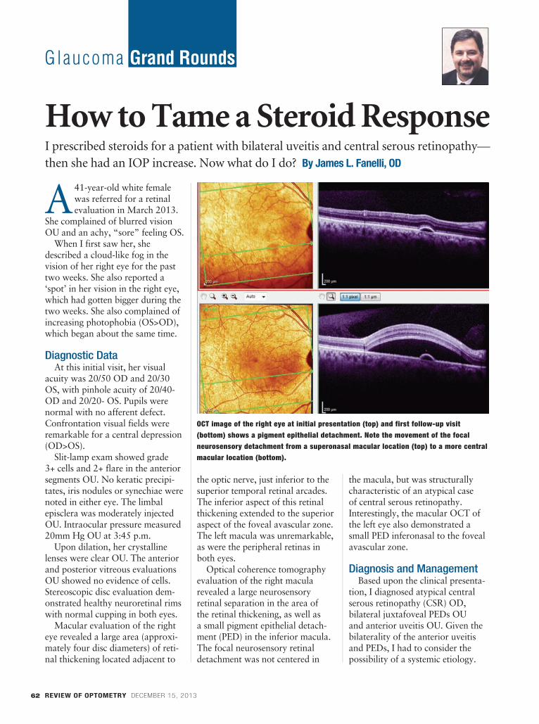
Glaucoma Grand Rounds
REVIEW OF OPTOMETRY DECEMBER 15, 201362
A 41-year-old white female was referred for a retinal evaluation in March 2013.
She complained of blurred vision OU and an achy, “sore” feeling OS.
When I first saw her, she described a cloud-like fog in the vision of her right eye for the past two weeks. She also reported a ‘spot’ in her vision in the right eye, which had gotten bigger during the two weeks. She also complained of increasing photophobia (OS>OD), which began about the same time.
Diagnostic DataAt this initial visit, her visual
acuity was 20/50 OD and 20/30 OS, with pinhole acuity of 20/40- OD and 20/20- OS. Pupils were normal with no afferent defect. Confrontation visual fields were remarkable for a central depression (OD>OS).
Slit-lamp exam showed grade 3+ cells and 2+ flare in the anterior segments OU. No keratic precipi-tates, iris nodules or synechiae were noted in either eye. The limbal episclera was moderately injected OU. Intraocular pressure measured 20mm Hg OU at 3:45 p.m.
Upon dilation, her crystalline lenses were clear OU. The anterior and posterior vitreous evaluations OU showed no evidence of cells. Stereoscopic disc evaluation dem-onstrated healthy neuroretinal rims with normal cupping in both eyes.
Macular evaluation of the right eye revealed a large area (approxi-mately four disc diameters) of reti-nal thickening located adjacent to
the optic nerve, just inferior to the superior temporal retinal arcades. The inferior aspect of this retinal thickening extended to the superior aspect of the foveal avascular zone. The left macula was unremarkable, as were the peripheral retinas in both eyes.
Optical coherence tomography evaluation of the right macula revealed a large neurosensory retinal separation in the area of the retinal thickening, as well as a small pigment epithelial detach-ment (PED) in the inferior macula. The focal neurosensory retinal detachment was not centered in
the macula, but was structurally characteristic of an atypical case of central serous retinopathy. Interestingly, the macular OCT of the left eye also demonstrated a small PED inferonasal to the foveal avascular zone.
Diagnosis and ManagementBased upon the clinical presenta-
tion, I diagnosed atypical central serous retinopathy (CSR) OD, bilateral juxtafoveal PEDs OU and anterior uveitis OU. Given the bilaterality of the anterior uveitis and PEDs, I had to consider the possibility of a systemic etiology.
I prescribed steroids for a patient with bilateral uveitis and central serous retinopathy—then she had an IOP increase. Now what do I do? By James L. Fanelli, OD
How to Tame a Steroid Response
OCT image of the right eye at initial presentation (top) and first follow-up visit (bottom) shows a pigment epithelial detachment. Note the movement of the focal neurosensory detachment from a superonasal macular location (top) to a more central macular location (bottom).
062_ro1213_ggr.indd 62 11/26/13 2:40 PM

Ask about optional Slit Lamp& Chart Projector configurations.
*Lease rate subject to credit approval, 1st payment is paid for by leasing company at signing with 59 remaining rental payments of $269 and a $1.00purchase option. Taxes, freight and installation additional. Hand Instruments optional. Quantities limited. Subject to change without notice.
Or Your Local Lombart Representative.
( 1 - 8 0 0 - 5 6 6 - 2 2 7 8 )
Corporate Office - 5358 Robin Hood Road, Norfolk, VA 23513-2430757-853-8888 | FAX 757-855-1232 | 800-566-2278 | 800-446-8092
www.lombartinstrument.com
ATLANTA•BALTIMORE/WASHINGTON D.C.•BOSTON•BOYNTON BEACH/MIAMI•BRADENTON•CHARLOT TECHICAGO•CINCINNATI•DALLAS•DENVER•DETROIT•GREENSBORO•HOUSTON•KANSAS CIT Y•KNOXVILLE•LOS ANGELES
MILWAUKEE•MINNEAPOLIS•NEW JERSEY/NEW YORK•NORFOLK•PORTLAND•SACRAMENTO•SAN DIEGO•SAN FRANCISCO
RP0812_Lombart.indd 1 7/10/12 11:03 AM

EDUCATION
EXHIBITS
EVENTS
SECOINTERNATIONAL.COM FACEBOOK.COM/SECO.INTERNATIONAL TWITTER.COM/SECO_INTL
REGISTRATION NOW OPEN
SECO Choice Package
Early Registration
$97 $147
RegistrationEarly Registration
$97 $147
Registration
OD AOP
Special Session Package
Early Registration
$357 $399
Registration
OD
Grand Experience Package
Early Registration
$247 $297
RegistrationEarly Registration
$657 $697
Registration
OD AOP
We have made registration fast and easy, with packages designed to fit your needs. We encourage you to visit secointernational.com and register online and look forward to seeing you in Atlanta from March 12-16.
Early Registration Ends January 15, 2014
NEW
RO1113_House Seco.indd 1 10/22/13 2:55 PM

REVIEW OF OPTOMETRY DECEMBER 15, 2013 65
Glaucoma Grand Rounds
Accordingly, I ordered the follow-ing initial labs: CBC with differen-tial, ANA, RA latex, ESR, CRP and ACE levels.
To quiet the anterior chamber reactions, I prescribed atropine 1% BID OU, Pred Forte (prednisolone acetate 1%, Allergan) Q4H OU and Acular LS (ketorolac trometh-amine 0.4%, Allergan) QID OU.
The patient returned in four days with slightly worsening vision in the right eye, and stabilization of the photophobia OU. She reported that she was compliant with her drops.
Pinhole acuity was unchanged OU. Intraocular pressure at this visit was 21mm Hg OD and 20mm Hg OS. Her anterior chambers were unchanged, still showing notable cells and flare OU. No syn-echiae were noted in either eye.
Her retinal evaluations, both at the slit lamp and via OCT imag-ing, demonstrated a gross inferior billowing shift of the focal neuro-sensory retinal detachment to a more central macular location, now encompassing the pre-existent PED. The PED in the left eye increased retinal thickness by 20µm. All of her lab findings were normal, except for a slightly elevated CRP level of 6.8mg/L.
Over the next several weeks, the patient remained moderately com-pliant with her medication sched-ule, although she did report on two occasions that she accelerated her steroid tapering “as my eyes felt better.” At one of the follow-up visits after she decreased her dosage of steroids, her anterior chambers had re-flared, as had the CSR OD, which previously begun to clear. As a result, I directed her to increase the topical steroids to the initially prescribed level.
Unfortunately, at about this same time, IOP in both eyes began to increase. I prescribed timolol 0.5%
QAM OU, and her IOP in both eyes soon dropped from the mid-30s to the upper teens.
By July 2013, her retinas were clear with no evidence of PEDs or CSR. Further labs, including thyroid function studies and chest films, were negative. Histo and Lyme titers were also normal. The anterior chambers were relatively quiet with trace cells and flare. I told her to gradually taper the steroids, with an anticipated cessa-tion in four to six weeks. I kept her on timolol 0.5% QAM OU, but adjusted Acular to QD OU.
Unfortunately, on follow-up in August 2013, she reported increased periocular discomfort and decreased vision. She also said that the timolol was irritating her eyes, so she discontinued that drop OU. At this visit, best-corrected visual acuity was 20/20 OU. Anterior chambers showed 1+ cells and flare, and IOP had risen to 48mm Hg OD and 51mm Hg OS. I prescribed Simbrinza (brinzolamide1%/brimo-nidine 0.2%, Alcon) BID OU.
At the patient’s most recent visit in early November 2013, her ante-rior uveitis had cleared (and has remained so for 30 days), and the CSR and PEDs also remained clear OU. IOP was 18mm Hg OD and 21mm Hg OS. The patient is now medicated OU with Acular QD, Pred Forte BID, Simbrinza BID and timolol 0.5% QAM.
Our current plan: a very slow taper of topical steroids to prevent re-flare, while continuing the IOP-lowering medications to control the steroid-induced glaucoma.
DiscussionThis case presents a challenge:
How can we control the anterior uveitis and retinal conditions while managing the steroid-induced IOP increase?
Keep in mind that when topical steroids are used to treat anterior uveitis, an IOP rise might occur for one of two reasons. First, the IOP rise may be due to the use of the steroid itself, which is believed to increase glycosaminoglycans and reduce outflow in steroid respond-ers. In such cases, the obvious treat-ment is to lower or eliminate the topical steroids.
On the other hand, an IOP rise may occur due to inflammation and inflammatory debris (originat-ing in the anterior chamber) that clogs the trabecular meshwork. When the inflammation itself is the root of the IOP rise, topical steroids may actually need to be increased to ultimately result in lower IOP (by reducing trabecular inflammation).
Trabeculitis is nearly impos-sible to visualize gonioscopically. However, inflammatory debris can occasionally be seen on gonioscopy. In this patient’s case, gonioscopy of the anterior chamber showed open angles with no discernable debris. Could the trabeculum be inflamed, causing her elevated IOP? Or could she simply be a steroid responder? In this case, the answer to both questions is “yes.” I concluded that she was a true steroid responder because the anterior uveitis was not murky or granulomatous in appear-ance, which is more likely to occur with trabecular inflammation.
Managing a steroid responder is rather straightforward. While we should ideally use steroids at a minimum, it’s really the inflamma-tory condition that dictates the ste-roid dosage. Consequently, elevated IOP can be reduced by a variety of pressure-lowering agents. The key is to lower IOP and keep it lowered during the duration of the elevated IOP—whether that’s for days or for years. ■
062_ro1213_ggr.indd 65 11/26/13 2:40 PM

Re t ina Quiz
REVIEW OF OPTOMETRY DECEMBER 15, 201366
A65-year-old Hispanic female presented with mild blur at distance and near in
both eyes that had persisted for six months (OD>OS). Her ocular his-tory was significant for episcleritis in her right eye five years earlier, which resolved uneventfully.
Her medical history was signifi-cant for high cholesterol, which was medically controlled (she couldn’t remember the drug’s name).
On examination, her best-corrected visual acuity measured 20/25 OD and 20/20 OS. Extra-ocular motility testing was normal. Her confrontation visual fields were full to careful finger count-ing OU. Her pupils were equally round and reactive, with no evi-dence of afferent defect. Her ante-rior segment was unremarkable. Intraocular pressure measured 15mm Hg OU.
On dilated fundus exam, the vit-reous was clear OU. She exhibited moderate-sized cups, with good rim coloration and perfusion in both eyes. The peripheral retina was normal in both eyes.
Both maculae showed obvi-ous changes (figures 1 and 2). Additionally, we obtained a spectral-domain optical coherence tomography (SD-OCT) scan of her right eye (figure 3).
Take the Retina Quiz1. What do the yellow-white
changes in both maculae repre-sent?
a. Drusen.b. Intraretinal flecks.c. Exudate.d. Crystals. 2. What does the SD-OCT scan
show on her inner retina? a. Vitreomacular traction.b. Macular telangiectasia.c. Epiretinal membrane (ERM).d. Retinal striae.
3. What does the SD-OCT show in the outer retinal layers?
a. Drusen.b. Choroidal neovascularization
(CNV).c. Disruption of the inner seg-
ment/outer segment (IS/OS) junc-tion.
d. Pigment epithelial detach-ment.
4. What is the correct diagnosis?a. Dry age-related macular
degeneration (AMD).b. Dry AMD and ERM.c. Wet AMD with CNV.d. Vitreomacular traction with
macular edema.
5. How should this patient be managed?
a. Observation.b. Intravitreal anti-VEGF injec-
tion.c. Intravitreal Jetrea (ocriplas-
min, ThromboGenics) injection.d. Vitrectomy and membrane
peel.
For answers, turn to page 86.
DiscussionThe yellow-white deposits seen
in both maculae represent drusen. Therefore, our patient has some form of macular degeneration. The SD-OCT scan detailed the irregular elevations of the drusen at the level of the retinal pigment epithelium (RPE), and revealed an intact IS/OS junction that was pushed forward from the drusen. We documented no fluid or CNV, which confirmed that the patient had dry AMD.
Aside from macular degenera-tion, it’s important to note that the SD-OCT showed a loss of the foveal depression and a slightly thickened macula OD. Indeed, the foveal thickness in her right eye measured 336µm, compared to 254µm in her left eye.
So, what else is going on with this patient? In addition to dry AMD, she has an ERM in her right eye. On the SD-OCT scan, the ERM can be visualized as a highly reflective entity at the level of the internal limiting membrane (ILM). More temporal to the fovea, you can see that the ILM is corrugated with focal elevations. Clinically, this is seen as retinal striae.
An ERM represents a fibrocel-lular membrane that grows onto the retinal surface. It is comprised of glial, RPE and collagen cells, as well as macrophages and fibro-cytes.1
The exact mechanism by which RPE cells migrate onto the surface of the retina is not completely
We uncovered two different retinal pathologies in this patient. What are they, and how severely will they affect her vision? By Mark T. Dunbar, OD
Two isn’t Better Than One
066_ro1213_rq.indd 66 11/26/13 2:41 PM

REVIEW OF OPTOMETRY DECEMBER 15, 2013 67
understood; however, it is believed that an ILM dehiscence might catalyze ERM development. Up to 90% of patients with ERMs also develop posterior vitreous detach-ments (PVDs). Thus, the process of PVD development may yield a small dehiscence in the ILM, per-mitting the fibrocellular membrane to form.1
Approximately 90% of ERMs remain stable and do not affect visual acuity significantly; how-ever, the remaining 10% typically progress.1 For patients who devel-op symptomatic ERM, pars plana vitrectomy with membrane peel often is indicated. Surgical success is dependent on ERM extent and severity, as well as the level of the visual acuity.
There is no definitive standard to determine when surgical inter-vention is necessary for ERM. Spectral-domain optical coher-ence tomography provides a good barometer for detecting structural changes within the retina––either at the level of the IS/OS junction, or if patients develop fluid or cys-tic changes within the retina. Once this occurs, most retinal specialists agree that surgery is warranted.
It is important to remember that postoperative visual acuity is dependent on the extent of retinal architecture changes. Typically, the anticipated visual recovery is approximately 50% better than the entering acuity level. In other words, ERM patients who present with an entering acuity of 20/60 can expect to achieve 20/30 or bet-ter following surgery.
Our patient’s vision is still very good, and she did not report any distortion or metamorphopsia. Our hope is that she will fall into that 90% category, and won’t exhibit ERM progression.
Nevertheless, she does have early dry age-related macular degeneration. So, we recommend-ed an AREDS2 vitamin supple-ment that con-tains both lutein and zeaxanthin. We instructed her to return in six months for additional monitor-ing and follow-up. ■
1. Smiddy WE, Maguire AM. Idiopathic epiretinal mem-branes. Ultrastructural characteristics and clinicopathologic correlation. Ophthalmology. 1989 Jun;96(6):811-20.
1-3. Our patient’s fundus examination revealed interesting macular changes (OD top, OS middle). Additionally, what does the spectral-domain optical coherence tomography scan of our patient’s right eye (bottom) reveal?
066_ro1213_rq.indd 67 11/26/13 2:41 PM

5 WAYS TO REGISTER:ONLINE: www.revoptom.com/aruba2014
EMAIL Lois DiDomenico: [email protected]: Review Group Meetings c/o Jobson
11 Campus Blvd., Ste. 100 Newtown Square, PA 19073
PHONE: 866-658-1772FAX: 610-492-1039
The Westin Resort & Casino, Aruba
· Posterior Segment Grand Rounds· Glaucoma Diagnosis & Management
· Anterior Segment Grand Rounds· Technological Innovations in Eye Care
· Ocular Manifestations of Systemic Disease· Ocular Surface Disease Diagnosis & Treatment
· New Meds / Pharmaceuticals· Nutrition
SPEAKERS:Paul Karpecki, OD
(Program Chair)
Jimmy Bartlett, OD
Ben Gaddie, OD
Kimberly Reed, OD
CE COURSE TOPICS:
*Approval Pending
REGISTER
NOW!
2014_aruba_spread.indd 1 11/27/13 9:49 AM

Payment Information Rate per person No. in party Subtotal
OD Registration - $595 $595 x ________ = $________(includes 14 hours of CE, breakfasts, reception) Call for daily rates.Additional Guest(s) - $45 (12 years and older, includes reception) $45 x ________ = $________Optional Catamaran/Snorkeling Activity, Saturday, January 18 - $125pp (includes lunch) $125 x ________ = $________ TOTAL = $________Check enclosed (make checks payable to Review of Optometry)Charge my: American Express Mastercard Visa
Credit Card Number Exp Date
Cardholder (print name)
Signature
CONFERENCE CANCELLATION POLICY
• Full refund on registration fee until December 16, 2013• 50% refund on registration fee until January 3, 2014• No refund past January 3, 2014
For more information or to register, contact Lois DiDomenico at 866-658-1772 or at [email protected].
Registration Information
Name License # (License numbers are now required for HCP reporting and will only be used for this purpose.)
Practice Affiliation
Mailing Address City State Zip Code
Telephone Fax Email
Name Badge Information (please print clearly)
My Name My Guest Additional Guests
THE WESTIN RESORT & CASINO
*Approval pending
Discounted Room Rate: $269/night
For reservations, call: 888-248-8749
Discounted room rates limited. Identifyyourself as a participant of “Review of Optometry” for group rate.
Room rates available 3 days pre/post conference.
5 WAYS TO REGISTER:ONLINE: www.revoptom.com/Aruba2014
EMAIL Lois DiDomenico: [email protected]: Review Group Meetings c/o Jobson
11 Campus Blvd., Ste. 100 Newtown Square, PA 19073
PHONE: 866-658-1772FAX: 610-492-1039
Photographs Courtesy of the Aruba Tourism Authority
2014_aruba_spread.indd 2 11/27/13 9:50 AM

Therapeu t i c Review
REVIEW OF OPTOMETRY DECEMBER 15, 201370
Ihave always enjoyed lecturing in other countries and meeting
colleagues from across the world. One of my favorite international gatherings is the Tasmania Lifestyle Congress in Hobart, Aus-tralia. It is a lovely event in a beautiful harbor town. The education is outstanding and the del-egates in attendance are among the brightest I have ever encountered. I enjoy making compar-isons between the practice of optometry in Australia and the United States, and genuinely believe what happens in eye care in one country will somehow be mirrored in the other.
Aussies vs. Yankees• Therapeutic privileges.
Optometrists gained therapeutic privileges long ago in the United States. It was a lengthy, arduous process that still presents several limitations today.
Clearly, optometrists in Austra-lia experienced many of the same problems and growing pains that we dealt with years ago. How-ever, it shouldn’t be surmised that optometrists in other countries trail far behind their American counterparts. Actually, in several ways, they may have advantages over US optometrists.
One progressive step that
Australian optometrists have made, which has yet to be taken here, is interstate parity in pre-scribing rights. For example––while oral prescribing laws vary from state to state in the US, the scope of optometry across Aus-tralia is now uniform. That is, the same practice standards exist in all Australian states, with consistent treatment privileges across the country.
• Specialist referrals. Recently,
Australian optometrists took another step forward with the dissolution of the glaucoma comanage-ment rule. Now, Aus-tralian optometrists are able to treat open-angle glaucoma independent of ophthalmologists, and no longer have to refer out for comanagement. This significantly increases access to care for Aus-tralian citizens, as the wait time for a specialist appointment often takes several months.
It remains to be seen how the Affordable Care Act will impact the practice of optometry
in the US. With increased access to insurance, more patients likely will be seeking care. Optometry has always been a gatekeeper for primary eye care. So, it is possible that the wait time for specialists could increase dramatically in the US, similar to that seen in Austra-lia and Canada.
Now more than ever, US optom-etrists need to embrace the thera-peutic aspect of practice. Patients
I wrote about my previous experiences in Australasia back in 2008 and 2012. Now it’s time for the hat trick. By Joseph W. Sowka, OD
Sowka Down Under, Part III
Interestingly, acute bacterial conjunctivitis can be diagnosed and treated independently by an Australian pharmacist––most likely with chloramphenicol.
Chloramphenicol for ConjunctivitisIn Australia, the most commonly prescribed topical antibiotic is chloramphenicol. Although the agent is very effective in treating bacterial conjunctivitis, chloramphenicol has not been commonly used in the US for many years due to perceived risks of aplastic anemia.2
Nonetheless, the Australians have no such worries. In fact, topical chloramphenicol is an over-the-counter drug in Australia, so patients don’t even need to have the medication prescribed by a doctor or pharmacist.
070_ro1213_tr.indd 70 11/26/13 2:43 PM

REVIEW OF OPTOMETRY DECEMBER 15, 2013 71
will need medical eye care at an unprecedented level. Longer wait times for specialist consultations means that we will need to shoul-der the therapeutic burden.
Australian Pharmacists’ Role in Eye Care
If these are not sufficiently com-pelling reasons for optometrists to embrace medical eye care, then let me share with you another change that recently occurred in Australia. Pharmacists have increased their scope of practice and now have therapeutic prescribing rights. Again, this stems from the exten-sive wait times for specialists in Australia.
For example, Australian phar-macists can provide a one-month supply of anti-hypertensive medi-cations, statins and glaucoma med-ications without a prescription. Keep in mind that pharmacists are not diagnosing and treating these conditions, but are providing an emergency supply to patients until they can see their doctor. Of course, it remains to be seen how many patients may abuse this assistance and keep getting an “emergency supply” month after month without seeing a doctor.
What was most interesting to learn, however, is that pharmacists can now actually diagnose and prescribe for common eye condi-tions without the patient/customer ever having to see a doctor. The most common condition that phar-macists independently diagnose and treat is bacterial conjunctivitis. As we know, pharmacists do not typically have biomicroscopes to assist them in evaluating ocular conditions. It is very commonly accepted that bacterial, viral and allergic conjunctivitis often have some overlap in signs and symp-
toms, sometimes making an accu-rate diagnosis difficult even with a biomicroscope.
In the vast majority of cases, acute bacterial conjunctivitis is a self-limiting disease. However, most published reports indicate that despite its benign, self-resolv-ing nature, bacterial conjunctivitis should be treated with topical antimicrobial therapy in order to shorten the disease course and improve the rate of clinical and microbiologic remission.1 This is especially true early in the clinical course.
A primary concern associated with pharmacists prescribing for eye conditions is that many diseas-es present with superficial likeness-es. Anterior uveitis could present early with similarities to simple conjunctivitis. Additionally, bacte-rial keratitis could appear similar to bacterial conjunctivitis. There-fore, a pharmacist not employing a biomicroscope could potentially under-treat a condition, leading to significant visual conse-quences.
To be fair, Austra-lian pharmacists are not likely to prescribe beyond their comfort level. In fact, during my discussion with an Australian pharmacist, she said that she would refer the patient to an optometrist if there were any uncertain-ties (likely because she knew the difficulty in obtaining an ophthal-mology appointment).
While the expand-ing role of Australian pharmacists may seem to have no bearing on
our US practices, we have to be cognizant that patients always will require care. And, if there is a dearth of providers to deliver that care, other health care profession-als must step in to fill the void.
Across the United States, phar-macists are successfully administer-ing immunizations for influenza, pneumonia and herpes zoster. I happily get my own immunizations from a pharmacist.
As more patients flood into the health care system, it is not incon-ceivable that pharmacists may someday have prescribing rights for ocular diseases. In other words, what has successfully transpired in Australia could someday occur in the US if there are not enough eye care practitioners available who are willing to practice medical eye care. ■
1. Sheikh A, Hurwitz B. Antibiotics versus placebo for acute bacterial conjunctivitis. Cochrane Database Syst Rev. 2006 Apr 19;(2):CD001211.2. Rose PW, Harnden A, Brueggemann AB, et al. Chloram-phenicol treatment for acute infective conjunctivitis in children in primary care: a randomized double-blind placebo-controlled
Over a snack, Joe and Joey compare notes on American and Austrialian eye care practices.
070_ro1213_tr.indd 71 11/26/13 2:43 PM

Research Review
REVIEW OF OPTOMETRY DECEMBER 15, 201372
We examine the lens for cataract in all patients, but it may provide an even
greater snapshot of diseases else-where in the body. The human lens is one of the few structures that contin-ues to grow throughout our lifetime. As it does, it accumulates deposits that could alert practitioners to diag-nose a systemic disease—not just connective tissue disorders like Weill-Marchesani syndrome in patients with microspherophakia or sublux-ated lenses in Marfan’s syndrome, but further-reaching diseases such as Alzheimer’s and diabetes mellitus.1
Alzheimer’s Disease (AD)An accumulation of amyloid beta
plaques in the brain results in neuro-logical effects for patients with AD. An imaging technique currently in phase II of FDA clinical trials can detect these amyloid plaques within the crystalline lens.
The technique begins with a fluorescent ligand marker applied topically in an ointment to the inner corner of one eyelid. The application is done three separate times, two hours apart, the night before Sap-phire II laser imaging (Cognoptix).
For imaging, the patient fixates on a target and a low-level laser allows for fluorescent measurements of the lens, providing a score relative to the level of amyloid detected. The entire procedure takes only a few minutes with imaging equipment the size of a personal computer. This technology could be beneficial to the diagnosis of AD, but perhaps even more criti-cal in the clinical trials of potential
AD therapies. A drug that results in amyloid deposition deceleration in the lens may also show reduction of deposition in the brain. Further research is certainly warranted.
Diabetes Mellitus (DM) Advanced glycation end products
(AGEs) are lipids or proteins that become gylcated after being exposed to sugars. They are prevalent in the blood vessels of patients with diabe-tes, but can also contribute to lens changes as they non-enzymatically bond to the lens proteins. Although AGEs form with natural aging, the process is accelerated by hyperglyce-mia and thus observed sooner in DM.
The glycation or condensation of the aldehyde and ketone groups in sugars with the amino groups in pro-teins also leads to a Schiff base—the initial step in the formation of cata-racts—and this also may be detected via autofluorescence. Although lens autofluorescence increases with age, the greater levels of fluorophores can be differentiated in diabetic dis-ease diagnosis. This can be captured through a scanning confocal lens fluorescence biomicroscope called the ClearPath DS-120 (Freedom Medi-tech), which received FDA premarket
clearance this year. The test is non-invasive and takes about six seconds, as the patient simply places their chin in a chin rest and looks at a target.
A study of 127 subjects between the ages of 21 and 70 showed a lin-ear model for lens autofluorescence intensity with age that was highly statistically significant.2 Multiple research studies have shown an increase in lens autofluorescence in patients with diabetes.3,4 Decades-old studies show this correlation, and only recently has technology become available for optometrists to measure it accurately.7,8
Statistical findings can also be obtained in both young and elderly patients with diabetes and in patients with early and late onset diabetes.7,8
CorrelationNot only does autofluorescence
of the lens help make a diagnosis of diabetes, but it can also correlate to the level of HbA1c. In one study comparing insulin-dependent dia-betics with high HbA1c to another group with low HbA1c, the group with low HbA1c during the disease period showed significantly lower lens fluorescence.9 Furthermore, lens autofluorescence provides informa-tion about long-term control of the disease, as research shows it can be delayed by good metabolic control.10
A1c measures give a snapshot of the last two to three months in a diabetes patient, whereas lens autofluores-cence measures the lifetime.
Another correlation exists with diabetic nephropathy, as one study compared 10 patients with diabetes
Research suggests that conditions such as diabetes or Alzheimer’s may be best diagnosed via the crystalline lens. By Diana L. Shechtman, OD, and Paul M. Karpecki, OD
The Lens: Window to Diagnosis?
Proliferative diabetic retinopathy.
072_ro1213_rr.indd 72 11/26/13 2:47 PM

and kidney disease to 11 otherwise comparable patients with diabetes without kidney disease. The patients with diabetic nephropathy had signif-icantly greater lens autofluorescence measurements and decreased lens transmittance.11
What makes autofluorescence of the lens even more compelling is that it can support early diagnosis. In one 14-year follow-up study, which confirmed that lens fluorescence was significantly related to mean HbA1c, researchers noted that although fluo-rophore accumulation in the lens of a diabetes patient was increased in proportion to glycemic control, it was not sufficient to explain the entire variation. Thus, lens fluorescence must be influenced by other factors before initiation and during the study.
The study authors further concluded that it may have been affected before the onset of DM.12 The underlying mechanism could be a variation in susceptibility to lens protein dena-turation by glycation. This means that patients who are more likely to develop diabetes may have the initial effects noted in their crystalline lens.
The ability to make a diagnosis of AD or DM via the crystalline lens prior to clinical diagnosis or symp-toms could solidify our role as pri-mary eye care providers and help save the vision and lives of our patients. ■
1. Chu BS. Weill-Marchesani syndrome and secondary glaucoma associated with ectopia lentis. Clin Exp Optom. 2006 Mar;89(2):95-9.2. Burd J, Stephen L, Cahn F et al. Simultaneous Noninvasive Clinical Measurement of Lens Autofluorescence and Rayleigh Scattering Using a Fluorescence Biomicroscope. J Diabetes Sci Technol. 2012 Nov 1;6(6):1251-9.
3. Bleeker JC, van Best JA, Vrij L, van der Velde EA, Oosterhuis JA. Autofluorescence of the lens in diabetic and healthy subjects by flu-orophotometry. Invest Ophthalmol Vis Sci. 1986 May;27(5):791-4.4. Kessel L, Sander B, Dalgaard P, Larsen M. Lens fluorescence and metabolic control in type 1 diabetic patients: a 14 year follow up study. Br J Ophthalmol. 2004 Sep;88(9):1169-72.5. Van Best JA, Vrij L, Oosterhuis JA. Lens transmission of blue-green light in diabetic patients as measured by autofluorophotom-etry. Invest Ophthalmol Vis Sci. 1985 Apr;26(4):532-6.6. Mosier MA, Occhipinti JR, Burstein NL. Autofluorescence of the crystalline lens in diabetes. Arch Ophthalmol. 1986 Sep;104(9):1340-3.7. Van Wirdum E, van Best J, Bruining GJ, et al. Blood-retinal and blood-aqueous barrier permeability, lens autofluorescence and transmission in insulin-dependent diabetic youngsters. Graefes Arch Clin Exp Ophthalmol. 1989;227(1):26-9.8. Moseir MA, Occhipinti JR, Burstein NL. Autofluorescence of the crystalline lens in diabetes. Arch Ophthalmol. 1986 Sep;104(9):1340-3.9. Kjer B, Larsen M, Bendtson I et al. Lens autofluorescence in the diabetes compared with the level of glycosylated hemoglobin A1c. Acta Ophthalmol Suppl.1987;182:100-2.10. Larsen M, Kjer B, Bendtson I, et al. Lens fluorescence in rela-tion to metabolic control of insulin-dependent diabetes mellitus. Arch Ophthalmol.1989 Jan;107(1):59-62.11. Larsen M, Kjer B, Bendtson I, et al. Lens fluorescence in rela-tion to nephropathy in insulin-dependent diabetes mellitus. Graefes Arch Clin Exp Ophthalmol. 1992;230(1):6-10.12. Kessel L, Sander B, Dalgaard P, Larsen M. Lens fluorescence and metabolic control in type 1 diabetic patients: a 14 year follow up study. Br J Ophthalmol. 2004 Sep;88(9):1169-72.
072_ro1213_rr.indd 73 11/26/13 2:44 PM

REVIEW OF OPTOMETRY DECEMBER 15, 201374
Contact LensesPureVision2 Multi-Focal
Clear and consistent vision at all distances can be a challenge for people with presbyopia, but the newly released PureVision2 Multi-Focal lenses from Bausch + Lomb seeks to change that. Featuring a three-zone progressive design, these lenses were designed for real-world use to improve both near and intermediate vision, whether you’re looking at your cell phone or sitting in a movie theater.
The lenses were also crafted to allow for predictable, quick fits for eye care professionals, the company says.
Visit www.bausch.com.
DiagnosticsGonio and Funduscopy Lenses
A new line of diagnostic lenses offers high-quality optics without the hassles of cleaning and disinfecting, says Sensor Medical Technology. The company’s wide range of lenses is inexpensive, sterile and disposable, according to the company. The line includes 3-Mirror, 4-Mirror, Single Mirror Gonioscopy, Fundus, Retina 90, Retina 165 and Retina 180 lenses.
Each lens has anti-reflective coating, according to Sen-sor Medical, which has also introduced a line of indirect ophthalmoscopy lenses with powers of 20D, 28D, 60D, 78D and 90D.
Visit sensormedtech.com.
AccessoriesFlōt Merchandising System
If you’re looking for a more interesting way to shelve your eyewear, Flōt may be what you need.
The merchandis-ing system, crafted by Optical Displays, features a compel-ling combo of shelves and interchangeable
accessories to visually captivate customers and improve your sales, according to the company. You can choose from a collection of various sizes and materials, from solid to Lucite, for design flexibility.
Visit www.opticaldisplays.com.
Lens EdgerWeco E.5
If you have an in-office spectacle lab, or are thinking of adding one but worry about a low-to-medium volume of prescriptions to fill, a new patternless edger may help. The Weco E.5 offers integrated drilling and wrap frame processing without compromising performance or value, according to manufacturer AIT Industries.
The Weco E.5 includes touch-screen controls, a grinding wheel capable of beveling all materials, automatic polishing, continuous 3D mapping, adjustable grooving angles and a superhydrophobic lens roughing process, among other features.
Visit www.aitindustries.com.
DiagnosticsSjö
Because the earliest stages of Sjögren’s syndrome mimic those of many other dry eye presentations, optometrists find themselves with a unique responsibility to identify this serious, progressive and underdiagnosed autoimmune disease as soon as possible. Lab testing can improve specificity, but may delay diagnosis.
Recently launched in the US by Nicox, Sjö is an advanced diagnostic panel for in-office use that com-bines traditional biomarkers with three novel ones as a means to detect Sjögren’s early enough to manage the condition and help avoid further complications.
Nicox has also formed a partnership with the Sjögren’s Syndrome Foundation to raise awareness of the disease, which is often undiagnosed until its late stages. Patients are typically diagnosed with traditional dry eye when their symptoms first manifest.
Visit www.nicox.com.
VitaminsAREDS 2 Plus
Patients who need ocular vitamins can choose from two products updated to reflect AREDS 2. One version, called EyePromise AREDS 2 Plus with a Multi-Vitamin, incorporates the AREDS 2 elements and a multi-vitamin for-mulation. Patients who want to nix the zinc can
P r o d u c t R e v i e w
074_ro1213_products.indd 74 11/27/13 2:33 PM

REVIEW OF OPTOMETRY DECEMBER 15, 2013 75
opt instead for EyePromise AREDS 2 Plus Zinc-Free. According to parent company ZeaVision, clini-
cal studies showed that many patients who took eye vitamins also enjoyed daily multi-vitamins, and some patients achieved better vision protection without the zinc. Hence, the new offerings, designed to encourage convenient and consistent daily use.
Visit www.eyepromise.com.
Retinal ImagingVolk Pictor
Weighing in at just one pound, the Volk Pictor is a compact and lightweight digital imaging system that takes retinal images in either infrared or white light, as well as anterior segment photos of the eye. According
to AIT Industries, this gives you the flexibility to take images in places like off-site clinics or hospitals, as well as in your office.
Pictor produces high-resolution images and video that are compatible with most imaging software, the company says. It’s also adaptable to any EMR that allows JPEG files to be attached to medical records. The retinal imaging module provides a wide 45-degree field of view of the fundus.
Visit www.aitindustries.com.
Chart systems, slit lamps, OCTs and autorefractors were among many new products introduced by Topcon Medical Systems at the American Academy of Opthalmology annual meeting. Not all are yet available in the US, but here’s a preview of what’s coming. Check with your Topcon rep for availability or visit topconmedical.com for more details.
3D OCT-1 MaestroThis fully automated, high-resolution FD-OCT device also offers nonmydriatic retinal camera functioning, and 12mm x 9mm widefield scan that allows for measurement and topographical maps of the optic nerve and macula in one scan.
KR-800S Subjective Auto Kerato-RefractometerThis device incorporates subjective visual acuity testing with autore-fraction technology to refine patient measurements. Includes a glare-testing function that assesses visual acuity under glare conditions.
IMAGEnet 5This DICOM-compliant computerized digital system allows you to capture, view and archive images of the eye from either local or remote locations.
DRI OCT-1 AtlantisWith a 1,050nm light source and scanning speed of 100,000 A-scans/second, this next-generation OCT device uses new swept-source scanning technology. The Atlantis provides uniform scanning sensitivity, which allows for superior visualization of the vitreous and choroid in the same scan, and can even scan through cataracts, Topcon says.
Synergy ODMImages and diagnostic data from Topcon and more than 130 other manufacturers’ systems are integrated into a single, secure, digital environment in this unique software platform that allows better digital connectivity.
TRK-2P Auto Kerato-Refractometer/TonometerThis four-in-one instrument increases efficiency by combining an autorefractor, keratometer, non-contact tonometer and pachymeter in one space.
SL-D791 and DC-4A new LED illumination source and high-quality optics in the SL-D791 slit lamp deliver sharp, clear and bright view of ocular anatomy. Additional features include a five-magnification drum-style changer from 6x to 40x, 20° tilt for gonioscopy observation, Galileo-type parallel-converging binoculars and smooth movement by universal joystick. The DC-4 digital photo attachment allows for image acquisi-tion in still mode, video clip and “smart capture” rapid sequence acquirement.
CC-100 LCD Acuity Chart SystemThis is a versatile computerized acuity chart system. The high-resolution LCD monitor ensures clear and bright chart display. All common visual acuity tests are available. You can also add your own video content.
IS-600NAn economical, yet fully featured chair and stand combination unit with phoropter arm, dual instrument sliding tabletop, halogen reading lamp and chart projector bracket.
SP-1PThe new Topcon Specular Microscope model SP-1P automatically centers, focuses and acquires the endothelial cell image. The entire operation takes a few seconds and requires minimal training, Topcon says.
MC-4S 3D Mirror ChartThis simulates a full 20’ lane in a 2’ to 5’ range, allowing for use in very small refraction areas and features a high resolution, polarized LCD display that includes all common visual acuity charts as well as the ability to show 3D movies or to show the effects of polarization on a patient’s glasses.
High-Tech Products, High-Quality Care
074_ro1213_products.indd 75 11/27/13 2:33 PM

REVIEW OF OPTOMETRY DECEMBER 15, 201376
Meet ings + Conferences
January 2014■ 11. 2014 Glaucoma Symposium. Willows Lodge, Woodinville,
Wash. Hosted by: Pacific University College of Optometry. CE
hours: 7. Contact Marti Fredericks at [email protected] or
(503) 352-2929. Visit www.pacificu.edu/optometry/ce.
■ 11. Coding Update & HIPAA & HR Compliance Training.
Doubletree Hilton, Little Rock, Ark. Hosted by: Arkansas
Optometric Association. CE hours: 8. Key faculty: John A.
McGreal, OD, Joe DeLoach, OD, BJ Avery. Email aroa@arkan-
sasoptometric.org. Visit www.arkansasoptometric.org.
■ 12. Winter CE. Hyatt Regency O’Hare, Rosemont, Ill. Hosted
by: Illinois Optometric Association. Key faculty: Paul Karpecki,
OD. CE hours: 6. Contact Charlene Marsh at [email protected]
or (217) 525-8012. Visit www.ioaweb.org.
■ 11-12. Eye Care Associates Annual Meeting and Continuing
Education Program. Williamsburg Hotel, Williamsburg, Va.
Hosted by: Eye Care Associates. Presenter: Scot Morris, OD. CE
hours: 12. Contact Linda Cavasos at [email protected] or
(804) 356-5165. Non-members are welcome.
■ 16-19. New Technology & Treatments in Vision Care. The
Westin Resort & Casino, Aruba. Program Chair: Paul Karpecki,
OD. Faculty: Jimmy Bartlett, OD, Ben Gaddie, OD, and Kimberly
Reed, OD. Hosted by: Review of Optometry. CE hours: 14.
Contact Lois DiDomenico at [email protected].
Visit www.revoptom.com/conferences.
■ 18-19. 30th Annual Gold Coast Educational Retreat. Hyatt
Regency Pier 66, Ft. Lauderdale, Fla. Hosted by: Broward
County Optometric Association. CE hours: 19. Email BCOA@
browardeyes.org or visit http://www.browardeyes.com.
■ 18-20. Kraskin Invitational Skeffington Symposium on Vision.
Hyatt Regency Bethesda, Bethesda, Maryland. Hosted by:
OEPF. CE hours: 19. Chair: Jeffrey Kraskin, OD. Contact Dr.
Kraskin at [email protected]. Visit www.skeffingtonsymposium.
org.
■ 18-20. Berkeley Practicum - 25th Annual. DoubleTree Hotel,
Berkeley Marina, Berkeley, Calif. Hosted by: University of
California, Berkeley, School of Optometry. CE hours: 20. Email
[email protected]. Visit http://optometry.berkeley.edu/ce/
berkeley-practicum.
■ 19-25. 2014 Island Eyes Conference. Grand Wailea, Maui,
Hawaii. Hosted by: Pacific University College of Optometry.
Contact Jeanne Oliver at [email protected] or (503) 352-
2740. Visit www.pacificu.edu/optometry/ce.
■ 22. Dermatology: General and Neoplastic Disease. West
Los Angeles VA, Los Angeles. Hosted by: Marshall B. Ketchum
University/SCCO. Email [email protected] or visit www.ketchum.
edu/ce.
■ 23-26. Global Specialty Lens Symposium. Rio Hotel and
Casino, Las Vegas. Hosted by: Contact Lens Spectrum. CE
hours: 30. Contact Maureen Platt at maureen.platt@pentavision-
media.com or visit www.pentavisionevents.com.
■ 24. 2014 Winter CE. PCLI, Pearl District, Portland, Ore.
Hosted by: Oregon Optometric Physicians Association. CE
hours: 8. Email [email protected]. Visit www.ore-
gonoptometry.org.
■ 24-25. High Performance Vision/Sports Vision Consulting
Weekend. Hollywood Beach Marriott, Hollywood, Fla. Hosted
by: Ultimate Events. Key faculty: Don Teig, OD. CE hours: 16.
Contact Dr. Teig at [email protected]. Visit www.ulti-
mateeventsllc.com.
■ 25-26. Vision Training Conference II. Embassy Suites
Rockside Road, Independence, Ohio. Hosted by: College of
Optometrists in Vision Development. Key faculty: W.C. Maples,
OD, MS. CE hours: 12. Contact Jackie Cencer at jcencer@covd.
org. Visit www.covd.org.
■ 26. Winter CE. The Westin Chicago North Shore, Wheeling,
Ill. Hosted by: Illinois Optometric Association. Key faculty: Ben
Gaddie, OD. CE hours: 6. Email [email protected] or (804) 643-
0309. Visit www.thevoa.org.
■ 26. VOA One-Day CE Conference. Richmond Marriott West,
Glen Allen, Va. Hosted by: Virginia Optometric Association.
Key faculty: Joe DeLoach, OD, BJ Avery. CE hours: 4. Contact
Charlene Marsh at [email protected] or (217) 525-8012. Visit
www.ioaweb.org.
■ 30-February 3. Women of Optometry Spa Cruise. Celebrity
Constellation, Bahamas. Hosted by: AEA Cruises Optometric
Cruise Seminars. Email [email protected]. Visit www.
OptometricCruiseSeminears.com.
February 2014■ 8-9. Mid Winter CE Meeting 2014. New Orleans Marriott, New
Orleans. Hosted by: Optometry Association of Louisiana. Email
[email protected]. Visit www.optla.org.
■ 9-10. 2014 Advocacy Boot Camp & Free CE. Salem
Conference Center/Grand Hotel, Salem, Ore. Hosted by: Oregon
Optometric Physicians Association. Email lynne@oregonoptom-
etry.org. Visit www.oregonoptometry.org.
■ 14-16. 53rd Annual Heart of America Contact Lens and
Primary Care Congress. Sheraton Kansas City Hotel at Crown
Center, Kansas City, Mo. Hosted by: Heart of America Contact
Lens Society. Email [email protected]. Visit www.
hoacls.org.
■ 27-March 1. 2014 Winter Educational Symposium. Huntley
Lodge, Big Sky, Mont. Hosted by: Montana Optometric
Association. Faculty: Blair Lonsberry, OD, Christopher Wolfe,
OD. CE hours: 13. Email [email protected].
Visit www.mteyes.com.
■ 28-March 1. 2014 Third Party/Practice Management
Seminar. Eugene Hilton, Eugene, Ore. Hosted by: Oregon
Optometric Physicians Association. For additional information,
email [email protected]. Visit www.oregonoptometry.
org.
076_ro1213_m&c.indd 76 11/26/13 4:30 PM

REVIEW OF OPTOMETRY DECEMBER 15, 2013 77
March 2014■ 12-16. SECO International 2014. Building A, Georgia World
Congress Center, Atlanta. CE hours: 400+. Email cweems@
secostaff.com. Visit www.seco2014.com.
■ 22-23. Spring Conference. Nova Southeastern University Ft.
Myers Campus, Ft. Myers, Fla. Hosted by: Nova Southeastern
University. Email [email protected]. Visit http://optometry.nova.
edu/ce/index.html.
April 2014■ 12-13. 2014 MOS Primary Care Spring Symposium.
Cincinnati Marriott Northeast, Mason, Ohio. Hosted by: The
MidWest Optometric Society and the Ohio State University
College of Optometry. Contact Marci at (513) 321-2020. Visit
www.midwestoptometricsociety.com.
■ 24-27. Arkansas Optometric Association Spring Convention.
The Peabody, Little Rock, Ark. Hosted by: Arkansas Optometric
Association. Email [email protected]. Visit www.
arkansasoptometric.org.
May 2014■ 2. Berkeley Glaucoma Day – 2nd Annual. DoubleTree
Hotel, Berkeley Marina, Berkeley, Calif. Hosted by: University
of California, Berkeley, School of Optometry. Email optoCE@
berkeley.edu.
■ 2-3. Educational Meeting 2014. Mission Inn, Howey-in-the-
Hills, Fla. Hosted by: Florida Chapter of the American Academy
of Optometry. Featured speakers: Leo Semes, OD, Albert
Woods, OD, and Tim Underhill, OD. CE hours: 10. Contact
Arthur T. Young, OD, at [email protected].
■ 3-4. Morgan Symposium – 29th Annual. DoubleTree Hotel,
Berkeley Marina, Berkeley, Calif. Hosted by: University of
California, Berkeley, School of Optometry. Email optoCE@
berkeley.edu. Visit http://optometry.berkeley.edu/ce/morgan-
symposium.
August 2014■ 1-3. Annual Educational Retreat 2014. South Seas Island
Resort, Sanibel Island, Fla. Hosted by: South West Florida
Optometric Association. Featured speakers: Ben Gaddie, OD,
Carlo Pelino, OD, April Jasper, OD, and Ron Foreman, OD. CE
hours: 18. Contact Brad Middaugh, OD, at [email protected] or
(239) 481-7799. Visit www.swfoa.com.
To list your meeting, please send the details to:Erin Kelly
Senior Associate Editor
Email: [email protected]
Phone: (610) 492-1005
Alcon Laboratories ........ 15, 21
..................23, 24, 45, 57, 85, 88
Phone ................ (800) 451-3937
Fax ..................... (817) 551-4352
Bausch + Lomb .........26-27, 28
Phone ................ (800) 323-0000
Fax ..................... (813) 975-7762
Eye Designs .......................... 53
Phone ................ (800) 346-8890
Fax ..................... (610) 489-1414
Freedom Meditech ................. 5
Phone ................ (858) 638-1433
www.freedom-meditech.com
HAI Laboratories .................. 59
Phone ................ (781) 862-9884
Fax ..................... (781) 860-7722
Keeler Instruments ............... 87
Phone ................ (800) 523-5620
Fax ..................... (610) 353-7814
Lombart Instruments ........... 63
Phone ................ (800) 446-8092
Fax ..................... (757) 855-1232
M&S Technologies ............... 18
Phone ................ (877) 225-6101
Fax ..................... (847) 763-9170
Marco Ophthalmic ................ 12
Phone ................ (800) 874-5274
Fax ..................... (904) 642-9338
Menicon ................................. 37
Phone ...............(800) MENICON
www.meniconamerica.com
Optos North America ........... 43
Phone .......(877) 455-8855 x 100
Fax ..................... (508) 486-9310
Rhein Medical ....................... 17
Phone ................ (800) 637-4346
Fax ..................... (727) 341-8123
SynergEyes, Inc...................... 7
Phone ................ (877) 733-2012
www.SynergEyes.com
Three Rivers Optical ............ 11
Phone ................ (800) 756-2020
Fax ..................... (800) 756-0034
Vistakon ........... 2-3, 18 A-B, 61
Phone ................ (800) 874-5278
Fax ..................... (904) 443-1252
Vmax Vision, Inc. .................. 49
Phone ................ (888) 413-7038
www.VmaxVision.com
ZeaVision, LLC ....................... 9
Phone ................ (866) 833-2800
www.eyepromise.com
This advertiser index is published as a convenience and not as part of the advertising contract. Every care will be taken to index correctly. No allowance will be made for errors due to spelling, incorrect page number, or failure to insert.
Adver t i se rs Index
076_ro1213_m&c.indd 77 11/26/13 4:30 PM

78 REVIEW OF OPTOMETRY DECEMBER 15, 2013
Review Classifi eds
Merchandise Offered
Do you have Productsand Services to offer?
CLASSIFIED ADVERTISING
WORKS
Contact us today at:
Toll free: 888-498-1460
E-mail: [email protected]
Products and Services
WWW.EYEWEAR4LESS.COMEVERYDAY LOW PRICES - HUGE SELECTION
“Per UOMO” FACTORY DIRECT ITALIAN EYEWEAR
BRAND NAME OVERSTOCKS, CLOSE-OUTS & SPECIALS
“YOUR PRACTICE YOUR PROFIT”
800-762-0480
ROPT1213.indd 78 11/26/13 7:08 AM
79REVIEW OF OPTOMETRY DECEMBER 15, 2013
Review Classifi eds
Merchandise Offered
Equipment and Supplies
FEATURES OF FLOT™
Can be used with a variety of materials
Flot plugs can be used on solidor lucite panels
Clean linear look, no frame holders
Optional LED lighting to edge light shelves
Interchangeable accessories from caseholders, signage and merchandising hooks
&
&
&
&
&
&
&
TM
NEW
OD-111
202
Equipment and Supplies
It’s What the BestPretest on!
(800) 522-2275www.optinomics.com
Looking to increase sales?
Place Your Ad here.Contact us today for classified advertising:
888-498-1460E-mail: [email protected]
ROPT1213.indd 79 11/25/13 6:55 PM

80 REVIEW OF OPTOMETRY DECEMBER 15, 2013
Review Classifi eds
Do you have Equipment and Supplies for Sale?
Contact us today for classified advertising:Toll free: 888-498-1460 • E-mail: [email protected]
Frames
Equipment and Supplies
Looking to increase sales?
Place Your Ad here.For classified advertising:
888-498-1460E-mail: [email protected]
QUIKEYES ONLINEWEB-BASED OPTOMETRY EHR
• $99 per month after low cost set-up fee• Quick Set-Up and Easy to Use • No Server Needed• Corporate and Private OD practices• 14 Day Free Demo Trial• Users Eligible for 44K incentives
www.quikeyes.com
SOFTWARE
ROPT1213.indd 80 11/25/13 6:55 PM

81REVIEW OF OPTOMETRY DECEMBER 15, 2013
Review Classifi eds
Place Your Ad Here!
Toll free: 888-498-1460
E-mail: [email protected]
www.PracticeConsultants.com
Practice Sales • Appraisals • Consulting
www.Pract iceConsultants .com
PRACTICES FOR SALENATIONWIDE
Visit us on the Web or call us to learnmore about our company and the
practices we have available.
800-576-6935
Practice For Sale
Practice for SaleBrevard County Florida
Revenue 440,000 on a 32 O.Dhour/week. Practice has shownconsistent revenue since itopened in 2007.
Great opportunity for a full tineO.D in Brevard County Florida.Free standing building, beautifulinterior, easy access and goodparking.
Call 321-961-6113
Continuing Education
SOFTWARE
Expert Services for:Buying or Selling a PracticePractice AppraisalPractice FinancingPartner Buy-in or Buy-out
PRACTICE SALES & APPRAISAL
Call for a Free Consultation(800) 416-2055
www.TransitionConsultants.com
ROPT1213.indd 81 11/26/13 7:09 AM

82 REVIEW OF OPTOMETRY DECEMBER 15, 2013
Review Classifi eds
Contact Lenses
Storage and Design
ROPT1213.indd 82 11/25/13 6:56 PM

83REVIEW OF OPTOMETRY DECEMBER 15, 2013
Review Classifi eds
Professional Opportunities
OPTOMETRIST Join a familyoriented team of MDs and ODs inbeautiful Napa, CA. We are seek-ing a full-time associate for ourNapa, St. Helena and American
Canyon offices. This position requires a practitionerwho has that special personality to “win friends andinfluence people”, in addition to an excellent academicbackground. This is a golden opportunity to practiceprimary eye care in a terrific setting in the North Bayregion of the San Francisco Bay area. The right candidate desires the dynamic of an MD/OD grouppractice setting. We offer a competitive pay rate,bonuses, paid continuing education, personal time offand health insurance. C.V.s should be submitted withletters of recommendation to David Yoho, MBA,Administrator Eye Care Center of Napa Valley, 895Trancas St., Napa, CA 94558. Electronic submittal:[email protected].
OPTOMETRIST WANTEDHealthDrive Eye Care Group provides optometric careto very deserving elderly patients residing in nursinghomes and assisted living facilities. We currentlyhave the following positions available:
POSITION LOCATIONS AND DETAILS:Location: Harrisburg, PA• Part-Time, 3 Days (No evenings or weekends)• Typical Hours: 8am or 9am to 3pm• 401K and Vacation offered• Permanent position (No Contracts)• Excellent pay and Clinical AutonomyLocation: Wallingford, CT• Full-Time, 5 Days (No evenings or weekends)• Typical Hours: 8am or 9am to 3pm• Full Benefits including dental and 401K• Permanent position (No Contracts)• Excellent pay (100K+) and Clinical Autonomy
RESPONSIBILITIES:• Provides routine, preventative examinations.• Conducts examinations for oral cancer screenings.• Performs routine extractions.• Performs denture fabrications, relines, and repairs.• Provides emergency services.• Enters patient information and completes
required clinical documentation in the electronichealth medical record.
Join our practice and benefit from our managementexpertise and commitment to quality care.Please contact Tanya Jones at 857-255-0293 [email protected] if you are interested inhearing more about our opportunities.
HealthDrive is an Equal Opportunity Employer.
FULL-TIMECLINICAL OPTOMETRY
The Wilmer Eye Institute of the Johns Hopkins UniversitySchool of Medicine is seeking to fill a full time clinical optometryposition. Primary care practice and co-management experiencedesired. Completion of an Optometric residency preferred.Position is eligible for academic/faculty track.
Please e-mail CV to:
Elliott Myrowitz, O.D., Chief of Optometryc/o Lynne Young at [email protected]
www.hopkinsmedicine.org/wilmer
Johns Hopkins University is an affirmative action/equal opportunity employer and is especially interested in identifying female and minority candidates.
EEO/MFDV, Smoking Drug Free
FOR CLASSIFIED ADVERTISINGCONTACT US
TODAY:
Toll free: 888-498-1460
E-mail: [email protected]
Do You Have Positions Available?CLASSIFIED ADVERTISING WORKS
Contact us today for classified advertising:Toll free: 888-498-1460
E-mail: [email protected]
ROPT1213.indd 83 11/25/13 6:57 PM

REVIEW OF OPTOMETRY DECEMBER 15, 201384
Surg ica l Minute
Radial keratotomy (RK), a popular procedure in the 1980s and early 1990s for
the correction of low to moderate myopia (-1D to -6D) and astigma-tism, seems primitive by today’s exacting standards. It primarily involved creating partial-thickness incisions through approximately 90% of the cornea in a radial pat-tern around the mid periphery, to flatten the central cornea while steepening the periphery.
Short-term complications of RK included pain, infection, delayed healing, poor scotopic vision due to radial corneal scars, epithelial ingrowth and neovascularization of incisions, corneal erosions, epithelial defects, gaping or non-healing inci-sions, micropunctures of Descemet’s membrane, corneal perforation and endothelial cell loss. Because RK was relatively imprecise and more risky than PRK and LASIK, it fell out of favor relatively soon after the advent of laser-based procedures.
Undoing the DamageRK’s long-term complications
were never well studied before it became popular, and were largely unknown. Although most patients enjoyed relatively stable vision for years, an alarmingly high number continued to experience progres-sive corneal flattening as they aged, leading to significant hyperopia and irregular astigmatism.
Daily fluctuating vision—due to diurnal intraocular pressure changes causing the cornea to change shape—was a common complica-
tion experienced by RK patients. Splitting or gaping incisions
are a less common complication that we still see decades later. The patient may notice a sudden onset of corneal pain or decreased vision, but can also present with mild or no symptoms. Surgical closure of these incisions is often warranted to improve anterior corneal shape, decrease risk of epithelial ingrowth, and to prevent neovascularization and permanent corneal warpage.
Gaping incision closure is typically done with non-dissolving nylon sutures. Partial corneal-depth sutures are used to close the wound, and then the knots are buried under the epithelium. Excessive tension is needed to ensure permanent wound closure; this often induces local steepening of the cornea that can temporarily reduce vision and per-sists until the sutures are removed at a later date.
Postoperatively, patients need to be monitored for infection, leaking suture sites and excessive inflamma-tion that could precipitate further corneal scarring. Patients should also be counseled that while the
sutures are inducing asymmetric tension to their cornea, the vision is likely to change. Sutures can be left in the cornea for months or years, until the surgeon feels comfortable that there is little risk of re-splitting the incisions upon removal. If sutures are left in for a long period of time, patients should be moni-tored for suture erosion and pos-sible related GPC.
This procedure can temporarily make vision worse, but patients should be counseled that the long-term medical risks from not closing the incisions can be more serious than the possible (and often tempo-rary) vision changes. ■
Decades later, we are still seeing adverse events caused by one of the earliest refractive surgery procedures. By Derek N. Cunningham, OD, and Walter O. Whitley, OD, MBA
RK: The Unkindest Cut
A gaping corneal RK incision. Gaping corneal RK incision taking up fluorescein.
Closure of gaping incisions using sutures.
084_ro1213_surgical.indd 84 11/27/13 10:55 AM

BRIEF SUMMARY OF PRESCRIBING INFORMATION
INDICATIONS AND USAGE ILEVRO™ Suspension is indicated for the treatment of pain and inflammation associated with cataract surgery.
DOSAGE AND ADMINISTRATION Recommended Dosing One drop of ILEVRO™ Suspension should be applied to the affected eye one-time-daily beginning 1 day prior to cataract surgery, continued on the day of surgery and through the first 2 weeks of the postoperative period. An additional drop should be administered 30 to 120 minutes prior to surgery.
Use with Other Topical Ophthalmic Medications ILEVRO™ Suspension may be administered in conjunction with other topical ophthalmic medications such as beta-blockers, carbonic anhydrase inhibi-tors, alpha-agonists, cycloplegics, and mydriatics. If more than one topical ophthalmic medication is being used, the medicines must be administered at least 5 minutes apart.
CONTRAINDICATIONS ILEVRO™ Suspension is contraindicated in patients with previously demon-strated hypersensitivity to any of the ingredients in the formula or to other NSAIDs.
WARNINGS AND PRECAUTIONS Increased Bleeding Time With some nonsteroidal anti-inflammatory drugs including ILEVRO™ Suspen-sion, there exists the potential for increased bleeding time due to interfer-ence with thrombocyte aggregation. There have been reports that ocularly applied nonsteroidal anti-inflammatory drugs may cause increased bleeding of ocular tissues (including hyphemas) in conjunction with ocular surgery. It is recommended that ILEVRO™ Suspension be used with caution in patients with known bleeding tendencies or who are receiving other medications which may prolong bleeding time.
Delayed Healing Topical nonsteroidal anti-inflammatory drugs (NSAIDs) including ILEVRO™ Suspension, may slow or delay healing. Topical corticosteroids are also known to slow or delay healing. Concomitant use of topical NSAIDs and topical steroids may increase the potential for healing problems.
Corneal Effects Use of topical NSAIDs may result in keratitis. In some susceptible patients, continued use of topical NSAIDs may result in epithelial breakdown, corneal thinning, corneal erosion, corneal ulceration or corneal perforation. These events may be sight threatening. Patients with evidence of corneal epithelial breakdown should immediately discontinue use of topical NSAIDs including ILEVRO™ Suspension and should be closely monitored for corneal health. Postmarketing experience with topical NSAIDs suggests that patients with complicated ocular surgeries, corneal denervation, corneal epithelial defects, diabetes mellitus, ocular surface diseases (e.g., dry eye syndrome), rheumatoid arthritis, or repeat ocular surgeries within a short period of time may be at increased risk for corneal adverse events which may become sight threatening. Topical NSAIDs should be used with caution in these patients.
Postmarketing experience with topical NSAIDs also suggests that use more than 1 day prior to surgery or use beyond 14 days post surgery may increase patient risk and severity of corneal adverse events.
Contact Lens Wear ILEVRO™ Suspension should not be administered while using contact lenses.
ADVERSE REACTIONS Because clinical studies are conducted under widely varying conditions, adverse reaction rates observed in the clinical studies of a drug cannot be directly compared to the rates in the clinical studies of another drug and may not reflect the rates observed in practice.
Ocular Adverse Reactions The most frequently reported ocular adverse reactions following cataract surgery were capsular opacity, decreased visual acuity, foreign body sen-sation, increased intraocular pressure, and sticky sensation. These events occurred in approximately 5 to 10% of patients.
Other ocular adverse reactions occurring at an incidence of approximately 1 to 5% included conjunctival edema, corneal edema, dry eye, lid margin crusting, ocular discomfort, ocular hyperemia, ocular pain, ocular pruritus, photophobia, tearing and vitreous detachment.
Some of these events may be the consequence of the cataract surgical procedure.
Non‐Ocular Adverse Reactions Non‐ocular adverse reactions reported at an incidence of 1 to 4% included headache, hypertension, nausea/vomiting, and sinusitis.
USE IN SPECIFIC POPULATIONS Pregnancy Teratogenic Effects. Pregnancy Category C: Reproduction studies performed with nepafenac in rabbits and rats at oral doses up to 10 mg/kg/day have revealed no evidence of teratogenicity due to nepafenac, despite the induction of ma-ternal toxicity. At this dose, the animal plasma exposure to nepafenac and amfenac was approximately 70 and 630 times human plasma exposure at the recommended human topical ophthalmic dose for rats and 20 and 180 times human plasma exposure for rabbits, respectively. In rats, maternally toxic doses ≥10 mg/kg were associated with dystocia, increased post-implantation loss, reduced fetal weights and growth, and reduced fetal survival.
Nepafenac has been shown to cross the placental barrier in rats. There are no adequate and well‐controlled studies in pregnant women. Because animal reproduction studies are not always predictive of human response, ILEVRO™ Suspension should be used during pregnancy only if the potential benefit justifies the potential risk to the fetus.
Non‐teratogenic Effects. Because of the known effects of prostaglandin biosynthesis inhibiting drugs on the fetal cardiovascular system (closure of the ductus arteriosus), the use of ILEVRO™ Suspension during late pregnancy should be avoided.
Nursing Mothers ILEVRO™ Suspension is excreted in the milk of lactating rats. It is not known whether this drug is excreted in human milk. Because many drugs are excreted in human milk, caution should be exercised when ILEVRO™ Suspension is administered to a nursing woman.
Pediatric Use The safety and effectiveness of ILEVRO™ Suspension in pediatric patients below the age of 10 years have not been established.
Geriatric Use No overall differences in safety and effectiveness have been observed between elderly and younger patients.
NONCLINICAL TOXICOLOGY Carcinogenesis, Mutagenesis, Impairment of Fertility Nepafenac has not been evaluated in long‐term carcinogenicity studies. Increased chromosomal aberrations were observed in Chinese hamster ovary cells exposed in vitro to nepafenac suspension. Nepafenac was not mutagenic in the Ames assay or in the mouse lymphoma forward mutation assay. Oral doses up to 5,000 mg/kg did not result in an increase in the for-mation of micronucleated polychromatic erythrocytes in vivo in the mouse micronucleus assay in the bone marrow of mice. Nepafenac did not impair fertility when administered orally to male and female rats at 3 mg/kg.
PATIENT COUNSELING INFORMATION Slow or Delayed Healing Patients should be informed of the possibility that slow or delayed healing may occur while using nonsteroidal anti‐inflammatory drugs (NSAIDs).
Avoiding Contamination of the Product Patients should be instructed to avoid allowing the tip of the dispensing container to contact the eye or surrounding structures because this could cause the tip to become contaminated by common bacteria known to cause ocular infections. Serious damage to the eye and subsequent loss of vision may result from using contaminated solutions.
Use of the same bottle for both eyes is not recommended with topical eye drops that are used in association with surgery.
Contact Lens Wear ILEVRO™ Suspension should not be administered while wearing contact lenses.
Intercurrent Ocular Conditions Patients should be advised that if they develop an intercurrent ocular condition (e.g., trauma, or infection) or have ocular surgery, they should immediately seek their physician’s advice concerning the continued use of the multi‐dose container.
Concomitant Topical Ocular Therapy If more than one topical ophthalmic medication is being used, the medi-cines must be administered at least 5 minutes apart.
Shake Well Before Use Patients should be instructed to shake well before each use. U.S. Patent Nos. 5,475,034; 6,403,609; and 7,169,767.
ALCON LABORATORIES, INC.Fort Worth, Texas 76134 USA
© 2013 Novartis 2/13 ILV13030JAD
RO1113_Alcon Ilevro PI.indd 1 10/25/13 11:18 AM

REVIEW OF OPTOMETRY DECEMBER 15, 201386
REVIEW OF OPTOMETRY (ISSN 0147-7633) IS PUBLISHED MONTHLY, 12 TIMES A YEAR BY JOBSON MEDICAL INFORMATION LLC, 100 AVENUE OF THE AMERICAS, NEW YORK,
NY 10013-1678. PERIODICALS POSTAGE PAID AT NEW YORK, NY AND ADDITIONAL MAILING OFFICES. POSTMASTER: SEND ADDRESS CHANGES TO REVIEW OF OPTOMETRY,
PO BOX 2025, SKOKIE, IL 60076. SUBSCRIPTION PRICES: US: ONE YEAR $56; TWO YEARS $97, CANADA: ONE YEAR $88, TWO YEARS $160, INT’L: ONE YEAR $209, TWO
YEARS $299. FOR SUBSCRIPTION INFORMATION CALL TOLL-FREE (877) 529-1746 (USA ONLY); OUTSIDE USA, CALL (847) 763-9630 OR FAX (847) 763-9631. PUBLICATIONS MAIL
AGREEMENT NO: 40612608. CANADA RETURNS TO BE SENT TO BLEUCHIP INTERNATIONAL, P.O. BOX 25542, LONDON, ON N6C 6B2.
Diagnos t i c Quiz
HistoryA 57-year-old black female
reported to the office following a fall that injured her face and left orbital rim. Her systemic history was remarkable for hypertension, diabetes and dyslipidemia, which were medically controlled. Her ocular history was non-contrib-utory. She reported no known allergies of any kind.
Diagnostic DataHer best-corrected entering
visual acuity measured 20/20 OD and 20/30 OS at distance and near, with no improvement upon pinhole testing. We observed no evidence of afferent pupillary defect OU. The biomicroscopic examination of the anterior seg-ment revealed mild cell and flare in her left eye.
Intraocular pressure measured 15mm Hg OD and 20mm Hg OS. There were peripheral pathologies OU. Our findings from the exter-
nal evaluation are illustrated in the photographs.
Your DiagnosisHow would you approach this
case? Does the patient require any additional tests? What is your diagnosis? How would you man-age this patient? What is the likely prognosis?
To find out, please visit www.revoptom.com. Click on the cover icon for this month’s issue, and then click “Diagnostic Quiz” under the table of contents. ■
Next Month in the MagJanuary features our Diagnostic Skills & Techniques Report. Topics include: • Tricky Retinal Disease Masqueraders––Unmasked! • Diagnostic Dilemmas: Give Yourself This Quiz • Essential Uses of Radiologic Testing (earn 2 CE credits)
FeedbackReview of Optometry welcomes questions and comments. E-mail Jack Persico, editor-in-chief, [email protected], with “Letter to the Editor” as the subject line. Or, write to Review of Optometry, 11 Campus Blvd., Suite 100, Newtown Square, PA 19073.
Retina Quiz Answers (from page 66): 1) a; 2) c; 3) a; 4) b; 5) a.
Worst Trip Ever!By Andrew S. Gurwood, OD
Gross exterior
photographs of our
57-year-old patient
who presented
with visually
significant facial
and orbital injuries
following a fall.
What is the most
likely diagnosis,
and how should
she be managed?
086_ro1213_dq.indd 86 11/26/13 2:47 PM

Keeler Instruments, Inc. • 456 Parkway • Broomall, PA 19008 • Tel: (800) 523-5620 • Fax: (610) 353-7814 • email: [email protected]
Tonometry Done Right
Intellipuff The standard for hand held mobility.
Pulsair Desktop Smallest footprint and simple to use!Purchase a Pulsair Desktop by December 31, 2013 and get a $1,300 Instant Rebate!
KATKeeler quality.
RO1013_Keeler.indd 1 10/3/13 3:13 PM

©2013 Novartis 2/13 ILV13030JAD
References: 1. Ke T-L, Graff G, Spellman JM, Yanni JM. Nepafenac, a unique nonsteroidal prodrug with potential utility in the treatment of trauma-induced ocular infl ammation, II: In vitro bioactivation and permeation of external ocular barriers. Infl ammation. 2000;24(4):371-384. 2. Data on fi le. 3. ILEVRO™ Suspension package insert.
Designed to put potency precisely where you need it 1,2
ILEVRO™ Suspension
One drop should be applied once daily beginning 1 day prior to surgery through 14 days post-surgery,with an additional drop administered 30 to 120 minutes prior to surgery3
Use of ILEVRO™ Suspension more than 1 day prior to surgery or use beyond 14 days post-surgery may increase patient risk and severity of corneal adverse events3
INDICATIONS AND USAGE
ILEVRO™ Suspension is a nonsteroidal, anti-infl ammatory prodrug indicated for the treatment of pain and infl ammation associated with cataract surgery.
Dosage and AdministrationOne drop of ILEVRO™ Suspension should be applied to the affected eye one-time-daily beginning 1 day prior to cataract surgery, continued on the day of surgery and through the fi rst 2 weeks of the postoperative period. An additional drop should be administered 30 to 120 minutes prior to surgery.
IMPORTANT SAFETY INFORMATION
ContraindicationsILEVRO™ Suspension is contraindicated in patients with previously demonstrated hypersensitivity to any of the ingredients in the formula or to other NSAIDs.
Warnings and Precautions • Increased Bleeding Time – With some nonsteroidal anti-infl ammatory
drugs including ILEVRO™ Suspension there exists the potential for increased bleeding time. Ocularly applied nonsteroidal anti-infl ammatory drugs may cause increased bleeding of ocular tissues (including hyphema) in conjunction with ocular surgery.
• Delayed Healing – Topical nonsteroidal anti-infl ammatory drugs (NSAIDs) including ILEVRO™ Suspension may slow or delay healing. Concomitant use of topical NSAIDs and topical steroids may increase the potential for healing problems.
• Corneal Effects – Use of topical NSAIDs may result in keratitis. In some patients, continued use of topical NSAIDs may result in epithelial breakdown, corneal thinning, corneal erosion, corneal ulceration or corneal perforation. These events may be sight threatening. Patients with evidence of corneal epithelial breakdown should immediately discontinue use.
Patients with complicated ocular surgeries, corneal denervation, corneal epithelial defects, diabetes mellitus, ocular surface diseases (e.g., dry eye syndrome), rheumatoid arthritis, or repeat ocular surgeries within a short period of time may be at increased risk for corneal adverse events which may become sight threatening. Topical NSAIDs should be used with caution in these patients.
Use more than 1 day prior to surgery or use beyond 14 days post-surgery may increase patient risk and severity of corneal adverse events.
• Contact Lens Wear – ILEVRO™ Suspension should not be administered while using contact lenses.
Adverse Reactions The most frequently reported ocular adverse reactions following cataract surgery occurring in approximately 5 to 10% of patients were capsular opacity, decreased visual acuity, foreign body sensation, increased intraocular pressure, and sticky sensation.
For additional information about ILEVRO™ Suspension, please refer to the brief summary of prescribing information on adjacent page.
ONCE-DAILYPOST-OP
RO1113_Alcon Ilevro.indd 1 10/25/13 11:09 AM