Dry Eye - Review of Ophthalmology
76
reviewofophthalmology.com September 2019 reviewofophthalmology.com September 2019 NEW APPS FOR IOL CALCULATIONS P. 12 • CMS MIPS UPDATE FOR 2020 P. 20 HOW TO SCREEN LASIK CANDIDATES P. 58 • NEW PRODUCTS P. 62 OPHTHALMIC RESEARCH UPDATE P. 65 • GLAUCOMA: INSIGHTS FROM THE PTVT STUDY P. 66 DRY-EYE FOCUS ALSO INSIDE: • The Latest in Retinal Surgical Instruments P. 46 • When and How to Treat EBMD P. 52 How to get to the root of the problem precisely (P. 28) and treat it effectively (P. 36). Zero In on Dry Eye
-
Upload
khangminh22 -
Category
Documents
-
view
2 -
download
0
Transcript of Dry Eye - Review of Ophthalmology

reviewofophthalmology.com
September 2019
reviewofophthalmology.com
September 2019
NEW APPS FOR IOL CALCULATIONS P. 12 • CMS MIPS UPDATE FOR 2020 P. 20
HOW TO SCREEN LASIK CANDIDATES P. 58 • NEW PRODUCTS P. 62
OPHTHALMIC RESEARCH UPDATE P. 65 • GLAUCOMA: INSIGHTS FROM THE PTVT STUDY P. 66
Review
of Oph
thalm
ology Vol. X
XV
I, No. 9 • S
eptemb
er 2019 • Dry-eye D
iagnosis an
d Treatmen
t • New
Instru
men
ts for Vitrectom
y • Epith
elial Basem
ent M
embran
e Dystroph
y Treatmen
t
DRY-EYE FOCUS
ALSO INSIDE:
• The Latest in Retinal Surgical Instruments P. 46
• When and How to Treat EBMD P. 52
How to get to the root of the problem precisely (P. 28) and treat it effectively (P. 36).
Zero In on Dry Eye
001_rp0919_fc.indd 1001_rp0919_fc.indd 1 8/23/19 5:47 PM8/23/19 5:47 PM

*Anterior blepharitis means blepharitis on the outer eyelid margin, including the eyelashes.
™ *
™
™
™
™
™ ™
™
The POWERof the PULSE
Advanced Doctor Treatment for Anterior Blepharitis*
Contact us today to and
to an AB Max™ at www.AB-Max.com or 1-800-721-8006.
RP0919_Myco.indd 1RP0919_Myco.indd 1 8/29/19 4:08 PM8/29/19 4:08 PM

NEWSRE
VIE
W
September 2019 | reviewofophthalmology.com | 3
Volume XXVI • No. 9 • September 2019
Cataract surgery reimbursement may
be cut by about 15 percent next year, according to the proposed rule changes to the 2020 Medicare physician fee schedule released in July by the Centers for Medicare and Medicaid Services.
Non-complicated cataract sur-gery (66984) may see a larger cut in reimbursement than complex cataracts (66982). The proposed Work Relative Value Unit (RVU) for complex cataracts is 10.25, compared to the current Work RVU of 11.08, a $47 reduction. For non-complicated cataracts, the proposed Work RVU is 7.35, com-pared to the current 8.52, a $97 reduction.1
After negotiations and efforts to retain reasonable reimbursement for cataract surgery, ASCRS and AAO agreed to the rate set by the AMA’s Relative Value Scale Update Committee (RUC), which is respon-sible for describing the resources re-quired to provide physician services. CMS takes RUC into account when developing RVUs. ASCRS notes that though it is a decrease, the rate is “equitable relative to payments of other physician services of similar time and intensity.”1
“We’re disappointed in the val-ue that we got, but we’re pleased it didn’t go down further,” says Michael Repka, MD, MBA, the vice chair for clinical practice at the Wilmer Eye Institute and the medical director for Governmental Affairs of AAO.
Reimbursement for cataract sur-gery has been progressively decreas-ing because ophthalmologists have gotten so good at it, says Douglas Grayson, MD, FACS, in practice
at Omni Eye Services in New York. “Technology improved,” he says. “Cataracts back in the 1990s used to be hour-long procedures, and now they can vary anywhere from fi ve to ten minutes. So basically, they’re paying for the time that it takes to do the surgery with some small factor added in for the complexity.”
Part of the decrease in valuation refl ects the proposed rule’s budget neutrality. “If cataract surgery goes down, those dollars get redistributed to other services in medicine,” says Dr. Repka. “Oftentimes, those dol-lars end up in evaluation and man-agement services, or the dollars go to
primary care.”The random sample survey of
AAO and ASCRS members required for RUC code revaluation showed a one-minute reduction in time to
perform 66984 and one less po-stop visit, which Nancy McCann, ASCRS director of Governmental Relations, says always equates to some kind of reduction.2 ASCRS and AAO demonstrated to the RUC cataract surgery’s unique in-tensity to bring the reduction to 15 percent, as opposed to 50 percent.2
The proposed rule has a 60-day comment period. Ms. McCann says ASCRS will submit com-ments, but will support the rec-ommended values along with the Academy. E/M values are also proposed to increase, a move both ASCRS and the surgical commu-nity oppose.1 Ms. McCann hopes that if these increases are made
they will be factored into cataract re-imbursement, which may bring the reduction in 66984 to the $90 range.2
“The only way we’ve been able to keep up with progressive cuts over the years is by fi nding new sources or new ways to maximize reimburse-ment,” explains Dr. Grayson. “At the end of the day, some doctors will say it’s too bad and take less reimburse-ment and some will try to fi gure out ways around it.”
Dr. Grayson anticipates in-creases in femto laser, multifocal lens and MIGS procedures such as iStent, Hydrus and Kahook gonioto-my—glaucoma procedures done in
Medicare Proposes Another Cut In Cataract Reimbursement
If the proposed rule changes go through, cataract
surgeons may face a 15-percent cut in
reimbursement.
003_rp0919_news.indd 3003_rp0919_news.indd 3 8/23/19 4:51 PM8/23/19 4:51 PM

4 | Review of Ophthalmology | September 2019
ED I T O R I A L STA F F
Editor in Chief
Walter C. Bethke(610) 492-1024
Senior Editor
Christopher Kent(212) 274-7031
Senior Editor
Sean McKinney(610) 492-1025
Associate Editor
Christine Leonard (610) 492-1008
Chief Medical Editor
Mark H. Blecher, MD
Art Director
Jared Araujo(610) 492-1032
Senior Graphic Designer
Matt Egger(610) 492-1029
Graphic Designer
Ashley Schmouder(610) 492-1048
International coordinator, Japan
Mitz [email protected]
Business Offi ces
11 Campus Boulevard, Suite 100Newtown Square, PA 19073
(610) 492-1000 Fax: (610) 492-1039Subscription inquiries:
United States — (877) 529-1746Outside U.S. — (845) 267-3065
E-mail:
[email protected]: www.reviewofophthalmology.com
News RE
VIE
W
conjunction with cataract surgery—to make up for the defi cit.
“Increased volume in cataract sur-gery is another concern, and they may audit more charts to make sure visual criteria are defi ned well enough and that patients truly need cataract sur-gery,” Dr. Grayson says. “Certainly, they’re going to look at MIGS more closely because that’s an expensive ticket item for Medicare and for the primary insurers, because not only do they have to pay for the surgical procedure, they also have to pay for the device.” Dr. Grayson notes that the iStent Inject device costs over a thousand dollars.
Dr. Grayson also says that de-creased reimbursement may cause surgeons to reevaluate their sched-ules. “If you’re not that great a sur-geon, it might not be cost-effective to go to the OR and do fi ve cataracts at a decreased reimbursement. You could actually do better in the offi ce just seeing a bunch of patients.”
Finding ways to streamline ser-vices and improve offi ce effi ciency is another way doctors might make up the reimbursement decrease, says Dr. Repka. “I expect that doctors will diversify and add some other ser-vices. Just be very careful about not charging for add-on services to try to recoup revenues, because those may or may not be legal, depending upon how they’re framed and billed to the patient.
“We could have done more poorly than we did, but it’s hard to spin a loss as a win, and I wouldn’t try to,” concludes Dr. Repka. “The good news is that this proposed rule did not have any other eye services that CMS considers possibly misvalued, which means we don’t have to defend anything next year. So that, at least, is a good thing.”
1. 2020 Medicare physician fee schedule (MPFS) proposed rule released. ASCRS. August 2019. http://ascrs.org/about-ascrs/news-about/2020-medicare-physician-fee-schedule-mpfs-proposed-rule-released2. McCann N. ASCRS special report: Key information about the
2020 Medicare physician fee schedule proposed rule. ASOA. August 2019. https://asoa.org/news/ascrs-special-report-key-information-about-2020-medicare-physician-fee-schedule-proposed-rule
Iodine Safevs. Viral Conjunctivitis
Researchers recently found that 5% povidone-iodine (PVP-I) used as a one-time treatment is safe and well-tolerated by patients with adenoviral conjunctivitis.1
A double-masked trial included 56 participants randomized to a one-time administration of PVP-I or preservative-free artifi cial tears. The team assessed visual acuity, and safe-ty using corneal fl uorescein staining, and tolerability using participant-rated overall ocular discomfort.
In the PVP-I group, the study au-thors discovered that corneal staining increased immediately post-adminis-tration but returned to baseline levels by day one. They noted no change in visual acuity between baseline and day one in either group. In the povi-done-iodine group, they also found no change in participant-rated overall discomfort immediately post-adminis-tration or on day one, compared with baseline.
In the artifi cial tear group, on the other hand, they note that participant-rated overall discomfort was lower immediately post-administration but returned to baseline levels by day one. The investigators add that there was one adverse event in the povidone-iodine group within the fi rst two days following drop adminstration that was unrelated to treatment.
1. Shorter E, Whiteside M, Harthan J, et al. Safety and tolerability of a one-time, in-offi ce administration of 5% povidone-iodine in the treatment of adenoviral conjunctivitis: The Reducing Adenoviral Patient Infected Days (RAPID) study. Ocul Surf. August 8, 2019 [epub ahead of print].
003_rp0919_news.indd 4003_rp0919_news.indd 4 8/23/19 4:51 PM8/23/19 4:51 PM

DID YOU KNOW?
KEELER
SLIT LAMP FEATURES
VISIT OUR WEBSITE FOR MORE PRODUCT DETAILS
has slit lamps!For over 100 years, we have been creating innovative products. The Keeler slit lamp is one of them – designed with you and your patients in mind. The KSL delivers a visually pleasing, customizable device equipped with excellent, high-quality optics.
www.keelerusa.com / 800-523-5620
Bright & white LED illumination
Sharp & clear Keeler Optics
Digital-ready & full digital units
Unique 1mm square for Uveitis
evaluation
KSL-Z series: lower illumination
KSL-H series: tower illumination
3x magnification drum (10x, 16x, 25x)
We also carry portable slit lamps!
5x magnification drum (6x, 10x, 16x,
25x, 40x)
RP0319_Keeler Slit.indd 1 2/14/19 3:16 PM

6 | Review of Ophthalmology | September 2019
EditorialBoardR
EV
IEW
CONTRIBUTORS
ADVISORY BOARD
REVIEW OF OPHTHALMOLOGY (ISSN 1081-0226; USPS No. 0012-345) is published monthly, 12 times per year by Jobson Medical Informa-tion. 395 Hudson Street, 3rd Floor, New York, NY 10014. Periodicals postage paid at New York, NY and additional mailing offi ces. Postmaster: Send address changes to Review of Ophthalmology, PO Box 71, Congers, NY 10929-0071. Subscription Prices: US One Year $63.00, US Two Year $112.00, Canada One Year $99.00, Canada Two Year $181.00, Int’l One Year $158.00, Int’l Two Year $274.00. For subscription information call (877) 529-1746 (USA only); outside USA, call (845-267-3065. Or email us at [email protected]. Canada Post: Publications Mail Agreement #40612608. Canada Returns to be sent to Bleuchip International, P.O. Box 25542, London, ON N6C 6B2.
BUSINESS OFFICES
11 CAMPUS BOULEVARD, SUITE 100
NEWTOWN SQUARE, PA 19073
SUBSCRIPTION INQUIRIES (877) 529-1746
(USA ONLY); OUTSIDE USA, CALL (847) 763-9630
BUSINESS STAFF
PUBLISHER
JAMES HENNE
(610) 492-1017 [email protected]
REGIONAL SALES MANAGER
MICHELE BARRETT
(610) 492-1014 [email protected]
REGIONAL SALES MANAGER
MICHAEL HOSTER
(610) 492-1028 [email protected]
CLASSIFIED ADVERTISING
(888)-498-1460
VICE PRESIDENT OF OPERATIONS
CASEY FOSTER
(610) 492-1007 [email protected]
PRODUCTION MANAGER
SCOTT TOBIN
(610) 492-1011 [email protected]
SUBSCRIPTIONS
$63 A YEAR, $99 (U.S.) IN CANADA,
$158 (U.S.) IN ALL OTHER COUNTRIES.
SUBSCRIPTIONS E-MAIL:
CIRCULATION
PO BOX 71, CONGERS, NY 10920-0071
(877) 529-1746
OUTSIDE USA: (845) 267-3065
SENIOR CIRCULATION MANAGER
HAMILTON MAHER
(212) 219-7870 [email protected]
CEO, INFORMATION GROUP SERVICES
MARC FERRARA
SENIOR VICE PRESIDENT, OPERATIONS
JEFF LEVITZ
VICE PRESIDENT, HUMAN RESOURCES
TAMMY GARCIA
VICE PRESIDENT, CREATIVE SERVICES & PRODUCTION
MONICA TETTAMANZI
CORPORATE PRODUCTION DIRECTOR
JOHN ANTHONY CAGGIANO
VICE PRESIDENT, CIRCULATION
EMELDA BAREA
395 Hudson Street, 3rd Floor,
New York, NY 10014
CHIEF MEDICAL EDITOR
Mark H. Blecher, MD
CONTACT LENSES
Penny Asbell, MD
CORNEA / ANTERIOR SEGMENT
Thomas John, MD
GLAUCOMA MANAGEMENT
Peter Netland, MD, PHDKuldev Singh, MD
MASTERS OF SURGERY
Taliva D. Martin, MDSara J. Haug, MD, PhD
MEDICARE Q & A
Paul M. Larson, MBA
PEDIATRIC PATIENT
Wendy Huang, MD
PLASTIC POINTERS
Ann P. Murchison, MD, MPH
REFRACTIVE SURGERY
Arturo S. Chayet, MD
RETINAL INSIDER
Carl Regillo, MD, FACSYoshihiro Yonekawa, MD
TECHNOLOGY UPDATE
Steven T. Charles, MDMichael Colvard, MD
WILLS RESIDENT CASE SERIES
Jason Flamendorf, MD
PENNY A. ASBELL, MD, MEMPHIS, TENN.
WILLIAM I. BOND, MD, PEKIN, ILL.
ALAN N. CARLSON, MD, DURHAM, N.C.
Y. RALPH CHU, MD, EDINA, MINN.
ADAM J. COHEN, MD, CHICAGO
UDAY DEVGAN, MD, FACS, LOS ANGELES
ERIC DONNENFELD, MD, ROCKVILLE CENTRE, N.Y.
DANIEL S. DURRIE, MD, KANSAS CITY, MO.
ROBERT EPSTEIN, MD, MCHENRY, ILL.
ROBERT D. FECHTNER, MD, NEWARK, N.J.
WILLIAM J. FISHKIND, MD, TUCSON, ARIZ.
JAMES P. GILLS, MD, TARPON SPRINGS, FLA.
HARRY GRABOW, MD, SARASOTA, FLA.
DOUGLAS K. GRAYSON, MD, NEW YORK CITY
THOMAS S. HARBIN, MD, MBA, ATLANTA
DAVID R. HARDTEN, MD, MINNEAPOLIS
KENNETH J. HOFFER, MD, SANTA MONICA, CALIF.
JACK T. HOLLADAY, MD, MSEE, HOUSTON
JOHN D. HUNKELER, MD, KANSAS CITY, MO.
THOMAS JOHN, MD, TINLEY PARK, ILL.
ROBERT M. KERSHNER, MD, MS, PALM BEACH GARDENS, FLA.
GUY M. KEZIRIAN, MD, PARADISE VALLEY, ARIZ.
TERRY KIM, MD, DURHAM, N.C.
TOMMY KORN, MD, SAN DIEGO
DAVID A. LEE, MD, HOUSTON
FRANCIS S. MAH, MD, PITTSBURGH
NICK MAMALIS, MD, SALT LAKE CITY
WILLIAM G. MARTIN, MD, OREGON, OHIO
MIKE S. MCFARLAND, MD, PINE BLUFF, ARK.
JEFFREY B. MORRIS, MD, MPH, ENCINITAS, CALIF.
MARLENE R. MOSTER, MD, PHILADELPHIA
ROBERT J. NOECKER, MD, FAIRFIELD, CONN.
ROBERT OSHER, MD, CINCINNATI
MARK PACKER, MD, WEST PALM BEACH, FLA.
STEPHEN PASCUCCI, MD, BONITA SPRINGS, FLA.
PAUL PENDER, MD, BEDFORD, N.H.
CHRISTOPHER J. RAPUANO, MD, PHILADELPHIA
AUGUST READER III, MD, SAN FRANCISCO
TONY REALINI, MD, MORGANTOWN, W.V.
KENNETH J. ROSENTHAL, MD, GREAT NECK, N.Y.
ERIC ROTHCHILD, MD, DELRAY BEACH, FLA.
SHERI ROWEN, MD, BALTIMORE
JAMES J. SALZ, MD, LOS ANGELES
INGRID U. SCOTT, MD, MPH, HERSHEY, PA.
JOEL SCHUMAN, MD, PITTSBURGH
GAURAV SHAH, MD, ST. LOUIS
DAVID R. STAGER JR., MD, DALLAS
KARL STONECIPHER, MD, GREENSBORO, N.C.
JAMES C. TSAI, MD, NEW YORK CITY
VANCE THOMPSON, MD, SIOUX FALLS, S.D.
FARRELL C. TYSON, MD, CAPE CORAL, FLA.
R. BRUCE WALLACE III, MD, ALEXANDRIA, LA.
ROBERT G. WILEY, MD, CLEVELAND
FRANK WEINSTOCK, MD, CANTON, OHIO
003_rp0919_news.indd 6003_rp0919_news.indd 6 8/23/19 4:52 PM8/23/19 4:52 PM

NOW JOINING THE EYEVANCE™ FAMILY of ophthalmic treatment options
Visit EYEVANCE BOOTH 7309
at AAO 2019
(fluorometholoneacetateophthalmicsuspension) 0.1%
© 2019 Eyevance Pharmaceuticals LLC. All rights reserved.
FLAREX® is a registered trademark of Alcon Research, Ltd.
FLA-08-19-AD-39
RP0919_Eyevance Flarex.indd 1RP0919_Eyevance Flarex.indd 1 8/13/19 12:00 PM8/13/19 12:00 PM

New Product Announcement!
P U N C TA L O C C LU D E R
(800) 367-8327 • DuPont, WA 98327 • [email protected] • www.lacrimedics.com©2019 Lacrimedics, Inc.
Call (800) 367-8327 or visit lacrimedics.com today.
INTRODUCTORY
PRICEDiscover the power of CPT #68761in your practice. Ask us how!
A proven system of Occlusion Therapy for Dry Eye including supportive materials and clinical training to enhance outcomes.
Come visit us at AAO 2019 San Francisco Booth #7318
Call now for50% off your first order of ComfortTip™
Mention code CPT19
� Collapsible tip to ease placement
� Excellentpatient comfort
� Superior retention
Designed for Occlusion Therapy in Dry Eye Disease
RP0919_Lacrimedics.indd 1 8/13/19 1:17 PM

September 2019 | reviewofophthalmology.com | 9
September 2019 • Volume XXVI No. 9 | reviewofophthalmology.com
Cover FocusDiagnosing Ocular Surface Disease
Sean McKinney, Senior EditorTake this systematic approach—but prepare to individualize your choices.
Knowledge and Tech:
Treating Dry Eye, 2019
Christopher Kent, Senior EditorAs our understanding of the problem and options for addressing it increase, patients benefit.
The Latest in Retinal Surgical Instruments
Christine Leonard, Associate EditorThe design and function of vitrectomy cutters have come a long way. Here’s a look at some of the most recent innovations.
When and How to Treat EBMD
Michelle Stephenson, Contributing EditorExpert advice on how to approach a treatment plan for the cases that warrant it.
28 |
36 |
46 |
52 |
Feature Articles
009_rp0919_toc.indd 9009_rp0919_toc.indd 9 8/23/19 3:15 PM8/23/19 3:15 PM

10 | Review of Ophthalmology | September 2019
DepartmentsReview News
Technology UpdateSmartphone Apps for Cataract SurgeryA look at two IOL calculators and an axial marking tool you can use on your smartphone or tablet.
Medicare Q & AWhat’s New for MIPS in 2020?Some changes have been made to the Merit-based Incentive Payment System. Here’s what you need to know.
Refractive/Cataract RundownHone Your Refractive Screening ProcessFollow these tips to safeguard corneal health while ruling candidates in or out.
Product NewsNew Contact Lens Debuts
Research ReviewThe Risks of Interrupting DR Treatment
Glaucoma ManagementWhat We’re Learning from the PTVT StudyThe Primary Tube vs. Trabeculectomy Study is revealing useful data regarding tubes versus trabs in virgin eyes.
Classifieds
Advertiser Index
3 |
12 |
20 |
58 |
62 |
65 |
66 |
72 |
72 |
58
14
62
009_rp0919_toc.indd 10009_rp0919_toc.indd 10 8/30/19 10:48 AM8/30/19 10:48 AM

Welcome to a platform where every image is remarkably accurate. Reliably repeatable. Proven to enable confident clinical decision-making. And ready for the advanced image analytics necessary for success in a changing healthcare environment. It’s a platform like none other. And now, one that’s accessible for any practice.
Experience powerful images that empower you to do more for your patients and your practice. Experience Heidelberg Image Quality™.
To learn more, please call 800-931-2230 or visit HeidelbergEngineering.com
©2018 Heidelberg Engineering Inc. 303082-001 US.AE18
IMAGES THAT EMPOWER.
RP0919_Heidelberg.indd 1 8/13/19 11:58 AM

Technology Update Edited by Michael Colvard, MD, and Steven Charles, MDR
EV
IEW
12 | Review of Ophthalmology | September 2019 This article has no commercial sponsorship.
It should come as no surprise that medical technology has found a
platform on our mobile devices. Smartphone use in clinical prac-tice is growing,1 and applications that can save surgeons time and money are also helping improve patient outcomes. Here, we’ll take a look at some of the recent ad-vances.
Panacea IOL Calculator
The Panacea IOL and Toric Cal-culator is a multi-program ophthal-mic application created by David Flikier, MD, medical director of the Instituto de Cirugía Ocular in San José, Costa Rica. Panacea con-siders two new corneal variables, the Gullstrand ratio (posterior-to-anterior corneal ratio) and corneal Q asphericity, which Dr. Flikier says increase predictability in nor-mal cases and also allow for the calculation of abnormal corneas with objective data.
Dr. Flikier says he designed P a n a c e a t o b e intuitive and easy to use. The app features 19 programs, including:
• “IOL Power and Toric Cal-culator.” This includes calculators for surgically induced astigmatism, postop toric calculation, Holla-day and SRK/T anterior chamber depth calculation and astigmatic keratotomy calculation;
• “Aphakic/Phakic Calcula-tor.” This allows both aphakic and phakic IOL calculation, posterior chamber intraocular phakic lens diameter and LASIK and PRK ab-lation thickness calculators; and
• “Optometric Formulas Calcu-lator.” This includes calculators for toric contact lenses, prism, vertex distance and abbe value.
“I’m very impressed with the Panacea software for IOL calcula-tions,” says Arturo Chayet, MD, of the Codet Vision Institute in La Jolla, California. “In my opinion it’s the most complete, effective but underrated IOL calculator. I’m using it with great success.”
Luis Lu, MD, senior member of Eye Consultants of Arizona and preceptor at Arizona State Univer-sity and Hyatt Medical Education, International University, agrees. “I use the program to compare
Christine Leonard, Associate Editor
A look at two IOL calculators and an axial marking tool you can use from your smartphone or tablet.
Smartphone Apps for Cataract Surgery
Figure 1. The Astigmatic Keratotomy Calculator in
Panacea calculates the toric power of the anterior
and posterior corneal surfaces; total corneal
astigmatism, including that induced by corneal
incisions; recommended arcuate keratotomy
with graphs according to age, optic zone, arc and
depth; and estimation of the necessary power at
the corneal plane to achieve the desired residual
astigmatism according to patient age.
David
Flik
ier, M
D
012_rp0919_tech-2.indd 12012_rp0919_tech-2.indd 12 8/23/19 5:09 PM8/23/19 5:09 PM

MaquiBright® Clinical trials show Maqui Berry increases
tear production and improves dry eye symptoms.
Omega-3 Fatty AcidsO3+Maqui contains 2420mg of highly refined
omega-3 fish oil in a re-esterified triglyceride form.
Patented Unigel™ TechnologyThe only patended technology that combines aliquid & powder tablet in one convenient softgel.
Better Nutrition to Support Healthy Vision
LEARN MORE & ORDER Visit O3Maqui.com or call (866) 752-6006
FocusLaboratories.com
COMBATS
DRY EYE SYMPTOMSAND PROMOTES HEALTHY TEARS
FLM191-1218-01
This statement has not been evaluated by the Food and Drug Administration. This product is not intended to diagnose, treat,
cure or prevent any disease.
RP0319_Focus.indd 1 2/26/19 3:17 PM

TechnologyUpdate R
EV
IEW
14 | Review of Ophthalmology | September 2019
the calculations done with the oth-er fourth-generation formulas that are available,” he says. “In normal corneas, in a few cases the calcula-tion can change a little, but my main use is on those with previous corneal surgeries. I believe this ‘fifth’-gen-eration program should be used in conjunction with the other formulas to improve the outcome of the target refraction. Panacea can calculate the toricity well, perhaps because it in-cludes factors or vectors not included in other programs.”
Dr. Lu says the Panacea formula works well in all kinds of eyes, so long as the individual’s posterior cor-neal power can be measured. This is where the advantage of Panacea lies, says Dr. Flikier. “To really get the advantage of Panacea, you need to introduce the posterior surface data through the Gullstrand ratio or posterior surface curvature,” he ex-plains.
While the app features several cal-culators and variables, “it does re-quire the surgeon to be able to cal-culate the power, radius and axis of the anterior and posterior cornea,” Dr. Lu says. One improvement he suggests is that “the data from the Pentacam, Galilei G4 or G5, IOL-Master 700 or any device capable of
measuring the total corneal power be directly integrated into the pro-gram.” He hopes that in the future more variables will be taken into ac-count, such as aqueous index of re-fraction, vitreous index of refraction, lens tilt and retinal tilt.
A 2017 study comparing method-ologies using estimated versus mea-sured values of total corneal astigma-tism for toric IOL power calculations found that the centroid prediction error, the error in the predicted mean of residual astigmatism for a series of patients, was 0.25 ±0.43 D at 173 degrees for the Panacea calculator.2
The latest results of a 2019 study headed by Filomena Ribeiro, MD, PhD, FEBO, director of the Oph-thalmology Service of the Hospital da Luz Lisboa and professor of oph-thalmology and biomedical engineer-ing at the University of Lisbon, found Panacea calculated a mean absolute error of intended versus achieved refraction of 0.291 D for the Alcon SN60WF and 0.305 D for the John-son & Johnson PCB00.3
Dr. Lu fi nds that his refractive out-comes have improved with Panacea. “Prior to this program, about 70 to 75 percent of my patients were within 0.5 D of the target refraction and 85 to 90 percent within 1 D. With
the use of Panacea as a comparative formula, my results are 85 percent within 0.5 D.”
The Panacea app is currently avail-able for iPad, as well as for desktop Macs and PCs. Android and iPhone versions are in the works. For more information, visit panaceaiolan-dtoriccalculator.com.
iToric Patwardhan
The iToric Patwardhan is an axial marking tool that checks the accu-racy of toric marking and suggests a new placement axis to reduce error in IOL placement. There’s no need for a slit lamp or bubble marker. All the app requires is an Android smart-phone with a good camera.
iToric Patwardhan was developed by Sourabh Patwardhan, FRCS, MD, medical director at India’s Nan-dadeep Eye Hospital and Institute. Using the smartphone’s built-in gyroscope, which can measure an-gular acceleration, iToric can pin-point the exact orientation of a mark in space within 1 degree of precision.
After taking a photo of the eye, the surgeon can zoom in and align the cornea within the outer calibration circle in the app. Once the eye is centered, the user places the marks
Figure 2. Comparison of prediction error for SN60WF, SA60AT and PCB00 toric IOLs.3
Filo
men
a R
iberi
ro, M
D
Predictability of Several IOL Formulas
SN60WF IOL outcomes (n=127)
Methodology SD MAE
Panacea 0.38268 0.29186*
Barrett 0.40479 0.29012**
Olsen 0.41403 0.31860
RBF 0.42127 0.32253
Haigis 0.42943 0.33471**
SRK-T 0.44529 0.33633*
SD=standard deviation; MAE=Mean Absolute Error*p=0.006; **p=0.008; p values from within-group ANOVA with Sidak correction.
SA60AT IOL outcomes (n=193)
Methodology SD MAE
Panacea 0.40922 0.31251
RBF 0.44125 0.33830
SRK-T 0.44421 0.33879
Barrett 0.44474 0.33767
Haigis 0.45974 0.35492
Olsen 0.46854 0.33867
CB00 IOL outcomes (n=105)
Methodology SD MAE
Panacea 0.40732 0.30547*
Barrett 0.41617 0.31576
Olsen 0.41885 0.30420
RBF 0.44655 0.33738
Haigis 0.45301 0.35125
SRK-T 0.51346 0.38155*
*p=0.002; p values from within-group ANOVA with Sidak correction.
012_rp0919_tech-2.indd 14012_rp0919_tech-2.indd 14 8/23/19 5:09 PM8/23/19 5:09 PM

WHEN RELIABILITY COUNTS
THE RIGHTPACK MATTERS
bvimedical.com
Minimize your pack-building time
Expedite your OR experience
Treat more patients
CustomEyes® your pack
BVI, BVI Logo and all other trademarks (unless noted otherwise) are property of BVI © 2019 1498237-06
Visit BVIBooth 6458 at
AAO 2019
Learn about how you can CustomEyes®
your surgical pack today! 866-906-8080
RP0919_Beaver.indd 1 8/26/19 10:39 PM

TechnologyUpdate R
EV
IEW
16 | Review of Ophthalmology | September 2019
on the cornea and enters the place-ment axis. The app will then suggest a new placement axis to correct any error in marking.
In Dr. Patwardhan’s experience, the iToric resulted in a decrease of average residual cylinder from 32 to 22 percent and, in 87 toric IOL cases, none of them was more than 5 degrees away from its intended axis.
The quick workfl ow, high accuracy and opportunity to avoid additional calculations are the main advantag-es of the app, says Dr. Patwardhan. “Patients are much more comfort-able with freehand pen marking,” he notes. “Children are also more cooperative with this than with metal toric marking instruments.”
Vinit Shah, MD, who practices at the Vinit Eye Clinic Retina and Laser Centre in Mumbai, India, agrees, saying, “iToric is very good in a busy operation theater where it might be diffi cult for surgeons to come out of the OT after every case to perform corneal marking at the slit lamp with
bubble markers. This even helps the patient, as the process is fast and less cumbersome.”
Zain Khatib, MD, in practice at the Khatib Eye Clinic, Mum-bai, has been using iToric for more than two years. “You can capture a photograph and then align the marks with the calibration circles later,” he says. “This is much more stable and easier to perform than working with real-time apps.
“Accuracy-wise, it’s excellent,” Dr. Khatib adds. “iToric almost matches the accuracy of a digital marking system.”
A 2018 study supports this con-clusion. Compared to manual marking methods, preoperative marking with smartphone gyro-scope-assisted marking signifi cant-ly improved accuracy.4
For a tutorial, watch this vid-eo: youtu.be/vHKrFGimkHw. Visit play.google.com/store/apps/details?id=com.itoric.app1 to
download the app.
Eye Pro
Eye Pro is a suite of programs for iOS that performs calculations such as post-LASIK biometry, vector astigmatism analysis and outcome analysis. Edmondo Borasio, MD, FEBO, Head of the Ophthalmology Department at Burjeel Day Surgery Center in Abu Dhabi and creator of the Borasio Edmondo Smith and Stevens (BESSt) formula,5 devel-oped Eye Pro in 2009, and he says it was the fi rst ophthalmological app released for iOS. The current version includes standard biometry formu-las like SRK/T and Hoffer Q, the BESSt formulas for post-refractive surgery patients,5 and toric IOL and SIA calculators. It also includes an aggregate astigmatism plotter, opti-cal formulas and converter programs for visual acuity notation, corneal to spectacle plane and Cartesian (x,y) to
polar (r,θ) notation, which describes a point in terms of distance from and angle of rotation around a point.
“You can bring it with you to the operating theater to recheck biom-etries on the spot,” says Dr. Borasio. “I also often use it in the clinic to perform post-laser refractive surgery biometries using BESSt 2 and Bora-sio Myopic/Hypermetropic Regres-sions (BMR/BHR),6 or for toric IOL calculations and for converting visual acuity notations.”
Surgeons say that Eye Pro’s abil-ity to analyze multiple patient data sets at once has helped them see im-portant trends in their work. “Being able to see aggregate plots of pre-, post- and induced astigmatism and the centroid calculation was an eye-opener that helped me to modify my surgical technique based on my results,” says Eduardo Viteri, MD, of Centro Oftalmológico Humana Vision, Ecuador. “I changed from a steeper axis to temporal incisions and was able to take into consider-ation the vector effect of my 2.2-mm incisions to decide on the IOL axis alignment.”
For a series of cases, Dr. Viteri explains, “you can easily obtain the mean astigmatism and standard de-viation, after conversion to Cartesian notation; plot two series simultane-ously on the same plot—for example, pre- and postop; and plot the astig-matism centroid.” The SIA plotter produces high-resolution, publica-tion-level, double-angle polar plots, says Dr. Borasio.
Charles Diaper, MD, an oculo-plastic surgeon with a general cata-ract practice in the National Health System in Scotland, says Eye Pro’s astigmatism plotting and group out-come analysis came in handy when he needed to generate audit output data for his department to show they were matching national audit benchmarks for surgically induced
Figure 3. The iToric Patwardhan app suggests a
new axis of placement to be used intraoperatively.
Sou
rab
h P
atw
ard
han
, MD
(Continued on page 19)
012_rp0919_tech-2.indd 16012_rp0919_tech-2.indd 16 8/23/19 5:10 PM8/23/19 5:10 PM

US-INV-1900123
Powered by AMPPLIFY™ Drug Delivery Technology
The
INVELTYS
corticosteroid
FDA approved for
of post-operative inflammation and painfollowing ocular surgery
(loteprednol etabonate ophthalmic suspension) 1%
IndicationINVELTYS (loteprednol etabonate ophthalmic suspension) 1% is
following ocular surgery.
Important Safety InformationINVELTYS is contraindicated in most viral diseases of the cornea and conjunctiva including epithelial herpes simplex keratitis (dendritic keratitis), vaccinia, and varicella, and also in mycobacterial infection of the eye and fungal diseases of ocular structures.
Prolonged use of corticosteroids may result in glaucoma with damage to
is used for 10 days or longer, IOP should be monitored.
Use of corticosteroids may result in posterior subcapsular cataract formation.
Use of steroids after cataract surgery may delay healing and increase the incidence of bleb formation. In those diseases causing thinning of the cornea or sclera, perforations have been known to occur with the use
of topical steroids. The initial prescription and renewal of the medication order should be made by a physician only after examination of the patient
Prolonged use of corticosteroids may suppress the host response and thus increase the hazard of secondary ocular infections. In acute purulent conditions, steroids may mask infection or enhance existing infection.
Use of a corticosteroid medication in the treatment of patients with a history of herpes simplex requires great caution. Use of ocular steroids may prolong the course and may exacerbate the severity of many viral infections of the eye (including herpes simplex).
Fungal infections of the cornea are particularly prone to develop coincidentally with long-term local steroid application. Fungus invasion must be considered in any persistent corneal ulceration where a steroid has been used or is in use.
In clinical trials, the most common adverse drug reactions were eye pain
been the consequence of the surgical procedure.
Please see Brief Summary of Prescribing Information for INVELTYS on the next page.
NOGENERIC
SUBSTITUTE
FOR INVELTYS
RP0919_Kala.indd 1 8/26/19 10:47 PM

INVELTYSTM (loteprednol etabonate ophthalmic suspension) 1%, for topical ophthalmic use
BRIEF SUMMARY OF FULL PRESCRIBING INFORMATION
INDICATIONS AND USAGEINVELTYS is a corticosteroid indicated for the treatment of post-operative inflammation and pain following ocular surgery.
CONTRAINDICATIONSINVELTYS is contraindicated in most viral diseases of the cornea and conjunctiva including epithelial herpes simplex keratitis (dendritic keratitis), vaccinia, and varicella, and also in mycobacterial infection of the eye and fungal diseases of ocular structures.
WARNINGS AND PRECAUTIONSIntraocular Pressure (IOP) Increase—Prolonged use of corticosteroids may result in glaucoma with damage to the optic nerve, as well as defects in visual acuity and fields of vision. Steroids should be used with caution in the presence of glaucoma. If this product is used for 10 days or longer, intraocular pressure should be monitored.
Cataracts—Use of corticosteroids may result in posterior subcapsular cataract formation.
Delayed Healing—Use of steroids after cataract surgery may delay healing and increase the incidence of bleb formation. In those diseases causing thinning of the cornea or sclera, perforations have been known to occur with the use of topical steroids. The initial prescription and renewal of the medication order should be made by a physician only after examination of the patient with the aid of magnification such as slit lamp biomicroscopy and, where appropriate, fluorescein staining.
Bacterial Infections—Prolonged use of corticosteroids may suppress the host response and thus increase the hazard of secondary ocular infections. In acute purulent conditions of the eye, steroids may mask infection or enhance existing infection.
Viral Infections—Use of corticosteroid medication in the treatment of patients with a history of herpes simplex requires great caution. Use of ocular steroids may prolong the course and may exacerbate the severity of many viral infections of the eye (including herpes simplex).
Fungal Infections—Fungal infections of the cornea are particularly prone to develop coincidentally with long-term local steroid application. Fungus invasion must be considered in any persistent corneal ulceration where a steroid has been used or is in use. Fungal cultures should be taken when appropriate.
Contact Lens Wear—The preservative in INVELTYS may be absorbed by soft contact lenses. Contact lenses should be removed prior to instillation of INVELTYS and may be reinserted 15 minutes following administration.
ADVERSE REACTIONSAdverse reactions associated with ophthalmic steroids include elevated intraocular pressure, which may be associated with infrequent optic nerve damage, visual acuity and field defects, posterior subcapsular cataract formation, delayed wound healing and secondary ocular infection from pathogens including herpes simplex, and perforation of the globe where there is thinning of the cornea or sclera.
Clinical Trial Experience—Because clinical trials are conducted under widely varying conditions, adverse reaction rates observed in the clinical trials of a drug cannot be directly compared to rates in the clinical trials of another drug and may not reflect the rates observed in practice. The most common adverse drug reactions in the clinical trials with INVELTYS were eye pain and posterior capsular opacification, both reported in 1% of patients. These reactions may have been the consequence of the surgical procedure.
USE IN SPECIFIC POPULATIONSPregnancy—Risk Summary: INVELTYS is not absorbed systemically following topical ophthalmic administration and maternal use is not expected to result in fetal exposure to the drug.
Lactation—Risk Summary: INVELTYS is not absorbed systemically by the mother following topical ophthalmic administration, and breastfeeding is not expected to result in exposure of the child to INVELTYS.
Pediatric Use—Safety and effectiveness in pediatric patients have not been established.
Geriatric Use—No overall differences in safety and effectiveness have been observed between elderly and younger patients.
NONCLINICAL TOXICOLOGYCarcinogenesis, Mutagenesis, Impairment of Fertility— Long-term animal studies have not been conducted to evaluate the carcinogenic potential of loteprednol etabonate. Loteprednol etabonate was not genotoxic in vitro in the Ames test, the mouse lymphoma thymidine kinase (tk) assay, or in a chromosome aberration test in human lymphocytes, or in vivo in the single dose mouse micronucleus assay.
For a copy of the Full Prescribing Information, please visit www.INVELTYS.com.
Manufactured for:Kala Pharmaceuticals, Inc. Waltham, MA 02453Marks designated by TM and ® are owned by Kala Pharmaceuticals, Inc. Patented. See www.kalarx.com/patents© 2018 Kala Pharmaceuticals, Inc. All rights reserved.US-INV-1800055 December 2018
RP0919_Kala PI.indd 1RP0919_Kala PI.indd 1 8/26/19 10:48 PM8/26/19 10:48 PM

TechnologyUpdate R
EV
IEW
September 2019 | reviewofophthalmology.com | 19
astigmatism. Dr. Diaper says his plots showed a change in incision width from 4.2-mm incisions to 2.2 mm, a decrease linked to a re-duction in SIA,7 which helped main-tain his department “when manage-ment wished to constrain resources.”
Both Dr. Viteri and Dr. Diaper fi nd the app easy to use, but point out the ever-present possibility of data input error. “For individual cases, the data input is intuitive,” Dr. Viteri says. “You just have to be careful. I suggest using a positive cylinder notation to avoid confusion.”
Though the app’s portability on iPads and iPhones is convenient, es-pecially when the surgeon, as Dr. Diaper puts it, “is away from biom-etry machinery,” Dr. Viteri says he’d like to be able to use Eye Pro on his computer to make data transfer easier. “It can be a little cumbersome to export and import the .cvs fi les to obtain the centroid and polar plots,
but it’s worth the effort,” he says. “Having it on my computer would just make data transfer easier for ag-gregate vector analysis in astigmatic correction.
“It would be great to have Eye Pro integrated with Pentacam AXL to take the back of the cornea into con-sideration, avoid data input error and improve efficiency,” continues Dr. Viteri. “I’d also like to be able to take or import anterior segment photo-graphs for axial marking.”
While Dr. Viteri doesn’t use Eye Pro routinely, instead sticking with Goniotrans, a virtual angle conveyor for axial marking, and a Pentacam AXL for biometry and IOL calcula-tion, he still considers Eye Pro a “must have” for cataract surgeons looking to improve their refractive results.
The latest release of the app con-tains three new features: a stream-lined prescription app; a toric IOL cal-culator that supports posterior corneal astigmatism as well as Naeser/Savini Optimized Keratometry regressions and different options for estimating the astigmatism component at the IOL plane, including toricity ratio and effective lens position; and a
new method of toric marking. The EB Toric marking tool employs a speculum and an ink mark applied anywhere on the sclera to orient the iPhone. A photo is taken, realigned by gyroscope, and digital marks can be added.
Comparing digital toric marking to direct marking, Dr. Borasio says it’s fast, inexpensive, convenient and safe, since there’s no need to unpack sterile instruments and there’s no risk of corneal abrasions.
For more information or to down-load a free trial of the new release, visit eb-eye.com.
1. Karthikeyan SK, Thangarajan R, Theruvedhi N, Srinvasan K. Android mobile applications in eye care. Oman J Ophthalmol 2019;12:2:73-77.2. Ferreira TB, Ribeiro P, Ribeiro F, O’Neill JG. Comparison of methodologies using estimated or measured values of total corneal astigmatism for toric intraocular lens power calculation. J Refract Surg 2017;33:12:794-800.3. Ribeiro F, et al. Hospital da Luz Lisboa. 2019. [Submitted]4. Khatib Z, Haldipurkar S, Shetty V, Setia M. Verion vs manual marking and smartphone-assisted manual marking in toric IOL implantation. September 23, 2018. Vienna 2018 36th Congress of ESCRS. Laxmi Eye Institute, Navi Mumbai. [pending publication]5. Borasio E, Stevens J, Smith GT. Estimation of true corneal power after keratorefractive surgery in eyes requiring cataract surgery: BESSt formula. J Cataract Refract Surg 2006;32:2004-2014. 6. Borasio E. IOL power calculation accuracy in post-myopic and hyperopic ablations using the Borasio Regression formula. March 2011. ASCRS Annual Meeting. San Diego.7. Masket S, Wang L, Belani S. Induced astigmatism with 2.2- and 3.0-mm coaxial phacoemulsifi cation incisions. J Refract Surg 2009;25:1:21-24
Figure 4. The new EB Toric IOL Calculator supports
different options for estimating astigmatism at
the plane of the IOL, including custom/auto
toricity ratio, custom/auto ELP, and a fully auto-
mated mode, recommended by Dr. Borasio.
Figure 5. Screenshot of the EB Toric marking tool after gyroscopic realignment, which
automatically rotates the photo an equal and opposite number of degrees in order to
maintain a perfectly straight-on view of the eye. The surgeon then aligns the blue dot with
the scleral mark and sets the location for the incision, marked by a red arrow. The toric IOL
axis is imported directly from the EB Toric IOL Calculator. Data can also be entered manu-
ally if another calculator is used.
Ed
mon
do B
ora
sio, M
DE
dm
on
do B
ora
sio, M
D
(Continued from page 16)
012_rp0919_tech-2.indd 19012_rp0919_tech-2.indd 19 8/23/19 5:10 PM8/23/19 5:10 PM

RE
VIE
W Medicare Q&A Paul M. Larson, MBA, MMSc, COMT, COE, CPC, CPMA
20 | Review of Ophthalmology | September 2019 This article has no commercial sponsorship.
Q I saw Medicare has released the 2020
Proposed Rule. Can you tell me if there are changes for the QPP/MIPS program next year?
AYes—and they are signifi cant, so planning is a key part of this. Let’s
review what is needed this year (2019) before moving on to next year. P a r t i c i p a -tion in QPP is still as either a group or an individual. Since few ophthalmolo-gists are in ad-vanced alternative payment models (APM), MIPS is likely their only opt ion under QPP. If you’re not exempt from QPP this year, you’ll need a minimum of 30 points to avoid the penalty in 2021 for your 2019 “performance year” activities. The “Cost” cat-egory is likely to impact any oph-
thalmologist who performs routine cataract surgery with IOL placement. If you aren’t exempt and don’t partici-pate as either a group or individual in MIPS, and aren’t part of an advanced APM, you would be penalized in 2021 the maximum 7 percent on all Part B services except office-administered Part B drugs.
QWhat score do I need next year to avoid the
maximum penalty in 2022?
AThis is the biggest change, in my opin-
ion. The bar is raised to 45 points for 2020 reporting.
Additionally, the maximum penalty is now 9 percent. The maximum bonus is up to
9 percent too, but it’s bud-get neutral, which has
significantly affected what providers have been able to get
each year. Even earn-ing a 100-percent score
hasn’t allowed providers to get what was theoretically possible.
Those doing claims-based reporting may have to work hard to achieve 45 points, although reaching the mid-30s and getting a much lower penalty is likely doable (it’s why most doctors can avoid the maximum 7-percent penalty in 2019). Historically, those who report via a Registry have a much better chance to score higher than those doing claims-based reporting.
QAre the categories being re-weighted again in 2020?
AYes. “Cost” rises to 20 percent from the current level of 15
percent. “Quality” goes down to 40 percent from the current 45-percent level, continuing the downward trend. “Program Interoperability” (a.k.a., “PI,” the EMR one) stays at 25 per-cent and Improvement Activities stays at 15 percent. In fact, CMS notes that by the 2022 performance year, Cost and Quality are each anticipated to be weighted at 30 percent.
QWhat’s going to happen to the Cost category of MIPS
in 2020?
Some changes have been made to the Merit-based Incentive Payment System. Here’s what you need to know.
What’s New for MIPS in 2020?
020_rp0919_mqa.indd 20020_rp0919_mqa.indd 20 8/23/19 6:19 PM8/23/19 6:19 PM

REDNESS RELIEVER EYE DROPSBRIMONIDINE TARTRATE OPHTHALMIC SOLUTION 0.025%
BEFORE AFTER
A REDNESS RELIEVER LIKE NO OTHER
Images representative of clinical trial results.1 Individual results may vary.
Learn more at LUMIFYDrops.com/Professional
95%patient
satisfaction*
*In Home Use Test, March 2018. n=301 †
Recommend LUMIFY to your patients today!
tartrate (0.025%)
Unique method of action†
1
1
RP0919_BL Lumify.indd 1 8/13/19 12:05 PM

MedicareQ&A R
EV
IEW
22 | Review of Ophthalmology | September 2019
A Another big change is possible in the category of Cost. Many of
you may remember the big change this year was the implementation of the Episode of Cost for Routine Cataract with IOL. Those doing routine, uncomplicated cases coded as 66984 for patients without certain concomitant diagnoses (such as age-related macular degeneration) are now scored in the Cost category. This Cost episode is proposed to continue for 2020 without change, but there is a proposal to modify one of the two other ways to be scored in this category (TPCC, or Total Per Capita Cost). While, historically, this hasn’t affected many eye-care providers, it doesn’t mesh well with how eye doctors practice and the proposed change was seen as unfair to those affected. The last way to get scored here (MPSB, or Medicare Spending Per Benefi ciary) remains, but might possibly change in a more subtle way.
Q Has there been a
proposal to change the EMR area of MIPS?
AAgain, the answer is yes. This area is now known as Program
Interoperability (PI). When MIPS started, it was known as “Advancing Care Information.” Before that, we knew it as “Meaningful Use.” This latest change isn’t going to impact many ophthalmologists. CMS proposes to remove the “Verify Opioid Treatment Agreement” measure and make the “Query of Prescription Drug Monitoring Program” optional. CMS proposes to keep the small-practice exceptions here.
Q How about Improvement Activities (IA)? Any
changes there?
ACMS proposes to survey doctors and groups about changes,
but plans no changes other than to require half of the doctors in a group to participate for IA to count (instead of only one doctor, as the rule states now). If a provider is hospital-based, the threshold will be 75 percent of providers.
The small practice doubling of IA scoring for those practices under 16 providers is slated to remain, as well, so you can still score 20 but yield 40 (the maximum) in 2020. CMS also proposes to begin developing a process for deciding how/when IA measures are removed.
Q What changes are afoot for the Quality area of
MIPS?
AOther than the Quality scoring weight changing to
40 as mentioned, the reporting thresholds are increasing to 70 percent for both claims-based reporters and those using Registries
or direct electronic health records reporting. For claims-based reporters, this is 70 percent of Part B patients, and for those with Registries or electronic health records systems it’s 70 percent of all patients. The Centers for Medicare and Medicaid Services proposes to remove measures 192 (Complications within 90 days of Cataract/IOL that require additional surgery) and 388 (Unplanned rupture of posterior capsule requiring unplanned vitrectomy). The process for removing Quality measures continues so it’s likely there may be fewer options for providers to choose from, or that scoring may become more diffi cult for those that remain in 2020.
Q What other changes should ophthalmologists
be aware of?
ACost and Quality reporting remains a full year, and PI and
IA stay at 90 days (no change). Finally, we haven’t covered everything. You can see the proposed 2020 changes for QPP and MIPS on the QPP Resource Library page at this link: https://qpp.cms.gov/about/resource-library. The downloadable document(s) that ophthalmologists will need for the 2020 billing year are near the top of the page right under the menu marked “Regulatory Resources.”
CMS also proposed a new “MIPS Value Pathways” (MVP) system for 2021 but that won’t change your 2020 reporting.
Mr. Larson is a senior consultant at the Corcoran Consulting Group. Contact him at [email protected].
Other than the Quality weight changing to
40 as mentioned, the reporting thresholds
are increasing to 70 percent for both
claims-based reporters and those using
Registries or direct EHR reporting.
020_rp0919_mqa.indd 22020_rp0919_mqa.indd 22 8/23/19 6:20 PM8/23/19 6:20 PM

NOW AVAILABLE
Indications for Use: TearCare® is a tool indicated for the application of localized heat when the current medical community recommends the application of warm compress to the eyelids. Such applications would include Meibomian Gland Dysfunction (MGD), Dry Eye, or Blepharitis. TearCare® may not be right for everyone. Please see Instructions for Use or visit TearCare.com for Contraindications, Warnings and Precautions.
©2019 Sight Sciences. All rights reserved. 06296.A
TearCare.com
This is what freedom looks like.See what’s new in dry eye.
Natural-blink design.
Patented smart system.
Ultra- precise clearance.
RP0919_Sight Sciences.indd 1 8/27/19 6:23 PM

24 | REVIEW OF OPHTHALMOLOGY | SEPTEMBER 15, 2019 Sponsored by
THERAPY FOR YOUR EYES
Hypochlorous Acid Re-Imagined for Eye Care
How This Naturally Occurring Substance Can Elevate Eyelid Hygiene and Help Manage Dry Eye Symptoms
Eyelid hygiene is a crucial compo-nent of ocular health, especially for patients with conditions such
as dry eye, blepharitis, or meibomian gland dysfunction. However, some older lid hygiene practices are not optimally effective, and many patients aren’t compliant with their eyelid cleansing regimens. In fact, studies have shown that though 93% of eye care professionals recommend eyelid cleansing for this group,1 only 19% of dry eye patients regularly cleanse their eyelids.2 The groundbreaking Dry Eye Workshop II (DEWS II) report strongly advised that clinicians utilize newer, more effi cacious hygiene solu-tions available on the market today to improve patient outcomes and com-pliance when it comes to lid hygiene, as opposed to more traditional strate-gies.3
One new way to elevate eyelid hy-giene and help increase patient adher-ence involves the use of hypochlorous
acid—a substance generated naturally in the body. Hypochlorous acid is pro-duced in neutrophils and functions as an antimicrobial agent that destroys bacteria, serving as an important part of the immune system. Studies are fi nding that solutions containing hypo-chlorous acid not only possess power-ful antimicrobial properties, but are well-tolerated for continuous use and yield minimal cytotoxic effects.4-6
The Need for ImprovedLid Cleansing Approaches
The importance of appropriate lid hygiene was emphasized in the DEWS II report, offering foundational guidance from 150 worldwide experts in the areas of ocular surface care and disease management. The authors stressed the need to appropriately manage a variety of lid conditions that result in dry eye, particularly blepharitis. If used correctly, they determined, lid hygiene could reduce
lipid byproducts and lipolytic bacteria associated with these conditions.3 The report also noted certain outdated lid hygiene practices that should be updated by eye care professionals, as well as a lack of patient compliance with best practices. For example, it revealed:3
• Though lid scrubs using diluted baby shampoo traditionally have been a widely accepted therapy,7-9 one Level 1 study found that a dedicated lid cleanser had reduced ocular surface MMP-9 levels and improved lipid layer quality, and was better tolerated than diluted baby shampoo.3,10 Baby shampoo also has been associated with reduced ocular surface MUC5AC levels, suggesting it might have an adverse effect on goblet cell function.8
• New, proprietary lid cleans-ing products that use a diversity of delivery mechanisms are recommend-
Hypochlorous Acid
By Marguerite McDonald, MD, FACS
H
O
CI
Hypochlorous
acid is a natural
substance that
plays an important
role in keeping the
body healthy.
Hypochlorous acid
EEEyyellid CCleaannsssinnng: EEsssennntiaall, Buuut OOffteenn OOOvverlloookkked
93%of eye care professionals recommend eyelid cleansing for certain patients1
ONLY
19%of dry eye sufferers regularly
cleanse their eyelids2
024_ro00919_TheraTears_RP_v4.indd 24024_ro00919_TheraTears_RP_v4.indd 24 8/23/19 6:35 PM8/23/19 6:35 PM

SEPTEMBER 15, 2019 | REVIEW OF OPHTHALMOLOGY | 25
ed over traditional lid cleansing strat-egies.3 In recent years, many lid hy-giene solutions have come to market as marked advancements over baby shampoo.
• Though lid hygiene is widely considered an effective therapy for MGD and blepharitis,11 compliance w i t h p ro v i d e r i n s t r u c t i o n s i s “notoriously poor.”3
It’s clear that modern approaches to managing lid hygiene, such as use of hypochlorous acid, are a more appropriate way to promote eyelid health than older methods using baby shampoo.
Expanding Access toRx-Strength Solutions
Until recently, two types of eyelid cleansers were available to patients in the following ways, with these traits:
• Cosmetics: Not fi led with the FDA, with large variation in ingredients and efficacy. Limited availability in retail stores, with most distribution in physi-cian’s offi ces and online.
• Rx products: Proven effi cacy, but inconvenient due to prescription re-quirement and potentially very costly depending on insurance.
Realizing the need was great to pro-vide patients with greater accessibility to prescription-strength daily eyelid cleansers, researchers embarked on developing such a product. The result is TheraTears® SteriLid® Antimicrobial Eyelid Cleanser and Facial Wash, now available at a variety of retail stores.
A Natural Opportunity for Hypochlorous Acid
In today’s world, many patients actively seek out products with more natural characteristics. Embodying such qualities, hypochlorous acid is organically produced as part of the cytotoxic myeloperoxidase system in neutrophils.12,13 This broad spectrum antimicrobial mimics the human sys-tem, and has been shown to cause rapid oxidation of nucleotides, inac-tivation of cell enzymes, disruption of cell membranes, and cell lysis when introduced to various microorganisms in vitro.14-17
From a clinical perspective, agents containing hypochlorous acid hold powerful antimicrobial properties that appear to be useful for ongoing use, exhibiting minimal toxic effects to cells in several studies.4-6 In addition, hypochlorous acid exerts a broad range of anti-infl ammatory and immu-nomodulatory activities, such as those involved in the pruritic cycle of certain
dermatologic conditions.18 For exam-ple, in atopic dermatitis, hypochlorous acid may decrease protease binding and modulate interleukins involved in the infl ammatory cascade.18
Clinically Advancing Lid Hygiene Practices
New products featuring hypochlo-rous acid offer leaps forward in the fol-lowing clinical areas of eyelid hygiene:
Antimicrobial & Antifungal Effi cacy: Research has documented the sig-nifi cant ability of antimicrobial agents such as hypochlorous acid to help maintain healthy skin—including near the eyelids—and to inhibit growth of pathogenic bacteria while promoting the proliferation of symbiotic bacteria.19
It also has determined the swift, broad-spectrum fungicidal activity of 0.01% hypochlorous acid.20 One literature review, which evaluated cases of fun-gal keratitis and endophthalmitis after Boston keratoprosthesis implantation during a 14-year period, noted the ability of 0.01% hypochlorous acid to reduce medically relevant yeast cells or mold conidia by 99.99% within 60
ADVERTORIAL
» AAAA MMMMMAAAAJJJOOOORRR MMMMMIILLLEEESSSSTONEThe fi rst FDA-Accepted antimicrobial eyelid cleanser, TheraTears® SteriLid®
Antimicrobial Eyelid Cleanser and Facial Wash (Hypochlorous acid 0.01%),
a convenient and affordable over-the-counter solution that is
as effective as a prescription:
• Cleanses away bacteria and irritants• pH-balanced formula which is gentle on eyelids• A rinse free formula to promote patient satisfaction• Accessibility at major retail stores• Patient-friendly pricing• 24-month shelf life open or un-opened
Research has revealed the ability of hypochlorous acid to reduce the bacterial load on the surface of the periocular skin shortly after application.1 Some solutions have even removed staphylococcal isolates resistant to multiple antibiotics.
1. Stroman DW, Mintun K, Epstein AB, et al. Reduction in bacterial load using hypochlorous acid hygiene solution on ocular skin. Clin Ophthalmol. 2017;13(11):707-14.
Hypochlorous Acid Reduces Ocular Skin Bacterial Load
A Diversity of Clinical Uses for
Hypochlorous Acid
Products with hypochlorous acid have
received many FDA and EPA approvals
across a broad range of medical mar-
kets including dermatology, ophthal-
mology, dentistry, and wound healing
care. They are also widely used
in veterinary and ostomy
applications.
>99%reduction in the staphylococcal load on
periocular skin 20 minutes after a solution
containing 0.01% pure hypochlorous acid
was applied.1
One study demonstrated a
024_ro00919_TheraTears_RP_v4.indd 25024_ro00919_TheraTears_RP_v4.indd 25 8/29/19 10:41 AM8/29/19 10:41 AM

26 | REVIEW OF OPHTHALMOLOGY | SEPTEMBER 15, 2019 Sponsored by
THERAPY FOR YOUR EYES
seconds, measured by an in vitro time kill assay.20
Tolerability: New hypochlorous acid daily eyelid cleansers designed to have low toxicity and pH-balanced formulas may prevent irritation of the delicate skin of the eyelids and eyelid margin.
Patient satisfaction: Rx-strength daily eyelid cleansers featuring non-irritating substances that are available at retail stores offer the possibility of high patient satisfaction and compli-ance due to greater comfort and easi-er access to effective eyelid cleansers.
A First in Daily Eyelid Cleansing
An innovation helping eye care professionals to advocate for daily eyelid hygiene, TheraTears® SteriLid®
Antimicrobial Eyelid Cleanser and Fa-cial Wash containing 0.01% hypochlo-rous acid is the fi rst eyelid cleanser to be FDA accepted as a medical device. It has a rinse-free formula, eliminating the need to clean away a residue, and refl ects patient-friendly pricing. In ad-dition, the cleanser has a 24-month
shelf life, opened or unopened. My f irsthand experience with
TheraTears® SteriLid® Antimicrobial Eyelid Cleanser & Facial Wash is that it is easy and quick to use, non-irritating, and effective. It can be applied long-term without irritation. My patients are pleased with how their eyes and lids look and feel after using the product, and I see the slit-lamp improvements.
An eye l id c leanser such as TheraTears® SteriLid® Antimicrobial is especially important for dry eye, pre-operative, and MGD patients, patients who use eye makeup, and those who wear artifi cial lashes. The MGD Work-shop21 recommends eyelid cleansing twice daily as a treatment starting at the earliest MGD stages, and men-tions the advantages of hypochlorous acid as an important and effective in-gredient in cleansing solutions.
With the exception of blepharitis/MGD that is graded as trace, or trace to 1+, my fi rst-line therapy for grade 2+ or greater starts with lid hygiene twice daily with an eyelid cleansing solution containing hypochlorous acid. In addition, I also use a host of other products mentioned in the DEWS II and MGD Workshop recommendations.
I have been very pleased with the results of new hypochlorous acid products. Not only are they effective, but they help improve daily eyelid hy-giene compliance because patients look and feel better rapidly. Fortu-nately, we have therapeutic options now such as the TheraTears® system, designed to offer more complete re-lief of dry eye symptoms for patients.
Dr. McDonald practices at Ophthalmic Consultants of Long Island, Dry Eye Cen-ter of Excellence in Lynbrook, New York. Dr. McDonald has received compensa-tion for the preparation of this article from Akorn Consumer Health, manufacturers of TheraTears®SteriLid® Antimicrobial Eyelid Cleanser and Facial Wash.1. Data on fi le Akorn Consumer Health. 2. The 2017 Gallup Study of Dry Eye Sufferers (Conducted by Multi-sponsor Surveys, Inc.)3. Nelson JD, Craig, JP, Esen A, et al. TFOS DEWS II Report. Ocul Surf 2017; 2017 July;15(3):269-650. 4. Whitfi eld N. Surgical skills beyond scientifi c management. Med Hist 2015;59:421-2.5. Kim HJ, Lee JG, Kang JW, et al. Effects of a low concentration hy-pochlorous acid nasal irrigation solution on bacteria, fungi, and virus. Laryngoscope 2008;118:1862-7.6. Crew JR, Thibodeaux KT, Speyrer MS, et al. Flow-through instil-lation of hypochlorous acid in the treatment of necrotizing fasciitis. Wounds 2016;28:40-7.7. McCulley JP, Shine WE. Changing concepts in the diagnosis and management of blepharitis. Cornea 2000;19(5):650-8.8. Key JE. A comparative study of eyelid cleaning regimens in chronic blepharitis. CLAO J 1996;22(3):209-12.9. Romero JM, Biser SA, Perry HD, et al. Conservative treatment of meibomian gland dysfunction. Eye Contact Lens 2004;30(1):14-19.10. Craig JP, Sung J, Wang MT, et al. Commercial lid cleanser outper-forms baby shampoo for management of blepharitis in randomized, double-masked clinical trial. Invest Ophthalmol Vis Sci 2017;58. E-abstract 2247–B0014.11. Geerling G, Tauber J, Baudouin C, et al. The international work-shop on meibomian gland dysfunction: report of the subcommittee on management and treatment of meibomian gland dysfunction. Invest Ophthalmol Vis Sci 2011;52(4):2050-64.12. Anagnostopoulos AG, Rong A, Miller D2, et al. 0.01% Hypochlo-rous Acid as an Alternative Skin Antiseptic: An In Vitro Comparison. Dermatol Surg. 2018 Dec;44(12):1489-93.13. Babior BM. The respiratory burst of phagocytes. J Clin Invest 1984;73:599-601.14. Albrich JM, McCarthy CA, Hurst JK. Biological reactivity of hypo-chlorous acid: implications for microbicidal mechanisms of leukocyte myeloperoxidase. Proc Natl Acad Sci U S A 1981;78:210-4.15. Clark RA. Modulation of the infl ammatory response by the neu-trophil myeloperoxidase system. Adv Exp Med Biol 1982;141:207-16.16. Slivka A, LoBuglio AF, Weiss SJ. A potential role for hypochlo-rous acid in granulocyte-mediated tumor cell cytotoxicity. Blood 1980;55:347-50.17. Clark RA, Szot S. The myeloperoxidase-hydrogen peroxide-halide system as effector of neutrophil-mediated tumor cell cytotoxicity. J Immunol 1981;126:1295-301.18. Friedman A, Cash K, Berman, B. (2013, January). Hypochlorous acid is anti-infl ammatory and immunomodulatory. Poster presented at the Winter Clinical Dermatology Conference, Kauai, HI.19. Grice EA, Kong HH, Conlan S, et al. Topographical and tempo-ral diversity of the human skin microbiome. Science. 2009 May 29; 324(5931):1190-92.20. Odorcic S, Haas W, Gilmore MS, et al. Fungal infections af-ter Boston type 1 keratoprosthesis implantation: literature review and in vitro antifungal activity of hypochlorous acid. Cornea. 2015 Dec;34(12):1599-605.21. Nichols KK, Foulks GN, Bron AJ, et al. The international workshop on meibomian gland dysfunction: Executive summary. Investigative Ophthalmology & Visual Science; March 2011;52:1922-9
Hypochlorous Acid
A Powerful Agent: TheraTears® SteriLid® Antimicrobial
KILLS
BACTERIAIN UNDER 30 SECONDS
8 TYPES OF BACTERIA TESTED:• Pityrosporum Ovale
• Serratia Marcescens
• Staphylococcus Aureus
• Pseudomonas
• Moraxella
• Staphylococcus Epidermidis
• Escherichia Coli
• Methicillin-Resistant
Staphylococcus Aureus (MRSA)1
1. Results from an in vitro laboratory study.
TheraTears® SteriLid® Antimicrobial Eyelid Cleanser
and Facial Wash showed effi cacy in reduction of
colony forming units for eight common eyelid organ-
isms. Data was captured at 30 and 60 seconds.
KILLS
BACTERIA99.9%
OF
» DDDDDDDDDDEEEEEEWWWWWWWWWWSSSS IIII The groundbreaking Dry Eye Workshop II (DEWS II) report strongly advised that clinicians utilize newer, more effi cacious hygiene solutions available on the market today to improve patient outcomes and compliance when it comes to lid hygiene, as opposed to more traditional strategies.3
024_ro00919_TheraTears_RP_v4.indd 26024_ro00919_TheraTears_RP_v4.indd 26 8/23/19 6:35 PM8/23/19 6:35 PM

Reference: 1. Results from an in vitro laboratory study. TheraTears® SteriLid® Antimicrobial Eyelid Cleanser and Facial Wash
showed effi cacy in reduction of colony forming units for eight common eyelid organisms. Data was captured at 30 and 60 seconds.
M18-069-00
Tough onBacteria.
Gentle onEyelids.
new!
Now available without a prescription at Walgreens, CVS Pharmacy®, Amazon, Rite Aid, and other retailers nationwide
Visit theratears.com to learn more
Advanced Hypochlorous Acid (0.01%) formula for superior cleansing
Cleanses away bacteria and irritants
pH balanced formula is gentle on eyelids
Effective daily eyelid and eyelash hygiene
No rinse – soap free
E Y E L I D C L E A N S E R
R E C O M M E N DTHE FIRST FDA-ACCEPTED
ANTIMICROBIAL
024_ro00919_TheraTears_RP_v4.indd 27024_ro00919_TheraTears_RP_v4.indd 27 8/23/19 6:36 PM8/23/19 6:36 PM

This article has no commercial sponsorship.
RE
VIE
W Cover Focus
28 | Review of Ophthalmology | September 2019
Dry Eye
Diagnosing Ocular Surface Disease
Guidelines for how to diagnose and characterize ocular sur-face disease have been avail-
able for years. Yet a 2019 survey by the American Society of Cataract and Refractive Surgery found that many surgeons don’t know the guidelines, even though they realize the disease can affect surgical outcomes.1
“I think most of us want to diagnose OSD accurately but, frankly, the dis-ease is more complex than a lot of us realize,” says Kenneth Beckman, MD, a clinical assistant professor of ophthal-mology at Ohio State University.
Performing cataract surgery on pa-tients with unrecognized OSD can lead to refractive errors, OSD exacer-bations and dissatisfaction with surgi-cal outcomes.1,2 Signifi cant lid destruc-tion3 can occur in nonsurgical patients, and the disease can destroy more than half of the meibomian glands of other-wise healthy patients in their 20s.
OSD patients of all ages can develop photophobia, corneal scarring, inter-mittent blurred vision, pain, limited ability to perform daily activities, re-duced vitality, poor general health and, in many cases, depression.4,5
Do you screen thoroughly enough to spare patients these problems? Find out how experts do so by balancing di-agnostic protocols against the need to respond to unique patient problems.
Overview of Disease Subtypes
Dr. Beckman has co-authored two of the fi ve major reports offering recom-mendations on OSD and dry eye since 2017, including the ASCRS Cornea Clinical Committee’s 2019 consensus-based algorithm for the preoperative diagnosis and treatment of OSD1 and a “clinical guide” by the Cornea, Ex-ternal Disease, and Refractive Society (CEDARS) that combines the latest evidence-based approaches. Below is an overview of the fi ve key CEDARS’ disease subtypes:
• Subtype 1: Aqueous defi ciency. This primary manifestation is charac-terized by a reduction in lacrimal gland secretions, which form the bulk of the
Sean McKinney, Senior Editor
Take this
systematic
approach—
but prepare to
individualize your
choices.Figure 1. Expressing meibum in patients
with meibomian gland dysfunction can
help remove bacteria and debris, as well as
stimulate the glands. For a video show-
ing Dr. Yeu performing meibomian gland
expression, visit vimeo.com/3555159648
Eliza
beth
Yeu
, MD
028_rp0919_f1.indd 28028_rp0919_f1.indd 28 8/23/19 5:37 PM8/23/19 5:37 PM

September 2019 | reviewofophthalmology.com | 29
aqueous component of the tear fi lm. Dr. Beckman says it may be caused by dysfunction or destruction of the lacrimal gland, or scarring and block-age of its ducts, which can prevent secretions from reaching the ocular surface. Injury, surgery, systemic con-ditions and topical agents can reduce corneal sensation, causing neurogenic infl ammation that leads to decreased gland activity, he says.6,7 You’ll see a decreased tear lake, increased tear-fi lm osmolarity and/or infl ammation.8,9
• Subtype 2: Blepharitis/meibo-mian gland dysfunction (evapora-tive and nonevaporative). MGD can be asymptomatic or symptomatic.10 Symptomatic cases may be restricted to the lids or be associated with MGD-related OSD that includes evaporative dry eye, according to the CEDARS re-port.11 “Insuffi cient meibum fl ow leads to an abnormal lipid component and excessive tear evaporation,” says Dr. Beckman.6 “However, you may also see patients who have inflammation without tear evaporation.”
• Subtype 3: Goblet cell defi cien-cy/mucin defi ciency. “This subtype is based on goblet cell disease, causing mucin defi ciency,” says Dr. Beckman. “These patients may have experienced a chemical burn, contact lens overwear, or have Stevens-Johnson Syndrome. Ocular medications such as glaucoma drops may cause goblet cell loss, de-creasing mucins.” Conjunctival tissue can be destroyed. “Tears evaporate too quickly,” he observes. “Typically, evaporating tears make us think of lid margin disease. But not all evaporative disease is lid margin disease.”
• Subtype 4: Exposure. “Exposure affects patients who can’t completely close their eyes,” says Dr. Beckman. “They could have an incomplete blink from previous ptosis surgery, or a his-tory of Bell’s palsy, trauma, scarring and Parkinson’s disease. You may fi nd normal tear production and TBUT, but the tears don’t last between blinks.”12
• Subtype 5: Dysfunctional Tear
Syndrome/Co-conspirators. “The term ‘co-conspirators’ refers to condi-tions affecting the tear fi lm and ocular surface that may either exacerbate dry eye or masquerade as dry eye,” says Dr. Beckman. These could include superior limbic keratoconjunctivitis, medicamentosa, Thygeson’s superfi-cial punctate keratitis, mucus fi shing syndrome, contact-chemical toxicity, allergic/atopic conjunctivitis, conjunc-tivochalasis, ocular allergy and glau-coma drops.
Overlapping Manifestations
Although subtyping can lead to an accurate diagnosis, Dr. Beckman and others note that overlapping manifes-tations will complicate your investiga-tion. The following symptoms may be found in all of the subtypes: ocular discomfort; dryness; burning; sting-ing; grittiness; foreign body sensation; photophobia; and blurred or fl uctuat-ing vision. Also, aqueous deficiency can overlap with blepharitis/MGD and exposure-related OSD. Blepharitis/MGD can also overlap with goblet cell defi ciency, which can additionally overlap with exposure-related disease.
“Individualizing your approach to every patient is critical,” says Elizabeth Yeu, MD, a surgeon from Norfolk, Vir-ginia who teamed with Dr. Beckman and others to write the CEDARS re-port. “There can be good concordance of signs and symptoms of dry-eye dis-ease or there can be misalignment.”
“Patients don’t always present with the same symptoms,” says Anat Galor, MD, associate professor of oph-
thalmology at the Bascom Palmer Eye Institute. “Individuals may report a va-riety of pain-related symptoms, such as sensations of dryness, burning or ach-ing, or they can have visual complaints, such as poor or fl uctuating vision. Dif-ferent disease subtypes may underlie these symptoms.”
Getting a thorough history is criti-cal for a patient who complains of dry eyes. Besides using three lead-ing questionnaires—SPEED, OSDI and DEQ5, all available online—ask patients if their eyes are affected by dry-eye symptoms, including pain, eye fatigue, light sensitivity, blurred vision, poor vision and night-time driving is-sues.
“I have a large dry-eye population, and I fi nd a spectrum of patient com-plaints and underlying fi ndings,” says Dr. Galor. “A basic clinical exam is still the most important thing we can do.”
She checks blink rate, lid closure of both lids, laxity and lid anatomy. She evaluates TBUT, performs ocu-lar staining and probes for comor-bidities, such as arthritis,13 Sjögren’s syndrome,14 diabetes,15 ocular aller-gies,16 depression and anxiety.17 Also important: Ocular and systemic medi-cations. Antihistamines, beta blockers, antispasmodics, diuretics and some psychotropic drugs reduce lacrimal se-cretion and may increase the potential for subtypes of dry eye.18 Remember-ing that tear secretion rates decrease in the elderly is also important.
Dr. Yeu listens carefully to patients, mindful that some experience more than one subtype and can be affected by nerve damage, such as neuropathy of the trigeminal nerve endings asso-ciated with diabetes or other corneal manifestations.19 “They may be hyper-esthetic or hypoesthetic,” she adds.
When To Test Tear Osmolarity
Many doctors evaluate tear osmo-larity, a biomarker of ocular surface health. The TearLab Osmolarity Sys-
Figure 2. Example of moderately severe
MGD with Grade 3 truncation and atrophy
of the meibomian glands in a patient’s right
eye. E
lizab
eth
Yeu
, MD
028_rp0919_f1.indd 29028_rp0919_f1.indd 29 8/23/19 5:37 PM8/23/19 5:37 PM

RE
VIE
W CoverFocus
30 | Review of Ophthalmology | September 2019
tem measures concentrations that range from 300 mOsm/L or below (stable) to above 340 mOsm/L (severe instability). A difference between eyes greater than 8 mOsm/L also indicates instability, even for readings below 300 mOsm/L.
Dr. Beckman uses the test to triage patients early. “Besides identifying dry eye, osmolarity helps us grade sever-ity,” he says. Even though it doesn’t differentiate by disease subtype, os-molarity can help monitor response to treatment. “Osmolarity is volatile,” he notes. “A one-time normal read-ing doesn’t rule out dry eye, just as a one-time normal blood sugar reading doesn’t rule out diabetes. But consis-tent normal osmolarity alerts us to look for other causes of OSD symptoms.”
Dr. Galor doesn’t use osmolarity testing in clinical practice, although she values it in research, where she documents the osmolarity of study par-ticipants. In her practice, she evaluates tear stability via TBUT, and aqueous production via Schirmer’s test. She also relies on Infl ammaDry (Quidel) to test for the presence of metalloproteinase 9 (MMP-9), an infl ammation marker found on the ocular surface.
In appropriate individuals, she uses adjuvant imaging tests such as the LipiScan (Tear Science) to evaluate meibomian gland morphology (mei-bography). Dr. Galor also uses in vivo confocal microscopy–both the Con-foscan CS4 (Nidek) and Heidelberg Retinal Tomograph with Rostock Cor-neal Module (HRT-RCM). One pri-mary use is to evaluate nerve anatomy and whether inflammatory cells are present within the cornea.
Why Advanced Testing?
Dr. Yeu says meibomian gland drop-out, congestion and atrophy are es-sential to evaluate on infrared meibog-raphy. These fi ndings provide insight into the presence and potential chro-nicity of a patient’s OSD, particularly
in patients who are asymptomatic and may have otherwise “slipped through the cracks.” As a surgeon who per-forms 1,800 cataract procedures a year, she sees many patients for surgical evaluations. Dr. Yeu examines many of them without the benefi t of prior diagnoses suggestive of OSD because referring primary care practices pro-vide limited insights.
“I need to determine if these pa-tients have inflammatory or tear os-molarity issues, so we objectively test patient tears,” she says. “I generally can’t test tear osmolarity and Infl amma Dry during the fi rst visit due to reim-bursement issues, so I perform a tear osmolarity test during the initial visit and an Infl ammaDry test during the follow-up visit.
“It’s better for me to fi nd out if their MMP-9 is positive, especially if they’ve been treated with cyclosporine (Resta-sis) or lifi tegrast (Xiidra). If they’re still positive for MMP-9, that tells me the treatment they’ve been receiving is not enough, as is often the case, because one medication for a disease process is often not enough.”
Positive and negative test results clarify the etiology and treatment op-tions. Dr. Yeu trusts that a patient who has a positive InflammaDry finding will respond more favorably to an anti-infl ammatory drop, including a steroid for acute cases and a steroid-sparing anti-infl ammatory in chronic care.
“A negative osmolarity test result doesn’t absolutely mean that the pa-tient is negative for OSD,” adds Dr. Yeu. “As Chris Starr, MD, and others demonstrated after completing one recent study, a negative tear osmolarity can also occur in OSD masqueraders and comorbidities, including MGD, allergic conjunctivitis and conjunctivo-chalasis.”20
Dr. Beckman also documents his patients’ tear osmolarity initially, then uses Infl ammaDry to get a quick read on his patients’ status, even if he sees no obvious signs of disease.
“If you get a positive MMP-9, you’re going to think this is an infl ammatory condition,” Dr. Beckman says. “If the test is negative, refl ecting lower levels of this infl ammatory marker, I may not treat as aggressively with anti-infl am-matories. Infl ammaDry helps monitor the patient. You can see over time if the MMP-9 level normalizes.”
Relying On Staining Too Much?
Dr. Yeu believes many ophthalmol-ogists still rely too heavily on ocular surface staining as the sole way to di-agnose OSD and dry eye, instead of performing a thorough ocular surface exam. “If patients sound like they have dry eye, the next step has classically been to seek out conjunctival and cor-neal staining,” she observes. “But if you’re going straight to the dye, then you are going to miss a whole lot of dry eye.”
As an example, she describes pa-tients who report fluctuating vision. “This symptom often hides aqueous deficiency, which often means they have MGD because they’re not ex-pressing enough meibum to stabilize their tear fi lm,” she says. “They may have allergies and may be taking daily doses of Benadryl or other antihista-mines, which can have an extreme dry-ing effect even after four days of use.”
Besides taking a careful history of such a patient, Dr. Yeu checks lid
Dry Eye
Figure 3. Central corneal staining with fl uo-
rescein reveals severe aqueous defi ciency.
Ken
neth
Beck
man
, MD
028_rp0919_f1.indd 30028_rp0919_f1.indd 30 8/23/19 5:37 PM8/23/19 5:37 PM

Intermediate layerAmnion Amnion
Amnion
Chorion Chorion
The new, next-generation Clearify™ process now preserves three layer.
973-989-1600 www.katena.com
®
THE Shape OF THINGS TO COME
Shaped
for easy
RP0919_Katena.indd 1 8/13/19 12:07 PM

RE
VIE
W CoverFocus
margin health, including elastic-ity, signs of notching, capping of the glands, telangiectasia and secretion quality. “Not understanding signs and symptoms often creates a disconnect,” she adds.
She also cautions against overlook-ing abnormal surface architecture.
“Conjunctivochalasis, anterior membrane dystrophy, Salzmann’s nod-ules, pterygium and pinguecula are all mechanical elevations that break up the pre-corneal tear fi lm,” she points out. “If a patient doesn’t have the abil-ity to refresh the tear fi lm, that is an issue. Alternatively, if a patient has poor sensation due to a neurotrophic
component architecturally, the patient won’t report any symptoms. This can be the source of problems or exacer-bate OSD.”
Staining Routinely
Dr. Beckman, who also closely checks lid margins and tests the meibomian glands for normal expres-sion, stains the eyes of nearly every OSD suspect. “I look for aqueous de-fi ciency, suggested by staining in the interpalpebral zone, which means the eye is exposed between the lids,” Dr. Beckman says. “For evaporative dis-ease, I typically look for rapid TBUT,
a marker of that subset. I check for goblet cell/mucin deficiency. Com-monly, we’ll see conjunctival scarring in later stages. For exposure, I see how quickly the patient is blinking and if it’s a complete blink. Staining typically shows around the inferior of the cor-nea because the lids aren’t closing all the way and the inferior cornea is get-ting damaged.”
Dr. Beckman says the CEDARS report showed staining patterns that are worth using as a reference. Su-perior staining, for example, should prompt you to look under the upper lid for signs of a foreign body or tri-chiasis. Other possibilities related to superior staining: fl oppy lid syndrome; superior limbic keratitis; blepharitis; conjunctival concretions; vernal kera-toconjunctivitis; infectious keratitis; superior entropion; and atopic kerato-conjunctivitis. “Superior staining also may prompt me to think of some of the co-conspirators, those conditions that can cause inflammation of the con-junctiva but don’t result from dry eye,” says Dr. Beckman.
He notes that while inferior stain-ing often refl ects exposure, “if you see inferior medial staining, leading to the punctum, the patient may have medi-camentosa,” he adds.
Individualizing Diagnostics
Our experts use the latest of to-day’s diagnostic technologies, making choices that depend on availability and their preferences. All of them say the diagnostics they use provide the neces-sary rigor to reach defi nitive conclu-sions. “We now have a lot of tests,” says Dr. Beckman. “You don’t have to do everything. It’s more important to be comfortable with what you’re doing. Simple tests such as osmolarity, stain-ing or Schirmer’s are easy to do, cheap and can make a big difference.” Dr. Yeu uses all available diagnostic tests, just not on the same day. (See “Cur-rent Tests for Diagnosing and Moni-
32 | Review of Ophthalmology | September 2019
Dry Eye
Current Tests for Diagnosing and Monitoring Ocular
Surface Disease
Either alone or in combination, these tests help diagnose a patient with OSD.• Meibography: Imaging study developed to directly visualize the morphology of meibo-
mian glands in vivo. Brands include the LipiScan Dynamic Meibomian Imager and LipiV-iew II Ocular Surface Interferometer (Johnson & Johnson Vision) and the Keratograph 5M (Oculus). Also, the HD Analyzer and Tear Film Analzyer (Visionmetrics), which provide non-invasive objective vision fl uctuation measurements.
• Osmolarity testing: The TearLab Osmolarity System measures osmolarity in a patient’s tear fi lm. The tear fi lm is unstable when osmolarity, a biomarker of ocular health, is con-sistently above 340 mOsm/L and when an osmolarity reading in one eye is greater than 8 mOsm/L when compared to in the fellow eye.
• Schirmer’s test: Determines if the eye is producing a suffi cient amount of tears. This test is used when a person experiences very dry eyes or excessive watering of the eyes. Offered by a number of companies.
• Ocular surface staining: Used with a slit lamp to diagnose and measure the effects of OSD. Fluorescein can detect damage to the cornea. Lissamine Green, applied to the bulbar conjunctiva, stains dead and degenerating tissue and can detect early signs of disease.
• Infl ammaDry (Quidel): Detects elevated levels of MMP-9, an infl ammatory marker that is consistently elevated in the tears of patients with OSD and dry eye.
• In vivo confocal microscopy: Helps detect changes in the corneal epithelium, immune and infl ammatory cells, corneal nerves, keratocytes, and meibomian gland structures on a cellular level. Allows easy identifi cation of conjunctival goblet cells, helping to detect corneal neuropathy and assess goblet cell and mucin defi ciency in patients with OSD and dry eye. Current options are the Confoscan CS4 (Nidek) and the Heidelberg Retinal Tomograph with Rostock Corneal Module (HRT-RCM).
• Optical coherence tomography: Several commercially available OCT devices can help you determine whether the tear volume is reduced by noninvasively measuring the tear meniscus.
• Corneal topography: Irregular mires suggest an irregular ocular surface, which could refl ect an issue with tears or the presence of a condition such as epithelial basement membrane dystrophy or subtle Salzmann’s nodular dystrophy. In very dry eyes, topogra-phy may show missing mires or missing keratometry data.
• Tear-fi lm breakup time: Options for this traditional test (using fl uorescein tear-fi lm breakup time measurements) now include the use of the Keratograph, which may im-prove discriminative ability for detecting dry eye.
028_rp0919_f1.indd 32028_rp0919_f1.indd 32 8/23/19 5:37 PM8/23/19 5:37 PM

CONTACT YOUR LOMBART REPRESENTATIVE TO SEE WHICH OCT BEST FITS YOUR PRACTICE.
800.LOMBARTLOMBARTINSTRUMENT.COM
FIVE OF A KIND BEATS ANY DEALER’S HAND. CONSULT LOMBART AND GET YOUR WINNING HAND.
Lombart Instrument is the nation’s largest distributor of ophthalmic instruments, services, and supplies. Since our founding in 1979, we have been dedicated to creating long-lasting partnerships and serving our customers: optometry and ophthalmology practices, universities, hospitals, and the US government. It is because of the effort of our incredible staff that we have been the industry leader since 1987, and we work hard every day to provide the highest quality service to our customers.
BROAD PORTFOLIO OF
NEW AND PRE-OWNED
DEDICATED, KNOWLEDGEABLE
TECHNOLOGY DIRECTORS
ONGOING TRAINING FROM OUR
CLINICAL SUPPORT TEAM
CONSULTATIVE APPROACH
TO HELP YOU FIND
THE RIGHT SOLUTION
RP0919_Lombart.indd 1 8/27/19 6:12 PM

RE
VIE
W CoverFocus
34 | Review of Ophthalmology | September 2019
toring Ocular Surface Disease” above.) An initial exam for a new patient who isn’t a surgical referral should always include osmolarity and meibography, she says. “I’ll do the Infl ammaDry test. I also monitor OSD once a year with topography. When necessary, I bring the patient back six months after or-dering treatment to see if the MMP-9 went down.”
Among today’s diagnostic choices are scans, imaging studies and topogra-phers that provide insights that weren’t available to practitioners in the past. For example, meibography (LipiScan) offers images of glands that reveal just how extensive undetected disease is in young and old patients.
“You can see dilation of the meibo-mian gland over time, leading to atro-phy and, eventually, loss of the glands,” says Dr. Beckman. “Often, when these glands are lost, they don’t regenerate. You see a thick pasty oil that’s become almost like a cement coming out of the meibomian glands. We try to salvage them. The glands need treatment so they can start producing healthier oil, or the patients’ eyes will dry out.”
Dr. Yeu agrees meibography needs to be followed over time. “Signifi cant insult to the meibomian gland can progress for years, causing signifi cant dropout,” she notes. “Atrophy and congestion that prevent expression of meibum contribute signifi cantly to OSD. I’ve seen 20-something-year-olds with destruction involving more than half of their meibomian glands. But I’ve also seen healthy meibomian gland architecture in the presence of a patient’s signifi cant meibomian gland dysfunction.
In general, destruction of the mei-bomian gland is a secondary pro-cess that results from poor egress of meibum. Poor egress leads to conges-tion of the meibomian glands, which leads to pressure atrophy and destruc-tion of the glands. We now know that assessing the health of the entire tear fi lm really requires us to understand
meibomian gland health in particular.”
Cataract Surgery Candidates
With the recent ASCRS report on managing OSD in cataract surgery candidates,1 more surgeons appreciate that OSD can complicate surgery. So how do the experts reduce the risk?
“Patients who are contemplating cataract and refractive surgery need to complete thorough testing to help us uncover OSD,” says Dr. Yeu. She may stain the ocular surface with fl uo-rescein to investigate the presence of abnormal morphology. Besides mei-bography, she also orders corneal to-pography and focuses on keratometry images. The views are important for evaluating the mires’ appearance, not just the K-values, she notes.
“The appearance of the rings yields good information,” she adds. “Irregu-lar mires suggests an irregular ocu-lar surface, either because of an issue with the tear fi lm or the presence of conditions such as epithelial basement membrane dystrophy or subtle Salz-mann’s nodular dystrophy.”
“For a very dry eye, you may discov-er mires and keratometry data—such as Atlas SimK values on an elevation map—that are dull or missing,” she observes. “In the keratometry view, in-stead of seeing smooth rings, you may fi nd mires that are warped or ‘squig-gly.’ An irregular tear film can also present as irregular astigmatism, and it can even be misleading enough to take
on a keratoconus-like appearance.”After diagnosing OSD, Dr. Yeu’s re-
sponse depends on the goals of her patients. “If they want to proceed with surgery, I begin treatment and postpone the surgery for fi ve to seven weeks. This allows for a follow-up of dry-eye treatment and ocular surface preparation in two to four weeks after the initial visit.”
Dr. Yeu often starts these patients on omega-3 capsules and prescribes in-terventional lid hygiene therapy, such as microblepharoexfoliation. “I won’t consider these patients for surgery, specifically a refractive IOL option, unless they make an agreement with me that they will manage their disease aggressively before and after surgery,” she says, noting that OSD symptoms may worsen three to six months after their procedures. “They may require a short course of acute treatments, such as steroids, or chronic steroid-sparing anti-inflammatory ther-apy,” says Dr. Yeu. “Identifying and counseling patients preoperatively about any subtype of OSD is the key. Symptom questionnaires, topography readings and meibography help alert me to any issues.”
If you don’t treat preop OSD aggres-sively, Dr. Yeu offers these blunt words of advice: “Prepare for extreme postop IOL dissatisfaction. It’s very important to see how patients will respond to treatment before surgery.”
Meeting Refractive Challenges
Dr. Yeu says any OSD that responds poorly to aggressive treatment will lim-it her use of IOLs to spherical or toric monofocals. Premium IOLs split light, reduce contrast sensitivity and scatter light amid multiple points of vision. “These characteristics can disturb the vision of OSD patients,” she says. “I will also not do corneal limbal relaxing incisions. I would rather patients be extremely satisfi ed with one focal point
Dry Eye
Figure 4. Patients can develop severe MGD
with plugging of the meibomian glands.
Ken
neth
Beck
man
, MD
(Continued on page 57)
028_rp0919_f1.indd 34028_rp0919_f1.indd 34 8/23/19 5:37 PM8/23/19 5:37 PM

Patented MediBeads® are why Bruder is the
No Silica Gel Beads | No Gels | No Grains
Improve Ocular Surface Health and Surgical Outcomes by Recommending Bruder Eye Care Products
Accept No Substitutes. Bruder is the ONLY moist heat compress in the world that contains patented MediBeads. Recommend the “Bruder Mask” to your patients and give them the exceptional performance, comfort, safety and durability of MediBeads.
• Provides uniform moist heat – No hot spots.
• Washable and reusable – Will not break down in water.
• Anti-bacterial – Like no other mask on the market.
• Flexible two-pod design – Comfortably fits all facial types.
bruder.com | 888-827-8337Order online today at order.bruder.com
Learn more at AAO Booth 5318
NEW! Bruder Hygienic Eyelid CleansersTM
Maintaining healthy eyelids and eyelashes doesn’t have to be complicated, but best results are often achieved with a complete routine that includes wiping away debris, spraying to reduce bacteria, and warming to open clogged glands.
Our Convenient Lid Hygiene Combo Pack contains 30-Bruder Hygienic Eyelid WipesTM and 1 fl. oz. Bruder Hygienic Eyelid SolutionTM ( 0.02% Pure Hypochlorous Acid Spray) - perfect for use with the Bruder Mask. Wipes and Solution are also sold separately.
RP0919_Bruder.indd 1 8/26/19 10:42 PM

36 | Review of Ophthalmology | September 2019 This article has no commercial sponsorship.
RE
VIE
W Cover Focus
Knowledge and Tech:Treating Dry Eye, 2019
Treating patients complaining of dry eye used to be a less-than-satisfying part of an ophthal-
mologist’s job; the problem wasn’t well understood and treatment options were limited. Today, that has changed dramatically. “Dry eye” is now under-stood to be a complex issue with mul-tiple etiologies, and treatment options for the different aspects of the disease are proliferating. But as the fi eld has expanded, the diffi culty of staying on top of the latest developments has also increased.
Here, experts share the latest think-ing about the group of concerns com-monly labeled “dry eye” and offer ad-vice for helping these patients achieve true, long-lasting relief.
Spotlight: Meibomian Glands
“In the old days, treating dry eye was just about supplementing inadequate tears using over-the-counter artifi cial tears,” recalls Esen Akpek, MD, a pro-fessor of ophthalmology and rheuma-tology at Johns Hopkins University School of Medicine, and director of the Ocular Surface Disease and Dry Eye Clinic at the Wilmer Eye Institute in Baltimore. “Then came the idea of treating inflammation to improve the quantity and quality of tears; that approach became popular with the ap-
proval of Restasis. “In the past fi ve years, the focus has
shifted again,” she says. “Now we’re much more aware of meibomian gland problems, and we know that pure aqueous tear defi ciency is a lot less common than meibomian gland dysfunction. In fact, meibomian gland dysfunction and aqueous tear defi cien-cy occur together in more than 80 per-cent of patients. The problem usually starts as meibomian gland dysfunction; over a period of decades, that causes an aqueous tear defi ciency as well.
“Today we know that each compo-nent needs to be addressed separate-ly,” she adds. “As a result of this, we’re trying to come up with better ways to diagnose meibomian gland dysfunc-tion, and we now have better ideas about how to address the problem. Indeed, the majority of new dry-eye treatments are focused on addressing meibomian gland dysfunction.”
Christopher J. Rapuano, MD, direc-tor of the cornea service at Wills Eye Hospital and a professor of ophthal-mology at Sidney Kimmel Medical College at Thomas Jefferson Univer-sity in Philadelphia, agrees. “I think about 80 percent of patients with ocu-lar surface disease have a component of meibomian gland dysfunction that should be treated,” he says. “Ignoring the meibomian gland problem—if it’s
Christopher Kent, Senior Editor
As our
understanding
of the problem
and options for
addressing it
increase, patients
are benefi ting.
Dry Eye
036_rp0919_f2 (1) (1).indd 36036_rp0919_f2 (1) (1).indd 36 8/23/19 6:00 PM8/23/19 6:00 PM

Learn more at www.TotalTears.com
��������������� ��������������������������������������
�������������������������������������������������������
�������������������������������� ������������������������
������������������ ������
��� �������������������������� ��
���������������������������������
�����������
����
��� �������
No Coupons No Pharmacy HasslesNo Prior Authorizations
������������������������������������ ���
*For professional use only. ImprimisRx specializes in customizing medications to meet unique patient and practitioner needs. ImprimisRx dispenses only to individually identified patients with valid prescriptions. No compounded medication is reviewed by the FDA for safety or efficacy. ImprimisRx does not compound essentially copies of commercially available products. References available upon request.
** Klarity-A, Klarity-B, Klarity-L offers tiered pricing at $59 for a single bottle, $99 for 2 bottles, and $119 for 3 bottles
Total Tears, Klarity Drops, Klarity-A, Klarity-B, Klarity-C Drops, Klarity-L, ImprimisRx are trademarks of Harrow Health, Inc. ©2019 ImprimisRx. All Rights Reserved. IMPO0382 Rev1 06/19
Azithromycin 1% ophthalmic solution PF
Betamethasone sodium phosphate 0.1% ophthalmic solution PF
Cyclosporine 0.1% ophthalmic emulsion PF
Loteprednol etabonate 0.5% ophthalmic solution PF
Klarity-A™ Drops
Klarity-B™ Drops
Klarity-C Drops®
Klarity-L™ Drops
Glycerin and dextran based vehicle ophthalmic solution PF Klarity Drops® 10mL
3.5mL
5.5mL
5.5mL
5mL
$22/bottle
$55/bottle
As low as$40/bottle**
When purchased as a pack of 3
When purchased as a pack of 3
As low as$40/bottle**
As low as$40/bottle**
When purchased as a pack of 3
844.446.6979
RP0919_Imprimis.indd 1 8/13/19 1:33 PM

38 | Review of Ophthalmology | September 2019
Dry EyeR
EV
IEW Cover
Focus
there—is bad for patients and doc-tors.”
Mark Milner, MD, FACS, an associ-ate clinical professor at Yale University School of Medicine, notes that doctors have always been aware of meibomian gland dysfunction. “However, it was underdiagnosed for years,” he says. “No treatments were approved for blepharitis, and getting insurance to pay for compounded or non-approved treatments was always diffi cult.
“Today we have more ways to diag-nose it, with devices that can image the meibomian glands and thermal pulsation devices like LipiFlow, iLux, TearCare and eyeXpress that heat the lids and help you express the glands,” he says. “As a result, we’re starting to embrace this problem.”
Dr. Akpek points out that meibo-mian gland dysfunction is actually an age-related problem. “It’s like wear and tear on your teeth as you grow older,” she says. “If you don’t take care of your teeth, you develop dental plaques and caries. Eventually, you lose your teeth and need dentures. The same thing is true with the meibomian glands; if you don’t care for them, they stop func-tioning correctly and symptoms of dry eye develop. Eventually the glands un-dergo permanent atrophy. So patients need to be caring for their meibomian glands on a regular basis.
“This idea of proactively taking care of your eyes is kind of new,” she notes. “In fact, patients should ideally have home treatment modalities that keep the meibomian glands in good shape; they shouldn’t have to come to the offi ce for frequent treatments. Hope-fully, these home treatments will end up being more impactful than just do-ing hot compresses with a rag.”
Addressing Infl ammation
Approved treatments such as Resta-sis and Xiidra have raised awareness of the importance of treating infl am-mation when managing dry eye, but
many ophthalmologists have reported that their dry-eye patients don’t always seem to get relief from these drops.
Dr. Milner says he believes that this is partly because of a misconception about the nature of dry-eye disease. “Like glaucoma, dry eye often requires more than one treatment to resolve the problem,” he says. “If a glaucoma patient has a pressure of 27 mmHg and needs to be at 17, one drop might only take him down to 22 mmHg.In that case, you wouldn’t stop the drop; you’d add another drop.
“In contrast, I think most doctors ex-pect these anti-infl ammatory dry-eye drops to be a panacea,” he continues. “If you use Restasis or Xiidra and the patient is 50 percent better, the prob-lem isn’t that the drops aren’t working, it’s that the patient needs more than one treatment. You might need to add punctal plugs, and maybe Azasite for the blepharitis, and maybe doxycycline for the meibomian gland dysfunction.
“About 85 percent of my patients have some success with Restasis and Xiidra,” he says. “That means anything from mild success to ‘This is a miracle drug.’ The other 10 to 15 percent will say either that they didn’t improve, or that the side effects such as burning were so bad that they couldn’t tolerate it. We try to get around the burning problem by having the patient refrig-erate the drops and/or use an artifi cial tear 10 minutes before and after. We educate them about the burning, and we may use a steroid off-label. Even
so, the burning is still too much for some patients, so they stop.”
In terms of adding other treatments, Dr. Milner says the CEDARS algo-rithm can be helpful. (To learn more about the CEDARS algorithm, check out “Three New Algorithms for Treat-ing Dry Eye” in the October 2017 is-sue of Review.) “Let’s say you start your patients on Restasis or Xiidra and they come back 40 percent better,” he says. “If Schirmer’s is still low, you can plug them. If their meibomian glands are still infl amed, you can do a LipiFlow, or Azasite off-label, or oral doxycy-cline. If Schirmer’s is OK but there’s still an evaporative problem, you might need an over-the-counter vitamin A ointment, used off-label, which may improve goblet cell health.
“The reality is, many patients need two or three different treatments,” he concludes. “When a doctor says a dry-eye treatment wasn’t successful, it probably was—it just didn’t solve the entire problem.”
Another question that arises regard-ing Restasis and Xiidra is whether they’re useful if the core of a patient’s dry-eye problem is meibomian gland dysfunction rather than aqueous defi -ciency. Dr. Milner says in his experi-ence, Restasis and Xiidra do help to address meibomian gland dysfunction, although this use is off-label. “The in-fl ammatory process in the meibomian glands is very similar to that in the lac-rimal glands,” he notes. “T-cells and infl ammatory cells are part of the mei-bomian gland disease process as well, and recent evidence suggests these drugs can help.1 But since this use is off-label, getting it covered by insur-ance has been diffi cult.”
Dr. Rapuano has used Restasis to treat meibomian gland dysfunction, and he agrees that it helps. “I don’t think it works as fast as when it’s used to treat aqueous defi ciency,” he says. “It takes about three months to have a reasonable effect on aqueous defi -ciency, but it takes about six months
Several years after LASIK, superfi cial
punctate keratopathy is present in this eye
over the central and inferior cornea.
Ch
ristop
her J. R
ap
uan
o, M
D
036_rp0919_f2 (1) (1).indd 38036_rp0919_f2 (1) (1).indd 38 8/23/19 6:00 PM8/23/19 6:00 PM

OCULUS Keratograph® 5M Dry Eye Evaluation and Topography
The Keratograph® 5M incorporates analysis of the upper and lower meibomian glands along with other essential ocular surface testing. Technician driven imaging brings efficiency to your pre- and post-op evaluations. All the information you need for your dry eye care!
Let’s Focus on Dry Eye!
Tear film assessment Anterior segment imaging Patient education and compliance
Toll free 888 - 519 - [email protected] www.oculususa.com
Follow us!
Please note: The availability of the products and features may differ in your country.
Specifications and design are subject to change. Please contact your local distributor for details.
OCUDry
The KemeibomTechnicevalua
Toll free [email protected] www.oculususa.com
F
RP0919_Oculus.indd 1 8/20/19 10:16 AM

40 | Review of Ophthalmology | September 2019
RE
VIE
WDry Eye
CoverFocus
for me to notice an improvement in meibomian gland dysfunction.”
Restasis Plus Xiidra?
Patients often ask Dr. Rapuano for assistance in deciding between a treatment course involving Restasis or Xiidra. “I say, ‘Restasis has been around for 14 or 15 years. It has a very good track record. It works a little bit more slowly, but it works very well. Xiidra’s the new kid on the block; it’s been around for a couple of years now. It tends to work a little faster, but it has a little taste issue and it can blur your vision. Both products work very well for most patients, but both of them can burn a little.’ I haven’t been as impressed with Xiidra for treating meibomian gland inflammation, but it seems to relieve dry-eye symptoms faster than Restasis,” he notes. “That’s the main thing going for it.”
Dr. Milner says that many dry-eye experts have noted that Restasis and Xiidra appear to be synergistic, al-though there’s no published data to support this claim. “The drugs work on different parts of the T-cell,” he says. “Many dry-eye specialists are now us-ing them concurrently, the way you might use a steroid and a nonsteroidal together because they work on differ-ent parts of the infl ammatory process. I have hundreds of patients who will tell you they got partial relief with one of the drops; then, when we added the other one, they got complete relief. Furthermore, if we then take them off one of the drops, they regress a little bit. So they appear to need both.
“We have those patients use both drops twice a day,” he adds. “I tell the patient to wait 10 or 15 minutes be-tween the drops. If patients want to, they can alternate—Restasis in the morning and at dinner, Xiidra at lunch and bedtime. But most patients just do them twice a day separated by a few minutes.”
Dr. Milner concedes that this raises
a practical concern: getting insurance to cover both drops. “Many patients aren’t on both,” he says, “not because the drugs wouldn’t work together, but because they can’t get coverage. Insur-ance companies have no problem pay-ing for two or three glaucoma drops, but they won’t pay for two different dry-eye treatments. That shows a lack of understanding.
“We fi ght with the insurance compa-nies, using information from our charts showing that these patients are doing better on both,” he adds. “But it’s a struggle. The insurance companies say they want to see a clinical trial show-ing the synergistic effect, and we don’t have that so far. But in my opinion, these drugs do work together to help patients, so we’re doing what we can to get both medications covered.”
Artifi cial Tears
Dr. Akpek says artifi cial tears are still relevant. “For milder episodic cases of dry eye, artifi cial tears are therapeu-tic,” she says. “They can even cure the problem, as long as the inciting factor has been eliminated. Unfortunately, by the time most patients come to see us, they don’t have a mild case. Patients have already tried over-the-counter drops. Some are using them every 10 minutes, which is wrong, because it disturbs the normal homeostasis of the tear fi lm.”
“Artificial tears will always have a
place,” agrees Dr. Milner. “Many new formulas are coming out, such as Al-lergan’s Refresh Optive Mega-3 tears, which may help with MGD. Freshkote isn’t new, but it’s now being re-mar-keted by Eyevance. The thing we like about Freshkote is that it’s preserva-tive-free, so it’s usable with contacts.”
The presence of preservatives in many of the products is an issue. “Even if you try to direct patients to preserva-tive-free drops, 99 percent of the time what they fi nd and end up using isn’t preservative-free,” says Dr. Rapuano. “That can be a problem, because if they’re using preserved tears more than three or four times a day, the pre-servatives are probably causing some of the ocular surface disease. In that case, the drops won’t help as much as the patient wants, and they may make the problem worse.
“Patients with ocular surface issues should use preservative-free drops as much as possible,” he says. “Likewise, if the patient has ocular surface disease and is on multiple glaucoma medi-cations—which many of our patients are—you should try to get that patient switched to less-toxic glaucoma medi-cations, or, ideally, preservative-free glaucoma medications.”
Dr. Milner notes that many surgeons wonder why drops with preservatives should be recommended at all. “The ITF guidelines published in 2006 proposed recommending preserved tears for level-one disease, with non-preserved tears for levels two to four,” he says.2 “When asked, the task force said that they included preserved tears in level one because artifi cial tears are a billion-dollar industry, and it’s un-realistic to expect people not to use preserved tears when they’re a big part of that market. So, we just restrict our recommendation to level-one disease.”
Devices for Treating MGD
Dr. Akpek says she has a low thresh-old for recommending an offi ce-based
Moderate lissamine green dye staining can
be seen on the conjunctiva nasally in this
patient complaining of severe dryness, but
with minimal corneal fl uorescent staining.
Ch
ristop
her J. R
ap
uan
o, M
D
036_rp0919_f2 (1) (1).indd 40036_rp0919_f2 (1) (1).indd 40 8/23/19 6:00 PM8/23/19 6:00 PM
meibomian gland procedure. “If a patient is already doing warm com-presses, and I’ve tried a combination of omega-3 acids and oral antibiotics for two to three months and the patient isn’t getting better, then I’d defi nitely recommend one,” she says. “Of course there are different options, and the treatment should be tailored to the pa-tient’s needs and the severity of mei-bomian gland dysfunction. We usually combine multiple treatments.
“Ironically, doctors often think that these modalities don’t work,” she notes. “That’s because they’re using them haphazardly. First of all, these are ex-pensive instruments, so most people will acquire just one. Then they keep using the same treatment on every sin-gle patient, which is wrong. Do we use insulin on every single diabetic patient? No. We try different options, in a step-wise approach. That’s what we should be doing with these patients.
“The second reason these modalities may not always work is that we don’t have any guidelines to recommend which treatment should be done for which kind of fi nding, and how often they should be done, and what to do between treatments,” she continues. “Most dry-eye specialists do the same thing on every single person despite different needs, different skin types, different severity of meibomian gland dysfunction and different etiologies of meibomian gland dysfunction. Then the treatment fails and gets a bad repu-tation. It’s not that it doesn’t work—we’re just using it incorrectly.”
Dr. Rapuano says he uses LipiFlow on some of his patients. “LipiFlow is a safe and easy treatment, although it’s somewhat expensive and not covered by insurance,” he notes. “That’s why I put it pretty high up the treatment stepladder. However, if a patient asks about it, I explain how it works.
“It’s important to remember that it’s not a cure-all,” he continues. “It makes things better, but it’s not a substitute for hot compresses, ointments and other
treatments. LipiFlow kick-starts the process by cleaning out the glands, but you have to keep them cleaned out or they’re just going to clog up again.”
Dr. Rapuano notes that the cost of LipiFlow has come down over the years. “When we first got LipiFlow about eight years ago, we charged about $1,800 for two eyes,” he says. “Now we charge $650 or $700 for two eyes, which is less than half of the old cost. I believe that’s mostly been pos-sible because the cost of the disposable parts has really come down.”
“I think all of these devices work well,” says Dr. Milner. “It comes down to doctor preference. The important thing is that this device does two things. First, it has to heat the glands above the temperature required for the solid secretions to turn to liquid, the phase transition temperature. Normally, mei-bomian gland secretions are liquid at body temperature, but when the secre-tions become abnormal, the secretions become solid at body temperature. Once that happens, you have to heat the lids up—usually to 108 degrees—to convert it back to liquid. Then, ei-ther you or the device need to massage the glands to get the oils out.
“The mistake doctors make,” he adds, “whether they’re accomplish-ing this with IPL, LipiFlow, iLux, eyeXpress, MiBo Thermoflo or the TearCare system, is that they fail to keep the patient on an anti-infl amma-tory drop so the oil glands can be main-tained at a healthy level with less in-fl ammation. That’s important. So don’t just clean out the glands; keep treating them with the anti-infl ammatories and maybe Azasite or doxycycline as well.”
(Other tools that help address the signs and symptoms of patients suffer-ing from meibomian gland dysfunction include devices such as BlephEx and NuLids that remove debris along the edge of the lashes; Cliridex, which kills Demodex; and Ocusoft and Sterilid, which help clean the surface of the lids.)
NEW
Fast and Accurate Measurements
Perfect for Mobile Situations
Color LCD Screen
Ergonomically Balanced
Children’s Mode with Melody and Colors
Focus Assist with Color Coded Guides
Flexible Positioning-Sitting, Standing, Supine
More Information: [email protected]
Addition To The
Family
Accurate Autorefraction/Keratometry
036_rp0919_f2 (1) (1).indd 41036_rp0919_f2 (1) (1).indd 41 8/23/19 6:00 PM8/23/19 6:00 PM

42 | Review of Ophthalmology | September 2019
Dry EyeR
EV
IEW Cover
Focus
Additional
Treatments
Many other op-tions are also avail-able:
• TrueTear. Dr. Rapuano says several of his patients like the TrueTear device. “It defi nitely creates tears,” he says. “The company also claims that if you use it mul-tiple times a day it trains your tear-pro-ducing glands to produce more tears on their own.
“Not too many patients have taken us up on using it,” he notes. “We tell them about it, but it’s expensive and it sounds unusual to some patients. Hav-ing said that, patients who have bought it and tried it have mostly really liked it. Furthermore, if your symptoms are making you miserable, that out-of-pocket expense may seem reasonable. So it could become more mainstream in the future. So far, it’s not.”
• Serum tears. Dr. Rapuano says serum tears seems to be gaining a little more traction as a dry-eye treatment. “You get your blood drawn, and it’s sent to a special compounding phar-macy,” he explains. “They make tears out of your serum, freeze it and mail it to you. We think it probably helps with aqueous-defi cient blepharitis because it has anti-infl ammatory components, but it’s not likely to do much for con-junctivochalasis, fl oppy eyelid or pem-phigoid.”
Dr. Rapuano notes that using se-rum tears requires effort, and it has to be repeated every several months. “We used to save this option for severe patients, but now we’re offering it to patients who are simply very unhappy,” he says. “It’s fairly expensive, and many insurances don’t want to cover it, but some patients fi nd it very helpful and thank us for suggesting it. These are
patients who have already tried many things on the treatment stepladder. This is toward the top of that steplad-der, but it’s easy and safe and often successful. There are published papers that say it helps between 50 and 75 percent of patients.
“In the past serum tears were hard to get, but today, this option seems to be more readily available,” he adds. “Still, it’s not for everybody.”
• Scleral lenses. Dr. Rapuano says this is another treatment option he used to reserve for severe patients. “The Prose lens is one of the original, best ones,” he says. “Today there are several different Prose lenses, and lots of other scleral lens options.
“For patients with pretty advanced ocular surface disease, a scleral lens keeps a good tear coating on the eye,” he explains. “The tears are captured under the lens, all day long. The lens designs have gotten better and bet-ter, and more optometrists are fi tting them, so it’s more mainstream now. It’s fairly expensive, including the fi t-ting, but it will last for years. Serum tears have to be recreated every sev-eral months, for example, and the Tru-eTear device requires the purchase of single-use components every month.”
• Nerve regeneration. An-other aspect of dry eye that’s now possible to treat is loss of cor-neal nerve function. “We un-
derstand more than ever that neurotrophic ker-atitis can play a signifi cant role in dry eye,” says Dr. Milner. “In fact, DEWS II added the neurosensory component to their definition of dry eye. That validates the idea that we need to start looking at sensation as well.
“This is actually a two-way street,” he continues. “Dry-eye patients often be-come neurotrophic because the neural feedback loop breaks down as a result of lacrimal gland infl ammation. That infl ammation causes a decrease in the quality and amount of tears. Neuro-trophic patients, who don’t have as much sensation, don’t blink as much, so they get dry eye.
“Now, however, we’re seeing a lot of new therapies that can help regener-ate nerves,” he says. “One therapy in-volves placing an amniotic membrane on the cornea for several days. Two recent studies [sponsored by Tissue-Tech] found an increase in corneal nerve density and corneal sensation—i.e., nerve regeneration—after put-ting [Tissue-Tech’s] Prokera amniotic membrane on the cornea for five or six days.3,4 That improvement can last for nine months or more. [Dehydrat-ed amniotic membrane such as Kat-ena’s AmbioDisk, is another option to consider in such cases.] In addition, Oxervate, from Dompe Pharmaceuti-cals, was recently approved. It’s a re-combinant nerve growth factor that helps regenerate nerves.”
“Right now these treatments are being used for neurotrophic kerati-tis patients,” he notes. “In the future, though, you might see some of them become accepted as treatments for dry eye.”
Factors That Trigger Dysfunctional Tear Syndrome
• Rheumatoid Arthritis• Lupus• Sjögrens• Graft vs. Host
• Environment• Medications• Contact Lenses• Surgery
• Postmenopause• Meibomian Gland Disease
IrritationInfl ammation
Tear Defi ciency/Instability
Mark
Miln
er, M
D, FA
CS
036_rp0919_f2 (1) (1).indd 42036_rp0919_f2 (1) (1).indd 42 8/23/19 6:01 PM8/23/19 6:01 PM
A Helpful Dry-Eye Model
“In the end, dry eye is a carousel of inflammation,” notes Dr. Milner. “I think of it as a cycle that has three parts. [See illustration, facing page.] Under-standing this cycle leads to a much clearer understanding of treatment.
“The fi rst part in the cycle is irrita-tion,” he explains. “If the eye becomes irritated, no matter what the cause, the result is an upregulation of T-cells and production of cytokines; that leads to infl ammation, which is the second part of the cycle. That, in turn, leads to the third part of the cycle: tear defi ciency and instability. Inflammation shuts down your lacrimal glands, and your meibomian glands become infl amed. Goblet cells are lost. That causes an un-stable tear fi lm, with a decrease in vol-ume and quality. That then leads back to the first part of the cycle—more irritation. That leads to more infl am-mation, and the cycle continues.
“The beauty of seeing the process this way is that a dry-eye problem can begin at any point in the cycle,” he continues. “For example, dry-eye trig-gers that jump onto the carousel at part one by causing irritation could include smoking, contact lenses, pollution, top-ical medications like glaucoma drops, refractive surgery such as LASIK or PRK, and cataract surgery. Irritants like these then lead to infl ammation, the next part of the cycle.
“Other triggers can start the dry-eye cycle by causing infl ammation fi rst,” he continues. “These would include Sjö-gren’s, graft vs. host disease, Wegener’s, rheumatoid arthritis, diabetes and so forth. These can cause infl ammation, leading to the next part of the cycle: tear defi ciency and instability.
“The dry-eye cycle can also be start-ed at the point represented by part three: tear instability,” he says. “These triggers can include menopause, be-cause androgens, which are critical to tear production and meibomian gland secretion, are decreased; rosacea,
which causes an unstable tear fi lm; and oral medications that shut down your lacrimal glands. These can cause an unstable tear fi lm, which leads to irrita-tion, which leads to infl ammation, and the cycle is underway.”
Dr. Milner explains that treatment always requires three key things. “First, address the thing that’s trigger-ing the cycle, if you know what it is,” he says. “If the trigger is anterior blepha-ritis, use antibiotics and lid wipes or lid sterilizers. If the trigger is contact lens wear, limit lens wear or change the fi t. If the trigger is glaucoma drops, get off the drops or decrease the preservative. If the trigger is rheumatoid arthritis, get systemically treated.
“Second, no matter what the trigger is, treat the infl ammation,” he contin-ues. “You won’t break the cycle until you do this. That’s where Restasis, Xi-idra, and the new Cequa [Sun Pharma] come in. The third key thing is to treat the problem chronically. Use multiple medications if you need to, and treat it for long periods of time. The CE-DARS algorithm can help you decide which specifi c tool and/or medication to use as your treatment.”
A Treatment Stepladder
Dr. Rapuano has what he calls “treatment stepladders” for the two main diagnoses he addresses—aque-ous defi ciency and blepharitis. “When a patient has aqueous defi ciency, the lowest level of the stepladder is what I call ‘situational dry eye,’ ” he explains. “If a patient says his eyes get dry ev-ery time he drives in his convertible, artifi cial tears are fi ne. If a patient is using artifi cial tears more than three or four times a day, I switch the patient to preservative-free tears. If tear usage is more frequent than that, I switch the patient to a thicker preservative-free artificial tear such as Celluvisc, and may also start a tear gel at nighttime.
“If the patient still has a problem, I’d prescribe Restasis or Xiidra,” he
COMBOCOMBINE WITH OUR
UNIQUE STAND
Eff ortless instrument positioning
Advancedergonomics
036_rp0919_f2 (1) (1).indd 43036_rp0919_f2 (1) (1).indd 43 8/23/19 6:01 PM8/23/19 6:01 PM

44 | Review of Ophthalmology | September 2019
Dry EyeR
EV
IEW Cover
Focus
continues. “The next step would be punctal plugs. Then I might try a short course of steroids. Rarely, we use ban-dage lenses. Finally we move to more heavy-duty options, like the TrueTear device, serum tears or scleral lenses.
“To treat blepharitis we start with warm compresses and lid scrubs,” he says. “Next, we might try a spray cleanser like the product Avenova. I also typically have the patient use an antibiotic ointment, such as erythro-mycin, at bedtime. You can keep mov-ing up the ladder and try Azasite gel drops at night, although I’ve found that product diffi cult to get these days. If that’s still not suffi cient, you can try doxycycline or minocycline pills for about six weeks, sometimes longer. After that I’d try LipiFlow or IPL.
“In most cases I treat the patient for both types of problem, using op-tions from both stepladders,” he adds. “Meanwhile, if you fi nd that the symp-toms are related to another issue such as conjunctivochalasis, I’d treat every-thing else first. If the patient is still symptomatic, I’d excise the chalasis.”
Strategies for Success
These tips can help you end up with a happy patient:
• Treat the right problem. Dr. Akpek says this comes down to two things: Listening carefully to the pa-tient’s complaint and doing a thorough exam. “There are subtle differences among the different conditions that may present as dry eye,” she notes. “Some patients complain of lid red-ness; some complain of discharge; some complain of foreign body sen-sation; some complain of dryness or burning. Foreign body sensation and occasional excessive tearing could in-dicate conjunctivochalasis rather than dryness. If the patient complains of itching and redness, the problem might be Demodex, which can easily be eradicated. We need to listen care-fully to the patient’s complaint and at-
tack that exact problem. “In addition, we need to do all the
key tests,” she says, “Schirmer’s, cor-neal staining, osmolarity, conjunctival staining and a good slit-lamp exam of the surface. We need to check for con-junctivochalasis. If there really is an aqueous deficiency, that can be ad-dressed by artifi cial tears, anti-infl am-matories, or other modalities such as punctal plugs or serum tears. Patients with severe aqueous-defi cient dry eye may have Sjögren’s. Many dry-eye pa-tients will have meibomian gland dys-function that needs attention.
“The bottom line is that we have to pay attention to what the patient is saying, correlate that to our ocular surface and tear-film findings so we understand the exact problem, and then attack that,” she concludes. “Then we need to try different treatment mo-dalities in a stepwise manner, and be creative, based on what we fi nd.”
• Measure signs only after stress-ing the ocular surface. “There’s a myth that patient symptoms and clini-cal fi ndings don’t correlate,” notes Dr. Akpek. “On the contrary, there’s a per-fect correlation—if you measure the signs under conditions similar to those that are bothering the patient. For ex-ample, when patients complain that they can’t see, it’s not that they can’t see to write a check; if you listen, they’ll say that they can’t see well long enough to read a book or do computer work. We don’t test that in the clinic.
“To make a more accurate assess-ment of the patient’s problem, some-times you can take simple steps to rec-reate the stressed ocular surface that bothers the patient,” she continues. “There are many ways to accomplish this. For example, you can ask the pa-tient to stare at something for several minutes, by asking them to read and fill out the symptom questionnaire. Or, you can check the corneal staining after IOP measurement. Numbing the ocular surface to take that measure-ment will have the side effect of reduc-ing blinking and tear secretion; that will worsen the corneal staining from baseline. Once you approximate the corneal stress the patient is encounter-ing in daily life, your measurements will refl ect the level of irritation that triggered the patient’s complaint.”
• Don’t try only one treatment. “It’s easy to offer one treatment to a dry-eye patient and then move on to other concerns,” notes Dr. Rapuano. “I think doctors feel more comfort-able treating problems for which they can offer a concrete solution. Further-more, these are chronic conditions, patients are often pretty miserable, and managing them can take up a lot of chair time. On the other hand, we have a lot of treatment options we didn’t have even 15 years ago.”
• When deciding how to treat, think outside the box. “Right now we have Restasis, Xiidra and soon we’ll have Cequa,” notes Dr. Milner. “But when anti-infl ammatories aren’t enough, doctors get frustrated: What else can I do? Well, for one thing, there are a lot of great compounded medica-tions you can use off-label. We’ve had great success with them. For example, the product Metrogel is a great treat-ment for facial rosacea dermatitis. We order a compound of its main ingredi-ent, metronidazole, into an ophthalmic preparation for posterior blepharitis or meibomian gland dysfunction, be-cause it reduces infl ammation on the meibomian glands and the lids just like
Lid margin irregularity and moderate
crusting can be seen in this eye with
chronic blepharitis, which caused corneal
staining and foreign body sensation.
Ch
ristop
her J. R
ap
uan
o, M
D
036_rp0919_f2 (1) (1).indd 44036_rp0919_f2 (1) (1).indd 44 8/23/19 6:01 PM8/23/19 6:01 PM
Metrogel does on the face. If a patient can’t tolerate oral doxycycline, we com-pound doxycycline drops. If you really want to help patients who aren’t get-ting relief with the obvious treatments, think outside the box.”
• Make sure your patients under-stand that these are chronic condi-tions. “Some patients will do the ag-gressive treatment you’ve prescribed, but when they get relief they decide that they’re cured and stop the treat-ment,” says Dr. Rapuano. “You have to drum into your patients that this is a chronic condition that they’ll need to address their whole life.”
• Let the patient know you’re in this for the long haul. “We need to let these patients know that we’re not going to throw in the towel if one treat-ment doesn’t work,” says Dr. Rapua-no. “Tell them that you’re going to try a treatment, and if it doesn’t work you’re going to keep trying options un-til you’ve improved their symptoms as much as possible. Patients want to know that you’re not going to give up on them just because something doesn’t work.”
Dr. Milner has financial interests with Allergan, Novartis, Shire, B+L, TearScience, Aldeyra, Eleven Bio-therapeutics, Ocular Sciences, Kala, Eyevance and Refocus Group. He’s a speaker and consultant for Allergan, Shire, TearScience, Dompe and Sun. Dr. Akpek has received research sup-port from Allergan and W.L. Gore & Associates and is currently a consultant with Shire, Novaliq and Regeneron. Dr. Rapuano has consulted for Sun, Bio-Tissue and Shire.
1. Donnenfeld ED, Perry HD, Nattis AS, et al. Lifi tegrast for the treatment of dry eye disease in adults. Expert Opin Pharmacother 2017;18:14:1517-1524.2. Behrens A, Doyle JJ, Stern L, Chuck RS, et al. Dysfunctional tear syndrome: A Delphi approach to treatment recommendations. Cornea 2006;25:8:900-907.3. John T, Tighe S, Sheha H. et al. Corneal nerve regeneration after self-retained amniotic membrane in dry eye disease. J Ophthalmol 2017;6404918:1-10.4. Morkin MI, Hamrah P. Effi cacy of self-retained cryopreserved amniotic membrane for treatment of neuropathic corneal pain. Ocul Surf 2018;16:1:132-138.
088_rp0315_varitronics_frac.indd 1 2/9/15 2:23 PM074_rp0415_ttops.indd 77 3/27/15 2:41 PM000_rp1015_varitronics_half.indd 1 8/9/17 10:58 AM036_rp0919_f2 (1) (1).indd 45036_rp0919_f2 (1) (1).indd 45 8/23/19 6:01 PM8/23/19 6:01 PM

46 | Review of Ophthalmology | September 2019 This article has no commercial sponsorship.
RE
VIE
W Feature
The Latest in Retinal Surgical Instruments
“Retina is a rapidly chang-ing field,” says Rahul Reddy, MD, in practice
in Phoenix, pointing to the diabetes pandemic, which has been steadily increasing surgeon caseloads. “As we look to the future, what we want is instrumentation that will allow us to get good outcomes and be as ef-fi cient as possible so we can help as many patients as possible.”
Here’s a look at some of the newest offerings available in vitreoretinal sur-gical instruments and how they may improve patient outcomes.
Stellaris Elite Bi-Blade
The newes t add i t ion to Bausch + Lomb’s Stellaris Elite Vision Enhancement system is the 23-gauge Bi-Blade cutter. Released in July, the 23-gauge cutter joins its fellow 25- and 27-gauge Bi-Blade cutters, introduced in March 2018, as the most effi cient option in the Bi-Blade portfolio, according to the company. Kevin Blinder, MD, profes-sor of clinical ophthalmol-ogy and visual sciences at The Retina Institute, Washington University School of Medicine,
says the Bi-Blade cutters are useful additions to the surgeon’s toolbox.
The Bi-Blade cuts backwards and forwards to achieve a cut rate of 15,000 cpm. With its perma-nently open port and 100-percent duty cycle, the Bi-Blade allows for continuous aspiration and holding force and a consistent fl ow rate, he says. Consistent flow is crucial to vitrectomy procedures, since fl uid acceleration is related to pressure variations and stress on the retina.1
Dr. Reddy says one advantage of the Bi-Blade is its predictability.
“When we’re working with instru-mentation in the eye, it has to be very predictable,” he says. “We’re working with delicate tissues and there’s no room for error. As we get closer to the retina, we want to know what those fl ow rates are doing, and we don’t want the ret-ina to get into our port, which
could cause damage.“When people talk about
vitrectomy systems,” he continues, “they tend to concentrate on a few con-cepts such as fl uid effi ciency, speed of vitreous removal
and illumination. But I think what is just as important—if not more impor tan t
Christine Leonard, Associate Editor
The design
and function of
vitrectomy cutters
have come a
long way. Here’s
a look at some of
the most recent
innovations.
Vitreoretinal Instruments
046_rp0919_f3_ija.indd 46046_rp0919_f3_ija.indd 46 8/23/19 6:09 PM8/23/19 6:09 PM

September 2019 | reviewofophthalmology.com | 47
—is the idea of what I call ‘fl uidic predictability.’ For me, that’s the biggest advantage of the Stellaris Bi-Blade. I know exactly what it’s going to do in every part of the eye. It’s not volatile at the tip like some of the other systems I’ve used, so I’m confi dent that I can get close to the retina without complications oc-curring. It’s very predictable.”
Both Dr. Blinder and Dr. Reddy point out that the Bi-Blade is not just for vitreous—it’s multifunction-al. For thick diabetic membranes, Dr. Blinder advises backing down the cut rate on the Bi-Blade or dropping to the slow-cut mode so the cutter can function like a pair of small-gauge intraocular scissors. Dr. Reddy agrees, noting that us-ing the cutter as scissors to treat surface pathology in the retina spares the cost of opening up an-other instrument.
“We’ve come a long way with our instrumentation. We’re getting smaller and better and more effi-cient in general,” says Dr. Reddy.
Dr. Reddy explains that with smaller-gauge surgery, “you’re less likely to have catch when you re-move instruments from port sites. They also don’t leak as much, so there’s less need for sutures, which increases our effi ciency in the oper-ating room and reduces complica-tions.” Smaller gauges also reduce postoperative infl ammation, making for quicker visual recovery.2
He prefers the 27-gauge Bi-Blade for its effi ciency, but says choice of gauge often comes down to surgeon preference and comfort. The intro-duction of the 23-gauge Bi-Blade accommodates surgeons who are used to 23-gauge cutters.
“My issue with 27-gauge in gener-al is that it takes longer to get rid of the vitreous,” says Dr. Reddy. “But out of all the 27-gauge instruments I’ve used, the Bi-blade appears to be the most effi cient. That has been
a bit of a surprise to me. I wasn’t anticipating that.”
Dr. Blinder’s probe of choice is presently the 25-gauge Bi-Blade. “I like the size, the effi ciency, and the wide range of instrumentation available,” he says. “I also like the 27-gauge Bi-Blade, but I’m still awaiting a few more additions to the range of available accessories.”
27G Ultra Short
The 27G Ultra Short vitrectomy kit (from DORC) was released in June of this year. It’s designed to meet the challenges presented by smaller eyes and to optimize mini-mally-invasive procedures, says the company. The kit includes three probes, the One-Step cannula sys-
tem, the Shielded TotalView En-doi l luminat ion Probe, and an 8,000 cpm two-dimensional cutter. The probes have a 20- to 26-percent shorter working length than stan-dard 27-gauge instruments.
“It’s hard to fi t our regular instru-ments in small eyes with the current diameters of the non-contact sys-tems,” says Audina Berrocal, MD, professor of clinical ophthalmology
and pediatrics at the Bascom Palm-er Eye Institute. Dr. Berrocal is the fi rst surgeon in the United States to use the 27G Ultra Short kit. “This instrumentation allows us to move around these very small eyes much more effi ciently and without caus-ing complications.”
DORC says the Ultra Short com-pensates for typical size-related shortcomings of 27-gauge vitrec-tomy instruments with increased light output and probe rigidity. “We haven’t had instruments that are short and stiff in 27-gauge before,” she says. “It’s not only the vitrector and light pipe, but also the trocars. They’re shorter and valved per-fectly for sutureless surgery. The only other short system out now is 25-gauge, but it doesn’t come with
short trocars that are valved. One of the greatest advances in vitreoreti-nal surgery has been valved trocars. They’ve reduced iatrogenic breaks, retina incarceration and supracho-roidal hemorrhages. They’ve also improved teaching, since they main-tain intraocular pressure stability during surgeon changes. And, if all these things are true for adult eyes, they are also true for the eyes of
046_rp0919_f3_ija.indd 47046_rp0919_f3_ija.indd 47 8/23/19 6:10 PM8/23/19 6:10 PM

48 | Review of Ophthalmology | September 2019
RE
VIE
WFeature
babies and kids with formed vitre-ous and thin sclerae that collapse easily.”
Dr. Berrocal says that with larger-gauge instruments in small eyes, closing the sclera can be challeng-ing. “The sclera is so thin that some-times even after suturing, the eyes still leak,” she says. That’s where the Ultra Short comes into play.
“It allows me to do sutureless vit-rectomy and leave patients phakic,” Dr. Berrocal adds. “With this in-strumentation, I can work on babies and kids too. When I tried the new system, I was able to do a traumat-ic retinal detachment repair and a lensectomy retinal detachment repair in eyes that measure 24 and 25 mm without a problem. Reach-ing the nerve to elevate the hyaloid wasn’t an issue.”
Stiffer probes are another key to successful small-gauge surgery, notes Dr. Berrocal. “I didn’t come out of the eye with bent instru-ments, and I was able to do anterior and posterior work without a prob-lem.”
Dr. Berrocal says the 27G Ultra Short doesn’t come with much of a learning curve. “It’s the same tech-nology but smaller,” she explains. “It fi ts better in these small eyes where there’s little room. It’s also safer, especially for trainees.”
As for the Ultra Short TDC probe, whose 8,000 cpm is half that of the full-size TDC (16,000 cpm), Dr. Berrocal says, “The TDC cutter is so effi cient that even at this small gauge I didn’t feel a difference from my usual 25- and 27-gauge surgery. The illumination was also great. No difference.” For the trocar, how-ever, Dr. Berrocal notes, “The di-ameter of the head of the trocar and the elevation of it could still be smaller and fl atter.”
One other thing she adds is that “the metal of the trocars inserted in the sclera have a nonpolished part
that creates more friction, making it less likely to come out during sur-gery. It creates a better chance at stability. A clever idea.”
Hypervit Dual Blade
The Hypervit Dual Blade vitrec-tomy probe (Alcon), available in 25- and 27-gauge, was introduced in July for a commercial release that’s planned for later this year. Its open port and dual pneumatic cutting allow for a cut rate of 20,000 cpm. Rishi Singh, MD, a staff physi-cian and Medical Director, Clini-
cal Systems Offi ce, at the Cole Eye Institute, says the Hypervit’s high cut rate lowers the “sphere of infl u-ence” on tissue, which allows sur-geons to get close to tissue without much vibration or movement. “It’s been helpful with dense vitreous hemorrhages and tractional retinal detachments, where you want to get close to the retinal surface and avoid incarcerating tissue,” he says. “The beveled tip also allows you to get much closer to the tissue. You can use it like scissors.”
Dr. Singh says there isn’t much adjustment needed when fi rst using the Hypervit, but he recommends starting out at a low vacuum level.
“It’s a very powerful instrument,” he says. “Start off the vacuum at 200, 250, 300 and increase proportion-ally. You can still maintain a rea-sonable duty cycle with the lower vacuum rate because the cut rate is so high.
“Be sure to check the intraocu-lar pressure when you start using the Hypervit,” Dr. Singh adds. “The duty cycle is strong and you want to make sure you’re not cutting too much vitreous too quickly or reduc-ing the IOP too rapidly. Watch your IOP infusion to make sure it’s keep-ing up with you.”
Hypersonic Vitesse
The Hypersonic Vitesse for the 23-gauge Stellaris Elite system (Bausch + Lomb) uses a piezoelec-tric ultrasound transducer to liquefy the vitreous at the port. The tip of the hypersonic vitrector vibrates at about 1.7 million vibrations per minute, creating smaller particles and less traction on the retina than traditional pneumatic cutters, the company says.
The fi rst in-human study investi-gating the safety and performance of the 23-gauge Vitesse concluded that the hypersonic vitrector was effective in core vitreous removal in all tested cases and a “promis-ing alternative” to current guillotine cutters.3
Dr. Blinder has tested the new vitrectomy system. “The Vitesse hy-personic vitrectomy (HV) technol-ogy represents a new and innova-tive approach to vitreous removal,” he says. “HV received U.S. FDA 510(K) clearance in April, 2017, and has had a limited release to a few surgeons in the United States and other countries during an observa-tional trial period. We recently pub-lished on our initial experience with 64 patients in the United States.4
“The rationale behind the devel-
Vitreoretinal Instruments
046_rp0919_f3_ija.indd 48046_rp0919_f3_ija.indd 48 8/23/19 6:10 PM8/23/19 6:10 PM

Endorsed by:
Review of Ophthalmology®
Supported by an independent medical education grant from:
Alcon
SAVE THESE DATES
www.reviewsce.com/CSE2ndYr2019-20For more information: Visit the registration site above or
Email: [email protected] • Call: Denette Holmes 866-627-0714
2019-2020 Residency Year
December 7-8, 2019(Saturday-Sunday)
Fort Worth, TXCourse Director:
Zaina Al-Mohtaseb, MD
REGISTER NOW
February 8-9, 2020(Saturday-Sunday)
Fort Worth, TXCourse Director:
Jonathan Rubenstein, MD
RESERVE YOUR SEAT
February 15-16, 2020(Saturday-Sunday)
Fort Worth, TXCourse Director:
Derek DelMonte, MD
RESERVE YOUR SEAT
2ND YEAR OPHTHALMOLOGY RESIDENTWET LAB PROGRAMS
Dear Resident Program Director and Coordinator,
We would like to invite you to review the upcoming 2nd-Year Ophthalmology Resident Wet Lab Programs for the 2019-2020 Residency Year in Fort Worth. These programs offer a unique educational opportunity for second-year residents. To better familiarize beginning ophthalmologists with cataractsurgery, these programs will consist of both didactic lectures and astate-of-the-art, hands-on wet lab experience. Technology and technique will be explained and demonstrated and surgeons will leave better prepared to optimize outcomes and manage complications when they arise.
The programs also serve as an opportunity for your residents to network with residents from other programs.
After reviewing the material, it is our hope that you will select and encourage your 2nd Year residents to attend one of these educational activities, which are CME accredited to ensure fair balance.
Sincerely, Zaina Al-Mohtaseb, MD, Jonathan Rubenstein, MD, and Derek DelMonte, MD
Satisfactory Completion - Learners must complete an evaluation form to receive a certifi cate of completion. Your chosen sessions must
be attended in their entirety. Partial credit of individual sessions is not available. If you are seeking continuing education credit for a
specialty not listed below, it is your responsibility to contact your licensing/certifi cation board to determine course eligibility for your
licensing/certifi cation requirement.
Physicians - In support of improving patient care, this activity has been planned and implemented by Amedco LLC and Review Education Group. Amedco LLC is jointly accredited by the Accreditation Council for Continuing
Medical Education (ACCME), the Accreditation Council for Pharmacy Education (ACPE), and the American
Nurses Credentialing Center (ANCC), to provide continuing education for the healthcare team
Credit Designation Statement - This activity has been approved for AMA PRA Category 1 CreditsTM.JOINTLY ACCREDITED PROVIDERTM
INTERPROFESSIONAL CONTINUING EDUCATION
Courses are restricted to 2nd-year residents enrolled in an ophthalmology residency program at the time of the course. There is no registration fee for this activity. Air, ground transportation in Forth Worth, hotel accommodations, and modest meals will be provided through an educational scholarship for qualified participants.
Jointly provided by:
REG(Review Education Group)

opment of HV was to replace the tube-inside-of-a-tube technology of the pneumatic cutters, simplify the technology with a hollow tube and an open port, and make the notion of duty cycle obsolete,” he explains. “The initial animal model trials went well, and the human tri-als subsequently have been success-ful.4,5 The low-power hypersonic vit-rectomy probe has been utilized in a wide variety of cases, ranging from a dropped nucleus to a rhegmatog-enous retinal detachment and re-moval of intraocular silicone oil. It’s presently only available in 23-gauge, but there’s active research on the smaller gauges of 25 and 27.”
Dr. Blinder notes there’s a sig-nifi cant learning curve and plenty of new terminology to become ac-customed to. “Stroke length is the amplitude of axial oscillation,” he says. “Varying the stroke length is one way to increase or decrease the rate of vitreous liquefaction in the eye. At the same time, the vacuum parameter can also affect the egress of vitreous out of the eye.
“These two parameters are con-trolled via the foot pedal in a dual yaw setting,” Dr. Blinder continues. “This concept is probably the most difficult one in transitioning from the pneumatic vitrector. There’s also a pulse mode that can be used to assist in vitreous removal. Thus,
the transition from the lab setting to the operating room has been a smooth one.”
Dr. Blinder feels that HV has a high potential for innovation. Changing the port opening or curv-ing the probe itself for use in pha-kic eyes are just some of the pos-sibilities. Dr. Reddy explains that a curved vitrectomy probe is only possible with hypersonic technolo-gy, since there are no moving blades within the needle. He’s currently working on a prototype for a curved probe.
“With the Vitesse technology, we don’t know what the limits are yet,” says Dr. Reddy. “I really think it’s going to be one of those stepping stones in retina where we are able to do things more effi ciently than we’ve ever been able to do before, so we’ll wait and see what happens with the Vitesse system.”
FreeFlow Infusion System
The FreeFlow infusion system for retina surgery (Bausch + Lomb) was launched in July for the Stellaris
Elite system. It offers a 40-percent increase of infusion fl ow compared to previous-generation infusion lines, says the company.
According to Bausch + Lomb, a larger inner lumen coupled with an infusion line going over the top of the trocar cannula, rather than inside of it, reduces resistance and makes the increased infusion fl ow possible. Additionally, the infusion line extends off the top of the can-nula at a 30-degree angle, which provides better access to the eye and direct infusion with less torque, says the company.
Sunir Garg, MD, professor of ophthalmology at the Sidney Kim-mel Medical College at Thomas Jefferson University, attending physician on the Retina Service at Wills Eye Hospital in Philadelphia, and partner at MidAtlantic Retina, says that having a higher fl ow rate is great. “The Bi-Blade cutters re-move vitreous effi ciently, and as a result we’ve needed better flow,” he says.
“The FreeFlow lies flat on the globe, which is different than what we’re used to,” Dr. Garg adds. “So it takes a couple of cases to get the hang of it, but it works really nicely.”
Drs. Blinder, Reddy and Garg are consultants to Bausch + Lomb. Dr. Singh is a researcher for Alcon.
1. Rossi T, Querzoli G, Angelini G, et al. Introducing new vitreous cutter blade shapes: A fl uid dynamics study. Retina 2014;34:1896-1904.2. Thompson JT. Advantages and limitations of small gauge vitrectomy. Surv Ophthalmol 2011 Jan 14;56:2:162-172. [Epub ahead of print]3. Stanga PE, Williams JI, Shaarawy SA, et al. First in-human clinical study to investigate the effectiveness and safety of pars plana vitrectomy surgery using a new hypersonic technology. Retina 2018 Oct 23:1-8. [PMID 30358763] [Epub ahead of print]4. Blinder KJ, Awh CC, Tewari A, Garg SJ, Srivastava SK, Kolesnitchenko V. Introduction to hypersonic vitrectomy. Curr Opin Ophthalmol 2019;30:133-137.5. Bausch + Lomb announces completion of 100th clinical case using Vitesse hypersonic vitrectomy system on Stellaris Elite Vision Enhancement System. Bausch + Lomb. Oct 16 2018.
RE
VIE
WFeature Vitreoretinal Instruments
50 | Review of Ophthalmology | September 2019
046_rp0919_f3_ija.indd 50046_rp0919_f3_ija.indd 50 8/23/19 6:10 PM8/23/19 6:10 PM
Reference: 1. JAMA
†
Recommend PreserVision AREDS 2 formula by name for your patients today!
Learn more at PreserVision.com
The National Eye Institute recommends an AREDS formulation for patients with
moderate to advanced AMD.†Do you?
Only PreserVision® AREDS 2 formula eye vitaminscontain the EXACT levels of 6 key nutrients recommended by the
National Eye Institute following the landmark AREDS2 study1,*
RP0919_BL Preservision.indd 1 8/13/19 12:04 PM

52 | Review of Ophthalmology | September 2019 This article has no commercial sponsorship.
RE
VIE
W Feature
When and How toTreat EBMD
Though epithelial basement membrane dystrophy isn’t dif-fi cult to diagnose, deciding on
the proper course of treatment for the cases that warrant it can be a chal-lenge. In this article, cornea experts review their approaches to manag-ing these cases, and how the type and severity of symptoms play into their decision-making process.
When to Treat
Most patients with epithelial base-ment membrane dystrophy don’t experience pain or visual symptoms and don’t require treatment, but some cases will require your inter-vention. Symptoms range from cor-neal erosion to pain and diplopia, surgeons say.
“Epithelial basement membrane dystrophy can manifest with a few different symptoms,” says Toronto’s Raymond Stein, MD. “It can interfere with vision when the abnormal epithe-lial basement membrane appears over the pupil. The other, probably more common, symptom is that patients can have recurrent corneal erosions in which the epithelial cells slough off from the cornea, causing severe pain.”
Christopher J. Rapuano, MD, Chief of Wills Eye Hospital Cornea Service in Philadelphia, agrees. “Patients with
these two main symptoms will often complain of poor vision or ‘double vision,’” he notes. “The irregularities can cause what I call ‘shadow vision,’ because patients see an image with a shadow next to it, as opposed to two equally distinct images. In addition to visual symptoms, this condition can also cause recurrent erosions. Pain from irregular, loose epithelium typi-cally presents at nighttime or upon awakening in the morning. This is be-cause epithelial basement membrane dystrophy causes the epithelial layer to not adhere properly. It can be pain-ful for just a few seconds after waking up or it can cause a big scratch on the cornea that can be painful for days.”
Michael B. Raizman, MD, who is in practice in Boston, says there is a third reason to treat. “I also treat when the
Michelle Stephenson, Contributing Editor
Figure 1. Signifi cant central epithelial
basement membrane dystrophy changes
can be seen in this eye, causing monocular
“shadow vision.”
Ch
ristop
her J. R
ap
uan
o, M
D
Expert advice on how to approach a treatment plan for the cases that warrant it.
EBMD
052_rp0919_f4.indd 52052_rp0919_f4.indd 52 8/23/19 5:39 PM8/23/19 5:39 PM

© 2019 AMETEK, Inc. & Reichert, Inc. (08-2019) · Ocular Response Analyzer is a trademark of Reichert, Inc. · Designed & assembled in USA ·
References: 1. Medeiros FA, Meira-Freitas D, Lisboa R, Kuang TM, Zangwill LM, Weinreb RN. Corneal hysteresis as a risk factor for glaucoma progression: a prospective longitudinal study. Ophthalmology. 2013 Aug;120(8):1533-40. 2. De Moraes CV, Hill V, Tello C, Liebmann JM, Ritch R. Lower corneal hysteresis is associated with more rapid glaucomatous visual field progression. J Glaucoma. 2012 Apr-May;21(4):209-13. 3. Susanna CN, Diniz-Filho A, Daga FB, Susanna BN, Zhu F, Ogata NG, Medeiros FA. Am J Ophthalmol. A Prospective Longitudinal Study to Investigate Corneal Hysteresis as a Risk Factor for Predicting Development of Glaucoma. 2018 Mar;187:148-152. doi: 10.1016/j.ajo.2017.12.018. 4. Felipe A. Medeiros, MD and Robert N. Weinreb, MD. Evaluation of the Influence of Corneal Biomechanical Properties on Intraocular Pressure Measurements Using the Ocular Response Analyzer. J Glaucoma 2006;15:364–370.
PASSIONATE ABOUT EYE CARE
A M O R E O BJ E CT I V E P R E D I CTO R O F P R O G R E S S I O N .Only Ocular Response Analyzer® G3 measures Corneal Hysteresis and Corneal Compensated IOP (IOPcc) using patented technology to assess the unique corneal biomechanical properties
of your patient. Corneal Hysteresis has shown to be an independent risk factor and more predictive of glaucoma development and progression than CCT or IOP1-3. Using biomechanics,
IOPcc is less influenced by corneal properties than Goldmann applanation tonometry4.
V I S I T U S AT AAO # 1 0 2 8
WATCH THE VIDEOS AT: R E I C H E R T. C O M / O R A
RP0919_Reichert.indd 1 8/26/19 10:31 PM

54 | Review of Ophthalmology | September 2019
RE
VIE
WEBMDFeature
basement membrane dystrophy af-fects my ability to calculate a lens im-plant power prior to surgery,” he adds.
Dr. Rapuano adds that many pa-tients have asymptomatic epithelial basement membrane dystrophy in the corneal periphery. “If they don’t have painful episodes, you don’t have to do anything about it,” he says. “But, once they have visual symptoms or recurrent erosions, then we’ve got to go look for it. One way to do this is to instill fl uorescein and then look for negative staining, where you put the fl uorescein in and look with the cobalt blue light to see where the mild lumpy bumpiness is lifting the epithelium up off the cornea, and the yellow dye is kind of pulled off those areas. It basically highlights the epithelial basement membrane dystrophy problem. I
would suggest that if you suspect an epithelial basement membrane dys-trophy problem, but you really can’t see it, put fl uorescein in and look for negative staining. If there’s negative staining in the visual axis, then it could be causing visual symptoms. If there’s negative staining in a patient with recurrent erosions, then that might be an area where they had a recent erosion,” he says.
How to Treat
Visual complaints can be treated medically or by removing the af-fected part of the epithelium.
Dr. Raizman uses either a round blade, a spatula, a diamond burr or an excimer laser to remove the epithelium down to Bowman’s layer. “If I’m removing the epithelium to improve the quality of vision or to create a smoother cornea to allow more accurate keratometry read-ings prior to surgery, I prefer to use a round metal blade, and I scrape off the epithelium without affect-ing Bowman’s layer,” he explains. “I generally remove between 5 and 7 mm of the epithelium centrally. I re-move a little bit more when I’m do-ing this for lens calculations because the mid-periphery can affect my keratometry readings. If I’m doing it simply to let the patient see better, then, depending on the size of his or her pupil, sometimes I can get away with removing just 4 or 5 mm cen-trally. Additionally, the basement membrane dystrophy is occasionally associated with Salzmann’s nodules, which are in the mid-periphery or even the periphery, and despite the nodules being out of the visual axis, they can disturb the tear fi lm and may affect the vision or the kera-tometry measurement. So, I some-times have to also remove nodules that are more peripheral. I also re-move those with the blade.”
Dr. Rapuano agrees. “If the vision
is irregular from a ‘lumpy bumpy’ cornea right in the center, you can try lubrication, which doesn’t usu-ally do very much,” he says. “Basi-cally, you need to remove it. You can do a simple epithelial debridement, where you just scrape off all of the loose epithelium. This usually works well. Sometimes, a microscopic ir-regular basement membrane is left underneath the epithelium, and if you want to remove that, you can use a diamond burr polisher. Usu-ally, I remove the epithelium, and then I sand down the cornea gently for fi ve to 10 seconds. This removes any part of the microscopic base-ment membrane that’s there. Then, I treat the patient with a bandage soft contact lens, antibiotic drops, lubrication, ice packs for the pain and pain pills—which may include narcotics. Then, I’ll see patients the next day and then a couple of days later. Usually I’ll take the contact lens out on day four or fi ve, and the epithelial defect will be healed. But then they still use ointment every night for three to six months, just to let the epithelium tack down.”
For patients with recurrent cor-neal erosions, Dr. Stein says he tries medical management first. “Typi-cally, we use hypertonic salt solu-tions,” he explains. “For example, I use a medication called Muro 128 (Bausch + Lomb), which comes in both drop and ointment forms. It draws water out of the epithelium and decreases epithelial swelling, which results in a decrease in cor-neal erosions. That’s the mainstay of treatment.”
Unfortunately, many patients have recurrent corneal erosions that start to interfere with their daily activities. “It’s one thing to have an erosion once every six months, but if the erosions are occurring a number of times each month, and the pain is persistent for 15 to 30 minutes or
(Continued on page 57)
Figure 2. Central epithelial basement mem-
brane dystrophy changes can be seen in
this eye, causing severe visual distortion.
Figure 3. Fluorescein dye has been placed
in the eye. When viewed with a cobalt
blue light, the negative staining pattern
becomes quite apparent, revealing areas of
elevated epithelium.
Ch
rist
op
her
J. R
ap
uan
o, M
DC
hri
stop
her
J. R
ap
uan
o, M
D
052_rp0919_f4.indd 54052_rp0919_f4.indd 54 8/23/19 5:39 PM8/23/19 5:39 PM

ZIOPTAN® is licensed by Santen Pharmaceutical Co., Ltd.
©2019 Akorn, Inc. All rights reserved. JA001 Rev 4/19
Cosopt® is a registered trademark of Merck Sharp & Dohme Corp and is used under license. ZIOPTAN® is a registered trademark of Merck Sharp & Dohme Corp and is used under license.
100% PRESERVATIVE-FREE
www.Zioptan.com www.CosoptPF.com
Visit us at Booth #6438
AAO 2019
DISPENSE AS WRITTEN
RP0819_Akorn.indd 1 7/11/19 11:13 AM

CME Accredited Surgical Training Videos Now Available Online: www.MackoolOnlineCME.com
We are excited to continue into our fourth year of Mackool Online CME. With the generous support of several ophthalmic companies, I am honored to have our viewers join me in the operating room as I demonstrate the technology and techniques that I have found to be most valuable, and that I hope are helpful to many of my colleagues. We continue to edit the videos only to either change camera perspective or to reduce down time – allowing you to observe every step of the procedure.
As before, one new surgical video will be released monthly, and physicians may earn CME credits or just observe the case. New viewers are able to obtain additional CME credit by reviewing previous videos that are located in our archives.
I thank the many surgeons who have told us that they have found our CME program to be interesting and instructive; I appreciate your comments, suggestions and questions. Thanks again for joining us on Mackool Online CME.
Richard J. Mackool, MD
MackoolOnlineCME.com MONTHLY Video Series
Episode 45:“Pupil Repair in a Patient
with Recurrent Iritis”
Surgical Video by:Richard J. Mackool, MD
Monthly
MACKOOL ONLINE CMECCCCCMMMMMEEEEE SSSSSEEEEERRRRRIIIIIEEEEESSSSS | SURGICAL VIDEOS
Richard Mackool, MD, a world renowned anterior segment ophthalmic microsurgeon, has assembled a web-based video collection of surgicalcases that encompass both routine and challenging cases, demonstrating both familiar and potentially unfamiliar surgical techniques using a varietyof instrumentation and settings.
This educational activity aims to present a series of Dr. Mackool’s surgical videos, carefully selected to address the specifi c learning objectives of this activity, with the goal of making surgical training available as needed online for surgeons motivated to improve or expand their surgical repertoire.
Learning Objective:
After completion of this educational activity, participants should be able to:• perform an ab externo technique of iris suturing that can be used to
repair iris deformities
Video Overview:
In this case I perform sutured iridoplasty to reduce the size of an
enlarged pupil using the Siepser technique to
alleviate debilitating glare in a pseudophakic eye with
recurrent iritis.
Satisfactory Completion - Learners must pass a post-test and complete an evaluation form to receive a certifi cate of completion. You must listen to/view the entire video as partial credit is not available. If you are seeking continuing education credit for a specialty not listed below, it is your responsibility to contact your licensing/certifi cation board to determine course eligibility for your licensing/certifi cation requirement.
Physicians - In support of improving patient care, this activity has been planned and implemented by Amedco LLC and Review Education Group. Amedco LLC is jointly accredited by the Accreditation Council for Continuing Medical Education (ACCME), the Accreditation Council for Pharmacy Education (ACPE), and the American Nurses Credentialing Center (ANCC), to provide continuing education for the healthcare team
Credit Designation Statement - Amedco designates this enduring material activity for a maximum of .25 AMA PRA Category 1 CreditsTM. Physicians should claim only the credit commensurate with the extent of their participation in the activity.
JOINTLY ACCREDITED PROVIDERTM
INTERPROFESSIONAL CONTINUING EDUCATION
To view CME videogo to:
www.MackoolOnlineCME.com
Additionally Supported by:
GlaukosMST
Crestpoint ManagementCarl Zeiss Meditec
Endorsed by: Review of Ophthalmology®
Video and Web Production by:JR Snowdon, Inc
Supported by an unrestricted independentmedical educational grant from:
AlconIn Kind Support:
Sony HealthcareSolutions&
Jointly provided by:
REG(Review Education Group)

September 2019 | reviewofophthalmology.com | 57
longer, then the patients need ad-ditional treatment, which is typically superficial keratectomy,” says Dr. Stein. “In the operating room, with a lid speculum in place, under topi-cal anesthesia, I use a Merocel spear and just touch the epithelium. If it’s loose, it comes off very easily. If the epithelium is normal, it’s quite ad-herent. We basically take off the ep-
ithelium that’s really loose, and we’ll often use a diamond polisher to pol-ish the surface of Bowman’s layer. This very quick procedure is fol-lowed by instilling an antibiotic drop and a non-steroidal drop and then fitting the patient with a bandage soft contact lens, which we keep in place typically for a few weeks. And with that treatment, there’s a very high probability of permanent success. We’ve found that about 85
percent of patients don’t have recur-rent erosions after that. The remain-ing 15 percent could still have some erosions, but, in most cases, they’re less severe and less frequent.”
For recurrent corneal erosions, Dr. Raizman also tries a simple de-bridement fi rst. “However, in my ex-perience, that is only effective about 50 percent of the time,” he says. “I like anterior stromal puncture for erosions if the recurrent erosions are out of the visual axis. I prefer not to do puncture in the visual axis because the puncture creates scars that can be permanent. While those scars aren’t often visually signifi-cant, they can be. Because the re-sults are unpredictable, if there are erosions in the visual axis, I would prefer to use a diamond burr or an excimer laser to treat those. I think the excimer laser is safer because there is a more controlled removal of Bowman’s layer and a more uni-form removal. The downside is that
(Continued from page 54)
Figure 4. Pseudokeratoconus in a patient with epithelial basement membrane dystrophy.
The irregular corneal epithelium induced topographic changes suggestive of keratoconus.
Epithelial debridement improved the topographic irregularity.
Raym
on
d S
tein
, MD
(Continued on page 64)
with a monofocal IOL—distance, for example—instead of marginally satisfied or unhappy with multiple points of vision, especially after they’ve invested in an elective upgrade. This is a tough conversation to have, but it’s the most important part of diagnosing and managing these patients.”
Being Prepared
By incorporating this sage advice into your practice and adopting new and old tests appropriately, you can minimize the often undetected but deleterious effects of OSD on your patients. Your reward? Knowing that you’re following the latest guidelines for diagnosing OSD and still retaining your individual practice style.
Dr. Beckman reports relevant
financial relationships with the fol-lowing companies: Allergan, Alcon, TearLab, Takeda, Sun, Ocular Sci-ence, Eyevance, Kala Pharmaceuti-cals, eyeXpress, Johnson & Johnson, Bruder, NovoLog, Bausch+Lomb and Dompé. Dr. Galor is a consultant to Al-lergan, NovoLog and Dompé. Dr. Yeu reports relevant fi nancial relationships with Alcon, Allergan, Bausch+Lomb/Valeant, Bio-Tissue, J & J Vision, Kala Pharmaceuticals, Merck, Novartis, Ocular Science, Ocular Therapeutix, OcuSoft, Oyster Point Pharma, Sci-enceBased Health, Shire, Sight Scienc-es, Sun Pharma, Surface Pharmaceu-ticals, Topcon, TearLab, TearScience and Zeiss.
1. Starr CE, Gupta PK, Farid M, et al. An algorithm for the preoperative diagnosis and treatment of ocular surface disorders. J Cataract Refract Surg 2019;45:5:669-684. 2. Epitropoulos AT, Matossian C, Berdy GJ, et al. Effect of tear osmolarity on repeatability of keratometry for cataract surgery planning. J Cataract Refract Surg 2015;41:8:1672-1677. 3. Rynerson JM, Perry HD. DEBS - a unifi cation theory for dry eye and blepharitis. Clin Ophthalmol 2016;2455-2467.
4. Dry Eye Syndrome Preferred Practice Pattern, American Academy of Ophthalmology, 2018. 5. Craig JP, Nelson JD, Azar DT3, et al, TFOS DEWS II Report Executive Summary. Ocul Surf. 2017;15:4:802-8126. Fiscella RG. Understanding dry eye disease: A managed care perspective. Am J Manag Care 2011;17:S432-S439. 7. Stern ME, Schaumberg CS, Pfl ugfelder SC. Dry eye as a mucosal auto-immune disease. Int Rev Immunol 2013; 32:19-41.8. Mishima S, Gasset A, Klyce SD, Baum JL. Determination of tear volume and tear fl ow. Invest Ophthalmol 1966:5:264-276. 9. Li DQ, Chen Z, Song XJ, et al. Stimulation of matrix metalloproteinases by hyperosmolarity via a JNK pathway in human corneal epithelial cells. Invest Ophthalmol Vis Sci 2004; 45:4302-4311.10. Donnenfeld ED, Solomon R, Roberts CW, et al. Cyclosporine 0.05% to improve visual outcomes after multifocal intraocular lens implantation. J Cataract Refract Surg 2010;36:1095-1100. 11. Milner MS, Beckman KA, Luchs J. Dysfunctional tear syndrome: Dry eye and associated tear fi lm disorders–new strategies for diagnosis and treatment. Curr Opin Ophthalmol. 2017;Suppl 1:3-47. 12. Tsubota K, Nakamori K. Effects of ocular surface area and blink rate on tear dynamics. Arch Ophthalmol 1995; 113:155-158. 13. The defi nition and classifi cation of dry eye disease: report of the Defi nition and Classifi cation Subcommittee of the International Dry Eye WorkShop (2007). Ocul Surf. 2007;5:2:75-92.14. Begley CG, Chalmers RL, Abetz L. The relationship between habitual patient reported symptoms and clinical signs among patients with dry eye of varying severity. Invest Ophthalmol Vis Sci 2003;4411:4753-6115. Yoo TK, Oh E.Diabetes mellitus is associated with dry eye syndrome: A meta-analysis. Int Ophthalmol 2019; May 7. [Epub ahead of print].16. Bielory L. Allergic conjunctivitis: the evolution of therapeutic options. Allergy Asthma Proc 2012;33:2:129-39.17 Wan KH, Chen LJ, Young AL. Depression and anxiety in dry eye disease: a systematic review and meta-analysis. Eye (Lond). 2016;30:2:1558-156718. Stapleton F, Alves M, Bunya VY, et al. TFOS DEWS II epidemiology report. Ocul Surf 2017;15:334-65. 19. Liu Y, Chou Y, Dong X, et al. Corneal Subbasal nerve analysis using In vivo confocal microscopy in patients with dry eye: Analysis and clinical correlations. Cornea 2019; Jul 10. [Epub ahead of print] 20. Brissette AR, Drinkwater OJ, Bohm KJ, Starr CE. The utility of
(Continued from page 34)
052_rp0919_f4.indd 57052_rp0919_f4.indd 57 8/23/19 7:03 PM8/23/19 7:03 PM

This article has no commercial sponsorship.58 | Review of Ophthalmology | September 2019
Avoiding unexpected and unwanted results of refractive surgery can
be just as important as striving to achieve ideal visual outcomes. More surgeons are coming to appreciate this today.
“I haven’t had a postop case of ectasia in seven years,” says Scott MacRae, MD, director of refrac-tive services in the department of ophthalmology at the University of Rochester. “This problem is so rare because we’ve gotten to be so good at screening these patients.”
Dr. MacRae and other leading surgeons have learned that the key to steering clear of ectasia and oth-er postop problems lies in how they approach the initial preop visit—specifi cally, how well they scrutinize the cornea, using cor-neal topography, keratometry, pachymetry and old-fashioned examination techniques. Here, they share strategies for selecting the right patients for surgery and the right surgical modality.
Getting Involved
“Preparing for refractive surgery is much more involved than doing
the actual procedure itself,” asserts Kendall E. Donaldson, MD, MS, pro-fessor of clinical ophthalmology, cor-nea/external disease/refractive surgery at the Bascom Palmer Eye Institute in Miami. “I’m not suggesting that refrac-tive surgery, which needs to be care-fully learned and perfected, is easy. But you have to spend more time on preop planning, evaluating all of the factors that could rule in or rule out surgery. We spend a few hours providing our fellows with instruction on surgical technique. But many more hours are spent on preop preparation.”
Before preop screening can even
begin, however, Stanford University’s Edward E. Manche, MD, prioritizes the need to optimize the ocular sur-face. “You have to make sure you’re imaging an eye that’s healthy,” he says. “The ocular surface needs to be pris-tine, unaffected by meibomian gland dysfunction, evaporative tear disorder, punctate epithelial erosions or other signs and symptoms of dry eye. Per-form tomography and topography only after you’ve treated any of these condi-tions successfully. Sometimes, patients are also affected by corneal warpage related to contact lens wear. If so, in-struct them to not wear soft contacts
for a week before their screenings. For torics, have them go two weeks or a month. For RGPs, defi nitely a month. And for Ortho K, several months are needed to get the cor-neas into normal shape.”
Dr. Manche’s techs work up a history on the patient to address all issues. “We scan the patients, and I try to correlate abnormali-ties with the scans,” he notes. “The way our clinic works, when I go in to see the patient, I want to do so with all of the data in front of me. Otherwise, it doesn’t make for good fl ow.”
Edited by Arturo Chayet, MD
Sean McKinney, Senior Editor
Follow these tips to safeguard corneal health while ruling candidates in or out.
Hone Your Refractive Screening Process
Refractive/CataractRundown R
EV
IEW
Sco
tt M
acR
ae,
MD
Figure 1. This case of keratoconus, as shown on an
Orbscan scan, is characterized by inferior steeping,
a crab claw appearance and a high posterior fl oat of
more than 0.055 mm.
058_rp0919_rcr.indd 58058_rp0919_rcr.indd 58 8/23/19 3:54 PM8/23/19 3:54 PM
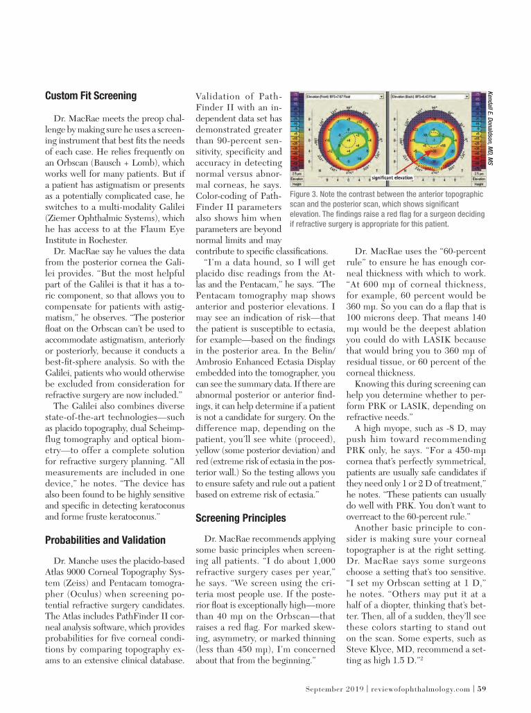
September 2019 | reviewofophthalmology.com | 59
Custom Fit Screening
Dr. MacRae meets the preop chal-lenge by making sure he uses a screen-ing instrument that best fi ts the needs of each case. He relies frequently on an Orbscan (Bausch + Lomb), which works well for many patients. But if a patient has astigmatism or presents as a potentially complicated case, he switches to a multi-modality Galilei (Ziemer Ophthalmic Systems), which he has access to at the Flaum Eye Institute in Rochester.
Dr. MacRae say he values the data from the posterior cornea the Gali-lei provides. “But the most helpful part of the Galilei is that it has a to-ric component, so that allows you to compensate for patients with astig-matism,” he observes. “The posterior fl oat on the Orbscan can’t be used to accommodate astigmatism, anteriorly or posteriorly, because it conducts a best-fi t-sphere analysis. So with the Galilei, patients who would otherwise be excluded from consideration for refractive surgery are now included.”
The Galilei also combines diverse state-of-the-art technologies—such as placido topography, dual Scheimp-flug tomography and optical biom-etry—to offer a complete solution for refractive surgery planning. “All measurements are included in one device,” he notes. “The device has also been found to be highly sensitive and specifi c in detecting keratoconus and forme fruste keratoconus.”
Probabilities and Validation
Dr. Manche uses the placido-based Atlas 9000 Corneal Topography Sys-tem (Zeiss) and Pentacam tomogra-pher (Oculus) when screening po-tential refractive surgery candidates. The Atlas includes PathFinder II cor-neal analysis software, which provides probabilities for five corneal condi-tions by comparing topography ex-ams to an extensive clinical database.
Validation of Path-Finder II with an in-dependent data set has demonstrated greater than 90-percent sen-sitivity, specificity and accuracy in detecting normal versus abnor-mal corneas, he says. Color-coding of Path-Finder II parameters also shows him when parameters are beyond normal limits and may contribute to specifi c classifi cations.
“I’m a data hound, so I will get placido disc readings from the At-las and the Pentacam,” he says. “The Pentacam tomography map shows anterior and posterior elevations. I may see an indication of risk—that the patient is susceptible to ectasia, for example—based on the fi ndings in the posterior area. In the Belin/Ambrosio Enhanced Ectasia Display embedded into the tomographer, you can see the summary data. If there are abnormal posterior or anterior fi nd-ings, it can help determine if a patient is not a candidate for surgery. On the difference map, depending on the patient, you’ll see white (proceed), yellow (some posterior deviation) and red (extreme risk of ectasia in the pos-terior wall.) So the testing allows you to ensure safety and rule out a patient based on extreme risk of ectasia.”
Screening Principles
Dr. MacRae recommends applying some basic principles when screen-ing all patients. “I do about 1,000 refractive surgery cases per year,” he says. “We screen using the cri-teria most people use. If the poste-rior fl oat is exceptionally high—more than 40 mµ on the Orbscan—that raises a red fl ag. For marked skew-ing, asymmetry, or marked thinning (less than 450 mµ), I’m concerned about that from the beginning.”
Dr. MacRae uses the “60-percent rule” to ensure he has enough cor-neal thickness with which to work. “At 600 mµ of corneal thickness, for example, 60 percent would be 360 mµ. So you can do a fl ap that is 100 microns deep. That means 140 mµ would be the deepest ablation you could do with LASIK because that would bring you to 360 mµ of residual tissue, or 60 percent of the corneal thickness.
Knowing this during screening can help you determine whether to per-form PRK or LASIK, depending on refractive needs.”
A high myope, such as -8 D, may push him toward recommending PRK only, he says. “For a 450-mµ cornea that’s perfectly symmetrical, patients are usually safe candidates if they need only 1 or 2 D of treatment,” he notes. “These patients can usually do well with PRK. You don’t want to overreact to the 60-percent rule.”
Another basic principle to con-sider is making sure your corneal topographer is at the right setting. Dr. MacRae says some surgeons choose a setting that’s too sensitive. “I set my Orbscan setting at 1 D,” he notes. “Others may put it at a half of a diopter, thinking that’s bet-ter. Then, all of a sudden, they’ll see these colors starting to stand out on the scan. Some experts, such as Steve Klyce, MD, recommend a set-ting as high 1.5 D.”2
Ken
dall E
. Don
ald
son
, MD
, MS
Figure 3. Note the contrast between the anterior topographic
scan and the posterior scan, which shows signifi cant
elevation. The fi ndings raise a red fl ag for a surgeon deciding
if refractive surgery is appropriate for this patient.
058_rp0919_rcr.indd 59058_rp0919_rcr.indd 59 8/23/19 3:55 PM8/23/19 3:55 PM

60 | Review of Ophthalmology | September 2019
Refractive/CataractRundown R
EV
IEW
Tertiary Approaches
Dr. Donaldson offers pointed preop screening advice based on her work in
a tertiary practice that receives many refractive surgery referrals. “We ben-efi t from insights on the many factors that might rule out surgery—more
so than the typical surgeon would in private practice,” she notes. “My ex-perience can help all surgeons keep in mind what to look for, even if 90 percent of their cases are good candi-dates, such as a 30-year-old with a -3 D sphere. ODs do 75 percent of our screening, so we never see bad pa-tients. But we need to screen further before deciding how to proceed.”
Dr. Donaldson relies on corneal to-pography, corneal tomography, kera-tometry, pachymetry and a thorough exam when screening her patients. Here are some important consider-ations she recommends keeping in mind:
• Anterior elevation map. Con-sider using keratometry to evaluate the shape of the cornea, looking for astigmatism and symmetry. “The corneas should be symmetrical,” she says. “Any elevation steeper than 47.2 D is a red fl ag. You need to investigate for keratoconus.” She also investigates when the manifest astigmatism in the patient’s ophthalmic lens prescription doesn’t match what she sees in topog-raphy. “The cause could be lenticular astigmatism,” she points out. “Or it could involve some other situation that would make us think twice before proceeding with a procedure.”
• Posterior elevation map. “Again, we’re looking at the keratom-etry numbers and the shape—this time from the posterior perspective,” says Dr. Donaldson. “The posterior aspect should match the anterior as-pect. Anything that doesn’t match is also a red fl ag for us.”
• Pachymetry. “Any measure-ment of the cornea that is below 470 microns is a red fl ag,” says Dr. Don-aldson. “We are also looking for thin points. If the center of the cornea is too thin or you fi nd thinness within a radius of 0.5 mm from the center, that should also raise a red fl ag.”
• Underlying pathology. “We make sure we check for ocular
Ken
dall
E. D
on
ald
son
, MD
, MS
Figures 4-A and 4-B. The white sections within the map on the left refl ect signs of dry
eye, while the map on the right shows an eye with a healthy tear fi lm. Your patient’s eyes
should be free of dry eye before screening for refractive surgery.
Figure 5. This patient
has anterior
basement membrane
dystrophy, which will
need to be resolved
before screening for
surgery. The placido
rings on the
topographic map are
not intact, unlike the
round shapes found in
healthy eyes.
Figure 6. This map for the Pentacam Belin/Ambrosio Enhanced Ectasia Display shows an
elevated posterior fl oat that’s bowed inward, a sign of corneal weakness not detected on
placido scans that could lead to ectasia. A surgeon should opt for PRK instead LASIK or
SMILE in these cases.
(Continued on page 73)
Ken
dall E
. Don
ald
son
, MD
, MS
058_rp0919_rcr.indd 60058_rp0919_rcr.indd 60 8/23/19 3:55 PM8/23/19 3:55 PM

References: 1. Holly FJ. Lacrophilic ophthalmic demulcents. US Ophthalmic Rev. 2007;3:38-41. 2. FRESHKOTE PF Drug Facts. Eyevance Pharmaceuticals LLC; 2018. 3. Holly FJ. Colloidal Osmosis — Oncotic Pressure. Grapevine, TX: Dry Eye Institute; 2006. 4. Fuller DG, Connor CG. Safety and effi cacy of FreshKote® used as a rewetting agent in Lotrafi lcon-A® contact lens wearers. Poster presented at: American Academy of Optometry Annual Meeting; November 17-20, 2010; San Francisco, CA. 5. Nemera. Novelia®. https://nemera.net/products/ophthalmic-novelia-eyedropper/. Accessed March 14, 2019.
nurtureTHEIR EYES
Take a fresh approach to treating dry eye symptoms
with FRESHKOTE® Preservative Free (PF)
Patented polymer blend of polyvinyl alcohol and povidone supports
all layers of the tear fi lm1,2
Triple-acting formula reduces evaporation and increases stability of the tear
fi lm while high oncotic pressure draws excess water from epithelial cells1,3
Compatible for use with contact lenses4
State-of-the-art multidose bottle provides more than a month’s worth
of preservative-free drops when dosed 4 times a day5
Visit EYEVANCE BOOTH 7309 at AAO 2019
© 2019 Eyevance Pharmaceuticals LLC. All rights reserved.FRESHKOTE® is a registered trademark of Eyevance Pharmaceuticals LLC.Novelia® is a registered trademark of Nemera Development.FPF-08-19-AD-24
RECOMMEND SOOTHING RELIEF
Visit FRESHKOTEPF.com to learn more
RP0919_EyevanceFresh.indd 1 8/13/19 12:01 PM
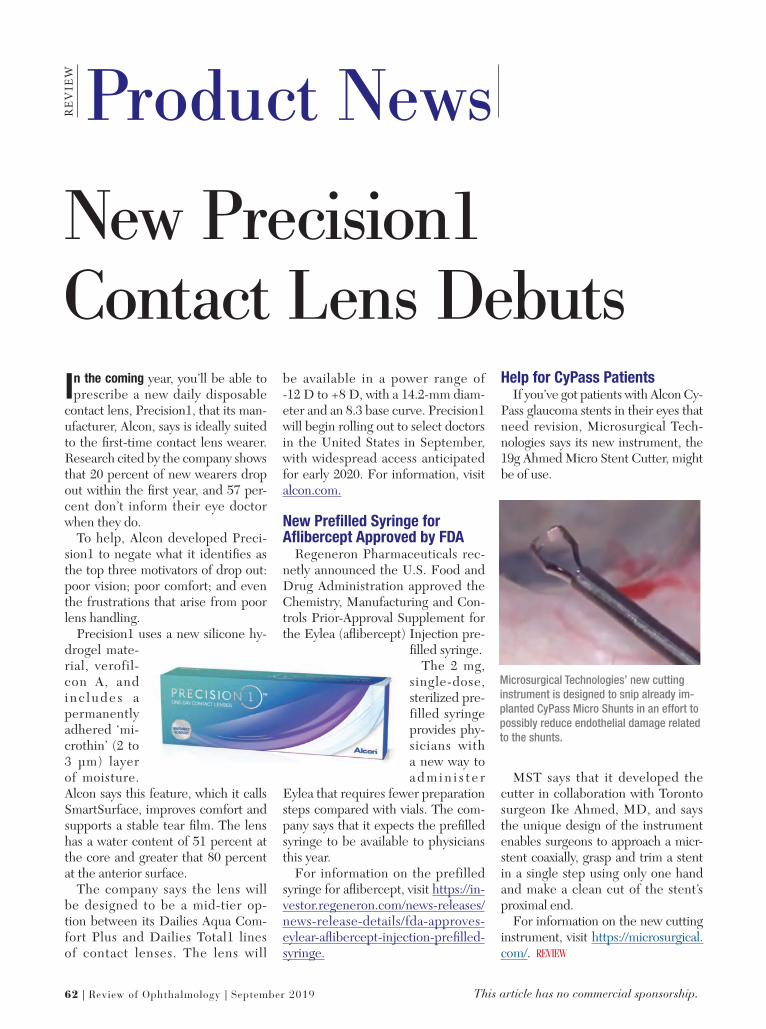
62 | Review of Ophthalmology | September 2019 This article has no commercial sponsorship.
Product NewsREV
IEW
In the coming year, you’ll be able to prescribe a new daily disposable
contact lens, Precision1, that its man-ufacturer, Alcon, says is ideally suited to the fi rst-time contact lens wearer. Research cited by the company shows that 20 percent of new wearers drop out within the fi rst year, and 57 per-cent don’t inform their eye doctor when they do.
To help, Alcon developed Preci-sion1 to negate what it identifi es as the top three motivators of drop out: poor vision; poor comfort; and even the frustrations that arise from poor lens handling.
Precision1 uses a new silicone hy-drogel mate-rial, verofil-con A, and i n c l u d e s a permanently adhered ‘mi-crothin’ (2 to 3 µm) layer of moisture. Alcon says this feature, which it calls SmartSurface, improves comfort and supports a stable tear fi lm. The lens has a water content of 51 percent at the core and greater that 80 percent at the anterior surface.
The company says the lens will be designed to be a mid-tier op-tion between its Dailies Aqua Com-fort Plus and Dailies Total1 lines of contact lenses. The lens will
be available in a power range of -12 D to +8 D, with a 14.2-mm diam-eter and an 8.3 base curve. Precision1 will begin rolling out to select doctors in the United States in September, with widespread access anticipated for early 2020. For information, visit alcon.com.
New Prefi lled Syringe for Afl ibercept Approved by FDA
Regeneron Pharmaceuticals rec-netly announced the U.S. Food and Drug Administration approved the Chemistry, Manufacturing and Con-trols Prior-Approval Supplement for the Eylea (afl ibercept) Injection pre-
fi lled syringe. The 2 mg,
single-dose, sterilized pre-filled syringe provides phy-sicians with a new way to a d m i n i s t e r
Eylea that requires fewer preparation steps compared with vials. The com-pany says that it expects the prefi lled syringe to be available to physicians this year.
For information on the prefilled syringe for afl ibercept, visit https://in-vestor.regeneron.com/news-releases/news-release-details/fda-approves-eylear-afl ibercept-injection-prefi lled-syringe.
Help for CyPass PatientsIf you’ve got patients with Alcon Cy-
Pass glaucoma stents in their eyes that need revision, Microsurgical Tech-nologies says its new instrument, the 19g Ahmed Micro Stent Cutter, might be of use.
MST says that it developed the cutter in collaboration with Toronto surgeon Ike Ahmed, MD, and says the unique design of the instrument enables surgeons to approach a micr-stent coaxially, grasp and trim a stent in a single step using only one hand and make a clean cut of the stent’s proximal end.
For information on the new cutting instrument, visit https://microsurgical.com/.
New Precision1 Contact Lens Debuts
Microsurgical Technologies’ new cutting
instrument is designed to snip already im-
planted CyPass Micro Shunts in an effort to
possibly reduce endothelial damage related
to the shunts.
062_rp0919_products.indd 62062_rp0919_products.indd 62 8/23/19 6:13 PM8/23/19 6:13 PM

RP0919_Topcon.indd 1 8/21/19 11:12 AM

RE
VIE
W
64 | Review of Ophthalmology | September 2019
the laser is more expensive, and in-surance usually won’t pay for it, so patients have to pay out of pocket; there’s a cost to the surgeon to use the laser that has to be passed on to the patient.
“The diamond burr is inexpen-sive, simple, and easy to use in the offi ce setting,” Dr Raizman contin-ues. “However, if the burr is used too aggressively, you can remove more of Bowman’s layer in some areas than others or even remove some stroma, and that can create irregular astigmatism and affect the patient’s vision sometimes. But I should emphasize the burr is quite practical, and it’s not hard to use it safely, so it’s often a good choice for surgeons who don’t have access to the excimer laser or for patients who can’t afford it.”
Dr. Raizman adds that while de-bridement is about 50-percent ef-fective in these cases, the excimer laser is 90 to 95 percent effective for resolving the recurrent erosions with a single treatment. “I don’t personally have enough experience with the diamond burr, but from the medical literature, it’s probably equally as effective as the excimer laser,” he says.
For recurrent erosions, Dr. Rapuano prefers a diamond burr treatment. “You can perform ex-cimer laser phototherapeutic kera-tectomy, but I don’t usually do that,” he says. “I usually use the diamond burr, which I find to be as effec-tive as the excimer laser and much faster, easier to schedule, and less expensive. However, if you’ve got some scarring on the anterior part of the cornea due to multiple erosions, then you can do excimer laser pho-totherapeutic keratectomy. In that case, I strip the epithelium off and
then, instead of using a diamond burr to polish it, I use the excimer laser. The nice thing about the ex-cimer is that it will polish a big area pretty uniformly.”
Pearls
According to Dr. Stein, refractive surgeons should always be looking for epithelial basement membrane dystrophy. Most patients with this condition are asymptomatic, but it can have a signifi cant impact on the outcome of refractive surgery. “If patients have epithelial basement membrane dystrophy, they are far better off having PRK than LASIK,” he says. “If they have loose epithe-lium that goes undetected, and they have LASIK, the epithelium can slough off during the procedure, and the healing can be very delayed. So, it’s important to rule out cases of epithelial basement membrane dystrophy in patients who are in-terested in laser vision correction. Often, the best way is the use of fl u-
orescein dye and a blue light using the slit lamp to look for abnormal breakup in focal areas of the cornea. That’s very suggestive of epithelial basement membrane dystrophy.” He adds that all LASIK surgeons need to ask about patients’ past his-tory of corneal erosions.
Epithelial basement membrane dystrophy can also mimic other condi-tions. “These patients can have false ectasia or pseudokeratoconus, and it’s very important that the surgeon iden-tify that it’s not keratoconus or pellucid marginal degeneration but is in fact epithelial basement membrane dys-trophy,” says Dr. Stein, “and they don’t require corneal cross-linking. The rest of the cornea is totally normal, and these patients don’t need corneal stiff-ening.”
Dr. Raizman adds that he is re-ferred a lot of patients with anterior basement membrane dystrophy for treatment prior to cataract surgery because the keratometry readings are a little bit irregular. “These patients often say that they don’t really mind wearing glasses after their surgery,” he says. “They’re not necessarily look-ing for the best uncorrected vision af-ter cataract surgery. And in that case, if the vision isn’t signifi cantly affected by the basement membrane dystro-phy, there’s not really a reason to do a debridement prior to your IOL calcu-lations. But for patients who want the best possible uncorrected vision and are looking for a specific refractive outcome, those patients should have a debridement prior to measuring the cornea. My point is that it’s not man-datory, so it doesn’t have to be done on every patient. It depends on the patient’s postop needs.”
None of the physicians quoted in the article have a financial interest in any of the products or procedures mentioned.
Dr. Raizman says that while epithelial debridement is about 50-percent effective, the excimer laser is 90- to 95-percent
effective for resolving the recurrent erosions
with a single laser treatment.
EBMDFeature
(Continued from page 57)
052_rp0919_f4.indd 64052_rp0919_f4.indd 64 8/23/19 5:41 PM8/23/19 5:41 PM

Research Review R
EV
IEW
September 2019 | reviewofophthalmology.com | 65This article has no commercial sponsorship.
Interrupting Diabetic Retinopathy TreatmentResearchers report increasing in-
terest in anti-VEGF therapy for treating proliferative diabetic reti-nopathy, since anti-VEGF therapy has been shown to be noninferior to panretinal photocoagulation, the heretofore standard treatment for achieving regression and stabilization of PDR. Their study shows, however, that interruptions in anti-VEGF treat-ment for PDR can result in marked progression of disease and potentially devastating visual consequences.
In a retrospective, multicenter case series, researchers analyzed 13 eyes of 12 patients with type 2 diabetes, aged 57 ±10 years. To refl ect real-world con-ditions in which diabetic patients tend to “underuse eye care services and are prone to signifi cant losses to follow-up” because of illness, fi nancial hardship or noncompliance, the study sample included only those patients who were temporarily lost to follow-up and treat-ed exclusively with anti-VEGF therapy for either PDR or nonproliferative dia-betic retinopathy, with or without dia-betic macular edema.
Baseline disease characteristics, cause and duration of treatment inter-ruption, resulting disease progression, complications and outcomes were as-sessed. Reasons for treatment hiatus (median: 12 months) included inter-current illness (31 percent), noncom-pliance (31 percent) and fi nancial is-sues (15 percent). The authors report
that 77 percent of eyes lost ≥3 lines of visual acuity on the Snellen chart, with 46 percent of eyes having a fi nal visual acuity of hand motion or worse.
Though studies have shown closely-monitored anti-VEGF therapy is ef-fective, especially for ischemic diabetic retinopathy and PDR, the study au-thors conclude that these controlled studies may give false assurance be-cause anti-VEGF therapy is unable to reverse retinal ischemia or fully ad-dress diabetic retinopathy. Addition-ally, the real-world situations faced by diabetic patients lead to interruptions in treatment that negatively affect out-comes.
J Ophthalmol 2019;204:13-18. Wubben TJ and Johnson MW. For the Anti-VEGF Treatment Interruption Study Group.
Impact of EBMD and SND on Biometry Measurements
In a retrospective case series, re-searchers from Duke University analyzed 39 eyes of 30 patients who were evaluated for cataract surgery with documented evidence of Salz-mann’s nodular degeneration (SND) or epithelial basement membrane dys-trophy (EBMD) and who were also scheduled for surgical intervention for corneal irregularities before cataract surgery. The study found that SND and EBMD can adversely affect kera-tometry and biometry measurements for IOL selection. Here are some of
the fi ndings:• The EBMD group (26 eyes).
The difference in K measurements before and after intervention showed a mean K value increase (p<0.001). For biometry, the predicted IOL spherical power closest to a spherical equiva-lent of zero (p<0.001) changed in 21 of 26 eyes (8=0.5 D; 9=1.0 D; 4>1.0 D). For toric IOL-eligible eyes, there was a mean predicted cylinder power change of 1.2 D; recommended toric power changed for 16 of 24 eyes.
• The SND group (13 eyes). The difference in K measurements before and after intervention showed a mean K value increase (p=0.023). For biom-etry, the predicted IOL spherical pow-er closest to a SE of zero (p<0.041) changed in 11 of 13 eyes (3=0.5 D; 3=1.0 D; 5>1.0 D). For toric IOL-eligible eyes, there was a mean pre-dicted cylinder power change of 1.5 D; recommended toric power changed for 10 of 11 eyes.
The researchers conclude that EBMD and SND have signifi cant ef-fects on biometry measurements and IOL calculations, and that optimizing the ocular surface and being aware of the effects of these conditions are important steps in planning cataract procedures.
J C a t a r a c t R e f r a c t S u r g 2019;45:1119-1123.Goerlitz-Jessen MF, Gupta PK, Kim T. De Salles MC, Amrén U, Kvanta A, and Epstein DL.
065_rp0919_rr.indd 65065_rp0919_rr.indd 65 8/23/19 5:00 PM8/23/19 5:00 PM

66 | Review of Ophthalmology | September 2019 This article has no commercial sponsorship.
Glaucoma Management Edited by Kuldev Singh, MD, MPH, and Peter A. Netland, MD, PhDR
EV
IEW
Glaucoma surgery is generally undertaken when medical therapy
and appropriate laser treatment fail to produce adequate intraocular pressure reduction. In recent years, the options for surgically managing glaucoma in that situation have great-ly expanded with the introduction of the new “minimally invasive glaucoma surgeries,” or MIGS. But despite be-ing known for their excellent safety profile, MIGS haven’t replaced the traditional glaucoma surgeries—trabeculectomy and tube shunt im-plantation. That’s true because tube shunts and trabeculectomies are still the most effective ways to achieve the very low IOP that’s required for some patients.
However, having two surgical op-tions that are capable of producing very low pressures has caused some surgeons to wonder whether one might be superior to the other in different surgical situations. Here, I’d like to share some of what the Primary Tube vs. Trabeculectomy Study is revealing about performing these procedures on eyes that have not undergone previous incisional ocular surgery.
Comparing Tubes and Trabs
Glaucoma specialists continue to have different opinions regarding the best surgical approach for patients with medically uncontrolled glaucoma. Medicare claims data indicate that tube shunts are in-creasingly being used as an alter-native to trabeculectomy. Surveys of the American Glaucoma Society membership have also demonstrated a growing preference for tube shunts over trabeculectomy in many clinical scenarios.
One clinical situation that’s part of this debate is whether a tube shunt or trabeculectomy is preferable when operating on a “virgin” eye that hasn’t undergone previous incisional surgery. In the most recent AGS survey, trabeculectomy has remained the most popular option as an initial procedure for eyes with POAG, but a growing number of glaucoma sur-geons prefer the use of tube shunts as an initial glaucoma procedure. Until now, however, this surgical choice was being made with limited data.
The Primary Tube Versus Tra-beculectomy (PTVT) Study was un-
dertaken to provide that data. It’s an investigator-initiated trial designed to compare the safety and effi cacy of trabeculectomy with mitomycin-C to tube shunt surgery, when performed as an initial procedure in eyes that haven’t had previous incisional ocular surgery.
Study Design
The design of the PTVT Study is similar to that of the Tube Versus Trabeculectomy (TVT) Study, an earlier trial that enrolled patients with prior cataract extraction and/or failed filtering surgery. In contrast, the PTVT Study recruited patients without prior incisional ocular sur-gery. Participants in the PTVT Study were 18 to 85 years old, with IOP ≥18 mmHg and ≤40 mmHg on max-imum tolerated medical therapy. The vast majority of the subjects had primary open-angle glaucoma, and there were no signifi cant differences between baseline demographic and ocular characteristics of the groups.
Reasons for exclusion included a narrow anterior chamber angle; secondary glaucomas such as neo-
The Primary Tube vs. Trabeculectomy Study is revealing useful data regarding tubes versus trabs in virgin eyes.
What We’re Learning From the PTVT Study
Steven J. Gedde, MD, Miami
066_rp0919_gm.indd 66066_rp0919_gm.indd 66 8/23/19 3:44 PM8/23/19 3:44 PM

September 2019 | reviewofophthalmology.com | 67
vascular, uveitic, iridocorneal en-dothelial syndrome, epithelial downgrowth and steroid-induced glaucoma; severe posterior blephar-itis; conjunctival scarring; a func-tionally signifi cant cataract; and any anticipated need for additional ocular surgery.
As in the TVT Study, participants in the PTVT Study were randomized to a 350-mm2 Baerveldt glaucoma implant or a trabeculectomy with MM-C, although the dosage of MM-C was lower than in the TVT Study. During the design phase of the study, we considered letting each glaucoma surgeon implant whichever tube shunt was preferred. However, the biostatisticians tasked with analyzing our data advised us to standardize the surgeries as much as possible.
It was a consensus opinion among the investigators in our study that the 350-mm2 Baerveldt implant offered the highest effi cacy among available implants due to its large surface area, and this has been supported by two landmark clinical trials—the Ahmed Baerveldt Comparison (ABC) Study and the Ahmed Versus Baerveldt (AVB) Study. So, a 350-mm2 Baerveldt glaucoma implant was used in all patients randomized to the tube group. We also standardized
other parts of the procedure, such as having the implant placed in the superotemporal quadrant with the tube in the anterior chamber, and a complete restriction of flow at the time of surgical implantation. We standardized aspects of the trabeculectomies as well. An MM-C dosage of 0.4 mg/ml for two minutes was used in all patients, and the surgery was performed superiorly.
Despite standardization of many aspects of each procedure, surgeons were allowed suffi cient fl exibility to perform both operations in a manner in which they were comfortable and proficient. For example, surgeons were allowed to decide whether a limbus- or fornix-based conjunctival flap was used. The method of tube ligation, the size of the trabeculectomy fl ap and the number of fl ap sutures were also left to the surgeon’s dis-cretion.
Sixteen clinical centers were in-volved in the study. Patients were randomly assigned to one of the two groups, with follow-up visits scheduled for one day, one week, one month, three months, one year, 18 months and two, three, four and five years postoperatively. The primary outcome measure was the rate of surgical failure, using criteria consistent with
recommendations from the World Glaucoma Association.
The Data (So Far)
Results from the PTVT Study at year three were presented at the 2019 Annual AGS Meeting. Highlights in-cluded:
• IOP reduction. The pressure reduction was greater in the tra-beculectomy group at all time points, and that difference was statistically significant—with the exception of the 18-month and two-year time points. Furthermore, the greater degree of pressure reduction in the trabeculectomy group was achieved with fewer postoperative glaucoma medications. This clearly suggests that trabeculectomy with MM-C was more effective at reducing IOP than tube shunt implantation in this population.
• Rate of surgical failure. The primary outcome measure for the study was the rate of surgical failure, using Kaplan-Meier survival analysis. The rate of surgical failure increased over time, which came as no surprise; even glaucoma surgeries that are initially successful can eventually fail. At one year, the tube shunt group had a signifi cantly higher failure rate
PTVT Study: Summary of Findings at Three Years
IOP reduction IOP reduction was greater in the trabeculectomy group at all time points, and fewer adjunctive medications were required to achieve this.
Rate of surgical failure The tube group had a higher failure rate at one year, but no signifi cant difference between the groups was seen at three years.
Reasons for failure In both groups the most common reason for treatment failure was insuffi cient IOP reduction. Failure because of a need for additional glaucoma surgery was more common in the tube group, while hypotony failures occurred exclusively in the trabeculectomy group.
Risk factors for failure Lower preoperative IOP was predictive of failure, especially in the tube shunt group.Medical therapy The tube group required more adjunctive glaucoma medications postoperatively.Complications Many postoperative complications were observed in both groups, but most were self-limited. More
complications occurred in the trabeculectomy group, especially during the fi rst month after surgery. Postoperative interventions Postoperative interventions were performed with similar frequency in both groups.Visual acuity No signifi cant difference in visual outcomes were seen between the groups.Cataract formation Cataract progression occurred at a similar rate in both groups.
066_rp0919_gm.indd 67066_rp0919_gm.indd 67 8/23/19 3:45 PM8/23/19 3:45 PM

GlaucomaManagement R
EV
IEW
68 | Review of Ophthalmology | September 2019
than the trabeculectomy group. The cumulative probability of failure in the trabeculectomy group continued to trend somewhat lower than the tube group over time. However, the difference in failure rate between the groups was no longer statistically sig-nifi cant at three years.
• Reasons for failure. We found some differences in the reasons for surgical failure between the two study groups. The most common reason for failure in both groups was insuffi cient pressure reduction; either the pressure was above 21 mmHg at two consecutive visits, or it wasn’t reduced 20 percent below baseline at two consecutive visits. Other rea-sons for failure included needing a reoperation for glaucoma or having hypotony. Interestingly, all of the hypotony failures occurred in the trabeculectomy group, while more reoperations were seen in the tube group.
• Need for adjunctive medical therapy. Patients who didn’t fail were subdivided into complete or qualified successes. Complete suc-cesses didn’t need postop adjunc-tive medical therapy; qualified suc-cesses were using glaucoma medica-tions. The rate of complete success was significantly higher in the trabeculectomy group than in the
tube shunt group. (That’s an im-portant consideration when man-aging a patient who can’t tolerate medications, or a patient who is poorly compliant.)
• Baseline factors associated with surgical failure. We performed a risk factor analysis to identify baseline factors that were associated with failure in the study. The only significant predictor of failure was preoperative IOP. The patients with lower preoperative pressures had a higher risk of failure, and this was especially true in the tube group. (See fi gure, above.) In my opinion, this is one of the study’s most interesting and important fi ndings.
• Complications. In terms of complications, the PTVT Study’s fi ndings have been similar to what’s been observed in other prospective glaucoma surgical trials, including the TVT, ABC and AVB studies. Postoperative complications are very common after traditional glaucoma surgery, whether it be a tube shunt or a trabeculectomy. Fortunately, most of these complications are self-limited and resolve without any specifi c in-tervention.
We did, however, find some dif-ferences between the types of com-plications occurring in the two treatment groups:
— Early postoperative comp-lications, defi ned as those occurring in the fi rst month after surgery, were more common in the trabeculectomy group than in the tube group.
— The rate of later postoperative complications (occurring after a month) wasn’t signifi cantly different between the two groups.
— The rate of serious compli-cations, defined as those requiring a reoperation to manage the com-plication, and/or resulting in a vision loss of two or more lines of Snellen visual acuity, wasn’t significantly different between the treatment groups.
— Overall, the rate of reoperation for complications trended higher in the trabeculectomy group compared to the tube group.
The complications data suggests that tube shunt surgery may have a slightly better safety profile than trabeculectomy with MM-C, at least during the first three years postop. If you combine all complications to-gether, they were signifi cantly more common in the trabeculectomy group than the tube group. However, the PTVT trial will continue out to five years, and the long-term data will be important for evaluating these two surgical procedures, in terms of both safety and effi cacy.
Probability of Surgical Failure Based on Preoperative IOP
Cum
ulat
ive
Prop
ortio
ns F
ailin
g 0.6
0.5
0.4
0.3
0.2
0.1
0.0
0.6
0.5
0.4
0.3
0.2
0.1
0.0
0.6
0.5
0.4
0.3
0.2
0.1
0.0
0 6 12 18 24 30 36 0 6 12 18 24 30 36 0 6 12 18 24 30 36
IOP < 21 mmHg IOP 21-25 mmHg IOP >25 mmHg
Follow-up (months)
60.0%
39.3%
13.8%
26.4%29.7%
10.0%
Tube group Trabeculectomy group
066_rp0919_gm.indd 68066_rp0919_gm.indd 68 8/23/19 3:45 PM8/23/19 3:45 PM

Join us for a presentation
and dinner.
San Francisco MoMA
151 3rd Street
San Francisco, CA 94103
Sunday, October 13, 2019
Registration: 6:30PM
Program & Dinner: 7:00PM
S U N D A Y , O C T O B E R 1 3 , 2 0 1 9 • S A N F R A N C I S C O , C A L I F O R N I A
Capacity is limited and reserved for individuals who are able to attend the entire educational session.© Johnson & Johnson Surgical Vision, Inc. 2019 P2019MLT4235
www.reviewofophthalmology.com/CataractSF
R E S E R V E Y O U R S E A T :
MODERNCATARACT SURGERY
REIMAGINED Presented by Johnson & Johnson Surgical Vision
Y O U A R E I N V I T E D T O A C O M P L I M E N TA R Y D I N N E R D I S C U S S I O N AT M o M A , S F .
Helga P. Sandoval, MD, MSCR
Director of ResearchCarolina EyeCare Physicians,
Charleston, SC
Karolinne Maia Rocha, MD, PhD
Director of Cornea & Refractive SurgeryStorm Eye Institute
Medical University of South CarolinaCharleston, SC
Mark Kontos, MD
Empire EyeSpokane Valley, WA
Paul Harasymowycz, MSc, MD, FRCSC
Chief of Glaucoma,University of Montreal
Medical Director,Bellevue Ophthalmology Clinics
Montreal, Canada
MODERATOR:
Kevin Waltz, OD, MD
Partner, Whitson VisionChair, Board of Directors -
Central American Eye ClinicsIndianapolis, IN
y Johnson & JoPresented byy ohnson Surgical Vision

GlaucomaManagement R
EV
IEW
70 | Review of Ophthalmology | September 2019
• Number of postoperative interventions. In my experience, postoperative management fol-lowing tube shunt surgery tends to be less involved than postoper-ative management following tra-beculectomy. However, in this study the number of postop interventions performed in the clinic, such as laser suture lysis or pulling a rip-cord suture, wasn’t signifi cantly different whether the patient received a tube or a trabeculectomy.
• Visual acuity. We didn’t find any signifi cant difference between the treatment groups with regard to visual acuity outcomes.
• Cataract formation. All of the patients in our study were phakic. After three years of follow-up, about a third of the patients in the study had undergone cataract extraction.
There’s ample evidence in the lit-erature to suggest that glaucoma surgery, whether it be trabeculectomy or tube shunt surgery, accelerates the development of cataract. This data suggests that cataract progression occurs at a similar rate among patients undergoing trabeculectomy or tube shunt surgery.
• Patient comfort. One of the complications we specifically asked patients about was dysesthesia, or patient discomfort. We found no significant difference in patient-re-ported dysesthesia between the two procedures.
Take-home Highlights (So Far)
What useful information can we take away from this study at the three-year follow-up mark?
• Preoperative IOP impacts the likelihood of success. When patients have lower levels of preoperative IOP—in our post hoc analysis the cutoff was less than 21 mmHg—trabeculectomy appears to be a more successful operation than tube shunt surgery. I suspect this is related to
the titratability of trabeculectomy; it allows us to do laser suture lysis or use releasable sutures to titrate the pressure to lower levels in the early postoperative period. I think that’s particularly valuable in patients who have initial low pressure.
• Both tube shunt surgery and trabeculectomy with MM-C are very effective at lowering IOP.
Mean postoperative pressures were in the low teens in both groups throughout three years of follow-up; most patients ended up with pressures of 14 mmHg or less. MIGS procedures seldom achieve this degree of IOP reduction. As pre-viously noted, traditional glaucoma surgeries are still an important part of our armamentarium.
• Your choice of tube or trabec-ulectomy may ultimately come down to safety vs. effi cacy. There seems to be an inevitable tradeoff
between safety and effi cacy in glau-coma surgery. In our study com-paring tube shunt implantation and trabeculectomy, the data suggests that trabeculectomy is more effective, but more likely to be associated with complications. A similar dichotomy was seen in the ABC and AVB stud-ies; the Baerveldt implant was more effective, while the Ahmed valve was safer. Likewise, when com-paring traditional glaucoma surgeries to MIGS, we find tube shunts and trabeculectomy are more effi cacious, but MIGS are safer. So added effi cacy seems to be achieved at the expense of reduced safety—at least with the existing glaucoma surgical options.
This shouldn’t be surprising. Many of the complications that we contend with in glaucoma surgery are related to hypotony, such as anterior chamber shallowing and choroidal effusions, and sometimes more serious complications, includ-ing suprachoroidal hemorrhage and hypotony maculopathy. (Of course, it’s possible that some future glaucoma surgery will be both highly effective and low-risk.)
A Few Final Thoughts
The purpose of a trial like the PTVT Study is to provide useful information that will help surgeons select the best glaucoma procedure for an individual patient. Data produced by the trial thus far hasn’t demonstrated clear su-periority of one of these procedures over the other.
However, there’s another important consideration when choosing a glaucoma surgical procedure, one that was not addressed in the PTVT Study: the surgeon’s experience and comfort with each procedure. All of the surgeons in the PTVT Study were profi cient and experienced with both of the procedures being studied, but this may not be the case with
Randomized clinical trials offer the highest level of evidence-based medicine, but caution
should be used in extrapolating study results to dissimilar patients. We now
have two trials that compared tube shunts and trabeculectomy
.... Remember to apply the lessons learned
only to the appropriate patient population.
(Continued on page 73)
066_rp0919_gm.indd 70066_rp0919_gm.indd 70 8/23/19 3:46 PM8/23/19 3:46 PM

www.reviewofophthalmology.com
The Review Group offers eyecare
practitioners quality informational
resources dedicated to the growth
and education of the profession. The
Review Group offers a variety of print
and online products to enrich your
patient care and practice needs.
Jobson Medical Information LLC
The Review Group
ENRICH YOUR PRACTICEReview of Ophthalmology delivers current and comprehensive information focusing on topics such as disease diagnosis, surgical techniques and new technologies.
The Review Group’s Ophthalmic Product Guide brings you the latest products and technology on the market. Published every February and July.
The Review Group also distributes a variety of supplements, guides and handbooks with your subscription to Review of Ophthalmology. These publications are designed to keep you informed on what’s new and innovative in the industry on topics ranging from cataract refractive surgery to ocular surface disease.
The Review Group also offers valuable continuing medical education sessions in both print and online formats, allowing a convenient way for you to earn CME credits. In addition, we also offer an impressive fleet
of free e-newsletters—such as Review of Ophthalmology Online and Review of Ophthalmology’s Retina Online—so you can keep up to date on breaking news and the latest research.
The Review Group also spearheads meetings and conferences, bringing together experts in the field and providing a forum for practitioners that allows you to educate, and learn from others in the profession. These meetings cover a broad range of topics in the form of educational or promotional roundtables and forums.
TowOdnro
TcbcI
r
2018_rp_tsrad.indd 902018_rp_tsrad.indd 90 4/13/18 11:28 AM4/13/18 11:28 AM

72 | Review of Ophthalmology | September 2019
REVIEW Classifi eds
Targeting Ophthalmologists?
CLASSIFIED ADVERTISING
WORKS
Contact us today for classified advertising:Toll free: 888-498-1460
E-mail: [email protected]
Career Opportunities
Do you have Productsand Services for sale?
CONTACT US TODAY FOR CLASSIFIED ADVERTISING
Toll free: 888-498-1460E-mail: [email protected]
ROPH0719.indd 62 6/9/19 9:54 PM
Akorn Consumer Health 24-27
Phone (800) 579-8327 www.akornconsumerhealth.com
Akorn Pharmaceuticals 55
Phone (800) 932-5676 www.akorn.com
Bausch + Lomb 21, 51
Phone (800) 323-0000Fax (813) 975-7762
Beaver-Visitec International, Inc. 15
Phone (866) 906-8080Fax (866) 906-4304 www.beaver-visitec.com
Bruder Ophthalmic Products 35
Phone (888) 827-8337 [email protected]
Eyevance Pharmaceuticals 7, 61
Phone (817) 677-6120 eyevance.com
Focus Laboratories, Inc. 13
Phone (866) 752-6006Fax (501) 753-6021 www.focuslaboratories.com
Heidelberg Engineering 11
Phone (800) 931-2230Fax (760) 598-3060
Imprimis Pharmaceuticals, Inc. 37
Phone (858) 704-4040Fax (858) 345-1745 www.imprimispharma.com
Kala Pharmaceuticals 17, 18
Phone (781) 996-5252Fax (781) 642-0399 [email protected] www.kalarx.com
Katena 31
Phone (800) 225-1195 www.katena.com
Keeler Instruments 5
Phone (800) 523-5620Fax (610) 353-7814
Lacrimedics, Inc 8
Phone (800) 367-8327Fax (253) 964-2699 [email protected] www.lacrimedics.com
Lombart Instruments 33
Phone (800) 446-8092Fax (757) 855-1232
Myco Industries, Inc./ AB Max™ 2
Phone (800) 721-8006
[email protected] www.AB-Max.com
Oculus, Inc. 39
Phone (888) 284-8004Fax (425) 867-1881
Reichert Technologies 53
Phone (888) 849-8955Fax (716) 686-4545 www.reichert.com
S4OPTIK 41, 43
Phone (888) 224-6012
Shire Ophthalmics 75, 76
www.shire.com
Sight Sciences 23
Phone (877) 266-1144 [email protected] www.sightsciences.com
Varitronics 45
Phone (800) 345-1244Fax (610) 356-1222
AdvertisingIndexR
EV
IEW
This advertiser index is published as a convenience and not as part of the advertising contract. Every care will be taken to index correctly. No allowance will be made for errors due to spelling, incorrect page number, or failure to insert.
072_rp0919_adindex.indd 72072_rp0919_adindex.indd 72 8/23/19 3:00 PM8/23/19 3:00 PM

GlaucomaManagement R
EV
IEW
September 2019 | reviewofophthalmology.com | 73
all surgeons. A surgeon’s familiarity with each operation is an important factor when deciding which surgery to perform.
One interesting question that our study may eventually help answer is how tube shunt and trabeculectomy function is impacted by subsequent cataract surgery. All eyes enrolled in our study were phakic, but a third of the patients had undergone cataract surgery by the three-year time point. Multiple studies have demonstrated that when performing cataract sur-gery in eyes with preexisting tra-beculectomy filtering blebs, there’s some risk of bleb failure. However, there’s less information in the medical literature about how tube shunts fare with subsequent cataract removal. I believe the data coming from a prospective, randomized clinical trial
will add signifi cant new information to help answer this question. However, we’re waiting for the fi ve-year data to do a full statistical analysis relating to this question.
As noted earlier, glaucoma surgical practice patterns appear to be shifting away from trabeculectomy and toward implanting tube shunts. The TVT Study provided data to support this trend, as tube shunt surgery was found to be more suc-cessful, with a lower rate of early postop complications relative to trabeculectomy with MM-C among patients with prior cataract and/or failed glaucoma surgery. Notably, this effi cacy result wasn’t seen in the PTVT Study. However, it’s important to realize that different patient popu-lations were studied in the TVT and PTVT Studies.
Randomized clinical trials offer
the highest level of evidence-based medicine, but caution should be used in extrapolating study results to dissimilar patients. We now have two trials that compared tube shunts and trabeculectomy with MM-C in different populations, and we’re seeing somewhat different results. So remember to apply the lessons learned only to the appropriate patient population.
Dr. Gedde is a professor of oph-thalmology and vice chair of educa-tion at Bascom Palmer Eye Institute. He is a study chairman for the PTVT Study. He has no personal fi nancial ties to any product mentioned, but notes that the PTVT Study was funded by grants from Johnson & Johnson Vision, the National Eye Institute and Research to Prevent Blindness.
(Continued from page 70)
surface disease, anterior basement membrane dystrophy, and (as mentioned previously) keratoconus,” says Dr. Donaldson.
Guarding Against Risks
Dr. MacRae offers this advice to avoid surgical risks: “If you see a marked red zone inferiorly, nasally or temporally, that’s another red flag, another sign of what we call skewing, where the bow tie is bigger down below than above,” he says. “This represents a clear-cut case of keratoconus. No surgery can be done on this patient.”
In a patient with Salzmann’s nodular degeneration, a topographical representation of the cornea will reveal significant irregular astigmatism, another concern, he says.
If Dr. Manche encounters a subtle abnormality, he considers using PRK instead of LASIK. “It’s also possible we’ll choose IOL surgery,” he notes.
He also remains vigilant whenever his screening exam turns up two other key fi ndings:
• A steep or a posterior slope that’s bowed in. “Some people say the earliest sign of keratoconus is a bulge on the posterior cornea,” he notes. “You might also see the anterior cornea bowed forward.”
• A view going from the central to the peripheral cornea that reveals several suggestive indices, including those associated with thinning.
Pulling It All Together
In summary, Dr. Donaldson advises all refractive surgeons to keep the potential outcome of every case in mind. “When you’re screening a patient for an elective procedure, you want to be extra careful and err on the conservative side,” she says. “We have to ask ourselves: How well am I going to make this patient see? Am I going to fl atten the tissue too much? Is the cornea too thin? Will
underlying conditions cause long-term distortion?
“We want to keep them in the normal range of corneal fi tness,” she continues. “Remember to always calculate preoperatively what their corneas will look like postoperatively. We’re looking at how LASIK will affect patients tomorrow and 10 years from now.”
Dr. Donaldson reports financial relationships with Alcon, Allergan, Johnson & Johnson Vision, Sun Pharmaceuticals, Shire, Bausch + Lomb, Kala Pharmaceuticals, EyeVance, Lumenis, Omeros and Carl Zeiss Meditech.
Dr. Manche is a consultant for Allergan, Avedro, J & J Vision and Carl Zeiss Meditec. He performs sponsored research for Allergan, Alcon, Avedro, Carl Zeiss Meditec and Presbia. He owns equity in Vacu-Site and RxSight. Dr. MacRae reports no fi nancial relationships with relevant companies.
(Continued from page 60)
066_rp0919_gm.indd 73066_rp0919_gm.indd 73 8/23/19 3:46 PM8/23/19 3:46 PM

A P U B L I C A T I O N B Y
Retina Specialist focuses on the latest advances in the diagnosis, medical management and surgical treatment of diseases of the retina, along with practical, real-world advice from leading clinicians and other experts on managing the successful retina practice.
Starting January 2019Retina Specialist will appear bimonthly.
Watch for issues in:
For inquiries contact [email protected]
Advertising opportunitiesJIM HENNE • PUBLISHER • 610-492-1017 • [email protected] MICHELE BARRETT • 215-519-1414 • [email protected]
MICHAEL HOSTER • 610-492-1028 • [email protected]
www.retina-specialist.com
» JANUARY » MARCH» MAY
» JULY» SEPTEMBER» NOVEMBER
2017_retinaspecialist_housead.indd 12017_retinaspecialist_housead.indd 1 11/26/18 10:42 AM11/26/18 10:42 AM

BRIEF SUMMARY: Consult the Full Prescribing Information for complete product information.
INDICATIONS AND USAGE Xiidra®
DOSAGE AND ADMINISTRATION Instill one drop of Xiidra twice daily (approximately 12 hours
Contact lenses should be removed prior to the administration
administration.
CONTRAINDICATIONS Xiidra is contraindicated in patients with known hypersensitivity
formulation.
ADVERSE REACTIONS Clinical Trials Experience Because clinical studies are conducted under widely varying conditions, adverse reaction rates observed in clinical studies of a drug cannot be directly compared to rates in the clinical
approximately 12 months. The majority of the treated patients
dysgeusia and reduced visual acuity. Other adverse reactions
conjunctival hyperemia, eye irritation, headache, increased lacrimation, eye discharge, eye discomfort, eye pruritus and sinusitis. Postmarketing Experience
postapproval use of Xiidra. Because these reactions are reported voluntarily from a population of uncertain size, it is not always possible to reliably estimate their frequency or establish a causal relationship to drug exposure.
Rare cases of hypersensitivity, including anaphylactic reaction, bronchospasm, respiratory distress, pharyngeal edema, swollen tongue, and urticaria have been reported. Eye swelling and rash have been reported.
USE IN SPECIFIC POPULATIONS Pregnancy There are no available data on Xiidra use in pregnant women to
relevant systemic exposures. Intravenous administration of
an increased incidence of omphalocele at the lowest dose
the recommended human ophthalmic dose [RHOD], based on
risk of Xiidra use in humans during pregnancy is unclear.
Animal Data
an increase in mean preimplantation loss and an increased
the RHOD of Xiidra, based on AUC. No teratogenicity was
an increased incidence of omphalocele was observed at the
the rabbit.
Lactation
milk, the effects on the breastfed infant, or the effects on milk
ocular administration is low. The developmental and health
the mother’s clinical need for Xiidra and any potential adverse effects on the breastfed child from Xiidra.
Pediatric Use
years have not been established.
Geriatric Use No overall differences in safety or effectiveness have been observed between elderly and younger adult patients.
NONCLINICAL TOXICOLOGY Carcinogenesis, Mutagenesis, Impairment of Fertility Carcinogenesis: Animal studies have not been conducted
Mutagenesis: in vitro
in vivo mouse micronucleus assay. In an in vitro chromosomal aberration assay using mammalian cells (Chinese
concentration tested, without metabolic activation. Impairment of fertility:
fertility and reproductive performance in male and female treated rats.
Marks designated ®
Patented: please see
Rx Only
RP0919_Shire PI.indd 1RP0919_Shire PI.indd 1 8/13/19 1:35 PM8/13/19 1:35 PM

Indication Xiidra®
Important Safety Information
References:
1. 2.
Ocul Surf 3.
4.
There’s no substitute.2,4
Check out patient resources, insurance coverage, and more at Xiidra-ECP.com
Xiidra is the only lymphocyte function-associated antigen-1 (LFA-1) antagonist treatment for Dry Eye Disease1,2
FIIRST IN CLASS
For additional safety information, see accompanying Brief Summary of Safety Information on the adjacent page and Full Prescribing Information on Xiidra-ECP.com.
®
RP0919_Shire.indd 1RP0919_Shire.indd 1 8/13/19 1:38 PM8/13/19 1:38 PM