RETINAL HOLES AND TEARS - Review of Optometry
100
www.reviewofoptometry.com Focus on Refraction: Sizing Up Strabismus, p. 24 June 15, 2019 10th ANNUAL RETINA REPORT Diabetes: Today and Tomorrow, p. 36 • Stay One Step Ahead of AMD, p. 42 www reviewofoptometry com J 15 2019 A Field Guide to RETINAL HOLES AND TEARS ALSO: My Patient Has Scleritis… Now What?, p. 66 The Real-world Cataract Evaluation, p. 72 INSIDE — EARN 2 FREE CE CREDITS Pathways to Premium IOL Outcomes & Successful Surgical Comanagement, p. 58 Review the clinical appearance of these elusive conditions. p. 52
-
Upload
khangminh22 -
Category
Documents
-
view
0 -
download
0
Transcript of RETINAL HOLES AND TEARS - Review of Optometry

www.reviewofoptometry.com
Focus on Refraction: Sizing Up Strabismus, p. 24
June 15, 2019
10th A N N U A L R E T I N A R E P O R T
Diabetes: Today and Tomorrow, p. 36 • Stay One Step Ahead of AMD, p. 42
www reviewofoptometry comJ 15 2019
A Field Guide to RETINAL HOLES AND TEARS
ALSO:
My Patient Has Scleritis… Now What?, p. 66 The Real-world Cataract Evaluation, p. 72
RE
VIE
W O
F O
PT
OM
ET
RY
■ V
OL
. 15
6 N
O. 6
■ JU
NE
15
, 20
19
■ A
NN
UA
L R
ET
INA
RE
PO
RT
■ C
AT
AR
AC
T E
VA
LU
AT
ION
■ M
AN
AG
ING
SCL
ER
ITIS
INSID
E — E
ARN 2 F
REE CE C
REDITS
Path
ways t
o Pr
emiu
m IO
L Out
com
es &
Succe
ssfu
l Sur
gica
l Com
anag
emen
t, p.
58
Review the clinical appearance of these elusive conditions. p. 52
001_ro0619_fc1.indd 1001_ro0619_fc1.indd 1 6/5/19 2:41 PM6/5/19 2:41 PM

VYZULTA and the V design are trademarks of Bausch & Lomb Incorporated or its affi liates. ©2019 Bausch & Lomb Incorporated or its affi liates. All rights reserved. VYZ.0065.USA.19
Only dual-action VYZULTA reduces intraocular pressure (IOP) by targeting the trabecular meshwork with nitric oxide and the uveoscleral pathway with latanoprost acid1
VYZULTA demonstrated safety profile
in clinical trials
Only 6 out of 811 patients discontinued due to ocular adverse events in APOLLO and LUNAR clinical trials1,8,9
VYZULTA achieved significant and sustained
long-term IOP reductions vs Timolol 0.5%
in pivotal trials7
P<0.001 vs baseline at all pre-specified visits over 12 months in a pooled analysis of APOLLO and LUNAR clinical trials (N=831)
INDICATION
VYZULTA® (latanoprostene bunod ophthalmic solution), 0.024% is indicated for the reduction of intraocular pressure (IOP) in patients with open-angle glaucoma or ocular hypertension.
IMPORTANT SAFETY INFORMATION
• Increased pigmentation of the iris and periorbital tissue (eyelid) can occur. Iris pigmentation is likely to be permanent
• Gradual changes to eyelashes, including increased length, increased thickness, and number of eyelashes, may occur. These changes are usually reversible upon treatment discontinuation
• Use with caution in patients with a history of intraocular infl ammation (iritis/uveitis). VYZULTA should generally not be used in patients with active intraocular infl ammation
• Macular edema, including cystoid macular edema, has been reported during treatment with prostaglandin analogs. Use with caution in aphakic patients, in pseudophakic patients with a torn posterior lens capsule, or in patients with known risk factors for macular edema
IMPORTANT SAFETY INFORMATION cont’d
• There have been reports of bacterial keratitis associated with the use of multiple-dose containers of topical ophthalmic products that were inadvertently contaminated by patients
• Contact lenses should be removed prior to the administration of VYZULTA and may be reinserted 15 minutes after administration
• Most common ocular adverse reactions with incidence 2% are conjunctival hyperemia (6%), eye irritation (4%), eye pain (3%), and instillation site pain (2%)
For more information, please see Brief Summary of Prescribing
Information on next page.
References: 1. VYZULTA Prescribing Information. Bausch & Lomb Incorporated. 2. Cavet ME. J Ocul Pharmacol Ther. 2018;34(1):52-60. DOI:10.1089/jop.2016.0188. 3. Wareham LK. Nitric Oxide. 2018;77:75-87. DOI:10.1016/j.niox.2018.04.010. 4. Stamer DW. Curr Opin Ophthalmol. 2012;23:135-143. DOI:10.1097/ICU.0b013e32834ff 23e. 5. Cavet ME. Invest Ophthalmol Vis Sci. 2015;56(6):4108-4116. 6. Kaufman PL. Exp Eye Research. 2008;861:3-17. DOI:10.1016/j.exer.2007.10.007. 7. Weinreb RN. J Glaucoma. 2018;27:7-15. 8. Weinreb RN. Ophthalmology. 2016;123(5):965-973. 9. Medeiros FA. Am J Ophthalmol. 2016;168:250-259.
Visit VYZULTANOW.com
to see our effi cacy results
EXPAND THE TRABECULAR MESHWORK WITH THE POWER OF NITRIC OXIDE2-6
RP0619_B & L Vyzulta.indd 1 5/15/19 9:37 AM

BRIEF SUMMARY OF PRESCRIBING INFORMATION
This Brief Summary does not include all the information needed to use VYZULTA safely and effectively. See full Prescribing Information for VYZULTA.
VYZULTA® (latanoprostene bunod ophthalmic solution), 0.024%, for
topical ophthalmic use. Initial U.S. Approval: 2017
1 INDICATIONS AND USAGE
VYZULTA® (latanoprostene bunod ophthalmic solution) 0.024% is indicated for the reduction of intraocular pressure (IOP) in patients with open-angle glaucoma or ocular hypertension.
4 CONTRAINDICATIONS
None
5 WARNINGS AND PRECAUTIONS
5.1 Pigmentation
VYZULTA® (latanoprostene bunod ophthalmic solution), 0.024% may cause changes to pigmented tissues. The most frequently reported changes with prostaglandin analogs have been increased pigmentation of the iris and periorbital tissue (eyelid).
Pigmentation is expected to increase as long as latanoprostene bunod ophthalmic solution is administered. The pigmentation change is due to increased melanin content in the melanocytes rather than to an increase in the number of melanocytes. After discontinuation of VYZULTA, pigmentation of the iris is likely to be permanent, while pigmentation of the periorbital tissue and eyelash changes are likely to be reversible in most patients. Patients who receive prostaglandin analogs, including VYZULTA, should be informed of the possibility of increased pigmentation, including permanent changes. The long-term effects of increased pigmentation are not known.
Iris color change may not be noticeable for several months to years. Typically, the brown pigmentation around the pupil spreads concentrically towards the periphery of the iris and the entire iris or parts of the iris become more brownish. Neither nevi nor freckles of the iris appear to be affected by treatment. While treatment with VYZULTA® (latanoprostene bunod ophthalmic solution), 0.024% can be continued in patients who develop noticeably increased iris pigmentation, these patients should be examined regularly [see Patient Counseling Information (17) in full Prescribing Information].5.2 Eyelash Changes
VYZULTA may gradually change eyelashes and vellus hair in the treated eye. These changes include increased length, thickness, and the number of lashes or hairs. Eyelash changes are usually reversible upon discontinuation of treatment.
5.3 Intraocular Inflammation
VYZULTA should be used with caution in patients with a history of intraocular inflammation (iritis/uveitis) and should generally not be used in patients with active intraocular inflammation as it may exacerbate this condition.
5.4 Macular Edema
Macular edema, including cystoid macular edema, has been reported during treatment with prostaglandin analogs. VYZULTA should be used with caution in aphakic patients, in pseudophakic patients with a torn posterior lens capsule, or in patients with known risk factors for macular edema.
5.5 Bacterial Keratitis
There have been reports of bacterial keratitis associated with the use of multiple-dose containers of topical ophthalmic products. These containers had been inadvertently contaminated by patients who, in most cases, had a concurrent corneal disease or a disruption of the ocular epithelial surface.
5.6 Use with Contact Lens
Contact lenses should be removed prior to the administration of VYZULTA because this product contains benzalkonium chloride. Lenses may be reinserted 15 minutes after administration.
6 ADVERSE REACTIONS
The following adverse reactions are described in the Warnings and Precautions section: pigmentation (5.1), eyelash changes (5.2), intraocular inflammation (5.3), macular edema (5.4), bacterial keratitis (5.5), use with contact lens (5.6).
6.1 Clinical Trials Experience
Because clinical trials are conducted under widely varying conditions, adverse reaction rates observed in the clinical trials of a drug cannot be directly compared to rates in the clinical trials of another drug and may not reflect the rates observed in practice.
VYZULTA was evaluated in 811 patients in 2 controlled clinical trials of up to 12 months duration. The most common ocular adverse reactions observed in patients treated with latanoprostene bunod were: conjunctival hyperemia (6%), eye irritation (4%), eye pain (3%), and instillation site pain (2%). Approximately 0.6% of patients discontinued therapy due to ocular adverse reactions including ocular hyperemia, conjunctival irritation, eye irritation, eye pain, conjunctival edema, vision blurred, punctate keratitis and foreign body sensation.
8 USE IN SPECIFIC POPULATIONS
8.1 Pregnancy
Risk Summary
There are no available human data for the use of VYZULTA during pregnancy to inform any drug associated risks.
Latanoprostene bunod has caused miscarriages, abortion, and fetal harm in rabbits. Latanoprostene bunod was shown to be abortifacient and teratogenic when administered intravenously (IV) to pregnant rabbits at exposures ≥ 0.28 times the clinical dose. Doses ≥ 20 μg/kg/day (23 times the clinical dose) produced 100%
embryofetal lethality. Structural abnormalities observed in rabbit fetuses included anomalies of the great vessels and aortic arch vessels, domed head, sternebral and vertebral skeletal anomalies, limb hyperextension and malrotation, abdominal distension and edema. Latanoprostene bunod was not teratogenic in the rat when administered IV at 150 mcg/kg/day (87 times the clinical dose) [see Data]. The background risk of major birth defects and miscarriage for the indicated population is unknown. However, the background risk in the U.S. general population of major birth defects is 2 to 4%, and of miscarriage is 15 to 20%, of clinically recognized pregnancies.
Data
Animal DataEmbryofetal studies were conducted in pregnant rabbits administered latanoprostene bunod daily by intravenous injection on gestation days 7 through 19, to target the period of organogenesis. The doses administered ranged from 0.24 to 80 mcg/kg/day. Abortion occurred at doses ≥ 0.24 mcg/kg/day latanoprostene bunod (0.28 times the clinical dose, on a body surface area basis, assuming 100% absorption). Embryofetal lethality (resorption) was increased in latanoprostene bunod treatment groups, as evidenced by increases in early resorptions at doses ≥ 0.24 mcg/kg/day and late resorptions at doses ≥ 6 mcg/kg/day (approximately 7 times the clinical dose). No fetuses survived in any rabbit pregnancy at doses of 20 mcg/kg/day (23 times the clinical dose) or greater. Latanoprostene bunod produced structural abnormalities at doses ≥ 0.24 mcg/kg/day (0.28 times the clinical dose). Malformations included anomalies of sternum, coarctation of the aorta with pulmonary trunk dilation, retroesophageal subclavian artery with absent brachiocephalic artery, domed head, forepaw hyperextension and hindlimb malrotation, abdominal distention/edema, and missing/fused caudal vertebrae.
An embryofetal study was conducted in pregnant rats administered latanoprostene bunod daily by intravenous injection on gestation days 7 through 17, to target the period of organogenesis. The doses administered ranged from 150 to 1500 mcg/kg/day. Maternal toxicity was produced at 1500 mcg/kg/day (870 times the clinical dose, on a body surface area basis, assuming 100% absorption), as evidenced by reduced maternal weight gain. Embryofetal lethality (resorption and fetal death) and structural anomalies were produced at doses ≥ 300 mcg/kg/day (174 times the clinical dose). Malformations included anomalies of the sternum, domed head, forepaw hyperextension and hindlimb malrotation, vertebral anomalies and delayed ossification of distal limb bones. A no observed adverse effect level (NOAEL) was established at 150 mcg/kg/day (87 times the clinical dose) in this study.
8.2 Lactation
Risk Summary
There are no data on the presence of VYZULTA in human milk, the effects on the breastfed infant, or the effects on milk production. The developmental and health benefits of breastfeeding should be considered, along with the mother’s clinical need for VYZULTA, and any potential adverse effects on the breastfed infant from VYZULTA.
8.4 Pediatric Use
Use in pediatric patients aged 16 years and younger is not recommended because of potential safety concerns related to increased pigmentation following long-term chronic use.
8.5 Geriatric Use
No overall clinical differences in safety or effectiveness have been observed between elderly and other adult patients.
13 NONCLINICAL TOXICOLOGY
13.1 Carcinogenesis, Mutagenesis, Impairment of Fertility
Latanoprostene bunod was not mutagenic in bacteria and did not induce micronuclei formation in the in vivo rat bone marrow micronucleus assay. Chromosomal aberrations were observed in vitro with human lymphocytes in the absence of metabolic activation.
Latanoprostene bunod has not been tested for carcinogenic activity in long-term animal studies. Latanoprost acid is a main metabolite of latanoprostene bunod. Exposure of rats and mice to latanoprost acid, resulting from oral dosing with latanoprost in lifetime rodent bioassays, was not carcinogenic.
Fertility studies have not been conducted with latanoprostene bunod. The potential to impact fertility can be partially characterized by exposure to latanoprost acid, a common metabolite of both latanoprostene bunod and latanoprost. Latanoprost acid has not been found to have any effect on male or female fertility in animal studies.
13.2 Animal Toxicology and/or Pharmacology
A 9-month toxicology study administered topical ocular doses of latanoprostene bunod to one eye of cynomolgus monkeys: control (vehicle only), one drop of 0.024% bid, one drop of 0.04% bid and two drops of 0.04% per dose, bid. The systemic exposures are equivalent to 4.2-fold, 7.9-fold, and 13.5-fold the clinical dose, respectively, on a body surface area basis (assuming 100% absorption). Microscopic evaluation of the lungs after 9 months observed pleural/subpleural chronic fibrosis/inflammation in the 0.04% dose male groups, with increasing incidence and severity compared to controls. Lung toxicity was not observed at the 0.024% dose.
U.S. Patent Numbers: 7,273,946; 7,629,345; 7,910,767; 8,058,467.
VYZULTA is a trademark of Bausch & Lomb Incorporated or its affiliates.
© 2019 Bausch & Lomb Incorporated or its affiliates.
Distributed by:
Bausch + Lomb, a division of
Valeant Pharmaceuticals North America LLC
Bridgewater, NJ 08807 USA
Based on 9612402 (Folded), 9612302 (Flat) 6/2018
VYZ.0058.USA.19 Issued: 3/2019
RP0619_B & L Vyzulta PI.indd 1RP0619_B & L Vyzulta PI.indd 1 5/15/19 9:39 AM5/15/19 9:39 AM
N e w s R e v i e w
4 REVIEW OF OPTOMETRY JUNE 15, 2019
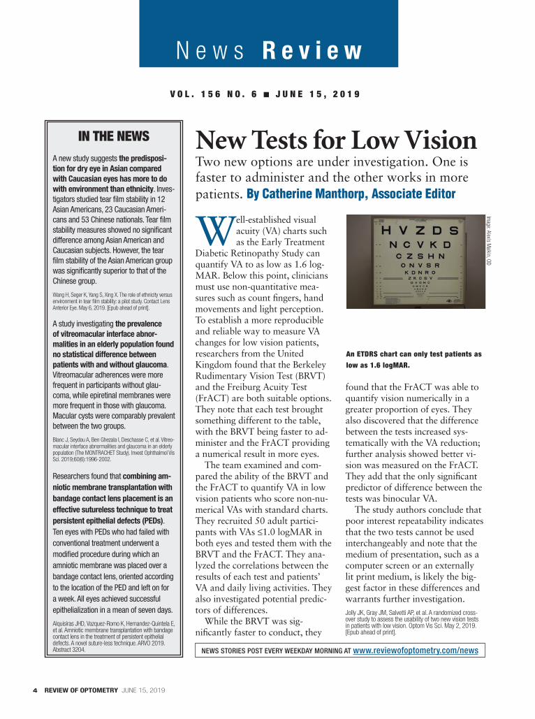
Well-established visual acuity (VA) charts such as the Early Treatment
Diabetic Retinopathy Study can quantify VA to as low as 1.6 log-MAR. Below this point, clinicians must use non-quantitative mea-sures such as count fi ngers, hand movements and light perception. To establish a more reproducible and reliable way to measure VA changes for low vision patients, researchers from the United Kingdom found that the Berkeley Rudimentary Vision Test (BRVT) and the Freiburg Acuity Test (FrACT) are both suitable options. They note that each test brought something different to the table, with the BRVT being faster to ad-minister and the FrACT providing a numerical result in more eyes.
The team examined and com-pared the ability of the BRVT and the FrACT to quantify VA in low vision patients who score non-nu-merical VAs with standard charts. They recruited 50 adult partici-pants with VAs ≤1.0 logMAR in both eyes and tested them with the BRVT and the FrACT. They ana-lyzed the correlations between the results of each test and patients’ VA and daily living activities. They also investigated potential predic-tors of differences.
While the BRVT was sig-nifi cantly faster to conduct, they
found that the FrACT was able to quantify vision numerically in a greater proportion of eyes. They also discovered that the difference between the tests increased sys-tematically with the VA reduction; further analysis showed better vi-sion was measured on the FrACT. They add that the only signifi cant predictor of difference between the tests was binocular VA.
The study authors conclude that poor interest repeatability indicates that the two tests cannot be used interchangeably and note that the medium of presentation, such as a computer screen or an externally lit print medium, is likely the big-gest factor in these differences and warrants further investigation.Jolly JK, Gray JM, Salvetti AP, et al. A randomized cross-over study to assess the usability of two new vision tests in patients with low vision. Optom Vis Sci. May 2, 2019. [Epub ahead of print].
IN THE NEWSA new study suggests the predisposi-
tion for dry eye in Asian compared
with Caucasian eyes has more to do
with environment than ethnicity. Inves-tigators studied tear fi lm stability in 12 Asian Americans, 23 Caucasian Ameri-cans and 53 Chinese nationals. Tear fi lm stability measures showed no signifi cant difference among Asian American and Caucasian subjects. However, the tear fi lm stability of the Asian American group was signifi cantly superior to that of the Chinese group.
Wang H, Seger K, Yang S, Xing X. The role of ethnicity versus environment in tear fi lm stability: a pilot study. Contact Lens Anterior Eye. May 6, 2019. [Epub ahead of print].
A study investigating the prevalence
of vitreomacular interface abnor-
malities in an elderly population found no statistical difference between
patients with and without glaucoma. Vitreomacular adherences were more frequent in participants without glau-coma, while epiretinal membranes were more frequent in those with glaucoma. Macular cysts were comparably prevalent between the two groups.
Blanc J, Seydou A, Ben Ghezala I, Deschasse C, et al. Vitreo-macular interface abnormalities and glaucoma in an elderly population (The MONTRACHET Study). Invest Ophthalmol Vis Sci. 2019;60(6):1996-2002.
Researchers found that combining am-
niotic membrane transplantation with
bandage contact lens placement is an
effective sutureless technique to treat
persistent epithelial defects (PEDs). Ten eyes with PEDs who had failed with conventional treatment underwent a modifi ed procedure during which an amniotic membrane was placed over a bandage contact lens, oriented according to the location of the PED and left on for a week. All eyes achieved successful epithelialization in a mean of seven days.
Alquisiras JHD, Vazquez-Romo K, Hernandez-Quintela E, et al. Amniotic membrane transplantation with bandage contact lens in the treatment of persistent epithelial defects. A novel suture-less technique. ARVO 2019. Abstract 3204.
New Tests for Low Vision
NEWS STORIES POST EVERY WEEKDAY MORNING AT www.reviewofoptometry.com/news
Two new options are under investigation. One is faster to administer and the other works in more patients. By Catherine Manthorp, Associate Editor
V O L . 1 5 6 N O . 6 ■ J U N E 1 5 , 2 0 1 9
An ETDRS chart can only test patients as low as 1.6 logMAR.
Image: A
lexis Malkin, O
D
004_ro0619_news.indd 4004_ro0619_news.indd 4 6/5/19 4:56 PM6/5/19 4:56 PM

T H E II NN TT EE LL LL II GG EE NN TT T OO N OO M E T E R T H E AA CC CC UU RR AA TT EE
TTTT OOOO NNNN OOOO MMMM EEEE TTTT EEEE RRRR TTTT HHHH EEEE CCCCC OOOOO NNNNN SSSSS IIIII SSSSS TTTTT EEEEE NNNNN TTTTT TTTT OOOO NNNN OOOO MMMM EEEE TTTT EEEE RRRR
TTTT HHHH EEEE OOOO BBBB JJJJ EEEE CCCC TTTT IIII VVVV EEEE TTTT OOOO NNNN OOOO MMMM EEEE TTTT EEEE RRRR TTTT HHHH EEEE RRRR EEEE PPPP EEEE AAAA TTTT AAAA BBBB LLLL EEEE
TTT OOO NNN OOO MMM EEE TTT EEE RRR TTT
TTTTT HHHHH EEEEE AAAA CCCC CCCC UUUU RRRR AAAA TTTT EEEE
TT OO NN OO MM EE TT EE RR TT HH
RRRRR EEEEE PPPPP EEEEE AAAAA TTTTT AAAAA BBBB LLLL EEEE TTTTT
TTT OOO NNN OO MMM E T E RR TT HH
CCCCC OOO NN
TT O N
TT HHHH EE
TTTT OOOO NN
E T O N O M E T E R T H E R E P E A T A B L E
HHH EEE IIII NNNN TTTT EEEE LLLL LLLL IIII GGGG EEEE NNNN TTTT TTT OOO NNN OOO MMM EEE TTT EEE RRR
EEEE TTTTT OOOOO NNNNN OOOOO MMMMM EEEEE TTTTT EEEEE RRRRR TTTTT HHHHH EEEEEEEEEEEEEEEEEEEEEEEEEEEEEEEEEEEEEEEEEEEEEEEEEEEEEEEEEEEEEEEEEEEEEEEEEEEEEEEEEEEEEEEEEEEEEEEEEEEEEEEEE CCCCCCCCCCCCCCCCCCCCCCCCCCCCC OOOOOOOOOOOOOOOOOOOOOO NNNNNNNNNNNNNNNNNNNNNNNNNNNNNNN SSSS IIII SSSS TTTT EEEE NNNN TTTT
HH EE OO BB JJ EE CC TT II VV EEEEEEEEEEEEEEEEEEEEEEEEEEEEEEEEEEEEEEEEEEEE TTTTTTTTTTTTTTTTTTTTTTTTTTTTTTTTTTTTTTTTTTTTTTTTTTTTTTTTTTTTTTTTTTTTTTTTTTTTTTTTTTTTTTTTTTTTTTTTTTTTTTTTTTTTTTTTTTTTTTTTTTTTTTTTTTTTTTTTTTTTTTTTT OOOOOOOOOOOOOOOOOOOOOOOOOOOOOOOOOOOOOOOOOOOOOOOOOOOOOOOOOOOOOOOOOOOOOOOOOOOOOOOOOOOOOOOOOOOOOOOOOOOOOOOOOOOOOOOOOOOOOOOOOOOOOOOOOOOOOOOOOOOOOOOOOOOOOOOOOOOOOOOOOOOOOOOOOOOOOOOOOOOOOOOOOOOOOOOOOOOOOOOOOOOOOOOOOOOOOOOOOOOOOOOOOOOOOOOOOOOOOOOOOOOOOOOOO NNNNNNNNNNNNNNNNNNNNNNNNNNNNNNNNNNNNNNNNNNNNNNNNNNNNNNNNNNNNNNNNNNNNNNNNNNNNNNN OOOOOOOOOOOOOOOOOOOOOOOOOOOOO MMMMMMMMMMMMMMMMMMMMMMMMMMMMMMMMMM EEEEEEEEEEEEEEEEEEEEEEEEEEEEEEEEEEEEEEEEEEEEEEEEEEEEEEEE TTTTTTTTTTTTTTTTTTTTTTTTTTTTTTTTTTTTTTTTTTTTTTTTTTTTTTTTTTTTTTTTTTTTTTTTTTTTTTTTTTTTTTTTTTTTTTTTTTTTTTTTTTTTTTTTTTTTTTTTTTTTTTTTTTTTTTTTTTTTTTTTTTTTTTTTTTTTTTTTTTTTTTTTTTTTTTTTTTTTTTTTTTTTTTTTTTTTTTTTTTTTTTTTTTTTTTTTTTTTTTTTTTTTTTTTTTTTTTTTTTTTTTTTTTTTTTTTTTTTTTTTTTTTT EE RR TT HH EE
TTTTTT OOOOOOO NNNNNN OOOOO MMMM EEEE TTTT EEEE RRRR TTTTT HHHHHH EEEEEEEEEEEEEEEEEEEEEEEEEEEEEEEEEEEEEEEEEEEEEEEEEEEEEEEEEEEEEEEEEEEEE IIIIIIIIIIIIIIIIIIIIIIIIIIIIIIIIIII NNNNNNN T E LLLLLLLLLLLLLLLLLLLLLLLLLLLLLLLLLLLLL LLLLL IIII GGGGG EEEEE NNNNN TTTTT
HH E A C C U R A TTTT EEEEE TTTTTTTTTTTTTTTTTTTTTTTTTTTTTTTTTTTTTTTTTTTTTTTTTTTTTTTTTTTTTTTTTTTTTTTTTTTTTTTTTTTTTTTTTTTTTTTTTTTTTTTTTTTTTTTTTTTTTTTTTTTTTTTTTTTTTTTTTTTTTTTTTTTTTTTTTTTTTTTTTTTTTTTTTTTTTTTTTTTTTTTTTTTTTTTTTTTTTTTTTTTTTTTTTTTTTTTTTTTTTTTTTTTTTTTTTTTTTTTTTTTTTTTTTTTTTTTTTTTTTTTTTTTTTTTTTTTTTTTTTTTTTTTTTTTTTTTTTTTTTTTTTTTTTTTTTTTTTTTTTTTTTTTTTTTTTTTTTTTTTTTTTTTTTTTTTTTTTTTTTTTTTTTTTTT OOOOOOOOOOOOOOOOOOOOOOOOOOOOOOOOOOOOOOOOOOOOOOOOOOOOOOOOOOOOOOOOOOOOOOOOOOOOOOOOOOOOOOOOOOOOOOOOOOOOOOOOOOOOOOOOOOOOOOOOOOOOOOOOOOOOOOOOOOOOOOOOOOOOOOOOOOOOOOOOOOOOOOOOOOOOOOOOOOOOOOOOOOOOOOOOOOOOOOOOOOOOOOOOOOOOOOOOOOOOOOOOOOOOOOOOOOOOOOOOOOOOOOOOOOOOOOO NNNNNNNNNNNNNNNNNNNNNNNNNNNNNNNNNNNNNNNNNNNNNNNNNNNNNNNNNNNNNNNNNNNNNNNNNNNNNNNNNNNNNNNNNNNNNNNNNNNNNNNNNNNNNNNNNNNNNNNNNNNNNNNNNNNNNNNNNNNNNNNNNNNNNNNNNNNNNNNNNNNNNNNNNNNNNN OOOOOOOOOOOOOOOOOOOOOOOOOOOOOOOOOOOOOOOOOOOOOOOOOOOOOOOOOOOOOOOOOOOOOOOOOOOOO MMMMMMMMMMMMMMMMMMMMMMMMMMMMMMMMMMMMMMMMMMMMMMMMMMMMMMMMMMMMMMMMMMMMMMMMMMMMMMMMMMMMMMMMMMMMMMMMMMMMMMMMMMMMMMMMMMMMMMMMMMMMMMMMMMMMMMMMMMMMMMMMMMMMMMMMMMMMMMMMMMMMMMMMMMMMMMMMMMMMMMMMMMMMMMMMMMMMMMMMMMMMMMMMMMMMMMMMMMMMMMMMMMMMMMMMMMMMMMMMMMMMMMMMMMMMMMMMMMMMMMMMMMMMMMMMMMMMMMMMMMMMMMMMMMMMMMMMMMMMMMMMMMMMMMMMMMMMMMMMMMMMMMMMMMMMMMMMMMMMMMMMMMMMMMMMMMMMMMMMMMMMMMMMMMMMMMMMMMMMMMMMMMMMMMMMMMMMMMMMMMMMMMMMMMMMMMMMMMMMMMMMMMMMMMMMMMMMMMMMMMMMMMMMMMMMMMMMMMMMMMMMMMMMMMMMMMMMMMMMMMMMMMMMMMMMMMMMMMMMMMMMMMMMMMMMMMMMMMMMMMMMMMMMMMMMMMMMMMMMMMMMM EEEEEEEEEEEEEEEEEEEEEEEEEEEEEEEEEEEEEEEEEEEEEEEEEEEEEEEEEEEEEEEEEEEEEEEEEEEEEEEEEEEEEEEEEEEEEEEEEEEEEEEEEEEEEEEEEEEEEEEEE TTTTTTTTTTTTTTTTTTTTTTTTTTTTTTTTTTTTTTTTTTTTTTT EEEE RRRRR TTTTT HHH EEE
RRR TTT HHHHHHHHHHHHHHHHHHHHHHHHHHHHHHHHHHHHHHHHHHHHHHHHHHHHHHHHHHHHHHHHHHHHHHHHHHHHHHHHHHHHHHHHHHHHHHH EEEEEEEEEEEEEEEEEEEEEEEEEEEEEEEEEEEEEEEEEEEEEEEEEEEEEEEEEEEEEEEEEEEEEEEEEEEEEEEEEEEEEE OOOOOOOOOOOOOOOOOOOOOOOOOOOOOOOOOOOOOO BBBB JJJJJJJJJJJJJJJJJJJJJJJJJJJJJJJJJJJJJJJJJJJJJJJJJJJJJJJJJJJJJJJJJJJJJJJJJJJJJJJJJJJJJJJJJJJJJJJJJJJJJJJJJJJJJJJJJJJJJJJJJJJJJJJJJ EEEEEEEEEEEEEEEEEEEEEEEEEEEEEEEEEEEEEEEEEEEEEEEEEEEEEEEEEEEEEEEEEEEEEEEEEEEEEEEEEEEEEEEEEEEEEEEEEEEEEEEEEEEEEEEEEEEEEEEEEEEEEEEEEEEEEEEEEEEE CCCC TTT II VV EEE
TTT AAA BBB LLL EEE TTTTTTTTTTTTTTTTTTTTTTTTTTTTTTTTTTTTTTTTTTTTTTTTTTTTTTTTTTTTTTTTTTTTTTTTTTTTTTTTTTTTTT OOOOOOOOOOOOOOOOOOOOOOOOOOOOOOOOOOOOOOOOOOOOOOOOOOOOOOOOOOOOOOOOOOOOOOOOOOOOOOOOOOOOOOOOOOOOOOOOOOOOOOOOOOOOOOOOOOOOOOOOOOOOOOOOOOOOOOOOOOOOOOOOOOOOOOOOOOOOOOOOOOOOOOOOOOOOOOOOO NNNNNNNNNNNNNNNNNNNNNNNNNNNNNNNNNNNNNNNNNNNNNNNNNNNNNNNNNNNNNNNNNNNNNNNNNNNNNNNNNNNNNNNNNNNNNNNNNNNNNNNNNNNNNN OOOOOOOOOOOOOOOOOOOOOOOOOOOOOOOOOOOOOOOOOOOOOOOOOOOOOOOOOOOOOOOOOOOOOOOOOOOOOOOOOOOOOOOOOOOOOOOOOOOOOOOOOOOOOOOOOOOOOOOOOOOOOOOOOOOOOOOOOOOOOOOOOOOOOOOOOOOOOOOOOOOO MMMMMMMMMMMMMMMMMMMMMMMMMMMMMMMMMMMMMMMMMMMMMMM EEEE T E RR
EEE RRRR TTTT HHHH EEEE RRR EE PPPPPPPPPPPPPPPPPPPPPPPPPPPPPPPPPPPPPPPPPPPPPPPPPPPPP EEEEEEEEEEEEEEEEEEEEEEEEEEEEEEEEEEEEEEEEEEEEEEEEEEEEEEEEEEEEEEEEEEEEEEEEEEEEEEEEEEEEEEEEEEEEEEEEEEEEEEEEEEEEEEEE AAAA TTTTTTTTTTTTTTTTTTTTTTTTTTTTTTTTTTTTTT AAA BBB LLL EEEE
III GG EE NN TT TTTT OOOOOOOOOOOOOOOOOOOOOOOOOOOOOOOOOOOOOOOOOOOOOOOOOOOOOOOOOOOOOOO NNNNNNNNNNNNNNNNNNNNNNNNNNNNN OOOOOOOOOOOO MMMMMMMMMMMMMMMM EE TT EEE RR
NNN OOOOOO MMMMMM EEE TTT EEE RRRR TTTT HHHH
NNN SSSS III SSSS TT EE NNN TTTTTT
N OOOO MMMM EEE TTT EEE RRRRRRRRRRRRRRRRRRRRRRRRRRRRRRRRRRRRRRRRRRRRRRRRRRRRRRRRRRRRRRRRR TTTTTTTTTTTTTTTTTTTTTTTTTTTTTTTTT
E OOO BBB JJ EE CCC TTT IIIIIIIIIIIIIIIIIIIIIIIIIIIIIII VVVVVVVVVVVVVVVVVV EEEEEEEE
N OO MMM EEE TTTT EEE RRRR TTTTTTTTTTTTTTTTTTTTTTTTTTTTTTTTTTTTTTTTTTTTTTTTTTTTTTTTTTTTTTTTTTTTTTTTTTTTTTTTTTTTTTTTTTTTTTTTTTTTTTTTT
HHHH EEEE AAAAA CCCC CCCCC UUUUU RRRRR AAAAA
TTTT OOO NNN OOO MMMM EEEE TTTT EEE
HHHHHHHHHHHHHHHHHHHHHHHHHHHHH EEEEEEEEEEEEEEEEEEEEEEEEEEEEEEEEEEEEEEEEE RRRRRRRRRRRRRRRRRRRRRRRRRRRRRRRRRRRRRRRRR EEE PPP EEE AAA TTT
EEEEEE TTTTTTTTTTTTTTTTTTTTTTTTTTTTTT OOOOOOOOOOOOOOOOOOOOOOOOOOOOOOOOOOOOOOOOOOOOOOOOOOOOOOOOOO NNNNNNNNNNNNNNNNNNNNNNNNNN OOOOOOOOOOOOOOOO MMMMMMMMMMMMMMMM EEE TTT EEE
HHHHHHHHH EEEEEEE IIII NNN TTTTT EEEEEEEEEEEEEEEEEEEEEEEEEE LLL LLLLFOR PATIENTS
FOR PROFESSIONALS
For more information, scan, email [email protected] or visit www.icare-usa.com.
RO0619_Icare.indd 1 5/23/19 2:17 PM

6 REVIEW OF OPTOMETRY JUNE 15, 2019
News Review For more, visit www.reviewofoptometry.com/news
Microcirculation Changes May Precede DR
Changes in foveal density and other parameters in diabetic eyes without clinically de-
tectable diabetic retinopathy may be important biomarkers in diagnosing early diabetes, researchers say.
The cross-sectional prospective study enrolled 60 patients with diabetes but without clinically detectable diabetic retinopathy and 57 age-matched children in the con-trol group. Researchers performed
optical coherence tomography angiography (OCT-A) and analyzed several parameters, including: the foveal avascular zone and non-fl ow areas; superfi cial and deep vessel densities; foveal avascular zone perimeter; acircularity index of the foveal avascular zone (the ratio of the perimeter of the foveal avascular zone and the perimeter of a circle with equal area); and foveal density (vessel density in 300µm around
the foveal avascular zone). The investigators then evaluated any correlations between the OCT-A parameters with diabetes duration and glycated hemoglobin (HbA1c) levels among the diabetes patients.
The study found statistically signifi cant differences between the groups in foveal avascular zone perimeter, acircularity index of the foveal avascular zone and foveal density. Researchers also reported statistically signifi cant differences between the groups for vessel densi-ties in the deep superior hemi-para-fovea and deep temporal parafovea, as well as the deep superior parafo-veal zones. Additionally, investiga-tors observed no signifi cant cor-relations between diabetes duration and HbA1c levels on the OCT-A parameters.
Researchers said these new pa-rameters might be sensitive imaging biomarkers to defi ne early diabetic retinopathy.
Inanc M, Tekin K, Kiziltoprak H, et al. Changes in retinal micro-
circulation precede the clinical onset of diabetic retinopathy in
children with type 1 diabetes mellitus. Am J Ophthalmol. April
29, 2019. [Epub ahead of print].
Disc Hemorrhage Linked with Progression
Researchers recently discov-ered a signifi cant propor-tion of primary open-angle
glaucoma (POAG) patients have minute-sized optic disc hemorrhages associated with earlier and faster visual fi eld (VF) progression.
The researchers analyzed POAG patients with macro disc hem-orrhages who had a follow-up period of at least seven years and more than nine VF results. Micro disc hemorrhages were less than
0.01mm2 and undetectable on conventional stereo disc photog-raphy but discernible by enhanced stereo disc photography. These photographs were enhanced by customized software to evaluate for the presence of hemorrhages. VF progression was confi rmed by stan-dard automated perimetry’s guided progression analysis.
Among 107 POAG eyes, the team noted micro hemorrhages pri-or to macro hemorrhages in 36.4%
of eyes with a median lag of 13.6 months. Over the course of follow-up, 53.8% with micro disc hemor-rhages—but only 27.9% without them—showed VF progression. In eyes with micro disc hemorrhages, the cumulative VF progression probability was signifi cantly greater and overall VF deterioration rate was much faster. ■
Ha A, Kim YK, Baek SU, et al. Optic disc microhemorrhage in primary open-angle glaucoma: clinical implications for visual fi eld progression. Invest Ophthalmol Vis Sci. 2019;60:1824-32.
OCT-A Detects Earliest Diabetic Eye ChangesMicrovascular changes to the macula and optic disc detected with OCT-A are signifi cant in the case of patients with diabetes, according to research presented at ARVO’s annual meeting in Vancouver. California-based researchers looked at the OCT-A analysis of 216 eyes of 124 patients with diabetes and an additional 94 control eyes of 67 patients. They found the diabetic eyes exhibited signifi cant worsening of angiographic measurements of the macula and optic disc despite no signifi cant changes in retinal nerve fi ber layer (RNFL) and macular ganglion cell complex thickness, suggesting OCT-A can identify structural changes earlier and on a smaller scale than other imaging technologies.
The severity of OCT-A changes corresponded with higher levels of DR and macular edema. OCT-A imaging showed signifi cantly reduced vascular diameter, vessel area density and vessel skeleton density, vessel perimeter index and vessel complex index in diabetic patients’ eyes than control eyes. The researchers also used OCT-A to measure fl ow impairment and found it signifi cantly increased in diabetic patients’ eyes compared with control eyes.
Huang L, Shariati A, Oh A, et al. A Comparison: Structural optical coherence tomography and angiography in diabetic retinopathy and diabetic macular edema. ARVO 2019. Abstract 3026.
004_ro0619_news.indd 6004_ro0619_news.indd 6 6/5/19 4:56 PM6/5/19 4:56 PM

Have you re-cently renovat-ed your offi ce
or redesigned a new space? Enter our offi ce design contest and share your new look with your colleagues!
Eligibility: Newly built offi ces, remodels or expansions com-pleted between June 1, 2017 and June 30, 2019 are eligible to enter the contest.
Judging: Entries will be judged by a panel of fellow optometrists previously recognized for their expertise in offi ce design.
Awards: “Offi ce Design of the Year” will be awarded to the best overall facility based on func-tional design, effi cient space planning, style and inte-gration of equipment. Two runners-up will be chosen based on the same standards.
Each winner will receive an engraved offi ce plaque recognizing the practice’s achievement, in addition to editorial coverage online and in our November 2019 print edition.
All entries must be received by September 1, 2019.
Scan or Click to Enter:To read the contest rules and enter your new space for a chance to win Offi ce Design of the Year, visit www.reviewofoptometry.com or scan this QR code. Send your high-resolution images to Rebecca Hepp, managing editor, at [email protected].
2019 Office Design Contest
Call for Entries
SK FAMILYVISUAL FIELD ANALYZER
& RETINAL CAMERA
Auto-calibration
Auto gaze tracking
Full compliance with the Goldman Standard
User-friendly printed report
DICOM Compatibility
Auto mosaic function
Optical red-free visual testing
Superior picture
image capture
TECHNOLOGY YOU EXPECTAT A MUCH LOWER PRICE
004_ro0619_news.indd 7004_ro0619_news.indd 7 6/5/19 4:56 PM6/5/19 4:56 PM

HEIDI Q. T. PHAM-MURPHY, ODVISIONS OPTOMETRY
“There are so many different sponsors of the VSP Global® Premier
Program. They all have different, amazing offerings for practices.
It is one more reason that I partner with Premier.”
HEIDI Q. T. PHAM-MURPHY, OD
UNLOCK THE POSSIBILITIES AT OFFERS.PATHTOPREMIER.COM.
RO0619_VSP.indd 2RO0619_VSP.indd 2 5/21/19 2:50 PM5/21/19 2:50 PM

©2019 Vision Service Plan. All rights reserved. VSP Global is a registered trademark of Vision Service Plan. All other brands or marks are the property of their respective owners. 45870
DISCOVER THE OPPORTUNITY
VSP Global® Premier Program partners with over a dozen industry-leading companies to offer more for your practice.
MODO, Optovue, Weave, Abyde, FatHeadz, Staples, and more.
Explore all Premier Program sponsor opportunities at vspadvantage.com
RO0619_VSP.indd 3RO0619_VSP.indd 3 5/21/19 2:50 PM5/21/19 2:50 PM
10 REVIEW OF OPTOMETRY JUNE 15, 2019
News Review
Evidence Mounts for Autologous Serum
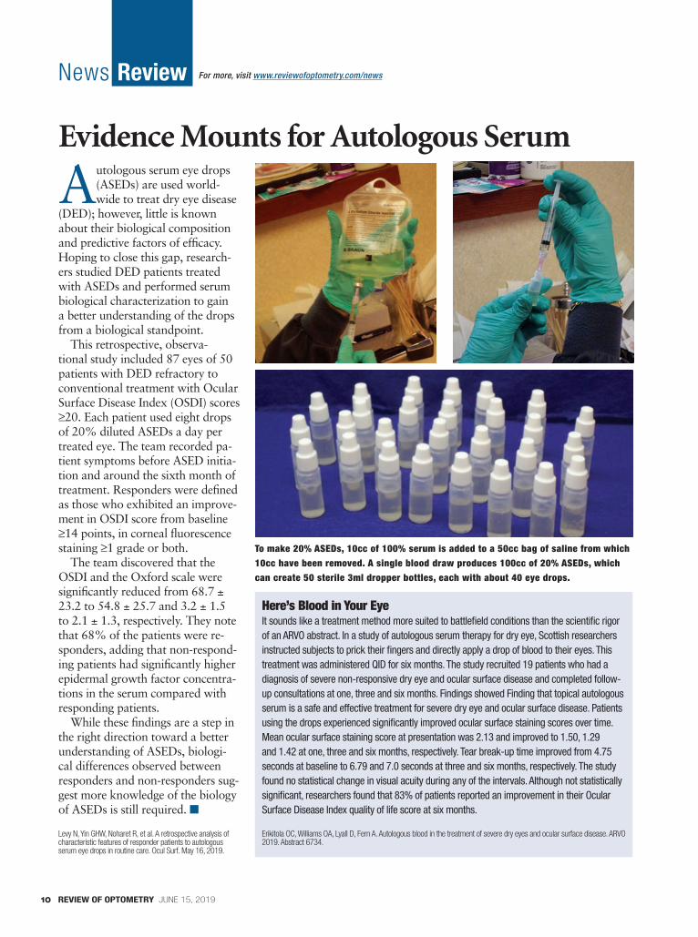
Autologous serum eye drops (ASEDs) are used world-wide to treat dry eye disease
(DED); however, little is known about their biological composition and predictive factors of effi cacy. Hoping to close this gap, research-ers studied DED patients treated with ASEDs and performed serum biological characterization to gain a better understanding of the drops from a biological standpoint.
This retrospective, observa-tional study included 87 eyes of 50 patients with DED refractory to conventional treatment with Ocular Surface Disease Index (OSDI) scores ≥20. Each patient used eight drops of 20% diluted ASEDs a day per treated eye. The team recorded pa-tient symptoms before ASED initia-tion and around the sixth month of treatment. Responders were defi ned as those who exhibited an improve-ment in OSDI score from baseline ≥14 points, in corneal fl uorescence staining ≥1 grade or both.
The team discovered that the OSDI and the Oxford scale were signifi cantly reduced from 68.7 ± 23.2 to 54.8 ± 25.7 and 3.2 ± 1.5 to 2.1 ± 1.3, respectively. They note that 68% of the patients were re-sponders, adding that non-respond-ing patients had signifi cantly higher epidermal growth factor concentra-tions in the serum compared with responding patients.
While these fi ndings are a step in the right direction toward a better understanding of ASEDs, biologi-cal differences observed between responders and non-responders sug-gest more knowledge of the biology of ASEDs is still required. ■
Levy N, Yin GHW, Noharet R, et al. A retrospective analysis of characteristic features of responder patients to autologous serum eye drops in routine care. Ocul Surf. May 16, 2019.
For more, visit www.reviewofoptometry.com/news
Here’s Blood in Your EyeIt sounds like a treatment method more suited to battlefi eld conditions than the scientifi c rigor of an ARVO abstract. In a study of autologous serum therapy for dry eye, Scottish researchers instructed subjects to prick their fi ngers and directly apply a drop of blood to their eyes. This treatment was administered QID for six months. The study recruited 19 patients who had a diagnosis of severe non-responsive dry eye and ocular surface disease and completed follow-up consultations at one, three and six months. Findings showed Finding that topical autologous serum is a safe and effective treatment for severe dry eye and ocular surface disease. Patients using the drops experienced signifi cantly improved ocular surface staining scores over time. Mean ocular surface staining score at presentation was 2.13 and improved to 1.50, 1.29 and 1.42 at one, three and six months, respectively. Tear break-up time improved from 4.75 seconds at baseline to 6.79 and 7.0 seconds at three and six months, respectively. The study found no statistical change in visual acuity during any of the intervals. Although not statistically signifi cant, researchers found that 83% of patients reported an improvement in their Ocular Surface Disease Index quality of life score at six months.
Erikitola OC, Williams OA, Lyall D, Fern A. Autologous blood in the treatment of severe dry eyes and ocular surface disease. ARVO 2019. Abstract 6734.
To make 20% ASEDs, 10cc of 100% serum is added to a 50cc bag of saline from which 10cc have been removed. A single blood draw produces 100cc of 20% ASEDs, which can create 50 sterile 3ml dropper bottles, each with about 40 eye drops.
004_ro0619_news.indd 10004_ro0619_news.indd 10 6/5/19 4:57 PM6/5/19 4:57 PM

IndicationLOTEMAX® SM (loteprednol etabonate ophthalmic gel) 0.38% is a corticosteroid indicated for the treatment of post-operative infl ammation and pain following ocular surgery.
Important Safety Information• LOTEMAX® SM, as with other ophthalmic corticosteroids, is contraindicated in
most viral diseases of the cornea and conjunctiva including epithelial herpes simplex keratitis (dendritic keratitis), vaccinia, and varicella, and also in mycobacterial infection of the eye and fungal diseases of ocular structures.
• Prolonged use of corticosteroids may result in glaucoma with damage to the optic nerve, defects in visual acuity and fi elds of vision. Steroids should be used with caution in the presence of glaucoma. If LOTEMAX® SM is used for 10 days or longer, IOP should be monitored.
• Use of corticosteroids may result in posterior subcapsular cataract formation.
Important Safety Information (cont.)• The use of steroids after cataract surgery may delay healing and increase
the incidence of bleb formation. In those with diseases causing thinning of the cornea or sclera, perforations have been known to occur with the use of topical steroids. The initial prescription and renewal of the medication order should be made by a physician only after examination of the patient with the aid of magnifi cation such as slit lamp biomicroscopy and, where appropriate, fl uorescein staining.
• Prolonged use of corticosteroids may suppress the host response and thus increase the hazard of secondary ocular infections. In acute purulent conditions, steroids may mask infection or enhance existing infections.
• Employment of a corticosteroid medication in the treatment of patients with a history of herpes simplex requires great caution. Use of ocular steroids may prolong the course and may exacerbate the severity of many viral infections of the eye (including herpes simplex).
• Fungal infections of the cornea are particularly prone to develop coincidentally with long-term local steroid application. Fungus invasion must be considered in any persistent corneal ulceration where a steroid has been used or is in use. Fungal cultures should be taken when appropriate.
• Contact lenses should not be worn when the eyes are infl amed.• There were no treatment-emergent adverse drug reactions that occurred in
more than 1% of subjects in the three times daily group compared to vehicle.
You are encouraged to report negative side eff ects of prescription drugs to the FDA. Visit www.fda.gov/medwatch or call 1-800-FDA-1088. Please see brief summary of Prescribing Information on adjacent page.
References: 1. LOTEMAX SM Prescribing Information. Bausch & Lomb, Incorporated. 2. Cavet ME, Glogowski S, DiSalvo C, Richardson ME. Ocular pharmacokinetics of submicron loteprednol etabonate ophthalmic gel, 0.38% following topical administration in rabbits. Poster presented at 2015 ARVO Annual Meeting; May 4, 2015; Denver, Colorado. 3. Data on fi le. Bausch & Lomb, Incorporated.
®/TM are trademarks of Bausch & Lomb Incorporated or its affi liates.© 2019 Bausch & Lomb Incorporated or its affi liates. All rights reserved. Printed in USA. LSM.0176.USA.19
S M A L L & M I G H T YS U B M I C R O N P A R T I C L E S
Visit www.LOTEMAXSM.com
SUBMICRON STRONGEngineered with SM Technology™ for effi cient
penetration at a low BAK level (0.003%)1,2
* Compared to LOTEMAX® GEL (loteprednol etabonate ophthalmic gel) 0.5%. Clinical signifi cance of these preclinical data has not been established.
~2× GREATER PENETRATIONto the aqueous humor2*
• 30% of LOTEMAX® SM patients had complete ACC resolution vs vehicle (15%) at Day 8 (N=371, P<0.0001)1,3†
• 74% of LOTEMAX® SM patients were completely pain-free vs vehicle (49%) at Day 8 (N=371, P<0.0001)1,3‡
† Pooled analysis of Phase 3 clinical studies. Study 1: 29% LOTEMAX® SM (N=171) vs 9% vehicle (N=172). Study 2: 31% LOTEMAX® SM (N=200) vs 20% vehicle (N=199); P<0.05 for all.
‡ Pooled analysis of Phase 3 clinical studies. Study 1: 73% LOTEMAX® SM (N=171) vs 48% vehicle (N=172). Study 2: 76% LOTEMAX® SM (N=200) vs 50% vehicle (N=199); P<0.05 for all.
PROVEN STRENGTH
RO0619_B & L Lotemax.indd 1 5/23/19 2:23 PM

BRIEF SUMMARY OF PRESCRIBING INFORMATION
This Brief Summary does not include all the information needed to use LOTEMAX® SM safely and effectively. See full prescribing information for LOTEMAX® SM.
LOTEMAX® SM (loteprednol etabonate ophthalmic gel) 0.38% For topical ophthalmic use Initial U.S. Approval: 1998
INDICATIONS AND USAGE LOTEMAX® SM is a corticosteroid indicated for the treatment of post-operative inflammation and pain following ocular surgery.
DOSAGE AND ADMINISTRATION Invert closed bottle and shake once to fill tip before instilling drops. Apply one drop of LOTEMAX® SM into the conjunctival sac of the affected eye three times daily beginning the day after surgery and continuing throughout the first 2 weeks of the post-operative period.
CONTRAINDICATIONS LOTEMAX® SM, as with other ophthalmic corticosteroids, is contraindicated in most viral diseases of the cornea and conjunctiva including epithelial herpes simplex keratitis (dendritic keratitis), vaccinia, and varicella, in mycobacterial infection of the eye and fungal diseases of ocular structures.
WARNINGS AND PRECAUTIONS Intraocular Pressure (IOP) Increase: Prolonged use of corticosteroids may result in glaucoma with damage to the optic nerve, defects in visual acuity and fields of vision. Steroids should be used with caution in the presence of glaucoma. If this product is used for 10 days or longer, intraocular pressure should be monitored. Cataracts: Use of corticosteroids may result in posterior subcapsular cataract formation. Delayed Healing: The use of steroids after cataract surgery may delay healing and increase the incidence of bleb formation. In those diseases causing thinning of the cornea or sclera, perforations have been known to occur with the use of topical steroids. The initial prescription and renewal of the medication order should be made by a physician only after examination of the patient with the aid of magnification such as slit lamp biomicroscopy and, where appropriate, fluorescein staining. Bacterial Infections: Prolonged use of corticosteroids may suppress the host response and thus increase the hazard of secondary ocular infections. In acute purulent conditions of the eye, steroids may mask infection or enhance existing infection. Viral infections: Employment of a corticosteroid medication in the treatment of patients with a history of herpes simplex requires great caution. Use of ocular steroids may prolong the course and may exacerbate the severity of many viral infections of the eye (including herpes simplex). Fungal Infections: Fungal infections of the cornea are particularly prone to develop coincidentally with long-term local steroid application. Fungus invasion must be considered in any persistent corneal ulceration where a steroid has been used or is in use. Fungal cultures should be taken when appropriate. Contact Lens Wear: Contact lenses should not be worn when the eyes are inflamed.
ADVERSE REACTIONS Because clinical trials are conducted under widely varying conditions, adverse reaction rates observed in the clinical trials of a drug cannot be directly compared to rates in the clinical trials of another drug and may not reflect the rates observed in practice. Adverse reactions associated with ophthalmic steroids include elevated intraocular pressure, which may be associated with infrequent optic nerve damage, visual acuity and field defects, posterior subcapsular cataract formation, delayed wound healing and secondary ocular infection from pathogens including herpes simplex, and perforation of the globe where there is thinning of the cornea or sclera. There were no treatment-emergent adverse drug reactions that occurred in more than 1% of subjects in the three times daily group compared to vehicle. USE IN SPECIAL POPULATIONS Pregnancy: Risk Summary: There are no adequate and well controlled studies with loteprednol etabonate in pregnant women. Loteprednol etabonate produced teratogenicity at clinically relevant doses in the rabbit and rat when administered orally during pregnancy. Loteprednol etabonate
produced malformations when administered orally to pregnant rabbits at doses 4.2 times the recommended human ophthalmic dose (RHOD) and to pregnant rats at doses 106 times the RHOD. In pregnant rats receiving oral doses of loteprednol etabonate during the period equivalent to the last trimester of pregnancy through lactation in humans, survival of offspring was reduced at doses 10.6 times the RHOD. Maternal toxicity was observed in rats at doses 1066 times the RHOD, and a maternal no observed adverse effect level (NOAEL) was established at 106 times the RHOD. The background risk of major birth defects and miscarriage for the indicated population is unknown. However, the background risk in the U.S. general population of major birth defects is 2 to 4%, and of miscarriage is 15 to 20%, of clinically recognized pregnancies. Data: Animal Data. Embryofetal studies were conducted in pregnant rabbits administered loteprednol etabonate by oral gavage on gestation days 6 to 18, to target the period of organogenesis. Loteprednol etabonate produced fetal malformations at 0.1 mg/kg (4.2 times the recommended human ophthalmic dose (RHOD) based on body surface area, assuming 100% absorption). Spina bifida (including meningocele) was observed at 0.1 mg/kg, and exencephaly and craniofacial malformations were observed at 0.4 mg/kg (17 times the RHOD). At 3 mg/kg (128 times the RHOD), loteprednol etabonate was associated with increased incidences of abnormal left common carotid artery, limb flexures, umbilical hernia, scoliosis, and delayed ossification. Abortion and embryofetal lethality (resorption) occurred at 6 mg/kg (256 times the RHOD). A NOAEL for developmental toxicity was not established in this study. The NOAEL for maternal toxicity in rabbits was 3 mg/kg/day. Embryofetal studies were conducted in pregnant rats administered loteprednol etabonate by oral gavage on gestation days 6 to 15, to target the period of organogenesis. Loteprednol etabonate produced fetal malformations, including absent innominate artery at 5 mg/kg (106 times the RHOD); and cleft palate, agnathia, cardiovascular defects, umbilical hernia, decreased fetal body weight and decreased skeletal ossification at 50 mg/kg (1066 times the RHOD). Embryofetal lethality (resorption) was observed at 100 mg/kg (2133 times the RHOD). The NOAEL for developmental toxicity in rats was 0.5 mg/kg (10.6 times the RHOD). Loteprednol etabonate was maternally toxic (reduced body weight gain) at 50 mg/kg/day. The NOAEL for maternal toxicity was 5 mg/kg. A peri-/postnatal study was conducted in rats administered loteprednol etabonate by oral gavage from gestation day 15 (start of fetal period) to postnatal day 21 (the end of lactation period). At 0.5 mg/kg (10.6 times the clinical dose), reduced survival was observed in live-RHOD) mg/kg (1066 times the RHOD) produced maternal toxicity (reduced body weight gain, death), decreased number of live-born offspring, decreased birth weight, and delays in postnatal development. A developmental NOAEL was not established in this study. The NOAEL for maternal toxicity was 5 mg/kg. Lactation: There are no data on the presence of loteprednol etabonate in human milk, the effects on the breastfed infant, or the effects on milk production. The developmental and health benefits of breastfeeding should
® SM and any potential adverse effects on the breastfed infant from LOTEMAX® SM. Pediatric Use: Safety and effectiveness of LOTEMAX® SM in pediatric patients have not been established. Geriatric Use: No overall differences in safety and effectiveness have been observed between elderly and younger patients.
NONCLINICAL TOXICOLOGY Carcinogenesis, Mutagenesis, Impairment of Fertility: Long-term animal studies have not been conducted to evaluate the carcinogenic potential of loteprednol etabonate. Loteprednol etabonate was not genotoxic in vitro in the Ames test, the mouse lymphoma tk assay, or in the chromosomal aberration test in human lymphocytes, or in vivo in the mouse micronucleus assay. Treatment of male and female rats with 25 mg/kg/day of loteprednol etabonate (533 times the RHOD based on body surface area, assuming 100% absorption) prior to and during mating caused preimplantation loss and decreased the number of live fetuses/live births. The NOAEL for fertility in rats was 5 mg/kg/day (106 times the RHOD). LOTEMAX is a trademark of Bausch & Lomb Incorporated or its affiliates. © 2019 Bausch & Lomb Incorporated Bausch + Lomb, a division of Valeant Pharmaceuticals North America LLC Bridgewater, NJ 08807 USA LSM.0091.USA.19 Based on 9669600-9669700 Revised: 02/2019
RO0619_B & L Lotemax PI.indd 1RO0619_B & L Lotemax PI.indd 1 5/23/19 2:25 PM5/23/19 2:25 PM

REVIEW OF OPTOMETRY JUNE 15, 2019 13
ContentsReview of Optometry June 15, 2019
A L S O I N S I D E
Earn 2 CE Credits: The Real-World Cataract EvaluationFor better post-op visual function and quality of life, focus on things that matter to patients. BY JACQUELINE THEIS, OD, PAGE 72
Diabetes: Today and TomorrowA deep dive into the latest research and drug development that’s influencing eye care for these patients. BY ANGELLA GENTRY, OD, CARRIE HO, OD, RICHARD ZIMBALIST, OD, AND EMILY O’BRIEN, OD
A36
How to Stay One Step Ahead of AMDNew diagnostic tools can help you ID suspects and initiate preventative measures. BY AMANDA LEGGE, ODHN42A Field Guide to Retinal Holes and TearsReview the clinical appearance of these elusive conditions. BY MOHAMMAD RAFIEETARY, OD, AND STEPHEN HUDDLESTON, MD52
10th A N N U A L R E T I N A R E P O R T
My Patient Has Scleritis… Now What?The initial diagnosis may be quick, but that’s only the beginning. Follow these steps to discover the root cause and set yourself on the right track for a successful outcome. BY KATHERINE SANFORD, OD, PAGE 66
013_ro0619_TOC.indd 13013_ro0619_TOC.indd 13 6/5/19 4:33 PM6/5/19 4:33 PM

14 REVIEW OF OPTOMETRY JUNE 15, 2019
DepartmentsReview of Optometry June 15, 2019
4 News Review
16 Outlook Losing Patience… and Patients JACK PERSICO
18 Through My Eyes Choose Your Doors Wisely PAUL M. KARPECKI, OD
20 Chairside Sunny With a Chance of No-shows MONTGOMERY VICKERS, OD
22 Clinical Quandaries Palsy Conundrum
PAUL C. AJAMIAN, OD
24 Focus on Refraction Sizing up Strabismus
MARC B. TAUB, OD, MS, AND
PAUL HARRIS, OD
28 Retina Dilemmas Tame the Swelling
DIANA SHECHTMAN OD,
JAY M. HAYNIE OD, AND
BRENDAN GIRSCHEK, MD
32 Coding Connection Coding the “Diabetic” Eye Exam
JOHN RUMPAKIS, OD, MBA
80 Cornea + Contact Lens Q&A The Lesser of Two Evils
JOSEPH P. SHOVLIN, OD
83 Ocular Surface Review Dry Eye Therapy: Getting Nosy
PAUL M. KARPECKI, OD
86 Retina Quiz Sugar Coated MARK T. DUNBAR, OD
91 Surgical Minute Replenish the Epithelium CECELIA KOETTING, OD,
DEREK N. CUNNINGHAM, OD, AND
WALTER O. WHITLEY, OD, MBA
92 Glaucoma Grand Rounds Breathing Room for the Nerve JAMES L. FANELLI, OD
94 Advertisers Index
95 Classifieds
98 Diagnostic Quiz Keeping in Contact ANDREW S. GURWOOD, OD
24
28
91
92
BUSINESS OFFICES11 CAMPUS BLVD., SUITE 100NEWTOWN SQUARE, PA 19073
CEO, INFORMATION SERVICES GROUP MARC FERRARA
(212) 274-7062 • [email protected]
PUBLISHERJAMES HENNE
(610) 492-1017 • [email protected]
REGIONAL SALES MANAGERMICHELE BARRETT
(610) 492-1014 • [email protected]
REGIONAL SALES MANAGERMICHAEL HOSTER
(610) 492-1028 • [email protected]
VICE PRESIDENT, OPERATIONS CASEY FOSTER
(610) 492-1007 • [email protected]
VICE PRESIDENT, CLINICAL CONTENT PAUL M. KARPECKI, OD, FAAO
PRODUCTION MANAGERSCOTT TOBIN
(610) 492-1011 • [email protected]
SENIOR CIRCULATION MANAGERHAMILTON MAHER
(212) 219-7870 • [email protected]
CLASSIFIED ADVERTISING(888) 498-1460
SUBSCRIPTIONS$56 A YEAR, $88 (US) IN CANADA,
$209 (US) IN ALL OTHER COUNTRIES.
SUBSCRIPTION INQUIRIES(877) 529-1746 (US ONLY)
OUTSIDE US CALL: (845) 267-3065
CIRCULATIONPO BOX 81
CONGERS, NY 10920TEL: (TOLL FREE): (877) 529-1746
OUTSIDE US: (845) 267-3065
CEO, INFORMATION SERVICES GROUP MARC FERRARA
SENIOR VICE PRESIDENT, OPERATIONS JEFF LEVITZ
VICE PRESIDENT, HUMAN RESOURCES TAMMY GARCIA
VICE PRESIDENT, CREATIVE SERVICES & PRODUCTION MONICA TETTAMANZI
CORPORATE PRODUCTION DIRECTORJOHN ANTHONY CAGGIANO
VICE PRESIDENT, CIRCULATION EMELDA BAREA
013_ro0619_TOC.indd 14013_ro0619_TOC.indd 14 6/5/19 4:34 PM6/5/19 4:34 PM
CONTRIBUTING EDITORSPAUL C. AJAMIAN, OD, ATLANTA
AARON BRONNER, OD, KENNEWICK, WASH.MILE BRUJIC, OD, BOWLING GREEN, OHIO
DEREK N. CUNNINGHAM, OD, AUSTIN, TEXAS
MARK T. DUNBAR, OD, MIAMI
ARTHUR B. EPSTEIN, OD, PHOENIX
JAMES L. FANELLI, OD, WILMINGTON, NCGARY S. GERBER, OD, HAWTHORNE, NJ
ANDREW S. GURWOOD, OD, PHILADELPHIA
ALAN G. KABAT, OD, MEMPHIS, TENN.DAVID KADING, OD, SEATTLE
PAUL M. KARPECKI, OD, LEXINGTON, KY.JEROME A. LEGERTON, OD, MBA, SAN DIEGO
JASON R. MILLER, OD, MBA, POWELL, OHIO
CHERYL G. MURPHY, OD, BABYLON, NYCARLO J. PELINO, OD, JENKINTOWN, PA.
JOSEPH PIZZIMENTI, OD, SAN ANTONIO, TEXAS.JOHN RUMPAKIS, OD, MBA, PORTLAND, ORE.
DIANA L. SHECHTMAN, OD, FORT LAUDERDALE, FLA.JEROME SHERMAN, OD, NEW YORK
JOSEPH P. SHOVLIN, OD, SCRANTON, PA.JOSEPH W. SOWKA, OD, FORT LAUDERDALE, FLA.MONTGOMERY VICKERS, OD, LEWISVILLE, TEXAS.
WALTER O. WHITLEY, OD, MBA, VIRGINIA BEACH, VA.
EDITORIAL REVIEW BOARDJEFFREY R. ANSHEL, OD, ENCINITAS, CALIF.
JILL AUTRY, OD, RPH, HOUSTON
SHERRY J. BASS, OD, NEW YORK EDWARD S. BENNETT, OD, ST. LOUIS
MARC R. BLOOMENSTEIN, OD, SCOTTSDALE, ARIZ.CHRIS J. CAKANAC, OD, MURRYSVILLE, PA.
JERRY CAVALLERANO, OD, PHD, BOSTON
WALTER L. CHOATE, OD, MADISON, TENN. BRIAN CHOU, OD, SAN DIEGO
A. PAUL CHOUS, MA, OD, TACOMA, WASH.ROBERT M. COLE, III, OD, BRIDGETON, NJGLENN S. CORBIN, OD, WYOMISSING, PA.
ANTHONY S. DIECIDUE, OD, STROUDSBURG, PA.S. BARRY EIDEN, OD, DEERFIELD, ILL.
STEVEN FERRUCCI, OD, SEPULVEDA, CALIF.MURRAY FINGERET, OD, HEWLETT, NYIAN BEN GADDIE, OD, LOUISVILLE, KY.
PAUL HARRIS, OD, MEMPHIS, TNMILTON HOM, OD, AZUSA, CALIF.
BLAIR B. LONSBERRY, MS, OD, MED, PORTLAND, ORE.THOMAS L. LEWIS, OD, PHD, PHILADELPHIA
DOMINICK MAINO, OD, MED, CHICAGO
KELLY A. MALLOY, OD, PHILADELPHIA
RICHARD B. MANGAN, OD, LEXINGTON, KY.RON MELTON, OD, CHARLOTTE, NC
PAMELA J. MILLER, OD, JD, HIGHLAND, CALIF.BRUCE MUCHNICK, OD, COATESVILLE, PA.
MARC MYERS, OD, COATESVILLE, PA.WILLIAM B. POTTER, OD, FREEHOLD, NJCHRISTOPHER J. QUINN, OD, ISELIN, NJ
MICHAEL C. RADOIU, OD, STAUNTON, VA.MOHAMMAD RAFIEETARY, OD, MEMPHIS, TNJOHN L. SCHACHET, OD, ENGLEWOOD, COLO.
JACK SCHAEFFER, OD, BIRMINGHAM, ALA.LEO P. SEMES, OD, BIRMINGHAM, ALA.
LEONID SKORIN, JR., OD, DO, ROCHESTER, MINN.JOSEPH W. SOWKA, OD, FORT LAUDERDALE, FLA.
BRAD M. SUTTON, OD, INDIANAPOLIS
LORETTA B. SZCZOTKA, OD, PHD, CLEVELAND
MARC TAUB, OD, MEMPHIS, TNTAMMY P. THAN, MS, OD, BIRMINGHAM, ALA.
RANDALL THOMAS, OD, CONCORD, NC SARA WEIDMAYER, OD, ANN ARBOR, MI
KATHY C. WILLIAMS, OD, SEATTLE
KAREN YEUNG, OD, LOS ANGELES
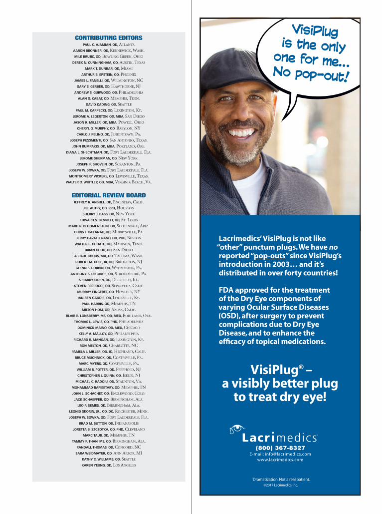
VisiPlug® –a visibly better plug
to treat dry eye!
Lacrimedics’ VisiPlug is not like “other” punctum plugs. We have no reported “pop-outs” since VisiPlug’s introduction in 2003… and it’s distributed in over forty countries!
FDA approved for the treatment of the Dry Eye components of varying Ocular Surface Diseases (OSD), after surgery to prevent complications due to Dry Eye Disease, and to enhance the efficacy of topical medications.
1Dramatization. Not a real patient. ©2017 Lacrimedics, Inc.
(800) 367-8327E-mail: [email protected]
www.lacrimedics.com
VisiPlugis the only one for me… No pop-out!
013_ro0619_TOC.indd 15013_ro0619_TOC.indd 15 6/5/19 4:34 PM6/5/19 4:34 PM

REVIEW OF OPTOMETRY JUNE 15, 201916
Outlook
My guess is you never wanted to be an internist, or else you’d have pursued
that calling. But more and more, optometrists need to be up to date on general wellness standards and be willing to bring them up with patients. Inevitably, this means hav-ing awkward conversations with people who might look askance at comments on obesity or smoking that come from the person they go to “for glasses.”
Nowhere is this more acute, or more common, than when seeing diabetes patients. It’s acute because these patients have the best chance of improving their long-term health through lifestyle modification. It’s commonplace because the footprint of diabetes is vast—about one third of the US population either has the disease or circumstances that qualify as “pre-diabetes.”
The medical need and urgency put you smack in the middle of the dia-betes epidemic, like it or not.
To make matters worse, it’s hap-pening in an environment that extends so much deference to patients that it only makes a tough conversation even harder. We hear a lot these days about how important it is to treat patients like customers, rolling out the red carpet for them in every conceivable way to keep them loyal to you, as a hedge against losing them to online providers of cut-rate care. People today and millennials especially—the mantra goes—will hold you to the same standards as their other retail experiences. If you can’t be as convenient as Amazon or as unctuous as Apple, you’ll lose out.
You know what? Too bad. A lot of diabetes and pre-diabetes patients need a wake-up call about their health. If you don’t put your foot down, they may lose theirs.
Not much good can come from an interaction where patients are inclined to treat doctors like a maître d’ and the doctors themselves are ill at ease discussing lifestyle modi-fication for fear of offending them. That’s two people looking for an easy out to a difficult situation. Skip-ping a conversation about weight loss or letting a diabetes patient take a pass on a dilated eye exam helps no one.
That means you’re going to lose some patients. So be it. Movie buffs (and Guns N’ Roses fans) know the line, “What we’ve got here is failure to communicate. Some men you just can’t reach.” Same goes with patients. Many will genuinely welcome advice and be grateful for your concern, but there will always be a few who chafe at it. If a diabetes patient refuses a dilated exam, or acts defensive about following up with their GP to discuss weight loss, note it in the record and send them on their way. But it’s your responsi-bility to bring those issues up.
The customer isn’t always right. Sometimes, they aren’t even a cus-tomer at all. In your dispensary? Sure. But in the exam room, they’re a patient and you’re an authority.
This month’s feature article on diabetes, plus a thoughtful column on billing do’s and don’ts for the diabetic eye exam, can help keep you connected to standards of care. Now all you have to do is enforce them. ■
To make a dent in the diabetes epidemic, you need to tackle sensitive topics head-on.
Losing Patience… and Patients
By Jack Persico, Editor-in-ChiefPRINTED IN USA
FOUNDING EDITOR, FREDERICK BOGER
1891-1913
EDITORIAL OFFICES
11 CAMPUS BLVD., SUITE 100NEWTOWN SQUARE, PA 19073
SUBSCRIPTION INQUIRIES
1-877-529-1746
CONTINUING EDUCATION INQUIRIES
1-800-825-4696
EDITOR-IN-CHIEF • JACK PERSICO
(610) 492-1006 • [email protected]
MANAGING EDITOR • REBECCA HEPP
(610) 492-1005 • [email protected]
SENIOR EDITOR • BILL KEKEVIAN
(610) 492-1003 • [email protected]
ASSOCIATE EDITOR • CATHERINE MANTHORP
(610) 492-1043 • [email protected]
ASSOCIATE EDITOR • MARK DE LEON
(610) 492-1021 • [email protected]
SPECIAL PROJECTS MANAGER • JILL HOFFMAN
(610) 492-1037 • [email protected]
ART DIRECTOR • JARED ARAUJO
(610) 492-1032 • [email protected]
DIRECTOR OF CE ADMINISTRATION • REGINA COMBS
(212) 274-7160 • [email protected]
EDITORIAL BOARD
CHIEF CLINICAL EDITOR • PAUL M. KARPECKI, OD
ASSOCIATE CLINICAL EDITORS • JOSEPH P. SHOVLIN, OD; ALAN G. KABAT, OD; CHRISTINE W. SINDT, OD
DIRECTOR OPTOMETRIC PROGRAMS • ARTHUR EPSTEIN, OD
CLINICAL & EDUCATION CONFERENCE ADVISOR
PAUL M. KARPECKI, OD
CASE REPORTS COORDINATOR • ANDREW S. GURWOOD, OD
CLINICAL CODING EDITOR • JOHN RUMPAKIS, OD, MBA
CONSULTING EDITOR • FRANK FONTANA, OD
COLUMNISTS
CHAIRSIDE • MONTGOMERY VICKERS, OD
CLINICAL QUANDARIES • PAUL C. AJAMIAN, OD
CODING CONNECTION • JOHN RUMPAKIS, OD
CORNEA & CONTACT LENS Q+A • JOSEPH P. SHOVLIN, OD
DIAGNOSTIC QUIZ • ANDREW S. GURWOOD, OD
THE ESSENTIALS • BISANT A. LABIB, OD
FOCUS ON REFRACTION • MARC TAUB, OD;
PAUL HARRIS, ODGLAUCOMA GRAND ROUNDS • JAMES L. FANELLI, OD
NEURO CLINIC • MICHAEL TROTTINI, OD;MICHAEL DELGIODICE, OD
OCULAR SURFACE REVIEW • PAUL M. KARPECKI, OD
RETINA DILEMMAS • DIANA L. SHECHTMAN, OD;
JAY M. HAYNIE, OD
RETINA QUIZ • MARK T. DUNBAR, OD
REVIEW OF SYSTEMS • CARLO J. PELINO, OD;JOSEPH J. PIZZIMENTI, OD
SURGICAL MINUTE • DEREK N. CUNNINGHAM, OD;WALTER O. WHITLEY, OD, MBA
THERAPEUTIC REVIEW • JOSEPH W. SOWKA, OD
THROUGH MY EYES • PAUL M. KARPECKI, OD
URGENT CARE • RICHARD B. MANGAN, OD
JOBSON MEDICAL INFORMATION LLC
016_ro0619_outlook.indd 16016_ro0619_outlook.indd 16 6/5/19 4:31 PM6/5/19 4:31 PM

The 1day Miru Toric flat pack is designed using SmartTouch™ Technology which minimizes lens handling and contamination concerns so contact lenses can be worn more comfortably and hygienically.
1day Miru toric employs a unique Smart Fit™ design that naturally orients the lens correctly no matter which way it is inserted.
A Unique Approach to Daily Disposable Soft Toric Lenses
For a trial pair, please email [email protected]
EASY,CLEAN,PORTABLE.
©2019 Menicon America, Inc. Miru, Smart Fit and SmartTouch are registered trademarks of Menicon Company Ltd.
RO0319_Menicon.indd 1RO0319_Menicon.indd 1 2/25/19 11:52 AM2/25/19 11:52 AM

Through My Eyes
18 REVIEW OF OPTOMETRY JUNE 15, 2019
Choose Your Doors Wisely
When optometric oppor-tunities present them-selves, we ODs are in a
privileged situation—we can choose whichever seems most interesting and enjoyable. However, we must carefully weigh our interests with those of our patients. What oppor-tunities will best serve our patient populations? Certainly myopia pro-gression, dry eye disease and cataract surgery comanagement are on the rise in just about every practice. Other areas of growth, highlighted in this month’s issue, include age-related macular degeneration (AMD) and diabetes management.
Ask the Right QuestionsThe first step to taking advantage of the opportunities is deciding which subspecialties would be ideal for you, your practice and your patients. This, of course, requires asking the right questions:1. Is it a significant population? 2. Can I make a difference in the
patients’ lives (i.e., is it treatable/manageable)?
3. Are there diagnostic technolo-gies that can readily identify and monitor these patients?
4. Does it have a reasonable chance of benefiting my practice?
5. Does it positively impact the patient?
Let’s answer some of these ques-tions for this month’s topics.
AMDThe number one cause of blind-ness in Caucasian patients, AMD
accounts for more than 50% of all blindness.1 Without a doubt, it’s a significant and growing population. Although not considered curable, it is more manageable than ever before with nutritional supplemen-tation, protection from ultraviolet and high-energy visible light and anti-vascular endothelial growth factor injections.2
New diagnostic technologies such as dark adaptation and OCT imag-ing, including OCT angiography for wet AMD, assist in accurate diagnosis and management. For example, making an early diagnosis, such as stage 1 or even sooner, with the help of a failed dark adaptation test allows you to recommend a carotenoid supplement as soon as possible to help slow progression as much as possible and preserve the patient’s vision.
These technologies carry CPT codes and also help dictate the proper follow-up—ensuring a positive impact on your practice. Obviously, AMD comes with a significant emotional component, and establishing an early diagnosis with proper management provides patients hope and trust in you as a physician.
Diabetic RetinopathyFew systemic conditions have ocular manifestations even remotely close to diabetes, and optometrists can play an enormous role in helping patients better manage the condi-tion. The disease affects more than 20% of all Hispanics and more than
16% of African Americans, and that’s just the minority populations in most practices.3
We should involve ourselves in systemic disease assessment by ask-ing patients about their A1c, choles-terol, blood pressure and smoking status—all of which contribute to diabetes progression. Simply questioning the patient often raises their awareness of these cofounding issues and helps them make healthi-er lifestlye choices.
Diabetes may affect the entire body, but the eyes are one of the key organs that manifest findings cru-cial for diagnosis and management. Dry eye affects more than 50% of patients with diabetes, and diabetic retinopathy is the leading cause of vision loss in working-age adults.4,5 All of this means we are integral to the care team for this enormous and rapidly growing patient population.
Opportunity is knocking…
be sure you are well prepared to answer the door. ■
Note: Dr. Karpecki consults for companies with products and ser-vices relevant to this topic.
1. Congdon N, O’Colmain B, Klaver CC, et al. Causes and preva-lence of visual impairment among adults in the United States. Arch Ophthalmol. 2004;122(4):477-85.2. Chew EY, Clemons T, SanGiovanni JP, et al. The Age-Related Eye Disease Study 2 (AREDS2): study design and baseline characteristics (AREDS2 Report Number 1). Ophthalmology. 2012;119(11):2282-89.3. Cowie CC, Rust KF, Ford ES, et al. Full accounting of diabetes and pre-diabetes in the U.S. population in 1988-1994 and 2005-2006. Diabetes Care. 2009;32(2):287-94.4. Zhang X, Zhao L, Deng S, et al. Dry eye syndrome in patients with diabetes mellitus: prevalence, etiology, and clinical charac-teristics. J Ophthalmol. 2016;2016:8201053.5. National Eye Institute. Facts About Diabetic Eye Disease. https://nei.nih.gov/health/diabetic/retinopathy. Accessed May 9, 2019.
What patient care opportunities are knocking? By Paul M. Karpecki, OD, Chief Clinical Editor
018_ro0619_TME.indd 18018_ro0619_TME.indd 18 6/5/19 3:59 PM6/5/19 3:59 PM

Review Education Group partners with Salus University for those ODs who are licensed in states that require university credit. See www.reviewsce.com/events for any meeting schedule changes or updates.
Earn up to
19 CE Credits*
REGISTER ONLINE: WWW.REVIEWSCE.COM/CHARLESTON2019
NEW TECHNOLOGIES & TREATMENTS IN
Eye CareNENEW W TNENEW W T &&& T&& T
201999
CHARLESTON, SOUTH CAROLINADate:November 1-3, 2019
Location:Charleston Marriott170 Lockwood Blvd., Charleston, SC 29403Reservations: 1-800-228-9290
A limited number of rooms have been reserved at the rate of $229/night. Please book with the hotel directly by calling the number above. Mention “Review’s New Technologies & Treatments” for group rate.
Early Bird Special: $420Register before September 9, 2019 for early bird pricing.
Three Ways to RegisterOnline: www.reviewsce.com/Charleston2019Call: 1-866-658-1772E-mail: [email protected]
*Approval pending
Administered by: Partially supported byunrestricted educational grants from:
Sun PharmaceuticalsBausch & LombCarl Zeiss Meditec
Alcon
Program Chair:Paul Karpecki, OD, FAAO

REVIEW OF OPTOMETRY JUNE 15, 201920
Cha i r Side
Sunny With a Chance of No-shows
It’s time to talk about the one thing no optometrist can control, and I don’t mean no-shows.
OK, maybe no-shows are indeed the number one thing we can’t control, but the number two thing is…
Come to think of it, the number two thing is not what I had in mind either. It’s the idiot in front of you merging onto the 70mph highway at 47mph.
But, down the list somewhere is my topic for the day. One of the mil-lion things optometrists just cannot control is the weather.
Some of our fearless leaders want us to believe eliminating fossil fuels and cow manure will fix the weather issues, but then how would we get a good burger and drive to the CE meetings our fearless leaders require of us to continue to practice?
Foul Weather PatientsSo, we really are at the weather’s mercy. In my 40 years in practice I have learned some things about optometry and the weather:
1. Patients may miss appoint-ments when the weather is horrible, but more often, they don’t show up when the weather is amazing. Who needs to protect their eyesight when you can catch some rays instead?
2. Snow, no matter how deep, will not prevent some patients from showing up on time—but only when you are running late because of the snow. They love standing knee-deep in the frozen tundra, staring into your windows, wondering why you are late. They also drove 20 miles to
get there. The patient from across the street no-shows because of the bad weather.
3. Patients buy more sunglasses from you when it’s dreary. That’s because you brought it up when they weren’t thinking about it. When it’s beautiful and sunny, you are too late. They already bought them somewhere else.
4. Does your yard need rain? Take a day off. If you want to know when I am working, look outside. Is it beautiful? I’ll be here until 7pm.
5. If you live in the desert, stop griping about the drought. If you live at the beach, stop griping about hurricanes. If you charge a $10 co-pay, stop griping about your bills.
6. The average American owns two umbrellas. Not bragging, but the average OD owns seven. Are we more successful or do we just forget where we put umbrellas?
7. Every tire I have ever owned has been used in all seasons, so what’s the big deal?
8. The average hail damage pay-out is $3,000. The average hailstorm lasts six minutes. That’s $80,000 per hour. The average physician makes $80 per hour. Hmmm.
Schooled by the WeatherWhen I was in school, every pre-med student had to take at least one course in every discipline of science. You could take one course pass/fail. In the Geology Department, the only course that seemed to have any value to me was meteorology. Unfortunately, meteorology has nothing to do with meteors. They taught me that the first day of class. I scored a 62% on my final and passed. My friend Rich got a 60% and failed. He’s now a commercial pilot, soaring through the clouds with no clue why they are there.
The lesson is this: respect the weather. Learn your weather pat-terns and how they affect your patients. When the weather is horri-ble or when it ruins your cash flow, just order pizza or fly off to where the weather is amazing—but not if the pilot’s name is Rich. ■
Patients, like the weather, can be fickle. Together, they can really drive you crazy. By Montgomery Vickers, OD
020_ro0619_Chairside.indd 20020_ro0619_Chairside.indd 20 6/5/19 2:43 PM6/5/19 2:43 PM

RO0619_Contamac.indd 1 5/29/19 9:47 AM
Cl in ica l Quandaries
REVIEW OF OPTOMETRY JUNE 15, 201922
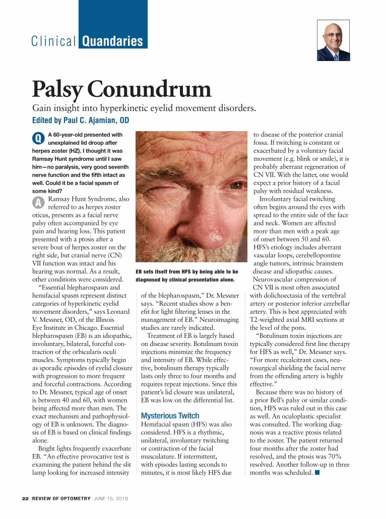
A 60-year-old presented with unexplained lid droop after
herpes zoster (HZ). I thought it was Ramsay Hunt syndrome until I saw him—no paralysis, very good seventh nerve function and the fifth intact as well. Could it be a facial spasm of some kind?
Ramsay Hunt Syndrome, also referred to as herpes zoster
oticus, presents as a facial nerve palsy often accompanied by eye pain and hearing loss. This patient presented with a ptosis after a severe bout of herpes zoster on the right side, but cranial nerve (CN) VII function was intact and his hearing was normal. As a result, other conditions were considered.
“Essential blepharospasm and hemifacial spasm represent distinct categories of hyperkinetic eyelid movement disorders,” says Leonard V. Messner, OD, of the Illinois Eye Institute in Chicago. Essential blepharospasm (EB) is an idiopathic, involuntary, bilateral, forceful con-traction of the orbicularis oculi muscles. Symptoms typically begin as sporadic episodes of eyelid closure with progression to more frequent and forceful contractions. According to Dr. Messner, typical age of onset is between 40 and 60, with women being affected more than men. The exact mechanism and pathophysiol-ogy of EB is unknown. The diagno-sis of EB is based on clinical findings alone.
Bright lights frequently exacerbate EB. “An effective provocative test is examining the patient behind the slit lamp looking for increased intensity
of the blepharospasm,” Dr. Messner says. “Recent studies show a ben-efit for light filtering lenses in the management of EB.” Neuroimaging studies are rarely indicated.
Treatment of EB is largely based on disease severity. Botulinum toxin injections minimize the frequency and intensity of EB. While effec-tive, botulinum therapy typically lasts only three to four months and requires repeat injections. Since this patient’s lid closure was unilateral, EB was low on the differential list.
Mysterious TwitchHemifacial spasm (HFS) was also considered. HFS is a rhythmic, unilateral, involuntary twitching or contraction of the facial musculature. If intermittent, with episodes lasting seconds to minutes, it is most likely HFS due
to disease of the posterior cranial fossa. If twitching is constant or exacerbated by a voluntary facial movement (e.g. blink or smile), it is probably aberrant regeneration of CN VII. With the latter, one would expect a prior history of a facial palsy with residual weakness.
Involuntary facial twitching often begins around the eyes with spread to the entire side of the face and neck. Women are affected more than men with a peak age of onset between 50 and 60. HFS’s etiology includes aberrant vascular loops, cerebellopontine angle tumors, intrinsic brainstem disease and idiopathic causes. Neurovascular compression of CN VII is most often associated
with dolichoectasia of the vertebral artery or posterior inferior cerebellar artery. This is best appreciated with T2-weighted axial MRI sections at the level of the pons.
“Botulinum toxin injections are typically considered first line therapy for HFS as well,” Dr. Messner says. “For more recalcitrant cases, neu-rosurgical shielding the facial nerve from the offending artery is highly effective.”
Because there was no history of a prior Bell’s palsy or similar condi-tion, HFS was ruled out in this case as well. An oculoplastic specialist was consulted. The working diag-nosis was a reactive ptosis related to the zoster. The patient returned four months after the zoster had resolved, and the ptosis was 70% resolved. Another follow-up in three months was scheduled. ■
Palsy ConundrumGain insight into hyperkinetic eyelid movement disorders.Edited by Paul C. Ajamian, OD
A
Q
EB sets itself from HFS by being able to be diagnosed by clinical presentation alone.
022_ro0619_CQ.indd 22022_ro0619_CQ.indd 22 6/5/19 2:44 PM6/5/19 2:44 PM

Free Air with the TonocarePlease contact our partners for information on our new tonometer.
calcoastophthalmic.com deviceoptical.com lombartinstrument.com innovamed.com
www.keelerusa.com • 3222 Phoenixville Pike - Bldg. #50 • Malvern, PA 19355Tel No: 1-610-353-4350 • Toll Free: 1-800-523-5620 • Fax: 1-610-353-7814
RO0619_Keeler.indd 1 5/23/19 2:20 PM

REVIEW OF OPTOMETRY JUNE 15, 201924
Focus on Refraction
Strabismus is a topic that, on the surface, might frighten the faint of heart.
Parents report their strabismic children’s eyes turn in or out. They might be seeking a second opinion after an ophthalmologist recommended surgery. They want you to help them understand what is happening and to offer the most appropriate treatment.
While the more common type of strabismus in chil-dren is esotropia, exotropia is found in about 20% of young patients with strabis-mus.1 Parents of children with exotropia often present stating that their child’s eye, or eyes, drifts out. This can happen if the child is looking far away and not concen-trating visually, if they are reading at near or in both cases. Exotropia can be intermittent or constant and monocular, alternating or binocular. We look further into it here.
Navigating TreatmentOne of the first questions parents of children with strabismus typically have is related to treatment. The second question usually has to do with whether the condition will become constant. Many of us have heard stories about our ophthalmological colleagues telling parents that their child must have surgery. But, thanks to the Pediatric Eye Disease Investigator Group, we can now confidently refute that advice.2 The study observed 183
children ages three to 10 for three years and found that only 15%—which they contend was actually an overestimation—deteriorated.2 Deterioration was defined as having a constant exotropia ≥10 prism diopters (PD) at distance and near or a decreased stereopsis of ≥20.6 log arcsec (tested with Randot Preschool Near Stereoacuity).2 The team also found that exotropia control, stereopsis and magnitude of the exodeviation at distance improved.2
The overall treatment goal for intermittent exotropia is to reduce the frequency and size of the turn to enhance fusion. While surgery is a potential treatment option, others include stimulating convergence with lenses (over-minus), prism therapy (full or partial), occlusion and vision therapy. A study found the following success rates:3
• surgery (with functional results included): 43%
• occlusion: 37%• over-minus: 28%• prism therapy: 28% • vision therapy: 59%Even though traditional
vision therapy offers the greatest chance of success, to say it is challenging in a younger patient population is an understatement, which is why we surmise it is not always the first-line treatment. With so many options, it can be difficult to know which is the right one for your patient. Now that we have research proving that the intermittent turn will likely stay that way, we need to
consider other treatment options to spur on improvement. Hopefully, this case series offers some guidance.
Case #1A three-year-old male was seen for a follow-up after having been prescribed over-minus at his last visit several months prior. At both visits (previously without, and at this visit with, spectacles), he showed a 40 PD intermittent alternating exotropia at distance (80% of the time, OD>OS) and orthophoria at near. His mother reported that he refused to wear the glasses, and when he did wear them, he looked over them. His visual acuities were 20/30 OU with the glasses and 20/20 OU without them. He showed gross stereopsis with the Keystone Basic Binocular (KBB) test at near. Even though the turn was occurring only at distance, we
While exotropia isn’t the most common in young patients, here’s how to approach it if it presents. By Marc B. Taub, OD, MS, and Paul Harris, OD
Sizing Up Strabismus
This child is completing the ball rolling exercise.
024_ro0619_FoR.indd 24024_ro0619_FoR.indd 24 6/5/19 4:14 PM6/5/19 4:14 PM

Reference: 1. JAMA
†
Recommend PreserVision AREDS 2 formula by name for your patients today!
Learn more at PreserVision.com
The National Eye Institute recommends an AREDS formulation for patients with
moderate to advanced AMD.†Do you?
Only PreserVision® AREDS 2 formula eye vitaminscontain the EXACT levels of 6 key nutrients recommended by the
National Eye Institute following the landmark AREDS2 study1,*
RO0619_B&L Preservision.indd 1 5/17/19 10:14 AM
REVIEW OF OPTOMETRY JUNE 15, 201926
Focus on Refraction
attempted prism to see if there were any changes and observed none.
Given that the child refused to wear the over-minus lenses and prism was ineffective, we instituted a modified home vision therapy program of basic bilateral eye movements. The key in this case was to encourage convergence-type movements. With this in mind, we instructed the parent to roll a ball (large at first) to the child from several feet away and play a balloon hitting game. Horizontal and vertical eye stretches, in which the child tracks the moving object back and forth and up and down several times, were instituted.
We will see the child in several months to evaluate his progress. As he gets older, we will consider a full round of therapy.
Case #2A three-year-old female was seen for a second opinion after surgery was recommended. Her uncorrected acuities were 20/25 OD, 20/30 OS and 20/25 OU, and the cover test showed an 18 PD intermittent right exotropia 50% of the time at distance and 4 exophoria at near. Stereopsis was positive using the
KBB, as the child localized several pictures. Retinoscopy showed plano -1.00x180 OU. An over-minus of -1.50 D was provided on top of the astigmatism correction, but the acuities dropped to 20/80 OU. We then moved on to try prism using a 3 PD base-in in each eye and found a modicum of success. The cover test now showed 10 exophoria at distance and orthophoria at near.
We prescribed the astigmatism along with the prism for full-time wear and requested basic eye stretches. She will be re-evaluated in two to three months.
Case #3A 15-year-old male presented for his yearly exam. Wearing -6.00 D OU, his acuities were 20/80 OU. The cover test showed 25 intermittent alternating exotropia at distance and near. Global stereopsis was absent. He was refracted and, with -7.50D OU (20/20 OU), showed 20 intermittent alternating exotropia at distance and 15 exophoria at near.
Our next treatment option was prism. With a 3 PD base-in in each eye, the cover test showed 10 exophoria at distance and near. KBB was retested, and a positive response
was recorded. He will be reevaluated in two to three months.
As discussed earlier, the purpose of treatment for intermittent exotropia is to provide the opportunity for fusion. In two of the case examples, we used over-minus without success, but that should not deter you from trying it with your own patients. With the second patient, the acuity actually decreased with the extra minus—not the expected outcome—and we were forced to try another treatment option. In all three cases, we employed the use of prism, and in two cases, we found success.
There is no magic amount of prism that will work with each patient. We recommend starting with about one-third of the amount of the eye turn and rechecking the cover test. Consider adjusting to find the most appropriate amount of prism at this time. Each of the two successful prism cases showed about 20 to 25 PD, so we selected a 3 PD base-in OU. In two of the cases, we instituted some basic therapy. In the first, it was the sole treatment, while it was adjunctive to the prism in the second. Anything that we can do to spur on looking and convergence
behavior will set the stage for a full program of vision therapy.
There is no rush to jump into the deep end and recommend surgery as a first-line treatment for strabismus, and, more specifically, for exotropia. The three treatment options discussed should be considered as alternatives before pursuing a more aggressive pathway. ■
1. Rutstein RP, Daum KM. Anomalies of Binocular Vision: Diagnosis and Management. St. Louis, MO: Mosby, 1998.2. Mohney BG, Cotter SA, Chandler DL. Three-year observa-tion of children 3 to 10 years of age with untreated intermittent exotropia. Ophthalmology. January 15, 2019. [Epub ahead of print].3. Coffey B, Wick B, Cotter S, et al. Treatment options in intermittent exotropia: a critical appraisal. Optom Vis Sci. 1992;69(5):386-404.
Break-out Therapy Activities • Ball Rolling: This bilateral activity encourages convergence. The parent and child sit on the
floor with their legs in a “V,” about five to six feet apart. Starting with a larger ball, the parent rolls the ball toward the child. The child watches the ball as it comes closer (convergence) and then rolls the ball back to the parent, watching as it gets further away (divergence). Use a smaller ball and increase the distance as the child masters the skill.
• Balloon Hitting: This bilateral activity also encourages convergence. The child is given a large balloon and is instructed to hit it up in the air as hard as they can. As they watch the balloon move further away, they are using divergence, and as it comes back down, they are using con-vergence. Use smaller balloons as the skill is mastered.
• Eye Stretches: This bilateral activity encourages looking behavior/attention. There are two methods for this activity: (1) The parent uses a fun target, which lights up, makes noise or does both, and moves it vertically and horizontally. All the child has to do is watch the target. The parent should place their hand on the top of the child’s head to ensure their eyes, and not their head, move. (2) The parent uses a fun target as described above and keeps it still. The child looks at the target, and the parent moves their head up and down and side to side.
024_ro0619_FoR.indd 26024_ro0619_FoR.indd 26 6/5/19 2:46 PM6/5/19 2:46 PM

Exceed their expectations with our complete line of digital refraction devices.Set your practice apart with the most advanced Phoroptor® ever, Phoroptor® VRx,
and our pixel-perfect ClearChart® 4 family of digital acuity systems.
W AT C H O U R N E W S E R I E S AT :
PASSIONATEABOUTEYECARE.COM
VISIT US AT OPTOMETRY’S MEETING/AOA #816
O F C O U R S E YO U A R E .
© 2019 AMETEK, Inc. & Reichert, Inc. (05-2019) Phoroptor and ClearChart are registered trademarks of Reichert, Inc. · Phoroptor and ClearChart are designed & assembled in USA ·
PASSIONATE ABOUT EYE CARE
RO0619_Reichert.indd 1 5/17/19 10:05 AM

Re t ina Dilemmas
REVIEW OF OPTOMETRY JUNE 15, 201928
Re t ina Dilemmas
Retinal vein occlusion (RVO) is second only to diabetic retinopathy as a leading
cause of retinal vascular disease. We typically encounter a case of RVO every day in our retina practice, and patients often present with a his-tory of hypertension or glaucoma. Although macular ischemia and neovascularization are complica-tions that contribute to the deterio-ration of vision, the most common cause of vision loss is macular edema. It is important to treat the macular edema promptly, given the fact that both clinical trials and clinical practice show a penalty in delaying treatment.
Intravitreal pharmacotherapy has expanded the treatment options for macular edema associated with vascular retinopathies such as RVO. Such a treatment protocol can provide substantial visual and struc-tural improvements.
Because macular edema is medi-ated by vascular endothelial growth factor (VEGF), treatment with anti-VEGF therapies such as Lucentis (ranibizumab, Genentech), Eylea (aflibercept, Regeneron) and Avastin (bevacizumab, Genentech) is par-ticularly effective. However, RVO is a multifactorial disease and inflam-mation also plays a critical role in its pathogenesis. Hypoxia promotes inflammatory mediators, which increase permeability and lead to retinal-blood barrier breakdown, resulting in macular edema. To combat this inflammatory process, clinicians should consider intravit-
real steroid implants, such as Ozurdex (Allergan), which inhibit vascular permeability and suppress inflammatory mediators. Treatment of macular edema related to RVO with Ozurdex can lead to a significant anatomical and functional improve-ment. The biodegradable implant delivers a high concentration of dexa-methasone into the vitre-ous over a period of time. We have often observed effects of its release for an average of four months, and lower concentrations may be observed for up to six months, based on clinical trial data.1,2 This may be an option for treat-ment when continuously monthly injections are not suitable.
Ozurdex, FDA approved for macular edema related to RVO in 2009 following the GENEVA study, has since become a useful therapy in our office for selective cases of macular edema associated with RVO.2 Although the implant provides positive anatomical outcomes associated with decreased central retinal thickness (correlating with visual improvement), it comes with some adverse effects. These include cataract formation and increased intraocular pressure (IOP). Clinical trials show that up to 20% of patients will exhibit an IOP rise.1,2
In our experience, any IOP increase is usually transient and peaks by four to six weeks. Most cases of increased IOP in our practice have subsided once the steroid wears off and have been effectively managed with anti-glaucoma topical medi-cations. However, clinicians must monitor the optic nerve, especially considering glaucoma may have been a contributing factor to the patient’s RVO.
When determining the best treat-ment options, clinicians must weigh the efficacy of the drug against the
Tame the SwellingIntravitreal pharmacotherapy is changing the treatment landscape for patients with macular edema and RVO. By Diana Shechtman OD, Jay M. Haynie OD, and Brendan Girschek, MD
Fig. 1. This patient presented with retinal hemorrhages and cotton-wool spots in all four quadrants of his left eye.
Fig. 2. SD-OCT imaging confirmed macular edema in the left eye.
028_ro0619_RD.indd 28028_ro0619_RD.indd 28 6/5/19 4:12 PM6/5/19 4:12 PM

833-3NULIDS(833-368-5437)NuSightMedical.com
It’s so easy your patients can
use it with their eyes closed!
Treat the disease, not just the symptoms
The NuLids System is a unique doctor directed, at-home treatment for patients with Dry Eye Disease. NuLids effectively alleviates symptoms by stimulating and rejuvenating the Meibomian glands back to a healthy state.
Now there is NuLids
Although there are dozens of at-home dry eye treatment options available today, most only treat the symptoms of dry eye and not its cause. Patients are left frustrated with no treatment plan, and with little to no relief from their debilitating symptoms.
*NuLids is indicated for use with the following: Blepharitis, Demodex, or when the lid margins show evidence of thickening, cylindrical dandruff, meibomian stasis, ectasia, scaling, skurf, or when the lids become scalloped, rolled, or hyperemic. NuLids is typically prescribed with a lubricating gel, a cleanser such as hypochlorous acid for Blepharitis, or tea tree oil when Demodex is present.
NuLids®
The innovative, doctor
directed home treatment
for Dry Eye Disease
*
NuLids creates a novel recurring revenue stream for your practice!
RO0619_NuSight Medical.indd 1RO0619_NuSight Medical.indd 1 6/4/19 9:51 AM6/4/19 9:51 AM

REVIEW OF OPTOMETRY JUNE 15, 201930
burden of re-injections and any potential associated complications.
One and DoneCase by Dr. ShechtmanA 62-year-old Caucasian male was referred for an evaluation of sus-pected central retinal vein occlusion (CRVO) OS. His medical history included hypertension for 15 years. His best-corrected visual acuity measured 20/25 OD and 20/40 OS. The dilated fundus examination was unremarkable for the right eye. The left eye revealed a healthy nerve with a cup-to-disc ratio of 0.4/0.4, dilated tortuous veins, retinal hemorrhages and a few cotton-wool spots in all four quadrants (Figure 1). Spectral-domain optical coherence tomog-raphy (SD-OCT) was consistent for the presence of macular edema OS (Figure 2). We confirmed the suspected diagnosis of CRVO with associated macular edema OS and initiated treatment with Lucentis.
At the one-month follow up, his macular edema had decreased and his visual acuity improved to 20/30. The patient had another injection of Lucentis and was scheduled for another follow up in a month. At the next follow-up the macular edema had increased with deteriorating vision to 20/50. We then decided to try Ozurdex. One month after inject-ing the implant, the patient expe-rienced a dramatic improvement of macular edema with restoration
of normal macular contour (Figure 3). This correlated with a visual acuity gain to 20/25. The patient remained stable over a two-month period without the need for another injection. He had no noticeable increase of IOP dur-ing follow up.
Careful MonitoringCommentary by Dr. Haynie
The treatment of macular edema secondary to a CRVO is crucial, considering chronic macular edema increases the risk of permanent visual loss, and the natural history of a CRVO is quite poor without intervention. When treating macu-lar edema secondary to a CRVO in my practice, the retina surgeons will typically initiate treatment with Avastin, Lucentis or Eylea. If the patient fails to respond (presenting with recurrent or persistent edema) to an induction treatment of three injections, the next consideration is the use of steroids, whether it’s Triesence (triamcinolone acetonide injectable suspension, Novartis), Kenalog (triamcinolone acetonide injectable suspension, Bristol-Myers Squibb) or Ozurdex.
As Dr. Shechtman noted, clini-cians must weigh the risks and ben-efits of intravitreal steroids, given the risk of elevated IOP. In addition, Ozurdex is not FDA approved for phakic patients without a planned cataract surgery with future intra-ocular lens placement.
Intravitreal steroids were found to be effective for the treatment of macular edema secondary to a RVO in SCORE 1 and SCORE 2 clinical trials and since have become great adjuncts to anti-VEGF compounds in cases of chronic or recurrent mac-ular edema.3,4 The risk of elevated IOP following intravitreal steroids can be as high as 20%, warranting
close follow up with IOP monitor-ing—monthly based on our practice protocol. If we are concerned about a patient’s steroid response, we consider a trial of topical Durezol (Novartis) QID. If they experience no increase in IOP within four weeks, the surgeons feel comfortable using intravitreal steroids.
In the event of elevated IOP, the patient should be managed with topical anti-hypertensive agents for at least six months from the date of the intravitreal steroid treatment. In these cases clinicians should avoid prostaglandin analogs, as they can contribute to inflammatory ocular disease and cause cystoid macular edema (both present in patient with RVOs). Provided the patient has no systemic contraindications to car-bonic anhydrase inhibitors, I tend to start with dorzolamide or Azopt (Novartis) TID, as this class of phar-maceutical can lower IOP and also reduce macular edema.
Patients with macular edema secondary to a CRVO should be referred for treatment based on clinical trial data that show a visual benefit from treatment that exceeds the natural history of the disease, which can lead to marked loss of visual function. ■
Dr. Girschek will be one of the primary retina specialists at Retina Macula Specialists of Miami, starting July 2019.
1. National Horizon Scanning Centre (NHSC). Dexamethasone intravitreal implant (Posurdex) for macular oedema secondary to central or branch retinal vein occlusion. Horizon Scanning Technology Briefing. Birmingham, UK: National Horizon Scanning Centre (NHSC); 2009. 2. Haller JA, Bandello F, Belfort R Jr, et al; Ozurdex Geneva Study Group. Randomized, sham-controlled trial of dexamethasone intravitreal implant in Ozurdex (dexamethasone intravitreal implant) patients with macular edema due to retinal vein occlusion. Ophthalmology. 2010;117(6):1134-46. 3. Scott IU, Vanveldhuisen PC, Oden NL, et al. Baseline charac-teristics and response to treatment of participants with hemiretinal compared with branch retinal or central retinal vein occlusion in the standard care vs corticosteroid for retinal vein occlusion (SCORE) study: SCORE study report 14. Arch Ophthalmol. 2012;130(12):1517-24.4. Scott IU, VanVeldhuisen PC, Ip MS, et al. Effect of bevacizumab vs aflibercept on visual acuity among patients with macular edema due to central retinal vein occlusion: the SCORE2 randomized clinical trial. JAMA. 2017;317(20):2072-87.
Re t ina Dilemmas
Fig. 3. One month after implanting Ozurdex, the patient’s edema was once again under control with a normal macular contour.
028_ro0619_RD.indd 30028_ro0619_RD.indd 30 6/5/19 4:13 PM6/5/19 4:13 PM

Hypochlorous Acid Solution
Daily Eyelid & Eyelash Cleanser• Naturally removes foreign matter• Nontoxic, nonirritating• Has been shown in studies to reduce
bacteria/microbes• Formulated to relieve symptoms of
MGD, Blepharitis and Dry Eye % HOCL spray and• Available in 0.01
0.0 85% HOCL gel
Hypochlorous Acid SolutionNOW Available Without a Prescription
EyEyEyEy•••
•
•
(866) 752-6006 – FocusLaboratories.comTrademarks: ZenoptiqTM (Paragon BioTeck, Inc.) | Copyright © 2018 Paragon BioTeck, Inc. All Rights Reserved.
LEARN MORE AT
zenoptiq.com
FLM190-0119-01
RO0219_Focus.indd 1 1/29/19 12:53 PM

Coding Connection
REVIEW OF OPTOMETRY JUNE 15, 201932
The CMS precedent-setting 2008 policy provided, for the first time, preventive services
for patients with diabetes, including an eye exam.1 Before, a patient had to have clinically evident signs and symptoms of ocular diabetic disease before Medicare would cover the exam. As of 2008, patients with dia-betes, and in the absence of diabetic retinopathy, are allowed a com-prehensive dilated eye exam on an annual basis.2 Subsequently, many, if not most, commercial carriers have followed suit with similar policies.
A Policy Often Misunderstood With this shift, many felt that, irre-spective of coverage, the medical carrier was always responsible for these exams. Many ODs rejoiced because they felt they could ignore the reduced reimbursement rate for a diabetes patient’s annual compre-hensive eye exam—a national aver-age of about $55 from a managed vision care plan—and instead bill the same exam at nearly triple the rate to the medical carrier.3 This, however, isn’t always the case.
The “diabetic eye exam” is a frequently discussed topic at opto-metric conferences. But I really don’t know why because no such thing exists. We can provide a comprehen-sive ophthalmic exam for a patient with diabetes in the absence of dia-betic retinopathy using codes 920X4 as medical policy guidance provides. This, of course, is where the devil is in the details and where our ethics as providers come into play.
Who Foots the Bill?When a diabetes patient has both a managed vision care plan and a medical plan—both of which cover a comprehensive ophthalmic exam—it’s not our choice which coverage to use or who to bill. It is the patient’s choice. Most ODs want to bill the medical carrier because it’s a “diabetic eye exam,” not a man-aged vision care exam, and those plans don’t pay enough anyway. Most choose to bill the carrier that pays the most—not a good strategy when defending yourself on audit.
My understanding of the CPT rules is that services provided are defined by the CPT code used to describe and bill for them. Therefore, a 92004 is a 92004, regardless of who is billed. In fact, some managed vision care plans likely have 92004 definitions (and requirements) that are more detailed than those of Medicare and the CPT. Additionally, there is no such thing as medical 920X4. CMS’s require-ments for preventive care related to a comprehensive ophthalmic exam require the patient to have a dilated fundus exam. This is not in align-ment with HEDIS requirements that allow a fundus photo as a proxy.
The pushback I receive from optometric peers is mostly based in remuneration, not logic—and that is not a good defense. If a patient has duplicative coverage and both of their carriers pay for a 920X4, we must inform the patient of this, pro-vide accurate information regarding copays and out of pocket costs (i.e.,
refraction if billed to medical car-rier) and let the patient make a well-informed decision of which policy to use. One caveat to keep in mind: some managed vision care plans do stipulate that they want you to bill the medical carrier and not the man-aged vision care plan in this circum-stance, so keep up with your carrier contract requirements.
In the past, patients would usually choose to bill the managed vision care policy. But a decade of this pol-icy has led to many medical carriers waiving the deductible or copay for a comprehensive ophthalmic exam for patients with diabetes and in the absence of diabetic retinopathy. The patient’s only out-of-pocket expense is the refraction (92015), which in many cases is similar to their man-aged vision care plan co-pay.
With proper patient education, you can often get paid more for your services while following the rules. This allows you to provide the highest level of services and protects you and your practice from unwanted scrutiny from third-party carriers. No one wants to be in the crosshairs for doing the wrong thing. ■
Send your coding questions to [email protected].
1. Centers for Medicare and Medicaid Services (CMS). Medicare Coverge of Diabetes Supplies and Services. www.medicare.gov/Pubs/pdf/11022-Medicare-Diabetes-Coverage.pdf. November 2008. Accessed April 28, 2019. 2.CMS. An overview of Medicare covered diabetes supplies and services. MLN Matters. www.cms.gov/Outreach-and-Education/Medicare-Learning-Network-MLN/MLNMattersArticles/down-loads/se0738.pdf. Accessed April 28, 2019. 3. CMS. Physician Fee Schedule. www.cms.gov/apps/physician-fee-schedule/search/search-results.aspx?Y=0&T=0&HT=0&CT=0&H1=92004&M=5. Accessed April 29, 2019.
Doing it the wrong way could put you in the line of fire. By John Rumpakis, OD, MBA, Clinical Coding Editor
Coding the “Diabetic” Eye Exam
032_ro0619_coding-JA.indd 32032_ro0619_coding-JA.indd 32 6/5/19 2:46 PM6/5/19 2:46 PM

FIVE OF A KIND BEATS ANY DEALER’S HAND. CONSULT LOMBART AND GET YOUR WINNING HAND.
Lombart Instrument is the nation’s largest distributor of ophthalmic instruments, services, and supplies. Since our founding in 1979, we have been dedicated to creating long-lasting partnerships and serving our customers: optometry and ophthalmology practices, universities, hospitals, and the US government. It is because of the effort of our incredible staff that we have been the industry leader since 1987, and we work hard every day to provide the highest quality service to our customers.
CONTACT YOUR LOMBART REPRESENTATIVE TO SEE WHICH OCT BEST FITS YOUR PRACTICE.
800.LOMBARTLOMBARTINSTRUMENT.COM
BROAD PORTFOLIO OF NEW
AND PRE-OWNED ADVANCED
TECHNOLOGY EQUIPMENT
DEDICATED,
KNOWLEDGEABLE ADVANCED
TECHNOLOGY DIRECTORS
CLINICAL SUPPORT TEAM FOR
ONGOING TRAINING
CONSULTATIVE APPROACH TO
FIND THE RIGHT SOLUTION
FOR YOU
RP0619_Lombart.indd 1 5/9/19 3:24 PM

42 minutes per day2 hours and 40 minutes
per day 3 hours per day
6 hours a day
9 hours a day
The
Children and Screens
These are
Nearly 70% of parents recognize that the amount of
meaning that optometrists have the opportunity to educate ¾ of this
Average hours spent on digital devices
Under 2 2 - 4 5 - 8
Age of Children
8 - 12 13 - 180
1 h
2 h
3 h
4 h
5 h
6 h
7 h
8 h
9 h
10 h
42 minutes
2 hours 40 minutes 3 hours
6 hours
9 hours
What a Daily Dose of Screen Time Can Do
RO0619_Zeavision.indd 2RO0619_Zeavision.indd 2 5/21/19 10:18 AM5/21/19 10:18 AM
References
4
7
RO0619_Zeavision.indd 3RO0619_Zeavision.indd 3 5/21/19 10:18 AM5/21/19 10:18 AM

REVIEW OF OPTOMETRY JUNE 15, 201936
We all get our manda-tory hours of con-tinuing education to keep our optometry
licenses active. How many of those hours are spent learning about evolving technology? How about the future of pharmacology? How will artificial intelligence impact eye care? A lot of changes are occurring in the field of medicine on a seemingly daily basis.
For example, more Americans are being diagnosed with diabetes mellitus every day and are in need of medical eye care to prevent sight loss. It is vital optometrists understand the pathways in which diabetic retinopathy (DR) develops, current management options and new and upcoming tools to detect DR. Will you be up-to-date on the latest technological advances and research?
Pathophysiology Chronic hyperglycemia, thought to be the fundamental prerequisite of DR, leads to vascular changes and subsequent retinal injury and ischemia.1,2 Vascular endothelial
growth factor (VEGF), expressed in response to ischemia and hypoxia, is an important factor in the devel-opment of both diabetic macular edema (DME) and proliferative diabetic retinopathy (PDR). VEGF alters retinal capillary permeability by increasing the phosphoryla-tion of proteins involved with tight-junctions such as zonula occludens.3,4 Since the discovery of VEGF, and subsequent reports of increased VEGF levels in eyes with PDR, investigators have extensively researched the physiological and pathological functions of VEGF.
The leading cause of vision loss in patients with DR is DME, char-acterized by swelling or thickening of the macula due to subretinal and intra-retinal accumulation of fluid in the macula triggered by the breakdown of the blood-retina barrier (BRB).4 Histological studies of diabetic eyes indicate that peri-cyte dropout from retinal capillary walls is responsible for breakdown of the inner BRB.2
New reporting supports the importance of microRNA (miRNA) in the pathogenesis of Type 2 dia-betes related to insulin resistance, glucose and lipid metabolism, inflammation, diabetic nephropa-thy, cardiovascular disease, cartilage destruction and wound healing, hearing impairment, keratopathy, natural compound and race.5 The first study to investigate the com-mon miRNA-146a single nucleo-tide polymorphism (SNP) and its relationship with diabetic micro-vascular complications show that this SNP may increase susceptibility to retinal damage via a pathway involved in both angiogenesis and BRB breakdown in Type 2 diabetes
Systemic Disease
Diabetes:A deep dive into the latest research and drug development that’s influencing eye care for
these patients. By Angella Gentry, OD, Carrie HO, OD, Richard Zimbalist, OD, and Emily O’Brien, OD
Today and Tomorrow
This image, obtained with fluorescein angiography, shows hyperfluorescence consistent with neovascularization.
Photo: S
teve Ferrucci, OD
10th Annual Retina Report
036_ro0619_F1.indd 36036_ro0619_F1.indd 36 6/5/19 10:53 AM6/5/19 10:53 AM
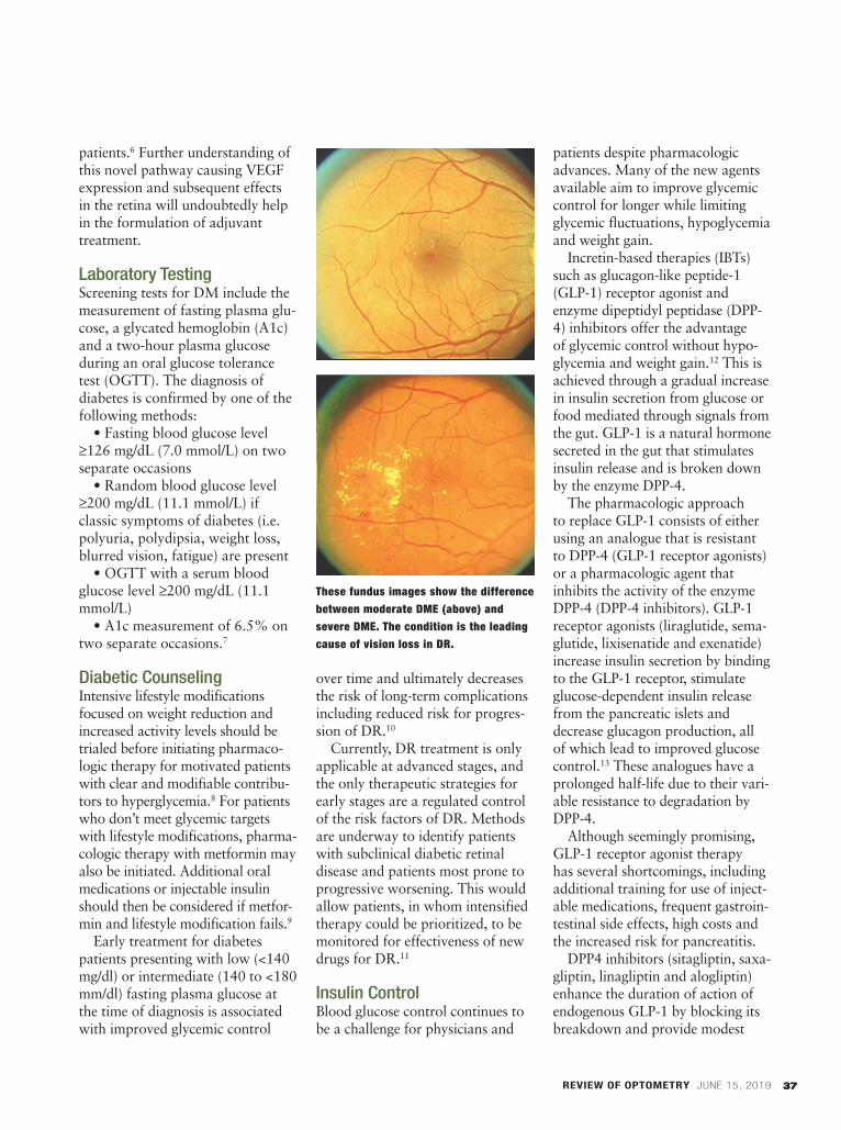
37
patients.6 Further understanding of this novel pathway causing VEGF expression and subsequent effects in the retina will undoubtedly help in the formulation of adjuvant treatment.
Laboratory TestingScreening tests for DM include the measurement of fasting plasma glu-cose, a glycated hemoglobin (A1c) and a two-hour plasma glucose during an oral glucose tolerance test (OGTT). The diagnosis of diabetes is confirmed by one of the following methods:
• Fasting blood glucose level ≥126 mg/dL (7.0 mmol/L) on two separate occasions
• Random blood glucose level ≥200 mg/dL (11.1 mmol/L) if classic symptoms of diabetes (i.e. polyuria, polydipsia, weight loss, blurred vision, fatigue) are present
• OGTT with a serum blood glucose level ≥200 mg/dL (11.1 mmol/L)
• A1c measurement of 6.5% on two separate occasions.7
Diabetic CounselingIntensive lifestyle modifications focused on weight reduction and increased activity levels should be trialed before initiating pharmaco-logic therapy for motivated patients with clear and modifiable contribu-tors to hyperglycemia.8 For patients who don’t meet glycemic targets with lifestyle modifications, pharma-cologic therapy with metformin may also be initiated. Additional oral medications or injectable insulin should then be considered if metfor-min and lifestyle modification fails.9
Early treatment for diabetes patients presenting with low (<140 mg/dl) or intermediate (140 to <180 mm/dl) fasting plasma glucose at the time of diagnosis is associated with improved glycemic control
over time and ultimately decreases the risk of long-term complications including reduced risk for progres-sion of DR.10
Currently, DR treatment is only applicable at advanced stages, and the only therapeutic strategies for early stages are a regulated control of the risk factors of DR. Methods are underway to identify patients with subclinical diabetic retinal disease and patients most prone to progressive worsening. This would allow patients, in whom intensified therapy could be prioritized, to be monitored for effectiveness of new drugs for DR.11
Insulin ControlBlood glucose control continues to be a challenge for physicians and
patients despite pharmacologic advances. Many of the new agents available aim to improve glycemic control for longer while limiting glycemic fluctuations, hypoglycemia and weight gain.
Incretin-based therapies (IBTs) such as glucagon-like peptide-1 (GLP-1) receptor agonist and enzyme dipeptidyl peptidase (DPP-4) inhibitors offer the advantage of glycemic control without hypo-glycemia and weight gain.12 This is achieved through a gradual increase in insulin secretion from glucose or food mediated through signals from the gut. GLP-1 is a natural hormone secreted in the gut that stimulates insulin release and is broken down by the enzyme DPP-4.
The pharmacologic approach to replace GLP-1 consists of either using an analogue that is resistant to DPP-4 (GLP-1 receptor agonists) or a pharmacologic agent that inhibits the activity of the enzyme DPP-4 (DPP-4 inhibitors). GLP-1 receptor agonists (liraglutide, sema-glutide, lixisenatide and exenatide) increase insulin secretion by binding to the GLP-1 receptor, stimulate glucose-dependent insulin release from the pancreatic islets and decrease glucagon production, all of which lead to improved glucose control.13 These analogues have a prolonged half-life due to their vari-able resistance to degradation by DPP-4.
Although seemingly promising, GLP-1 receptor agonist therapy has several shortcomings, including additional training for use of inject-able medications, frequent gastroin-testinal side effects, high costs and the increased risk for pancreatitis.
DPP4 inhibitors (sitagliptin, saxa-gliptin, linagliptin and alogliptin) enhance the duration of action of endogenous GLP-1 by blocking its breakdown and provide modest
REVIEW OF OPTOMETRY JUNE 15, 2019 37
These fundus images show the difference between moderate DME (above) and severe DME. The condition is the leading cause of vision loss in DR.
036_ro0619_F1.indd 37036_ro0619_F1.indd 37 6/5/19 10:53 AM6/5/19 10:53 AM

REVIEW OF OPTOMETRY JUNE 15, 201938
glycemic control. DPP-4 inhibitors’ advantages include ease of adminis-tration using oral route, good toler-ability and lack of association with weight gain or hypoglycemia. Com-monly reported side effects include headache, nasopharyngitis and upper respiratory tract infection.14 The long-term safety with DPP-4 inhibitors has not been established.
Sodium-glucose cotransporter (SGLT)-2 inhibitors (canagliflozin, dapagliflozin, empagliflozin and ertugliflozin) reduce blood glucose by increasing urinary glucose excre-tion.15,16 SGLT-2 inhibitors are easy to use and only require once-daily oral administration. They work on all phases of glucose metabo-lism and modestly reduce blood pressure, weight loss and risk of hypoglycemia. The disadvantages include increased risk of genitouri-nary tract and fungal genital infec-tions. SGLT-2 inhibitors should be used with caution in conjunction with other medications or comor-bidities that predispose an indi-vidual to acute renal injury.
Research shows a new basal insulin analogue, degludec, is lon-ger acting than insulin glargine and has less nocturnal hypoglycemia. Continuous glucose monitoring systems, when used together with pump therapy, also show reduc-tions in the risk for hypoglycemia.12
Concomitant MorbiditiesSeveral trials show strict glycemic and blood pressure control are bene-ficial for the prevention and progres-sion of retinopathy.16 The Action to Control Cardiovascular Risk in Diabetes (ACCORD) Eye Study shows a reduction of retinopathy progression in an intensive glycemic therapy group.17 In patients with dyslipidemia, retinopathy progres-sion was also slowed by fenofibrate similar to those receiving intensive
glycemic treatment in the FIELD photographic sub-study. Other research shows metabolic syndrome is a strong, independent indicator of DR, even to the same extent as gly-cemic control.18 Moreover, research shows that PDR in Type 2 diabetes is a stronger independent correlation factor for peripheral arterial disease (PAD) than a diabetic duration of 10 years.19
A timely diagnosis of these con-comitant morbidities in patients with diabetes provides valuable information regarding the risk of DR. Screening for and treating con-comitant morbidities minimizes the risk of irreversible blindness from DR.
Diabetic Retinal Imaging A variety of imaging modalities can help guide the diagnosis and treat-ment of DR. In recent years, several technological advancements such as ultra-widefield (UWF) fundus pho-tography, UWF fluorescein angiog-raphy (FA) and optical coherence tomography angiography (OCT-A) have been adopted by eye care practitioners for screening, evalua-tion and diagnosis.
Fundus photography is still a crucial tool in the education and management of DR. The standard fundus photo image captures 30˚ of the posterior pole, including the optic nerve and macula.20 These static images can easily be manipu-lated and magnified to detect changes in progression or improve-ment in diabetic eye disease.
Widefield fundus photography includes the posterior retina up to vortex vein ampullae, captures up to 105˚ field and allows for imag-ing of the peripheral retina.21,22 In comparison, newer UWF images capture at least four vortex ampul-lae in a single image and up to 200˚ of the retina.20-23 The increased
use of widefield and UWF imaging for DR has begun to reveal more information about the pathology of diabetic eye disease.24,25 Using UWF retinal imaging, a 2015 study demonstrated that patients with predominately peripheral diabetic lesions had a more than fourfold greater chance of progressing to proliferative disease than those with more centrally located reti-nopathy.26
Fluorescein angiography (FA) can help evaluate retinal vascula-ture changes associated with DR. Recently, a technique was devel-oped that improved peripheral retina imaging through combining widefield imaging with FA. UWF-FA provides valuable information about the retina, blood vessels, peripheral neovascularization and the extent of retinal nonperfusion.
Detection of nonperfused areas in the peripheral retina has given new insight into the pathogenesis of DME. Investigators hypoth-esized that areas of nonperfused peripheral retina are a source of VEGF, which can contribute to the formation of DME.27 Targeted retinal photocoagulation (TRP) can help reduce VEGF production, resulting decreased severity and extent of macular edema.28,29
The combination of macular laser, anti-VEGF therapy and TRP may prove to be an important treatment approach for DME while also minimizing the side effects of visual field loss from retinal laser. It should be noted that this is theo-retical in nature, as no conclusive evidence shows that TRP is supe-rior to panretinal photocoagula-tion (PRP).
Optical coherence tomography (OCT) uses high definition resolu-tion to evaluate retinal anatomy and is imperative in the assessment of the macula in diabetic patients.
Systemic Disease
036_ro0619_F1.indd 38036_ro0619_F1.indd 38 6/5/19 10:53 AM6/5/19 10:53 AM
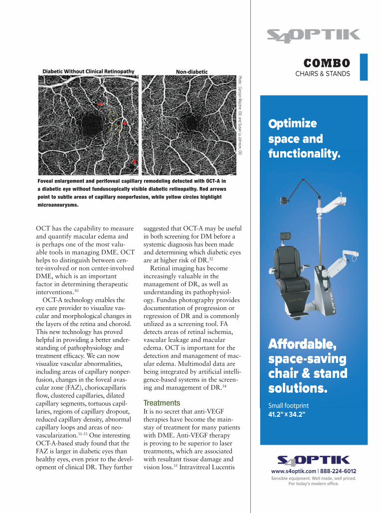
OCT has the capability to measure and quantify macular edema and is perhaps one of the most valu-able tools in managing DME. OCT helps to distinguish between cen-ter-involved or non center-involved DME, which is an important factor in determining therapeutic interventions.30
OCT-A technology enables the eye care provider to visualize vas-cular and morphological changes in the layers of the retina and choroid. This new technology has proved helpful in providing a better under-standing of pathophysiology and treatment efficacy. We can now visualize vascular abnormalities, including areas of capillary nonper-fusion, changes in the foveal avas-cular zone (FAZ), choriocapillaris flow, clustered capillaries, dilated capillary segments, tortuous capil-laries, regions of capillary dropout, reduced capillary density, abnormal capillary loops and areas of neo-vascularization.31-33 One interesting OCT-A-based study found that the FAZ is larger in diabetic eyes than healthy eyes, even prior to the devel-opment of clinical DR. They further
suggested that OCT-A may be useful in both screening for DM before a systemic diagnosis has been made and determining which diabetic eyes are at higher risk of DR.32
Retinal imaging has become increasingly valuable in the management of DR, as well as understanding its pathophysiol-ogy. Fundus photography provides documentation of progression or regression of DR and is commonly utilized as a screening tool. FA detects areas of retinal ischemia, vascular leakage and macular edema. OCT is important for the detection and management of mac-ular edema. Multimodal data are being integrated by artificial intelli-gence-based systems in the screen-ing and management of DR.34
Treatments It is no secret that anti-VEGF therapies have become the main-stay of treatment for many patients with DME. Anti-VEGF therapy is proving to be superior to laser treatments, which are associated with resultant tissue damage and vision loss.35 Intravitreal Lucentis
Foveal enlargement and perifoveal capillary remodeling detected with OCT-A in a diabetic eye without funduscopically visible diabetic retinopathy. Red arrows point to subtle areas of capillary nonperfusion, while yellow circles highlight microaneurysms.
Photo: C
arolyn Majcher, O
D, and S
usan Ly Johnson, OD
COMBOCHAIRS & STANDS
Aspace-saving chair & stand solutions.
Optimize space and functionality.
Aspace-saving chair & standsolutions.
fuff nctionality.yy
Small footprint 41.2” x 34.2”
036_ro0619_F1.indd 39036_ro0619_F1.indd 39 6/5/19 10:53 AM6/5/19 10:53 AM

REVIEW OF OPTOMETRY JUNE 15, 201940
(ranibizumab, Genentech) and Eyelea (aflibercept, Regeneron) are FDA-approved therapies often used for DME, whereas intravitreal Avastin (bevacizumab, Genentech) is administrated off-label.36,37 The RISE, RIDE, VIVID and VISTA studies all support anti-VEGF agents over laser and early inter-vention in patients with DME to maximize potential visual acu-ity.38,39
In a two-year randomized clini-cal trial, Protocol T compared three anti-VEGF agents (ranibizumab 0.5mg, aflibercept 2.0mg and bevacizumab 1.25mg) for the treat-ment of DME.40 After two years of follow up, patients with an initial BCVA of 20/50 or better achieved similar visual improvement with all three drugs. Aflibercept was associ-ated with the greatest reduction in mean central macular subfield thickness on OCT. Patients present-ing with BCVA of 20/50 or worse had the greatest visual improve-ment at one year with aflibercept; however, the results diminished and were no longer statistically signifi-cant at the two-year mark.
For the treatment of center-involved DME, there may be rea-son to prefer one anti-VEGF over another; however, all three agents investigated in Protocol T were beneficial for most patients.
The treatment paradigm for PDR is also starting to shift in favor of injectable therapeutic agents. The Diabetic Retinopathy Clini-cal Research Network conducted a major randomized clinical trial, Protocol S, for the pharmacologic treatment of PDR using ranibi-zumab compared with PRP.41,42 The two-year findings of Protocol S revealed that ranibizumab was non-inferior to PRP with a mean gain of +2.8 letters vs. +0.2 letters in the PRP group.43 Although these
results were quite promising, the role of ocular anti-VEGF therapy for PDR is less clear. Patients may require indefinite injections and monitoring due to the limited half-life of these drugs.
The CLARITY study is the first randomized controlled trial of intravitreal aflibercept in PDR. The results provide substantial evidence that the visual outcome in those with active PDR at one year with aflibercept is superior to PRP.44
The ongoing PANORMA study is evaluating aflibercept injections in patients with moderate and severe non-proliferative diabetic retinopathy (NPDR). Results are impressive with patients exhibiting a two-step or greater improvement based on the Diabetic Retinopathy Severity Scale (DRSS).45 The impli-cations of this study are significant as there is a realistic potential for many patients to opt for earlier pharmacological intervention if worsening stages of NPDR or PDR can be prevented.
It should be noted that anti-VEGF agents have gained favor-ability for treating DME and PDR over the past decade; however, focal/grid photocoagulation and PRP remain the gold standard treatments for DME and PDR, respectively.
Artificial IntelligenceThe infusion of artificial intelli-gence (AI) into eye care is progress-ing every day. The field of AI has recently experienced significant advancements in image recognition due to a technique called deep learn-ing (DL) which, when applied to current imaging, results in improved diabetic eye disease recognition and identification of progression. One study applied DL to retinal pho-tography to create an algorithm to automatically detect the presence
of DR and DME using 128,175 macular-centered retinal images. Results show astounding sensitiv-ity and specificity for referable DR more than 90%.46 DL has also been applied to macular OCT scans. A 2018 study shows that DL achieves excellent accuracy in the detection of retinal fluid across several macular diseases and OCT devices. Further-more, AI results were in high con-cordance with manual expert clinical assessments.47,48
Such AI devices and software provide a quantitative and quali-tative analysis without the need for manual interpretation. This opens the realistic potential of AI-based programs for use outside the optometrists and ophthalmologists’ office. The integration of AI can be expected to radically change clinical practice with more people getting screened for retinopathy remotely. AI will continue to expand and become an auxiliary component and useful tool in DR screening, but AI cannot replace the role of eye care providers in clinical diagnosis and management. Although there are challenges ahead, AI will likely impact optom-etry practices in the coming decades by providing increased efficiency, reducing overall costs, improv-ing access to care and ultimately reducing the strain on our current healthcare system. ■
Dr. Gentry practices at the Harry S. Truman Memorial Veterans’ Hos-pital in Columbia, Mo.
Dr. Ho practices at the Central Texas Veterans Health Care System Austin Outpatient Clinic in Austin, Texas.
Dr. Zimbalist practices at the Harry S. Truman Memorial Veter-ans’ Hospital in Columbia, Mo.
Dr. O’Brien is a 2019 graduate of the University of Missouri-St. Louis College of Optometry.
Systemic Disease
036_ro0619_F1.indd 40036_ro0619_F1.indd 40 6/5/19 11:05 AM6/5/19 11:05 AM

1. Kusuhara S, Fukushima Y, Ogura S, et al. Pathophysiology of diabetic retinopathy: The old and the new. Diabetes Metab J. 2018;42(5):364-76.2. McCulloch D, Robertson R. Pathogenesis of type 2 diabetes mellitus. UpToDate. www.uptodate.com/contents/pathogenesis-of-type-2-diabetes-mellitus. Accessed March 8, 2019.3. Gupta N, Mansoor S, Sharma A, et al. Diabetic retinopathy and VEGF. Open Ophthalmol J. 2013;7:4-10.4. Wang W, Lo A. Diabetic retinopathy: Pathophysiology and treat-ments. Int. J. Mol Sci. 2018;19(11):E1816.5. Miao C, Zhang G, Xie Z, Chang J. MicroRNAs in the pathogen-esis of type 2 diabetes: new research progress and future direc-tion. Can J Physiol Pharmacol 2018;96(2):103-12. 6. Kaidonis G, Gillies M, Abhary S, et al. A single-nucleotide polymorphism in the MicroRNA-146a gene is associated with diabetic nephropathy and sight-threatening diabetic retinopathy in Caucasion patients. Acta Diabetol. 2016;53:643-650. 7. McCulloch D, Hayward R. Screening for type 2 diabetes mel-litus. UpToDate. www.uptodate.com/contents/screening-for-type-2-diabetes-mellitus. Accessed March 7, 2019.8. Wexler D. Initial management of blood glucose in adults with type 2 diabetes mellitus. UpToDate. www.uptodate.com/contents/initial-management-of-blood-glucose-in-adults-with-type-2-dia-betes-mellitus. March 21, 2019. Accessed May 20, 2019.9. McCulloch D. Oveview of medical care in adults with diabetes mellitus. UpToDate. www.uptodate.com/contents/overview-of-medical-care-in-adults-with-diabetes-mellitus. March 21, 2019. Accessed May 20, 2019.10. Colagiuri S, Cull C, Holman R, UKPDS Group. Are lower fast-ing plasma glucose levels at diagnosis of type 2 diabetes associ-ated with improved outcomes?: U.K. prospective diabetes study 61. Diabetes Care 2002; 25:1410.11. Simó-Servat O, Simó R, Hernández C. Circulating biomarkers of diabetic retinopathy: an overview based on physiopathology. J Diabetes Res. 2016;2016:5263798. 12. Fonseca V. New developments in diabetes management: medications of the 21st century. Clinical Therapeutics 2014 April;36(4):477-84. 13. Dugan K, DeSantis A. Glucagon-like peptide-1 receptor agonists for the treatment of type 2 diabetes mellitus. UpToDate. www.uptodate.com/contents/glucagon-like-peptide-1-receptor-agonists-for-the-treatment-of-type-2-diabetes-mellitus. January 14, 2019. Accessed March 8, 2019.14. Dugan K, DeSantis A. Dipeptidyl peptidase-4 (DPP-4) inhibi-tors for the treatment of type 2 diabetes mellitus. UpToDate. www.uptodate.com/contents/dipeptidyl-peptidase-4-dpp-4-in-hibitors-for-the-treatment-of-type-2-diabetes-mellitus. January 22, 2019. Accessed March 10, 2019. 15. De Santis A. Sodium-glucose co-transporter 2 inhibitors for the treatment of type 2 diabetes mellitus. UpToDate. Sodium-glucose co-transporter 2 inhibitors for the treatment of type 2 diabetes mellitus. UpToDate. December 10, 2018. Accessed March 10, 2019.16. Mohamed Q, Gillies M, Wong T: Management of diabetic reti-nopathy: a systematic review. JAMA 2007;298(8):902-16. 17. Chew E, Davis M, Danis R, et al. The effects of medical man-agement on the progression of diabetic retinopathy in persons with type 2 diabetes: the ACCORD eye study. Ophthalmology. 2014;121(12): 2443-51. 18. Gao L, Xin Z, Yuan M, et al. High prevalence of diabetic retinopathy in diabetic patients concomitant with metabolic syn-drome. PLoS ONE. 2016;11(1):e0145293. 19. Chen Y, Wang Y, Zhao D, et al. High prevalence of lower extremity peripheral artery disease in type 2 diabetes patients with proliferative diabetic retinopathy. PLoS One. 2015 Mar; 10(3):e0122022.20. Salz DA, Witkin AJ. Imaging diabetic retinopathy. Middle East Afr J Ophthalmol. 2015;22(2):145-150.21. Patrick JS, Tyler ME. Fundus photography overview. In: Ophthalmic Photography: Retinal Photography, Angiography, and Electronic Imaging, 2nd ed. New York, NY: Butterworth-Heinemann Medical; 2001. 22. Bethke W. The devil’s in the distant details. Review of Oph-thalmol. 2014;20(8):26-28.23. Kiss S. Going ultra-wide. Retina Today. 2018;13(10)46-8.24. Brown K, Sewell JM, Trempe C, et al. Comparison of image-assisted versus traditional fundus examination. Eye and Brain. 2013;2013(5):1-8. 25. Wessel M, Aaker G, Parlitsis G, et al. Ultra-wide-field angi-ography improves the detection and classification of diabetic retinopathy. Retina. 2012;32(4):785-91.26. Silva P, Cavallerano J, Haddad N, et al. Peripheral lesions identified on ultrawide field imaging predict increased risk of
diabetic retinopathy progression over 4 years. Ophthalmol. 2015;122(5):949-56.27. Oliver C, Schwartz S. Ultra-widefield fluorescein angiography. In: Arevalo JF, ed. Retinal Angiography and Optical Coherence Tomography, 1st ed. New York, NY: Springer Science and Busi-ness Media, LLC; 2009:407-417. 28. Reddy S, Hu A, Schwartz SD. Ultra-wide field fluorescein angi-ography guided targeted retinal photocoagulation (TRP). Semin Ophthalmol. 2009;24(1):9-14. 29. Muquit M, Marcellino G, Henson D, et al. Optos-guided pat-tern scan laser (Pascal)-targeted retinal photocoagulation in pro-liferative diabetic retinopathy. Acta Ophtalmol. 2013;91(3):251-8.30. Cunha-Vaz J, Coscas G. Diagnosis of macular edema. Oph-tahlmologica. 2010;224(suppl 1):2-7.31. de Carlo TE, Chin AT, Bonini-Filho MA, et al. Detection of microvascular changes in eyes of patients with diabetes but not clinical diabetic retinopathy using optical coherence tomography angiography. Retina. 2015;35(11):2364-70.32. Takase N, Nozaki M, Kato A, et al. Enlargement of foveal avas-cular zone in diabetic eyes evaluated by en face optical coher-ence tomography angiography. Retina. 2015;35(11):2377-83.33. Choi W, Waheed NK, Moult EM, et al. Ultrahigh speed swept source optical coherence tomography angiography of retinal and choriocapillaris alterations in diabetic patients with and without retinopathy. Retina. 2017;37(1):11-21.34. Schmidt-Erfurth U, Sadeghipour A, Gerendas BS, et al. Artificial intelligence in retina. Prog Retin Eye Res November 2018;67:1-29.35. Nguyen QD, Brown DM, Marcus DM, et al. Ranibizumab for diabetic macular edema: results from 2 phase III randomized tri-als: RISE and RIDE. Ophthalmology. 2012;119(4):789-801.36. Ferrara N., Adamis AP. Ten years of anti-vascular endothelial growth factor therapy. Nat Rev Drug Discov. 2016;15(6):385-403. 37. van Asten F, Michels CTJ, Hoyng CB, et al. The cost-effective-ness of bevacizumab, ranibizumab and aflibercept for the treat-ment of age-related macular degeneration: A cost-effectiveness analysis from a societal perspective. PloS ONE 2018;13(5):1-14.38. Brown DM, Schmidt-Erfurth U, Do DV, et al. Intravitreal afliber-cept for diabetic macular edema: 100-week results from VISTA and VIVID studies. Ophthalmol. 2015;122(10):2044-5239. Brown D, Nguyen Q, Marcus D, et al. Long-term outcomes of ranibizumab therapy for diabetic macular edema: the 36-month results from two phase III trials: RISE and RIDE. Ophthalmol. 2013;120(10):2013-22.40. Diabetic Retinopathy Clinical Research Network, Wells JA, Glassman AR, Ayala AR, et al. Aflibercept, bevacizumab, or ranibizumab for diabetic macular edema. N Engl J Med. 2015;372(13):1193-203.41. Wells J, Glassman A, Ayala A, et al. Diabetic Retinopathy Clinical Research Network. Aflibercept, bevacizumab, or ranibi-zumab for diabetic macular edema: two-year results from a comparative effectiveness randomized clinical trial. Ophthalmol. 2016;123(6):1351-9. 42. Heier J, Bresler N, Avery R, et al. Comparison of aflibercept, bevacizumab, and ranibizumab for treatment of diabetic macular edema: Extrapolation of data to clinical practice. JAMA Ophthal-mol. 2016;134(1):95-9.43. Writing Committee for the Diabetic Retinopathy Clinical Research Network; Gross JC, Glassman AR, Jampol LM, et al. Panretinal photocoagulation vs intravitreous ranibizumab for proliferative diabetic retinopathy: a randomized clinical trial JAMA. 2015;314(20):2137-46.44. Sivaprasad S, Prevost AT, Vasconcelos JC, et al. Clinical efficacy of intravitreal aflibercept versus panretinal photocoagula-tion for best corrected visual acuity in patients with proliferative diabetic retinopathy at 52 weeks (CLARITY): a multicentre, single-blinded, randomised, controlled, phase 2b, non-inferiority trial. Lancet. 2017;389(10085):2193-203.45. Eylea (aflibercept) injection improves diabetic retinopathy and reduces vision-threatening complications in phase 3 trial. Up to date. www.biospace.com/article/releases/eylea-aflibercept-injection-improves-diabetic-retinopathy-and-reduces-vision-threatening-complications-in-phase-3-trial/. 46. Gulshan V, Peng L, Coram M, et al. Development and valida-tion of a deep learning algorithm for detection of DR in retinal fundus photographs. JAMA. December 13, 2016. jamanetwork.com/journals/jama/fullarticle/2588763. Accessed May 20, 2019.47. Schlegl T, Waldstein S, Bogunovic H, et al. Fully automated detection and quantification of macular fluid in OCT using deep learning. Ophthalmol. 2018;125(4):549-58.48. Walton OB 4th, Garoon RB, Weng CY, et al. Evaluation of automated teleretinal screening program for diabetic retinopathy. JAMA Ophthalmol. 2016;134(2):204-9.
250 Cooper Ave., Suite 100 T onawanda NY 14150
ww w.s4optik .com I 888-224-6012
Sensible equipmen t. Well made, well priced .
SLITLAMPS
EXCEPTIONAL OPTICS
S4OPTIK’s converging binoculars
maintenance of fusion.
European craftsmanship and engineering provide reliable optics at
examinations.
Vertical and compact
H-Series
Z-Series
SLIT LAMPS
036_ro0619_F1.indd 41036_ro0619_F1.indd 41 6/5/19 11:05 AM6/5/19 11:05 AM
REVIEW OF OPTOMETRY JUNE 15, 201942
Age-related macular degen-eration (AMD), a bilateral, progressive retinal disease, is a major health problem
that is too often undiagnosed. Although most optometrists think they have more glaucoma patients in their practice than they do AMD patients, AMD is three times as prevalent as glaucoma. In fact, AMD is more common than all of glaucoma and diabetic retinopathy combined.1-3
A recent study found approxi-mately 25% of eyes rated as normal during a primary eye care pro-vider’s dilated eye examination had macular characteristics indicative of AMD, revealed by fundus pho-tography and trained raters.4 The reasons for the missed diagnoses remain unclear, but improved AMD detection strategies are needed to ensure patients receive appropriate treatment before irreversible vision loss occurs.
Traditionally, AMD is diagnosed when a dilated fundus examination reveals clinically evident drusen or RPE changes. A litany of diagnostic testing for AMD exists, includ-ing optical coherence tomography (OCT), OCT angiography (OCT-A), multispectral imaging, fundus autofluorescence and photography, microperimetry, electroretinography and retinal angiography.
But optometrists can truly stay ahead of AMD if they change the way they approach the disease, by identifying AMD risk during rou-tine examinations and by using a new diagnostic tool, dark adapta-tion, which can identify the earliest functional biomarker of AMD. This method of identifying “AMD sus-pects” is much like diagnosing glau-coma suspects—a careful history and clinical examination may reveal risk factors that warrant further testing to determine if the disease is truly present.
A New Look at AMD The macula encompasses the most posterior region of the retina within the vascular arcades and, as a whole, has significantly more rods than cones—only the central fovea is cone dominated in the macula.5
Rod loss is the beginning of AMD, as rods are affected earlier and more severely than cones through-out the disease progression. Only much later in the disease process are cones affected, which correlates with a decrease in visual acuity.6
Because rod deterioration happens in the earliest stages of AMD, dark adaptation becomes affected much earlier than visual acuity declines.7
Research shows AMD causes anatomic and functional conse-quences before clinical signs of dru-sen appear. However, by the time drusen are visible, the patient has already had AMD for several years. Drusen are a manifestation of cho-lesterol locally produced by the
Early Diagnosis
New diagnostic tools can help you ID suspects and initiate preventative measures. By Amanda Legge, OD
How to Stay One Step Ahead of AMD
10th Annual Retina Report
042_ro0619_F2.indd 42042_ro0619_F2.indd 42 6/5/19 11:40 AM6/5/19 11:40 AM

REVIEW OF OPTOMETRY JUNE 15, 2019 43
retinal pigment epithelium (RPE) and deposited as a wash under the entire macula in Bruch’s membrane. This microscopic cholesterol layer is the hallmark defect of AMD, as electron microscopy in donor eyes has proven patients without AMD do not have this abnormal layer of cholesterol. Although the layer is not yet visible in living eyes using current diagnostic tools, it does significantly delay dark adaptation, even early in the disease before clinical signs are evident.7,8
As the disease progresses, this cholesterol layer continues to accu-mulate, resulting in focal areas suf-ficiently thickened to be clinically visible as drusen. This additive cholesterol accumulation causes inflammation, oxidative stress and disruption of oxygen and nutrients (such as vitamin A, a deficiency of which further hinders dark adapta-tion) supplied to the outer retina. Ultimately, this causes organized apoptosis of photoreceptors in the macula.9,10 The retina naturally combats oxidative stress with its own antioxidants: the human mac-
ula pigments consisting of lutein and zeaxanthin.11 In addition, anti-oxidant nutritional supplementa-tion can help patients delay vision loss in AMD.
Macular pigment optical density (MPOD) measures the macular density of these pigments to deter-mine if patients have an adequate amount to combat this stress.
Identifying AMD RiskRisk factors for AMD identifiable with careful history include age, family history, Caucasian race, smoking, obesity and comorbid conditions such as hypertension, hypercholesterolemia and diabe-tes.12-14 While declining night vision and contrast sensitivity occur in normal aging, these symptoms occur more severely and more quickly in macular degeneration; therefore, it is reasonable to con-sider these symptoms as potential risk factors when diagnosing AMD.4 During clinical examina-tion, any evident drusen, RPE mottling or both can be considered structural risk factors for AMD,
similar to how increased or asym-metric optic nerve cupping causes glaucoma suspicion.15 As with vari-able cup-to-disc ratios in glaucoma, drusen and RPE changes can be normal age-related changes or early
This 71-year-old Caucasian female with small, hard drusen OU had abnormal dark adaptation and was diagnosed with AMD in 2016 (top OCT images). Even with treatment, the size and number of her drusen worsened and her dark adaptation progressed over time. She was being monitored routinely with dilated examinations every three months due to this quick progression and high risk of converting to wet AMD. On one of these routine exams, her visual acuity was 20/25-1 OD and 20/25+2 OS. She had no symptoms to report, and her Amsler grid had been stable during both at-home and in-office testing. Dilated exam revealed macular thickening OS, which was confirmed as subretinal fluid on OCT. The patient was referred to a retinologist who confirmed the presence of CNV with fluorescein angiography and initiated anti-VEGF treatment the same week. Today, her visual acuity remains 20/25 OS and she is still asymptomatic (bottom OCT images).
The patient’s color photography shows drusen with mild RPE changes in both eyes with mild macular thickening beginning in the left eye (bottom) that coincides with subretinal fluid identified with OCT.
042_ro0619_F2.indd 43042_ro0619_F2.indd 43 6/5/19 11:40 AM6/5/19 11:40 AM

REVIEW OF OPTOMETRY JUNE 15, 201944
signs of true macular degenera-tion—risk factors, not diagnostic signs. Patients with drusen 125µm or larger, indistinct soft or reticular drusen, total drusen area of half the disc or more and hyperpigmenta-tion are statistically at higher risk for AMD.15
Newer testing can now identify additional risk factors such as reduced macular pigment density. MPOD instruments measure the ratio of central blue light absorp-tion to the peripheral absorption rate to determine the amount of macular pigment present. The more blue light absorbed centrally, the higher the MPOD score, which cor-relates to denser macular pigment.16 Research shows higher MPOD scores are associated with a slower AMD progression rate and lower risk of advancing to severe AMD. Low MPOD levels are associated with increased risk of progression to late AMD.17
The Age Related Eye Disease Study 2 (AREDS2) found that add-ing lutein and zeaxanthin to the original AREDS 1 formulation and removing the beta carotene reduced the risk of progression to advanced AMD compared with those taking the original AREDS 1 formula-tion.18 This effect was even more pronounced in patients who had low intake of dietary lutein and zeaxanthin. However, participants in AREDS2 participants had signifi-cantly higher lutein and zeaxanthin serum levels compared with the general population. But even these well-nourished participants had significantly reduced risk of progres-sion with lutein and zeaxanthin sup-plementation than those without.18
Thus, although low MPOD is not diagnostic of AMD, it is a strong risk factor for developing AMD and the likelihood of disease progression. Moreover, MPOD is
a modifiable risk factor because supplementation can increase low MPOD scores and decrease risk of progression to severe AMD.4,19,20
Determining genetic risk is some-what complex; at least 19 genes are known to be involved in vari-ous AMD pathways, including the complement, immune/inflamma-tory, extracellular matrix and DNA repair/protein binding pathways.21,22 A growing body of literature sug-gests a subset of genetic variables along with demographic, behavioral and ocular signs can be highly pre-dictive of risk to advanced AMD and vision loss.22 Individuals at a higher genetic risk should be con-sidered for earlier screening or, if diagnosed with the disease, should be monitored closer as they are at a higher risk of progression.
New Technology for Diagnosis and ManagementEvaluating structure and function together is extremely helpful in clinical practice to help diagnose AMD and possibly determine the rate and extent of progression. Studies of structure to diagnose AMD and its associated findings including include OCT, OCT-A, color photography, multispectral imaging, fundus autofluorescence and fluorescein angiography. Stud-ies of function include hyperacuity perimetry, microperimetry, Amsler
grid and visual acuity. As clinicians, enhanced confidence in detailed macular evaluation and proper interpretation of test findings aids in earlier diagnosis of AMD and differentiating it from other macu-lar diagnoses. Here is a look at several new tools available to help clinicians diagnose AMD as early as possible and manage patients throughout the treatment regimen:
Dark adaptation. This is the only functional biomarker of AMD and does not measure risk.23-27 Although dark adaptation does have a tendency to decline with normal aging process, it is statisti-cally more severe in AMD. It can be measured using the AdaptDx adaptometer (Maculogix), which uses a bleaching flash and then records the response to stimuli until the rod intercept (the time it takes for the eye to adjust from bright light to darkness) is reached. The test experience is similar to a visual field and takes from a few moments up to 20 minutes. The AdaptDx has normative values to differentiate between normal age-related night vision decline and true abnormali-ties in the rod intercept time found in early AMD. For the detection of AMD, the test has a high sensitiv-ity, correctly identifying 90.6% of confirmed AMD cases as well as a high specificity, identifying 90.5% of confirmed normal cases.7,23
Fundus autofluorescence (FAF). Because the RPE plays a signifi-cant role in the development and advancement of AMD, understand-ing its metabolic health is extremely helpful. General observations of FAF patterns in AMD suggest that elevated lipofuscin causes increased FAF in early stages of AMD associated with drusen and RPE metabolic stress, while decreasing RPE lipofuscin is associated with advancing stages of AMD as the
DA DifferentialsConditions other than AMD that can present with poor dark adaptation include genetic retinal disorders and vitamin A deficiency. In the aging population at risk for AMD, these differential conditions will present with retinal findings of inherited retinal disease or will have a systemic history of malnutrition. Thus, they are fairly simple to rule out with patient history, clinical exami-nation and ancillary testing.23-27
Early Diagnosis
042_ro0619_F2.indd 44042_ro0619_F2.indd 44 6/5/19 11:40 AM6/5/19 11:40 AM

AMD is more common than you think.AdaptDx® provides a clear, objective measurement of dark adaptation function that is 90% sensitive and specific for the presence of AMD. This simple test
can make a big difference.
Detect AMD three years earlier - when night vision problems begin, before drusen are visible.
They’re in YOUR practice
AMD
affects 1 in 8
over age 60
maculogix.com | 1.717.914.4067©2019 MacuLogix, Inc. All rights reserved. MM-113
Find out how AdaptDx can improve patient outcomes and practice revenue.
Talk with an AMD Expert from MacuLogix.
RO0619_Maculogix.indd 1 5/23/19 2:16 PM
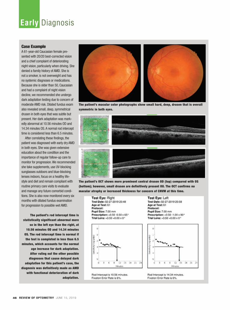
REVIEW OF OPTOMETRY JUNE 15, 201946
Case ExampleA 61-year-old Caucasian female pre-sented with 20/20 best-corrected vision and a chief complaint of deteriorating night vision, particularly when driving. She denied a family history of AMD. She is not a smoker, is not overweight and has no systemic diagnoses or medications. Because she is older than 50, Caucasian and had a complaint of night vision decline, we recommended she undergo dark adaptation testing due to concern of moderate AMD risk. Dilated fundus exam also revealed small, deep, symmetrical drusen in both eyes that was subtle but present. Her dark adaptation was mark-edly abnormal at 10.56 minutes OD and 14.34 minutes OS. A normal rod intercept time is considered less than 6.5 minutes.
After correlating these findings, the patient was diagnosed with early dry AMD in both eyes. She was given extensive education about the condition and the importance of regular follow-up care to monitor for progression. We recommended she take supplements, use UV-blocking sunglasses outdoors and blue-blocking lenses indoors, focus on a healthy life-style and diet and remain compliant with routine primary care visits to evaluate and manage any future comorbid condi-tions. She is also now monitored every six months with dilated fundus examination for progression to possible wet AMD.
The patient’s macular color photographs show small hard, deep, drusen that is overall symmetric in both eyes.
The patient’s OCT shows more prominent central drusen OD (top) compared with OS (bottom); however, small drusen are definitively present OU. The OCT confirms no macular atrophy or increased thickness for concern of CNVM at this time.
The patient’s rod intercept time is statistically significant abnormal more
so in the left eye than the right, at 10.56 minutes OD and 14.34 minutes
OS. The rod intercept time is normal if the test is completed in less than 6.5
minutes, which accounts for the normal age increase for dark adaptation.
After ruling out the other possible diagnoses that cause delayed dark
adaptation for this patient’s case, the diagnosis was definitively made as AMD
with functional deterioration of dark adaptation.
Early Diagnosis
042_ro0619_F2.indd 46042_ro0619_F2.indd 46 6/5/19 11:40 AM6/5/19 11:40 AM

VISIT THE GATEWAY TO
SAVINGS.
Get reduced pricing and exclusive offers on Reliance Medical Products. Take advantage of these offers while they last, now through June 30, 2019.
800.735.0357 | Builttosupport.com© 2019 Haag-Streit USA. All Rights Reserved.
520 Chair + 7900 IC StandPrice: $6,950 + Free 5346 Exam Stool
520 Chair + 7900 NC StandPrice: $6,720 + Free 5346 Exam Stool
Visit 138 for Reliance deals.
RO0619_Reliance.indd 1 5/23/19 2:19 PM

REVIEW OF OPTOMETRY JUNE 15, 201948
RPE is damaged and ultimately stops lipofuscin production.28,29
FAF is particularly helpful in identifying basal laminar dru-sen, which signal a higher risk of advancement to wet AMD.30 Basal laminar drusen present on FAF as drusenoid lesions that significantly outnumber clinically evident drusen or drusen visible with other modali-ties.31 Identifying basal laminar dru-sen earlier in disease process allows earlier intervention and close moni-toring for the development of cho-roidal neovascularization (CNV).
Studies show patterns of FAF in both CNV and geographic atrophy (GA).32,33 On FAF, classic CNV presents as focal darkening in an area of confirmed retinal thicken-ing––likely due to the position of CNV and blockage of natural auto-fluorescence. An increase in autoflu-orescence may be noted inferior to the precise location of leakage, illus-trating a gravitational pooling of lipofuscin. However, these findings are inconsistent for recent-onset, small-area occult CNV or retinal angiomatous proliferative lesions.32
FAF is more accurate in delineat-ing the area and progression of GA, as it reveals a markedly decreased (black) signal with sharp borders. In some cases, a border of hype-rautofluorescence is seen around the GA lesion—a strong indicator of likely GA progression.33 The exact borders of GA are more easily identified by FAF than by color fun-duscopy, yielding a more accurate assessment of the extent and antici-pated progression.
OCT and OCT-A. The cross-sectional, semi-histological images obtained with OCT have become invaluable in AMD management. Not only does OCT report qualita-tive data but also quantitative mea-surements of increased thickness due to drusen or fluid or decreasing
atrophic changes in micrometers with serial imaging. OCT can local-ize and detect RPE detachments, subretinal fluid, intraretinal fluid, RPE tears, disciform scars and new neovascular membranes.34
OCT-A is a new and promising diagnostic tool for the detection of CNV, although it is still considered a noninvasive complement to cur-rent testing such as fluorescein angi-ography. Currently, OCT-A does not image slow leakage well, as it can only detect blood flow above a minimum threshold.35 However, future research will improve these capabilities to hopefully decrease the need for more invasive testing.35
Macular microperimetry and hyperacuity perimetry. This is a test of macular function that correlates sensitivity abnormalities with a precise fundus location based on the visual skill of hyperacuity. By correlating function with structure, early metamorphopsia functional change can be identified and con-firmed with other structural imag-ing techniques. In intermediate AMD, microperimetry can detect localized functional changes of atrophy or neovascularization.36 By comparing these functional changes to a specific retinal locale with OCT or FAF, clinicians can identify the precise size and location of advanc-ing disease. Microperimetry can also track progression over time by determining what changes are statistically significant and not an inherent variation.37
The ForeseeHome automated, at-home device is a tool that transmits monthly hyperacuity testing reports to the prescribing doctor to review. In addition, if a statistically signifi-cant change is noted the doctor is immediately alerted to perform a dilated fundus examination sooner than the routinely scheduled visit. Research shows 94% of patients
using the ForeseeHome device who converted to wet AMD retained better than 20/40 vision compared with only 62% of patients using conventional methods.38
Management With no cure for AMD, the goal is to delay or prevent the onset of late-stage AMD with early diagnosis and therapy. In addition, early diagnosis of conversion to wet AMD allows early intervention with anti-vascular endothelial growth factor (VEGF) therapy, which shows improved long-term visual prognosis. How-ever, despite long-term anti-VEGF therapy, patients still have a risk of AMD progression, itself resulting in vision loss.39,40
By using risk factor analysis, new technologies such as dark adapta-tion and careful dilated exam, AMD can be diagnosed earlier than ever before. Patient education and man-agement recommendations include lifestyle changes such as weight control, exercise and healthy diet as well as control of comorbid condi-tions such as diabetes, hypertension and high cholesterol—all of which can improve long-term outcomes. Studies show UV protection out-doors and blue-blocking lens protec-tion indoors can help to reduce the risk of progression of AMD in stud-ies.41-44 A serious recommendation of smoking cessation is essential, as this is the foremost modifiable risk factor that contributes to the devel-opment of advanced AMD.45
Nutritional supplementation has become a controversial topic in recent years, but overall research recommends prescribing a high quality carotenoid supplement over no supplementation.46 Because nutraceuticals are not FDA regu-lated, it is important that clinicians recommended a specific brand at the doctor’s discretion.
Early Diagnosis
042_ro0619_F2.indd 48042_ro0619_F2.indd 48 6/5/19 11:40 AM6/5/19 11:40 AM

Open your eyes to what’s on the
horizon in dry eye.
Sign up for updates at TearCare.com
C O M I N G 2 0 1 9
TearCare is indicated for the application of localized heat when the current medical community recommends the application of a
warm compress to the eyelids. Such applications would include Meibomian Gland Dysfunction (MGD), Dry Eye, or Blepharitis.
©2018 Sight Sciences. All rights reserved. 06221.A
RO0119_Sight Sciences.indd 1 1/4/19 12:29 PM

REVIEW OF OPTOMETRY JUNE 15, 201950
To promptly identify conversion to wet AMD, clinicians must prop-erly educate patients on the differ-ence between dry and wet AMD and the importance of watchful waiting with routine dilated exami-nations. Patients should be moni-tored in two to six month intervals, depending on the individual patient and their risk factors and the rate of progression over time. Follow-up should consist of a careful dilated macular examination as well as testing.
Serial dark adaptation testing can help clinicians assess how quickly the patient’s disease state is progressing and make an informed decision about how often the patient should be monitored. The dark adaptation rod intercept score can change significantly over time, and monitoring it provides greater confidence when determining moni-toring schedules.
Detecting conversion to wet AMD as soon as possible is crucial to avoid rapid vision loss. There-fore, to truly stay ahead of AMD, clinicians must diagnose the disease in the earliest stages possible, initi-ate treatment and provide patient education—all of which will hope-fully prevent advancement to late-stage disease. ■
Dr. Legge is in private practice in Wyomissing, PA, and has an advanced studies certification in ret-inal disease from Salus University. Disclosure: Dr. Legge is a speaker for Maculogix.
1. Klein R, Chou CF, Klein BE, et al. Prevalence of age-related
macular degeneration in the US population. Arch Ophthalmol.
2011;129(1):75-80.
2. Kempen JH, O’Colmain BJ, Leske MC, et al; Eye Diseases
Prevalence Research Group. The prevalence of diabetic reti-
nopathy among adults in the United States. Arch Ophthalmol.
2004;122(4):552-63.
3. National Eye Institute. Open-angle glaucoma defined.
https://nei.nih.gov/eyedata/glaucoma. Accessed April 18,
2019.
4. Neely DC, Bray KJ, Huisingh CE, et al. Prevalence of undi-
agnosed age-related macular degeneration in primary eye
care. JAMA Ophthalmol. 2017;135(6):570-5.
5. Curcio CA, Sloan KR, Kalina RE, Hendrickson AE. Human
photoreceptor topography. J Comp Neurol. 1990;292:497-
523.
6. Curcio CA, Medeiros NE, Millican CL. Photoreceptor loss in
age-related macular degeneration. Invest Ophthalmol Vis Sci.
1996;37(7):1236-49.
7. Owsley C, McGwin G, Clark M, et al. Delayed rod-mediated
dark adaptation is a functional biomarker for incident
early age-related macular degeneration. Ophthalmology.
2016;123(2):344-51.
8. Pikuleva IA, Curcio CA. Cholesterol in the retina: the best is
yet to come. Prog Retin Eye Res. 2014;41:64-89.
9. Owsley C, McGwin G, Jackson GR, et al. Effect of short-
term, high-dose retinol on dark adaptation in aging and
early age-related maculopathy. Invest Ophthalmol Vis Sci.
2006;47(4):1310-18.
10. Hecht S, Mandelbaum J. The relation between vitamin A
and dark adaptation. JAMA Ophthalmol. 1939;112(19):1910-
16.
11. Beatty S, Murray IJ, Henson DB, et al. Macular pigment
and risk for age-related macular degeneration in subjects
from a northern European population. Invest Ophthalmol Vis
Sci. 2001Feb;42(2):439-46.
12. Chakarvarthy U, Wong TY, Fletcher A, et al. Clinical risk
factors for age-related macular degeneration: a system-
atic review and meta-analysis. BMC Ophthalmol. 2010
Dec;10:31.
13. Flamendorf J, Agrón E, Wong WT, et al. Impairments in
dark adaptation are associated with age-related macular
degeneration severity and reticular pseudodrusen. Ophthal-
mology. 2015;122(10):2053-62.
14. Kleiner R, Enger C, Alexander MF, Fine SL. Contrast sen-
sitivity in age-related macular degeneration. Arch Ophthalmol.
1988;106(1):55-57.
15. Wang JJ, Foran S, Smith W, Mitchell P. Risk of age-related
macular degeneration in eyes with macular drusen or hyper-
pigmentation. The Blue Mountains Eye Study Cohort. Arch
Ophthalmol. 2003;121(5):658-63.
16. Sharpe LT, Stockman A, Knau H, Jgle H. Macular pigment
densities derived from central and peripheral spectral sensi-
tivity differences. Vision Res. 1998;38(21):3233-9.
17. Richer S, Stiles W, Statkute L, et al. Double-masked,
placebo-controlled, randomized trial of lutein and antioxidant
supplementation in the intervention of atrophic age-related
macular degeneration: the Veterans LAST study (Lutein Anti-
oxidant Supplementation Trial). Optometry. 2004;75(4):216-
30.
18. Age-Related Eye Disease Study 2 Research Group.
Lutein + zeaxanthin and omega-3 fatty acids for age-
related macular degeneration: the Age-Related Eye Dis-
ease Study 2 (AREDS2) randomized clinical trial. JAMA.
2013;309(19):2005-15.
19. Hammond BR, Johnson EJ, Russell RM, et al. Dietary
modification of human macular pigment density. Invest Oph-
thalmol Vis Sci. 1996;38(9):1795-1801.
20. Weigert G, Kaya S, Pemp B, et al. Effects of lutein supple-
mentation on macular pigment optical density and visual acu-
ity in patients with age-related macular degeneration. Invest
Ophthalmol Vis Sci. 2011;52(11):8174-78.
21. Seddon JM, Rosner, B. Validated prediction models for
macular degeneration progression and predictors of visual
acuity loss identify high-risk individuals. Am J Ophthalmol.
2019 Feb;198:223-61.
22. Ratnapriya R, Chew EY. Age-related macular degen-
eration—clinical review and genetics update. Clin Genet.
2013;84(2):160-66.
23. Jackson GR, Scott IU, Kim IK, et al. Diagnostic sensitivity
and specificity of dark adaptometry for detection of age-
related macular degeneration. Invest Ophthalmol Vis Sci.
2014;55(3):1427-31.
24. Yang GQ, Chen T, Tao Y, Zhang ZM. Recent advances
in the dark adaptation investigations. Int J Ophthalmol.
2015;8(6):1245-52.
25. Iannaccone A. Measuring dark adaptation in the elderly: a
predictor of who may develop macular degeneration? Invest
Ophthalmol Vis Sci. 2014;55(8):4790.
26. Owsley C, Huisingh C, Jackson GR, et al. Associations
between abnormal rod-mediated dark adaptation and health
and functioning in older adults with normal macular health.
Invest Ophthalmol Vis Sci. 2014;55(8):4776-89.
27. Jackson GR, Owsley C, Curcio CA. Photoreceptor degen-
eration and dysfunction in aging and age-related maculopa-
thy. Ageing Res Rev. 2002;1(3):381-96.
28. Solbach U, Keilhauer C, Knabben H, Wolf S. Imaging of
retinal autofluorescence in patients with age-related macular
degeneration. Retina 1997;17(5):385-9.
29. Delori F, Fleckner MR, Goger DG, et al. Autofluorescence
distribution associated with drusen in age-related macular
degeneration. Invest Ophthalmol Vis Sci. 2000;41(2):496-
504.
30. Curcio C, Millican CL. Basal linear deposit and large
drusen are specific for early age-related maculopathy. Arch
Opthalmol. 1999;117(3):329-39.
31. Meyerle C, Smith RT, Barbazetto IA, Yannuzzi LA. Autofluo-
rescence of basal laminar drusen. Retina. 2007;27(8):1101-
6.
32. Rufino S, Bandello F, Cunha-Vaz JG. Fundus autofluores-
cence in exudative age-related macular degeneration. Br J
Ophthalmol. 2007;91(4):491-6.
33. Holz FG, Bindewald-Wittich A, Fleckenstein M, et al.
Progression of geographic atrophy and impact of fundus
autofluorescence patterns in age-related macular degenera-
tion. Am J Ophthalmol. 2007;143(3):463-72.
34. Hee M, Baumal CR, Puliafito CA, et al. Optical coherence
tomography of age-related macular degeneration and choroi-
dal neovascularization. Ophthalmology. 1996;103(8):1260-
70.
35. Rodriguez F, Staurenghi G, Gale R. The role of OCT-A in
retinal disease management. Graefes Arch Clin Exp Ophthal-
mol. 2018;256(11):2019-26.
36. Midena E, Radin PP, Pilotto E, et al. Fixation pattern and
macular sensitivity in eyes with subfoveal choroidal neovas-
cularization secondary to age-related macular degeneration.
A microperimetry study. Sem in Ophthalmol. 2004;19(1-
2):55-61.
37. Midena E, Vujosevic S, Convento E, et al. Micrope-
rimetry and fundus autofluorescence in patients with
early age-related macular degeneration. Br J Ophthalmol.
2007;91(11):1499-503.
38. Chew EY, Clemons TE, Bressler SB, et al; AREDS2-HOME
Study Research Group. Randomized trial of a home monitor-
ing system for early detection of choroidal neovascularization
home monitoring of the Eye (HOME) study. Ophthalmology.
2014;121(2):535-44.
39. Zarubina A, Gal-Or O, Huisingh C, Owsley C. Macular
atrophy development and subretinal drusenoid deposits in
vascular endothelial growth factor treated age-related macu-
lar degeneration. Invest Ophthal Vis Sci. 2017;58(14):6038-
45.
40. Danis RP, Lavine JA, Domalpally A. Geographic atrophy in
patients with advanced dry age-related macular degeneration:
current challenges and future prospects. Clin Ophthalmol.
2015 Nov;9:2159-74.
41. Sui G-Y, Liu G-C, Liu G-Y, et al. Is sunlight exposure a risk
factor for age-related macular degeneration? A systematic
review and meta-analysis. Br J Ophthalmol. 2013;97(4):389-
94.
42. Pipis A, Touliou E, Pillunat LE, Augustin AJ. Effect of the
blue filter intraocular lens on the progression of geographic
atrophy. Eur J Ophthalmol. 2015;25(2):128-33.
43. Glazer-Hockstein C, Dunaief J. Could blue light–blocking
lenses decrease the risk of age-related macular degenera-
tion? Retina. 2006;26(1):1-4.
44. Taylor HR, Muñoz B, West S, et al. Visible light and risk of
age-related macular degeneration. Trans Am Ophthalmol Soc.
1990;88:163-78.
Early Diagnosis
042_ro0619_F2.indd 50042_ro0619_F2.indd 50 6/5/19 11:41 AM6/5/19 11:41 AM

RO0619_BioD.indd 1 5/21/19 10:12 AM

Retinal holes and tears are commonly encountered during dilated fundus examination of both
symptomatic and asymptomatic patients. Retinal defects come in different shapes and sizes and may be either partial or full thickness. Their causes are equally varied.
What follows is a pictorial, instructive guide depicting and describing various types of retinal holes and tears, their possible eti-ologies and management strategies.
Atrophic Retinal HolesThese are most often found dur-ing routine exam of the peripheral retina (Figure 1). Most patients exhibiting these have no associated symptoms. While atrophic holes occur secondary to focal degenera-tion of the neurosensory retina and are not resultant from vitreous traction, they can exhibit surround-ing areas of abnormal vitreoretinal adhesion (Figure 2).1,2 Often, these holes are contained within, or are adjacent to, lattice degeneration and may be partial or full thickness (Figures 3 and 4).1,2
REVIEW OF OPTOMETRY JUNE 15, 201952
Clinical Diagnosis
Review the clinical appearance of these elusive conditions. By Mohammad Rafieetary, OD, and Stephen Huddleston, MD
A Field Guide to Retinal Holes and Tears
Fig. 2. The above images display atrophic retinal holes (blue arrows) with surrounding regions of vitreoretinal adhesion (red arrows) visible in the images above and also at left.
Fig. 1. Atrophic retinal hole (red arrows) noted both on (A) fundus photograph and (B) OCT. The ring of pigmentation (blue arrows) is a reactive repair due to separation of neurosensory retina and the retinal pigment epithelium. (C) A large atrophic hole noted in routine examination and subsequently treated with laser.
10th Annual Retina Report
052_ro0619_F3_Rafieetary.indd 52052_ro0619_F3_Rafieetary.indd 52 6/5/19 11:42 AM6/5/19 11:42 AM

53REVIEW OF OPTOMETRY JUNE 15, 2019 53
Fig. 3. Lattice degeneration with multiple retinal holes. OCT shows a full-thickness break (blue arrow) and surrounding sub-and intraretinal fluid (red arrow) in the so called “cuff of fluid.” The progression of this fluid can lead to a chronically progressive rhegmatogenous retinal detachment.
Fig. 6. Laser prophylaxis is noted around a single atrophic
hole (previously noted in Figure 1A) and multiple holes (noted in Figure 3B). Here, in (A), you can see blanching of
the laser immediately after application, while in (B) a
typical hyperpigmentation is noted with time.
Fig. 4. The small crevices (blue arrow) noted within patches of lattice usually represent a partial excavation of the neurosensory retina. As these can be considered “partial-thickness holes,” there is no threat for chronic flux of fluid to the subretinal space. Careful examination and scleral depression is needed to distinguish between full-thickness and partial-thickness retinal holes, as their management can vary. OCT can help make the distinction.
Fig. 5. Chronic inferotemporal retinal detachments (red
arrows) noted in these asymptomatic patients. In one
patient (A) the atrophic hole (blue arrows) is an isolated
finding, whereas in a different patient (B) the atrophic hole
(blue arrow) is associated with lattice degeneration (yellow
arrow).
052_ro0619_F3_Rafieetary.indd 53052_ro0619_F3_Rafieetary.indd 53 6/5/19 11:43 AM6/5/19 11:43 AM
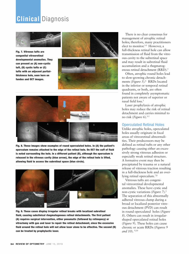
There is no clear consensus for management of atrophic retinal holes; therefore, many practitioners elect to monitor.3-5 However, a full-thickness retinal hole can allow transmission of fluid from the vitre-ous cavity to the subretinal space and may result in subretinal fluid accumulation and a rhegmatog-enous retinal detachment (RRD).5
Often, atrophic round holes lead to slow-growing chronic detach-ments (Figure 5).6 RRDs located in the inferior or temporal retinal quadrants, or both, are often found in completely asymptomatic patients not aware of superior or nasal field loss.5
Laser prophylaxis of atrophic holes may reduce the risk of retinal detachment and carries minimal to no risk (Figure 6).3-5
Operculated Retinal Holes Unlike atrophic holes, operculated holes usually originate in focal areas of vitreoretinal abnormali-ties. Their predecessors may be defined as retinal tufts or any other pathology causing either an exces-sively strong vitreous adhesion or especially weak retinal structure. A formative event may then be precipitated by trauma or a natural release of vitreous traction resulting in a full-thickness hole and an over-lying retinal operculum.7,8
Vitreous tufts are congeni-tal vitreoretinal developmental anomalies. These have cystic and non-cystic variations (Figure 7).2 The separation of this abnormally adhered vitreous clump during a broad or localized posterior vitre-ous detachment (PVD) can result in round operculated holes (Figure 8). Others can result in irregular-shaped operculated retinal holes (Figure 9). These holes can cause chronic or acute RRDs (Figures 9 and 10). 9,10
REVIEW OF OPTOMETRY JUNE 15, 201954
Fig. 7. Vitreous tufts are congenital vitreoretinal developmental anomalies. They can present as (A) non-cystic tuft, (B) cystic tufts or (C) tufts with an adjacent partial-thickness hole, seen here on fundus and OCT images.
Fig. 8. These images show examples of round operculated holes. In (A) the patient’s operculum remains attached to the edge of the retinal hole. On OCT the cuff of fluid is noted surrounding the hole. In a different patient (B), although the operculum is released in the vitreous cavity (blue arrow), the edge of the retinal hole is lifted, allowing fluid to access the subretinal space (blue circle).
Fig. 9. These cases display irregular retinal breaks with localized subretinal fluid, causing subclinical rhegmatogenous retinal detachments. The first patient (A) requires surgical intervention, either pneumatic (followed by retinopexy) or vitrectomy with gas and laser to repair the retinal detachment, since the excessive fluid around the retinal hole will not allow laser alone to be effective. The second (B) can be treated by prophylactic laser.
Clinical Diagnosis
052_ro0619_F3_Rafieetary.indd 54052_ro0619_F3_Rafieetary.indd 54 6/5/19 11:43 AM6/5/19 11:43 AM

55REVIEW OF OPTOMETRY JUNE 15, 2019 55
Fig. 10. Patient (A)’s images reveal an operculated hole resulting in an acute
RRD. Patient (B), who had a pre-existing lattice, displays an operculated hole
during PVD resulting in acute RRD.
Fig. 11. The patient in Figure 8B is shown here following prophylactic laser
treatment. The partial resolution of the fluid cuff is noted while progression to
retinal detachment is avoided.
Fig. 12. Image (A) reveals HSRT (blue arrow) with no associated symptom or
retinal detachment; however, a small retinal hole (red arrow) has resulted in a
slowly progressing retinal detachment, causing a slow-moving shadow in the
patient’s peripheral vision. In (B), a horseshoe retinal break (blue arrow)
associated with lattice degeneration (red arrow) is seen in an asymptomatic patient.
Fig. 13. Following PVD symptoms this patient (A) developed a superotemporal
RRD caused by a small HSRT (blue arrow), while another patient (B) has developed an inferior macula off RRD
from HSRT (blue arrow). These patients required surgical intervention. The RRD
in (C) is caused by more than one retinal tear, not an uncommon finding.
052_ro0619_F3_Rafieetary.indd 55052_ro0619_F3_Rafieetary.indd 55 6/5/19 12:05 PM6/5/19 12:05 PM

No consensus exists on treating symptomatic and asymptomatic operculated holes. Prophylactic laser is usually recommended for symptomatic cases. Treatment of asymptomatic holes may reduce the risk of retinal detachment with minimal to no risk to the patient (Figure 11).4,5
Horseshoe or Flap TearsHorseshoe retinal tears (HSRT) are full-thickness breaks of the neuro-sensory retina that occur as a result of vitreous traction and are usually caused by PVD. Risk factors for HSRT include aging, pre-existing lattice degeneration, myopia and trauma. Although flashes of light and floaters are common symp-toms associated with this finding,
these can be observed in asymp-tomatic patients, particularly in the absence of RRD (Figure 12).9,10
Horseshoe retinal tears are the cause of most RRDs (Figure 13).9,10 All HSRT should be surrounded by laser soon after diagnosis (Figure 14).
Giant Retinal Breaks and Retinal DialysisGiant retinal breaks are full-thickness tears that extend at least three clock hours (Figure 15). Retinal detachments caused by giant breaks may require different surgical modalities compared to other RRDs. For instance, perfluo-rocarbon (PFO) may be used to unfold a retinal detachment intra-operatively.11,12
Retinal dialysis is a break in which the anterior portion is adjacent to the ora serrata (Figure 16A). The majority of these cases are asso-ciated with ocular trauma, and most are diagnosed in young patients without posterior vitreous detachment. Manag-ing retinal dialysis can be challenging as well.
Retinal detachment may be prevented by treating the extent of the dialysis with laser retinopexy. If retinal detachment is pres-ent, treatment needs to be tailored to the individual patient. Options include: vitrectomy with gas or oil, scleral buck-ling with or without vitrectomy, and laser
retinopexy may still be used if the detachment is limited (Figure 16B). Making the appropriate surgical decision leads to good outcomes in the vast majority of patients.13-15
In ConclusionRetinal holes and tears come in all shapes and sizes with varying management strategies and risk for complication. The one complica-tion associated to some degree with all forms of retinal breaks is retinal detachment, which can result in significant vision loss. As such, all retinal holes and tears —regardless of size, location or shape—should be thoroughly evaluated and properly managed to reduce risk of vision loss for affected patients. ■
REVIEW OF OPTOMETRY JUNE 15, 201956
Fig. 14. Far left, horseshoe tears without retinal detachment (A) are treated with laser. The immediate and long-term of effect of laser (B) is shown, while in long-term hyperpigmentation.
Clinical Diagnosis
052_ro0619_F3_Rafieetary.indd 56052_ro0619_F3_Rafieetary.indd 56 6/5/19 12:07 PM6/5/19 12:07 PM
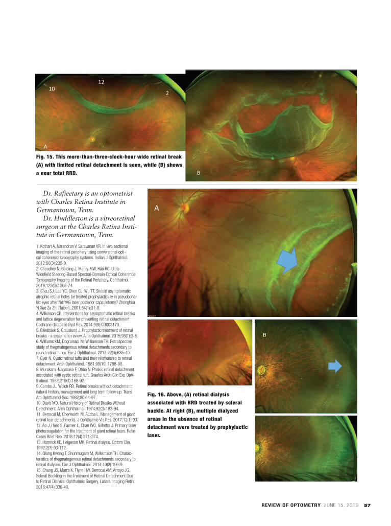
REVIEW OF OPTOMETRY JUNE 15, 2019 57
Dr. Rafieetary is an optometrist with Charles Retina Institute in Germantown, Tenn.
Dr. Huddleston is a vitreoretinal surgeon at the Charles Retina Insti-tute in Germantown, Tenn.
1. Kothari A, Narendran V, Saravanan VR. In vivo sectional
imaging of the retinal periphery using conventional opti-
cal coherence tomography systems. Indian J Ophthalmol.
2012;60(3):235-9.
2. Choudhry N, Golding J, Manry MW, Rao RC. Ultra-
Widefield Steering-Based Spectral-Domain Optical Coherence
Tomography Imaging of the Retinal Periphery. Ophthalmol.
2016;123(6):1368-74.
3. Sheu SJ, Lee YC, Chen CJ, Wu TT. Should asymptomatic
atrophic retinal holes be treated prophylactically in pseudopha-
kic eyes after Nd:YAG laser posterior capsulotomy? Zhonghua
Yi Xue Za Zhi (Taipei). 2001;64(1):31-8.
4. Wilkinson CP. Interventions for asymptomatic retinal breaks
and lattice degeneration for preventing retinal detachment.
Cochrane database Syst Rev. 2014;9(9):CD003170.
5. Blindbaek S, Grauslund J. Prophylactic treatment of retinal
breaks - a systematic review. Acta Ophthalmol. 2015;93(1):3-8.
6. Williams KM, Dogramaci M, Williamson TH. Retrospective
study of thegmatogenous retinal detachments secondary to
round retinal holes. Eur J Ophthalmol. 2012;22(4):635-40.
7. Byer N. Cystic retinal tufts and their relationship to retinal
detachment. Arch Ophthalmol. 1981;99(10):1788-90.
8. Murakami-Nagasako F, Ohba N. Phakic retinal detachment
associated with cystic retinal tuft. Graefes Arch Clin Exp Oph-
thalmol. 1982;219(4):188-92.
9. Combs JL, Welch RB. Retinal breaks without detachment:
natural history, management and long term follow-up. Trans
Am Ophthalmol Soc. 1982;80:64-97.
10. Davis MD. Natural History of Retinal Breaks Without
Detachment. Arch Ophthalmol. 1974;92(3):183-94.
11. Berrocal M, Chenworth M, Acaba L. Management of giant
retinal tear detachments. J Ophthalmic Vis Res. 2017;12(1):93.
12. Ao J, Horo S, Farmer L, Chan WO, Gilhotra J. Primary laser
photocoagulation for the treatment of giant retinal tears. Retin
Cases Brief Rep. 2018;12(4):371-374.
13. Hamrick KE, Helgeson MK. Retinal dialysis. Optom Clin.
1992;2(3):93-112.
14. Qiang Kwong T, Shunmugam M, Williamson TH. Charac-
teristics of rhegmatogenous retinal detachments secondary to
retinal dialyses. Can J Ophthalmol. 2014;49(2):196-9.
15. Chang JS, Marra K, Flynn HW, Berrocal AM, Arroyo JG.
Scleral Buckling in the Treatment of Retinal Detachment Due
to Retinal Dialysis. Ophthalmic Surgery, Lasers Imaging Retin.
2016;47(4):336-40.
Fig. 16. Above, (A) retinal dialysis associated with RRD treated by scleralbuckle. At right (B), multiple dialyzed areas in the absence of retinal detachment were treated by prophylactic laser.
Fig. 15. This more-than-three-clock-hour wide retinal break (A) with limited retinal detachment is seen, while (B) shows a near total RRD.
052_ro0619_F3_Rafieetary.indd 57052_ro0619_F3_Rafieetary.indd 57 6/5/19 12:09 PM6/5/19 12:09 PM
Release Date: June 15, 2019Expiration Date: June 30, 2020Goal Statement: Upon completing this educational activity, optometrists should have improved the skills to evaluate the right patients for potential premium IOL technology, address ocular surface issues in advance of procedures, comanage patients with the surgical center, and offer optimal care to patients pre- and postoperatively.Faculty/Editorial Board: Marc Bloomenstein OD, FAAO; Paul M. Karpecki, OD, FAAO; Amy Nau, OD, ODCredit Statement: This course is COPE approved for 2 hours of CE credit. COPE ID is 62523-PO. Please check your state licensing board to see if this approval counts toward your CE requirement for relicensure.Joint-Sponsorship Statement: This continuing education course is joint sponsored by the University of Alabama School of Optometry.Disclosure Statement: Dr. Karpecki is a consultant for Aerie Pharmaceuticals, Abbott Medical Optics, Akorn, Alcon, Allergan, Anthem, Avellino Labs, Bausch + Lomb, Beaver-Visitec, Bio-Tissue, BlephEx, Bruder Healthcare, Cambium Pharmaceuticals, Calhoun Vision/Rx Sight, Essilor, eyeBrain, Eye Gate Medical, Eyemaginations/Rendia, Eyes4Lives, Focus Labs, iCare USA, Imprimis Pharmaceuticals, Katena, Konan Medical, Lubris Inc., MacuLogix, Oasis Medical, Ocular Therapeutix, Oculus, OcuSoft, Paragon Biotek, PECAA, Perrigo, Quark Pharmaceuticals, Regeneron, Reichert/Ametek, Retina Sciences, Science Based Health, SightRisk, Shire Pharmaceuticals, TearFilm Innovations, TearLab, TearScience, TelScreen, Topcon, Visiometrics, Vision Care Inc. and Vmax; and a research grant principal inves-tigator for Shire Pharmaceuticals; has received honoraria for the speaker’s bureau of Glaukos; is a stockholder in Eye Therapies; is on the board of Optometric Medical Solutions, Bruder Healthcare and TearLab. Dr. Bloomenstein is a consultant for OcuSoft and Reichert; is a stock shareholder in TearLab; and is on the speaker’s bureau of Allergan, Bausch + Lomb and Alcon.Dr. Nau is on the speaker’s bureau of Johnson & Johnson Vision.Supported by: An unrestricted educational grant from Johnson & Johnson Vision.
Correcting Presbyopia
Using Innovative IOL
TechnologyBY MARC BLOOMENSTEIN, OD, FAAO
Patient lifestyles and evolving technology are trans-forming the way we treat the growing population of
presbyopic patients. Today’s presbyopes live their lives differently than previous generations. They are more active, they are working longer, and their near vision is defi ned as the ability to view cell phones and other dig-ital devices clearly. Moreover, they want to look younger and be free from the burden of eyeglasses.
We need to keep in mind that today’s presbyopia patients have higher expectations regarding presbyopia correction, so traditional options most likely will fall short.
DISCUSSING PRESBYOPIA WITH
PATIENTS
We play an important role in educating our patients about the visual changes that occur as they age. The term dysfunctional lens syndrome (DLS)—which de-scribes the natural changes that occur in the crystalline lens after about age 42—is a good way to explain this natural process to patients. We should begin the conver-sation about DLS when patients are in their 30s and 40s.Wrinkles, gray hair, and other changes occur as we age,
Administered byReview of Optometry®
Supported by an unrestricted educational grant from
Johnson & Johnson VisionApproved
Pathways to Premium IOL Outcomes & Successful Surgical Comanagement How the right approach to pre- and postoperative care can ensure optimal IOL outcomes for your patients.
0619_JnJ_CE.indd 580619_JnJ_CE.indd 58 5/31/19 9:42 AM5/31/19 9:42 AM

2 CE Credits(COPE Approved)
and the crystalline lens is not immune to the aging process. Over time, layers are added to the lens, which become more compact. In addition, disulfi de bonds accumulate, and the lens stiffens, leading to changes in optics. Diagnostic technology can help us evaluate how patients’ vision quality is declining.
Figure 1 shows DLS stages.In stage 1, we can prescribe reading glasses, mul-tifocal contact lenses, corneal inlays, or blended or monovision laser vision correction or offer refractive lens exchange. In stage 2, we need to talk to patients about subjec-tive symptoms, knowing that lenticular changes will affect their vision even in the absence of a refractive change. Therefore, we should reassure patients that we will continue monitoring their eyes and ask them to notify us of vision changes. In stage 2, presbyopia can be treated with reading glasses or multifocal contact lenses, as well as the surgical opportunities of stage 1 DLS.In stage 3, cataract surgery becomes necessary, with monofocal intraocular lenses (IOLs) (distance or monovision) or presbyopia-correcting IOLs.
When our patients have cataract surgery, we should be providing preoperative care, referrals, and postoperative care. As optometrists, we should select surgeons whose philosophies match our own and forge a strong relationship them. We need to share relevant information with them about the patients they will treat, and we need to talk to patients about what to expect when they see the surgeon.
PRESBYOPIA-CORRECTION OPTIONS
Each presbyopia-correction option has advantages and disadvantages. For example, studies have shown
that progressive lenses can contribute to falls, while patients often dislike the interference of reading glasses. Presbyopic contact lenses can be challeng-ing because of comfort issues in older eyes, visual acuity problems, and chair time for the practitioner, although advances have been made to remedy some
of these limitations. With regard to IOL options, here are some observations:
Presbyopia-correcting or lifestyle IOLs, as I refer to them, appeal to patients who desire spectacle independence for near vision (Figure 2).
Monofocal monovision provides monofocal quality of vision, and many patients have been satisfi ed with this option.1 However, some patients have reported reduced depth perception, a feeling of imbalance, and limited intermediate vision. And patients still need to use glasses for night driving and prolonged reading. Monovision with an IOL is not the same as the contact lens version because the lens always adds some accommodative effect, although small.
In recent years, multifocal technology has ad-vanced.2 Low-add multifocal IOLs provide near and distance vision, although intermediate vision may be limited. Patients may also have dysphotopsia and need to neuroadapt to the lenses.
Extended depth of focus (EDOF) IOLs are dif-fractive lenses that extend vision instead of offering discrete close, intermediate, or distance vision. The extension of the focal range means there is more than one sweet spot.3 Patients are more tolerant of unexpected residual refractive error, which is also helpful as a patient’s ocular surface changes with age.4 Patients have a full range of high-quality contin-uous vision with a low incidence of visual symptoms and require glasses less often. I fi nd that patients only need a slight magnifi cation for very small print. We have obtained good results with this lens in
R E V I E W O F O P T O M E T R Y 5 9 J u n e 2 0 1 9
Figure 1.Stages of Dysfunctional Lens Syndrome Images: Marc Bloomenstein, OD, FAAO
Figure 2. The Ideal Presbyopia-Correcting Lens
0619_JnJ_CE.indd 590619_JnJ_CE.indd 59 5/31/19 10:08 AM5/31/19 10:08 AM

patients with macular degeneration or previous refractive surgery. Approximately 85% of patients with EDOF IOLs wear glasses sometimes.5
PERSONALIZED VISION
Surgeons can personalize patients’ vision with mini-monovision, or mix and match within a platform of presbyopia-correcting IOLs. In our practice, we implant the EDOF IOL in the dominant eye. If patients cannot read as well as they would like, we personalize their nondominant eye with a low-add multifocal IOL.6 If patients are pleased with their vision in the fi rst eye, we implant the EDOF lens in the other eye. Mixing and matching presbyopic IOLs offers an increased range of vision and improves patients’ near vision.
MAKING A MATCH
When matching presbyopia-correction technology with the appropriate patient, questionnaires can help us learn more about our patients. We need to know patients’ preferred reading distance, their leisure pursuits and career demands, and how they rate near, intermediate, and distance in terms of importance. I give patients the near point card upside down and then I drop it on the fl oor. When they pick it up, they automatically adjust it to where they want to read and we can see their natural reading position.
We cannot underestimate the importance of opti-mizing intermediate distance. Patients spend the vast majority of their time using distance and intermediate vision—looking at the dashboard, driving, using a computer, and watching television.
In our practice, we do not offer patients a range of options. We write down the option that best fi ts their needs and desires. In fact, patients are not looking for choices; they are looking for the solution that will provide the vision they need.
We need to manage patients’ expectations before surgery, discussing their postoperative vision and healing, as well as the potential need for glasses for small print and the possibility of glare and halos during adaptation. In addition, we must inform them that postoperative enhancement occasionally is needed.
It is especially important to manage expectations in myopic patients. These patients should recognize that monofocal IOLs will permanently give them distance vision.
During preoperative visits, optometrists need to prepare patients for the choices they will make when they see the surgeon. It helps to provide information
on your website or give patients information to bring home. We also need to examine and treat the ocular surface well ahead of the time for referral. In fact, ocular surface treatment should start at stage 1 DLS.
POSTOPERATIVE CARE
When examining patients after cataract surgery with presbyopic IOLs, it’s important to check their binocular uncorrected vision. We should remind patients of restrictions and that they initially may feel off balance, but by emphasizing their binocular vision, we can reinforce that they use both eyes.
One week after surgery, ask patients how they are feeling and examine them, looking for signs of infection or increased infl ammation. One month after surgery, make sure to examine patients again and check how they are functioning. During postoperative visits, we remind them that it will take time to neuro-adapt to the new lens. I cannot emphasize enough that patients expect the optometrist to let them know how they are doing.
If results are not as expected, I learned an easy way to remember what to search for from a renowned surgeon, Eric Donnenfeld, MD. Dr. Donnenfeld recommends looking at the fi ve “C”s—check the corneal surface, the capsule (and do a glare test), cystoid macular edema, correction, and centration. By evaluating the fi ve “C”s, you almost always fi nd a reason for results that are less than perfect.
After each examination, results should be sent to the surgeon.
MAKING THE CHOICE EARLY
Our patients are living longer, working longer, and are active. The notion of optimizing reading vision is one that becomes more interpretative. The route patients choose to correct presbyopia will impact their daily lives, and we can help them navigate this journey earlier rather than later.
1. Ito M, Shimizu K, Iida Y, Amano R. Five-year clinical study of patients with pseudophakic monovision. J Cataract Refract Surg. 2012; 38:1440–1445.2. Kim JS, Jung JW, Lee JM, et al. Clinical outcomes following implantation of diffractive multifo-cal intraocular lenses with varying add powers. Am J Ophthalmol. 2015; 160:702–709.3. Cochener B. Tecnis Symfony intraocular lens with a “sweet spot” for tolerance to postopera-tive residual refractive errors. Open J Ophthalmol. 2017; 7:14–20. 4. Carones F. Residual astigmatism threshold and patient satisfaction with bifocal, trifocal and extended range of vision intraocular lenses (IOLs). Open J Ophthalmol. 2017; 7:1–7. 5. TECNIS Symfony® Extended Range of Vision IOLs DFU. Santa Ana, Calif. Johnson & Johnson Surgical Vision, Inc.6. Black S. A clinical assessment of visual performance of combining the TECNIS® Symfony Extended Range of Vision IOL (ZXR00) with the +3.25 D TECNIS Multifocal 1-piece IOL (ZLB00) in
subjects undergoing bilateral cataract extraction. Clin Ophthalmol. 2018; 12:2129–2136.
Marc Bloomenstein, OD, FAAO, practices with the Schwartz Laser Eye Center in Scottsdale, Ariz.
R E V I E W O F O P T O M E T R Y 6 0 J u n e 2 0 1 9
0619_JnJ_CE.indd 600619_JnJ_CE.indd 60 5/31/19 10:09 AM5/31/19 10:09 AM

Managing
Astigmatism Using
Innovative IOL
TechnologyBY PAUL M. KARPECKI, OD, FAAO
To help patients achieve good uncorrected vision from cataract surgery, it is critical to correct
astigmatism; this will greatly improve vision at all distances.
And while more than 50% of patients with astigma-tism are eligible for correction with a toric intraocular lens (IOL), only 20% of patients with clinically signifi -cant astigmatism receive these lenses, according to the 2018 ASCRS Clinical Survey.1-3
In my experience, one reason patients do not have their astigmatism treated during cataract surgery is because they are not aware this option is possible. As the eyecare practitioners who spend the most time with patients, optometrists are in a key position to educate them about astigmatism and recommend solutions.
PREOPERATIVE DIAGNOSTICS
Keratometry or an autorefraction is usually ade-quate when assessing astigmatism power and axis preoperatively in most toric IOL candidates. Optical biometry, corneal topography, and optical coherence tomography are also useful tools for preoperative as-sessments. If astigmatism is present on the posterior surface of the cornea, it will appear on the Ks.4
During the preoperative examination, keratome-try values should be compared with cylinder in the manifest refraction. If a discrepancy is identifi ed, consider other causes of cylinder, such as lenticular astigmatism or dry eye disease. We can also assess patients’ refractive stability by monitoring the mani-fest refraction.
When evaluating data from assessments, consis-tency is critical. If readings are inconsistent, assess-ments should be repeated. We also need to compare previous measurements to determine stability. Axis measurements should be within 10 degrees, and magnitude measurements should be within 0.5D of previous readings. Keratometry readings should be within 1.0D of the fellow eye.
Measurements also should be repeated if there is high astigmatism in either eye (2.50D or greater) or the patient has small or large eyes (corneal diameter
less than 10.75mm or greater than 13.0mm), or steep or fl at eyes (40.0D or less or greater than 47.0D).
The most frequent reason for inconsistency is oc-ular surface disease. We need to be sure the patient has a healthy tear fi lm before performing preoper-ative measurements.5 If topography shows signs of ocular surface disease, evaluate and treat the tear fi lm before repeating measurements. To reduce error, it is also important to use the same device before and after surgery, use select technicians, and perform measurements before eyedrops are instilled.
CORRECTING ASTIGMATISM
There are several alternatives when performing cat-aract surgery in patients with astigmatism—monofo-cal IOLs with incisional correction (via on-axis incision, or corneal relaxing incisions with a blade or femtosec-ond laser) for patients with less than 1.0D of astigma-tism and toric IOLs (e.g., monofocal, extended depth of focus, multifocal, and pseudo-accommodating).
The goals of implanting toric IOLs are to deliver excellent visual acuity, reduce or eliminate astigmatic error, and achieve patient satisfaction. In a meta-anal-ysis, Kessel et al. determined that postoperative cor-rected distance vision and spectacle independence for distance were better in patients who received toric IOLs versus those who had non-toric IOLs, with or without relaxing incisions.6 Black reported that 94% of eyes achieved 0.50D or less residual refractive error after implantation of toric IOLs.7
Toric IOLs are placed closest to the nodal point, so patients have very little distortion or rotation. These IOLs yield better vision than patients can achieve with glasses or contact lenses. That said, patients with high levels of astigmatism have a slight amount of peripheral distortion with toric IOLs, but still not as much as with glasses.
PATIENT SELECTION
The right presbyopic toric IOL candidates should want good uncorrected distance vision without glasses, but be comfortable with glasses for near. It is important to rule out signifi cant or unstable corneal disease affecting the ocular surface or shape.
In general, patients with more than 0.5D of regular astigmatism are good candidates for toric IOLs (Figure 1). Among patients who have with-the-rule astigmatism, those with 0.75D or more cylinder are good candidates (Figure 2). However, it is important to determine that the astigmatism is corneal astigma-tism, using corneal topography or keratometry (not
R E V I E W O F O P T O M E T R Y 6 1 J u n e 2 0 1 9
0619_JnJ_CE.indd 610619_JnJ_CE.indd 61 5/31/19 10:10 AM5/31/19 10:10 AM

the refraction). If astigmatism is in the lens, it will be corrected when the cataract is extracted.
If a patient has against-the-rule or oblique astigma-tism, correcting 0.5D of astigmatism will signifi cantly improve vision.
IMPACT OF RESIDUAL ASTIGMATISM
The most likely cause of patient dissatisfaction with toric IOLs is postoperative residual astigma-tism, according to anecdotal reports. In addition, one study reported that if more than 1% of rota-tion occurred, it decreased the effect of the toric IOL by 3.3%; with 10% rotation, patients lost 33% of the effect.8
Patients receiving presbyopia-correcting IOLs have very high expectations of their visual outcomes. Although monofocal IOL patients pre-viously tolerated astigmatism, toric presbyopic IOL patients have paid to reduce their depen-
dence on glasses, so they have less tolerance for astigmatism.
MANAGING RESIDUAL ASTIGMATISM
If patients have residual astigmatism after im-plantation of a toric IOL, begin by looking at the manifest refraction. It should be repeated multiple times to determine whether it is stable before making a treatment decision.
We also need to examine the ocular surface. If signifi cant ocular surface disease is present, it should be treated and then the manifest refraction repeated. In addition, we need to be sure the toric IOL is aligned properly.
There are several options to correct residual refractive error: glasses or contact lenses, limbal relaxing incisions or arcuate incisions, LASIK or PRK, IOL rotation, or IOL exchange.
Glasses or contact lenses are the easiest choice, but glasses may result in ghosting if the patient has signifi cant astigmatism. Standard rigid gas permeable contact lenses unmask lenticular astigmatism, and corrected distance visual acuity may be suboptimal with toric rigid gas permeable lenses.
Limbal relaxing and arcuate incisions are low risk and relatively reliable. They can be made at the slit lamp. Make sure to treat refractive cylinder.
Toric IOL rotation may be considered in a dissat-isfi ed patient in whom the IOL is off axis. This may occur if the target axis was incorrect, and there is a larger impact with higher toric corrections. The patient should have no signifi cant spherical error, and the posterior capsule should be intact.
If there was an error in the spherical and cylin-drical target, the IOL may be exchanged. We must fi rst determine that astigmatism is regular or if spherical error is signifi cant. If it is still early and the bag is intact, the surgeon can exchange the toric IOL. If not, a monofocal IOL should be con-sidered. An exchange is dangerous if the capsule is open.
Note that IOL rotation or exchange should be done before a YAG procedure. It is also important to check for cystoid macular edema or other possible causes of decreased vision. If there is residual astigmatism, delay surgery on the second eye.
EDUCATING PATIENTS EARLY ON
Growing numbers of patients will need cataract
R E V I E W O F O P T O M E T R Y 6 2 J u n e 2 0 1 9
Figure 2.With/Against-the-rule AstigmatismPatients who have with-the-rule astigmatism (left) can tolerate 0.75D without signifi cant impact on patient sat-isfaction and visual quality. In those with against-the-rule/oblique astigmatism (right), toric IOLs generally have a greater impact on patient satisfaction and visual quality.
Figure 1. Regular vs. Irregular AstigmatismPatients with regular astigmatism (left) are generally good candidates for toric IOLs, whereas those with irregular astigmatism (right) are often more diffi cult to address with toric IOLs.
0619_JnJ_CE.indd 620619_JnJ_CE.indd 62 5/31/19 10:11 AM5/31/19 10:11 AM

surgery in the future. To achieve optimal out-comes, astigmatism must be corrected. Optome-trists play an important role in educating patients about astigmatism correction options during surgery and providing preoperative and postoper-ative care.
1. East Valley Ophthalmology website. Distribution of corneal astigmatism in normal adult population. Keratometry database. https://www.doctor-hill.com/iol-main/astigmatism_chart.htm Accessed April 1, 2019.2. AcrySof® IQ Toric [product information]. Fort Worth, TX: Alcon Laboratories, Inc; 2009. 3. ASCRS 2018 Clinical Survey. http://supplements.eyeworld.org/eyeworld-supplements/decem-ber-2018-clinical-survey4. Koch DD, Ali SF, Weikert MP, et al. Contribution of posterior corneal astigmatism to total corneal astigmatism. J Cataract Refract Surg. 2012; 38:2080–2087.5. Epitropoulos AT, Matossian C, Berdy GJ, et al. Effect of tear osmolarity on repeatability of keratometry for cataract surgery planning. J Cataract Refract Surg. 2015; 41:1672–1677.6. Kessel L, Andresen J, Tendal B, et al. Toric intraocular lenses in the correction of astigmatism during cataract surgery: a systematic review and meta-analysis. Ophthalmology. 2016; 123:275–286.7. Black D. Effi cacy of hydrophobic acrylic toric IOL to correct astigmatism in cataract patients. Presented at ASCRS 2015.8. Bauer NJ, de Vries NE, Webers CA, et al. Astigmatism management in cataract surgery with
the AcrySof toric intraocular lens. J Cataract Refract Surg. 2008; 34:1483-1488.
Dr. Karpecki is director of Corneal Services, the Ad-vanced Ocular Surface Disease Center and Research at Kentucky Eye Institute in Lexington, Kentucky, and practices in the ocular surface disease clinic at Gaddie Eye Centers, Louisville, Ky.
Optimizing the
Ocular Surface with
Advanced TherapiesBY AMY NAU, OD, FAAO
It is our responsibility to address ocular surface dis-ease (OSD) as part of preventative care. However,
if patients are not in acute distress, noncompliance with recommendations is common. If we anticipate that a patient will require lid surgery, ocular surgery, or ocular injections, we must have a frank discussion about the importance of treating OSD to optimize surgical outcomes. In this context, patients are often much more adherent to therapy and continue treatment indefi nitely.
TARGETING OSD
Contemporary practice offers a wide range of options to treat OSD. Success is achievable when the root cause of the condition is identifi ed and treated with the appropriate combination of target-ed therapies.
The most common cause of OSD is meibomian gland dysfunction (MGD); thus, a thorough eval-uation of the lids and lashes is a mandatory fi rst
step. Additional testing for lacrimal gland function, allergies, poor nocturnal lid seal, and neuropathic pain using a proparacaine challenge also should be included.
MGD is extremely common, but it is easy to over-look if we do not pay specifi c attention to the struc-ture and function of the meibomian glands. Figure 1 shows potential treatments for MGD. Omega-3 fatty acid supplements often are used to treat OSD, but research varies on their effectiveness.1
When patients have symptoms of dryness in the morning, we use the Korb-Blackie light test to de-termine if there is a poor nocturnal lid seal.2 In such cases, ointment before bedtime and/or a sleep mask will help alleviate symptoms and reduce infl amma-tion by preventing overnight desiccating stress.
If patients’ MMP-9 results are positive, and they have a high tear osmolarity level or chronically injected red eyes, I prescribe a pulse dose of topical corticosteroids for two to four weeks.3 Non-pre-served steroids have been shown to be more effec-tive but need to be compounded.4 If patients have adhered to our recommendations and show limited improvement, and MMP-9 results are still positive at 10 to 12 weeks, I consider cyclosporine A drops or lifi tegrast. If infl ammation and symptoms persist after six months, autologous serum drops or amniotic fl uid drops are good add-on medications. Compounded testosterone drops help with MGD and aqueous dry eye. Punctal plugs are effective, but contraindicated when infl ammation is present. For patients with low tear volumes, nasal neuro-stimulation and hydroxypropyl cellulose ophthal-mic inserts can help, but moisture chamber glasses and scleral lenses will help increase tear volumes by mechanical means. Amniotic membrane
R E V I E W O F O P T O M E T R Y 6 3 J u n e 2 0 1 9
Figure 1. Potential Treatments for Meibomian Gland Dysfunction
Image: Amy Nau, OD, FAAO
0619_JnJ_CE.indd 630619_JnJ_CE.indd 63 5/31/19 10:12 AM5/31/19 10:12 AM

To obtain two hours of continuing education credit, complete the exam by recording the best answer to each self-assessment question online at:
https://www.reviewofoptometry.com/ce/pathways-to-premium-iol-outcomes-june19 Or, mail the Examination Answer Sheet on the next page to: Jobson Healthcare Information, LLC, Attn: CE Processing, 395 Hudson Street – 3rd Floor, New York, NY 10014. A minimum score of 70% is required to obtain a certification of completion. The fee for this course is free.
1. It is recommended that eye care professionals begin discussing dysfunctional lens syndrome with their patients when _____________.a. Cataract surgery is necessaryb. They are in their 30s and 40sc. They are in their 50s and 60sd. They have symptoms of ocular surface disease
2. Which of the following is necessary in patients with stage 3 DLS?a. Corneal inlays b. Monovision laser vision correction c. Multifocal contact lensesd. Cataract surgery
3. What is true about monofocal monovision with IOLs?a. It can reduce depth perceptionb. It decreases chair time for the practitionerc. It can cause ocular surface discomfort in older patientsd. All of the above
4. It is stated that low-add multifocal IOLs ________________.a. Provide intermediate and distance vision, but near vision may be limited
b. Provide near and distance vision, but intermediate vision may be limitedc. Provide near and intermediate vision, but distance vision may be limitedd. Provide accommodation for near vision
5. Approximately _____ of patients with extended depth of focus IOLs wear glasses at times.a. 55%b. 65%c. 85%d. 95%
6. To achieve a greater range of vision with presbyopia-correcting IOLs, surgeons can personal-ize patients’ vision with ________.a. Mini-monovisionb. Identical low-add IOLs in both eyesc. Monofocal toric IOLs in both eyesd. Progressive lenses
7. Which of the following is not one of the five “C”s recommended by Eric Donnenfeld, MD, when searching for reasons for a suboptimal postoperative outcome?a. Cystoid macular edemab. Corneal surfacec. Cataractd. Capsule
8. _________ patients with astigmatism are eligible for correction with toric IOLs.a. Less than 20%b. Less than 40%c. More than 75%d. More than 50%
9. During the preoperative assessment, if a discrep-
ancy is found when keratometry values are com-pared with cylinder in the manifest refraction, what is stated to be another potential cause of cylinder?a. Previous refractive surgeryb. Lenticular astigmatismc. IOL tiltd. Posterior capsule opacification
10. When comparing measurements with previous readings to determine stability, axis measurements should be within ________ of previous readings.a. 1 degreeb. 5 degreesc. 10 degreesd. 20 degrees
11. To reduce measurement errors, it is recom-mended that __________________.a. The same device is used before and after surgeryb. Measurements are performed after eyedrops are instilledc. Measurements are performed before ocular sur-face treatment beginsd. B & C
12. The most likely cause of patient dissat-isfaction with toric IOLs is reported to be ________________.a. Preoperative optical coherence tomographyb. Preoperative assessments for ocular surface dis-easec. Postoperative residual astigmatismd. Uncorrected distance visual acuity
13. If _________ rotation of the toric IOL occurs, it decreases the effect of the toric IOL by 33%.a. 5%b. 10%
CE TEST
bandage lenses improve persistent corneal defects and reduce infl ammation, but will not treat underlying conditions.
PATIENT EDUCATION AND INVOLVEMENT
It is very important for patients to understand their condition. In cases of MGD, we invoke a dental model, showing them their lids and explaining that the condi-tion is similar to gingivitis, requiring ongoing hygiene and care. Showing them pictures and spending time reviewing their specifi c condition will enhance com-pliance. Patients need to understand that it often will take time for improvement, dry eye is often chronic,
and lifestyle modifi cations such as reduced screen time and environmental changes are necessary. Our staff members play a vital role in reinforcing our treatment philosophy and providing very specifi c instructions.
1. Dry Eye Assessment and Management Study Research Group, et al. n-3 fatty acid supplementa-tion for the treatment of dry eye disease. N Engl J Med. 2018; 378:1681–1690.2. Korb D, Blackie C. The Korb-Blackie lid light test. Invest Ophthalmol Vis Sci. 2013;54:942.3. Avunduk AM, Avunduk MC, Varnell ED, et al. The comparison of effi cacies of topical corticoste-roids and nonsteroidal anti-infl ammatory drops on dry eye patients: a clinical and immunocyto-chemical study. Am J Ophthalmol. 2003; 136:593–602.4. Hong S, Kim T, Chung SH, et al. Recurrence after topical nonpreserved methylprednisolone therapy for keratoconjunctivitis sicca in Sjögren’s syndrome. J Ocul Pharmacol Ther. 2007; 23:78–82.
Amy Nau, OD, FAAO, is managing partner with Korb and Associates in Boston, a practice focusing on dry eye and medical contact lenses.
0619_JnJ_CE.indd 640619_JnJ_CE.indd 64 5/31/19 10:13 AM5/31/19 10:13 AM

c. 20%d. 30%
14. If residual astigmatism is present after implantation of a toric IOL, it is recommended that the practitioner assess for ________________________.a. Toric lens misalignmentb. Ocular surface diseasec. Glaucomad. A & B
15. If a toric IOL requires rotation postoperatively, it is recommended that it is performed ___________.a. In patients with no significant spherical errorb. In patients with an open posterior capsulec. After a YAG procedure is performedd. In patients with significant spherical error
16. Meibomian gland dysfunction may be overlooked if optometrists do not pay attention to structure and ________ of the meibomian glands.a. Colorb. Sizec. Functiond. None of the above
17. If patients have symptoms of eye dryness in the morning, it is recommended that practitioners check for _________.a. Open-angle glaucomab. Viral conjunctivitisc. Poor nocturnal lid seald. Anterior uveitis
18. If patients’ MMP-9 results are positive and they have high tear osmolarity or chronically injected red eyes, which of the following is recommended?a. Lubricating eye dropsb. A pulse dose of topical corticosteroids for two to four weeksc. Nighttime application of lubricating eye ointmentd. Punctal plugs
19. Amniotic membrane bandage lenses can improve ___________.a. Persistent corneal defectsb. Meibomian gland obstructionc. Demodexd. Aqueous-deficient dry eye
20. Punctal plugs are not recommended when _____________.a. Tear osmolarity exceeds 300 mOsm/Lb. Patients do not comply with OSD therapiesc. Inflammation is presentd. Aqueous-deficient dry eye is present
CE TESTExamination Answer Sheet
Valid for credit through June 30, 2020
This exam can be taken online at: https://www.reviewofoptometry.com/ce/pathways-to-premium-iol-outcomes-june19
Upon passing the exam, you can view your results immediately and download a real-time CE certificate. You can also view your test history at any time from the website.
Pathways to Premium IOL Outcomes & Successful Surgical Comanagement
Directions: Select one answer for each question in the exam and completely darken the appropriate circle. A minimum score of 70% is required to earn credit.
To submit the Examination Answer Sheet by mail, send to: Jobson Healthcare Information, LLC, Attn: CE Processing, 395 Hudson Street – 3rd Floor, New York, NY 10014.
COPE approved for 2 hours of CE credit. COPE ID is 62523-PO.
This course is supported by an unrestricted educational grant from Johnson & Johnson Vision.There is an eight- to 10-week processing time for this exam.
1. A B C D 1 = Excellent 2 = Very Good 3 = Good 4 = Fair 5 = Poor
2. A B C D Rate the effectiveness of how well the activity: 3. A B C D
4. A B C D 21. Met the goal statemenA 1 2 3 4 5
5. A B C D 22. Related to your practice needs: 1 2 3 4 5
6. A B C D 23. Will help you improve patient care: 1 2 3 4 5
7. A B C D 24. Avoided commercial bias/influence: 1 2 3 4 5
8. A B C D 25. How would you rate the overall 9. A B C D quality of the material presented? 1 2 3 4 5
10. A B C D 26. Your knowledge of the subject was increased: 11. A B C D Greatly Somewhat Little 12. A B C D 27. The difficulty of the course was: 13. A B C D Complex Appropriate Basic 14. A B C D How long did it take to complete this course? 15. A B C D
16. A B C D Comments on this course: 17. A B C D
18. A B C D
19. A B C D Suggested topics for future CE articles: 20. A B C D
Please retain a copy for your records. Please print clearly. You must choose and complete one of the following three identifier types:
1 SS # - -
Last 4 digits of your SS # and date of birth State Code and License #: (Example: NY12345678)
2 - 3
First Name
Last Name
The following is your: Home Address Business Address
Business Name
Address
City State
ZIP
Telephone # - -
Fax # - -
By submitting this answer sheet, I certify that I have read the lesson in its entirety and completed the self-as-sessment exam personally based on the material presented. I have not obtained the answers to this exam by any fraudulent or improper means.
Signature Date
Lesson #118353 RO-UAB-0619
The opinions expressed in this supplement to Review of Optometry® do not refl ect the views of or imply endorsement
by the publisher of the host publication. Copyright 2019 Jobson Medical Information LLC.
Rate how well the activity supported your achievement of these learning objectives:
1=Poor, 2=Fair, 3=Neutral, 4=Good, 5=Excellent
21. Increased my knowledge of the growing list of premium IOL technologies used in the United States to address presbyopia correction and astigmatism.
22. Learned about some of the ways to assess candidates for premium IOLs.
23. Became familiar with some techniques to educate patients about premiumIOLs and ensure that they have realistic expectations.
24. Gained a basic knowledge of how to provide preoperative and postoperative care for patients receiving premium IOLs.
25. Acquired a basic understanding of how residual astigmatism is managed after implantation of toric IOLs.
26. Better understood the fundamentals of treating ocular surface disease with current therapies.
Rate the quality of the material provided:1=Strongly disagree, 2=Somewhat disagree, 3=Neutral, 4=Somewhat agree, 5=Strongly agree
27. The content was evidence-based.
28. The content was balanced and free of bias.
29. The information presented was clear and effective.
30. Additional comments on this course:
Answers to CE exam: Post-activity evaluation questions
1 2 3 4 5
1 2 3 4 5
1 2 3 4 5
1 2 3 4 5
1 2 3 4 5
1 2 3 4 5
1 2 3 4 5
1 2 3 4 5
1 2 3 4 5
0619_JnJ_CE.indd 650619_JnJ_CE.indd 65 5/31/19 10:14 AM5/31/19 10:14 AM

REVIEW OF OPTOMETRY JUNE 15, 201966
For many clinicians, the diag-nosis and treatment of scleritis can be daunting tasks. This troubling condition frequently
requires an arduous course of treat-ment and brings significant risk for destruction of ocular structures and permanent vision loss. The diagno-sis of scleritis is based primarily on clinical observation and the patient’s presenting constellation of symp-toms. Pain will be the hallmark, and it is constant, may radiate to the periorbital region and will be exacer-bated by eye movement.1 A duration of one to two months is not uncom-
mon, as the onset of scleritis is often insidious and patients may not seek care until the pain becomes severe.2
Slit lamp and dilated fundus exami-nations, optical coherence tomogra-phy (OCT), B-scan ultrasound and computed tomography (CT) are all helpful diagnostic tools to confirm even the most difficult cases.
Although diagnosis is often straightforward, properly classify-ing and identifying the cause of a patient’s scleritis isn’t always easy—and yet it is critical in determining the treatment regimen and obtaining the best possible visual outcome.
ClassificationAfter the initial diagnosis, the next essential step in managing scleritis is determining the type the patient is manifesting. Clinical signs and
symptoms vary significantly between anterior and posterior, diffuse and nodular, necrotizing and non-nec-rotizing scleritis. The likelihood and type of ocular sequelae is also deter-mined according to type.
Diffuse anterior scleritis is the most common but relatively benign form of scleritis and presents with extensive redness and edema.2,3
Inflammation of the trabecular meshwork may occur, causing tra-beculitis and secondary elevated intraocular pressure (IOP).4 In iso-lated anterior scleritis, no posterior segment findings are present. It is uncommon for these patients to progress to more aggressive forms.
Nodular anterior scleritis is characterized by localized areas of inflammation that present as dis-tinct, tender nodules on the sclera. The nodules are deep red, immobile and can vary in size and number. Of patients diagnosed with nodular anterior scleritis, 42% present with multiple nodules and 20% have superimposed conjunctival conges-tion and episcleritis involving the entire anterior episclera.2,5
Necrotizing anterior scleritis is the most severe and destructive form of the disease, posing a significant threat to vision. Compared with the
Scleritis
The initial diagnosis may be quick, but that’s only the beginning. Follow these steps to discover the root cause and set yourself on the right track for a successful outcome. By Katherine Sanford, OD
This B scan ultrasound demonstrates scleral thickening in the absence of the pathognomonic “T sign”.
My Patient Has Scleritis…Now What?
About this SeriesTo help optometrists strengthen their pro-tocols for managing conditions that require ongoing—perhaps life-long—care, this series explains the steps to take after con-firming a diagnosis, from day one through long-term management. Each installment in the five-part “Now What?” series will cover a different chronic condition:
March—glaucomaApril—RCEMay—diabetic retinopathyJune—scleritisJuly—AMD
Be sure to check www.reviewofoptometry.com for any articles you may have missed.
066_ro0619_F4.indd 66066_ro0619_F4.indd 66 6/5/19 12:38 PM6/5/19 12:38 PM

67
other forms, this condition more commonly affects females and has a higher incidence of associated systemic inflammatory conditions. Necrotizing scleritis is characterized by intense pain, which may become excruciating with time. Localized or generalized necrosis is the result of areas of intense vasculitis that close the deep episcleral plexus. Progres-sion may occur rapidly to expose the choroid. These patients have acute congestion of the conjunctival, episcleral and scleral vasculature surrounding the necrotic areas. Inflammation commonly spreads to involve the cornea (peripheral ulcer-ative keratitis), ciliary body (anterior uveitis) and trabecular meshwork (ocular hypertension).3,5,6
Scleromalacia perforans is a rare form of necrotizing anterior scleri-tis without adjacent inflammation caused by an obliterative arteritis in the deep episcleral plexus. Patients are typically asymptomatic with no pain or injection but may pres-ent with blurred vision from high astigmatism caused by loss of scleral integrity. The sclera will be thin and may have a white, marble- or por-celain-like appearance. The choroid can become exposed, and staphy-loma may form, particularly in the presence of elevated IOP. Consistent with those diagnosed with necrotiz-ing scleritis, these patients typically have long histories of systemic inflammatory disease.3,5,6
Posterior scleritis represents scleral inflammation posterior to the insertion of the rectus muscles. The inflammation may be localized or diffuse in nature, similar to diffuse and nodular scleritis.7 An associated anterior scleritis may be present, or the posterior scleritis may occur in isolation. If isolated, anterior seg-ment findings are rarely present, though an inflamed sclera may be visible in gaze extremes. Presentation
varies widely based on loca-tion and severity, but vision loss and pain are common to all posterior scleritis diagno-ses.8 Patients with posterior scleritis may experience diplo-pia or decreased visual acuity due to swelling around the optic nerve or retinal changes. Extraocular motilities will exacerbate the patient’s pain, and in the gaze extremes you may note inflammation of the posterior sclera. Other findings in posterior scleritis include chorioretinal granu-lomas and serous retinal detachments.9 Retinal OCT can be helpful in differentiating retinal pathology and determining whether macular edema is present.
B-scan ultrasonography is essen-tial in visualizing the posterior scleral thickening and fluid accumu-lation that can occur. The scan will show increased thickness of the eye wall, whether widespread or local-ized. Scleral thickness greater than 2mm is considered abnormal.7 Fluid may accumulate in the episcleral space and can extend posteriorly around the optic nerve. This fluid in Tenon’s capsule forms the character-istic “T sign” considered pathogno-monic for posterior scleritis.1,8
Posterior scleritis patients are typically younger, and a unilateral presentation occurs twice as often as bilateral involvement.6,8
CauseOnce you have identified the type of scleritis, your next step is deter-mining the cause of the patient’s inflammation. While many cases of scleritis are idiopathic, up to 50% will have an associated systemic dis-ease.10 Accounting for 40% to 50% of cases, autoimmune conditions are the most common conditions associ-ated with scleritis. While rheumatoid
arthritis (RA), particularly with vasculitis such as Wegener granu-lomatosis, occurs most frequently, inflammatory bowel disease, sys-temic lupus erythematosus (SLE) and relapsing polychondritis are all associated with scleritis.11
Infectious scleritis is rare, com-prising 4% to 10% of cases. While this is a relatively low percentage, it is imperative to rule out infectious etiologies prior to initiating treat-ment, particularly with steroids. Recent trauma or surgery may serve as a source for infection, as can extension of a corneal keratitis.3,12 Potential pathogens include viral, fungal and bacterial, although the most common agent is herpes zoster, implicated in 8% of infectious scle-ritis cases. Herpetic cases are typi-cally unilateral, sudden onset and associated with moderate to severe pain.3,13 Any systemic history of herpes simplex, Lyme, tuberculosis or syphilis is a potentially significant finding.14 Infectious etiologies should be addressed with an antimicrobial agent appropriate to the condition.
Surgically-induced necrotizing scleritis (SINS) is a non-infectious granulomatous inflammation that can occur months, even years, after
REVIEW OF OPTOMETRY JUNE 15, 2019 67
This slit lamp photo demonstrates diffuse anterior scleritis in a 58-year-old male with a subsequent negative systemic workup. The inflammation resolved completely with oral indomethacin.
066_ro0619_F4.indd 67066_ro0619_F4.indd 67 6/5/19 12:38 PM6/5/19 12:38 PM
intraocular surgery. Procedures most commonly associ-ated with SINS include pterygium removal, cataract extraction and scleral buckle.3,14,15 It can be difficult to differentiate from postoperative infectious causes of scleritis; thus, cultures, biopsies and empiric treatment with antibiotics is critical, particularly following pte-rygium surgery. Of those who develop SINS, 75% have undergone two or more surgical procedures.16
Malignancies do not cause a scleritis in the truest sense, but conjunctival tumors, lymphoma or malig-nant deposits from melanoma may mimic the inflam-mation seen in scleritis. In the absence of a therapeutic response, an early biopsy should be considered to iden-tify these rare cases.3,5
While a patient’s medical history and clinical picture can provide a clear indication of a potential cause, certain baseline laboratory investigations, and possibly imaging, are indicated. Regardless of presentation, clinicians should obtain a complete blood count (CBC) and an anti-neutrophil cytoplasmic antibody (ANCA) test for all scleritis patients prior to initiating treatment (Table 1). The CBC is imperative for identifying infec-tious processes. ANCA positivity is a marker for vas-culitis, which typically has a grimmer clinical picture.3 Even in the absence of clinical evidence of vasculitis, a positive ANCA may be a marker for refractory scle-ritis.3 Additional systemic workup should be guided by patient history, review of systems and the physical examination.
ManagementNow that you have determined the type and cause of the scleritis, it’s time to establish an initial treatment protocol. Systemic treatment is required because scleral inflammation is rarely affected by topical treatments.17 A number of patients respond to nonsteroidal anti-inflammatory drugs (NSAIDs) alone, but one study showed that 67% of patients required either high-dose glucocorticoids or a combination of high-dose gluco-corticoids and another immunosuppressive agent to establish control.11
Diffuse and nodular types of scleritis are the most likely to respond to NSAIDs alone. Frequently used agents include indomethacin, naproxen, ibuprofen, piroxicam, diclofenac and celecoxib. Studies show nonselective COX inhibitors (indomethacin) and selec-tive COX inhibitors (celecoxib) are equally effective in controlling inflammation in 78% to 81% of scleritis patients.18 In Europe, flurbiprofen is an option, but many investigators maintain that indomethacin is more effective than any other available NSAID.11,19 Thus,
Scleritis
066_ro0619_F4.indd 68066_ro0619_F4.indd 68 6/5/19 12:38 PM6/5/19 12:38 PM

REVIEW OF OPTOMETRY JUNE 15, 2019 69
your first step will likely be prescribing indomethacin 25mg orally four times a day. Be aware that gastroin-testinal side effects from indomethacin occur in 22.2% of patients. Patients at high risk for NSAID gastropathy may benefit from a selective COX inhibitor or be pre-scribed a histamine H2 receptor antagonist (e.g., raniti-dine) or a proton pump inhibitor.18
Necrotizing anterior scleritis, posterior scleritis and refractory anterior scleritis all necessitate more aggres-sive treatment. For these patients, initial therapy will consist of oral corticosteroids given as a one-time dose of oral prednisone 1mg/kg up to a maximum of 80mg. For these forms of scleritis, many practitioners advocate for a combined treatment of steroids plus an immuno-suppressive agent from the start.
Subconjunctival corticosteroid injections in the man-agement of scleritis have historically been met with controversy due to reports of scleral thinning, though a more recent study shows complete resolution with no cases of scleral melting.3,20,21 Ocular hypertension is the main side effect encountered with the use of subcon-junctival injections and may require hypotensive agents or surgical intervention.21
Be sure to coordinate care with rheumatology and uveitis specialists when necessary—especially for patients presenting with necrotizing scleritis, sclero-malacia perforans and posterior scleritis—as rheu-matologic disease should be considered in all scleritis patients. Immunosuppressive agents are often indicated when treating these forms and should be administered by providers well versed in their use. A variety of immunosuppressive agents can aid in managing inflam-mation while decreasing the need for corticosteroids, including antimetabolites (methotrexate, azathioprine, mycophenolate mofetil), alkylating agents (chlorambu-cil, cyclophosphamide), T-cell inhibitors (cyclosporine, tacrolimus), tumor necrosis factor inhibitors (inflix-imab, adalimumab) and the chimeric monoclonal anti-body rituximab.19,23-26
When referring to a specialty provider, include a letter rather than merely forwarding copies of your electronic record. A summary referral letter, which is far more concise, particularly with multiple documented visits related to the scleritis, ensures the specialist knows what tests you have completed and what treatment you initiated. Referrals to subspecialists for corneal, retinal or glaucoma care can be urgent, and providing a sum-mary of care and current ocular status of the patient can help the receiving provider triage the patient and offer timely care. The referral letter should include:
• The reason for referral
066_ro0619_F4.indd 69066_ro0619_F4.indd 69 6/5/19 12:38 PM6/5/19 12:38 PM
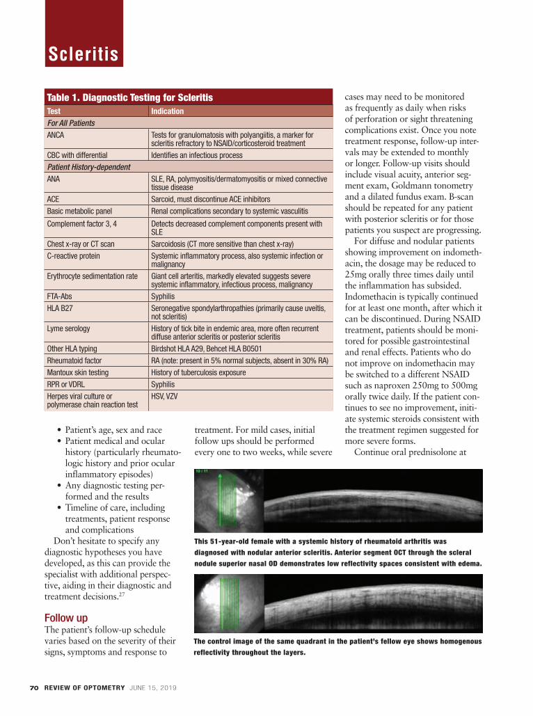
REVIEW OF OPTOMETRY JUNE 15, 201970
• Patient’s age, sex and race• Patient medical and ocular
history (particularly rheumato-logic history and prior ocular inflammatory episodes)
• Any diagnostic testing per-formed and the results
• Timeline of care, including treatments, patient response and complications
Don’t hesitate to specify any diagnostic hypotheses you have developed, as this can provide the specialist with additional perspec-tive, aiding in their diagnostic and treatment decisions.27
Follow upThe patient’s follow-up schedule varies based on the severity of their signs, symptoms and response to
treatment. For mild cases, initial follow ups should be performed every one to two weeks, while severe
cases may need to be monitored as frequently as daily when risks of perforation or sight threatening complications exist. Once you note treatment response, follow-up inter-vals may be extended to monthly or longer. Follow-up visits should include visual acuity, anterior seg-ment exam, Goldmann tonometry and a dilated fundus exam. B-scan should be repeated for any patient with posterior scleritis or for those patients you suspect are progressing.
For diffuse and nodular patients showing improvement on indometh-acin, the dosage may be reduced to 25mg orally three times daily until the inflammation has subsided. Indomethacin is typically continued for at least one month, after which it can be discontinued. During NSAID treatment, patients should be moni-tored for possible gastrointestinal and renal effects. Patients who do not improve on indomethacin may be switched to a different NSAID such as naproxen 250mg to 500mg orally twice daily. If the patient con-tinues to see no improvement, initi-ate systemic steroids consistent with the treatment regimen suggested for more severe forms.
Continue oral prednisolone at
This 51-year-old female with a systemic history of rheumatoid arthritis was diagnosed with nodular anterior scleritis. Anterior segment OCT through the scleral nodule superior nasal OD demonstrates low reflectivity spaces consistent with edema.
The control image of the same quadrant in the patient’s fellow eye shows homogenous reflectivity throughout the layers.
Table 1. Diagnostic Testing for ScleritisTest Indication
For All Patients
ANCA Tests for granulomatosis with polyangiitis, a marker for scleritis refractory to NSAID/corticosteroid treatment
CBC with differential Identifies an infectious processPatient History-dependent
ANA SLE, RA, polymyositis/dermatomyositis or mixed connective tissue disease
ACE Sarcoid, must discontinue ACE inhibitors Basic metabolic panel Renal complications secondary to systemic vasculitis
Complement factor 3, 4 Detects decreased complement components present with SLE
Chest x-ray or CT scan Sarcoidosis (CT more sensitive than chest x-ray)C-reactive protein Systemic inflammatory process, also systemic infection or
malignancyErythrocyte sedimentation rate Giant cell arteritis, markedly elevated suggests severe
systemic inflammatory, infectious process, malignancyFTA-Abs SyphilisHLA B27 Seronegative spondylarthropathies (primarily cause uveitis,
not scleritis)Lyme serology History of tick bite in endemic area, more often recurrent
diffuse anterior scleritis or posterior scleritisOther HLA typing Birdshot HLA A29, Behcet HLA B0501Rheumatoid factor RA (note: present in 5% normal subjects, absent in 30% RA)Mantoux skin testing History of tuberculosis exposureRPR or VDRL SyphilisHerpes viral culture or polymerase chain reaction test
HSV, VZV
Scleritis
066_ro0619_F4.indd 70066_ro0619_F4.indd 70 6/5/19 12:39 PM6/5/19 12:39 PM

REVIEW OF OPTOMETRY JUNE 15, 2016 71
the initial dosage for no longer than four to six weeks until the scleritis is quiescent, then taper in an individu-alized manner to minimize poten-tial corticosteroid morbidity.3,11,28 Although the exact dosages will vary, an appropriate taper schedule would be to reduce the steroid dos-age by 10mg each week from 60mg to 40mg, by 5mg increments weekly from 40mg to 20mg, by 2.5mg weekly from 20mg to 10mg and by 1mg increments every two weeks from 10mg to 5mg.11
Many patients may require an additional immunosuppressive agent, including those who have persistent inflammation after two to three weeks of high-dose steroid treatment, patients who progress to a more severe variant of the disease (demonstrating signs consistent with necrotizing or posterior scleritis) and patients who cannot tolerate steroid treatment.15 Patients typically con-tinue immunosuppressive therapy until they reach a disease-free state for six months to one year.
Extended observation is necessary for these patients so that medica-tions can be adjusted in a manner
individualized to the patient’s thera-peutic response. Even with proper treatment, a significant number of patients will have a recurrence of inflammation and must continue to be followed closely even after reach-ing a disease-free state.
Complications may arise through-out the management period both from the scleritis and the treatment regimen. Research shows one-fifth of patients—particularly those with necrotizing scleritis—will experience ocular hypertension stemming from a possible combination of trabeculi-tis, ciliary body rotation and steroid response.5 Clinicians should strongly consider consulting with an anterior segment/cornea specialist for patients with any degree of scleral or corneal thinning, as grafting may be neces-sary. Cataracts will occur in 17% of patients over time following the scleritis.5 Some degree of permanent vision loss occurs in 10% of anterior diffuse scleritis, 25% of nodular scle-ritis and 75% to 85% of necrotizing or posterior scleritis.11,29 A referral to low vision can be of great value for patients with long-term sequelae and vision loss following the resolution of inflammation but can also be con-sidered during active treatment to maximize the patient’s usable vision.
Scleritis is a complex, potentially vision-threatening condition that must be managed aggressively. Clini-cians can reduce ocular and systemic morbidity with timely treatment, which can include a combination of non-steroidal, steroidal and immu-nosuppressive agents. These patients frequently require a multidisciplinary approach and benefit greatly from cooperative care between optom-etrists, ophthalmology subspecialists and rheumatologists. ■
Dr. Sanford is an attending optometrist at the Memphis VA Medical Center.
1. Levin LA, Albert DM. Ocular Disease: Mechanisms and Man-agement. Philadelphia: Saunders; 2010. 2. Watson PG, Hayreh SS. Scleritis and episcleritis. Br J Oph-thalmol. 1976;60(3):163-91.3. Diaz JD, Sobol E, Gritz D. Treatment and management of scleral disorders. Surv Ophthalmol. 2016;61(6):701-17. 4. Wilhelmus KR, Grierson I, Watson PG. Histopathologic and clinical associations of scleritis and glaucoma. Am J Ophthal-mol. 1981;91(6):697-705. 5. Okhravi N, Odufuwa B, McCluskey P, et al. Scleritis. Surv Ophthalmol. 2005;50(4):351-63. 6. Cunningham E, McCluskey P, Pavesio C, et al. Scleritis. Ocu-lar Immunol Inflamm. 2016;24(1):2-5. 7. McCluskey PJ, Watson PG, Lightman S, et al. Posterior scleritis: clinical features, systemic associations, and outcome in a large series of patients. Ophthalmology. 1999;106(12):2380-86.8. Lavric A, Gonzalez-Lopez JJ, Majumder PD, et al. Posterior scleritis: analysis of epidemiology, clinical factors, and risk of recurrence in a cohort of 114 patients. Ocul Immunol Inflamm. 2016;24(1):6-15.9. Benson WE. Posterior scleritis. Surv Ophthalmol. 1988;32(5):297-316. 10. Sims J. Scleritis: presentations, disease associations and management. Postgrad Med J. 2012;88(1046):713-18. 11. Jabs DA, Mudun A, Dunn JP, Marsh MJ. Episcleritis and scleritis: clinical features and treatment results. Am J Ophthal-mol. 2000;130(4):469-76.12. Bowling B. Kanski’s Clinical Ophthalmology. Philadelphia: Saunders; 2015.13. Gonzalez-Gonzalez LA, Molina-Prat N, Doctor P, et al. Clinical features and presentation of infectious scleritis from herpes viruses: A report of 35 cases. Ophthalmology. 2012;119(7):1460-4.14. Ramanaden ER, Raiji VR. Clinical characteristics and visual outcomes in infectious scleritis: a review. Clin Ophthalmol. 2013;7:2113-22. 15. Doshi RR, Harocopos GJ, Schwab IR, et al. The spec-trum of postoperative scleral necrosis. Surv Ophthalmol. 2013;58(6):620-33. 16. O’Donoghue E, Lightman S, Tuft S, et al. Surgically induced necrotising sclerokeratitis (SINS)- precipitating factors and response to treatment. Br J Ophthalmol. 1992;76(1):17-21. 17. Lachmann SM, Hazleman BL, Watson PG. Scleritis and associated disease. Br Med J. 1978;1:88-90. 18. Kolomeyer AM, Ragam A, Shah K, et al. Cyclo-oxygenase inhibitors in the treatment of chronic non-infectious, non-necrotizing scleritis and episcleritis. Ocul Immunol Inflamml. 2012;20(4):293-99. 19. Tuft SJ, Watson PG. Progression of scleral disease. Ophthal-mology. 1991;98(4):467-71.20. Ahn SJ, Oh JY, Kim MK, et al. Clinical features, predisposing factors, and treatment outcomes of scleritis in the Korean popu-lation. Korean J Ophthalmol. 2010;24(6):331-35. 21. Sohn EH, Wang R, Read R, et al. Long-term, multicenter evaluation of subconjunctival injection of triamcinolone for nonnecrotizing, noninfectious anterior scleritis. Ophthalmology. 2011;118(10):1932-37. 22. Rachitskaya A, Mandelcorn E, Albini T. An update on the cause and treatment of scleritis. Curr Opin Ophthalmol. 2010;21:463-67. 23. Sen N, Sangave A, Hammel K, et al. Infliximab for the treat-ment of active scleritis. Canadian J Ophthalmol. 2009;44(3):e9-12.24. Gangaputra S, Newcomb CW, Liesegang TL, et al. Metho-trexate for ocular inflammatory diseases. Ophthalmology. 2009;116(11):2188-98.25. Suhler EB, Lim LL, Beardsley RM, et al. Rituximab therapy for refractory scleritis: results of a phase I/II dose-ranging, ran-domized, clinical trial. Ophthalmology. 2014;121(10):1885-91. 26. Albini TA, Rao NA, Smith RE. The diagnosis and manage-ment of anterior scleritis. Int Ophthalmol Clin. 2005;45(2):191-204.27. Bachali A, Sahli H, Tekaya R, et al. Analysis of referral letters to rheumatology consultation in Tunisia. The Egyptian Rheuma-tologist. 2017;39(3):179-82. 28. Albini TA, Zamir E, Read RW, et al. Evaluations of subcon-junctival triamcinolone for nonnecrotizing anterior scleritis. Ophthalmology. 2005;112(10):1814-20.29. Pakrou N, Selva D, Leibovitch I. Wegener’s granulomatosis: ophthalmic manifestations and management. Semin Arthritis Rheum. 2006;35(5):284-92. 31. Calthorpe CM, Watson PG, McCartney AC. Posterior scleri-tis: a clinical and histological survey. Eye. 1988;2:267-77. 30. Snell R, Lemp M. Clinical anatomy of the eye. Malden: Blackwell; 1989.
For a full-size downloadable version of this patient handout, read this article online at www.reviewofoptometry.com.
066_ro0619_F4.indd 71066_ro0619_F4.indd 71 6/5/19 12:47 PM6/5/19 12:47 PM

REVIEW OF OPTOMETRY JUNE 15, 201972
2 CE Credits(COPE approved)
2 CE Credits(COPE approved)
The high success rate of cataract surgery in the United States has made the diagnosis of cataracts
routine. Since they are no longer the sight-threatening diagnosis they once were, cataract patient education may also become routine. As primary eye care doctors, we have the opportu-
nity to take a more individualized approach to cataract evaluation and treatment.
Optometrists are often the first to detect, diagnose and counsel patients regarding cataracts, and we must take this responsibility seriously. Cataracts are still the leading cause of vision loss in the United States.1
By 2020, an estimated 30.1 million Americans will have cataracts.1
In spite of the high success rate of cataract surgery, it is not without risk, so we must ensure that the benefits outweigh the risks of com-plications for each patient. Cataract evaluation requires an individualized approach beyond measuring visual
THE REAL-WORLD
CATARACTEVALUATION
For better post-op visual function and quality of life, focus on things that matter to patients. By Jacqueline Theis, OD
Release Date: June 15, 2019Expiration Date: June 15, 2022Estimated Time to Complete Activity: 2 hours
Jointly provided by Postgraduate Institute for
Medicine (PIM) and Review Education Group
Educational Objectives: After completing this activity, the participant should be better able to:• Individualize the cataract evaluation based on the patient’s par-
ticular needs and routines. • Recognize the real-life vision test (or similar objective real-life
visual function testing) as a potential adjunctive means of cataract evaluation.
• Identify underlying diseases that increase cataract risk and affect post-op cataract outcomes.
• Discuss IOL choices based not only on clinical measures, but also on patient’s subjective needs and preferences as well as real-life vision testing results.
• Describe the benefits and risks of simultaneous bilateral cataract surgery.
Target Audience: This activity is intended for optometrists engaged
in the care of patients with cataracts.Accreditation Statement: In support of improving patient care, this activity has been planned and implemented by the Postgraduate Institute for Medicine and Review Education Group. Postgraduate Institute for Medicine is jointly accredited by the Accreditation Council for Continuing Medical Education, the Accreditation Council for Pharmacy Education, and the American Nurses Credentialing Center, to provide continuing education for the healthcare team. Postgraduate Institute for Medicine is accredited by COPE to provide continuing education to optometrists.Faculty/Editorial Board: Jacqueline Theis, OD, Credit Statement: This course is COPE approved for 2 hours of CE credit. Course ID is 62685-AS . Check with your local state licens-ing board to see if this counts toward your CE requirement for relicensure.Disclosure Statements:
Dr. Theis consults for C. Light Technologies. Managers and Editorial Staff: The PIM planners and managers have nothing to disclose. The Review Education Group planners, managers and editorial staff have nothing to disclose.
072_ro0619_F6_OSC.indd 72072_ro0619_F6_OSC.indd 72 6/5/19 12:23 PM6/5/19 12:23 PM

REVIEW OF OPTOMETRY JUNE 15, 2019 73
acuity and performing a slit-lamp examination. We must understand how a patient uses their vision in their daily life. We need to know what type of cataract they have and, when combined with patient demo-graphics, what this may indicate about their general health. Finally, we need to set appropriate patient expectations for pre- and postopera-tive care.
To do this, we need to bring the real world into the exam room and consider each patient’s visual needs and experiences as they relate to the objective exam findings.
Individualized EvaluationThe lens of the eye is an anatomically simple yet metabolically complicated structure composed of four nuclei, a cortex and a capsule. The biochemi-cal properties of the lens allow for its continued growth while maintaining transparent functionality, but also makes the lens vulnerable to opaci-fication.
Technically, a cataract is any opac-ity in the lens—whether a small focal opacity or a diffuse loss of transpar-ency. Cataracts become clinically significant when the patient has a significant reduction in visual acuity, a noticeable functional impairment in their life or both.
When a patient presents to your office with cataract complaints, how should you begin your evaluation?
Case history. For cataract evalua-tion, the case history is critical. Every person is unique in his or her occu-pation, hobbies, responsibilities and visual demands. Getting to know how your patient uses their eyes in their day-to-day life will uncover how their reduced vision will impact their needs and routines, and wheth-er the pursuit of surgery is indicated.
Additionally, taking time to estab-lish a detailed case history helps foster the doctor-patient relationship, which is essential as the patient needs
to trust that the recommendations regarding their cataract management is based on their individual needs.
Visual acuity. A variety of visual symptoms related to cataract devel-opment may bring patients into your office. The most obvious is reduced visual acuity, with complaints such as, “Things just aren’t as clear as they used to be,” “My vision seems blurry,” “I can’t read small font,” or “I have to get very close to road signs before I can read them.” These common visual acuity complaints are easily quantified by measuring the patient’s best-corrected visual acuity (BCVA) for each eye and both eyes open using standard distance acuity charts and near reading cards.
Surgical intervention is usually indicated when a patient’s BCVA is 20/40 or worse, as this level of vision may disqualify them from driving. Studies show that physical and legal impediments to driving can lead to loss of independence, which may increase rates of depression.2,3 How-ever, some patients may not be the driver in their household and would prefer to delay surgery until their vision is further reduced and inter-feres with their everyday activities. It’s important to ask patients with reduced BCVA if their vision makes them feel unsafe or prohibits them
from pursuing other activities they enjoy beyond driving.
It’s always the patient’s choice to pursue or delay surgery. It’s our job to educate them on the benefits and possible complications of pursuing or delaying cataract surgery, as well as inform them of what and how their reduced visual function may impact their lifestyle. Patients need to know that cataracts will continue to grow and cause gradual decreased vision with time. If a patient prefers to delay surgery, we must educate the patient on the risks of increasing vision loss.
For instance, patients with ambu-latory and mobility issues or degen-erative diseases such as Parkinson’s must understand that their reduced vision may further compromise their health and safety, as reduced vision will increase their risk of falling.
Additionally, if the patient waits too long for surgery, their risk of postoperative complications increas-es. Late-stage cataracts are more optically dense and may require more phacoemulsification energy, which can cause greater corneal edema and endothelial cell loss.2,3 Providers should follow up with patients who delay cataract surgery to determine if functional visual dis-ability develops.
Patients with BCVA better than 20/40 who feel their vision prohib-its them from functioning in their activities of daily living can be more complicated to manage. While high-contrast threshold acuity of individual letters read at a distance is documented at every eye examina-tion, this does not always translate to real-world visual tasks, including reading phrases and sentences. To objectively assess near vision com-plaints, a variety of reading cards are available—in high and low contrast editions, with letters, words, phrases, and sentences—to better understand how the patient’s reduced vision
Posterior subcapsular cataracts, such as this one in retroillumunation, are not always part of the nornal aging process.
Photo: M
arcellus Chow
, OD
072_ro0619_F6_OSC.indd 73072_ro0619_F6_OSC.indd 73 6/5/19 12:24 PM6/5/19 12:24 PM
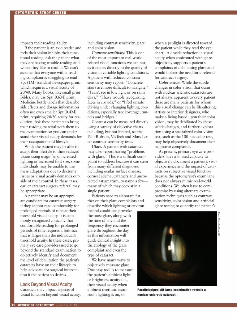
REVIEW OF OPTOMETRY JUNE 15, 201974
OPTOMETRIC STUDY CENTER
impacts their reading ability. If the patient is an avid reader and
feels their vision inhibits their func-tional reading, ask the patient what they are having trouble reading and where they like to read it. We can’t assume that everyone with a read-ing complaint is struggling to read 8pt (1M) standard newspaper print, which requires a visual acuity of 20/40. Many books, like small print Bibles, may use 5pt (0.6M) print. Medicine bottle labels that describe side effects and dosage information often use even smaller 3pt (0.4M) print, requiring 20/20 acuity for res-olution. Ask these patients to bring their reading material with them to the examination so you can under-stand their visual acuity demands for their occupation and lifestyle.
While the patient may be able to adapt their lifestyle to their reduced vision using magnifiers, increased lighting or increased font size, some individuals may be unable to use these adaptations due to dexterity issues or visual acuity demands out-side of their control. In these cases, earlier cataract surgery referral may be appropriate.
A patient may be an appropri-ate candidate for cataract surgery if they cannot read comfortably for prolonged periods of time at their threshold visual acuity. It is com-monly recognized clinically that comfortable reading for prolonged periods of time requires a font size that is larger than the individual’s threshold acuity. In these cases, pri-mary eye care providers need to go beyond the standard examination to objectively identify and document the level of debilitation the patient’s cataracts have on their lifestyle to help advocate for surgical interven-tion if the patient so desires.
Look Beyond Visual AcuityCataracts may impact aspects of visual function beyond visual acuity,
including contrast sensitivity, glare and color vision.
Contrast sensitivity. This is one of the most important real-world-related visual functions we can test, as it relates directly to the quality of vision in variable lighting conditions. A patient with reduced contrast sensitivity may report: “Concrete stairs are more difficult to navigate,” “I can’t see in low light or on rainy days,” “I have trouble recognizing faces in crowds,” or “I feel unsafe driving under changing lighting con-ditions, especially tree coverage, tun-nels and bridges.”
Contrast can be measured directly using a variety of objective charts including, but not limited, to: the Pelli-Robson, VisTech and Mars Let-ter contrast sensitivity tests.
Glare. A patient with cataracts may also report having “problems with glare.” This is a difficult com-plaint to address because it can stem from many different diagnoses, including ocular surface disease, corneal edema, cataracts and uncor-rected astigmatism, to name a few—many of which may coexist in a single patient.
Patients need to elaborate fur-ther on their glare complaints and describe which lighting or environ-mental conditions provoke the most glare, along with the time of day and the frequency they encounter glare throughout the day, as this information will guide clinical insight into the etiology of the glare complaint and even the type of cataract.
We have many ways to objectively measure glare. One easy tool is to measure the patient’s ambient light or brightness acuity (i.e., their visual acuity when ambient overhead exam room lighting is on, or
when a penlight is directed toward the patient while they read the eye chart). A drastic reduction in visual acuity when confronted with glare objectively supports a patient’s complaints of debilitating glare and would bolster the need for a referral for cataract surgery.
Color vision. While the subtle changes in color vision that occur with nuclear sclerotic cataracts are not always apparent to every patient, there are many patients for whom this visual change can be life-altering. Artists or interior designers, who make a living based upon their color vision, may be debilitated by these subtle changes, and further explora-tion using a specialized color vision test, such as the 100-hue color test, may help objectively document their subjective complaints.
At present, primary eye care pro-viders have a limited capacity to objectively document a patient’s visu-al experience and the impact of cata-racts on subjective visual function because the optometrist’s exam lane does not always mimic real-world conditions. We often have to com-promise by using alternate exami-nation techniques such as contrast sensitivity, color vision and artificial glare testing to quantify the patient’s
Parallelepiped slit lamp examination reveals a nuclear sclerotic cataract.
Photo: M
arcellus Chow
, OD
072_ro0619_F6_OSC.indd 74072_ro0619_F6_OSC.indd 74 6/5/19 12:24 PM6/5/19 12:24 PM
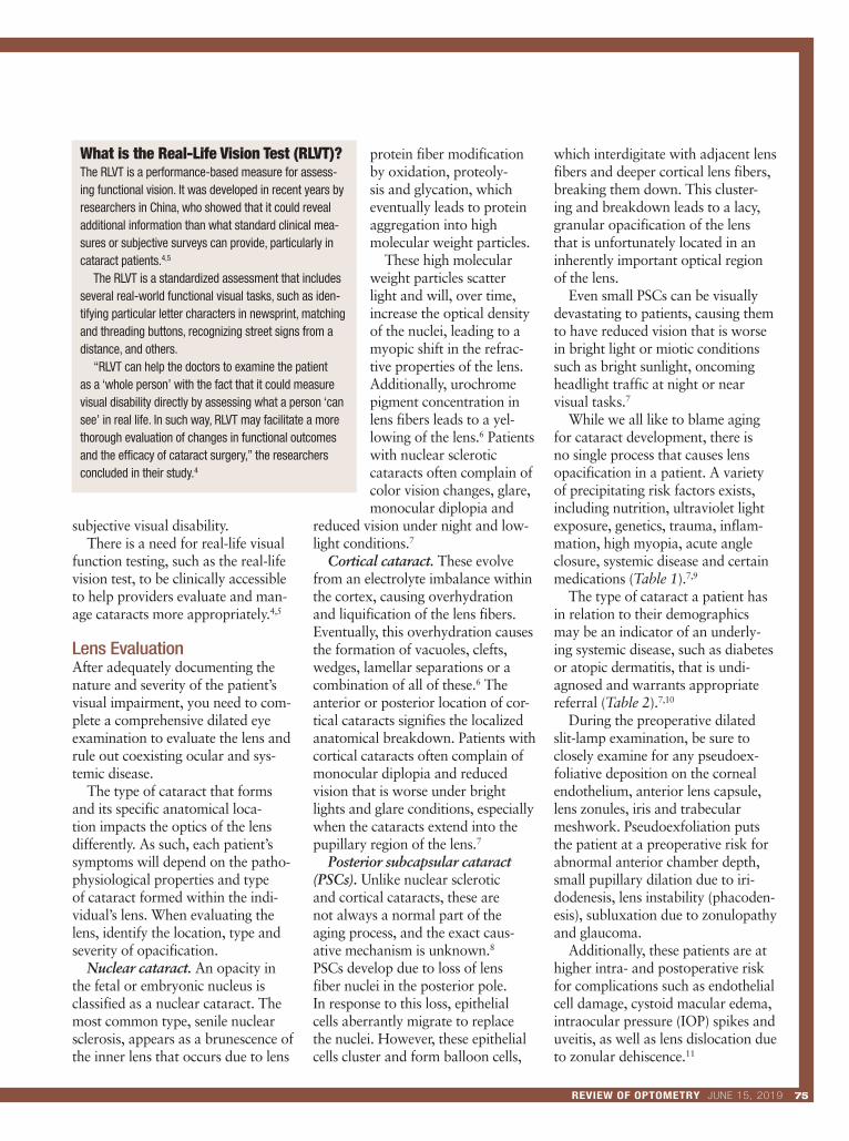
REVIEW OF OPTOMETRY JUNE 15, 2019 75
subjective visual disability. There is a need for real-life visual
function testing, such as the real-life vision test, to be clinically accessible to help providers evaluate and man-age cataracts more appropriately.4,5
Lens EvaluationAfter adequately documenting the nature and severity of the patient’s visual impairment, you need to com-plete a comprehensive dilated eye examination to evaluate the lens and rule out coexisting ocular and sys-temic disease.
The type of cataract that forms and its specific anatomical loca-tion impacts the optics of the lens differently. As such, each patient’s symptoms will depend on the patho-physiological properties and type of cataract formed within the indi-vidual’s lens. When evaluating the lens, identify the location, type and severity of opacification.
Nuclear cataract. An opacity in the fetal or embryonic nucleus is classified as a nuclear cataract. The most common type, senile nuclear sclerosis, appears as a brunescence of the inner lens that occurs due to lens
protein fiber modification by oxidation, proteoly-sis and glycation, which eventually leads to protein aggregation into high molecular weight particles.
These high molecular weight particles scatter light and will, over time, increase the optical density of the nuclei, leading to a myopic shift in the refrac-tive properties of the lens. Additionally, urochrome pigment concentration in lens fibers leads to a yel-lowing of the lens.6 Patients with nuclear sclerotic cataracts often complain of color vision changes, glare, monocular diplopia and
reduced vision under night and low-light conditions.7
Cortical cataract. These evolve from an electrolyte imbalance within the cortex, causing overhydration and liquification of the lens fibers. Eventually, this overhydration causes the formation of vacuoles, clefts, wedges, lamellar separations or a combination of all of these.6 The anterior or posterior location of cor-tical cataracts signifies the localized anatomical breakdown. Patients with cortical cataracts often complain of monocular diplopia and reduced vision that is worse under bright lights and glare conditions, especially when the cataracts extend into the pupillary region of the lens.7
Posterior subcapsular cataract (PSCs). Unlike nuclear sclerotic and cortical cataracts, these are not always a normal part of the aging process, and the exact caus-ative mechanism is unknown.8 PSCs develop due to loss of lens fiber nuclei in the posterior pole. In response to this loss, epithelial cells aberrantly migrate to replace the nuclei. However, these epithelial cells cluster and form balloon cells,
which interdigitate with adjacent lens fibers and deeper cortical lens fibers, breaking them down. This cluster-ing and breakdown leads to a lacy, granular opacification of the lens that is unfortunately located in an inherently important optical region of the lens.
Even small PSCs can be visually devastating to patients, causing them to have reduced vision that is worse in bright light or miotic conditions such as bright sunlight, oncoming headlight traffic at night or near visual tasks.7
While we all like to blame aging for cataract development, there is no single process that causes lens opacification in a patient. A variety of precipitating risk factors exists, including nutrition, ultraviolet light exposure, genetics, trauma, inflam-mation, high myopia, acute angle closure, systemic disease and certain medications (Table 1).7,9
The type of cataract a patient has in relation to their demographics may be an indicator of an underly-ing systemic disease, such as diabetes or atopic dermatitis, that is undi-agnosed and warrants appropriate referral (Table 2).7,10
During the preoperative dilated slit-lamp examination, be sure to closely examine for any pseudoex-foliative deposition on the corneal endothelium, anterior lens capsule, lens zonules, iris and trabecular meshwork. Pseudoexfoliation puts the patient at a preoperative risk for abnormal anterior chamber depth, small pupillary dilation due to iri-dodenesis, lens instability (phacoden-esis), subluxation due to zonulopathy and glaucoma.
Additionally, these patients are at higher intra- and postoperative risk for complications such as endothelial cell damage, cystoid macular edema, intraocular pressure (IOP) spikes and uveitis, as well as lens dislocation due to zonular dehiscence.11
What is the Real-Life Vision Test (RLVT)?The RLVT is a performance-based measure for assess-ing functional vision. It was developed in recent years by researchers in China, who showed that it could reveal additional information than what standard clinical mea-sures or subjective surveys can provide, particularly in cataract patients.4,5
The RLVT is a standardized assessment that includes several real-world functional visual tasks, such as iden-tifying particular letter characters in newsprint, matching and threading buttons, recognizing street signs from a distance, and others.
“RLVT can help the doctors to examine the patient as a ‘whole person’ with the fact that it could measure visual disability directly by assessing what a person ‘can see’ in real life. In such way, RLVT may facilitate a more thorough evaluation of changes in functional outcomes and the efficacy of cataract surgery,” the researchers concluded in their study.4
072_ro0619_F6_OSC.indd 75072_ro0619_F6_OSC.indd 75 6/5/19 12:24 PM6/5/19 12:24 PM

REVIEW OF OPTOMETRY JUNE 15, 201976
OPTOMETRIC STUDY CENTER
Comorbidities and ComplicationsPatients with concomitant cataracts and glaucoma can be challenging to manage. Cataract surgery may lower IOP in some patients, but may cause IOP elevation/fluctuation in other patients intra- and postopera-tively.12 You and your patient need to weigh the possible risks of glau-coma exacerbation with the benefits of improved visual acuity, contrast sensitivity and quality of life post-cataract surgery prior to referral.13
Preoperative corneal disease and anterior segment findings should be identified and proactively managed, as they can impact postop-erative outcomes. Anterior segment findings that may predispose the patient to postoperative endophthalmitis include dacryocystitis, blepha-ritis, chronic conjunctivitis, lagophthalmos, ectropion, entropion and ocular surface disease. Appropriate man-agement of these conditions can reduce infection risk and improve visual outcomes.14
While rare, cataract surgery can cause recurrence of herpes simplex keratitis in patients with preexisting episodes.15 Prophylactic antiviral treat-ment may be indicated pre- and postoperatively to reduce this risk.
Patients with corneal stromal dystrophies or scar-ring may require referral to a corneal specialist who can perform a simultaneous kera-toplasty. In addition, those diagnosed with guttata or Fuchs’ corneal dystrophy may require additional pre-op edu-cation, as cataract surgery can further endothelial cell loss, and guttata may increase the risk of prolonged postopera-tive corneal edema.16
Educating patients preoperatively about their individual corneal and anterior segment disease, and what they can expect after recovery, will provide them with a better subjective postoperative experience.
While cataract surgery is low-risk, complications may still arise during or after surgery, including retinal detachment, exacerbation of glau-coma, cystoid macular edema (CME) and, rarely, infection. Patients who are younger, have high myopia, are male or have a posterior capsular rupture during their surgery are at a higher risk for developing a rheg-matogenous retinal detachment after
cataract surgery.17 The most frequent cause of poor
visual outcome after uneventful cata-ract surgery is postoperative CME. Patients with preexisting hyperten-sion, diabetes, capsule rupture, epiretinal membrane, uveitis, retinal vein occlusion or retinal detachment repair are at higher risk for this post-operative complication.18-20
To appropriately set postopera-tive vision expectations, preoperative potential acuity testing is recom-mended for any patient with coexist-ing ocular disease. This can be done using potential acuity pinhole (PAP) or pinhole acuity meter (PAM) test-
ing. If a patient with a cataract has PAP or PAM testing worse than 20/20, clinicians should further evaluate for comorbid etiologies such as corneal, retinal or optic nerve disease, in addition to the cataract that may be causing reduced vision.
IOL SelectionOnce you and your patient have decided the benefits of cataract surgery outweigh the risks, you need to discuss the IOL options based on clinical measures, as well as the patient’s subjective needs and preferences.
Lifestyle-specific questions are imperative in guiding the patient in IOL decisions. Patients often feel that they need to purchase the most expensive option to have the best visual outcome. This misconception may lead a patient to choose a premium IOL that is not in their best interest, leading to postop-erative visual complaints.
The key to individualized IOL selection is understanding the patient’s postoperative goals relating to how they want to use their eyes, how glasses-depen-dent they prefer to be, and if they have the ability to adapt to
Table 1. Causes of Cataracts7,8
IdiopathicAgingCongenital/maternal infectionGeneticsHereditary disorders
• Retinitis pigmentosa• Leber’s congenital amaurosis• Gyrate atrophy• Stickler syndrome
TraumaInflammation
• Chronic anterior uveitisAcute congestive angle closure (glaucomaflaken)High myopiaSystemic disease
• Diabetes• Myotonic dystrophy• Atopic dermatitis• Neurofibromatosis type 2• Hypocalcemia• Renal impairment
Metabolic disordersNutritional disorders
• Alcohol• Low levels of antioxidants (vitamins C, C, carotenoids)• Cigarette smoking
Radiation• UV-B
Medications• Corticosteroids• Thiazines (phenothiazine/chlorpromazine)• Statins
072_ro0619_F6_OSC.indd 76072_ro0619_F6_OSC.indd 76 6/5/19 12:25 PM6/5/19 12:25 PM
REVIEW OF OPTOMETRY JUNE 15, 2019 77
some of the inherent imperfections of the lens options. For example, some patients might like the idea of mono-vision at first. But after a contact lens trial, they realize that they are unable to adapt to this modality and decide not to pursue this IOL option.
Just as there are many types of progressive lenses, there are many types of premium, non-monofocal IOLs, including accommodating, multifocal and extended depth-of-focus IOLs—any of which may be appropriate for an individual’s needs. So, for patients who want to be free of glasses and see all distances com-fortably, a premium IOL might be the answer.
However, due to the high optical demands of premium lenses, patients who are highly visually sensitive, have precise visual demands, have moderate to high astigmatism that requires toric correction, have macu-lar pathology or are at risk for zonu-lar dehiscence are poorer candidates for these lenses. Patients with mod-erate to high corneal astigmatism who want to be less dependent on distance glasses would benefit from monofocal toric IOLs.
To adequately guide your patient, collaborate with your local ophthal-mologist to understand the specific IOL options they provide in their office, what the appropriate patient expectations should be for each lens,
the types of surgical equipment they use and the procedures they offer.
Cataract ProceduresPhacoemulsification with a femtosecond laser has different risks than the manual ultrasound procedure. Additionally, while the majority of cataract surgeons offer delayed
sequential bilateral cataract surgery (DSBCS)—where the patient has one eye operated on, followed by the second eye on a later date—some surgeons now offer immediately sequential bilateral cataract surgery (ISBCS), where the patient has both eye operations on the same day.21 ISBCS has the ability to increase sur-gical efficiency, which can increase patient access to surgery by reduc-ing wait time and cost to both the patient and healthcare payer.22
In patients who are adequate candidates for ICBCS (i.e., low risk for postoperative complications and have home/mobility support needed for postoperative care), recent stud-ies show equivalent postoperative visual outcomes and patient satisfac-tion without increased complication risk.23 However, patients at higher risk for postoperative complications, such as high myopes or patients with diabetes or corneal guttata, may risk bilateral complications, in which case they would be a better candidate for DSBCS.
Many barriers to accessing sur-gical treatment exist, including insurance, treatment costs, surgical availability and patient awareness. We need to advocate for our patients to help break down these barriers.
As the number of patients with cataracts continues to rise, and the life expectancy and inherent visual
demands increase, optometrists need to individualize their cataract evalu-ation and management to align with the needs of the patient. A thorough understanding of the patient’s life-style and functional visual needs will help the optometrist guide the patient in deciding when to pursue cataract surgery and which surgical options will enable the patient to visually pursue and enjoy their life. ■
Dr. Theis is a staff optometrist at Kaiser Permanente in San Rafael, Calif.
1. Centers for Disease Control and Prevention. Vision Health Initiative: Common Eye Disorders. September 2015. www.cdc.gov/visionhealth/basics/ced.2. Gimbel HV, Dardzhikova AA. Consequences of waiting for cataract surgery. Curr Opin Ophthalmol. 2011;22(1):28-30. 3. Hodge W, Horsley T, Albiani D, et al. The consequences of waiting for cataract surgery: a systemic review. CMAJ. 2007;176(9):1285-90. 4. Ni W, Li X, Hou Z, et al. Impact of cataract surgery on vision-related life performances: the usefulness of Real-Life Vision Test for cataract surgery outcomes evaluation. Eye. 2015;29(12):1545-54.5. Ni W, Li X, Ao M, et al. Using the real-life vision test to assess the functional vision of age-related cataract patients. Eye. 2012;26(11):1402-11.6. Datiles MB, Kinoshita JH. Pathogenesis of Cataracts. In: Tasman W, Jaeger EA, eds. Duane’s Clinical Ophthalmology, Vol 1. Philadelphia: JB Lippincott; 1991:1-14.7. Kanski JJ. Ch. 12–Lens. In: Clinical Ophthalmology: A Systematic Approach, 6th ed. Edinburgh: Butterworth Heineman Elsevier; 2007.8. Remington LA. Ch. 5–Crystalline Lens. Clinical Anatomy of the Visual System, 2nd ed. St Louis: Elsevier; 2005.9. Hutnik CM, Nichols BD. Cataracts in systemic diseases and syn-dromes. Curr Opin Ophthalmol. 1999:10(1):22-28.10. Bagheri N, Wajda BN. Ch. 13–General Ophthalmic Problems. In: The Wills Eye Manual, 7th ed. Philadelphia: Wolters Kluwer; 2017.11. Fontana L, Coassin M, Lovieno A, et al. Cataract surgery in patients with pseudoexfoliation syndrome: current updates. Clin Ophthalmol. 2017 Jul;11:1377-83. 12. Law SK, Riddle J. Management of cataracts in patients with glau-coma. Int Ophthalmol Clin. 2011;51(3):1-18.13. Xu X, Sun Q. Vision-related quality of life outcomes of cataract sur-gery in advanced glaucoma patients. J Glaucoma. 2016;25(1):e5-11.14. Movahedan A, Djalilian AR. Cataract surgery in the face of ocular surface disease. Curr Opin Ophthalmol. 2012;23(1):68-72.15. Barequet IS, Wasserzug Y. Herpes simplex keratitis after cataract surgery. Cornea. 2007;26(5):615-7.16. Greene JB, Mian SI. Cataract surgery in patients with corneal disease. Curr Opin Ophthalmol. 2013;24(1):9-14.17. Haug SJ, RB Bhisitkul. Risk factors for retinal detachment following cataract surgery. Curr Opin Ophthalmol. 2012;23(1):7-11.18. Yonekawa Y, Kim IK. Pseudophakic cystoid macular edema. Curr Opin Ophthalmol. 2012;23(1):26-32.19. Chu CJ, Johnston RL, Buscombe C. Risk factors and incidence of macular edema after cataract surgery: a database study of 81984 eyes. Ophthalmology. 2016;123(2):316-23.20. Boscia F, Giancipoli E, D’ Amico Ricci G, Pinna A. Management of macular oedema in diabetic patients undergoing cataract surgery. Curr Opin Ophthalmol. 2017;28(1):23-28.21. Popovic M, Campos-Moller X, Schlenker MB, Ahmed IIK. Efficacy and safety of femtosecond laser-assisted cataract surgery compared with manual cataract surgery. Ophthalmology. 2016;123(10):2113-26.22. Singh R, Dohlman TH, Sun G. Immediately sequential bilateral cataract surgery: advantages and disadvantages. Curr Opin Ophthalmol. 2017;28(1):81-86.23. Herrington LJ, Liu L, Alexeff S, et al. Immediate sequential vs. delayed sequential bilateral cataract surgery: Retrospective comparison of postoperative visual outcomes. Ophthalmology. 2017;124(8):1126-35.
Table 2. Cataracts and Systemic Disease7
Systemic Disease Type of CataractMyotonic dystrophy Blue dot cortical opacities
Posterior subcapsularAtopic dermatitis Posterior subcapsularNeurofibromatosis type 2 Posterior subcapsular
Posterior cortical opacitiesHypocalcemia White cortical opacitiesDiabetes Snowflake
Posterior subcapsularCortical wedges
Retinal dystrophies (RP, gyrate atrophy)
Posterior subcapsular
072_ro0619_F6_OSC.indd 77072_ro0619_F6_OSC.indd 77 6/5/19 12:25 PM6/5/19 12:25 PM

REVIEW OF OPTOMETRY JUNE 15, 201978
OPTOMETRIC STUDY CENTER
Y ou can obtain continuing education credit through the Optometric Study Center. Com plete the test form
and return it with the $35 fee to: Jobson Healthcare Information, LLC, Attn.: CE Processing, 395 Hudson Street, 3rd Floor New York, New York 10014. To be eligible, please return the card within three years of publication. You can also access the test form and submit your answers and pay-ment via credit card at Review Education Group online, www.reviewsce.com.
You must achieve a score of 70 or higher to receive credit. Allow four weeks for pro-cessing. For each Optomet ric Study Center course you pass, you earn 2 hours of credit from Pennsyl vania College of Optometry.
Please check with your state licensing board to see if this approval counts toward your CE requirement for relicensure.
1. A cataract is considered clinically signifi-cant when the patient has:a. A significant reduction in visual acuity.b. Grade 3 nuclear sclerosis.c. A noticeable functional impairment in their life.d. Both A and C.
2. At what visual acuity is surgical interven-tion indicated due to possible ineligibility to drive?a. 20/40 or worse.b. 20/30.c. 20/25.d. Any of the above.
3. What visual acuity level is needed to see the information on most medicine bottles?a. 20/80.b. 20/40.c. 20/20.d. 20/30.
4. Which subjective patient complaint is likely due to reduced contrast sensitivity?a. “I have difficulty seeing past oncoming traffic headlights.”b. “I feel unsafe driving under changing light-ing conditions.”c. “I have difficulty reading on my computer.”d. “I need to get close to road signs to resolve them.”
5. Which test objectively measures contrast sensitivity?a. Pelli-Robson.b. VisTech.c. Mars.d. All of the above.
6. Nuclear sclerotic cataracts are caused bya. Electrolyte imbalance induced overhydra-tion of lens fibers.b. Lens protein fiber modification by oxida-tion, proteolysis and glycation.c. Atopic dermatitis.d. Loss of lens fiber nuclei in the posterior pole.
7. Which lens finding is consistent with corti-cal cataracts?a. Brunescence of the inner lens.b. Lacy, granular opacification in the posterior pole.c. Water vacuoles.d. White, lacy opacification on the anterior capsule.
8. Each of the following is a precipitating risk factor for cataract development, except:a. High hyperopia.b. High myopia.c. Acute angle closure.d. Corticosteroids.
9. Posterior subcapsular cataracts may be found in patients with the following systemic diseases, except:a. Myotonic dystrophy.b. Hypocalcemia.c. Neurofibromatosis type 2.d. Diabetes.
10. Pseudoexfoliation puts the patient at a preoperative risk ofa. Abnormal anterior chamber depth.b. Cystoid macular edema.c. Uveitis.d. Endothelial cell damage.
11. Cataract surgery in glaucoma patients may causea. Increased IOP.b. Decreased IOP.c. No effect on IOP.d. All of the above.
12. Which condition can cause or predispose a patient to endophthalmitis?a. History of herpes simplex keratitis.b. Blepharitis post-cataract surgery.c. Blepharitis pre-cataract surgery.d. Conjunctivitis post-cataract surgery.
13. Which condition increases the risk of postoperative corneal edema?a. Blepharitis.b. High myopia.c. Corneal guttata.d. Punctate epithelial keratitis.
14. Which postoperative complication is the most frequent cause of poor visual outcome after uneventful cataract surgery?a. Cystoid macular edema.b. Retinal detachment.c. Exacerbation of glaucoma.d. Infection.
15. If a patient has a preoperative pinhole acuity potential worse than 20/20, the optometrist must rule out the patient does not have concomitanta. Corneal disease.b. Retinal disease.c. Optic nerve disease.d. All of the above.
16. IOL discussions should be based ona. Objective clinical measures.b. Patients’ subjective needs and preferences.c. Availability of surgical procedures.d. All of the above.
17. A patient with preexisting macular dis-ease and high visual demands would be a good candidate for which lens option? a. Accommodating IOL.b. Monofocal IOL.c. Multifocal IOL.d. Extended depth-of-focus IOL.
18. The following statements about imme-diately sequential bilateral cataract surgery (ISBCS) and delayed sequential bilateral cataract surgery (DSBCS) are true, except:a. ISBCS is a great option for every patient.b. ISBCS has the potential to reduce cost to patients and healthcare payers.c. ISBCS has the potential to reduce patients’ wait time for surgery.d. For appropriate candidates, ISBCS has equivalent postoperative visual outcomes to DSBCS without increased complication risk.
19. The increased risk of lens subluxation due to zonular dehiscence in patients with pseudoexfoliation makes them a poor candi-date for all of the following IOLs, except: a. Accommodating IOLs.b. Multifocal IOLs.c. Monofocal IOLs.d. Toric IOLs.
20. In the United States, the following are barriers to access for surgical treatment of cataracts, except: a. Treatment costs.b. Insurance.c. Lack of treatment awareness.d. Visual acuity better than 20/40.
OSC QUIZ
OPTOMETRIC STUDY CENTER
072_ro0619_F6_OSC.indd 78072_ro0619_F6_OSC.indd 78 6/5/19 12:25 PM6/5/19 12:25 PM

REVIEW OF OPTOMETRY JUNE 15, 2019 79
Please retain a copy for your records. Please print clearly.
First Name
Last Name
The following is your: Home Address Business Address
Business Name
Address
City State
ZIP
Telephone # - -
Fax # - -
By submitting this answer sheet, I certify that I have read the lesson in its entirety and completed the self-assessment exam personally based on the material presented. I have not obtained the answers to this exam by any fraudulent or improper means.
Signature Date
Lesson 118378 RO-OSC-0619
Answers to CE exam: 1. A B C D
2. A B C D
3. A B C D
4. A B C D
5. A B C D
6. A B C D
7. A B C D
8. A B C D
9. A B C D
10. A B C D
11. A B C D
12. A B C D
13. A B C D
14. A B C D
15. A B C D
16. A B C D
17. A B C D
18. A B C D
19. A B C D
20. A B C D
Examination Answer Sheet The Real-world Cataract EvaluationValid for credit through June 15, 2022
Online: This exam can be taken online at www.reviewsce.com. Upon passing the exam, you can view your results immediately and download a real-time CE certifi-cate. You can also view your test history at any time from the website.
Directions: Select one answer for each question in the exam and completely darken the appropriate circle. A minimum score of 70% is required to earn credit.
Mail to: Jobson Healthcare Information, LLC, Attn.: CE Processing, 395 Hudson Street, 3rd Floor New York, New York 10014
Payment: Remit $35 with this exam. Make check payable to Jobson Healthcare Information, LLC.
Credit: This course is COPE approved for 2 hours of CE credit. Course ID is 62685-AS .
Jointly provided by Postgraduate Institute for Medicine and Review Education Group.
Salus University has sponsored the review and approval of this activity.
Processing: There is a four-week processing time for this exam.
Post-activity evaluation questions:
Rate how well the activity supported your achievement of these learning objectives: 1=Poor, 2=Fair, 3=Neutral, 4=Good, 5=Excellent
21. Individualize the cataract evaluation based on the patient’s particular needs and routines.
22. Recognize the real-life vision test (or similar objective real-life visual function testing) as a potential adjunctive means of cataract evaluation.
23. Identify underlying diseases that increase cataract risk and affect post-op cataract outcomes.
24. Discuss IOL choices based not only on clinical measures, but also on patient’s subjective needs and preferences as well as real-life vision testing results.
25. Describe the benefits and risks of simultaneous bilateral cataract surgery.
26. Based upon your participation in this activity, do you intend to change your practice behavior?(choose only one of the following options) A I do plan to implement changes in my practice based on the information presented. B My current practice has been reinforced by the information presented. C I need more information before I will change my practice.
27. Thinking about how your participation in this activity will influence your patient care, how many of your patients are likely to benefit? (please use a number):
32. The content was evidence-based.
33. The content was balanced and free of bias.
34. The presentation was clear and effective.
Rate the quality of the material provided: 1=Strongly disagree, 2=Somewhat disagree, 3=Neutral, 4=Somewhat agree, 5=Strongly agree
1 2 3 4 5
1 2 3 4 5
1 2 3 4 5
1 2 3 4 5
1 2 3 4 5
1 2 3 4 5
1 2 3 4 5
28. If you plan to change your practice behavior, what type of changes do you plan to implement? (check all that apply)
a Apply latest guidelines b Change in pharmaceutical therapy c Choice of treatment/management approach d Change in current practice for referral e Change in non-pharmaceutical therapy f Change in differential diagnosis g Change in diagnostic testing h Other, please specify: _________________________________
______________________________________________________________________________________
29. How confident are you that you will be able to make your intended changes?
a Very confident b Somewhat confident c Unsure d Not confident
30. Which of the following do you anticipate will be the primary barrier to implementing these changes? a Formulary restrictions b Time constraints c System constraints d Insurance/financial issues e Lack of interprofessional team support f Treatment related adverse events g Patient adherence/compliance h Other, please specify:
31. Additional comments on this course:___________________________________________________________________________________________________________________________________________________________________________________________________
1 2 3 4 5
072_ro0619_F6_OSC.indd 79072_ro0619_F6_OSC.indd 79 6/5/19 12:25 PM6/5/19 12:25 PM
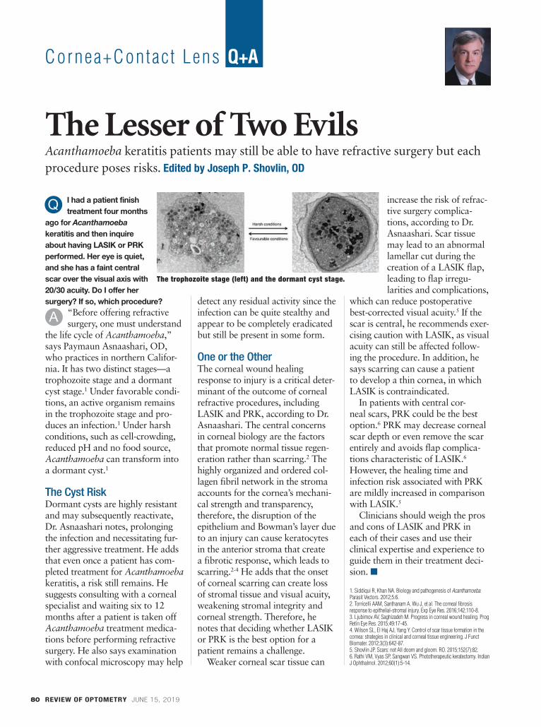
REVIEW OF OPTOMETRY JUNE 15, 201980
Cornea+Contac t Lens Q+A
I had a patient finish treatment four months
ago for Acanthamoeba keratitis and then inquire about having LASIK or PRK performed. Her eye is quiet, and she has a faint central scar over the visual axis with 20/30 acuity. Do I offer her surgery? If so, which procedure?
“Before offering refractive surgery, one must understand
the life cycle of Acanthamoeba,” says Paymaun Asnaashari, OD, who practices in northern Califor-nia. It has two distinct stages—a trophozoite stage and a dormant cyst stage.1 Under favorable condi-tions, an active organism remains in the trophozoite stage and pro-duces an infection.1 Under harsh conditions, such as cell-crowding, reduced pH and no food source, Acanthamoeba can transform into a dormant cyst.1
The Cyst RiskDormant cysts are highly resistant and may subsequently reactivate, Dr. Asnaashari notes, prolonging the infection and necessitating fur-ther aggressive treatment. He adds that even once a patient has com-pleted treatment for Acanthamoeba keratitis, a risk still remains. He suggests consulting with a corneal specialist and waiting six to 12 months after a patient is taken off Acanthamoeba treatment medica-tions before performing refractive surgery. He also says examination with confocal microscopy may help
detect any residual activity since the infection can be quite stealthy and appear to be completely eradicated but still be present in some form.
One or the OtherThe corneal wound healing response to injury is a critical deter-minant of the outcome of corneal refractive procedures, including LASIK and PRK, according to Dr. Asnaashari. The central concerns in corneal biology are the factors that promote normal tissue regen-eration rather than scarring.2 The highly organized and ordered col-lagen fibril network in the stroma accounts for the cornea’s mechani-cal strength and transparency, therefore, the disruption of the epithelium and Bowman’s layer due to an injury can cause keratocytes in the anterior stroma that create a fibrotic response, which leads to scarring.2-4 He adds that the onset of corneal scarring can create loss of stromal tissue and visual acuity, weakening stromal integrity and corneal strength. Therefore, he notes that deciding whether LASIK or PRK is the best option for a patient remains a challenge.
Weaker corneal scar tissue can
increase the risk of refrac-tive surgery complica-tions, according to Dr. Asnaashari. Scar tissue may lead to an abnormal lamellar cut during the creation of a LASIK flap, leading to flap irregu-larities and complications,
which can reduce postoperative best-corrected visual acuity.5 If the scar is central, he recommends exer-cising caution with LASIK, as visual acuity can still be affected follow-ing the procedure. In addition, he says scarring can cause a patient to develop a thin cornea, in which LASIK is contraindicated.
In patients with central cor-neal scars, PRK could be the best option.6 PRK may decrease corneal scar depth or even remove the scar entirely and avoids flap complica-tions characteristic of LASIK.6 However, the healing time and infection risk associated with PRK are mildly increased in comparison with LASIK.5
Clinicians should weigh the pros and cons of LASIK and PRK in each of their cases and use their clinical expertise and experience to guide them in their treatment deci-sion. ■
1. Siddiqui R, Khan NA. Biology and pathogenesis of Acanthamoeba. Parasit Vectors. 2012;5:6.2. Torricelli AAM, Santhanam A, Wu J, et al. The corneal fibrosis response to epithelial-stromal injury. Exp Eye Res. 2016;142:110-8.3. Ljubimov AV, Saghizadeh M. Progress in corneal wound healing. Prog Retin Eye Res. 2015;49:17-45.4. Wilson SL, El Haj AJ, Yang Y. Control of scar tissue formation in the cornea: strategies in clinical and corneal tissue engineering. J Funct Biomater. 2012;3(3):642-87.5. Shovlin JP. Scars: not All doom and gloom. RO. 2015;152(7):82.6. Rathi VM, Vyas SP, Sangwan VS. Phototherapeutic keratectomy. Indian J Ophthalmol. 2012;60(1):5-14.
Acanthamoeba keratitis patients may still be able to have refractive surgery but each procedure poses risks. Edited by Joseph P. Shovlin, OD
The Lesser of Two Evils
Q
A
The trophozoite stage (left) and the dormant cyst stage.
080_ro0619_CLQA.indd 80080_ro0619_CLQA.indd 80 6/5/19 2:48 PM6/5/19 2:48 PM

Download a QR scanner app. Launch app and hold your mobile device over the code to view https://www.reviewofoptometry.com/publications/archive
www.reviewofoptometry.com/publications/archive
The future is in your
hands. One tap, many
possibilities.
Experience the digital edition on your handheld device. Use your smart device to scan the code below or visit:
Innovative products to enhance your practice
Product GuideOPHTHALMICOPHTHALMIC
2018_opg_ad_fullpge.indd 12018_opg_ad_fullpge.indd 1 6/6/19 11:28 AM6/6/19 11:28 AM

Visit ECPs4Cures.org to make a donation
FightBlindness.org
WE’RE SEEING AMAZING RESULTS. AND SO ARE THEY.Foundation Fighting Blindness is shining a light in the darkness of Inherited Retinal Degenerations. We are the world’s leading organization searching for treatments and cures, and with many treatments already found, today’s innovations are illuminating a future of possibilities.
Patients with Inherited Retinal Degenerations are urged to partner with us to accelerate the discovery of treatment and cures.
We have robust disease information, a national network of local chapters and support groups, local educational events, and our My Retina Tracker patient registry helps to keep your patients connected with clinical and research advancements.
RP1218_House Fight Stars.indd 1 11/13/18 11:02 AM

Ocula r Sur face Review
REVIEW OF OPTOMETRY JUNE 15, 2019 83
Dry Eye Therapy: Getting NosyA review of the science behind nasal stimulation can help you understand the new tech targeting the trigeminal nerve. By Paul M. Karpecki, OD
Dry eye therapy has taken several unex-pected turns over
the years, with a new class of drugs and several new formulations of a tried-and-true therapy. Now, it seems neuroscience is taking a turn at the wheel. Scientists have discovered that the trigemi-nal nerve plays a critical role in ocular surface health and symptomatology. Although a misalignment of the eyes can fatigue the trigeminal nerve and cause dry eye symptoms, stimulating the trigeminal nerve can also promote tear production.
Research shows the para-sympathetic nervous system (PNS), via the trigeminal parasym-pathetic pathway, controls tear film homeostasis through innervation of the lacrimal functional unit (LFU), which includes the cornea, conjunc-tiva and the structures that secrete tear film components, such as the lacrimal glands, meibomian glands and goblet cells.1-3 The PNS can most easily be accessed through the nose (Figure 1). In fact, 34% of basal tear production is due to sensory stimula-tion from inhaled air through the nasal passage.4
Investigators have developed or are developing nasal neurostimula-tion devices and pharmaceuticals to take advantage of this nasal LFU access point—a novel approach to producing a complete tear film in patients with dry eye disease.
More Than WaterWhen nasal neurostimulation hit the research scene, investigators weren’t sure if the tears generated would be quality tears. Many assumed neuro-stimulation would generate reflex tears, which do not have the same composition as basal tears. Others wondered if the technique would only stimulate the lacrimal glands or increase tear volume by producing aqueous without the other compo-nents needed for a healthy tear film.
Increasingly, however, the evidence suggests that nasal neurostimula-tion affects all parts of the LFU and increases natural, basal tearing. Stud-ies show goblet cell degranulation, which could lead to the release of mucins, and intranasal tear neuro-stimulation resulted in the release of
mucin to the ocular surface.5,6 Neurostimulation leads to
changes in meibomian gland activity and lipid layer thick-ness, although a fairly long duration of use (eight min-utes) was required to achieve this.7 Others found that tears collected post-neurostimula-tion had lipid composition equivalent to that of subjects’ basal tears.8 These early stud-ies have been encouraging, as have the clinical results in patients treated with various forms of neurostimulation.
Nasal NeurostimulationThe first device on the market was TrueTear (Allergan), an intranasal tear stimulator. It
activates the nasolacrimal reflex by delivering small electrical currents to sensory neurons of the nasal cavity, temporarily increasing tear produc-tion. The device is designed for home use for at least two minutes and up to 30 minutes per day. Studies show that regular use for six months results in a statistically significant increase in tear production and reduction in dry eye symptoms, as well as an increase in tear meniscus height.9,10
One challenge with TrueTear is the logistics of dispensing the device to patients and training them on its use. To find the “sweet spot” for deliver-ing the electrical pulses, the tips need to be inserted as far as possible into the nose and then tilted so they con-tact the nerves just under the bridge of the nose (Figure 2).11
Fig. 1. Efferent parasympathetic nerves innervate the LFU. The 0.1% dose of the OC-01 nAChR agonist nasal spray that targets these nerves led to a statistically significant improvement in dry eye symptoms.
Image: O
yster Point P
harma
083_ro0619_OSR.indd 83083_ro0619_OSR.indd 83 6/5/19 2:48 PM6/5/19 2:48 PM
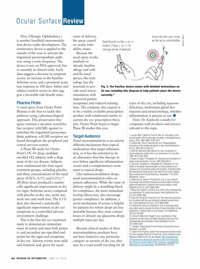
REVIEW OF OPTOMETRY JUNE 15, 201984
Ocula r Sur face Review
iTear (Olympic Ophthalmics ) is another handheld neuromodula-tion device under development. The noninvasive device is applied to the outside of the nose to activate the trigeminal parasympathetic path-way using a sonic frequency. The device is not yet FDA approved, but is currently in clinical trials. Early data suggest a decrease in symptom scores, an increase in the baseline Schirmer score and a persistent acute tear response at 180 days. Safety and subject comfort scores to date sug-gest a favorable risk-benefit ratio.
Pharma FirstsA nasal spray from Oyster Point Pharma is the first to tackle this pathway using a pharmacological approach. This preservative-free spray contains a nicotinic acetylcho-line receptor (nAChR) agonist to stimulate the trigeminal parasympa-thetic pathway. nAChR receptors are found throughout the peripheral and central nervous system.
A Phase IIb study for Oyster Point’s OC-01 drug candidate enrolled 182 subjects with a diag-nosis of dry eye disease. Subjects were randomized into four equal treatment groups, including placebo and three concentrations of the nasal spray (0.02%, 0.1% and 0.2%).12 All three doses produced a statisti-cally significant improvement in dry eye signs (Schirmer score) compared with placebo at day one, week one, week two and week four. The 0.1% dose also showed a statistically significant improvement in dry eye symptoms in a controlled adverse environment challenge.
This is the first dry eye treatment study to demonstrate immediate onset of action and meet both prima-ry and secondary pre-specified end-points for the signs and symptoms of dry eye. Adverse events were mild and transient and, given the nasal
route of delivery, the spray caused no ocular toler-ability issues.
Because the nasal spray works similarly to already familiar allergy and cold and flu nasal sprays, this tech-nology has the potential to pro-vide nasal neuro-stimulation with improved patient acceptance and reduced training time. The company also expects it to be a widely available prescription product with reimbursed similar to current dry eye prescription thera-pies. Oyster Point hopes to begin Phase III studies this year.
Target AudienceNasal neurostimulation is an entirely different mechanism than topical medications that target inflamma-tion, so it has the potential to be an alternative first-line therapy in eyes before significant inflammation occurs and a complementary treat-ment to topical drops.
Like immunomodulator drops, nasal neurostimulation relies on patient adherence. While the route of delivery might be a stumbling block for compliance, the more immediate tearing effects may also encourage greater compliance. In addition, a novel mechanism of action is helpful for patients for whom drops are less desirable because they wear contact lenses or already use glaucoma drops multiple times per day.
Because clinical studies of these neurostimulation products have not been limited to any particular category or severity of dry eye, they may be a road worth traveling for all
types of dry eye, including aqueous-deficiency, meibomian gland dys-function and mixed etiology, whether inflammation is present or not. ■
Note: Dr. Karpecki consults for companies with products and services relevant to this topic.
1. van der Werf F, Baljet B, Prins M, Otto JA. Innervation of the lacrimal gland in the cynomolgous monkey: a retrograde tracing study. J Anat. 1996;188(Pt 3):591-601.2. LeDoux MS, Zhou Q, Murphy RB, et al. Parasympathetic innervation of the meibomian glands in rats. Invest Ophthalmol Vis Sci. 2001;42(11):2434-41.3. Dartt DA, McCarthy DM, Mercer HJ, et al. Localization of nerves adjacent to goblet cells in rat conjunctiva. Curr Eye Res. 1995;14(11):993-1000.4. Gupta A, Heigle T, Pflugfelder SC. Nasolacrimal stimulation of aqueous tear production. Cornea. 1997;16(6):645-8. 5. Gumus K, Schuetzle KL, Pflugfelder SC, Ackerman, M. The effects of intranasal neurostimulation on tear production and clearance and conjunctival goblet cell secretion. Presented at the 8th International Conference on the Tear Film & Ocular Surface: Basic Science and Clinical Relevance, September 7-10, 2016; Montpellier, France.6. Dieckmann G, Jamali A, Pondelis N, et al. In vivo confocal microscopy demonstrates intranasal neurostimulation-induced goblet cell alterations in patients with dry eye disease. Invest Ophthalmol Vis Sci. 2017;58(8):2694.7. Watson M, Angjeli E, Orrick B, et al. Effect of the intranasal tear neurostimulator on meibomian glands. Invest Ophthalmol Vis Sci. 2017;58(8):4387.8. Green KB, Kamat M, Franke M, et al. Tear total lipid concentra-tion in patients with dry eye following intranasal neurostimula-tion. Invest Ophthalmol Vis Sci. 2017;58(8):2693.9. Friedman NJ, Butron K, Robledo N, et al. A nonrandomized, open-label study to evaluate the effect of nasal stimulation on tear production in subjects with dry eye disease. Clin Ophthalmol. 2016;10:795-804. 10. Orrick B, Watson M, Angjeli E, et al. Quantitation of tear production by tear meniscus height following acute use of the intranasal tear neurostimulator. Invest Ophthalmol Vis Sci. 2017;58(8):2692.11. Allergan. Patient guide for the truetear intranasal tear neuro-stimulator. July 2018.12. Oyster Point Pharma. Oyster Point announces positive results from two separate Phase 2b clinical trials of the company’s inves-tigational treatments for dry eye disease. https://oysterpointrx.com/oyster-point-announces-positive-results-from-two-sepa-rate-phase-2b-clinical-trials-of-the-companys-investigational-treatments-for-dry-eye-disease. Accessed April 25 2019.
Fig. 2. The TrueTear device comes with detailed instructions on its use, including this diagram to help patients place the device correctly.11
Image: A
llergan
083_ro0619_OSR.indd 84083_ro0619_OSR.indd 84 6/5/19 2:49 PM6/5/19 2:49 PM

· Scientif ical ly Formulated ·
· Cl inical ly Tested · Al l Natural ·
· Gluten Free · Cruelty Free ·
· 100% Proven Results ·
Visit www.macuhealth.com for more informationCall 1-866-704-0845 Press 1 for Customer Service Or email [email protected]
Ask your Eye Care Professional about our Triple Carotenoid Formulaclinically proven to rebuild macular pigment, improve contrast sensitivity,
and optimize visual performance.
®
1
Visit us in St. Louis at the AOA,Booth # 605
RO0619_MacuHealth.indd 1 5/21/19 10:16 AM
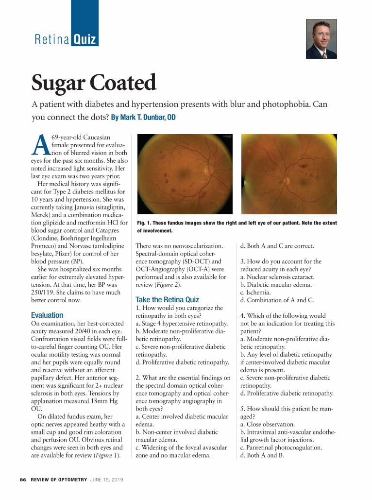
Re t ina Quiz
A 69-year-old Caucasian female presented for evalua-tion of blurred vision in both
eyes for the past six months. She also noted increased light sensitivity. Her last eye exam was two years prior.
Her medical history was signifi-cant for Type 2 diabetes mellitus for 10 years and hypertension. She was currently taking Januvia (sitagliptin, Merck) and a combination medica-tion glipizide and metformin HCl for blood sugar control and Catapres (Clondine, Boehringer Ingelheim Promeco) and Norvasc (amlodipine besylate, Pfizer) for control of her blood pressure (BP).
She was hospitalized six months earlier for extremely elevated hyper-tension. At that time, her BP was 250/119. She claims to have much better control now.
EvaluationOn examination, her best-corrected acuity measured 20/40 in each eye. Confrontation visual fields were full-to-careful finger counting OU. Her ocular motility testing was normal and her pupils were equally round and reactive without an afferent papillary defect. Her anterior seg-ment was significant for 2+ nuclear sclerosis in both eyes. Tensions by applanation measured 18mm Hg OU.
On dilated fundus exam, her optic nerves appeared heathy with a small cup and good rim coloration and perfusion OU. Obvious retinal changes were seen in both eyes and are available for review (Figure 1).
There was no neovascularization. Spectral-domain optical coher-ence tomography (SD-OCT) and OCT-Angiography (OCT-A) were performed and is also available for review (Figure 2).
Take the Retina Quiz1. How would you categorize the retinopathy in both eyes?a. Stage 4 hypertensive retinopathy.b. Moderate non-proliferative dia-betic retinopathy.c. Severe non-proliferative diabetic retinopathy. d. Proliferative diabetic retinopathy.
2. What are the essential findings on the spectral domain optical coher-ence tomography and optical coher-ence tomography angiography in both eyes?a. Center involved diabetic macular edema.b. Non-center involved diabetic macular edema.c. Widening of the foveal avascular zone and no macular edema.
d. Both A and C are correct.
3. How do you account for the reduced acuity in each eye?a. Nuclear sclerosis cataract.b. Diabetic macular edema.c. Ischemia.d. Combination of A and C.
4. Which of the following would not be an indication for treating this patient?a. Moderate non-proliferative dia-betic retinopathy.b. Any level of diabetic retinopathy if center-involved diabetic macular edema is present.c. Severe non-proliferative diabetic retinopathy.d. Proliferative diabetic retinopathy.
5. How should this patient be man-aged?a. Close observation.b. Intravitreal anti-vascular endothe-lial growth factor injections.c. Panretinal photocoagulation.d. Both A and B.
Sugar Coated
Fig. 1. These fundus images show the right and left eye of our patient. Note the extent of involvement.
A patient with diabetes and hypertension presents with blur and photophobia. Can
you connect the dots? By Mark T. Dunbar, OD
REVIEW OF OPTOMETRY JUNE 15, 201986
086_ro0619_RQ.indd 86086_ro0619_RQ.indd 86 6/5/19 2:51 PM6/5/19 2:51 PM
See the condition.
Treat the condition.
Monitor the recovery.
Make A Difference by Using this Versatile System:
Anterior-segment imaging, Retinal imaging, and Infrared imaging all from an EyeResTM slit lamp and EyeResTM digital imaging system.
Save time, money, and space while enhancing the patient’s appreciation for your expertise.
Call 800.769.4933 today or email Wes Harris at [email protected]
www.TelScreen.com
Photos courtesy of Mile Brujic, O.D.
TTTTTTTTTTTTTTTTTTTTTeeeeeeeeeeeeeeeeeeeeelllllllllllllllllllllSSSSSSSSSSSSSSSSSSSSScccccccccccccccccccccrrrrrrrrrrrrrrrrrrrrreeeeeeeeeeeeeeeeeeeeeeeeeeeeeeeeeeeeeeeeeennnnnnnnnnnnnnnnnnnnnTTTTTTTTTTTTTTTTTTTTTTTTTTTTTTTTTTTTTeeeeeeeeeeeeeeeeeeeeeeeeeeeeeeeeeeeeeeelllllllllllllllllllllllllllllllllllSSSSSSSSSSSSSSSSSSSSSSSSSSSSSSSSSSSSSSScccccccccccccccccccccccccccccccccccccccrrrrrrrrrrrrrrrrrrrrrrrrrrrrrrrrrrrreeeeeeeeeeeeeeeeeeeeeeeeeeeeeeeeeeeeeeeeeeeeeeeeeeeeeeeeeeeeeeeeeeeeeeeeeeeeeennnnnnnnnnnnnnnnnnnnnnnnnnnnnnnnnnnnnnn
Digital Imaging SystemsDi it l I i S t
TM
RO0519_Telscreen.indd 1 4/23/19 10:49 AM

Re t ina Quiz
REVIEW OF OPTOMETRY JUNE 15, 201988
DiagnosisSignificant retinal hemorrhages are noticeable in all four quadrants in both our patient’s eyes, as are scat-tered cotton wool spots. On careful examination, no neovascularization can be seen in either eye. Based on the early treatment diabetic reti-nopathy study (ETDRS) classifica-tion, these findings meet the criteria for severe non-proliferative diabetic retinopathy (NPDR).
The diagnosis of severe NPDR is based on the 4:2:1 rule of the ETDRS classification:
4: Significant retinal hemor-rhages in all 4 quadrants,
2: Venous beading in 2 or more quadrants
1: Intraretinal microvascular abnormalities in at least 1 quad-rant.
Any one of these findings meet the criteria for severe NPDR.
DiscussionOnce a patient progresses to severe NPDR, their risk of progressing to proliferative diabetic retinopathy (PDR) in one year increases more than 50%, compared with mild or moderate NPDR where the risk of PDR in one year is 5% and 12% respectively.1 Because of this increased risk for progressing to PDR, patients with severe NPDR should be seen every three to four months compared with mild or moderate NPDR in which the fol-low up is one year.
The indications for treatment of DR has evolved in recent years. Traditionally, treatment was recom-mend for patients with clinically significant macular edema (CSME) or if there is PDR. In the era of OCT and treating DME with intravitreal anti-vascular endothe-lial growth factor (VEGF) injec-tions, the treatment for DME has evolved. Instead of determining if
macular edema is clinical signifi-cant, now retinal specialists want to know if the DME is center involved (CI-DME) or non-center involved based on OCT imaging. CI-DME is generally treated with an anti-VEGF injection whereas non CI-DME may be observed. The anti-VEGF drugs have sup-planted laser for the treatment of DME and is now considered the standard for the treatment of DME.
The treatment of choice for PDR is less certain. The DRCR.net established the efficacy of ranibizumb in the Protocol S study, which compared traditional panret-inal photocoagulation (PRP) with intravitreal ranibizumab.2 Both drugs were efficacious however, the patients with ranibizumab had slightly better outcomes. Specifi-cally, the ranibizumab patients had better visual field preservation and
were less likely to need pars plana vitrectomy over a five-year period. Visual acuity outcomes were simi-lar in both groups. Despite these outcomes, most retinal specialists still use PRP as a first line therapy or in combination with the anti-VEGF medications. The preference of PRP may be due to the burden of treatment that is required with the anti-VEGF drugs, as well as cost.
The ResearchWhat about treating patients ear-lier, before they go on to develop PDR? This was the question posed in a post hoc analysis of 756 patients that were part of the RIDE and RISE clinical trials.3
The purpose of their retro-spective study was to examine DR outcomes after two years in patients who at baseline had severe NPDR (and DME). In this study
Fig. 2. These OCT-A images represent 6x6mm of the right and left eyes. Note the changes within each macula.
086_ro0619_RQ.indd 88086_ro0619_RQ.indd 88 6/5/19 2:52 PM6/5/19 2:52 PM

diabetic retinopathy patients were categorized based on the Diabetic Retinopathy Severity Scale (DRSS), which ranges from 10 to 75. Patients with moderately severe to severe NPDR were categorized as levels 47 to 53. What the authors discovered was that 78% of the eyes with moderately severe and severe NPDR treated with ranibi-zumab saw a ≥2-step regression in the severity of their DR compared with only 12% that were treated in the sham group.3
The author conducted a similar study looking at Aflibercept in patients with level 47 to 53 reti-nopathy on the DRSS. However, this was a prospective study of 402 patients that were randomized to receive a sham injection vs. afliber-cept given either every eight weeks or every 16 weeks (after three initial monthly doses). Their out-
comes were quite similar to what was seen with ranibizumab. At one year, up to 80% of patients treated with aflibercept had a >2-step regression in the severity of their DR compared with only 15% in the sham group.4
The results of both of these stud-ies suggest that patients with severe NPDR would benefit from anti-VEGF therapy instead of waiting until patients develop PDR. What’s more, with earlier treatment we can actually see a significant regression in diabetic retinopathy. Unfor-tunately most retinal specialists have been very slow to adopt this changing paradigm, again, likely due to the burden of patients need-ing multiple treatments as well as the costs and risks associated with anti-VEGF treatments. It’s also important to realize that most all of these patients have presumably
normal vision given they don’t have any DME.
Getting back to our patient, we did refer her to a retinal specialist for consideration of treatment. We felt her vision was reduced due to a combination of cataracts and some ischemia in the fovea. She did not have DME but on OCT-A we could see some widening and irregularity of the foveal avascular zone in both eyes, more so in the left eye due to ischemia. ■
1. Chan S, Bubela T, Dimopoulos I, et al. Choroideremia research: report and perspectives on the second international scientific symposium for choroideremia. Ophthalmic Genet. 2016 Sep;37(3):267-75.2. Freund P, Sergeev Y, MacDonald I. Analysis of a large cho-roideremia dataset does not suggest a preference for inclusion of certain genotypes in future trials of gene therapy. Molecular Genetics & Genomic Medicine. 2016;4(3):344-58.3. Imani S, Ijaz I, Shasaltaneh M, et al. Molecular genetics characterization and homology modeling of the CHM gene mutation: a study on its association with chorioderemia. Muta-tion Research. 2018;775(2):39-50.4. MacDonald I, Hume S, Chan S, et al. Chorioderemia. GeneReviews. February 26, 2015. www.ncbi.nlm.nih.gov/books/NBK1337/. Accessed June 6, 2018.
“I want to practice my way.”
Dream it. Do it.
At IDOC, we are committed to your success as an independent optometrist. You will get the personalized attention and customized business support your practice needs—plus advanced performance analytics, peer networking and exclusive vendor discounts.
Call 203-853-3333 or visit IDOC.net to join now. Attending Optometry’s Meeting? Visit IDOC at Booth #812.
086_ro0619_RQ.indd 89086_ro0619_RQ.indd 89 6/5/19 2:51 PM6/5/19 2:51 PM

.
GET SOCIAL WITH US! #VISIONEXPO
SAVE THE DATE
VisionExpoWest.com
EDUCATION SEPTEMBER 18-21EXHIBIT HALL SEPTEMBER 19-21
RO0619_House VEW.indd 1 5/30/19 1:52 PM

REVIEW OF OPTOMETRY JUNE 15, 2019 91
Surg ica l Minute
Regeneration of the corneal epithelium, crucial for ocular health, is maintained by lim-
bal stem cells. Trauma, contact lens overwear or inflammatory issues can cause these cells to struggle, lead-ing to limbal stem cell deficiency (LSCD). The condition can also be congenital in patients with aniridia or autoimmune disorders.
Early LSCD may be controlled with topical treatment to decrease inflammation, including topical ste-roids, ocular cyclosporine or amni-otic membrane grafts or drops. If contact lenses are the cause, discon-tinuing wear or refitting in a daily option or scleral lens can help mini-mize further limbal stem cell dam-age. But once these patients progress to more severe LSCD and their vision becomes threatened by recur-rent, non-healing epithelial defects, corneal scarring and melts, it is time to consider surgical options.
ProceduresMultiple approaches to limbal stem cell transplantation exist. Depending on the surgeon’s preference and the extent of LSCD damage, differ-ent techniques may be better than others. The most common option is keratolimbal allograft (KLAL), which uses allogeneic limbal tissue attached to a corneoscleral carrier from a cadaveric donor. This pro-cedure is common for those with
bilateral LSCD who may not have a related donor, or those with unilat-eral LSCD and the other eye is not a viable donor.1 Ideal candidates have LSCD caused by aniridia, contact lens wear and iatrogenic LSCD with minimal to no conjunctival involve-ment. KLAL can also help to treat mucous membrane pemphigoid and Stevens-Johnson syndrome, if they are controlled and quiet for at least a year prior to surgery.
The surgical technique uses two donor corneoscleral rims that are divided into four crescents. Three of these crescents are then placed around the recipient’s cornea with the donor tissue’s anterior corneal edge overlying the recipient’s lim-bus. As the patient heals, the gaps between the KLAL segments fill in with repopulated healthy cells.
Post-op ManagementAt the time of surgery, a bandage contact lens is placed in the opera-tive eye and maintained until the corneal epithelium is healed. Patients are prescribed topical
lifitegrast or cyclosporine, a topical steroid, a fluoroquinolone and fre-quent use of preservative-free artifi-cial tears. Once the bandage contact lens is removed, the antibiotic is discontinued and the topical steroid tapered. Long-term, the patient continues cyclosporine or lifitegrast twice daily.
Systemic immunosuppression is important for the long-term suc-cess of the transplant. Using the Cincinnati Eye Institute protocol, clinicians start patients on medi-cal treatment prior to surgery with oral prednisone 1mg/kg/day and tapered over one to three months, oral valganciclovir 225mg QD, trimethoprim/sulfamethoxazole (TMP/SMX) every MWF, oral tacrolimus 4mg BID and oral myco-phenolate mofetil (MMF) 1g BID. Valganciclovir and TMP/SMX are stopped at 12 months, tacrolimus is tapered after six months and MMF after 12 months.
Although patients are free of medical treatment at three years, cli-nicians should monitor them closely for possible rejection.2
1. Cheung AY, Holland EJ. Keratolimbal allograft. Curr Opin Ophthalmol. 2017;28(4):377-81.2. Holland EJ, Mogilishetty G, Skeens HM, et al. Systemic immunosuppression in ocular surface stem cell transplantation: results of a 10-year experience. Cornea. 2012;31(6):655-61.
Patients with limbal stem cell deficiency may require surgical intervention to protect against severe adverse events. By Cecelia Koetting, OD
Edited By Derek N. Cunningham, OD, and Walter O. Whitley, OD, MBA
Replenish the Epithelium
To see a video of this procedure, visit www.reviewofoptometry.com, or scan the QR code.
During the procedure, lenticules from a donor cornea are sutured along the limbus and secured with tissue glue.
Other Surgical Options• Conjunctival limbal autograft• Living-related conjunctival limbal
allograft• Combined conjunctival limbal and KLAL• Cultured limbal epithelial transplantation• Simple limbal epithelial transplantation
091_ro0619_sm.indd 91091_ro0619_sm.indd 91 6/5/19 3:01 PM6/5/19 3:01 PM

Glaucoma Grand Rounds
REVIEW OF OPTOMETRY JUNE 15, 201992
Last November a 72-year-old African-American male was referred from his usual OD for glau-
coma evaluation. The patient recently presented to his OD with complaints of blurry vision and dry, ‘irritated’ eyes. The patient presented to my office for the initial evaluation along with his 76-year-old sister, who delivered most of his medical history. It was obvious within a few minutes that the patient was somewhat mentally challenged, and, while fully func-tional, he was apparently not able to live without his sister’s care. It is hard to articulate, but we all have patients who can get by, but need a bit of assistance or guidance to fully succeed.
In any event, he presented with blurry vision for several years, with apparent worsening in the previous few months. He also complained of chronic waxing and waning foreign body sensation in both eyes. Significant in his medical his-tory was a “nervous breakdown” approximately two years earlier, which required hospitalization. As best I could garner, his care involved admittance into the local mental health unit for several weeks. Current medications at that visit included meloxicam qd, propranolol qd, acetylsalicylic acid PRN and acetaminophen PRN. He reported no known allergies to medications.
His entering visual acuities that day were 20/40- OD and 20/60- OS through hyperopic astigmatic
and presbyopic correction. Pinhole acuities were 20/40 OD and 20/50 OS. Confrontation fields were constricted peripherally and showed some central defects as well. Amsler testing was difficult to ascertain.
A slit lamp examination of his anterior segments was character-ized by moderate SPK present on both corneas, open angles on slit lamp estimation, quiet anterior chambers OU with no evidence of cell nor flare, and mild episcleral injection. The episcleral injection was consistent clinically with the appearance of the SPK, which was located throughout the inferior cor-neas, consistent with evaporative dry eye.
ExaminationApplanation tensions were 35mm Hg OD, 37mm Hg OS at 2:45pm. Pachymemtry readings were obtained and they showed rela-tively thin corneas of 505µm OD and 512µm OS.
Through dilated pupils his crys-talline lenses were characterized by early nuclear sclerosis bilater-ally, as well as cortical spoking off the visual axis. His cataracts were estimated to account for one, and perhaps two, lines of visual acuity decrease. We also noted posterior vitreous detachment.
His cup-to-disc ratios were 0.05x0.07 OD and 0.75x0.85 OS. Both neuroretinal rims were char-
Advanced glaucoma and advanced vascular disease pose challenges, but can be managed. By James L. Fanelli, OD
Breathing Room for the Nerve
Note the significantly disseminated arteriolarsclerosis in this multicolor laser composite of the right optic nerve and vasculature.
In this green laser image of the left optic nerve and macular vasculature, note the segmental atherosclerotic changes to the retinal vasculature, most readily seen in the superior temporal artery.
092_ro0619_GGR.indd 92092_ro0619_GGR.indd 92 6/5/19 3:05 PM6/5/19 3:05 PM

REVIEW OF OPTOMETRY JUNE 15, 2019 93
acterized by thinning temporally, especially superotemporally in the right eye and temporally in the left from 2 o’clock to 5 o’clock. Additionally, both remaining neu-roretinal rims were slightly pale and mildly atrophic. No disc hem-orrhages were present.
Macular evaluations were con-sistent with RPE mottling and fine drusen. Of clinical significance, his retinal vasculature, in particular his retinal arterial vascular picture, was consistent with marked arterio-larsclerosis OU. The retinal arteries were segmentally sclerosed, with narrowing, and secondarily induc-ing moderate crossing changes. Blood pressure in the office was 128/80, with a resting pulse of 72. The retinal vascular picture was more consistent with atheroscle-rotic cardiovascular disease moreso than hypertension. His peripheral retinal evaluations were normal.
ImagingOnce the patient was dilated and examined, I had my techs obtain optical coherence tomography (OCT) images of the optic nerves as well as multimodal shots of the posterior segments. The OCT dem-onstrated significant thinning of the neuroretinal rim as well as loss of perioptic retinal nerve fiber layer (RNFL) tissue on all three-diameter circle scans. The multimodal imag-ing documented the extent and severity of the arteriolarsclerosis present OU.
Given the findings of both open angle glaucoma of moderate-to-severe staging, as well as clinical evidence of an overlying vascular component, the patient was asked to return within the week for threshold field studies, gonioscopy and UBM imaging. While the patient was clearly going to need intervention to reduce his IOP, I
chose not to medicate the patient that day as only having one pre-treatment IOP reading makes it difficult to assess post treatment efficacy, especially in light of poten-tial IOP fluctuations, which may be significant in a vasculopathic eye such as his. The patient was also scheduled for carotid ultrasonog-raphy.
He returned a few days later as requested. At this visit his IOPs by applanation were 34mm Hg OD and OS at 9:55am. Threshold visual field studies demonstrated significant global depression, along with central field loss, and field loss in the classic arcuate area 10-20 from fixation OU. Gonioscopy demonstrated open angles with moderate trabecular pigmentation 360 degrees OU with normal angle anatomy. Ultrasound biomicros-copy (UBM) studies demonstrated no angle abnormalities that would be complicating the management of the glaucoma. At this follow-up visit, carotid studies were pending.
DiscussionThis case highlights the classic pre-sentation of an untreated pressure dependent glaucoma patient who also has a vascular component. At this point, the extent of the vascu-lopathic disease is not yet clear, as the initial workup—including the carotid ultrasonographic results—have not been investigated.
Given that we now have had two separate IOP readings, which show relatively little variation between afternoon and morning readings, it is imperative to begin IOP lower-ing therapy. As such, the patient received a sample of Vyzulta (latanoprostene bunod ophthalmic solution, Bausch + Lomb) to use QHS OU, and he was scheduled for another follow-up visit in three weeks to assess the efficacy of this
medication and to evaluate anterior segment anatomy using OCT imag-ing and UBM ultrasonography.
Prior to the next follow-up visit, the Doppler image results were sent and they showed bilateral minimally significant plaque at the bifurcations of the common and internal carotid arteries on both sides. The good news is that he has no overt carotid artery disease, but on the other hand, his vascular picture is consistent with athero-sclerotic cardiovascular disease; accordingly a discussion was had with his doctor regarding tight lipid control (and re-evaluation of lipids, which haven’t been performed in more than two years) as well as anticoagulation therapy to assist with vascular perfusion to the eye.
The Heart of the MatterSo how did we jump to the discus-sion of anticoagulant therapy so soon? Quite simply: though his carotid arteries were not dem-onstrating clinically significant
Note the generalized depression and central visual field defects, which are correlated to the vascular insufficiency more so than the glaucomatous damage in these threshold field studies of the patient’s left eye. While the visual fields show no frank progression, the reliability indices and repeatability were not optimum.
092_ro0619_GGR.indd 93092_ro0619_GGR.indd 93 6/5/19 3:05 PM6/5/19 3:05 PM

disease, certainly his retinal arteries were. And the common vessel between these two is the ophthalmic artery, which, by clinical presentation of the retinal vasculature, I would have to assume were (as the reti-nal findings were bilateral) also manifesting athero-sclerosis. And therein lies the key to this case.
It has been discussed for many years the role of optic nerve perfusion in the context of glaucoma. Simply put, an eye with a compromised vascular supply, is at increased risk for many ophthalmic conditions, including normal pressure (pressure inde-pendent) glaucoma and worsening glaucoma. There are many pieces to the puzzle insofar as ophthalmic perfusion pressure, and there are several proposed formulae for estimating ocular perfusion pressure. In a case such as this, the significant appearance of reti-nal arteriolar vascular disease would naturally imply ophthalmic artery disease as a comorbid process, and consequently, decreased ocular perfusion. And how is any arterial occlusive disease dealt with? The answer is increasing perfusion, whether that is a carotid end-arterectomy in the case of carotid artery disease, or anticoagulant therapy in the case of coronary artery disease. It’s the same disease process, its just that dif-ferent end organs are affected depending on which vessels are problematic. Of course, any surgical inter-vention or anticoagulant therapy needs to be evalu-ated against possible complications, but it is a natural step in increasing perfusion to the eye.
After consulting with the patients primary care provider and subsequent labs, the patient was found to have elevated cholesterol and lipid levels, and the patient was medicated with a statin. He was also anti-coagulated with 81mg ASA and 75mg Plavix as the patient’s primary care provider was also concerned about cerebral ischemia.
Since the initiation of Vyzulta QHS OU, the patient’s IOPs have averaged 15mm Hg OD and 17mm Hg OS on three separate visits. Visual fields as shown have remained stable, as have the optic nerve OCTs and physical evaluations of the nerves. The reti-nal vasculature has remained stable.
The patient is also currently scheduled for an neu-roimaging, as part of further investigation of dissemi-nated vascular disease.
This is optometry: caring for glaucoma, mild, mod-erate and severe. Working in collaboration with inter-nal medicine, this patient can be adequately cared for without involving ophthalmology. ■
REVIEW OF OPTOMETRY JUNE 15, 201994
Glaucoma Grand Rounds
Alcon Laboratories ......100Phone ............(800) 451-3937Fax .................(817) 551-4352
Bausch + Lomb .....2, 3, 11..............................12, 25, 99Phone ............(800) 323-0000Fax .................(813) 975-7762
BioD, LLC ........................51Phone ............(877) 675-4149.............. [email protected].............. www.biodlogics.com
Coburn Technologies .......7Phone ............(800) 262-8761www.coburntechnologies.com
Contamac ........................21...............www.contamac.com
Focus Laboratories, Inc. 31Phone ............(866) 752-6006Fax .................(501) 753-6021....www.focuslaboratories.com
Icare USA ..........................5Phone ............(888) 389-4022................www.icare-usa.com
IDOC ................................89Phone ............(203) 853-3333......................... [email protected]. optometry.idoc.net/learnmore
Johnson & Johnson Surgical Vision, Inc. .. 58-65...https://surgical.jnjvision.com
Keeler Instruments .........23Phone ............(800) 523-5620Fax .................(610) 353-7814
Lacrimedics, Inc .............15Phone ............(800) 367-8327Fax .................(253) 964-2699............. [email protected]
Lombart Instruments .....33Phone ............(800) 446-8092Fax .................(757) 855-1232
MacuHealth LLC 18 A-H, 85Phone ............(866) 530-3222........................macuhealth.com
Maculogix ........................45.... www.MacuLogix.com/AMD
Menicon ...........................17Phone ........... (800) [email protected].... www.meniconamerica.com
NuSight MedicalOperations ......................29Phone ............(833) 468-5437......www.NuSightMedical.com
Reichert Technologies ...27Phone ............(888) 849-8955Fax .................(716) 686-4545...................www.reichert.com
Reliance Medical ............47Phone ............(800) 735-0357Fax .................(513) 398-0256
S4OPTIK ....................39, 41Phone ............(888) 224-6012
Sight Sciences ................49Phone ............(877) [email protected]......... www.sightsciences.com
TelScreen ........................87............... [email protected]
Veatch ........................68, 69Phone ............(800) 447-7511Fax .................(602) 838-4934
ZeaVision, LLC ......... 34-35Phone ............(866) 833-2800...... [email protected]............ www.eyepromise.com
This advertiser index is published as a convenience and not as part of the advertising contract. Every care will be taken to index correctly. No allowance will be made for errors due to spelling, incorrect page number or failure to insert.
Adver t i se rs Index
092_ro0619_GGR.indd 94092_ro0619_GGR.indd 94 6/5/19 5:04 PM6/5/19 5:04 PM

95REVIEW OF OPTOMETRY JUNE 15, 2019
Review Classifi eds
Merchandise Offered
Faculty
ASSISTANT PROFESSOR POSITIONS: PEDIATRICS, PRIMARY CARE/OCULAR DISEASE
-
a) •
• • •
•
b) •
• -
• -
•
•
c) -
ROPT0619.indd 95 5/28/19 9:44 PM

96 REVIEW OF OPTOMETRY JUNE 15, 2019
Review Classifi eds
Continuing EducationProfessional Opportunities
MACULAR DEGENERATION
I’ll teach you how to have them call you. I’ll teach you how to help them achieve their goals.
I get paid only if you get paid!THE INTERNATIONAL ACADEMY OF LOW VISION SPECIALISTS
HAS THE FOLLOWING TERRITORIES AVAILABLE:
TAKE OVER A THRIVING LOW VISION PRACTICE!
/ 951 286 2020 CELL
The College of Optometry at the University of Missouri-Saint Louis invites applications for full-time non-tenure track positions with an opportunity to join a dynamic and progressive academic community. Successful applicants will receive a nine-month appointment. Initial rank for the full-time clinical appointments will be commensurate with prior experi-
the possibility for a summer instructional assignment if mutually agreeable.
Applications are encouraged from a variety of areas including:
• Eye and Vision Research • Sports Vision and Performance • External Disease and Dry Eye • Ocular and Systemic Disease including diagnostic and therapeutic procedures • Primary Eye Care
Responsibilities - Successful candidates for clinical ranks are expected to provide instruction in the professional program and serve as a mentor for student research. The primary areas of emphasis
and candidate interests.
- All positions require:• Ability to contribute to the development, evaluation, and enhancement of optometric education• Ability to contribute to the mission and strategic priorities of the College of Optometry• Open to development and use of innovative instructional strategies and technology• Commitment to effective dissemination of evidence based practice and translating research into clinical care and education.• Demonstrated knowledge in area of emphasis and contemporary issues in optometry and healthcare.
The positions require a Doctor of Optometry (OD) degree, license to practice optometry in Missouri, a commitment to work with diverse student and patient populations, and alternative teaching styles such as learner-centered and case-based approaches. A license to practice in Illinois is desirable. Candidates with a Masters or Doctoral Degree with a record of scholarship or who have completed an ACOE-accredited residency are preferred.
The University of Missouri-St. Louis is a public, metropolitan land-grant institution committed to basic and applied research, teaching and service with 17,000 students and 1,325 full and part-time faculty members. UMSL is the largest university in the
St. Louis region and the 3rd largest in Missouri with 131 degree and associate programs. For additional information about UMSL see: umsl.edu The College of Optometry includes a 4-year professional degree (O.D.) program and post-professional residency programs. For additional information about the College see:optometry.umsl.edu
Those who wish to be considered a candidate for a position must provide an application that includes a letter of interest, curriculum vitae and a list of four professional references. Formal submissions via the University website: www.umsl.jobs. Applications will be accepted and reviewed immediately. The positions will
Questions may be directed to:Julie DeKinder, ODDirector, Academic [email protected]
Full-time Faculty Positions Available Non-Tenure TrackAssistant, Associate, or Clinical Professor (Various Emphasis Areas)
The University of Missouri-Saint Louis is an equal opportunity/
diversity.
optometry.umsl.edu
Faculty
Targeting Optometrists?CLASSIFIED ADVERTISING WORKS
• JOB OPENINGS • CME PROGRAMS• PRODUCTS & SERVICES • AND MORE. . .
Contact us today for classified advertising:Toll free: 888-498-1460
E-mail: [email protected]
ROPT0619.indd 96 5/28/19 9:43 PM

97REVIEW OF OPTOMETRY JUNE 15, 2019
Review Classifi eds
Place Your Ad Here!
Toll free: 888-498-1460
E-mail: [email protected]
Targeting Optometrists?CLASSIFIED ADVERTISING WORKS
• JOB OPENINGS • CME PROGRAMS• PRODUCTS & SERVICES • AND MORE. . .
Contact us today for classified advertising:Toll free: 888-498-1460
E-mail: [email protected]
Practice For Sale
www.PracticeConsultants.com
Practice Sales • Appraisals • Consulting
www.Pract iceConsultants .com
PRACTICES FOR SALENATIONWIDE
Visit us on the Web or call us to learnmore about our company and the
practices we have available.
Equipment and Supplies
Staff Optometrist WantedBard Optical is a family owned full-serviceretail optometric practice with 22 offices (andgrowing) throughout Central Illinois. BardOptical prides itself on having a progressiveoptometric staff whose foundation is based onone-on-one patient service. We are currentlyaccepting CV/resumes for Optometrists to joinour medical model optometric practice thatincludes extended testing. The practiceincludes but is not limited to general optometry,contact lenses and geriatric care. Salaried, full-time positions are available with excellentbase compensation and incentive programsand benefits. Some part-time opportunitiesmay also be available.
Current positions are available inBloomington/Normal, Decatur/Forsyth,
Peoria, Sterling and Canton as we continueto grow with new and established offices.
Please email your information to [email protected] or call Mick at 309-693-9540 ext 225.
Mailing address if more convenient is: Bard Optical
Attn: Mick Hall, Vice President8309 N Knoxville Avenue
Peoria, IL 61615
Bard Optical is a proud Associate Member of the Illinois Optometric Association.
www.bardoptical.com
Career Opportunities
CALIFORNIA - CONTRA COSTA
-
NEW YORK – MID-HUDSON VALLEY
-
FOR SALE
100% Financing Available
800-416-2055
Do you have Equipment and Supplies for Sale?
Contact us today for classifi ed advertisingToll free: 888-498-1460 • E-mail: [email protected]
ROPT0619.indd 97 5/28/19 9:43 PM

REVIEW OF OPTOMETRY JUNE 15, 201998
Diagnos t i c Quiz
REVIEW OF OPTOMETRY (ISSN 0147-7633) IS PUBLISHED MONTHLY, 12 TIMES A YEAR BY JOBSON MEDICAL INFORMATION LLC, 395 HUDSON STREET, 3RD FLOOR, NEW YORK,
NY 10014. PERIODICALS POSTAGE PAID AT NEW YORK, NY AND ADDITIONAL MAILING OFFICES. POSTMASTER: SEND ADDRESS CHANGES TO REVIEW OF OPTOMETRY, PO BOX
81, CONGERS, NY 10920-0081. SUBSCRIPTION PRICES: US: ONE YEAR $56; TWO YEARS $97, CANADA: ONE YEAR $88, TWO YEARS $160, INT’L: ONE YEAR $209, TWO YEARS
$299. FOR SUBSCRIPTION INFORMATION CALL TOLL-FREE (877) 529-1746 (USA); OUTSIDE USA, CALL (845) 267-3065. OR EMAIL US AT [email protected].
PUBLICATIONS MAIL AGREEMENT NO: 40612608. CANADA RETURNS TO BE SENT TO BLEUCHIP INTERNATIONAL, P.O. BOX 25542, LONDON, ON N6C 6B2.
Retina Quiz Answers (from page 88): 1) c; 2) c; 3) d; 4) a; 5) d.
Keeping in ContactBy Andrew S. Gurwood, OD
Next Month in the MagComing in July, Review of Optometry will present its Annual Glaucoma Report. Topics include:
• Understanding Pressure Gradients in Glaucoma
• How to Minimize the Side Effects of Glaucoma Medications (Earn 2 CE Credits)
• How Comorbidities Complicate Glaucoma Care
Also in this issue:
• Visual Disorders in Children and Management Strategies
• My Patient Has AMD... Now What?
HistoryA 27-year-old female presented to office urgently with a red, painful left eye of three days’ duration. The patient also complained of blurred vision and photophobia in the same eye. Her history was positive for extended soft contact lens wear. The patient explained that she regularly slept in lenses. She had no other ocular disease history and said that something like this had never happened before. She denied any exposure to trauma, had no systemic disease, took no medica-tions and had no allergies.
Diagnostic DataUsing contact lenses, her best-cor-rected entering visual acuities were 20/20 OD and 20/40 OS, with improvement to 20/20 OS upon the pinhole. The external examina-tion observation is demonstrated in the photograph. Her extraocu-lar muscle motilities were normal and her confrontation visual fields
were full. The patient’s color vision found no abnormalities using the red, green, blue and yellow cap screening test and no afferent pupillary defects were observed.
Given the urgent nature of the presentation, refraction was not completed on this visit. Biomicros-copy revealed tightly fitting, poorly moving soft contact lenses in both
eyes. After the patient’s contact lenses were removed, the left eye demonstrated conjunctival injection of 360 degrees, corneal inflamma-tion with diffuse punctate sodium fluorescein staining, an old circular corneal scar at 9 o’clock and two circular subepithelial inflammatory areas with mild overlying sodium fluorescein staining. Her intraocu-lar pressures measured 17mm Hg OU by Goldmann applanation. The posterior segments demonstrated normal, round nerves exhibiting 30% cupping and normal posterior poles with no peripheral pathology.
Your DiagnosisDoes the case presented require any additional tests, history or information? What steps would you take to manage this patient? Based on the information provided, what do you believe would be the patient’s diagnosis? What is her prognosis? To find out, visit www.reviewofoptometry.com. ■
This 67-year-old woman is blind in her left eye, but her family was concerned that the eye itself was shrinking. Can this external examination help explain her condition?
098_ro0619_DQ.indd 98098_ro0619_DQ.indd 98 6/5/19 3:07 PM6/5/19 3:07 PM

The fi rst and only lens of its kind available as a standard offering.
This advanced lens combines 3-Zone Progressive™ Design and
OpticAlign™ Design for consistent power and minimized lens rotation.
BAUSCH + LOMB ULTRA® MULTIFOCAL FOR ASTIGMATISM
®/™ are trademarks of Bausch & Lomb Incorporated or its affi liates.
©2019 Bausch & Lomb Incorporated or its affi liates. UMT.0122.USA.19
S E A M L E S S T R A N S I T I O N S
R E L I A B L E S TA B I L I T Y
RO0619_BL Ultra.indd 1 5/29/19 10:06 AM

Introducing
Important information for DAILIES® COLORS (nelfilcon A) contact lenses: For daily wear, single use. Contact lenses, even if worn for cosmetic reasons, are medical devices that must only be worn under the prescription, direction and supervision of an eye care professional. Serious eye health problems may occur as a result of sharing contact lenses.
Important information for AIR OPTIX® COLORS (lotrafilcon B) contact lenses: For daily wear only for near/far-sightedness. Contact lenses, even if worn for cosmetic reasons, are prescription medical devices that must only be worn under the prescription, direction and supervision of an eye care professional. Serious eye health problems may occur as a result of sharing contact lenses. Although rare, serious eye problems can develop while wearing contact lenses. Side effects like discomfort, mild burning or stinging may occur. To help avoid these problems, patients must follow the wear and replacement schedule and the lens care instructions provided by their eye doctor.
See product instructions for complete wear, care and safety information. © 2019 Alcon Inc. 04/19 US-DOC-19-E-0926
cosmetic reasons, are medical devices that y occur as a result of sharing contact lenses.
enses, even if worn for cosmetic reasons, are Serious eye health problems may occur as a scomfort mild burning or stinging may occur
monthly replacement
A New Way to PPPPPlllaayyy with Eye ColorThe latest in daily disposable lenses is here, and it’s about to make a splash—of color. With a selection of unique shades, what’s next in color contact lenses is what your beauty-centric patients have been waiting for. Get ready to play with an eye-enhancing pop of color.
Contact your Alcon representative to learn more.
Patients can also play in AIR OPTIX® COLORS monthly replacement contact lenses—available in 12 stunning shades.
RO0619_Alcon Dailies Colors.indd 1 5/17/19 10:07 AM