scleritis - Review of Optometry
125
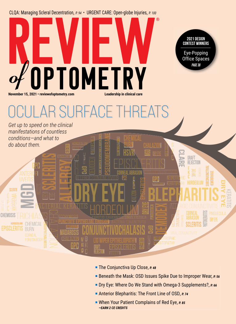
■ The Conjunctiva Up Close, P. 48 ■ Beneath the Mask: OSD Issues Spike Due to Improper Wear, P. 56 ■ Dry Eye: Where Do We Stand with Omega-3 Supplements?, P. 66 ■ Anterior Blepharitis: The Front Line of OSD, P. 74 ■ When Your Patient Complains of Red Eye, P. 85 —EARN 2 CE CREDITS CLQA: Managing Scleral Decentration, P. 94 • URGENT CARE: Open-globe Injuries, P. 100 Get up to speed on the clinical manifestations of countless conditions—and what to do about them. November 15, 2021 • reviewofoptometry.com Leadership in clinical care 2021 DESIGN CONTEST WINNERS Eye-Popping Office Spaces Page 38 edema red eye ye hordeolum m scleritis scarring hyperemia chalazion DRY eye B HARITIS LEPH ane pseudomembra GPC GPC G P P C E K C G P C V K C V K C V K C MGD scleritis D Demodex allergy allergy ophic neurotrop thy keratopat conjunctivochalasis madarosis GP GPC scleritis scleritis photokeratitis graft rejection graft rejection lid wiper epitheliopathy subconjunctival hemorrhage corneal abrasion chemical burn bur chemosis dry eye dry eye dry eye dry eye MGD leritis episcle trichiasi sis red eye red eye red eye keratoconjunctivitis allergic conjunctivitis bacterial conjunctivitis bacterial terial conjunctivitis tis viral conjunctivitis corneal foreign body Demodex VK HSVK endo dophthalmitis pinguecula episcleritis episcleritis piscleritis corneal abrasion GPC GPC PCF hordeolum chalazion thy lid wiper epitheliopat Dry eye punctate epithelial erosion rosion DRY EYE endophthalmitis end carotid-cavernous sinus fistula carotid-cavernous azion chalazio CLARE E K C E K C M M G D lum hordeolum episcleritis episcleritis episcleritis episcleritis ba bacterial kerat atitis chemical burn ecchymosis hordeolum chalazion rotrophic neuro keratopathy ker ula pinguecu tes infiltrates Demodex pinguecula anterio or uveitis keratitis lergic alle conjunctivitis OCULAR SURFACE THREATS Scan to find more answers about genetic testing and retesting at EyesOnGenes.com. To date, science has discovered more than 270 genes related to inherited retinal diseases. 1 With the evolution of genetic testing comes the ability to more precisely diagnose your patients. More answers may uncover more possibilities. THEIR GENES GAVE THEM BUTTON NOSES, BROWN HAIR, AND VISION LOSS © Janssen Pharmaceuticals, Inc. 2021 09/21 cp-250095v1 MOVE FORWARD WITH MORE ANSWERS Reference: 1. Branham K, Schlegel D, Fahim AT, Jayasundera KT. Genetic testing for inherited retinal degenerations: triumphs and tribulations. Am J Med Genet C Semin Med Genet. 2020;184(3):571-577.
-
Upload
khangminh22 -
Category
Documents
-
view
1 -
download
0
Transcript of scleritis - Review of Optometry

■ The Conjunctiva Up Close, P. 48
■ Beneath the Mask: OSD Issues Spike Due to Improper Wear, P. 56
■ Dry Eye: Where Do We Stand with Omega-3 Supplements?, P. 66
■ Anterior Blepharitis: The Front Line of OSD, P. 74
■ When Your Patient Complains of Red Eye, P. 85 —EARN 2 CE CREDITS
CLQA: Managing Scleral Decentration, P. 94 • URGENT CARE: Open-globe Injuries, P. 100
Get up to speed on the clinical manifestations of countless conditions—and what to do about them.
November 15, 2021 • reviewofoptometry.com Leadership in clinical care
2021 DESIGN CONTEST WINNERS
Eye-Popping Office Spaces
Page 38
edem
a
red eyered eyehordeolumhordeolum
scleritis
scarring
hyper
emia
chalaz
ionDRY eye BLEPHARITISLEPHARITIS
pseu
dom
embr
ane
pseu
dom
embr
ane
GPC
GPCGPPC
EKC
GPC
VKC
VKC
VKC
MGD scl
eritis
Demod
exDem
odex
alle
rgy
alle
rgy
neurotrophic neurotrophic keratopathykeratopathy
conjunctivochalasismadarosis
GPCGPC
scleri
tis
scleritis photokeratitis
graft rejection
graft rejection
lid wiper epitheliopathy
subc
onju
nctiv
al
hemo
rrha
gecorneal abrasion
chemical burnburn
chemosis dry eye
dry eye
dry eye
dry eye
MGD
episcleritisepiscleritis
trichiasistrichiasis
red
eye
red eye
red
eye
keratoconjunctivitisallergic conjunctivitis
bacterial conjunctivitis
bact
erial
bact
erial
conju
nctiv
itisco
njunc
tivitis
viral conjunctivitiscorneal foreign body
Demo
dex
HSVKHSVK
endophthalmitisendophthalmitis
ping
uecu
la
episcleritis
episc
lerit
isep
iscle
ritis
corneal abrasion
GPC
GPCPCFhordeolum
chalazion
lid
wip
er
ep
ith
el
iop
at
hy
lid
wip
er
ep
ith
el
iop
at
hy
Dry
eye
punctate epithelial erosionpunctate epithelial erosion
DR
Y E
YE
endophthalmitis
endophthalmitis
c a r o t i d - c av e r n o u s s i n u s f i s t u l ac a r o t i d - c av e r n o u s s i n u s f i s t u l a
chalazionchalazion
CLARE
EKCE
KC
MMGD
hordeolumhordeolum
episcleritis
episc
leri
tisepiscleritis
episcleritis
b a c t e r i a l k e r at i t i sb a c t e r i a l k e r at i t i sb a c t e r i a l k e r at i t i s
chemical burn
ecchymosis
hordeolum
chalazion neurotrophic neurotrophic keratopathykeratopathy
pingu
ecul
apin
guec
ula
infil
trat
esin
filtr
ates
Demo
dex
pinguecula
anterior anterior uveitis
keratitisallergicallergic conjunctivitis
OCULAR SURFACE THREATS
Scan to fi nd more answers about genetic testing and retesting at EyesOnGenes.com.
To date, science has discovered more than 270 genes related to inherited retinal diseases.1 With the evolution of genetic testing comes the ability to more precisely diagnose your patients. More answers may uncover more possibilities.
THEIR GENES GAVE THEM BUTTON NOSES, BROWN HAIR, AND VISION LOSS
© Janssen Pharmaceuticals, Inc. 2021 09/21 cp-250095v1
MOVE FORWARD WITH MORE ANSWERS
Reference: 1. Branham K, Schlegel D, Fahim AT, Jayasundera KT. Genetic testing for inherited retinal degenerations: triumphs and tribulations. Am J Med Genet C Semin Med Genet. 2020;184(3):571-577.
S:7"
S:5"
T:7.75"
B:8"

■ The Conjunctiva Up Close, P. 48
■ Beneath the Mask: OSD Issues Spike Due to Improper Wear, P. 56
■ Dry Eye: Where Do We Stand with Omega-3 Supplements?, P. 66
■ Anterior Blepharitis: The Front Line of OSD, P. 74
■ When Your Patient Complains of Red Eye, P. 85—EARN 2 CE CREDITS
CLQA: Managing Scleral Decentration, P. 94 • URGENT CARE: Open-globe Injuries, P. 100
Get up to speed on the clinicalmanifestations of countlessconditions—and what todo about them.
November 15, 2021 • reviewofoptometry.com Leadership in clinical care
2021 DESIGNCONTEST WINNERS
Eye-PoppingOffice Spaces
Page 38ed
ema
red eyered eyehordeolumhordeolum
scleritis
scarring
hyper
emia
chalaz
ionDRY eye BLEPHARITISLEPHARITIS
pseu
dom
embr
ane
pseu
dom
embr
ane
GPC
GPCGPPC
EKC
GPC
VKC
VKC
VKC
MGD scl
eritis
Demod
exDem
odex
alle
rgy
alle
rgy
neurotrophicneurotrophickeratopathykeratopathy
conjunctivochalasismadarosis
GPCGPC
scleri
tis
scleritis photok
eratiti
s
graftrejection
graftrejection
lid wiper epitheliopathy
subc
onju
nctiv
alhe
morr
hage
cornealabrasion
chemicalburnburn
chemosis dry eye
dry eye
dry eye
dry eye
MGD
episcleritisepiscleritis
trichiasistrichiasis
red
eye
red eye
red
eye
keratoconjunctivitisallergic conjunctivitis
bacterial conjunctivitis
bact
erial
bact
erial
conj
unct
ivitis
conj
unct
ivitis
viral conjunctivitiscornealforeign body
Demo
dex
HSVKHSVK
endophthalmitisendophthalmitis
ping
uecu
la
episcleritis
episc
lerit
isep
iscle
ritis
corneal abrasion
GPC
GPCPCFhordeolum
chalazion
lid
wip
er
ep
ith
el
iop
at
hy
lid
wip
er
ep
ith
el
iop
at
hy
Dry
eye
punctate epithelial erosionpunctate epithelial erosion
DR
Y E
YE
endophthalmitis
endophthalmitis
c a r o t i d - c av e r n o u s s i n u s f i s t u l ac a r o t i d - c av e r n o u s s i n u s f i s t u l a
chalazionchalazion
CLARE
EKCE
KC
MMGD
hordeolumhordeolum
episcleritis
episc
leri
tisepiscleritis
episcleritis
b a c t e r i a l k e r at i t i sb a c t e r i a l k e r at i t i sb a c t e r i a l k e r at i t i s
chemicalburn
ecchymosis
hordeolum
chalazion neurotrophicneurotrophickeratopathykeratopathy
pingu
ecul
apin
guec
ula
infil
trat
esin
filtr
ates
Demo
dex
pinguecula
anterioranterioruveitis
keratitisallergicallergic conjunctivitis
OCULAR SURFACE THREATS

* Pivotal study designs: Two Phase 3, randomized, multicenter, parallel-group studies, APOLLO and LUNAR, evaluating noninferiority of once-daily VYZULTA vs twice-daily timolol maleate 0.5% in patients with open-angle glaucoma or ocular hypertension. Primary endpoint was IOP measured at 9 assessment time points in study eye. APOLLO (VYZULTA, n=284; timolol, n=133) and LUNAR (VYZULTA, n=278; timolol, n=136).2,3
INDICATION
VYZULTA® (latanoprostene bunod ophthalmic solution), 0.024% is indicated for the reduction of intraocular pressure (IOP) in patients with open-angle glaucoma or ocular hypertension.
IMPORTANT SAFETY INFORMATION
• Increased pigmentation of the iris and periorbital tissue (eyelid) can occur. Iris pigmentation is likely to be permanent
• Gradual changes to eyelashes, including increased length, increased thickness, and number of eyelashes, may occur. These changes are usually reversible upon treatment discontinuation
• Use with caution in patients with a history of intraocular infl ammation (iritis/uveitis). VYZULTA should generally not be used in patients with active intraocular infl ammation
• Macular edema, including cystoid macular edema, has been reported during treatment with prostaglandin analogs. Use with caution in aphakic patients, in pseudophakic patients with a torn posterior lens capsule, or in patients with known risk factors for macular edema
• There have been reports of bacterial keratitis associated with the use of multiple-dose containers of topical ophthalmic products that were inadvertently contaminated by patients
• Contact lenses should be removed prior to the administration of VYZULTA and may be reinserted 15 minutes after administration
• Most common ocular adverse reactions with incidence ≥2% are conjunctival hyperemia (6%), eye irritation (4%), eye pain (3%), and instillation site pain (2%)
VYZULTA and the V design are trademarks of Bausch & Lomb Incorporated or its a� liates. Any other product/brand names and/or logos are trademarks of the respective owners. ©2021 Bausch & Lomb Incorporated or its a� liates. All rights reserved. VYZ.0258.USA.20
References: 1. VYZULTA Prescribing Information. Bausch & Lomb Incorporated. 2. Weinreb RN, Scassellati Sforzolini B, Vittitow J, Liebmann J. Latanoprostene bunod 0.024% versus timolol maleate 0.5% in subjects with open-angle glaucoma or ocular hypertension: the APOLLO study. Ophthalmology. 2016;123(5):965-973. 3. Medeiros FA, Martin KR, Peace J, Scassellati Sforzolini B, Vittitow JL, Weinreb RN. Comparison of latanoprostene bunod 0.024% and timolol maleate 0.5% in open-angle glaucoma or ocular hypertension: the LUNAR study. Am J Ophthalmol. 2016;168:250-259.
For more information, please see Brief Summary of full Prescribing Information on adjacent page.
TAKE A TEST RIDE AT VYZULTAHCP.COM
THE HORSEPOWER YOU NEED
TO LOWER IOPPowerful IOP reduction with excellent tolerability1,2
VYZULTA delivered up to 9.1 mmHg mean IOP reduction
from baseline in pivotal trials.1,2*
BRIEF SUMMARY OF PRESCRIBING INFORMATION
This Brief Summary does not include all the information needed to use VYZULTA safely and effectively. See full Prescribing Information for VYZULTA.
VYZULTA® (latanoprostene bunod ophthalmic solution), 0.024%, for topical ophthalmic use. Initial U.S. Approval: 2017
1 INDICATIONS AND USAGE
VYZULTA® (latanoprostene bunod ophthalmic solution) 0.024% is indicated for the reduction of intraocular pressure (IOP) in patients with open-angle glaucoma or ocular hypertension.
4 CONTRAINDICATIONS
None
5 WARNINGS AND PRECAUTIONS
5.1 Pigmentation
VYZULTA® (latanoprostene bunod ophthalmic solution), 0.024% may cause changes to pigmented tissues. The most frequently reported changes with prostaglandin analogs have been increased pigmentation of the iris and periorbital tissue (eyelid).
Pigmentation is expected to increase as long as latanoprostene bunod ophthalmic solution is administered. The pigmentation change is due to increased melanin content in the melanocytes rather than to an increase in the number of melanocytes. After discontinuation of VYZULTA, pigmentation of the iris is likely to be permanent, while pigmentation of the periorbital tissue and eyelash changes are likely to be reversible in most patients. Patients who receive prostaglandin analogs, including VYZULTA, should be informed of the possibility of increased pigmentation, including permanent changes. The long-term effects of increased pigmentation are not known.
Iris color change may not be noticeable for several months to years. Typically, the brown pigmentation around the pupil spreads concentrically towards the periphery of the iris and the entire iris or parts of the iris become more brownish. Neither nevi nor freckles of the iris appear to be affected by treatment. While treatment with VYZULTA® (latanoprostene bunod ophthalmic solution), 0.024% can be continued in patients who develop noticeably increased iris pigmentation, these patients should be examined regularly [see Patient Counseling Information (17) in full Prescribing Information].5.2 Eyelash Changes
VYZULTA may gradually change eyelashes and vellus hair in the treated eye. These changes include increased length, thickness, and the number of lashes or hairs. Eyelash changes are usually reversible upon discontinuation of treatment.
5.3 Intraocular In�ammation
VYZULTA should be used with caution in patients with a history of intraocular in�ammation (iritis/uveitis) and should generally not be used in patients with active intraocular in�ammation as it may exacerbate this condition.
5.4 Macular Edema
Macular edema, including cystoid macular edema, has been reported during treatment with prostaglandin analogs. VYZULTA should be used with caution in aphakic patients, in pseudophakic patients with a torn posterior lens capsule, or in patients with known risk factors for macular edema.
5.5 Bacterial Keratitis
There have been reports of bacterial keratitis associated with the use of multiple-dosecontainers of topical ophthalmic products. These containers had been inadvertently contaminated by patients who, in most cases, had a concurrent corneal disease or a disruption of the ocular epithelial surface.
5.6 Use with Contact Lens
Contact lenses should be removed prior to the administration of VYZULTA because this productcontains benzalkonium chloride. Lenses may be reinserted 15 minutes after administration.
6 ADVERSE REACTIONS
The following adverse reactions are described in the Warnings and Precautions section:pigmentation (5.1), eyelash changes (5.2), intraocular in�ammation (5.3), macular edema (5.4), bacterial keratitis (5.5), use with contact lens (5.6).
6.1 Clinical Trials Experience
Because clinical trials are conducted under widely varying conditions, adverse reaction rates observed in the clinical trials of a drug cannot be directly compared to rates in the clinical trials of another drug and may not re�ect the rates observed in practice.
VYZULTA was evaluated in 811 patients in 2 controlled clinical trials of up to 12 months duration. The most common ocular adverse reactions observed in patients treated with latanoprostene bunod were: conjunctival hyperemia (6%), eye irritation (4%), eye pain (3%), and instillation site pain (2%). Approximately 0.6% of patients discontinued therapy due to ocular adverse reactions including ocular hyperemia, conjunctival irritation, eye irritation, eye pain, conjunctival edema, vision blurred, punctate keratitis and foreign body sensation.
8 USE IN SPECIFIC POPULATIONS
8.1 Pregnancy
Risk Summary
There are no available human data for the use of VYZULTA during pregnancy to inform any drugassociated risks.
Latanoprostene bunod has caused miscarriages, abortion, and fetal harm in rabbits. Latanoprostene bunod was shown to be abortifacient and teratogenic when administered intravenously (IV) to pregnant rabbits at exposures ≥ 0.28 times the clinical dose. Doses ≥ 20 μg/kg/day (23 times the clinical dose) produced 100% embryofetal lethality. Structural abnormalities observed in rabbit fetuses included anomalies of the great vessels and aortic arch vessels, domed head, sternebral and vertebral skeletal anomalies, limb hyperextension
and malrotation, abdominal distension and edema. Latanoprostene bunod was not teratogenic in the rat when administered IV at 150 mcg/kg/day (87 times the clinical dose) [see Data]. The background risk of major birth defects and miscarriage for the indicated population is unknown. However, the background risk in the U.S. general population of major birth defects is 2 to 4%, and of miscarriage is 15 to 20%, of clinically recognized pregnancies.
Data
Animal DataEmbryofetal studies were conducted in pregnant rabbits administered latanoprostene bunod dailyby intravenous injection on gestation days 7 through 19, to target the period of organogenesis. The doses administered ranged from 0.24 to 80 mcg/kg/day. Abortion occurred at doses ≥ 0.24 mcg/kg/day latanoprostene bunod (0.28 times the clinical dose, on a body surface area basis, assuming 100% absorption). Embryofetal lethality (resorption) was increased in latanoprostene bunod treatment groups, as evidenced by increases in early resorptions at doses ≥ 0.24 mcg/kg/day and late resorptions at doses ≥ 6 mcg/kg/day (approximately 7 times the clinical dose). No fetuses survived in any rabbit pregnancy at doses of 20 mcg/kg/day (23 times the clinical dose) or greater. Latanoprostene bunod produced structural abnormalities at doses ≥ 0.24 mcg/kg/day (0.28 times the clinical dose). Malformations included anomalies of sternum, coarctation of the aorta with pulmonary trunk dilation, retroesophageal subclavian artery with absent brachiocephalic artery, domed head, forepaw hyperextension and hindlimb malrotation, abdominal distention/edema, and missing/fused caudal vertebrae.
An embryofetal study was conducted in pregnant rats administered latanoprostene bunod daily by intravenous injection on gestation days 7 through 17, to target the period of organogenesis. The doses administered ranged from 150 to 1500 mcg/kg/day. Maternal toxicity was produced at 1500 mcg/kg/day (870 times the clinical dose, on a body surface area basis, assuming 100% absorption), as evidenced by reduced maternal weight gain. Embryofetal lethality (resorption and fetal death) and structural anomalies were produced at doses ≥ 300 mcg/kg/day (174 times the clinical dose). Malformations included anomalies of the sternum, domed head, forepaw hyperextension and hindlimb malrotation, vertebral anomalies and delayed ossi�cation of distallimb bones. A no observed adverse effect level (NOAEL) was established at 150 mcg/kg/day (87 times the clinical dose) in this study.
8.2 Lactation
Risk Summary
There are no data on the presence of VYZULTA in human milk, the effects on the breastfedinfant, or the effects on milk production. The developmental and health bene�ts of breastfeeding should be considered, along with the mother’s clinical need for VYZULTA, and any potential adverse effects on the breastfed infant from VYZULTA.
8.4 Pediatric Use
Use in pediatric patients aged 16 years and younger is not recommended because of potential safety concerns related to increased pigmentation following long-term chronic use.
8.5 Geriatric Use
No overall clinical differences in safety or effectiveness have been observed between elderly and other adult patients.
13 NONCLINICAL TOXICOLOGY
13.1 Carcinogenesis, Mutagenesis, Impairment of Fertility
Latanoprostene bunod was not mutagenic in bacteria and did not induce micronuclei formation in the in vivo rat bone marrow micronucleus assay. Chromosomal aberrations were observed in vitro with human lymphocytes in the absence of metabolic activation.
Latanoprostene bunod has not been tested for carcinogenic activity in long-term animal studies. Latanoprost acid is a main metabolite of latanoprostene bunod. Exposure of rats and mice to latanoprost acid, resulting from oral dosing with latanoprost in lifetime rodent bioassays, was not carcinogenic.
Fertility studies have not been conducted with latanoprostene bunod. The potential to impact fertility can be partially characterized by exposure to latanoprost acid, a common metabolite of both latanoprostene bunod and latanoprost. Latanoprost acid has not been found to have any effect on male or female fertility in animal studies.
13.2 Animal Toxicology and/or Pharmacology
A 9-month toxicology study administered topical ocular doses of latanoprostene bunod to one eye of cynomolgus monkeys: control (vehicle only), one drop of 0.024% bid, one drop of 0.04% bid and two drops of 0.04% per dose, bid. The systemic exposures are equivalent to 4.2-fold, 7.9-fold, and 13.5-fold the clinical dose, respectively, on a body surface area basis (assuming 100% absorption). Microscopic evaluation of the lungs after 9 months observed pleural/subpleural chronic �brosis/in�ammation in the 0.04% dose male groups, with increasing incidence and severity compared to controls. Lung toxicity was not observed at the 0.024% dose.
U.S. Patent Numbers: 7,273,946; 7,629,345; 7,910,767; 8,058,467.
VYZULTA is a trademark of Bausch & Lomb Incorporated or its af�liates.
© 2020 Bausch & Lomb Incorporated or its af�liates.
Distributed by:
Bausch + Lomb, a division of
Bausch Health US, LLC
Bridgewater, NJ 08807 USA
Based on 9612403 (Folded), 9612303 (Flat) 5/2019
VYZ.0109.USA.20 Issued: 5/2020

REVIEW OF OPTOMETRY | NOVEMBER 15, 20214
No fewer than four newophthalmic drugs receivedFDA approval in October,and each hopes to distin-
guish itself with a unique approach tocare. Retina specialists will have accessto a ranibizumab-dispensing implantfor wet age-related macular degenera-tion (AMD) and a suprachoroidal injec-tion to treat uveitic macular edema.Optometrists will be able to prescribethe first of several forthcoming eyedrops for presbyopia and a nasal sprayto treat dry eye.
For the ODsCorrective lenses for presbyopia have anew competitor to contend with. Thefirst drug for such use—Vuity, from Al-lergan—has finally entered the market.
The pupil-constricting drop (pilocar-pine 1.25%) reaches peak efficacy onehour after use, then begins to wane.Patients begin experiencing visualimprovement in as little as 15 minutes,and at least some positive effects canlast up to six hours, according to thedrug’s FDA trial data. Dosing is QD.
Vuity’s two Phase III trials (Gemini1 and 2) used a primary endpoint ofachieving three lines of near vision im-provement under mesopic conditionswithout losing more than one line ofdistance vision, when measured at day30, hour three, of use. This was met by31% and 26% of subjects, respectively,in the two studies. Both groups showedstatistically significant improvementover placebo, a press release notes.
Headache and conjunctival hyper-emia were the most common adverse
events. Thecompany saysthe drug’svehicle isformulated toadapt to thepH of the eyeto reduce blurand discom-fort.
In a first fordry eye pa-tients, a nasal
spray that stimulates the trigeminalparasympathetic pathway offers a newmeans of boosting tear production.Tyrvaya (varenicline 0.03mg, OysterPoint Pharma) is a cholinergic agonistthat triggers basal tear production, thecompany says. Patients gained 10mmor more in Schirmer’s scores.
For the MDsWhile the average patient with wetage-related macular degeneration(AMD) today is treated via monthlyor bimonthly anti-VEGF injections,a new therapeutic approach—using arefillable ocular implant that releases ra-nibizumab continuously—may reducethe number of yearly treatments totwo. Susvimo (Genentech), previouslyreferred to as the Port Delivery System,gained approval from the FDA afterdemonstrating in clinical trials its abilityto produce results comparable to that ofmonthly anti-VEGF injections.
The device is implanted into thepatient’s eye during a one-time surgicalprocedure and must be refilled by anophthalmologist every six months; after
implantation, Susvimo delivers 100mg/mL ranibizumab into the eye, a pressrelease from Genentech explains. Thecompany notes that, if needed, supple-mental ranibizumab treatment can begiven while the implant is in place.During clinical trials, 98% of patientstreated with Susvimo were shown toachieve and maintain vision gains at thesame rate as those receiving monthlyinjections (+0.2 and +0.5 letters frombaseline), the company reports.
The most frequent complicationobserved was endophthalmitis, whichoccurred at three times the rate forpatients with the Susvimo implant (2%total) than those receiving monthlyranibizumab injections. Genentechadvises that close monitoring andearly detection with surgical repair ofconjunctival retractions or erosions mayreduce the risk of endophthalmitis.
Also approved last month was Xipere(triamcinolone acetonide, Bausch +Lomb/Clearside Biomedical) a supra-choroidal injection for uveitic macularedema. That route of administrationprovides more targeted delivery andrapid dispersion of drug to the affectedarea, B+L says.
New Therapeutics Expand Intervention Options
news reviewClinical, legislative and practice development updates for ODs.
CA, NY Expand Scope of Practice, p. 6 >> E-CIGS tied to visual impairment, p. 9 >> Charles Bonnet Syndrome AND GLAUCOMA, p. 10 >> dr increases cataract risk, p. 12 >> Same-day bilateral Cataract Surgery Favored p. 14
Get the latest atwww.reviewofoptometry.com/news
Stories post every weekday
The innovations include a refillable implant that treats AMD with far fewer injections, an eyedrop for presbyopia that constricts pupils and a nasal spray that boosts tear production.
Photo: Allergan
Photo: Anat Loewenstein, M
D
Surgeons implant the Susvimo drugreservoir such that it can be refilled with a needle every six months.
Pharma companieshave their eyes onpresbyopia. Vuity isthe first to market.

The ease and accuracy you’re looking for.
Practice Management and EHR
Experience the Eyefinity®difference and learn more at:
800.269.3666 option 2
Complete Practice Management and EHR Software
©2021 Eyefinity, Inc. All rights reserved.Eyefinity is a registered trademark of Eyefinity, Inc. All other brands or marks are the property of their respective owners. 98067 Classification: Public
Equip your practice with reliable cloud-based technology—built to simplify day-to-day tasks and support the success of your entire team.
www.eyefinity.com/cloud
REVIEW OF OPTOMETRY | NOVEMBER 15, 20216
NEWS REVIEW | Get the latest at www.reviewofoptometry.com/news
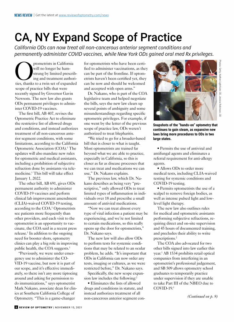
Optometrists in Californiawill no longer be ham-strung by limited prescrib-ing and treatment authori-
ties, thanks to a twin set of expandedscope of practice bills that wererecently signed by Governor GavinNewsom. The new law also grantsODs permanent privileges to admin-ister COVID-19 vaccines.
The first bill, AB 407, revises theOptometric Practice Act to eliminatethe restrictive list of allowed drugsand conditions, and instead authorizestreatment of all non-cancerous ante-rior segment conditions, with somelimitations, according to the CaliforniaOptometric Association (COA).1 Theupdates will also mandate new rulesfor optometric and medical assistants,including a prohibition of subjectiverefraction done by assistants via tele-medicine.1 This bill will take effectJanuary 1, 2022.
The other bill, AB 691, gives ODspermanent authority to administerCOVID-19 vaccines and performclinical lab improvement amendment(CLIA)-waived COVID-19 testing,according to the COA.1 Optometristssee patients more frequently thanother providers, and each visit to theoptometrist is an opportunity to vac-cinate, the COA said in a recent pressrelease.1 In addition to the ongoingneed for booster shots, optometryclinics can play a big role in improvingpublic health, the COA suggests.1
“Previously, we were under emer-gency use to administer the CO-VID-19 vaccine, but now it is part ofour scope, and it’s effective immedi-ately, so there isn’t any more tiptoeingaround and asking for permission todo immunizations,” says optometristMark Nakano, associate dean for clin-ics at Southern California College ofOptometry. “This is a game-changer
for optometrists who have been certi-fied to administer vaccinations, as theycan be part of the frontline. If optom-etrists haven’t been certified yet, theycan be now and should be welcomedand accepted with open arms.”
Dr. Nakano, who is part of the COAlegislative team and helped negotiatethe bills, says the new law clears upseveral points of ambiguity and somemisunderstandings regarding specificoptometric privileges. For example, ifone went by the letter of the previousscope of practice law, ODs weren’tauthorized to treat blepharitis.
“We tried to go for a broader-basedbill that is closer to what is taught.Most optometrists are trained farbeyond what we are able to practice,especially in California, so this iscloser as far as disease processes thatwe can treat and medications we canuse,” Dr. Nakano explains.
The previous law, which Dr. Na-kano describes as being very “pre-scriptive,” only allowed ODs to treatlimited types of inflammation in indi-viduals over 18 and prescribe a smallamount of antiviral medications.
“Now we can treat almost everytype of viral infection a patient may beexperiencing, and we’re not limitedto certain medications, so this reallyopens up the door for optometrists,”Dr. Nakano says.
The new law will also allow ODsto perform tests for systemic condi-tions that may be related to an ocularproblem, he adds. “It’s important thatODs in California can now order anytests, imaging or cultures, as we wererestricted before,” Dr. Nakano says.
Specifically, the new scope expan-sion law includes the following:2
• Eliminates the lists of alloweddrugs and conditions in statute, andinstead authorizes treatment of allnon-cancerous anterior segment con-
ditions, with some limitations, as wellas all kinds of inflammation in adultsand some in children.
• Permits the use of antiviral andantifungal agents and eliminates areferral requirement for anti-allergyagents.
• Allows ODs to order moremedical tests, including CLIA-waivedtesting for systemic conditions andCOVID-19 testing.
• Permits optometrists the use of ascalpel to remove foreign bodies, aswell as intense pulsed light and low-level light therapy.
The new law also outlines rulesfor medical and optometric assistantsperforming subjective refractions, re-quiring direct and on-site supervisionand 45 hours of documented training,and precludes their ability to writeprescriptions.2
The COA also advocated for twoother bills signed into law earlier thisyear.1 AB 1534 prohibits retail opticalcompanies from interfering in anoptometrist’s professional judgement,and SB 509 allows optometry schoolgraduates to temporarily practiceunder supervision if they are unableto take Part III of the NBEO due toCOVID-19.1
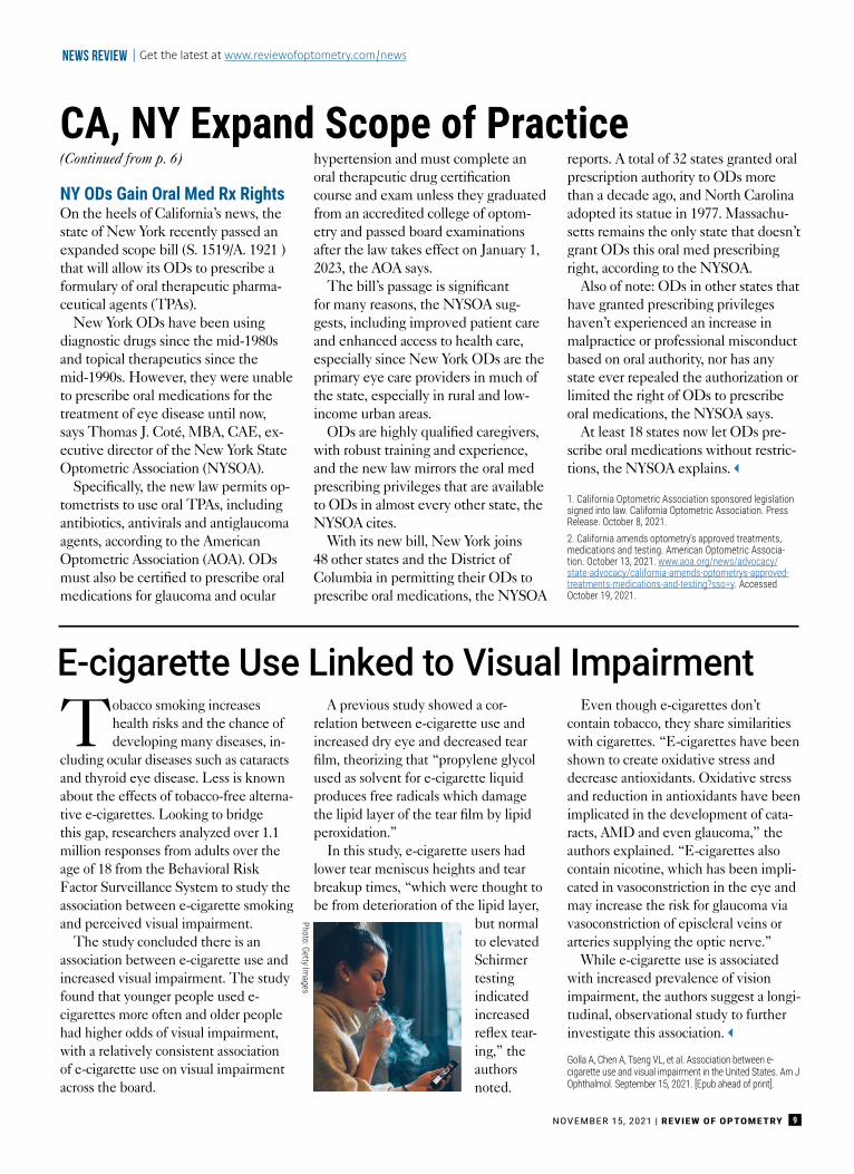
CA, NY Expand Scope of Practice
Snapshots of the “hands-on” optometry thatcontinues to gain steam, as expansive newlaws bring more procedures to ODs in twolarge states.
California ODs can now treat all non-cancerous anterior segment conditions andpermanently administer COVID vaccines, while New York ODs gained oral med Rx privileges.
(Continued on p. 9)
Photos: Getty, Nathan Lighthizer, OD, Jessica Robinson, OD
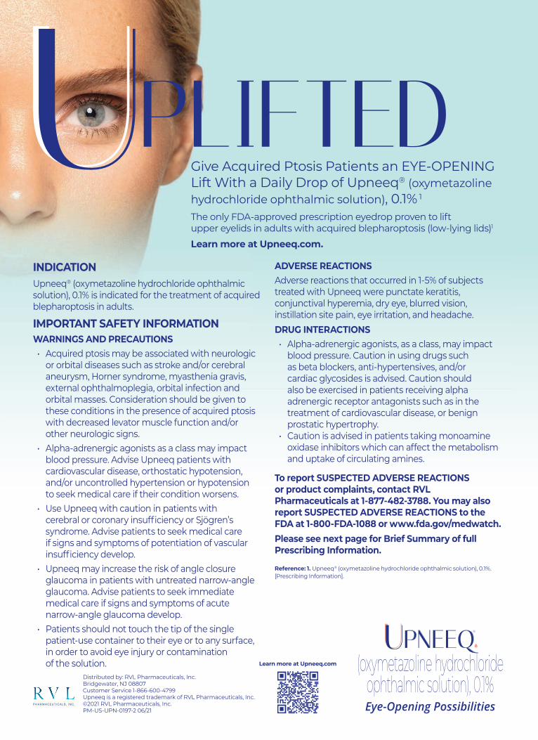
(oxymetazoline hydrochlorideophthalmic solution), 0.1%
UPNEEQU ®
Distributed by: RVL Pharmaceuticals, Inc.Bridgewater, NJ 08807Customer Service 1-866-600-4799Upneeq is a registered trademark of RVL Pharmaceuticals, Inc.©2021 RVL Pharmaceuticals, Inc. PM-US-UPN-0197-2 06/21
Learn more at Upneeq.com
PMS 137 CPMS 541 CPMS 908 C
PMS 541 C PMS 908 C
INDICATIONUpneeq® (oxymetazoline hydrochloride ophthalmic solution), 0.1% is indicated for the treatment of acquired blepharoptosis in adults.
IMPORTANT SAFETY INFORMATIONWARNINGS AND PRECAUTIONS• Acquired ptosis may be associated with neurologic
or orbital diseases such as stroke and/or cerebral aneurysm, Horner syndrome, myasthenia gravis, external ophthalmoplegia, orbital infection and orbital masses. Consideration should be given to these conditions in the presence of acquired ptosis with decreased levator muscle function and/or other neurologic signs.
• Alpha-adrenergic agonists as a class may impact blood pressure. Advise Upneeq patients with cardiovascular disease, orthostatic hypotension, and/or uncontrolled hypertension or hypotension to seek medical care if their condition worsens.
• Use Upneeq with caution in patients with cerebral or coronary insuffi ciency or Sjögren’s syndrome. Advise patients to seek medical care if signs and symptoms of potentiation of vascular insuffi ciency develop.
• Upneeq may increase the risk of angle closure glaucoma in patients with untreated narrow-angle glaucoma. Advise patients to seek immediate medical care if signs and symptoms of acute narrow-angle glaucoma develop.
• Patients should not touch the tip of the single patient-use container to their eye or to any surface, in order to avoid eye injury or contamination of the solution.
ADVERSE REACTIONSAdverse reactions that occurred in 1-5% of subjects treated with Upneeq were punctate keratitis, conjunctival hyperemia, dry eye, blurred vision, instillation site pain, eye irritation, and headache.DRUG INTERACTIONS• Alpha-adrenergic agonists, as a class, may impact
blood pressure. Caution in using drugs such as beta blockers, anti-hypertensives, and/or cardiac glycosides is advised. Caution should also be exercised in patients receiving alpha adrenergic receptor antagonists such as in the treatment of cardiovascular disease, or benign prostatic hypertrophy.
• Caution is advised in patients taking monoamine oxidase inhibitors which can affect the metabolism and uptake of circulating amines.
To report SUSPECTED ADVERSE REACTIONS or product complaints, contact RVL Pharmaceuticals at 1-877-482-3788. You may also report SUSPECTED ADVERSE REACTIONS to the FDA at 1-800-FDA-1088 or www.fda.gov/medwatch.Please see next page for Brief Summary of full Prescribing Information.
Give Acquired Ptosis Patients an EYE-OPENING Lift With a Daily Drop of Upneeq® (oxymetazoline hydrochloride ophthalmic solution), 0.1% 1
The only FDA-approved prescription eyedrop proven to lift upper eyelids in adults with acquired blepharoptosis (low-lying lids)1
Learn more at Upneeq.com.
Reference: 1. Upneeq® (oxymetazoline hydrochloride ophthalmic solution), 0.1%. [Prescribing Information].

NOVEMBER 15, 2021 | REVIEW OF OPTOMETRY 9
NEWS REVIEW | Get the latest at www.reviewofoptometry.com/news
E-cigarette Use Linked to Visual Impairment
Tobacco smoking increaseshealth risks and the chance ofdeveloping many diseases, in-
cluding ocular diseases such as cataractsand thyroid eye disease. Less is knownabout the effects of tobacco-free alterna-tive e-cigarettes. Looking to bridgethis gap, researchers analyzed over 1.1million responses from adults over theage of 18 from the Behavioral RiskFactor Surveillance System to study theassociation between e-cigarette smokingand perceived visual impairment.
The study concluded there is anassociation between e-cigarette use andincreased visual impairment. The studyfound that younger people used e-cigarettes more often and older peoplehad higher odds of visual impairment,with a relatively consistent associationof e-cigarette use on visual impairmentacross the board.
A previous study showed a cor-relation between e-cigarette use andincreased dry eye and decreased tearfilm, theorizing that “propylene glycolused as solvent for e-cigarette liquidproduces free radicals which damagethe lipid layer of the tear film by lipidperoxidation.”
In this study, e-cigarette users hadlower tear meniscus heights and tearbreakup times, “which were thought tobe from deterioration of the lipid layer,
but normalto elevatedSchirmertestingindicatedincreasedreflex tear-ing,” theauthorsnoted.
Even though e-cigarettes don’tcontain tobacco, they share similaritieswith cigarettes. “E-cigarettes have beenshown to create oxidative stress anddecrease antioxidants. Oxidative stressand reduction in antioxidants have beenimplicated in the development of cata-racts, AMD and even glaucoma,” theauthors explained. “E-cigarettes alsocontain nicotine, which has been impli-cated in vasoconstriction in the eye andmay increase the risk for glaucoma viavasoconstriction of episcleral veins orarteries supplying the optic nerve.”
While e-cigarette use is associatedwith increased prevalence of visionimpairment, the authors suggest a longi-tudinal, observational study to furtherinvestigate this association.
Golla A, Chen A, Tseng VL, et al. Association between e-cigarette use and visual impairment in the United States. Am JOphthalmol. September 15, 2021. [Epub ahead of print].
NY ODs Gain Oral Med Rx RightsOn the heels of California’s news, thestate of New York recently passed anexpanded scope bill (S. 1519/A. 1921 )that will allow its ODs to prescribe aformulary of oral therapeutic pharma-ceutical agents (TPAs).
New York ODs have been usingdiagnostic drugs since the mid-1980sand topical therapeutics since themid-1990s. However, they were unableto prescribe oral medications for thetreatment of eye disease until now,says Thomas J. Coté, MBA, CAE, ex-ecutive director of the New York StateOptometric Association (NYSOA).
Specifically, the new law permits op-tometrists to use oral TPAs, includingantibiotics, antivirals and antiglaucomaagents, according to the AmericanOptometric Association (AOA). ODsmust also be certified to prescribe oralmedications for glaucoma and ocular
hypertension and must complete anoral therapeutic drug certificationcourse and exam unless they graduatedfrom an accredited college of optom-etry and passed board examinationsafter the law takes effect on January 1,2023, the AOA says.
The bill’s passage is significantfor many reasons, the NYSOA sug-gests, including improved patient careand enhanced access to health care,especially since New York ODs are theprimary eye care providers in much ofthe state, especially in rural and low-income urban areas.
ODs are highly qualified caregivers,with robust training and experience,and the new law mirrors the oral medprescribing privileges that are availableto ODs in almost every other state, theNYSOA cites.
With its new bill, New York joins48 other states and the District ofColumbia in permitting their ODs toprescribe oral medications, the NYSOA
reports. A total of 32 states granted oralprescription authority to ODs morethan a decade ago, and North Carolinaadopted its statue in 1977. Massachu-setts remains the only state that doesn’tgrant ODs this oral med prescribingright, according to the NYSOA.
Also of note: ODs in other states thathave granted prescribing privilegeshaven’t experienced an increase inmalpractice or professional misconductbased on oral authority, nor has anystate ever repealed the authorization orlimited the right of ODs to prescribeoral medications, the NYSOA says.
At least 18 states now let ODs pre-scribe oral medications without restric-tions, the NYSOA explains.
1. California Optometric Association sponsored legislationsigned into law. California Optometric Association. PressRelease. October 8, 2021.2. California amends optometry’s approved treatments,medications and testing. American Optometric Associa-tion. October 13, 2021. www.aoa.org/news/advocacy/state-advocacy/california-amends-optometrys-approved-treatments-medications-and-testing?sso=y. AccessedOctober 19, 2021.
CA, NY Expand Scope of Practice
Photo: Getty Images
(Continued from p. 6)
REVIEW OF OPTOMETRY | NOVEMBER 15, 202110
NEWS REVIEW | Get the latest at www.reviewofoptometry.com/news
Charles Bonnet syndrome (CBS)causes complex visual halluci-nations and presents in indi-
viduals with vision loss or impairment.Rather than a cognitive or neurologicaldisease, CBS is a result of the brain’snatural response to fill in for the im-ages no longer being processed by thevisual system. The person is typicallyaware that these hallucinations are notreal.
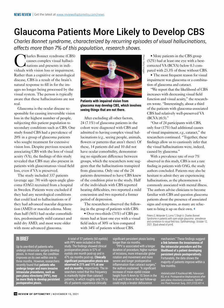
Glaucoma is the ocular disease re-sponsible for causing irreversible visionloss in the highest number of people,subjecting this patient population tosecondary conditions such as CBS. Onestudy found CBS had a prevalence of20% in a group of glaucoma patientswho sought treatment for extensivevision loss. Despite previous researchassociating CBS with the loss of visualacuity (VA), the findings of this studyrevealed that CBS may also present inpatients with glaucomatous visual fieldloss, even if VA is preserved.
The study included 337 patients(average age: 78) with open-angle glau-coma (OAG) recruited from a hospitalin Sweden. Patients were excluded ifthey had any neurological conditionsthat could lead to hallucinations or ifthey had advanced macular degenera-tion (AMD) or macular edema. Morethan half (56%) had ocular comorbidi-ties, predominantly mild cataract andmild dry AMD, and most were olderwith more advanced glaucoma.
After excluding all other factors,24 (7.1%) of glaucoma patients in thecohort were diagnosed with CBS andadmitted to having complex visual hal-lucinations (e.g., seeing people, animals,flowers or patterns that aren’t there). Ofthese, 14 patients did and 10 did nothave ocular comorbidity, demonstrat-ing no significant difference betweengroups, which the researchers note sug-gests that the hallucinations transpiredfrom glaucoma. Only one of the 24patients determined to have CBS knewof the condition prior to the study. Halfof the individuals with CBS reportedhearing difficulties, two reported a mildconcussion and two reported a formerperiod of depression.
The researchers observed the follow-ing in the group of patients with CBS:
• Over two-thirds (71%) of CBS pa-tients had at least one eye with a visualfield index of 30% or less comparedwith 34% of patients without CBS.
• More patients in the CBS group(52%) had at least one eye with a best-corrected VA (BCVA) below 0.3 com-pared with 23.3% of those without CBS.
• The most frequent reason for visualimpairment was glaucoma or a combina-tion of glaucoma and cataract.
“We report that the likelihood of CBSincreases with decreasing visual fieldfunction and visual acuity,” the research-ers wrote. “Interestingly, about a thirdof the patients with glaucoma-associatedCBS had relatively well-preserved VA(BCVA ≥0.5).”
“Out of 24 participants with CBS,only four (17%) had additional causesof visual impairment, e.g., cataract,” theresearchers continued. “Together, thesefindings allow us to cautiously infer thatthe visual hallucinations were, indeed,caused by glaucoma.”
With a prevalence rate of over 7%observed in this study, CBS is not a rarefinding in patients with OAG, the studyauthors concluded. Patients may also behesitant to admit they are experiencinghallucinations, as the phenomenon iscommonly associated with mental illness.The authors advise clinicians to becomefamiliar with CBS and question glaucomapatients about the presence of associatedsigns and symptoms, as many are reluc-tant to bring it up on their own.
Peters D, Molander S, Lomo T, Singh A. Charles BonnetSyndrome in patients with open-angle glaucoma - prevalenceand correlation to visual field loss. Ophthalmology. October 12, 2021. [Epub ahead of print].
Glaucoma Patients More Likely to Develop CBSCharles Bonnet syndrome, characterized by recurring episodes of visual hallucinations,affects more than 7% of this population, research shows.
Photo: Getty Images
Patients with impaired vision fromglaucoma may develop CBS, which involvesseeing things that are not there.
IN BRIEFUp to one-third of patients whoundergo intraocular surgery developptosis. In most cases, the conditionimproves on its own within one tothree months. However, researchersrecently found that patients whoundergo longer and more invasiveintraocular procedures, such aspars plana vitrectomy (PPV), maybe more likely to develop persistentpostoperative ptosis.
A total of 57 patients (60 eyelids)with PPV were included in thisstudy. The findings showed clinical postoperative ptosis in 57% ofeyelids one month post-op and in47% six months post-op. Clinicallysignificant postoperative ptosis was observed in 21% and 11% at oneand six months, respectively. The re-searchers noted that this frequencyis markedly higher than cataractpatients, in which roughly only 3% to4% of patients experience clinically
significant persistent ptosis lasting longer than six months.
“PPV is associated with a longerduration of procedure, higher numberof incisions, more intraocular globerotation and movement and moresevere and longer postoperativeinflammation than cataract surgery,” the authors explained. “A significant increase of mean eyelid creaseheight from 9.4mm to 10.5mm in thefirst and 9.8mm in the last follow-up could imply a levator dehiscence
mechanism.” These findings suggest a link between the invasiveness ofthe intraocular procedure and thelikelihood of a patient developingpersistent ptosis postoperatively.Fortunately, the data shows themajority of patients improve within afew months.
Abdolalizadeh P, Kashkouli MB, FalavarjaniKG, et al. Postoperative blepharoptosis afterpars plana vitrectomy procedure. Ophthal-mic Plast Reconstr Surg. 2021;37(5):431-4.

C O O P E R V I S I O N . C O M / T O R I C
The same optical design features of Biofinity® toric, the # 1 most prescribed toric contact lens in the US¹, are replicated in MyDay® toric.
With lenses available to prescribe in a monthly and a daily disposable,
MyDay®Biofinity®
toric toric
YOU PRESCRIBE
SO MUCH, WE MADE IT
TWICE.
IT’S TORIC 2 WAYS.
1. CooperVision data on file, 2017-2019. Based on number of US soft contact lens fits. Includes CooperVision branded and customer-branded equivalent lenses. US industry reports and internal estimates. © 2021 CooperVision 11727 08/21

REVIEW OF OPTOMETRY | NOVEMBER 15, 202112
Diabetes patients have up toa five-fold increased risk ofcataract formation in their
lifetime; however, not all of thesecases are sight-threatening or requiresurgical intervention. Certain risk fac-tors, including diabetic retinopathy(DR), place some patients with dia-betes at a higher risk of developingcataracts severe enough to warrantsurgery.
Taiwanese researchers recentlygathered data on 3,297 DR patients,13,188 diabetes control patientsand 13,188 non-diabetes controls.Participant data was traced from2000 through 2016. Throughout thisperiod, 919 sight-threatening cataractevents (27.9%) occurred in the DRpatients, demonstrating a far greaterrelative risk than control subjectswith diabetes (8.4%) or without(7.3%).
Sight-threatening cataracts werethree times as likely among patientswith DR vs. controls. Patients withproliferative DR or nonproliferativeDR were found to have a comparablerisk for sight-threatening cataracts,far higher than those with diabeteswithout any retinopathy. Other iden-tified risk factors for sight-threateningcataracts included older age (>60years), cardiovascular disease, chronicpulmonary disease, inflammatory
disease, ocular disorders and oral ortopical steroid use.
“The major pathophysiology of[diabetes] that induces cataracts isthought to be the fluid accumulationprocess,” the researchers explained intheir study. “Hyperglycemia leads tothe additional production of sorbitolfrom the polyol pathway for glu-cose metabolism in the lens. Due toelevated osmotic pressure caused bythe buildup of sorbitol, excess fluidinfluxes the lens, which leads to cellswelling, membrane impairment andsubsequent cataracts.”
By contrast, diabetic reti-nopathy “leads to intraocularhypoxia, ischemia and inflam-mation,” they continued.“A previous review articleconcerning proliferative DRindicated that reactive oxygenspecies, inflammatory cyto-kines and vascular endothelialgrowth factors were increasedin the vitreous cavity, andprevious laboratory studiesindicated that oxidative stressis also a risk factor for cataractformation.” Therefore, DRlikely influences the micro-environment in the eye more,increasing the catalysts forcataract development, theyconcluded.
Unlike previous studies that haveunanimously associated diabeteswith cataracts, the findings from thisstudy suggest that patients with DRface a significantly more elevatedrisk of sight-threatening cataractscompared with those without ocularmanifestations. Closely monitor andinform this patient population of anyphysical or clinical signs of cataractdevelopment.
Nien CW, Lee CY, Chen HC, et al. The elevated risk ofsight-threatening cataract in diabetes with retinopathy:a retrospective population-based cohort study. BMCOphthalmol. 2021;21:349.
NEWS REVIEW | Get the latest at www.reviewofoptometry.com/news
Patients with DR at Higher Risk for Cataract
IN BRIEFAn association between retinal de-tachment (RD) and atopic dermatitis(AD) may not be top of mind duringroutine eye exams but the connec-tion warrants attention, researcherssay. A team in Korea determinedthat atopic patients develop RDat a younger age with a poorerprognosis due to a high incidence ofproliferative vitreoretinopathy (PVR)or recurrence. AD patients also had amuch higher risk of RD after cataractsurgery.
The retrospective review analyzed2,257 patients who underwent RDsurgery between 2008 and 2018. Theresearchers found 61 patients whowere diagnosed as AD and assignedthem into the experimental group.They randomly selected and as-signed 100 patients who did not haveAD into the control group.
The study found post-op VA andprognosis were significantly worse, and bilateral involvement of RD wasmore common in the AD group thanthe control group. Characteristics ofretinal breaks were different between
the two groups. Despite similarmacula-off rates and preopera-tive VAs between the two groups,vitrectomy and encircling togetheras an initial operative method wasperformed more in the AD group thanthe control group (38% vs. 3%).
The study also found that the riskof developing RD within one year af-ter cataract surgery was significantly higher in pseudophakic patients ofthe AD group than the control group.In phakic eyes, the movement of thevitreous body might be limited dueto the lens. Thus, the progression of
RD could be suppressed to a certainextent.
“Atopic patients could also bevulnerable to RD due to their specific immune reactions and the resultingdegeneration of the vitreous body.”
They concluded that AD patientsneed extensive treatment andmanagement including regularexaminations to achieve the bestpatient outcomes."
Lee Y, Park WK, Kim RY, et al. Characteristicsof retinal detachment associated with atopicdermatitis. BMC Ophthalmol. October 11, 2021.[Epub ahead of print].
Those with ocular manifestations of this multisystem condition may face three timesthe likelihood, study shows.
Photo: Jay Haynie, OD
A study finds the oxidative stress that occurs from diabetic retinopathy may also contribute to lenticularchanges.
NEWNEWP R O D U C T S
Dry eye symptom reliefinspired by the biology of the eye
Soothes eye dryness caused by screen and environmental stressors
Helps maintain ocular surface homeostasis
Gently cleanse and hydrate the eyelid area
Lid hygiene supports the health of the Meibomian glands1
Contact lenscompatible*
Informed byTFOS DEWS II
report2,†
HA, a moisturizer found naturally in tears§
Potassium helps maintainocular surface homeostasis
Free from preservativesthat can cause irritation
pH balancedAntioxidant protects hyaluronan(HA) against free radicals
LEARN MORE AT
biotrue.com/professional
*Based on standardized testing of soft contact lenses. Not meant to lubricate or rewet lenses. †TFOS DEWS II, Tear Film & Ocular Surface Society, Dry Eye Workshop II.‡In-Home Use Study: N=728 dry eye sufferers; April 2021. §Hyaluronan is sourced from a large-scale natural fermentation process.
References: 1. Bron AJ, et al. Ocul Surf. 2017;15(3):438-510. doi:10.1016/j.tos.2017.05.011 2. Jones L, et al. Ocul Surf. 2017;15(3):575-628. doi:10.1016/j.jtos.2017.05.006
Biotrue is a trademark of Bausch & Lomb Incorporated or its affiliates.© 2021 Bausch & Lomb Incorporated or its affiliates. PN09976 BDB.0101.USA.21
98% patient satisfaction with Biotrue® Hydration Boost Eye Drops‡
REVIEW OF OPTOMETRY | NOVEMBER 15, 202114
NEWS REVIEW | Get the latest at www.reviewofoptometry.com/news
Surgeons now operate in the con-text of elevated patient expecta-tions for outcomes, regardless
of whether or not a patient has chosena premium IOL, and many have dis-cussed improving patient satisfaction(and surgical efficiency) by offeringbilateral procedures.
“Although it is the patient whostands to benefit from the convenienceof the immediate procedure and hasto bear the consequences from anycomplications, there are few studies ofpatient experience with this surgicaloption,” researchers at Kaiser Perman-ente in Northern California noted in arecent paper they published.
The team conducted a study tounderstand patient experiences anddecision-making regarding immediatesequential bilateral cataract surgery inthe era of heightened patient expecta-tions about refractive results followingthe procedure. The study reportedhigh levels of satisfaction in patientswho underwent the immediate sur-gery and the majority of them wouldrecommend the procedure to a friendor family member. Of the immedi-ate bilateral cataract surgery patientssurveyed, 96% would choose thatprocedure again, while 80% of delayedbilateral surgery patients would choosetheir procedure again.
The survey asked 1,290 cataractsurgery patients (672 immediate and618 delayed) to explain their reasonsfor choosing their specific procedure,concerns about cataract surgery andwhether the loss of opportunity tomodify the surgical plan for the secondeye affected their decision to undergoimmediate sequential bilateral cataractsurgery.
Among the immediate surgerypatients, 65% indicated they choseit because of convenience, with thesecond highest reason being surgeonrecommendation (56%). For thedelayed surgery patients, recommen-dation of the surgeon was by far themost common reason chosen (68%).Still, 94% of patients having under-
gone immediate bilateral surgerywould recommend the same tofriends and family, whereas only68% the delayed surgery patientswould recommend that approachto friends and family. More de-layed surgery patients (20%) thanimmediate (4%) indicated that,if suitable, they would changeto the other format for cataractsurgery.
The researchers note that,for suitable patients, immediatesequential bilateral cataract sur-gery is an option that is efficient
for healthcare systems and cataractsurgeons, convenient for patients andhas been shown to have high levels ofpatient satisfaction.
However, they also recognize thatpresenting that surgical option may in-crease the complexity of preoperativesurgical decision-making. The teamconsiders whether using a personalizeddecision aid that incorporates ocularcomorbidity burden and any potentialrefractive benefit of delayed surgerywill help decide between the twobilateral surgery procedures and resultin improved patient satisfaction.
Carolan JA, Amsden LB, Lin A, et al. Patient experienceand satisfaction with immediate sequential and delayedsequential bilateral cataract surgery. Am J Ophthalmol.September 25, 2021. [Epub ahead of print].
Patients Favor Same-day Bilateral Cataract SurgeryConvenience was the leading reason some chose the immediate option, while surgeonrecommendation was the top reason others chose to delay the second procedure.
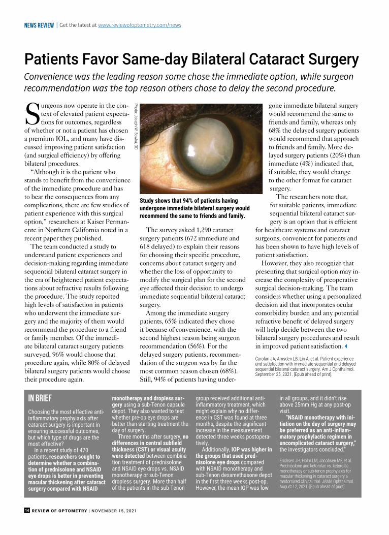
Study shows that 94% of patients havingundergone immediate bilateral surgery wouldrecommend the same to friends and family.
Photo: Joseph W. Sow
ka, OD
IN BRIEFChoosing the most effective anti-inflammatory prophylaxis after cataract surgery is important in ensuring successful outcomes, but which type of drugs are the most effective?
In a recent study of 470 patients, researchers sought todetermine whether a combina-tion of prednisolone and NSAIDeye drops is better in preventingmacular thickening after cataractsurgery compared with NSAID
monotherapy and dropless sur-gery using a sub-Tenon capsule depot. They also wanted to test whether pre-op eye drops are better than starting treatment the day of surgery.
Three months after surgery, nodifferences in central subfield thickness (CST) or visual acuitywere detected between combina-tion treatment of prednisolone and NSAID eye drops vs. NSAID monotherapy or sub-Tenon dropless surgery. More than half of the patients in the sub-Tenon
group received additional anti-inflammatory treatment, which might explain why no differ-ence in CST was found at three months, despite the significant increase in the measurement detected three weeks postopera-tively.
Additionally, IOP was higher inthe groups that used pred-nisolone eye drops compared with NSAID monotherapy and sub-Tenon dexamethasone depot in the first three weeks post-op. However, the mean IOP was low
in all groups, and it didn’t rise above 25mm Hg at any post-op visit.
“NSAID monotherapy with ini-tiation on the day of surgery maybe preferred as an anti-inflam-matory prophylactic regimen inuncomplicated cataract surgery,”the investigators concluded."
Erichsen JH, Holm LM, Jacobsen MF, et al.Prednisolone and ketorolac vs. ketorolacmonotherapy or sub-tenon prophylaxis formacular thickening in cataract surgery arandomized clinical trial. JAMA Ophthalmol.August 12, 2021. [Epub ahead of print].

LOMBART IS PART OF ADVANCING EYECARE ONE ALLIANCE • SIX INDUSTRY LEADERS • COMPREHENSIVE OPHTHALMIC SOLUTIONS
1-800-LOMBART LOMBARTINSTRUMENT.COM
BUNDLE & SAVELOMBART LANE PACKAGESSTARTING AT$14,495
ALL THE ESSENTIALS
Lombart CS–6 Chair and StandMarco B2 Slit LampS4Optik SL–Y100 RefractorLombart CVS–2 or VA–1 Acuity SystemLombart Brewer Stool with Back
CORE
STANDARDGREAT VALUE
Marco Encore Manual ChairMarco Deluxe StandMarco Ultra M2 Slit LampLombart CVS–2 or VA–1 Acuity SystemMarco RT–700 Illuminated RefractorMarco Circular Ring Stool with Back
PROFESSIONALWOW YOUR PATIENTS
Marco EZ Tilt or Encore Automatic ChairMarco Deluxe StandMarco Ultra M5 Slit LampLombart CVS–2 or VA–1 Acuity SystemMarco RT–700 Illuminated RefractorMarco Circular Ring Stool with Back
PRICES SUBJECT TO CHANGE

WE’LL HELP YOU LISTEN.GENES ARE TALKING.
75 genes and >2,000 variants power AvaGen™, the first and leading personalized genetic eye test. AvaGen quantifies the genetic
risk or presence of keratoconus and other corneal genetic disorders caused by
gene variants. AvaGen delivers a valuable tool for early and accurate decision-
making that protects and improves vision for patients and their families.
Avellino.com/avagenKnow early. Act personally. Decide confidently.
© 2021 Avellino Lab USA, Inc.
Know the genetic risk or presence of keratoconus and other corneal conditions.

NOVEMBER 15, 2021 | REVIEW OF OPTOMETRY 17
featuresREVIEW OF OPTOMETRY • Vol. 158, No. 11 • NOVEMBER 15, 2021
CATCH UP ON THE LATEST NEWSStories post online every weekdayWeekly recap emailed every Sunday
66Dry Eye: Where DoWe Stand with Omega-3Supplements?A review of recent research for andagainst this treatment supplementation.By Luis Rojas, OD
382021 DESIGN CONTEST
Eye-Popping Office SpacesODs bounced back from the COVID doldrumswith bold and adventurous new design ideas.
74Anterior Blepharitis:The Front Line of OSDWe dissect everything you need to knowabout this common condition, includingproper treatment and management, andhow it can be a sign of Demodex.By Andrew McLeod, OD,and Amy Nau, OD
48The ConjunctivaUp CloseHere’s how to recognize andmanage a few common conditionsthat affect this part of the eye.By Megan Mannen, OD
56Beneath the Mask:OSD Issues Spike Dueto Improper WearSince the onset of COVID, doctors havenoted an increase in dry eye, chalazion,blepharitis and hordeolum cases. Here,several experts offer diagnosis andtreatment tips.By Jane Cole, Contributing Editor
OCULAR SURFACE ISSUE
85When Your Patient Complains of Red EyeThe key to uncovering the root cause is a thorough patient history and clinical exam.By Suzanne Sherman, OD, and Christina Cherny, OD
—EARN 2 CE CREDITS

* Xiidra blocks LFA-1 on T cells from binding with ICAM-1 that may be overexpressed on the ocular surface in dry eye disease andmay prevent formation of an immunologic synapse which, based on in vitro studies, may inhibit T-cell activation, migration ofactivated T cells to the ocular surface, and reduce cytokine release. The exact mechanism of action of Xiidra in DED is not known.1,2,5
† The safety and effi cacy of Xiidra were assessed in four 12-week, randomized, multicenter, double-masked, vehicle controlled studies (N=2133). Patients were dosed twice daily. The mean age was 59 years (range, 19-97 years). The majority of patients were female(76%). Use of artifi cial tears was not allowed during the studies. The study end points included assessment of signs (based on Inferior fl uorescein Corneal Staining Score [ICSS] on a scale of 0 to 4) and symptoms (based on patient-reported EDS on a visual analogue scale of 0 to 100). Effects on symptoms of dry eye disease: a larger reduction in EDS favoring Xiidra was observed in all studies at day 42 and day 84. Xiidra reduced symptoms of eye dryness at 2 weeks (based on EDS) compared to vehicle in 2 out of 4 clinical trials. Effects on signs of dry eye disease: at day 84, a larger reduction in ICSS favoring Xiidra was observed in 3 out of the 4 studies.1
IndicationXiidra® (lifi tegrast ophthalmic solution) 5% is indicated for the treatment of signs and symptoms of dry eye disease (DED).Important Safety Information• Xiidra is contraindicated in patients with known hypersensitivity to lifi tegrast or to any of the other ingredients.
Novartis Pharmaceuticals CorporationEast Hanover, New Jersey 07936-1080
When patients rely on artifi cial tears alone, infl ammation may persist. Xiidra can disrupt the chronic infl ammatory cycle in dry eye disease.*
It can provide lasting symptom relief in as little as 2 weeks.1-5†
Dry eyes deserve a change
References: 1. Xiidra [package insert]. East Hanover, NJ: Novartis Pharmaceuticals Corp; June 2020. 2. Bron AJ, de Paiva CS, Chauhan SK, et al.TFOS DEWS II Pathophysiology Report. Ocul Surf. 2017;15(3):438-510. 3. US Food and Drug Administration. Code of Federal Regulations, Title 21,Volume 5 (21CFR349). Accessed May 25, 2021. https:/www.accessdata.fda.gov/scripts/cdrh/cfdocs/cfcfr/CFRSearch.cfm?CFRPart=349&showFR=14. Jones L, Downie LE, Korb D, et al. TFOS DEWS II Management and Therapy Report. Ocul Surf. 2017;15(3):575-628. 5. Pfl ugfelder SC, Stern M, Zhang S, Shojaei A. LFA-1/ICAM-1 interaction as a therapeutic target in dry eye disease. J Ocul Pharmacol Ther. 2017;33(1):5-12.
XIIDRA, the XIIDRA logo and ii are registered trademarks of Novartis AG.
123809 © 2021 Novartis 5/21
Important Safety Information (cont)• In clinical trials, the most common adverse reactions reported in 5-25% of patients were instillation site irritation,
dysgeusia and reduced visual acuity. Other adverse reactions reported in 1% to 5% of the patients were blurredvision, conjunctival hyperemia, eye irritation, headache, increased lacrimation, eye discharge, eye discomfort,eye pruritus and sinusitis.
• To avoid the potential for eye injury or contamination of the solution, patients should not touch the tip of thesingle-use container to their eye or to any surface.
• Contact lenses should be removed prior to the administration of Xiidra and may be reinserted 15 minutesfollowing administration.
• Safety and effi cacy in pediatric patients below the age of 17 years have not been established.
For additional safety information about XIIDRA®, please refer to the brief summary of PrescribingInformation on adjacent page.
KEN JEONG,REAL DRY EYE PATIENT.
XIIDRA® (lifitegrast ophthalmic solution), for topical ophthalmic use Initial U.S. Approval: 2016 BRIEF SUMMARY: Please see package insert for full prescribing information. 1 INDICATIONS AND USAGE
Xiidra® (lifitegrast ophthalmic solution) 5% is indicated for the treatment of the signs and symptoms of dry eye disease (DED).
4 CONTRAINDICATIONS Xiidra is contraindicated in patients with known hypersensitivity to lifitegrast or to any of the other ingredients in the formulation [see Adverse Reac-tions (6.2)].
6 ADVERSE REACTIONS The following serious adverse reactions are described elsewhere in the labeling: • Hypersensitivity [see Contraindications (4)] 6.1 Clinical Trials Experience Because clinical trials are conducted under widely varying conditions, adverse reaction rates observed in clinical trials of a drug cannot be directly compared to rates in the clinical trials of another drug and may not reflect the rates observed in practice. In five clinical trials of DED conducted with lifitegrast ophthalmic solution, 1401 patients received at least one dose of lifitegrast (1287 of which received lifitegrast 5%). The majority of patients (84%) had less than or equal to 3 months of treatment exposure. One hundred-seventy patients were exposed to lifitegrast for approximately 12 months. The majority of the treated patients were female (77%). The most common adverse reac-tions reported in 5%-25% of patients were instillation-site irritation, dys-geusia, and reduced visual acuity. Other adverse reactions reported in 1%-5% of the patients were blurred vision, conjunctival hyperemia, eye irritation, headache, increased lacri-mation, eye discharge, eye discomfort, eye pruritus, and sinusitis. 6.2 Postmarketing Experience The following adverse reactions have been identified during post-approval use of Xiidra. Because these reactions are reported voluntarily from a pop-ulation of uncertain size, it is not always possible to reliably estimate their frequency or establish a causal relationship to drug exposure. Rare serious cases of hypersensitivity, including anaphylactic reaction, bronchospasm, respiratory distress, pharyngeal edema, swollen tongue, urticaria, allergic conjunctivitis, dyspnea, angioedema, and allergic derma-titis have been reported. Eye swelling and rash have also been reported [see Contraindications (4)].
8 USE IN SPECIFIC POPULATIONS 8.1 Pregnancy Risk Summary There are no available data on Xiidra use in pregnant women to inform any drug-associated risks. Intravenous (IV) administration of lifitegrast to
pregnant rats, from premating through gestation day 17, did not produce teratogenicity at clinically relevant systemic exposures. Intravenous administration of lifitegrast to pregnant rabbits during organogenesis produced an increased incidence of omphalocele at the lowest dose tested, 3 mg/kg/day (400-fold the human plasma exposure at the recommended human ophthalmic dose [RHOD], based on the area under the curve [AUC] level). Since human systemic exposure to lifitegrast following ocular administration of Xiidra at the RHOD is low, the applicability of animal findings to the risk of Xiidra use in humans during pregnancy is unclear [see Clinical Pharmacology (12.3) in the full prescribing information]. Data Animal Data Lifitegrast administered daily by IV injection to rats, from premating through gestation day 17, caused an increase in mean pre-implantation loss and an increased incidence of several minor skeletal anomalies at 30 mg/kg/day, representing 5,400-fold the human plasma exposure at the RHOD of Xiidra, based on AUC. No teratogenicity was observed in the rat at 10 mg/kg/day (460-fold the human plasma exposure at the RHOD, based on AUC). In the rabbit, an increased incidence of omphalocele was observed at the lowest dose tested, 3 mg/kg/day (400-fold the human plasma exposure at the RHOD, based on AUC), when administered by IV injection daily from gestation days 7 through 19. A fetal no observed adverse effect level (NOAEL) was not identified in the rabbit. 8.2 Lactation Risk Summary There are no data on the presence of lifitegrast in human milk, the effects on the breastfed infant, or the effects on milk production. However, sys-temic exposure to lifitegrast from ocular administration is low [see Clinical Pharmacology (12.3) in the full prescribing information]. The develop-mental and health benefits of breastfeeding should be considered, along with the mother’s clinical need for Xiidra and any potential adverse effects on the breastfed child from Xiidra. 8.4 Pediatric Use Safety and efficacy in pediatric patients below the age of 17 years have not been established. 8.5 Geriatric Use No overall differences in safety or effectiveness have been observed between elderly and younger adult patients.
Distributed by: Novartis Pharmaceuticals Corporation One Health Plaza East Hanover, NJ 07936 T2020-87

NOVEMBER 15, 2021 | REVIEW OF OPTOMETRY 21
departments4NEWS REVIEW
30THROUGH MY EYESOwning OSDNew treatments can help us keep up with thisperennial problem.
Paul M. Karpecki, OD
REVIEW OF OPTOMETRY • NOVEMBER 15, 2021
96THE ESSENTIALSReach for the DyeFluorescein is most commonly used for cornealevaluation and, as such, its instillation should beproperly understood.
Bisant A. Labib, OD
104RETINA QUIZA Closer LookA surprising discovery led to this diagnosis.Mark Dunbar, OD
32CHAIRSIDEMore Like Mid-career CrisisWhatever the catalyst, you should just go on andget it over with.Montgomery Vickers, OD
26LETTERS TO THE EDITORFeedback and ideas from the optometric community.
100URGENT CARESafety FirstAn open globe can cause vision loss and intraocularinfection, but preventing these injuries is possible.
Kristen Walton, OD
Facebook: www.facebook.com/revoptomTwitter: twitter.com/revoptom
Instagram: www.instagram.com/revoptomVISIT US ON SOCIAL MEDIA
36CODING CONNECTIONInnovate for SuccessEducate yourself when approaching newtechnology.
John Rumpakis, OD, MBA
24OUTLOOKGood VibrationsPatients experience your office environment beforethey see you. Use it to create a great first impression.
Jack Persico, Editor-in-Chief
108PRODUCT REVIEWNew items to improve clinical care.
34CLINICAL QUANDARIESOut of OrbitProlapse of fat can disturb patients but may not beharmful.
Paul C. Ajamian, OD
94CORNEA AND CONTACT LENS Q+AScleral Decentration:The WorkaroundCenter-near aspherics help achieve multifocalcorrection with this modality, but they don’t comewithout problems.Joseph P. Shovlin, OD
114DIAGNOSTIC QUIZA Spot of TroubleNo one wants to see a dark patch in the posteriorsegment. What factors help you weigh its significance?Andrew S. Gurwood, OD

REVIEW OF OPTOMETRY | NOVEMBER 15, 202122
CONTRIBUTING EDITORS
Leadership in clinical care
EDITORIAL ADVISORY BOARD
REVIEW OF OPTOMETRY (ISSN 0147-7633) IS PUBLISHED MONTHLY, 12 TIMES A YEAR BY JOBSON MEDICAL INFORMATION LLC, 395 HUDSON STREET, 3RD FLOOR FLOOR, NEW YORK, NY 10014.PERIODICALS POSTAGE PAID AT NEW YORK, NY AND ADDITIONAL MAILING OFFICES. POSTMASTER: SEND ADDRESS CHANGES TO REVIEW OF OPTOMETRY, PO BOX 81, CONGERS, NY 10920-0081.SUBSCRIPTION PRICES: US: ONE YEAR $56; TWO YEARS $97, CANADA: ONE YEAR $88, TWO YEARS $160, INT’L: ONE YEAR $209, TWO YEARS $299. FOR SUBSCRIPTION INFORMATION CALL TOLL-FREE(877) 529-1746 (USA); OUTSIDE USA, CALL (845) 267-3065. OR EMAIL US AT [email protected]. PUBLICATIONS MAIL AGREEMENT NO: 40612608. CANADA RETURNS TO BE SENT TOBLEUCHIP INTERNATIONAL, P.O. BOX 25542, LONDON, ON N6C 6B2.
Business Offices19 Campus Boulevard, Suite 101
Newtown Square, PA 19073Subscription inquiries (877) 529-1746 (USA only)
outside USA, call (847) 763-9630
PUBLISHERMICHAEL HOSTER
(610) [email protected]
EXECUTIVE DIRECTORJAMES HENNE(610) 492-1017
SENIOR MANAGER, STRATEGIC ACCOUNTSMICHELE BARRETT
(610) [email protected]
REGIONAL SALES MANAGERJONATHAN DARDINE
(610) [email protected]
PRODUCTION MANAGERFARRAH APONTE
212-274-7057 [email protected]
PRODUCTION MANAGERKAREN LALLONE
(610) 492-1010 [email protected]
CLASSIFIED ADVERTISING(888)-498-1460
SUBSCRIPTIONS$63 PER YEAR, $99 (US) IN CANADA,$158 (US) IN ALL OTHER COUNTRIES
CIRCULATIONPO BOX 71, CONGERS, NY 10920-0071
(877) 529-1746OUTSIDE USA: (845) 267-3065
SENIOR CIRCULATION MANAGERHAMILTON MAHER
(212) [email protected]
CEO, INFORMATION GROUP SERVICESMARC FERRARA
SENIOR VICE PRESIDENT, OPERATIONSJEFF LEVITZ
VICE PRESIDENT, HUMAN RESOURCESTAMMY GARCIA
VICE PRESIDENT, CREATIVE SERVICES & PRODUCTIONMONICA TETTAMANZI
CORPORATE PRODUCTION DIRECTORJOHN ANTHONY CAGGIANO
VICE PRESIDENT, CIRCULATIONJARED SONNERS
Jobson Health Information/WebMD395 Hudson Street, 3rd Floor, New York, NY 10014
PAUL C. AJAMIAN, OD, ATLANTA
DEREK N. CUNNINGHAM, OD, AUSTIN, TEXAS
MARK T. DUNBAR, OD, MIAMI
JAMES L. FANELLI, OD, WILMINGTON, NC
ANDREW S. GURWOOD, OD, PHILADELPHIA
PAUL HARRIS, OD, MEMPHIS, TENN.
PAUL M. KARPECKI, OD, LEXINGTON, KY.
BISANT LABIB, OD, ELKINS PARK, PA.
RICHARD B. MANGAN, OD, BOULDER, COLO.
JOHN RUMPAKIS, OD, MBA, PORTLAND, ORE.
JOSEPH P. SHOVLIN, OD, SCRANTON, PA.
JOSEPH W. SOWKA, OD, FORT LAUDERDALE, FLA.
MARC TAUB, OD, MEMPHIS, TENN.
MONTGOMERY VICKERS, OD, DALLAS, TEXAS
WALTER O. WHITLEY, OD, MBA, VIRGINIA BEACH, VA.
JEFFREY R. ANSHEL, OD, ENCINITAS, CALIF.
JILL AUTRY, OD, RPH, HOUSTON
SHERRY J. BASS, OD, NEW YORK
EDWARD S. BENNETT, OD, ST. LOUIS
MARC R. BLOOMENSTEIN, OD, SCOTTSDALE, ARIZ.
AARON BRONNER, OD, KENNEWICK, WASH.
MILE BRUJIC, OD, BOWLING GREEN, OHIO
CHRIS J. CAKANAC, OD, MURRYSVILLE, PA
JERRY CAVALLERANO, OD, PhD, BOSTON
WALTER L. CHOATE, OD, MADISON, TENN.
BRIAN CHOU, OD, SAN DIEGO
MICHAEL CHAGLASIAN, OD, CHICAGO
A. PAUL CHOUS, MA, OD, TACOMA, WASH.
GLENN S. CORBIN, OD, WYOMISSING, PA
MICHAEL DELGIODICE, OD, CLIFTON, NJ
ANTHONY S. DIECIDUE, OD, STROUDSBURG, PA
S. BARRY EIDEN, OD, DEERFIELD, ILL.
ARTHUR B. EPSTEIN, OD, PHOENIX
STEVEN FERRUCCI, OD, SEPULVEDA, CALIF.
MURRAY FINGERET, OD, HEWLETT, NY
IAN BEN GADDIE, OD, LOUISVILLE, KY
GARY S. GERBER, OD, HAWTHORNE, NJ
PAUL HARRIS, OD, MEMPHIS, TENN.
MILTON HOM, OD, AZUSA, CALIF.
DAVID KADING, OD, SEATTLE
JEROME A. LEGERTON, OD, MBA, SAN DIEGO
THOMAS L. LEWIS, OD, PhD, PHILADELPHIA
BLAIR B. LONSBERRY, MS, OD, MED, PORTLAND, ORE.
DOMINICK MAINO, OD, MED, CHICAGO
KELLY A. MALLOY, OD, PHILADELPHIA
RICHARD B. MANGAN, OD, BOULDER, COLO.
DANICA MARRELLI, OD, HOUSTON, TEX.
RON MELTON, OD, CHARLOTTE, NC
PAMELA J. MILLER, OD, JD, HIGHLAND, CALIF.
BRUCE MUCHNICK, OD, COATESVILLE, PA
MARC MYERS, OD, COATESVILLE, PA
CARLO J. PELINO, OD, JENKINTOWN, PA
JOSEPH PIZZIMENTI, OD, FORT LAUDERDALE, FLA.
CHRISTOPHER J. QUINN, OD, ISELIN, NJ
MICHAEL C. RADOIU, OD, STAUNTON, VA
MOHAMMAD RAFIEETARY, OD, MEMPHIS, TENN.
JOHN L. SCHACHET, OD, ENGLEWOOD, COLO.
JACK SCHAEFFER, OD, BIRMINGHAM, ALA.
LEO P. SEMES, OD, BIRMINGHAM, ALA.
DIANA L. SHECHTMAN, OD, FORT LAUDERDALE, FLA.
JEROME SHERMAN, OD, NEW YORK, NY
LEONID SKORIN, JR., OD, DO, ROCHESTER, MINN.
JOSEPH W. SOWKA, OD, FORT LAUDERDALE, FLA.
BRAD M. SUTTON, OD, INDIANAPOLIS
LORETTA B. SZCZOTKA, OD, PhD, CLEVELAND
MARC TAUB, OD, MEMPHIS, TENN.
TAMMY P. THAN, MS, OD, BIRMINGHAM, ALA.
RANDALL THOMAS, OD, CONCORD, NC
SARA WEIDMAYER, OD, ANN ARBOR, MICH.
KAREN YEUNG, OD, LOS ANGELES
CLINICAL EDITORSCHIEF CLINICAL EDITOR ~ PAUL M. KARPECKI, OD
ASSOCIATE CLINICAL EDITORS ~ JOSEPH P. SHOVLIN, OD, CHRISTINE SINDT, OD

REVIEW OF OPTOMETRY | NOVEMBER 15, 202124
Founded 1891Founding Editor, Frederick Boger
EDITOR-IN-CHIEFJACK PERSICO
(610) 492-1006 • [email protected]
SENIOR EDITORJULIE SHANNON
(610) 492-1005 • [email protected]
SENIOR ASSOCIATE EDITORCATHERINE MANTHORP
(610) 492-1043 • [email protected]
SENIOR ASSOCIATE EDITORMARK DE LEON
(610) 492-1021 • [email protected]
ASSOCIATE EDITORLEANNE SPIEGLE
(610) 492-1026 • [email protected]
SPECIAL PROJECTS MANAGERJILL GALLAGHER
(610) 492-1037 • [email protected]
SENIOR ART DIRECTORJARED ARAUJO
(610) 492-1032 • [email protected]
DIRECTOR OF CE ADMINISTRATIONREGINA COMBS
(212) 274-7160 • [email protected]
Clinical Editors
Chief Clinical Editor • Paul M. Karpecki, OD
Associate Clinical EditorsJoseph P. Shovlin, OD, Christine W. Sindt, OD
Clinical & Education Conference AdvisorPaul M. Karpecki, OD
Case Reports Coordinator • Andrew S. Gurwood, OD
Clinical Coding Editor • John Rumpakis, OD, MBA
ColumnistsChairside – Montgomery Vickers, OD
Clinical Quandaries – Paul C. Ajamian, ODCoding Connection – John Rumpakis, OD
Cornea and Contact Lens Q+A – Joseph P. Shovlin, ODDiagnostic Quiz – Andrew S. Gurwood, OD
The Essentials – Bisant A. Labib, ODFocus on Refraction – Marc Taub, OD, Paul Harris, OD
Glaucoma Grand Rounds – James L. Fanelli, ODOcular Surface Review – Paul M. Karpecki, OD
Retina Quiz – Mark T. Dunbar, ODSurgical Minute – Derek Cunningham, OD, Walter Whitley, OD
Therapeutic Review – Joseph W. Sowka, ODThrough My Eyes – Paul M. Karpecki, OD
Urgent Care – Richard B. Mangan, OD
Editorial Offices
19 Campus Blvd., Suite 101• Newtown Square, PA 19073
Jobson Medical Information/WebMD395 Hudson Street, 3rd Floor, New York, NY 10014
Subscription inquiries: (877) 529-1746Continuing Education inquiries: (800) 825-4696
Printed in USA
The first optometry office I wentto—as a kid in the 1970s—wasin the converted basement of myfriend Isaac’s house. It had, yes,
the infamous shag carpeting and wood-paneled walls of that era. Dependingon the time of your appointment, youmay very well have been able to hearIsaac’s mom making dinner upstairs.The whole place was cozy and dishev-eled and informal, just like my friend’sdad, the OD of the practice.
That vibe prevailed at lots of optom-etry offices in the ’70s, and surely wellinto the ’80s too at many locations. Butas the optometry profession migratedmore and more toward mainstreammedical care in the intervening years,the aesthetic of most practices camealong with it. White coats, clean spaces,salaried staff (instead of family mem-bers) and increasingly more high-techequipment all became the standardlook and feel.
Fair or not, presentation mattersat least to some extent in what thepublic thinks and expects, especiallyof the professionals they turn to for thefrequently sensitive work of discuss-ing their bodies and lives. So, I wasencouraged to see the impressive arrayof entries in our biennial office designcontest, the results of which are show-cased this month on page 38.
Again and again we saw, in the essaysthat contestants sent in, how muchthese doctors prioritize the “feel”of being at their practices instead ofmerely the results.
“The aesthetics of the office is oftenthe highlight of the overall experi-ence,” said one practice owner in thecontest. “Patients who are passing bythe office often take a moment to come
inside just to take photos.” Imaginewanting to go to a doctor’s office evenif you don’t need to. Can you say thatabout the physicians you see? Meneither.
Typical redesign goals cited bymost of this year’s contestants in-cluded open floorplans, upscale décor,accommodations for special needs,quiet and uncluttered waiting rooms,high-quality lighting and other fixtures,social media–friendly spaces… the listgoes on and on. I won’t spoil the funbut there’s definitely also some realpersonality shining through in at leastone of the entries this year.
None of this means that you abso-lutely need to plow hundreds of thou-sands of dollars into a splashy officedesign to succeed in this profession.For many practices, that’s just not onthe table, especially these days as wecollectively are still trying to reboundfrom a pandemic-induced downturn.
So, pick your battles. Not everyoneneeds an espresso bar and an Instagramwall and other niceties. Maybe yourphysical space limitations preclude theairy layouts that others can achieve.But it’s still worth putting yourself inyour patients’ shoes once in a while tosee how moving through the office, sta-tion by station, makes you feel.
Most practices have already donean overhaul to accommodate COVIDsafety protocols. The addition of PPEhas already imbued your practicewith a more medical feel if that hadbeen lacking. That’s great—just don’toverlook whatever comforts, physicalor emotional, you can create, too.
And if your design philosophy isretro kitsch, I know where you can geta good deal on vintage shag. g
Patients experience your office environment before (and after)they see you. Use it to create a great first impression.
Good Vibrations
By Jack PersicoEditor-in-Chief
OUTLOOK
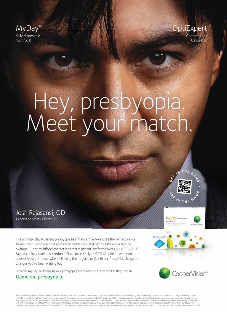
Josh Rajasansi, OD Experts on Sight | Gilbert, AZ
Hey, presbyopia.Meet your match.
OptiExpert™MyDay®
daily disposablemultifocal
Contact LensCalculator
1. 3.5 times more patients preferred MyDay® multifocal for overall vision clarity than with DAILIES TOTAL1® Multifocal. Strongly prefer/slight prefer MyDay® multifocal 58.6%, DAILIES TOTAL1® Multifocal 17.2%, no preference 24.1%; p=0.002. For statistical analysis, “no preference” response evenly distributed between two products.] [REF: CVI data on file 2021. Prospective, subject-masked, randomized, bilateral, two-week cross-over dispensing study at 5 US sites with MyDay® multifocal and DAILIES TOTAL1® Multifocal; n=58 habitual multifocal contact lens wearers. 2. CVI data on file 2021. Prospective, subject-masked, randomized, bilateral, two-week cross-over dispensing study at 5 US sites with MyDay® multifocal and DAILIES TOTAL1® Multifocal; n=58 habitual multifocal contact lens wearers. 3. CVI data on file 2020. Prospective, double-masked, bilateral, one-week dispensing study UK with MyDay® multifocal; n=104 habitual multifocal contact lens wearers; CVI data on file 2021. Prospective, subject-masked, randomized, bilateral, two-week dispensing study at 5 US sites with MyDay® multifocal; n=58 habitual multifocal contact lens wearers. 11082A
Game on, presbyopia. Prescribe MyDay® multifocal to your presbyopic patients and help them see like they used to.
The ultimate play to defeat presbyopia has finally arrived—and it’s the winning move to keep your presbyopic patients in contact lenses. MyDay® multifocal is a silicone hydrogel 1-day multifocal contact lens that is patient-preferred over DAILIES TOTAL1®
Multifocal for vision1 and comfort.2 Plus, successfully fit 98% of patients with two pairs of lenses or fewer when following the fit guide or OptiExpert™ app.3 It’s the game changer you’ve been waiting for.
GGEE TT II NN TT HH EE GG AA MM
EE
GGEE
TT II NN TT HH EE GG AAMM
EE
Low Vision Can Flourishin Private Practice■ The August issue contains anexcellent article on intraprofessionalcollaboration called, “Mastering OD-to-OD Referrals,” which includes asection on the importance of address-ing low vision with every affected pa-tient. Thank you for raising awarenessof the need for more low vision care,whether provided directly or coman-aged with a doctor suitably skilledfor it.
However, there is acomment in the lowvision discussion that is,in our opinion, inaccu-rate. A doctor states that“proper low vision careis time-consuming andvery diffi cult to fi t into aprivate practice model.”The InternationalAcademy of Low VisionSpecialists (IALVS),established in 2006, has 40 low visionoptometrists throughout the UnitedStates and Canada who are in privatepractice. All of our doctors are fullytrained and experienced to providelow vision care and rehabilitation inthe private practice setting.
—Richard J. Shuldiner, OD, FAAOPresident, International Academy
of Low Vision Specialists
Clinical Director, Low VisionOptometry of Southern California
Low Vision Diplomate Emeritus,American Academy of Optometry
Vision Rehab Can Seta Practice Apart■ Reading the feature, “MasteringOD-to-OD Referrals,” I was blownaway by some of the opinions regard-ing low vision—and not in a good way.
There’s a comment stating thatmost low vision services are offeredtoday at academic and non-profi tinstitutions rather than in privatepractice and that this isn’t likely tochange “until insurance reimburse-
ment structures forlow vision devices andservices are altered tolessen the time andfi nancial burdens thatmake it impractical forprivate practice ODs totake part.”
My wife, Dr. LynneNoon, and myself are1982 graduates of theNew England Collegeof Optometry. I chose a
career in commercial optometry whileLynne chose vision rehabilitation andsubsequently achieved Diplomatestatus in the discipline of low vision.
She started her solo practice fromscratch doing only vision rehab.She spent countless hours meetingwith state agencies and other visionrehabilitation experts. She traveledthroughout Arizona and the sur-rounding Southwest region doing lowvision exams and lecturing to sup-port groups, optometrists and othermedical professionals. Through herhard work and that of others, shehelped Arizona adopt biopic drivingprivileges.
We have both recently retired. Buthere’s the kicker! It literally tookyears to fi nd someone to purchase ourpractice even though it grossed over$2 million annually. What does thatsay about our profession? The medicalmodel is being taught and pushed topractitioners, yet insurance reimburse-ments are not adequate. The key toany successful business is to provide aservice no one else provides and, bet-ter yet, a business that doesn’t rely oninsurance reimbursement for products.
Low vision rehabilitation currentlymeets those two criteria.
The “social welfare model” for lowvision care is ancient history. Do notassume these patients are poor andunable to pay for such products andservices.
And, as has been proven time andtime again, insurance companies arenot our friends! We rely on them atour own peril—and our patients’ too.
—Robert Kanocz, ODMesa, AZ
From the Editor: The experience ofDr. Shuldiner and his colleagues in theIALVS does show that low vision canthrive in private practice. However, it’snot a contradiction to also acknowledgethat ramping up a practice devoted tosuch care—and ensuring such patientsreceive the additional time and atten-tion that a typically frail populationrequires—can be time-consuming and,to many, intimidating.
Dr. Kanocz’s anecdote about thediffi culty he encountered selling hispractice seems to document that wide-spread hesitation does exist amongindividual doctors and/or investors.Rightly or wrongly, there’s at least aperceived barrier to entry. We thankboth letter-writers for modelling suc-cess in this important area of care. Let’shope others follow in their footsteps.
letters to the editor SHARE YOUR THOUGHTSLetters are welcome. Write to:
Submissions may be edited for length,content or clarity.Feedback and ideas from the optometric community.
Don’t be so quick to write off its business viability, readers argue.The Lowdown on Low Vision
Here’s how to build lasting relationships withyour peers to provide more complete care.
Page 58
August 15, 2021 • reviewofoptometry.com
EARN 2 CE CREDITS: WHAT TO DO WHEN YOU SEE UVEIT IS, Page 64
• New Products in Practice• Lens/Tear Interaction• Toric Lens Fitting Tips
• CL Drug Delivery
Leadership in clinical care
masteringOD-to-ODreferrals
COMANAGEMENTCONNECTIONS
Series Finale
Part 6 of 6
45th ANNUALCONTACT LENS REPORT
Begins p. 32
REVIEW OF OPTOM
ETRY • VOL. 158, NO. 8 • AUGUST 15, 2021 • New
Contact Lenses • Lens/Tear Interaction
• Toric Lens Fitting Principles • Contact Lens Drug Delivery
• Intraprofessional Referrals • Uveitis Care
001_ro0821_fc.indd 1 8/4/21 2:17 PM
26 REVIEW OF OPTOMETRY | NOVEMBER 15, 2021

CEQUA is engineered to delivercyclosporine (CsA) where it’s needed most1,2
Encapsulates CsA1
Penetrates the aqueous layer1
Delivers medicine to the ocular tissue1,3
NCELL Technology:
Watch the mechanism of NCELLin action at CequaPro.com
INDICATIONS AND USAGECEQUA™ (cyclosporine ophthalmic solution) 0.09% is acalcineurin inhibitor immunosuppressant indicated to increasetear production in patients with keratoconjunctivitis sicca(dry eye).
IMPORTANT SAFETY INFORMATIONWARNINGS AND PRECAUTIONSPotential for Eye Injury and Contamination: To avoidthe potential for eye injury and contamination, advisepatients not to touch the vial tip to the eye or other surfaces.
Use with Contact Lenses: CEQUA should not beadministered while wearing contact lenses. If contact lensesare worn, they should be removed prior to administration ofthe solution. Lenses may be reinserted 15 minutes followingadministration of CEQUA ophthalmic solution.
ADVERSE REACTIONSThe most common adverse reactions reported in greaterthan 5% of patients were pain on instillation of drops (22%)and conjunctival hyperemia (6%). Other adverse reactionsreported in 1% to 5% of patients were blepharitis, eyeirritation, headache, and urinary tract infection.
Please see brief summary of Full PrescribingInformation on the following page.
References: 1. US Patent 9,937,225 B2. 2. Cholkar K, Patel A, VadlapudiAD, Mitra AK. Novel nanomicellar formulation approaches for anteriorand posterior segment ocular drug delivery. Recent Pat Nanomed.2012;2(2):82-95. 3. Data on � le. Cranbury, NJ: Sun PharmaceuticalIndustries, Inc.
© 2021 Sun Ophthalmics, a division of Sun Pharmaceutical Industries, Inc. All rights reserved. CEQUA and NCELL are trademarks of Sun Pharma Global FZE.PM-US-CQA-0831 08/2021
Brief Summary of Prescribing Information for CEQUA™ (cyclosporine ophthalmic solution) 0.09%, for topical ophthalmic use
CEQUA™ (cyclosporine ophthalmic solution) 0.09%See package insert for Full Prescribing Information.
INDICATIONS AND USAGECEQUA ophthalmic solution is a calcineurin inhibitor immunosuppressant indicated to increase tear production in patients with keratoconjunctivitis sicca (dry eye).
CONTRAINDICATIONSNone.
WARNINGS AND PRECAUTIONSPotential for Eye Injury and Contamination To avoid the potential for eye injury and contamination, advise patients not to touch the vial tip to the eye or other surfaces.
Use with Contact Lenses CEQUA should not be administered while wearing contact lenses. If contact lenses are worn, they should be removed prior to administration of the solution. Lenses may be reinserted 15 minutes following administration of CEQUA ophthalmic solution.
ADVERSE REACTIONSClinical Trials Experience Because clinical trials are conducted under widely varying conditions, adverse reaction rates observed in the clinical trials of a drug cannot be directly compared to rates in the clinical trials of another drug and may not re�ect the rates observed in practice.
In clinical trials, 769 patients received at least 1 dose of cyclosporine ophthalmic solution. The majority of the treated patients were female (83%).
The most common adverse reactions reported in greater than 5% of patients were pain on instillation of drops (22%) and conjunctival hyperemia (6%). Other adverse reactions reported in 1% to 5% of patients were blepharitis, eye irritation, headache, and urinary tract infection.
USE IN SPECIFIC POPULATIONSPregnancyRisk SummaryThere are no adequate and well-controlled studies of CEQUA administration in pregnant women to inform a drug-associated risk. Oral administration of cyclosporine to pregnant rats or rabbits did not produce teratogenicity at clinically relevant doses.
DataAnimal DataOral administration of cyclosporine oral solution (USP) to pregnant rats or rabbits was teratogenic at maternally toxic doses of 30 mg/kg/day in rats and 100 mg/kg/day in rabbits, as indicated by increased pre- and postnatal mortality, reduced fetal weight, and skeletal retardations. These doses (normalized to body weight) were approximately 3200 and 21,000 times higher than the maximum recommended human ophthalmic dose (MRHOD) of 1.5 mcg/kg/day, respectively. No adverse embryofetal effects were observed in rats or rabbits receiving cyclosporine during organogenesis at oral doses up to 17 mg/kg/day or 30 mg/kg/day, respectively (approximately 1800 and 6400 times higher than the MRHOD, respectively).
An oral dose of 45 mg/kg/day cyclosporine (approximately 4800 times higher than MRHOD) administered to rats from Day 15 of pregnancy until Day 21 postpartum produced maternal toxicity and an increase in postnatal mortality in offspring. No adverse effects in dams or offspring were observed at oral doses up to 15 mg/kg/day (approximately 1600 times greater than the MRHOD).
LactationRisk SummaryCyclosporine blood concentrations are low following topical ocular administration of CEQUA. There is no information regarding the presence of cyclosporine in human milk following topical administration or on the effects of CEQUA on breastfed infants and milk production. Administration of oral cyclosporine to rats during lactation did not produce adverse effects in offspring at clinically relevant doses. The developmental and health bene�ts of breastfeeding should be considered along with the mother’s clinical need for CEQUA and any potential adverse effects on the breastfed child from cyclosporine.
Pediatric UseThe safety and ef�cacy of CEQUA ophthalmic solution have not been established in pediatric patients below the age of 18.
Geriatric UseNo overall differences in safety or effectiveness have been observed between elderly and younger adult patients.
PATIENT COUNSELING INFORMATIONHandling the Vial Advise patients to not allow the tip of the vial to touch the eye or any surface, as this may contaminate the solution. Advise patients also not to touch the vial tip to their eye to avoid the potential for injury to the eye.
Use with Contact Lenses CEQUA should not be administered while wearing contact lenses. Patients with decreased tear production typically should not wear contact lenses. Advise patients that if contact lenses are worn, they should be removed prior to the administration of the solution. Lenses may be reinserted 15 minutes following administration of CEQUA ophthalmic solution.
Administration Advise patients that the solution from one individual single-use vial is to be used immediately after opening for administration to one or both eyes, and the remaining contents should be discarded immediately after administration.
Rx OnlyDistributed by: Sun Pharmaceutical Industries, Inc. Cranbury, NJ 08512
© 2018 Sun Ophthalmics, a division of Sun Pharmaceutical Industries, Inc. All rights reserved. CEQUA is a trademark of Sun Pharma Global FZE.PLR-00020 2018
Who Defi nes What Constitutes an “Expert”?■ I read the article, “Keep GlaucomaCare Close to Home,” in the July 2021issue with interest. Well done!
One comment: Dr. Danica Marrelli isreferred to in the piece as a “glaucomaexpert.” I believe sucha reference is subjectiveand borderline inappro-priate. Instead, Dr. Mar-relli’s accomplishment asa Clinical Diplomate inthe Glaucoma Section ofthe American Academyof Optometry shouldhave been the salientdistinguishing creden-tial that documents her“expertise,” which shemost certainly possesses.
As optometry moves toward clearrecognition of subspecialization (asdiscussed at length in your October2020 issue), it should be the goal torecognize appropriate “expertise” thatis certifi ed by a body of similar subspe-cialists (i.e., the Academy Diplomateprogram).
Thank you for the great service youprovide to the profession.
—Jeffrey C. Krohn, OD, FAAOFresno, CA
Diplomate, American Board of Optometry and American
Academy of Optometry
Chair, AAO Section on Cornea, ContactLenses & Refractive Technology
Administrator, Vision Source
From the Editor: The above perspec-tive is an interesting one, but the needfor precision in discussing credentialsmay be more acute in messages thepublic sees rather than those withinthe optometric community.
Patients need credible resources thatdirect them to the best doctor for theircondition. As discussed in the October2020 article mentioned by Dr. Krohn,any OD can choose to self-identify as a
“specialist” or an “expert” if they feeljustifi ed in doing so. There’s at leastthe potential for patients to wonderabout the basis for such a claim.
By contrast, within the professionexperts like Dr. Marrelli are well-recognized as such, so we frequently
do use the shorthanddescriptions “specialist”and “expert” withoutelaboration—as a matterof brevity and a nod ofrespect.
Still, we do see thevalue of conveying morefully the accomplish-ments of our contribu-tors and will make theeffort to do so in thefuture. In fact, we’ve al-
ready started: the letters here from Dr.Krohn and Dr. Shuldiner both list theirprofessional affi liations in detail.
Stop Ignoring Third-party Plans■ A recent letter entitled, “Low Marksfor Low Vision,” in July 2021 voicedconcerns about the lack of suffi cientcoverage for that important topic.
I’d like to add another low markfor neglected coverage—this timeabout third-party vision plans and thedamage they do to our profession.These plans and their administrators,oppressive fee schedules and complexExplanation of Benefi ts rules domi-nate our profession and consume mostof our offi ce time, but the optometricpress is woefully defi cient in providingany articles, discussions, criticism oreven praise for these companies andtheir policies.
Throughout the years, I have oftenpetitioned numerous publications toaddress this vital part of practice, onlyto have such calls fall on deaf ears.My suggestion was to create an openforum that would include a monthlyarticle by an OD or a vision insuranceexecutive, with printed responses fromreaders.
Review is the premier journal for ourprofession. Its articles are superb, andit keeps us well informed. It is my
hope that Review would give seriousconsideration to this suggestion.
—Alan Frank, ODKingston, PA
From the Editor: We thank Dr. Frankfor his praise as well as his criticism.The topic of third-party plans is cer-tainly on the minds of many ODs, andwe welcome the opportunity to enableconversation about it right here—inour Letters to the Editor section.Readers who wish to share anecdotes,gripes, success stories, horror stories oranything else are encouraged to writeto [email protected]. Tellus your tales!
Femto Rationalization Falls Short■ Thank you for publishing my letter,“Femto ‘Facts’—or Fake News?”along with the letter from Paul Ajami-an, OD, in the October issue. FLACSis a major advance in cataract surgeryand deserves a complete and open dis-cussion. Thank you for beginning that.
However, I am almost amused bythe editor’s response to my letter.Their defense for the inadequatereferences used to belittle FLACS inthe Clinical Perspectives on Patient Caresupplement is the authors’ “decades ofhands-on expertise that also informedtheir views.” While it is true they havedecades of experience, it has been incomprehensive optometry, includinggeneral exams, contact lenses, glau-coma treatment and so on. I wonderhow that equates to the “decades of ex-perience” of Dr. Ajamian, who helpedfound and establish comanagement ofcataract surgery.
Dr. Ajamian has 40 years of experi-ence being the lead optometrist in asurgical practice. Along the same lines,I have 36+ years of experience in acataract and refractive surgery prac-tice. It is our experience that tells usFLACS is a superior procedure in thehands of an experienced surgeon.
—Howell M. Findley, ODLexington, KY
LETTERS TO THE EDITOR
To comment on these discussions, or start yourown, write to [email protected].
50 REVIEW OF OPTOMETRY | JULY 15, 2021
Keep GLaucoma CareClose to Home
While comanagement is a key component of long-term success,ODs must take on the lion’s share of day-to-day responsibilities for these patients.
Referrals are a critical aspect ofoptometric practice, even amongODs who practice to the full ex-tent of their license. Therefore,
they must be prepared to collaboratewith specialists while maintainingtheir position as the primary eyecare provider. This involves offeringdetailed recommendations to anyspecialist to whom they are entrustingtheir patient’s care.
“The role of the OD as the managerof their patients’ vision and medicaleye wellness is a critical component ofoptometry that has been taken on byour profession very appropriately overthe last decade or so,” notes JamesThimons, OD, of Ophthalmic Consul-tants of Connecticut.
“Your patients have chosen you astheir primary eye care provider, theindividual who will serve their bestinterests and needs,” he emphasizes.“One way we do this is by knowingwhen to refer to a specialist. The con-cept of shared patient care has becomethe standard that we live by.”
Why is a collaborative approachimportant? First and foremost, thequality of care provided to the patientis—in most cases—markedly better,
according to Dr. Thimons. It offerspatients convenience as well as theongoing support of the primary eyecare provider they know and trust.
For optometrists, it keeps them con-nected to the patient’s medical carewhile providing the additional benefitof an extra set of eyes. It also offers anavenue for professional development.ODs can use this relationship to growtheir skills as well as their practice.
“Glaucoma is a disease that involvesa lifetime of continued management,”says Dr. Thimons. “And so, the co-management of these patients is verydifferent than the episodic relation-ship we see with other diseases. It ismuch more intimate and evolved. Itis also the one—in my opinion—withthe highest level of responsibility butalso holds the greatest rewards.”
Establishing Relationshipsand Effective CollaborationSuccessful collaboration depends onthe skills of both the optometrist andspecialist as well as a strong relation-ship founded in mutual trust andrespect. This is especially true when itcomes to glaucoma—a chronic condi-tion that requires ongoing care andclose comanagement.
Collaborative care begins by foster-ing connections in your community.
“Connecting or meeting with surgeonsin the area is a great first start to builda relationship,” suggests Abagail Kirk,OD, of Memphis. “It’s important foran optometrist to introduce him orherself to the new ophthalmic com-munity they are joining,” and thatincludes both other optometrists andophthalmologists, she notes.
“At my practice, we have a dedi-cated community outreach team thatmeets and introduces new optom-etrists in the area to our practice andmission,” she continues. “This way,the optometrist feels welcomed andalready has the names and numbersof specialists that can participate inpatient care.”
When choosing the right glaucomasurgeon—or any comanagementpartner—there are a number of factorsto take into consideration, includingtraining and expertise. Another criticalcomponent is making sure you areworking with someone who alignswith your practice.
“Connecting with someone whoshares your philosophies when itcomes to glaucoma management iskey,” notes Dr. Thimons. “What youdon’t want is to join forces with some-one whose approach conflicts withyour own; this can only be derivedthrough discussion.”
By catlin nalleycontributing writer
C O M A N A G E M E N TC O N N E CT I O N S
Series
Part 5 of 6
050_ro0721_F4_Nalley.indd 50 7/7/21 12:49 PM
29NOVEMBER 15, 2021 | REVIEW OF OPTOMETRY
30 REVIEW OF OPTOMETRY | NOVEMBER 15, 2021
Ocular surface disease (OSD) is adouble threat, inducing chronicdiscomfort and hindering vision,the latter of which complicates
every corrective method we have. Theresult: spectacle remakes, contact lensdrop-out and post-cataract surgery dis-satisfaction. It’s important to properlytreat all types of OSD, especially thosethat may not be on your radar. Wediscuss them below, along with effec-tive treatments and new drugs on thehorizon that could give your patientsmuch-needed relief.
Mucin-deficient Dry EyeAlthough we treat lipid-deficient andaqueous-deficient dry eye, many of usexclude mucin deficiency as a thirdpotential subtype. I believe there arethree stages to this form:
(1) conjunctival staining(2) staining with mucin strands(3) filamentary keratitisIf a patient exhibits staining, start
with a short course of steroids. VitaminA ointment Qhs (Hylo Night, Optase)is surprisingly effective for goblet cellrecovery. Immunomodulators (e.g.,lifitegrast, cyclosporine) have beenshown to aid goblet cells, and biologicdrops are quite helpful. These includeautologous serum (Vital Tears) andcytokine-extract drops (Regener-Eyes).Amniotic membrane (BioTissue, Atlas)would only apply in the presence offilamentary keratitis, although a 15mmversion could assist with conjunctivalstaining. In non-responsive filamentarykeratitis, consider compoundingacetylcysteine 10% drops TID, whichhas a bad odor but can dissolve mucin.
BlepharitisTo identify this condition, look closelyat the eyelids for telangiectasia, tylosisor capped glands, and become profi-cient at meibomian gland expression.Also, have the patient gaze down andscan the base of the upper lashes look-ing for collarettes, which are pathogno-monic for Demodex blepharitis.
New to market is MyboClean, abrush and lid spray that combines aloe,coconut oil and Manuka honey (knownto have antimicrobial, antiparasitic andanti-inflammatory properties). BlephExin-office treatments, IPL and low-levellight therapy have also shown efficacy.
A therapeutic candidate known asTP-03 (Tarsus Pharmaceuticals) isshowing almost complete eradicationof the Demodex mite with BID dosingfor six weeks. Given the strong clinicaldata and safety profile, this drug couldreceive FDA approval next year.
Omega Fatty AcidsIf someone told me there is a nutri-tional supplement that could effec-tively treat dry eye disease, I would beskeptical. Having treated over 1,200patients with a gamma-linolenic acid(GLA)/fish oil combination (HydroEye,SBH), I’m convinced it exists. Every
day I have patients without symptomsthat are only using hydrating com-presses (Bruder), lid scrubs (OcuSoft)and HydroEye. They comment on thenoticeable difference and, should theystop using HydroEye, their symptomsreturn quickly. But don’t rely on myexperience: a GLA/fish oil combinationhas more studies on its efficacy thanany other omega supplement to date,including positive results in aqueous-deficient DED, post-PRK DED, SS-KCS, contact lens intolerance, MGDand post-menopausal DED.1-7
Mask-associated Dry EyeLately, we’ve been seeing a signifi-cant increase in not only hordeola andchalazia, but also blepharitis, DED andeven conditions like recurrent cornealerosion (RCE). The key is to performthe Korb-Blackie lid light test. Patientswho show inadequate lid closure aredeveloping more DED and even RCE.I personally have suffered from recal-citrant RCE for more than 25 years;I’ve had more recurrences since maskswere introduced than over the last 20years combined. That doesn’t mean Iwon’t wear a mask to see patients, butwe need to be aware of the effects ofredirected air flow on our patients.
OSD comprises the largest patientpopulation an OD will see. Fortunately,our talents and our treatments continueto keep pace with today’s needs. ■
1. Barabino S, Rolando M, Camicione P, et al. Systemic linoleicand gamma-linolenic acid therapy in dry eye syndrome with aninflammatory compoment. Cornea. 2003;22(2):97–101.2. Macri A, Giuffrida S, Amico V, et al. The effect of LA and GLA on tear production, tear clear-ance and on the ocular surface after PRK surgery. Graefes Arch Clin Exp Ophthalmol. 2003;41:561-6.3. Aragona P, Bucolo C, Spinella R, et al. Invest Ophthalmol Vis Sci. 2005;46(12):4474-9.4. Kokke KH, Morris JA, Lawrenson JG. Contact Lens Anterior Eye. 2008;31(3):141-6.5. Pinna A, Piccinini P, Carta F. Cornea. 2007;26:260-4.6. Brignole-Baudouin F, Baudouin C, Aragona P, et al. Acta Ophthalmol. 2011;89(7):e591-7.7. Sheppard JD, Singh R, McClellan AJ, et al. Cornea. 2013;32(10):1297-1304.
New treatments can help us keep up with this perennial problem.Owning OSD
Dr. Karpecki is medical director for Keplr Vision and the Dry Eye Institutes of Kentucky and Indiana. He is the Chief Clinical Editor for Review of Optometry andchair of the New Technologies & Treatments conferences. A fixture in optometric clinical education, he consults for a wide array of ophthalmic clients, includingones discussed in this article. Dr. Karpecki’s full disclosure list can be found in the online version of this article at www.reviewofoptometry.com.
AboutDr. Karpecki
By Paul M. Karpecki, ODChief Clinical Editor
Through my eyes
Ocular surface diseasecomprises the largest patientpopulation an OD will see.Fortunately, our talents and ourtreatments continue to keeppace with today’s needs.
2021. MacuLogix, Inc. All rights reserved. MM-487
AMD is one of optometry’s biggestopportunities to impact patient lives.
With the AdaptDx Pro® guided by Theia™, you can leveragethe science of dark adaptation and the power of artifi cialintelligence to help detect and manage AMD in your practice!
Discover how our AMD Excellence Program® gives you the
hands-on training and best practices to change the future
of AMD care for your patients and your practice.
CHANGETHE FUTURE
GET THE LATESTMUST-HAVE AMD RESOURCE:
19 peers share theirexperiences.
maculogix.com/ebook

REVIEW OF OPTOMETRY | NOVEMBER 15, 202132
Have you had your mid-careercrisis yet? If not, why not?The only good excuse is youjust started your career or you
are in the process of ending yourcareer. Everyone else needs to geton with it.
An optometrist’s mid-career crisisoften seems to be triggered by asingle adverse event. For example,you may have had a bad online re-view, perhaps posted by your wifewhen you forgot your anniversary.I may or may not be speaking frompersonal experience. But it was notmy fault. Ruth, who owned a giftshop next to my office, was specifi-cally instructed to remind me. Thisone is on you, Ruth.
Maybe your mid-career crisiswas triggered by something quiteimportant like the impending iceage or global warming or climatechange or whatever they are call-ing it these days. Maybe yourmid-career crisis was triggered bysomething that’s not as easy forpoliticians to manipulate like, Idon’t know, COVID-19?
Whatever it is, there is certainlysomething. There are many, manycommon warning signs that youare entering your mid-career crisisphase. Here’s a few:
1. You start to like other people’skids.
2. You start sitting in the frontrow at CE courses so nobody willtalk to you.
3. You want a cat.4. You donate to charity, but not
for tax deduction purposes.5. You consider taking up jogging
again but order pizza instead.6. You ignore your father’s advice
to never clean the inside of yourear with a cotton swab.
7. You don’t try to convince that-11.00D myope to pay for scanninglaser ophthalmoscopy. You just doit.
8. You Google CBD.9. You read about online eye
exams and wonder what thesecompanies would pay you to workthere.
10. You buy a belt that’snot on sale.
11. You try to find yourhigh school ring again.
12. You chat with themailman.
13. You floss.14. You don’t
tell the patientwho abuses hiscontacts not todo that (for the 12thtime).
15. You blame theweather for a no-show.
16. You realize there’smore than one kind ofCRT.
17. You stop orderingmore khaki pants.
18. You realize the kidswon’t ever shut up.
19. You don’t really care if thereceptionist texts all day.
20. You beg off babysitting thegrandchildren because the collegecornhole championship is on TV.
21. You finally put your footdown and raise your exam fee by$50 even though it has zero effectwhatsoever on what the vision planpays you.
22. You pat yourself on the backfor not having a beer with lunch—not just one anyway.
23. You lay yourself off.24. You reenergize the practice
with newer technology. They callit Netflix.
25. You protect your patientsfrom COVID-19 by keeping themin the parking lot.
26. You relocate but cannot re-member where you relocated.
27. You schedule an office meet-ing for all the doctors in your solopractice.
28. You have your techs dothe refraction because theyknow more about vision thanyou do. I should mention
you have to become anophthalmologistfirst.
Everyonehas their trig-gers. Yours may
not have madethe list, and forthat I apolo-gize. There
are just toomany mid-career crisiscatalysts to count. Thepoint is that you should
go ahead and have your mid-career crisis anyway. Then shake
it off and get your psycho self backinto the exam room. What mattersis always in the next room. g
Whatever the catalyst, you should just go on and get it over with.
More Like Mid-careerCrisis
Dr. Vickers received his optometry degree from the Pennsylvania College of Optometry in 1979 and was clinical director at Vision Associates in St. Albans, WV, for36 years. He is now in private practice in Dallas, where he continues to practice full-scope optometry. He has no financial interests to disclose.
AboutDr. Vickers
By Montgomery Vickers, OD
ChairSide

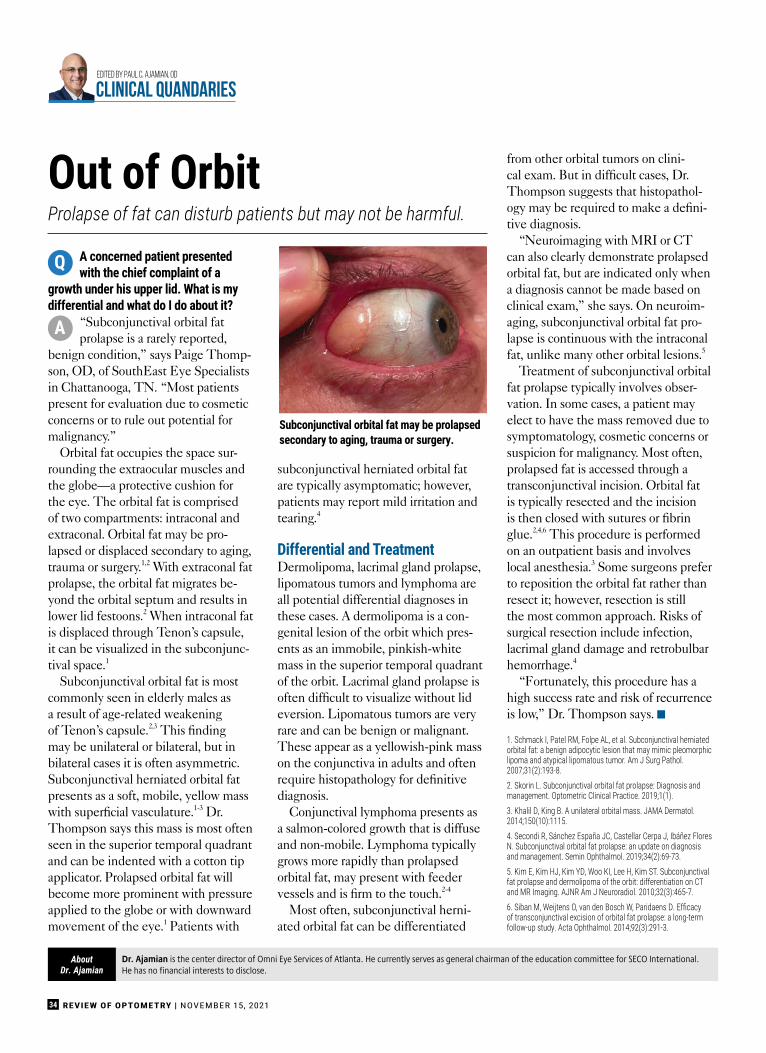
REVIEW OF OPTOMETRY | NOVEMBER 15, 202134
A concerned patient presentedwith the chief complaint of a
growth under his upper lid. What is mydifferential and what do I do about it?
“Subconjunctival orbital fatprolapse is a rarely reported,
benign condition,” says Paige Thomp-son, OD, of SouthEast Eye Specialistsin Chattanooga, TN. “Most patientspresent for evaluation due to cosmeticconcerns or to rule out potential formalignancy.”
Orbital fat occupies the space sur-rounding the extraocular muscles andthe globe—a protective cushion forthe eye. The orbital fat is comprisedof two compartments: intraconal andextraconal. Orbital fat may be pro-lapsed or displaced secondary to aging,trauma or surgery.1,2 With extraconal fatprolapse, the orbital fat migrates be-yond the orbital septum and results inlower lid festoons.2 When intraconal fatis displaced through Tenon’s capsule,it can be visualized in the subconjunc-tival space.1
Subconjunctival orbital fat is mostcommonly seen in elderly males asa result of age-related weakeningof Tenon’s capsule.2,3 This findingmay be unilateral or bilateral, but inbilateral cases it is often asymmetric.Subconjunctival herniated orbital fatpresents as a soft, mobile, yellow masswith superficial vasculature.1-3 Dr.Thompson says this mass is most oftenseen in the superior temporal quadrantand can be indented with a cotton tipapplicator. Prolapsed orbital fat willbecome more prominent with pressureapplied to the globe or with downwardmovement of the eye.1 Patients with
subconjunctival herniated orbital fatare typically asymptomatic; however,patients may report mild irritation andtearing.4
Differential and TreatmentDermolipoma, lacrimal gland prolapse,lipomatous tumors and lymphoma areall potential differential diagnoses inthese cases. A dermolipoma is a con-genital lesion of the orbit which pres-ents as an immobile, pinkish-whitemass in the superior temporal quadrantof the orbit. Lacrimal gland prolapse isoften difficult to visualize without lideversion. Lipomatous tumors are veryrare and can be benign or malignant.These appear as a yellowish-pink masson the conjunctiva in adults and oftenrequire histopathology for definitivediagnosis.
Conjunctival lymphoma presents asa salmon-colored growth that is diffuseand non-mobile. Lymphoma typicallygrows more rapidly than prolapsedorbital fat, may present with feedervessels and is firm to the touch.2-4
Most often, subconjunctival herni-ated orbital fat can be differentiated
from other orbital tumors on clini-cal exam. But in difficult cases, Dr.Thompson suggests that histopathol-ogy may be required to make a defini-tive diagnosis.
“Neuroimaging with MRI or CTcan also clearly demonstrate prolapsedorbital fat, but are indicated only whena diagnosis cannot be made based onclinical exam,” she says. On neuroim-aging, subconjunctival orbital fat pro-lapse is continuous with the intraconalfat, unlike many other orbital lesions.5
Treatment of subconjunctival orbitalfat prolapse typically involves obser-vation. In some cases, a patient mayelect to have the mass removed due tosymptomatology, cosmetic concerns orsuspicion for malignancy. Most often,prolapsed fat is accessed through atransconjunctival incision. Orbital fatis typically resected and the incisionis then closed with sutures or fibringlue.2,4,6 This procedure is performedon an outpatient basis and involveslocal anesthesia.3 Some surgeons preferto reposition the orbital fat rather thanresect it; however, resection is stillthe most common approach. Risks ofsurgical resection include infection,lacrimal gland damage and retrobulbarhemorrhage.4
“Fortunately, this procedure has ahigh success rate and risk of recurrenceis low,” Dr. Thompson says. g
1. Schmack I, Patel RM, Folpe AL, et al. Subconjunctival herniatedorbital fat: a benign adipocytic lesion that may mimic pleomorphiclipoma and atypical lipomatous tumor. Am J Surg Pathol.2007;31(2):193-8.2. Skorin L. Subconjunctival orbital fat prolapse: Diagnosis andmanagement. Optometric Clinical Practice. 2019;1(1).3. Khalil D, King B. A unilateral orbital mass. JAMA Dermatol.2014;150(10):1115.4. Secondi R, Sánchez España JC, Castellar Cerpa J, Ibáñez FloresN. Subconjunctival orbital fat prolapse: an update on diagnosisand management. Semin Ophthalmol. 2019;34(2):69-73.5. Kim E, Kim HJ, Kim YD, Woo KI, Lee H, Kim ST. Subconjunctivalfat prolapse and dermolipoma of the orbit: differentiation on CTand MR Imaging. AJNR Am J Neuroradiol. 2010;32(3):465-7.6. Siban M, Weijtens O, van den Bosch W, Paridaens D. Efficacy of transconjunctival excision of orbital fat prolapse: a long-termfollow-up study. Acta Ophthalmol. 2014;92(3):291-3.
Prolapse of fat can disturb patients but may not be harmful.Out of Orbit
Dr. Ajamian is the center director of Omni Eye Services of Atlanta. He currently serves as general chairman of the education committee for SECO International.He has no financial interests to disclose.
AboutDr. Ajamian
Q
A
Edited by Paul C. Ajamian, OD
CLINICAL QUANDARIES
Subconjunctival orbital fat may be prolapsedsecondary to aging, trauma or surgery.

Dream it. Do it.Let IDOC
Dream it. Do it.Let IDOC
We’re writing an exciting new chapter in the IDOC experience, and we want you tobe a part of it. Since the beginning, IDOC has made innovation central to our valuefor independent ODs. Now, we’re taking it to a whole new level with growth-focusedservices that save you time and money.
Ready to meet the new IDOC?Just turn the page.
It’s a new day at IDOC.Innovation is what we do. With your success in mind.

When it comes to practice management,we’re here to lighten the load.As a private practice owner, you’ve made a commitment to your patients and yourbusiness, but if you’re like most independent ODs, there’s rarely enough time for both.That where IDOC Services come in. Our new focus on turn-key service offerings provides a unique combination of hands-on practice management support combined with thetraditional benefits of IDOC membership. From financial tools, HR management and bookkeeping to vendor discounts, it’s the perfect synergy for driving practice growth.
Relinquish the work. Call (203) 853-3333 or visit IDOC.net today.
IDOC Financial ServicesOur financial services experts offer customized, one-on-one guidance and advice as well as the most advanced business analytics in theindustry. With IDOC, your practice is positioned for growth.
IDOC Books & Benchmarks handles all of your routine bookkeepingand payroll support and helps significantly simplify year-end tax preparation.
IDOC Insider™ offers new insight into business performance with data analytics and real-time displays of KPIs.
IDOC Navigation Services helps you choose the right path for practicetransitions—sales, acquisitions, partnerships and more.
IDOC HR ServicesIDOC HR Now is the ultimate on-demand resource for everyindependent optometry practice. With hotline access to HR experts, legal updates, standardized forms, templates and more, you’ll stay ahead of complex human resource requirements in the mostaffordable way possible.
Look for additional HR services coming over the next several months.
IDOC Marketing ServicesIf growth is your goal, it pays to increase your visibility and promoteyour practice. IDOC Marketing Services experts creates carefully craftedmessages and handles every detail of your digital marketing campaigns,from creative execution to backend analysis.
IDOC Social Media creates engaging customized content to give your practice a memorable presence on Facebook and Instagram.
IDOC Web & Search brings you custom, best-in-class website design alongwith proven techniques to put your practice at the top of the list of onlinesearch results.
IDOC Digital Media leverages the full spectrum of our online marketingexpertise, combining social media, website development and optimized web-based search results.
IDOC Optical ServicesFor any private practice, optical sales can be a significant part of practicerevenue. New IDOC Optical Services offers enhanced Optical Consulting, along with a complete program of data-focused tools and best practicesdesigned to make retail sales a profit center for your practice:
• Create an optical action plan customized for your practice• Talk with our experts about product and vendor selection• Spot key retail trends with our Brand Analysis Reports• Get in touch with your vendors anytime with Rep Connect
Plus, over the next 12 months, we’ll be adding even more sales tools,products, and services for your practice, including inventory managementand analysis.
IDOC Specialty Services Coming SoonLook for upcoming announcements about this exciting new categoryof products, coming soon.At IDOC, we see a growing demand for new patient treatments, and IDOCSpecialty Services is here to help. We’re creating a collaborative networkof eyecare professionals who are working together to exchange ideas,share experiences and build a clinical knowledge bank—all designed todifferentiate your practice and offer new high levels of patient care.
Dream it. Do it.Let IDOC

When we say “Let IDOC do it” we mean it.With a member base of over 3,200+ optometrists, IDOC is more than an alliance—we’re a service-focused, hands-on partner committed to your success as an independent OD. We offer:
Relinquish the work.Call (203) 853-3333 or visit IDOC.net today.
A growing list of turnkey practicemanagement services supportedby industry experts with decades ofexperience—even if you’re not currentlya member of IDOC
Advanced analytics technology with IDOC Insider™ that measuresperformance and provides insights toinform better decision-making
Strategic consulting with experiencedindustry experts on HR, marketing, finance, retail and more
Engaging education and networkingevents that expand knowledge and buildrelationships
Members-only vendor rebate and discountprograms on leading products andservices that are unmatched
“I recommend ThinkHR for anyoptometry offi ce. Having this service is like having another employee that is anexpert in HR and always up to date onthe latest legislation.”
~ Sharon Powell, East Fishkill Eye Care, NY
“IDOC’s Books & Benchmark servicehas allowed me to better manage mypractice fi nances and focus more time on growing my practice!”
~ Dr. Matthew Shirley, Shirley Eye Care, PA

Relinquish the work. Call (203) 853-3333 or visit IDOC.net today.
Do practice management tasks dominate your day?
IDOC Services lighten the load.
Whether you join IDOC as a member or use ourservices “a la carte,” we take care of business, soyou can take care of patients.
“I’d like to spendmore time doingmy books”
— said no OD ever.
Dream it. Do it.Let IDOC
REVIEW OF OPTOMETRY | NOVEMBER 15, 202136
With November comes one ofmy favorite meetings—theAmerican Academy of Op-tometry annual convention.
The Academy meeting always makesme think of the great innovation tak-ing place in our profession. New tech-nologies will be on display, temptingclinicians with clinical and productiv-ity advances. Yet, this cutting-edgescience and innovation also brings tomind the great responsibility we allhave in educating ourselves aboutcoding, coverage and reimbursementspecifics before we can implementthem in our practices.
Educating ourselves about newtechnologies or procedures is key be-fore spending a pretty penny to adoptthem. What are the key questions andcritical items you need to know priorto the purchase? After all, you don’twant to own tech that doesn’t providea clinical benefit, especially whenyou haven’t planned on compliance,coverage and reimbursement details inrespect to its use. Let’s lay them out:
Code it RightCoding is the process of legally trans-lating the service provided into anappropriate CPT, HCPCS Level II orCategory 3 code. There are differenc-es between what these codes describeand therefore their use. Make sureyou know the appropriate code to use.The characteristics of that code arealso paramount before employing thetechnology or procedure. For example,is the procedure unilateral or bilateral?Does it require physician supervision?If a surgical code, what is the globalperiod associated with it?
I see a lot of claims submitted tocarriers with improper or inappropri-ate CPT codes used, which createstremendous exposure for you and yourpractice.
EHRs are Not Always RightUpdate your codes in your EHR toensure the system reflects the propercoding for the new technology orprocedure. EHR companies are alldifferent, and many may not be timelyin updating the new CPT, HCPCS II,Category 3 or ICD-10 codes. How-ever, you, the practitioner, are alwaysresponsible for using the correct ones.
Carrier PolicyBefore purchasing or deploying anynew technology or procedure, under-stand where your carriers (both medi-cal and managed vision care) stand oncoding, coverage and reimbursement.If they are covered, ask about theindications and limitations of medi-cal necessity, frequency or utilizationguidelines and, finally, contractedreimbursement amounts.
If these procedures or technologiesare not covered, i.e., designated as“investigational or experimental,”then make sure that all doctors andstaff understand the requirements forproviding an Advanced BeneficiaryNotice (ABN) or waiver of liability
form as required by the specificcarrier.
Scope of PracticeLegislative victories in many stateshave resulted in an acceleration of anincreased scope of practice. It is easy toget excited about new technology thatcomes with increased scope when wehear the successes of our colleagues.Since optometry is still a state-by-state legislated profession, what maybe within the scope of practice of onepractitioner may not be within thescope of your practice. Determinewhether you can perform the newprocedure or use the new tech in yourpractice before investing in it. The bestsources of information are your stateboard and your state law.
Informed ConsentIf the new technology or procedurerequires informed consent by state lawor by carrier policy, make sure to createa specific informed consent packetto present to the patient. Have an at-torney who is familiar with these lawsreview these forms on an annual basisto make sure each is in alignment withcurrent legal interpretation for yourgiven jurisdiction.
‘Do it Right,’ Not ‘Do it Over’There is a lot to love about advancingtechnology in our profession. The CO-VID-19 pandemic certainly acceleratedthe development and deployment ofgreat innovations to provide better pa-tient care. Attending meetings, such asthe Academy’s, provides us the forumto learn more about this cutting-edgescience. Be prepared to incorporate itproperly into your practice and createa process that allows you to do it withlittle risk and a lot of benefits. ■
Send your coding questions [email protected].
Educate yourself when approaching new technology.Innovate for Success
Dr. Rumpakis is president and CEO of Practice Resource Management, a firm that provides consulting, appraisal and management services for healthcareprofessionals and industry partners. As a full-time consultant, he provides services to a wide array of ophthalmic clients. Dr. Rumpakis’s full disclosure list can befound in the online version of this article at www.reviewofoptometry.com.
AboutDr. Rumpakis
By John Rumpakis, OD, MBAClinical Coding EditoR
CODING CONNECTION
Determine whether you canperform the new procedure oruse the new tech in your practicebefore investing in it.

REQUEST YOURFREE DESIGNKIT TODAY!
Success.
Success.
Success.
CU
STO
M &
SE
MI-
CU
STO
M O
PTI
ON
S
TheThe Eye Eye Designs Designs Group Group is is the the complete complete resource resource for for all allofofof your your practice practice needs, needs, from from Space Space Planning Planning & & Interior InteriorDesignDesign to to Manufacturing, Manufacturing, Exam Exam Solutions, Solutions, Merchandising, Merchandising,Seating,Seating, Optical Optical Art Art and and beyond. beyond.
WWW.EYEDESIGNS.COMWWW.EYEDESIGNS.COM800.346.8890800.346.8890800.346.8890800.346.8890
SPA
CE
SPA
CE
PLA
NN
ING
. P
LAN
NIN
G. M
AN
UFA
CTU
RIN
G.
MA
NU
FAC
TUR
ING
. DE
SIG
N D
ESI
GN

Review’s 2021 Office Design Contest shows whatoptometrists can do to stand out when they settheir minds to it. Each practice that enteredshowcased their creativity, and all had the same
goal: improving the patient experience.We always look forward to our biennial Office
Design Contest and seeing optometrists from all overthe country show off their style and, in some cases,their personality. This year, we were thrilled to have 24practices enter the contest. The photo collage to theright includes one sample image from each participant.We wanted to showcase the breadth of contributionsthis time to show the range of what has been done overthe past two years. There was a great deal of passionput into each space—we can’t wait for you to see theunique designs that may help you reimagine your ownoffice space.
Peruse the collage, pick your faves, then turn thepage for a celebration of this year’s winners!
38 REVIEW OF OPTOMETRY | NOVEMBER 15, 2021
2021 DESIGN CONTEST
EYE-POPPINGOFFICE SPACES
In 2019, these practices excelled with their own office renovations. Thisyear, they provided expert feedback to pick the cream of the crop.
Scot Morris, ODEye Consultants
of ColoradoConifer, CO2019 Winner
Kumar Patel, ODPersonaleyesVision Care
Flower Mound, TX2019 1st Runner-up
Rob Szeliga, ODSpring Hill Eyecare
Spring Hill, TN2019 2nd Runner-up
MEET THE JUDGES
O F F I C E D E S I G NContest
ODs bounced back from the COVIDdoldrums with bold and adventurous
new design ideas.

39NOVEMBER 15, 2021 | REVIEW OF OPTOMETRY
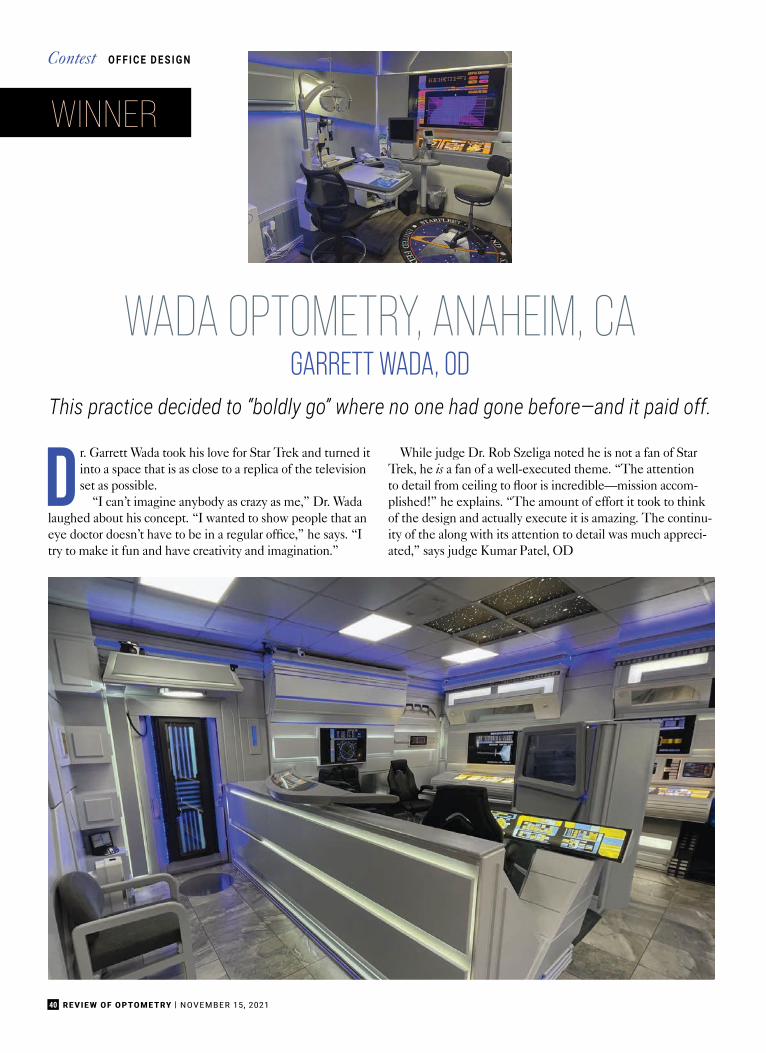
40 REVIEW OF OPTOMETRY | NOVEMBER 15, 2021
WADA OPTOMETRY, ANAHEIM, CAGARRETT WADA, OD
This practice decided to “boldly go” where no one had gone before—and it paid off.
Dr. Garrett Wada took his love for Star Trek and turned itinto a space that is as close to a replica of the televisionset as possible.
“I can’t imagine anybody as crazy as me,” Dr. Wadalaughed about his concept. “I wanted to show people that aneye doctor doesn’t have to be in a regular office,” he says. “Itry to make it fun and have creativity and imagination.”
While judge Dr. Rob Szeliga noted he is not a fan of StarTrek, he is a fan of a well-executed theme. “The attentionto detail from ceiling to floor is incredible—mission accom-plished!” he explains. “The amount of effort it took to thinkof the design and actually execute it is amazing. The continu-ity of the along with its attention to detail was much appreci-ated,” says judge Kumar Patel, OD
O F F I C E D E S I G NContest
WINNER
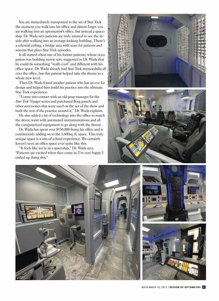
41NOVEMBER 15, 2021 | REVIEW OF OPTOMETRY
You are immediately transported to the set of Star Trekthe moment you walk into his office and almost forget youare walking into an optometrist’s office, but instead a space-ship. Dr. Wada says patients are truly amazed to see the in-side after walking into an average-looking building. There’sa celestial ceiling, a bridge area with seats for patients andscreens that plays Star Trek episodes.
It all started when one of his former patients, whose occu-pation was building movie sets, suggested to Dr. Wada thathe could do something “really cool” and different with hisoffice space. Dr. Wada already had Star Trek memorabilia allover the office, but this patient helped take the theme to awhole new level.
Then Dr. Wada found another patient who has an eye fordesign and helped him build his practice into the ultimateStar Trek experience.
“I came into contact with an old prop manager for theStar Trek: Voyager series and purchased Borg panels andother accessories that were used on the set of the show andbuilt the rest of the practice around it,” Dr. Wada explains.
He also added a lot of technology into the office to matchthe decor, went with automated instrumentations and allthe computerized equipment to go along with the theme.
Dr. Wada has spent over $150,000 fixing his office and iscontinuously adding on to the 1,600sq. ft. space. This trulyunique space is a one-of-a-kind experience. We certainlyhaven’t seen an office space ever quite like this.
“It feels like we’re in a spaceship,” Dr. Wada says.“Patients are excited when they come in. I’m very happy Iended up doing this.”

42 REVIEW OF OPTOMETRY | NOVEMBER 15, 2021
THE VISION CENTERAT SEASIDE FARMS,
MOUNT PLEASANT, SCBRAD BODKIN, OD
O F F I C E D E S I G NContest
1st RUNNER-UP
“Stylish Southern charm are the words that come to mind,”says Dr. Scot Morris, one of this year’s judges, about TheVision at Seaside Farms. “I love the mix of modern whitewalls and glass with the classic wood.” Adding to that
charm is the use of large barn doors throughout the practice. Practiceowner Brad Bodkin, OD, originally added them to help with ef-ficiency, but they have been an aesthetic hit with patients, too.
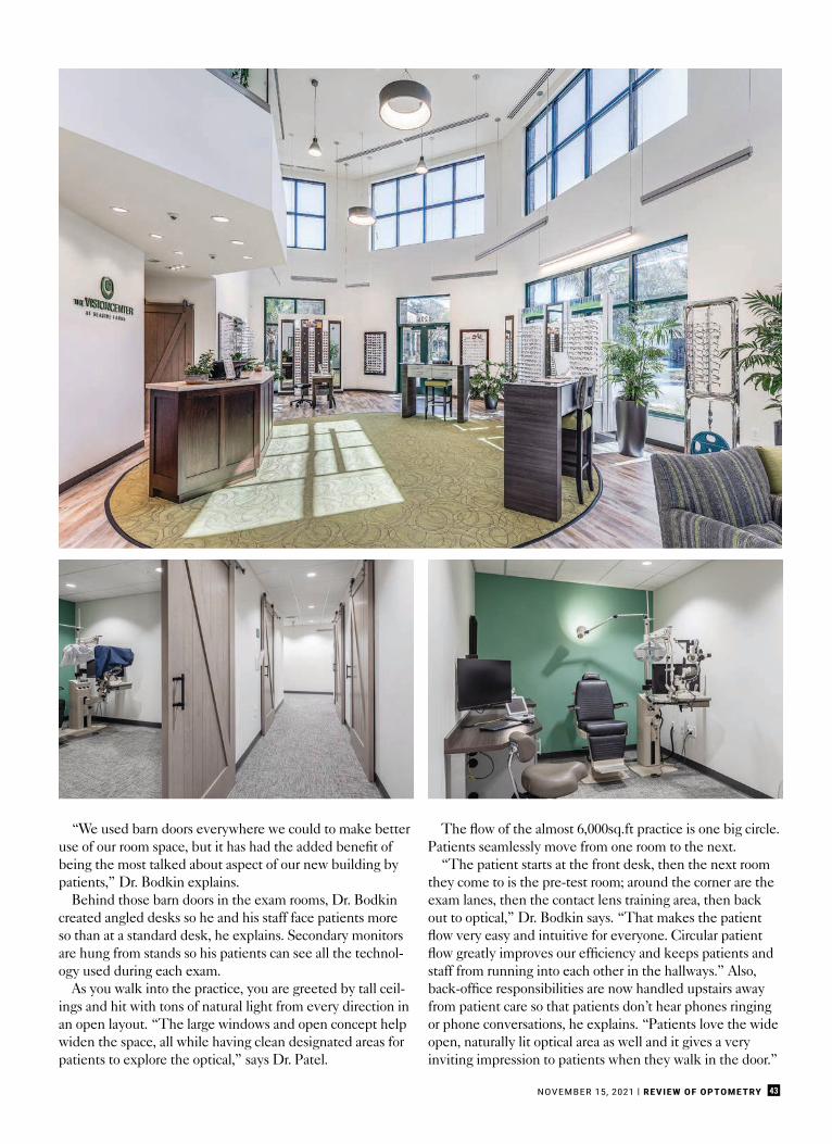
43NOVEMBER 15, 2021 | REVIEW OF OPTOMETRY
“We used barn doors everywhere we could to make betteruse of our room space, but it has had the added benefit ofbeing the most talked about aspect of our new building bypatients,” Dr. Bodkin explains.
Behind those barn doors in the exam rooms, Dr. Bodkincreated angled desks so he and his staff face patients moreso than at a standard desk, he explains. Secondary monitorsare hung from stands so his patients can see all the technol-ogy used during each exam.
As you walk into the practice, you are greeted by tall ceil-ings and hit with tons of natural light from every direction inan open layout. “The large windows and open concept helpwiden the space, all while having clean designated areas forpatients to explore the optical,” says Dr. Patel.
The flow of the almost 6,000sq.ft practice is one big circle.Patients seamlessly move from one room to the next.
“The patient starts at the front desk, then the next roomthey come to is the pre-test room; around the corner are theexam lanes, then the contact lens training area, then backout to optical,” Dr. Bodkin says. “That makes the patientflow very easy and intuitive for everyone. Circular patientflow greatly improves our efficiency and keeps patients andstaff from running into each other in the hallways.” Also,back-office responsibilities are now handled upstairs awayfrom patient care so that patients don’t hear phones ringingor phone conversations, he explains. “Patients love the wideopen, naturally lit optical area as well and it gives a veryinviting impression to patients when they walk in the door.”

44 REVIEW OF OPTOMETRY | NOVEMBER 15, 2021
DRY EYE & SPECIALTY CENTERSUGAR LAND, TXFAHEEM INAYATALI, OD
2nd RUNNER-UP
O F F I C E D E S I G NContest
Cutting-edge was one of the first words used to describe our sec-ond runner-up, Dry Eye & Specialty Center, due to its modernand futuristic feel and advanced technology incorporated in allaspects of the office.
White and blue are the only colors used, giving the space it aclean, pristine look throughout, with white floors, unique lightfixtures and a treatment room surrounded by glass that immediatelycatches your eye.
“This style instills confidence that they are on the cutting edge,”notes judge Dr. Rob Szeliga. “I like the choice of colors and how

45NOVEMBER 15, 2021 | REVIEW OF OPTOMETRY
they are incorporated throughout the office. It’s a verysleek, clean and modern look.”
The glass-walled treatment area was specificallydesigned with an ergonomic exterior that prevents IPLlight from shining through during treatments.
“[It] has an iPad and large HD screen that are wire-lessly connected to have an interactive session describ-ing their ocular health with every patient encounter,”Dr. Inayatali notes. “As patients begin treatment, awaterfall behind the chair creates an ambiance that isrelaxing to ease any anxiety. The exam room is locateddirectly across from the treatment room so the doctorand technician can switch for added efficiencies usingin-office communication software (Bluenotes) to keepour team linked together and reduce wait time.”
Dr. Inayatali also integrated technology—while im-proving functionality—by creating an app that generatesa QR code with information that patients can take homeregarding any ocular conditions that need to be man-aged. Gone are the days of badly photocopied patienthandouts strewn about the waiting room.
As much as the technology has enhanced the patientexperience and played a role in changing the entire lookof the office, Dr. Inayatali says the aesthetics are thehighlight of the overall experience, with people passingby often coming inside just to take pictures of the space.
“Our in-ceiling LED lights and waterfall create acontemporary appeal that has attracted patients from allover the nation, plus Mexico and Canada,” he explains.

46 REVIEW OF OPTOMETRY | NOVEMBER 15, 2021
HONORABLE MENTIONSThese four offi ces stood out by crafting unique expressions
shaped by their personalities and practice setting.
LYMER VISIONColorado Springs, CO
Ted Lymer, OD
FLORENCE EYE CENTERFlorence, AL
Ben Kachelman, OD
EYES ON SHEPPARDToronto, ON
Kerry Salsberg, OD
VILLAGE OPTOMETRYCarlsbad, CA
Douglas Osborne, OD
O F F I C E D E S I G NContest

35+
ARCHITECTS WITHOPTOMETRIC FOCUS
“THE PERFECTBALANCE OF SPACE
DESIGN AND BEAUTY”
years of architecturalexperience
EXTERIOR &INTERIOR DESIGNS
ARCHITECTSOPTOMETRIC
www.optometricarchitects.comwww.optometricarchitects.comwww.optometricarchitects.comwww.optometricarchitects.com
319-240-0222
CALL TODAY FOR A FREEDESIGN CONSULTATION!
48 REVIEW OF OPTOMETRY | NOVEMBER 15, 2021
The conjunctivaup close
Here’s how to recognize and manage a few common conditions that affect this part of the eye.
The conjunctiva is a thin mucus membrane whoseprimary functions are to provide barrier protection,immunity and lubrication to the ocular surface.There are three distinct anatomical locations of con-
junctival tissue: the palpebral, the bulbar and the forni-ceal. The conjunctiva itself is made up of the epitheliumand the stroma (also known as the substantia propria).The epithelium is a few cell layers thick and consistsof various cell types. Stratified cuboidal cells are foundover the tarsus, columnar cells are found in the fornicesand squamous cells are found on the bulbar conjunctiva.1
The stroma is made up of loose, vascularized connectivetissue rich in elastic fibers. This gives the conjunctiva theability to flex and stretch upon blinking and eye move-ment.2 The stroma is also rich in lymphocytes and playsan important role in the immunologic response of theocular surface.
The conjunctiva is dense in mucin-producing glandsincluding goblet cells, Crypts of Henle and glands ofManz. Goblet cells are located in the conjunctival epithe-lium, Crypts of Henle are located in the palpebral con-junctiva and glands of Manz are located near the limbus.The mucin layer is the innermost layer of the tear filmand is made up of glycoproteins. On the ocular surface,hydrophilic mucins are responsible for adherence of thetear film to the ocular surface, decreasing surface tensionand ensuring the overlying aqueous can spread evenlyacross the front of the eye. This is critical for protecting
the conjunctival and corneal epithelium by preventingdryness, clearing debris and fending off pathogens.3
The accessory lacrimal glands of Krause and Wolfringcan also be found in the conjunctiva. They are locatedin the fornices and the tarsus, respectively. These glandsproduce aqueous secretions to supplement the lacrimalgland.4 The accessory lacrimal glands do not containparasympathetic innervation and are thought to be re-sponsible only for basal secretions.5
Now that we have reviewed the anatomy, let’s take acloser look at how several common conjunctival condi-tions manifest clinically. Understanding the innerwork-ings of this ocular structure will help you differentiateand diagnose the following:
Acute ConjunctivitisConjunctival inflammation is marked by a variety of signsincluding injection, chemosis and discharge. Accuratediscrimination of the causes of inflammation is importantto direct appropriate treatment, patient education andfollow-up. An important differentiation in conjunctivitisis a follicular vs. papillary response. Follicles are ag-gregations of lymphoid tissue and appear as gray/whitedome-like elevations with a small surrounding bloodvessel. They are formed within the conjunctival stroma.Follicular conjunctivitis is associated with acute viral andchronic chlamydial infections, Parinaud’s oculoglandularsyndrome and hypersensitivity to topical ophthalmicmedications.4
Papillae, in contrast, contain a central vessel making theelevated tissue red in color. They are formed by the hy-perplastic conjunctival epithelium. Papillary conjunctivitis
C O N J U N CT I VA L C O N D I T I O N SFeature PEER REVIEWED
By megan mannen, odNorthern Utah
Dr. Mannen practices at a private optometry and ophthalmology clinic in northern Utah. She completed her residency at the Walla Walla Veteran Affairs Medical Centerand Pacific Cataract and Laser Institute. She is a Fellow of the American Academy of Optometry. She has no financial interests to disclose.
Aboutthe author
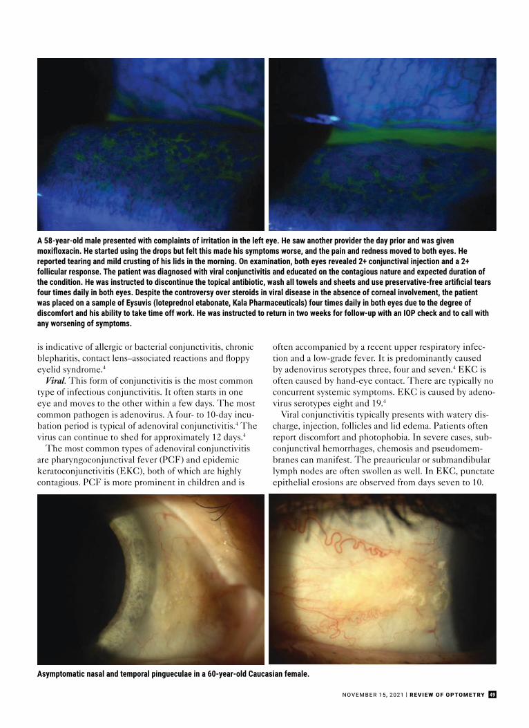
49NOVEMBER 15, 2021 | REVIEW OF OPTOMETRY
is indicative of allergic or bacterial conjunctivitis, chronicblepharitis, contact lens–associated reactions and floppyeyelid syndrome.4
Viral. This form of conjunctivitis is the most commontype of infectious conjunctivitis. It often starts in oneeye and moves to the other within a few days. The mostcommon pathogen is adenovirus. A four- to 10-day incu-bation period is typical of adenoviral conjunctivitis.4 Thevirus can continue to shed for approximately 12 days.4
The most common types of adenoviral conjunctivitisare pharyngoconjunctival fever (PCF) and epidemickeratoconjunctivitis (EKC), both of which are highlycontagious. PCF is more prominent in children and is
often accompanied by a recent upper respiratory infec-tion and a low-grade fever. It is predominantly causedby adenovirus serotypes three, four and seven.4 EKC isoften caused by hand-eye contact. There are typically noconcurrent systemic symptoms. EKC is caused by adeno-virus serotypes eight and 19.4
Viral conjunctivitis typically presents with watery dis-charge, injection, follicles and lid edema. Patients oftenreport discomfort and photophobia. In severe cases, sub-conjunctival hemorrhages, chemosis and pseudomem-branes can manifest. The preauricular or submandibularlymph nodes are often swollen as well. In EKC, punctateepithelial erosions are observed from days seven to 10.
A 58-year-old male presented with complaints of irritation in the left eye. He saw another provider the day prior and was givenmoxifloxacin. He started using the drops but felt this made his symptoms worse, and the pain and redness moved to both eyes. He reported tearing and mild crusting of his lids in the morning. On examination, both eyes revealed 2+ conjunctival injection and a 2+ follicular response. The patient was diagnosed with viral conjunctivitis and educated on the contagious nature and expected duration of the condition. He was instructed to discontinue the topical antibiotic, wash all towels and sheets and use preservative-free artificial tears four times daily in both eyes. Despite the controversy over steroids in viral disease in the absence of corneal involvement, the patient was placed on a sample of Eysuvis (loteprednol etabonate, Kala Pharmaceuticals) four times daily in both eyes due to the degree of discomfort and his ability to take time off work. He was instructed to return in two weeks for follow-up with an IOP check and to call with any worsening of symptoms.
Asymptomatic nasal and temporal pingueculae in a 60-year-old Caucasian female.
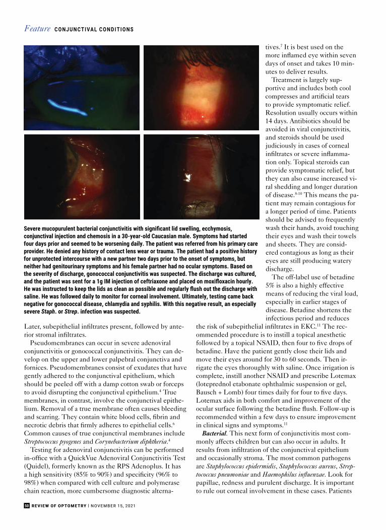
50 REVIEW OF OPTOMETRY | NOVEMBER 15, 2021
Later, subepithelial infiltrates present, followed by ante-rior stromal infiltrates.
Pseudomembranes can occur in severe adenoviralconjunctivitis or gonococcal conjunctivitis. They can de-velop on the upper and lower palpebral conjunctiva andfornices. Pseudomembranes consist of exudates that havegently adhered to the conjunctival epithelium, whichshould be peeled off with a damp cotton swab or forcepsto avoid disrupting the conjunctival epithelium.4 Truemembranes, in contrast, involve the conjunctival epithe-lium. Removal of a true membrane often causes bleedingand scarring. They contain white blood cells, fibrin andnecrotic debris that firmly adheres to epithelial cells.6
Common causes of true conjunctival membranes includeStreptococcus pyogenes and Corynebacterium diphtheria.4
Testing for adenoviral conjunctivitis can be performedin-office with a QuickVue Adenoviral Conjunctivitis Test(Quidel), formerly known as the RPS Adenoplus. It hasa high sensitivity (85% to 90%) and specificity (96% to98%) when compared with cell culture and polymerasechain reaction, more cumbersome diagnostic alterna-
tives.7 It is best used on themore inflamed eye within sevendays of onset and takes 10 min-utes to deliver results.
Treatment is largely sup-portive and includes both coolcompresses and artificial tearsto provide symptomatic relief.Resolution usually occurs within14 days. Antibiotics should beavoided in viral conjunctivitis,and steroids should be usedjudiciously in cases of cornealinfiltrates or severe inflamma-tion only. Topical steroids canprovide symptomatic relief, butthey can also cause increased vi-ral shedding and longer durationof disease.8-10 This means the pa-tient may remain contagious fora longer period of time. Patientsshould be advised to frequentlywash their hands, avoid touchingtheir eyes and wash their towelsand sheets. They are consid-ered contagious as long as theireyes are still producing waterydischarge.
The off-label use of betadine5% is also a highly effectivemeans of reducing the viral load,especially in earlier stages ofdisease. Betadine shortens theinfectious period and reduces
the risk of subepithelial infiltrates in EKC.11 The rec-ommended procedure is to instill a topical anestheticfollowed by a topical NSAID, then four to five drops ofbetadine. Have the patient gently close their lids andmove their eyes around for 30 to 60 seconds. Then ir-rigate the eyes thoroughly with saline. Once irrigation iscomplete, instill another NSAID and prescribe Lotemax(loteprednol etabonate ophthalmic suspension or gel,Bausch + Lomb) four times daily for four to five days.Lotemax aids in both comfort and improvement of theocular surface following the betadine flush. Follow-up isrecommended within a few days to ensure improvementin clinical signs and symptoms.11
Bacterial. This next form of conjunctivitis most com-monly affects children but can also occur in adults. Itresults from infiltration of the conjunctival epitheliumand occasionally stroma. The most common pathogensare Staphylococcus epidermidis, Staphylococcus aureus, Strep-tococcus pneumoniae and Haemophilus influenzae. Look forpapillae, redness and purulent discharge. It is importantto rule out corneal involvement in these cases. Patients
Severe mucopurulent bacterial conjunctivitis with significant lid swelling, ecchymosis, conjunctival injection and chemosis in a 30-year-old Caucasian male. Symptoms had started four days prior and seemed to be worsening daily. The patient was referred from his primary care provider. He denied any history of contact lens wear or trauma. The patient had a positive history for unprotected intercourse with a new partner two days prior to the onset of symptoms, but neither had genitourinary symptoms and his female partner had no ocular symptoms. Based on the severity of discharge, gonococcal conjunctivitis was suspected. The discharge was cultured, and the patient was sent for a 1g IM injection of ceftriaxone and placed on moxifloxacin hourly. He was instructed to keep the lids as clean as possible and regularly flush out the discharge with saline. He was followed daily to monitor for corneal involvement. Ultimately, testing came back negative for gonococcal disease, chlamydia and syphilis. With this negative result, an especially severe Staph. or Strep. infection was suspected.
C O N J U N CT I VA L C O N D I T I O N SFeature
The Vantage BIO is greatfor ROP screening! It’slightweight, has settingsfor different pupil sizes,a cool, white LED lightand the longest batteryever!!”
I’m a big fan of the All PupilBIO. I had issues with othermodels so when I started[my practice], I knew theAll Pupil would be my go-toBIO...I greatly appreciatethe new custom fit KeelerBIO shields as an addedsafety layer.”
I chose my [Vantage Plus]for the optics and value...withother brands, I had difficultyfocusing up close during mydilated fundus exams. [Theoculars] made my eyes feelmore relaxed, and I felt likemy view was better.”
[I’ve] been seeingemergent and urgentcases every day duringthe COVID19 pandemic.I really like [the VantageBIO] because [it’s a] verygood quality and providesa super clear view.”
Dra. Paulina Ramirez Neria
Dr. Annie BaconDr. Michelle Hammond Dr. Reza Moradi
Helping Heroes See Clear And Stay Safe
A world without vision loss
www.keelerusa.com • 3222 Phoenixville Pike - Bldg. #50 • Malvern, PA 19355Tel No: 1-610-353-4350 • Toll Free: 1-800-523-5620 • Fax: 1-610-353-7814
Choose option #1 or #2 below when you purchase (or lease) a BIO*(Expires December 31, 2021)
Contact us at 800-523-5620 or [email protected] to learn more or place your order. This promo cannot be combined with any other Keeler offers.
RECEIVE A 24-MONTH RECEIVE
lease as low as $128/month*bottles of phenylephrine
2.5%, 15mLcredit towards any PPE
$850 0% 10 FREE
*Valid for wireless indirects: Vantage Plus and/or All Pupil II
*All Pupil II: $127.92/month; Vantage Plus: $155/month(shipping and taxes not included).
*If you lease the BIO, you may also choose the PPE credit OR the phenylephrine option.

52 REVIEW OF OPTOMETRY | NOVEMBER 15, 2021
may complain of burning, a gritty sensation and their lidssticking together, especially in the morning.
Bacterial conjunctivitis is often self-limiting, resolv-ing within 10 to 14 days, especially in milder forms ofdisease. Broad-spectrum antibiotics, however, can al-leviate symptoms, shorten duration of the condition andreduce the risk of recurrence.12,13 Antibiotics should alsobe strongly considered in contact lens wearers due tothe risk for corneal involvement and a higher prevalenceof gram-negative pathogens. Contact lens wear shouldbe discontinued until symptoms have ceased. Patientsshould resume wear when appropriate with a new set ofcontact lenses and a new case. Instruct patients to keeptheir lids and lashes clean and clear of discharge.
Acceptable broad-spectrum options include Polytrim(polymyxin B and trimethoprim, Allergan), Azasite(azithromycin, Akorn), erythromycin ointment and fluo-roquinolones.14 Polymyxin B and trimethoprim, erythro-mycin and fluoroquinolones are dosed four times daily,while azithromycin is dosed twice daily for five to sevendays. Patients should respond to treatment within 24 to48 hours. Although later-generation fluoroquinolonesare highly effective agents at treating a variety of bacte-rial strains, alternative antibiotics should be consideredfor routine bacterial conjunctivitis. Avoiding antibioticresistance is critical, as fourth-generation fluoroquino-
lones are the first lineof treatment for cornealulcers and severe bacte-rial conjunctivitis andmust remain efficaciousin treating more sight-threatening infections.
Hyperacute bacterialconjunctivitis shouldbe cultured and treatedrapidly. This conditionis caused by Neisseriagonorrhoeae and appearswith severe mucopu-rulent discharge within12 hours of exposure.Concurrent genito-urinary symptoms arecommon. Patients withsuspected gonococcalconjunctivitis shouldbe asked about theirrecent sexual historyand possible source ofexposure.
The recommendedtreatment is 1g of in-tramuscular ceftriaxoneand a topical fluoro-
quinolone.15 These patients should be monitored daily,as Neisseria gonorrhoeae can penetrate an intact cornea.Generally, culturing is not required in acute infectiousconjunctivitis, but it is warranted in suspected gonococ-cal disease due to the risk of rapid corneal ulceration andits unique treatment. Neisseria gonorrhoeae has developedresistance to many antibiotic classes, and culturing allowstesting for antibiotic susceptibility. In patients where aconcurrent infection of chlamydia has not been ruled out,a seven-day course of 100mg doxycycline twice daily isalso recommended.15
Allergic. This type of conjunctivitis is a bilateral con-dition marked by ocular itching. Other symptoms may in-clude tearing, burning and foreign body sensation. Uponexamination, eyelid edema, conjunctival hyperemia,papillae and ropy white discharge may also be observed.A careful case history is important to determine the caus-ative agent(s).
Allergic conjunctivitis encompasses acute, seasonal andperennial conjunctivitis. It is a type 1 hypersensitivityreaction. In this condition, circulating allergens trig-ger the release of IgE and go on to stimulate mast cellproduction and histamine release. Mast cells are presentin high concentrations in the conjunctival epithelium inthese patients.16 Allergic conjunctivitis is more prominentin young adults, as symptoms tend to improve with age,
True conjunctival membranes and residual conjunctival scarring in the same 30-year-old Caucasian malein the previous set of photos with severe bilateral bacterial conjunctivitis. The membranes were confirmed when a damp cotton swab was unsuccessful in removal. Once discharge subsided and the ocular surfacewas considered sterile, the patient was treated with aggressive topical steroids to minimize conjunctivalscarring and resolve corneal infiltrates.
C O N J U N CT I VA L C O N D I T I O N SFeature
and eczema is a common concurrent condition in thesepatients.17
First-line treatment includes topical antihistamines ora combination of antihistamine and mast cell stabilizers.Over-the-counter combination options include Pataday(olopatadine, Alcon), Zaditor (ketotifen, Alcon) andAlaway (ketotifen, Bausch + Lomb), the latter of whichis now available in a preservative-free formulation. Solotopical antihistamine options include Bepreve (bepotas-tine besilate, Bausch + Lomb), Lastacaft (alcaftadine,Allergan) and Zerviate (cetirizine, Eyevance Pharma-ceuticals). All three of these formulations are availableby prescription only.
Oral antihistamines can also be considered, especiallywhen nasal symptoms accompany ocular symptoms.These are best used preventatively. Another supple-mental option is a topical mast cell stabilizer such asCrolom (cromolyn sodium, Bausch + Lomb). Coolcompresses and artifi cial tears can provide adjunctivesymptomatic relief. In more severe forms of allergicconjunctivitis, soft steroids can be considered such asAlrex (loteprednol etabonate, Bausch + Lomb), whichis FDA-approved for allergic conjunctivitis, Lotemax(loteprednol, Bausch + Lomb) and FML (fl uorometho-lone, Allergan). It is also important to educate patientsto avoid eye rubbing and known allergens.
Vernal keratoconjunctivitis (VKC) is a more severeform of allergic conjunctivitis and can be sight-threat-ening. It is a recurrent, bilateral condition marked bycobblestone papillae on the upper palpebral conjunc-tiva. It is more common in young males in dry, warmclimates. Patients suffering from VKC will experi-ence severe itching, increased blink rates and mucusdischarge. VKC is both IgE- and cell-mediated and ismarked by signifi cant eosinophil, mast cell and fi bro-blast release. It resembles both type one, IgE-mediatedand type four, delayed hypersensitivity reactions.18,19
Patients should be monitored carefully for limbal andcorneal involvement. Treatment is very similar to thatof allergic conjunctivitis. Combination antihistamineand mast cell stabilizers should be considered fi rst-linetherapy, but topical steroids may be required in moresignifi cant disease.4
Giant Papillary Conjunctivitis (GPC)This non-allergic condition occurs secondary to mechan-ical rubbing on the tarsal conjunctiva and an immune-mediated infl ammatory response. Although not fullyunderstood, it also exhibits features of both type oneand type four hypersensitivity reactions.20 GPC mostcommonly occurs due to contact lens wear but can alsobe stimulated by exposed sutures, fi ltering blebs, ocularprostheses and scleral buckles. When induced by con-tact lenses, symptoms tend to be worse following lensremoval. Patients will often report foreign body sensa-
The Fundamentalsof Lid Hygiene
Oasis TEARS®
Oasis LID & LASH® +tea tree oil
• 60 Presoaked Pads• Gentle Eyelid
Cleansing• Hydrates Delicate
Skin• Soothing Tea Tree
Oasis TEARS®
Hypochlorous™
• Misting spray• Natural
antimicrobial• Reduces Eyelid
bioburden• pH neutral
www.oasismedical.com
Call To Order: (800) 820-8940Also Available on MY OASIS™
Platform.
Scan to Selecta Date for an
OTC for Dry EyeImplementation
Workshop
LIT-HLL-AD Rev 0 10.2021

54 REVIEW OF OPTOMETRY | NOVEMBER 15, 2021
tion, itching, redness and increased mucus discharge.The condition is marked by redness and papillae of thesuperior palpebral conjunctiva. Protein deposition is alsocommonly found on contact lenses in lens-induced GPC.
The first line of treatment is to remove the irritant fora couple of weeks. For lens-induced GPC, the patientshould switch to a different contact lens material or todaily disposable contact lenses when wear is resumed. Pa-tients should also be discouraged from overwearing theirlenses. Topical steroids can be utilized to treat inflam-mation. In cases where daily disposables are not a viablealternative, it is important to ensure proper cleaning ofcontact lenses. Thorough disinfection is also imperativein cases of GPC induced by an ocular prosthesis. An ocu-larist can be consulted in these instances.
ConjunctivochalasisThis condition is evident by redundant conjunctival tis-sue which occurs most commonly with increasing age aselastic fibers are lost.21 Ocular rubbing, mechanical forces,trauma and previous surgeries can also increase the riskof conjunctivochalasis development.21,22 It most com-monly occurs in the infratemporal quadrant of the bulbarconjunctiva. Although frequently asymptomatic, symp-toms such as ocular surface discomfort, epiphora and dry-ness can occur. Conjunctivochalasis blocks normal tearmovement and disrupts the tear meniscus.23,24 Symptomsoften worsen with downgaze and with an increased blinkrate.25 Conjunctivochalasis is frequently under-diagnosedas a source of ocular discomfort.
In asymptomatic patients, no treatment is required.In cases of mild discomfort, frequent artificial tears can
be utilized. This helps to ensure that an adequate sup-ply of tears is spread across the ocular surface despite adisrupted tear meniscus. If the redundant conjunctivais exposed overnight, consider lubricating ointment orpatching. A short, soft topical steroid course is the nextline of treatment if supportive therapy is unsuccessful.This is especially helpful in cases of increased inflam-mation. Cyclosporine may also be beneficial based on itsinhibitory effect on inflammatory cytokines, which are of-ten found in the tear film of those with conjunctivochala-sis. In patients with more severe symptoms, the chalasiscan be surgically excised and resected, or thermal cauterycan be performed on the redundant tissue.
Pinguecula and PingueculitisA pinguecula is an extremely common benign, yellow/white elevated lesion found on the bulbar conjunctiva.It is often bilateral and asymptomatic. Pingueculae arecaused by UV exposure and other elemental factorsincluding wind and dust. This leads to elastic degenera-tion of collagen fibers in the conjunctival stroma. Similarto pterygia, they are more commonly found nasally.4 Thisis thought to be due to the internal reflection of temporalincident light across the cornea, resulting in 20-times theconcentration of light focused at the nasal limbus.26,27
Mild symptoms of discomfort, itching and foreign bodysensation can be treated with artificial tears. In casesof more significant inflammation, i.e., pingueculitis, ashort course of soft topical steroids can be considered.UV protection is recommended to prevent progression.Surgical excision or removal with argon laser photocoagu-lation is rarely performed, except for cosmetic reasons.
A 59-year-old Caucasian female presented with irritation and foreign body sensation in the left eye greater than the right for the last fewdays. She reported sleeping in her two-week disposable contact lenses prior to the onset of irritation. Examination revealed GPC in the lefteye greater than the right. The patient was instructed to discontinue contact lens wear for two weeks and placed on Tobradex (tobramycinand dexamethasone, Novartis) QID for one week. Based on her high-risk contact lens history, mild crusting of the lid margin and mildcorneal edema in the left eye on examination, an antibiotic-steroid combination was selected. The patient was instructed to return in twoweeks for follow-up, at which point she was switched to a daily disposable contact lens modality. She responded well to therapy.
C O N J U N CT I VA L C O N D I T I O N SFeature
TakeawaysThe conjunctiva serves a critical role in ocular surfaceintegrity. It does so by producing mucins and basalaqueous secretions to moisturize the ocular surface. Italso serves as a physical barrier to foreign irritants andpathogens. As a highly vascularized tissue, it providesnutrients to its surrounding tissues and immunologicprotection. Signs and symptoms of conjunctival infl am-mation can be nonspecifi c and have signifi cant overlapbetween etiologies. It is critical to perform a thoroughcase history and careful ocular examination to tease outthe nuances to make an accurate diagnosis and appropri-ate treatment plan. ■
1. Weisenthal RW, Afshari NA, Bouchard CS, et al. Chapter 1: Structure and Function of theExternal Eye and Cornea. In: External Disease and Cornea. American Academy of Ophthalmol-ogy. 2013:3-4.2. Hodges RR, Dartt DA. Tear fi lm mucins: front line defenders of the ocular surface; compari-son with airway and gastrointestinal tract mucins. Exp Eye Res. 2013;117:62-78.3. Mantelli F, Argüeso P. Functions of ocular surface mucins in health and disease. Curr OpinAllergy Clin Immunol. 2008;8(5):477-83.4. Kanski JJ, Bowling B, Nischal KK, et al. Chapter 5: Conjunctiva. In: Clinical Ophthalmology: A Systematic Approach. 7th Edition. Elsevier/Saunders; 2011:132-66.5. Conrady CD, Joos ZP, Patel BC. Review: the lacrimal gland and its role in dry eye. J Ophthal-mol. 2016;2016:7542929.6. Kirkwood BJ, Billing KJ. Membranous conjunctivitis from adverse drug reaction: Stevens–Johnson syndrome. Clin Exp Optom. 2011;94(2):236-9.7. Sambursky R, Trattler W, Tauber S, et al. Sensitivity and specifi city of the AdenoPlus test for diagnosing adenoviral conjunctivitis. JAMA Ophthalmol. 2013;131:17-22.8. Romanowski EG, Roba LA, Wiley L. et al. The effects of corticosteroids of adenoviral replication. Arch Ophthalmol. 1996;114:581-5.9. Romanowski EG, Yates KA, Gordon YJ. Topical corticosteroids of limited potency promote adenovirus replication in the Ad5/NZW rabbit ocular model. Cornea. 2002;21:289-91.10. Shekhawat NS, Shtein RM, Blachley TS, et al. Antibiotic prescription fi lls for acute con-junctivitis among enrollees in a large United States managed care network. Ophthalmology. 2017;124(8):1099-107.11. Melton R, Thomas R. Stop EKC with a ‘Silver Bullet.’ Rev Optom. 2008;145:11.12. Weisenthal RW, Afshari NA, Bouchard CS, et al. Chapter 5: Infectious Diseases of the External Eye: Microbial and Parasitic Infections. In: External Disease and Cornea. American Academy of Ophthalmology. 2013:135-45.13. Karpecki P, Paterno MR, Comstock TL. Limitations of current antibiotic treatment of bacte-rial conjunctivitis. Optom Vis Sci. 2010;87(11):908-19.14. Hutnik C, Mohammad-Shahi MH. Bacterial conjunctivitis. Clin Ophthalmol. 2010;4:1451-7.15. Seña A, Cohen HS. Treatment of uncomplicated Neisseria gonorrhoeae infections. In: Marrazzo J and Bloom A, ed. UpToDate. Waltham, Massachusetts: UpToDate, 2021.16. Tsubota K. Detection by brush cytology of mast cells and eosinophils in allergic and vernal conjunctivitis. Cornea. 1991;10(6):525.17. Hamrah P, Dana R. Patient Education: Allergic Conjunctivitis (Beyond the Basics). In: Jacobs DS and Feldweg AM, ed. UpToDate. Waltham, Massachusetts: UpToDate, 2021.18. McGill JI, Bacon A. Allergic eye disease mechanism. Br J Ophthalmol. 1998;82:1203-14.19. Fujishima H, Saito I, Takeuchi T, et al. Immunological characteristics of patients with vernal keratoconjunctivitis. Jpn J Ophthalmol. 2002;46(3):244-8.20. Allansmith MR. Treatment of giant papillary conjunctivitis. In: Flattau EP, ed. Consider-ations in Contact Lens Use Under Adverse Conditions: Proceedings of a Symposium. 1991.21. Watanabe A, Yokoi N, Kinoshita S, et al. Clinicopathologic study of conjunctivochalasis. Cornea. 2004;23:294-8.22. Francis IC, Chan DG, Kim P, et al. Case-controlled clinical and histopathological study of conjunctivochalasis. Br J Ophthalmol. 2005;89(3):302-5.23. Liu D. Conjunctivochalasis: a cause of tearing and its management. Ophthal Plast Recon-str Surg. 1986;2(1):25-8.24. Erdogan-Poyraz C, Mocan MC, Irkec M, et al. Delayed tear clearance in patients withconjunctivochalasis is associated with punctal occlusion. Cornea. 2007;26(3):290-3.25. Balci O. Clinical characteristics of patients with conjunctivochalasis. Clin Ophthalmol. 2014;28(8):1655-60.26. Coroneo MT, Muller-Stolzenburg MW, Ho A. Peripheral light focusing by the anterior eye and the ophthalmohelioses. Ophthalmic Surg. 1991;22:705-11.27. Coroneo MT. Pterygium as an early indicator of ultraviolet insolation: a hypothesis. Br J Ophthalmol. 1993;77(11):734-9.
The Fundamentals ofOcular Surface Lubrication
Call To Order: (800) 820-8940Also Available on MY OASIS™
Deliver Simple Truths for At-home Care
• Oasis TEARS® PLUS and introducingOasis TEARS® PF PLUS bring relief togritty, burning, dryness of the eye.
• Oasis TEARS® and Oasis TEARS® PFalso soothe dryness and lubricate theocular surface to calm irritation fromwind or smoke.
• Oasis REST & RELIEF® Eye Mask is awarm compress that delivers heat tothe eyelids and surrounding glandswith its broad coverage.
Scan QR Code &Choose a Date
Interested in learning how toimplement OTC for Dry Eyein your practice?
www.oasismedical.com
LIT-OT-Ad1021 Rev 0 10/25/2021

56 REVIEW OF OPTOMETRY | NOVEMBER 15, 2021
Beneath the Mask: OSD IssuesSpike Due to Improper Wear
Since the onset of COVID, doctors have noted an increase in dry eye, chalazion, blepharitis and hordeolum cases.Here, several experts offer diagnosis and treatment tips.
Face mask wear has become anorm since the onset of thepandemic in 2020 and is oftenconsidered a first line of protec-
tion against the spread of COVID-19.While masks have now been inte-grated into many daily routines—fromlogging eight-hour days at the office toquick trips to the grocery store—eyec-are practitioners are seeing a notablerise in dry eye disease (DED) andother ocular surface disease (OSD)issues they attribute to improper maskwear, and possibly even extendedmask wear.
Overall, poorly worn masks cancause significant problems for the ocu-lar surface and may even affect lowerlid motility, alter blink rate and blinkefficacy to add a different dimensionto what has been offered as causesfor ocular surface disease, says JosephShovlin, OD, of Scranton, PA. “Whenpatients come into the office, theysometimes have the upper portionof their mask close to or even ridingabove their lower lid,” he says.
These mask-induced OSD issuesthat doctors are seeing in their chairsare mirrored in the literature. A recentstudy that looked at the effects of
daily face mask wear on the ocularsurface found that improper use maylead to increased ocular irritation andDED symptoms.1 Additionally, a casereport published in Eye & Contact Lensfound a heightened number of DEDsymptoms in regular mask-wearingpatients with no previous history ofocular surface issues.2
“When there isn’t an appropriateseal between the skin and the mask,it creates an artificially challengingmicroenvironment on the surface ofthe eye,” explains Mile Brujic, OD, ofBowling Green, OH.
The challenge here is twofold: theinappropriate mask seal causes ad-ditional airflow that is directed towardthe eyes, and it increases microbeexposure to the ocular surface. “Withthis in mind, it’s incumbent on eyec-are practitioners to educate patientson appropriate mask wear and providethem with the best treatments forwhat is now commonly referred to as‘mask-induced dry eye’,” Dr. Brujicsays.
An Uptick in SymptomsMask wear has become the tippingpoint for DED development in manypreviously healthy patients, whileit has worsened symptoms in otherswho already had the condition, says
Kambiz Silani, OD, chief clinical di-rector of Beverly Hills Optometry andfounder of its Advanced Dry Eye Cen-ter in Beverly Hills, CA. “In the lattergroup, some individuals are having ahard time with productivity at work,while others have debilitating symp-toms that make them unable to driveor cause them to rely on sunglasses tohelp with sensitivity,” he says.
However, the disruption of theocular surface caused by mask weardoesn’t appear to be limited to just dryeye alone, as DED has been linkedto blepharitis and the development ofchalazion.3
A team of Australian researchersfound improper face mask wear ledto inadvertent corneal abrasions andsubsequent, recurrent corneal erosionsyndrome and infections.2
Lid margin diseases, such ashordeola, chalazia and blepharitis,are the most prevalent mask-relatedOS conditions seen by optometristPaul Karpecki, chief medical direc-tor for Keplr Vision and the Dry EyeInstitutes of Kentucky and Indiana.Additionally, Dr. Karpecki is treat-ing more individuals with a history ofrecurrent corneal erosion—who hadprevious well controlled conditions—only to have them resurface due tomask wear.
By Jane ColeContributing Editor
M A S K-W E A R O S DFeature
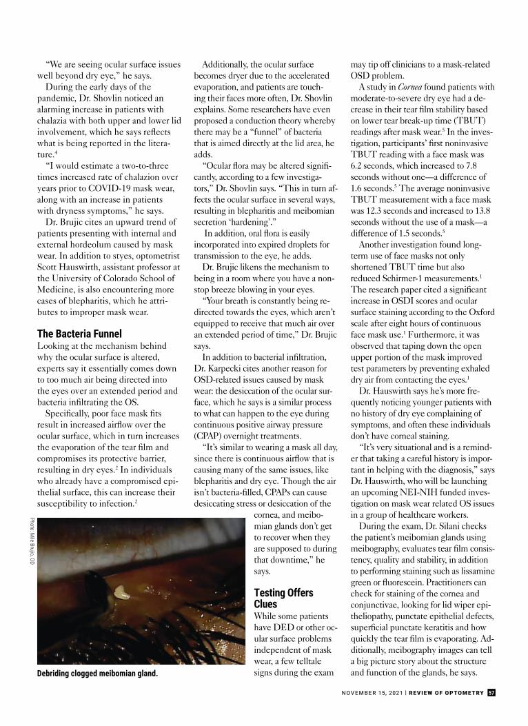
57NOVEMBER 15, 2021 | REVIEW OF OPTOMETRY
“We are seeing ocular surface issueswell beyond dry eye,” he says.
During the early days of thepandemic, Dr. Shovlin noticed analarming increase in patients withchalazia with both upper and lower lidinvolvement, which he says reflectswhat is being reported in the litera-ture.4
“I would estimate a two-to-threetimes increased rate of chalazion overyears prior to COVID-19 mask wear,along with an increase in patientswith dryness symptoms,” he says.
Dr. Brujic cites an upward trend ofpatients presenting with internal andexternal hordeolum caused by maskwear. In addition to styes, optometristScott Hauswirth, assistant professor atthe University of Colorado School ofMedicine, is also encountering morecases of blepharitis, which he attri-butes to improper mask wear.
The Bacteria FunnelLooking at the mechanism behindwhy the ocular surface is altered,experts say it essentially comes downto too much air being directed intothe eyes over an extended period andbacteria infiltrating the OS.
Specifically, poor face mask fitsresult in increased airflow over theocular surface, which in turn increasesthe evaporation of the tear film andcompromises its protective barrier,resulting in dry eyes.2 In individualswho already have a compromised epi-thelial surface, this can increase theirsusceptibility to infection.2
Additionally, the ocular surfacebecomes dryer due to the acceleratedevaporation, and patients are touch-ing their faces more often, Dr. Shovlinexplains. Some researchers have evenproposed a conduction theory wherebythere may be a “funnel” of bacteriathat is aimed directly at the lid area, headds.
“Ocular flora may be altered signifi-cantly, according to a few investiga-tors,” Dr. Shovlin says. “This in turn af-fects the ocular surface in several ways,resulting in blepharitis and meibomiansecretion ‘hardening’.”
In addition, oral flora is easilyincorporated into expired droplets fortransmission to the eye, he adds.
Dr. Brujic likens the mechanism tobeing in a room where you have a non-stop breeze blowing in your eyes.
“Your breath is constantly being re-directed towards the eyes, which aren’tequipped to receive that much air overan extended period of time,” Dr. Brujicsays.
In addition to bacterial infiltration,Dr. Karpecki cites another reason forOSD-related issues caused by maskwear: the desiccation of the ocular sur-face, which he says is a similar processto what can happen to the eye duringcontinuous positive airway pressure(CPAP) overnight treatments.
“It’s similar to wearing a mask all day,since there is continuous airflow that iscausing many of the same issues, likeblepharitis and dry eye. Though the airisn’t bacteria-filled, CPAPs can causedesiccating stress or desiccation of the
cornea, and meibo-mian glands don’t getto recover when theyare supposed to duringthat downtime,” hesays.
Testing OffersCluesWhile some patientshave DED or other oc-ular surface problemsindependent of maskwear, a few telltalesigns during the exam
may tip off clinicians to a mask-relatedOSD problem.
A study in Cornea found patients withmoderate-to-severe dry eye had a de-crease in their tear film stability basedon lower tear break-up time (TBUT)readings after mask wear.5 In the inves-tigation, participants’ first noninvasiveTBUT reading with a face mask was6.2 seconds, which increased to 7.8seconds without one—a difference of1.6 seconds.5 The average noninvasiveTBUT measurement with a face maskwas 12.3 seconds and increased to 13.8seconds without the use of a mask—adifference of 1.5 seconds.5
Another investigation found long-term use of face masks not onlyshortened TBUT time but alsoreduced Schirmer-1 measurements.1
The research paper cited a significantincrease in OSDI scores and ocularsurface staining according to the Oxfordscale after eight hours of continuousface mask use.1 Furthermore, it wasobserved that taping down the openupper portion of the mask improvedtest parameters by preventing exhaleddry air from contacting the eyes.1
Dr. Hauswirth says he’s more fre-quently noticing younger patients withno history of dry eye complaining ofsymptoms, and often these individualsdon’t have corneal staining.
“It’s very situational and is a remind-er that taking a careful history is impor-tant in helping with the diagnosis,” saysDr. Hauswirth, who will be launchingan upcoming NEI-NIH funded inves-tigation on mask wear related OS issuesin a group of healthcare workers.
During the exam, Dr. Silani checksthe patient’s meibomian glands usingmeibography, evaluates tear film consis-tency, quality and stability, in additionto performing staining such as lissaminegreen or fluorescein. Practitioners cancheck for staining of the cornea andconjunctivae, looking for lid wiper epi-theliopathy, punctate epithelial defects,superficial punctate keratitis and howquickly the tear film is evaporating. Ad-ditionally, meibography images can tella big picture story about the structureand function of the glands, he says.Debriding clogged meibomian gland.
Photo: Mile Brujic, OD
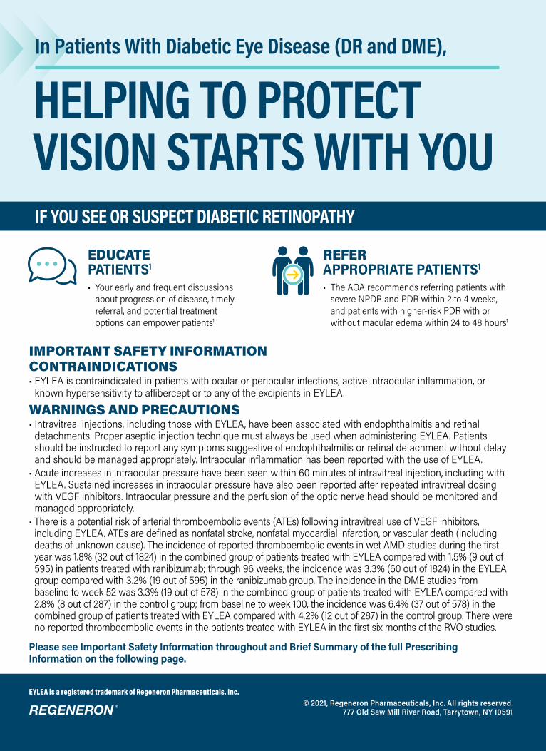
IF YOU SEE OR SUSPECT DIABETIC RETINOPATHY
EYLEA is a registered trademark of Regeneron Pharmaceuticals, Inc.© 2021, Regeneron Pharmaceuticals, Inc. All rights reserved.
777 Old Saw Mill River Road, Tarrytown, NY 10591
HELPING TO PROTECTVISION STARTS WITH YOU
EDUCATEPATIENTS1
• Your early and frequent discussionsabout progression of disease, timelyreferral, and potential treatmentoptions can empower patients1
REFERAPPROPRIATE PATIENTS1
• The AOA recommends referring patients withsevere NPDR and PDR within 2 to 4 weeks,and patients with higher-risk PDR with orwithout macular edema within 24 to 48 hours1
In Patients With Diabetic Eye Disease (DR and DME),
IMPORTANT SAFETY INFORMATIONCONTRAINDICATIONS• EYLEA is contraindicated in patients with ocular or periocular infections, active intraocular inflammation, or
known hypersensitivity to aflibercept or to any of the excipients in EYLEA.
WARNINGS AND PRECAUTIONS• Intravitreal injections, including those with EYLEA, have been associated with endophthalmitis and retinal
detachments. Proper aseptic injection technique must always be used when administering EYLEA. Patientsshould be instructed to report any symptoms suggestive of endophthalmitis or retinal detachment without delayand should be managed appropriately. Intraocular inflammation has been reported with the use of EYLEA.
• Acute increases in intraocular pressure have been seen within 60 minutes of intravitreal injection, including withEYLEA. Sustained increases in intraocular pressure have also been reported after repeated intravitreal dosingwith VEGF inhibitors. Intraocular pressure and the perfusion of the optic nerve head should be monitored andmanaged appropriately.
• There is a potential risk of arterial thromboembolic events (ATEs) following intravitreal use of VEGF inhibitors,including EYLEA. ATEs are defined as nonfatal stroke, nonfatal myocardial infarction, or vascular death (includingdeaths of unknown cause). The incidence of reported thromboembolic events in wet AMD studies during the firstyear was 1.8% (32 out of 1824) in the combined group of patients treated with EYLEA compared with 1.5% (9 out of595) in patients treated with ranibizumab; through 96 weeks, the incidence was 3.3% (60 out of 1824) in the EYLEAgroup compared with 3.2% (19 out of 595) in the ranibizumab group. The incidence in the DME studies frombaseline to week 52 was 3.3% (19 out of 578) in the combined group of patients treated with EYLEA compared with2.8% (8 out of 287) in the control group; from baseline to week 100, the incidence was 6.4% (37 out of 578) in thecombined group of patients treated with EYLEA compared with 4.2% (12 out of 287) in the control group. There wereno reported thromboembolic events in the patients treated with EYLEA in the first six months of the RVO studies.
Please see Important Safety Information throughout and Brief Summary of the full PrescribingInformation on the following page.
07/2021EYL.21.03.0229
The more you know about anti-VEGF agents and other potential treatmentsfor DR, the better you can help inform your patients. Find out more by visitingdiabeticretinaldisease.com.
CONTINUE TO MONITORPATIENTS1
• The AOA recommends frequent monitoringof patients1
– At least every 6 to 9 months in patients withmoderate NPDR and more frequently forpatients with greater disease severity
FOLLOW UPWITH PATIENTS• Encourage referred patients to
promptly visit a retina specialist
anti-VEGF, anti–vascular endothelial growth factor; AOA, American Optometric Association; NPDR, nonproliferative diabetic retinopathy;PDR, proliferative diabetic retinopathy.
ADVERSE REACTIONS• Serious adverse reactions related to the injection procedure have occurred in <0.1% of intravitreal injections
with EYLEA including endophthalmitis and retinal detachment.• The most common adverse reactions (≥5%) reported in patients receiving EYLEA were conjunctival
hemorrhage, eye pain, cataract, vitreous detachment, vitreous floaters, and intraocular pressure increased.• Patients may experience temporary visual disturbances after an intravitreal injection with EYLEA and the
associated eye examinations. Advise patients not to drive or use machinery until visual function hasrecovered su� iciently.
INDICATIONSEYLEA® (aflibercept) Injection 2 mg (0.05 mL) is indicated for the treatment of patients with Neovascular (Wet)Age-related Macular Degeneration (AMD), Macular Edema following Retinal Vein Occlusion (RVO), DiabeticMacular Edema (DME), and Diabetic Retinopathy (DR).
Reference: 1. Eye care of the patient with diabetes mellitus. American Optometric Association. Accessed April 2, 2021. http://aoa.uberflip.com/i/1183026-evidence-based-clinical-practice-guideline-eye-care-of-the-patient-with-diabetes-mellitus-second-edition/
Brought to you by

1 INDICATIONS AND USAGE EYLEA is a vascular endothelial growth factor (VEGF) inhibitor indicated for the treatment of patients with:Neovascular (Wet) Age-Related Macular Degeneration (AMD), Macular Edema Following Retinal Vein Occlusion (RVO), Diabetic Macular Edema (DME), Diabetic Retinopathy (DR).4 CONTRAINDICATIONS4.1 Ocular or Periocular Infections EYLEA is contraindicated in patients with ocular or periocular infections. 4.2 Active Intraocular Inflammation EYLEA is contraindicated in patients with active intraocular inflammation. 4.3 Hypersensitivity EYLEA is contraindicated in patients with known hypersensitivity to aflibercept or any of the excipients in EYLEA. Hypersensitivity reactions may manifest as rash, pruritus, urticaria, severe anaphylactic/anaphylactoid reactions, or severe intraocular inflammation.5 WARNINGS AND PRECAUTIONS 5.1 Endophthalmitis and Retinal Detachments Intravitreal injections, including those with EYLEA, have been associated with endophthalmitis and retinal detachments [see Adverse Reactions (6.1)]. Proper aseptic injection technique must always be used when administering EYLEA. Patients should be instructed to report any symptoms suggestive of endophthalmitis or retinal detachment without delay and should be managed appropriately [see Patient Counseling Information (17)].5.2 Increase in Intraocular Pressure Acute increases in intraocular pressure have been seen within 60 minutes of intravitreal injection, including with EYLEA [see Adverse Reactions (6.1)]. Sustained increases in intraocular pressure have also been reported after repeated intravitreal dosing with vascular endothelial growth factor (VEGF) inhibitors. Intraocular pressure and the perfusion of the optic nerve head should be monitored and managed appropriately.5.3 Thromboembolic Events There is a potential risk of arterial thromboembolic events (ATEs) following intravitreal use of VEGF inhibitors, including EYLEA. ATEs are defined as nonfatal stroke, nonfatal myocardial infarction, or vascular death (including deaths of unknown cause). The incidence of reported thromboembolic events in wet AMD studies during the first year was 1.8% (32 out of 1824) in the combined group of patients treated with EYLEA compared with 1.5% (9 out of 595) in patients treated with ranibizumab; through 96 weeks, the incidence was 3.3% (60 out of 1824) in the EYLEA group compared with 3.2% (19 out of 595) in the ranibizumab group. The incidence in the DME studies from baseline to week 52 was 3.3% (19 out of 578) in the combined group of patients treated with EYLEA compared with 2.8% (8 out of 287) in the control group; from baseline to week 100, the incidence was 6.4% (37 out of 578) in the combined group of patients treated with EYLEA compared with 4.2% (12 out of 287) in the control group. There were no reported thromboembolic events in the patients treated with EYLEA in the first six months of the RVO studies.6 ADVERSE REACTIONS The following potentially serious adverse reactions are described elsewhere in the labeling: • Hypersensitivity [see Contraindications (4.3)] • Endophthalmitis and retinal detachments [see Warnings and Precautions (5.1)] • Increase in intraocular pressure [see Warnings and Precautions (5.2)] • Thromboembolic events [see Warnings and Precautions (5.3)]6.1 Clinical Trials Experience Because clinical trials are conducted under widely varying conditions, adverse reaction rates observed in the clinical trials of a drug cannot be directly compared to rates in other clinical trials of the same or another drug and may not reflect the rates observed in practice.A total of 2980 patients treated with EYLEA constituted the safety population in eight phase 3 studies. Among those, 2379 patients were treated with the recommended dose of 2 mg. Serious adverse reactions related to the injection procedure have occurred in <0.1% of intravitreal injections with EYLEA including endophthalmitis and retinal detachment. The most common adverse reactions (≥5%) reported in patients receiving EYLEA were conjunctival hemorrhage, eye pain, cataract, vitreous detachment, vitreous floaters, and intraocular pressure increased.
Neovascular (Wet) Age-Related Macular Degeneration (AMD). The data described below reflect exposure to EYLEA in 1824 patients with wet AMD, including 1223 patients treated with the 2-mg dose, in 2 double-masked, controlled clinical studies (VIEW1 and VIEW2) for 24 months (with active control in year 1).Safety data observed in the EYLEA group in a 52-week, double-masked, Phase 2 study were consistent with these results.
Table 1: Most Common Adverse Reactions (≥1%) in Wet AMD StudiesBaseline to Week 52 Baseline to Week 96
Adverse ReactionsEYLEA
(N=1824)
Active Control (ranibizumab)
(N=595)EYLEA
(N=1824)
Control (ranibizumab)
(N=595)Conjunctival hemorrhage 25% 28% 27% 30%Eye pain 9% 9% 10% 10%Cataract 7% 7% 13% 10%Vitreous detachment 6% 6% 8% 8%Vitreous floaters 6% 7% 8% 10%Intraocular pressure increased 5% 7% 7% 11%Ocular hyperemia 4% 8% 5% 10%Corneal epithelium defect 4% 5% 5% 6%Detachment of the retinal pigment epithelium 3% 3% 5% 5%Injection site pain 3% 3% 3% 4%Foreign body sensation in eyes 3% 4% 4% 4%Lacrimation increased 3% 1% 4% 2%Vision blurred 2% 2% 4% 3%Intraocular inflammation 2% 3% 3% 4%Retinal pigment epithelium tear 2% 1% 2% 2%Injection site hemorrhage 1% 2% 2% 2%Eyelid edema 1% 2% 2% 3%Corneal edema 1% 1% 1% 1%Retinal detachment <1% <1% 1% 1%
Less common serious adverse reactions reported in <1% of the patients treated with EYLEA were hypersensitivity, retinal tear, and endophthalmitis.
Macular Edema Following Retinal Vein Occlusion (RVO). The data described below reflect 6 months exposure to EYLEA with a monthly 2 mg dose in 218 patients following central retinal vein occlusion (CRVO) in 2 clinical studies (COPERNICUS and GALILEO) and 91 patients following branch retinal vein occlusion (BRVO) in one clinical study (VIBRANT).
Table 2: Most Common Adverse Reactions (≥1%) in RVO StudiesCRVO BRVO
Adverse ReactionsEYLEA
(N=218)Control (N=142)
EYLEA (N=91)
Control (N=92)
Eye pain 13% 5% 4% 5%Conjunctival hemorrhage 12% 11% 20% 4%Intraocular pressure increased 8% 6% 2% 0%Corneal epithelium defect 5% 4% 2% 0%Vitreous floaters 5% 1% 1% 0%Ocular hyperemia 5% 3% 2% 2%Foreign body sensation in eyes 3% 5% 3% 0%Vitreous detachment 3% 4% 2% 0%Lacrimation increased 3% 4% 3% 0%Injection site pain 3% 1% 1% 0%Vision blurred 1% <1% 1% 1%Intraocular inflammation 1% 1% 0% 0%Cataract <1% 1% 5% 0%Eyelid edema <1% 1% 1% 0%
Less common adverse reactions reported in <1% of the patients treated with EYLEA in the CRVO studies were corneal edema, retinal tear, hypersensitivity, and endophthalmitis.
Diabetic Macular Edema (DME) and Diabetic Retinopathy (DR). The data described below reflect exposure to EYLEA in 578 patients with DME treated with the 2-mg dose in 2 double-masked, controlled clinical studies (VIVID and VISTA) from baseline to week 52 and from baseline to week 100.
Table 3: Most Common Adverse Reactions (≥1%) in DME StudiesBaseline to Week 52 Baseline to Week 100
Adverse ReactionsEYLEA
(N=578)Control
(N=287)EYLEA
(N=578)Control
(N=287)Conjunctival hemorrhage 28% 17% 31% 21%Eye pain 9% 6% 11% 9%Cataract 8% 9% 19% 17%Vitreous floaters 6% 3% 8% 6%Corneal epithelium defect 5% 3% 7% 5%Intraocular pressure increased 5% 3% 9% 5%Ocular hyperemia 5% 6% 5% 6%Vitreous detachment 3% 3% 8% 6%Foreign body sensation in eyes 3% 3% 3% 3%Lacrimation increased 3% 2% 4% 2%Vision blurred 2% 2% 3% 4%Intraocular inflammation 2% <1% 3% 1%Injection site pain 2% <1% 2% <1%Eyelid edema <1% 1% 2% 1%
Less common adverse reactions reported in <1% of the patients treated with EYLEA were hypersensitivity, retinal detachment, retinal tear, corneal edema, and injection site hemorrhage. Safety data observed in 269 patients with nonproliferative diabetic retinopathy (NPDR) through week 52 in the PANORAMA trial were consistent with those seen in the phase 3 VIVID and VISTA trials (see Table 3 above).6.2 Immunogenicity As with all therapeutic proteins, there is a potential for an immune response in patients treated with EYLEA. The immunogenicity of EYLEA was evaluated in serum samples. The immunogenicity data reflect the percentage of patients whose test results were considered positive for antibodies to EYLEA in immunoassays. The detection of an immune response is highly dependent on the sensitivity and specificity of the assays used, sample handling, timing of sample collection, concomitant medications, and underlying disease. For these reasons, comparison of the incidence of antibodies to EYLEA with the incidence of antibodies to other products may be misleading. In the wet AMD, RVO, and DME studies, the pre-treatment incidence of immunoreactivity to EYLEA was approximately 1% to 3% across treatment groups. After dosing with EYLEA for 24-100 weeks, antibodies to EYLEA were detected in a similar percentage range of patients. There were no differences in efficacy or safety between patients with or without immunoreactivity.
8 USE IN SPECIFIC POPULATIONS8.1 Pregnancy Risk SummaryAdequate and well-controlled studies with EYLEA have not been conducted in pregnant women. Aflibercept produced adverse embryofetal effects in rabbits, including external, visceral, and skeletal malformations. A fetal No Observed Adverse Effect Level (NOAEL) was not identified. At the lowest dose shown to produce adverse embryofetal effects, systemic exposures (based on AUC for free aflibercept) were approximately 6 times higher than AUC values observed in humans after a single intravitreal treatment at the recommended clinical dose [see Animal Data].Animal reproduction studies are not always predictive of human response, and it is not known whether EYLEA can cause fetal harm when administered to a pregnant woman. Based on the anti-VEGF mechanism of action for aflibercept, treatment with EYLEA may pose a risk to human embryofetal development. EYLEA should be used during pregnancy only if the potential benefit justifies the potential risk to the fetus.All pregnancies have a background risk of birth defect, loss, or other adverse outcomes. The background risk of major birth defects and miscarriage for the indicated population is unknown. In the U.S. general population, the estimated background risk of major birth defects and miscarriage in clinically recognized pregnancies is 2-4% and 15-20%, respectively.DataAnimal Data In two embryofetal development studies, aflibercept produced adverse embryofetal effects when administered every three days during organogenesis to pregnant rabbits at intravenous doses ≥3 mg per kg, or every six days during organogenesis at subcutaneous doses ≥0.1 mg per kg. Adverse embryofetal effects included increased incidences of postimplantation loss and fetal malformations, including anasarca, umbilical hernia, diaphragmatic hernia, gastroschisis, cleft palate, ectrodactyly, intestinal atresia, spina bifida, encephalomeningocele, heart and major vessel defects, and skeletal malformations (fused vertebrae, sternebrae, and ribs; supernumerary vertebral arches and ribs; and incomplete ossification). The maternal No Observed Adverse Effect Level (NOAEL) in these studies was 3 mg per kg. Aflibercept produced fetal malformations at all doses assessed in rabbits and the fetal NOAEL was not identified. At the lowest dose shown to produce adverse embryofetal effects in rabbits (0.1 mg per kg), systemic exposure (AUC) of free aflibercept was approximately 6 times higher than systemic exposure (AUC) observed in humans after a single intravitreal dose of 2 mg.8.2 Lactation Risk SummaryThere is no information regarding the presence of aflibercept in human milk, the effects of the drug on the breastfed infant, or the effects of the drug on milk production/excretion. Because many drugs are excreted in human milk, and because the potential for absorption and harm to infant growth and development exists, EYLEA is not recommended during breastfeeding. The developmental and health benefits of breastfeeding should be considered along with the mother’s clinical need for EYLEA and any potential adverse effects on the breastfed child from EYLEA.8.3 Females and Males of Reproductive Potential ContraceptionFemales of reproductive potential are advised to use effective contraception prior to the initial dose, during treatment, and for at least 3 months after the last intravitreal injection of EYLEA.
InfertilityThere are no data regarding the effects of EYLEA on human fertility. Aflibercept adversely affected female and male reproductive systems in cynomolgus monkeys when administered by intravenous injection at a dose approximately 1500 times higher than the systemic level observed humans with an intravitreal dose of 2 mg. A No Observed Adverse Effect Level (NOAEL) was not identified. These findings were reversible within 20 weeks after cessation of treatment.8.4 Pediatric Use The safety and effectiveness of EYLEA in pediatric patients have not been established.8.5 Geriatric Use In the clinical studies, approximately 76% (2049/2701) of patients randomized to treatment with EYLEA were ≥65 years of age and approximately 46% (1250/2701) were ≥75 years of age. No significant differences in efficacy or safety were seen with increasing age in these studies.17 PATIENT COUNSELING INFORMATION In the days following EYLEA administration, patients are at risk of developing endophthalmitis or retinal detachment. If the eye becomes red, sensitive to light, painful, or develops a change in vision, advise patients to seek immediate care from an ophthalmologist [see Warnings and Precautions (5.1)]. Patients may experience temporary visual disturbances after an intravitreal injection with EYLEA and the associated eye examinations [see Adverse Reactions (6)]. Advise patients not to drive or use machinery until visual function has recovered sufficiently.
BRIEF SUMMARY—Please see the EYLEA full Prescribing Information available on HCP.EYLEA.US for additional product information.
Manufactured by: Regeneron Pharmaceuticals, Inc. 777 Old Saw Mill River Road Tarrytown, NY 10591
EYLEA is a registered trademark of Regeneron Pharmaceuticals, Inc. © 2020, Regeneron Pharmaceuticals, Inc. All rights reserved.
Issue Date: 08/2019 Initial U.S. Approval: 2011
Based on the August 2019 EYLEA® (aflibercept) Injection full Prescribing Information.
EYL.20.09.0052
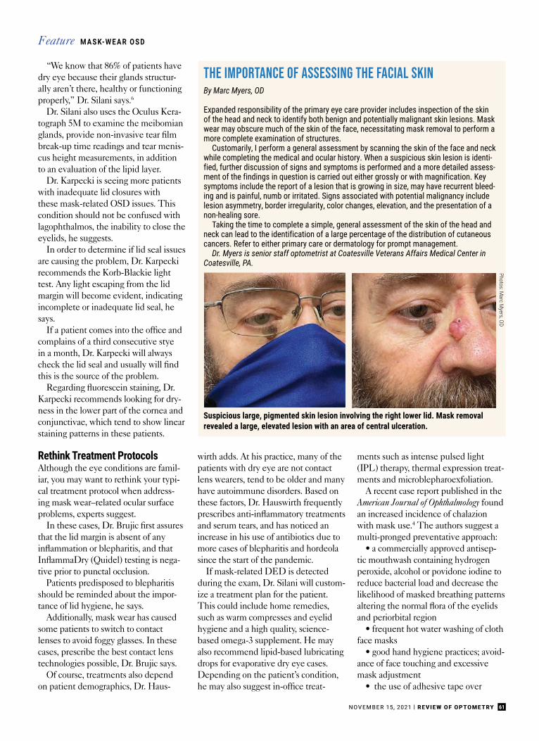
61NOVEMBER 15, 2021 | REVIEW OF OPTOMETRY
“We know that 86% of patients havedry eye because their glands structur-ally aren’t there, healthy or functioningproperly,” Dr. Silani says.6
Dr. Silani also uses the Oculus Kera-tograph 5M to examine the meibomianglands, provide non-invasive tear filmbreak-up time readings and tear menis-cus height measurements, in additionto an evaluation of the lipid layer.
Dr. Karpecki is seeing more patientswith inadequate lid closures withthese mask-related OSD issues. Thiscondition should not be confused withlagophthalmos, the inability to close theeyelids, he suggests.
In order to determine if lid seal issuesare causing the problem, Dr. Karpeckirecommends the Korb-Blackie lighttest. Any light escaping from the lidmargin will become evident, indicatingincomplete or inadequate lid seal, hesays.
If a patient comes into the office andcomplains of a third consecutive styein a month, Dr. Karpecki will alwayscheck the lid seal and usually will findthis is the source of the problem.
Regarding fluorescein staining, Dr.Karpecki recommends looking for dry-ness in the lower part of the cornea andconjunctivae, which tend to show linearstaining patterns in these patients.
Rethink Treatment ProtocolsAlthough the eye conditions are famil-iar, you may want to rethink your typi-cal treatment protocol when address-ing mask wear–related ocular surfaceproblems, experts suggest.
In these cases, Dr. Brujic first assuresthat the lid margin is absent of anyinflammation or blepharitis, and thatInflammaDry (Quidel) testing is nega-tive prior to punctal occlusion.
Patients predisposed to blepharitisshould be reminded about the impor-tance of lid hygiene, he says.
Additionally, mask wear has causedsome patients to switch to contactlenses to avoid foggy glasses. In thesecases, prescribe the best contact lenstechnologies possible, Dr. Brujic says.
Of course, treatments also dependon patient demographics, Dr. Haus-
wirth adds. At his practice, many of thepatients with dry eye are not contactlens wearers, tend to be older and manyhave autoimmune disorders. Based onthese factors, Dr. Hauswirth frequentlyprescribes anti-inflammatory treatmentsand serum tears, and has noticed anincrease in his use of antibiotics due tomore cases of blepharitis and hordeolasince the start of the pandemic.
If mask-related DED is detectedduring the exam, Dr. Silani will custom-ize a treatment plan for the patient.This could include home remedies,such as warm compresses and eyelidhygiene and a high quality, science-based omega-3 supplement. He mayalso recommend lipid-based lubricatingdrops for evaporative dry eye cases.Depending on the patient’s condition,he may also suggest in-office treat-
ments such as intense pulsed light(IPL) therapy, thermal expression treat-ments and microblepharoexfoliation.
A recent case report published in theAmerican Journal of Ophthalmology foundan increased incidence of chalazionwith mask use.4 The authors suggest amulti-pronged preventative approach:
• a commercially approved antisep-tic mouthwash containing hydrogenperoxide, alcohol or povidone iodine toreduce bacterial load and decrease thelikelihood of masked breathing patternsaltering the normal flora of the eyelidsand periorbital region
• frequent hot water washing of clothface masks
• good hand hygiene practices; avoid-ance of face touching and excessivemask adjustment
• the use of adhesive tape over
M A S K-W E A R O S DFeature
THE IMPORTANCE OF ASSESSING THE FACIAL SKINBy Marc Myers, OD
Expanded responsibility of the primary eye care provider includes inspection of the skinof the head and neck to identify both benign and potentially malignant skin lesions. Maskwear may obscure much of the skin of the face, necessitating mask removal to perform amore complete examination of structures.
Customarily, I perform a general assessment by scanning the skin of the face and neckwhile completing the medical and ocular history. When a suspicious skin lesion is identi-fied, further discussion of signs and symptoms is performed and a more detailed assess-ment of the findings in question is carried out either grossly or with magnification. Keysymptoms include the report of a lesion that is growing in size, may have recurrent bleed-ing and is painful, numb or irritated. Signs associated with potential malignancy includelesion asymmetry, border irregularity, color changes, elevation, and the presentation of anon-healing sore.
Taking the time to complete a simple, general assessment of the skin of the head andneck can lead to the identification of a large percentage of the distribution of cutaneouscancers. Refer to either primary care or dermatology for prompt management.
Dr. Myers is senior staff optometrist at Coatesville Veterans Affairs Medical Center inCoatesville, PA.
Suspicious large, pigmented skin lesion involving the right lower lid. Mask removalrevealed a large, elevated lesion with an area of central ulceration.
Photos: Marc M
yers, OD

62 REVIEW OF OPTOMETRY | NOVEMBER 15, 2021
a mask on the bridge of the nose tominimize the upward direction of airtowards the eyes.4
Additionally, the authors suggest theuse of a 1% hypochlorous acid solu-tion eyelid scrub as part of daily eyelidhygiene to act as both an antiviral andantibacterial blepharitis deterrent.4
Due to airflow and inadequate lidclosure issues, Dr. Karpecki is recom-mending thicker lubricating agents forthese patients. “I’m more likely to usea gel that has some staying power andwill stay on the eye instead of goingaway more quickly,” Dr. Karpecki says.
In addition to thicker tear ointments,Dr. Karpecki points to a new product hehelped developed that will be comingsoon for those individuals with lid sealproblems: SleepTite (Ophthalmic Re-sources Partners), which is a single-usedevice intended to hold the eye closedto prevent it from unintended expo-sure to fluids, airborne contaminants orexcessive drying due to incomplete lidclosure.
Dr. Karpecki is also finding manypatients do well with hydrating com-presses, especially if they are gettinghordeolum, and he’s turning to in-officeprocedures such as blepharoexfolia-tion, IPL, low level light therapy andin-office thermal expression.
“It seems many of these patientsare having a lot more issues with theireyelid margins drying and accumulatingkeratin plugs or biofilm. For example,I’m finding that if I don’t do a quick liddebridement, the patients tend not toget a good response, and they may getmultiple hordeola otherwise,” he says.
Patient Education is KeyIn addition to reaching for your Rxpad, patient education on proper maskwear can also help mitigate OSD issues.In choosing a mask, the CDC recom-mends that it has two or more layers ofwashable, breathable fabric, completelycovers the nose and mouth, fits snuglyagainst the sides of the face and doesn’thave gaps and also is equipped with anose wire to prevent air from leakingout of the top.7 Reusable masks shouldbe washed as soon as they becomedirty, or at least once a day.7 If you havea disposable face mask, throw it awayafter wearing it once, the CDC advises.7
Dr. Silani suggests asking patientshow long they wear masks throughoutthe day, as prolonged use can contributeto mask associated dry eye or styes.
“We recommend that patientsremove their masks when they are ableor alone, to make sure the mask top halfis pinned down correctly to minimizeupward airflow and to tape down thetop half of the mask if the individual isnoticing their glasses are fogging up,”he says.
Additionally, some patients arebringing up their masks too high andlimiting their lower eyelid from fullymaking contact with the upper eyelid,so placement or positioning of the maskis important, Dr. Silani adds.
“If a patient is complaining of mask-related dry eye, consider suggesting dif-ferent mask options—specifically onesthat have a reinforced barrier across thebridge of the nose and across the uppercheek that allow for an air-tight fit,” Dr.Hauswirth says.
Researchers from Wills Eye Hospitalwho looked into bacterial dispersionwith patient face mask use during simu-lated intravitreal injections found altera-tions of bacterial dispersion around theeye during treatment based on the typeof mask worn.10 The authors notedsignificantly more bacterial dispersionwhen wearing a tight-fitting face maskwithout tape vs. a tight-fitting one withtape. Additionally, their study found nodifference in bacterial dispersion be-tween tight-fitting surgical masks withtape and N95 masks.8
In addition to recommendingpatients tape their mask on the bridgeof the nose to minimize an upwarddraft toward the eye and take care notto affect the normal blink dynamics oreyelid closure, Dr. Shovlin also suggestsreminding the patient to avoid touch-ing their face or adjusting their mask asmuch as possible.
Patients with pre-existing DEDand other ocular surfaces issues suchas recurrent corneal erosion syndromemay need additional care with propermask fitting.2
Despite the OSD issues occurring,mask wear is a necessity for health andsafety, Dr. Silani adds.
“We are very pro-health and safety.Yes, mask-associated dry eye does exist,and taking steps to avoid and treat it isimportant. But doctors should recom-mend patients wear masks to protectthemselves and others. Patients shouldtape down the top of their masks, andwhen they aren’t around people or ina safe place, they can take it off or uselubricating drops at work if they can’tremove their mask,” Dr. Silani says.
Another recent study found mostindividuals didn’t experience a changein ocular symptoms, but a significantportion still reported an increase inocular discomfort while wearing amask.9 The investigation suggests notto underestimate all the symptomsthat could discourage the populationfrom using masks, and that ODs shouldverify the presence of clinical signs inthose complaining about mask-inducedeye discomfort and recommend ways tomitigate these issues.
Lid margin integrity and meibum function improving due to office-based treatments in a 27-year-old female patient with DED secondary to MGD. She works in a healthcare facility and prolonged face mask wear has exacerbated her dry eye condition.
M A S K-W E A R O S DFeature
Photo: Kambiz Silani, OD

PRECISION ACCURACY REPEATABILIT Y
Made in the USA
Proven technology to help your clinic reachnew heights of accuracy and ease-of-use
Smart System® 2 | 2020Shown with optional Glare Lights and Tablet
For over 30 years M&S has focused on providing the highestquality software and advanced computerized testing systemsto enable eye-care professionals to better care for their patients.
We want to sincerely thank our customers and pledgeour continued commitment to perfecting vision testingtechnology while bringing increased e�ciency and �exibilityto their practices.
Over 1 Billion eye exams & counting
The First Choice in Vision Testing Systems
YEARS
mstech-eyes.com
©2021 M&S and Smart System are registered trademarks of the Hilsinger Company Parent LLC. All rights reserved. The Hilsinger Company Parent LLC holdsUS Patents 7,354,155; 7,926,948; 8,425,040; 8,167,429; 8,419,184; 8,550,631; 8,992,022; 9,433,347; 9,820,644; 10,244,938 and 10,182,713. Other Patents Pending.
Find out why the M&S Smart System® is the right choice.
Call: 847-763-0500

Mask-Related OSD TrendsNormally, one would expect that people who work at jobsthat involve heavy computer use would have more mask-related OSD issues, but Dr. Brujic says he’s not seeing thisoccurring. “At the beginning of the pandemic, a majority ofthese people were working from home and didn’t have towear a mask,” he explains.
On the other hand, Dr. Brujic was treating more OSD casesin individuals who worked at venues that required staff inmasks to be on site in stores, plants and factories.
As far as age posing a potential risk factor, Dr. Hauswirthsays most of his symptomatic patients over the last year-and-a-half are younger, healthy individuals who never had ocularsurface issues before, despite the fact they are computer us-ers. “We are seeing a lot of people naïve to the disorder, whoweren’t really dry eye patients beforehand,” Dr. Hauswirthsays.
“Other patients with existing issues, such as rosacea andblepharitis, are also increasingly presenting with OSD is-sues due to mask wear,” Dr. Karpecki says. Additionally, heis noticing an uptick in contact lens-wearing patients withmask-related complications.
Even before the pandemic, the Tear Film & OcularSurface Society (TFOS) had already reported thatdiscomfort while wearing contact lenses were associatedwith a reduction in the compatibility between CLs and theocular environment.10
Exploring the impact of contact lenses and mask wear, ateam of researchers from Portugal found mask use increaseddry eye symptoms in contact lens wearers and negativelyimpacted their visual quality.11 Additionally, lens wear signifi -cantly decreased after the onset of COVID, and the sensationof dry eye was found to be worse in those using monthlyreplacement contact lenses.11
Looking AheadWith face mask use likely extending into at least the nearfuture, eye dryness, irritation and keratopathy from maskwear may become a problem for a large percentage of thepopulation.14 If present, this mask-associated ocular irrita-tion raises concerns about eye health and increased risk ofdisease transmission with prolonged wear.12
Considering this forecast, additional investigations maybe warranted. One consideration for future studies could bean analysis of geographical differences from one region toanother and a comparison of ocular surface prevalence issuesin areas that are more compliant in mask-wearing practices orin areas with higher rates of COVID-19, Dr. Shovlin suggests.
Along with mask-induced dry eye, Dr. Silani also stressesthat it is important to consider potential comorbidities, suchas lagophthalmos, ocular allergies and anterior blepharitis, asmultiple, contributing factors could be at play.
Another point to consider: look for any suspicious lesionsunder the mask that may warrant testing, experts advise.
“Masks are here to stay,” Dr. Brujic says. “We don’t knowwhat the capacity of masks are going to be, but they’ll bepart of our society. Today, it’s not uncommon to see peoplein masks, but it’s important for optometrists to be askingabout it. We see the patient for 15 minutes in our offi ce,but we need to know how much or how little they’rewearing their masks outside of the offi ce and provide themappropriate solutions.” ■
1. Aksoy M, Simsek M. Evaluation of ocular surface and dry eye symptoms in face mask users.Eye Contact Lens. 2021;47(10):555-8.2. Tang YF, Chong EWT. Face mask–associated recurrent corneal erosion syndrome andcorneal infection. Eye Contact Lens. 2021;47(10):573-4.3. Nemet AY, Vinker S, Kaiserman I. Associated morbidity of chalazia. Cornea. 2011;30:1376-81.4. Silkiss RZ, Paap MK, Ugradar S. Increased incidence of chalazion associated with face maskwear during the COVID-19 pandemic. Am J Opthalmol case report. 2021 Jun;22:10103.5. Arriola-Villalobos P, Burgos-Blasco B, Vidal-Villegas B, et al. Effect of face mask on tear fi lm stability in eyes with moderate-to-severe dry eye disease. Cornea. 2021;40(10):1336-9.6. Lemp MA, Crews LA, Bron AJ, Foulks GN, Sullivan BD. Distribution of aqueous-defi cient and evaporative dry eye in a clinic-based patient cohort: a retrospective study. Cornea.2012;31(5):472-478.7. CDC. Your guide to masks. www.cdc.gov/coronavirus/2019-ncov/prevent-getting-sick/about-face-coverings.html. Accessed Oct. 23, 2021.8. Patel SM, Mahmoudzadeh R, Salabati M, et al. Bacterial dispersion associated with vari-ous patient face mask designs during simulated intravitreal injections. Am J Ophthalmol.2021;223:178-3.9. Boccardo L. Self-reported symptoms of mask-associated dry eye: a survey study of 3,605people. Contact Lens Ant Eye. January 20, 2021. [Epub ahead of print].10. Nichols JJ, Willcox MD, Bron AJ, et al.; members of the TFOS International Workshop onContact Lens Discomfort. The TFOS international workshop on contact lens discomfort: execu-tive summary. Invest Ophthalmol Vis Sci. 2013;54:TFOS7-TFOS13.11. Martinez-Perez C, Monterio B, Soares M, et al. Infl uence of face masks on the use of contact lenses. Int J Environ Res Public Health. 2021;18(14):7407.12. Moshirfar M, West WB, Marx DP. Face mask-associated ocular irritation and dryness.Ophthalmol Ther. 2020;9(3):397-400.
64
.
M A S K-W E A R O S DFeature
To Register, Visit FinalEyesCE.com/Registration
6COPEAPPROVEDCE HOURS
ON THE AGENDA: Ptosis and Pseudotosis: Evaluationand Treatment Options • Graves’ Ophthalmopathy •Technological Advancements in Medical Aesthetics •An Overview of Anterior Segment Ocular Infections •Systemic Factors Masquerading as Dry Eye and OtherPrimary Ocular Disease • Glaucoma: Interactive CaseDiscussion • Jurisprudence • Medical Errors
JOIN
US
at
A Continuing Education Event Hosted By
19-20 2022FEBRUARY
UNF ADAM W. HERBERTUNIVERSITY CENTER12000 Alumni DriveJacksonville, Florida 32224
64 REVIEW OF OPTOMETRY | NOVEMBER 15, 2021

The Roseof Jericho
Trehalose is a disaccharide that can be found in plantswith moisture retention properties that help organismssurvive in absence of water*. In ophthalmic products, trehalose formulations can enhance active ingredients to help1:
• Protect corneal cells from desiccation
• Restore osmotic balance to the ocular surface
• Maintain the homeostasis of corneal cells
-2017 DEWS ll Report
DRY EYE RELIEFINSPIRED BY NATURE
Learn about our complete line of
dry eye therapy products at theratears.com
TheraTears® EXTRA® Dry Eye Therapy
is enhanced with trehalose
*TheraTears® EXTRA® contains synthetic trehalose.Reference: 1. Jones L, Downie L, Korb D, et al. TFOS Dews ll Management
and Therapy Report. The Ocular Surface Jul 2017; 575-628.© 2021 Prestige Consumer Healthcare | M17-047-01

66 REVIEW OF OPTOMETRY | NOVEMBER 15, 2021
Dry eye: Where Do We Standwith Omega-3 Supplements?
A review of recent research for and against this treatment supplementation.
It has been three years since the release of the three-yearrandomized clinical trial by the National Institute ofHealth (NIH) known as the DREAM Study. This wasthe first multicenter, double-blind study comparing the
efficacy of 3,000mg fish-derived oral omega-3 (EPA/DHA)daily supplementation for the treatment of moderate-to-se-vere dry eye disease (DED) vs. a placebo group containingrefined olive oil. The findings shocked clinicians, as well aspatients, after results showed no significant benefit betweenomega-3 and the olive oil placebo group. It is worth not-ing that 61% of those in the treatment arm and 54% in thecontrol group achieved a 10-point improvement in OSDIscore; however, the difference between the two groups wasnot statistically significant.1
The buzz surrounding these headlines made patientsquestion the role these supplements have on their dryeye regimens. This created a divide between practitionerswho had been using this supplement for years as adjunctor stand-alone therapy with great clinical results and thosewho were already on the fence about its therapeutic effectand role in dry eye management. This article will reviewthe latest discourse surrounding this supplement optionand what impact the DREAM Study will have on treat-ment today.
The Discussion So FarSome criticism was aimed toward the DREAM study’smethodology based on its inclusion and/or exclusioncriteria, as it allowed patients to continue their current dry
eye regimen to mimic a “real-world” scenario and includedthose with moderate-to-severe disease.2-4 Others saw thisas a positive dialogue regarding what roles overall nutri-tion and supplements may play in adjunct therapy.4 Shortlyafter, the follow-up DREAM Extension Study results werereleased and corroborated the study’s original findings.
The 12-month Extension Study pulled 43 patients fromthe original treatment arm and randomized them into twogroups to compare the effects between those who contin-ued omega-3 supplementation vs. those who discontinuedthe treatment. The authors reported no significant dif-ference in the mean change between the treatment andplacebo groups for OSDI score, conjunctival and cornealstaining, TBUT and Schirmer’s test.5
By luis Rojas, ODMcKinney, TX
O M E G A-3 S U P P L E M E N TAT I O NFeature
Dr. Rojas is a cofounder of DeNovo Eye in McKinney, TX where he specializes in ocular disease, specialty contact lenses and myopia management. He is aconsultant for Johnson & Johnson Vision.
Aboutthe author
LASIK-induced dry eye with diffuse SPK and micorostriae.
PEER REVIEWED


68 REVIEW OF OPTOMETRY | NOVEMBER 15, 2021
A Cochrane review in 2019 summarized research regard-ing the effects of omega-3 and omega-6 polyunsaturatedfatty acids (PUFA) supplementation on dry eye signs andsymptoms. The review included 34 randomized controlledtrials with more than 4,314 participants from 13 countries.Supplementing omega-3 as monotherapy showed little tono benefit on dry eye symptoms, relative to placebo, but im-proved clinical signs. However, dry eye symptoms improvedwhen omega-3 was used as adjunct therapy along with otherdry eye treatments (artificial tears, warm compresses and/ortopical steroids), and when compared with omega-6 supple-mentation. The authors concluded that additional researchis needed to improve our understanding on how to useomega-3 and omega-6 depending on the type and severityof DED.6
Research on the effect of omega-3 supplements on dryeye is ongoing in other subspecialties in eye care, such asin pre- and postoperative management, to investigate itsanti-inflammatory properties. A recent prospective compara-tive cohort study in Korea evaluated the clinical outcomesof systemic re-esterified triglyceride (rTG) form omega-3 inpatients with dry eye symptoms after cataract surgery. Theresearchers randomly assigned 32 of the 66 patients to theomega-3 group who received two tablets twice daily for twomonths of rTG form omega-3 supplement (containing a to-tal of 1,680mg of EPA and 506mg DHA) along with artificialtears four times per day for the treatment of dry eye symp-toms following uncomplicated cataract surgery. The remain-ing 34 patients in the control group only received artificialtears four times a day without omega-3 supplementation. Atthe eight-week mark, subjects in the treatment arm showeda significant improvement in Oxford corneal staining score
(p-value 0.004), OSDI score (p-value0.007), DEQ score (p-value 0.004) andMMP-9 level (p-value 0.027) comparedwith the control group.
Interestingly there was no significantimprovement in both Schirmer’s scoreand TBUT given the short eight-weekcourse; however, the authors did observea positive trend in both parametersstating a longer follow-up would haveshown significant improvements asseen in other studies. Limitations inthis prospective study include exclusioncriteria, duration of eight weeks, smallsample size of 66 patients and lack ofa double-blind study design. Despitethis, omega-3 had a positive therapeuticeffect on reducing ocular surface inflam-mation and improvement in both signsand symptoms of post-surgical dry eyesyndrome.3
Furthermore, a 2019 meta-analysiscompared the efficacy of omega-3
supplementation with placebo for the treatment of DED.7
A total of seventeen clinical trials were assessed providing3,363 patients’ outcome measures—dry eye symptoms,tear break up time (TBUT), Schirmer test and cornealfluorescein staining across several countries. Interestingly,this analysis noted a significant association between theefficacy of omega-3 and the study’s country of origin.
For example, improvements in dry eye symptoms andTBUT were significantly higher in studies performed inIndia compared with other countries. The authors theorizethat this higher efficacy of omega-3 supplementation maybe partly due to differences in diet, as Indian culture ispredominantly vegetarian with lower intake of fish-derivedomega-3. The use of omega-6–containing cooking oils,such as safflower and sunflower oil, is also believed to in-hibit the conversion from ALA to DHA and raise omega-6/omega-3 ratios. The outcomes of this large meta-analysisprovide sufficient evidence that omega-3 fatty acid supple-mentation significantly improves symptoms and signs andis an effective treatment for DED.7
A recent literature review looked at several clinical tri-als within the past two decades regarding the efficacy ofomega-3 PUFAs on DED. Trials included in this reviewconsisted of randomized clinical trials, prospective compara-tive studies, and non-comparative interventional studieswith levels of evidence ranging from I to III. Study samplesize ranged from 12 to 905, study duration from one to 12months, and daily dosages from 360mg to 2,000mg of EPAand 240mg to 1,050mg of DHA. This review showed thatomega-3 supplementation may be an effective treatmentstrategy for improving both objective and subjective out-comes in DED and meibomian gland dysfunction (MGD),
Interstitial keratitis with advanced dry eye following a recalcitrant case of herpes zosterophthalmicus (HZO).
O M E G A-3 S U P P L E M E N TAT I O NFeature

Chill outyour patientsyour patients
regenereyes.com | 877-206-0706© 2021 Regener-Eyes®, All rights reserved
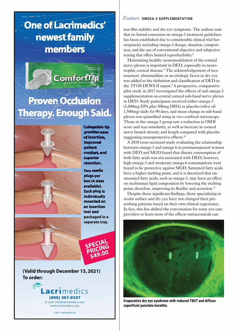
tear film stability and dry eye symptoms. The authors notethat no formal consensus on omega-3 treatment guidelineshas been established due to considerable clinical trial het-erogeneity including omega-3 dosage, duration, composi-tion, and the use of conventional objective and subjectivetesting that offers limited reproducibility.8
Maintaining healthy neuromodulation of the cornealnerve plexus is important in DED, especially in neuro-trophic corneal disease.4 The acknowledgement of neu-rosensory abnormalities as an etiologic factor in dry eyewas added to the definition and classification of DED inthe TFOS DEWS II report.9 A prospective, comparativepilot study in 2017 investigated the effects of oral omega-3supplementation on central corneal sub-basal nerve plexusin DED. Study participants received either omega-3(1,000mg EPA plus 500mg DHA) or placebo (olive oil1,500mg) daily for 90 days, and mean change in sub-basalplexus was quantified using in vivo confocal microscopy.Those in the omega-3 group saw a reduction in OSDIscore and tear osmolarity, as well as increase in cornealnerve branch density and length compared with placebosuggesting neuroprotective effects.10
A 2018 cross-sectional study evaluating the relationshipbetween omega-3 and omega-6 in postmenopausal womenwith DED and MGD found that dietary consumption ofboth fatty acids was not associated with DED; however,high omega-3 and moderate omega-6 consumptions werefound to be protective against MGD. Saturated fatty acidshave a higher melting point, and it is theorized that un-saturated fatty acids, such as omega-3, may have an effecton meibomian lipid composition by lowering the meltingpoint; therefore, improving its fluidity and secretion.11
Despite these significant findings, those specializing inocular surface and dry eye have not changed their pre-scribing patterns based on their own clinical experience.In fact, this has shifted the conversation for some eye careproviders to learn more of the effects nutraceuticals can
Evaporative dry eye syndrome with reduced TBUT and diffusesuperficial punctate keratitis.
O M E G A-3 S U P P L E M E N TAT I O NFeature
©2021 Lacrimedics, Inc.
(800) 367-8327E-mail: [email protected]
www.lacrimedics.com
One of Lacrimedics’newest family
members
Proven OcclusionTherapy. Enough Said.
Collapsible tipprovides easeof insertion,improvedpatientcomfort, andsuperiorretention.
Two sterileplugs perbox (4 sizesavailable).Each plug isindividuallymounted onan insertiontool andpackaged in aseparate tray.
(Valid through December 15, 2021)To order:
SPECIAL
PRICING
$49.00
OneOneOneOneOneOneOneOneOneOneOneOneOneOne of of of of of of of of of Lacrimedics’ Lacrimedics’ Lacrimedics’ Lacrimedics’ Lacrimedics’ Lacrimedics’ Lacrimedics’ Lacrimedics’ Lacrimedics’ Lacrimedics’ Lacrimedics’ of Lacrimedics’ of of Lacrimedics’ of of Lacrimedics’ of of Lacrimedics’ of of Lacrimedics’ of of Lacrimedics’ of of Lacrimedics’ of of Lacrimedics’ ofnewestnewestnewestnewestnewestnewestnewestnewestnewestnewestnewestnewestnewestnewestnewestnewestnewestnewestnewestnewestnewestnewestnewestnewest family family family family family family family family family family family family family family family family family family family family family family familynewest familynewestnewest familynewestnewest familynewestnewest familynewestnewest familynewestnewest familynewestnewest familynewest
membersmembersmembersmembersmembersmembersmembersmembersmembersmembersmembersmembersmembersmembersmembersmembersmembersmembersmembers
ProvenProvenProvenProvenProvenProvenProvenProvenProvenProvenProvenProvenProvenProvenProvenProvenProvenProvenProvenProvenProvenProvenProvenProvenProvenProvenProvenProvenProvenProvenProvenProvenProvenProvenProvenProvenProvenProvenProvenProvenProvenProvenProvenProvenProvenProvenProvenProvenProvenProvenProvenProvenProvenProvenProvenProvenProvenProvenProvenProvenProvenProvenProvenProvenProvenProvenProvenProvenProvenProvenProvenProvenProvenProvenProvenProvenProvenProvenProvenProvenProvenProvenProvenProvenProvenProvenProvenProvenProvenProvenProvenProvenProvenProvenProvenProvenProvenProvenProvenProvenProvenProvenProvenProvenProvenProvenProvenProvenProvenProvenProvenProvenProvenProvenProvenProvenProvenProvenProvenProvenProvenProven Occlusion Occlusion Occlusion Occlusion Occlusion Occlusion Occlusion Occlusion Occlusion Occlusion Occlusion Occlusion Occlusion Occlusion Occlusion Occlusion Occlusion Occlusion Occlusion Occlusion Occlusion Occlusion Occlusion Occlusion Occlusion Occlusion Occlusion Occlusion Occlusion Occlusion Occlusion Occlusion Occlusion Occlusion Occlusion Occlusion Occlusion Occlusion Occlusion Occlusion Occlusion Occlusion Occlusion Occlusion Occlusion Occlusion Occlusion Occlusion Occlusion Occlusion Occlusion Occlusion Occlusion Occlusion Occlusion Occlusion Occlusion Occlusion Occlusion Occlusion Occlusion Occlusion Occlusion Occlusion Occlusion Occlusion Occlusion Occlusion Occlusion Occlusion Occlusion Occlusion Occlusion Occlusion Occlusion Occlusion Occlusion Occlusion Occlusion Occlusion Occlusion Occlusion Occlusion Occlusion Occlusion Occlusion Occlusion Occlusion Occlusion Occlusion Occlusion Occlusion Occlusion Occlusion Occlusion Occlusion Occlusion Occlusion Occlusion Occlusion Occlusion Occlusion Occlusion Occlusion Occlusion Occlusion Occlusion Occlusion Occlusion Occlusion Occlusion Occlusion Occlusion Occlusion Occlusion Occlusion Occlusion OcclusionTherapy.Therapy.Therapy.Therapy.Therapy.Therapy. Enough Enough Enough Enough Enough Enough Enough Enough Enough Enough Enough Enough Said. Said. Said. Said. Said. Said.
CollapsibleCollapsibleCollapsibleCollapsibleCollapsible tip tip tipprovidesprovidesprovidesprovidesprovidesprovides ease easeofofofofof insertion, insertion, insertion,of insertion,ofof insertion,ofimprovedimprovedimprovedimprovedimprovedimprovedpatientpatientpatientpatientpatientcomfort,comfort, and andcomfort, andcomfort,comfort, andcomfort,superiorsuperiorretention.retention.
TwoTwoTwoTwo sterile sterile sterile sterileplugs perbox (4 sizesavailable).Each plug isindividuallyindividuallymounted onan insertiontool and
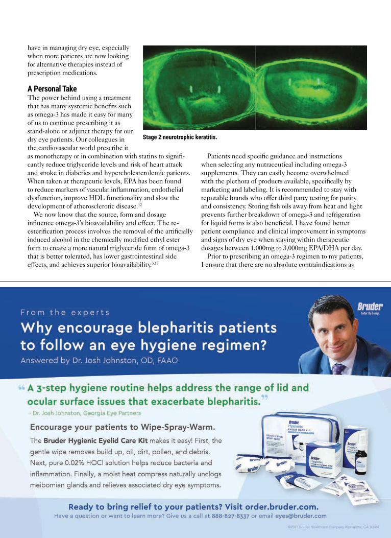
have in managing dry eye, especiallywhen more patients are now lookingfor alternative therapies instead ofprescription medications.
A Personal TakeThe power behind using a treatmentthat has many systemic benefits suchas omega-3 has made it easy for manyof us to continue prescribing it asstand-alone or adjunct therapy for ourdry eye patients. Our colleagues inthe cardiovascular world prescribe itas monotherapy or in combination with statins to signifi-cantly reduce triglyceride levels and risk of heart attackand stroke in diabetics and hypercholesterolemic patients.When taken at therapeutic levels, EPA has been foundto reduce markers of vascular inflammation, endothelialdysfunction, improve HDL functionality and slow thedevelopment of atherosclerotic disease.12
We now know that the source, form and dosageinfluence omega-3’s bioavailability and effect. The re-esterification process involves the removal of the artificiallyinduced alcohol in the chemically modified ethyl esterform to create a more natural triglyceride form of omega-3that is better tolerated, has lower gastrointestinal sideeffects, and achieves superior bioavailability.3,13
Patients need specific guidance and instructionswhen selecting any nutraceutical including omega-3supplements. They can easily become overwhelmedwith the plethora of products available, specifically bymarketing and labeling. It is recommended to stay withreputable brands who offer third party testing for purityand consistency. Storing fish oils away from heat and lightprevents further breakdown of omega-3 and refrigerationfor liquid forms is also beneficial. I have found betterpatient compliance and clinical improvement in symptomsand signs of dry eye when staying within therapeuticdosages between 1,000mg to 3,000mg EPA/DHA per day.
Prior to prescribing an omega-3 regimen to my patients,I ensure that there are no absolute contraindications as
Stage 2 neurotrophic keratitis.
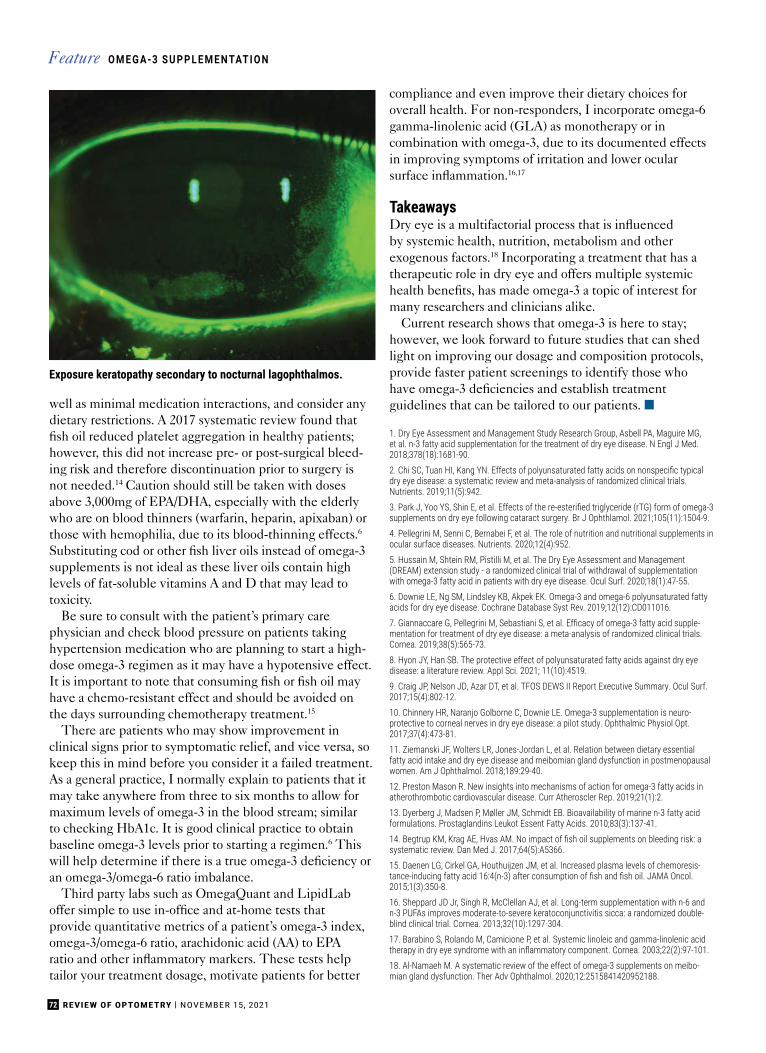
well as minimal medication interactions, and consider anydietary restrictions. A 2017 systematic review found thatfish oil reduced platelet aggregation in healthy patients;however, this did not increase pre- or post-surgical bleed-ing risk and therefore discontinuation prior to surgery isnot needed.14 Caution should still be taken with dosesabove 3,000mg of EPA/DHA, especially with the elderlywho are on blood thinners (warfarin, heparin, apixaban) orthose with hemophilia, due to its blood-thinning effects.6
Substituting cod or other fish liver oils instead of omega-3supplements is not ideal as these liver oils contain highlevels of fat-soluble vitamins A and D that may lead totoxicity.
Be sure to consult with the patient’s primary carephysician and check blood pressure on patients takinghypertension medication who are planning to start a high-dose omega-3 regimen as it may have a hypotensive effect.It is important to note that consuming fish or fish oil mayhave a chemo-resistant effect and should be avoided onthe days surrounding chemotherapy treatment.15
There are patients who may show improvement inclinical signs prior to symptomatic relief, and vice versa, sokeep this in mind before you consider it a failed treatment.As a general practice, I normally explain to patients that itmay take anywhere from three to six months to allow formaximum levels of omega-3 in the blood stream; similarto checking HbA1c. It is good clinical practice to obtainbaseline omega-3 levels prior to starting a regimen.6 Thiswill help determine if there is a true omega-3 deficiency oran omega-3/omega-6 ratio imbalance.
Third party labs such as OmegaQuant and LipidLaboffer simple to use in-office and at-home tests thatprovide quantitative metrics of a patient’s omega-3 index,omega-3/omega-6 ratio, arachidonic acid (AA) to EPAratio and other inflammatory markers. These tests helptailor your treatment dosage, motivate patients for better
compliance and even improve their dietary choices foroverall health. For non-responders, I incorporate omega-6gamma-linolenic acid (GLA) as monotherapy or incombination with omega-3, due to its documented effectsin improving symptoms of irritation and lower ocularsurface inflammation.16,17
TakeawaysDry eye is a multifactorial process that is influencedby systemic health, nutrition, metabolism and otherexogenous factors.18 Incorporating a treatment that has atherapeutic role in dry eye and offers multiple systemichealth benefits, has made omega-3 a topic of interest formany researchers and clinicians alike.
Current research shows that omega-3 is here to stay;however, we look forward to future studies that can shedlight on improving our dosage and composition protocols,provide faster patient screenings to identify those whohave omega-3 deficiencies and establish treatmentguidelines that can be tailored to our patients. ■
1. Dry Eye Assessment and Management Study Research Group, Asbell PA, Maguire MG,et al. n-3 fatty acid supplementation for the treatment of dry eye disease. N Engl J Med.2018;378(18):1681-90.2. Chi SC, Tuan HI, Kang YN. Effects of polyunsaturated fatty acids on nonspecific typical dry eye disease: a systematic review and meta-analysis of randomized clinical trials.Nutrients. 2019;11(5):942.3. Park J, Yoo YS, Shin E, et al. Effects of the re-esterified triglyceride (rTG) form of omega-3 supplements on dry eye following cataract surgery. Br J Ophthlamol. 2021;105(11):1504-9.4. Pellegrini M, Senni C, Bernabei F, et al. The role of nutrition and nutritional supplements inocular surface diseases. Nutrients. 2020;12(4):952.5. Hussain M, Shtein RM, Pistilli M, et al. The Dry Eye Assessment and Management(DREAM) extension study - a randomized clinical trial of withdrawal of supplementationwith omega-3 fatty acid in patients with dry eye disease. Ocul Surf. 2020;18(1):47-55.6. Downie LE, Ng SM, Lindsley KB, Akpek EK. Omega-3 and omega-6 polyunsaturated fattyacids for dry eye disease. Cochrane Database Syst Rev. 2019;12(12):CD011016.7. Giannaccare G, Pellegrini M, Sebastiani S, et al. Efficacy of omega-3 fatty acid supple-mentation for treatment of dry eye disease: a meta-analysis of randomized clinical trials.Cornea. 2019;38(5):565-73.8. Hyon JY, Han SB. The protective effect of polyunsaturated fatty acids against dry eyedisease: a literature review. Appl Sci. 2021; 11(10):4519.9. Craig JP, Nelson JD, Azar DT, et al. TFOS DEWS II Report Executive Summary. Ocul Surf.2017;15(4):802-12.10. Chinnery HR, Naranjo Golborne C, Downie LE. Omega-3 supplementation is neuro-protective to corneal nerves in dry eye disease: a pilot study. Ophthalmic Physiol Opt.2017;37(4):473-81.11. Ziemanski JF, Wolters LR, Jones-Jordan L, et al. Relation between dietary essentialfatty acid intake and dry eye disease and meibomian gland dysfunction in postmenopausalwomen. Am J Ophthalmol. 2018;189:29-40.12. Preston Mason R. New insights into mechanisms of action for omega-3 fatty acids inatherothrombotic cardiovascular disease. Curr Atheroscler Rep. 2019;21(1):2.13. Dyerberg J, Madsen P, Møller JM, Schmidt EB. Bioavailability of marine n-3 fatty acidformulations. Prostaglandins Leukot Essent Fatty Acids. 2010;83(3):137-41.14. Begtrup KM, Krag AE, Hvas AM. No impact of fish oil supplements on bleeding risk: a systematic review. Dan Med J. 2017;64(5):A5366.15. Daenen LG, Cirkel GA, Houthuijzen JM, et al. Increased plasma levels of chemoresis-tance-inducing fatty acid 16:4(n-3) after consumption of fish and fish oil. JAMA Oncol. 2015;1(3):350-8.16. Sheppard JD Jr, Singh R, McClellan AJ, et al. Long-term supplementation with n-6 andn-3 PUFAs improves moderate-to-severe keratoconjunctivitis sicca: a randomized double-blind clinical trial. Cornea. 2013;32(10):1297-304.17. Barabino S, Rolando M, Camicione P, et al. Systemic linoleic and gamma-linolenic acidtherapy in dry eye syndrome with an inflammatory component. Cornea. 2003;22(2):97-101. 18. Al-Namaeh M. A systematic review of the effect of omega-3 supplements on meibo-mian gland dysfunction. Ther Adv Ophthalmol. 2020;12:2515841420952188.
Exposure keratopathy secondary to nocturnal lagophthalmos.
72 REVIEW OF OPTOMETRY | NOVEMBER 15, 2021
O M E G A-3 S U P P L E M E N TAT I O NFeature

VeraPlug™
FlexFit™
Designed for simple
sizing, easy insertion,
patient comfort and
retention—a proven long-term
treatment for chronic dry eye.
Vera180™
Synthetic Absorbable Lacrimal Plugs
Extended temporary
occlusion lasting
approximately 6 months—
ideal for treatment of post-
surgical dry eye.
lacrivera.com (855) 857-0518
Winning dry eye relief—for a sprint or a marathon.
Take the lead and give your patients proven, comfortable relief.
From temporary occlusion lasting approximately 6 months
to long-term silicone plugs, Lacrivera offers a complete line
of innovative products for the treatment of dry eye.
© 2021 Lacrivera, a division of Stephens Instruments. All rights reserved.
A F R E S H P E R S P E C T I V E ™

74 REVIEW OF OPTOMETRY | NOVEMBER 15, 2021
Anterior Blepharitis:The Front Line of OSD
We dissect everything you need to know about this common condition, including proper treatment andmanagement, and how it can be a sign of Demodex.
In 2017, the Tear Film & OcularSurface Society’s Dry Eye Workshop(DEWS) II Report updated the defi-nition of dry eye disease, describing
it as ‘‘a multifactorial disease of thetears and ocular surface that results insymptoms of discomfort, visual dis-turbance and tear film instability withpotential damage to the ocular surface.It is accompanied by increased osmo-larity of the tear film and inflamma-tion of the ocular surface.”1 Recently,treatment has focused on meibomiangland dysfunction (MGD), since it isthe most common cause of evapora-tive dry eye.2 However, one must notlose sight of the multifactorial natureof dry eye, which requires treatmentof each aspect of the disease, includingblepharitis. Addressing eyelash and lidmargin hygiene is an often overlookedcomponent of a comprehensive treat-ment strategy.
Eyelashes protect the eye by pre-venting debris and particulate matterfrom reaching the ocular surface.
Fig. 1. Mild anterior blepharitis in a younger male patient with collarettes at the base of thelashes and mild flaking throughout. Although mild, it is important to treat early to prevent disease progression. This patient undergoes quarterly in-office treatments.
B L E P H A R I T I SFeature
Aboutthe authors
Dr. McLeod is a partner at Korb & Associates in Boston, MA. Most recently, he was an associate professor within the department of specialty and advancedcare and chief of the Cornea & Contact Lens Residency at New England College of Optometry (NECO). He has no financial disclosures. Dr. Nau is a partner atKorb & Associates and an adjunct associate clinical professor at the NECO. She is on the advisory board for Oyster Point and a speaker for EyeEco. The two are Fellows of the American Academy of Optometry and members of the American Optometric Association and the Massachusetts Society of Optometrists.
By Andrew McLeod, OD,and Amy Nau, ODBoston
PEER REVIEWED
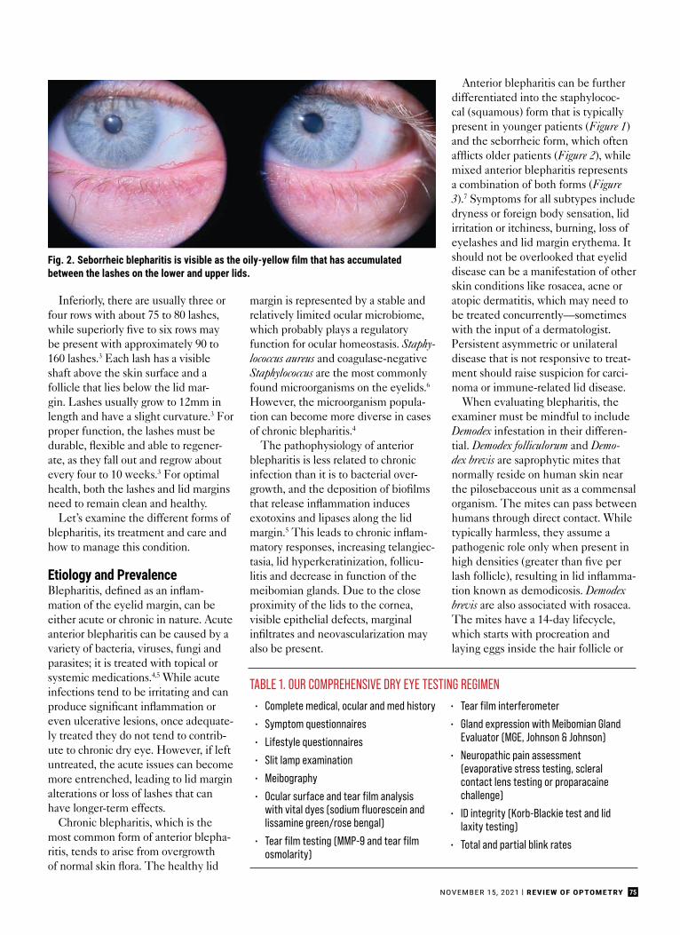
75NOVEMBER 15, 2021 | REVIEW OF OPTOMETRY
Inferiorly, there are usually three orfour rows with about 75 to 80 lashes,while superiorly five to six rows maybe present with approximately 90 to160 lashes.3 Each lash has a visibleshaft above the skin surface and afollicle that lies below the lid mar-gin. Lashes usually grow to 12mm inlength and have a slight curvature.3 Forproper function, the lashes must bedurable, flexible and able to regener-ate, as they fall out and regrow aboutevery four to 10 weeks.3 For optimalhealth, both the lashes and lid marginsneed to remain clean and healthy.
Let’s examine the different forms ofblepharitis, its treatment and care andhow to manage this condition.
Etiology and PrevalenceBlepharitis, defined as an inflam-mation of the eyelid margin, can beeither acute or chronic in nature. Acuteanterior blepharitis can be caused by avariety of bacteria, viruses, fungi andparasites; it is treated with topical orsystemic medications.4,5 While acuteinfections tend to be irritating and canproduce significant inflammation oreven ulcerative lesions, once adequate-ly treated they do not tend to contrib-ute to chronic dry eye. However, if leftuntreated, the acute issues can becomemore entrenched, leading to lid marginalterations or loss of lashes that canhave longer-term effects.
Chronic blepharitis, which is themost common form of anterior blepha-ritis, tends to arise from overgrowthof normal skin flora. The healthy lid
margin is represented by a stable andrelatively limited ocular microbiome,which probably plays a regulatoryfunction for ocular homeostasis. Staphy-lococcus aureus and coagulase-negativeStaphylococcus are the most commonlyfound microorganisms on the eyelids.6
However, the microorganism popula-tion can become more diverse in casesof chronic blepharitis.4
The pathophysiology of anteriorblepharitis is less related to chronicinfection than it is to bacterial over-growth, and the deposition of biofilmsthat release inflammation inducesexotoxins and lipases along the lidmargin.5 This leads to chronic inflam-matory responses, increasing telangiec-tasia, lid hyperkeratinization, follicu-litis and decrease in function of themeibomian glands. Due to the closeproximity of the lids to the cornea,visible epithelial defects, marginalinfiltrates and neovascularization mayalso be present.
Anterior blepharitis can be furtherdifferentiated into the staphylococ-cal (squamous) form that is typicallypresent in younger patients (Figure 1)and the seborrheic form, which oftenafflicts older patients (Figure 2), whilemixed anterior blepharitis representsa combination of both forms (Figure3).7 Symptoms for all subtypes includedryness or foreign body sensation, lidirritation or itchiness, burning, loss ofeyelashes and lid margin erythema. Itshould not be overlooked that eyeliddisease can be a manifestation of otherskin conditions like rosacea, acne oratopic dermatitis, which may need tobe treated concurrently—sometimeswith the input of a dermatologist.Persistent asymmetric or unilateraldisease that is not responsive to treat-ment should raise suspicion for carci-noma or immune-related lid disease.
When evaluating blepharitis, theexaminer must be mindful to includeDemodex infestation in their differen-tial. Demodex folliculorum and Demo-dex brevis are saprophytic mites thatnormally reside on human skin nearthe pilosebaceous unit as a commensalorganism. The mites can pass betweenhumans through direct contact. Whiletypically harmless, they assume apathogenic role only when present inhigh densities (greater than five perlash follicle), resulting in lid inflamma-tion known as demodicosis. Demodexbrevis are also associated with rosacea.The mites have a 14-day lifecycle,which starts with procreation andlaying eggs inside the hair follicle or
Fig. 2. Seborrheic blepharitis is visible as the oily-yellow film that has accumulated between the lashes on the lower and upper lids.
• Complete medical, ocular and med history
• Symptom questionnaires
• Lifestyle questionnaires
• Slit lamp examination
• Meibography
• Ocular surface and tear film analysiswith vital dyes (sodium fluorescein andlissamine green/rose bengal)
• Tear film testing (MMP-9 and tear filmosmolarity)
• Tear film interferometer
• Gland expression with Meibomian GlandEvaluator (MGE, Johnson & Johnson)
• Neuropathic pain assessment(evaporative stress testing, scleralcontact lens testing or proparacainechallenge)
• ID integrity (Korb-Blackie test and lidlaxity testing)
• Total and partial blink rates
TABLE 1. OUR COMPREHENSIVE DRY EYE TESTING REGIMEN
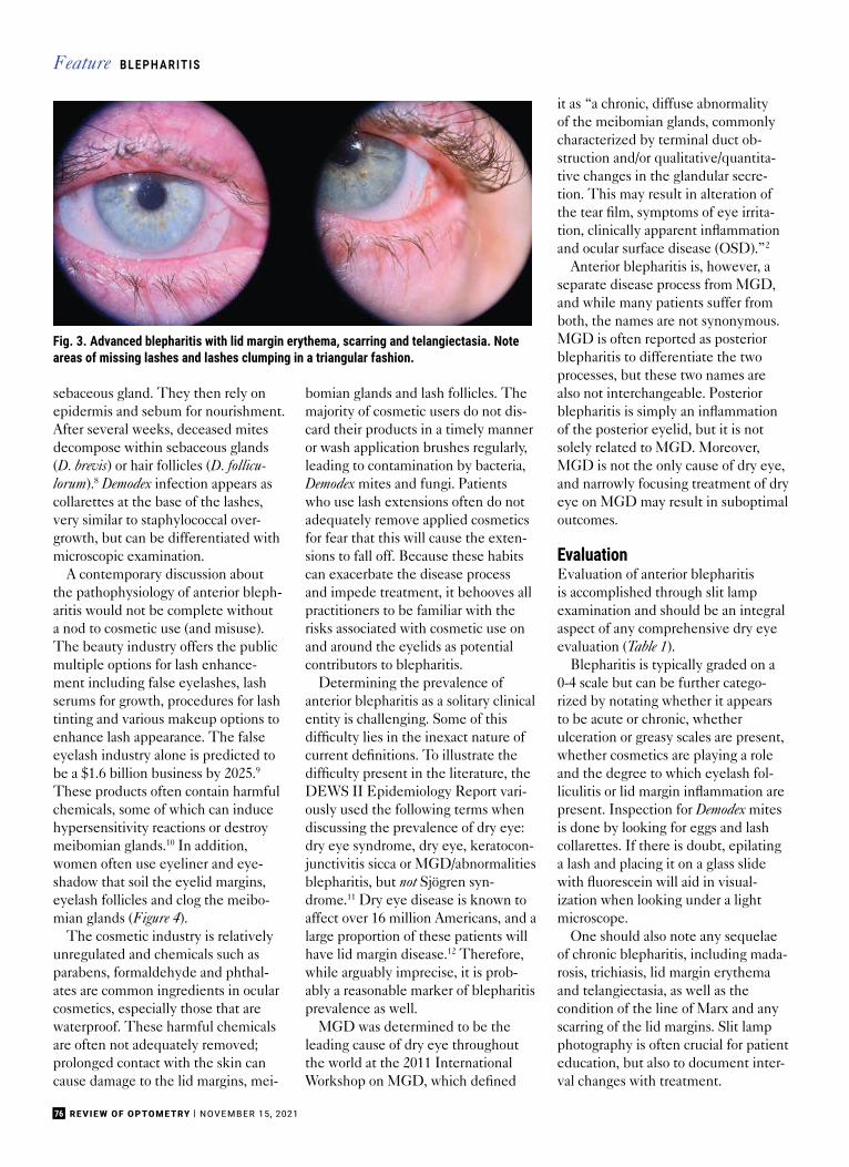
76 REVIEW OF OPTOMETRY | NOVEMBER 15, 2021
sebaceous gland. They then rely onepidermis and sebum for nourishment.After several weeks, deceased mitesdecompose within sebaceous glands(D. brevis) or hair follicles (D. follicu-lorum).8 Demodex infection appears ascollarettes at the base of the lashes,very similar to staphylococcal over-growth, but can be differentiated withmicroscopic examination.
A contemporary discussion aboutthe pathophysiology of anterior bleph-aritis would not be complete withouta nod to cosmetic use (and misuse).The beauty industry offers the publicmultiple options for lash enhance-ment including false eyelashes, lashserums for growth, procedures for lashtinting and various makeup options toenhance lash appearance. The falseeyelash industry alone is predicted tobe a $1.6 billion business by 2025.9
These products often contain harmfulchemicals, some of which can inducehypersensitivity reactions or destroymeibomian glands.10 In addition,women often use eyeliner and eye-shadow that soil the eyelid margins,eyelash follicles and clog the meibo-mian glands (Figure 4).
The cosmetic industry is relativelyunregulated and chemicals such asparabens, formaldehyde and phthal-ates are common ingredients in ocularcosmetics, especially those that arewaterproof. These harmful chemicalsare often not adequately removed;prolonged contact with the skin cancause damage to the lid margins, mei-
bomian glands and lash follicles. Themajority of cosmetic users do not dis-card their products in a timely manneror wash application brushes regularly,leading to contamination by bacteria,Demodex mites and fungi. Patientswho use lash extensions often do notadequately remove applied cosmeticsfor fear that this will cause the exten-sions to fall off. Because these habitscan exacerbate the disease processand impede treatment, it behooves allpractitioners to be familiar with therisks associated with cosmetic use onand around the eyelids as potentialcontributors to blepharitis.
Determining the prevalence ofanterior blepharitis as a solitary clinicalentity is challenging. Some of thisdifficulty lies in the inexact nature ofcurrent definitions. To illustrate thedifficulty present in the literature, theDEWS II Epidemiology Report vari-ously used the following terms whendiscussing the prevalence of dry eye:dry eye syndrome, dry eye, keratocon-junctivitis sicca or MGD/abnormalitiesblepharitis, but not Sjögren syn-drome.11 Dry eye disease is known toaffect over 16 million Americans, and alarge proportion of these patients willhave lid margin disease.12 Therefore,while arguably imprecise, it is prob-ably a reasonable marker of blepharitisprevalence as well.
MGD was determined to be theleading cause of dry eye throughoutthe world at the 2011 InternationalWorkshop on MGD, which defined
it as “a chronic, diffuse abnormalityof the meibomian glands, commonlycharacterized by terminal duct ob-struction and/or qualitative/quantita-tive changes in the glandular secre-tion. This may result in alteration ofthe tear film, symptoms of eye irrita-tion, clinically apparent inflammationand ocular surface disease (OSD).”2
Anterior blepharitis is, however, aseparate disease process from MGD,and while many patients suffer fromboth, the names are not synonymous.MGD is often reported as posteriorblepharitis to differentiate the twoprocesses, but these two names arealso not interchangeable. Posteriorblepharitis is simply an inflammationof the posterior eyelid, but it is notsolely related to MGD. Moreover,MGD is not the only cause of dry eye,and narrowly focusing treatment of dryeye on MGD may result in suboptimaloutcomes.
EvaluationEvaluation of anterior blepharitisis accomplished through slit lampexamination and should be an integralaspect of any comprehensive dry eyeevaluation (Table 1).
Blepharitis is typically graded on a0-4 scale but can be further catego-rized by notating whether it appearsto be acute or chronic, whetherulceration or greasy scales are present,whether cosmetics are playing a roleand the degree to which eyelash fol-liculitis or lid margin inflammation arepresent. Inspection for Demodex mitesis done by looking for eggs and lashcollarettes. If there is doubt, epilatinga lash and placing it on a glass slidewith fluorescein will aid in visual-ization when looking under a lightmicroscope.
One should also note any sequelaeof chronic blepharitis, including mada-rosis, trichiasis, lid margin erythemaand telangiectasia, as well as thecondition of the line of Marx and anyscarring of the lid margins. Slit lampphotography is often crucial for patienteducation, but also to document inter-val changes with treatment.
B L E P H A R I T I SFeature
Fig. 3. Advanced blepharitis with lid margin erythema, scarring and telangiectasia. Noteareas of missing lashes and lashes clumping in a triangular fashion.

Red, ItchyAllergy Eyes?
Seasonal allergies can cause red, itchy, irritated eyes andeyelids. Allergy eye drops relieve eye symptoms but do notcalm the irritated eyelids. OCuSOFT® Lid Scrub® Allergyremoves oil, debris, pollen, and other contaminants from theeyelids while utilizing Green Tea Extract, Tea Tree Oil, andPSG-2™ to effectively reduce redness, irritation and itchingsensation of allergy eyelids.
Call (800) 233-5469 for Samples
© 2021 OCuSOFT Inc., Rosenberg, TX 77471
Remove Allergens FromThe Eyelids For Added Relief
Cleans, Soothes,Calms, Moisturizes
Reduces Redness & Irritation
Relieves Itching Sensation
Clinically Proven PSG-2™
Formula to Increase Moisture
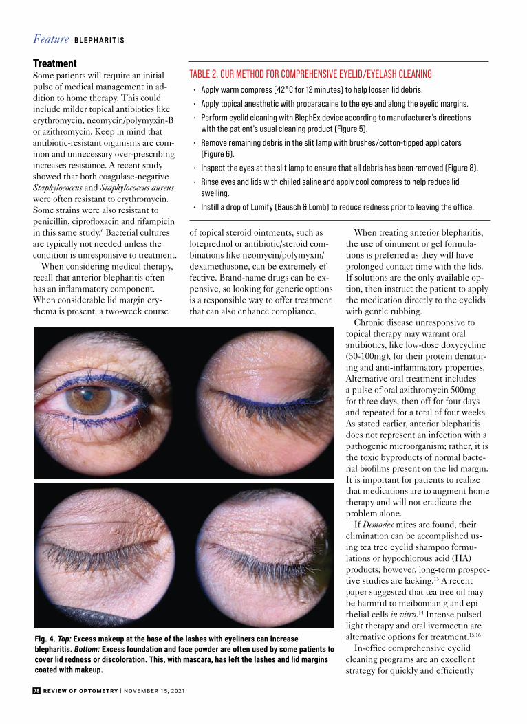
78 REVIEW OF OPTOMETRY | NOVEMBER 15, 2021
TreatmentSome patients will require an initialpulse of medical management in ad-dition to home therapy. This couldinclude milder topical antibiotics likeerythromycin, neomycin/polymyxin-Bor azithromycin. Keep in mind thatantibiotic-resistant organisms are com-mon and unnecessary over-prescribingincreases resistance. A recent studyshowed that both coagulase-negativeStaphylococcus and Staphylococcus aureuswere often resistant to erythromycin.Some strains were also resistant topenicillin, ciprofloxacin and rifampicinin this same study.6 Bacterial culturesare typically not needed unless thecondition is unresponsive to treatment.
When considering medical therapy,recall that anterior blepharitis oftenhas an inflammatory component.When considerable lid margin ery-thema is present, a two-week course
of topical steroid ointments, such asloteprednol or antibiotic/steroid com-binations like neomycin/polymyxin/dexamethasone, can be extremely ef-fective. Brand-name drugs can be ex-pensive, so looking for generic optionsis a responsible way to offer treatmentthat can also enhance compliance.
When treating anterior blepharitis,the use of ointment or gel formula-tions is preferred as they will haveprolonged contact time with the lids.If solutions are the only available op-tion, then instruct the patient to applythe medication directly to the eyelidswith gentle rubbing.
Chronic disease unresponsive totopical therapy may warrant oralantibiotics, like low-dose doxycycline(50-100mg), for their protein denatur-ing and anti-inflammatory properties.Alternative oral treatment includesa pulse of oral azithromycin 500mgfor three days, then off for four daysand repeated for a total of four weeks.As stated earlier, anterior blepharitisdoes not represent an infection with apathogenic microorganism; rather, it isthe toxic byproducts of normal bacte-rial biofilms present on the lid margin.It is important for patients to realizethat medications are to augment hometherapy and will not eradicate theproblem alone.
If Demodex mites are found, theirelimination can be accomplished us-ing tea tree eyelid shampoo formu-lations or hypochlorous acid (HA)products; however, long-term prospec-tive studies are lacking.13 A recentpaper suggested that tea tree oil maybe harmful to meibomian gland epi-thelial cells in vitro.14 Intense pulsedlight therapy and oral ivermectin arealternative options for treatment.15,16
In-office comprehensive eyelidcleaning programs are an excellentstrategy for quickly and efficiently
B L E P H A R I T I SFeature
• Apply warm compress (42°C for 12 minutes) to help loosen lid debris.
• Apply topical anesthetic with proparacaine to the eye and along the eyelid margins.
• Perform eyelid cleaning with BlephEx device according to manufacturer’s directionswith the patient’s usual cleaning product (Figure 5).
• Remove remaining debris in the slit lamp with brushes/cotton-tipped applicators(Figure 6).
• Inspect the eyes at the slit lamp to ensure that all debris has been removed (Figure 8).
• Rinse eyes and lids with chilled saline and apply cool compress to help reduce lidswelling.
• Instill a drop of Lumify (Bausch & Lomb) to reduce redness prior to leaving the office.
TABLE 2. OUR METHOD FOR COMPREHENSIVE EYELID/EYELASH CLEANING
Fig. 4. Top: Excess makeup at the base of the lashes with eyeliners can increaseblepharitis. Bottom: Excess foundation and face powder are often used by some patients tocover lid redness or discoloration. This, with mascara, has left the lashes and lid marginscoated with makeup.

D O W N L O A D A N I N F O R M A T I O N P A C K E T A T
vitaltears.org/reviewO R C A L L T O L L - F R E E
(800) 360-9592
Think serum tears are hard to get?Learn how Vital Tears has simplified the process.
Serum TearsMade Simple.
At Vital Tears, our mission is to make serum tears
easily available and a�ordable for your patients.
We’ve done that for over 10,000 patients across the
country through our:
� Rapid serum drop delivery
� Convenient blood draw options
� A�ordable payment options
� Superior customer service

80 REVIEW OF OPTOMETRY | NOVEMBER 15, 2021
getting ahead of anterior blepharitisand providing patients instructionsabout proper at-home procedures.Semi-annual cleanings performedin-office is a reasonable approach, butadoption will be patient-dependent.Patients with poor vision, limiteddexterity, cognitive impairment/de-mentia or who are averse to touchingtheir eyes may need to be seen morefrequently. Women who habituallyget lash extensions should come in fora comprehensive cleaning betweenapplications to remove glue, lashes,desquamated epithelium and cos-metic residue. Patients with anteriorblepharitis should have a compre-
hensive cleaning before any surgicalconsultation.
In terms of patient educationregarding cosmetic use, some practi-tioners may simply tell them to stopusing makeup, but most individualswill not be willing to do so. How-ever, educating patients about thedeleterious effects of some cosmeticingredients in a non-patronizing wayis usually appreciated if done properly.It can be more prudent to providetips on application, placement andremoval of periocular cosmetics, andeven recommend dry eye–friendlyproduct lines, such as Èyes Are theStory, that improve patients’ appear-ance by reducing inflammatory reac-tions to these chemicals.
Home CareDespite the realization that vision isone of the most vital senses in every-day life, ocular personal care is oftencompletely neglected by patients. Partof this problem is the reactive natureof the eyecare profession, which hasfailed for decades to impart the neces-sity of preventative eyelid health,starting in middle school.
In our practice, the dental modelis invoked as an analogy, since this isa concept relatable to patients, evenat an early age. A consistent mes-sage for every patient, at every visit,should include a recommendation forroutine home care with intermittentoffice visits for more in-depth clean-ing if required. This is analogous tobrushing one’s teeth at home and
having intermittent cleanings in theoffice. Frequency of home care can betailored to the severity of the condi-tion. This can vary from lid cleaninga few times per week in milder casesto twice daily in more advanced casesof blepharitis or for regular heavymakeup wearers. Fortunately, unlikewarm compress therapy, which isnotorious for patient non-adherence,eyelash cleaning is simpler and quick-er, and tends to be better accepted bypatients.
When introducing a patient to ahome care routine, it is very importantto outline a schedule, provide writ-ten instructions and spend the timedemonstrating the techniques in theoffice (Table 2). This will increasepatient compliance and lead to moresuccessful clinical outcomes. First,recommend the type of product youwish the patient to use. Long goneare the days of saline, mineral oil ordiluted baby shampoo as the onlyoptions for lid hygiene. Industry hasresponded to data showing that eyelidhygiene is an effective method to treatanterior blepharitis and eyelid diseasein general, including dry eye.
A plethora of new safe and effec-tive products are available. In broadterms, these can be classified assurfactant/soap, tea tree oil or HA-based products. All three categorieshave different brands and concentra-tion levels, so it is best to familiarizeyourself with these and curate thoseproducts in which you have confi-dence. Patients with sensitive skinshould be started at lower concentra-tions of the active ingredient to bettermonitor for adverse events. Tea treeand HA products are recommended ifa concurrent infestation with Demodexmites is present.
After selecting the cleaning prod-uct, it is very important to recom-mend a type of applicator (Figure 6).Several products on the market can besprayed onto the lids, but we recom-mend lightly scrubbing the lid marginand lashes. For the same reason,manual rubbing of contact lenses isrecommended, effective removal of
Fig. 5. Doctor using BlephEx electronic cleaning system on upper and lower lids duringin-office cleaning. It is important to get both the lashes and the lid margin, including the meibomian gland orifices during the treatment.
Fig. 6. Cleaning tools that can be used for home or office cleaning. Cotton-tipped applicators, bristle brushes and makeup rounds that can be moistened with various products. Pre-moistened towelettes arealso available.
B L E P H A R I T I SFeature B L E P H A R I T I SFeature

The easiest to fit scleral lens on the market with truly customizable, independent zone design options for any corneal SAG or scleral shape.
• Quadrant-specific scleral and limbal zone• 14.5 diameter for normal corneas• Up to 17.5 diameter for deep SAG’s• Oblate multifocal design
Atlantis™SCLERAL
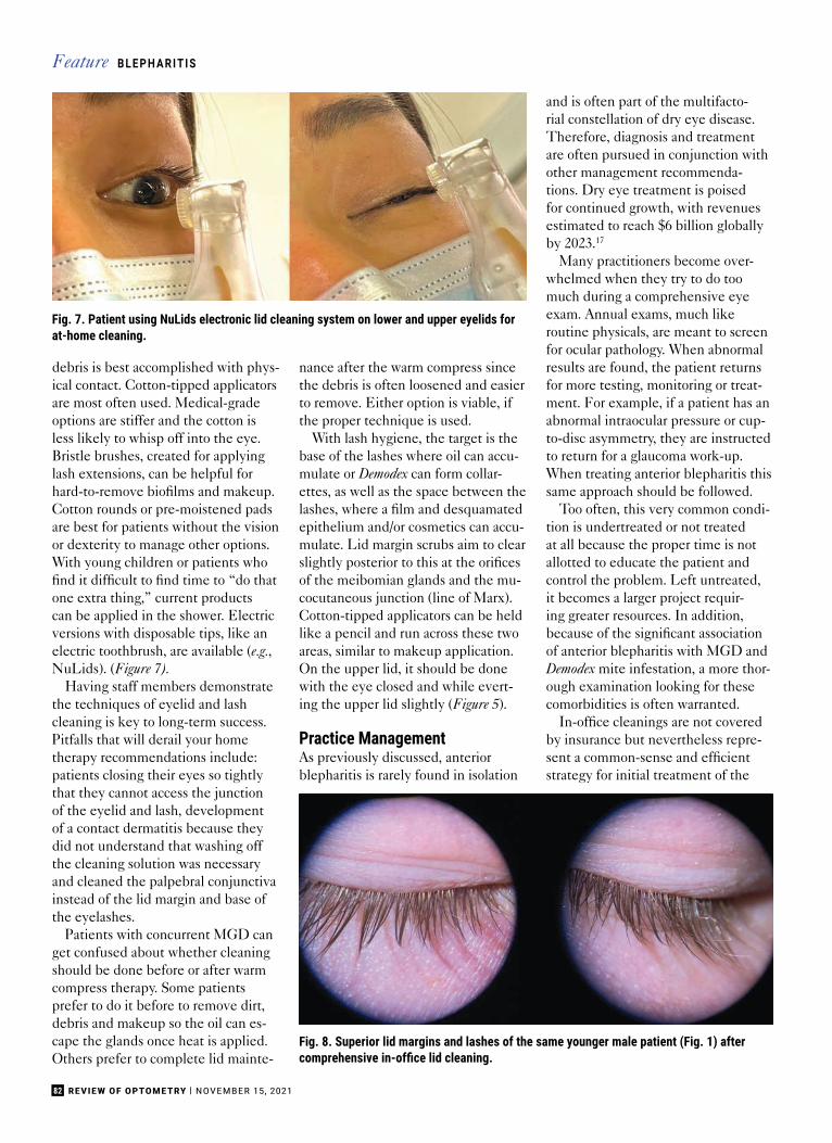
82 REVIEW OF OPTOMETRY | NOVEMBER 15, 2021
debris is best accomplished with phys-ical contact. Cotton-tipped applicatorsare most often used. Medical-gradeoptions are stiffer and the cotton isless likely to whisp off into the eye.Bristle brushes, created for applyinglash extensions, can be helpful forhard-to-remove biofilms and makeup.Cotton rounds or pre-moistened padsare best for patients without the visionor dexterity to manage other options.With young children or patients whofind it difficult to find time to “do thatone extra thing,” current productscan be applied in the shower. Electricversions with disposable tips, like anelectric toothbrush, are available (e.g.,NuLids). (Figure 7).
Having staff members demonstratethe techniques of eyelid and lashcleaning is key to long-term success.Pitfalls that will derail your hometherapy recommendations include:patients closing their eyes so tightlythat they cannot access the junctionof the eyelid and lash, developmentof a contact dermatitis because theydid not understand that washing offthe cleaning solution was necessaryand cleaned the palpebral conjunctivainstead of the lid margin and base ofthe eyelashes.
Patients with concurrent MGD canget confused about whether cleaningshould be done before or after warmcompress therapy. Some patientsprefer to do it before to remove dirt,debris and makeup so the oil can es-cape the glands once heat is applied.Others prefer to complete lid mainte-
nance after the warm compress sincethe debris is often loosened and easierto remove. Either option is viable, ifthe proper technique is used.
With lash hygiene, the target is thebase of the lashes where oil can accu-mulate or Demodex can form collar-ettes, as well as the space between thelashes, where a film and desquamatedepithelium and/or cosmetics can accu-mulate. Lid margin scrubs aim to clearslightly posterior to this at the orificesof the meibomian glands and the mu-cocutaneous junction (line of Marx).Cotton-tipped applicators can be heldlike a pencil and run across these twoareas, similar to makeup application.On the upper lid, it should be donewith the eye closed and while evert-ing the upper lid slightly (Figure 5).
Practice ManagementAs previously discussed, anteriorblepharitis is rarely found in isolation
and is often part of the multifacto-rial constellation of dry eye disease.Therefore, diagnosis and treatmentare often pursued in conjunction withother management recommenda-tions. Dry eye treatment is poisedfor continued growth, with revenuesestimated to reach $6 billion globallyby 2023.17
Many practitioners become over-whelmed when they try to do toomuch during a comprehensive eyeexam. Annual exams, much likeroutine physicals, are meant to screenfor ocular pathology. When abnormalresults are found, the patient returnsfor more testing, monitoring or treat-ment. For example, if a patient has anabnormal intraocular pressure or cup-to-disc asymmetry, they are instructedto return for a glaucoma work-up.When treating anterior blepharitis thissame approach should be followed.
Too often, this very common condi-tion is undertreated or not treatedat all because the proper time is notallotted to educate the patient andcontrol the problem. Left untreated,it becomes a larger project requir-ing greater resources. In addition,because of the significant associationof anterior blepharitis with MGD andDemodex mite infestation, a more thor-ough examination looking for thesecomorbidities is often warranted.
In-office cleanings are not coveredby insurance but nevertheless repre-sent a common-sense and efficientstrategy for initial treatment of the
Fig. 7. Patient using NuLids electronic lid cleaning system on lower and upper eyelids forat-home cleaning.
Fig. 8. Superior lid margins and lashes of the same younger male patient (Fig. 1) aftercomprehensive in-office lid cleaning.
B L E P H A R I T I SFeature
patient with chronic anterior blephari-tis. Regardless of insurance coverage,all testing and in-office treatmentsshould be documented with a CPTcode. BlephEx and lid cleaning pro-cedures do not currently have a CPTcode, so 92499 (unlisted ophthalmo-logical service or procedure) shouldbe used.18 Patients should always beprovided with an Advanced Benefi-ciary Notice to outline testing andtreatments that may be reimbursedby their insurance and those for whichthe patient may be responsible.
TakeawaysAs our knowledge of OSD expands,it’s important to remember it is amultifactorial disease that requirescomprehensive recommendations thataddress each contributing factor. Thistypically includes patient educationand a foundation of home therapywith lid and lash hygiene, warmcompresses, blinking exercises andlubricating tears or meds as needed.
In addition, lifestyle modificationsespecially with respect to cosmeticuse are necessary. Invoking the dentalmodel of preventative and restorativecare should be pursued with regularoffice visits and therapies used in con-junction with home care for increasedsuccess. Patients should be remindedthat blepharitis and dry eye are bothchronic in nature and require their on-going participation for self-care. Long-term prevention and maintenance,rather than reactive therapies, requiresa change in mindset for practitionersand patients alike. ■
1. Craig JP, Nichols KK, Akpek EK, et al. TFOS DEWS IIDefinition and Classification Report. Ocul Surf. 2017 Jul;15(3):276-83.2. Nichols KK, Foulks GN, Bron AJ, et al. The international workshop on meibomian gland dysfunction: executive sum-mary. Invest Ophthalmol Vis Sci. 2011 Mar 30;52(4):1922-9.3. Aumond S, Bitton E. The eyelash follicle features and anomalies: A review. J Optom. 2018;11(4):211–22.4. Krachmer, JH, Mannis, MJ, Holland, EJ. Cornea: funda-mentals, diagnosis and management. 3rd Edition, Elsevier Inc. 2011. 403-5.5. Pflugfelder, S, Bauerman, R, Stern, ME. (2004). Dry eye and ocular surface disorders (1st ed.). CRC Press. 255-7.
6. de Paula A, Oliva G, Barraquer RI, de la Paz MF. Prevalence and antibiotic susceptibility of bacteria isolated in patients affected with blepharitis in a tertiary eye centre in Spain. Eur J Ophthalmol. 2020;30(5):991-7.7. Duncan K, Jeng BH. Medical management of blepharitis. Curr Opin Ophthalmol. 2015;26(4):289-94.8. Rather PA, Hassan I. Human demodex mite: the versatile mite of dermatological importance. Indian J Dermatol. 2014;59(1):60-6.9. Website: False eyelashes market size worth $1.6 billion by 2025. Grandview Research. Accessed 10/2021. 10. Chen X, Sullivan DA, Sullivan AG. Toxicity of cosmetic preservatives on human ocular surface and adnexal cells. Exp Eye Res. 2018;170:188-197.11. Stapleton F, Alves M, Bunya VY, et al. TFOS DEWS II Epidemiology Report. Ocul Surf. 2017;15(3):334-65.12. Ferrand KF, Fridman M, Stillman IO, Schaumberg DA. Prevalence of diagnosed dry eye disease in the United States among adults aged 18 years and older. Am J Ophthalmol 2017;182:90-8.13. Savla K, Le JT, Pucker AD. Tea tree oil for De-modex blepharitis. Cochrane Database Syst Rev. 2020;6(6):CD013333.14. Chen D, Wang J, Sullivan DA, et al. Effects of Terpinen-4-ol on meibomian gland epithelial cells in vitro. Cornea. 2020 Dec;39(12):1541-6.15. Fishman HA, Periman LM, Shah AA. Real-time video mi-croscopy of In Vitro Demodex death by intense pulsed light. Photobiomodul Photomed Laser Surg. 2020;38(8):472-6.16. Holzchuh FG, Hida RY, Moscovici BK, et al. Clinical treatment of ocular Demodex folliculorum by systemic ivermectin. Am J Ophthalmol. 2011;151(6):1030-4.e1.17. Dalton M. Understanding prevalence, demographics of dry eye disease. Ophthalmology Times. 2019.18. Rumpakis J. Ocular Surface Coding Potpourri. Rev Optom. 2015.

www.reviewedu.com/winteroptometry
*Approval pendingReview Education Group partners with Salus University for those ODs who are licensed in states that require university credit.
DECEMBER 11–12, 2021HYATT REGENCY NEWPORT BEACH
1107 JAMBOREE ROAD, NEWPORT BEACH, CALIFORNIA
LIVE COPE*
Earn up to 11 LIVE COPE credits*
CHAIRMohammad Rafi eetary, OD, FAAOConsultative Optometric PhysicianCharles Retina InstituteFellow of the American Academy of Optometryand Optometric Retina Society
CO-CHAIRSteven Ferrucci, OD, FAAOChief of OptometrySepulveda VA Ambulatory Care Centerand Nursing HomeProfessor, Southern California Collegeof Optometry at Marshall B. Ketchum UniversityEditorial BoardReview of Optometry and Optometry TimesMember, American Optometric Associationand the California Optometric AssociationFellow, American Academy of Optometryand Optometric Retinal Society
PRESENTING FACULTYCarolyn Majcher, OD, FAAOAssociate ProfessorDirector of Residency ProgramsOklahoma College of OptometryNortheastern State University
Leo P. Semes, OD, FAAOProfessor EmeritusDepartment of Optometry and Vision ScienceThe University of Alabama at Birmingham
Jim Williamson, OD, FAAORemote Clinical Skills ExaminerNational Board of Examiners in Optometry

OCTOBER 15, 2021 | REVIEW OF OPTOMETRY 85
A“red eye” is one of the mostcommon clinical signs andsymptoms encountered in eyecare. A variety of etiologies exist,
making the clinical history an impor-tant component in narrowing downthe list of differential diagnoses. Athorough examination of clinical signsand symptoms is necessary to appro-priately diagnose and manage thesepatients.
Red eye is a broad term, encom-passing anything from the garden vari-ety viral infection to allergic reactions,or a vision-threatening infection. Thisarticle will discuss the pathophysiolo-gy behind the red eye and how to takea careful ocular, medical and socialhistory from the patient. We will alsodelve into what to look for during theexam to find clues that will lead to theunderlying cause and how to proceedaccordingly.
Clinical Historyand ExamA systematic ap-proach to patienthistory and clinicalexamination is keyin properly manag-ing each patientwho presents witha red eye. Topicsto ask the patientabout includelaterality, symp-tom duration andcourse, rapid orgradual onset, qual-ity and quantity ofdischarge, presence of diffuse or sec-toral hyperemia, pain, photophobia,decreased vision, prior or concurrentmedical or topical treatment, contactlens use, allergies, systemic disease,previous trauma and surgical and so-cial history.1 As data is collected duringthe exam, a working diagnosis shouldbegin to form, along with several alter-native differentials.
Conditions WithMild to Moderate PainPatients can present with red eyedue to any number of problems, fromcontact lens discomfort and allergiesto episcleritis, conjunctivitis and dryeye, to name a few. The many causesof red eye are associated with varyinglevels of discomfort and pain. Toensure an appropriate diagnosis and
when your patientcomplains of RED EYE
The key to uncovering the root cause is a thorough patient history and clinical exam.
Dr. Sherman is an assistant professor and director of optometric sciences at Columbia University Irving Medical Center. She specializes in complex and medically necessarycontact lens fittings, anterior segment disease and primary care. Dr. Cherny is a resident at the Massachusetts Eye and Ear Infirmary, where she is focusing on cornea and specialty contact lenses, as well as ocular disease and emergency eye care. She graduated from the SUNY College of Optometry with a micro-credential in advanced cornea and contact lenses. Neither author has any financial interests to disclose.
About theauthors
Earn 2 CE Credits(COPE APPROVED)
By SUZANNE SHERMAN, OD,1
AND CHRISTINA CHERNY, OD 2
1 new york city, 2 boston
PEER REVIEWEDOptometric Study Center R E D E Y E M A N A G E M E N T
NOVEMBER 15, 2021 | REVIEW OF OPTOMETRY 85
This neurotrophic cornea developed a bacterial ulcer. The patientcame in acutely due to redness but was not in any pain.
REVIEW OF OPTOMETRY | NOVEMBER 15, 202186
treatment, optometrists must havethe knowledge to differentiate thesevarious conditions:
Conjunctivitis. The most commoncause of red eye symptoms, thiscondition is an infl ammation of thebulbar and palpebral conjunctiva.1
Infl ammation of the conjunctiva isprecipitated by infectious pathogens,noninfectious irritants and dryness.1-4
This leads to injection or dilationof the conjunctival vessels, result-ing in the characteristic hyperemiaand edema seen in conjunctivitis.4
Additional symptoms include a mildburning sensation, epiphora anddischarge.2,4 Minimal pain and pho-tophobia are expected. Visual acuity,pupils and the anterior chamber aretypically unaffected, and sodiumfl uorescein uptake is not expected.2
Noninfectious etiologies includeallergies or ocular irritants; causativeagents may vary seasonally.1,2 Infec-tious etiologies include viral andbacterial organisms and are typicallyself-limiting, although topical treat-ment may be administered in manycases to help speed up resolution orprevent secondary infection.
Viral conjunctivitis. Viral infectionis the most predominant conjunc-tivitis etiology. Viral conjunctivitistypically presents with watery dis-charge in the setting of a hyperemiceye.1-3 An upper respiratory infectionmay precede ocular symptoms, and
preauricular lymph nodes may beenlarged.1-3
The peak season for common va-riety strains of viral conjunctivitis isin the summer.3 The most commoncausative pathogen is the adenovi-rus, and spontaneous resolution ofsigns and symptoms often occurswithin one to two weeks.1-3 Due tothe highly contagious nature thatpersists one to two weeks from onset,contact precautions and hygieneare necessary, and sterilization ofexam rooms is imperative after eachpatient encounter.1,2 Treatment forviral infections includes supportivecare with preservative-free artifi cialtears up to eight times per day, whichcan be cooled for added comfort, aswell as cold compresses and oculardecongestants.1
In cases of viral keratitis second-ary to herpetic infections as well asepidemic keratoconjunctivitis (EKC)strains, pain and photophobia maybe present.5 A betadine rinse may beuseful early on in an EKC infectionto reduce the viral load and expeditehealing. Furthermore, care must betaken in EKC infections to removepseudomembranes every few daysto prevent symblepharon formation,with lid eversion performed at eachvisit to detect superiorly locatedpseudomembranes.
Herpetic infections due to thevaricella zoster virus (VZV) may be
preceded by vesicular facial lesionsalong the V1 distribution of thetrigeminal nerve.5 Corneal signs mayinclude pseudodendritic epithelialdefects in VZV caused by nerveinfl ammation (referred to as meta-herpetic keratopathy), while truebranching dendrites with terminalbulbs are apparent in herpes simplexinfections (shedding viral particles,referred to as dendritiform kera-topathy). Viral conjunctivitis infec-tions caused by the herpes family ofviruses are not as likely to spread ascommon strains.1,2 Herpetic ocularinfections are treated with systemicand topical antivirals, although topi-cal steroids may also be indicated incertain cases (so long as they are usedconcurrently with topical antivirals).5
Metaherpetic keratopathy is oftentreated with topical antibiotic dropsor ointments. Referral to ophthal-mology is warranted in cases that donot resolve after a week as well as insuspected herpetic cases.1
Bacterial conjunctivitis. This condi-tion secondary to common causativeorganisms typically presents withmucopurulent discharge and bilateralmatting/crusting of the eyelashes, aswell as a lack of prior conjunctivitisand itching.2 However, bacterialconjunctivitis due to Neisseria gonor-rhoeae is responsible for a sudden,hyperacute presentation and is as-sociated with profuse mucopurulent
Release Date: November 15, 2021
Expiration Date: November 15, 2024
Estimated Time to Complete Activity: 2 hours
Jointly provided by Postgraduate Institute for Medicine(PIM) and Review Education Group
Educational Objectives: After completing this activity, the participant shouldbe better able to:
• Discuss the pathophysiology behind red eye.
• Conduct a careful and comprehensive patient history.
• Recognize what to look for during the clinical exam.
• Identify and diagnose the underlying cause of the red eye.
Target Audience: This activity is intended for optometrists engaged inmanaging patients who present with red eye.
Accreditation Statement: In support of improving patient care, this activity
has been planned and implemented by the Postgraduate Institute for Medicineand Review Education Group. Postgraduate Institute for Medicine is jointlyaccredited by the Accreditation Council for Continuing Medical Education(ACCME), the Accreditation Council for Pharmacy Education (ACPE) and theAmerican Nurses Credentialing Center (ANCC) to provide continuing educationfor the healthcare team. Postgraduate Institute for Medicine is accredited byCOPE to provide continuing education to optometrists.
Reviewed by: Salus University, Elkins Park, PA
Faculty/Editorial Board: Suzanne Sherman, OD, and Christina Cherny, OD
Credit Statement: This course is COPE approved for 2 hours of CE credit. Ac-tivity #122799 and course ID 75277-GO. Check with your local state licensingboard to see if this counts toward your CE requirement for relicensure.
Disclosure Statements: Authors: Drs. Sherman and Cherny have no fi nancial interests to disclose. Managers and Editorial Staff: The PIM planners andmanagers have nothing to disclose. The Review Education Group planners,managers and editorial staff have nothing to disclose.
Optometric Study Center R E D E Y E M A N A G E M E N T

NOVEMBER 15, 2021 | REVIEW OF OPTOMETRY 87
discharge, pain, corneal perforationand vision loss.1,2
Signs and symptoms of acute bacte-rial conjunctivitis generally persist forless than four weeks and are self-lim-iting, although topical antibiotics mayshorten duration.1-3 Bacterial conjunc-tivitis is considered highly contagiousand spreads through direct contactwith infected objects.1 This conditionmost commonly occurs during thewinter season.3 Causative organismsare often Staphylococcus aureus inadults and Streptococcus pneumoniaeand Haemophilus influenza in children,although cases of Chlamydia trachoma-tis, diphtheria and Neisseria gonorrheacan also be seen.1-3 Treatment forgonococcal conjunctivitis consists ofintravenous ceftriaxone, and concur-rent treatment of chlamydial conjunc-tivitis is necessary.2 Systemic testingfor sexually transmitted infectionsmay be indicated for the patient, aswell as for their partner(s).
In cases of chronic bacterialconjunctivitis, signs and symptomsmay last a minimum of four weeksand can repeatedly relapse.1 Whenconjunctivitis signs and symptomsare refractory to standard antibiotictreatment, chlamydial conjunctivitisshould be considered, particularly if achronic follicular response is pres-ent.1 Other characteristic featuresinclude an Arlt’s line on the superiorpalpebral conjunctiva as well as Her-bert’s pits along the superior limbus.Treatment includes topical erythro-mycin ointment, a single dose of oralazithromycin or a two-week course ofdoxycycline, as well as treatment ofsexual partners. Prompt ophthalmol-ogy referral is necessary in cases ofsuspected gonococcal conjunctivitisand chronic bacterial conjunctivitis,particularly if the patient is refractoryto therapy.1
Allergic conjunctivitis. This typicallypresents with symptoms of itchingand eye rubbing.1,3 It occurs in 25%of the United States population andis associated with atopic disease, in-cluding rhinitis, asthma and eczema.1
Allergic conjunctivitis can be season-
specific or perennial,which continues through-out the year.1 The seasonsmost often associatedwith allergic conjunctivitisare spring and summer(vernal or catarrhal con-junctivitis).3 Avoidance ofa patient’s allergen(s) isrecommended to allevi-ate symptoms.1,3 Addi-tional treatment optionsinclude over-the-counterantihistamine or vasocon-strictor drops, prescrip-tion anti-allergy drops,such as topical histamineH1 receptor antagonists, mast cellstabilizers and combinations, andmild topical steroids in severe cases.1,2
Avoidance of eye rubbing should bestressed, as this has been associatedwith the development of keratoconus,an ectatic corneal condition.6
Blepharitis. This inflammation ofthe eyelids may be accompanied byflaking, seborrheic dermatitis andobstructed meibomian gland orifices,as well as redness of the nose and/orcheeks in cases of rosacea.1,7 Anteriorblepharitis involves the eyelashes andis often considered to be infectious,caused by bacterial, viral or parasiticagents, while posterior blepharitisinvolves the meibomian glands andis considered metabolic.8 Treatmentof blepharitis generally begins withlong-term eyelid hygiene includingeye scrubs, lid massage and warmcompress application.1,8 In moresevere cases, topical ophthalmic anti-biotics or steroid/antibiotic combina-tions may be considered. Meibomiangland dysfunction often requires theaddition of oral antibiotics such asdoxycycline or tetracycline.1,8 If Demo-dex mites are present, green tea treeoil and oral ivermectin may be usedin addition to lid hygiene.9
Dry eye. Also known as keratocon-junctivitis sicca, this is a commonlyencountered ocular condition thatoccurs due to poor quality or de-creased quantity of the tear film.1
Patients may present with complaints
of dryness, burning, tearing, stinging,grittiness photophobia and red-ness.1,7,9 Some patients experiencesymptoms that are worse than signs,while others present with significantclinical signs and abnormal diagnostictesting despite minimal symptomol-ogy.1,9 Conjunctival hyperemia in thesetting of dry eye has a non-specificpathophysiology, generally stemmingfrom concurrent conjunctival inflam-mation.10 This hyperemia may beworsened by topical over-the-countervasoconstrictor use, which temporar-ily decreases redness by constrictingepiscleral vasculature but ultimatelyresults in a rebound effect and furtherhyperemia.10
Characteristic signs of dry eyeinclude punctate epitheliopathy,meibomian gland dysfunction and ashort tear breakup time, among oth-ers.9 Risk factors for dry eye includeolder age and female gender, as wellas certain underlying systemic condi-tions and medications.1 While dryeye diagnosis is often made basedon clinical findings, diagnostic testscan be used to further classify thecondition, including Schirmer, phenolred, tear osmolarity and inflammatorybiomarker testing.9
Treatment of dry eye varies basedon etiology (evaporative, tear quan-tity, mucus deficiency), as well ason severity of signs and symptoms.The initial treatment modalityusually begins with frequent ocular
This patient has a presumed Pseudomonas ulcer.

REVIEW OF OPTOMETRY | NOVEMBER 15, 202188
lubrication, such as daily instillationof artificial tears and gel drops orointments at bedtime.1 Preservative-free, single-use formulations areoften beneficial to minimize the riskof further dryness or irritation.1 Inmore advanced dry eye cases, topicalophthalmic treatment with cyclo-sporine, lifitegrast, corticosteroids orautologous serum tears may be neces-sary.1,9 Additional treatments includemeibomian gland expression (viaapplication of heat or manual expres-sion), humidifiers, eye shield goggles,punctal occlusion or cautery, systemicomega-3 fatty acid therapy and oraldoxycycline.1,9
Carotid-cavernous sinus fistula. Thisoccurs when an abnormal communi-cation is formed between the internalcarotid artery and the cavernoussinus, and can be direct or indirect(dural).5,11 This may result in tortuousdilatation of the conjunctival and epi-scleral vessels, leading to a significanthyperemic appearance to the eye.5,11
Clinical signs and symptoms mayinclude an often painless pulsatileexophthalmos, diplopia, strabismus,increased intraocular pressure (IOP),chemosis, orbital congestion, head-ache, perception of a rushing orbitalsound on auscultation (ocular bruit),stasis retinopathy and central retinalvein occlusion in severe cases.5,11-13
The fistula may be idiopathic or a
result of trauma.5 Imaging such asmagnetic resonance angiography isindicated, and the patient shouldcontinue long-term care with oph-thalmology.5 Treatment may includeobservation or endovascular surgicaltechniques with the aim of preservingthe internal carotid artery.11
Episcleritis. In this condition,the superficial vascularized tissuebetween the conjunctiva and sclera—the episclera—becomes inflamed andmildly hyperemic in a localized areadue to immune cell activation, releaseof inflammatory mediators, vasodila-tion and increased vascular perme-ability.1,2,14,15 Patients may be symp-tomatic for sectoral and interpalpebralredness, mild irritation and pain incases of nodular episcleritis.2,14 Thiscondition generally resolves in a self-limiting fashion within several hoursto several days without intervention.14
In more symptomatic cases, treat-ment includes topical lubrication withartificial tears, oral NSAIDs, topicalNSAIDs and topical steroids.1,2
Most cases of episcleritis areidiopathic, but further workup forsystemic associations is warrantedin recurrent or worsening cases.1
This condition is more common infemales in the fifth decade of lifeand those with underlying autoim-mune conditions.1,2 Episcleritis canbe distinguished from scleritis by its
response to topical 10%phenylephrine; in theformer, redness dissipatesafter 10 to 15 minutespost-drop instillation dueto vascular constriction,with maximum effect atone minute.1,2,14 Further-more, the vasculaturein episcleritis is mobile,while in scleritis theinflamed vessels do notmove with a cotton-tipped applicator.2
Subconjunctival hemor-rhage. This conditionresults from rupturedconjunctival blood ves-sels that lead to accumu-
lation of blood beneath the con-junctiva.1,2,16 These hemorrhages aretypically painless and do not involvevision loss.2 Although patients maypresent with a high level of concerndue to the acute appearance of bloodon the surface of their eye, patienteducation is essential to convey thebenign nature of this condition. Thiscondition is self-limiting, and fullblood resorption is expected withina few weeks without intervention,although artificial tear use and warmcompresses may help to amelioratesymptoms.1,2
Neurotrophic keratopathy. Thesepatients develop reduced or absentcorneal sensation with subsequentcorneal epitheliopathy.17 Theyoften present with ocular signs thatare disproportionately worse thansymptoms and, in many cases, maybe asymptomatic.18 Patient-reportedsymptoms may include hyperemiaand decreased vision, while a spec-trum of clinical signs can be evidentranging from mild epithelial andtear film changes to severe cornealulceration, stromal melting and/orperforation.17,19 Treatments vary basedon severity and can include copiouslubrication, topical anti-infective oranti-inflammatory agents, autologousserum tears, bandage contact lenses,amniotic membranes, punctal plugs,tarsorrhaphy and topical cenegermindrops.17,20
Conditions WithModerate to Severe PainOther conditions—some more com-mon than others—are associatedwith moderate to severe pain. Theseinclude anterior uveitis, scleritis, mi-crobial keratitis, contact lens-inducedacute red eye (CLARE), acute angle-closure glaucoma and photokeratitis,to name a few.
Anterior uveitis. Breakdown of theblood-aqueous barrier leads to inflam-mation of the uveal tract, includingof the iris and/or ciliary body, andis known as anterior uveitis.2,21,22
Patients are typically symptomaticfor moderate to severe ocular pain,This patient has sectoral episcleritis.
Optometric Study Center R E D E Y E M A N A G E M E N T

NOVEMBER 15, 2021 | REVIEW OF OPTOMETRY 89
redness, epiphora and photophobia,and signs include presence of cellsand flare in the anterior chamberand a circum ciliary flush near thelimbus.2 While first-time occur-rences, particularly if unilateral, aretypically idiopathic, certain systemicconditions and medications mayunderlie anterior uveitis and includespondyloarthropathies (with patientsoften testing positive for HLA-B27),infectious disease (e.g., syphilis,tuberculosis, Lyme, herpes virus) anduse of sulfonamides, cidofovir andrifabutin.2,21
Treatment of anterior uveitis in-cludes frequent instillation of topicalophthalmic steroids to address localocular inflammation as well as topicalcycloplegic agents to stabilize thepermeability of leaky and inflamediris vessels and paralyze the ciliarybody to provide analgesia. A labora-tory workup may be considered toelucidate underlying autoimmune orinfectious etiologies, particularly inrecurrent or bilateral and granuloma-tous cases.2,21
Scleritis. In anterior scleritis, scleralinflammation results in vascularengorgement of the deep episcleralplexus, which is also displaced out-ward by an edematous sclera.23 Thisleads to a characteristic blue-violettinge to the sclera, redness that doesnot blanch with the installation oftopical phenylephrine, severe andboring ocular pain, which is worse atnight and with eye movements, andphotophobia.2,14,24
Anterior scleritis can be diffuse,nodular or necrotizing. This conditionis often associated with autoimmuneand systemic diseases including rheu-matoid arthritis and granulomatosiswith polyangiitis, although infectiousetiologies may also be possible.2,14 Inthe necrotizing form, peripheral ulcer-ative keratitis may be present markedby substantial amounts of peripheralsubepithelial corneal infiltration.2
A common cause of this is Stevens-Johnson syndrome, which is oftenprecipitated by recent oral penicillinantibiotic use. Topical cycloplegia and
topical steroid use mustbe coupled with switch-ing the oral antibioticand systemic anti-in-flammatory treatment toprevent scleral meltingand globe perforation;close interdisciplinarycare with a rheumatolo-gist is essential.2,14
Microbial keratitis.Bacterial and fungalkeratitis, also known ascorneal ulcers, manifestas corneal epithelialdefects that stain withsodium fluoresceinoverlying stromal in-filtrates.2,25 Symptomsof microbial keratitisinclude photophobia, moderate to se-vere pain, conjunctival injection dueto concurrent conjunctivitis, tearingand discharge.2,25 Its impact on visionis minimal if the ulcer is not withinthe visual axis. An anterior chamberreaction and hypopyon may also benoted, and severe cases may result incorneal perforation.2
Microbial keratitis is typicallyassociated with predisposing risk fac-tors, which include contact lens wear,ocular trauma, topical corticosteroiduse, ocular surface disease, systemicdiseases such as diabetes and agricul-tural work.2 Common causative organ-isms include Pseudomonas aeruginosa,Staphylococcus aureus and coagulase-negative staphylococci.2
Treatment consists of frequentinstillation of fortified topical antibi-otics and frequent ophthalmologicalfollow-up.2 Clinical appearance ofcorneal infections may not always re-liably predict the offending pathogen;therefore, specimen cultures maybe obtained in order to help guidetherapy.25
CLARE. Previously referred to as“contact lens overwear syndrome”or “immobile lens syndrome,” this isan inflammatory corneal infiltrativeevent generally associated with softcontact lens overwear.26 Symptomsgenerally include mild pain or irrita-
tion, moderate to severe hyperemiain a circumferential pattern due toconcurrent conjunctivitis, tearing andphotophobia.2 CLARE may consist ofmultiple small (less than 1mm in size)focal infiltrates or diffuse infiltrationwithout corneal haze, generally foundin the midperiphery or periphery ofthe cornea with minimal punctatestaining overlying the infiltrates.26
The etiology of CLARE may berelated to contact lens care solutionsand cases contaminated with mi-crobes, particularly in the setting ofcontact lens overwear, poor contactlens hygiene practices or tightly fit-ting lenses secondary to improper de-sign or lens dehydration.27 Treatmentincludes temporary discontinuation ofcontact lens wear until complete reso-lution of signs and symptoms, copiousocular lubrication with preservative-free artificial tears and instillation oftopical antibiotics with or without theuse of topical ophthalmic steroids inmore severe cases.28,29
Patient education is vital; wearingtime should be decreased, over-night wear should be discontinuedand contact lens hygiene should bestressed, with single-use daily contactlens wear modalities preferred.29 Thefit should be reassessed before usebegins again.
Acute angle-closure glaucoma. Thisoccurs when the anterior chamber
Sectoral episcleritis from a different angle in the samepatient in the previous photo.
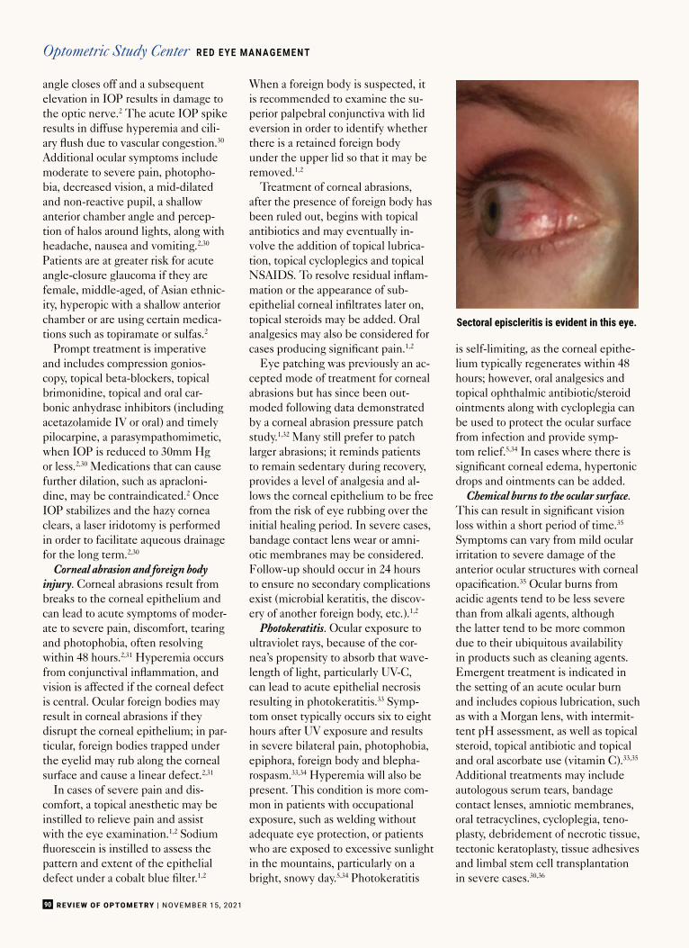
REVIEW OF OPTOMETRY | NOVEMBER 15, 202190
angle closes off and a subsequentelevation in IOP results in damage tothe optic nerve.2 The acute IOP spikeresults in diffuse hyperemia and cili-ary flush due to vascular congestion.30
Additional ocular symptoms includemoderate to severe pain, photopho-bia, decreased vision, a mid-dilatedand non-reactive pupil, a shallowanterior chamber angle and percep-tion of halos around lights, along withheadache, nausea and vomiting.2,30
Patients are at greater risk for acuteangle-closure glaucoma if they arefemale, middle-aged, of Asian ethnic-ity, hyperopic with a shallow anteriorchamber or are using certain medica-tions such as topiramate or sulfas.2
Prompt treatment is imperativeand includes compression gonios-copy, topical beta-blockers, topicalbrimonidine, topical and oral car-bonic anhydrase inhibitors (includingacetazolamide IV or oral) and timelypilocarpine, a parasympathomimetic,when IOP is reduced to 30mm Hgor less.2,30 Medications that can causefurther dilation, such as apracloni-dine, may be contraindicated.2 OnceIOP stabilizes and the hazy corneaclears, a laser iridotomy is performedin order to facilitate aqueous drainagefor the long term.2,30
Corneal abrasion and foreign bodyinjury. Corneal abrasions result frombreaks to the corneal epithelium andcan lead to acute symptoms of moder-ate to severe pain, discomfort, tearingand photophobia, often resolvingwithin 48 hours.2,31 Hyperemia occursfrom conjunctival inflammation, andvision is affected if the corneal defectis central. Ocular foreign bodies mayresult in corneal abrasions if theydisrupt the corneal epithelium; in par-ticular, foreign bodies trapped underthe eyelid may rub along the cornealsurface and cause a linear defect.2,31
In cases of severe pain and dis-comfort, a topical anesthetic may beinstilled to relieve pain and assistwith the eye examination.1,2 Sodiumfluorescein is instilled to assess thepattern and extent of the epithelialdefect under a cobalt blue filter.1,2
When a foreign body is suspected, itis recommended to examine the su-perior palpebral conjunctiva with lideversion in order to identify whetherthere is a retained foreign bodyunder the upper lid so that it may beremoved.1,2
Treatment of corneal abrasions,after the presence of foreign body hasbeen ruled out, begins with topicalantibiotics and may eventually in-volve the addition of topical lubrica-tion, topical cycloplegics and topicalNSAIDS. To resolve residual inflam-mation or the appearance of sub-epithelial corneal infiltrates later on,topical steroids may be added. Oralanalgesics may also be considered forcases producing significant pain.1,2
Eye patching was previously an ac-cepted mode of treatment for cornealabrasions but has since been out-moded following data demonstratedby a corneal abrasion pressure patchstudy.1,32 Many still prefer to patchlarger abrasions; it reminds patientsto remain sedentary during recovery,provides a level of analgesia and al-lows the corneal epithelium to be freefrom the risk of eye rubbing over theinitial healing period. In severe cases,bandage contact lens wear or amni-otic membranes may be considered.Follow-up should occur in 24 hoursto ensure no secondary complicationsexist (microbial keratitis, the discov-ery of another foreign body, etc.).1,2
Photokeratitis. Ocular exposure toultraviolet rays, because of the cor-nea’s propensity to absorb that wave-length of light, particularly UV-C,can lead to acute epithelial necrosisresulting in photokeratitis.33 Symp-tom onset typically occurs six to eighthours after UV exposure and resultsin severe bilateral pain, photophobia,epiphora, foreign body and blepha-rospasm.33,34 Hyperemia will also bepresent. This condition is more com-mon in patients with occupationalexposure, such as welding withoutadequate eye protection, or patientswho are exposed to excessive sunlightin the mountains, particularly on abright, snowy day.5,34 Photokeratitis
is self-limiting, as the corneal epithe-lium typically regenerates within 48hours; however, oral analgesics andtopical ophthalmic antibiotic/steroidointments along with cycloplegia canbe used to protect the ocular surfacefrom infection and provide symp-tom relief.5,34 In cases where there issignificant corneal edema, hypertonicdrops and ointments can be added.
Chemical burns to the ocular surface.This can result in significant visionloss within a short period of time.35
Symptoms can vary from mild ocularirritation to severe damage of theanterior ocular structures with cornealopacification.35 Ocular burns fromacidic agents tend to be less severethan from alkali agents, althoughthe latter tend to be more commondue to their ubiquitous availabilityin products such as cleaning agents.Emergent treatment is indicated inthe setting of an acute ocular burnand includes copious lubrication, suchas with a Morgan lens, with intermit-tent pH assessment, as well as topicalsteroid, topical antibiotic and topicaland oral ascorbate use (vitamin C).33,35
Additional treatments may includeautologous serum tears, bandagecontact lenses, amniotic membranes,oral tetracyclines, cycloplegia, teno-plasty, debridement of necrotic tissue,tectonic keratoplasty, tissue adhesivesand limbal stem cell transplantationin severe cases.30,36
Optometric Study Center R E D E Y E M A N A G E M E N T
Sectoral episcleritis is evident in this eye.
NOVEMBER 15, 2021 | REVIEW OF OPTOMETRY 91
Graft rejection. When corneal healthis compromised or irregularities pre-clude useful vision, corneal transplan-tation (keratoplasty) may be neces-sary, in which the host is surgicallygrafted with donor tissue.37 As withany transplantation surgery, severalrisks are involved, including the host’simmunological rejection of the donorcorneal tissue, which may occur in anycorneal layer at any point after kerato-plasty and may lead to graft failure.37,38
Patients may be symptomatic forredness, photophobia, pain, epiphoraand/or decreased vision, while clinicalsigns of immunological rejection in-clude corneal edema, vascularization,keratic precipitates, subepithelial orstromal infiltrates, a Khodadoust lineor an epithelial rejection line.39
Treatment of graft rejection in-volves the use of topical immunosup-pressants such as steroids, which aregenerally also used in the long term toprevent rejection episodes. Systemicimmunosuppressants may be consid-ered in high-risk grafts.40
Conjunctivitis and keratitis withvisual manifestations. While most casesof infectious conjunctivitis generallyinvolve minimal pain and vision loss,gonococcal (bacterial) conjunctivitisand EKC (viral) are exceptions. Theseconditions may present with acutepresentations of conjunctivitis involv-ing copious discharge, hyperemia andvision loss.41,42 Similarly, while micro-bial keratitis can often spare visionwhen the ulcer is non-central, ulcer-ation within the visual axis can lead tomarked vision loss.2,25
Endophthalmitis. This is a microbialinfection of the aqueous and vitre-ous humor.2,43 This condition resultsin symptoms of moderate to severepain, photophobia, decreased vision,eyelid edema and ocular discharge,and signs may include a hypopyon,iris synechiae, fibrinous membranesin the anterior chamber and vitrealinflammation.2,11,44 Significant conjunc-tival hyperemia may be seen due toconjunctivitis and circum-corneal con-gestion.44 Infection is often exogenousand can occur about 12 hours to seven
days after penetrating ocular surgery,trauma or injections, as well as fromsevere corneal infections, hospital-ization with IV, parenteral nutritionor broad-spectrum antibiotic use.2,43
Emergent referral to ophthalmology iswarranted, and treatment may consistof intravitreal antimicrobial injec-tions or vitrectomy, with varied visualoutcomes.2,43
TakeawaysWhile patients will often present witha subjective complaint of red eyes, theunderlying etiologies are numerous,and an accurate history and clinicalexamination are vital for appropriatecare. Taking a systematic approachto the exam is imperative and will inmany cases lead to the correct diagno-sis or a narrower list of differentials. g
1. Cronau H, Kankanala RR, Mauger T. Diagnosis and man-agement of red eye in primary care. Am Fam Physician.2010;81(2):137-44.2. Gilani CJ, Yang A, Yonkers M, et al. Differentiating urgentand emergent causes of acute red eye for the emergencyphysician. West J Emerg Med. 2017;18(3):509-17.3. Høvding G. Acute bacterial conjunctivitis. Acta Ophthal-mol. 2008;86(1):5-17.4. Ryder EC, Benson S. Conjunctivitis. In: StatPearls [Inter-net]. Treasure Island (FL): StatPearls Publishing; 2021.5. Frings A, Geerling G, Schargus M. Red eye: a guide fornon-specialists. Dtsch Arztebl Int. 2017;114(17):302-12.6. Najmi H, Mobarki Y, Mania K, et al. The correlationbetween keratoconus and eye rubbing: a review. Int JOphthalmol. 2019;12(11):1775-81.7. Messmer EM. The pathophysiology, diagnosis, and treat-ment of dry eye disease. Dtsch Arztebl Int. 2015;112(5):71-82.8. Bernardes TF, Bonfioli AA. Blepharitis. Semin Ophthal-mol. 2010;25(3):79-83.9. Rouen PA, White ML. Dry eye disease: prevalence,assessment, and management. Home Healthc Now.2018;36(2):74-83.10. Golden MI, Meyer JJ, Patel BC. Dry Eye Syndrome.In: StatPearls [Internet]. Treasure Island (FL): StatPearlsPublishing; 2021.11. Henderson AD, Miller NR. Carotid-cavernous fistula: current concepts in aetiology, investigation, and manage-ment. Eye (Lond). 2018;32(2):164-72.12. Lau FH, Yuen HK, Rao SK, Lam DS. Spontaneouscarotid cavernous fistula in a pediatric patient: case report and review of literature. J AAPOS. 2005;9(3):292-4.13. Kochar B, Shan SJ, Anand G, et al. Totally one-sided:painless unilateral proptosis. Am J Med. 2015;128(4):361-3.14. Héron E, Bourcier T. Scleritis and episcleritis. J FrOphtalmol. 2017;40(8):681-95.15. Schonberg S, Stokkermans TJ. Episcleritis. In:StatPearls [Internet]. Treasure Island (FL): StatPearlsPublishing; 2021.16. Sahinoglu-Keskek N, Cevher S, Ergin A. Analysis of sub-conjunctival hemorrhage. Pak J Med Sci. 2013;29(1):132-4.17. Dua HS, Said DG, Messmer EM, et al. Neurotrophickeratopathy. Prog Retin Eye Res. 2018;66:107-31.
18. Mantelli F, Nardella C, Tiberi E, et al. Congenital cornealanesthesia and neurotrophic keratitis: diagnosis and man-agement. Biomed Res Int. 2015;2015:805876.19. Rama P, Sacchetti M. Neurotrophic keratopathy.Orphanet. March 2017. www.orpha.net/consor/cgi-bin/OC_Exp.php?lng=EN&Expert=137596. Accessed Septemer10, 2021.20. Sheha H, Tighe S, Hashem O, et al. Update on ceneg-ermin eye drops in the treatment of neurotrophic keratitis.Clin Ophthalmol. 2019;13:1973-80.21. Gueudry J, Muraine M. Anterior uveitis. J Fr Ophtalmol.2018;41(1):e11-21.22. Duplechain A, Conrady CD, Patel BC, et al. Uveitis.In: StatPearls [Internet]. Treasure Island (FL): StatPearlsPublishing; 2021.23. Krachmer J, Mannis M, Holland E. Cornea, 2nd ed.Elsevier Mosby; 2005, 1284-314.24. Okhravi N, Odufuwa B, McCluskey P, et al. Scleritis. SurvOphthalmol. 2005;50(4):351-63.25. Farahani M, Patel R, Dwarakanathan S. Infectious cornealulcers. Dis Mon. 2017;63(2):33-7.26. Efron N, Morgan PB. Can subtypes of contact lens-asso-ciated corneal infiltrative events be clinically differentiated? Cornea. 2006;25(5):540-4.27. Khan SA, Lee CS. Recent progress and strategiesto develop antimicrobial contact lenses and lens casesfor different types of microbial keratitis. Acta Biomater.2020;113:101-18.28. Holden BA, La Hood D, Grant T, et al. Gram-negative bac-teria can induce contact lens related acute red eye (CLARE)responses. CLAO J. 1996;22(1):47-52.29. Shovlin J. Clear cause of CLARE. Rev Optom.2004;141(9).30. Pohl H, Tarnutzer AA. Acute angle-closure glaucoma. NEngl J Med. 2018;378(10):e14.31. Ahmed F, House RJ, Feldman BH. Corneal abrasions andcorneal foreign bodies. Prim Care. 2015;42(3):363-75.32. Turner A, Rabiu M. Patching for corneal abrasion.Cochrane Database Syst Rev. 2006;(2):CD004764.33. Sharma N, Kaur M, Agarwal T, et al. Treatment of acuteocular chemical burns. Surv Ophthalmol. 2018;63(2):214-35.34. Cullen AP. Photokeratitis and other phototoxic effects onthe cornea and conjunctiva. Int J Toxicol. 2002;21(6):455-64.35. Adepoju FG, Adeboye A, Adigun IA. Chemical eye injuries:presentation and management difficulties. Ann Afr Med. 2007;6(1):7-11.36. Baradaran-Rafii A, Eslani M, Haq Z, et al. Current and upcoming therapies for ocular surface chemical injuries.Ocul Surf. 2017;15(1):48-64.37. Akanda ZZ, Naeem A, Russell E, et al. Graft rejection rate and graft failure rate of penetrating keratoplasty (PKP)vs lamellar procedures: a systematic review. PLoS One.2015;10(3):e0119934.38. Velásquez-Monzón K, Navarro-Peña MC, Klunder-Klunder M, et al. Pediatric penetrating keratoplasty and graft rejec-tion: experience at the Hospital Infantil de México FedericoGómez. Bol Med Hosp Infant Mex. 2020;77(1):23-7.39. Anderson, E, Chang V, Bunya V, et al. Corneal allograftrejection and failure. AAO Eyewiki. April 12, 2021. eyewiki.aao.org/Corneal_Allograft_Rejection_and_Failure#Definition_of_graft_rejection_vs_failure. Accessed September 6, 2021.40. Di Zazzo A, Kheirkhah A, Abud TB, et al. Management of high-risk corneal transplantation. Surv Ophthalmol.2017;62(6):816-27.41. Wegman DH, Guinee VF, Milian SJ. Epidemickeratoconjunctivitis. Am J Public Health Nations Health.1970;60(7):1230-7.42. Belga S, Gratrix J, Smyczek P, et al. Gonococcalconjunctivitis in adults: case report and retrospective reviewof cases in Alberta, Canada, 2000-2016. Sex Transm Dis.2019;46(1):47-51.43. Durand ML. Endophthalmitis. Clin Microbiol Infect.2013;19(3):227-34.44. Simakurthy S, Tripathy K. Endophthalmitis. In: StatPearls[Internet]. Treasure Island (FL): StatPearls Publishing; 2021.

REVIEW OF OPTOMETRY | NOVEMBER 15, 202192
O P TO M E T R I C S T U D Y C E N T E R Q U I Z
To obtain continuing education credit through the Optometric Study Center, complete the test form on the following page and return it with the$35 fee to: Jobson Healthcare Information, LLC, Attn.: CE Processing, 395 Hudson Street, 3rd Floor New York, New York 10014. To be eligible,please return the card within three years of publication. You can also access the test form and submit your answers and payment via credit card
online at revieweducationgroup.com. You must achieve a score of 70 or higher to receive credit. Allow four weeks for processing. For each Optomet-ric Study Center course you pass, you earn 2 hours of credit. Please check with your state licensing board to see if this approval counts toward your CErequirement for relicensure.
1. All of the following clinical case historyquestions are essential to ask for a chiefcomplaint of red eye except:
a. Which eye is affected?b. Are you experiencing any discharge?c. How long ago was your last eye exam?
d. When did your symptoms first start?
2. Which symptoms are expected in a patientwith garden variety viral conjunctivitis?
a. Hyperemia, serous discharge, severe pain.b. Hyperemia, headache, itching.
c.Hyperemia, significant vision loss, copiousmucopurulent discharge.
d. Hyperemia, serous discharge, burning.
3. Which is a characteristic clinical sign ofviral keratitis secondary to herpes simplexvirus?
a.Ring infiltrate.b. Dendritic epitheliopathy.c. Vesicular rash.d. Bilateral hyperemia.
4. All may be considered as viablemanagement options for common bacterialconjunctivitis except:
a. Topical ophthalmic antibiotic drops.b. Observation.c. Topical ocular lubricants.d. IV ceftriaxone.
5. Pulsatile exophthalmos is a classicsymptom of which condition?
a. Allergic conjunctivitis.b. Blepharitis.
c.Carotid-cavernous fistula.d. EKC.
6. Characteristic clinical signs of episcleritisinclude:
a. Severe, boring pain and sectoral hyperemia.b. Mild itching and severe vision loss.c. Copious discharge and moderate pain.d. Sectoral hyperemia and minor irritation.
7. Which statement is TRUE aboutsubconjunctival hemorrhages?
a. They are considered a medical emergency.b. Due to their benign nature, systemic workup
is never required.c. Vision loss is rarely seen in most cases of
subconjunctival hemorrhages.d. Subconjunctival hemorrhages present with
painful hyperemia.
8. A 54-year-old female with rheumatoidarthritis presents with mild hyperemia OUfor several months. Associated symptomsinclude grittiness and burning. Mildpunctate corneal staining is seen in botheyes. Which is the most likely diagnosis?
a. Scleritis.b. Dry eye.c. Bacterial conjunctivitis.d. Herpes simplex keratitis.
9. A reasonable first-line treatment for the patient in question #8 would be:
a. Topical ophthalmic steroid drops.b. Topical ophthalmic antibiotic drops.
c.Topical preservative-free artificial tears.d. Oral NSAIDs.
10. The patient in question #8 returns to clinicthree months after having started thefirst-line treatment you selected in question #9 but reports she is still moderatelysymptomatic. All would be reasonablesecond-line treatments except:
a. Oral antiviral medications.b. Punctal occlusion.
c.Topical lifitegrast.d. Topical ophthalmic ointments at bedtime.
11. Which is TRUE about a patient presentingwith a dense, central neurotrophic cornealulcer?
a. The patient presents with associatedsymptoms of extreme pain.
b. The patient can be treated with preservative-free artificial tears twice daily.
c. The patient is likely experiencing a decreasein vision.
d. The patient’s symptoms are commensuratewith your clinical exam findings.
12. Scleritis differs from episcleritis in that:a. Scleritis blanches with phenylephrine.b. Scleritis results in sectoral hyperemia and
mild burning.c. Episcleritis is associated with a violet-blue
hue of the sclera.d. Scleritis is associated with deep, boring pain.
13. Which is true regarding CLARE?a. CLARE is a form of bacterial keratitis.b. CLARE consists of multiple, small peripheral
corneal infiltrates.c. Contact lenses are safe to wear while
actively symptomatic for CLARE.d. Using the same contact lens case for an
entire year is helpful to prevent CLARE.
14. Risk factors for acute angle-closure include:a. Asian descent, myopia, older age.b. Topiramate use, miotic pupil size.c. Deep anterior chamber, hyperopia, female.d. Hyperopia, female, middle-age.
15. A 35-year-old male presents to your practicewith a complaint of severe, bilateral ocularpain and photophobia that began acutelythis evening. He reports his eyes felt fine all morning on his skip trip. On clinical exam,you note bilateral, diffuse keratopathy. Theproper course of action includes:
a. Prescribing topical proparacaine to usehourly in both eyes for the next three days.
b. Explaining to the patient that this is a severeinfection and requires IV antibiotics.
c. Ordering a systemic workup including labwork and imaging.
d. Recommending copious lubrication andeducating the patient that symptoms areexpected to resolve within 48 hours.
16. All of these clinical signs and symptomsincrease suspicion for immunologicalrejection of a corneal graft except:
a. Diffuse corneal edema.b. Severe itching.c. Photophobia.d. Keratic precipitates.
17. Which is NOT a reasonable treatment optionfor a patient with a corneal abrasion?
a. Topical ophthalmic steroid drops.b. Topical ophthalmic antibiotic drops.c. Oral analgesics.
d.Frequent preservative-free artificial tearinstillation.
18. Which are expected in a patient presentingwith a bacterial ulcer?
a. Mild irritation, diffuse hyperemia, priorcontact lens wear.
b. Moderate to severe pain, photophobia,discharge.
c. Diabetes, contact lens wear, antibiotic use.d. Diffuse hyperemia, minimal pain.
19. A 27-year-old female presents witha complaint of a painful, red eye andsignificant photophobia. On clinical exam, 3+ cells and a 2+ flare anterior chamber reaction is noted. Which is not a reasonabletreatment option?
a. Topical cycloplegic drop instillation.b. Topical anti-fungal drop instillation.c. Oral analgesic use.d. Topical steroid drop instillation.
20. The patient in question #19 reports that shehas experienced several such episodes inthe past. This next step is indicated:
a. Systemic lab workup.b. Discontinue topical steroid use.c. Discontinue topical cycloplegic use.d. Educate the patient that recurrences are
expected and this is nothing to worry about.
Optometric Study Center R E D E Y E M A N A G E M E N T

OCTOBER 15, 2021 | REVIEW OF OPTOMETRY 93
Please retain a copy for your records. Please print clearly.
First Name
Last Name
The following is your: Home Address Business Address
Business Name
Address
City State
ZIP
Telephone # - -
Fax # - -
OE Tracker Number
Answers to CE exam:1. A B C D
2. A B C D
3. A B C D
4. A B C D
5. A B C D
6. A B C D
7. A B C D
8. A B C D
9. A B C D
10. A B C D
11. A B C D
12. A B C D
13. A B C D
14. A B C D
15. A B C D
16. A B C D
17. A B C D
18. A B C D
19. A B C D
20. A B C D
Examination Answer SheetWhen Your Patient Complains of Red EyeValid for credit through November 15, 2024
Online: This exam can be taken online at revieweducationgroup.com. Upon passingthe exam, you can view your results immediately and download a real-time CEcertificate. You can also view your test history at any time from the website.
Directions: Select one answer for each question in the exam and completely darkenthe appropriate circle. A minimum score of 70% is required to earn credit.
Mail to: Jobson Healthcare Information, LLC, Attn.: CE Processing, 395 HudsonStreet, 3rd Floor New York, New York 10014
Payment: Remit $35 with this exam. Make check payable to Jobson HealthcareInformation, LLC.
Credit: This course is COPE approved for 2 hours of CE credit. Course ID is 75277-GO.
Processing: There is a four-week processing time for this exam.
Jointly provided by Postgraduate Institute for Medicine and Review Education Group.Salus University has sponsored the review and approval of this activity.
31. The content was evidence-based.
32. The content was balanced and free of bias.
33. The presentation was clear and effective.
Rate the quality of the material provided:
1=Strongly disagree, 2=Somewhat disagree,3=Neutral, 4=Somewhat agree, 5=Strongly agree
1 2 3 4 5
1 2 3 4 5
By submitting this answer sheet, I certify that I have read the lesson in its entirety and completed the self-assessment exam personally based on the material presented.I have not obtained the answers to this exam by any fraudulent or improper means.
Signature Date Lesson 122074 RO-OSC-1121
Post-activity evaluation questions:
Rate how well the activity supported your achievement of these learning objectives. 1=Poor, 2=Fair, 3=Neutral, 4=Good, 5=Excellent
21. Discuss the pathophysiology behind red eye.
22. Conduct a careful and comprehensive patient history.
23. Recognize what to look for during the clinical exam.
24. Identify and diagnose the underlying cause of the red eye.
25. Based upon your participation in this activity, do you intend to change your practice behavior? (Choose only one of the following options.) A I do plan to implement changes in my practice based on the information presented. B My current practice has been reinforced by the information presented. C I need more information before I will change my practice.
26. Thinking about how your participation in this activity will influence your patient care, how many of your patients are likely to benefit?(please use a number):
27. If you plan to change your practice behavior, what type of changes do you plan to implement? (Check all that apply.)
28. How confident are you that you will be able to make your intended changes? A Very confident B Somewhat confident C Unsure D Not confident
29. Which of the following do you anticipate will be the primary barrier to implementing these changes?
30. Additional comments on this course:_______________________________________________________________________________________________________
1 2 3 4 5
1 2 3 4 5
1 2 3 4 5
1 2 3 4 5
A Formulary restrictions B Time constraints C System constraints
D Insurance/financial issues E Lack of interprofessional team support F Treatment related adverse events
G Patient adherence/compliance H Other, please specify:____________________________________________
A Apply latest guidelines B Change in diagnostic methods C Choice of management approach
D Change in current practice for referral E Change in vision correction offerings F Change in differential diagnosis
G More active monitoring and counseling H Other, please specify: _______________________________________________________________
1 2 3 4 5
NOVEMBER 15, 2021 | REVIEW OF OPTOMETRY 93
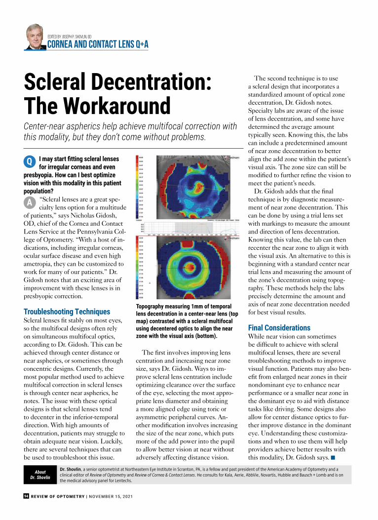
I may start fi tting scleral lenses for irregular corneas and even
presbyopia. How can I best optimize vision with this modality in this patient population?
“Scleral lenses are a great spe-cialty lens option for a multitude
of patients,” says Nicholas Gidosh,OD, chief of the Cornea and ContactLens Service at the Pennsylvania Col-lege of Optometry. “With a host of in-dications, including irregular corneas,ocular surface disease and even highametropia, they can be customized towork for many of our patients.” Dr.Gidosh notes that an exciting area ofimprovement with these lenses is inpresbyopic correction.
Troubleshooting TechniquesScleral lenses fi t stably on most eyes,so the multifocal designs often relyon simultaneous multifocal optics,according to Dr. Gidosh. This can beachieved through center distance ornear aspherics, or sometimes throughconcentric designs. Currently, themost popular method used to achievemultifocal correction in scleral lensesis through center near aspherics, henotes. The issue with these opticaldesigns is that scleral lenses tendto decenter in the inferior-temporaldirection. With high amounts ofdecentration, patients may struggle toobtain adequate near vision. Luckily,there are several techniques that canbe used to troubleshoot this issue.
The fi rst involves improving lenscentration and increasing near zonesize, says Dr. Gidosh. Ways to im-prove scleral lens centration includeoptimizing clearance over the surfaceof the eye, selecting the most appro-priate lens diameter and obtaininga more aligned edge using toric orasymmetric peripheral curves. An-other modifi cation involves increasingthe size of the near zone, which putsmore of the add power into the pupilto allow better vision at near withoutadversely affecting distance vision.
The second technique is to usea scleral design that incorporates astandardized amount of optical zonedecentration, Dr. Gidosh notes.Specialty labs are aware of the issueof lens decentration, and some havedetermined the average amounttypically seen. Knowing this, the labscan include a predetermined amountof near zone decentration to betteralign the add zone within the patient’svisual axis. The zone size can still bemodifi ed to further refi ne the vision tomeet the patient’s needs.
Dr. Gidosh adds that the fi naltechnique is by diagnostic measure-ment of near zone decentration. Thiscan be done by using a trial lens setwith markings to measure the amountand direction of lens decentration.Knowing this value, the lab can thenrecenter the near zone to align it withthe visual axis. An alternative to this isbeginning with a standard center neartrial lens and measuring the amount ofthe zone’s decentration using topog-raphy. These methods help the labsprecisely determine the amount andaxis of near zone decentration neededfor best visual results.
Final ConsiderationsWhile near vision can sometimesbe diffi cult to achieve with scleralmultifocal lenses, there are severaltroubleshooting methods to improvevisual function. Patients may also ben-efi t from enlarged near zones in theirnondominant eye to enhance nearperformance or a smaller near zone inthe dominant eye to aid with distancetasks like driving. Some designs alsoallow for center distance optics to fur-ther improve distance in the dominanteye. Understanding these customiza-tions and when to use them will helpproviders achieve better results withthis modality, Dr. Gidosh says.
REVIEW OF OPTOMETRY | NOVEMBER 15, 202194
Center-near aspherics help achieve multifocal correction withthis modality, but they don’t come without problems.
Scleral Decentration: The Workaround
Dr. Shovlin, a senior optometrist at Northeastern Eye Institute in Scranton, PA, is a fellow and past president of the American Academy of Optometry and aclinical editor of Review of Optometry and Review of Cornea & Contact Lenses. He consults for Kala, Aerie, AbbVie, Novartis, Hubble and Bausch + Lomb and is onthe medical advisory panel for Lentechs.
AboutDr. Shovlin
Edited by Joseph P. Shovlin, OD
CORNEA and CONTACT LENS Q+A
Q
A
Topography measuring 1mm of temporal lens decentration in a center-near lens (top map) contrasted with a scleral multifocal using decentered optics to align the near zone with the visual axis (bottom).

®2021 Menicon Co. Ltd. See instructions for use in the Package Insert. US-MI053
A new generation of silicone hydrogel materialbalanced for health and comfort incorporatinga unique combination of high oxygen and ultralow modulus.
Miru 1day UpSide contact lenses are alwaysthe right way up and ready to wear, thanks toSmart Touch™ technology.
Miru 1day UpSideIntroducing,
silicone hydrogel contact lenseswith Smart Touch™ technology.
www.meniconamerica.com
Find us on:
7 th1951- 2021anniversary7

REVIEW OF OPTOMETRY | NOVEMBER 15, 202196
Dr. Labib graduated from Pennsylvania College of Optometry, where she now works as an associate professor. She completed her residency in primary care/oculardisease and is a fellow of the American Academy of Optometry and a diplomate in the Comprehensive Eye Care section. She has no financial interests to disclose.
AboutDr. Labib
Acorneal assessment is a routineand integral part of any ocularexamination. Its elaborate andunique organization gives the
cornea the necessary transparencyto refract and transmit light rays tothe retina for vision. This also makesexamination difficult—as the cor-nea is an inherently clear structure,its very nature poses a challenge indetermining if corneal pathology ispresent. There are some tools thatcan facilitate assessment, one of thosebeing the use of vital dyes.
One Dye, Many UsesOf the available ophthalmic dyes,fluorescein is most commonly usedfor corneal evaluation. The agent,
fluorescein sodium, is an orange-redhydrocarbon with low molecularweight.1 It is available in topical andintravenous forms for ocular use. Forthe evaluation of corneal pathology,it comes impregnated in a paper stripand dissolves when moistened withnormal saline solution to be intro-duced onto the surface of the eye.This is done by placing the moist-ened strip in the inferior conjunctivaor fornix and having the patient blinkmultiple times to allow the agent tospread across the ocular surface.
One of its unique properties is thatit reacts to wavelengths in the visiblespectrum, ranging from 465nm to490nm. It is most evident under co-balt blue light. Under normal lighting
conditions, it appears orange on theocular surface. However, on obser-vation of fluorescein sodium underthe cobalt blue filter, it fluoresces towavelengths of 520nm to 530nm, ap-pearing bright green to the observer.The charged ends of this compoundare attracted to the hydrophilic endsof the cell membrane in the cornea,thus illuminating areas where cellsare broken down or missing.2 Theseproperties allow for its many ophthal-mic uses:
• Evaporative dry eye assessment.The diagnosis of dry eye syndrome,particularly the evaporative form,is made possible through the use ofa fluorescein sodium strip and slitlamp. A commonly used diagnostictest called tear breakup time is mostoptimally performed by placing afluorescein strip onto the eye, wait-ing two minutes and then viewingthe cornea with the cobalt blue filterunder high illumination at 10x magni-fication. The patient is asked to blinkonce and then keep their eyes openas long as they can. Simultaneously,the examiner observes the corneafor the first appearance of a dry spot,which presents as a dark area amongthe otherwise uniformly green ap-pearance of the cornea, and countsthe number of seconds that it takesto manifest. Studies have indicatedthat a measurement of less than 10seconds is abnormal.3,4
• Aqueous-deficient dry eye assessment.In more aqueous-deficient forms ofdry eye syndrome, fluorescein canalso be used to illuminate the pres-ence, severity and course of dryness.This is done in a different mannerthan evaporative dry eye, in that thedye is inserted and observation of thecornea is best performed betweenfour and eight minutes followinginstillation.
Fluorescein is most commonly used for corneal evaluationand, as such, its instillation should be properly understood.
Reach for the Dye
B
By Bisant A. Labib, OD
THE ESSENTIALS
Punctate epithelial erosions in a dry eye patient as revealed by fluorescein dye.
Photo: WR Buie

TEN
I’m looking for a reputable solution for a digital acuity system – any suggestions?
D i s a s t e r p r o o f b y d e s i g n | a c u i t y p r o . c o m / 5 8 0 - 2 4 3 - 1 3 0 1
S l o a n / S n e l l e n / N u m b e r s / C o n t r a s t / E T D R S / W h i t e O n B l a c k O p t i o n / R a n d o m i z eC u s t o m R e m o t e / M a r c o I n t e g r a t i o n / F r e e S u p p o r t / N o A n n u a l F e e s
slide to reply
Have used Acuity Pro for over 10 years; love it!
Mindy Seaman now
Pat Hamilton now
slide to reply
Our office loves Acuity pro. Six lanes.
Andrew Hart now
slide to reply
Great customer support when you have questions.
Harvey Yamamoto now
slide to reply
Acuity Pro. Best ever. Highly recommend.
Acuity Pro is developed and owned by an OD. Highly recommend.
Wayne Gilmore now
slide to reply
Another vote for Acuity Pro!Ted Bryant now
slide to reply
10:10Friday February 19
78%
Acuity ProDigital Acuity Systems
®
Take advantage of section 179 tax
break on new/used equipment by end
of year!
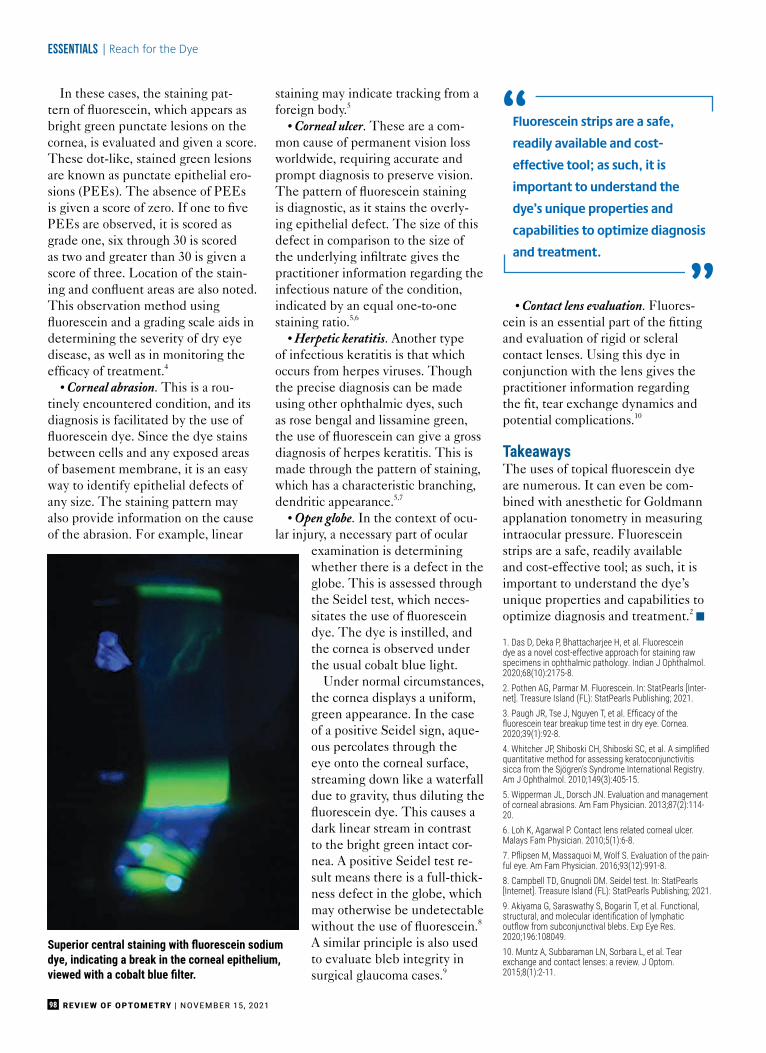
REVIEW OF OPTOMETRY | NOVEMBER 15, 202198
In these cases, the staining pat-tern of fluorescein, which appears asbright green punctate lesions on thecornea, is evaluated and given a score.These dot-like, stained green lesionsare known as punctate epithelial ero-sions (PEEs). The absence of PEEsis given a score of zero. If one to fivePEEs are observed, it is scored asgrade one, six through 30 is scoredas two and greater than 30 is given ascore of three. Location of the stain-ing and confluent areas are also noted.This observation method usingfluorescein and a grading scale aids indetermining the severity of dry eyedisease, as well as in monitoring theefficacy of treatment.4
• Corneal abrasion. This is a rou-tinely encountered condition, and itsdiagnosis is facilitated by the use offluorescein dye. Since the dye stainsbetween cells and any exposed areasof basement membrane, it is an easyway to identify epithelial defects ofany size. The staining pattern mayalso provide information on the causeof the abrasion. For example, linear
staining may indicate tracking from aforeign body.5
• Corneal ulcer. These are a com-mon cause of permanent vision lossworldwide, requiring accurate andprompt diagnosis to preserve vision.The pattern of fluorescein stainingis diagnostic, as it stains the overly-ing epithelial defect. The size of thisdefect in comparison to the size ofthe underlying infiltrate gives thepractitioner information regarding theinfectious nature of the condition,indicated by an equal one-to-onestaining ratio.5,6
• Herpetic keratitis. Another typeof infectious keratitis is that whichoccurs from herpes viruses. Thoughthe precise diagnosis can be madeusing other ophthalmic dyes, suchas rose bengal and lissamine green,the use of fluorescein can give a grossdiagnosis of herpes keratitis. This ismade through the pattern of staining,which has a characteristic branching,dendritic appearance.5,7
• Open globe. In the context of ocu-lar injury, a necessary part of ocular
examination is determiningwhether there is a defect in theglobe. This is assessed throughthe Seidel test, which neces-sitates the use of fluoresceindye. The dye is instilled, andthe cornea is observed underthe usual cobalt blue light.
Under normal circumstances,the cornea displays a uniform,green appearance. In the caseof a positive Seidel sign, aque-ous percolates through theeye onto the corneal surface,streaming down like a waterfalldue to gravity, thus diluting thefluorescein dye. This causes adark linear stream in contrastto the bright green intact cor-nea. A positive Seidel test re-sult means there is a full-thick-ness defect in the globe, whichmay otherwise be undetectablewithout the use of fluorescein.8
A similar principle is also usedto evaluate bleb integrity insurgical glaucoma cases.9
• Contact lens evaluation. Fluores-cein is an essential part of the fittingand evaluation of rigid or scleralcontact lenses. Using this dye inconjunction with the lens gives thepractitioner information regardingthe fit, tear exchange dynamics andpotential complications.10
TakeawaysThe uses of topical fluorescein dyeare numerous. It can even be com-bined with anesthetic for Goldmannapplanation tonometry in measuringintraocular pressure. Fluoresceinstrips are a safe, readily availableand cost-effective tool; as such, it isimportant to understand the dye’sunique properties and capabilities tooptimize diagnosis and treatment.2
g
1. Das D, Deka P, Bhattacharjee H, et al. Fluoresceindye as a novel cost-effective approach for staining rawspecimens in ophthalmic pathology. Indian J Ophthalmol.2020;68(10):2175-8.2. Pothen AG, Parmar M. Fluorescein. In: StatPearls [Inter-net]. Treasure Island (FL): StatPearls Publishing; 2021.3. Paugh JR, Tse J, Nguyen T, et al. Efficacy of the fluorescein tear breakup time test in dry eye. Cornea. 2020;39(1):92-8.4. Whitcher JP, Shiboski CH, Shiboski SC, et al. A simplified quantitative method for assessing keratoconjunctivitissicca from the Sjögren’s Syndrome International Registry.Am J Ophthalmol. 2010;149(3):405-15.5. Wipperman JL, Dorsch JN. Evaluation and managementof corneal abrasions. Am Fam Physician. 2013;87(2):114-20.6. Loh K, Agarwal P. Contact lens related corneal ulcer. Malays Fam Physician. 2010;5(1):6-8.7. Pflipsen M, Massaquoi M, Wolf S. Evaluation of the pain-ful eye. Am Fam Physician. 2016;93(12):991-8.8. Campbell TD, Gnugnoli DM. Seidel test. In: StatPearls [Internet]. Treasure Island (FL): StatPearls Publishing; 2021.9. Akiyama G, Saraswathy S, Bogarin T, et al. Functional,structural, and molecular identification of lymphatic outflow from subconjunctival blebs. Exp Eye Res. 2020;196:108049.10. Muntz A, Subbaraman LN, Sorbara L, et al. Tearexchange and contact lenses: a review. J Optom. 2015;8(1):2-11.
Superior central staining with fluorescein sodium dye, indicating a break in the corneal epithelium, viewed with a cobalt blue filter.
ESSENTIALS | Reach for the Dye
Fluorescein strips are a safe,readily available and cost-effective tool; as such, it isimportant to understand thedye’s unique properties andcapabilities to optimize diagnosisand treatment.

BRING BETTER BALANCE.Refract safely from across the room or across the Internet with the most advanced Phoroptor®, Phoroptor® VRx, and the
pixel-perfect ClearChart® 4 Digital Acuity System. Multitasker Lisa Genovese, OD, has brought balance to her practice andpersonal life all while managing Insight Eye Care’s multiple locations. She e� ciently juggles being a full-time optometrist,
a full-time entrepreneur, and a full-time parent with the help of Reichert’s complete line of digital refraction devices.
· © 2021 AMETEK, Inc. & Reichert, Inc. (08-2021) · Phoroptor and ClearChart are registered trademarks of Reichert, Inc. · Phoroptor and ClearChart are designed & assembled in USA.
LEARN
MORE
REICHERT.COM/ORAWATCH THE VIDEOS AT
BRING BETTER BALANCE.Refract safely safely from from across across the the room room or or across across across the the Internet Internet with with the the most most advanced advanced Phoroptor®, Phoroptor®, Phoroptor® Phoroptor® VRx, VRx, and and the the
pixel-perfect ClearChart® ClearChart® 4 4 Digital Digital Acuity Acuity System. System. Multitasker Multitasker Lisa Lisa Lisa Genovese, Genovese, OD, OD, has has brought brought balance balance to to her her practice practice practice and andpersonal life life all all while while managing managing Insight Insight Eye Eye Care’s Care’s multiple multiple locations. locations. She She e� e� ciently ciently ciently e� ciently e� juggles juggles being being a a full-time full-time optometrist, optometrist,
a full-time full-time entrepreneur, entrepreneur, and and a a full-time full-time parent parent with with the the help help of of Reichert’s Reichert’s complete complete line line of of digital digital refraction refraction devices. devices.
· · © © 2021 2021 AMETEK, AMETEK, Inc. Inc. & & Reichert, Reichert, Inc. Inc. (08-2021) (08-2021) · · Phoroptor Phoroptor and and ClearChart ClearChart are are registered registered trademarks trademarks of of Reichert, Reichert, Inc. Inc. · · Phoroptor Phoroptor and and ClearChart ClearChart are are designed designed & & assembled assembled in in USA. USA.
LEARN
MORE
REICHERT.COM/ORAREICHERT.COM/ORAREICHERT.COM/ORAWATCH THE THE VIDEOS VIDEOS AT AT

100 REVIEW OF OPTOMETRY | NOVEMBER 15, 2021
edited by richard mangan, oD
URGENT CARE
By Kristen Walton, ODomaha, ne
A41-year-old white male weldingstudent presented to the clinicfor an emergency visit. He hadbeen sharpening tungsten, a type
of metal, in class that morning withoutwearing safety glasses when a fragmentpierced his left eye. His vision wasseverely impacted, and he reportedimmense pain, which he describedas a throbbing, aching sensation, andconstant tearing.
The patient had type 2 diabetes, hismedications and social history werenot pertinent to the case and he had noknown drug allergies.
CaseThe patient’s entering visual acuitywas 20/150 with no improvement withpinhole in the left eye. Intraocularpressure was not evaluated. Upon slitlamp exam, a full-thickness corneallaceration was present nasally, extend-ing from five to 10 o’clock with iris pro-lapse. Upon fundus exam, the posteriorpole appeared normal with a limitedview of the periphery. There were nosigns of an obvious break, hole or tear.
The patient was diagnosed withan open globe secondary to a corneallaceration of the left eye, and urgentrepair was recommended. The patientwas informed that his visual progno-sis would be guarded. Prednisoloneacetate and moxifloxacin were initiatedfollowing surgery with instructions totake one drop of each eight times daily.
The patient was to return to the clinicthe following day.
At the one-day post-op exam, thepatient presented with a visual acuityof 20/1250 with no improvement withpinhole in the left eye. Intraocularpressure was 12.3mm Hg. The corneallaceration was well apposed, with anoverlying epithelial defect. Within theanterior chamber, a small hyphemawas present and the pupil was draggedtemporally with no apparent signs ofentrapment within the wound.
At the two-week mark followingglobe repair, the patient’s visual acuitywas 20/300 with a stable intraocularpressure, and the epithelial defect hadhealed. Upon dilation of the left eye,the optic nerve appeared well perfusedwith no clinical retinal nerve fiber layerdefects. No breaks, holes or tears wereobserved within the periphery of theretina. Moxifloxacin was discontinued,
with prednisolone acetate decreased toone drop four times daily. The patientwas scheduled for a follow-up exam inanother two weeks.
Four weeks post-open globe repair,the patient was 20/250 with his left eyeand pinholed to 20/100. All findingsof the anterior and posterior segmentswere stable with a macular OCTdemonstrating normal macular contour.Prednisolone acetate was to be taperedby one drop each week. A four- tosix-week follow-up was scheduled tocontinue monitoring healing, and thepatient was asked to return to the clinicsooner if he noticed any changes in hissigns or symptoms.
DiscussionOpen-globe injuries are not particu-larly common, occurring in up to fiveof 100,000 people per year.1,2 Youngermales between the ages of 10 and30 tend to be the population most atrisk.1,2 Worksite hazards can increasethe chances of an open globe injury.2
The two most common mechanismsthat lead to this type of injury includebeing struck by another individual oran object, followed by a sharp foreignbody piercing the eye.1
An open globe can cause vision loss and intraocular infection,but preventing these injuries is completely possible when theproper precautions are taken.
Safety First
Dr. Mangan is a board-certified consultative optometrist from Boulder, CO, with a focus on ocular disease and surgical comanagement, as well as a fellow of the AmericanAcademy of Optometry. He has recently accepted a position at Sun Pharma; however, the content of this article was created prior to that relationship and had no bearing on itsdevelopment. Previously, he was an assistant professor in the department of ophthalmology at the University of Colorado School of Medicine.
AboutDr. Mangan
A full-thickness corneal laceration with iris prolapse in a patient with a similar injury to thecase described here.
Photo: Brian Shafer, MD

www.reviewedu.com/winteroptometry
*Approval pendingReview Education Group partners with Salus University for those ODs who are licensed in states that require university credit.Partially supported by an unrestricted educational grant from Alcon.
DECEMBER 10–11, 2021HYATT REGENCY NEWPORT BEACH
1107 JAMBOREE ROAD, NEWPORT BEACH, CALIFORNIA
LIVE COPE*
Earn up to 12 LIVE COPE credits*
CO-CHAIRSMurray Fingeret, OD, FAAOChief of the Optometry SectionBrooklyn/St. Albans CampusDepartment of Veterans AdministrationNew York Harbor Health Care SystemClinical ProfessorSUNY, College of Optometry
Robert N. Weinreb, MDChair and Distinguished Professorof OphthalmologyDirector, Shiley Eye InstituteDirector, Hamilton Glaucoma CenterMorris Gleich, M.D. Chair in GlaucomaBoard Certifi cation in Ophthalmology
PRESENTING FACULTYBen Gaddie, OD, FAAOOwner and Director, Gaddie Eye CentersFellow, American Academy of OptometryPresident, Optometric Glaucoma Society (OGS)Co-Chairman, International Vision Expo MeetingsEditorial board member, Review of Optometry,Optometric Management, Primary Care OptometryNews and Optometry Times
Alex Huang, MD, PhDAssociate Professor of OphthalmologyUCLA Doheny Eye InstituteUCLA Stein Eye Institute
Richard Madonna, OD, MA, FAAOProfessor and Chair, Department of Clinical EducationDirector, O� ce of Continuing Professional EducationSUNY, College of Optometry
REVIEW OF OPTOMETRY | NOVEMBER 15, 2021102
Upon initial evaluation of thesepatients, a detailed history shouldbe taken to figure out the nature ofthe incident and possible worker’scompensation paperwork. It is good toknow what type of object injured theeye, what speed it was traveling andwhat environment the patient was inat the time of injury.2 The environ-ment is essential to know to helppredict the chances and nature of pos-sible infection.2 If the patient reportsthe involvement of a sharp object,a fast-moving object or blunt forcetrauma, this should raise immediatesuspicion.2
In all emergent cases, it is vital toestablish a baseline visual acuity to de-note damage acquired and determinepossible visual recovery.2,3 With sus-pected open globes, the eye should bemanipulated as little as possible withintraocular pressure measurement andeyelid eversion excluded from thework-up.2,3 This precaution decreasesthe risk of any intraocular material be-ing expelled from the eye.2
Pupils should be tested to discernany irregularities that may be pres-ent.2,3 A peaked pupil is a strong indi-
cator that the patient has undergonean open-globe injury due to the pupilbeing pulled toward the wound.2 Acomprehensive slit lamp examinationshould be performed to look for anyremnants of the inflicting foreign bodyand potential lacerations or penetra-tion sites.2 If there is any question asto the depth of a wound, Seidel test-ing should be performed.2,3
The anterior chamber should beassessed for adequate depth, presenceof hyphema, cells, flare or any vitreousmaterial.2 With ocular trauma, evaluat-ing the integrity and positioning of thelens becomes important due to thepossibility of dislocation.2 With anypossible ruptured globe, be sure torule out hemorrhaging, retinal detach-ment and possible intraocular foreignbodies.2 If an exam is not obtainable atthe slit lamp, non-contrast computedtomography is the imaging of choicefor open globe injuries, especiallyin the case of metallic foreign bodyinvolvement.2,3
When a patient needs to be referredfor an open-globe repair, it is crucial toeducate them not to eat or drink any-thing prior to anesthesia, to prevent
a delay in care.2,3 Placing a patch overthe affected eye could be consideredfor protection prior to surgical inter-vention.3 Positioning of the patient toavoid bending and lifting is importantas well to limit intraocular pressureelevation.2,3
One of the most devastating compli-cations that can result from an openglobe, besides potential vision loss, isendophthalmitis.2-4 Because the intra-ocular contents are exposed, the rateof endophthalmitis is around 6.8%.2,4
Patients at a higher risk for develop-ing this complication include thosewho delayed seeking care, who had alens capsule rupture during the timeof injury or who had a wound inflictedwith an unsanitary foreign body.3
Getting the wound repaired as soon aspossible, preferably within 24 hours,and initiating prophylactic coveragewith a broad-spectrum antibiotic is thebest prevention.2
Following surgery, the patientshould be advised to wear a protectiveshield while sleeping.2 Restrictionspost-globe repair typically includelimiting excessive movement such asexercise and extensive lifting, alongwith no swimming.2 These restrictionsare put in place to prevent wound de-hiscence and to decrease the chanceof infection.2 The best preventativeadvice to offer is that proper eyewearprotection is of the utmost importanceto guard the precious gift of sight. g
1. Mir TA, Canner JK, Zafar S, et al. Characteristics of openglobe injuries in the United States from 2006 to 2014. JAMAOphthalmology. 2020;138(3):268-75.2. Wang D, Deobhakta A. Open globe injury: assessment andpreoperative management. AAO. August 2020. www.aao.org/eyenet/article/open-globe-injury. Accessed September29, 2021.3. Blair K, Alhadi SA, Czyz CN. Globe rupture. In: StatPearls[Internet]. Treasure Island (FL): StatPearls Publishing; 2020.4. Essex RW, Yi Q, Charles PG, et al. Post-traumatic endo-phthalmitis. Ophthalmology. 2004;111(11):2015-22.
Dr. Walton graduated with honorsfrom the Indiana University School ofOptometry. She completed her residencyat Vance Thompson Vision in Sioux Falls,SD, managing ocular disease, refractivecases and pre- and post-op ocularsurgery. She currently works at Vance
Thompson Vision in Omaha, NE. She has no financial interests to disclose.
ABOUT THE AUTHOR
A FAREWELL FROM DR. MANGANHow time flies. After a 30-year career in medical optometry, I finally get to take a fewminutes to reflect on my time as an author and board member for Review of Optometry.It’s hard to believe that it’s coming to a close. What a pleasure it has been to collaboratewith so many amazing professionals.
I’m honored to have served as a columnist for Review for six years. When we firstdiscussed the topic of ocular urgencies and emergencies as a potential column idea,we felt that we might be able to find enough interesting cases to support such a columnfor maybe three years. I never imagined that the column would still be going strong allthese years later, and I’m thrilled to announce that Alison Bozung, OD, a rising star fromBascom Palmer Eye Institute, will be taking over—and taking it to new heights.
There are so many people to thank: First, Editor-in-Chief Jack Persico. Thank you forgiving me the opportunity to take on this very important topic for Review. Your supportover the years is so appreciated. I couldn’t have done it without you!
Second, the amazing editorial staff I have had the pleasure of working with for so manyyears. You are the true artists behind the scenes that make the articles and case presen-tations come to life!
Third, all of the amazing optometric physicians who have contributed to the column.You have made a tremendous impact in strengthening our ability to understand and man-age ocular urgencies and emergencies as they present.
Lastly, and most importantly, you, my colleagues who have shown that this is a topicthat still resonates. Thank you for being loyal readers of my column and Review.
Signing off,Rich
URGENT CARE | Safety First

Read the Latest from Dr. Art Epstein
G REVIEWGROUPR WEEKLY E-NEWSLETTERS/
OPTOMETRIC PHYSICIAN:
Reaches 42,000 ODs weekly
Content: Clinical information & Industry news
Metrics: 18.45% average opens
Michael Hoster: [email protected]
Michele Barrett: [email protected]
Jon Dardine: [email protected]
FOR ADVERTISING OPPORTUNITIES, PLEASE CONTACT
If you aren’t already a subscriber, please besure to sign up for Dr. Art Epstein’s weeklye-journal, Optometric Physician, distributedby Review of Optometry.
Join the thousands of ODs, MDsand ophthalmic industry readers who turnto Optometric Physician each week for Dr.Epstein’s very direct—and sometimescontroversial—take on eye care and theworld. You will also keep up with the latest,most relevant clinical studies, condensedand curated for quick reference, as well asa current digest of industry news.
Visit: JMI | Newsletter Signupwww.jhihealth.com/globalemail/to sign up for Optometric Physician, and othere-newsletters distributed by Jobson Medical Information.

REVIEW OF OPTOMETRY | NOVEMBER 15, 2021104
by Mark Dunbar, OD
reTINA QUIZ
A49-year-old Hispanic malefirst presented with com-plaints of blurred vision inhis left eye during a tele-
medicine exam. He also reported afew months earlier seeing “brokenglass,” or what looked like coloredcrystals, in his vision in the sameeye, which was accompanied bytingling on the left side of his body.
Nothing was seen during theexam but, given his symptoms andhistory of blurred vision, it was rec-ommended that he come in for anin-person visit the following week,where he underwent a comprehen-sive eye exam and reaffirmed hethought vision in his left eye wasslightly blurry.
His medical history was significantfor hypertension and high blood sugarbut reported that he is on a vegan dietand lost over 20 pounds. His bloodpressure is under control and he takeslisinopril.
Upon exam, his uncorrected dis-tance visual acuities measured 20/20in each eye. Confrontation visualfields were full to careful countingOU. His pupils were equally roundand reactive to light; there was noafferent pupillary defect. His extra-ocular motility was full and cover testwas ortho at distance. His anterior seg-ments and intraocular pressures wereunremarkable.
On dilated fundus exam, thevitreous in both eyes was clear. Hehad small cups with good rim color-ation and perfusion in each eye. Thefundus exam findings of the right eyewere completely normal. In the left
eye, there were obvious changes ofnote (Figure 1). Fundus photos, OCTand OCT-A were obtained of themacula and peripheral retina.
Take the Retina Quiz1. What is the most likely diagnosis?a. Congenital retinal telangiectasis,
Type 1.b. Peripheral choroidal neovasculariza-
tion.c. Coats disease.d. Retinal arterial microaneurysm
(RAM).
2. How should this patient be managed?a. Observation.b. Focal laser.c. Anti-VEGF injection.d. Either B or C.
3. What is the most common underlyingcondition?a. Congenital vascular incompetency.
b. Diabetes.c. Hypertensiond. Hyperlipidemia.
4. Which statement is most accurate forthis condition?a. Mainly develops from dysfunc-
tion within the superficial capillarynetwork.
b. Develops from dysfunction withthe venous system.
c. Develops from dysfunction of thearterial system.
d. Develops subretinally fromincreased VEGF expression.
5. Which patient is more likely to developthis condition?a. Young males.b. Young females.c. Middle-aged males.d. Elderly females.
For answers, see page 114.
DiagnosisIt was a surprise to discover a largearea of circinate exudate superotem-porally in the left eye of this patient.Within the center of the exudate,there is a yellow-white oval lesion thatinvolves one of the branches of eitherthe central retinal vein or centralretinal artery, but which is it? At initialinspection, it looks like the centrallesion is from one of the retinal veins,as there is significant sheathing of theveins above and below the lesion. Butif you look more closely and followthe lesion back, it actually involvesone of the branches of a retinal artery.Indeed, this is a retinal arterial micro-aneurysm (RAM).
DiscussionRAM represents a focal aneurysmaldilatation of one of the branches of thecentral retinal artery and usually occurswithin the first three bifurcations of
Dr. Dunbar is the director of optometric services and optometry residency supervisor at the Bascom Palmer Eye Institute at the University of Miami. He is a foundingmember of the Optometric Glaucoma Society and the Optometric Retina Society. Dr. Dunbar is a consultant for Carl Zeiss Meditec, Allergan, Regeneron and Genentech.
AboutDr. Dunbar
Fig. 1. A widfield fundus image of the retina in the patient. What is the diagnosis?
A surprising discovery led to this diagnosis.A Closer Look
Follow us on Instagram at @revoptom for striking clinical images, daily news headlines,issue previews and great content from the magazine—all formatted for mobile
A NEW WAY TO EXPERIENCEREVIEW OF OPTOMETRY
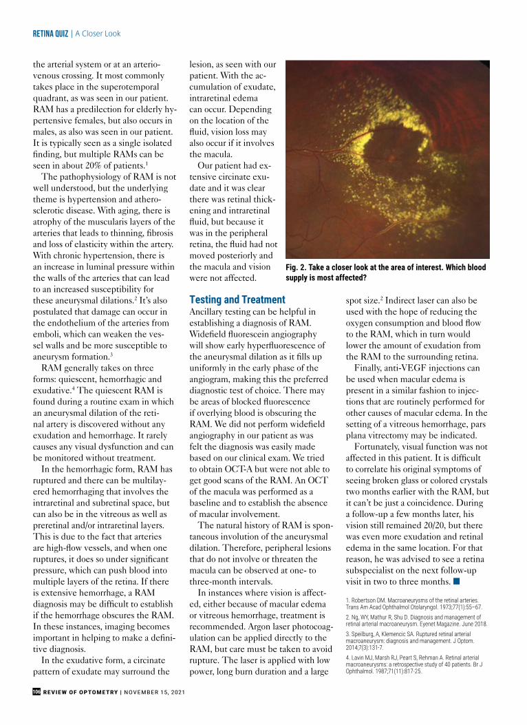
REVIEW OF OPTOMETRY | NOVEMBER 15, 2021106
the arterial system or at an arterio-venous crossing. It most commonlytakes place in the superotemporalquadrant, as was seen in our patient.RAM has a predilection for elderly hy-pertensive females, but also occurs inmales, as also was seen in our patient.It is typically seen as a single isolatedfinding, but multiple RAMs can beseen in about 20% of patients.1
The pathophysiology of RAM is notwell understood, but the underlyingtheme is hypertension and athero-sclerotic disease. With aging, there isatrophy of the muscularis layers of thearteries that leads to thinning, fibrosisand loss of elasticity within the artery.With chronic hypertension, there isan increase in luminal pressure withinthe walls of the arteries that can leadto an increased susceptibility forthese aneurysmal dilations.2 It’s alsopostulated that damage can occur inthe endothelium of the arteries fromemboli, which can weaken the ves-sel walls and be more susceptible toaneurysm formation.3
RAM generally takes on threeforms: quiescent, hemorrhagic andexudative.4 The quiescent RAM isfound during a routine exam in whichan aneurysmal dilation of the reti-nal artery is discovered without anyexudation and hemorrhage. It rarelycauses any visual dysfunction and canbe monitored without treatment.
In the hemorrhagic form, RAM hasruptured and there can be multilay-ered hemorrhaging that involves theintraretinal and subretinal space, butcan also be in the vitreous as well aspreretinal and/or intraretinal layers.This is due to the fact that arteriesare high-flow vessels, and when oneruptures, it does so under significantpressure, which can push blood intomultiple layers of the retina. If thereis extensive hemorrhage, a RAMdiagnosis may be difficult to establishif the hemorrhage obscures the RAM.In these instances, imaging becomesimportant in helping to make a defini-tive diagnosis.
In the exudative form, a circinatepattern of exudate may surround the
lesion, as seen with ourpatient. With the ac-cumulation of exudate,intraretinal edemacan occur. Dependingon the location of thefluid, vision loss mayalso occur if it involvesthe macula.
Our patient had ex-tensive circinate exu-date and it was clearthere was retinal thick-ening and intraretinalfluid, but because itwas in the peripheralretina, the fluid had notmoved posteriorly andthe macula and visionwere not affected.
Testing and TreatmentAncillary testing can be helpful inestablishing a diagnosis of RAM.Widefield fluorescein angiographywill show early hyperfluorescence ofthe aneurysmal dilation as it fills upuniformly in the early phase of theangiogram, making this the preferreddiagnostic test of choice. There maybe areas of blocked fluorescenceif overlying blood is obscuring theRAM. We did not perform widefieldangiography in our patient as wasfelt the diagnosis was easily madebased on our clinical exam. We triedto obtain OCT-A but were not able toget good scans of the RAM. An OCTof the macula was performed as abaseline and to establish the absenceof macular involvement.
The natural history of RAM is spon-taneous involution of the aneurysmaldilation. Therefore, peripheral lesionsthat do not involve or threaten themacula can be observed at one- tothree-month intervals.
In instances where vision is affect-ed, either because of macular edemaor vitreous hemorrhage, treatment isrecommended. Argon laser photocoag-ulation can be applied directly to theRAM, but care must be taken to avoidrupture. The laser is applied with lowpower, long burn duration and a large
spot size.2 Indirect laser can also beused with the hope of reducing theoxygen consumption and blood flowto the RAM, which in turn wouldlower the amount of exudation fromthe RAM to the surrounding retina.
Finally, anti-VEGF injections canbe used when macular edema ispresent in a similar fashion to injec-tions that are routinely performed forother causes of macular edema. In thesetting of a vitreous hemorrhage, parsplana vitrectomy may be indicated.
Fortunately, visual function was notaffected in this patient. It is difficultto correlate his original symptoms ofseeing broken glass or colored crystalstwo months earlier with the RAM, butit can’t be just a coincidence. Duringa follow-up a few months later, hisvision still remained 20/20, but therewas even more exudation and retinaledema in the same location. For thatreason, he was advised to see a retinasubspecialist on the next follow-upvisit in two to three months. ■
1. Robertson DM. Macroaneurysms of the retinal arteries.Trans Am Acad Ophthalmol Otolaryngol. 1973;77(1):55–67.2. Ng, WY, Mathur R, Shu D. Diagnosis and management ofretinal arterial macroaneurysm. Eyenet Magazine. June 2018.3. Speilburg, A, Klemencic SA. Ruptured retinal arterialmacroaneurysm: diagnosis and management. J Optom.2014;7(3):131-7.4. Lavin MJ, Marsh RJ, Peart S, Rehman A. Retinal arterialmacroaneurysms: a retrospective study of 40 patients. Br JOphthalmol. 1987;71(11):817-25.
Fig. 2. Take a closer look at the area of interest. Which bloodsupply is most affected?
RETINA QUIZ | A Closer Look
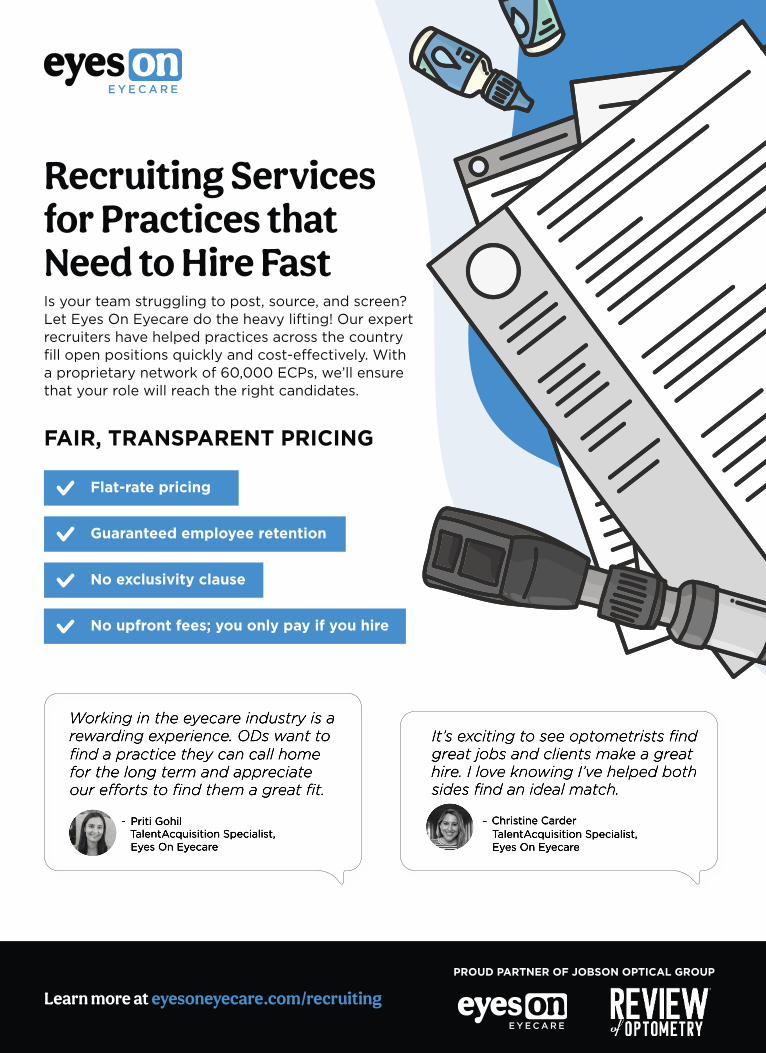
Recruiting Servicesfor Practices thatNeed to Hire FastIs your team struggling to post, source, and screen? Let Eyes On Eyecare do the heavy lifting! Our expert recruiters have helped practices across the country fill open positions quickly and cost-effectively. With a proprietary network of 60,000 ECPs, we’ll ensure that your role will reach the right candidates.
FAIR, TRANSPARENT PRICING
PROUD PARTNER OF JOBSON OPTICAL GROUP
Learn more at eyesoneyecare.com/recruiting
It’s exciting to see optometrists find great jobs and clients make a great hire. I love knowing I’ve helped both sides find an ideal match.
- Christine CarderTalentAcquisition Specialist,Eyes On Eyecare
Working in the eyecare industry is a rewarding experience. ODs want to find a practice they can call home for the long term and appreciate our efforts to find them a great fit.
- Priti GohilTalentAcquisition Specialist,Eyes On Eyecare
Flat-rate pricing
Guaranteed employee retention
No exclusivity clause
No upfront fees; you only pay if you hire

REVIEW OF OPTOMETRY | NOVEMBER 15, 2021108
CONTACT LENS fittingNew Contact Lens Offers Simpler, Safer InsertionA silicone hydrogel contact lens, recently launched fromMenicon, aims to improve the experience of lens han-dling. The Miru 1day UpSide is packaged with the con-vex front surface facing forward to eliminate contact withthe back surface, removing a potential source of infectionfrom fingertip exposure, a press release explains. In theblister pack, the lens rests on a small bump that allowsthe wearer to more easily grasp it with a pinching motion,a concept Menicon calls “Smart Touch.” The Miru 1dayFlat Pack uses a similar idea.
The lens is made of midafilcon A material with 56%water content, 91 Dk/t and a low modulus (0.36), whichthe company says givesit the softness of ahydrogel lens but thehandling properties ofSiHy. A plasma surfacetreatment (which thecompany calls “Nano-Gloss”) resists adhesionof debris and reducesfriction with the ocularsurface, Menicon says. Miru 1day UpSide is available nowin half-diopter steps from -13.00D to -6.00D and quarter-diopter steps from -6.00D to +6.00D. The company saysit will add a multifocal version in the first half of 2022.
ESP Adds Option to Fit Specialty LensesIf your practice has an Eye Surface Profiler (ESP) fromEaglet Eye, you may now have the option of fittingcertain scleral or ortho-K lenses for patients more easily,following the company’s recent addition of three specialtylens algorithms.
The additional scleral designs include the AccuLensMaxim 3D and BostonSight Scleral Smart360. The
ortho-K design (the first offeredfor the ESP) is for CooperVision’sParagon CRT. These three newdesigns bring the total number ofavailable algorithms on the ESPto 40 across 20 labs, according toa company press release. Eaglet
Eye says that the new algo-rithms will improve doctors’ability to fit complicated eyes,
helping to reach more patients inneed of specialty lenses.
diagnostic technologyUltra-widefield Lens for iCare Eidon CameraFundus photography does a great job of documentingretinal concerns and establishing a baseline status for futurecomparison, but most cameras prioritize the posterior poleat the expense of the periphery. To help practices that havean iCare Eidon fundus camera, an ultra-widefield lens wasjust added to the availableretinal imaging modalities.
By capturing 120˚ images ina single shot or up to 200˚ inpanoramic (“mosaic”) modeand outperforming a standardfield lens, this module can pro-vide critical diagnostic informa-tion about the retinal periph-ery, a press release explains.
The module features what the company calls “TrueColorConfocal Technology,” which allows for sharp image qualitycapable of detecting small details and signs of pathologiesfrom the center to the periphery of the retina, according tothe company. iCare says that the module can be retrofittedto most Eidon fundus imaging systems.
As some early signs of retinal problems can appear firston the periphery, using technology such as ultra-widefieldimaging to observe this region can help you detect potentialthreats to a patient’s eye health or vision, iCare says.
Three VR Vision Tests Added to Heru HeadsetFollowing the August launch of Heru’s virtual reality-based platform for visual field testing, the company justreleased a software update that includes three additionaltesting modalities: color vision, contrast sensitivity and darkadaptation. Plus, all the tests fall under CPT codes and arebillable to insurance, according to the company.
Two color vision tests are now offered to screen patients forcolor blindness—the Ishihara and the Farnsworth D-15—thesecond of which will begin automatically if patients fail thefirst, says Heru.
The new contrast sensitivity exam can be done in naturallighting, so you don’t have to bother dimming the room forevery patient, Heru said in a press release. This kind of testis particularly useful for screening or monitoring patients withage-related macular degeneration (AMD), Heru says, as ithelps track subtle and slow-changing effects of diminishingvision.
The third test added to the platform measures darkadaptation, which may also aid in early detection of AMD.
By having the option to screen every patient who walks
product review ONLINE FIRST:GET THE LATEST
PRODUCT NEWS ATwww.reviewofoptometry.com
New items to improve clinical care and strengthen your practice.

NOVEMBER 15 2021 | REVIEW OF OPTOMETRY 109
into your office for ocular diseases such as AMD, more casescan be detected and treated sooner, the company suggests.
LOW VISIONLighter and Sleeker Solution for Low VisionHeavy and cumbersome low vision headsets weighed downby smartphones impose a burden on patients that canundermine their motivation to use them regularly. A newlow vision wearable from IrisVision, called Inspire, allowsthe phone to remain in the wearer’s pocket rather thanhoused inside the headset, reducing its weight to half thatof the company’s previous model, a press release explains.
The device captures real-world images and remapsthem in real time to functional areas of the patient’sretina, the company says. Software allows for improvedvisualization during distance, intermediate and near tasksand offers autofocus and a 70° field of view with up to
14x magnification, according toIrisVision.The
company websitealso mentions that
Inspire comes withunlimited cellular dataconnectivity and includes a
hands-free voice control function to give commands suchas “reading mode,” “reduce brightness” or “zoom in.”The headset features a padded eye guard that can be usedfor full light blocking or removed for wearers who want asleeker profile.
The IrisVision Inspire may offer a more patient-friendlysolution to those with low vision, the company suggests.
Video Magnifier for Patients with Vision LossA new portable device aims to simplify the lives ofpeople with vision loss due to macular degeneration orother eye diseases by improving their ability to read,write and perform hands-free tasks, the manufacturersays. The Smartlux Digital featuresan HD camera, a five-inch display and avariety of settings toaccommodate individualuser preferences, a pressrelease explains.
Smartlux has four color-coded buttons below the screenthat control its main functions. One of these is a menubutton where users can adjust settings such as LEDbrightness, viewing modes (including 14 color contrastmodes), magnification type (step or continuous) andorientation of the underline. Built into the device is astand that allows it to be used in three different ways:fully retracted for hand-held use, fully extended forplacing on reading material and partially extended for
writing under the display. A refresh rate of 50 frames persecond provides a clear image even when the device is inmotion, according to the press release.
The video magnifier has 8GB of internal storage can beconnected to an external screen or computer for imageupload via an HDMI or USB cable. The company notesthat the Smartlux comes with a removable handle and abumper for drop protection, as well as a zippered case,charger, cleaning cloth and lanyard.
DRY EYE MANAGEMENTA New Route to ReliefApplying topical eye drops has long been a mainstay oftreating dry eye disease (DED), but patient noncompli-ance and the potential for ocular irritation can dampenenthusiasm for such a regimen. An alternative approach—nasal stimulation of the trigeminal parasympathetic path-way—has been tried before with mechanical devices andnow is possible with a pharmaceutical agent, vareniclinesolution 0.03mg(Tyrvaya) from newcompany OysterPoint Pharma, re-cently approved bythe FDA.
Tyrvaya, a cho-linergic agonist,triggers basal tearproduction, the com-pany explains. After four weeks of BID administration,about half of patients using the treatment demonstrateda 10mm or greater improvement in Schirmer’s scores,according to a company press release. Patient-reported as-sessment of symptoms on a 1-100 scale also improved, therelease says, as treatment reduced the mean score from 59to about 40, an effect superior to the control group. Themost common patient complaint was sneezing, reportedin 82% of study participants.
The aqueous spray will be packaged in a carton con-taining two multidose bottles, each with enough medi-cation for 15 days, and will be available by prescriptionbeginning in November, Oyster Point says.
Brush, Gel for at-home Lid CareYou likely instruct your patients with dry eye disease ormeibomian gland dysfunction to clean their eyelids regu-larly at home to help minimize discomfort and controlsymptoms. But, as always, compliance is an issue. A newbrush and cleanser system from Danelli Ocular Creationsaims to help more blepharitis and dry eye patients stayon top of their lid hygiene between office visits. Thecompany says that its first products, the MyboClean eye-lid brush and cleansing gel, are easy to use and suitablefor those with sensitive skin.

REVIEW OF OPTOMETRY | NOVEMBER 15, 2021110
CooperVision ............................................................................11...................................................... www.coopervision.com/toric
CooperVision ...........................................................................25.............................................................................www.coopervision.com
Eye Designs.............................................................................37...........................................................................................(800) 346-8890............................................................................... www.eyedesigns.com
Eyefinity....................................................................................5........................................................................................... (800) 269-3666....................................................................... www.eyefinity.com/cloud
Eyeris ......................................................................................33............................................................................................ (833) 439-3747..................................................wwww.myeyeris.com/doctor-sign-up
Florida Eye Specilaists.............................................................64.......................................................www.finaleyesce.com/registration
IDOC.........................................................................................35............................................................................................(203) 853-3333...............................................................................................www.idoc.net
IDOC................................................................................... Insert.................................................................optomery.idoc.net/learnmore
Janssen Pharmaceuticals, Inc. ..................................... Cover Tip............................................................................www.eyesongenes.com
Keeler Instruments ................................................................. 51........................................................................................... (800) 523-5620..................................................................................www.keelerusa.com
Lacrimedics, Inc......................................................................70............................................................................................(800) 367-8327.............................................................................. info@lacrimedics.com...............................................................................www.lacrimedics.com
Lacrivera.................................................................................73............................................................................................ (855) 857-0518....................................................................................www.lacrivera.com
Lombart : Advancing Eyecare .................................................. 15............................................................................................ (800) LOMBART.................................................................www.lombartinstrument.com
M&S Technologies....................................................................63........................................................................................... (847) 763-0500............................................................................www.mstech-eyes.com
MacuLogix............................................................................... 31................................................................... www.maculogix.com/ebook
Menicon America.....................................................................95..................................................................... www.meniconamerica.com
Novartis Pharmaceuticals .......................................... 18, 19 & 20.....................................................................................www.novartis.com
Oasis .............................................................................. 53 & 55...........................................................................................(800) 820-8940...................................................customerservice@oasismedical.com............................................................................www.oasismedical.com
Ocusoft ...................................................................................77........................................................................................... (800) 233-5469......................................................................................www.ocusoft.com
Optometric Architects.............................................................47............................................................................................ (319) 240-0222..................................................... [email protected]............................................................ www.optometricarchitects.com
Regener-Eyes..........................................................................69............................................................................................(877) 206-0706.............................................................................www.regenereyes.com
Regeneron Pharmaceuticals, Inc.............................. 58 , 59 & 60................................................................................. www.regeneron.com
Reichert Technologies.............................................................99.............................................................................www.reichert.com/ora
RVL Pharmaceuticals, Inc. ................................................... 7 & 8...................................................................................... www.Upneeq.com
Sun Pharma..................................................................... 27 & 28...................................................................................www.cequapro.com
Tarsus ............................................................................... Insert......................................................................www.tarsusrx.com
US Ophthalmic.........................................................................67..............................................................................................(888) 881-1122.......................................................................... info@usophthalmic.com...........................................................................www.usophthalmic.com
Vital Tears ...............................................................................79........................................................................................... (800) 360-9592......................................................................www.vitaltears.org/review
X-Cel Specialty ........................................................................23............................................................................................. (800) 241-9312...................................................xcelinfo@xcelspecialtycontacts.com..........................................www.xcelspecialtycontacts.com/remlens
X-Cel Specialty ........................................................................ 81............................................................................................. (800) 241-9312...................................................xcelinfo@xcelspecialtycontacts.com........................................................... www.xcelspecialtycontacts.com
The cleansing brush is made from silicone and featuresa contoured design, making it easier to gripand maneuver, Danelli suggests. The taperedbristles help remove lid margin scurf and lashcollarettes associatedwith blepharitis,a companypress releaseexplains. Thebrushes willcome in a package of four and are made to be replacedquarterly.
The cleansing gel features organic ingredients (e.g.,manuka honey, coconut oil, aloe vera, sea salt) withanti-inflammatory and moisturizing properties, the pressrelease states. The solution goes onto the brush as a gelbut forms into a foam once massaged onto the eyelid.Clinical trials for this new system are ongoing.
PRESCRIBING INFORMATIONSearch Over 2,000 Meds OnlineApple’s pitch for the iPod was “1,000 songs in your pocket,”a revolutionary idea at the time. Now, eyecare providers with
a smartphone can have 2,000 drugs in their pocket, thanksto a new website called EyeDock/Meds. The free resourceoffers detailed information on over 2,000 prescription andOTC FDA-approved ophthalmic drugs, including select oralmedications, a press release announced.
EyeDock/Meds features a search tool with filters forname, active ingredient, category, company, method ofaction, Rx/OTC, brand/generic, form (e.g., suspension, solu-tion, gel, ung) and preserved/non-preserved. There’s alsoan alphabetical category list whereyou can choose to view only certaingroups of medications (e.g., allergy,antibiotic, glaucoma). After find-ing the drug you want, the websiteprovides product details such asits active and inactive ingredients,packaging options, usage, dosageand administration, as well as anyrelevant disclaimers from the FDA.
The online database is designedfor easy navigation on both desktopsand mobile devices, making itaccessible from anywhere at anytime, according to its developers. g
PRODUCT REVIEW
This index is published as a convenience and not as partof the advertising contract. Every care will be taken toindex correctly. No allowance will be made for errors dueto spelling, incorrect page number, or failure to insert.
ADVERTISER INDEX

FightingBlindness.orgHelp accelerate our mission by donating at ECPs4Cures.org.
WE’RE SEEING AMAZING RESULTS.
AND SO ARE THEY.At the Foundation Fighting Blindness our mission is everybody’s vision.
Our work shines a light on the darkness of inherited retinal diseases (IRDs).
We’re the world’s leading organization searching for treatments and cures. We need your help to fuel the discovery of innovations that will illuminate the future
for so many. We have robust disease information, a national network of local chapters and support groups, local educational events, and our My Retina Tracker® Registry
to help keep your patients connected with clinical and research advancements.

REVIEW OF OPTOMETRY NOVEMBER 15, 2021
Review Classifi eds
Practice For Sale
Do you have Productsand Services for sale?
CONTACT US TODAYFOR CLASSIFIED ADVERTISING
Toll free: 888-498-1460E-mail: [email protected]
Do you haveProducts and
Services for sale?CLASSIFIED
ADVERTISING WORKS
• JOB OPENINGS• CME PROGRAMS
• PRODUCTS• AND MORE. . .
Contact us today forclassified advertising:
Toll free: 888-498-1460E-mail: [email protected]
Continuing Education
MEDICAL OPTOMETRISTSThe American Board of Certification in
Medical Optometry (ABCMO) is recognized atJoint Commission (JC) accredited medicalfacilities as issuing board certification in thespecialty of medical optometry and thoseABCMO certifies are eligible for credentialingat these facilities as specialists rather thangeneral optometry practitioners.^
The Joint Commission, the acceptednational Gold Standard, reviews and accreditsover 21,000 federal, state and local-charteredmedical facilities.
To Be Eligible for ABCMO board certification:
1. Complete an accredited residencyin medical optometry
2. Pass the national Advanced Competencein Medical Optometry Examination
3. Practice in a medical setting for aminimum of two years.#
www.abcmo.org
Visit www.abcmo.org to understand howJC accredited medical facilities credentialspecialists and why specialty certification canenhance the careers of optometrists whocomplete residencies in medical optometry.
For Application procedures seewww.abcmo.org
or contact [email protected]^ At this time, 127 JC accredited hospitals, clinics and teaching institutions
recognize ABCMO specialist certification.* www.jointcommission.org# Waived for two years after residency
creo
REVIEW OF OPTOMETRY NOVEMBER 15, 2021
Review Classifi eds
Targeting Optometrists?CLASSIFIED ADVERTISING WORKS
• JOB OPENINGS • CME PROGRAMS• PRODUCTS & SERVICES • AND MORE. . .
Contact us today for classified advertising:Toll free: 888-498-1460
E-mail: [email protected]
:
.
a)
Faculty
creo
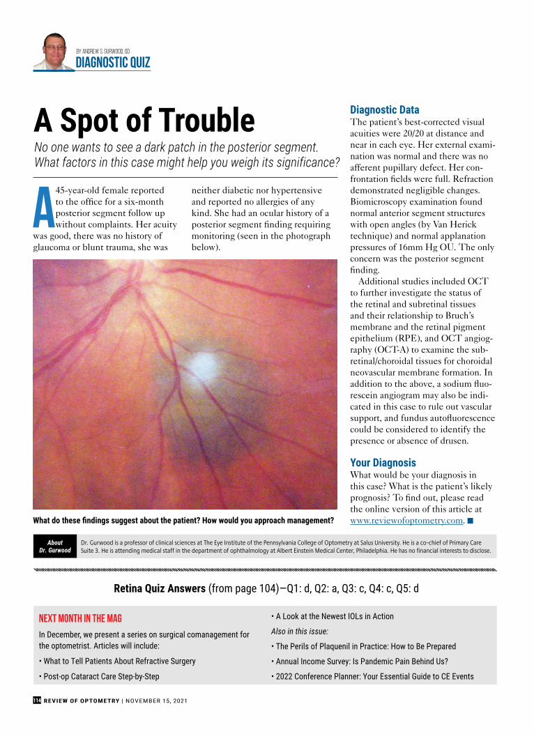
REVIEW OF OPTOMETRY | NOVEMBER 15, 2021114
A45-year-old female reportedto the office for a six-monthposterior segment follow upwithout complaints. Her acuity
was good, there was no history ofglaucoma or blunt trauma, she was
neither diabetic nor hypertensiveand reported no allergies of anykind. She had an ocular history of aposterior segment finding requiringmonitoring (seen in the photographbelow).
Diagnostic DataThe patient’s best-corrected visualacuities were 20/20 at distance andnear in each eye. Her external exami-nation was normal and there was noafferent pupillary defect. Her con-frontation fields were full. Refractiondemonstrated negligible changes.Biomicroscopy examination foundnormal anterior segment structureswith open angles (by Van Hericktechnique) and normal applanationpressures of 16mm Hg OU. The onlyconcern was the posterior segmentfinding.
Additional studies included OCTto further investigate the status ofthe retinal and subretinal tissuesand their relationship to Bruch’smembrane and the retinal pigmentepithelium (RPE), and OCT angiog-raphy (OCT-A) to examine the sub-retinal/choroidal tissues for choroidalneovascular membrane formation. Inaddition to the above, a sodium fluo-rescein angiogram may also be indi-cated in this case to rule out vascularsupport, and fundus autofluorescencecould be considered to identify thepresence or absence of drusen.
Your DiagnosisWhat would be your diagnosis inthis case? What is the patient’s likelyprognosis? To find out, please readthe online version of this article atwww.reviewofoptometry.com. g
No one wants to see a dark patch in the posterior segment.What factors in this case might help you weigh its significance?
A Spot of Trouble
By Andrew S. Gurwood, OD
diagnostic quiz
Next Month in the Mag
In December, we present a series on surgical comanagement forthe optometrist. Articles will include:
• What to Tell Patients About Refractive Surgery
• Post-op Cataract Care Step-by-Step
• A Look at the Newest IOLs in Action
Also in this issue:
• The Perils of Plaquenil in Practice: How to Be Prepared
• Annual Income Survey: Is Pandemic Pain Behind Us?
• 2022 Conference Planner: Your Essential Guide to CE Events
Dr. Gurwood is a professor of clinical sciences at The Eye Institute of the Pennsylvania College of Optometry at Salus University. He is a co-chief of Primary CareSuite 3. He is attending medical staff in the department of ophthalmology at Albert Einstein Medical Center, Philadelphia. He has no financial interests to disclose.
AboutDr. Gurwood
What do these findings suggest about the patient? How would you approach management?
Retina Quiz Answers (from page 104)—Q1: d, Q2: a, Q3: c, Q4: c, Q5: d
OFFER PATIENTS AN
EXCEPTIONAL LENS EXPERIENCEEye Care Professionals across the nation are sharing their success with
Bausch + Lomb INFUSE®. See the impact it can have on your practice and patients, too.
HERE’S A SNEAK PEEK AT WHAT YOUR PEERS ARE SAYING:
“INFUSE® helps address symptoms
of contact lens dryness.”
– Dr Kevin Nehaul
“The lens speaks for itself.”
– Dr Scott Moscow
“INFUSE® has great comfort, vision,
and oxygen transmissibility.”
– Dr Michelle Mumford
SCAN CODE TO WATCH PEER TESTIMONIALS
Bausch + Lomb INFUSE is a trademark of Bausch & Lomb Incorporated or its affiliates.
©2021 Bausch & Lomb Incorporated or its affiliates. INF.0207.USA.21

Our full-service, dedicated team is here to assist you and your staff with all your REFRESH® needs.
© 2021 AbbVie. All rights reserved. All trademarks are the property of their respective owners. REF144609 04/21
DISCOVER EXCLUSIVE ACCESS TO ALL THINGS REFRESH®.
Call us and see how REFRESH® Concierge can help you bring relief to your patients.
833-REF-SMPL 7:30 a.m.–7 p.m. CT, M–F
REFRESH® SAMPLES
PATIENT COUPONS
PRODUCT EDUCATION
REFRESH® DIRECT ECOMMERCE
OPEN YOUR EYES TO

Click to access the Scleral Lens
Education Initiative eResource
Everything from the history and basics of getting started with scleral lenses to all the
information and resources you need when working with them.
SCLERAL LENSES 2021
This eResource is brought to you by
THE SCLERAL LENS E D U C A T I O N I N I T I A T I V E


Everything from the history and basics of getting started with scleral lenses to all the
information and resources you need when working with them.
SCLERAL LENSES 2021
This eResource is brought to you by
THE SCLERAL LENS E D U C A T I O N I N I T I A T I V E