A Preformed Complex of Postsynaptic Proteins Is Involved in Excitatory Synapse Development
Upload
independentCategory
view
2download
0
Neuron 49, 547–562, February 16, 2006 ª2006 Elsevier Inc. DOI 10.1016/j.neuron.2006.01.015
A Preformed Complex of Postsynaptic ProteinsIs Involved in Excitatory Synapse Development
Kimberly Gerrow,1 Stefano Romorini,3
Shahin M. Nabi,1 Michael A. Colicos,2 Carlo Sala,3
and Alaa El-Husseini1,*1Department of Psychiatry and the
Brain Research CentreUniversity of British ColumbiaVancouver, British ColumbiaCanada, V6T 1Z32Department of Physiology & BiophysicsHotchkiss Brain InstituteUniversity of CalgaryAlberta T2N 4N1Canada3CNR Institute of NeuroscienceDepartment of PharmacologyUniversity of MilanoMilanoItaly
Summary
Nonsynaptic clusters of postsynaptic proteins have
been documented; however, their role remains elu-sive. We monitored the trafficking of several candidate
proteins implicated in synaptogenesis, when nonsy-naptic clusters of scaffold proteins are most abun-
dant. We find a protein complex consisting of two pop-ulations that differ in their content, mobility, and
involvement in synapse formation. One subpopulationis mobile and relies on actin transport for delivery to
nascent and existing synapses. These mobile clusterscontain the scaffolding proteins PSD-95, GKAP, and
Shank. A proportion of mobile clusters that exhibitsslow movement and travels short distances contains
neuroligin-1. The second group consists of stationarynonsynaptic scaffold complexes that mainly contain
neuroligin-1, can recruit synaptophysin-containingaxonal transport vesicles, and are readily transformed
to functional presynaptic contacts that recycle thevital dye FM 4-64. These results postulate a mechanism
whereby preformed scaffold protein complexes serve
as predetermined postsynaptic hotspots for establish-ment of new functional excitatory synapses.
Introduction
Communication between neuronal cells relies on signalstransmitted through an array of heterogeneous contactsknown as synapses (Li and Sheng, 2003; Sanes andLichtman, 2001; Waites et al., 2005). This process in-volves the establishment of thousands of excitatoryand inhibitory synapses, each with characteristic com-ponents that determine synapse specificity. Excitatoryneurotransmission in the mammalian brain is primarilymediated by glutamate, and the proteinaceous networkof neurotransmitter receptors, scaffolding proteins, ad-
*Correspondence: [email protected]
hesion molecules, and signal transduction enzymes atglutamatergic synapses is collectively referred to asthe postsynaptic density (PSD) (Kennedy, 1997). Scaf-folding proteins constitute one major group of proteinspresent at the PSD. Many of these molecules containmultiple domains for protein-protein interaction, suchas PDZ domains, and thus mediate the recruitment ofseveral other proteins to a single cellular site (Kim andSheng, 2004; Rao et al., 1998).
The postsynaptic density protein PSD-95 is believedto play a role in synapse maturation, as it is one of theearliest detectable proteins in the PSD (Kim and Sheng,2004; Rao et al., 1998). PSD-95 induces clustering ofa number of neurotransmitter receptors and scaffoldingproteins (El-Husseini et al., 2000b; Kim and Sheng, 2004;Prange et al., 2004). PSD-95 mutant animals exhibit de-fects in synaptic transmission associated with plasticityand result in enhanced LTP and impaired learning(Migaud et al., 1998). Moreover, knock down of PSD-95 diminishes excitatory synapse number and clusteringof AMPA-type glutamate receptors (Nakagawa et al.,2004; Prange et al., 2004). Interaction of the scaffoldingprotein, GKAP, with PSD-95 is thought to be importantfor the coupling of GKAP to Shank, a protein implicatedin the regulation of spine morphology (Sala et al., 2001).PSD-95 may control the balance of excitatory and inhib-itory synaptic contacts through the clustering of the celladhesion molecule neuroligin-1 (Levinson et al., 2005;Levinson and El-Husseini, 2005; Prange et al., 2004).These findings suggest that PSD-95 has the potentialto drive the formation of a core postsynaptic proteincomplex containing essential scaffolding and cell adhe-sion proteins needed to coordinate the formation andmaturation of pre- and postsynaptic elements, as wellas dictate their number and specificity.
Nonsynaptic clusters of postsynaptic proteins inyoung neurons have been documented in several stud-ies (Liu et al., 1998; Rao et al., 1998; Sans et al., 2003;Waites et al., 2005; Washbourne et al., 2002, 2004). Clus-ters containing PSD-95 and GKAP present at nonsynap-tic sites have been observed in hippocampal neurons invitro (Rao et al., 1998), and nonsynaptic membrane spe-cializations, or ‘‘free PSDs,’’ have been observed withthe use of EM analysis in vivo during the initial stagesof synaptogenesis (Blue and Parnavelas, 1983; Fialaet al., 1998; Hinds and Hinds, 1976; Steward and Falk,1986). Taken together, these findings postulate thatpostsynaptic mechanisms exist to facilitate synapse for-mation. The findings that postsynaptic molecules suchas neuroligins and SynCAM can induce presynaptic as-sembly further highlight the importance of postsynapticproteins in driving the formation of new contacts (Bie-derer et al., 2002; Scheiffele et al., 2000).
Recent advances in imaging techniques have re-vealed some of the molecular events underlying the for-mation of excitatory synapses in the CNS (Ahmari andSmith, 2002; Bresler et al., 2004; Friedman et al., 2000;Sanes and Lichtman, 2001; Shapira et al., 2003; Waiteset al., 2005; Ziv and Garner, 2004). These elegant studiesdemonstrated that the major components of the

Neuron548
synaptic vesicle release machinery travel in transportpackets and are rapidly recruited to contact sites. Otherstudies revealed transport packets and protein com-plexes that regulate delivery of NMDA receptors to na-scent neuronal contacts (Sans et al., 2003; Washbourneet al., 2002, 2004). The recruitment of NMDA receptorclusters slightly preceded that of synaptic vesicle pro-teins at newly formed contacts between axonal growthcones and dendrites (Washbourne et al., 2002). Live im-aging has also identified discrete and mobile PSD-95clusters (Marrs et al., 2001; Prange and Murphy, 2001).This suggested the existence of prefabricated PSD-95clusters potentially involved in the formation and/or sta-bilization of synapses. However, others reported thatpresynaptic differentiation precedes recruitment ofPSD-95 to newly formed synaptic sites and that PSD-95 accumulation occurs gradually (Bresler et al., 2004;Friedman et al., 2000; Waites et al., 2005).
Here we assessed the trafficking of several scaffold-ing proteins in young neurons, prior to the formation ofthe majority of synapses. We found that (1) stationarycomplexes containing PSD-95, GKAP, and Shank areabundant in young neurons at both synaptic and nonsy-naptic sites, (2) a small fraction of this scaffold complexis mobile and can be recruited to nascent and existingpresynaptic contacts, (3) a subset of stationary proteincomplex clusters contains neuroligin-1 and recruits syn-aptophysin-positive axonal transport vesicles that canrecycle the vital dye FM 4-64, (4) assembly of the scaf-fold protein complex requires PSD-95, as interferingwith PSD-95 expression by siRNA disrupts clusteringof GKAP and Shank and reduces the number of excit-atory synapses in young hippocampal neurons, and fi-nally, (5) knock down of PSD-95 results in an overallincrease in VGAT puncta positive for neuroligin-1, indi-cating a shift in neuroligin-1 localization from excitatoryto inhibitory contacts.
Results
PSD-95, GKAP, and Shank Colocalize at Nonsynaptic
Sites in Young NeuronsPrevious studies showed clusters of PSD-95 and GKAPat early stages of neuronal development and that manyof these clusters exist at nonsynaptic sites (Rao et al.,1998). We further evaluated the content and location ofthese clusters at different stages of neuronal develop-ment. At day in vitro (DIV) 7, PSD-95 colocalized indiscrete clusters with GKAP and Shank scaffolding pro-teins along the dendritic shaft (87.6% 6 2.8% of PSD-95and GKAP coclusters with Shank; Figure 1A). Similar co-localization of these proteins was observed at DIV14(86.6% 6 2.3% of PSD-95 and GKAP coclusters withShank; Figure 1B). Although most of these clusterswere synaptic at DIV14 (78.2% 6 3.7% of PSD-95 andShank coclusters with synaptophysin), only half of theclusters containing PSD-95, GKAP, and Shank weresynaptic at DIV7 (47.2% 6 5.2% of PSD-95 and Shankcoclusters with synaptophysin, p < 0.001, Figures 1Cand 1D). AMPA receptors, as assessed by GluR1 stain-ing, did not significantly colocalize with Shank at DIV7,and only half of the synaptic Shank clusters containthe glutamate receptor subunit GluR1 (51.5% 6 6.2%of Shank and synaptophysin coclusters with GluR1,
p < 0.001, Figures 1E and 1F), suggesting that most syn-apses formed at this early stage are silent (Bredt andNicoll, 2003). The tight correlation in localization ofPSD-95, GKAP, and Shank at nonsynaptic sites at a pe-riod that precedes the majority of synapse formationsuggests that these proteins form a preassembled com-plex that participates in excitatory synapse develop-ment.
Existence of Mobile and Stationary Nonsynaptic
Clusters Containing PSD-95, GKAP, and ShankTo visualize the formation and assembly of clusters ofPSD-95, GKAP, and Shank, we performed time-lapsemicroscopy of fluorescently tagged versions of theseproteins in DIV5–6 hippocampal neurons with the useof an environmentally controlled chamber, with imagestaken every 2–10 min for periods of up to 2 hr. To avoidany changes associated with protein overexpression,several criteria were followed to ensure that the levelsof fluorescently labeled molecules were similar to en-dogenous proteins (see the Supplemental ExperimentalProcedures and Figure S1 available online).
The majority of coclusters containing PSD-95 GFP,GKAP DsRED, and Shank CFP were stationary (77.9%6 2.2% of total coclusters). Noteworthy, clusters ofthese proteins were also mobile and moved perfectlyin concert along dendrites in both anterograde and ret-rograde directions. These characteristics were similarin neurons expressing PSD-95 GFP alone (Figure 2Aand Movie S1), coexpressed with GKAP DsRED (Fig-ure 2B), coexpressed with Shank CFP (Figure 2C andMovie S2), and Shank CFP alone (Figure 2F). The meanvelocity of mobile clusters in a specific direction was0.83 6 0.08 mm/min, over distances ranging from 2 to15 mm (Figure 2G). Velocity was not constant, with clus-ters often pausing briefly before continuing. This move-ment is consistent with previous reports of PSD-95alone at this age (Bresler et al., 2001, 2004; Friedmanet al., 2000; Shapira et al., 2003; Washbourne et al.,2002), albeit lower than those typically associated withvesicles propelled by molecular motors (0.1–10 mm/s)and faster than previous reports of PSD-95 alone inolder neurons (0.48 mm/min; Bresler et al., 2001). Fusionand splitting of coclusters of these proteins were ob-served along the dendrite, with modest, less than2 mm, lateral movement (12.7% 6 1.3% of total coclus-ters, Figure 2D and Movie S3). These data indicate thatdifferent pools of nonsynaptic scaffold complexes exist,which are distinguishable by differences in mobility. Thedetection of both stationary and mobile clusters con-taining PSD-95, GKAP, and Shank is intriguing and sug-gests that these proteins constitute a complex that canmove as a bonafide transport packet. Because the pro-tein clusters described here contain the major scaffold-ing proteins present at excitatory synapses, we will referto them as the preformed scaffold complex.
Stationary Nonsynaptic Preformed Scaffold
Complex Participates in Development of FunctionalPresynaptic Terminals
Analysis of DIV5 neurons showed that the number ofendogenous PSD-95 puncta was about 2-fold higherthan sites positive for the excitatory presynaptic markervesicular glutamate transporter-1 (VGLUT). Between

Postsynaptic Scaffold Complex at Nonsynaptic Sites549
Figure 1. Detection of Nonsynaptic Clusters
of Postsynaptic Scaffolding Proteins
Endogenous localization of synaptic proteins
at DIV7 (left panels) and DIV14 (right panels)
by immunostaining. (A and B) Scaffolding
molecules Shank (green), GKAP (red), and
PSD-95 (blue) are colocalized at DIV7
(87.6% 6 2.8%) and DIV14 (86.6% 6 2.3%).
(C and D) At DIV7, 47.2% 6 5.2% of Shank
(green) and PSD-95 (red) coclusters were ap-
posed by synaptophysin (SYN) (blue). At
DIV14, 78.2% 6 3.7% of PSD-95 and Shank
coclusters are apposed by SYN. (E and F) At
DIV7, only 51.5% 6 6.2% of synaptic sites
containing Shank (green) and SYN (blue) con-
tain the AMPA receptor subunit GluR1 (red).
GluR1 clusters are present at 82.4% 6 0.3%
of synaptic Shank sites at DIV14. Data repre-
sent the analysis of neurons from at least two
experiments, n = 11–15 neurons per group,
R800 clusters per group. Scale bar, 1mm.
***p < 0.001. Data represent mean 6 SEM.
DIV5 and DIV9, there was an increase in the total numberof colocalized VGLUT and PSD-95 puncta, without a sig-nificant increase in the total number of PSD-95 clusters(Figures S2A and S2B). This suggests that existing PSD-95 clusters may have been utilized for building new con-tacts positive for VGLUT within this time period. To ex-plore this possibility, time-lapse imaging of PSD-95GFP was performed in conjunction with successiveloading and unloading of the synaptic vital dye FM 4-64 (Friedman et al., 2000; Prange and Murphy, 1999;Shapira et al., 2003). Sites positive for FM 4-64 werealso labeled for VGLUT (83.6% 6 19% of total FM 4-64-positive sites), confirming the validity of using FM4-64 to label excitatory presynaptic terminals (Fig-ure S2C). When compared to their synaptic, FM 4-64-positive counterparts over a time course of 4 hr, therewas no statistically significant difference in relative fluo-rescence intensity of PSD-95 GFP: final GFP fluores-cence intensity was 90.2% 6 1.6% of initial fluorescenceintensity for nonsynaptic PSD-95 GFP and was 92.9% 64.1% of initial fluorescence intensity for synaptic PSD-95 GFP (n = 10 neurons per group, 378 puncta, p =0.6). The fluorescence stability of stationary PSD-95
GFP clusters that were negative for FM 4-64 suggeststhat they are not simply remnants of recently lost synap-ses, which have been shown to disassemble within 1 hr(Okabe et al., 2001). Within 45 min of imaging, a smallfraction of presynaptically naive sites opposed to stablePSD-95 GFP clusters became FM 4-64 positive (4.5% 61.4% of total PSD-95 puncta, 10.4% 6 3.7% of presyn-aptically naive PSD-95 puncta). This was observed in 6/12 of the neurons analyzed (Figures 3A and 3E). After 2 hrof imaging, this phenomenon was observed more fre-quently (11.3% 6 2.0% of total PSD-95 puncta, 18.6%6 3.4% of presynaptically naive PSD-95 puncta), in 13/14 neurons examined (Figures 3B and 3E). These datasuggest that preformed scaffold complexes can act assites for recruitment of active presynaptic machineryand that this process can occur in as little as 45 min.
Delivery of Mobile Synaptophysin Clustersto Contact Sites Apposed to Stationary
Preformed Scaffold ComplexAlthough FM dyes are reliable markers for labelingactive presynaptic terminals, this method does not read-ily allow for visualization of the delivery of presynaptic

Neuron550
Figure 2. Visualization of Mobile and Stationary Clusters Containing PSD-95, GKAP, and Shank
Time-lapse imaging of hippocampal neurons transfected at DIV5–6 with PSD-95 GFP alone, PSD-95 GFP and GKAP DsRED, or with PSD-95 YFP
and Shank CFP and analyzed 24–36 hr later. Open arrowheads point to mobile clusters of interest, and closed arrowheads demarcate their orig-
inal position. (A and B) A small fraction of clusters of (A) PSD-95 GFP alone or with (B) GKAP DsRED move in anterograde and retrograde direction
along the dendrites. (C) Mobile coclusters of PSD-95 YFP and Shank CFP were also observed. (D) Coclusters of PSD-95 GFP and GKAP DsRED
were observed to split and merge with other existing clusters. (E) A graph summarizes the percentage of mobile (10.5% 6 0.9%), splitting
(12.7% 6 1.3%), and stationary (77.9% 6 2.2%) coclusters containing PSD-95 GFP and GKAP DsRED (n = 13 neurons, 886 puncta). (F) Mean
velocity of mobile PSD-95 and GKAP coclusters was 0.83 6 0.08 mm/min and was not significantly different when PSD-95 was transfected alone
(0.75 6 0.03 mm/min, n = 21 neurons) or when cotransfected with Shank (0.74 6 0.08 mm/min, n = 10 neurons) or Shank alone (0.77 6 0.05 mm/min,
n = 9 neurons, p = 0.9). (G) Frequency of mean velocities of mobile clusters for PSD-95 GFP alone (black bars) and PSD-95 cotransfected with
GKAP (gray bars) show similar distribution. Scale bar, 10 mm. Data represent mean 6 SEM.
elements to these sites. To visualize axonal transportpacket recruitment at contact sites, we used synapto-physin fused to DsRED (SYN DsRED) introduced atDIV0 by electroporation in order to label a large propor-tion of presynaptic terminals and their axons, followedby transfection of PSD-95 GFP as described earlier. Us-ing fast interframe intervals (15 s), we found that mobile
SYN DsRED travels on average velocities of 0.47 mm/s(ranging from 0.14 to 1.3 mm/s; Figures S3A and S3C)and is similar to presynaptic vesicle markers such asVAMP (Ahmari et al., 2000). Frequent rests in the traffick-ing of SYN DsRED-positive transport packets averaged54 s (ranging from 15 s to 6.5 min; Figures S3B and S3Dand Movie S4). Recruitment of SYN DsRED-positive

Postsynaptic Scaffold Complex at Nonsynaptic Sites551
transport packets at specific sites for a minimum of15 min was counted as stable in order to distinguish itfrom these rest stops. No significant accumulation ofSYN DsRED from a diffuse pool was observed. Expres-sion of SYN DsRED by transfection with electroporationdid not result in a significant protein overexpression (p =0.3), increased cluster area (p = 0.6), nor enhanced PSD-95 clusters at sites of accumulation (p = 0.5; Figure S3).
Time-lapse imaging revealed that 36.3% 6 5.4% ofPSD-95 GFP clusters contacting SYN DsRED-positiveaxons showed stable PSD-95 GFP clusters apposedby stable accumulations of SYN DsRED, suggestingthat these are synaptic partners. Most contacts betweenPSD-95 GFP puncta contacting SYN DsRED-positiveaxons showed no axonal varicosities (bulges at least1.5 times greater than the average width of the axon)and no significant enrichment of SYN DsRED (increasesin fluorescence at least 1.5 times background). How-ever, a number of these contacts were seen to developinto axonal varicosities or recruit and stabilize mobileaxonal transport packets positive for SYN DsRED within1 hr (5.9% 6 1.7% of total PSD-95 GFP clusters [5/88],15.6% 6 1.4% of presynaptically naive PSD-95 GFPclusters [5/31]; Figures 3C, 3D, 3F, and 3G). Some ofthe newly recruited clusters of SYN DsRED showedrapid morphological rearrangements upon delivery ofSYN DsRED-positive transport packets to sites ap-posed to stationary PSD-95 clusters, suggesting thatthese changes are triggered by crosstalk between thesepre- and postsynaptic elements (Figure 3C, Figures S4Aand S4B, and Movies S5–S7). Longer time-lapse periods(2 hr) revealed that 32.5% 6 4.6% of presynaptically na-ive contacts positive for PSD-95 GFP (11/32) were ableto recruit SYN DsRED (Figure 3F and Figures S4A andS4B). These results indicate that stationary clusters ofpreformed postsynaptic scaffolds can recruit axonaltransport packets for initiation and/or stabilization ofnew sites of contact and suggests a paradigm wherebypreformed postsynaptic complexes may induce presyn-aptic differentiation.
Previous studies have indicated that differentiation ofthe presynaptic compartment occurs before recruit-ment of postsynaptic proteins (Bresler et al., 2001;Okabe et al., 2001; Waites et al., 2005). To further assesswhether both of these modes exist in DIV5–7 neurons,we monitored the accumulation of PSD-95 GFP at sitesinitially lacking PSD-95 but positive for SYN DsRED.Within 2 hr of imaging, 26.7% 6 3.6% of stable SYNDsRED clusters recruited PSD-95 GFP at sites initiallynaive for this postsynaptic protein (5/25; Figure 3G).Three of these events resulted from recruitment of dis-crete PSD-95 GFP clusters that were in the vicinity ofthe contact. In two other events, no discernable clusterswere recruited and may represent the accumulation ofa diffuse pool as previously described (Bresler et al.,2001; Okabe et al., 2001; Waites et al., 2005; Wash-bourne et al., 2002). Thus, both pre- and postsynapticmechanisms participate in the recruitment of synapticelements at sites of contact. In contrast with these find-ings, only 2.5% 6 1.4% of contacts that were initiallynegative for both PSD-95 GFP and SYN DsRED were ob-served to recruit and stabilize SYN DsRED within 2 hr (1/41; Figure 3G). Thus, a contact per se is not sufficient todrive synapse formation. These results emphasize the
importance of the preformed scaffold complex in in-creasing the probability of accumulation of presynapticelements.
Accumulation of pre- and postsynaptic proteins weremainly observed at existing contacts between trans-fected neurons; thus, it remains unclear how postsynap-tic clusters attract axons to these sites. We have ex-plored this by searching for events in which axonalgrowth cones contacted dendrites of transfected cells.We were able to capture a total of six growth cones con-tacting dendrites of cells transfected with PSD-95 GFP,and five of these events maintained contacts with exist-ing PSD-95 GFP clusters (Figure S5 and Movies S8–S10).
Stationary Nonsynaptic Preformed Scaffold
Complexes Contain Neuroligin-1To define signals that contribute to the stability of pre-formed scaffold complex, we analyzed the localizationof neuroligin-1, a binding partner of PSD-95 that is suffi-cient to induce presynaptic differentiation (Scheiffeleet al., 2000). In DIV7 neurons, 56.5% 6 0.5% of Shankclusters are positive for neuroligin-1 (Figures 4A and4B). However, only 28.1% 6 5.3% of these clusterswere associated with excitatory presynaptic terminals,as assessed by VGLUT staining (Figures 4C and 4D).Thus, neuroligin-1 can be associated with the preformedcomplex at synaptic and nonsynaptic sites (Figure S7).At DIV14, in contrast, neuroligin-1 clusters mainly existwith scaffold protein complexes (62.2% 6 6.5%) andsynaptophysin-positive sites (64.8% 6 4.7%).
To further dissect the pool of preformed protein com-plexes containing neuroligin-1, time-lapse imaging ofPSD-95 GFP and GKAP DsRED transfected neuronswas performed, followed by retrospective immunostain-ing for endogenous neuroligin-1. Neuroligin-1 clusterswere detected in most stationary PSD-95 and GKAPcoclusters (75.1% 6 3.1% of total stationary PSD-95clusters). A fraction of the mobile PSD-95/GKAP coclus-ters were found associated with neuroligin-1 puncta(33.3% 6 11.4% of mobile PSD-95 clusters; Figures 4Eand 4G), representing w3% of the total number ofPSD-95 clusters, and moved distances less than 2.5mm. To determine the relationship between neuroligin-1, stationary PSD-95 clusters, and active presynapticterminals, neurons expressing PSD-95 GFP were loadedand unloaded with FM 4-64, time-lapse imaging wasperformed over a 1–4 hr period, and neurons were fixedand stained for endogenous neuroligin-1 and synapto-physin. Neuroligin-1 was found associated with station-ary PSD-95 clusters, regardless of their apposition toactive presynaptic terminals (58.9% 6 8.6% of totalFM 4-64 and synaptophysin-positive clusters versus53.2% 6 9.0% of total negative clusters, p = 0.7; Figures4F and 4H).
Consistent with these findings, time-lapse imaging ofDIV6 neurons expressing PSD-95 YFP and neuroligin-1CFP showed that a large proportion of the PSD-95YFP clusters were stationary during the imaging period(88.7% 6 2.5% of total PSD-95 clusters, Figure 5A)and that most of these stationary PSD-95 YFP clusterscontained neuroligin-1 CFP (89.7% 6 2.5%; Figures5A–5C). Similar to retrospective staining for endogenousneuroligin-1, only a small fraction of the mobile pool of

Neuron552
Figure 3. Sites Apposed to Stationary Nonsynaptic Scaffold Clusters Are Readily Transformed to Active Presynaptic Terminals
(A and B) DIV5 hippocampal neurons were transfected with PSD-95 GFP and analyzed 24–36 hr post-transfection for changes in the number
of active presynaptic terminals by subsequent loading (left panels) and unloading (right panels) of FM 4-64. Examples of stationary FM 4-64-
negative PSD-95 GFP (closed arrowhead, load 1), which became FM 4-64 positive within 45 min (A and A0) and 2 hr (B and B0) of initial load
(load 2, n = 12, 14 neurons). (C and D) Sequential transfection of DsRED-tagged synaptophysin (SYN DsRED) at DIV0 and PSD-95 GFP at
DIV5 was used to visualize dynamic recruitment of postsynaptic and presynaptic proteins at DIV6. (C and D) Two examples of presynaptically
naive PSD-95 GFP clusters (closed arrowheads) are shown to recruit SYN DsRED clusters, which remained as stable apposed pairs for the
duration of the imaging period. (E) Quantitative analysis of sites positive for PSD-95 GFP clusters that become FM 4-64 positive within 45 min
and 2 hr, respectively (n = 12–14 cells per group, R500 puncta). (F and G) Graph summarizing (F) the percentage of PSD-95 GFP puncta, including
both those initially negative and positive for SYN DsRED clusters or (G) those only negative (presynaptically naive) for SYN DsRED clusters, that
recruit SYN DsRED transport packets within 1 hr (n = 20 neurons) or 2 hr (n = 11 neurons) of imaging. (H) Comparison of different modes of recruit-
ment of PSD-95 GFP and SYN DsRED occurred within the imaging period. Within 2 hr of imaging, 32.5% 6 4.6% of presynaptically naive contacts

Postsynaptic Scaffold Complex at Nonsynaptic Sites553
PSD-95 YFP clusters at this age contained neuroligin-1CFP (27.3% 6 3.9% of mobile PSD-95 clusters; Figures5B and 5C). Interestingly, the average velocity of the fewmobile PSD-95 YFP clusters positive for neuroligin-1CFP was significantly slower than those that were nega-tive for neuroligin-1 (0.48 6 0.06 versus 0.84 6 0.06 mm/min, respectively, p < 0.001; Figure 5D) and movedshorter distances (on average, 2.2 mm versus 6.0 mm, re-spectively; Figure 5E). Thus, with retrospective immu-nostaining and live time-lapse imaging, a large propor-tion of neuroligin-1 is found associated with stationaryPSD-95 clusters.
We next examined whether nonsynaptic clusters ofneuroligin-1 recruit presynaptic proteins. Indeed, sta-tionary clusters of neuroligin-1 CFP were observed to re-cruit axonal transport packets positive for SYN DsREDat sites of contact within 1 hr (16.6% 6 3.6% of total con-tacts [13/67 contacts], 47.7% 6 7.9% of SYN DsRED na-ive contacts [13/27contacts]; Figure 5F and Movies S11and S12). Retrospective immunostaining revealed that80% (16/20 events) of stationary clusters of neuroligin-1 CFP that recruited SYN DsRED contain endogenousPSD-95 (Figure 5G). These data suggest that associa-tion of neuroligin-1 with the preformed scaffold complexmay be one of the adhesion molecules that facilitate re-cruitment of the presynaptic release machinery.
Mobility of Preformed Scaffold Clusters Is ActinDependent
If stationary preformed scaffold clusters act as signalsfor the recruitment of active presynaptic terminals,what is the role of the mobile clusters? Transport ofthese preassembled complexes is slower than reportedvalues for known vesicular transport. As such, move-ment of PSD-95 and GKAP clusters appears to involvea novel mechanism of transport. To determine the cyto-skeletal elements required for the transport of mobilePSD-95 clusters, time-lapse imaging of transfected neu-rons was performed in the presence of 4 mM cytochala-sin B or 3 mM nocodozole, pharmacological agentsknown to disrupt actin- and microtubule-based trans-port, respectively, through filament depolymerization(Sabo and McAllister, 2003). The relative velocities ofmobile clusters were assessed for 30 min before and30 min to 1 hr after drug treatment. Movement of PSD-95 GFP clusters was abolished upon treatment with cy-tochalasin B (0.75 6 0.06 mm/min before versus 0.10 60.07 mm/min during cytochalasin B treatment, p <0.001; Figure 6B), whereas no significant effect wasseen with nocodozole (0.78 6 0.03 mm/min before ver-sus 0.69 6 0.05 mm/min after nocodozole treatment,p = 0.14; Figures 6A and 6C). These experiments sug-gest that the mobility of preformed scaffold complexesinvolves an actin-based trafficking.
Mobile Preformed Scaffold Clusters Are Recruited
to Nascent and Existing Postsynaptic SitesDo mobile clusters contribute to synapse formation orremodeling? Simultaneous time-lapse imaging of PSD-95 YFP, GKAP CFP, and FM 4-64 revealed that presyn-
aptic terminals labeled with FM 4-64 recruited coclus-ters of PSD-95 YFP and GKAP CFP to nascent and exist-ing postsynaptic sites. Recruitment of PSD-95 YFP andGKAP CFP coclusters to existing puncta of postsynapticproteins occurred more frequently (1.8% 6 0.6% of totalPSD-95 puncta, 30.1% 6 13.0% of mobile PSD-95puncta) and was observed in 6/10 neurons during the1 hr time-lapse period (Figures 7A and 7C). Recruitmentof PSD-95 YFP and GKAP CFP coclusters to nascentpresynaptic terminals was a more rare event (0.6% 60.3% of total PSD-95 puncta, 10.0% 6 5.1% of mobilePSD-95 puncta), observed in 4/10 of neurons (Figures7B and 7C). These data demonstrate that recruitmentof preformed scaffold complexes to both existing andnascent presynaptic terminals can occur through thetransport of discrete clusters. Further analysis of FM 4-64-positive versus FM 4-64-negative coclusters re-vealed differences in their behavior. A significantly largerfraction of nonsynaptic clusters was mobile during theimaging period (2.3% 6 1.2% of total FM 4-64-positivePSD-95 and GKAP coclusters versus 17.6% 6 3.0% oftotal FM 4-64-negative PSD-95 and GKAP coclusters,p < 0.001). Together these data demonstrate that mobilepreformed clusters are more likely to be nonsynapticand can be utilized as transport packets for the forma-tion of a PSD at nascent and existing synapses.
Knock Down of PSD-95 Results in a Decreasein GKAP, Shank, and VGLUT Clusters, but an
Increase in VGAT Clusters Positive for Neuroligin-1To determine whether PSD-95 is central to the nucle-ation of the preformed scaffold complexes, siRNA thathas previously been shown to knock down PSD-95 ex-pression was transfected at DIV2, and the effects wereassessed at DIV7–8 (Nakagawa et al., 2004). A signifi-cant decrease in the number of PSD-95 clusters was ob-served, from 8.2 6 0.6 puncta/mm in neurons transfectedwith scrambled siRNA, to 2.2 6 0.3 puncta/mm in neu-rons transfected with PSD-95 siRNA (Figure 8). Knockdown of PSD-95 also resulted in a significant reductionin the number of GKAP (4.3 6 0.4) and Shank (3.1 60.4) clusters when compared to neurons transfectedwith scrambled siRNA (7.2 6 0.5 and 8.4 6 0.6 puncta/10 mm, respectively; Figures 8A, 8B, and 8E). We next ex-amined whether a reduction in the number of clusters ofPSD-95, GKAP, and Shank is associated with a changein the number of the excitatory presynaptic markerVGLUT. Indeed, a significant decrease in the numberof VGLUT-positive puncta contacting neurons trans-fected with PSD-95 siRNA was observed (1.1 6 0.2puncta/10 mm) versus scrambled siRNA (2.6 6 0.5puncta/10 mm; Figures 8C and 8G).
In contrast with these findings, the total number ofneuroligin-1 clusters was not significantly altered in neu-rons transfected with either scrambled (9.0 6 0.5puncta/10 mm) or PSD-95 siRNA (10.3 6 0.6 puncta/10mm). Consistent with this, the number of neuroligin-1clusters positive for Shank decreased in PSD-95 siRNAtransfected neurons (Figure 8F). This paralleled with a169% 6 19.1% increase in the number of VGAT-positive
positive for PSD-95 GFP recruited SYN DsRED, whereas 26.7% 6 3.6% of stable SYN DsRED clusters recruited PSD-95 GFP at sites initially naive
for this postsynaptic protein. Only 2.5% 6 1.4% of contacts initially negative for both PSD-95 GFP and SYN DsRED were observed to recruit and
stabilize SYN DsRED (n = 11). Scale bar, 1 mm. Data represent mean 6 SEM.

Neuron554
Figure 4. Neuroligin-1 Is Associated with Synaptic and Nonsynaptic Preformed Scaffold Clusters
Representative images of the localization of endogenous neuroligin-1 (NLG1) with respect to postsynaptic and presynaptic proteins as assessed
by immunostaining at DIV7 (left panels) and DIV14 (right panels). (A–D) Localization of NLG1 and Shank clusters with (A and B) GKAP and (C and
D) VGLUT at DIV7 and DIV14 (n = 11 neurons at DIV7; 12 neurons at DIV14; R1000 clusters). (E and F) Time-lapse imaging of PSD-95 GFP alone (F)
and with GKAP DsRED (E) was performed, followed by retrospective immunostaining for endogenous NLG1 (blue). (E) Open arrowhead high-
lights a mobile cluster positive for PSD-95 GFP and GKAP DsRED that is negative for endogenous NLG1. (F) Clusters of PSD-95 GFP negative
for both FM 4-64 and endogenous synaptophysin (SYN) contain NLG1. PSD-95 GFP-expressing neurons were first loaded and then unloaded
with FM4-64 at the beginning of the imaging period (0 min). Neurons were imaged for 1 hr, followed by a second round of loading and unloading
of FM4-64. Cells were then fixed and stained for SYN. Closed arrowheads demarcate stationary nonsynaptic PSD-95 GFP clusters positive for
NLG1. (G) Endogenous NLG1 is present at 75.1% 6 3.1% of stationary PSD-95 GFP and GKAP DsRED coclusters. In contrast, 33.3% 6 11.4% of
mobile PSD-95 GFP and GKAP DsRED coclusters contain NLG1 (n = 12 neurons; 321 puncta). (H) Percentage of PSD-95 GFP clusters positive for
NLG1 at sites positive (Synaptic) or negative (Nonsynaptic) for both FM 4-64 and SYN (n = 12 neurons, 299 puncta, p = 0.7). Scale bar, 1 mm (A and
C), 5 mm (E and F). ***p < 0.001. Data represent mean 6 SEM.
puncta in PSD-95 siRNA transfected cells (44.6 6 4.4puncta/neuron versus 26.4 6 2.7 in scrambled siRNAcontrol neurons; Figure 8H). This suggests that loss ofthe preformed scaffold complexes may have resultedin mislocalization of neuroligin-1, potentially to inhibitory
contacts. To address this, we first analyzed the propor-tion of VGAT-positive contacts that contain neuroligin-1in DIV7 neurons and found that 27.9% 6 6.2% of VGAT-positive sites are apposed by neuroligin-1. Next, we con-trasted the percentage of neuroligin-1 puncta present at
Postsynaptic Scaffold Complex at Nonsynaptic Sites555
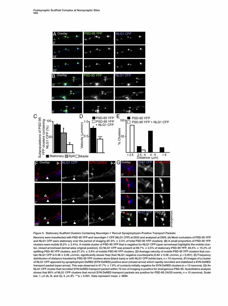
Figure 5. Stationary Scaffold Clusters Containing Neuroligin-1 Recruit Synaptophysin-Positive Transport Packets
Neurons were transfected with PSD-95 YFP and neuroligin-1 CFP (NLG1 CFP) at DIV5 and analyzed at DIV6. (A) Most coclusters of PSD-95 YFP
and NLG1 CFP were stationary over the period of imaging (87.8% 6 2.5% of total PSD-95 YFP clusters). (B) A small proportion of PSD-95 YFP
clusters were mobile (9.2% 6 2.4%). A mobile cluster of PSD-95 YFP that is negative for NLG1 CFP (open arrowhead highlights the mobile clus-
ter, closed arrowhead demarcates original position). (C) NLG1 CFP was present at 89.7% 6 2.5% of stationary PSD-95 YFP, 63.5% 6 15.2% of
splitting PSD-95 YFP clusters, and 27.3% 6 3.9% of mobile PSD-95 YFP clusters. (D) Average velocity of mobile PSD-95 YFP clusters that con-
tain NLG1 CFP is 0.48 6 0.06 mm/min, significantly slower than their NLG1-negative counterparts (0.84 6 0.06 mm/min, p < 0.001). (E) Frequency
distribution of distance traveled by PSD-95 YFP clusters alone (black bars) or with NLG1 CFP (white bars, n = 10 neurons). (F) Images of a cluster
of NLG1 CFP apposed by synaptophysin DsRED (SYN DsRED) positive axon (closed arrow) which rapidly recruited and stabilized a SYN DsRED
transport packet (open arrow). This was observed in 47.7% 6 7.9% of contacts initially negative for SYN DsRED clusters (n = 13 neurons). (G) An
NLG1 CFP cluster that recruited SYN DsRED transport packet within 75 min of imaging is positive for endogenous PSD-95. Quantitative analysis
shows that 80% of NLG1 CFP clusters that recruit SYN DsRED transport packets are positive for PSD-95 (16/20 events, n = 15 neurons). Scale
bar, 1 mm (A, B, and G), 5 mm (F). ***p < 0.001. Data represent mean 6 SEM.

Neuron556
Figure 6. PSD-95 Movement Is Actin Depen-
dent
Time-lapse imaging of PSD-95 GFP was per-
formed in the presence of nocodozole or cy-
tochalasin B to assess changes in velocity
of PSD-95 GFP clusters. (A) Addition of 3
mM nocodozole (+NOCOD) did not signifi-
cantly alter mobility of PSD-95 GFP clusters
(open arrowhead highlights a mobile cluster,
closed arrowhead demarcates initial posi-
tion). (B) Addition of 4 mM cytochalasin B
(+CYTOB) immobilized PSD-95 GFP clusters
(open arrowhead). (C) Average velocity be-
fore nocodozole treatment was 0.78 6 0.03
mm/min and 0.69 6 0.05 mm/min during treat-
ment (p = 0.14, n = 10 neurons). Average ve-
locity before cytochalasin B treatment was
0.75 6 0.06 mm/min and 0.10 6 0.07 mm/min
during treatment (p < 0.001, n = 9 neurons).
Scale bar, 5 mm. ***p < 0.001. Data represent
mean 6 SEM.
VGAT-positive sites in PSD-95 siRNA transfected neu-rons versus controls. We found that the proportion ofneuroligin-1 puncta positive for VGAT increased by150% 6 14.5% in PSD-95 siRNA transfected cells(Figure 8I). These results indicate that preformed scaf-fold complexes may serve a role in maintaining the bal-ance between newly formed excitatory and inhibitorysynapses.
Discussion
Recent studies have shown that synapse formation in-volves rapid delivery of transport packets containingpresynaptic proteins to new sites of contact (Ahmariand Smith, 2002; Craig and Boudin, 2001; Garner et al.,2002; Li and Sheng, 2003; Sanes and Lichtman, 2001;Sans et al., 2003; Waites et al., 2005). Others have docu-mented nonsynaptic postsynaptic protein complexes(Blue and Parnavelas, 1983; Fiala et al., 1998; Raoet al., 1998; Sans et al., 2003; Washbourne et al.,2002). Here we show that mobile and stationary pre-formed protein complexes containing PSD-95, GKAP,and Shank can participate in excitatory synapse devel-
opment via recruitment of presynaptic transportpackets positive for synaptophysin. A significant pro-portion of these stationary nonsynaptic clusters containneuroligin-1 and are readily transformed into FM 4-64-positive axonal terminals, suggesting that the pre-formed postsynaptic protein complexes assist in trans-formation of these sites to active presynaptic contacts.These results postulate a mechanism whereby station-ary preformed clusters of postsynaptic proteins prede-termine the sites at which excitatory synapses areformed.
The presence of PSDs without corresponding func-tional presynaptic terminals have been observed byEM in vivo (Blue and Parnavelas, 1983; Fiala et al.,1998; Hinds and Hinds, 1976; Steward and Falk, 1986);however, it was unknown whether these ‘‘free PSDs’’could eventually recruit presynaptic release machinery.Our analysis shows that postsynaptic differentiation canoccur prior to formation of a functional presynaptic ac-tive zone in young hippocampal neurons. This is consis-tent with previous findings that demonstrated the exis-tence of postsynaptic protein complexes containingNMDA receptors (Sans et al., 2003; Washbourne et al.,

Postsynaptic Scaffold Complex at Nonsynaptic Sites557
Figure 7. Mobile Preformed Scaffold Clusters Are Recruited to Nascent and Existing Postsynaptic Sites
Time-lapse imaging of PSD-95 YFP and GKAP CFP in conjunction with FM 4-64. (A) A mobile cocluster of PSD-95 YFP and GKAP CFP (open
arrowhead highlights a mobile cluster, closed arrowhead demarcates initial position) recruited to a site positive for FM 4-64 and PSD-95
YFP/GKAP CFP (yellow arrowhead). (B) An example of a mobile cluster containing PSD-95 YFP and GKAP CFP (open arrowhead highlights a mo-
bile cluster, closed arrowhead demarcates initial position) recruited to a site positive for FM 4-64 but negative for PSD-95 YFP/GKAP CFP (yellow
arrowhead). (C) Quantitative analysis of mobile clusters expressed as percentage of total PSD-95 YFP and GKAP CFP coclusters (left panel) or as
percentage of mobile clusters alone (right panel) recruited to FM4-64-positive sites that are already positive for PSD-95 YFP and GKAP coclus-
ters (Existing) or negative (Nascent). (D) Graph of the various pools of PSD-95 and GKAP coclusters (mobile, split, and stationary) and their re-
cruitment to FM 4-64-positive and -negative sites. n = 10 neurons, 448 puncta. Scale bar, 1 mm. ***p < 0.001. Data represent mean 6 SEM.
2002, 2004). Live imaging of GFP-tagged NMDA recep-tor subunit NR1 in young neurons also revealed a modu-lar transport of NR1 and SAP-102 along microtubulesand that NR1 recruitment at new contacts slightly pre-cedes or follow the recruitment of presynaptic proteins(Washbourne et al., 2002, 2004). These NR1 transport
packets were lacking PSD-95, indicating that deliveryof NMDA receptor subunits to synaptic sites involvesa mechanism independent of recruitment of the pre-formed scaffold complexes examined in this study.The availability of a readily accessible pool of preformedscaffold complexes could facilitate synapse formation

Neuron558
Figure 8. Knock Down of PSD-95 Reduces Clustering of GKAP, Shank, and VGLUT
Neurons transfected with PSD-95 siRNA (right panels) or scrambled siRNA (left panels) at DIV2 and stained at DIV7–8 for (A) GKAP and PSD-95,
(B) Shank and PSD-95, (C) Shank and VGLUT, or (D) NLG1 and VGAT. (E) A significant reduction in the number of puncta/10 mm of (E) PSD-95 (n =
15), GKAP (n = 11), and Shank (n = 13). (F) Reduced number of coclusters positive for NLG1 and Shank in PSD-95 siRNA transfected cells (n = 30).
(G and H) An increase in the number of (G) VGLUT, but a decrease in the number of (H) VGAT-positive contacts apposed to neurons transfected
with PSD-95 siRNA. (I) Number of VGAT and NLG1 coclusters increased in PSD-95 siRNA transfected cells (n = 10 neurons). Scale bar, 5 mm. *p <
0.05, **p < 0.01, *** p < 0.001. (J) A model summarizes the role of stationary and mobile preformed scaffold clusters in excitatory synapse de-
velopment. Stationary (orange) and mobile (yellow) preformed scaffolding clusters are abundant early in development, with a few mature post-
synaptic densities (brown) apposed to functional presynaptic terminals. Stationary preformed scaffold clusters are NLG1 positive and can attract
immature presynaptic terminals. NLG1 can drive the recruitment of synaptophysin transport packets. Mobile preformed clusters lack NLG1 and
are transported to stationary preformed scaffolding clusters or mature postsynaptic sites. Splitting and mobile clusters can act as a reservoir that
generates new stationary clusters. These in turn serve as new hot spots for the development of excitatory synapses. This model postulates
a postsynaptic mechanism whereby preassembled nonsynaptic clusters containing scaffolding proteins and cell adhesion molecules dictate
the location and number of newly formed excitatory contacts. Data represent mean 6 SEM.

Postsynaptic Scaffold Complex at Nonsynaptic Sites559
at this early stage of development. However, the in-volvement of preformed postsynaptic complexes in-volved in synapse formation has been disputed byothers who reported that presynaptic differentiationprecedes the recruitment of postsynaptic proteinswhich occurs gradually (Bresler et al., 2001, 2004). Thesestudies were performed in relatively older neurons andmay have therefore failed to observe some of the eventsthat occur in younger neurons. A shift in mechanismsused for synapse assembly at different developmentalstages may explain these discrepancies.
We were able to capture both pre- and postsynapticmechanisms of recruitment of synaptic proteins to na-scent contacts in DIV5–7 neurons. Analysis of synapto-physin DsRED-positive axons contacting dendrites ofcells transfected with PSD-95 GFP showed that w33%of contacts that were positive for the postsynaptic scaf-fold clusters but initially naive for presynaptic elementsrecruited synaptophysin within 2 hr. Moreover, w27%of contacts initially naive for PSD-95 clusters recruitedPSD-95 GFP. In contrast with these observations, onlyw2.5% of sites lacking both PSD-95 GFP and synapto-physin clusters resulted in the accumulation of presyn-aptic elements within 2 hr, indicating that a contact be-tween two neurons per se is not sufficient to drivesynapse formation. This further highlights the impor-tance of the preformed scaffold complexes in triggeringthe accumulation of synaptic elements at these sites.
Most of the new synapses that we observed in ouranalysis were en passant, and we frequently observedchanges in axon morphology accompanied by the accu-mulation of synaptophysin at sites contacting PSD-95clusters. Events in which axonal growth cones madea contact with PSD-95 clusters were rare; however,most events in which axonal growth cones contactedPSD-95 clusters resulted in the formation of a stablecontact. It remains unclear why axonal growth coneswere initially attracted to the postsynaptic clusters. Se-creted factors associated with the postsynaptic scaffoldmay attract extending axons; however, there is no evi-dence in the current study to support this possibility. Itis possible that during their growth, extending axonsrandomly contact multiple sites; however, once they en-counter a postsynaptic protein complex containing theappropriate adhesion molecules, they can be rapidlystabilized. The presence of neuroligin-1 in the preformedcomplex may facilitate initial contact stabilization; how-ever, our data do not exclude the involvement of othercell adhesion molecules in this process.
There are inherent advantages to both the presynapticand postsynaptic modes of synapse formation forproper connectivity of a neuronal circuit. In early devel-opment, guidance cues drive axons into their respectivetarget fields and help them to contact the correct neu-ron, and thus presynaptic mechanisms may ensure re-cruitment of the appropriate scaffold and receptorsthat matches the neurotransmitter present in axonal ter-minals. Conversely, a dendrite primed with the appropri-ate postsynaptic complex may serve to determine thenumber of sites to be stabilized or eliminated upon en-countering an axon en passant, and this may dictatethe number and type of synapses that a neuron receives.Considering that the preformed pool is scarce in olderneurons, this postsynaptic mechanism may not signifi-
cantly contribute to synapse formation in more matureneurons and must rely on synapse addition by presyn-aptic induction on a point-by-point basis. Experimentsstudying the adhesion complex formed by neuroliginsand neurexins suggest that both presynaptic and post-synaptic mechanisms stimulate synapse assembly:when expressed in non-neuronal cells, b-neurexin is suf-ficient to drive the recruitment of postsynaptic proteins,and conversely, neuroligins are sufficient to drive the re-cruitment of the presynaptic release machinery (Levin-son and El-Husseini, 2005). Studies suggest that scaf-folding proteins play an important role in dictating thebehavior of cell adhesion molecules in synapse develop-ment. For instance, PSD-95 enhances neuroligin-1 clus-tering and maturation of excitatory synapses at the ex-pense of inhibitory contacts (Prange et al., 2004). Thus,factors that govern the early assembly of these proteinsat nonsynaptic sites may be critical for controlling thenumber of newly formed synapses.
We analyzed a subset of postsynaptic proteins pres-ent at nonsynaptic clusters; however, it is possible thatthis complex may contain many other scaffolding pro-teins and adhesion molecules. The association ofPSD-95 and GKAP in young hippocampal neurons innonsynaptic clusters has been previously observed(Rao et al., 1998); however, the finding that Shank isalso present is intriguing. Shank is a large protein withmultiple motifs for protein-protein interaction. Hence,a preformed complex containing PSD-95, GKAP, andShank may be able to recruit many proteins requiredfor excitatory synapse maturation. This hypothesis issupported by the fact that Shank and GKAP form aggre-somes that are degraded via the proteosomal pathwayin the absence of PSD-95 (Romorini et al., 2004). Shankis functionally involved in the morphogenesis of spinesand requires the interaction of Shank with Homer, a pro-tein that binds metabotropic glutamate receptors, andinositol 1,4,5-triphosphate receptors (Sala et al., 2001;Tu et al., 1999). The synapses of young hippocampalneurons observed in this study are predominantly onthe shafts of dendrites. Thus, the presence of Shank inthe preformed postsynaptic complex suggests the in-volvement of shaft synapses in the later developmentof spines.
Although less numerous than their stable counter-parts, mobile nonsynaptic clusters of PSD-95, GKAP,and Shank were observed. Previous studies have shownsome movement of PSD-95 clusters, including splitting,lateral movement in shafts, and movement into and outof spines and filopodia (Marrs et al., 2001; Prange andMurphy, 2001). These studies did not address the rela-tionship of these moving clusters with respect to otherscaffolding proteins, adhesion molecules, or active pre-synaptic terminals. In this study, we found that mobileclusters of multiple scaffolding proteins are recruitedto nascent and existing synapses. The existence of largepreformed clusters of postsynaptic proteins that fre-quently split suggests that these clusters may serve asa reservoir for rapid delivery of preassembled com-plexes to nascent and established sites. Most intriguingis the finding that the majority of mobile PSD-95 clusterscontaining neuroligin-1 significantly moved slower andfor distances less than 2.5 mm. The association of neuro-ligin-1 with a small pool of mobile clusters that exhibit

Neuron560
slow movement and travel for short distances suggeststhat neuroligin-1 and PSD-95 may initially traffic sepa-rately, but once they merge, neuroligin-1 restricts themobility of the preformed complexes and promotes theirdocking at specific hot spots to prime them to associatewith newly encountered axons arriving from excitatoryneurons. Our analysis cannot rule out the possibilitythat individual neuroligin molecules rather than clustersassociate with the preformed scaffold complex duringnew contact formation, mainly because of the methodsused here, which only visualized clusters of these pro-teins. Future experiments that can track the movementof individual neuroligin molecules will be important toclarify this issue.
Signals that dictate the splitting, transport, and dock-ing of preformed clusters at specific subcellular loca-tions remain unclear. EM studies showed associationof PSD-95 in vesiculotubular structures that resembleendosomes (El-Husseini et al., 2000a). Other studies re-vealed a fraction of PSD-95 is associated with yet un-identified intracellular membranes (Bresler et al., 2001).Thus, mobile clusters may represent a form of endoso-mal structures, but with a speed distinct from that previ-ously reported. Our analysis suggests that the mobilityof preformed postsynaptic clusters is actin dependent.Actin-based trafficking in hippocampal neurons hasbeen shown to be carried out by the myosin family ofmotor proteins, and several members are expressed inthe dendrites of hippocampal neurons (Kim and Sheng,2004). Myosin 5a is a likely candidate, since it is highlyexpressed in young hippocampal neurons, can be foundat nonsynaptic sites containing PSD-95, and interactswith GKAP through dyenin light chain (Naisbitt et al.,2000).
Prange et al. (2004) analyzed changes in synapsesupon overexpression of PSD-95 and neuroligin-1 andshowed that PSD-95 enhanced accumulation of neuroli-gin-1 at excitatory synapses, thus limiting the number ofnew synapses induced by neuroligin-1. The currentstudy sheds more light into the potential mechanismthat involves assembly of these proteins at neuronalcontacts with near-physiological levels of expression.Preformed scaffold complexes may act to control thefunction of neuroligin by sequestering it at ‘‘hot spots,’’eventually leading to the recruitment of presynaptic re-lease machinery. Thus, the number of existing hot spotsmay be critical for controlling the balance of excitatoryand inhibitory contacts. Consistent with this role, se-questration of specific neuroligins to either excitatoryor inhibitory contacts has been recently reported (Grafet al., 2004; Levinson et al., 2005; Levinson and El-Hus-seini, 2005; Varoqueaux et al., 2004). Knock down ofthese proteins results in a reduction of both excitatoryand inhibitory synapses (Chih et al., 2005). It remains un-clear how neuroligin retention/function is modulated atinhibitory sites. One possibility is that molecules suchas gephyrin may act to regulate neuroligin accumulationat inhibitory contacts.
If the preformed scaffold participates in excitatorysynapse development, then disruption of this complexmay result in a decrease in the number of excitatory syn-apses. Indeed, loss of PSD-95 results in a reduction ofGKAP and Shank clusters as well as a decrease inVGLUT-positive sites, indicating a decrease in excit-
atory contact number. The reduction of GKAP, Shank,and VGLUT clusters correlated with an increase in thenumber of VGAT sites containing neuroligin-1. Thismay explain our previous observation that knock downof PSD-95 decreases the excitatory-to-inhibitory synap-tic ratio (Prange et al., 2004). PSD-95 knock down onlyresults in partial loss of excitatory synapses and partialredistribution of some of the associated proteins, thusother factors must be involved for excitatory synapsedevelopment. In contrast, animals mutant for PSD-95exhibit normal basal excitatory transmission, indicatingthat PSD-95 is not essential for excitatory synapse for-mation, but recent data by Sheng and colleaguesshowed that acute knock down of either PSD-95 orSAP-97 diminishes excitatory synapse transmissionand glutamate receptor clustering at the synapse (Naka-gawa et al., 2004). Thus, future studies are required toclarify whether functional redundancy has compensatedfor the loss of PSD-95 in mutant animals or whetherthese proteins serve functions unrelated to synapse for-mation. Despite this controversy, data presented herereveal that a preformed nonsynaptic complex of theseproteins can precede presynaptic maturation.
We propose a mechanism by which stationary clus-ters of postsynaptic proteins may serve to regulate thenumber and location of synapses formed at early stagesof synaptogenesis (Figure 8G). A preassembled proteincomplex may help guiding axons to form functional pre-synaptic contacts. The constituents of this protein com-plex will also help determine whether the nascentcontact is an excitatory or inhibitory synapse. The ob-servation that transport packets of the presynaptic pro-tein synaptophysin can be delivered to these sites sug-gest that the preassembled complex of postsynapticproteins may instruct the delivery of presynaptic com-ponents required for synthesis, transport, and releaseof glutamate. The number of these stationary sitesmay then determine the location and number of new ex-citatory synapses formed. Such a mechanism may berequired for the precise assembly of postsynaptic ele-ments that perfectly match the identity of the neuro-transmitter to be recruited to presynaptic terminals. Inthe future, it will be important to determine how neuronscontrol the number and location for docking the preas-sembled protein complex.
Experimental Procedures
cDNA Constructs
Constructs for PSD-95 GFP, PSD-95 YFP, GKAP CFP, and GKAP
DsRED were constructed by PCR and subcloned in-frame into Hin-
dIII and EcoRI restriction sites of GW1-GFP, eYFP-C1, eCFP-C1,
and pDsRED2-C1 vectors, respectively (Clonetech). Shank-YFP
and Shank-CFP were generated as previously described (Romorini
et al., 2004). Neuroligin-1 CFP was a gift from Dr. P. Washbourne
(University of Oregon) and was made as described (Fu et al.,
2003). Synaptophysin was constructed by PCR in-frame with DsRED
into the XhoI and BamHI restriction sites of the pDsRED2-C1 (Clone-
tech). PSD-95 and scrambled siRNA were subcloned into pSUPER
(Clonetech) as previously described (Nakagawa et al., 2004).
Neuronal Cell Culture and Transfection
Dissociated primary neuronal cultures were prepared from hippo-
campi of embryonic day 18/19 Wistar rats and maintained in Neuro-
Basal media (GIBCO-Invitrogen) supplemented with B-27, penicillin,
streptomycin, and L-glutamine. Neurons were plated at 100,000 per

Postsynaptic Scaffold Complex at Nonsynaptic Sites561
35 mm glass-bottom microwell dish (MatTek). Transfection was pre-
formed using 0.2 mg DNA and 0.2 ml of Lipofectamine-2000 (GIBCO-
Invitrogen) for 3 hr. Nucleofection was performed at plating as de-
scribed by the manufacturer (Amaxa). Briefly, 4 million neurons
were suspended, with 4 mg of DNA in the nuleofection solution pro-
vided. Cells were plated at a final density of 100,000 and allowed to
recover in DMEM with 10% calf serum for 1 hr before replacement
with supplemented NeuroBasal Media. For siRNA experiements,
plasmids were transfected using calcium phosphate precipitation
at DIV2 and stained at DIV8 as previously described (Passafaro
et al., 2003).
Immunocytochemistry
Coverslips were fixed in 220ºC methanol and immunolabeled for
synaptic proteins. Mouse monoclonal antibodies include PSD-95
(1:500; Affinity BioReagents, Golden CO), NR1 (1:200; Synaptic Sys-
tems), and neuroligin-1 (1:500; Synaptic Systems). Rabbit polyclonal
antibodies include GKAP (1:200; gift from M. Sheng), synaptophysin
(1:1000; PharMingen), GluR1 (1:500; Upstate Biotechnology, Lake
Placid, NY), and VGLUT1 (1:1000; Synaptic Systems). Guinea pig
polyclonal antibodies include GFP (1:300) and Shank (1:500; gift
from M. Sheng). Secondary antibodies were generated in goat and
were conjugated with Alexa 488 (1:1000), Alexa 568 (1:1000), or
Alexa 360 (1:400; Molecular Probes). All antibody reactions were
preformed in blocking solution (2% normal goat serum) for 1 hr at
room temperature or overnight at 4ºC.
Assessment of Presynaptic Terminal Function by Use of FM 4-64
15 mM FM 4-64 (Molecular Probes) was loaded for 30 s into presyn-
aptic terminals using a hyperkalemic solution of 90 mM KCl2 in mod-
ified HBSS, where equimolar NaCl2 was omitted for final osmolality
of 310 mOsm. Neurons were rinsed three times and maintained in
HBSS without Ca2+ and in the presence of 5 mM Mg2+ to prevent un-
loading during image acquisition. 1 mM ADVESAP-7 (Sigma) was
added to quench nonspecific signal. A minimum of three images
were captured to confirm that the positive sites of FM loading
were stationary presynaptic terminals and not orphan sites (Krueger
et al., 2003). Unloading was performed for 30 s in the same hyperka-
lemic solution and were washed three times with NeuroBasal media
for continued imaging. To confirm the fidelity of this dye to label ex-
citatory presynaptic terminals, sites positive for FM 4-64 were also
labeled for the excitatory presynaptic marker VGLUT (83.6% 6
19%, n = 9 neurons, 367 puncta; Figure S2C). Unloading of FM 4-
64 was observed in most of the labeled presynaptic terminals (see
examples in Figures 3A and 3B). Puncta that did not significantly un-
load to 15% of initial intensity were not included in the analysis.
Imaging and Analysis
Imaging was preformed 24–36 hr post-transfection in an environ-
mentally controlled stage (37ºC and 5% CO2) with an objective
heater. Images were aquired on a Zeiss Axiovert M200 motorized
mocroscope with a 633 1.4 NA ACROMAT oil-immersion lens and
a monochrome 14 bit Zeiss Axiocam HR charged-coupled camera
with 1300 3 1030 pixels. Filter sets from Chroma include CFP
(D436/20x, 455CLP, D480/40m), GFP (HQ470/40x, Q494LP,
HQ525/50m), YFP (HQ500/20x, Q515LP, HQ535/30m), DsRED
(HQ535/50x, Q564LP, HQ610/75m), and FM 4-64 (HQ535/50x,
Q564LP, HQ785/60m). No visible bleedthrough or cross-excitation
was detectable at 3000 ms (Figure S7). Exposures were preformed
at 1/3 saturation (200–800 ms) and binned 2 3 2 to minimize photo-
damage to live cells. To correct for out-of-focus clusters within the
field of view, focal plane (z-) stacks were acquired and maximum in-
tensity projections performed offline. Images were scaled to 16 bits
and analyzed in Northern Eclipse (Empix Imaging, Missasauga, Can-
ada), by using user-written software routines (see Supplemental Ex-
perimental Procedures). Two-tailed parametric Student’s t test was
preformed to calculate statistical significance of results between ex-
perimental groups. All n values represent the number of neurons ex-
amined from two to six independent experiments and are indicated
in the figure legends. SEM values were calculated based on number
of neurons examined.
Supplemental Data
The Supplemental Data for this article can be found online at http://
www.neuron.org/cgi/content/full/49/4/547/DC1/.
Acknowledgments
We thank Joshua Levinson for comments on the manuscript and
Catherine Campbell and Esther Yu for technical assistance. This
work was supported by grants to A.E.-H. from the Canadian Insti-
tutes for Health Research (CIHR), Neuroscience Canada, EJLB foun-
dation, and infrastructure unit from Michael Smith foundation for
Health Research (MSFHR). A.E.-H. is a CIHR new investigator,
MSFHR Scholar, and University Distinguished Scholar. C.S. is sup-
ported by the Giovanni Armenise-Harvard Foundation Career Devel-
opment Program and by European Community (LSHM-CT-2004-
511995, SYNSCAFF). M.A.C. is supported by Alberta Ingenuity
Fund. K.G. is supported by MSFHR training award.
Received: June 30, 2005
Revised: August 18, 2005
Accepted: January 20, 2006
Published: February 15, 2006
References
Ahmari, S.E., and Smith, S.J. (2002). Knowing a nascent synapse
when you see it. Neuron 34, 333–336.
Ahmari, S.E., Buchanan, J., and Smith, S.J. (2000). Assembly of pre-
synaptic active zones from cytoplasmic transport packets. Nat.
Neurosci. 3, 445–451.
Biederer, T., Sara, Y., Mozhayeva, M., Atasoy, D., Liu, X., Kavalali,
E.T., and Sudhof, T.C. (2002). SynCAM, a synaptic adhesion mole-
cule that drives synapse assembly. Science 297, 1525–1531.
Blue, M.E., and Parnavelas, J.G. (1983). The formation and matura-
tion of synapses in the visual cortex of the rat. I. Qualitative analysis.
J. Neurocytol. 12, 599–616.
Bredt, D.S., and Nicoll, R.A. (2003). AMPA receptor trafficking at ex-
citatory synapses. Neuron 40, 361–379.
Bresler, T., Ramati, Y., Zamorano, P.L., Zhai, R., Garner, C.C., and
Ziv, N.E. (2001). The dynamics of SAP90/PSD-95 recruitment to
new synaptic junctions. Mol. Cell. Neurosci. 18, 149–167.
Bresler, T., Shapira, M., Boeckers, T., Dresbach, T., Futter, M., Gar-
ner, C.C., Rosenblum, K., Gundelfinger, E.D., and Ziv, N.E. (2004).
Postsynaptic density assembly is fundamentally different from pre-
synaptic active zone assembly. J. Neurosci. 24, 1507–1520.
Chih, B., Engelman, H., and Scheiffele, P. (2005). Control of excit-
atory and inhibitory synapse formation by neuroligins. Science
307, 1324–1328.
Craig, A.M., and Boudin, H. (2001). Molecular heterogeneity of cen-
tral synapses: afferent and target regulation. Nat. Neurosci. 4, 569–
578.
El-Husseini, A.E., Craven, S.E., Chetkovich, D.M., Firestein, B.L.,
Schnell, E., Aoki, C., and Bredt, D.S. (2000a). Dual palmitoylation
of PSD-95 mediates its vesiculotubular sorting, postsynaptic target-
ing, and ion channel clustering. J. Cell Biol. 148, 159–172.
El-Husseini, A.E., Schnell, E., Chetkovich, D.M., Nicoll, R.A., and
Bredt, D.S. (2000b). PSD-95 involvement in maturation of excitatory
synapses. Science 290, 1364–1368.
Fiala, J.C., Feinberg, M., Popov, V., and Harris, K.M. (1998). Synapto-
genesis via dendritic filopodia in developing hippocampal area CA1.
J. Neurosci. 18, 8900–8911.
Friedman, H.V., Bresler, T., Garner, C.C., and Ziv, N.E. (2000). As-
sembly of new individual excitatory synapses: time course and tem-
poral order of synaptic molecule recruitment. Neuron 27, 57–69.
Fu, Z., Washbourne, P., Ortinski, P., and Vicini, S. (2003). Functional
excitatory synapses in HEK293 cells expressing neuroligin and glu-
tamate receptors. J. Neurophysiol. 90, 3950–3957.
Garner, C.C., Zhai, R.G., Gundelfinger, E.D., and Ziv, N.E. (2002). Mo-
lecular mechanisms of CNS synaptogenesis. Trends Neurosci. 25,
243–251.

Neuron562
Graf, E.R., Zhang, X., Jin, S.X., Linhoff, M.W., and Craig, A.M. (2004).
Neurexins induce differentiation of GABA and glutamate postsynap-
tic specializations via neuroligins. Cell 119, 1013–1026.
Hinds, J.W., and Hinds, P.L. (1976). Synapse formation in the mouse
olfactory bulb. I. Quantitative studies. J. Comp. Neurol. 169, 15–40.
Kennedy, M.B. (1997). The postsynaptic density at glutamatergic
synapses. Trends Neurosci. 20, 264–268.
Kim, E., and Sheng, M. (2004). PDZ domain proteins of synapses.
Nat. Rev. Neurosci. 5, 771–781.
Krueger, S.R., Kolar, A., and Fitzsimonds, R.M. (2003). The presyn-
aptic release apparatus is functional in the absence of dendritic con-
tact and highly mobile within isolated axons. Neuron 40, 945–957.
Levinson, J.N., and El-Husseini, A. (2005). Building excitatory and in-
hibitory synapses: balancing neuroligin partnerships. Neuron 48,
171–174.
Levinson, J.N., Chery, N., Huang, K., Wong, T.P., Gerrow, K., Kang,
R., Prange, O., Wang, Y.T., and El-Husseini, A. (2005). Neuroligins
mediate excitatory and inhibitory synapse formation: Involvement
of PSD-95 and neurexin-1{beta} in neuroligin-induced synaptic
specificity. J. Biol. Chem. 280, 17312–17319.
Li, Z., and Sheng, M. (2003). Some assembly required: the develop-
ment of neuronal synapses. Nat. Rev. Mol. Cell Biol. 4, 833–841.
Liu, X.B., Munoz, A., and Jones, E.G. (1998). Changes in subcellular
localization of metabotropic glutamate receptor subtypes during
postnatal development of mouse thalamus. J. Comp. Neurol. 395,
450–465.
Marrs, G.S., Green, S.H., and Dailey, M.E. (2001). Rapid formation
and remodeling of postsynaptic densities in developing dendrites.
Nat. Neurosci. 4, 1006–1013.
Migaud, M., Charlesworth, P., Dempster, M., Webster, L.C., Watabe,
A.M., Makhinson, M., He, Y., Ramsay, M.F., Morris, R.G., Morrison,
J.H., et al. (1998). Enhanced long-term potentiation and impaired
learning in mice with mutant postsynaptic density-95 protein. Nature
396, 433–439.
Naisbitt, S., Valtschanoff, J., Allison, D.W., Sala, C., Kim, E., Craig,
A.M., Weinberg, R.J., and Sheng, M. (2000). Interaction of the post-
synaptic density-95/guanylate kinase domain-associated protein
complex with a light chain of myosin-V and dynein. J. Neurosci.
20, 4524–4534.
Nakagawa, T., Futai, K., Lashuel, H.A., Lo, I., Okamoto, K., Walz, T.,
Hayashi, Y., and Sheng, M. (2004). Quaternary structure, protein dy-
namics, and synaptic function of SAP97 controlled by L27 domain
interactions. Neuron 44, 453–467.
Okabe, S., Miwa, A., and Okado, H. (2001). Spine formation and cor-
related assembly of presynaptic and postsynaptic molecules.
J. Neurosci. 21, 6105–6114.
Passafaro, M., Nakagawa, T., Sala, C., and Sheng, M. (2003). Induc-
tion of dendritic spines by an extracellular domain of AMPA receptor
subunit GluR2. Nature 424, 677–681.
Prange, O., and Murphy, T.H. (1999). Correlation of miniature synap-
tic activity and evoked release probability in cultures of cortical neu-
rons. J. Neurosci. 19, 6427–6438.
Prange, O., and Murphy, T.H. (2001). Modular transport of postsyn-
aptic density-95 clusters and association with stable spine precur-
sors during early development of cortical neurons. J. Neurosci. 21,
9325–9333.
Prange, O., Wong, T.P., Gerrow, K., Wang, Y.T., and El-Husseini, A.
(2004). A balance between excitatory and inhibitory synapses is con-
trolled by PSD-95 and neuroligin. Proc. Natl. Acad. Sci. USA 101,
13915–13920.
Rao, A., Kim, E., Sheng, M., and Craig, A.M. (1998). Heterogeneity in
the molecular composition of excitatory postsynaptic sites during
development of hippocampal neurons in culture. J. Neurosci. 18,
1217–1229.
Romorini, S., Piccoli, G., Jiang, M., Grossano, P., Tonna, N., Passa-
faro, M., Zhang, M., and Sala, C. (2004). A functional role of postsyn-
aptic density-95-guanylate kinase-associated protein complex in
regulating Shank assembly and stability to synapses. J. Neurosci.
24, 9391–9404.
Sabo, S.L., and McAllister, A.K. (2003). Mobility and cycling of syn-
aptic protein-containing vesicles in axonal growth cone filopodia.
Nat. Neurosci. 6, 1264–1269.
Sala, C., Piech, V., Wilson, N.R., Passafaro, M., Liu, G., and Sheng,
M. (2001). Regulation of dendritic spine morphology and synaptic
function by Shank and Homer. Neuron 31, 115–130.
Sanes, J.R., and Lichtman, J.W. (2001). Induction, assembly, matu-
ration and maintenance of a postsynaptic apparatus. Nat. Rev. Neu-
rosci. 2, 791–805.
Sans, N., Prybylowski, K., Petralia, R.S., Chang, K., Wang, Y.X.,
Racca, C., Vicini, S., and Wenthold, R.J. (2003). NMDA receptor traf-
ficking through an interaction between PDZ proteins and the exo-
cyst complex. Nat. Cell Biol. 5, 520–530.
Scheiffele, P., Fan, J., Choih, J., Fetter, R., and Serafini, T. (2000).
Neuroligin expressed in nonneuronal cells triggers presynaptic de-
velopment in contacting axons. Cell 101, 657–669.
Shapira, M., Zhai, R.G., Dresbach, T., Bresler, T., Torres, V.I., Gun-
delfinger, E.D., Ziv, N.E., and Garner, C.C. (2003). Unitary assembly
of presynaptic active zones from Piccolo-Bassoon transport vesi-
cles. Neuron 38, 237–252.
Steward, O., and Falk, P.M. (1986). Protein-synthetic machinery at
postsynaptic sites during synaptogenesis: a quantitative study of
the association between polyribosomes and developing synapses.
J. Neurosci. 6, 412–423.
Tu, J.C., Xiao, B., Naisbitt, S., Yuan, J.P., Petralia, R.S., Brakeman,
P., Doan, A., Aakalu, V.K., Lanahan, A.A., Sheng, M., and Worley,
P.F. (1999). Coupling of mGluR/Homer and PSD-95 complexes by
the Shank family of postsynaptic density proteins. Neuron 23, 583–
592.
Varoqueaux, F., Jamain, S., and Brose, N. (2004). Neuroligin 2 is ex-
clusively localized to inhibitory synapses. Eur. J. Cell Biol. 83, 449–
456.
Waites, C.L., Craig, A.M., and Garner, C.C. (2005). Mechanisms of
vertebrate synaptogenesis. Annu. Rev. Neurosci. 28, 251–274.
Washbourne, P., Bennett, J.E., and McAllister, A.K. (2002). Rapid re-
cruitment of NMDA receptor transport packets to nascent synapses.
Nat. Neurosci. 5, 751–759.
Washbourne, P., Liu, X.B., Jones, E.G., and McAllister, A.K. (2004).
Cycling of NMDA receptors during trafficking in neurons before syn-
apse formation. J. Neurosci. 24, 8253–8264.
Ziv, N.E., and Garner, C.C. (2004). Cellular and molecular mecha-
nisms of presynaptic assembly. Nat. Rev. Neurosci. 5, 385–399.
Copyright © 2022 FDOKUMEN


![Activation of HydA ΔEFG Requires a Preformed [4Fe4S] Cluster](https://static.fdokumen.com/doc/165x107/63164e0f0c69af6c1c0050c7/activation-of-hyda-defg-requires-a-preformed-4fe4s-cluster.jpg)