Modality-specific thalamocortical inputs instruct the identity of postsynaptic L4 neurons
Upload
independentCategory
view
0download
0
Interaction of the Postsynaptic Density-95/Guanylate KinaseDomain-Associated Protein Complex with a Light Chain ofMyosin-V and Dynein
Scott Naisbitt,1 Juli Valtschanoff,2 Daniel W. Allison,3 Carlo Sala,1 Eunjoon Kim,4 Ann Marie Craig,3Richard J. Weinberg,2 and Morgan Sheng1
1Howard Hughes Medical Institute and Department of Neurobiology, Massachusetts General Hospital and HarvardMedical School, Boston, Massachusetts 02114, 2Department of Cell Biology and Anatomy, University of North Carolina,Chapel Hill, North Carolina 27599, 3Department of Cell and Structural Biology, University of Illinois, Urbana-Champaign,Illinois 61801, and 4Department of Pharmacology, Pusan National University, Kumjeong-ku, Pusan 609–735, South Korea
NMDA receptors interact directly with postsynaptic density-95(PSD-95), a scaffold protein that organizes a cytoskeletal–signaling complex at the postsynaptic membrane. The molec-ular mechanism by which the PSD-95-based protein complex istrafficked to the postsynaptic site is unknown but presumablyinvolves specific motor proteins. Here we demonstrate a directinteraction between the PSD-95-associated protein guanylatekinase domain-associated protein (GKAP) and dynein lightchain (DLC), a light chain subunit shared by myosin-V (anactin-based motor) and cytoplasmic dynein (a microtubule-based motor). A yeast two-hybrid screen with GKAP isolatedDLC2, a novel protein 93% identical to the previously cloned 8kDa dynein light chain (DLC1). A complex containing PSD-95,
GKAP, DLC, and myosin-V can be immunoprecipitated from ratbrain extracts. DLC colocalizes with PSD-95 and F-actin indendritic spines of cultured neurons and is enriched in bio-chemical purifications of PSD. Immunogold electron micros-copy reveals a concentration of DLC in the postsynaptic com-partment of asymmetric synapses of brain in which it isassociated with the PSD and the spine apparatus. We discussthe possibility that the GKAP/DLC interaction may be involvedin trafficking of the PSD-95 complex by motor proteins.
Key words: postsynaptic density; motor protein; NMDA re-ceptors; spine apparatus; receptor trafficking; cytoplasmicdynein
NMDA-type ionotropic glutamate receptors are clustered atpostsynaptic sites at which they bind to the postsynapticdensity-95 (PSD-95) family of scaffold proteins via the cytoplas-mic tail of NMDA receptor NR2 subunits (Kornau et al., 1995;Niethammer et al., 1996). PSD-95 is a multidomain polypeptidethat in turn binds to other cytoplasmic signaling and cytoskeletalproteins, such as SynGAP, neuronal nitric oxide synthase(nNOS), CRIPT, and guanylate kinase domain-associated pro-tein (GKAP) (for review, see Craven and Bredt, 1998; Sheng andPak, 2000). PSD-95 thus organizes a specific protein complex thatis physically and functionally linked to NMDA receptors(NMDARs) in the PSD. Although the developmental appear-ance of several components have been described previously (Raoet al., 1998; Sans et al., 2000), the sequence of events by which thePSD-95 complex is assembled at the synapse remains unclear.Similarly, virtually nothing is known about the molecular mech-anisms that mediate the trafficking of the PSD-95 complex to
postsynaptic sites, despite the identification of several essentialsynaptic targeting determinants within the PSD-95 molecule(Arnold and Clapham, 1999; Craven et al., 1999).
GKAP [also known as SAPAP (synapse-associated protein-associated protein) and DAP (Discs-large-associated protein)]family proteins bind the guanylate kinase-like domain of PSD-95(Kim et al., 1997; Naisbitt et al., 1997; Satoh et al., 1997; Takeuchiet al., 1997) and of S-SCAM (synaptic scaffolding molecule), apostsynaptic protein distantly related to PSD-95 (Hirao et al.,1998). GKAP proteins are highly enriched and specifically local-ized in the PSD, but their function is unclear. Recently, we haveshown that GKAP interacts with a set of PSD proteins (termedShank1–3) that may be important in linking together the NMDAreceptor/PSD-95 complex and the metabotropic glutamate recep-tor/Homer complex (Naisbitt et al., 1999; Tu et al., 1999). Thus,GKAP may function in part as a scaffolding protein within thePSD. GKAP binding to Shank proteins is mediated by a shortC-terminal sequence common to all four genes of the GKAPfamily (Naisbitt et al., 1999), whereas binding to PSD-95 ismediated by the N-terminal region (containing multiple 14 aminoacid repeats), which is highly conserved in all GKAP proteins(Kim et al., 1997).
We sought to gain more clues to GKAP function by searchingfor proteins that interact with GKAP outside of the N-terminaland C-terminal regions. Here we report that dynein light chain(DLC), an 8 kDa light chain subunit of cytoplasmic dynein and ofmyosin-V, binds to a central segment of the GKAP polypeptide.We have identified and characterized a novel second gene thatencodes DLC. Biochemical evidence is presented for an associa-
Received Jan. 19, 2000; revised March 14, 2000; accepted March 24, 2000.This work was supported by National Institutes of Health Grants NS35050 (M.S.),
NS29879 (R.J.W.), and NS33184 (A.M.C.). M.S. is an Assistant Investigator of theHoward Hughes Medical Institute. C.S. is a Harvard-Armenise Foundation Fellow(Department of Biological and Technical Research, Hospital San Raffaelle, Scien-tific Institute, San Raffaele, Italy). We are grateful to Richard E. Cheney formyosin-V antibodies and for valuable discussions and scientific input. We thankStephen M. King for DLC antibody R4058.
Correspondence should be addressed to Morgan Sheng, Howard Hughes MedicalInstitute/Massachusetts General Hospital, Wellman 423, 50 Blossom Street, Boston,MA 02114. E-mail: [email protected].
Dr. Craig’s present address: Department of Anatomy and Neurobiology, Wash-ington University, St. Louis, MO 63110.Copyright © 2000 Society for Neuroscience 0270-6474/00/204524-11$15.00/0
The Journal of Neuroscience, June 15, 2000, 20(12):4524–4534

tion between GKAP, DLC, and myosin-V in rat brain. At theultrastructural level, DLC is specifically associated with the PSD,but even more strikingly with the spine apparatus, in the postsyn-aptic compartment of excitatory synapses. This combination offindings raises the possibility that the GKAP/DLC interaction isinvolved in the postsynaptic trafficking of GKAP (and the asso-ciated PSD-95 complex) by motor proteins.
MATERIALS AND METHODSYeast two-hybrid screen and analysis of GK AP2DLC interaction. Two-hybrid screening and assays were performed as described previously(Niethammer and Sheng, 1998) using the L40 yeast strain harboringHIS3 and b-galactosidase (b-gal) as reporter genes. Full-length GKAP1aor GKAP1b(74–627) (termed GKAP or hGKAP, respectively, by Kimet al., 1997) were subcloned into pBHA (LexA fusion vector) and usedseparately to screen ;1 3 10 6 clones each from human cDNA librariesconstructed in pGAD10 (GAL4 activation domain vector; Clontech,Palo Alto, CA). Both screens yielded DLC2, the open reading frame ofwhich was in turn subcloned into pBHA and used to screen ;1 3 10 6
human cDNA library clones. For analysis of specificity and bindingdomains, desired cDNA segments of DLC2 or GKAP1a were amplifiedby PCR using specific primers and subcloned into pBHA or pGAD10.DLC1 was isolated by PCR from a human cDNA library and subclonedinto pGAD10. Control constructs of PSD-95 and Shank domains havebeen described previously (Naisbitt et al., 1999).
Northern blotting and sequence comparison. Human poly(A 1) mRNAMulti Tissue Northern (Clontech) was probed with 32P-labeled DLC2mRNA (clone 4) under high-stringency wash conditions and exposed at280 on XAR-5 (Eastman Kodak, Rochester, NY), stripped, and re-probed with 32P-labeled DLC1 (open reading frame). DLC sequencealignment and dendrogram were obtained using GCG pileup.
Antibodies. DLC antibodies were raised in rabbits against either aDLC2 peptide (termed aDLC2/95) or a hexahistadine fusion of theentire DLC2 protein (termed aDLC2/12). Anti-DLC2/95 was generatedusing residues 35–50 (EKDIAAYIKKEFDKKY) as antigen and puri-fied using an antigen-coupled affinity column (Sulfolink; Pierce, Rock-ford, IL). Anti-DLC2/12 was purified by affinity chromatography onglutathione S-transferase (GST)-DLC2 or thioredoxin (TRX)-DLC2 col-umns. Both anti-DLC2/95 and anti-DLC2/12 detected an 8 kDa band onimmunoblots of brain extracts and on immunoblots of DLC1- or DLC2-transfected COS7 cell lysates and were unable to distinguish immuno-histochemically between DLC1 and DLC2 (data not shown). Anti-myosin-V antibodies were a gift from Richard Cheney (University ofNorth Carolina, Chapel Hill, NC) (Espreafico et al., 1992). Synaptophy-sin SVP38 mouse monoclonal and NMDAR1 monoclonal 54.1 wereobtained from Sigma (St. Louis, MO) and PharMingen (San Diego, CA),respectively. Rabbit polyclonal antibodies GKAP N1564 and C9589,Shank 56/e, glutamate receptor-interacting protein (GRIP), and guineapig PSD-95 antibodies have been described previously (Kim et al., 1995;Naisbitt et al., 1997, 1999; Wyszynski et al., 1998). Anti-PSD-95 familymouse monoclonal antibodies (k28/86; Upstate Biotechnology, LakePlacid, NY) detect PSD-95, chapsyn-110/PSD-93, and synapse-associated protein 97 (SAP 97). Other antibodies are described under theassays in which they were used.
Pull-down and direct binding. For the direct competitive binding ex-periments, recombinant purified DLC2 was prepared by thrombin cleav-age of GST-DLC2 as described previously (Ausubel et al., 1993). Thisreleased the DLC2 protein with a two residue (glycine and serine)N-terminal extension. In preliminary experiments, beads charged with;1–2 mg of GST alone or GST-GKAP(319–384) were incubated for 3 hrat room temperature with various concentrations, from 10 nM to 10 mMas indicated, of hexahistidine-tagged DLC2 (H6-DLC2) in (in mM): 380KCl, 10 NaCl, 2 MgCl2, 2.5 EGTA, 40 HEPES, pH 7.4, and 10 mg/mlBSA, washed twice in the incubation buffer (minus the BSA), and twicein 2 MgCl2, 0.5 EGTA, 50 HEPES, pH 7.6, and 0.5% Triton X-100(Tx-100). H6-DLC2 was unable to bind GST alone but was able torobustly and saturably bind GST-GKAP(319–384), with ;90% maximalbinding at 1 mM H6-DLC2 (data not shown). Binding of 1 mM H6-DLC2to GST-GKAP(319–384) beads was then competed by untagged DLC2competitor at the indicated concentrations, and bound H6 ligand wasimmunoblotted using aT7.tag antibody (Novagen, Madison WI). Forpull-down experiments, the insoluble pellet after whole rat brain homog-enization in buffer “Q” [1% Tx-100, 40 mM HEPES, pH 7.7, 5 mM ATP,
0.08% b-mercaptoethanol, and protease inhibitors (1 mM PMSF, 0.2 mMAEBPF, 10 mg/ml leupeptin, 2 mg/ml pepstatin A, and 2 mg/ml aproti-nin)] was solubilized in 1% SDS, 40 mM HEPES, pH 7.7, 5 mM ATP, and0.08% b-mercaptoethanol, and diluted fourfold in buffer Q (to quenchthe SDS with Tx-100, as suggested by Muller et al., 1996). QuenchedSDS/Tx-100 extract was incubated 15 min at 4°C, cleared at 100,000 3 gfor 45 min, and stored at 280°C until use. For each pull-down, 500 mg ofcleared soluble extract was incubated with glutathione Sepharose 4B(Amersham Pharmacia Biotech, Arlington Heights, IL) coupled to 30 mg(10–15 ml bed volume) of GST alone or GST fusions of GKAP(319–384), DLC1(1–89), DLC2(1–89), or myosin-V tail (clone 45) in buffer Qplus 2 mM CaCl2 and freshly added 1 mM ATP. After 3–4 hr incubationat 4°C, beads were washed four times in 0.5% Tx-100, 40 mM HEPES, pH7.6, 150 mM NaCl, and 1 mM CaCl2, and bound proteins were immuno-blotted for the indicated antigens.
Immunoprecipitation, immunoblotting, and biochemical f ractionation.Immunoprecipitations from rat forebrain synaptosomes (P2 fraction)extracted in 1% deoxycholate (DOC) and dialyzed into 0.1% Tx-100were performed as described previously (Dunah et al., 1998; Naisbitt etal., 1999; Wyszynski et al., 1999). GKAP was immunoprecipitated usinga 1:1 mixture of N1564 and C9589 antibodies, Shank using a 1:1 mixtureof 55/e and 56/e, DLC using a 1:1 mixture of aDLC07 and aDLC12, andcontrol IgG using nonimmune rabbit immunoglobulin (Sigma). Proteinsprecipitated from 50 mg of extract by 5 mg of antibody and proteinsremaining in the supernatant were immunoblotted in parallel using theantibodies 0.4 mg/ml anti-myosin-V, 2 mg/ml anti-GKAP N1564, 1:7500anti-PSD-95 family K28/86, and 1.5 mg/ml anti-DLC2/12, and visualizedby peroxidase-conjugated secondary antibodies (1:2500 dilution). Forantigen competition controls, TRX-gk2.1 (Kim et al., 1997) or TRX-GKAP1a(446–666) at 50 mg/ml concentration was present during allantibody incubation steps. C9589 antibodies were used for the GKAPimmunoblot, and blots were also probed for synaptophysin (SVP38,1:500) and NMDAR1 (54.1, 1 mg/ml). Detergent-extracted PSD fractionsI-III were prepared as described previously (Cho et al., 1992).
Neuron culture and electron microscopy. Hippocampal neuronal cul-tures prepared from 18 day embryonic rats were fixed and permeabilizedafter 2 d or 3 weeks in culture as described previously (Banker andCowan, 1977; Goslin et al., 1998). Cells were double-immunolabeled asfollows: 1.7 mg/ml anti-DLC2/95; 3 mg/ml anti-PSD95 family (clone6G6–1C9; raised against PSD-95/SAP90 but cross-reacts with chapsyn-110/PSD-93 and SAP102; Affinity BioReagents, Golden, CO); 1:100anti-SV2 (Developmental Studies Hybridoma Bank, Iowa City, IA); and4 mg/ml anti-tubulin (clone DM1a; Sigma). FITC- and Texas Red-conjugated secondary antibodies (Vector Laboratories, Burlingame, CA)were used at 1:200. F-Actin was visualized using rhodamine phalloidin(1:10,000; Molecular Probes, Eugene, OR). Fluorescent images werecaptured using a cooled CCD camera mounted on a Zeiss (Oberkochen,Germany) Axioskop microscope with OncorImage software and printedusing Photoshop (Adobe Systems, San Jose, CA).
For EM immunocytochemistry, nine male Sprague Dawley rats (225–375 gm; Charles River, Raleigh, NC) were anesthetized with Nembutal(50 mg/kg, i.p.) and perfused transcardially with 50 ml of saline, followedby a mixture of paraformaldehyde and 0.2–2.0% glutaraldehyde. Brainswere removed and post-fixed in the same fixative overnight at 4°C.Fifty-micrometer-thick parasagittal or coronal sections of somatic sen-sory cortex were cut on a vibratome and collected in phosphate buffer.Sections were processed for osmium-free embedment (Phend et al.,1995). Briefly, sections on ice were treated in 1% tannic acid in 0.1 Mmaleate buffer, pH 6.0 (MB), for 45 min, rinsed in MB, subsequentlyimmersed in 1% uranyl acetate for 40 min, 0.5% iridium tetrabromide(Pfaltz and Bauer, Waterbury, CT) in MB, 50 and 70% ethanol for 5 min,1% para-phenylenediamine hydrochloride in 70% ethanol for 15 min, 1%uranyl acetate in 70% ethanol for 40 min, and then dehydrated inethanol. Sections were immersed in propylene oxide and infiltrated with100% Epon-Spurr (Electron Microscopy Sciences, Fort Washington, PA)and then sandwiched between strips of plastic film and polymerized at60°C for 24 hr. Thin sections were cut and collected on 300 meshuncoated nickel grids and immunolabeled for DLC using postembeddingimmunogold. To study colocalization, we used double-postembeddingstaining for DLC and PSD-95 (against amino acids 353–504, raised inmouse; Transduction Laboratories, Lexington, KY) or single labeling onserial thin sections for DLC and GKAP.
Postembedding immunogold labeling was generally performed accord-ing to methods described by Phend et al. (1992, 1995). Briefly, aftertreatment with 4% para-phenylenediamine in Tris-buffered saline con-
Naisbitt et al. • DLC-Mediated Interaction of GKAP/PSD-95 and Myosin-V J. Neurosci., June 15, 2000, 20(12):4524–4534 4525
taining detergent (TBST) (0.1 M Tris, pH 7.6, with 0.005% TergitolNP-10) for 1 min, grids were rinsed and incubated overnight at 37°C inprimary antibody (1:300 anti-DLC2/95; 1:500 anti-DLC2/12; 1:200 anti-GKAP C9589), rinsed in TBST, pH 7.6, transferred to TBST, pH 8.2,and incubated for 1 hr in the secondary antibody. For double stainingwith PSD-95, grids were processed according to the same protocol witha mixture of the two primary antibodies (1:300 DLC; 1:100 PSD-95). IgGconjugated to 10 nm (Amersham Pharmacia Biotech) or 18 nm (E-YLabs, San Mateo, CA) gold particles (1:25 in TBST, pH 8.2) were used assecondary antibodies. For optimal localization, we also used 10 nm goldparticles conjugated to the F(ab9)2 fragment of IgG (Jackson Immu-noResearch, West Grove, PA) or 1 nm gold/F(ab9) with silver intensifi-cation (LI Silver; Nanoprobes, Stony Brook, NY). Grids were then rinsedand counterstained with uranyl acetate and Sato’s lead.
For control, we routinely processed sections in which PBS or preim-mune normal serum (from the same animal species as the secondaryantibody) were substituted for the primary antibody. With the formertreatment, virtually no gold particles were detected. With the lattertreatment, sparse labeling contrasted markedly with that seen underexperimental conditions, showing no particular affinity for synapses.
Grids were examined on a Jeol (Peabody, MA) 200CX electronmicroscope at 80 kV. For quantitative study, micrographs at 29,000–40,0003 original magnification were digitized on a flatbed scanner.Measurements of positions and locations of gold particles coding forDLC were made interactively with NIH Image software running on aMacintosh (Apple Computers, Cupertino, CA) platform. For each gridstudied, only the first 10 synapses that had clearly visible synapticstructures (presynaptic membrane, synaptic cleft, postsynaptic mem-brane, and PSD), labeled with at least one gold particle within 500 nm oneither side of the postsynaptic membrane, were considered. To define“axodendritic” position, the distance was measured between the centerof each gold particle and the outer leaflet of the postsynaptic membrane.To define “lateral” synaptic position of a gold particle, the distance wasmeasured from each end of the synapse (defined by the end of thepostsynaptic density) to a line drawn perpendicular to the line of thesynapse and running through the center of the particle. In perforatedsynapses, the entire synapse, including all fragments with PSD and allperforations, was measured as a single length.
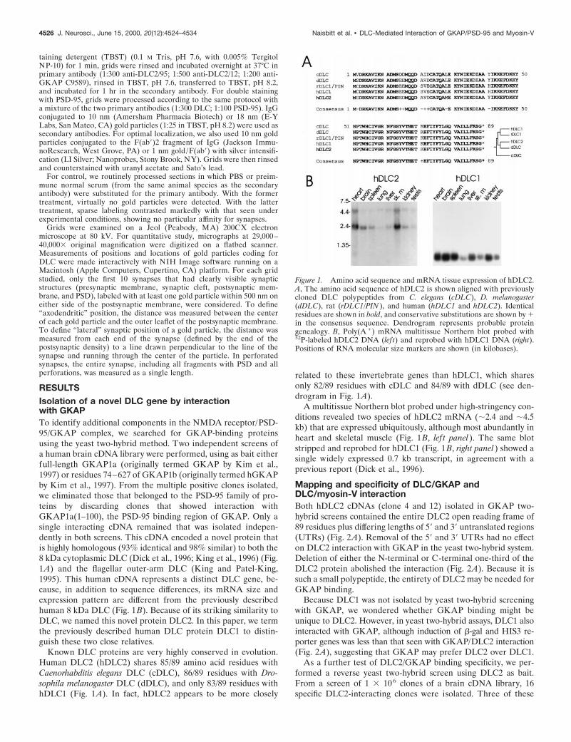
RESULTSIsolation of a novel DLC gene by interactionwith GKAPTo identify additional components in the NMDA receptor/PSD-95/GKAP complex, we searched for GKAP-binding proteinsusing the yeast two-hybrid method. Two independent screens ofa human brain cDNA library were performed, using as bait eitherfull-length GKAP1a (originally termed GKAP by Kim et al.,1997) or residues 74–627 of GKAP1b (originally termed hGKAPby Kim et al., 1997). From the multiple positive clones isolated,we eliminated those that belonged to the PSD-95 family of pro-teins by discarding clones that showed interaction withGKAP1a(1–100), the PSD-95 binding region of GKAP. Only asingle interacting cDNA remained that was isolated indepen-dently in both screens. This cDNA encoded a novel protein thatis highly homologous (93% identical and 98% similar) to both the8 kDa cytoplasmic DLC (Dick et al., 1996; King et al., 1996) (Fig.1A) and the flagellar outer-arm DLC (King and Patel-King,1995). This human cDNA represents a distinct DLC gene, be-cause, in addition to sequence differences, its mRNA size andexpression pattern are different from the previously describedhuman 8 kDa DLC (Fig. 1B). Because of its striking similarity toDLC, we named this novel protein DLC2. In this paper, we termthe previously described human DLC protein DLC1 to distin-guish these two close relatives.
Known DLC proteins are very highly conserved in evolution.Human DLC2 (hDLC2) shares 85/89 amino acid residues withCaenorhabditis elegans DLC (cDLC), 86/89 residues with Dro-sophila melanogaster DLC (dDLC), and only 83/89 residues withhDLC1 (Fig. 1A). In fact, hDLC2 appears to be more closely
related to these invertebrate genes than hDLC1, which sharesonly 82/89 residues with cDLC and 84/89 with dDLC (see den-drogram in Fig. 1A).
A multitissue Northern blot probed under high-stringency con-ditions revealed two species of hDLC2 mRNA (;2.4 and ;4.5kb) that are expressed ubiquitously, although most abundantly inheart and skeletal muscle (Fig. 1B, lef t panel). The same blotstripped and reprobed for hDLC1 (Fig. 1B, right panel) showed asingle widely expressed 0.7 kb transcript, in agreement with aprevious report (Dick et al., 1996).
Mapping and specificity of DLC/GKAP andDLC/myosin-V interactionBoth hDLC2 cDNAs (clone 4 and 12) isolated in GKAP two-hybrid screens contained the entire DLC2 open reading frame of89 residues plus differing lengths of 59 and 39 untranslated regions(UTRs) (Fig. 2A). Removal of the 59 and 39 UTRs had no effecton DLC2 interaction with GKAP in the yeast two-hybrid system.Deletion of either the N-terminal or C-terminal one-third of theDLC2 protein abolished the interaction (Fig. 2A). Because it issuch a small polypeptide, the entirety of DLC2 may be needed forGKAP binding.
Because DLC1 was not isolated by yeast two-hybrid screeningwith GKAP, we wondered whether GKAP binding might beunique to DLC2. However, in yeast two-hybrid assays, DLC1 alsointeracted with GKAP, although induction of b-gal and HIS3 re-porter genes was less than that seen with GKAP/DLC2 interaction(Fig. 2A), suggesting that GKAP may prefer DLC2 over DLC1.
As a further test of DLC2/GKAP binding specificity, we per-formed a reverse yeast two-hybrid screen using DLC2 as bait.From a screen of 1 3 106 clones of a brain cDNA library, 16specific DLC2-interacting clones were isolated. Three of these
Figure 1. Amino acid sequence and mRNA tissue expression of hDLC2.A, The amino acid sequence of hDLC2 is shown aligned with previouslycloned DLC polypeptides from C. elegans (cDLC), D. melanogaster(dDLC), rat (rDLC1/PIN ), and human (hDLC1 and hDLC2). Identicalresidues are shown in bold, and conservative substitutions are shown by 1in the consensus sequence. Dendrogram represents probable proteingenealogy. B, Poly(A 1) mRNA multitissue Northern blot probed with32P-labeled hDLC2 DNA (lef t) and reprobed with hDLC1 DNA (right).Positions of RNA molecular size markers are shown (in kilobases).
4526 J. Neurosci., June 15, 2000, 20(12):4524–4534 Naisbitt et al. • DLC-Mediated Interaction of GKAP/PSD-95 and Myosin-V

clones encoded distinct overlapping regions of GKAP1a (Fig. 2B,clones 44, 68, 71). That GKAP and DLC2 were able to mutuallyisolate each other from a complex pool containing ;1 3 106
potential partners provides compelling evidence for the relativespecificity of the GKAP/DLC2 interaction.
The minimal DLC-binding domain within GKAP was deter-mined by testing deletion constructs of GKAP for binding toDLC2 in the yeast two-hybrid system (Fig. 2B). GKAP1a residues319–384 were sufficient for DLC2 interaction (Fig. 2B), whereasfurther deletions into this domain [GKAP1a(319–363) orGKAP1a(333–384)] were unable to interact (data not shown).Other regions of GKAP, GKAP1a(1–100) and GKAP1a(591–666), did not bind DLC2, although they robustly bound thePSD-95 GK domain and the Shank PDZ [PSD-95/Discs large
(Dlg)/zona occludens 1 (ZO-1)] domain, respectively (Fig. 2B)(Kim et al., 1997; Naisbitt et al., 1999). The minimal DLC-binding domain GKAP1a(319–384) bound neither the PSD-95GK domain nor the Shank PDZ domain. Thus, different regionsof GKAP mediate specific interactions with distinct proteinpartners.
In addition to GKAP, the yeast two-hybrid screen with DLC2as bait pulled out three copies of a short segment of the taildomain of human myosin-V (gene name MYH12, residues 1236–1420) (Fig. 2C). This segment is highly alternatively spliced, andthe variant we isolated (with exons ABCEG) (Fig. 2C) is appar-ently brain-specific (Seperack et al., 1995; Huang et al., 1998).Biochemical studies of myosin-V have revealed DLC to be a lightchain of myosin-V, as well as of cytoplasmic dynein, and DLC isassociated with the myosin-V tail domain after proteolytic frac-tionation of purified myosin-V (Espindola et al., 1996; Benashskiet al., 1997). Our yeast two-hybrid screen result corroborates thisconclusion and further narrows the DLC-binding domain in themyosin-V tail (total length of ;800 amino acids) down to 184residues (Fig. 2C). Both DLC1 and DLC2 bound robustly to thissegment of the myosin-V tail (clone 45) in the yeast two-hybridsystem, suggesting that both of these proteins may be light chainsubunits of myosin-V in vivo.
Direct in vitro binding of DLC2 and GKAPTo show a direct biochemical association between GKAP andDLC2, in vitro binding experiments were performed using puri-fied recombinant proteins. H6-DLC2 bound efficiently to a GSTfusion protein of the minimal DLC-binding domain, GST-GKAP1a(319–384), but not to GST alone (Fig. 3A and data notshown). Binding of H6-DLC2 (1 mM) to beads charged withGST-GKAP1a(319–384) was competitively inhibited using vari-ous concentrations of untagged DLC2 full-length protein (Fig.
Figure 2. Interaction between DLC2 and GKAP in the yeast two-hybridsystem. A, Human DLC2 cDNAs (clones 4 and 12) isolated from yeasttwo-hybrid screens using GKAP1a or GKAP1b as bait are shown at thetop. Both clones (4 and 12) contain the entire hDLC2 open reading frame(residues 1–89; thick black line) plus different amounts of UTRs (thinline). Full-length and deletion constructs of hDLC2 are aligned below.Interaction of full-length GKAP with hDLC2 constructs (black) and withfull-length hDLC1 ( gray) were assayed by b-gal /HIS3 induction in theyeast two-hybrid system (semi-quantified, as by Kim et al., 1995). B,GKAP1a cDNAs (clones 44, 68, 71, in bracket) isolated from the yeasttwo-hybrid screen using hDLC2 as bait are depicted. These clones andvarious GKAP1a deletion constructs are aligned beneath a schematic offull-length GKAP1a. Numbers refer to the amino acid residues at theboundaries of each construct. Binding to full-length hDLC2 was assayedin the yeast two-hybrid system as in A. The DLC-binding domain,GKAP1a(319–384), did not interact with PSD-95 or Shank (data notshown). C, The human myosin-V (MYH12) cDNA (clone 45) isolatedthree times from the yeast two-hybrid screen using hDLC2 as bait isshown (black line) aligned beneath a schematic of full-length myosin-V [inwhich head, neck, coiled coil (CC), and tail domains are depicted]. Anenlargement below shows the alternatively spliced exons in this region(modified from Huang et al., 1998); ABCEG are present in clone 45.Interaction of myosin-V (clone 45) with DLC1 and DLC2 (DLC binding)was indistinguishable in the yeast two-hybrid system.
Figure 3. In vitro binding of DLC2 and GKAP and pull-down from ratbrain showing DLC-GKAP/PSD-95 and GKAP-DLC2/myosin-V inter-action. A, Beads charged with GST-GKAP1a(319–384) were incubatedwith 1 mM purified H6-hDLC2 in the presence of varying concentrationsof untagged hDLC2 competitor. Bound protein was immunoblotted forT7 tag present in H6-hDLC2. H6-hDLC2 showed insignificant binding toGST alone (data not shown). B, Beads charged with GST alone or GSTfusions of GKAP1a(319–384), DLC1, DLC2, or M5 tail [clone 45,MYH12(1236–1420)] were incubated with whole rat brain extracts.Bound proteins were immunoblotted for the indicated proteins. Input lanewas loaded with 10% of the extract used for pull-down. Increased back-ground or extra bands of inappropriate size are seen in the myosin-5(M5), GKAP, and DLC pull-down lanes when immunoblotted for them-selves; these arise because the immunoblotting antibodies recognize thefusion proteins used for pull-down.
Naisbitt et al. • DLC-Mediated Interaction of GKAP/PSD-95 and Myosin-V J. Neurosci., June 15, 2000, 20(12):4524–4534 4527

3A). Untagged DLC2 was able to compete away H6-DLC2 bind-ing to GKAP in a dose-dependent manner, with half-maximalinhibition at submicromolar concentrations.
In additional experiments (data not shown), GST-DLC2 andthioredoxin-GKAP(248–445) fusion proteins, respectively, wereable to pull down GKAP and DLC2 expressed heterologously inCOS7 cells.
Pull-down of a native DLC/myosin-V complex by GKAPand of a native GKAP/PSD-95 complex by DLC2We raised independent antibodies to study native DLC proteinsin vivo, but not an antibody generated against a DLC2 peptide(termed aDLC2/95), an antibody raised against the entire H6-DLC2 fusion protein (termed aDLC2/12), or an antibody gener-ated against DLC1 (termed R4058; a gift from Stephen King,University of Connecticut, Farmington, CT) (King et al., 1996)could distinguish between DLC1 and DLC2 by immunoblottingor immunostaining (data not shown). All DLC antibodies recog-nized an 8 kDa band on immunoblots of brain and of DLC1- orDLC2-transfected COS7 cells. Hence, we will use the genericterm DLC for the brain proteins recognized by our antibodies.The inability of our antibodies to distinguish between DLC1 andDLC2 is not surprising considering the 98% similarity betweenthese proteins.
We used the DLC antibodies in GST pull-down experimentsfrom rat brain extracts to verify biochemical association of GKAPand DLC. Beads charged with a GST fusion protein ofGKAP(319–384) precipitated a large fraction of DLC in theoffered extract (Fig. 3B, lane 3). In addition to DLC, GST-GKAP(319–384) brought down a significant amount ofmyosin-V, suggesting that GST-GKAP(319–384) is able to bindDLC in a native complex with myosin-V. These pull-downs werespecific, because GST alone pulled down none of the examinedproteins (Fig. 3B, lane 2), and GRIP, an AMPA receptor bindingprotein, was not bound by any of the GST fusion proteins.
In parallel experiments, GST fusion proteins of DLC1 andDLC2 specifically pulled down GKAP from brain extracts (Fig.3B). Although both GST-DLC1 (lane 4) and GST-DLC2 (lane 5)were able to do so, GST-DLC2 was more effective than GST-DLC1 at precipitating GKAP from the offered extract. Signifi-cantly, however, DLC1 and DLC2 were equally effective in pull-down of myosin-V in the same reactions. These biochemicalfindings corroborate the yeast two-hybrid result, suggesting thatGKAP may prefer binding to DLC2 over DLC1 (Fig. 1A).PSD-95 was also pulled down together with GKAP by GST-DLC1 and by GST/DLC2 (Fig. 3B). Because DLC does not binddirectly to PSD-95, this result suggests that DLC can interact withGKAP that is complexed with PSD-95.
The ability of GST-DLC1 and -DLC2 to pull down myosin-Vwith similar efficiency (Fig. 3B, lanes 4, 5) provides additionalevidence for a biochemical interaction between myosin-V andDLC1 or DLC2. In the reverse direction (lane 6), a GST fusionof clone 45, the fragment of the myosin-V tail isolated by yeasttwo-hybrid screening with DLC2 (clone 45), was able to pulldown DLC from brain extracts. This biochemical result substan-tiates the yeast two-hybrid finding that residues 1236–1420 ofmyosin-V heavy chain contain the DLC attachment site.
GST-M5 tail did not significantly precipitate GKAP andPSD-95 in these experiments (Fig. 3B, lane 6). This is probablybecause GST-M5-tail can only pull down GKAP/PSD-95 throughbridging by DLC, and there is a large excess of soluble DLC in thebrain extracts that is not associated with GKAP (data not shown).
Given the small percentage of total DLC associated with GKAP,it is not surprising that we failed to detect GKAP and PSD-95 bypull-down using myosin-V. This difficulty does not arise in theother orientation (pull-down of myosin-V by GKAP), becauseeach purified myosin-V particle contains two DLC molecules(Espindola et al., 1996; Benashski et al., 1997); there appears tobe much higher percentage of total DLC complexed withmyosin-V than GKAP.
Association of myosin-V/DLC and GKAP/PSD-95 inbrain extractsTo show association of endogenous myosin-V/DLC and PSD-95/GKAP, immunoprecipitations were performed from rat braincrude synaptosomal membranes solubilized in 1% DOC (Fig.4A). Each immunoprecipitation in Figure 4A is loaded in pairedlanes (P, the immunoprecipitated pellet; S, the supernatant re-maining after immunoprecipitation). Shank, another componentof the PSD-95/GKAP complex (Naisbitt et al., 1999), was alsoexamined in this experiment.
GKAP antibodies (N1564) immunoprecipitated approximatelyhalf of the offered GKAP protein and coprecipitated lesseramounts of PSD-95, chapsyn-110/PSD-93 (Fig. 4A, lane 8), andShank (data not shown), in accord with previous results. Inaddition, a small but significant fraction of DLC and myosin-Vcoprecipitated with GKAP (lane 8). In the opposite direction,DLC antibodies immunoprecipitated DLC proteins efficiently and
Figure 4. Immunoprecipitation of a myosin-V/DLC-PSD-95/GKAPcomplex from rat brain. A, Extracts of rat forebrain synaptosomal mem-branes were immunoprecipitated with GKAP, DLC, Shank, or controlnonimmune IgGs (IgG), as indicated. Each immunoprecipitation is shownin two lanes: P, the immunoprecipitated pellet; S, the supernatant remain-ing after immunoprecipitation. Input lane was loaded with 50% of theextract used for each immunoprecipitation reaction. Immunoprecipitatesand supernatants were immunoblotted for myosin-V (M5), GKAP, thePSD-95 family, and DLC, as indicated. Only the top 130 kDa GKAP bandis shown (both bands were precipitated similarly). B, Specificity of coim-munoprecipitation shown by antigen block. GKAP antibodies raisedagainst the N-terminal region of GKAP (N1564) were used for immuno-precipitation of rat forebrain extracts. Immunoprecipitation of GKAPand coimmunoprecipitation of myosin-5 (M5), DLC, PSD-95, and NR1were blocked by a thioredoxin fusion protein of the N-terminal region ofGKAP (the antigen used to generate N1564) but not by a fusion proteinof the C-terminal region of GKAP. Immunoblotting for GKAP wasperformed with antibodies directed against the C-terminal region ofGKAP (C9589). Synaptophysin (Syn) was not precipitated in eitherreaction.
4528 J. Neurosci., June 15, 2000, 20(12):4524–4534 Naisbitt et al. • DLC-Mediated Interaction of GKAP/PSD-95 and Myosin-V

coprecipitated myosin-V, GKAP, PSD-95, and chapsyn-110 (lane4). Immunoprecipitations with Shank also coprecipitated faintbut detectable amounts of myosin-V and DLC in addition toGKAP and PSD-95 (lane 2, and data not shown). Control non-immune IgGs immunoprecipitated none of these proteins (Fig.4A, lane 6), and synaptophysin was not precipitated by any of theantibodies used (data not shown).
As an additional test of specificity, we checked that the coim-munoprecipitation of DLC and myosin-V with GKAP could beblocked by competition with the specific antigen (Fig. 4B). GKAPantibodies raised against the N-terminal region of GKAP(N1564) were unable to immunoprecipitate GKAP or coprecipi-tate DLC, myosin-V, PSD-95, or NR1 when incubated in thepresence of excess N-terminal GKAP fusion protein antigen.However, immunoprecipitation of GKAP by N1564 and copre-cipitation of DLC, myosin-V, PSD-95, and NR1 remained effec-tive in the presence of an antigenically unrelated protein encom-passing the C-terminal region of GKAP (Fig. 4B).
Synaptic localization of DLC in cultured neuronsThe biochemical experiments above indicate that DLC andmyosin-V are at least partly associated with the GKAP/PSD-95complex in brain homogenates. Is DLC also concentrated atpostsynaptic sites, like GKAP and PSD-95? Double-labeled im-munofluorescence staining of cultured hippocampal neurons re-vealed punctate DLC immunoreactivity distributed along thedendritic shaft and in dendritic spines (Fig. 5B,E). The DLCimmunostaining pattern colocalized with PSD-95 (Fig. 5A) andwith F-actin (Fig. 5D). DLC also colocalized with F-actin inneuronal growth cones (Fig. 5G). DLC-immunoreactive punctaalso overlapped with the presynaptic marker SV2, but comparedwith its tight colocalization with PSD-95 or F-actin, DLC ap-peared slightly offset from the SV2 staining (Fig. 5, compare Hwith C and F), suggesting a postsynaptic localization of DLC thatextends away from the postsynaptic membrane. The primarilypunctate distribution of DLC showed little colocalization withmicrotubules, which are concentrated in dendritic shafts (Fig. 5I).Thus, at the light microscopy level, DLC appears to be a synaptic
protein concentrated in spines, consistent with its interaction withthe GKAP/PSD-95 complex.
Ultrastructural localization of DLC in dendritic spinesand PSDThe subcellular distribution of DLC was also examined at highresolution in brain tissue by postembedding immunoelectron mi-croscopy (immunoEM). Immunogold labeling of rat cerebralcortex was analyzed using two independently generated and pu-rified DLC antibodies, aDLC2/95 and aDLC2/12 (see Materialsand Methods). Because these reagents are unable to distinguishimmunohistochemically between DLC1 and DLC2 expressed intransfected cells (data not shown), we emphasize that the immu-noEM characterization presented here may reflect the distribu-tion of either or both DLC proteins.
Both anti-DLC antibodies gave similar patterns, with immuno-gold particles consistently concentrated in the postsynaptic com-partment of asymmetric synapses (Fig. 6A–F). In all blocks, goldparticles coding for DLC were predominantly associated with thespine apparatus and PSD of dendritic spines. Outside of synapses,some immunoreactivity was also found in small dendrites inwhich gold particles labeled fibrillary structures. In perikarya, lowlevels of labeling were encountered over endoplasmic reticulum.
Within labeled postsynaptic profiles, a subset of DLC particleslay directly over the PSD (Fig. 6B,E). However, many goldparticles “trailed” into the deep part of the postsynaptic spine(Fig. 6A–D, F), and particularly dense labeling for DLC wasassociated with the spine apparatus (arrows). Indeed, in somesynapses, only deep parts of the postsynaptic profile were labeled,whereas the PSD was not (Fig. 6C,D). Presynaptic DLC particleswere also seen (Fig. 7B) but with much less frequency. Not allasymmetric synapses were labeled for DLC; approximately one-third of asymmetric synapses completely lacked gold particles.
Quantitative analysis of DLC immunogold distribution (Fig.6G,H) confirmed that DLC immunoreactivity is predominantlyon the postsynaptic side of the synapse, although a small presyn-aptic peak exists. The distribution profile of DLC particles in thepostsynaptic compartment is very broad, with a peak that extends
Figure 5. Colocalization of DLC withPSD-95 and F-actin at excitatory syn-apses in cultured hippocampal neurons.A–C, Hippocampal neurons (low den-sity, 3 weeks in vitro) double stained forPSD-95 (A, red) and DLC (B, green),showing punctate colocalization (C, yel-low) within dendritic spines. D–G,Staining for F-actin with rhodaminephalloidin (D) and for DLC (E) alsocolocalizes in spines (F). G, Colocaliza-tion of F-actin (red) and DLC ( green)within a neuronal growth cone at 2 d invitro. H, DLC immunoreactivity ( green)appears to be apposed to, rather thanexactly overlapping, the presynapticmarker SV2 (red), suggestive of apostsynaptic localization of DLC. I,DLC ( green) does not appear to be asmarkedly concentrated along microtu-bules (anti-tubulin, red) as it is withF-actin (D–F). Scale bar, 10 mm.
Naisbitt et al. • DLC-Mediated Interaction of GKAP/PSD-95 and Myosin-V J. Neurosci., June 15, 2000, 20(12):4524–4534 4529

from ;40 to ;120 nm inside the postsynaptic membrane (Fig.6G). In most synapses, this peak coincides with the inner (indis-tinct) edge of the PSD and subsynaptic web. A large fraction oftotal immunoreactivity extended further (.300 nm) into thepostsynaptic spine (Fig. 6G), often clustering over the spineapparatus (typically 300–600 nm from the nearest edge of theactive zone). This quantitative description of DLC distributiondiffers from that of PSD-95 or GKAP; these proteins show muchnarrower postsynaptic peaks within 25 nm of the postsynapticmembrane (Naisbitt et al., 1999; Valtschanoff et al., 1999). In thelateral plane of the synapse, DLC was broadly distributed, with aslight enrichment midway between the center and the edge of thePSD (Fig. 6H). There seemed to be a positive correlation be-tween length of the active zone and particle density, i.e., synapseswith longer active zones had more DLC particles deeper in thepostsynaptic compartment. Overall, these ultrastructural studiesin brain confirm and extend the light microscopic findings in
cultured neurons; DLC is concentrated specifically in the postsyn-aptic compartment of excitatory synapses, with a distributionextending from the PSD to the spine apparatus of dendriticspines.
Although most DLC immunogold particles were associatedwith the postsynaptic cytoplasm and spine apparatus, many local-ized directly over the PSD. To explore further whether DLC is anintegral component of the PSD, we studied the fractionation ofDLC during purification of rat brain PSDs (Fig. 6 I). DLC wasenriched in PSDI (PSD fraction extracted once with TritonX-100) and PSDII (extracted twice with Triton) but was depletedin PSDIII (extracted with Triton X-100 and sarkosyl). This bio-chemical data suggests that DLC is loosely associated with thePSD, contrasting with GKAP, a core component of the PSD thatresists extraction by sarkosyl (Fig. 6 I). Myosin-V behaves simi-larly to DLC in its PSD fractionation, being enriched in PSDI butnot in PSDIII (Fig. 6 I). These biochemical data further support
Figure 6. Quantitative immunogoldEM localization of DLC in the PSD anddendritic spine, and enrichment of DLCand myosin-V in PSD fractions. A–F,Immunogold labeling of DLC in sec-tions from rat cerebral cortex (layersII /III) using two independent antibod-ies. Scale bar, 0.25 mm. A, B, Examplesof spines labeled by aDLC2/95 antibody.Some particles lie within the PSD; mosttrail deeper into the cytoplasm of thepostsynaptic spine. Spine apparatus (ar-row) is densely labeled. C, D, Examplesof dendritic spines labeled withaDLC2/12 antibody. Again, labeling isfound prominently deep to the PSD, inpostsynaptic cytoplasm and spine appa-ratus (arrows). E, F, Examples of asym-metric synapses probed with aDLC2/95showing denser labeling of PSD relativeto postsynaptic cytoplasm. Arrow indi-cates a labeled spine apparatus in F. G,H, Quantitative analysis of the distribu-tion of DLC immunogold particles atsynapses. G, Distribution of gold parti-cles in the axodendritic axis. Labelingdensity (arbitrary units) of particles ateach distance is plotted. DLC immuno-gold distribution peaks very broadly at;40–120 nm postsynaptic to the plasmamembrane. H, The distance of immuno-gold particles from the center of thePSD in the lateral plane of the synapse(normalized by PSD length) is shown asa histogram, with particles per bin alongthe ordinate. DLC labeling is distrib-uted throughout the lateral plane of thePSD, with slightly increased densitymidway between the center and edge. I,Immunoblot analysis of brain and PSDfractions (amount of protein loaded ineach lane is indicated). WB, Wholebrain homogenate; P2, crude synaptoso-mal fraction. PSD fractions were ex-tracted with Triton X-100 once (PSDI ),twice (PSDII ), or with Triton X-100 fol-lowed by sarkosyl (PSDIII ). Blots wereimmunoblotted for the indicated pro-teins. M5, Myosin-V.
4530 J. Neurosci., June 15, 2000, 20(12):4524–4534 Naisbitt et al. • DLC-Mediated Interaction of GKAP/PSD-95 and Myosin-V

the presence of myosin-V and DLC at postsynaptic sites. Therelatively modest enrichment of DLC in PSD fractions is consis-tent with the immunoEM findings, which show only partial local-ization of DLC immunogold over the PSD.
Colocalization of DLC with GKAP/PSD-95 byelectron microscopyFinally, we looked for colocalization of DLC with GKAP andPSD-95 in the same dendritic spine at EM resolution. In double-stained material, many, but not all, postsynaptic profiles thatstained for PSD-95 or GKAP were also immunopositive for DLC.In consecutive thin sections, DLC labeling (silver-enhanced im-munogold) (Fig. 7A1) was found within the same PSD as GKAP(Fig. 7A2). As is typical, DLC (but not GKAP) also labeled thespine apparatus within this dendritic spine (Fig. 7A1, arrow).
In double-labeling experiments, PSD-95 immunogold particles(18 nm gold) codistributed with DLC (10 nm gold particles) indendritic spines of asymmetric synapses (Fig. 7B–E). Immuno-gold particles coding for PDS-95 localized almost exclusivelywithin the PSD in a position very close to the postsynapticmembrane. On the other hand, DLC particles extended moredeeply into the postsynaptic profile and were typically denser overthe spine apparatus than at the PSD (Fig. 7C,D). Some DLC
particles localized directly over the PSD-95-immunopositive PSD(Fig. 7C). Thus, DLC and PSD-95/GKAP are present in the samedendritic spine, but their relative distributions suggest an overlaprather than a complete colocalization of these proteins in thepostsynaptic compartment.
DISCUSSIONThe major findings of this study are as follows: (1) GKAP, amajor component of the PSD-95 complex, binds directly to DLC,a light chain shared by myosin-V and cytoplasmic dynein; (2) aportion of GKAP and PSD-95 is associated with DLC andmyosin-V in a coimmunoprecipitable complex in brain homoge-nates; and (3) DLC is highly enriched in dendritic spines, colo-calizing with GKAP/PSD-95 in the PSD but also prominentlyassociated with the spine apparatus in the deep part of the spine.
DLC: a potential link between GKAP andmotor proteins?Motor proteins usually consist of heavy chains (which contain thedefining ATPase and motor activities) plus accessory subunits ofpoorly defined function (termed intermediate or light chains,depending on their molecular size). DLC was first isolated as an8 kDa light chain of cytoplasmic dynein, a minus end-directed
Figure 7. Colocalization of DLC withGKAP and PSD-95 by immunoEM. A,Two consecutive thin sections of cere-bral cortex immunolabeled for DLC(A1) or GKAP (A2) showing an asym-metric synapse labeled for both antigens.DLC is also present in spine apparatus(arrow in A1). Silver-enhanced 1 nmgold was used to visualize DLC in thisexperiment. B, C, Examples of doubleimmunostaining for DLC (10 nm gold,aDLC2/95) and PSD-95 (18 nm gold).Immunogold particles coding forPDS-95 are close to the postsynapticmembrane, whereas DLC tends to dis-tribute deeper into the postsynaptic pro-file. D, E, Two additional examples ofdouble labeling for DLC (10 nm gold)and PSD-95 (18 nm gold), using an inde-pendent DLC antibody (aDLC2/12). Ar-rows, Spine apparatus. Scale bar, 0.25 mm.
Naisbitt et al. • DLC-Mediated Interaction of GKAP/PSD-95 and Myosin-V J. Neurosci., June 15, 2000, 20(12):4524–4534 4531

microtubule motor that exists as a massive multi-subunit complex(King et al., 1996; Hirokawa, 1998; Karki and Holzbaur, 1999).More recently, DLC was also found to be stoichiometricallyassociated with myosin-V (Espindola et al., 1996; Benashski et al.,1997). Because DLC binds to GKAP in vivo, an attractive hy-pothesis is that DLC mediates a biochemical interaction betweenGKAP and these actin- and microtubule-based motors, therebyallowing GKAP and its associated proteins to be translocated asa cargo.
The idea that an accessory subunit of a motor mediates theinteraction between a motor and its cargo has its precedents. Theintermediate chain of dynein (the 74 kDa dynein intermediatechain) has been proposed to indirectly anchor dynein to itscargoes via binding to the dynactin complex (Vallee et al., 1995;Vaughan and Vallee, 1995). More recently, the 14 kDa dyneinlight chain (named Tctex-1) was found to bind directly to thecytoplasmic tail of rhodopsin and to link rhodopsin-bearing ves-icles to cytoplasmic dynein and to microtubule-based movement(Tai et al., 1999). Based on the Tectex-1 precedent in particular,it seems reasonable to speculate that the 8 kDa DLC can alsofunction as an adaptor between motor and cargo and that GKAPcould act as a receptor for DLC-mediated motor interactions. Invitro motility assays are required, however, to prove that GKAPcan be moved as a cargo by myosin-V and dynein motors in aDLC-dependent manner. If DLC truly does act as a motor–cargoadaptor, then its presence in both myosin-V and cytoplasmicdynein suggests a mechanism whereby the same cargo can beferried along either microtubule or actin tracks. This is consistentwith previous evidence in mice and yeast suggesting a physicalinteraction between myosin V and microtubule-based motors (forreview, see Brown, 1999).
DLC has also been shown to bind other proteins in addition toGKAP, including IkBa (Crepieux et al., 1997) and nNOS (Jaffreyand Snyder, 1996; Rodriguez-Crespo et al., 1998). The variety ofprotein–protein interactions mediated by DLC is compatible withits potential role as an adaptor that links myosin-V and dyneinmotors to multiple cargos. Additional studies of DLC-mediatedinteractions with GKAP may provide more insight into the poorlyunderstood mechanisms of motor–cargo recognition and itsregulation.
DLC and myosin-V in dendritic spinesDLC is concentrated in dendritic spines, as shown by immuno-staining at LM and EM levels. F-Actin is the predominant cy-toskeletal element in dendritic spines, which are believed to berelatively devoid of microtubules (Harris and Kater, 1994). In-deed, DLC showed extensive colocalization with phalloidin butcomparatively little staining in dendritic shafts. The fact thatDLC codistributes most closely with the actin cytoskeleton led usto focus on the actin-based motor myosin-V rather than cytoplas-mic dynein. Moreover, we found myosin-V to be concentrated inthe PSD, as suggested by mass spectrometry analysis of PSDpreparations (Walikonis et al., 2000). In addition, yeast two-hybrid screening with DLC isolated multiple copies of myosin-Vheavy chain (plus GKAP and several other proteins) but notdynein, consistent with a more abundant association of DLC withmyosin-V than with dynein in neurons. The immunoprecipitationof a complex containing GKAP, DLC, and myosin-V from deter-gent extracts of brain supports the hypothesis that these proteinsmay interact in vivo.
What might be the functional significance of DLC in thepostsynaptic compartment? We noted a striking association of
DLC with the spine apparatus, an organelle characterized bystacks of smooth endoplasmic reticulum (SER) membranes sur-rounded by amorphous dense material that is found particularlycommonly in larger spines. The role of the spine apparatus ismysterious, although many speculate that it acts as a repository oreven a site of synthesis for membrane proteins trafficking to orfrom the synapse. For instance, a pool of intracellular glutamatereceptors can be detected by immunoEM at the spine apparatus(Nusser et al., 1998). The enrichment of DLC at the spineapparatus is consistent with DLC playing a role as an accessorysubunit of myosin-V involved in trafficking of proteins (such asGKAP) between spine apparatus and the postsynaptic mem-brane. Although the distribution of GKAP differs from DLC inbeing concentrated predominantly in the PSD rather than in thespine apparatus (Naisbitt et al., 1997), this is not incompatiblewith DLC-mediated transport of GKAP. In photoreceptors, rho-dopsin is highly concentrated in the outer segment, whereasTctex-1, the 14 kDa dynein light chain receptor for rhodopsin, isenriched in the inner segment (Tai et al., 1999). The steady-stateasymmetry of distribution of motor and cargo presumably reflectsthe vectorial nature of motor transport, the basis of which remainspoorly understood.
In light of the enrichment of DLC at the spine apparatus (anorganelle probably derived from SER membranes), it is particu-larly noteworthy that myosin-V has been implicated in SERmovements in dendritic spines. Dilute mice are mutant for themyosin-Va gene and deficient in pigment granule movement inskin melanocytes (this underlies their coat color phenotype)(Mercer et al., 1991; Wu et al., 1997). Dilute mice also haveneurological symptoms and lack SER membranes in dendriticspines of Purkinje neurons (Takagishi et al., 1996). This geneticevidence supports a possible role for myosin-V in membranetrafficking in dendritic spines. Additionally, in non-neural systemsfrom yeast to mammals, there is abundant biochemical and ge-netic evidence indicating that class V myosins function in or-ganelle transport and protein and/or mRNA localization in cells(for review, see Mermall et al., 1998). Not least of this evidenceis that myosin-V can act as a processive motor supporting or-ganelle movement along actin tracks (Evans et al., 1998; Mehta etal., 1999). There is also evidence that myosin-V may be involvedin presynaptic function by interacting with synaptic vesicle pre-cursors (Prekeris and Terrian, 1997; Evans et al., 1998; Bridgman,1999), and our finding of a minor fraction of DLC in presynapticterminals is consistent with such a possibility.
Diversity of DLC and its binding to myosin-Vheavy chainThis study has identified a novel mammalian protein (DLC2) thatis identical in size and highly similar in sequence (93% identity atthe amino acid level) to the previously described human DLC butthat clearly derives from a distinct gene. The structural similarityof these proteins prevented us from raising antibodies that distin-guish between them in native tissue. In earlier biochemical studiesof native 8 kDa DLC protein, it is unlikely that the reagents usedwould have distinguished between DLC1 and DLC2. In our exper-iments using recombinant proteins, both DLC1 and DLC2 can bindto myosin-V and to GKAP. Thus, DLC1 and DLC2 seem to haveessentially similar biochemical activities, as expected of such highlyrelated proteins. It remains to be determined whether DLC1 andDLC2 have distinct roles in vivo and whether they are differen-tially associated with different motors, different protein partners,or different subcellular compartments. We do know that DLC1
4532 J. Neurosci., June 15, 2000, 20(12):4524–4534 Naisbitt et al. • DLC-Mediated Interaction of GKAP/PSD-95 and Myosin-V

and DLC2 can bind to each other as well as to themselves in theyeast two-hybrid system (S. Naisbitt, unpublished observations),consistent with previous reports that DLC exists in a dimericconfiguration (Benashski et al., 1997). Thus, based on sequenceconservation, ubiquitous overlapping mRNA expression patterns,and their ability to heteromultimerize, DLC1 and DLC2 mayhave overlapping functions in vivo.
In the course of this study, we narrowed the DLC-bindingdomain in the myosin-V tail to 184 residues. This result providesthe first evidence for a direct interaction between DLC and themyosin-V heavy chain, previously shown to be in the same activeprotein complex (Espindola et al., 1996; Benashski et al., 1997).Interestingly, this DLC-binding region of myosin-V tail is highlyalternatively spliced in a tissue-specific manner. The splice vari-ant ABCEG we isolated by interaction with DLC appears to berelatively brain-specific (Seperack et al., 1995; Huang et al., 1998).It will be interesting to test whether differential splicing of themyosin-V tail might affect a tissue-specific association with DLCproteins. The results of this study therefore suggest new avenuesof research in motor protein structure–function, as well as in theroles of motor protein interactions in organization of the postsyn-aptic specialization.
REFERENCESArnold DB, Clapham DE (1999) Molecular determinants for subcellular
localization of PSD-95 with an interacting K 1 channel. Neuron23:149–157.
Ausubel FM, Brent R, Kingston RE, Moore DD, Seidman JG, Smith JA,Struhl K (1993) Expression and purification of glutathione-S-transferase fusion proteins. In: Current protocols in molecular biology(Jansson K, ed), pp 16.7.1–16.7.8. New York: Wiley.
Banker GA, Cowan WM (1977) Rat hippocampal neurons in dispersedcell culture. Brain Res 126:397–425.
Benashski SE, Harrison A, Patel-King RS, King SM (1997) Dimeriza-tion of the highly conserved light chain shared by dynein and myosin V.J Biol Chem 272:20929–20935.
Bridgman PC (1999) Myosin Va movements in normal and dilute-lethalaxons provide support for a dual filament motor complex. J Cell Biol146:1045–1060.
Brown SS (1999) Cooperation between microtubule- and actin-basedmotor proteins. Annu Rev Cell Dev Biol 15:63–80.
Cho K-O, Hunt CA, Kennedy MB (1992) The rat brain postsynapticdensity fraction contains a homolog of the Drosophila discs-large tumorsuppressor protein. Neuron 9:929–942.
Craven SE, Bredt DS (1998) PDZ proteins organize synaptic signalingpathways. Cell 93:495–498.
Craven SE, El-Husseini AE, Bredt DS (1999) Synaptic targeting of thepostsynaptic density protein PSD-95 mediated by lipid and proteinmotifs. Neuron 22:497–509.
Crepieux P, Kwon H, Leclerc N, Spencer W, Richard S, Lin R, Hiscott J(1997) I kappaB alpha physically interacts with a cytoskeleton-associated protein through its signal response domain. Mol Cell Biol17:7375–7385.
Dick T, Ray K, Salz HK, Chia W (1996) Cytoplasmic dynein (ddlc1)mutations cause morphogenetic defects and apoptotic cell death inDrosophila melanogaster. Mol Cell Biol 16:1966–1977.
Dunah A, Luo J, Wang Y, Yasuda R, Wolfe B (1998) Subunit compo-sition of N-methyl-D-aspartate receptors in the central nervous systemthat contain the NR2D subunit. Mol Pharmacol 53:429–437.
Espindola FS, Cheney RE, King SM, Suter DM, Mooseker MS (1996)Myosin-V and Dynein share a similar light chain. Mol Biol Cell 7:372.
Espreafico EM, Cheney RE, Matteoli M, Nascimento AA, De CamilliPV, Larson RE, Mooseker MS (1992) Primary structure and cellularlocalization of chicken brain myosin-V (p190), an unconventional my-osin with calmodulin light chains. J Cell Biol 119:1541–1557.
Evans LL, Lee AJ, Bridgman PC, Mooseker MS (1998) Vesicle-associated brain myosin-V can be activated to catalyze actin-basedtransport. J Cell Sci 111:2055–2056.
Goslin K, Asmussen H, Banker G (1998) Rat hippocampal neurons inlow density culture. In: Culturing nerve cells (Banker G, Goslin K,eds), pp 339–370. Cambridge, MA: MIT.
Harris K, Kater S (1994) Dendritic spines: cellular specializations im-parting both stability and flexibility to synaptic function. Annu RevNeurosci 17:341–371.
Hirao K, Hata Y, Ide N, Takeuchi M, Irie M, Yao I, Deguchi M, ToyodaA, Sudhof TC, Takai Y (1998) A novel multiple PDZ domain-containing molecule interacting with N-methyl-D-aspartate receptorsand neuronal cell adhesion proteins. J Biol Chem 273:21105–21110.
Hirokawa N (1998) Kinesin and dynein superfamily proteins and themechanism of organelle transport. Science 279:519–526.
Huang JD, Mermall V, Strobel MC, Russell LB, Mooseker MS, CopelandNG, Jenkins NA (1998) Molecular genetic dissection of mouse uncon-ventional myosin-VA: tail region mutations. Genetics 148:1963–1972.
Jaffrey SR, Snyder SH (1996) PIN: an associated protein inhibitor ofneuronal nitric oxide synthase. Science 274:774–777.
Karki S, Holzbaur EL (1999) Cytoplasmic dynein and dynactin in celldivision and intracellular transport. Curr Opin Cell Biol 11:45–53.
Kim E, Niethammer M, Rothschild A, Jan YN, Sheng M (1995) Clus-tering of shaker-type K 1 channels by interaction with a family ofmembrane-associated guanylate kinases. Nature 378:85–88.
Kim E, Naisbitt S, Hsueh Y-P, Rao A, Rothschild A, Craig AM, ShengM. (1997) GKAP, a novel synaptic protein that interacts with theguanylate kinase-like domain of the PSD-95/SAP90 family of channelclustering molecules. J Cell Biol 136:669–678.
King SM, Patel-King RS (1995) The M(r) 5 8,000 and 11,000 outer armdynein light chains from Chlamydomonas flagella have cytoplasmichomologues. J Biol Chem 270:11445–11452.
King SM, Barbarese E, Dillman JFI, Patel-King RS, Carson JH, PfisterKK (1996) Brain cytoplasmic and flagellar outer arm dyneins share ahighly conserved Mr 8,000 light chain. J Biol Chem 271:19358–19366.
Kornau H-C, Schenker LT, Kennedy MB, Seeburg PH (1995) Domaininteraction between NMDA receptor subunits and the postsynapticdensity protein PSD-95. Science 269:1737–1740.
Mehta AD, Rock RS, Rief M, Spudich JA, Mooseker MS, Cheney RE(1999) Myosin-V is a processive actin-based motor. Nature400:590–593.
Mercer JA, Seperack PK, Strobel MC, Copeland NG, Jenkins NA (1991)Novel myosin heavy chain encoded by murine dilute coat colour locus.Nature 349:709–713.
Mermall V, Post PL, Mooseker MS (1998) Unconventional myosins incell movement, membrane traffic, and signal transduction. Science279:527–533.
Muller BM, Kistner U, Kindler S, Chung WJ, Kuhlendahl S, Lau L-F,Veh RW, Huganir RL, Gundelfinger ED, Garner CC (1996) SAP102,a novel postsynaptic protein that interacts with the cytoplasmic tail ofthe NMDA receptor subunit NR2B. Neuron 17:255–265.
Naisbitt S, Kim E, Weinberg RJ, Rao A, Yang F-C, Craig AM, Sheng M(1997) Characterization of guanylate kinase-associated protein, apostsynaptic density protein at excitatory synapses that interacts di-rectly with postsynaptic density-95/synapse-associated protein 90.J Neurosci 17:5687–5696.
Naisbitt S, Kim E, Tu JC, Xiao B, Sala C, Valtschanoff J, Weinberg RJ,Worley PF, Sheng M (1999) Shank, a novel family of postsynapticdensity proteins that binds to the NMDA receptor/PSD-95/GKAPcomplex and cortactin. Neuron 23:569–582.
Niethammer M, Sheng M (1998) Identification of ion channel-associatedproteins using the yeast two-hybrid system. In: Methods in enzymology(Conn PM, ed), pp 104–122. New York: Academic.
Niethammer M, Kim E, Sheng M (1996) Interaction between the Cterminus of NMDA receptor subunits and multiple members of thePSD-95 family of membrane-associated guanylate kinases. J Neurosci16:2157–2163.
Nusser Z, Lujan R, Laube G, Roberts JD, Molnar E, Somogyi P (1998)Cell type and pathway dependence of synaptic AMPA receptor num-ber and variability in the hippocampus. Neuron 21:545–559.
Phend KD, Weinberg RJ, Rustioni A (1992) Techniques to optimizepost-embedding single and double staining for amino acid neurotrans-mitters. J Histochem Cytochem 40:1011–1020.
Phend KD, Rustioni A, Weinberg RJ (1995) An osmium-free method ofEpon embedment that preserves both ultrastructure and antigenicityfor postembedding immunocytochemistry. J Histochem Cytochem43:283–292.
Prekeris R, Terrian DM (1997) Brain myosin V is a synaptic vesicle-
Naisbitt et al. • DLC-Mediated Interaction of GKAP/PSD-95 and Myosin-V J. Neurosci., June 15, 2000, 20(12):4524–4534 4533
associated motor protein: evidence for a Ca 21-dependent interactionwith the synaptobrevin-synaptophysin complex. J Cell Biol137:1589–1601.
Rao A, Kim E, Sheng M, Craig AM (1998) Heterogeneity in the molec-ular composition of excitatory postsynaptic sites during development ofhippocampal neurons in culture. J Neurosci 18:1217–1229.
Rodriguez-Crespo I, Straub W, Gavilanes F, Ortiz de Montellano PR (1998)Binding of dynein light chain (PIN) to neuronal nitric oxide synthase inthe absence of inhibition. Arch Biochem Biophys 359:297–304.
Sans N, Petralia RS, Wang Y-X, Blahos II J, Hell JW, Wenthold RJ(2000) A developmental change in NMDA receptor-associated pro-teins at hippocampal synapses. J Neuroscience 20:1260–1271.
Satoh K, Yanai H, Senda T, Kohu K, Nakamura T, Okumura N, Mat-sumine A, Kobayashi S, Toyoshima K, Akiyama T (1997) DAP-1, anovel protein that interacts with the guanylate kinase-like domains ofhDLG and PSD-95. Genes Cells 2:415–424.
Seperack PK, Mercer JA, Strobel MC, Copeland NG, Jenkins NA (1995)Retroviral sequences located within an intron of the dilute gene alterdilute expression in a tissue-specific manner. EMBO J 14:2326–2332.
Sheng M, Pak DTS (2000) Ligand-gated ion channel interactions withcytoskeletal and signaling proteins. Annu Rev Physiol 62:755–778.
Tai AW, Chuang JZ, Bode C, Wolfrum U, Sung CH (1999) Rhodopsin’scarboxy-terminal cytoplasmic tail acts as a membrane receptor forcytoplasmic dynein by binding to the dynein light chain Tctex-1. Cell97:877–887.
Takagishi Y, Oda S, Hayasaka S, Dekker-Ohno K, Shikata T, Inouye M,Yamamura H (1996) The dilute-lethal (dl) gene attacks a Ca 21 storein the dendritic spine of Purkinje cells in mice. Neurosci Lett215:169–172.
Takeuchi M, Hata Y, Hirao K, Toyoda A, Irie M, Takai Y (1997)
SAPAPs, a family of PSD-95/SAP90-associated proteins localized atpostsynaptic density. J Biol Chem 272:11943–11951.
Tu JC, Xiao B, Naisbitt S, Yuan JP, Petralia RS, Brakeman P, Doan A,Aakalu VK, Lanahan AA, Sheng M, Worley PF (1999) Coupling ofmGluR/Homer and PSD-95 complexes by the Shank family of postsyn-aptic density proteins. Neuron 23:583–592.
Vallee RB, Vaughan KT, Echeverri CJ (1995) Targeting of cytoplasmicdynein to membranous organelles and kinetochores via dynactin. ColdSpring Harbor Symp Quant Biol 60:803–811.
Valtschanoff JG, Burette A, Wenthold RJ, Weinberg RJ (1999) Expres-sion of NR2 receptor subunit in rat somatic sensory cortex: synapticdistribution and colocalization with NR1 and PSD-95. J Comp Neurol410:599–611.
Vaughan KT, Vallee RB (1995) Cytoplasmic dynein binds dynactinthrough a direct interaction between the intermediate chains andp150Glued. J Cell Biol 131:1507–1516.
Walikonis RS, Jensen ON, Mann M, Mercer JA, Kennedy MB (2000)Identification of proteins in the postsynaptic density fraction by massspectrometry. J Neurosci, in press.
Wu X, Bowers B, Wei Q, Kocher B, Hammer III JA (1997) Myosin Vassociates with melanosomes in mouse melanocytes: evidence thatmyosin V is an organelle motor. J Cell Sci 110:847–859.
Wyszynski M, Kim E, Yang F-C, Sheng M (1998) Biochemical andimmunocytochemical characterization of GRIP, a putative AMPAreceptor anchoring protein, in rat brain. Neuropharmacology37:1335–1344.
Wyszynski M, Valtschanoff JG, Naisbitt S, Dunah AW, Kim E, StandaertDG, Weinberg RJ, Sheng M (1999) Association of AMPA receptorswith a subset of glutamate receptor-interacting protein in vivo. J Neu-rosci 19:6528–6537.
4534 J. Neurosci., June 15, 2000, 20(12):4524–4534 Naisbitt et al. • DLC-Mediated Interaction of GKAP/PSD-95 and Myosin-V
Copyright © 2022 FDOKUMEN


![Differential Occupancy of Somatodendritic and Postsynaptic 5HT1A Receptors by Pindolol: A Dose-Occupancy Study with [11C]WAY 100635 and Positron Emission Tomography in Humans](https://static.fdokumen.com/doc/165x107/631c4aedd5372c006e045dcf/differential-occupancy-of-somatodendritic-and-postsynaptic-5ht1a-receptors-by-pindolol-1675040353.jpg)