The Motile/ /IC1 Subunit of Sea Urchin Sperm Outer Arm Dynein Does Not Form a Rigor Bond
12
The Motile/ /IC1 Subunit of Sea Urchin Sperm Outer Arm Dynein Does Not Form a Rigor Bond Anthony G. Moss, Jean-Luc Gatti, and George B. Witman Cell Biology Group and Male Fertility Program, Worcester Foundation for Experimental Biology, Shrewsbury, Massachusetts 01545 Abstract. We used in vitro translocation and cosedimentation assays to study the microtubule bind- ing properties of sea urchin sperm outer arm dynein and its/3/IC1 subunit. Microtubules glided on glass- absorbed sea urchin dynein for a period of time directly proportional to the initial MgATtn- concentra- tion and then detached when 70-95 % of the MgATP2- was hydrolyzed. Detachment resulted from MgATP2- depletion, because (a) perfusion with fresh buffer con- taining MgATP2- reconstituted binding and gliding, (b) microtubules glided many minutes with an ATP- regenerating system at ATP concentrations which alone supported gliding for only 1-2 min, and (c) microtubules detached upon total hydrolysis of ATP by an ATP-removal system. The products of ATP hydroly- sis antagonized binding and gliding; as little as a threefold excess of ADP/Pi over ATP resulted in com- plete loss of microtubule binding and translocation by the/~/IC1 subunit. In contrast to the situation with sea urchin dynein, microtubules ceased gliding but re- mained bound to glass-absorbed Tetrahymena outer arm dynein when MgATP 2- was exhausted. Cosedimentation assays showed that/ktrahymena outer arm dynein sedimented with microtubules in an ATP- sensitive manner, as previously reported (Porter, M.E., and K. A. Johnson. J. Biol. Chem. 258: 6575-6581). However, the/3/IC1 subunit of sea urchin dynein did not cosediment with microtubules in the absence of ATE Thus, this subunit, while capable of generating motility, lacks both structural and rigor- type microtubule binding. YNelNS are large, multicomponent ATPases responsi- ble for minus-end-directed microtubule-based cell motility, such as ciliary and flagellar beating, certain types of intraceUular transport, and organeUe sorting (65). In cilia and flagella, they form the inner and outer rows of arms which connect the outer doublet microtubules. Several lines of evidence indicate that the arms are permanently bound to the A-tubule of each doublet, and that, concomitant with a cycle of ATP binding and hydrolysis, they undergo a cycle of attachment to and detachment from the B-tubule of the adjacent doublet, giving rise to interdoublet sliding (42, 43, 52). When this sliding is resisted by structures within the axoneme, axonemal bending results (49). Because the arms appear to push on an adjacent doublet only in a base-to-tip direction (6, 41), it has been proposed that at any one time the arms on only one side of an axonemal bend are actively generating sliding, while arms on the opposite side of the ax- oneme must be disengaged to permit passive interdoublet sliding in the opposite direction (44). When the axoneme bends back the other way, the previously active arms must be switched off and the previously disengaged arms switched Dr. Gatti's present address is Laboratoire de Physiologic de la Reproduc- tion, INRA, 37380 Monnale, France. Address all correspondence to Dr. Witman. on. Bend formation, coordinated wave propagation, and cy- clic beating must be achieved by controlling the timing and positioning of dynein arm activity and resistance to sliding (3, 4, 51, 68). The best understood of all dyneins is the outer arm dynein, which is composed of three ATPase-containing subunits in Chlamydomonas and Tetrahymena, but only two such sub- units in sea urchins and vertebrates (7, 67). In all cases, each subunit is organized around a single copy of a high molecular weight polypeptide (o~,/~, and in Chlamydomonas and Tetra- hymena, 3' heavy chains); each subunit also contains one or more intermediate and/or light chains (for reviews see refer- ences 67, 69, 70). In cases where the individual subunits have been isolated and characterized, they have been found to differ substantially in their enzymatic and structural proper- ties (1, 20, 21, 22, 35, 36, 54, 55). However, little is known about the specific roles of the different subunits in flagellar movement. A complete understanding of how ciliary and flagellar movement is generated and controlled will require detailed knowledge of how the inner and outer arms and their in- dividual subunits interact with outer doublet microtubules to produce force and regulate both active and passive sliding. The microtubule-binding properties of axonemal dynein have been investigated in the intact axoneme (8, 9, 10, 29, 30, 33, 50, 62), but the presence of both inner and outer 9 The Rockefeller University Press, 0021-9525/92/09/1177/12 $2.00 The Journal of Cell Biology, Volume 118, Number 5, September 1992 1177-1188 1177 on November 14, 2013 jcb.rupress.org Downloaded from Published September 1, 1992
Transcript of The Motile/ /IC1 Subunit of Sea Urchin Sperm Outer Arm Dynein Does Not Form a Rigor Bond
The Motile/ /IC1 Subunit of Sea Urchin Sperm Outer Arm Dynein Does Not Form a Rigor Bond Anthony G. Moss, Jean-Luc Gatti, and George B. Witman Cell Biology Group and Male Fertility Program, Worcester Foundation for Experimental Biology, Shrewsbury, Massachusetts 01545
Abstract. We used in vitro translocation and cosedimentation assays to study the microtubule bind- ing properties of sea urchin sperm outer arm dynein and its/3/IC1 subunit. Microtubules glided on glass- absorbed sea urchin dynein for a period of time directly proportional to the initial MgATt n- concentra- tion and then detached when 70-95 % of the MgATP 2- was hydrolyzed. Detachment resulted from MgATP 2- depletion, because (a) perfusion with fresh buffer con- taining MgATP 2- reconstituted binding and gliding, (b) microtubules glided many minutes with an ATP- regenerating system at ATP concentrations which alone supported gliding for only 1-2 min, and (c) microtubules detached upon total hydrolysis of ATP by an ATP-removal system. The products of ATP hydroly- sis antagonized binding and gliding; as little as a
threefold excess of ADP/Pi over ATP resulted in com- plete loss of microtubule binding and translocation by the/~/IC1 subunit. In contrast to the situation with sea urchin dynein, microtubules ceased gliding but re- mained bound to glass-absorbed Tetrahymena outer arm dynein when MgATP 2- was exhausted. Cosedimentation assays showed that/ktrahymena outer arm dynein sedimented with microtubules in an ATP- sensitive manner, as previously reported (Porter, M.E., and K. A. Johnson. J. Biol. Chem. 258: 6575-6581). However, the/3/IC1 subunit of sea urchin dynein did not cosediment with microtubules in the absence of ATE Thus, this subunit, while capable of generating motility, lacks both structural and rigor- type microtubule binding.
YNelNS are large, multicomponent ATPases responsi- ble for minus-end-directed microtubule-based cell motility, such as ciliary and flagellar beating, certain
types of intraceUular transport, and organeUe sorting (65). In cilia and flagella, they form the inner and outer rows of arms which connect the outer doublet microtubules. Several lines of evidence indicate that the arms are permanently bound to the A-tubule of each doublet, and that, concomitant with a cycle of ATP binding and hydrolysis, they undergo a cycle of attachment to and detachment from the B-tubule of the adjacent doublet, giving rise to interdoublet sliding (42, 43, 52). When this sliding is resisted by structures within the axoneme, axonemal bending results (49). Because the arms appear to push on an adjacent doublet only in a base-to-tip direction (6, 41), it has been proposed that at any one time the arms on only one side of an axonemal bend are actively generating sliding, while arms on the opposite side of the ax- oneme must be disengaged to permit passive interdoublet sliding in the opposite direction (44). When the axoneme bends back the other way, the previously active arms must be switched off and the previously disengaged arms switched
Dr. Gatti's present address is Laboratoire de Physiologic de la Reproduc- tion, INRA, 37380 Monnale, France.
Address all correspondence to Dr. Witman.
on. Bend formation, coordinated wave propagation, and cy- clic beating must be achieved by controlling the timing and positioning of dynein arm activity and resistance to sliding (3, 4, 51, 68).
The best understood of all dyneins is the outer arm dynein, which is composed of three ATPase-containing subunits in Chlamydomonas and Tetrahymena, but only two such sub- units in sea urchins and vertebrates (7, 67). In all cases, each subunit is organized around a single copy of a high molecular weight polypeptide (o~,/~, and in Chlamydomonas and Tetra- hymena, 3' heavy chains); each subunit also contains one or more intermediate and/or light chains (for reviews see refer- ences 67, 69, 70). In cases where the individual subunits have been isolated and characterized, they have been found to differ substantially in their enzymatic and structural proper- ties (1, 20, 21, 22, 35, 36, 54, 55). However, little is known about the specific roles of the different subunits in flagellar movement.
A complete understanding of how ciliary and flagellar movement is generated and controlled will require detailed knowledge of how the inner and outer arms and their in- dividual subunits interact with outer doublet microtubules to produce force and regulate both active and passive sliding. The microtubule-binding properties of axonemal dynein have been investigated in the intact axoneme (8, 9, 10, 29, 30, 33, 50, 62), but the presence of both inner and outer
�9 The Rockefeller University Press, 0021-9525/92/09/1177/12 $2.00 The Journal of Cell Biology, Volume 118, Number 5, September 1992 1177-1188 1177
on Novem
ber 14, 2013jcb.rupress.org
Dow
nloaded from
Published September 1, 1992
arms, and of multiple ATPase-containing subunits within each arm, has made the results of these studies difficult to interpret in terms of the function of individual dyneins and dynein subunits. Therefore, to better understand dynein- microtubule interactions at the molecular level, these inter- actions have been investigated in progressively simpler sys- tems, including rebinding of isolated dynein to extracted axonemes or outer doublet microtubules (Fay and Witman, 1977. J. CellBiol. 75:286(abstract); and 25, 26, 53, 63, 64), and ultimately to microtubules assembled in vitro from pu- rified tubulin (for reviews see references 5, 14). These stud- ies, all of which were carried out using dynein isolated either from Chlamydomonas or Tetrahymena, indicated that dynein binds to microtubules in a tight "rigor" complex in the ab- sence of ATP, and this has been accepted as a fundamental property of dynein.
We demonstrated previously that the force-transducing ac- tivity of purified dynein could be examined in vitro using a gliding microtubule assay (32). This assay revealed for the first time that outer arm dynein by itself was capable of force production, and the assay subsequently has been used for in- vestigating the force-producing properties of one of the sub- units of the outer arm (40, 58, 59). We now show that the same assay can be used to investigate directly the micro- tubule-binding properties of motility-competent dynein and dynein subunits. A major advantage of this assay is that bind- ing and force production can be monitored simultaneously in precisely the same population of dynein molecules. More- over, because the dynein is immobilized on a glass surface and bound microtubules are held in close proximity to that surface, the physical conditions of the assay resemble those that exist in the axoneme. Finally, this solid-phase assay re- quires lesser amounts of both dynein and microtubules than binding assays based on cosedimentation or turbidometric methods, and thus should be very useful for studying the dyneins of those organisms from which only limited amounts of material are available.
The results presented here show that microtubules bind to and are translocated by isolated sea urchin outer arm dynein and its purified/3/IC1 subunit absorbed to a glass coverslip as long as sufficient ATP is present, but are released upon depletion of MgATt n-. These observations are contrary to the generally accepted notion that dynein forms "rigor cross- bridges" to microtubules in the absence of ATP. In the ac- companying paper (27), we show that intact sea urchin outer arm dynein does form rigor cross-bridges in solution; pre- sumably this property is not evident in the solid-state binding assay because the ~ subunit, which appears to be responsible for rigor binding, is not available to interact with the microtubules. Thus, the/3/IC1 subunit of sea urchin outer ann dynein has microtubule-binding properties significantly different from those of the intact outer ann dynein of either sea urchin or Tetrahymena (37). It is possible that one or more force-generating subunits of all dyneins have a low affinity for microtubules in the absence of ATE but that in solution assays this has been masked by the properties of the other subunits in the intact dynein.
Materials and Methods
Isolation of Axonemal Dynein Sea urchins (Strongylocentrotus purpuratus) were spawned by intraeoe-
lomic injection of 0.5 M KCI, and their sperm collected either dry onto a plastic Petri dish on ice, or into ice-cold unbuffered 0.5 M NaCI. Intact 21S outer ann dynein was extracted as described previously (32) by a one-step homogenization in I% Triton-X100 or by the sucrose homogenization/NP- 40 method of Sale and Fox (40), followed by sedimentation in a 5-20% su- crose gradient prepared in 10 mM Tris/HC1, pH 7.3, 200 mM NaC1, 4 mM MgSO4, 0.2 mM EDTA, 0.2 mM PMSE The 13/IC1 subunit was isolated by the method of Sale and Fox (40). Motile 7ktrahymena 22S outer arm dynein was generously provided by Drs. S. Marchese-Ragona and K. John- son and Mr. K. Facemyer (Pennsylvania State University, State College, PA). Motile 7btrahymena 14S dynein was prepared by the method of John- son (19) except that cultures were grown in 4-liter diptheria toxin flasks aer- ated at 32~ and the crude high salt extract was layered directly on a 5-25 % sucrose gradient for ultracentrifugation. The sucrose gradient-purified dyneins were used fresh, or frozen in 100-200-/zl aliquots by immersion in liquid nitrogen and stored in liquid nitrogen until use. The ATPase and motile properties of the frozen dynein were unchanged from fresh material.
Taxol-stabilized Microtubules Taxol-stabilized bovine brain microtubules were routinely prepared as de- scribed by Vallee (60). Briefly, microtubule protein was carried through three cycles of assembly/disassembly and DEAE-Sephadex column purification, and frozen as purified tubulin in 50-#1 aliquots by immersion in liquid nitrogen for subsequent storage at -80"C. Aliquots were thawed, and microtubules polymerized at 37~ for 20 min and then stabilized by the addition of 250-300/zl of motility buffer (see below) containing 20/~M taxol. Microtubules used for the motility assay were typically 2-10 #m in length. Microtubules used for sedimentation assays were washed once by centrifugation at 39,000 g for 30 rain at 22~ in taxol-containing motility buffer, and resuspended to '~6 mg/ml stock concentration, as determined by protein dye binding (2).
Solid Phase In Vitro Motility Assay Sucrose gradient-purified dynein was routinely diluted to ,0200 #g/ml in cold TEMK (10 mM Tris/HCl, pH 7.8, 0.5 mM EDTA, 4 mM MgSO4, 25 mM KC1) or TEMA (10 mM Tris/HCl, pH 7.8-8.0, 0.5 mM EDTA, 4 mM MgSO4, 100 mM CH3COOK) motility buffer before application to the mo- tility chamber. The dynein was then perfused through a '~20/~1 motility chamber consisting of a clean cover glass and slide sealed on two sides with vacuum grease (Dow Coming, Coming, NY) (In early trials, we found that Vaseline petroleum jelly inhibited motility). After 3-5 min, the chamber was flushed with 200/zl of TEMK containing 20/~M taxol. Subsequently, 200 #1 of the test solution in TEMA with or without 200-300 ~g/ml taxol- stabilized bovine brain microtubules (previously shown to have no con- taminating ATPase activity) were similarly perfused through the chamber. All perfusions and observations were carded out at 28"C. For experiments in which ADP was added to the motility buffer, the concentrations of MgATP 2- and MgADP- were computed as described previously (16). Both slides and cover slips were detergent washed, using a 1:100 dilution of Con- trad 70 (Curtin Matheson Scientific, Wilmington, MA) with sonication for 15 min, followed by extensive rinsing with 18 MOhm deionized water.
Video Microscopy Microtubules were imaged with an inverted microscope (IM-35; Carl Zeiss, Oberkochen, Germany) equipped with a 100x planachromat oil immersion objective (1.25 NA), differential interference contrast optics, and a 16x projection ocular. The final magnification to the video monitor (12-inch screen measured diagonally) was 7,500x. Illumination was provided by a high pressure mercury arc lamp model 910426; Carl Zeiss. Images were digitized, irregularities in illumination subtracted, and contrast enhanced using computer-driven video processing sohware and hardware (Image-l; Universal Imaging, Media, PA). Sequences were recorded on a high resolu- tion 3/4 inch U-Matic (model 5800H; Sony, Montvale, NJ) video recorder. Successive fields were permanently numbered using a field/frame counter (model VFF6030; QSI Systems, Newton, MA).
Analysis of Translocating Microtubules The number of gliding and "nongliding-bound" microtubules was deter- mined by counting the number of each class of microtubules per field over a 30-s or l-rain period. The field of view was changed every 30 s or 1 min by the operator. "Nongliding-bound" micmtubules were not moving and ad- hered closely along their length to the cover slip. A few microtubules were
The Journal of Cell Biology, Volume 118, 1992 1178
on Novem
ber 14, 2013jcb.rupress.org
Dow
nloaded from
Published September 1, 1992
attached by their tips; these could not be reliably quantified because of their variable orientation and weak image. Gliding rates were determined without regard to microtubuie length, since it was previously shown that there is no relationship between length and gliding rate (32).
Measurement of MgAT1 ~- Hydrolysis by Glass-adsorbed Dynein ATP hydrolysis was measured in motility chambers as follows: chambers were coated with dynein as described above and washed with 100 #1 of TEMA. [3,-32p]ATP in TEMA with microtubules was perfosed into the chamber as usual, and allowed to incubate until the microtubuies released from the cover slip. 2/tl of the solution was then removed from the motility chamber, immediately spotted on a 0.l-ram thick polyethyleneamine- impregnated cellulose-coated plate and chromatographed using a 0.5 M LiCI, 1.0 M formic acid solvent system. The locations of the ATP and phos- phate on the plate were determined by autoradiography, the spots cut out, and the radioactivity in each determined by liquid scintillation counting. Percent hydrolysis was expressed as the cpm in the phosphate spot divided by the total cpm in the ATP and phosphate spots. This system separates phosphate (Rf~-0.604 + 0.006, n=9) very effectively from ADP (Rf=0.478 + 0.009, n=7) and ATP (Rf=0.069 + 0~ n=17), and provides accurate quantification of the relative hydrolysis of ATE
Cosedimentation Assays
Cosedimentation assays were performed as follows: 10-20/~l of dynein- containing solution (1 mg/ml stock, diluted as necessary) and 10-20 #1 of microtubule-containing solution (~5 mg/ml) in TEMA plus 20 ~M taxol were mixed in 1.5-ml microcentrifuge tubes with additional TEMA to bring the total volume to 10 times the dynein volume. The tubes then were capped and allowed to incubate at room temperature for 30 rain. In some samples, MgATP 2- was added to a final concentration of 1 mM (pH 7.8-8.0) 5 rain before the end of the incubation period. Samples were then centrifuged at 39,000 g (18,000 rpm) (SS-34 rotor; Sorvall Instruments, Newton, CT) for 30 rain at 20-22~ The supernatant was carefully pipeted away from the pellet, and samples brought to identical final volumes in gel electrophoresis sample buffer. Samples were boiled before gel electrophoresis.
Gel Electrophoresis Polypeptide composition was assayed on discontinuous SDS-polyacryl- amide gels (23). 8-cm long, 8 % polyacrylamide slab gels were used to assay tubulin and dynein content of pellets and supernatants from cosedimenta- tion experiments, t5-cm gels consisting of 3-6% linear gradients of acryl- amide (12) were used to resolve the dynein heavy chains.
Reagents Hepes, SDS, apyrase, and hexokinase were from Sigma Chemical Co. (St. Louis, MO), as were all nucleotides and analogs, except adenosine 5'-tri- phosphate (ATP) and adenosine 5'-o-thiotriphosphate (ATP-ffS), which were obtained from Bochringer Mannheirn Biochemicals (Indianapolis, IN). Tris and ultra-pure sucrose were from Schwartz/Mann (Spring Valley, NY). Phosphocreatine and phosphocreatine kinase were from Boehringer Mannheim Biochemicals. Potassium acetate was from Fisher Scientific Co. (Pittsburg, PA). All remaining salts were from Mallinkrodt Inc. (St. Louis, MO). To eliminate contaminating ATP (34), AMP-PNP was treated with apyrase (10 U/ml) for I h before use. Taxol was kindly provided by Dr. M. Suffness of the National Cancer Institute (NIH, Bethesda, MD).
For experiments investigating the effects of ADP on ATP-dependent bind- ing and gliding, ADP was further purified by anion exchange chromatogra- phy using a Mono-Q column (FPLC system; Pharmacia, Uppsala, Sweden). The ADP sample was applied at ,x,1 mM in 500-/~1 increments to a 4-cm- long by 0.5-cm-diam MonoQ column equilibrated with 10 mM ammonium bicarbonate (pH 8.0). The column was then flushed with a 80-250 mM am- monium bicarbonate gradient (pH 8.0). The ADP eluted at "~160 mM am- monium bicarbonate. After determining purity by thin-layer chromatogra- phy (see above), samples were vacuum evaporated in a centrifugal rotary evaporator (model RH40-12, Speed Vac concentrator; Savant, Farmingdale, NY). ADP solutions were reconstituted from the dried sample and concen- tration determined by direct measurement at A2~0 using a molar extinction coefficient of 15.4 x 10³ M-lcm (66). ATP concentrations were similarly determined for the competition experiments.
Results
Microtubule Gliding Rate Is Dependent upon MgATI ~- Concentration
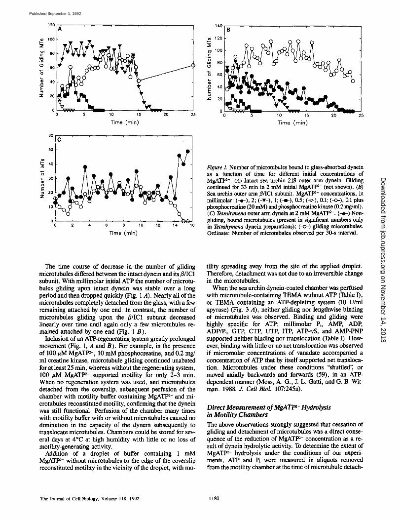
Perfusion of the chamber with microtubule- and MgATP 2-- containing buffer resulted in binding of microtubules to the sea urchin dynein-coated cover slip. Immediately upon binding, microtubules began gliding across the cover slip. The number of bound microtubules increased rapidly during the first few minutes (Fig. 1, A and B); after this time gliding microtubules were probably in equilibrium with free micro- tubules, as detachment of bound microtubules and attach- ment of free microtubules was commonly observed. Gliding microtubules underwent net unidirectional displacement by a series of starts and stops and transient backward steps (Moss, A. G., J.-L. Gatti, and G. B. Witman. 1988. J. Cell Biol. 107:245a) (59), and exhibited considerable lateral mo- bility (32).
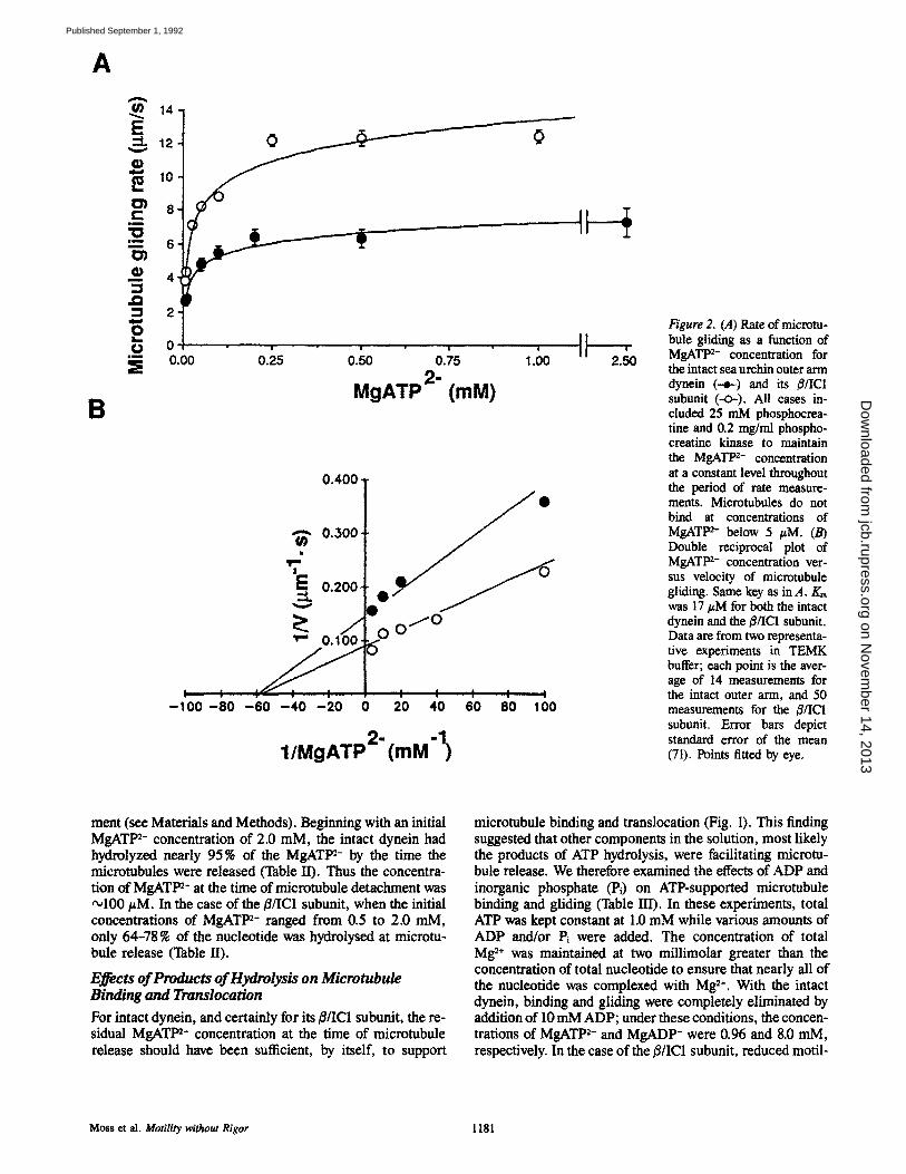
Gliding rate was dependent upon the concentration of MgATP 2- in buffer containing an ATP-regenerating system (10-25 mM phosphocreatine, 0.1-0.2 mg/mi phosphocrea- tine kinase) (Fig. 2, A and B). Both the intact outer ann dynein and its/3/IC1 subunit had an apparent Km for gliding of •17 #M MgATP 2-. The intact outer arm had a Vm~ of ~6.9/~m/s, whereas V~,~ was ,'~11.5 #m/s for the/3/IC1 subunit, confirming the observation of Sale and Fox (40) that the isolated/3/IC1 subunit translocates microtubules at a rate faster than that of the intact dynein.
Both the intact outer arm dynein and its/3/IC1 subunit sup- ported motility even at 5 pM MgATlm- in the presence of an ATP-regenerating system, but fewer microtubules were observed to glide at such low MgATP 2- concentrations, and those that did moved in a more saltatory manner. Microtu- bules did not bind to the cover slip at MgATP 2- concentra- tions <5 #M. The translocation rate for both the intact dynein and its ~/IC1 subunit increased up to 250/~M free MgATP 2- and then leveled off (Fig. 2 A). Microtubules translocated by the/~/IC1 subunit had relatively more lateral mobility, and progressed in a more discontinuous manner than those being translocated by the intact dynein (data not shown). A consistent feature of the microtubule-sea urchin dynein interaction was that there were very few nonmotile, bound microtubules (Fig, 3 A). Those that did bind without gliding were usually attached at only one point, or by one end, not along their full lengths, as were gliding microtu- bules.
Binding of Microtubules by Sea Urchin Dynein Requires MgAT1 ~- In the absence of an ATP-regenerating system, the microtu- bules eventually detached from the cover slip. The length of the motile period before detachment was directly propor- tional to the amount of added MgATP 2- (Figs. 1 A and B). Motility became less steady over time, and shorter microtu- bules were released first, so that the observed population was composed progressively of longer and longer microtubules. These results strongly suggest that the number of dynein- microtubule cross-bridges per unit length of microtubule de- creased over time; longer microtubules could interact with more dynein arms, and so remain attached to the glass for a longer period of time.
Moss et al. Motility without Rigor 1179
on Novem
ber 14, 2013jcb.rupress.org
Dow
nloaded from
Published September 1, 1992
100 �9 ~ 12o :E :5
== 8o E Ioo
~ 60
,
7= #" 20
0 5 10 15 20 25 0 5 10 15 20 25
Time (rain) Time (min)
60
50
I~" #0
~_ 30
E 20
z I 10
!c
i i i i i i
2 4 6 8 10 12 14
T i m e ( r a i n )
16
Figure 1. Number of microtubules bound to glass-absorbed dynein as a function of time for different initial concentrations of MgATP 2-. (A) Intact sea urchin 21S outer arm dynein. Gliding continued for 33 min in 2 mM initial MgATP 2- (not shown). (B) Sea urchin outer arm ~/IC1 subunit. MgATP 2- concentrations, in millimolar: (--~-), 2; (-v-), 1; (--l-), 0.5; (-~-), 0.1; (-<>-), 0.1 plus phosphocreatine (20 mM) and phosphocreatine kinase (0.2 mg/ml). (C) Tetrahymena outer arm dynein at 2 mM MgATP 2-. (--e-) Non- gliding, bound mierotubules (present in significant numbers only in Tetrahymena dynein preparations); (--o-) gliding microtubules. Ordinate: Number of mierotubules observed per 30-s interval.
The time course of decrease in the number of gliding microtubules differed between the intact dynein and its #/IC1 subunit. With millimolar initial ATP the number of microtu- bules gliding upon intact dynein was stable over a long period and then dropped quickly (Fig. 1 A). Nearly all of the microtubules completely detached from the glass, with a few remaining attached by one end. In contrast, the number of microtubules gliding upon the ~/IC1 subunit decreased linearly over time until again only a few microtubules re- mained attached by one end (Fig. 1 B).
Inclusion of an ATP-regenerating system greatly prolonged movement (Fig. 1, A and B). For example, in the presence of 100 #M MgATt n-, 10 mM phosphocreatine, and 0.2 mg/ ml creatine kinase, microtubule gliding continued unabated for at least 25 min, whereas without the regenerating system, 100 #M MgATt n- supported motility for only 2-3 min. When no regeneration system was used, and microtubules detached from the coverslip, subsequent perfusion of the chamber with motility buffer containing MgATP 2- and mi- crotubules reconstituted motility, confirming that the dynein was still functional. Perfusion of the chamber many times with motility buffer with or without microtubules caused no diminution in the capacity of the dynein subsequently to translocate microtubules. Chambers could be stored for sev- eral days at 4~ at high humidity with little or no loss of motility-generating activity.
Addition of a droplet of buffer containing 1 mM MgATP 2- without microtubules to the edge of the coverslip reconstituted motility in the vicinity of the droplet, with mo-
tility spreading away from the site of the applied droplet. Therefore, detachment was not due to an irreversible change in the microtubules.
When the sea urchin dynein-coated chamber was perfused with microtubule-containing TEMA without ATP (Table I), or TEMA containing an ATP-depleting system (10 U/ml apyrase) (Fig. 3 A), neither gliding nor lengthwise binding of microtubules was observed. Binding and gliding were highly specific for ATP; millimolar Pi, AMP, ADP, ADP/Pi, GTP, CTP, UTP, ITP, ATP-'yS, and AMP-PNP supported neither binding nor translocation (Table I). How- ever, binding with little or no net translocation was observed if micromolar concentrations of vanadate accompanied a concentration of ATP that by itself supported net transloca- tion. Microtubules under these conditions "shuttled", or moved axially backwards and forwards (59), in an ATP- dependent manner (Moss, A. G., J.-L. Gatti, and G. B. Wit- man. 1988. J. Cell Biol. 107:245a).
Direct Measurement of MgATP 2- Hydrolysis in Motility Chambers
The above observations strongly suggested that cessation of gliding and detachment of microtubules was a direct conse- quence of the reduction of MgATP 2- concentration as a re- sult of dynein hydrolytic activity. To determine the extent of MgATP ~- hydrolysis under the conditions of our experi- ments, ATP and Pi were measured in aliquots removed from the motility chamber at the time of microtubule detach-
The Journal of Cell Biology, Volume 118, 1992 1180
on Novem
ber 14, 2013jcb.rupress.org
Dow
nloaded from
Published September 1, 1992
A
B
o 14
N 4 .Q
2
0 i._
=i
0
0 ............ i �9 | J i " " ' i
0.00 0.25 0.50 0.75 1.00 2-
MgATP (mM)
0 . 4 0 0
0 . . 300 '
o. ooi F O j O
I I : . 1 I ............. I : : : : I - I 0 0 - 8 0 - 6 0 - 4 0 - 2 0 0 2 0 4 0 6 0 8 0 1 0 0
1/MgATp2" (raM "1 )
A
i''"' ' i 2.50
Figure 2. (A) Rate of microtu- bule gliding as a function of MgATP 2- concentration for the intact sea urchin outer arm dynein (--~) and its i$/IC1 subunit (-O-). All cases in- eluded 25 mM phosphocrea- title and 0.2 mg/ml phospho- creatine kinase to maintain the MgATt n- concentration at a constant level throughout the period of rate measure- ments. Mierotubules do not bind at concentrations of MgATt n- below 5 #M. (B) Double reciprocal plot of MgATP 2- concentration ver- sus velocity of microtubule gliding. Same key as in A. Km was 17 #M for both the intact dynein and the/$/IC1 subunit. Data are from two representa- tive experiments in TEMK buffer; each point is the aver- age of 14 measurements for the intact outer arm, and 50 measurements for the #/IC1 subunit. Error bars depict standard error of the mean (71). Points fitted by eye.
ment (see Materials and Methods). Beginning with an initial MgATP 2- concentration of 2.0 raM, the intact dynein had hydrolyzed nearly 95% of the MgATt m- by the time the microtubules were released (Table H). Thus the concentra- tion of MgAT1 m- at the time of microtubule detachment was ~100 #M. In the case of the B/IC1 subunit, when the initial concentrations of MgATt n- ranged from 0.5 to 2.0 mM, only 64-78 % of the nucleotide was hydrolysed at microtu- bule release (Table II).
Effects of Products of Hydrolysis on Microtubule Binding and ~,anslocation
For intact dynein, and certainly for its B/IC1 subunit, the re- sidual MgAT/n- concentration at the time of microtubule release should have been sufficient, by itself, to support
microtubule binding and translocation (Fig. I). This finding suggested that other components in the solution, most likely the products of ATP hydrolysis, were facilitating microtu- bule release. We therefore examined the effects of ADP and inorganic phosphate (Pi) on ATP-supported microtubule binding and gliding (Table III). In these experiments, total ATP was kept constant at 1.0 mM while various amounts of ADP and/or Pi were added. The concentration of total Mg 2+ was maintained at two millimolar greater than the concentration of total nucleotide to ensure that nearly all of the nucleotide was complexed with Mg ~+. With the intact dynein, binding and gliding were completely eliminated by addition of 10 mM ADP; under these conditions, the concen- trations of MgATP 2- and MgADP- were 0.96 and 8.0 raM, respectively. In the case of the/~/IC1 subunit, reduced motil-
Moss et al. Motility withous Rigor 1181
on Novem
ber 14, 2013jcb.rupress.org
Dow
nloaded from
Published September 1, 1992
14.0
120
100
~ s o
~ 4o
20
0
A ' ' ' 4 ; ' ' 4 ; ' '
2 4. 6 8 10 12 14. 16 18 20
Time ( m i n )
I-- ~E
k_ G3
..a
Z
30
25
20
15
10
5
0 0
B . . . . . 4; ' ~ . . . .
w
1 2 3 4. 5 6 7 8 9 10 11 12 13 14 15
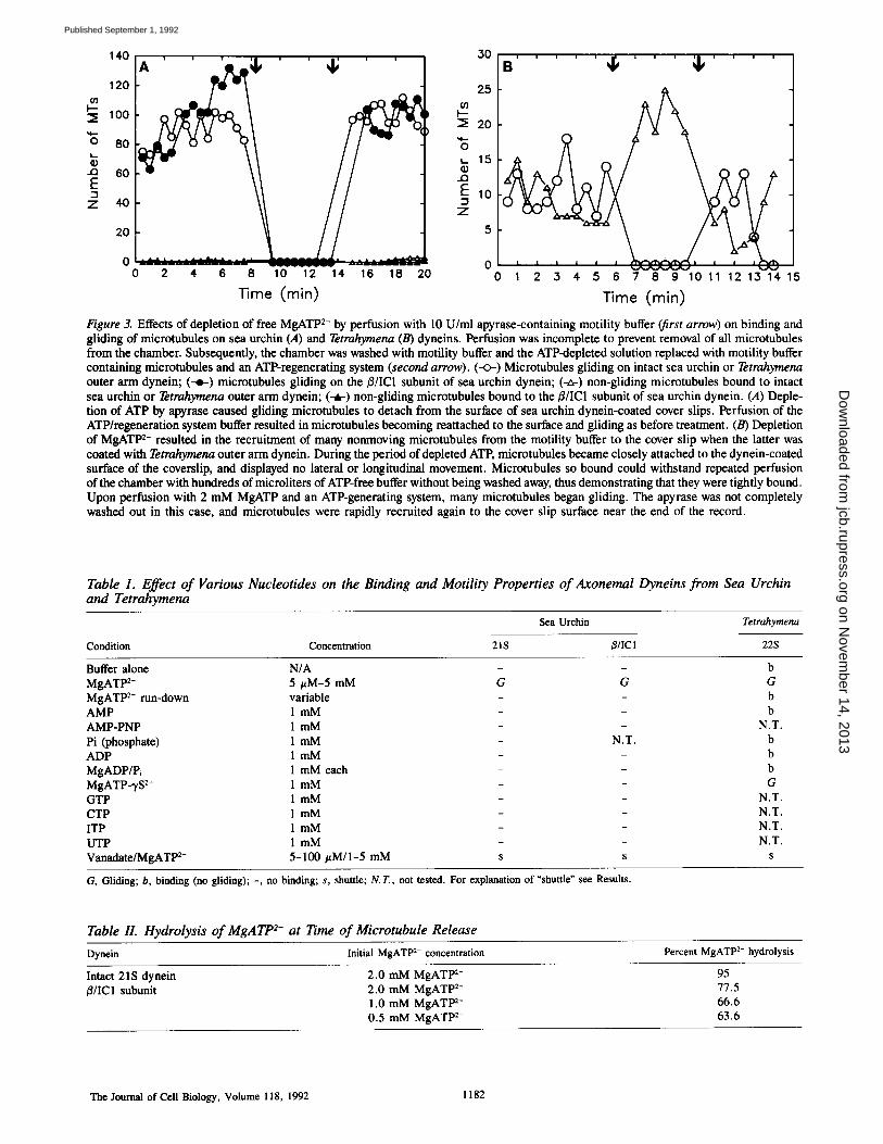
Time (min) Figure 3. Effects of depletion of free MgATP 2- by perfusion with 10 U/ml apyrase-containing motility buffer (first arrow) on binding and gliding of microtubules on sea urchin (A) and Tetrahymena (B) dyneins. Perfusion was incomplete to prevent removal of all microtubules from the chamber. Subsequently, the chamber was washed with motility buffer and the ATP-depleted solution replaced with motility buffer containing microtubules and an ATP-regenerating system (second arrow). (--0-) Microtubules gliding on intact sea urchin or Tetrahymena outer arm dynein; (--o-) microtubules gliding on the/3/IC1 subunit of sea urchin dynein; (-~-) non-gliding microtubules bound to intact sea urchin or Tetrahymena outer arm dynein; (-~-) non-gliding microtubules bound to the/3/IC1 subunit of sea urchin dynein. (A) Deple- tion of ATP by apyrase caused gliding microtubules to detach from the surface of sea urchin dynein-coated cover slips. Perfusion of the ATP/regeneration system buffer resulted in microtubules becoming reattached to the surface and gliding as before treatment. (B) Depletion of MgATP 2- resulted in the recruitment of many nonmoving microtubules from the motility buffer to the cover slip when the latter was coated with Tetrahymena outer arm dynein. During the period of depleted ATP, microtubules became closely attached to the dynein-coated surface of the coverslip, and displayed no lateral or longitudinal movement. Microtubules so bound could withstand repeated perfusion of the chamber with hundreds of microliters of ATP-free buffer without being washed away, thus demonstrating that they were tightly bound. Upon perfusion with 2 mM MgATP and an ATP-generating system, many microtubules began gliding. The apyrase was not completely washed out in this case, and microtubules were rapidly recruited again to the cover slip surface near the end of the record.
Table 1. Effect of Various Nucleotides on the Binding and Motility Properties of Axonemal Dyneins from Sea Urchin and Tetrahymena
Sea Urchin Tetrahymena
Condition Concentration 21S j3/IC 1 22S
Buffer alone N/A - b MgATP ~- 5/~M-5 mM G G G MgATP 2- run-down variable - b AMP 1 m M - b AMP-PNP 1 mM - N.T. Pi (phosphate) 1 mM - N.T. b ADP 1 mM - b MgADP/P~ 1 mM each - b MgATP--rS 2- 1 mM - - G GTP 1 mM - - N.T. CTP 1 rnM - - N.T. ITP 1 mM - - N.T. UTP 1 mM - - N.T. Vanadate/MgATP 2- 5-100/~M/1-5 mM s s s
G, Gliding; b, binding (no gliding); -, no binding; s, shuttle; N.T., not tested. For explanation of "shuttle" see Results.
Table II. Hydrolysis of MgATP 2- at Time of Microtubule Release
Dynein Initial MgATP 2- concentration Percent MgATP 2- hydrolysis
Intact 21S dynein 2.0 mM MgATI ~- 95 /3/IC1 subunit 2.0 mM MgATP 2- 77.5
1.0 mM MgATP 2- 66.6 0.5 mM MgATP 2- 63.6
T h e Journal of Cell Biology, Volume 118, 1992 1182
on Novem
ber 14, 2013jcb.rupress.org
Dow
nloaded from
Published September 1, 1992
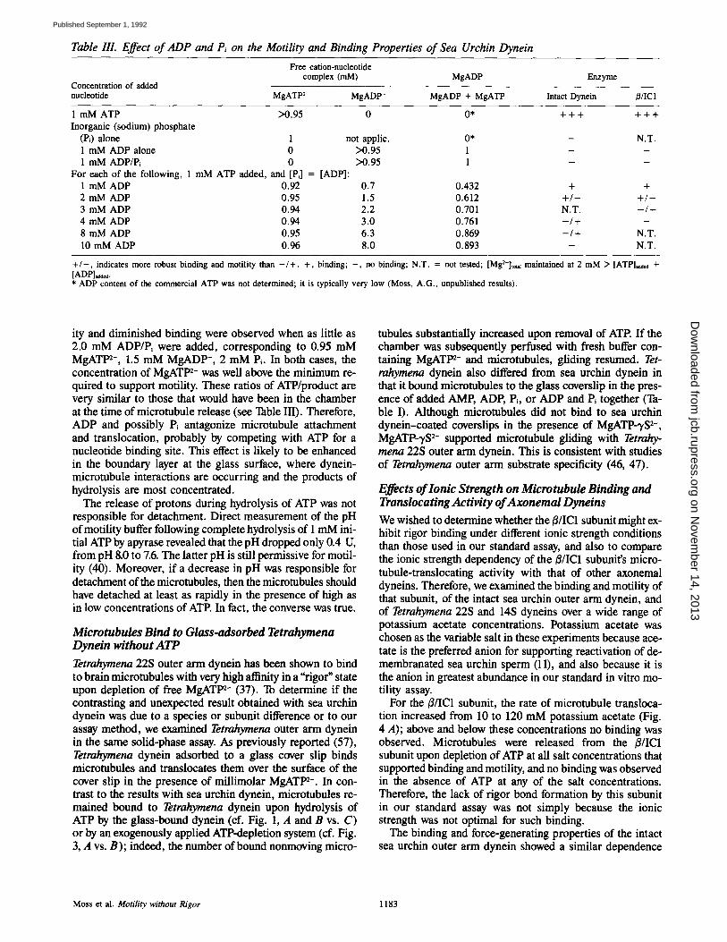
Table IlL Effect of ADP and Pi on the Motility and Binding Properties of Sea Urchin Dynein
Free cation-nucleotide complex (mM) MgADP Enzyme
Concentration of added nucleotide MgATP 2- MgADP- MgADP + MgATP Intact Dynein B/ICI
1 mM ATP >0.95 0 0* + + + + + + Inorganic (sodium) phosphate
(P0 alone 1 not applic. 0* - N.T. 1 mM ADP alone 0 >0.95 1 - - 1 mM ADP/P~ 0 >0.95 1 - -
For each of the following, 1 mM ATP added, and [Pd = [ADP]: 1 mM ADP 0.92 0.7 0.432 + + 2 mM ADP 0.95 1.5 0.612 + / - + / - 3 mM ADP 0.94 2.2 0.701 N.T. - / + 4 mM ADP 0.94 3.0 0.761 - / + - 8 mM ADP 0.95 6.3 0.869 - / + N.T, 10 mM ADP 0.96 8.0 0.893 - N.T.
+ / - , indicates more robust binding and motility than - / + . +, binding; - , no binding; N.T. = not tested; [Mg2+]t~ maintained at 2 mM > [ A T P ] ~ 4- [ADP]~ . * ADP content of the commercial ATP was not determined; it is typically very low (Moss, A.G., unpublished results).
ity and diminished binding were observed when as little as 2.0 mM ADP/Pi were added, corresponding to 0.95 mM MgATP 2-, 1.5 mM MgADP-, 2 mM P~. In both cases, the concentration of MgATP 2- was well above the minimum re- quired to support motility. These ratios of ATP/product are very similar to those that would have been in the chamber at the time of microtubule release (see Table III). Therefore, ADP and possibly Pi antagonize microtubule attachment and translocation, probably by competing with ATP for a nucleotide binding site. This effect is likely to be enhanced in the boundary layer at the glass surface, where dynein- microtubule interactions are occurring and the products of hydrolysis are most concentrated.
The release of protons during hydrolysis of ATP was not responsible for detachment. Direct measurement of the pH of motility buffer following complete hydrolysis of 1 mM ini- tial ATP by apyrase revealed that the pH dropped only 0.4 U, from pH 8.0 to 7.6. The latter pH is still permissive for motil- ity (40). Moreover, if a decrease in pH was responsible for detachment of the microtubules, then the microtubules should have detached at least as rapidly in the presence of high as in low concentrations of ATE In fact, the converse was true.
Microtubules Bind to Glass-adsorbed Tetrahymena Dynein without ATP
Tetrahymena 22S outer arm dynein has been shown to bind to brain microtubules with very high affinity in a "rigor" state upon depletion of free MgATP 2- (37). To determine if the contrasting and unexpected result obtained with sea urchin dynein was due to a species or subunit difference or to our assay method, we examined Tetrahymena outer arm dynein in the same solid-phase assay. As previously reported (57), Tetrahymena dynein adsorbed to a glass cover slip binds microtubules and translocates them over the surface of the cover slip in the presence of millimolar MgATP 2-. In con- trast to the results with sea urchin dynein, microtubules re- mained bound to Tetrahymena dynein upon hydrolysis of ATP by the glass-bound dynein (cf. Fig. 1, A and B vs. C) or by an exogenously applied ATP-depletion system (cf. Fig. 3, A vs. B); indeed, the number of bound nonmoving micro-
tubules substantially increased upon removal of ATE If the chamber was subsequently perfused with fresh buffer con- taining MgATP 2- and microtubules, gliding resumed. Tet- rahymena dynein also differed from sea urchin dynein in that it bound microtubules to the glass coverslip in the pres- ence of added AMP, ADP, Pi, o r ADP and Pi together (Ta- ble I). Although microtubules did not bind to sea urchin dynein-coated coverslips in the presence of MgATP-3,S 2-, MgATP-?S 2- supported microtubule gliding with Tetrahy- mena 22S outer arm dynein. This is consistent with studies of Tetrahymena outer arm substrate specificity (46, 47).
Effects of Ionic Strength on Microtubule Binding and Translocating Activity of Axonemal Dyneins
We wished to determine whether the B/IC1 subunit might ex- hibit rigor binding under different ionic strength conditions than those used in our standard assay, and also to compare the ionic strength dependency of the/3/IC1 subunit's micro- tubule-translocating activity with that of other axonemal dyneins. Therefore, we examined the binding and motility of that subunit, of the intact sea urchin outer arm dynein, and of Tetrahymena 22S and 14S dyneins over a wide range of potassium acetate concentrations. Potassium acetate was chosen as the variable salt in these experiments because ace- tate is the preferred anion for supporting reactivation of de- membranated sea urchin sperm (11), and also because it is the anion in greatest abundance in our standard in vitro mo- tility assay.
For the ~/IC1 subunit, the rate of microtubule transloca- tion increased from 10 to 120 mM potassium acetate (Fig. 4 A); above and below these concentrations no binding was observed. Microtubules were released from the /~/IC1 subunit upon depletion of ATP at all salt concentrations that supported binding and motility, and no binding was observed in the absence of ATP at any of the salt concentrations. Therefore, the lack of rigor bond formation by this subunit in our standard assay was not simply because the ionic strength was not optimal for such binding.
The binding and force-generating properties of the intact sea urchin outer arm dynein showed a similar dependence
Moss et al. Motility without Rigor 1183
on Novem
ber 14, 2013jcb.rupress.org
Dow
nloaded from
Published September 1, 1992
I0
I 60
E
o" C~ k _
CD r ~ "X2 ~
(_9 i 0
i t
I I I
- 1 [ S a l t ] , m m o l I
I U)
2.5
B
2.0'
E c : I .
1.5
- 4 - J
0 L. 1.0
O~
:• 0.~
0.0 200 0 200 50 100 150
- 1 [Salt], m m o l I
Figure 4. Effect of salt concentration on the gliding rate of microtubules upon glass-adsorbed axonemal dyneins. Abscissa indicates the concentration of potassium acetate. (A) Sea urchin dynein: (-o-) intact 21S outer arm; (-r B/IC1 subunit. (B) Tetrahymena 14S (-<>-) and 22S dynein (--e-). Error bars, standard error of the mean. Data represent microtubules that were detectably moving; all nonmoving microtubules were ignored. The background of nonmoving microtubules was inversely related to the ionic strength in the intact dynein samples, with the most pronounced effect occurring with Tetrahymena 14S dynein.
on ionic strength from 10 to 120 mM potassium acetate (Fig. 4 A). However, some microtubules remained bound to the intact dynein and translocated slowly even in the absence of potassium acetate. Above 120 mM potassium acetate, the number of bound microtubules dropped off dramatically but the rate of gliding of the remaining microtubules continued to increase up to 180 mM potassium acetate, at which con- centration the gliding rate matched the maximum rates ob- served for microtubules being translocated by the purified B/IC1 subunit. Above 180 mM potassium acetate, no bind- ing or gliding was observed. These results suggest that as ionic strength increases, sea urchin dynein proceeds through its mechanochemical cycle more rapidly, and its affinity for microtubules diminishes. Like the B/IC1 subunit, the intact arm released microtubules upon depletion of ATP and did not bind microtubules in the absence of ATP at all ionic strengths tested.
Tetrahyraena 22S outer arm dynein translocated microtu- bules at a maximal rate of 1.5/~m/s at 120 mM potassium ace- tate (Fig. 4 B); the rate of gliding decreased above and below this concentration, although many microtubules remained bound but did not glide at both the higher and lower ionic strengths. In contrast, Tetrahymena 14S dynein bound and translocated microtubules maximally at low ionic strength, and exhibited only marginal motility at potassium acetate concentrations of 100 mM or higher (Fig. 4 B). These re- sults suggest that these two dyneins interact with microtu- bules via different types of bonds. In contrast to the situation with the sea urchin dyneins, microtubules remained bound to the glass-adsorbed Tetrahymena 22S and 14S dyneins in the absence of ATP or upon depletion of ATP at all salt con- centrations that supported motility.
All glass adsorbed dyneins recovered translocating activ- ity after high or low salt treatment; therefore, loss of activity at these concentrations did not result in irreversible denatu- ration of the enzyme.
Cosedimentation Assays To determine if soluble dynein had the same microtubule-
binding properties as glass-adsorbed dynein, we investigated the microtubule-binding characteristics of sucrose gradient- purified Tetrahymena and S. purpuratus dyneins using con- ventional cosedimentation assays.
As previously reported (37), Tetrahymena 22S dynein bound to microtubules in the absence of MgATP2-; almost all of the 22S dynein sedimented with microtubules up to a molar ratio of -,1 dynein/29 tubulin monomers, at which saturation of binding was clearly evident (Fig. 5). Addition of 2 mM MgATP ~- released most of the Tetrahymena dynein from the microtubules (data not shown; see reference 27), indicating that most of the Tetrahymena dynein was bound by ATP-sensitive sites, again as previously reported (37).
Most of the intact sea urchin dynein partitioned into the pellet at low molar ratios of dynein/tubulin, but increasing amounts of dynein remained in the supernatant as the molar ratio increased. Saturation of binding became evident at a molar ratio of "~1 dynein molecule/39 tubulin monomers (Fig. 5). A portion of this bound dynein was released from the microtubules by MgATt n- (data not shown; see refer- ence 27), indicating that some of the intact arms were bound by rigor bonds whereas the remainder were bound by ATP- insensitive bonds. In the accompanying paper (27), we inves- tigate the origin of ATP-sensitive and -insensitive binding in intact sea urchin dynein, and conclude that both are proper- ties of the o~ subunit.
In contrast to the situation with the intact sea urchin and Tetrahymena dyneins, the sea urchin /~/IC1 subunit never sedimented with microtubules, even at a high molar ratio of 1 dynein molecule/8.8 tubulin monomers (Fig. 5). Addition of 2 mM MgATt n- to the mixture had no effect on the sedi- mentation behavior of the/~/IC1 subunit (data not shown; see reference 27). Therefore, the cosedimentation assay, like the solid-phase assay, provides no evidence for rigor bond for- marion by this subunit. Moreover, these results indicate that the isolated/~/IC1 subunit also is incapable of ATP-insensi- five, structural binding to microtubules.
Electrophoretic analysis of supernatant and pellet samples
The Journal of Cell Biology, Volume 118, 1992 1184
on Novem
ber 14, 2013jcb.rupress.org
Dow
nloaded from
Published September 1, 1992
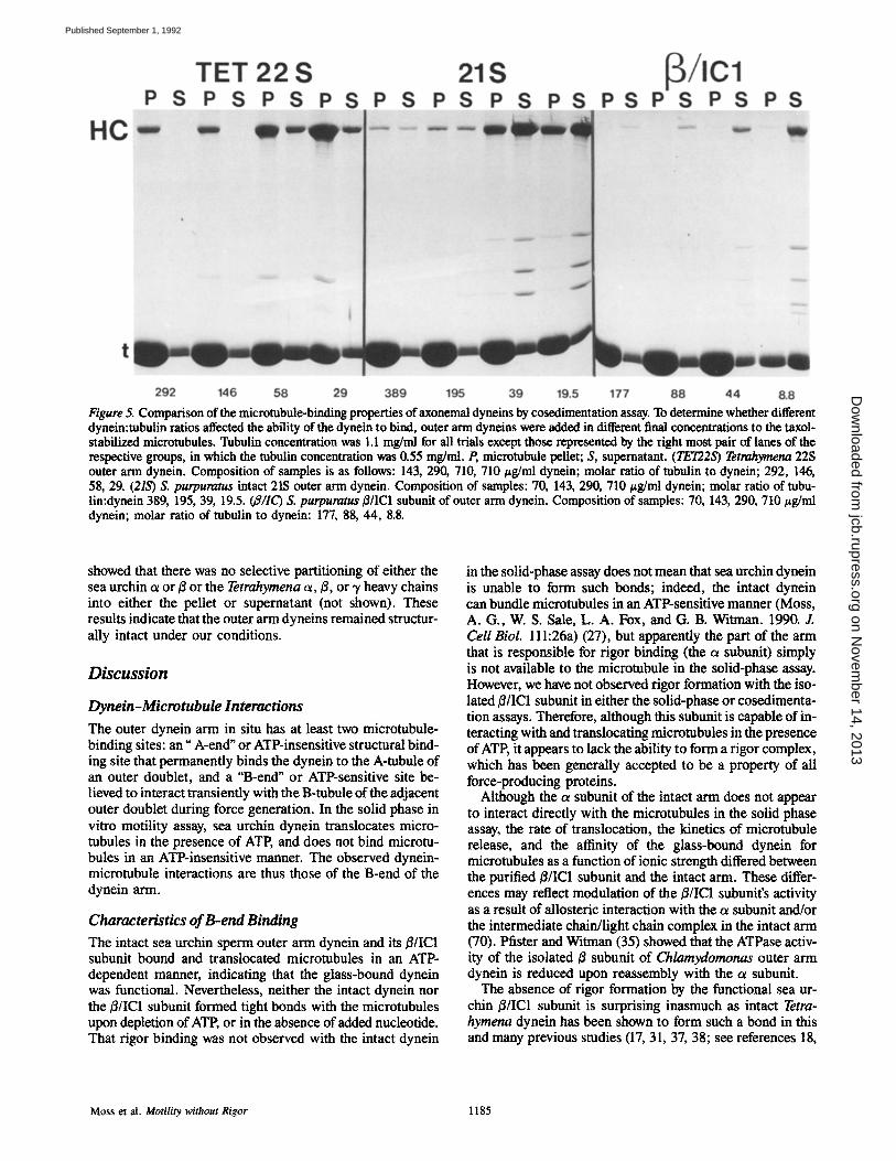
Figure 5. Comparison of the microtubule-binding properties of axonemal dyneins by cosedimentation assay. To determine whether different dynein:tubulin ratios affected the ability of the dynein to bind, outer arm dyneins were added in different final concentrations to the taxol- stabilized microtubules. Tubulin concentration was 1.1 mg/ml for all trials except those represented by the right most pair of lanes of the respective groups, in which the tubulin concentration was 0.55 mg/ml. P, microtubule pellet; S, supematant. (TET22S) Tetrahymena 22S outer arm dynein. Composition of samples is as follows: 143, 290, 710, 710 #g/ml dynein; molar ratio of tubulin to dynein; 292, 146, 58, 29. (21S) S. purpuratus intact 2tS outer arm dynein. Composition of samples: 70, 143, 290, 710/~g/ml dynein; molar ratio of tubu- lin:dynein 389, 195, 39, 19.5. (a/IC) S. purpuratus B/IC1 subunit of outer arm dynein. Composition of samples: 70, 143, 290, 710/~g/ml dynein; molar ratio of tubulin to dynein: 177, 88, 44, 8.8.
showed that there was no selective partitioning of either the sea urchin u or/~ or the Tetrahymena a, ~, or ,y heavy chains into either the pellet or supernatant (not shown). These results indicate that the outer arm dyneins remained structur- ally intact under our conditions.
Discussion
Dynein-Microtubule Interactions
The outer dynein arm in situ has at least two microtubule- binding sites: an" A-end" or ATP-insensitive structural bind- ing site that permanently binds the dynein to the A-tubule of an outer doublet, and a "B-end" or ATP-sensitive site be- lieved to interact transiently with the B-tubule of the adjacent outer doublet during force generation. In the solid phase in vitro motility assay, sea urchin dynein translocates micro- tubules in the presence of ATP, and does not bind microtu- bules in an ATP-insensitive manner. The observed dynein- microtubule interactions are thus those of the B-end of the dynein arm.
Characteristics of B-end Binding
The intact sea urchin sperm outer arm dynein and its ~/IC1 subunit bound and translocated microtubules in an ATP- dependent manner, indicating that the glass-bound dynein was functional. Nevertheless, neither the intact dynein nor the ~/IC1 subunit formed tight bonds with the microtubulcs upon depletion of ATP, or in the absence of added nucleotidc. That rigor binding was not observed with the intact dynein
in the solid-phase assay does not mean that sea urchin dynein is unable to form such bonds; indeed, the intact dynein can bundle microtubules in an ATP-sensitive manner (Moss, A. G., W. S. Sale, L. A. Fox, and G. B. Witman. 1990. Z Cell Biol. 111:26a) (27), but apparently the part of the arm that is responsible for rigor binding (the a subunit) simply is not available to the microtubule in the solid-phase assay. However, we have not observed rigor formation with the iso- lated/3/IC1 subunit in either the solid-phase or cosedimenta- tion assays. Therefore, although this subunit is capable of in- teracting with and translocating microtubules in the presence of ATP, it appears to lack the ability to form a rigor complex, which has been generally accepted to be a property of all force-producing proteins.
Although the tx subunit of the intact arm does not appear to interact directly with the microtubules in the solid phase assay, the rate of translocation, the kinetics of microtubule release, and the affinity of the glass-bound dynein for microtubules as a function of ionic strength differed between the purified/~/IC1 subunit and the intact arm. These differ- ences may reflect modulation of the B/IC1 subunit's activity as a result of allosteric interaction with the c~ subunit and/or the intermediate chain/light chain complex in the intact arm (70). Pfister and Witman (35) showed that the ATPase activ- ity of the isolated B subunit of Chlamydomonas outer arm dynein is reduced upon reassembly with the ct subunit.
The absence of rigor formation by the functional sea ur- chin/3/IC1 subunit is surprising inasmuch as intact Tetra- hymena dynein has been shown to form such a bond in this and many previous studies (17, 31, 37, 38; see references 18,
Moss et aL Motility without Rigor 1185
on Novem
ber 14, 2013jcb.rupress.org
Dow
nloaded from
Published September 1, 1992
39 for reviews). A possible explanation for the different be- haviors is that the/~/IC1 subunit of sea urchin dynein and the force-generating subunit(s) of Tetrahymena dynein differ in their relative affinities for microtubules during one or more stages of the mechanochemical cycle. Kinetic analysis of the isolated Tetrahymena outer arm dynein has clearly estab- lished that microtubules bind to the Tetrahymena dynein/ ADP/P~ complex and remain tightly bound during product release and in the absence of bound products; dissociation of the microtnhule/dynein complex occurs rapidly upon ad- dition of ATP (15, 17, 31, 38). Our finding that the/~/IC1 subunit of sea urchin dynein releases microtubules when ATP is depleted and does not bind microtubules in the ab- sence of added MgATP 2- suggests that this subunit, in con- trast to Tetrahymena dynein, has a lower affinity for microtu- bules when its hydrolytic site is empty than when the site is occupied (isolated sea urchin outer arm dynein is unlikely to contain bound nucleotide at its hydrolytic sites, because vanadate-dependent photolysis of the heavy chains is not de- tected without added ADP or ATP [24]).
Addition of excess ADP and Pi enhanced release of microtubules from sea urchin dynein in the presence of ATE suggesting that the /~/IC1/ADP and /~/ICI/ADP/Pi com- plexes also have relatively low affinities for microtubules. However, it is possible that the complexes formed by the ad- dition of exogenous product differ in their affinities for microtubules from those formed by the hydrolysis of bound ATP (Shimizu, T., S. Ohashi, and T. Katsura. 1990. J. Cell Biol. 111:118a).
Schemes for the mechanochemical cycle of the /~/IC1 subunit of sea urchin dynein also can be constructed that are in agreement with our data and that differ more profoundly from that of Tetrahymena dynein. It will be necessary to de- termine the rate constants for specific steps in the cycle to know if such schemes are realistic. Nevertheless, it appears that the ability to form a rigor bond is not a prerequisite for microtubule binding and force generation.
An alternative explanation for the observed differences be- tween the ~/IC1 subunit of sea urchin dynein and intact Tetrahymena dynein is that the activity of the former may be regulated in an ATP-dependent manner not seen in the latter. The/3 heavy chain contains multiple ATP-binding consensus sequences (13, 28), suggesting that it binds ATP at more than one site. It is possible that loss or hydrolysis of ATP at one site affects the activity at a second site coupled to microtu- bule binding and force generation. Current models for the mechanism of axonemal bending dictate that the arms must have a low affinity for microtubules during at least one-half of each beat cycle to permit passive sliding of the outer dou- blet microtubules facing them (51, 61). The existence of a biochemical regulatory mechanism that turned the arms on and off at the appropriate time in the beat cycle would pro- vide more precise control over interdoublet sliding than could be achieved if the "off' time were simply determined by how fast the arm independently proceeded through its mechanochemical cycle. Such active control of dynein/mi- crotubule interactions probably is necessary for the regula- tion of flagellar beat frequency and curvature.
The above discussion notwithstanding, the differences in the rigor-forming capabilities of intact Tetrahymena dynein and the/~/IC1 subunit of sea urchin dynein do not necessarily mean that the force-generating moieties of these dyneins are
fundamentally different. The intact Tetrahymena dynein con- tains three subunits, and although the head of each subunit appears to interact directly with microtubules (45), increas- ing biochemical and structural data indicate that there are differences between the subunits (55, 56, 58). If one of the heads binds tightly to microtubules in the absence of ATE that interaction could prevent detection of weak ATP- dependent binding by another head in the intact particle. Further analysis of isolated subunits of protistan dyneins will be necessary to determine if a low affinity for microtubules in the absence of ATP is a property that is common to at least one subunit (possibly the motility-generating subunit) of all outer arms but which has gone unnoticed because of the rigor-forming properties of one or more other subunits in the intact particle.
A-end Binding A substantial proportion of the intact outer arm dynein of sea urchin sperm flagella bound to microtubules in the cosedi- mentation assay. In the accompanying paper (27), we show that some of this bound dynein is not released by ATE sug- gesting that these molecules are interacting with the microtu- bules via their structural A-end binding site. Such binding was not observed with the isolated ~/IC1 subunit, indicating that structural A-end binding requires a dynein component either not present or not functional in the ~/IC1 preparation. This component appears to be the a subunit (27).
The Utility of Various Assays for Determining Dynein/Microtubule Binding Properties In general, comparable qualitative results were obtained with the solid-phase and solution microtubule-binding as- says. In the absence of ATE the/3/IC1 subunit of sea urchin dynein neither bound microtubules to the glass coverslip nor cosedimented with microtubules. Similarly, intact Tetra- hymena dynein bound microtubules tightly to the coverslip in the absence of ATP and cosedimented with microtubules in an ATP-sensitive manner. However, some dynein/micro- tubule interactions were apparent in one assay but not the other. For example, ATP-insensitive A-end binding of the in- tact sea urchin dynein was apparent in the solution assay but not the solid phase assay (27). Conversely, because the sea urchin/~/IC1 subunit does not form a rigor bond, its transient B-end interactions with microtubules could be detected only in the solid-phase assay. Therefore, both assays are of value for studying dynein-microtubule interactions. The solid- phase assay is quicker and can be carried out with much less dynein and tubulin, and allows real-time observation of mo- lecular interactions, whereas the solution assay is at present more readily quantified.
Implications for the Isolation of Novel Microtubule-associated Motors An increasingly popular method for isolating microtubule- associated motors takes advantage of the rigor-forming prop- erties of the motor: the motor is bound to microtubules in the absence of nucleotide, sedimented with the microtu- bules, and specifically released into the supernatant in the presence of nucleotide. Our findings suggest that not all microtubule motors may be identified and isolated on the ba- sis of rigor formation. Some mechanochemical transducers
The Journal of Cell Biology, Volume 118, 1992 1186
on Novem
ber 14, 2013jcb.rupress.org
Dow
nloaded from
Published September 1, 1992
may remain in the original homogenate under ATP-depleted conditions. This should be kept in mind when attempting to isolate uncharacterized force-generating molecules from cytosolic extracts.
We wish to thank Drs. Stephen King and Winfield Sale for many stimulating discussions during the course of this work. We are grateful to Dr. Boyd Haley for suggesting the thin-layer chromatography protocol. We also wish to thank Drs. S. Marchese-Ragona and K. Johnson for the generous girls of Tetrahymena 22S outer arm dynein.
Supported by National Institutes of Health grant GM30626 to G. B. Wit- man, NIH NRSA GM12240 to A. G. Moss, and a grant from the Mellon Foundation.
Received for publication 20 February 1992 and in revised form 1 June 1992.
References
1. Bell, C. W., and I. R. Gibbons. 1982. Structure of the dynein-1 outer arm in sea urchin sperm flagella. II. Analysis by proteolytic cleavage. J. Biol. Chem. 257:516-522.
2. Bradford, M. M. 1976. A rapid and sensitive method for the quantitation of microgram quantities of protein utilizing the principle of protein-dye binding. Anal. Biochem. 72:248-254.
3. Brokaw, C. J. 1986. Future directions for studies of mechanisms for gener- ating flagellar bending waves. J. Cell $ci (Suppl). 4:103-113.
4. Brokaw, C. J. 1989. Operation and regulation of the flagellar oscillator. Chapter 20. In Cell Movement. Vol. 1. The Dynein ATPases. F. D. Warner, P. Satir, I. R. Gibbons, editors. 267-279.
5. Chiicote, T. J., and K. A. Johnson. 1989. Microtubule-dynein cross-bridge cycle and the kinetics of 5'-adenylyl imidodiphosphate (AMPPNP) bind- ing. Chapter 17. In Cell Movement. Vol. 1. The Dynein ATPases. E D. Warner, P. Satir, I. R. Gibbons, editors. 235-243.
6. Fox, L. A., and W. S. Sale. 1987. Direction of force generated by the inner row of dyne in arms on flagellar microtubules. J. Cell Biol. 105:1781-1787.
7. Gatti, J.-L., S. M. King, A. G. Moss, and G. B. Witman. 1989. Outer arm dynein from trout spermatozoa. I. Purification, polypeptide composition, and enzymatic properties. J. Biol. Chem. 264:11450-11457.
8. Gibbons, I. R. 1975. The molecular basis of flagellar motility in sea urchin spermatozoa. In Molecules and Cell Movement. Inoue, S., and R. E. Stephens. Raven Press, New York. 207-232.
9. Gibbons, B. H., and I. R. Gibbons. 1974. Properties of flagellar "rigor waves" produced by abrupt removal of adenosine triphosphate from ac- tively swimming sea urchin sperm. J. Cell Biol. 63:970-985.
10. Gibbons, B. H., and I. R. Gibbons. 1979. Relationship between the latent adenosine triphosphatase state of dynein 1 and its ability to recombine functionally with KCl-extracted sea urchin sperm flagella. J. Biol. Chem. 254:19%201.
11. Gibbons, B. H., W.-J. Y. Tang, and I. R. Gibbons. 1985. Organic ions stabi- lize the reactivated motility of sperm flagella and the latency of Dynein 1 ATPase activity. J. Cell Biol. 101:1281-1287.
12. Gibbons, I. R., and E. Fronk. 1979. A latent adenosine triphosphatase form of dynein 1 from sea urchin sperm flagella. J. Biol. Chem. 254:187-196.
13. Gibbons, I. R., B. H. Gibbons, B. Mocz, and D. J. Asai. 1991. Multiple nucleotide-binding sites in the sequence of dynein ~ heavy chain. Nature (Lond.). 352:640-643.
14. Hairno, L. T. 1989. Polarity of dynein binding to cytoplasmic microtubules. Chapter 11. In Cell Movement. Vol. 1. The Dynein ATPases. E D. Warner, P. Satir, and I. R. Gibbons, editors. 155-165.
15. Holzbanr, E. L. E, and K. A. Johnson. 1989. ADP release is rate limiting in steady-state turnover by the dynein adenosinetriphosphatase. Biochem- istry. 28:5577-5585.
16. Ishijima, S., and G. B. Witman. 1987. Flagellar movement of intact and de- membranated, reactivated ram spermatozoa. Cell MatU. and Cytoskele- ton. 8:375-391.
17. Johnson, K. A. 1983. The pathway of ATP hydrolysis by dynein. Kinetics of a presteady state phosphate burst. J. Biol. Chem, 258:13825-13832.
18. Johnson, K. A. 1985. Pathway of the microtubule-dynein ATPase and the structure of dynein: A comparison with actomyosin. Ann. Rev. Biophys. Biophys. Chem, 14:161-188.
19. Johnson, K. A. 1986. Preparation and properties of dynein from Tetra- hymena cilia. Methods Enzymol. 134:306-317.
20. King, S. M., and G. B. Witman. 1987. Structure of the ~x and ~ heavy chains of the outer ann dynein from Chlamydomonas flagella. Masses of chains and sites of ultraviolet-induced vanadate-dependent cleavage. J. Biol. Chem. 262:17596-17604.
21. King, S. M., and G. B. Witman. 1988. Structure of the ~ and ~ heavy chains of the outer arm dynein from Chlamydomonas flagella. Location of epitopes and protease-sensitive sites. J. Biol. Chem. 263:9244-9255.
22. King, S. M., J.-L. Gatti, A. G. Moss, and G. B. Witman. 1990. Outer-arm dynein from trout spermatozoa: substructural organization. Cell Motil. and Cytoskeleton. 16:266-278.
23. Laemmli, U. K. 1970. Cleavage of structural proteins during the assembly of the head of bacteriophage T4. Nature (Lond.). 227:680-685.
24. I_e,e-Eiford, A., R, A. Ow, and I. R. Gibbons. 1986. Specific cleavage of dynein heavy chains by ultraviolet irradiation in the presence of ATP and vanadatr J. Biol. Chem. 261:2337-2342.
25. Mitchell, D. A., and F. D. Warner. 1980. Interactions of dynein arms with B subfibers of Tetrahymena cilia: quantification of the effects of mag- nesium and adenosine triphosphate. J. Cell Biol. 87:84-97.
26. Mitchell, D. A., and F. D. Warner. 1981. Binding of dynein 218 ATPase to microtubules. Effects of ionic conditions and substrate analogs. J. Biol. Chem. 256:12535-12544.
27. Moss, A. G., W. S. Sale, L. A. Fox, and G. B. Witman. 1992. The cx subunit of sea urchin outer arm dynein mediates structural and rigor bind- ing to microtubules. J. Cell Biol. 118:1189-1200.
28. Ogawa, K. 1991. Four ATP-binding sites in the midregion of the f3 heavy chain of dynein, Nature (Lond.). 352:643-645.
29. Okuno, M. 1980. Inhibition and relaxation of sea urchin sperm flagella by vanadate. J. Cell Biol. 85:712-725.
30. Okuno, M., and C. J. Brokaw. 1979. Inhibition of movement of triton- demembranated sea-urchin sperm flagella by Mg 2+, ATP 4-, ADP and P~. J. Cell Sci. 38:105-123.
31. Omoto, C. M., and K. A. Johnson. 1986. Activation of the dynein adenosinetriphosphatase by microtubules. Biochemistry. 25:419-427.
32. Paschal, B. M., S. M. King, A. G. Moss, C. A. Collins, R. B. Vallee, and G. B. Witman. 1987. Isolated flagellar outer arm dynein translocates brain microtubules in vitro. Nature (Lond.). 330:672-674.
33. Penningroth, S., and G. B. Witman. 1978. Effects of adenylylimidodi- phosphate, a nonhydrolyzable adenosine triphosphate analog, on reacti- vated and rigor wave sea urchin sperm. J. Cell Biol. 79:827-832.
34. Penningroth, S. M., K. Olehnik, and A. Cheung. 1980. ATP formation from adenyl-5'-yl imidodiphosphate, a nonhydrolyzable ATP analog. J. Biol. Chem. 255:9545-9548.
35. Pfister, K. K., and G. B. Witman. 1984. Subfractionation of Chlamydo- monas 18S dynein into two unique subunits containing ATPase activity. J. Biol. Chem. 259:12072-12080.
36. Pfister, K. K., B. E. Haley, and G. B. Witman. 1984. The photoaflinity probe 8-azidoadenosine 5'-triphosphate selectively labels the heavy chain of Chlamydomonas 12S dynein. J. Biol. Chem. 259:8499-8504.
37. Porter, M. E., and K. A. Johnson. 1983a. Characterization of the ATP- sensitive binding of Tetrahymena 30S dynein to bovine brain microtu- bules. J. Biol. Chem. 258:6575-6581.
38. Porter, M. E., and K. A. Johnson. 1983b. Transient steady-state analysis of the ATP-induced dissociation of the dynein-microtubule complex. J. Biol. Chem. 258:6582-6587.
39. Porter, M. E., and K. A. Johnson. 1989. Dynein structure and function. Annu. Rev. Cell Biol. 5:119-151.
40. Sale, W. S., and L. Fox. 1988. Isolated B-heavy chain subunit of dynein translocates microtubules in vitro. J. Cell Biol. 107:1793-1797.
41. Sale, W. S., and P. Satir. 1977. Direction of active sliding of microtubules in Tetrahymena cilia. Proc. Natl. Acad. Sci. USA. 74:2045-2049.
42. Satir, P. 1985. Studies on cilia. II. Examination of the distal region of the ciliary shaft and the role of the filaments in motility. J. Cell Biol. 26:805-834.
43. Satir, P. 1968. Studies on cilia. HI. Further studies on the cilium tip and a "sliding filament" model of ciliary motility. J. Cell Biol. 93:77-94.
44. Satir, P. 1982. Mechanisms and controls of microtubule sliding in cilia. Soc. Exp. Biol. Symp. 35:179-201.
45. Shimizu, T., and K. A. Johnson. 1983. Kinetic evidence for multiple dynein ATPasr sites. J. Biol. Chem. 258:13841-13846.
46. Shimizu, T., and K. Furosawa. 1986. Phosphorothioate analogues of aden- osine 5'-triphosphate as substrates of dynein from Tetrahymena cilia. Bio- chemistry. 25:5787-5792.
47. Shimizu, T., K. Furosawa, S. Ohashi, Y. Y. Toyoshima, M. Okuno, E Ma- lik, and R. D. Vale. 1991. Nucleotide specificity of the enzymatic and mo- tile activities of dynein, kinesin, and heavy meromyosin. J. Cell Biol. 112:1189-1197.
48. Deleted in proof. 49. Shingyoji, C., A. Murakami, and K. Takahashi. 1977. Local reactivation of
triton-extracted flagella by iontophoretic application of ATP. Nature (Lond.). 265:269-270.
50. Spungin, B., J. Avolio, S. Arden, and P. Satir. 1987. Dynein arm attachment probed with a non-hydrolysable ATP analog. J. Mol. Biol. 197:671-677.
51. Sugino, K., and Y. Naitoh. 1982. Simulated cross-bridge patterns corre- sponding to ciliary beating in Paramecium. Nature (Lond.). 295:609-611.
52. Summers, K. E., and I. R. Gibbons. 1971. Adenosine triphosphate-induced sliding of tubules in trypsin-tw~ted flagella of sea-urchin sperm. Proc. Natl. Acad. Sci. USA. 68:3092-3096.
53. Takahashi, M., and Y. Tonomoura. 1978. Binding of 30S dynein with the B-tubule of the outer doublet of axonemes from Tetrahymena pyriformis and adenosine triphosphate-induced dissociation of the complex. J. Bio- chem. 84:1339-1355.
54. Tang, W.-J. Y., C. W. Bell, W. S. Sale, and I. R. Gibbons. 1982. Structure
Moss et al. Motility without Rigor 1187
on Novem
ber 14, 2013jcb.rupress.org
Dow
nloaded from
Published September 1, 1992
of the dynein-1 outer arm in sea urchin sperm flagella. I. Analysis by sepa- ration of subunits. J. Biol. Chem. 257:508-516.
55. Toyoshima, Y. Y. 1987a. Chymotryptic digestion of ~trahymena 22S dynein. I. Decomposition of three-headed 22S dynein to one- and two- headed particles. J. Cell Biol. 105:887-895.
56. Toyoshima, Y. Y. 1987b. Chymotryptic digestion of ~trahymena 22S dyuein. II. Pathway of the degradation of 22S dynein heavy chains. J. Cell Biol. 105:897-901.
57. Vale, R. D., and Y. Y. Toyoshima. 1988. Rotation and translocation of microtubules in vitro induced by dyneins from ~trahymena cilia. Cell. 52:459--469.
58. Vale, R. D., and Y. Y. Toyoshima. 1989. Microtubule translocation proper- ties of intact and proteolyticaily digested dyneins from ~trahymena cilia. J. Cell Biol. 108:2327-2334.
59. Vale, R. D., D. R. Soil, andI. R. Gibbons, 1989. One-dimensional diffusion of microtubules bound to flagellar dynein. Cell. 59:915-925.
60. Vallee, R. B. 1986. Reversible assembly purification ofmicrotubules without assembly-promoting agents and further purification of tubulin, microtu- bule associated proteins, and MAP fragments. Methods Enzymol. 134:89-115.
61. Wais-Steider, J., and P. Satir. 1979. Effect of vanadate on gill cilia: switch- ing mechanism in ciliary beat. J. Supramol. Struct. 11:339-347.
62. Warner, F. D. 1978. Cation-induced attachment of ciliary dynein cross-
bridges. J. Cell Biol. 78:R19-R26. 63. Warner, F. D., and J. H. McIlvain. 1982. Binding stoichiometry of 21S
dynein to A and B subflber microtubules. Cell Motil. 2:429-443. 64. Warner, F. D., J. G. Perrault, and J. H. McIlvain. 1985. Rebinding of
Tetrahymena 13S and 21S dynein ATPases to extracted doublet microtu- bules. The inner row and outer row dynein arms. J. Cell Sci. 77:263-287.
65. Warner, F. D., P. Satir, andI. R. Gibbons. editors. 1989. Cell Movement. Vol. 1. The Dynein ATPases. A. R. Liss, New York. 337 pp.
66. Windholz, M., S. Budavari, L. Y. Stroumtsos, and M. N. Fertig. 1976. The Merck Index. Ninth ed. Merck and Co., Inc., Rahway, NJ. 148.
67. Witman, G. B. 1989. Composition and molecular organization of the dyneins. Chapter 2. In Cell Movement. Vol. 1. The Dynein ATpase. F. D. Warner, P. Satir, I. R. Gibbons, editors. 25-35.
68. Witman, O. B. 1990. Introduction to cilia and flagella. Chapter 1. In Struc- tore and function of ciliary and flagellar surfaces. (Bloodgood, R. A., edi- tor. Plenum Publishing Corp., New York. 1-30.
69. Witman, G. B. 1992. Axonemal dyneins. Curr. Opin. Cell Biol. 4:74-79. 70. Witman, G. B., S. M. King, A. G. Moss, and C. G. Wilkersen. 1991. The
intermediate chain/light chain complex: an important structural entity of outer arm dynein. In Comparative Spermatology 20 Years After. B. Bac- cetti, editor. Raven Press, New York. 439--446.
71. Zar, J. H. 1984. Biostatistical Analysis. 2nd ed. Prentice-Hall, Englewood Cliffs, NJ.
The Journal of Cell Biology, Volume 118, 1992 1188
on Novem
ber 14, 2013jcb.rupress.org
Dow
nloaded from
Published September 1, 1992


![[Beating frequency of motile cilia lining the third cerebral ventricle is finely tuned by the hypothalamic peptide MCH]](https://static.fdokumen.com/doc/165x107/6334fe6f3e69168eaf07256d/beating-frequency-of-motile-cilia-lining-the-third-cerebral-ventricle-is-finely.jpg)