Cellular Pathology (Histopathology & Cytopathology) BASE ...
Upload
independentCategory
view
1download
0
Identi¢cation of novel cellular proteins that bind to the LC8 dyneinlight chain using a pepscan technique
Ignacio Rodr|guez-Crespoa;*, Belen Yelamosa, Fernando Roncalb, Juan Pablo Albarb,Paul R. Ortiz de Montellanoc, Francisco Gavilanesa
aDepartamento de Bioqu|mica y Biolog|a Molecular, Facultad de Ciencias Qu|micas, Universidad Complutense de Madrid, 28040 Madrid, SpainbDepartamento de Inmunolog|a y Oncolog|a, Centro Nacional de Biotecnolog|a, CSIC, Campus de la Universidad Autonoma de Madrid,
28049 Madrid, SpaincDepartment of Pharmaceutical Chemistry, School of Pharmacy, University of California, 513 Parnassus Ave, San Francisco, CA 94143-0446, USA
Received 29 June 2001; revised 19 July 2001; accepted 19 July 2001
First published online 31 July 2001
Edited by Gianni Cesareni
Abstract Dynein is a minus end-directed microtubule motorthat serves multiple cellular functions. We have performed a finemapping of the 8 kDa dynein light chain (LC8) binding sitesthroughout the development of a library of consecutive syntheticdodecapeptides covering the amino acid sequences of the variousproteins known to interact with this dynein member according tothe yeast two hybrid system. Two different consensus sequenceswere identified: GIQVD present in nNOS, in DNA cytosinemethyl transferase and also in GKAP, where it is present twice inthe protein sequence. The other LC8 binding motif is KSTQT,present in Bim, dynein heavy chain, Kid-1, protein 4 and also inswallow. Interestingly, this KSTQT motif is also present inseveral viruses known to associate with microtubules duringretrograde transport from the plasma membrane to the nucleusduring viral infection. ß 2001 Published by Elsevier ScienceB.V. on behalf of the Federation of European Biochemical So-cieties.
Key words: Migration; Microtubule; Dynein; Nitric oxide;Yeast two hybrid; Pepscan
1. Introduction
Dyneins are large multi-component, microtubule-based mo-lecular motors that generate force towards the minus end ofmicrotubules and are involved in several cellular processesincluding retrograde transport in axons, ciliary/£agellar beat-ing, vesicular transport, movement of endosomes and lyso-somes and positioning of the mitotic spindle [1^3]. From astructural and functional point of view, the two major classesof dyneins are the axonemal dyneins involved in £agellar andciliary movement, and the cytoplasmic dyneins. In genericterms, cytoplasmic dyneins are constructed around one ormore heavy chains (HCs), each of which forms a multilobedglobular head structure where the ATP hydrolysis responsiblefor the microtubule motor activity takes place. These HCs are
associated to several (probably two) copies of a 74 kDa inter-mediate chain (IC), four light intermediate chains (LICs) of50^60 kDa, and several light chains (LCs) with molecularmasses lower than 22 kDa. In the case of cytoplasmic dynein,this global association of several polypeptide chains results ina total molecular mass of V1.25 MDa [1^3].
Recently, King and coworkers have identi¢ed and charac-terised in detail the three LC components of 8 kDa, 14 kDaand 22 kDa within cytoplasmic dynein [4^7]. The 8 kDa dy-nein LC (LC8, actual mass 10 kDa) was ¢rst identi¢ed as anintegral component of the Chlamydomonas reinhardtii outerdynein arm, in association with the ICs at the base of thesoluble particle [8]. To date, at least 10 di¡erent proteinsunder study in independent laboratories were used as bait ina yeast two hybrid experiment and retrieved LC8 as a higha¤nity interacting protein. The cellular LC8-associated pro-teins include the N-terminus region of neuronal nitric oxidesynthase (nNOS) [9], IUBK [10], the renal transcription factorKid-1 (Dr Ralph Witzgall, University of Heidelberg, personalcommunication), the proapoptotic member of the Bcl-2 familyBim [11], the product of the Drosophila swallow gene [12],myosin V [13] and a guanylate kinase domain-associated pro-tein (GKAP) [13].
We describe herein a new approach that allows a fast anal-ysis of the LC8 binding sites to be performed within sequencesof proteins that associate with microtubules and becometransported in a retrograde manner. Likewise, we have alsoscreened other protein sequences suggested to be interactingwith LC8 according to biochemical and structural data.
2. Materials and methods
2.1. MaterialsEscherichia coli BL21 DE3 competent cells were from Novagen.
Antibodies against the hexahistidine tag were purchased from Sigma.The Ni^nitrilotriacetic acid resin was from Qiagen. ECL reagents wereobtained from Amersham-Pharmacia Biotech. Photography ¢lm waspurchased from Kodak. All the other reagents were from Sigma.
2.2. Peptide synthesisOverlapping dodecapeptides for mapping studies were prepared by
automated spot synthesis (Abimed, Langenfeld, Germany) onto anamino-derivatised cellulose membrane, immobilised by their C-terminivia a polyethylene glycol spacer, and N-terminal acetylated. We useda sequential displacement of two amino acids during the synthesis,which leads to an overlap of 10 residues between each two consecutivedodecapeptides, an 8 residue overlap among three consecutive do-
0014-5793 / 01 / $20.00 ß 2001 Published by Elsevier Science B.V. on behalf of the Federation of European Biochemical Societies.PII: S 0 0 1 4 - 5 7 9 3 ( 0 1 ) 0 2 7 1 8 - 1
*Corresponding author. Fax: (34)-91-394 4159.E-mail address: [email protected] (I. Rodr|guez-Crespo).
Abbreviations: LC8, 8 kDa dynein light chain; GKAP, guanylatekinase domain-associated protein; HC, heavy chain; IC,intermediate chain; KRAB, Kru«ppel-associated box; LC, lightchain; LIC, light intermediate chain; nNOS, neuronal nitric oxidesynthase
FEBS 25126 13-8-01
FEBS 25126 FEBS Letters 503 (2001) 135^141

Fig. 1. Binding of LC8 to nNOS. Recombinant LC8 was incubated with a cellulose membrane bearing 144 spots with synthetic dodecapeptidesthat cover the ¢rst 300 amino acids of the protein (A). 25 dodecapeptides were synthesised on each line (except line 6 with 19 peptides). Themembrane was developed by ECL after incubation with a peroxidase-labelled anti-hexahistidine tag antibody (B). The underlined sequence(KDTGIQVD) on line 5 indicates the overlap among peptides 112, 113 and 114.
FEBS 25126 13-8-01
I. Rodr|guez-Crespo et al./FEBS Letters 503 (2001) 135^141136

decapeptides, a 6 residue overlap among four consecutive dodecapep-tides, and so on.
2.3. Expression and puri¢cation of the recombinant LC8 proteinsThe pET-LC8 plasmid was used to routinely transform BL21 DE3
cells, and LC8 was expressed and puri¢ed as previously reported [14].In summary, a fresh colony was picked and used to inoculate 2 ml ofLB medium that was grown overnight at 37³C. This culture was used
to initiate a larger culture of 1 l of 2UYT medium that was grown in2 l £asks at 230 rpm at 25³C. Puri¢ed protein eluted from the Ni^nitrilotriacetic acid column was dialysed against 100 mM (NH4)HCO3 and frozen in liquid nitrogen. Protein storage was performedat 320³C in small aliquots in order to avoid repetitive freeze^thawing.
2.4. Recombinant LC8 binding assays on cellulose-bound peptidesThe cellulose membranes were coated with 1% non-fat dried milk in
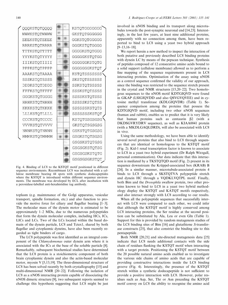
Fig. 2. Binding of LC8 to various cellular targets. Recombinant LC8 was incubated with cellulose membranes bearing spots with synthetic do-decapeptides that cover the indicated sequences. Each spot covers a sequence with two amino acids displaced compared to the previous se-quence. Selected positive spots are boxed and correlated with their corresponding sequence. The membrane was developed by ECL after incu-bation with a peroxidase-labelled anti-hexahistidine tag antibody.
FEBS 25126 13-8-01
I. Rodr|guez-Crespo et al./FEBS Letters 503 (2001) 135^141 137

TBS (50 mM Tris, pH 7.0, 137 mM NaCl, 2.7 mM KCl) for 4 h atroom temperature. Incubation with the recombinant LC8 (0.13 WM)was done overnight at room temperature. Three washes (25 ml each)were performed with TBS^Tween 20 (0.05%). Subsequently, the mem-brane was incubated for 2 h at room temperature with a commercialantibody against the hexahistidine tag present in the recombinantprotein (1:100 000 dilution in TBS). Three additional 10 min washeswere performed with TBS^Tween 20 (0.05%), followed by three more10 min washes with TBS alone. Development of the membrane wasperformed by ECL following the manufacturer's instructions. Thequanti¢cation of the intensity of each spot was performed utilisinga UVI-tec digital image analyser (UVItec, Cambridge, UK) and thesoftware UVIband V97. In every case, spots corresponding to thedodecapeptides of the various proteins synthesised onto the samemembrane were compared with each other. Controls performed withthe antibody in the absence of recombinant LC8 were performed inevery case, in order to subtract the non-speci¢c binding due to thereactivity of the antibody against certain synthetic dodecapeptides.
3. Results
3.1. Binding of LC8 to nNOSSince a crystallographic, three-dimensional structure of a
peptide of nNOS bound to LC8 is available [15], we decidedto validate our methodology covering a large portion ofnNOS in the form of overlapping synthetic dodecapeptidesand performing a binding assay to recombinant LC8 (Fig.1). Development was performed using a peroxidase-labelledantibody that recognises the hexahistidine tag present in therecombinant LC8. Binding of LC8 to peptides 112, 113 and114 reveals that the common amino acid sequence to thesethree peptides is the KDTGIQVD motif, in accordance withthe yeast two hybrid and crystallographic data [9,15]. Therecognition of LC8 toward peptides that possess this bindingmotif is exquisite, since no other dodecapeptide present in themembrane displays any comparable reactivity. Accordingly,we undertook the analysis of protein sequences known tointeract to LC8 according to previous yeast two hybrid infor-mation, as well as several proteins known to be associated tomicrotubules.
3.2. Binding of LC8 to cellular targetsIn order to perform a ¢ne mapping of the recognition prop-
erties of LC8 towards other cellular proteins, we screenedfragments of 13 di¡erent polypeptides ranging in size from58 residues (in most of the cases, 24 spots) to 184 residues(myosin V). Seven of them (Kid-1, Bim, DNA cytosine methyltransferase, protein 4, dynein HC, swallow and GKAP) re-turned a positive interaction which converge at two to fourconsecutive spots (Fig. 2). In every case, a clear, distinct pat-tern of interaction was observed. Since two consecutive over-lapping peptides possess 10 residues in common, and threeconsecutive overlapping peptides possess eight residues incommon, we were able to map down the binding motifs tothe minimal recognition sequence. This recognition motifs fallinto two distinct categories, a GIQVD motif and a KSTQTmotif (Table 1), with all the positive sequences falling into oneof the two groups.
The GIQVD motif accepts another hydrophobic L-branched residue in place of the Ile residue at position two.In a similar fashion, a Glu residue can substitute for an Aspresidue of position ¢ve. However, the Gly, Gln and Val res-idues are invariably present in all cases at positions one, threeand four. The KSTQT motif also accept some conservativesubstitutions on its sequence, although the Lys, Gln and Thr
residues remain mostly invariant. Position two can tolerateresidues such as Ser, Ala, Leu and Gln, whereas at positionthree only residues such as Thr, Ser and Val are observed.
Large regions of six additional proteins failed to show anypositive interaction according to the pepscan technique. Theseincluded IUBK and the actin-based molecular motor myosinV, in spite of the fact that both of them reportedly do so inthe yeast two hybrid system [10,13]. Thus, the pepscan tech-nique does not account for the interaction between these pro-teins and LC8. The other four proteins that failed to give apositive interaction were known to associate with microtu-bules (although their interaction with LC8 has not been re-ported) in addition to having polypeptide sequences that weresomehow similar to the KSTQT or KDTGIQVD motifs.These were dynamin (with a DSWLQVQ stretch), myosinHC (KDTQLQLDD), AMP kinase (KDTGISCDPA) andL-tubulin (GDSDLQLDR).
3.3. Binding of LC8 to viral targetsRecently, it has been shown that rabies virus and its homo-
logue, Mokola virus, bind to LC8 through their P protein,and both proteins can be co-immunoprecipitated in infectedcells [16,17]. On the other hand, African Swine Fever virususes a protein called p54, essential during the ¢rst infectivesteps to bind to LC8, and both proteins co-localise at themicrotubular organisation centre during viral infection [18].This discovery that several viruses are transported in a retro-grade manner toward the nucleus bound to the dynein multi-protein complex led us to consider the analysis of sequencescorresponding to these three viral proteins (Fig. 3). By meansof the pepscan technique, we could conclude that the threeviral proteins resulted in positive binding to LC8, and exhibitbinding sequences that fall into the KSTQT motif (Table 1,Fig. 3).
3.4. Binding of LC8 to the KSTQT motif positioned in di¡erentsequence environments
A fast inspection of any eukaryotic sequence database re-veals that there are hundreds of proteins that display eitherthe KSTQT or the KDGIQVDR motifs, and just a small
Table 1Summary of the sequences identi¢ed in proteins that interact withLC8
GIQVD motifKDT GIQVDR rat nNOSKDM GIQVDR human nNOSa
RDT GVQVDR rabbit nNOSa
KDL GIQVDR DNA cytosine methyl transferaseLSI GIQVDD GKAPQSV GVQVEE GKAP
GIQVD general consensusKSTQT motifCD KSTQTPS BimTT KSTQTQD Kid-1SA KATQTDF swallowGS KSTQTVA microtubule-associated proteinQD KLVQTPL dynein HCED KSTQTPE Mokola virusED KSTQTTS Rabies virusQN TASQTMS African swine virus
KSTQT general consensusaThe human and rabbit sequences of nNOS are depicted here in or-der to show the non-identical residues that can be accommodated£anking the GIQVD motif.
FEBS 25126 13-8-01
I. Rodr|guez-Crespo et al./FEBS Letters 503 (2001) 135^141138

subset of them become associated with the dynein multipro-tein motor. On the other hand, both the NMR and crystalstructures of LC8 bound to target peptides reveal that the£anking residues also establish binding interactions that sta-bilise the structure of the complex. Accordingly, we decided toinspect if the residues that appear just before and after thisminimal binding sequence might somehow modulate the bind-ing to LC8 and synthesised the KSTQT pentapeptide in di¡er-ent sequence environments, starting with the twenty naturalamino acids (Fig. 4, left column). Although the LC8 recogni-tion of the KSTQT motif is favoured by £anking residuessuch as Gln, Glu, Thr, Ser, Asp, Gly, His or Lys, it is virtuallyabolished by £anking residues such as Trp, Tyr, Ile, Pro, Cys,Val and Asn. Amino acids in the ¢rst group are either chargedor polar (with the exception of Gly) while the amino acids inthe second group are mostly apolar (with the exception ofAsn). These pieces of data are in agreement with the £ankingresidues that appear in the ten proteins characterised previ-ously that exhibit the KSTQT motif (Table 1). Next, we an-
alysed the e¡ect of the position of the KSTQT motif in thedodecapeptide by keeping a constant sequence (G, S or GS) atboth ends. Since the dodecapeptides are synthesised startingfrom the C-terminus end, the N-terminus is likely to be moremobile, while the C-terminus end might be, in principle, lessaccessible for LC8 to bind. Surprisingly, the general bindingtrend seems to favour positions close to the C-terminus end ofthe dodecapeptide, with the last three positions being clearlypreferred in any of the three groups of £anking residues (Fig.4, right column). Strangely enough, when the KSTQT motif ispositioned between residues 5 and 9, it displays a lower inter-action with LC8 than at positions 4^8 or 6^11, probably dueto unpredictable conformational e¡ects of the synthetic pep-tides.
4. Discussion
Dyneins are highly complex, microtubule-based molecularmotors that are involved in multiple motile events in the cy-
Fig. 3. Binding of LC8 to viral targets. Recombinant LC8 was incubated with a cellulose membrane bearing 75 spots with synthetic dodecapep-tides that cover sequences corresponding to three di¡erent viral proteins (A). 25 dodecapeptides were synthesised on each line. The membranewas developed by ECL after incubation with a peroxidase-labelled anti-hexahistidine tag antibody (B). The underlined sequences represent theoverlap region among consecutive positive spots.
FEBS 25126 13-8-01
I. Rodr|guez-Crespo et al./FEBS Letters 503 (2001) 135^141 139
toplasm (e.g. maintenance of the Golgi apparatus, vesiculartransport, spindle formation, etc.) and also function to pro-vide the motive force for ciliary and £agellar beating [1^3].The molecular mass of the dynein motor is estimated to beapproximately 1^2 MDa, due to the numerous polypeptidesthat form the dynein molecular complex, including HCs, ICs,LICs and LCs. Two of the LCs located within the basal do-main of the dynein particle, LC8 and Tctex1, shared by both£agellar and cytoplasmic dyneins, have also been recently re-garded as tight binders of cargo.
The LC8 polypeptide was ¢rst identi¢ed as an integral com-ponent of the Chlamydomonas outer dynein arm where it isassociated with the ICs at the base of the soluble particle [8].Remarkably, subsequent biochemical studies have determinedthat the LC8 protein is a stoichiometric component of bothbrain cytoplasmic dynein and also the actin-based molecularmotor, myosin V [5,13,19]. The three-dimensional structure ofLC8 has been solved recently by both crystallography [15] andmulti-dimensional NMR [20^22]. Following the isolation ofLC8 as a nNOS interacting protein capable of dissociating thenNOS dimeric structure [9], two subsequent reports seemed tochallenge this hypothesis suggesting that LC8 might be just
involved in nNOS binding and its transport along microtu-bules towards the post-synaptic neuronal end [14,23]. Interest-ingly, in the last few years, at least nine additional proteins,apparently with no connection among them, have been re-ported to bind to LC8 using a yeast two hybrid approach[9^13,16^18].
We report herein a new method to inspect the interaction ofboth putative and previously described LC8 binding proteinswith dynein LC by means of the pepscan technique. Synthesisof peptides composed of 12 consecutive amino acids bound toa solid support (cellulose membrane) allowed us to perform a¢ne mapping of the sequence requirements present in LC8interacting proteins. Optimisation of the assay using nNOSas a control sequence con¢rmed the validity of our approach,since the binding was restricted to the sequence stretch presentin the crystal and NMR structures [15,20^22]. Two homolo-gous sequences to the nNOS motif KDTGIQVD were foundin GKAP (LSIGIQVDD and also QSVGVQVEE) and in cy-tosine methyl transferase (KDLGIQVDR) (Table 1). Se-quence comparison among the proteins that possess theKDTGIQVD motif, including two other nNOS sequences(human and rabbit), enables us to predict that it is very likelythat human proteins such as centaurin L2 (with aDKDSGVRYDRV sequence), as well as KIAA0041 protein(with a MKDLGAQLDRD), will also be associated with LC8in vivo.
Using the same methodology, we have been able to identifyseveral novel proteins that also bind to LC8 through sequen-ces that are identical or homologous to the KSTQT motif(Fig. 2). Kid-1 renal transcription factor is known to associateto LC8 in a yeast two hybrid experiment (Dr Ralph Wirzgall,personal communication). Our data indicate that this interac-tion is mediated by a TKSTQTQD motif (Fig. 2) present in itssequence downstream the Kru«ppel-associated box (KRAB) B[24]. In a similar manner, microtubule-associated protein 4binds to LC8 through a SKSTQTVA polypeptide stretchand dynein HC through a VQDKLVQTPL motif. Finally,both Bim and the Drosophila swallow protein, two other pro-teins known to bind to LC8 in a yeast two hybrid method-ology display the KSTQT and KATQT motifs respectively,and also interact strongly with LC8 according to our results.
When all the polypeptide sequences that successfully inter-act with LC8 were compared to each other, we could inferthat although the KSTQT motif is highly conserved amongLC8 interacting proteins, the Ser residue at the second posi-tion can be substituted by Ala, Leu or even Gln (Table 1).Support for this is provided by random mutagenesis studies ofthe LC8 binding sites of Bim [16] and glutathione S-transfer-ase constructs [25], that also constrict the binding site to thispentapeptide.
Both NMR [20,21] and site-directed mutagenesis data [25]indicate that LC8 needs additional contacts with the sidechain of residues £anking the KSTQT motif when interactingwith a target protein. Positioning the KSTQT motif betweenthe 20 possible natural amino acids enabled us to investigatethe various side chains of amino acids that are capable ofproviding constructive interactions inside the LC8 bindinggroove (Fig. 4). Interestingly, the presence of the KSTQTstretch within a synthetic dodecapeptide is not su¤cient toprovide a positive interaction with LC8. However, polar res-idues such as Asp, Ser, Thr or Asn preceding the KSTQTmotif convey on LC8 the ability to recognise the amino acid
Fig. 4. Binding of LC8 to the KSTQT motif positioned in di¡erentsequence environments. Recombinant LC8 was incubated with a cel-lulose membrane bearing 44 spots with synthetic dodecapeptideswhere the KSTQT is introduced within di¡erent sequence environ-ments. The membrane was developed by ECL after incubation witha peroxidase-labelled anti-hexahistidine tag antibody.
FEBS 25126 13-8-01
I. Rodr|guez-Crespo et al./FEBS Letters 503 (2001) 135^141140

pattern (Table 1, Fig. 4), unlike hydrophobic residues such asTyr, Ile, Pro, Cys or Val, that abrogate this interaction (Fig.4).
It is tempting to speculate that due to the dimeric structureof LC8 [14,15,26], one of the binding sites might be occupiedby members of the dynein protein complex, with the other onebeing used for cargo transport. This inference is strengthenedby the observed binding of LC8 to the dynein HC through theVQDKLVQTPL motif (Fig. 2) and by sequence alignmentprograms that revealed that dynein IC, known to be presentwith LC8 in a 1:1 stoichiometric ratio in cytoplasmic dynein[5], displays a YSKETQTPL motif in its sequence. In addi-tion, an elegant report from Zhang and coworkers has re-cently demonstrated that dynein IC is also able to interactwith LC8 [25].
It is also noteworthy, in this context, that two viruses fromdistant families share a common way of interaction with LC8that most likely has an immediate importance in the viral lifecycle. In both cases, this interaction results in the direct tar-geting towards the nuclear membrane, suggesting that viralproteins are able to hijack the cellular machinery in order togain access to the nuclear membrane.
Acknowledgements: Authors would like to thanks Dr Ralph Witzgall(University of Heidelberg) for sharing unpublished results. We arealso indebted to Silas Cook for carefully reading the manuscript,and Dr Lisardo Bosca, Dr Covadonga Alonso, Dr Santiago Lamasand Dr Mar|a Dolores Perez-Sala for fruitful comments and sugges-tions. Belen Yelamos is a postdoctoral fellow of the Fundacion Fer-rer. This work was partially supported by Projects from the Universi-dad Complutense (08.4/0018/99) and the Spanish DGICYT(BMC2000-0545).
References
[1] Vallee, R.B. and Sheetz, M.P. (1996) Science 271, 1539^1544.[2] Hirokawa, N., Noda, Y. and Okada, Y. (1998) Curr. Opin. Cell.
Biol. 10, 60^73.[3] King, S.M. (2000) Biochim. Biophys. Acta 1496, 60^75.[4] King, S.M. and Patel-King, R.S. (1995) J. Biol. Chem. 270,
11445^11542.
[5] King, S.M., Barbarese, E., Dillman III, J.F., Patel-King, R.S.,Carson, J.H. and P¢ster, K.K. (1996) J. Biol. Chem. 271, 19358^19366.
[6] Patel-King, R.S., Benashski, S.E., Harrison, A. and King, S.M.(1997) J. Cell. Biol. 137, 1081^1090.
[7] Harrison, A., Olds-Clarke, P. and King, S.M. (1998) J. Cell. Biol.140, 1137^1147.
[8] Piperno, G. and Luck, D.J. (1979) J. Biol. Chem. 254, 3084^3090.
[9] Ja¡rey, S.R. and Snyder, S.H. (1996) Science 274, 774^777.[10] Crepieux, P., Kwon, H., Leclerc, N., Spencer, W.R., Lin, S. and
Hiscott, J. (1997) Mol. Cell. Biol. 17, 7375^7385.[11] Puthalakath, H., Huang, D.C., O'Reilly, L.A., King, S.M. and
Strasser, A. (1999) Mol. Cell 3, 287^296.[12] Schnorrer, F., Bohmann, K. and Nusslein-Volhard, C. (2000)
Nat. Cell. Biol. 2, 185^190.[13] Naisbitt, S., Valtschano¡, J., Allison, D.W., Sala, C., Kim, E.,
Craig, A.M., Weinberg, R.J. and Sheng, M. (2000) J. Neurosci.20, 4525^4534.
[14] Rodr|guez-Crespo, I., Straub, W., Gavilanes, F. and Ortiz deMontellano, P.R. (1998) Arch. Biochem. Biophys. 359, 297^304.
[15] Liang, J., Ja¡rey, S.R., Guo, W., Snyder, S.H. and Clardy, J.(1999) Nat. Struct. Biol. 6, 735^740.
[16] Jacob, Y., Badrane, H., Ceccaldi, P.-E. and Tordo, N. (2000)J. Virol. 74, 10217^10222.
[17] Raux, H., Flamand, A. and Blondel, D. (2000) J. Virol. 74,10212^10216.
[18] Alonso, C., Miskin, J., Fernandez-Zapatero, P., Hernaez, B.,Soto, L., Canto., C., Rodr|guez-Crespo, I., Dixon, L., and Escri-bano, J.M. (2001) J. Virol., in press.
[19] Espindola, F.S., Suter, D.M., Partata, L.B., Cao, T., Wolenski,J.S., Cheney, R.E., King, S.M. and Mooseker, M.S. (2000) Cell.Motil. Cytoskeleton 47, 269^281.
[20] Tochio, H., Ohki, S., Zhang, Q., Li, M. and Zhang, M. (1998)Nat. Struct. Biol. 5, 965^969.
[21] Fan, J., Zhang, Q., Tochio, H., Li, M. and Zhang, M. (2001)J. Mol. Biol. 306, 97^108.
[22] Fan, J.-S., Zhang, Q., Li, M., Tochio, H., Yamazaki, T., Shimi-zu, M. and Zhang, M. (1998) J. Biol. Chem. 273, 33472^33481.
[23] Hemmens, B., Woschitz, S., Pitters, E., Klosch, B., Volker, C.,Schmidt, K. and Mayer, B. (1998) FEBS Lett. 430, 397^400.
[24] Elser, B., Kriz, W., Bonventre, J.V., Englert, C. and Witzgall, R.(1997) J. Biol. Chem. 272, 27908^27912.
[25] Lo, K.W., Naisbitt, S., Fan, J.S., Sheng, M. and Zhang, M.(2001) J. Biol. Chem. 276, 14059^14066.
[26] Benashki, S.E., Harrison, A., Patel-King, R.S. and King, S.M.(1997) J. Biol. Chem. 272, 20929^20935.
FEBS 25126 13-8-01
I. Rodr|guez-Crespo et al./FEBS Letters 503 (2001) 135^141 141
Copyright © 2022 FDOKUMEN