Cellular Physiology - Jones & Bartlett Learning
30
© Biophoto Associates/Science Source 1 1 Cellular Physiology Case 1 Introduction Compartmentalization: Cells Are Separated from Extracellular Fluid by a Plasma Membrane Compartmentalization: Extracellular Fluid Is Separated from Vascular ECF Some Transport Requires Energy Communication: How Do Cells Coordinate Activities or Change Function? How Does Cellular Communication Result from an Action Potential? Cellular Receptors Transduce a Signal Across the Cell Membrane Without Permitting Molecules to Cross the Membrane Hormonal Signals Are Slower and Sustained Making Proteins To Do the Work: How Are Proteins Synthesized? Energy Production: Fueling Cellular Work Muscle Contraction: A Symphony of Cellular Communication Summary Key Concepts Key Terms Application: Pharmacology Clinical Case Study: Type 2 Diabetes Mellitus, Part 1 © Jones & Bartlett Learning, LLC. NOT FOR SALE OR DISTRIBUTION
-
Upload
khangminh22 -
Category
Documents
-
view
3 -
download
0
Transcript of Cellular Physiology - Jones & Bartlett Learning
© Biophoto Associates/Science Source
1
1Cellular Physiology
Case 1
Introduction
Compartmentalization: Cells Are Separated from Extracellular Fluid by a Plasma Membrane
Compartmentalization: Extracellular Fluid Is Separated from Vascular ECF
Some Transport Requires Energy
Communication: How Do Cells Coordinate Activities or Change Function?
How Does Cellular Communication Result from an Action Potential?
Cellular Receptors Transduce a Signal Across the Cell Membrane Without Permitting Molecules to Cross the Membrane
Hormonal Signals Are Slower and Sustained
Making Proteins To Do the Work: How Are Proteins Synthesized?
Energy Production: Fueling Cellular Work
Muscle Contraction: A Symphony of Cellular Communication
Summary
Key Concepts
Key Terms
Application: Pharmacology
Clinical Case Study: Type 2 Diabetes Mellitus, Part 1
9781284030341_CH01.indd 1 11/4/2013 6:16:58 PM
© Jones & Bartlett Learning, LLC. NOT FOR SALE OR DISTRIBUTION
2 CHAPTER 1 — Cellular Physiology
Case 1Last month, you decided to increase your leg muscle strength, which you notice is working nicely. It is late afternoon, and you
have just finished your regular three-mile run. The workout has made you both hungry and thirsty, so you gratefully break for a
large bottle of water and a candy bar before you take a shower. As you stand in the shower, you begin to wonder about where
the water and sugar from the candy bar were going after you swallowed them. How did the candy bar contribute to renewed
energy? What is happening to your legs to increase muscle strength? How do muscles contract? How do cells communicate
with one another?
IntroductionIf the cells within tissues are to work together to perform the “task” of the tissue, there must be communication between cells and responsiveness to the external environment. Cellular physiology is concerned with the mechanism of transport of nutrients, ions, and water into and out of the cell, as well as how cells communicate with each other through signaling pathways, or respond to external cues. In this chapter we will explore (1) how the body is separated into compartments, between which all transport is regu-lated, (2) how cells communicate with each other, electrically or chemically, (3) how proteins are made, and (4) how we make the energy to support all this activity.
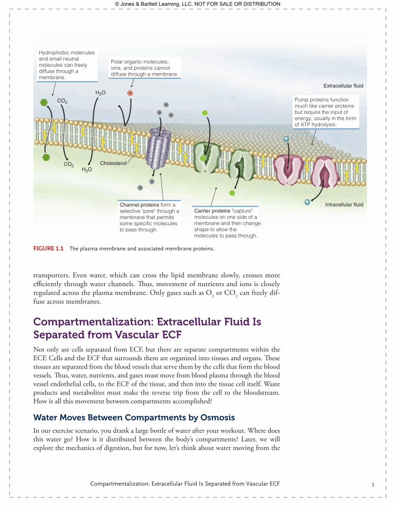
Compartmentalization: Cells Are Separated from Extracellular Fluid by a Plasma MembraneCells are enclosed by a lipid bilayer—a double layer of phospholipids that is imperme-able to large molecules and charged ions. The basic composition of the lipid bilayer is two layers of phospholipids that arrange spontaneously, with the phosphate heads facing the extracellular fluid (ECF) and intracellular fluid (ICF) (i.e., the water lay-ers), and the fatty acid tails oriented toward the center, the hydrophobic core of the membrane ( FIgurE 1.1). Cholesterol is an inherent part of the membrane and serves to stiffen it. The plasma membrane is highly fluid, with the consistency of olive oil, yet this oil-like layer is an effective barrier to large, charged, or hydrophilic molecules. Little movement across this membrane would be possible if it were not for the proteins that float in this lipid sea. The proteins, partially mobile within the bilayer, form chan-nels and transporters, which regulate movement of large or charged molecules between the ECF and the inside of the cell. This lipid bilayer with its integral proteins forms a semipermeable membrane through which water, ions, and nutrients can cross in a regulated way.
The ECF on the outside of this cell has a very different composition from the fluid within the cytoplasm. The ECF is high in Na+, high in Ca2+, and low in K+, while the ICF is low in Na+, low in Ca2+, and high in K+. This inequality of ions is maintained by the lipid bilayer and, as we will see later, is a source of potential energy. Movement of these ions from the ECF to the ICF or from the ICF to ECF can occur only through membrane proteins, such as ion channels or pumps. Larger molecules, such as glucose or amino acids, must also be moved across the plasma membrane via
9781284030341_CH01.indd 2 11/4/2013 6:16:58 PM
© Jones & Bartlett Learning, LLC. NOT FOR SALE OR DISTRIBUTION
Compartmentalization: Extracellular Fluid Is Separated from Vascular ECF 3
transporters. Even water, which can cross the lipid membrane slowly, crosses more efficiently through water channels. Thus, movement of nutrients and ions is closely regulated across the plasma membrane. Only gases such as O2 or CO2 can freely dif-fuse across membranes.
Compartmentalization: Extracellular Fluid Is Separated from Vascular ECFNot only are cells separated from ECF, but there are separate compartments within the ECF. Cells and the ECF that surrounds them are organized into tissues and organs. These tissues are separated from the blood vessels that serve them by the cells that form the blood vessels. Thus, water, nutrients, and gases must move from blood plasma through the blood vessel endothelial cells, to the ECF of the tissue, and then into the tissue cell itself. Waste products and metabolites must make the reverse trip from the cell to the bloodstream. How is all this movement between compartments accomplished?
Water Moves Between Compartments by Osmosis
In our exercise scenario, you drank a large bottle of water after your workout. Where does this water go? How is it distributed between the body’s compartments? Later, we will explore the mechanics of digestion, but for now, let’s think about water moving from the
+
+
+
+
++
Hydrophobic molecules and small neutral molecules can freely diffuse through a membrane.
Polar organic molecules, ions, and proteins cannot diffuse through a membrane.
Channel proteins form a selective "pore" through a membrane that permits some specific molecules to pass through.
Carrier proteins "capture" molecules on one side of a membrane and then change shape to allow the molecules to pass through.
Pump proteins function much like carrier proteins but require the input of energy, usually in the form of ATP hydrolysis.
CO2
Intracellular fluid
Cholesterol
Extracellular fluidH2O
H2OCO2
FIgurE 1.1 The plasma membrane and associated membrane proteins.
9781284030341_CH01.indd 3 11/4/2013 6:17:01 PM
© Jones & Bartlett Learning, LLC. NOT FOR SALE OR DISTRIBUTION
4 CHAPTER 1 — Cellular Physiology
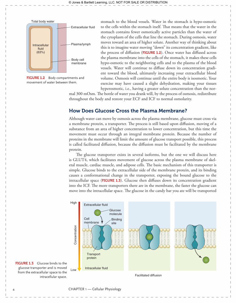
stomach to the blood vessels. Water in the stomach is hypo-osmotic to the cells within the stomach itself. That means that the water in the stomach contains fewer osmotically active particles than the water of the cytoplasm of the cells that line the stomach. During osmosis, water moves toward an area of higher solute. Another way of thinking about this is to imagine water moving “down” its concentration gradient, like the process of diffusion (FIgurE 1.2). Once water has diffused across the plasma membrane into the cells of the stomach, it makes these cells hypo-osmotic to the neighboring cells and to the plasma of the blood vessels. Water will continue to diffuse down its concentration gradi-ent toward the blood, ultimately increasing your extracellular blood volume. Osmosis will continue until the entire body is isosmotic. Your exercise may have caused a slight dehydration, making your tissues hyperosmotic, i.e., having a greater solute concentration than the nor-
mal 300 mOsm. The bottle of water you drank will, by the process of osmosis, redistribute throughout the body and restore your ECF and ICF to normal osmolarity.
How Does glucose Cross the Plasma Membrane?
Although water can move by osmosis across the plasma membrane, glucose must cross via a membrane protein, a transporter. The process is still based upon diffusion, moving of a substance from an area of higher concentration to lower concentration, but this time the movement must occur through an integral membrane protein. Because the number of proteins in the membrane will limit the amount of glucose transport possible, this process is called facilitated diffusion, because the diffusion must be facilitated by the membrane protein.
The glucose transporter exists in several isoforms, but the one we will discuss here is GLUT4, which facilitates movement of glucose across the plasma membrane of skel-etal muscle, cardiac muscle, and adipose cells. The basic mechanism of this transporter is simple. Glucose binds to the extracellular side of the membrane protein, and its binding causes a conformational change in the transporter, exposing the bound glucose to the intracellular space (FIgurE 1.3). Glucose then diffuses down its concentration gradient into the ICF. The more transporters there are in the membrane, the faster the glucose can move into the intracellular space. The glucose in the candy bar you ate will be transported
Total body water
Extracellular fluid
Plasma/lymphIntracellularfluid
(63%)
Body cellmembrane
FIgurE 1.2 Body compartments and movement of water between them.
Extracellular fluid
Intracellular fluid
Transportprotein
Bindingsite
Glucosemolecule
Cellmembrane
Low
High
Con
cent
ratio
n
Facilitated diffusion
FIgurE 1.3 Glucose binds to the glucose transporter and is moved from the extracellular space to the
intracellular space.
9781284030341_CH01.indd 4 11/4/2013 6:17:02 PM
© Jones & Bartlett Learning, LLC. NOT FOR SALE OR DISTRIBUTION

Some Transport Requires Energy 5
from the blood plasma, to ECF, to ICF via facilitated diffusion through membrane trans-porters. In the absence of transporters, glucose cannot enter the ICF.
Some Transport requires EnergyFacilitated diffusion is a process that does not require energy. It is simple diffusion through a protein carrier. However, some membrane proteins engage in active transport, or trans-port that requires adenosine triphosphate (ATP) or physiological “work”. The most ubiq-uitous of these is the Na+/K+ ATPase, also known as the Na+K+ pump. As you recall, the concentration of Na+ in the ECF is much higher than in the ICF. At the same time, K+ is in higher concentration on the inside of the cell and lower on the outside, in the ECF. The protein that helps to maintain this disequilibrium is the Na+K+ pump (FIgurE 1.4). An increase in intracellular Na+ allows binding of Na+ to the cytosolic side of the Na+K+ pump, a change in conformation, and a release of Na+ to the extracellular space. K+ binds to the extracellular face of the Na+K+ pump and is transported into the cell. However, both of these ions are moving against their concentration gradient, so diffusion is not possible.
ATP
ADP
ATP
ADP
ATP
3 Na+
2 K+
+Na+K+K+
Intracellular
Extracellular1
2
3
4
5
6
C-terminalpathway
Extracellularpathway
N-terminalpathway
III
III
Pi
3 Na+
3 Na+
3 Na+
3 Na+
2 K+
2 K+
2 K+
H2O
2 K+
3 Na+
3 Na+
FIgurE 1.4 Na+ and K+ are moved across the membrane by a series of molecular conformational changes accompanied by ATP hydrolysis.
9781284030341_CH01.indd 5 11/4/2013 6:17:06 PM
© Jones & Bartlett Learning, LLC. NOT FOR SALE OR DISTRIBUTION
6 CHAPTER 1 — Cellular Physiology
Movement of an ion against a concentration gradient, i.e., from an area of lower con-centration to an area of higher concentration, requires energy in the form of ATP. For each ATP hydrolyzed, three Na+ ions are pumped out of the cytosol and two K+ ions are brought into the cytosol from the ECF. This imbalance of positive charges—fewer on the inside than on the outside—contributes to resting membrane potential, as we will see later. The Na+K+ pump is expressed in nearly every cell of the body and is so active that its opera-tion accounts for 30% of our resting energy use.
While the Na+K+ pump is the most common active transporter, there are many others. In fact, any time an ion is moved against a concentration gradient, an ATPase or ion pump will be required. Ca2+ is moved against a concentration gradient by a Ca2+ ATPase within the endoplasmic reticulum (ER) membrane, or a different Ca2+ATPase within the plasma membrane. H+ ions are similarly moved by H+ ATPases. The basic principle to keep in mind is that movement of an ion or molecule against a concentration gradient will always require energy.
Communication: How Do Cells Coordinate Activities or Change Function?In order for organ systems to work together and tissues to perform the same function, cells must communicate—and they do so continuously. Communication can be fast and short-lived, or slower and sustained. Fast communication is usually neuronal and is accomplished by action potentials. Sustained communication usually occurs via chemical transmitters binding to cellular receptors. While very different, both forms of communica-tion are essential.
The resting Membrane Potential: Cells Poised for Action
If we were to set up a voltmeter, insert a fine electrode inside of a resting neuron, place another reference electrode on the exterior face of the plasma membrane, and then mea-sure the difference in voltage from inside to outside, we would record a negative voltage, about -70 mV. What does this mean? The negative value means that the inside of the cell is negatively charged relative to the outside of the cell, i.e., it has fewer positive charges. This electrical potential difference is the resting membrane potential, which can provide energy for communication.
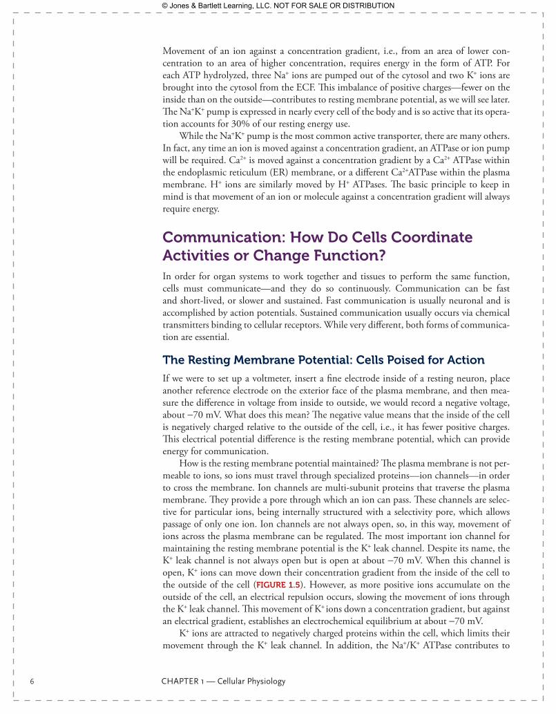
How is the resting membrane potential maintained? The plasma membrane is not per-meable to ions, so ions must travel through specialized proteins—ion channels—in order to cross the membrane. Ion channels are multi-subunit proteins that traverse the plasma membrane. They provide a pore through which an ion can pass. These channels are selec-tive for particular ions, being internally structured with a selectivity pore, which allows passage of only one ion. Ion channels are not always open, so, in this way, movement of ions across the plasma membrane can be regulated. The most important ion channel for maintaining the resting membrane potential is the K+ leak channel. Despite its name, the K+ leak channel is not always open but is open at about -70 mV. When this channel is open, K+ ions can move down their concentration gradient from the inside of the cell to the outside of the cell (FIgurE 1.5). However, as more positive ions accumulate on the outside of the cell, an electrical repulsion occurs, slowing the movement of ions through the K+ leak channel. This movement of K+ ions down a concentration gradient, but against an electrical gradient, establishes an electrochemical equilibrium at about -70 mV.
K+ ions are attracted to negatively charged proteins within the cell, which limits their movement through the K+ leak channel. In addition, the Na+/K+ ATPase contributes to
9781284030341_CH01.indd 6 11/4/2013 6:17:07 PM
© Jones & Bartlett Learning, LLC. NOT FOR SALE OR DISTRIBUTION
Communication: How Do Cells Coordinate Activities or Change Function? 7
the magnitude of the resting membrane potential. Without the Na+/K+ ATPase, the resting membrane potential would be about 5 mV more positive. Cer-tainly the distribution of other ions and the probability of other ion chan-nel openings could affect resting mem-brane potential, and does so in disease or because of some drugs. However, in a normal, healthy person, the K+ leak channel is the primary determinant of resting membrane potential. The rest-ing membrane potential is a potential energy for opening of channels and generation of an action potential, the fastest form of intercellular communication.
Remember that the ion distribution across a plasma membrane is asymmetrical: there is a high concentration of Na+ on the outside of the cell, but low concentration inside and a high concentration of K+ ions inside the cell, but a low concentration outside. There is also a high extracellular [Ca2+] relative to the intracellular space. Each of these ions passes through specific ion channels that are voltage-gated. The existence of voltage-gated ion channels is simple to understand as long as we recall protein structure. Amino acids are decorated with side chains, many of which are charged. The primary amino acid chain folds into a secondary α-helix or β-sheet, then forms into a tertiary structure, and finally assem-bles with other protein chains for the quaternary structure. These side chains attract or repel each other, forming stable ion channel conformations and a voltage sensor. The voltage sen-sor is simply an area of the protein, generally embedded in the intramembrane section of the channel, that responds to changes in local voltage. The protein’s response is simply to flex toward or away from the nearby charge, thus changing the overall protein conformation. The opening of voltage-gated ion channels is regulated by these voltage sensors. Each ion channel type has a specific range of voltages that cause it to assume an open conformation.
The Na+ Channel: A Typical Voltage-gated Ion Channel
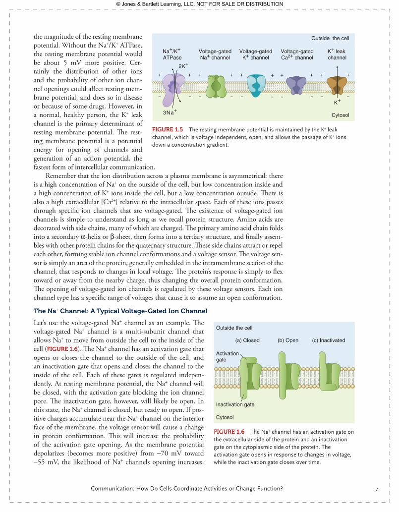
Let’s use the voltage-gated Na+ channel as an example. The voltage-gated Na+ channel is a multi-subunit channel that allows Na+ to move from outside the cell to the inside of the cell (FIgurE 1.6). The Na+ channel has an activation gate that opens or closes the channel to the outside of the cell, and an inactivation gate that opens and closes the channel to the inside of the cell. Each of these gates is regulated indepen-dently. At resting membrane potential, the Na+ channel will be closed, with the activation gate blocking the ion channel pore. The inactivation gate, however, will likely be open. In this state, the Na+ channel is closed, but ready to open. If pos-itive charges accumulate near the Na+ channel on the interior face of the membrane, the voltage sensor will cause a change in protein conformation. This will increase the probability of the activation gate opening. As the membrane potential depolarizes (becomes more positive) from -70 mV toward -55 mV, the likelihood of Na+ channels opening increases.
K+
+
-
+
-
+
-
+
-
+
-
+
-
+
-
+
-
+
-
+
-
3Na+
2K+
Outside the cell
Cytosol
Na+/K+ ATPase
Voltage-gatedNa+ channel
Voltage-gatedCa2+ channel
Voltage-gatedK+ channel
K+ leak channel
FIgurE 1.5 The resting membrane potential is maintained by the K+ leak channel, which is voltage independent, open, and allows the passage of K+ ions down a concentration gradient.
Outside the cell
Cytosol
Activation gate
Inactivation gate
(a) Closed (c) Inactivated(b) Open
FIgurE 1.6 The Na+ channel has an activation gate on the extracellular side of the protein and an inactivation gate on the cytoplasmic side of the protein. The activation gate opens in response to changes in voltage, while the inactivation gate closes over time.
9781284030341_CH01.indd 7 11/4/2013 6:17:09 PM
© Jones & Bartlett Learning, LLC. NOT FOR SALE OR DISTRIBUTION
8 CHAPTER 1 — Cellular Physiology
A voltage of -55 mV is generally considered a threshold voltage at which most Na+ chan-nel activation gates will open. The channel stays open for about 2 milliseconds before the inactivation gate on the intracellular side closes, inactivating the channel. Even though the activation gate is still open, no ions can pass through the protein pore. Over time, several milliseconds usually, the inactivation gate will return to its open or resting state, the acti-vation gate will close, and the channel will return to its original state—closed, but ready to open. The time required for recovery from inactivation is important in action potential conduction, as we will see later. Most ion channels behave similarly to the Na+ channel. The gates may be shaped differently or may have different voltage sensitivities or kinetics, but this basic pattern of opening and closing is common to most ion channels.
generation of an Action Potential
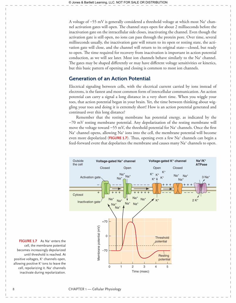
Electrical signaling between cells, with the electrical current carried by ions instead of electrons, is the fastest and most common form of intercellular communication. An action potential can carry a signal a long distance in a very short time. When you wiggle your toes, that action potential began in your brain. Yet, the time between thinking about wig-gling your toes and doing it is extremely short! How is an action potential generated and continued over this long distance?
Remember that the resting membrane has potential energy, as indicated by the -70 mV resting membrane potential. Any depolarization of the resting membrane will move the voltage toward -55 mV, the threshold potential for Na+ channels. Once the first Na+ channel opens, allowing Na+ ions into the cell, the membrane potential will become even more depolarized (FIgurE 1.7). Thus, opening even a few Na+ channels can begin a feed-forward event that depolarizes the membrane and causes many Na+ channels to open.
FIgurE 1.7 As Na+ enters the cell, the membrane potential
becomes increasingly depolarized until threshold is reached. At
positive voltages, K+ channels open, allowing positive K+ ions to leave the
cell, repolarizing it. Na+ channels inactivate during repolarization.
0 1 2 3 4 5
Mem
bran
e po
tent
ial (
mV
)
–70
0
+ + + + + + +
+ + +
– – –
+ + +
– – –
– – – – – – –
+ +
– –
+70
Voltage-gated K+ channelVoltage-gated Na+ channelOutside the cell
Cytosol
Closed
Activation gate
Inactivation gate
Open ClosedOpen
Na+Na+Na+
Na+Na+
Na+
Na+
Na+
Na+Na+
Na+
Na+
Na+
Na+K+
K+ K+
K+K+
K+
Time (msec)
Threshold potential
Restingpotential
+
–
3 Na+
2 K+
Na+/K+ ATPase
9781284030341_CH01.indd 8 11/4/2013 6:17:10 PM
© Jones & Bartlett Learning, LLC. NOT FOR SALE OR DISTRIBUTION
How Does Cellular Communication Result from an Action Potential? 9
This is the beginning of an action potential. The membrane actually depolarizes to +30 mV in response to this increase in intracellular Na+ ions. Once opened, the Na+ channels will inactivate and become refractory, meaning they will fail to open again until they are reactivated. This is a short time, but a finite time. New Na+ channels farther along the neuron can open, but the ones already opened will become unavailable. This phenomenon “moves” the action potential along to new portions of the neuron and gives the action potential a direction. The initial section of the neuron, where the action potential began, has reached 0 mV to +30 mV, a voltage range at which voltage-gated K+ channels can open. The electrochemical gradient is favorable for K+ flow out of the cell into the extra-cellular space. Once again, the opening of K+ channels proceeds down the neuron as the membrane potential enters the voltage range of these channels, 0 mV or more positive. The movement of positive charges out of the cell restores the resting membrane potential of -70 mV before the K+ channels close. Only a small number of Na+ and K+ must cross the membrane to create the necessary change in voltage, and these are returned to respec-tive spaces by the Na+K+ pump.
How Does Cellular Communication result from an Action Potential?An action potential is a simple change in membrane voltage that propagates along a neu-ron, muscle, or any excitable cell. How does that qualify as communication? By itself, the action potential serves to move a potential signal from one place to another but doesn’t usually convey information on its own. Let’s use an action potential in an α-motor neu-ron, connecting to a skeletal muscle cell, as an example. The α-motor neuron starts in the spinal cord and connects to skeletal muscle cells. We can think specifically about muscle cells in the legs, because you have gone out for a run this afternoon.
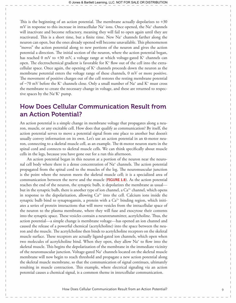
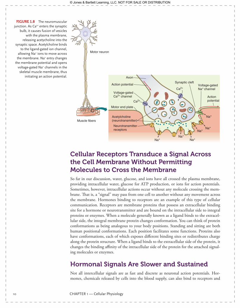
An action potential began in this neuron at a portion of the neuron near the neuro-nal cell body where there is a dense concentration of Na+ channels. The action potential propagated from the spinal cord to the muscles of the leg. The neuromuscular junction is the point where the neuron meets the skeletal muscle cell; it is a specialized area of communication between the nerve and the muscle (FIgurE 1.8). As the action potential reaches the end of the neuron, the synaptic bulb, it depolarizes the membrane as usual—but in the synaptic bulb, there is another type of ion channel, a Ca2+ channel, which opens in response to the depolarization, allowing Ca2+ into the cell. Calcium ions inside the synaptic bulb bind to synaptogamin, a protein with a Ca2+ binding region, which initi-ates a series of protein interactions that will move vesicles from the intracellular space of the neuron to the plasma membrane, where they will fuse and exocytose their contents into the synaptic space. These vesicles contain a neurotransmitter, acetylcholine. Thus, the action potential—a simple change is membrane voltage—has opened an ion channel and caused the release of a powerful chemical (acetylcholine) into the space between the neu-ron and the muscle. The acetylcholine then binds to acetylcholine receptors on the skeletal muscle surface. These receptors are actually ligand-gated ion channels, which open when two molecules of acetylcholine bind. When they open, they allow Na+ to flow into the skeletal muscle. This begins the depolarization of the membrane in the immediate vicinity of the neuromuscular junction. Voltage-gated Na+ channels located on the skeletal muscle membrane will now begin to reach threshold and propagate a new action potential along the skeletal muscle membrane, so that the communication of signal continues, ultimately resulting in muscle contraction. This example, where electrical signaling via an action potential causes a chemical signal, is a common theme in intercellular communication.
9781284030341_CH01.indd 9 11/4/2013 6:17:10 PM
© Jones & Bartlett Learning, LLC. NOT FOR SALE OR DISTRIBUTION
10 CHAPTER 1 — Cellular Physiology
Cellular receptors Transduce a Signal Across the Cell Membrane Without Permitting Molecules to Cross the MembraneSo far in our discussion, water, glucose, and ions have all crossed the plasma membrane, providing intracellular water, glucose for ATP production, or ions for action potentials. Sometimes, however, intracellular actions occur without any molecule crossing the mem-brane. That is, a “signal” may pass from one cell to another without any movement across the membrane. Hormones binding to receptors are an example of this type of cellular communication. Receptors are membrane proteins that possess an extracellular binding site for a hormone or neurotransmitter and are bound on the intracellular side to integral proteins or enzymes. When a molecule generally known as a ligand binds to the extracel-lular side, the integral membrane protein changes conformation. You can think of protein conformations as being analogous to your body positions. Standing and sitting are both human positional conformations. Each position facilitates some functions. Proteins also have conformations, each of which exposes different binding sites or redistributes charge along the protein structure. When a ligand binds to the extracellular side of the protein, it changes the binding affinity of the intracellular side of the protein for the attached signal-ing molecules or enzymes.
Hormonal Signals Are Slower and SustainedNot all intercellular signals are as fast and discrete as neuronal action potentials. Hor-mones, chemicals released by cells into the blood supply, can also bind to receptors and
FIgurE 1.8 The neuromuscular junction. As Ca2+ enters the synaptic
bulb, it causes fusion of vesicles with the plasma membrane,
releasing acetycholine into the synaptic space. Acetylcholine binds
to the ligand-gated ion channel, allowing Na+ ions to move across
the membrane. Na+ entry changes the membrane potential and opens
voltage-gated Na+ channels in the skeletal muscle membrane, thus
initiating an action potential.
Motor neuron
Muscle fibers
Synaptic cleft
Axon
Acetylcholine(neurotransmitter)
Action potential
Action potential
Neurotransmitterreceptors
Voltage-gatedCa2+ channel
Voltage-gatedNa+ channel
Motor end plate
Ca2+
Na+Na+
Na+
Na+Na+
Na+
Ca2+
9781284030341_CH01.indd 10 11/4/2013 6:17:12 PM
© Jones & Bartlett Learning, LLC. NOT FOR SALE OR DISTRIBUTION
Hormonal Signals Are Slower and Sustained 11
cause changes in cellular function in tissues far distant to the cells that released them. This type of chemical signaling takes longer to have its effect, but the effects are generally longer-lived, lasting from minutes to days instead of milliseconds. The most important thing to remember about hor-monal signaling is that the hormone will bind to a receptor, and it is the receptor that determines the intracellular response to the binding. Let’s use epi-nephrine, also known as adrenaline, as an example.
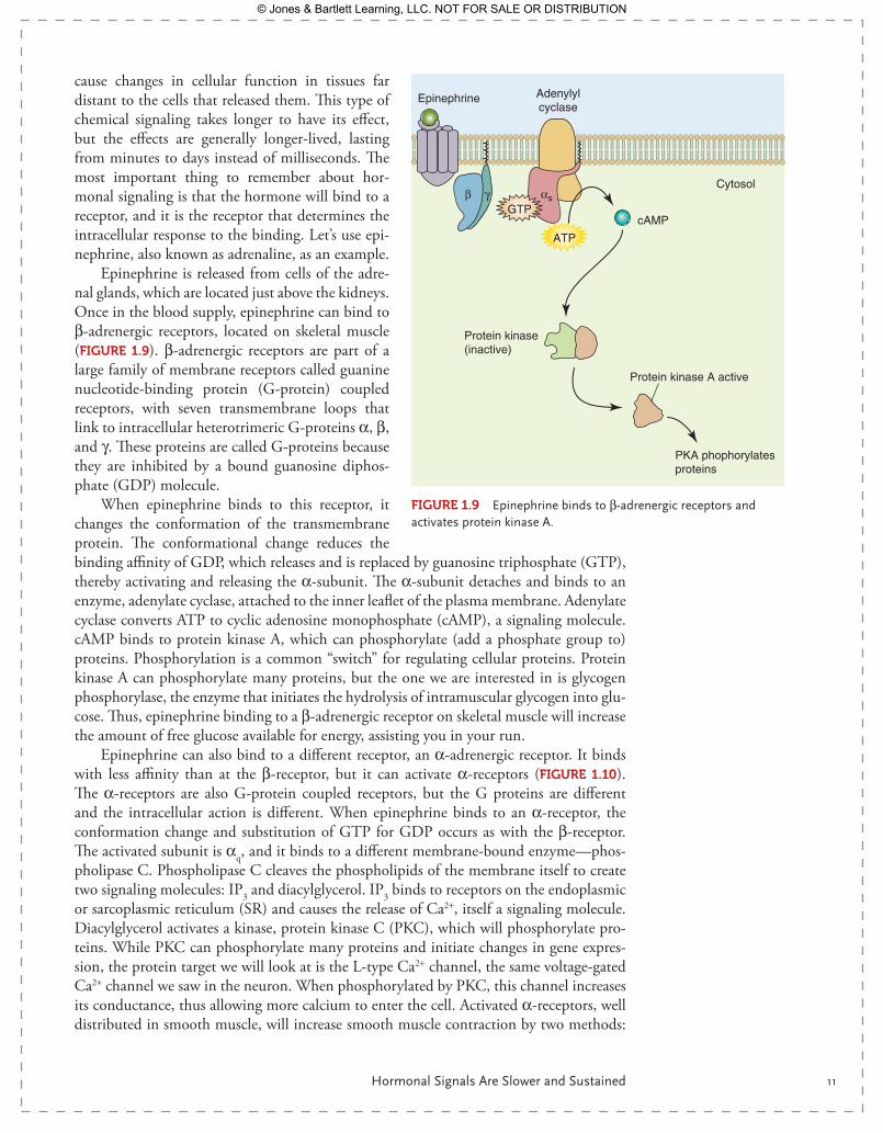
Epinephrine is released from cells of the adre-nal glands, which are located just above the kidneys. Once in the blood supply, epinephrine can bind to β-adrenergic receptors, located on skeletal muscle (FIgurE 1.9). β-adrenergic receptors are part of a large family of membrane receptors called guanine nucleotide-binding protein (G-protein) coupled receptors, with seven transmembrane loops that link to intracellular heterotrimeric G- proteins α, β, and γ. These proteins are called G-proteins because they are inhibited by a bound guanosine diphos-phate (GDP) molecule.
When epinephrine binds to this receptor, it changes the conformation of the transmembrane protein. The conformational change reduces the binding affinity of GDP, which releases and is replaced by guanosine triphosphate (GTP), thereby activating and releasing the α-subunit. The α-subunit detaches and binds to an enzyme, adenylate cyclase, attached to the inner leaflet of the plasma membrane. Adenylate cyclase converts ATP to cyclic adenosine monophosphate (cAMP), a signaling molecule. cAMP binds to protein kinase A, which can phosphorylate (add a phosphate group to) proteins. Phosphorylation is a common “switch” for regulating cellular proteins. Protein kinase A can phosphorylate many proteins, but the one we are interested in is glycogen phosphorylase, the enzyme that initiates the hydrolysis of intramuscular glycogen into glu-cose. Thus, epinephrine binding to a β-adrenergic receptor on skeletal muscle will increase the amount of free glucose available for energy, assisting you in your run.
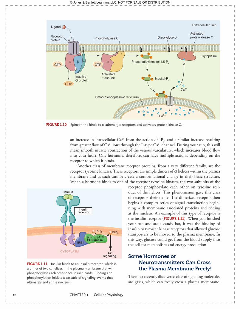
Epinephrine can also bind to a different receptor, an α-adrenergic receptor. It binds with less affinity than at the β-receptor, but it can activate α-receptors (FIgurE 1.10). The α-receptors are also G-protein coupled receptors, but the G proteins are different and the intracellular action is different. When epinephrine binds to an α-receptor, the conformation change and substitution of GTP for GDP occurs as with the β-receptor. The activated subunit is αq, and it binds to a different membrane-bound enzyme—phos-pholipase C. Phospholipase C cleaves the phospholipids of the membrane itself to create two signaling molecules: IP3 and diacylglycerol. IP3 binds to receptors on the endoplasmic or sarcoplasmic reticulum (SR) and causes the release of Ca2+, itself a signaling molecule. Diacylglycerol activates a kinase, protein kinase C (PKC), which will phosphorylate pro-teins. While PKC can phosphorylate many proteins and initiate changes in gene expres-sion, the protein target we will look at is the L-type Ca2+ channel, the same voltage-gated Ca2+ channel we saw in the neuron. When phosphorylated by PKC, this channel increases its conductance, thus allowing more calcium to enter the cell. Activated α-receptors, well distributed in smooth muscle, will increase smooth muscle contraction by two methods:
cAMP
Cytosol
Epinephrine
β γ
Adenylylcyclase
αsGTP
ATP
Protein kinase(inactive)
PKA phophorylatesproteins
Protein kinase A active
FIgurE 1.9 Epinephrine binds to β-adrenergic receptors and activates protein kinase A.
9781284030341_CH01.indd 11 11/4/2013 6:17:13 PM
© Jones & Bartlett Learning, LLC. NOT FOR SALE OR DISTRIBUTION
12 CHAPTER 1 — Cellular Physiology
an increase in intracellular Ca2+ from the action of IP3, and a similar increase resulting from greater flow of Ca2+ ions through the L-type Ca2+ channel. During your run, this will mean smooth muscle contraction of the venous vasculature, which increases blood flow into your heart. One hormone, therefore, can have multiple actions, depending on the receptor to which it binds.
Another class of membrane receptor proteins, from a very different family, are the receptor tyrosine kinases. These receptors are simple dimers of α helices within the plasma membrane and as such cannot create a conformational change in their basic structure. When a hormone binds to one of the receptor tyrosine kinases, the two subunits of the
receptor phosphorylate each other on tyrosine resi-dues of the helices. This phenomenon gave this class of receptors their name. The dimerized receptor then begins a complex series of signal transduction begin-ning with membrane associated proteins and ending at the nucleus. An example of this type of receptor is the insulin receptor (FIgurE 1.11). When you finished your run and ate a candy bar, it was the binding of insulin to tyrosine kinase receptors that allowed glucose transporters to be moved to the plasma membrane. In this way, glucose could get from the blood supply into the cell for metabolism and energy production.
Activatedα subunit
GDP
GTP
InactiveG protein
Phospholipase CReceptorprotein
Ligand
α αβ γ
Cytoplasm
Extracellular fluid
GTP
PP
P
PP
P
PP P
Phosphatidylinositol 4,5-P2
DiacylglycerolActivated protein kinase C
Inositol-P3
Ca2+
Ca2+
Smooth endoplasmic reticulum
FIgurE 1.10 Epinephrine binds to α-adrenergic receptors and activates protein kinase C.
PIP2 PIP3
Aktsignaling
CYTOPLASM
Insulinreceptor
p85 p110
IRS1PI 3-kinase
Insulin
FIgurE 1.11 Insulin binds to an insulin receptor, which is a dimer of two α-helices in the plasma membrane that will phosphorylate each other once insulin binds. Binding and phosphorylation initiate a cascade of signaling events that ultimately end at the nucleus.
Some Hormones or Neurotransmitters Can Cross the Plasma Membrane Freely!
The most recently discovered class of signaling molecules are gases, which can freely cross a plasma membrane.
9781284030341_CH01.indd 12 11/4/2013 6:17:15 PM
© Jones & Bartlett Learning, LLC. NOT FOR SALE OR DISTRIBUTION
Hormonal Signals Are Slower and Sustained 13
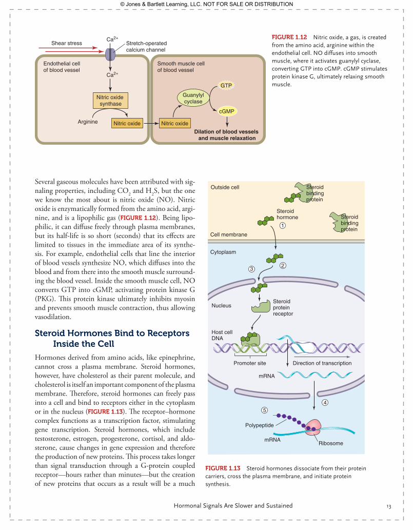
Several gaseous molecules have been attributed with sig-naling properties, including CO2 and H2S, but the one we know the most about is nitric oxide (NO). Nitric oxide is enzymatically formed from the amino acid, argi-nine, and is a lipophilic gas (FIgurE 1.12). Being lipo-philic, it can diffuse freely through plasma membranes, but its half-life is so short (seconds) that its effects are limited to tissues in the immediate area of its synthe-sis. For example, endothelial cells that line the interior of blood vessels synthesize NO, which diffuses into the blood and from there into the smooth muscle surround-ing the blood vessel. Inside the smooth muscle cell, NO converts GTP into cGMP, activating protein kinase G (PKG). This protein kinase ultimately inhibits myosin and prevents smooth muscle contraction, thus allowing vasodilation.
Steroid Hormones Bind to receptors Inside the Cell
Hormones derived from amino acids, like epinephrine, cannot cross a plasma membrane. Steroid hormones, however, have cholesterol as their parent molecule, and cholesterol is itself an important component of the plasma membrane. Therefore, steroid hormones can freely pass into a cell and bind to receptors either in the cytoplasm or in the nucleus ( FIgurE 1.13). The receptor–hormone complex functions as a transcription factor, stimulating gene transcription. Steroid hormones, which include testosterone, estrogen, progesterone, cortisol, and aldo-sterone, cause changes in gene expression and therefore the production of new proteins. This process takes longer than signal transduction through a G- protein coupled receptor—hours rather than minutes—but the creation of new proteins that occurs as a result will be a much
FIgurE 1.12 Nitric oxide, a gas, is created from the amino acid, arginine within the endothelial cell. NO diffuses into smooth muscle, where it activates guanylyl cyclase, converting GTP into cGMP. cGMP stimulates protein kinase G, ultimately relaxing smooth muscle.GTP
Endothelial cell of blood vessel
Stretch-operatedcalcium channel
Smooth muscle cell of blood vessel
Shear stress
Arginine Nitric oxide Nitric oxide
Guanylylcyclase
Nitric oxide synthase
cGMP
Dilation of blood vessels and muscle relaxation
Ca2+
Ca2+
Polypeptide
Ribosome
Cell membrane
Outside cell
Cytoplasm
Nucleus
Steroid hormone
Steroid bindingprotein
Steroid bindingprotein
Steroidproteinreceptor
mRNA
mRNA
Promoter site
Host cellDNA
Direction of transcription
1
23
45
FIgurE 1.13 Steroid hormones dissociate from their protein carriers, cross the plasma membrane, and initiate protein synthesis.
9781284030341_CH01.indd 13 11/4/2013 6:17:17 PM
© Jones & Bartlett Learning, LLC. NOT FOR SALE OR DISTRIBUTION
14 CHAPTER 1 — Cellular Physiology
more sustained response. Thus, steroid hormones can regulate cellular function for hours, days, and weeks.
If steroid hormones can pass freely into a cell, why aren’t their actions dominant all the time? The same hydrophobicity that allows steroid hormones to slip through a membrane makes them insoluble in water, the primary component of plasma. Steroid hormones are carried by proteins in the circulating blood. At low concentrations, steroid hormones remain bound to their carrier proteins. At higher concentrations, e.g., following release from their tissue of origin, they will exist in a dynamic equilibrium with carrier proteins and will unbind periodically, allowing their passage across the membrane.
In our workout example, you became thirsty during the run. An increase in appar-ent K+ plasma concentration, which occurs with dehydration, is sensed by cells of your adrenal glands, which release the steroid hormone aldosterone. Aldosterone circulates in the blood and has as its target the tubules of the kidney. Once inside the cells of the kid-ney tubule, aldosterone causes an increase in Na+ ion channel expression, which results in concentration of urine, providing a means for conservation of water. The end result is that you produce less urine and maintain fluid balance. We will explore this mechanism more thoroughly in the endocrine chapter.
Making Proteins to Do the Work: How Are Proteins Synthesized?Protein synthesis is a complex event, with regulatory steps at each junction. While it is important for us to know the fundamentals of this process in our study of human physiol-ogy, we will reserve the details of protein synthesis for cell and molecular biology texts. Let us review the basic tenets of protein synthesis.
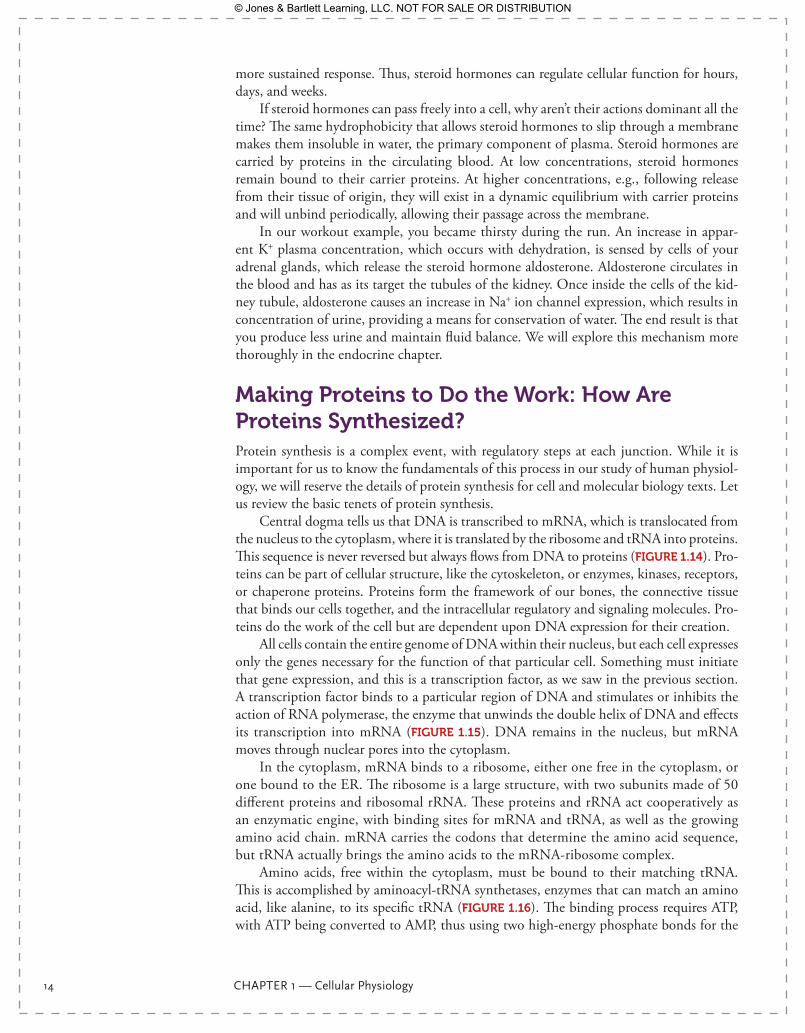
Central dogma tells us that DNA is transcribed to mRNA, which is translocated from the nucleus to the cytoplasm, where it is translated by the ribosome and tRNA into proteins. This sequence is never reversed but always flows from DNA to proteins ( FIgurE 1.14). Pro-teins can be part of cellular structure, like the cytoskeleton, or enzymes, kinases, receptors, or chaperone proteins. Proteins form the framework of our bones, the connective tissue that binds our cells together, and the intracellular regulatory and signaling molecules. Pro-teins do the work of the cell but are dependent upon DNA expression for their creation.
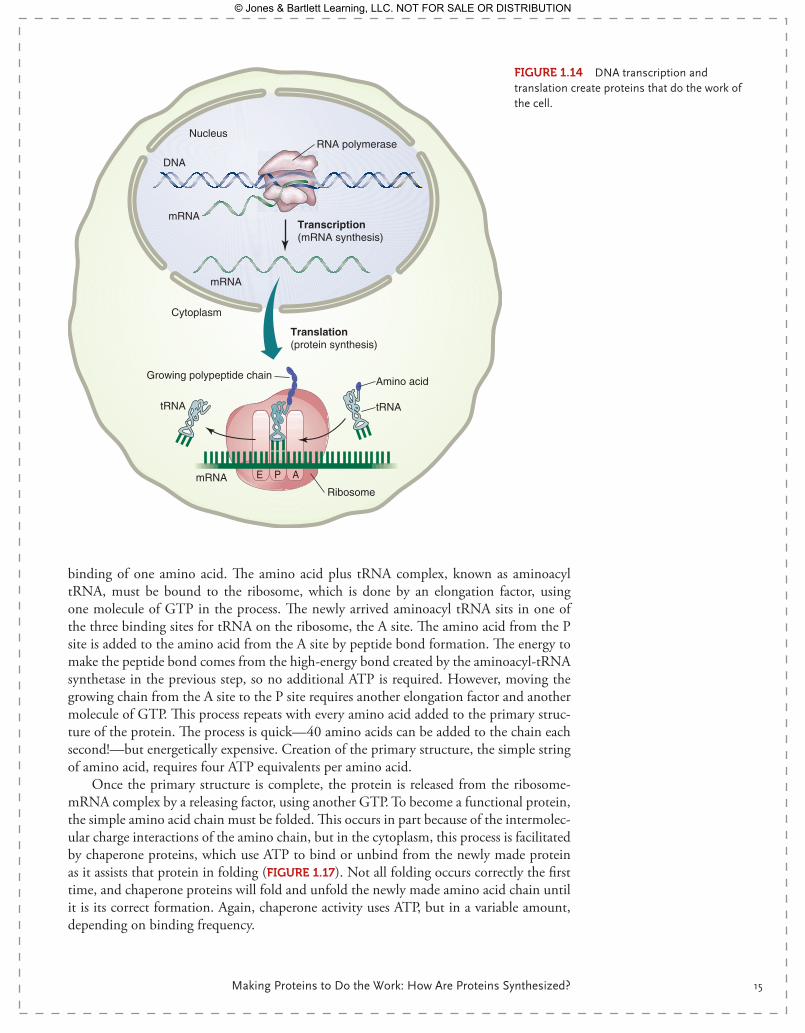
All cells contain the entire genome of DNA within their nucleus, but each cell expresses only the genes necessary for the function of that particular cell. Something must initiate that gene expression, and this is a transcription factor, as we saw in the previous section. A transcription factor binds to a particular region of DNA and stimulates or inhibits the action of RNA polymerase, the enzyme that unwinds the double helix of DNA and effects its transcription into mRNA (FIgurE 1.15). DNA remains in the nucleus, but mRNA moves through nuclear pores into the cytoplasm.
In the cytoplasm, mRNA binds to a ribosome, either one free in the cytoplasm, or one bound to the ER. The ribosome is a large structure, with two subunits made of 50 different proteins and ribosomal rRNA. These proteins and rRNA act cooperatively as an enzymatic engine, with binding sites for mRNA and tRNA, as well as the growing amino acid chain. mRNA carries the codons that determine the amino acid sequence, but tRNA actually brings the amino acids to the mRNA-ribosome complex.
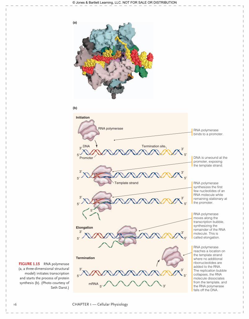
Amino acids, free within the cytoplasm, must be bound to their matching tRNA. This is accomplished by aminoacyl-tRNA synthetases, enzymes that can match an amino acid, like alanine, to its specific tRNA (FIgurE 1.16). The binding process requires ATP, with ATP being converted to AMP, thus using two high-energy phosphate bonds for the
9781284030341_CH01.indd 14 11/4/2013 6:17:17 PM
© Jones & Bartlett Learning, LLC. NOT FOR SALE OR DISTRIBUTION
Making Proteins to Do the Work: How Are Proteins Synthesized? 15
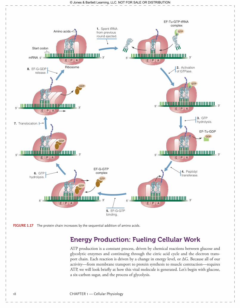
binding of one amino acid. The amino acid plus tRNA complex, known as aminoacyl tRNA, must be bound to the ribosome, which is done by an elongation factor, using one molecule of GTP in the process. The newly arrived aminoacyl tRNA sits in one of the three binding sites for tRNA on the ribosome, the A site. The amino acid from the P site is added to the amino acid from the A site by peptide bond formation. The energy to make the peptide bond comes from the high-energy bond created by the aminoacyl-tRNA synthetase in the previous step, so no additional ATP is required. However, moving the growing chain from the A site to the P site requires another elongation factor and another molecule of GTP. This process repeats with every amino acid added to the primary struc-ture of the protein. The process is quick—40 amino acids can be added to the chain each second!—but energetically expensive. Creation of the primary structure, the simple string of amino acid, requires four ATP equivalents per amino acid.
Once the primary structure is complete, the protein is released from the ribosome-mRNA complex by a releasing factor, using another GTP. To become a functional protein, the simple amino acid chain must be folded. This occurs in part because of the intermolec-ular charge interactions of the amino chain, but in the cytoplasm, this process is facilitated by chaperone proteins, which use ATP to bind or unbind from the newly made protein as it assists that protein in folding (FIgurE 1.17). Not all folding occurs correctly the first time, and chaperone proteins will fold and unfold the newly made amino acid chain until it is its correct formation. Again, chaperone activity uses ATP, but in a variable amount, depending on binding frequency.
FIgurE 1.14 DNA transcription and translation create proteins that do the work of the cell.
PE A
mRNA
mRNARibosome
mRNA
DNA
RNA polymeraseNucleus
Cytoplasm
tRNA tRNA
Amino acid
Transcription(mRNA synthesis)
Translation(protein synthesis)
Growing polypeptide chain
9781284030341_CH01.indd 15 11/4/2013 6:17:21 PM
© Jones & Bartlett Learning, LLC. NOT FOR SALE OR DISTRIBUTION
16 CHAPTER 1 — Cellular Physiology
RNA polymerase binds to a promoter.
DNA is unwound at the promoter, exposing the template strand.
RNA polymerase synthesizes the first few nucleotides of an RNA molecule while remaining stationary at the promoter.
RNA polymerase moves along the transcription bubble, synthesizing the remainder of the RNA molecule. This is called elongation.
RNA polymerase reaches a location on the template strand where no additional ribonucleotides are added to the RNA. The replication bubble collapses, the RNA molecule dissociates from the template, and the RNA polymerase falls off the DNA.
RNA polymerase
(b)
(a)
Initiation
Elongation
Termination
DNA
mRNA
Promoter
Template strand
Termination site
5′
5′
5′ 5′ 3′
3′
3′ 3′
5′ 5′
3′ 3′
5′ 5′
3′ 3′
5′ 5′
3′
5′
3′
3′
5′
3′
FIgurE 1.15 RNA polymerase (a, a three-dimensional structural
model) initiates transcription and starts the process of protein synthesis (b). (Photo courtesy of
Seth Darst.)
9781284030341_CH01.indd 16 11/4/2013 6:17:25 PM
© Jones & Bartlett Learning, LLC. NOT FOR SALE OR DISTRIBUTION
Making Proteins to Do the Work: How Are Proteins Synthesized? 17
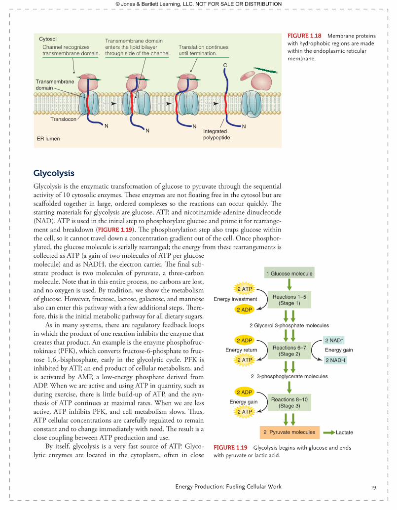
Proteins that will remain free in the cytoplasm are made in the fashion described above and can be made by free ribosomes. Proteins that will be inserted into the plasma mem-brane, like ion channels or G-protein coupled receptors, contain hydrophobic regions that span the plasma membrane. These regions, being hydrophobic, cannot be exposed to the aqueous environment of the cytoplasm. These proteins are made on ribosomes attached to the membrane of the ER. As the chain elongates off the ribosome, it enters the lumen of the ER through a protein pore. Chaperone proteins like binding immunoglobulin protein bind to the hydrophobic regions of the forming protein within the ER, protecting it from the aqueous environment of the ER lumen (FIgurE 1.18). Once completed, these proteins will be glycosylated within the ER and then transported by vesicle to the Golgi appara-tus for further processing. Vesicles bud off the Golgi apparatus and fuse with the plasma membrane, inserting the new membrane proteins in their place. Glycosylation is so com-mon in membrane proteins that under high magnification, as seen in electron microscopy, the entire exterior of a plasma membrane appears to have a sugar “halo.” Glycosylation is important in cellular self-recognition and charge distribution across the membrane.
Hormones and signaling mechanisms within the cell regularly stimulate or inhibit protein synthesis as a method of regulating cell function or allowing cellular adaptation to changing environmental conditions. Protein synthesis takes some time, as we saw from the steroid hormone series of events, but it is a longer-term form of response to chang-ing conditions. Protein formation is also energetically expensive, requiring four ATPs per amino acid, in addition to the ATPs required for initiation, termination, and folding. If we consider that an average-sized protein is 450 amino acids long, and the largest protein known, titin, is 27,000 amino acids long, the energy required for protein synthesis is sig-nificant. Where does this energy come from?
tRNA
Aminoacyl-tRNA(”activated”)
Amino acid Aminoacyl-tRNAsynthetase
ATPPPP
Pi Pi
AMPP
AMPP
ATPPPP
FIgurE 1.16 The enzyme aminoacyl-tRNA synthetase uses ATP to bind an amino acid to its appropriate tRNA.
9781284030341_CH01.indd 17 11/4/2013 6:17:26 PM
© Jones & Bartlett Learning, LLC. NOT FOR SALE OR DISTRIBUTION
18 CHAPTER 1 — Cellular Physiology
Energy Production: Fueling Cellular WorkATP production is a constant process, driven by chemical reactions between glucose and glycolytic enzymes and continuing through the citric acid cycle and the electron trans-port chain. Each reaction is driven by a change in energy level, or ΔG. Because all of our activity—from membrane transport to protein synthesis to muscle contraction—requires ATP, we will look briefly at how this vital molecule is generated. Let’s begin with glucose, a six-carbon sugar, and the process of glycolysis.
1. Spent tRNA from previous round ejected.
2. Activation of GTPase.
3. GTP hydrolysis.
4. Peptidyl transferase.
5. EF-G-GTP binding.
6. GTP hydrolysis.
7. Translocation.
8. EF-G-GDP release.
PE A3′5′mRNA
Start codon
Amino acids
Ribosome
PE A
EF-Tu-GTP-tRNA complex
3′5′
GTP
PE A3′5′
GTP
EF-Tu-GDP
GDP
PE A3′5′
PE A3′5′
EF-G-GTP complex
PE A3′5′
GTP
GTP
3′5′
GDP
PE A3′5′
GDP
PE A
FIgurE 1.17 The protein chain increases by the sequential addition of amino acids.
9781284030341_CH01.indd 18 11/4/2013 6:17:27 PM
© Jones & Bartlett Learning, LLC. NOT FOR SALE OR DISTRIBUTION
Energy Production: Fueling Cellular Work 19
glycolysis
Glycolysis is the enzymatic transformation of glucose to pyruvate through the sequential activity of 10 cytosolic enzymes. These enzymes are not floating free in the cytosol but are scaffolded together in large, ordered complexes so the reactions can occur quickly. The starting materials for glycolysis are glucose, ATP, and nicotinamide adenine dinucleotide (NAD). ATP is used in the initial step to phosphorylate glucose and prime it for rearrange-ment and breakdown (FIgurE 1.19). The phosphorylation step also traps glucose within the cell, so it cannot travel down a concentration gradient out of the cell. Once phosphor-ylated, the glucose molecule is serially rearranged; the energy from these rearrangements is collected as ATP (a gain of two molecules of ATP per glucose molecule) and as NADH, the electron carrier. The final sub-strate product is two molecules of pyruvate, a three-carbon molecule. Note that in this entire process, no carbons are lost, and no oxygen is used. By tradition, we show the metabolism of glucose. However, fructose, lactose, galactose, and mannose also can enter this pathway with a few additional steps. There-fore, this is the initial metabolic pathway for all dietary sugars.
As in many systems, there are regulatory feedback loops in which the product of one reaction inhibits the enzyme that creates that product. An example is the enzyme phosphofruc-tokinase (PFK), which converts fructose-6-phosphate to fruc-tose 1,6,- bisphosphate, early in the glycolytic cycle. PFK is inhibited by ATP, an end product of cellular metabolism, and is activated by AMP, a low-energy phosphate derived from ADP. When we are active and using ATP in quantity, such as during exercise, there is little build-up of ATP, and the syn-thesis of ATP continues at maximal rates. When we are less active, ATP inhibits PFK, and cell metabolism slows. Thus, ATP cellular concentrations are carefully regulated to remain constant and to change immediately with need. The result is a close coupling between ATP production and use.
By itself, glycolysis is a very fast source of ATP. Glyco-lytic enzymes are located in the cytoplasm, often in close
NN
N N
C
Translocon
ER lumen
Cytosol
Channel recognizestransmembrane domain.
Translation continuesuntil termination.
Integrated polypeptide
Transmembrane domainenters the lipid bilayerthrough side of the channel.
Transmembranedomain
FIgurE 1.18 Membrane proteins with hydrophobic regions are made within the endoplasmic reticular membrane.
2 ADP
2 ATP
2 ATP
2 NAD+
2 NADH
2 ADP
2 ATP
2 ADP
1 Glucose molecule
2 Glycerol 3-phosphate molecules
Reactions 1–5(Stage 1)
Energy investment
2 3-phosphoglycerate molecules
Reactions 6–7(Stage 2)
Energy return
2 Pyruvate molecules
Reactions 8–10(Stage 3)
Energy gain
Energy gain
Lactate
FIgurE 1.19 Glycolysis begins with glucose and ends with pyruvate or lactic acid.
9781284030341_CH01.indd 19 11/4/2013 6:17:40 PM
© Jones & Bartlett Learning, LLC. NOT FOR SALE OR DISTRIBUTION

20 CHAPTER 1 — Cellular Physiology
proximity to the proteins that will use the ATP produced. In muscle, for example, gly-colytic enzymes are located within the assembly of contractile proteins. The product of glycolysis, pyruvate, also functions as a negative modulator of glycolysis—so as pyruvate accumulates, it inhibits further glycolysis. Fortunately, pyruvate can be converted to lac-tate, and lactate can be transported out of the cell, allowing glycolysis to continue. In the absence of oxygen, this is precisely what happens, contributing to lactic acid build-up in the blood. Lactate can be reconverted to pyruvate within the heart and the liver and used for further metabolism, so lactic acid production is a way of preventing pyruvate inhibi-tion of glycolysis within the cell and allowing energy recycling by other tissues. Most of the time, O2 is available, and then a more efficient form of metabolism breaks down pyruvate to yield 34 ATP/glucose molecules, instead of the two ATP we saw in glycolysis. Aerobic metabolism is dependent upon mitochondria, the cellular organelle that uses almost all of the O2 we breathe.
Mitochondria: Organelles That Produce Most of Our ATP
What do we know about mitochondria? Mitochondria are double-membraned organelles that exist in all human cells except for mature red blood cells. These organelles are thought to have originated a billion years ago as free-living organisms, which later became incorpo-rated into a host organism, producing the eukaryotic cell we now recognize. The number of mitochondria within a cell is uncertain. It not only depends on cell type, but also on mitochondrial morphology, which is still unclear. The classic oval-shaped mitochondrion portrayed in scientific illustrations may be an artifact of histological section (FIgurE 1.20). There is evidence that mitochondria are tubular, dynamic organelles that change shape, twist, and undergo fission and fusion. Mitochondria may proliferate in active, aerobic tis-sues. Mitochondria are not evenly distributed within a cell but are concentrated at sites of ATP use. In muscle, they are found near contractile fibers and in nerves at the sites of protein synthesis in the cell body and in the synaptic bulb, where neurotransmitters are made and stored.
Mitochondria contain their own circular DNA, known as mtDNA, which we inherit from our mothers only. mtDNA does not encode all the proteins required for mitochon-
drial function. In fact, it codes for only 13 genes, all related to the electron transport chain proteins. There are many genes within the cell’s nuclear DNA that encode struc-tural and regulatory proteins necessary for mitochondrial function. Cooperative expression between two genomes is necessary for proper mitochondrial operation. Mutations within either genome can affect organelle function and therefore ATP production.
While mitochondria lack the sophisticated DNA repair mechanisms of the nucleus, they are capable of fis-sion (division) and fusion (joining of two or more mito-chondria). Fission and fusion allow genetic mixing between mitochondria and isolation of damaged mtDNA that can be eliminated from the organelle. Fission and fusion also allow more homogeneity of mtDNA within a large cell, such as a neuron. Mitochondria also move, which is linked to the processes of fission and fusion. In neurons, for example, mitochondria must be located both within the cell body of the neuron and at the distant synaptic bulb. Without movement, mitochondria fail to distribute to the
FIgurE 1.20 Mitochondria may resemble tubular arrays within the cytoplasm that appear oval when cells are sectioned. (© Dr. Gopal Murti/Science Source.)
9781284030341_CH01.indd 20 11/4/2013 6:17:50 PM
© Jones & Bartlett Learning, LLC. NOT FOR SALE OR DISTRIBUTION

Energy Production: Fueling Cellular Work 21
synapse, causing neuronal malfunction. Genes that regulate fission and fusion are within the cell’s nuclear DNA, highlighting the interdependence of mitochondria and the cell they inhabit.
Energy Production Within the Mitochondria
Mitochondria are famous for their role in ATP synthesis. Indeed, life as we know it is not possible without the ATP provided by mitochondria. Glycolysis alone cannot supply the ATP demands required for human life. Once glucose or other sugars are converted to pyruvate, the rest of the metabolic process must proceed within a mitochondrion. Pyru-vate, produced in the cytoplasm, is transported into the mitochondria through pyruvate transporters. Once inside the mitochondrial matrix, pyruvate is decarboxylated and linked to coenzyme A to become acetyl coenzyme A. This reaction requires several cofactors known as vitamins, including pantothenic acid (vitamin B5), which is a component of CoA, and thiamine (vitamin B1). The conversion produces NADH as a product. NAD+ is also formed from a vitamin, niacin (vitamin B3). Acetyl-CoA can also be formed from a two-carbon product of β-oxidation, which is how fat metabolism feeds into this metabolic framework.
Fatty Acids Are a Source of Acetyl-CoA
Most of the fat we ingest is stored in the form of triglycerides composed of a glycerol backbone linked to long-chain fatty acids. Triglycerides are broken down by removal of glycerol, which is metabolized along the same pathway as glucose. The remaining fatty acids are broken down within the mitochondria, in a cycle known as β-oxidation. The final product of β-oxidation is acetyl-CoA, which feeds into the citric acid cycle just as acetyl-CoA from pyruvate metabolism does (FIgurE 1.21). The energy yield from fatty acids is
C
O OH
CH2
CH2
H2C
CH2
H2C
CH2
H2C
CH2
H2C
CH2
H2C
CH2
H2C
CH3
CH3 C
H
H
Fatty acyl-CoA
Fatty acyl-CoA(2 carbon atoms shorter)
Acetyl-CoA
Fatty acid
H
H
O
C C SCoA
Hydration
Dehydration
Addition of CoA
(CH2)n
CH3
O
C+
SCoA(CH2)n CH3
O
C SCoA
H2C
β-oxidation is the oxidation of fatty acids by successive removal of two-carbon units.
FIgurE 1.21 β-oxidation involves the sequential oxidation of fatty acid chains two carbons at a time, creating acetyl-CoA.
9781284030341_CH01.indd 21 11/4/2013 6:17:51 PM
© Jones & Bartlett Learning, LLC. NOT FOR SALE OR DISTRIBUTION
22 CHAPTER 1 — Cellular Physiology
very high, making fat our most efficient energy source. However, fatty acids require O2 for metabolism.
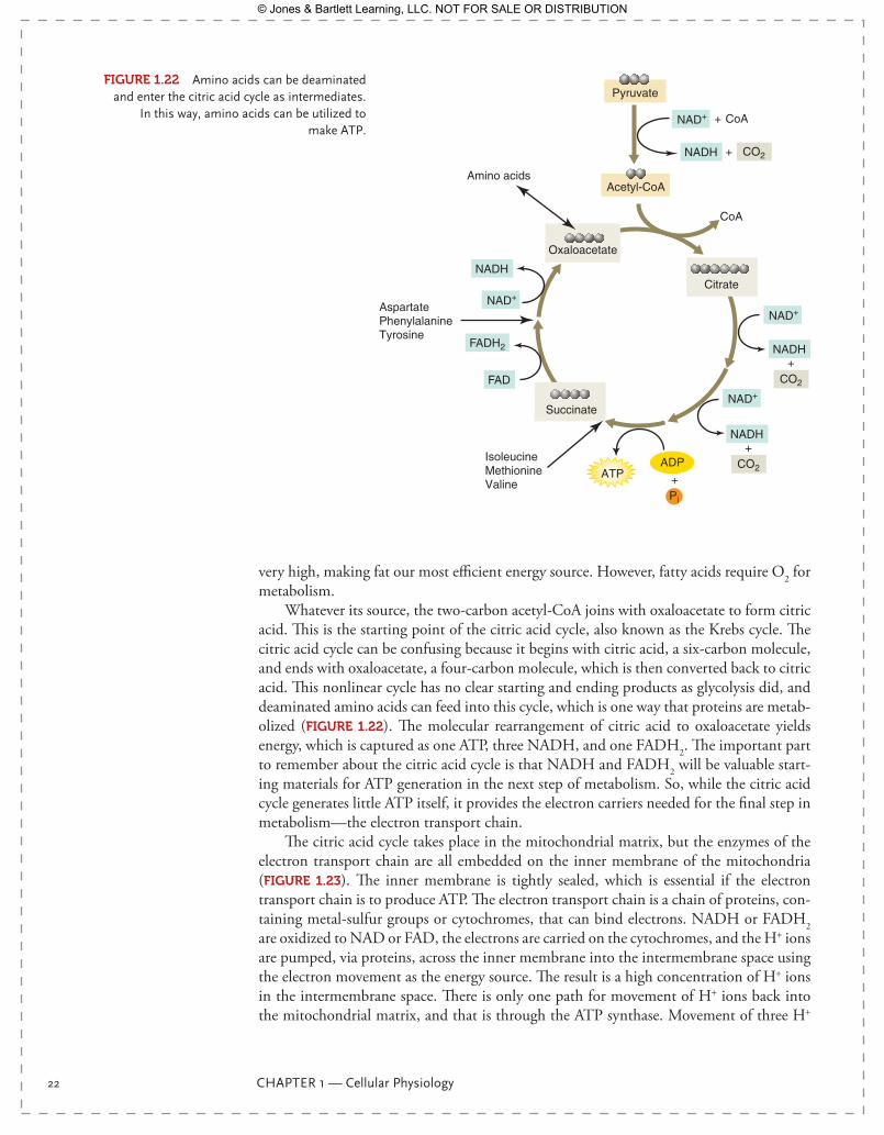
Whatever its source, the two-carbon acetyl-CoA joins with oxaloacetate to form citric acid. This is the starting point of the citric acid cycle, also known as the Krebs cycle. The citric acid cycle can be confusing because it begins with citric acid, a six-carbon molecule, and ends with oxaloacetate, a four-carbon molecule, which is then converted back to citric acid. This nonlinear cycle has no clear starting and ending products as glycolysis did, and deaminated amino acids can feed into this cycle, which is one way that proteins are metab-olized ( FIgurE 1.22). The molecular rearrangement of citric acid to oxaloacetate yields energy, which is captured as one ATP, three NADH, and one FADH2. The important part to remember about the citric acid cycle is that NADH and FADH2 will be valuable start-ing materials for ATP generation in the next step of metabolism. So, while the citric acid cycle generates little ATP itself, it provides the electron carriers needed for the final step in metabolism—the electron transport chain.
The citric acid cycle takes place in the mitochondrial matrix, but the enzymes of the electron transport chain are all embedded on the inner membrane of the mitochondria (FIgurE 1.23). The inner membrane is tightly sealed, which is essential if the electron transport chain is to produce ATP. The electron transport chain is a chain of proteins, con-taining metal-sulfur groups or cytochromes, that can bind electrons. NADH or FADH2 are oxidized to NAD or FAD, the electrons are carried on the cytochromes, and the H+ ions are pumped, via proteins, across the inner membrane into the intermembrane space using the electron movement as the energy source. The result is a high concentration of H+ ions in the intermembrane space. There is only one path for movement of H+ ions back into the mitochondrial matrix, and that is through the ATP synthase. Movement of three H+
ADPATP
NAD+
NADH
Pi
Pyruvate
Citrate
Amino acidsAcetyl-CoA
Oxaloacetate
Succinate
CO2
+
NADH
CO2
+
NAD+
NAD+
NAD+
NADH
FADH2
FAD
NADH CO2+
+ CoA
CoA
+
IsoleucineMethionineValine
AspartatePhenylalanineTyrosine
FIgurE 1.22 Amino acids can be deaminated and enter the citric acid cycle as intermediates.
In this way, amino acids can be utilized to make ATP.
9781284030341_CH01.indd 22 11/4/2013 6:17:53 PM
© Jones & Bartlett Learning, LLC. NOT FOR SALE OR DISTRIBUTION

Energy Production: Fueling Cellular Work 23
Outer membrane
Innermembrane
Intermembrane space
Intermembrane space
ATP transporter
Glucose
Pyruvate
Pyruvate
Acetyl-CoA
NADH
FADH2
NADH
NADH
CO2
CO2
H2OO2
H+ H+
H+
H+
H+
H+
H+
H+H+ H+
H+H+
H+
H+
ATP
ATP
H+
ADPATP
ATPPi
+
H+
ADPATP
Pi
+
+
Stage 1: Pyruvate oxidation
Stage 2: Krebs cycle
Glycolysis
Stage 3: Electron transport and proton pumping
Stage 4: ATP synthesis
e–
e–
e–
e–
Matrix
FIgurE 1.23 ATP production from glucose begins in the cytoplasm, continues in the mitochondrial matrix, and finishes with ATP being transported back into the cytoplasm.
9781284030341_CH01.indd 23 11/4/2013 6:17:59 PM
© Jones & Bartlett Learning, LLC. NOT FOR SALE OR DISTRIBUTION
24 CHAPTER 1 — Cellular Physiology
ions down their concentration gradient through this ion pore provides enough energy to generate one ATP from one ADP. Thus, the H+ ions carried by NAD and FAD are the pri-mary producers of ATP within the mitochondria. ATP produced within the mitochondria is transported through adenine nucleotide transporters into the cytosol for use.
At the end of the electron transport chain, electrons are transferred to O2. This is the ultimate job of an oxygen molecule—to accept the electrons from oxidized NADH ( Figure 1.23). Without O2, the movement of electrons ceases and ATP production stops. This is why we are obligate aerobic animals. Our ATP production depends on O2 as the final electron acceptor. All of your respiratory efforts as you run are for one purpose: to provide enough O2 to fuel this process.
However, acquiring O2 is only half of the breathing process. As you run, you inhale O2, but you also exhale CO2. This CO2 is generated as a by-product of metabolism and is a significant waste product that must be eliminated. The first CO2 comes from pyruvate metabolism, when it loses a carbon to become acetyl-CoA. The next two come from the citric acid cycle as a 6-carbon citric acid becomes a 4-carbon oxaloacetate. Each pyruvate thus generates three CO2 molecules that will need to be eliminated.
As you can see, the mitochondria are essential to our lives. The metabolic processes described above are how mitochondria function in health. We would like to think that these organelles function efficiently throughout our lives. However, mitochondrial mal-function, or an inefficiency in function, often occurs, with serious consequences for our health as an organism. Mitochondria are a primary source of oxygen free radicals, which can damage the mitochondria itself. Within this text, we will examine some of the ways in which mitochondria can contribute to human disorders, disease, and aging.
Muscle Contraction: A Symphony of Cellular CommunicationWe began this chapter with your afternoon run, which has caused such a change in your whole body physique. The muscle work involved in exercise exemplifies many of the cel-lular mechanisms we have discussed throughout this chapter. Let’s use muscle contraction to apply the concepts we have learned thus far, and to elucidate the process of muscle contraction itself.
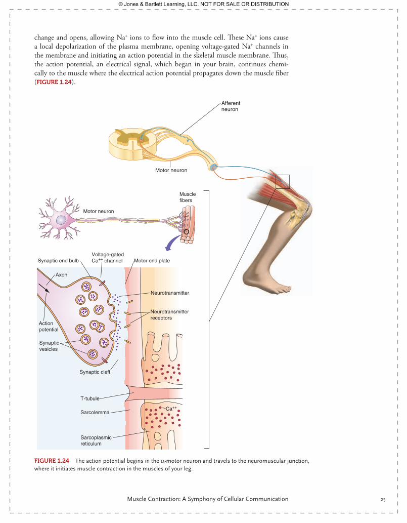
The Action Potential—Fast Long-Distance Communication
As you stand on the track, you decide to run. This is voluntary muscle contraction, driven by the motor cortex of the brain, as we will explore more fully later on. The action poten-tial along the nerve running from your brain to the spinal cord travels precisely as we discussed earlier, via the opening and closing of voltage-gated Na+ and K+ channels. At the spinal cord, this action potential is transferred to the α-motor neuron, which originates in the spinal cord and ends at skeletal muscle, at the neuromuscular junction—in this case, the skeletal muscle of your legs. The action potential terminates at the synaptic bulb of the neuron, where the depolarization causes Ca2+ channels to open, allowing the intra-cellular concentration of calcium to rise within the synaptic bulb. Calcium triggers the exocytosis mechanism, and vesicles of acetylcholine fuse with the plasma membrane of the neuron and release acetylcholine into the synaptic space. Acetylcholine binds to nicotinic acetylcholine receptors on the plasma membrane of the muscle cell. Nicotinic acetylcho-line receptors, also known simply as acetylcholine receptors, are ligand-gated ion chan-nels. Once acetylcholine binds, the ligand-gated ion channel undergoes a conformational
9781284030341_CH01.indd 24 11/4/2013 6:18:00 PM
© Jones & Bartlett Learning, LLC. NOT FOR SALE OR DISTRIBUTION
Muscle Contraction: A Symphony of Cellular Communication 25
change and opens, allowing Na+ ions to flow into the muscle cell. These Na+ ions cause a local depolarization of the plasma membrane, opening voltage-gated Na+ channels in the membrane and initiating an action potential in the skeletal muscle membrane. Thus, the action potential, an electrical signal, which began in your brain, continues chemi-cally to the muscle where the electrical action potential propagates down the muscle fiber ( FIgurE 1.24).
Afferentneuron
Motor neuron
Motor neuron
Muscle fibers
Synaptic end bulb
Synaptic vesicles
Synaptic cleft
Axon
Neurotransmitter
Action potential
Neurotransmitterreceptors
Voltage-gatedCa++ channel
Sarcolemma
Sarcoplasmicreticulum
T-tubule
Motor end plate
Ca++
FIgurE 1.24 The action potential begins in the α-motor neuron and travels to the neuromuscular junction, where it initiates muscle contraction in the muscles of your leg.
9781284030341_CH01.indd 25 11/4/2013 6:18:02 PM
© Jones & Bartlett Learning, LLC. NOT FOR SALE OR DISTRIBUTION
26 CHAPTER 1 — Cellular Physiology
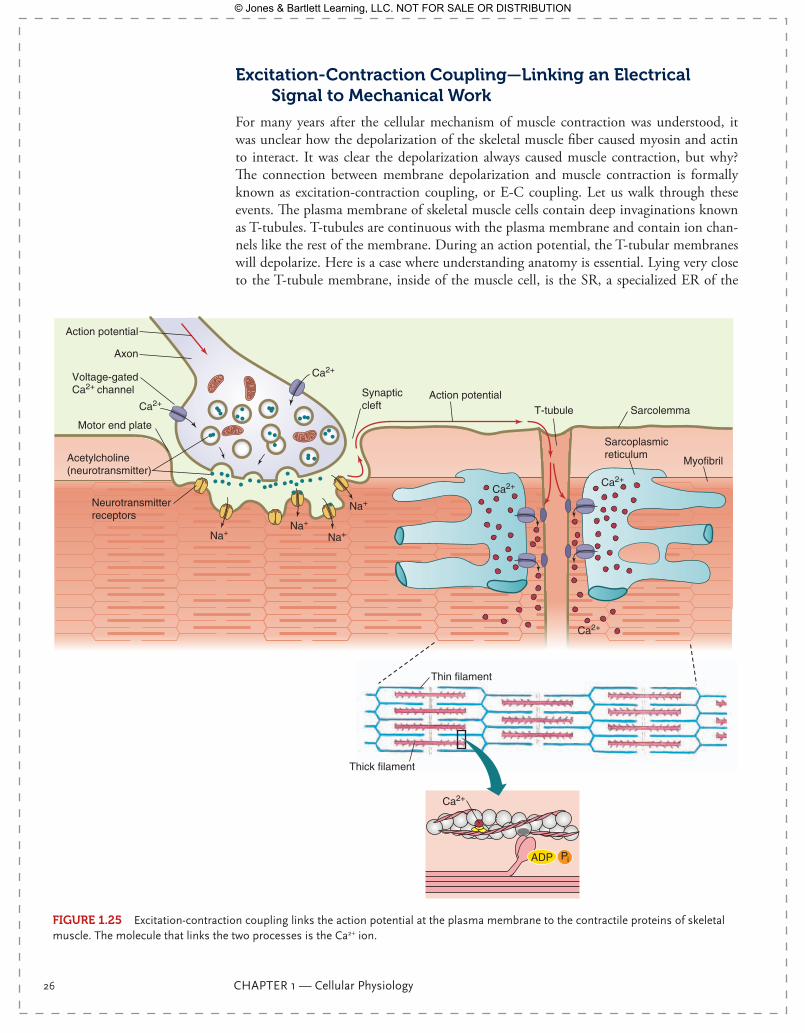
Excitation-Contraction Coupling—Linking an Electrical Signal to Mechanical Work
For many years after the cellular mechanism of muscle contraction was understood, it was unclear how the depolarization of the skeletal muscle fiber caused myosin and actin to interact. It was clear the depolarization always caused muscle contraction, but why? The connection between membrane depolarization and muscle contraction is formally known as excitation-contraction coupling, or E-C coupling. Let us walk through these events. The plasma membrane of skeletal muscle cells contain deep invaginations known as T-tubules. T-tubules are continuous with the plasma membrane and contain ion chan-nels like the rest of the membrane. During an action potential, the T-tubular membranes will depolarize. Here is a case where understanding anatomy is essential. Lying very close to the T-tubule membrane, inside of the muscle cell, is the SR, a specialized ER of the
ADP Pi
Sarcolemma
Myofibril
T-tubule
Synaptic cleft
Axon
Acetylcholine(neurotransmitter)
Action potential
Action potential
Neurotransmitterreceptors
Voltage-gatedCa2+ channel
Motor end plate
Ca2+
Na+Na+
Na+
Na+
Ca2+
Sarcoplasmicreticulum
Ca2+
Ca2+
Ca2+
Ca2+
Thick filament
Thin filament
FIgurE 1.25 Excitation-contraction coupling links the action potential at the plasma membrane to the contractile proteins of skeletal muscle. The molecule that links the two processes is the Ca2+ ion.
9781284030341_CH01.indd 26 11/4/2013 6:18:05 PM
© Jones & Bartlett Learning, LLC. NOT FOR SALE OR DISTRIBUTION
Muscle Contraction: A Symphony of Cellular Communication 27
muscle cell. The SR lies so close to the T-tubule that proteins within the SR membrane will experience a voltage change during the action potential. There is no action potential along the SR membrane, but there is a change in SR membrane voltage sufficient to open Ca2+ release channels in the SR. SR is a Ca2+ storage vesicle within skeletal muscle, and when the Ca2+ release channel opens, the intracellular [Ca2+] of the skeletal muscle cell increases (FIgurE 1.25). The increase in skeletal muscle calcium concentration is the signal trans-duction mechanism that links an action potential to mechanical work. Calcium ions were the mystery molecule of E-C coupling!
How Does Skeletal Muscle Contract?
Skeletal muscle is called striated muscle because of its orderly “striped” appearance. Within a skeletal muscle cell, myosin and actin proteins are aligned next to each other, both connected to Z-disks that link to the plasma membrane. The space between Z-disks, a sarcomere, is the repeating protein structure of the fiber. Each sarcomere is arranged as myosin proteins (thick filaments) lying between actin proteins (thin filaments).
Myosin proteins have several sections: the tail, the hinge, and the head. The head con-tains the actin binding site, while the hinge can assume several stable conformations, each of which is important to muscle contraction. Actin filaments are composed of globular actin polymerized into chains. Actin filaments also possess a binding site that can be occupied by myosin. If actin and myosin were left in this simple state, our skeletal muscle cells would be contracted all of the time. However, actin filaments are encircled by tropomyosin, which covers the binding site on actin, making it inaccessible to the myosin head. Attached to tropomyosin are the troponin proteins, the most important of which is troponin C. Actin, with its associated tropomyosin and troponin, is the regulator of skeletal muscle contraction.
How is exposure of the actin-myosin binding site regulated? Remember that Ca2+ is the link between the action potential and muscle contraction. Troponin C has a bind-ing site for the Ca2+ ion. As [Ca2+] rises intracellularly, troponin C binds the calcium ion ( FIgurE 1.26). This causes a conformational change in this protein, moving tropomyosin away from the actin-myosin binding site. Once exposed, myosin can bind to this site, initiating the molecular events of muscle contraction.
Myosin Binding, Cross Bridge Cycling, and ATP Hydrolysis
As you have probably noticed, to this point, none of the physiological events of muscle contraction have required ATP. The action potential, neurotransmitter release, opening of ligand-gated ion channels, the initiation of an action potential in the muscle cell, and E-C coupling have all been accomplished with virtually no ATP hydrolysis. Yet, we know that exercise and muscle contraction take work, which means ATP consumption. Cross-bridge cycling of myosin is where ATP is used. Now, let’s look in detail at how it is used.
Let’s begin where we left off, with an elevated intracellular [Ca2+] and an exposed binding site on actin. Myosin will quickly bind under these conditions. The myosin head is now connected to actin, with an associated ADP and Pi. Myosin is not only a struc-tural protein and an important part of your skeletal muscle, but it is also an enzyme, an ATPase capable of cleaving ATP into ADP and Pi. Instead of diffusing away from the myosin head, these hydrolysis products remain attached for a time. Pi leaves first, and when it does, it causes a conformational change in the position of the hinge, inducing a 45° bend. This is the power stroke of skeletal muscle contraction; it is how myosin pulls along actin and shortens the sarcomere, causing what we see, grossly, as muscle contrac-tion. ADP diffuses away next, and the myosin head remains attached to actin. The now “naked” myosin head binds a new molecule of ATP, which allows detachment from actin.
9781284030341_CH01.indd 27 11/4/2013 6:18:06 PM
© Jones & Bartlett Learning, LLC. NOT FOR SALE OR DISTRIBUTION

28 CHAPTER 1 — Cellular Physiology
ADP P
ADP P
(b)
Ca++
Ca++
Step 1: Action potential
ADP P
ADP P
(a)
Actin
TroponinTropomyosin
Resting
Myosin
ADP P
ADP P
(c) CatchMyosin binding site
Ca++
Ca++
Step 2: Myosin-actin binding
ATP
ATP
(e) Release
Step 4: ATP binding and actin-myosin release
ADP P
ADP P
(f) Recover
Step 5: ATP cleavage
ADP P
ADP P
(d) Drive
Step 3: Power stroke
FIgurE 1.26 The events of muscle contraction.
ATP is immediately hydrolyzed to ADP and Pi, and the cycle begins again. Notice that ATP is required for relaxation, and the power stroke is a result of Pi detachment from the myosin head ( Figure 1.26). The easiest way to remember this unexpected mechanism for muscle contraction is to consider the events of rigor mortis. Rigor mortis sets in when muscle cells have exhausted their supplies of ATP. When no ATP is available for muscle relaxation, skeletal muscle contracts, making the body rigid.
We have illustrated muscle contraction with a single myosin head for clarity. But in life, myosin heads are arranged in circular arrays, and cross-bridge cycling occurs thousands of times during a simple muscle contraction. This is where the expenditure of energy occurs.
SummaryYour afternoon run and simple snack actually require complex physiological mechanisms to accomplish! We will build on these mechanisms through the text as we learn about each of the organ systems.
Key ConceptsOsmosisDiffusionFacilitated diffusion
9781284030341_CH01.indd 28 11/4/2013 6:18:19 PM
© Jones & Bartlett Learning, LLC. NOT FOR SALE OR DISTRIBUTION
29Clinical Case Study
Na+K+ ATPaseVoltage-gated Na+ channelK+ leak channelResting membrane potentialAction potentialLigand-gated ion channelsReceptor signalingKinasesReceptor tyrosine kinasesNitric oxideSteroid hormonesProtein synthesisTranscriptionTranslationGlycolysisCitric acid cycleElectron transport chainExcitation-contraction couplingMuscle contraction
Key TermsAcetylcholineβ-adrenergic receptorα-adrenergic receptorTranscription factorChaperone protein
Application: Pharmacology1. Muscle strains are sometimes treated with dantrolene sodium, a drug that
prevents calcium release from the SR. How would this reduce muscle work?2. Botulinum toxin, known commonly as Botox, prevents the release of
acetylcholine into the synaptic space. Why might this be used to reduce wrinkles?
Clinical Case StudyType 2 Diabetes Mellitus, Part I
BACKgrOuND
Type 2 diabetes mellitus (T2DM), also sometimes known as metabolic syndrome or insulin resistance syndrome, is a set of clinical conditions that were once thought to be associated and are now known to be interrelated. The key elements of this syndrome are defined as:
��� Insulin resistance/diabetes��� Hyperinsulinemia��� Hyperlipidemia
9781284030341_CH01.indd 29 11/4/2013 6:18:22 PM
© Jones & Bartlett Learning, LLC. NOT FOR SALE OR DISTRIBUTION
30 CHAPTER 1 — Cellular Physiology
��� Obesity��� Hypertension��� Atherosclerosis��� Endothelial dysfunction
Insulin is a hormone released from the β-cells of the pancreas into the blood, where it then binds to receptors on insulin-dependent tissues, primarily skeletal muscle, adi-pose tissue, and liver. These receptors are not G-protein coupled but are a pair of simple α-helices in the plasma membrane that are capable of mutual phosphorylation and cou-pling. Once the hormone insulin binds to the receptor and the phosphorylation reaction begins, a tyrosine kinase phosphorylates and activates intracellular proteins, including insulin-receptor substrate 1, which is essential for muscle glycogen synthesis. Thus, insu-lin binding immediately begins the process of glucose storage within muscle and, by a related kinase reaction, in the liver as well. Insulin receptors also stimulate another kinase, phosphotidyl-inositol-3-kinase (PI-3 kinase), which stimulates glucose transport by mov-ing intracellular vesicles of glucose transporters to the plasma membrane, thus increasing the number of glucose transporters (GLUT4) in the membrane and the number of glucose molecules that can transit them. Finally, a different signaling pathway is stimulated by the insulin receptor, which will promote GLUT4 transcription and translation, thus increas-ing the total number of glucose transporters.
In T2DM, the insulin receptor becomes resistant to signaling by the hormone, espe-cially at the PI-3 kinase step. Thus, insulin binds the receptor, but the intracellular signal-ing pathway fails to increase the number of glucose transporters in the membrane. Exercise is generally prescribed for people suffering from T2DM, in part because exercise increases the concentration of glucose transporters in muscle membrane in an insulin-independent manner.
Normally, insulin release from the pancreatic β-cells is stimulated by glucose in a closely coupled cellular mechanism. As glucose levels fall, insulin release ceases. However, high circulating glucose levels cause continuous release of insulin, resulting in hyperin-sulinemia and insulin resistance. If blood glucose levels remain high long enough, it will cause overt T2DM. While the high circulating insulin levels fail to increase glucose uptake by cells, they have effects on other tissues that are uninhibited by T2DM. We will see some of these effects in other sections of this clinical case study.
THE CASE
Your aunt has been diagnosed with T2DM because of her elevated fasting glucose levels (140 mg/dL). She has been placed on a low-sugar, low-carbohydrate diet and a regular regime of exercise. After two weeks of carefully following her diet and exercising, she has noted that her fasting glucose level has dropped to 110 mg/dL. Normal is 90–100 mg/dL, so she still has some work to do but, at this point, she is encouraged.
THE QuESTIONS
1. Why is glucose remaining in her blood so long after a meal? 2. How does sugar normally get into a cell? 3. Describe the action of insulin and how it facilitates sugar transport. Be specific
about the signaling pathway.4. How does daily exercise lower blood sugar?
9781284030341_CH01.indd 30 11/4/2013 6:18:22 PM
© Jones & Bartlett Learning, LLC. NOT FOR SALE OR DISTRIBUTION