Mutations in CCDC 39 and CCDC 40 are the Major Cause of Primary Ciliary Dyskinesia with Axonemal...
42
1 UNCORRECTED ACCEPTED ARTICLE Research Article Human Mutation DOI 10.1002/humu.22261 Supporting Information for this preprint is available from the Human Mutation editorial office upon request ([email protected] ) Mutations in CCDC39 and CCDC40 are the major cause of primary ciliary dyskinesia with axonemal disorganisation and absent inner dynein arms Dinu Antony, 1,a* Anita Becker-Heck, 2,b* Maimoona A Zariwala, 3* Miriam Schmidts, 1 Alexandros Onoufriadis, 1 Mitra Forouhan, 1 Robert Wilson, 4 Theresa Taylor-Cox, 5 Ann Dewar, 5 Claire Jackson, 6 Patricia Goggin, 6 Niki T Loges, 2 Heike Olbrich, 2 Martine Jaspers, 7 Mark Jorissen, 7 Margaret W Leigh, 8 Whitney E Wolf, 9 M. Leigh Anne Daniels, 9 Peader G Noone, 9 Thomas W Ferkol, 10 Scott D Sagel, 11 Margaret Rosenfeld, 12 Andrew Rutman, 13 Abhijit Dixit, 14 Christopher O’Callaghan, 13 Jane S Lucas, 6 Claire Hogg, 5 Peter J Scambler, 1 Richard D Emes, 15 UK10K, 16 Eddie MK Chung, 17 Amelia Shoemark, 5 Michael R Knowles, 9§ Heymut Omran, 2§ Hannah M Mitchison, 1§ © 2012 Wiley Periodicals, Inc. This article has been accepted for publication and undergone full peer review but has not been through the copyediting, typesetting, pagination and proofreading process, which may lead to differences between this version and the Version of Record. Please cite this article as [DOI: 10.1002/humu.22261]. UNCORRECTED ACCEPTED ARTICLE
-
Upload
independent -
Category
Documents
-
view
1 -
download
0
Transcript of Mutations in CCDC 39 and CCDC 40 are the Major Cause of Primary Ciliary Dyskinesia with Axonemal...

1
UNCORRECTED ACCEPTED ARTICLE
Research Article
Human Mutation
DOI 10.1002/humu.22261
Supporting Information for this preprint is available from the
Human Mutation editorial office upon request ([email protected])
Mutations in CCDC39 and CCDC40 are the major cause of primary ciliary dyskinesia with
axonemal disorganisation and absent inner dynein arms
Dinu Antony,1,a* Anita Becker-Heck,2,b* Maimoona A Zariwala,3* Miriam Schmidts,1 Alexandros
Onoufriadis,1 Mitra Forouhan,1 Robert Wilson,4 Theresa Taylor-Cox,5 Ann Dewar,5 Claire
Jackson,6 Patricia Goggin,6 Niki T Loges,2 Heike Olbrich,2 Martine Jaspers,7 Mark Jorissen,7
Margaret W Leigh,8 Whitney E Wolf,9 M. Leigh Anne Daniels,9 Peader G Noone,9 Thomas W
Ferkol,10 Scott D Sagel,11 Margaret Rosenfeld,12 Andrew Rutman,13 Abhijit Dixit,14 Christopher
O’Callaghan,13 Jane S Lucas,6 Claire Hogg,5 Peter J Scambler,1 Richard D Emes,15 UK10K,16
Eddie MK Chung,17 Amelia Shoemark,5 Michael R Knowles,9§ Heymut Omran,2§ Hannah M
Mitchison,1§
© 2012 Wiley Periodicals, Inc.
This article has been accepted for publication and undergone full peer review but has not been through the
copyediting, typesetting, pagination and proofreading process, which may lead to differences between this
version and the Version of Record. Please cite this article as [DOI: 10.1002/humu.22261].
UNCORRECTED ACCEPT
ED ARTIC
LE

2
1 Molecular Medicine Unit and Birth Defects Research Centre, University College London
(UCL) Institute of Child Health, London, UK
2 Department of Pediatrics and Adolescent Medicine, University Hospital Muenster, Muenster,
Germany
3 Department of Pathology and Laboratory Medicine, UNC School of Medicine, Chapel Hill,
NC, USA
4 Respiratory Medicine, Royal Brompton and Hare eld NHS Trust, London, UK
5 Department of Paediatric Respiratory Medicine and Electron Microscopy Unit, Royal
Brompton and Hare eld NHS Trust, London, UK
6 Primary Ciliary Dyskinesia Group, University of Southampton Faculty of Medicine,
Southampton NIHR Respiratory Biomedical Research Unit, Southampton General Hospital,
Southampton, UK
7 University Hospitals Leuven, Department of Otorhinolaryngology, Herestraat 49 bus721, 3000
Leuven, Belgium
8 Department of Pediatrics, UNC School of Medicine, Chapel Hill, NC, USA.
9 Department of Medicine, UNC School of Medicine, Chapel Hill, NC, USA.
10 Department of Pediatrics, Washington University School of Medicine, St. Louis, MO, USA.
11 Department of Pediatrics, University of Colorado School of Medicine, Aurora, CO, USA
12 Children’s Hospital and Regional Medical Center, Seattle, WA, USA.
13 Department of Infection, Immunity and Inflammation, University of Leicester, Leicester Royal
Infirmary, Leicester, UK.
14 Department of Clinical Genetics, Nottingham City Hospital, Nottingham, UK
UNCORRECTED ACCEPT
ED ARTIC
LE
3
15 School of Veterinary Medicine and Science, University of Nottingham, Leicestershire, UK
16 uk10k.org.uk
17 General and Adolescent Paediatric Unit, University College London (UCL) Institute of Child
Health, 30 Guilford Street, London WC1N 1EH, UK
a Current affiliation: Dasman Genome Center Unit, P.O.Box 1180, Dasman 15462, Kuwait
b Current affiliation: Developmental and Molecular Pathways, Novartis Institutes for BioMedical
Research, Forum 1, Novartis Campus Basel, CH-4056, Basel, Switzerland
*These authors contributed equally to the work.
§Equal contribution.
Correspondence to:
Hannah M. Mitchison
Molecular Genetics Unit and Birth Defects Research Centre
University College London (UCL) Institute of Child Health
30 Guilford Street
London WC1N 1EH
Email. [email protected]
Tel. +44 20 7905 2866
Fax. +44 20 404 6191
Heymut Omran
UNCORRECTED ACCEPT
ED ARTIC
LE
4
Department of Pediatrics and Adolescent Medicine
University Hospital Muenster
Muenster
Germany
Email. [email protected]
Tel. +49 (251) 83 477 32
Fax. +49 (251) 83 477 35
Michael R. Knowles
Department of Medicine
UNC School of Medicine, Chapel Hill,
NC, USA
Email. [email protected]
Tel. +1 919 966-6780
Grant sponsorship:
Funding for UK10K was provided by the Wellcome Trust under award WT091310. MRK,
MWL, TWF, SDS, MR and MAZ are supported by National Institute of Health research grant 5
U54 HL096458-06, funded by the Office of the Director, and supported by ORDR and NHLBI,
NIH. MRK and MAZ are supported by National Institute of Health grant 5 R01HL071798, in
part by grant ULI TR000083 from the National Center of Research Resources, and grant 5 U54
HL096458-06 from National Center for Research Resources (NCRR), a component of NationalUNCORRECTED ACCEPT
ED ARTIC
LE

5
Institute of Health (NIH). AB-H, NTL, HOl and HO are supported by Kindness for Kids
Foundation, grants from the European Community (SYS-CILIA) and donation from the
"Schröder-Stiftung". DA, MS, AO, EMKC and HMM are funded by Action Medical Research
and Newlife Foundation. DA is supported by The Henry Smith Charity.
Abstract
Primary ciliary dyskinesia (PCD) is a genetically heterogeneous disorder caused by cilia and
sperm dysmotility. About 12% of cases show perturbed 9+2 microtubule cilia structure and inner
dynein arm (IDA) loss, historically termed ‘radial spoke defect’. We sequenced CCDC39 and
CCDC40 in 54 ‘radial spoke defect’ families, as these are the two genes identified so far to cause
this defect. We discovered biallelic mutations in a remarkable 69% (37/54) of families, including
identification of 25 (19 novel) mutant alleles (12 in CCDC39 and 13 in CCDC40). All the
mutations were nonsense, splice and frameshift predicting early protein truncation, which
suggests this defect is caused by ‘null’ alleles conferring complete protein loss. Most families
(73%; 27/37) had homozygous mutations, including families from outbred populations. A major
putative hotspot mutation was identified, CCDC40 c.248delC, as well as several other possible
hotspot mutations. Together, these findings highlight the key role of CCDC39 and CCDC40 in
PCD with axonemal disorganisation and IDA loss, and these genes represent major candidates
for genetic testing in families affected by this ciliary phenotype. We show that radial spoke
structures are largely intact in these patients and propose this ciliary ultrastructural abnormality
be referred to as ‘IDA and nexin-dynein regulatory complex (N-DRC) defect’, rather than ‘radial
spoke defect’.UNCORRECTED ACCEPT
ED ARTIC
LE

6
Key Words: primary ciliary dyskinesia, cilia, CCDC39, CCDC40, radial spoke, dynein
regulatory complex, nexin link
Introduction
Primary ciliary dyskinesia (PCD) is a recessively inherited disorder which arises from
cilia/sperm dysmotility that is associated with a number of different axonemal ultrastructural
abnormalities. The symptoms of deficient mucociliary clearance are usually obvious from birth,
and patients have recurrent respiratory tract infections leading to irreversible lung damage
(bronchiectasis). They also manifest with otitis media, chronic sinusitis, and subfertility. Internal
organ laterality is randomised with about half of patients having situs inversus, an association
reflecting dysmotility of nodal cilia during embryonic development.
Motile flagella and cilia are related organelles found on the surface of cells evolutionarily
conserved across 1.6 billion years from the flagella of the green alga Chlamydomonas to the
ciliated respiratory and embryonic node cells of vertebrates (Pazour, 2004). In humans, motile
cilia have a common microtubule-based ultrastructure (axoneme), comprising 9 peripheral
microtubule doublets either surrounding (in ‘9+2’ motile respiratory and fallopian tube cilia, and
sperm flagella) or lacking (in ‘9+0’ motile nodal cilia) a central microtubule pair (Becker-Heck
et al., 2012; Fliegauf et al., 2007), and linked to a variety of microtubule-associated proteins.
These include the inner and outer dynein arm motor complexes, which project from the
peripheral microtubule doublets; the radial spokes, which provide a radial scaffold between the
UNCORRECTED ACCEPT
ED ARTIC
LE

7
central pair and peripheral microtubules and facilitate signal transduction from the centre out to
the dynein arms to govern ciliary beat and waveform (Becker-Heck et al., 2012); and nexin-
dynein regulatory complexes (N-DRC), which attach between adjacent peripheral doublets to
facilitate inner dynein arm attachment and regulate dynein activity (Heuser et al., 2009). This
complex superstructure creates the rigid organisation along the entire length of the axoneme
which is required for motor ATPase signalling to generate a uniquely coordinated and self-
propagating beat (Mitchison and Mitchison, 2010).
PCD is genetically heterogeneous with 18 identified genes causing non-syndromic disease. The
range of genetic defects cause a small number of defective ciliary ultrastructural subtypes by
current imaging resolution, and no correlations have been defined between ultrastructural defects
and the course of disease (Kispert et al., 2003). Mutations in genes that cause axonemal outer
dynein arm (ODA) defects (DNAH5, DNAI1, DNAI2, DNAL1, TXNDC3 and CCDC114) are the
genetic basis of the majority of PCD cases (Olbrich et al., 2002; Pennarun et al., 1999; Loges et
al., 2008; Mazor et al., 2011; Duriez et al., 2007; Bartoloni et al., 2002; Onoufriadis et al., 2012;
Knowles et al., 2012a). Mutations in genes encoding components of the radial spoke head
(RSPH4A and RSPH9) cause defects involving the central pair microtubules (Castleman et al.,
2009). Mutations in genes encoding cytoplasmic or dual location proteins involved in the
assembly and transport of dynein arm components into the axoneme from the cell body
(DNAAF1/LRRC50, DNAAF2/KTU, DNAAF3/PF22, CCDC103, HEATR2 and LRRC6) cause
inner and outer dynein arm defects (Loges et al., 2009; Duquesnoy et al., 2009; Omran et al.,
2008; Mitchison et al., 2012; Panizzi et al., 2012; Horani et al., 2012; Kott et al., 2012). Lastly,
two genes associated with PCD give rise to no discernible ultrastructural defects of the axoneme
UNCORRECTED ACCEPT
ED ARTIC
LE
8
(DNAH11) or defects of the central microtubule pair C2b projection that are too subtle to be
easily detected (HYDIN) (Bartoloni et al., 2002; Knowles et al., 2012b; Olbrich et al., 2012).
Mutations in two genes, CCDC39 (MIM# 613798) (Merveille et al., 2011) and CCDC40 (MIM#
613799) (Becker-Heck et al., 2011), have recently been shown in PCD patients that have a defect
of the cilia axoneme, involving loss of the inner dynein arms accompanied by a variably
expressed disorganisation of the 9+2 microtubule arrangement. Recent transmission electron
microscopy (TEM) surveys of several different PCD cohorts estimated that at least ~12% of all
PCD cases have this defect (Chilvers et al., 2003; Shoemark et al., 2012; Papon et al., 2010). The
nine peripheral microtubules are retained but are mislocalised, often becoming more centralised,
while the central microtubule pair may be variously lost (9+0), eccentrically positioned towards
the periphery (9+2), or increased in number with a supernumerary central pair present (9+4); and
the inner dynein arm structures are reduced or absent. Furthermore, in axonemes with this defect
abnormal and/or absent N-DRC complexes have been recorded (Schneeberger et al., 1980;
Konradova et al., 1982) and radial spokes (Sturgess et al., 1979; Antonelli et al., 1981). This
ultrastructural defect has historically often been referred to as ‘radial spoke defect’. The reasons
for the various disarrangements are unclear, but since a range of microtubule disorganisations
may be seen within a single TEM sample of respiratory epithelia from a patient, it is even
possible that a variety of changes may occur along the length of the axoneme. These
perturbations of structure create a characteristic ciliary motility defect with a beating pattern of
mixed appearance, where stiff cilia displaying a reduced amplitude (70%) and cilia that are fully
immotile (30%) are both visible (Chilvers et al., 2003; Merveille et al., 2011; Becker-Heck et al.,
2011). The partially retained motility may be explained by activity of the outer dynein arm
UNCORRECTED ACCEPT
ED ARTIC
LE

9
motors, but cilia waveform is misregulated and the mean ciliary beat frequency is reduced
(Chilvers et al., 2003).
The CCDC39 and CCDC40 genes encode structurally related coiled-coil domain-containing
proteins of unknown function that are localised to the axoneme (Merveille et al., 2011; Becker-
Heck et al., 2011; Becker-Heck et al., 2012). The loss of IDAs and N-DRCs from ciliary
axonemes of CCDC39 and CCDC40 patients has been previously confirmed using antibodies to
DNALI1 and the N-DRC component GAS11/8 respectively (Merveille et al., 2011; Becker-Heck
et al., 2011), and it is proposed that CCDC39 and CCDC40 proteins interact with N-DRC
components and play a role in inner dynein arm attachment. Neither protein has yet been
identified in proteomic studies of N-DRC composition (Lin 2011); thus, rather than being
integral N-DRC proteins, they may be otherwise involved in N-DRC assembly, microtubule
attachment, or protein interactions. The role of the CCDC39 and CCDC40 proteins in the
structure and function of radial spokes is not known, and radial spokes in cilia from ‘radial spoke
defect’ patients have never been examined at the molecular level using antibody staining
techniques.
In this study, we applied a combination of candidate gene Sanger sequencing and next-
generation whole exome sequencing to identify the genetic cause of PCD in a large cohort of
‘radial spoke defect’ patients with cilia dysmotility associated with axonemal microtubule
disorganisation and absence of IDAs. We also used antibody and immunofluorescence
techniques to investigate radial spoke perturbations in nasal ciliated cells in patients with
CCDC39/40 mutations.
UNCORRECTED ACCEPT
ED ARTIC
LE
10
Materials and Methods
Subjects
A total of 54 unrelated PCD families were involved in the screen. Parallel mutational analysis on
a collection of 17 families from UCL-ICH (labelled in the text by ‘PCD’ prefix), 4 from
Belgium/UHM (University Hospital Muenster) (‘OP’ and ‘KUL’ prefix) and 33 from UNC
(‘UNC’ prefix) was performed using genomic DNA samples obtained from peripheral blood
cells. All families agreed under informed consent to participate in this study in accordance with
protocols approved by the ethical committees of the Institute of Child Health/Great Ormond
Street Hospital and University College London Hospital NHS Trust, and those of collaborating
institutions.
CCDC39 and CCDC40 mutation identification
The transcripts referred to are CCDC39 NM_181426.1 and CCDC40 NM_017950.2. Sanger
sequencing was performed by amplifying and sequencing the coding exons and flanking intronic
sequences of CCDC39 and CCDC40 (primer sequence available on request). Sequence
alignments for variant identification were made using Sequencher software (Gene Codes
Corporation). Whole exome sequencing was performed for most samples by capturing the exons
using the Agilent SureSelect All Exon Human V3 (50 Mb) kit. TruSeq Pair End Cluster kit V3
was used for cluster generation and 100 bp. paired-end reads were generated on an Illumina
HiSeq 2000 analyser using Illumina TruSeq V3 SBS sequencing chemistry. The BWA alignment
tool (Li and Durbin, 2010) was used to map sequence reads back to the genome (human
reference hg19) then the GATK tool suite (McKenna et al., 2010) was used to process the
alignments and identify variations. The SNP-Effects (http://snpeff.sourceforge.net/) andUNCORRECTED ACCEPT
ED ARTIC
LE

11
ANNOVAR (Wang et al., 2010) programs were used to annotate variations. The details of the
whole exome sequencing are presented in Supp. Table S1.
In one case (PCD22) exome sequencing was performed as part of the Wellcome Trust Sanger
Institute UK10K Project as described previously (Olbrich et al., 2012; Onoufriadis et al., 2012).
Exome variant analysis was achieved by filtering of the total variant list according to consistent
autosomal recessive inheritance pattern, novelty in comparison to human polymorphism
databases including the 1000 Genomes (1000 Genomes Project Consortium, 2010) and NHLBI
Exome Sequencing projects (http://evs.gs.washington.edu/EVS) and dbSNP v135, and finally for
their functional significance. This analysis required the presence of at least one homozygous or
two heterozygous changes occurring with an estimated frequency <0.01, and all the patients
included in this study had clear-cut biallelic variants in the CCDC39 and CCDC40 genes that
were identified via excellent coverage in all cases, without any other obvious causal candidates
indicated. Sanger sequencing was used to confirm all the identified variants from exome
sequencing and to verify the segregation pattern of each change in other unaffected family
members, identified by both methods.
Nucleotide numbering reflects cDNA numbering with +1 corresponding to the A of the ATG
translation initiation codon in the reference sequence, according to journal guidelines
(www.hgvs.org/mutnomen). The initiation codon is codon 1.
UNCORRECTED ACCEPT
ED ARTIC
LE

12
Electron microscopy and high-speed video light microscopy
Respiratory epithelial cells were obtained using a cytology brush or rhinoprobe from the inferior
turbinate of participants and immediately analysed for cilia beat frequency and beat pattern under
light microscopy as described (Chilvers et al., 2003). Samples were fixed in glutaraldehyde and
an ultrastructural defect of the cilia was confirmed by electron microscopy (Rutland et al., 1982).
Immunofluorescence analysis
The effect of the CCDC40 splice mutation in patient 114 II:1 was confirmed by
Immunofluorescence analysis. Respiratory epithelial cells obtained by nasal brush biopsy were
suspended in cell culture medium. Samples were spread onto glass slides, air dried and stored at -
800C until use. Cells were fixed with 4% PFA for 4 minutes at room temperature, washed 5X
with PBS and then permeabilized with 0.5% TritonX100 for 10 minutes. After 5 more washes
with PBS cells were incubated with 5% bovine serum albumin (Sigma) in PBS for 1 hour. The
cells were then incubated with primary antibodies overnight at room temperature using the
following dilutions: CCDC39 antibody 1:100 (rabbit polyclonal Sigma); RSPH4A 1:100 (rabbit
polyclonal Sigma); ROPN1L 1:200 (rabbit polyclonal Sigma) monoclonal mouse anti-acetylated
and gamma tubulin 1:500 (Sigma) over night at room temperature. After 5 washes with PBS
cells were incubated with secondary anti-rabbit antibody (Alexa Fluor 488 Molecular Probes
Invitrogen) and secondary anti-mouse antibody (Alexa Fluor 594 Molecular Probes Invitrogen).
DNA was stained using DAPI (Invitrogen). Cells were finally washed 5X with PBS, mounted in
Vectashield (Vector Laboratories) and confocal images were taken using a Zeiss LSM 710.
UNCORRECTED ACCEPT
ED ARTIC
LE

13
Protein modelling
Conserved domains were predicted using SMART (Letunic et al., 2012) and CDD (Marchler-
Bauer et al., 2011), and coiled-coil protein folds predicted using Paircoil2 (McDonnell et al.,
2006) with minimum window size of 28 amino acids. Protein homologies and network
predictions were identified using PSI-BLAST (Altschul et al., 1997) to search the nr database
and STRING 9.0 (Szklarczyk et al., 2011).
Results
The entire coding region and flanking intronic sequences of the CCDC39 and CCDC40 genes
were sequenced in a cohort of 59 patients from 54 PCD families that displayed ciliary
dysmotility and a similar axonemal ultrastructural phenotype to that associated with CCDC39
and CCDC40 mutations (Merveille et al., 2011; Becker-Heck et al., 2011). The patients were all
diagnosed based on having a classic PCD phenotype, including recurrent respiratory tract
infections, pneumonia, rhinosinusitis, otitis media usually requiring repeated grommet insertion,
and age-dependent bronchiectasis, where chest CT data was available. Most patients showed
symptoms in the neonatal period, with respiratory distress, as well as recurrent airway infections.
In addition, patients showed abnormal cilia ultrastructure and motility at the electron and light
microscopic levels respectively, as described below.
Combined whole exome sequencing and Sanger sequencing analysis in the 54 families identified
a total of 25 different putative mutations, 12 in CCDC39 and 13 in CCDC40 that affected a total
of 47 patients in 37 families (Table 1). All the changes consisted of frameshift, nonsense and
UNCORRECTED ACCEPT
ED ARTIC
LE

14
essential splice site mutations, the latter all affecting the 100% conserved splicing consensus
intronic nucleotides. None of the identified variants are present in dbSNP (Sherry et al., 2001),
or the 1000 Genomes Project (1000 Genomes Project Consortium, 2010) and NHLBI ESP
Exome Variant Server (http://evs.gs.washington.edu/EVS/) exome repositories, nor where they
detected in 180 exomes available via the UK10K project (uk10k.org). Genotyping of all
available members of the affected families carrying CCDC39/40 mutations showed that all the
identified variants segregated correctly in association with disease status, having an autosomal
recessive inheritance pattern. Of the 37 families carrying CCDC39/40 mutations, segregation
was possible for 26 families, the other 11 families being represented by single affected patients
(Figs. 1A, B and Supp. Fig. S1). Thus, in this study just 17/54 families did not carry CCDC39
and CCDC40 variants, so these two genes accounted for 69% (37/54) of families.
The clinical details for the affected families are shown in Supp. Table S2. In all patients with
either CCDC39 or CCDC40 mutations, TEM of respiratory bronchial epithelial cells showed
similar microtubule disorganisation comprising disorganisation of the peripheral microtubule
doublets, absent or shifted central pairs. In all samples there was a documented reduction or
complete loss of the inner dynein arms. Therefore, the CCDC39 and CCDC40 mutations cause
defects that are indistinguishable by TEM.
A detailed clinical review was performed for six CCDC39 and three CCDC40 patients under the
care of the Royal Brompton Hospital London that were part of the UCL-ICH cohort, then
compared to findings on the UNC cohort of 20 patients. Their nasal nitric oxide levels were lowUNCORRECTED ACCEPT
ED ARTIC
LE

15
in both cohorts at 45-79 ppb and 6-34 nl/min respectively, as expected for PCD patients where
the normal mean level is 639 ppb (range 422-890) (Shoemark and Wilson, 2009). TEM of at
least 30 cilia cross sections was examined in UCL-ICH patients which revealed disarrangement
of the outer microtubular doublets in 43% (CCDC39) and 36% (CCDC40) of cilia cross sections,
mainly involving translocation of peripheral microtubular doublets and acentric microtubular
central pairs (Fig. 1Cb-d). Other less prominent features included additional central microtubules
or absence of the central pair structures (Fig. 1Cc-e). Inner dynein arms were absent from 69%
(CCDC39) and 90% (CCDC40) of cilia cross sections (Fig 1Cb-e). The outer dynein arm was
apparent throughout. In one subject with CCDC40 mutations a fallopian tube biopsy revealed
similar abnormalities (Fig. 1Ce). The UNC study found similar results in TEM of at least 30 cilia
cross-sections from CCDC39/40 patients, with 26% of cilia on average showing microtubule
disorganisation and 92% showing IDAs lost on average, and outer dynein arms present in all.
High speed video analysis of ciliated nasal brush biopsies of UCL-ICH patients with CCDC39
mutations showed the majority of cilia to be static at 37ºC (~ 75%), and also at 25ºC in UNC
CCDC39/40 patients. In patches where movement was present, the CCDC39 mutant cilia beat
pattern was typically stiff, rigid and ineffective (Supp. Movie. S1, S2). This was
indistinguishable from CCDC40 (Supp. Movie S3). A control sample is shown in Supp. Movie
S4. In cilia which demonstrated motility there were a wide range of ciliary beat frequencies
(range 3.3-13.0 Hz). Mean 37ºC CBF was 8.1Hz in CCDC39 patients and 9.2Hz in CCDC40
patients, where the normal range is 11-16 Hz. In UNC CCDC39/40 patients the mean 25ºC CBF
was reduced to 4.3Hz from the normal mean value of 7.3Hz. In three CCDC39 and one CCDC40
patients, sperm dysmotility was also recorded (data not shown).
UNCORRECTED ACCEPT
ED ARTIC
LE

16
We used a number of protein modelling tools to identify the location of CCDC39 and CCDC40
mutations identified in this study, and those previously published, in relation to putative protein
functional domains. CCDC39 has 941 residues and 10 predicted coiled-coils, in agreement with
previous structural modelling (Merveille et al., 2011), and CCDC40 has 1142 residues and 8
predicted coiled-coils (Fig. 2A, B). Both proteins contain two large Structural Maintenance of
Chromosomes (SMC) conserved domains which, as previously discussed, are found in several
ciliary proteins and likely play a role in microtubule-based ciliary transport processes (Merveille
et al., 2011). A conserved BRE1 domain (Kim et al., 2005) was identified in CCDC40, but the
significance is not clear. STRING predicted interactions between CCDC39 and protein
phosphatase 1 F-actin cytoskeleton targeting subunit (phostensin), an actin filament-binding
protein that can modulate actin dynamics that has not been connected with cilia functions (Lai et
al., 2009); and between CCDC40 and MCTP1, a membrane protein with putative calcium
mediated signalling functions (Shin et al., 2005).
Of the 25 mutations reported here, 19 are novel to this study. Fig. 2 shows the location of the
identified mutations in both genes and encoded CCDC39 and CCDC40 proteins. All the
identified mutations predict premature protein truncation via nonsense (5/12 and 5/13 for
CCDC39 and CCDC40 respectively) and frameshift effects, the latter either arising from small
indels within the coding sequence (5/12 and 6/13 respectively), or from single base substitutions
of the essential splice site residues at the immediate exon-intron boundaries (2/12 and 2/13,
respectively). All these variants are likely to give rise to null alleles via nonsense-mediated
decay, indicating that the inner dynein arm and microtubule disorganisation phenotype arises
from complete loss of these proteins and consequent loss-of-function. This supports the
UNCORRECTED ACCEPT
ED ARTIC
LE

17
published evidence that mutations in CCDC39 and CCDC40 have a similar functional effect in
being highly deleterious (Merveille et al., 2011; Becker-Heck et al., 2011). No particular
clustering of mutations is evident since in both proteins the changes that we have identified, and
previously published mutations, are evenly distributed across the gene and protein structure. This
suggests that protein termination at any point leads to the same deleterious dysfunction.
This study identified six mutations that have already been reported: a CCDC39 splice site
mutation c.357+1G>C (Merveille et al., 2011) and the CCDC40 frameshift mutations c.248delC
and c.3129delC; and nonsense mutations c.2440C>T, c.961C>T and c.1345C>T (Becker-Heck et
al., 2011; Nakhleh et al., 2012). All of these mutations are only present in patients from families
of Northern European descent, except for CCDC39 c.357+1G>C which was reported in 1
Turkish and 3 Northern European families previously. In addition, two mutations that are novel
to this study were shared amongst different families within our cohort: a CCDC39 frameshift
mutation c.526_527delCT was common to both a UK and a Zimbabwean origin family, and a
CCDC40 splice site mutation c.2712-1G>T was shared in two UK and one US families.
We found that the CCDC40 frameshift mutation c.248delC is extremely common, and is
restricted to families of N. European origin spread worldwide, suggesting an ancient shared
ancestry and past Founder effect mutation occurrence. c.248delC accounts for 34/56 N.European
ancestry disease chromosomes in CCDC40 disease, a remarkable 63% (Table 1). It is not known
whether the other smaller putative ‘hotspot’ mutations may also represent Caucasian N.
European-origin Founder effect mutations, or whether they are functionally significant changes
UNCORRECTED ACCEPT
ED ARTIC
LE

18
that have occurred separately many times in different countries. Despite the fact that only five
out of the total 37 CCDC39/40 families carrying mutations were consanguineous, there was an
overall predominance of homozygous changes amongst the families, with 27/37 (73%) of
CCDC39/40 families carrying homozygous changes due to identical alleles being inherited from
both parents. The Turkish-origin in one family for CCDC39 c.357+1G>C (Merveille et al., 2011)
and Zimbabwean origin in one family for CCDC39 c.526_527delCT indicates a more complex
evolution of these common mutations than a N. European Founder effect, and the possibility of
Non-Founder mutation hotspots. However, firm conclusions are precluded by the small family
numbers. It should be noted that although the family UCL210 carrying a homozygous nonsense
mutation in CCDC40, c.2245G>T, was not aware of familial consanguinity, they orginate from a
small Punjabi-speaking isolate located in Afghanistan likely to have underlying ancestral
endogamy and consanguinity.
The ciliary ultrastructure of patients with CCDC39 and CCDC40 mutations has been referred to
as ‘radial spoke defect’, and several reports have suggested that radial spokes are defective
(Sturgess et al., 1979; Antonelli et al., 1981; Merveille et al., 2011; Becker-Heck et al., 2011).
We sought to investigate radial spoke structures in the patients at the molecular level, by high-
resolution immunofluorescence analysis of nasal epithelial cells. In one CCDC40 mutation
patient 114 II:1, we confirmed an absence of the CCDC39 protein along the length of the
axoneme in all cilia analysed in this patient (Figs. 3A, B). These findings are consistent with
published information reporting the loss of CCDC39 from axonemes of CCDC40 mutant cilia.
However, both RSPH4A which is a radial spoke ‘head’ component (Yang et al., 2006) and
ROPN1L/RSP11 which is a radial spoke ‘stalk’ component (Yang et al., 2006; O'Toole et al.,UNCORRECTED ACCEPT
ED ARTIC
LE

19
2012) were present in axonemes from the patient as well as controls, with no differences in
protein levels observable (Figs. 3C, D and Supp. Fig. S2). This shows for the first time that
components of the radial spoke structures are present in axonemes of patients that have long
been typically referred to as ‘radial spoke defect’. These data do not however prove that these
radial spoke proteins are properly localized in the cilia superstructure, thus they may be present
but not functioning correctly.
Discussion
This study shows that CCDC39 and CCDC40 mutations are the major cause of PCD in patients
with the previously termed ‘radial spoke defect’, which is characterised by a ciliary axonemal
loss of inner dynein arms and axonemal disorganisation. We report the identification of 25
different allelic mutations in CCDC39 and CCDC40 affecting a total of 46 PCD patients in 37
families. Nineteen of the mutations are novel to this study, while six are shared with patients in
three previously published reports (Merveille et al., 2011; Becker-Heck et al., 2011; Nakhleh et
al., 2012). A report of additional mutations not referred to here was also published during
preparation of this manuscript (Blanchon et al., 2012). In our cohort, two of the novel mutations
are found in more than one family across different population origins. Therefore in our patients
and also in those reported elsewhere, there are putative common or ‘hotspot’ mutations for both
genes; specifically c.357+1G>C and c.526_527delCT in CCDC39, and c.248delC, c.3129delC,
c.2440C>T, c.961C>T, c.1345C>T and c.2712-1G>T in CCDC40. The c.248delC mutation is
very common, affecting 18/28 CCDC40 families (64%) in this study, in particular US-origin
families. The total number of different alleles in CCDC39/40 is smaller than might be expected
UNCORRECTED ACCEPT
ED ARTIC
LE

20
from the outbred populations we have analysed. This collective evidence is rather unusual for
PCD disease and supports the idea that critical regions or important functional residues within
the two proteins may be repeatedly vulnerable to mutation arising separately within different
populations, probably combined with some localised Founder effects.
Mutations in either gene give rise to the same ciliary and clinical phenotypes, and ciliary defects
that are indistinguishable using current methodologies of TEM and high-speed video
microscopy. Our TEM and cilia dysmotility findings are consistent with published findings for
CCDC39 and CCDC40 mutation (Merveille et al., 2011; Becker-Heck et al., 2011). Therefore,
mutations in these genes give rise to analogous defects, which is consistent with similarities in
their protein architecture, axonemal localisation, and putative biological role(s). The large
number of patients within the ‘radial spoke defect’ subtype of PCD that carry mutations in
CCDC39/40 is striking given the extensive underlying genetic heterogeneity in PCD.
Notably, all CCDC39/40 mutations give rise to null alleles, due to nonsense, frameshift or
conserved splice site effects. This suggests that complete loss of protein is required to give the
characteristic ultrastructural defects, and individuals carrying ‘milder’ effect alleles would likely
not express a PCD-like disease phenotype, although this might affect disease severity of other
airway diseases. This is in direct contrast with other ciliopathy disorders (affecting non-motile
primary cilia) where missense mutations predominantly confer disease, and it is reported that two
null alleles are never seen in patients because they affect development so severely that they are
incompatible with embryonic survival (Dagoneau et al., 2009; Beales et al., 2007; Davis et al.,UNCORRECTED ACCEPT
ED ARTIC
LE

21
2011; Schmidts et al., 2012). An intriguing alternative hypothesis is that CCDC39/CCDC40
missense mutations are not observed because they may be more deleterious than loss of function
mutations, rather than less deleterious, and are thus selected against in the surviving clinical
patient population. This model is also plausible, for example, if CCDC39/CCDC40 were to
orchestrate cilia assembly in multiprotein complexes, such that loss of function mutations could
be better tolerated than gain-of-function missense mutations.
Combining our data with that of the three previous reports, a total of 26 mutations in CCDC39
and 21 mutations in CCDC40 have been identified, all encoding predicted null alleles. In N-DRC
Chlamydomonas null-mutant strains, loss of any one of the N-DRC components gives rise to
complete loss of the entire N-DRC structure (Piperno et al., 1994). CCDC39 and CCDC40
mutation patients also lack at least some of the N-DRC structure, and this also appears to be
associated with mutations causing complete loss of protein.
The predicted loss-of-function reported for all CCDC39 and CCDC40 mutations correlates with
the widespread distribution of mutations across both genes, without any evidence of clustering.
We expect that all mutations identified in these two genes would make the protein subject to
nonsense mediated decay; however, both proteins contain multiple functionally important
domains that are highly evolutionarily conserved across species and likely critical to function.
The possible role of the BRE1 domain in CCDC40 is less clear, but the large SMC domains in
both proteins are thought to be involved in microtubule transporting (Merveille et al., 2011).
Both proteins have multiple coiled-coil domains, which leads to a compositional bias of charged
UNCORRECTED ACCEPT
ED ARTIC
LE
22
and hydrophobic residues containing many lysine, leucine and glutamic acid residues which are
generally located at the ‘surface’ of the coiled-coil domains involved in conferring solubility and
interaction properties. The 10 nonsense mutations identified in this study mostly affect glutamic
acid or arginine residues, mostly in coiled-coil domains; however, their codon composition
makes these residues prone to nonsense mutations, rather than indicating specific functional
importance at those residues. Coiled-coil domains are found in many different proteins with
diverse functions (Strauss and Keller, 2008), including structural and motor proteins. They are
also associated with signal transduction functions, and assembly/disassembly of protein
complexes, which may relate to the inner dynein arm-N-DRC-radial spoke complexes of cilia. If
the stability provided by multiple coiled-coils is mutated, this may result in reduced efficiency or
loss of protein function. Alternatively, some motor proteins have intrinsic instability in the
coiled-coil domains to allow the relaxing of tertiary structure and movement of the motor
proteins, which could be important in the cilia.
In contrast with N-DRC components, components of the radial spokes, such as the spoke ‘head’
protein RSPH4A and spoke ‘stalk’ protein ROPN1L, are present in CCDC40 mutant axonemes.
This provides the first evidence that the ultrastructural defect in patients with CCDC39
/CCDC40 mutations, commonly referred to as ‘radial spoke defect’, may not reflect a loss of
spokes, but proves supporting evidence that they may remain largely intact. We have not been
able to exclude the possibility that radial spoke components may be present, but mislocalised,
misattached, incomplete or non-functional for other reasons. However, we can conclude that this
defect may be more accurately referred to as ‘IDA and N-DRC defect’, rather than ‘radial spoke
defect’.
UNCORRECTED ACCEPT
ED ARTIC
LE

23
It is not yet clear how this ciliary ultrastructural defect arises, because little is known about the
formation and co-assembly of the human inner dynein arms, radial spokes and N-DRC
structures, which are all attached in close proximity at the peripheral doublets. These
components are all positioned at regular periodicity along the entire axoneme length, and form a
sophisticated regulatory network governing dynein activity that is key to cilia motility. The loss
of IDAs and N-DRC complexes together in CCDC39 and CCDC40 mutant axonemes is
consistent with the role that the N-DRC plays in tethering of IDA components, as shown in
Chlamydomonas. The resultant disorganisation of the peripheral microtubules and the resultant
instability of the central pair apparatus, indicates that these structures are critical for integrity and
motility of the axoneme. Studies of Chlamydomonas motility mutant strains suggest the N-DRC
has a number of roles apart from binding of the inner dynein arms to the peripheral microtubules,
which includes mediating signalling from the central pair-radial spoke complex to the dynein
arms, and influencing dynein-controlled axonemal bending (Piperno et al., 1994; Lin et al., 2011;
Lin et al., 2011). Thus, a deficiency in these regulatory mechanisms could also explain the
defect. In contrast, even though the radial spokes are located close to the IDA-N-DRC
components of axonemes, they may not be directly involved in the dysmotility in CCDC39 and
CCDC40 mutation patients, but are bystanders that are perturbed secondarily to microtubule
disorganisation.
The IDA-N-DRC defect accounts for at least 12% of PCD cases, and the great majority of these
are caused by mutations in CCDC39 and CCDC40 (69% in this study). Mutations in these genes
UNCORRECTED ACCEPT
ED ARTIC
LE

24
may have an specially increased prevalence within the isolated and consanguineous Afghanistan-
Punjabi and UK-based Pakistani populations. The high prevalence of these two genes, together,
as causative for this subtype of PCD, makes these results significant for clinical application,
including the development of prenatal and carrier genetic tests in at-risk families, and for
development of genetic therapeutic strategies.
Acknowledgments
We would like to thank all the PCD patients and families, Michele Manion and the U.S. PCD
Foundation, Fiona Copeland and the U.K. PCD Family Support Group. The authors have no
conflicts of interest to declare. We thank Angelina Heer, Carmen Kopp, Denise Nergenau and
Karin Sutter for excellent technical assistance. This work was supported by the Kindness for
Kids Foundation, grants from the European Community (SYS-CILIA) and donation from the
"Schröder-Stiftung" for HO. We thank Drs. John Carson, Milan Hazucha, Hilda Metjian,
Stephanie Davis, Ms. Susan Minnix, Ms. Kimberly Burns, Peter Noone, Christine Olson, and Lu
Huang from UNC Chapel Hill; Consortium PIs, Drs. Sharon Dell, Carlos Milla and Ken Olivier,
and all the coordinators for the “Genetics Disorders of Mucociliary Clearance Consortium
(GDMCC) that is part of the Rare Disease Clinical Research Network
(http://rarediseasenetwork.epi.usf.edu/gdmcc/index.htm). We thank Robert Mueller, Yannick
Crow, Astrid Weber, Maggie Meeks, Rahul Chodhari and R. Mark Gardiner for patient
recruitment and their involvement in the project. We thank Mellisa Dixon and Sarah Donovan
for electron and light microscopy at Royal Brompton Hospital. MRK, MWL, TWF, SDS, MR
and MAZ are supported by National Institute of Health research grant 5 U54 HL096458-06,UNCORRECTED ACCEPT
ED ARTIC
LE

25
funded by the Office of the Director, and supported by ORDR and NHLBI, NIH. MRK and
MAZ are supported by National Institute of Health grant 5 R01HL071798. This work was
supported in part by grant ULI TR000083 from the National Center of Research Resources. This
publication was made possible by grant 5 U54 HL096458-06 from National Center for Research
Resources (NCRR), a component of National Institute of Health (NIH). Its contents are solely
the responsibility of the authors and do not necessarily represent the official view of NCRR or
NIH. We thank all the participants of the UK10K RARE group, as listed in the Supporting
Information file, that is part of the UK10K Consortium (uk10k.org.uk) in particular Matthew
Hurles, Saeed Al Turki and Philip Beales. Funding for UK10K was provided by the Wellcome
Trust under award WT091310. DA, MS, AO, EMKC and HMM are funded by Action Medical
Research and Newlife Foundation. DA is supported by The Henry Smith Charity.
UNCORRECTED ACCEPT
ED ARTIC
LE

26
References
1000 Genomes Project Consortium. 2010. A map of human genome variation from population-
scale sequencing. Nature 467:1061-1073.
Adzhubei IA, Schmidt S, Peshkin L, Ramensky VE, Gerasimova A, Bork P, Kondrashov AS,
Sunyaev SR. 2010. A method and server for predicting damaging missense mutations. Nat
Methods 7:248-249.
Altschul SF, Madden TL, Schaffer AA, Zhang J, Zhang Z, Miller W, Lipman DJ. 1997. Gapped
BLAST and PSI-BLAST: a new generation of protein database search programs. Nucleic
Acids Res 25:3389-3402.
Antonelli M, Modesti A, De AM, Marcolini P, Lucarelli N, Crifo S. 1981. Immotile cilia
syndrome: radial spokes deficiency in a patient with Kartagener's triad. Acta Paediatr Scand
70:571-573.
Bartoloni L, Blouin JL, Pan Y, Gehrig C, Maiti AK, Scamuffa N, Rossier C, Jorissen M,
Armengot M, Meeks M, Mitchison HM, Chung EM, DeLozier-Blanchet CD, Craigen WJ,
Antonarakis SE. 2002. Mutations in the DNAH11 (axonemal heavy chain dynein type 11)
gene cause one form of situs inversus totalis and most likely primary ciliary dyskinesia. Proc
Natl Acad Sci U S A 99:10282-10286.
Beales PL, Bland E, Tobin JL, Bacchelli C, Tuysuz B, Hill J, Rix S, Pearson CG, Kai M, Hartley
J, Johnson C, Irving M, Elcioglu N, Winey M, Tada M, Scambler PJ. 2007. IFT80, which
encodes a conserved intraflagellar transport protein, is mutated in Jeune asphyxiating
thoracic dystrophy. Nat Genet 39:727-729.UNCORRECTED ACCEPT
ED ARTIC
LE

27
Becker-Heck A, Loges NT, Omran H. 2012. Dynein Dysfunction as a Cause of Primary Ciliary
Dyskinesia and Other Ciliopathies. In: King SM, editor. Dyneins: Academci Press, Elsevier.
p 602-628.
Becker-Heck A, Zohn IE, Okabe N, Pollock A, Lenhart KB, Sullivan-Brown J, McSheene J,
Loges NT, Olbrich H, Haeffner K, Fliegauf M, Horvath J, Reinhardt R, Nielsen KG, Marthin
JK, Baktai G, Anderson KV, Geisler R, Niswander L, Omran H, Burdine RD. 2011. The
coiled-coil domain containing protein CCDC40 is essential for motile cilia function and left-
right axis formation. Nat Genet 43:79-84.
Blanchon S, Legendre M, Copin B, Duquesnoy P, Montantin G, Kott E, Dastot F, Jeanson L,
Cachanado M, Rousseau A, Papon JF, Beydon N, Brouard J, Crestani B, Deschildre A, Desir
J, Dollfus H, Leheup B, Tamalet A, Thumerelle C, Vojtek AM, Escalier D, Coste A, de BJ,
Clement A, Escudier E, Amselem S. 2012. Delineation of CCDC39/CCDC40 mutation
spectrum and associated phenotypes in primary ciliary dyskinesia. J Med Genet 49:410-416.
Castleman VH, Romio L, Chodhari R, Hirst RA, de Castro SC, Parker KA, Ybot-Gonzalez P,
Emes RD, Wilson SW, Wallis C, Johnson CA, Herrera RJ, Rutman A, Dixon M, Shoemark
A, Bush A, Hogg C, Gardiner RM, Reish O, Greene ND, O'Callaghan C, Purton S, Chung
EM, Mitchison HM. 2009. Mutations in radial spoke head protein genes RSPH9 and
RSPH4A cause primary ciliary dyskinesia with central-microtubular-pair abnormalities. Am
J Hum Genet 84:197-209.
Chilvers MA, Rutman A, O'Callaghan C. 2003. Ciliary beat pattern is associated with specific
ultrastructural defects in primary ciliary dyskinesia. J Allergy Clin Immunol 112:518-524.
UNCORRECTED ACCEPT
ED ARTIC
LE

28
Dagoneau N, Goulet M, Genevieve D, Sznajer Y, Martinovic J, Smithson S, Huber C, Baujat G,
Flori E, Tecco L, Cavalcanti D, Delezoide AL, Serre V, Le MM, Munnich A, Cormier-Daire
V. 2009. DYNC2H1 mutations cause asphyxiating thoracic dystrophy and short rib-
polydactyly syndrome, type III. Am J Hum Genet 84:706-711.
Davis EE, Zhang Q, Liu Q, Diplas BH, Davey LM, Hartley J, Stoetzel C, Szymanska K,
Ramaswami G, Logan CV, Muzny DM, Young AC, Wheeler DA, Cruz P, Morgan M, Lewis
LR, Cherukuri P, Maskeri B, Hansen NF, Mullikin JC, Blakesley RW, Bouffard GG, Gyapay
G, Rieger S, Tonshoff B, Kern I, Soliman NA, Neuhaus TJ, Swoboda KJ, Kayserili H,
Gallagher TE, Lewis RA, Bergmann C, Otto EA, Saunier S, Scambler PJ, Beales PL,
Gleeson JG, Maher ER, Attie-Bitach T, Dollfus H, Johnson CA, Green ED, Gibbs RA,
Hildebrandt F, Pierce EA, Katsanis N. 2011. TTC21B contributes both causal and modifying
alleles across the ciliopathy spectrum. Nat Genet 43:189-196.
Duquesnoy P, Escudier E, Vincensini L, Freshour J, Bridoux AM, Coste A, Deschildre A, de BJ,
Legendre M, Montantin G, Tenreiro H, Vojtek AM, Loussert C, Clement A, Escalier D,
Bastin P, Mitchell DR, Amselem S. 2009. Loss-of-function mutations in the human ortholog
of Chlamydomonas reinhardtii ODA7 disrupt dynein arm assembly and cause primary ciliary
dyskinesia. Am J Hum Genet 85:890-896.
Duriez B, Duquesnoy P, Escudier E, Bridoux AM, Escalier D, Rayet I, Marcos E, Vojtek AM,
Bercher JF, Amselem S. 2007. A common variant in combination with a nonsense mutation
in a member of the thioredoxin family causes primary ciliary dyskinesia. Proc Natl Acad Sci
U S A 104:3336-3341.
UNCORRECTED ACCEPT
ED ARTIC
LE

29
Fliegauf M, Benzing T, Omran H. 2007. When cilia go bad: cilia defects and ciliopathies. Nat Rev
Mol Cell Biol 8:880-893.
Heuser T, Raytchev M, Krell J, Porter ME, Nicastro D. 2009. The dynein regulatory complex is the
nexin link and a major regulatory node in cilia and flagella. J Cell Biol 187:921-933Horani A,
Druley TE, Zariwala MA, Patel AC, Levinson BT, Van Arendonk LG, Thornton KC, Giacalone
JC, Albee AJ, Wilson KS, Turner EH, Nickerson DA, Shendure J, Bayly PV, Leigh MW,
Knowles MR, Brody SL, Dutcher SK, Ferkol TW. 2012. Whole-exome capture and sequencing
identifies HEATR2 mutation as a cause of primary ciliary dyskinesia. Am J Hum Genet 91:685-
93.
Kim J, Hake SB, Roeder RG. 2005. The human homolog of yeast BRE1 functions as a
transcriptional coactivator through direct activator interactions. Mol Cell 20:759-770.
Kispert A, Petry M, Olbrich H, Volz A, Ketelsen UP, Horvath J, Melkaoui R, Omran H,
Zariwala M, Noone PG, Knowles M. 2003. Genotype-phenotype correlations in PCD
patients carrying DNAH5 mutations. Thorax 58:552-554.
Knowles MR, Leigh MW, Carson JL, Davis SD, Dell SD, Ferkol TW, Olivier KN, Sagel SD,
Rosenfeld M, Burns KA, Minnix SL, Armstrong MC, Lori A, Hazucha MJ, Loges NT,
Olbrich H, Becker-Heck A, Schmidts M, Werner C, Omran H, Zariwala MA; Genetic
Disorders of Mucociliary Clearance Consortium. 2012a. Mutations of DNAH11 in patients
with primary ciliary dyskinesia with normal ciliary ultrastructure. Thorax 67:433-41.
Knowles MR, Leigh MW, Ostrowski LE, Huang L, Carson JL, Hazucha MJ, Yin W, Berg JS,
Davis SD, Dell SD, Ferkol TW, Rosenfeld M, Sagel SD, Milla CE, Olivier KN, Turner EH,
UNCORRECTED ACCEPT
ED ARTIC
LE

30
Lewis AP, Bamshad MJ, Nickerson DA, Shendure J, Zariwala MA. 2012b. Exome
sequencing identifies mutations in CCDC114 as a cause of pirmary ciliary dyskinesia. Am J
Hum Genet, in press.
Kott E, Duquesnoy P, Copin B, Legendre M, Dastot-Le Moal F, Montantin G, Jeanson L,
Tamalet A, Papon JF, Siffroi JP, Rives N, Mitchell V, de Blic J, Coste A, Clement A,
Escalier D, Touré A, Escudier E, Amselem S. 2012. Loss-of-function mutations in LRRC6, a
gene essential for proper axonemal assembly of inner and outer dynein arms, cause primary
ciliary Dyskinesia. Am J Hum Genet 91:958-64.
Konradova V, Vavrova V, Hlouskova Z, Copova M, Tomanek A, Houstek J. 1982.
Ultrastructure of bronchial epithelium in children with chronic or recurrent respiratory
diseases. Eur J Respir Dis 63:516-525.
Lai NS, Wang TF, Wang SL, Chen CY, Yen JY, Huang HL, Li C, Huang KY, Liu SQ, Lin TH,
Huang HB. 2009. Phostensin caps to the pointed end of actin filaments and modulates actin
dynamics. Biochem Biophys Res Commun 387:676-681.
Letunic I, Doerks T, Bork P. 2012. SMART 7: recent updates to the protein domain annotation
resource. Nucleic Acids Res 40:D302-D305.
Li H, Durbin R. 2010. Fast and accurate long-read alignment with Burrows-Wheeler transform.
Bioinformatics 26:589-595.
Lin J, Tritschler D, Song K, Barber CF, Cobb JS, Porter ME, Nicastro D. 2011. Building blocks
of the nexin-dynein regulatory complex in Chlamydomonas flagella. J Biol Chem 286:29175-
29191.UNCORRECTED ACCEPT
ED ARTIC
LE

31
Loges NT, Olbrich H, Becker-Heck A, Haffner K, Heer A, Reinhard C, Schmidts M, Kispert A,
Zariwala MA, Leigh MW, Knowles MR, Zentgraf H, Seithe H, Nurnberg G, Nurnberg P,
Reinhardt R, Omran H. 2009. Deletions and point mutations of LRRC50 cause primary
ciliary dyskinesia due to dynein arm defects. Am J Hum Genet 85:883-889.
Loges NT, Olbrich H, Fenske L, Mussaffi H, Horvath J, Fliegauf M, Kuhl H, Baktai G, Peterffy
E, Chodhari R, Chung EM, Rutman A, O'Callaghan C, Blau H, Tiszlavicz L, Voelkel K, Witt
M, Zietkiewicz E, Neesen J, Reinhardt R, Mitchison HM, Omran H. 2008. DNAI2 mutations
cause primary ciliary dyskinesia with defects in the outer dynein arm. Am J Hum Genet
83:547-558.
Marchler-Bauer A, Lu S, Anderson JB, Chitsaz F, Derbyshire MK, DeWeese-Scott C, Fong JH,
Geer LY, Geer RC, Gonzales NR, Gwadz M, Hurwitz DI, Jackson JD, Ke Z, Lanczycki CJ,
Lu F, Marchler GH, Mullokandov M, Omelchenko MV, Robertson CL, Song JS, Thanki N,
Yamashita RA, Zhang D, Zhang N, Zheng C, Bryant SH. 2011. CDD: a Conserved Domain
Database for the functional annotation of proteins. Nucleic Acids Res 39:D225-D229.
Mazor M, Alkrinawi S, Chalifa-Caspi V, Manor E, Sheffield VC, Aviram M, Parvari R. 2011.
Primary ciliary dyskinesia caused by homozygous mutation in DNAL1, encoding dynein
light chain 1. Am J Hum Genet 88:599-607.
McDonnell AV, Jiang T, Keating AE, Berger B. 2006. Paircoil2: improved prediction of coiled
coils from sequence. Bioinformatics 22:356-358.
McKenna A, Hanna M, Banks E, Sivachenko A, Cibulskis K, Kernytsky A, Garimella K,
Altshuler D, Gabriel S, Daly M, DePristo MA. 2010. The Genome Analysis Toolkit: a
UNCORRECTED ACCEPT
ED ARTIC
LE

32
MapReduce framework for analyzing next-generation DNA sequencing data. Genome Res
20:1297-1303.
Merveille AC, Davis EE, Becker-Heck A, Legendre M, Amirav I, Bataille G, Belmont J, Beydon
N, Billen F, Clement A, Clercx C, Coste A, Crosbie R, de BJ, Deleuze S, Duquesnoy P,
Escalier D, Escudier E, Fliegauf M, Horvath J, Hill K, Jorissen M, Just J, Kispert A, Lathrop
M, Loges NT, Marthin JK, Momozawa Y, Montantin G, Nielsen KG, Olbrich H, Papon JF,
Rayet I, Roger G, Schmidts M, Tenreiro H, Towbin JA, Zelenika D, Zentgraf H, Georges M,
Lequarre AS, Katsanis N, Omran H, Amselem S. 2011. CCDC39 is required for assembly of
inner dynein arms and the dynein regulatory complex and for normal ciliary motility in
humans and dogs. Nat Genet 43:72-78.
Mitchison HM, Schmidts M, Loges NT, Freshour J, Dritsoula A, Hirst RA, O'Callaghan C, Blau
H, Al DM, Olbrich H, Beales PL, Yagi T, Mussaffi H, Chung EM, Omran H, Mitchell DR.
2012. Mutations in axonemal dynein assembly factor DNAAF3 cause primary ciliary
dyskinesia. Nat Genet 44:381-382.
Mitchison TJ, Mitchison HM. 2010. Cell biology: How cilia beat. Nature 463:308-309.
Nakhleh N, Francis R, Giese RA, Tian X, Li Y, Zariwala MA, Yagi H, Khalifa O, Kureshi S,
Chatterjee B, Sabol SL, Swisher M, Connelly PS, Daniels MP, Srinivasan A, Kuehl K,
Kravitz N, Burns K, Sami I, Omran H, Barmada M, Olivier K, Chawla KK, Leigh M, Jonas
R, Knowles M, Leatherbury L, Lo CW. 2012. High prevalence of respiratory ciliary
dysfunction in congenital heart disease patients with heterotaxy. Circulation 125:2232-2242.
UNCORRECTED ACCEPT
ED ARTIC
LE

33
O'Toole ET, Giddings TH, Jr., Porter ME, Ostrowski LE. 2012. Computer-assisted image
analysis of human cilia and Chlamydomonas flagella reveals both similarities and differences
in axoneme structure. Cytoskeleton (Hoboken ).
Olbrich H, Haffner K, Kispert A, Volkel A, Volz A, Sasmaz G, Reinhardt R, Hennig S, Lehrach
H, Konietzko N, Zariwala M, Noone PG, Knowles M, Mitchison HM, Meeks M, Chung
EM, Hildebrandt F, Sudbrak R, Omran H. 2002. Mutations in DNAH5 cause primary ciliary
dyskinesia and randomization of left-right asymmetry. Nat Genet 30:143-144.
Olbrich H, Schmidts M, Werner C, Onoufriadis A, Loges NT, Raidt J, Banki NF, Shoemark A.,
Burgoyne T, al Turki S, Hurles ME, UK10 Consortium, Kohler G, Schroeder J, Nurnberg G,
Nurnberg P, Chung EMK, Reinhardt R, Nielsen KG, Mitchison HM, Omran H. 2012.
Recessive HYDIN mutations cause primary ciliary dyskinesia without randomization of
left/right body asymmetry. Am J Hum Genet 91:672-684.
Omran H, Kobayashi D, Olbrich H, Tsukahara T, Loges NT, Hagiwara H, Zhang Q, Leblond G,
O'Toole E, Hara C, Mizuno H, Kawano H, Fliegauf M, Yagi T, Koshida S, Miyawaki A,
Zentgraf H, Seithe H, Reinhardt R, Watanabe Y, Kamiya R, Mitchell DR, Takeda H. 2008.
Ktu/PF13 is required for cytoplasmic pre-assembly of axonemal dyneins. Nature 456:611-
616.
Onoufriadis A, Paff T, Antony D, Shoemark A, Micha D, Kuyt B, Schmidts M, Petridi S,
Dankert-Roelse J, Haarman E, Daniels JMA, Emes RD, Wilson R, Hogg C, Scambler JS,
Chung EMK, UK10K, Pals G, Mitchison HM. 2012. Splice site mutations in the axonemal
outer dynein arm docking complex gene CCDC114 cause primary ciliary dyskinesia. Am J
Hum Genet, in press.
UNCORRECTED ACCEPT
ED ARTIC
LE

34
Panizzi JR, Becker-Heck A, Castleman VH, Al-Mutairi DA, Liu Y, Loges NT, Pathak N,
Austin-Tse C, Sheridan E, Schmidts M, Olbrich H, Werner C, Haffner K, Hellman N,
Chodhari R, Gupta A, Kramer-Zucker A, Olale F, Burdine RD, Schier AF, O'Callaghan C,
Chung EM, Reinhardt R, Mitchison HM, King SM, Omran H, Drummond IA. 2012.
CCDC103 mutations cause primary ciliary dyskinesia by disrupting assembly of ciliary
dynein arms. Nat Genet 44:714-9.
Papon JF, Coste A, Roudot-Thoraval F, Boucherat M, Roger G, Tamalet A, Vojtek AM,
Amselem S, Escudier E. 2010. A 20-year experience of electron microscopy in the diagnosis
of primary ciliary dyskinesia. Eur Respir J 35:1057-1063.
Pazour GJ. 2004. Comparative genomics: prediction of the ciliary and basal body proteome. Curr
Biol 14:R575-R577.
Pennarun G, Escudier E, Chapelin C, Bridoux AM, Cacheux V, Roger G, Clement A, Goossens
M, Amselem S, Duriez B. 1999. Loss-of-function mutations in a human gene related to
Chlamydomonas reinhardtii dynein IC78 result in primary ciliary dyskinesia. Am J Hum
Genet 65:1508-1519.
Piperno G, Mead K, LeDizet M, Moscatelli A. 1994. Mutations in the "dynein regulatory
complex" alter the ATP-insensitive binding sites for inner arm dyneins in Chlamydomonas
axonemes. J Cell Biol 125:1109-1117.
Rutland J, Dewar A, Cox T, Cole P. 1982. Nasal brushing for the study of ciliary ultrastructure. J
Clin Pathol 35:357-359.
Schmidts M, Arts HH, Bongers EMHF, Yap Z, Oud MM, Antony D, Duijkers L, Emes RD,
Stalker J, Yntema J-BL, Plagnol V, Hoischen A, Gilissen C, Forsythe E, Lausch E, Veltman,
JA, Roeleveld N, Superti-Furga A, Kutkowska-Kazmierczak A, Kamsteeg E-J, Elcioglu N,
UNCORRECTED ACCEPT
ED ARTIC
LE

35
van Maarle MC, Graul-Neumann L, Devriendt K, Smithson S, Wellesley D, Verbeek NE,
Hennekam RCM, Kayserili H, Scambler PJ, Beales PL, UK10K, Knoers NVAM, Roepman
R, Mitchison HM. 2012. Exome sequencing identifies DYNC2H1 mutations as a common
cause of asphyxiating thoracic dystrophy (Jeune syndrome) without major polydactyly, renal
or retinal involvement. Hum Mut. in press.
Schneeberger EE, McCormack J, Issenberg HJ, Schuster SR, Gerald PS. 1980. Heterogeneity of
ciliary morphology in the immotile-cilia syndrome in man. J Ultrastruct Res 73:34-43.
Sherry ST, Ward MH, Kholodov M, Baker J, Phan L, Smigielski EM, Sirotkin K. 2001. dbSNP: the
NCBI database of genetic variation. Nucleic Acids Res 29:308-311.
Shin OH, Han W, Wang Y, Sudhof TC. 2005. Evolutionarily conserved multiple C2 domain
proteins with two transmembrane regions (MCTPs) and unusual Ca2+ binding properties. J
Biol Chem 280:1641-1651.
Shoemark A, Dixon M, Corrin B, Dewar A. 2012. Twenty-year review of quantitative
transmission electron microscopy for the diagnosis of primary ciliary dyskinesia. J Clin
Pathol 65:267-271.
Shoemark A, Wilson R. 2009. Bronchial and peripheral airway nitric oxide in primary ciliary
dyskinesia and bronchiectasis. Respir Med 103:700-706.
Strauss HM, Keller S. 2008. Pharmacological interference with protein-protein interactions
mediated by coiled-coil motifs. Handb Exp Pharmacol:461-482.
Sturgess JM, Chao J, Wong J, Aspin N, Turner JA. 1979. Cilia with defective radial spokes: a
cause of human respiratory disease. N Engl J Med 300:53-56.
UNCORRECTED ACCEPT
ED ARTIC
LE

36
Szklarczyk D, Franceschini A, Kuhn M, Simonovic M, Roth A, Minguez P, Doerks T, Stark M,
Muller J, Bork P, Jensen LJ, von MC. 2011. The STRING database in 2011: functional
interaction networks of proteins, globally integrated and scored. Nucleic Acids Res 39:D561-
D568.
Wang K, Li M, Hakonarson H. 2010. ANNOVAR: functional annotation of genetic variants
from high-throughput sequencing data. Nucleic Acids Res 38:e164.
Yang P, Diener DR, Yang C, Kohno T, Pazour GJ, Dienes JM, Agrin NS, King SM, Sale WS,
Kamiya R, Rosenbaum JL, Witman GB. 2006. Radial spoke proteins of Chlamydomonas
flagella. J Cell Sci 119:1165-1174.
UNCORRECTED ACCEPT
ED ARTIC
LE

37
Figure Legends
Figure 1. Mutation segregation and ultrastructural defects in selected CCDC39 and CCDC40
patients. A, B: PCD family pedigrees from the UCL-ICH cohort with autosomal recessive
inheritance of CCDC39 and CCDC40 mutations. Affected individuals are indicated by black
symbols, consanguineous marriages by a double horizontal line. Segregation patterns for the rest
of the cohort are shown in Supp. Figure S1. C: (a) Normal 9+2 ciliary ultrastructure shown;
ODA, outer dynein arm; IDA, inner dynein arm; N-DRC, nexin-dynein regulatory complex.
Representative transmission electron micrographs of cells from nasal brush biopsy of CCDC40
patient 114 II:1 show (b) consistent loss of inner dynein arms (lightening strikes), typical
translocation of peripheral outer doublet microtubules (white arrow), accentric microtubular
central pairs (white arrow head). In addition both extra central microtubules (c, black arrow) and
total absence of the central microtubular pair (d, black arrowhead) were occasionally seen. (d)
Similar findings were observed in a fallopian tube biopsy of the same patient. Scale bar, 200 nm.
UNCORRECTED ACCEPT
ED ARTIC
LE

38
UNCORRECTED ACCEPT
ED ARTIC
LE
39
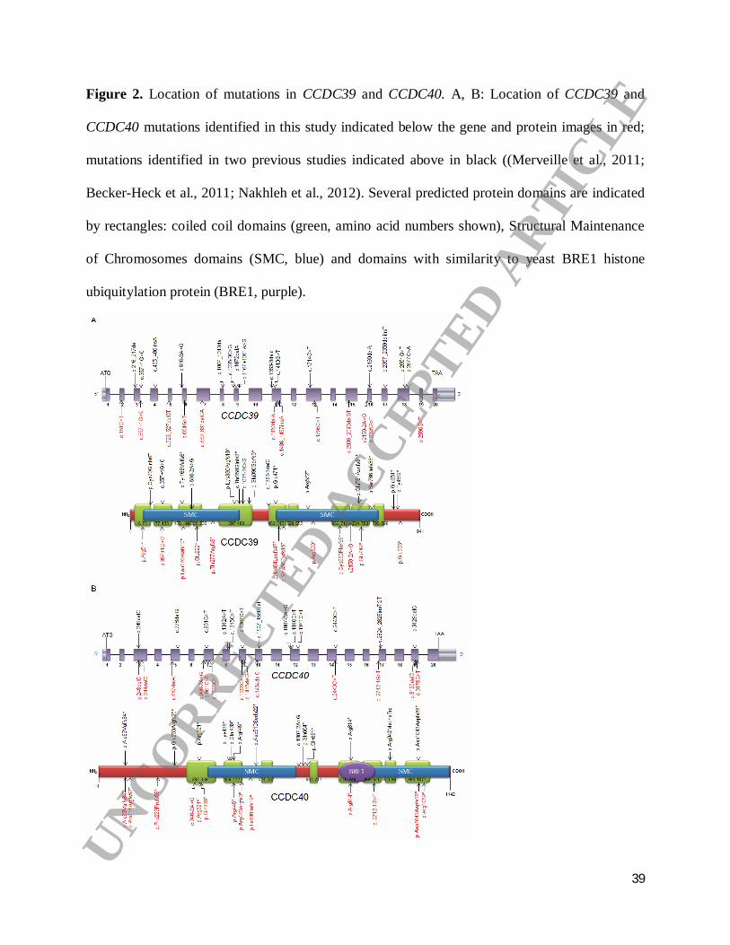
Figure 2. Location of mutations in CCDC39 and CCDC40. A, B: Location of CCDC39 and
CCDC40 mutations identified in this study indicated below the gene and protein images in red;
mutations identified in two previous studies indicated above in black ((Merveille et al., 2011;
Becker-Heck et al., 2011; Nakhleh et al., 2012). Several predicted protein domains are indicated
by rectangles: coiled coil domains (green, amino acid numbers shown), Structural Maintenance
of Chromosomes domains (SMC, blue) and domains with similarity to yeast BRE1 histone
ubiquitylation protein (BRE1, purple).
UNCORRECTED ACCEPT
ED ARTIC
LE

40
Figure 3. Localisation of axonemal components in CCDC40 patient respiratory cells. A:
Representative immunofluorescent images of respiratory cells localise CCDC39 protein (green)
along the length of the ciliary axonemes in control cells. B: Complete absence of CCDC39 from
axonemes from a patient 114 II:1 carrying CCDC40 mutations. Dual staining with anti-
acetylated alpha tubulin (axonemes) and anti-gamma tubulin (basal bodies) was used to stain the
cilia (red). C, D: RSPH4A (green) localises along the length of axonemes at similar levels in
cells from both control and patient 114 II:1 carrying CCDC40 mutations. DNA in nuclei was
stained using DAPI. DIC indicates differential interference contrast microscopy images.
UNCORRECTED ACCEPT
ED ARTIC
LE

41
Table 1. CCDC39 and CCDC40 mutations identified in PCD patients in this study
Family Origin Cons Method Gene Allele 1 Effect Location Allele 2 Effect Location
PCD22 N.Europe(Germany) Y WES CCDC39 c.1486_1487insA;
p.Ser496Tyrfs15* fs Exon 11 c.2159-2A>G;essential splice site splice Intron 15
PCD31 N.Europe(UK) N Sanger CCDC39 c.2596G>T;
p.Glu866* nonsense Exon 19 c.2596G>T;p.Glu866* nonsense Exon 19
PCD102 N.Europe(UK) N WES CCDC39 c.357+1G>C;
essential splice site splice Intron 3 c.151C>T; p.Arg51* nonsense Exon 2
PCD113 N.Europe(UK) N Sanger CCDC39 c.1795C>T;
p.Arg599* nonsense Exon 13 c.1795C>T;p.Arg599* nonsense Exon 13
PCD186 Zimbabwe N WES CCDC39 c.2039_2040delGT;p.Cys680Phefs9* fs Exon 15 c.526_527delCT;
p.Leu176Alafs10* fs Exon 5
PCD206 N.Europe(UK) N WES CCDC39 c.664G>T;
p.Glu222* nonsense Exon 6 c.526_527delCT;p.Leu176Alafs10* fs Exon 5
PCD210Afghanistan(Pubjabiisolate)
N WES CCDC39 c.2245G>T;p.Glu749* nonsense Exon 16 c.2245G>T;
p.Glu749* nonsense Exon 16
PCD211 N.Europe(Portugal) N WES CCDC39 c.1450delA;
p.Ile484Leufs47* fs Exon 11 c.357+1G>C;essential splice site splice Intron 3
UNC64 N. Europe(USA) N Sanger CCDC39 c.830_831delCA;
p.Thr277Argfs3* fs Exon 7 c.830_831delCA;p.Thr277Argfs3* fs Exon 7
PCD8 N.Europe(UK) N Sanger CCDC40 c.2712-1G>T;
essential splice site splice Intron 16 c.2712-1G>T;essential splice site splice Intron 16
PCD17 N.Europe(Belgium) N Sanger CCDC40 c.248delC;
p.Ala83Valfs84* fs Exon 3 c.248delC;p.Ala83Valfs84* fs Exon 3
PCD114 N.Europe(UK) N WES CCDC40 c.2712-1G>T;
essential splice site splice Intron 16 c.2712-1G>T;essential splice site splice Exon 17
PCD129 Pakistan Y Sanger CCDC40 c.1415delG;p.Arg472fs3* fs Exon 9 c.1415delG;
p.Arg472fs3* fs Exon 9
PCD133 Pakistan Y Sanger CCDC40 c.1006C>T;p.Gln336* nonsense Exon 7 c.1006C>T;
p.Gln336* nonsense Exon 7
PCD147 N.Europe(UK) N WES CCDC40 c.248delC;
p.Ala83Valfs84* fs Exon 3 c.248delC;p.Ala83Valfs84* fs Exon 3
PCD199 N.Europe(UK) N Sanger CCDC40 c.248delC;
p.Ala83Valfs84* fs Exon 3 c.248delC;p.Ala83Valfs84* fs Exon 3
PCD216 N. Europe(UK) N WES CCDC40 c.248delC;
p.Ala83Valfs84* fs Exon 3 c.248delC;p.Ala83Valfs84* fs Exon 3
OP-559 S. Europe(Turkish) N Sanger CCDC40 c.3175C>T;
p.Arg1059* nonsense Exon 19 c.3175C>T;p.Arg1059* nonsense Exon 19
OP-560 N. Europe(Belgian) N Sanger CCDC40 c.248delC;
p.Ala83Valfs84* fs Exon 3 c.248delC;p.Ala83Valfs84* fs Exon 3
OP-561 Africa(Moroccan) Y Sanger CCDC40 c.1464delC;
p.Ile488Ilefs19* fs Exon 10 c.1464delC;p.Ile488Ilefs19* fs Exon 10
KUL-001
N. Europe(Belgian) N WES CCDC40 c.248delC;
p.Ala83Valfs84* fs Exon 3 c.687delA;p.Pro229Profs58* fs Exon 5
UNC120 N. Europe(USA) N Sanger CCDC40 c.248delC;
p.Ala83Valfs84* fs Exon 3 c.248delC;p.Ala83Valfs84* fs Exon 3
UNC122 N. Europe(USA) N Sanger CCDC40 c.248delC;
p.Ala83Valfs84* fs Exon 3 c.248delC;p.Ala83Valfs84* fs Exon 3
UNC130 N. Europe(USA) Y Sanger CCDC40 c.2440C>T;
p.Arg814* nonsense Exon 14 c.2440C>T;p.Arg814* nonsense Exon 14
UNC175 N. Europe(USA) N Sanger CCDC40 c.961C>T;
p.Arg321* nonsense Exon 7 c.3129delC;p.Asp1043Aspfs36* fs Exon 19
UNC176 N. Europe(USA) N Sanger CCDC40 c.248delC;
p.Ala83Valfs84* fs Exon 3 c.248delC;p.Ala83Valfs84* fs Exon 3
UNC188 N. Europe(USA) N Sanger CCDC40 c.248delC;
p.Ala83Valfs84* fs Exon 3 c.248delC;p.Ala83Valfs84* fs Exon 3
UNC281 N. Europe(USA) N Sanger CCDC40 c.248delC;
p.Ala83Valfs84* fs Exon 3 c.248delC;p.Ala83Valfs84* fs Exon 3
UNC299 N. Europe(USA) N Sanger CCDC40 c.940-2A>G;
essential splice site splice Intron 6 c.344delC;p.Pro115Argfs52* fs Exon 3
UNC336 N. Europe(USA) N Sanger CCDC40 c.248delC;
p.Ala83Valfs84* fs Exon 3 c.248delC;p.Ala83Valfs84* fs Exon 3UNCORRECTED A
CCEPTED A
RTICLE

42
UNC337 N. Europe(USA) N Sanger CCDC40 c.248delC;
p.Ala83Valfs84* fs Exon 3 c.248delC;p.Ala83Valfs84* fs Exon 3
UNC455 N. Europe(USA) N Sanger CCDC40 c.248delC;
p.Ala83Valfs84* fs Exon 3 c.248delC;p.Ala83Valfs84* fs Exon 3
UNC507 N. Europe(USA) N Sanger CCDC40 c.248delC;
p.Ala83Valfs84* fs Exon 3 c.248delC;p.Ala83Valfs84* fs Exon 3
UNC533 N. Europe(USA) N Sanger CCDC40 c.248delC;
p.Ala83Valfs84* fs Exon 3 c.961C>T;p.Arg321* nonsense Exon 7
UNC553 N. Europe(USA) N Sanger CCDC40 c.248delC;
p.Ala83Valfs84* fs Exon 3 c.248delC;p.Ala83Valfs84* fs Exon 3
UNC609 N. Europe(USA) N Sanger CCDC40 c.248delC;
p.Ala83Valfs84* fs Exon 3 c.248delC;p.Ala83Valfs84* fs Exon 3
UNC660 N. Europe(USA) N Sanger CCDC40 c.1345C>T;
p.Arg449* nonsense Exon 9 c.2712-1G>T;essential splice site splice Intron 16
Nucleotide numbering reflects cDNA, +1 corresponds to the A of the ATG translation initiationcodon in the reference sequences for CCDC39 (NM_181426.1) and CCDC40 (NM_017950.2),according to journal guidelines (www.hgvs.org/mutnomen). The initiation codon is codon 1.
UNCORRECTED ACCEPT
ED ARTIC
LE