Basal ganglia contribution to the initiation of corrective submovements
Upload
independentCategory
view
1download
0
1
2
3Q1
4
56Q4
7
89101112131415161718192021
42
43
44
45
46
47
48
49
50
51
52
53
54
55
Neurobiology of Disease xxx (2013) xxx–xxx
YNBDI-03055; No. of pages: 14; 4C: 3, 5, 6, 7, 8, 10, 11, 12, 13
Contents lists available at ScienceDirect
Neurobiology of Disease
j ourna l homepage: www.e lsev ie r .com/ locate /ynbd i
Immediate-early gene expression in structures outside the basal gangliais associated to L-DOPA-induced dyskinesia
OFMatthieu F. Bastide, Sandra Dovero, Giselle Charron, Gregory Porras, Christian E. Gross,
Pierre-Olivier Fernagut, Erwan Bézard ⁎Univ. de Bordeaux, Institut des Maladies Neurodégénératives, UMR 5293, 33000 Bordeaux, FranceCNRS, Institut des Maladies Neurodégénératives, UMR 5293, 33000 Bordeaux, France
⁎ Corresponding author at: Institut des Maladies NBordeaux Segalen, Bât 3B 1er étage, 146 rue Léo Saignat, 3
E-mail address: [email protected] (E. BézAvailable online on ScienceDirect (www.sciencedir
0969-9961/$ – see front matter © 2013 Published by Elsehttp://dx.doi.org/10.1016/j.nbd.2013.09.020
Please cite this article as: Bastide, M.F., et alinduced dyskinesia, Neurobiol. Dis. (2013), h
O
a b s t r a c t
a r t i c l e i n f o22
23
24
25
26
27
28
29
30
31
Article history:Received 14 September 2013Accepted 27 September 2013Available online xxxx
Keywords:L-DOPADyskinesiaIEGWhole brainStereology
32
33
34
35
36
37
38
39
ECTED P
RLong-term L-3,4-dihydroxyphenylalanine (L-DOPA) treatment in Parkinson's disease (PD) leads to L-DOPA-induced dyskinesia (LID), a condition thought to primarily involve the dopamine D1 receptor-expressing striatalmedium spiny neurons. Activation of the D1 receptor results in increased expression of several molecularmarkers, in particular themembers of the immediate-early gene (IEG) family, a class of genes rapidly transcribedin response to an external stimulus. However, several dopaminoceptive structures in the brain that are likely tobe affected by the exogenously produced DA have received little attention although they might play a key role inmediating those L-DOPA-induced abnormal behaviours. ΔFosB, ARC, FRA2 and Zif268 IEGs expression patternswere thus characterised, using unbiased stereological methods, in the whole brain of dyskinetic and non-dyskinetic rats to identify brain nuclei displaying a transcriptional response specifically related to LID. Withinthe basal ganglia, the striatum and the substantia nigra pars reticulata showed an increased expression of allfour IEGs in dyskinetic compared to non-dyskinetic rats. Outside the basal ganglia, therewas a striking increasedexpression of the four IEGs in the motor cortex, the bed nucleus of the stria terminalis, the dorsal hippocampus,the pontine nuclei, the cuneiform nucleus and the pedunculopontine nuclei. Moreover, the zona incerta and thelateral habenula displayed an overexpression of ΔFosB, ARC and Zif268. Among these structures, the IEG expres-sion in the striatum, the bed nucleus of the stria terminalis, the lateral habenula, the pontine nuclei and thecuneiform nucleus correlate with LID severity. These results illustrate a global transcriptional response to adyskinetic state in the whole brain suggesting the possible involvement of these structures in LID.
© 2013 Published by Elsevier Inc.
4041
R R56
57
58
59
60
61
62
63
64
65
66
67
68
UNCOIntroduction
The most effective symptomatic therapy in Parkinson's disease (PD)remains the dopamine precursor L-3,4-dihydroxyphenylalanine (L-DOPA). Long-term treatment leads to involuntary aimless movementscalled L-DOPA-induced dyskinesia (LID) (Fahn, 2008; Stocchi et al.,1997). Loss of dopamine in PD induces complex modifications in cellu-lar signalling with numerous pathways showing altered responses todopaminergic stimulation in the dopamine-depleted striatum (Bezardet al., 2001; Jenner, 2008). Chronic L-DOPA treatment further enhancesthe signalling alterations. The striatal dopamine D1 receptor (D1R) sig-nalling pathways have consistently been shown to be critically involvedin LID genesis andmanifestation (Bezard et al., 2001; Jenner, 2008). D1R
69
70
71
72
73
74
eurodégénératives, Université3076 Bordeaux cedex, France.ard).ect.com).
vier Inc.
., Immediate-early gene exprettp://dx.doi.org/10.1016/j.nb
stimulation results in increased expression of several molecularmarkers (Feyder et al., 2011), in particular the members of theimmediate-early gene (IEG) family, a class of genes rapidly transcribedin response to an external stimulus (Okuno, 2011). The ΔFosB, activity-regulated cytoskeleton-associated protein (ARC) (also known asArg3.1), FRA2 and Zif268 IEGs (Granado et al., 2008; Westin et al.,2007; Wirtshafter, 2007) show a concomitant increased expression inthe striatum of dyskinetic rats with different expression patterns(Cenci et al., 1999; Ebihara et al., 2011; Sgambato-Faure et al., 2005).
As the main target of the nigral DA neurons, the striatum has re-ceived much attention in regard to understanding the pathophysiologyof LID. However, themyriads of dopaminoceptive structures in the brainthat are likely to be affected by the exogenously produced dopaminehave received little, if any, attention although they might play a keyrole in mediating those L-DOPA-induced abnormal behaviours. There-fore, we here characterised ΔFosB, ARC, FRA2 and Zif268 expressionsin the whole brain of dyskinetic and non-dyskinetic rats to identifythe brain nuclei displaying a transcriptional response specifically relatedto LID.
ssion in structures outside the basal ganglia is associated to L-DOPA-d.2013.09.020

T
75
76
77
78
79
80
81
82
83
84
85
86
87
88
89
90
91Q5
92
93
94
95
96
97
98
99
100
101
102
103
104
105
106
107Q6
108
109
110
111
112
113
114
115
116
117
118
119
120
121
122
123
124
125
126
127
128
129
130
131
132
133
134
135
136
137
138Q7
139
140
141
142
143
144
145
146
147
148
149
150
151
152
153
154
155
156
157
158
159
160
161
162
163
164
165
166
167
168
169
170
171
172
173
174
175
176
177
178
179
180
181
182
183
184
185
186
187
188
189
190
191
2 M.F. Bastide et al. / Neurobiology of Disease xxx (2013) xxx–xxx
UNCO
RREC
Material and methods
Experimental protocol
Adult Sprague–Dawley male rats (Charles River Laboratories, Lyon,France), weighing 175–200g at the beginning of the experiment, wereused. They were housed under standard laboratory condition in a 12-hour light/12-hour dark cycle with free access to food and water. Theexperimental protocol was approved by the Ethical Committee of theBordeaux Segalen University CE50 under licence no. 5012099-A.
On day 0, unilateral injection of 6-hydroxydopamine (2.5μl at 3μg/μl)was performed in the right medial forebrain bundle (AP=−3.7mm;ML=+1.6mm; DV=−8mm relative to Bregma) in rats treated 30min before with citalopram at 1 mg/kg i.p. (an inhibitor of serotoninre-uptake) and with desipramine hydrochloride at 20mg/kg i.p. (an in-hibitor of noradrenalin re-uptake) according to previously publishedprocedures (Berthet et al., 2009; Porras et al., 2012; Schuster et al.,2008). Only the animals displaying both an impaired stepping test(Olsson et al., 1995; Pioli et al., 2008) assessed on days 18 to 20 and aloss of tyrosine hydroxylase-immunopositive fibres in the striatumgreater than 95% (Bezard et al., 2001; Jenner, 2008) were retained forfinal analysis (Fig. 1M).
From day 21 onwards, rats received once daily an i.p. injection of acombined dose of benserazide (15 mg/kg, i.p.) and L-DOPA (3 mg/kg,i.p.) for 18 days. This L-DOPA dose is similar to the EC50 value (12.5mg/kg of benserazide and 3.2 mg/kg of L-DOPA) required to allow agradual development of dyskinesia (Putterman et al., 2007). On day39, 1/3 of the rats were found non-dyskinetic while 2/3 were founddyskinetic, after being rated by a trained investigator as previously de-scribed (Berthet et al., 2012; Meissner et al., 2006; Porras et al., 2012;Schuster et al., 2008, 2009). The 4 abnomal involuntary movements(AIMs) categories (limb, axial, orolingual, and locomotive) were scoredusing a validated rating scale (Cenci et al., 1998; Lundblad et al., 2002)for 1min every 20min for 2 h (total 4 observations; maximal score foreach observation, 16; maximal total score per session, 64). Since thestudy aimed at defining the transcriptional response induced by chronicL-DOPA treatment, non-dyskinetic lesioned rats (n=5) were taken asthe reference experimental group of dyskinetic lesioned rats (n=5).
Tissue preparation
On day 40, 1 h after the last L-DOPA injection, i.e. at the peak of be-havioural effect, rats were deeply anesthetised with chloral hydrate(400 mg/kg, i.p., VWR) and perfused transcardially with 0.9% NaClfollowed by ice-cold 4% formaldehyde in 0.1M sodiumphosphate buffer(PBS). Brains were removed, postfixed overnight in the same fixative (4°C), then cryoprotected for 48h at 4 °C in 20% sucrose (diluted in PBS).Brains were frozen in isopentane at −45 °C and stored at −80 °C untilsectioning. 50 μm-thick cryostat-cut coronal sections were collectedin PBS containing 0.2% sodium azide and stored at 4 °C pendingimmunohistochemistry.
Immunohistochemistry
After three washes in PBS, free-floating sections were incubated for10min in 3%H2O2 (Sigma-Aldrich) at room temperature (RT) to quenchendogenous peroxidase. Sectionswere then transferred for 30min at RTin a blocking solution containing 1/50 bovine serum albumine (BSA)and 0.3% Triton X-100 (Sigma-Aldrich) in PBS. Sections were incubatedfor 12h at RT with rabbit polyclonal anti-FosB/ΔFosB (sc-48), anti-ARC(sc-15325), anti-Zif268 (sc-189), anti-FRA2 antibody (sc-604) (SantaCruz Biotechnology), all diluted at 1:500 or mouse monoclonal anti-tyrosine hydroxylase (MAB318, Millipore) diluted at 1:10,000 in PBScontaining 1/500 BSA and 0.3% Triton X-100. After three washes inPBS (10min each), sections were incubated for 30min at RT with anti-rabbit or anti-mouse labelled Polymer-HRP (Dako). After thorough
Please cite this article as: Bastide, M.F., et al., Immediate-early gene expreinduced dyskinesia, Neurobiol. Dis. (2013), http://dx.doi.org/10.1016/j.nb
ED P
RO
OF
washing, the staining was revealed with a DAB peroxidase substratekit (Vector). The specificity of the immunostaining was assessed byomission of the primary or the secondary antibody. After processing,tissue sections were mounted onto gelatin-coated slides, air-dried,dehydrated and coverslipped with Eukitt mounting medium (Sigma-Aldrich) for light microscopic inspection.
Data analysis
The number of IEG-immunopositive neurons was obtained apply-ing the optical fractionator (Engeln et al., 2012; Pioli et al., 2008;West et al., 2004) unbiased stereological method using a LeicaDM6000B microscope with Mercator Pro software (ExploraNova,version 7.9.8). Immuno-labelled cells were counted by a blind inves-tigator on every 6th section, a sampling adapted to the studied brainnuclei. For each section, the boundaries of the regions were firstdelineated at low magnification (×2.5) and counting was performedat high magnification (×40). Stereological details for each analysedbrain nucleus are presented in Table 1. Mean ± standard deviation(S.D.) of these values was calculated for each group (5 ratsper group). The stereological error coefficients range between 3%(e.g. striatum) and 17% (e.g. rostral zona incerta) depending on thesize and thickness of the structure in accordance with the literaturee.g. (Gundersen and Jensen, 1987; Schmitz and Hof, 2000;Slomianka and West, 2005; West et al., 1991).
Statistical analysis
Statistical analyses were performed using a two-way ANOVA. If sig-nificant, ANOVAs were followed by post hoc t tests corrected for multi-ple comparisons by the method of Bonferroni. All data were normallydistributed, and significance levels of t test comparisons were adjustedfor inequality of varianceswhen appropriate. The provided F values cor-respond to the interaction between independent variables: Condition(dyskinetic or non-dyskinetic)×side (lesioned or unlesioned). Correla-tions between LID and cell counts were performed using Spearmancorrelation.
Results
The basal ganglia display an overexpression of IEGs in LID
Within the cortico-basal ganglia motor loops, as expected, ΔFosB,ARC, FRA2 and Zif268 IEGs are significantly overexpressed in the dorso-lateral, ventrolateral, dorsomedial and ventromedial part of the lesionedstriatum of dyskinetic rats compared to non-dyskinetic rats (Figs. 1CF,2CF, 3CF and 4CF). Likewise, in the motor cortex (M1) and thesubstantia nigra pars reticulata (SNr), the 4 IEGs displayed a significantincreased expression between the lesioned side of dyskinetic and non-dyskinetic rats (Figs. 1IL, 2IL, 3IL and 4IL). However, the other structuresof the basal ganglia, the subthalamic nucleus and the external globuspallidus (GPe) displayed no staining.
LID increase IEGs expression outside the basal ganglia
In the limbic system, 3 brain nuclei of the bed nucleus of the striaterminalis (BST): the oval (oBST), juxta capsular (jBST) and medial(mBST) nuclei exhibited a significant increased expression of the 4IEGs on the lesioned side of dyskinetic rats compared to non-dyskinetic (Figs. 5CF, 6CF, 7CF and 8CF). A similar expression patternwas also found in the hippocampus where the IEGs were significantlyoverexpressed in CA1, CA3 and in the dentate gyrus (DG) (Figs. 5U,6U, 7U and 8U).
At the crossroad between the limbic system, the basal ganglia andthe dopamine/serotonine pathways, the lateral part of the habenula(LHb) displayed a significant overexpression of ΔFosB, ARC and Zif268
ssion in structures outside the basal ganglia is associated to L-DOPA-d.2013.09.020

UNCO
RRECTED P
RO
OF
Fig. 1. Stereological counting ofΔFosB immuno-positive cells in the cortico-basal gangliamotor loop in dyskinetic (light blue) and non-dyskinetic (dark blue) 6-OHDA-lesioned rats. Representative examples of staining, scale bar 300μm(with an insetmagnification, scale bar 20 μm), are shown on the left side while quantitative results are displayed on the right side (shown as mean± SD; *p b 0.05). A–C dorsolateral (DL) F[1,16]=84.27, p b 0.001 and dorsomedian (DM) F[1.16]= 29.9, p b 0.001striatum; D–F ventrolateral (VL) F[1.16]=40.46, p b 0.001 and ventromedian (VM) F[1.16]= 138 p b 0.001 striatum; G–I, M1 motor cortex F[1.16]=7.527, p b 0.05; J–L, substantia nigra pars reticulata (SNr) F[1.16]=13.31, p b 0.01. M, Representativeexample of tyrosine hydroxylase immunostaining in the striatum of L-DOPA-treated unilateral 6-OHDA lesioned rats. O, Representative example of tyrosine hydroxylase immunostaining in the SNc of L-DOPA-treated unilateral 6-OHDA lesioned rats. 3
M.F.Bastide
etal./Neurobiology
ofDisease
xxx(2013)
xxx–xxx
Pleasecite
thisarticle
as:Bastide,M
.F.,etal.,Im
mediate-early
geneexpression
instructures
outsidethe
basalgangliaisassociated
toL-D
OPA
-induced
dyskinesia,Neurobiol.D
is.(2013),http://dx.doi.org/10.1016/j.nbd.2013.09.020

UNCO
RRECT
192
193
194
195
196
197
198
199
200
201
202
203
204
205
206
207
208
209
210
211
212
213
214
215
216
217
218
219
220
221
222
223
224
225
226
227
228
229
230
231
232
233
234
235
236
237
238
239
240
241
242
243
244
245
246
247
248
249
250
251
252
Q3
Table1
t1:1
t1:2
Stereo
logicalcou
ntingpa
rameters.ST
R=
striatum
,M1=
motor
cortex
,mBS
ToB
STjBST
=med
ial,ov
alan
djuxtapa
rtof
thebe
dnu
cleu
sof
thestriaterm
inalis,lHban
dmHb=
lateraland
med
ialp
arto
fthe
habe
nula,D
G=
dentategy
rus,rZI=
rostral
t1:3
zona
incerta,SN
r=substantia
nigrareticu
lata,P
n=
pontinenu
clei,P
Tg=
pedu
nculop
ontine
tegm
entaln
ucleus
,CnF
=cu
neifo
rmnu
cleu
s.
t1:4
STR
M1
mBS
ToB
STjBST
lHb
mHb
CA1
CA2
CA3
DG
rZI
SNr
PnPT
gCn
F
t1:5
Coun
ting
fram
es(μm)
60×
8080
×80
60×
6060
×60
60×
6060
×60
60×
6080
×40
80×
4080
×40
80×
4080
×40
80×
6080
×60
60×
6060
×60
t1:6
Spacing(μm)
200×
250
300×
300
150×
150
80×
8080
×80
100×
100
80×
8020
0×
5010
0×
5020
0×
5020
0×
5020
0×
5024
0×
180
250×
200
150×
150
150×
150
t1:7
Num
berof
sections
1010
11
13
34
44
43
63
22
4 M.F. Bastide et al. / Neurobiology of Disease xxx (2013) xxx–xxx
Please cite this article as: Bastide, M.F., et al., Immediate-early gene expreinduced dyskinesia, Neurobiol. Dis. (2013), http://dx.doi.org/10.1016/j.nb
ED P
RO
OF
while only ARC expression was increased in the medial part (MHb)(Figs. 5L, 6L and 8L). The same expression pattern was found in the ros-tral part of the zona incerta (rZI) where 3 IEGs were significantlyoverexpressed (Figs. 5I, 6I, and 8I).
In the brainstem, the number of ΔFosB, ARC, FRA2 and Zif268immuno-positive cells was significantly greater in the pontine (Pn)and cuneiform nuclei (CnF) (Figs 5OR, 6OR, 7OR and 8OR) whereasthe pedunculopontine tegmental nucleus (PTg) only displayed anoverexpression of ΔFosB, ARC and Zif268 (Figs. 5R, 6R and 8R) on thelesioned side of dyskinetic rats compared to non-dyskinetic.
IEG expression is altered on the unlesioned side of dyskinetic rats
In dyskinetic rats, the 4 IEGs showed an overexpression in all thestructures mentioned above on the lesioned side compared to theunlesioned side. However, interestingly, IEGs displayed an increasedexpression on the unlesioned side of dyskinetic rats compared to non-dyskinetic rats, both in the basal ganglia and in some structures outside.
In the basal ganglia, the number of ARC and FRA2 immuno-positivecells was significantly greater in the 4 parts of the striatumwhile ΔFosBwas overexpressed only in the dorsolateral, ventrolateral and ventro-medial parts (Figs. 1CF, 2CF and 3CF). SNr and M1 also showed a signif-icant increased expression of ΔFosB and Zif268 whereas ARC wasoverexpressed only in the SNr (Figs. 1IL, 2IL and 4IL).
Outside the basal ganglia, oBST, mBST and LHb displayed a signifi-cant overexpression of ΔFosB and ARC (Figs. 5CFL and 6CFL) while rZIshowed an increased expression only for ARC (Fig. 6I). In the hippocam-pus, the number of ARC immune-positive cells was significantly greaterin CA1, CA3 and DG (Fig. 6U). Zif268 and FRA2were overexpressed onlyin CA3 (Figs. 7U and 8U) and ΔFosB in DG (Fig. 5U). In the brainstem,only ARC displayed an increased expression in the CnF (Fig. 6R).
Discussion
L-DOPA, the gold standard treatment for PD, rapidly induces fluctua-tions and LID, the latter being so far associated with both presynapticand postsynaptic mechanisms at the striatal level (Bezard et al., 2001;Jenner, 2008). The present study, building upon scarce but intriguingevidences in the literature (e.g. (Guigoni et al., 2005b; Halje et al.,2012; Miguelez et al., 2011)), systematically assessed the dyskinesia-related increases in expression of 4 IEGs: ΔFosB, ARC, FRA2 and Zif268in the whole brain of a rat model of PD. While the striatum is undoubt-edly central in LID pathophysiology as local infusions in the striatumelicit LID (Buck and Ferger, 2008; Carta et al., 2006), the present studyunravels stunning correlations between IEG expression and LID severityin brain nuclei outside the basal ganglia, such as the BST, lateralhabenula, pontine nuclei and cuneiform nucleus.
The study is however not without limitations. L-DOPA dose wascarefully selected to be just enough for inducing dyskinesia in themajority of animals while still allowing some to have a score of 0(Putterman et al., 2007). Most studies actually compare highly dyski-netic to low dyskinetic 6-OHDA-lesioned rats (Fiorentini et al., 2006;Rangel-Barajas et al., 2011) while the non-dyskinetic animals heredisplayed a score of 0. As the extent of the lesion is an obvious factorfor susceptibility to develop LID (Guigoni et al., 2005a), we selected an-imals with the exact same extent and pattern of nigrostriatal denerva-tion (Fig. 1M) (Berthet et al., 2012; Porras et al., 2012; Schuster et al.,2008). Finally, while it would be tempting to relate the changes in IEGexpression to changes in electrophysiological activity of the consideredneuronal structures, one should bear in mind that such a relationship,although generally assumed, has not been demonstrated for most IEGs(Loebrich and Nedivi, 2009) and in particular for those studied here.Therefore, increased expression of an IEG should be seen as an increasedtranscriptional activity and not taken as an increase in electrophysiolog-ical activity that remains to be demonstrated.
ssion in structures outside the basal ganglia is associated to L-DOPA-d.2013.09.020

UNCO
RRECTED P
RO
OF
Fig. 2. Stereological counting of ARC immuno-positive cells in the cortico-basal gangliamotor loop in dyskinetic (light blue) and non-dyskinetic (dark blue) 6-OHDA-lesioned rats. Representative examples of staining, scale bar 300 μm(with an insetmagnification, scale bar 20 μm), are shown on the left side while quantitative results are displayed on the right side (shown as mean±SD; *p b 0.05). A–C dorsolateral (DL) F[1.16]=71.03, p b 0.001 and dorsomedian (DM) F[1.16]=29.71, p b 0.001striatum; D–F ventrolateral (VL) F[1.16]= 19.84, p b 0.001 and ventromedian (VM) F[1.16]= 13.54, p b 0.001 striatum; G–I, M1 motor cortex F[1.16]= 7.344, p b 0.05; J–L, substantia nigra pars reticulata (SNr) F[1.16]= 24.74, p b 0.001. 5
M.F.Bastide
etal./Neurobiology
ofDisease
xxx(2013)
xxx–xxx
Pleasecite
thisarticle
as:Bastide,M
.F.,etal.,Im
mediate-early
geneexpression
instructures
outsidethe
basalgangliaisassociated
toL-D
OPA
-induced
dyskinesia,Neurobiol.D
is.(2013),http://dx.doi.org/10.1016/j.nbd.2013.09.020

UNCO
RRECTED P
RO
OF
Fig. 3. Stereological counting of FRA2 immuno-positive cells in the cortico-basal gangliamotor loop in dyskinetic (light blue) and non-dyskinetic (dark blue) 6-OHDA-lesioned rats. Representative examples of staining, scale bar 300μm(with an insetmagnification, scale bar 20 μm), are shown on the left side while quantitative results are displayed on the right side (shown as mean±SD; *p b 0.05). A–C dorsolateral (DL) F[1.16]=152.1, p b 0.001 and dorsomedian (DM) F[1.16]=28.72, p b 0.001striatum; D–F ventrolateral (VL) F[1.16]= 80.18, p b 0.001 and ventromedian (VM) F[1.16]= 23.30, p b 0.001 striatum; G–I, M1 motor cortex F[1.16]= 10.83, p b 0.01; J–L, substantia nigra pars reticulata (SNr) F[1.16]= 13.47, p b 0.01.
6M.F.Bastide
etal./Neurobiology
ofDisease
xxx(2013)
xxx–xxx
Pleasecite
thisarticle
as:Bastide,M
.F.,etal.,Im
mediate-early
geneexpression
instructures
outsidethe
basalgangliaisassociated
toL-D
OPA
-induced
dyskinesia,Neurobiol.D
is.(2013),http://dx.doi.org/10.1016/j.nbd.2013.09.020
UNCO
RRECTED P
RO
OF
Fig. 4. Stereological counting of Zif268 immuno-positive cells in the cortico-basal gangliamotor loop in dyskinetic (light blue) and non-dyskinetic (dark blue) 6-OHDA-lesioned rats. Representative examples of staining, scale bar 300μm(with an insetmagnification, scale bar 20 μm), are shown on the left side while quantitative results are displayed on the right side (shown as mean±SD; *p b 0.05). A–C dorsolateral (DL) F[1.16]=27.13, p b 0.001 and dorsomedian (DM) F[1.16]=44.31, p b 0.001striatum; D–F ventrolateral (VL) F[1.16]= 28.87, p b 0.001 and ventromedian (VM) F[1.16]= 24.66, p b 0.001 striatum; G–I, M1 motor cortex F[1.16]= 59.01, p b 0.001; J–L, substantia nigra pars reticulata (SNr) F[1.16]= 55.05, p b 0.001. 7
M.F.Bastide
etal./Neurobiology
ofDisease
xxx(2013)
xxx–xxx
Pleasecite
thisarticle
as:Bastide,M
.F.,etal.,Im
mediate-early
geneexpression
instructures
outsidethe
basalgangliaisassociated
toL-D
OPA
-induced
dyskinesia,Neurobiol.D
is.(2013),http://dx.doi.org/10.1016/j.nbd.2013.09.020

UNCO
RRECTED P
RO
OF
8 M.F. Bastide et al. / Neurobiology of Disease xxx (2013) xxx–xxx
Please cite this article as: Bastide, M.F., et al., Immediate-early gene expression in structures outside the basal ganglia is associated to L-DOPA-induced dyskinesia, Neurobiol. Dis. (2013), http://dx.doi.org/10.1016/j.nbd.2013.09.020

T
253
254
255
256
257
258
259
260
261
262
263
264
265
266
267
268
269
270
271
272
273
274
275
276
277
278
279
280
281
282
283
284
285
286
287
288
289
290
291
292
293
294
295
296
297
298
299
300
301
302
303
304
305
306
307
308
309
310
311
312
313
314
315
316
317
318
319
320
321
322
323
324
325
326
327
328
329
330
331
332
333
334
335
336
337
338
339
340
341
342
343
344
345
346
347
348
349
350
351
352
353
354
355
356
357
358
359
360
361362363364365366
9M.F. Bastide et al. / Neurobiology of Disease xxx (2013) xxx–xxx
UNCO
RREC
The rat model of LID, interesting and insightful as it is (Cenci et al.,2002), is a unilateral model of PD and LID. A number of changes cantherefore affect the unlesioned hemisphere, either as a consequence ofthe contralateral lesion or subsequently to the L-DOPA treatment.Accordingly, we report numerous increased IEG expressions on theunlesioned side of dyskinetic rats compared to the unlesioned side ofnon-dyskinetic ones in structures both inside and outside the basalganglia. Thus, despite an intact dopaminergic system, the unlesionedside of dyskinetic rats is transcriptionally more active than the lesionedand unlesioned side of non-dyskinetic rats and significantly less activethan the lesioned side of dyskinetic rats regarding the 4 IEGs assessedin this study. These results highlight that the unlesioned side may notbe considered as a non-affected reference when assessing gene expres-sion in the dyskinetic 6-OHDA hemiparkinsonian rat model.
The number of IEG-immunopositive cells in various structures,both within and outside the basal ganglia, nicely correlates with the se-verity of LID (Fig. 9). Within the basal ganglia, the number of ΔFosBimmuno-positive cells correlated with the intensity of LID in the dorso-lateral (r2=0.87, Pb0.001) and in the ventrolateral (r2=0.90, Pb0.001)striatum (Fig. 9C), a finding consistent with previous reports(Andersson et al., 1999; Cenci and Konradi, 2010; Sgambato-Faureet al., 2005; Valastro et al., 2007) while the present is the first demon-stration using stereological methods. Outside the basal ganglia, 2 nucleiof the BST showed significant correlations between the intensity of LIDand, respectively, the number of ΔFosB-positive cells (r2 = 0.91, P b
0.001) for the oBST and FRA2-positive cells for the jBST (r2=0.65, P b0.05) (Fig. 9D). The BST is a cluster of nuclei that receive robust mono-aminergic inputs featuring serotonin (5-HT), noradrenealine (NA, ornorepinephrine) and dopamine (DA) (Phelix et al., 1992). The BST DAinputs originate from the ventral tegmental area (VTA), theperiaqueductal grey region and the retrorubral field. They form a fairlydiffuse input to the dorsolateral BST with dense DA terminal fields inthe oBST and the jBST (Freedman and Cassell, 1994; Hasue andShammah-Lagnado, 2002; Meloni et al., 2006). More importantly, inthe oBST, recent data indicate that exogenous DA can reduce the inhib-itory synaptic transmission in a D2 like dopamine receptor-dose-dependent manner in a brain slice of drug-naïve rats (Krawczyk et al.,2011) and that cocaine maintenance rats display an overexpression ofD1R (Krawczyk et al., 2013), underlying the hypothesis of a potentialrole of the BST in LID.
In the epithalamus, the lHb showed a significant correlation be-tween the LID intensity and the number of ARC immuno-positive cells(r2=0.85, Pb0.001) (Fig. 9G). Themedial part of the lHb is primarily in-nervated by the limbic system (Herkenham and Nauta, 1977; Hikosakaet al., 2008) while the lateral part receive basal ganglia afferents, espe-cially from the internal part of the globus pallidus (GPi) (Hong andHikosaka, 2008), the main output structure of the basal ganglia. ThelHb projects mainly to the monoaminergic brain regions like the VTA,the SNc, the serotoninergic dorsal and medial raphe and also to thecholinergic laterodorsal tegmentum (Bernard and Veh, 2012; Geislerand Trimble, 2008; Hikosaka et al., 2008). Thus, the lHb acts as a junc-tion connecting the limbic system and the basal ganglia to themonoam-inergic centres. As the false transmitter hypothesis involving theserotoninergic system in LID pathophysiology addresses the presynap-tic component of LID pathophysiology (Carta and Bezard, 2011;Navailles et al., 2010), the efferent connectivity of the lHb suggeststhat it may play a role in controlling serotoninergic output. ImpairedlHb input would thus participate in the aberrant dopamine releasefrom 5-HT terminals (Carta et al., 2007, 2008a,b; Navailles et al., 2011;Rylander et al., 2010).
Fig. 5. Stereological counting of ΔFosB immuno-positive cells outside the basal ganglia in dyskexamples of staining, scale bar 300 μm (with an inset magnification, scale bar 20 μm), are shomean ± SD; *p b 0.05). A–C, oval (oBST) F[1.16] = 9.626, p b 0.01 and juxta (jBST) F[1.16] = 6.79.438, pb 0.01; G–I, rostral zona incerta (rZI) F[1.16]=8.398, p b 0.05; J–L, lateral habenula (lHb)nucleus (CnF) F[1.16]=16.90, pb 0.001 and pedunculopontine tegmental nucleus (PTg) F[1.16]=0.33, CA3 F[1.16]= 11.69, p b 0.05 and dentate gyrus (DG) F[1.16]= 29.87, p b 0.001.
Please cite this article as: Bastide, M.F., et al., Immediate-early gene expreinduced dyskinesia, Neurobiol. Dis. (2013), http://dx.doi.org/10.1016/j.nb
OO
F
In the brainstem, 2 nuclei, the Pn and CnF, displayed a significantcorrelation between LID intensity and, respectively, the number ofZif268 immuno-positive cells (r2= 0.73, P b 0.01) and FRA2 immuno-positive cells (r2=0.82, P b 0.001) (Figs. 9JK). Both the Pn and the CnFreceive afferents from the basal ganglia, i.e. from the STN (Wu andHallett, 2013) and the SNr (Rolland et al., 2011), respectively. The Pnprimarily projects to the cerebellum (Brodal, 1979, 1980) and, thus,could be considered as a relay structure from the basal ganglia throughthe STN (Bostan et al., 2010;Wu andHallett, 2013). As the cerebellum isinvolved in fine tuning motor behaviour (Bastian, 2006; Thach et al.,1992; Timmann et al., 2010), it should not be surprising that a L-DOPA-induced modification of the Pn neurons' transcriptional activitycould influence cerebellum-driven motor functions and impact LIDpathophysiology. The CnF, known as a mesencephalic locomotor area,receives DA inputs (Rolland et al., 2009; Takakusaki et al., 2003). Al-though the origin of its dopaminergic innervation is still unclear, recentdata indicate that MPTP monkeys undergo a dramatic decrease in CnFDA content (Rolland et al., 2009), providing information about a puta-tive role in parkinsonian symptoms. Moreover, as the CnF projectsback to the SNc (Watabe-Uchida et al., 2012), both CnF and SNc/SNrneurons form a direct loop that could play a role in motor-relatedbehaviour, and hence support the involvement of the CnF in LID.
ED P
R
Conclusion
In conclusion, both motor and non-motor domains of cortico-sub-cortical loops showed significant correlations between the number ofΔFosB, ARC, FRA2 and Zif268 immunopositive cells and LID severity. Acorrelation does not necessarily imply a causal relationship but mightreflect the concomitance of unrelated events. One should not thereforeeliminate the possibility that these animals are experiencing other L-DOPA-induced side effects that were not investigated. Defining theprecise role of these structures in LID pathophysiology now requiresmodulating the electrophysiological activity of these identified brainnuclei and assess the impact of suchmodulation uponmotor behaviourin general and LID severity in particular.
Acknowledgments
This work was supported by the Agence Nationale de la Recherchegrants (EB: ANR-07-MNP-Trafinlid). MB is the recipient of an MESRgrant. The Université Bordeaux Segalen and the Centre National de laRecherche Scientifique provided infrastructural support.
Financial disclosure
EB has an equity stake inMotac Holding Ltd. and receives consultan-cy payments from Motac Neuroscience Ltd. Current grant supportincludes Agence Nationale de la Recherche (EB, CG), China ScienceFund (EB), MJFF (EB), FP7 from EU (EB), France Parkinson (EB, POF),Fondation de France (EB), Cariplo Foundation (EB).
References
Andersson, M., et al., 1999. Striatal fosB expression is causally linked with L-DOPA-induced abnormal involuntarymovements and the associated upregulation of striatalprodynorphin mRNA in a rat model of Parkinson's disease. Neurobiol. Dis. 6,461–474.
Bastian, A.J., 2006. Learning to predict the future: the cerebellum adapts feedforwardmovement control. Curr. Opin. Neurobiol. 16, 645–649.
inetic (light blue) and non-dyskinetic (dark blue) 6-OHDA-lesioned rats. Representativewn on the left side while quantitative results are displayed on the right side (shown as50, p b 0.05 bed nucleus of the stria terminalis (BST); D–F, medial BST (mBST) F[1.16] =F[1.16]=47.5 pb 0.001; M–O, pontine nuclei (Pn) F[1.16]=6.657, p b 0.05; P–R, cuneiform20.93, pb 0.001; S–U, hippocampus CA1 F[1.16]=5.807, pb 0.05, CA2 F[1.16]=0.9688, p=
ssion in structures outside the basal ganglia is associated to L-DOPA-d.2013.09.020

UNCO
RRECTED P
RO
OF
10 M.F. Bastide et al. / Neurobiology of Disease xxx (2013) xxx–xxx
Please cite this article as: Bastide, M.F., et al., Immediate-early gene expression in structures outside the basal ganglia is associated to L-DOPA-induced dyskinesia, Neurobiol. Dis. (2013), http://dx.doi.org/10.1016/j.nbd.2013.09.020
UNCO
RRECTED P
RO
OF
367368369370371372
373374375376377378
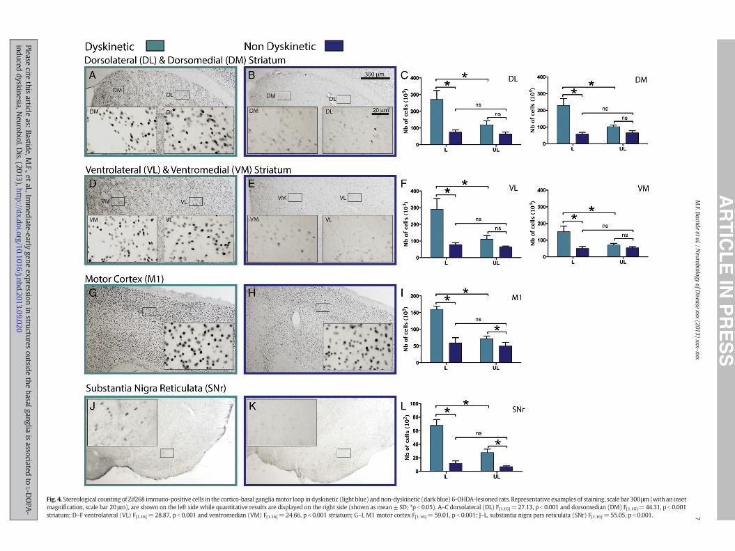
Fig. 7. Stereological counting of FRA2 immuno-positive cells outside the basal ganglia in dyskinetic (light blue) and non-dyskinetic (dark blue) 6-OHDA-lesioned rats. Representative ex-amples of staining, scale bar 300μm (with an inset magnification, scale bar 20μm), are shown on the left sidewhile quantitative results are displayed on the right side (shown asmean±SD; *pb0.05). A–C, oval (oBST) F[1.16]=13.42, pb0.01 and juxta (jBST) F[1.16]=24.23, pb0.001 bed nucleus of the stria terminalis (BST); D–F, medial BST (mBST) F[1.16]=16.94, pb0.01;G–I, pontine nuclei (Pn) F[1.16]=12.18, p b 0.01; J–L, cuneiform nucleus (CnF) F[1.16]=22.13, p b 0.001; M–O, hippocampus CA1 F[1.16]=11.83, p b 0.01, CA2 F[1.16]=0.042, p b 0.84, CA3F[1.16]= 13.18, p b 0.01 and dentate gyrus (DG) F[1.16]= 4.661, p b 0.05.
Q8
11M.F. Bastide et al. / Neurobiology of Disease xxx (2013) xxx–xxx
Bernard, R., Veh, R.W., 2012. Individual neurons in the rat lateral habenular complex pro-ject mostly to the dopaminergic ventral tegmental area or to the serotonergic raphenuclei. J. Comp. Neurol. 520, 2545–2558.
Berthet, A., et al., 2009. Pharmacological analysis demonstrates dramatic alteration of D1dopamine receptor neuronal distribution in the rat analog of L-DOPA-induced dyski-nesia. J. Neurosci. 29, 4829–4835.
Fig. 6. Stereological counting of ARC immuno-positive cells outside the basal ganglia in dyskineamples of staining, scale bar 300μm (with an inset magnification, scale bar 20μm), are shown oSD; *pb0.05). A–C, oval (oBST) F[1.16]=13.04, pb0.01 and juxta (jBST) F[1.16]=15.19, pb0.001G–I, rostral zona incerta (rZI) F[1.16]=8.729, pb0.01; J–L,medial (mHb) F[1.16]=23.53, pb0.001pb0.01; P–R, cuneiformnucleus (CnF) F[1.16]=31.27, pb0.001 and pedunculopontine tegmentaF[1.16]= 0.082, p= 0.78, CA3 F[1.16]= 10.42, p b 0.01 and dentate gyrus (DG) F[1.16]= 8.112, p
Please cite this article as: Bastide, M.F., et al., Immediate-early gene expreinduced dyskinesia, Neurobiol. Dis. (2013), http://dx.doi.org/10.1016/j.nb
Berthet, A., et al., 2012. L-DOPA impairs proteasome activity in parkinsonism through D1dopamine receptor. J. Neurosci. 32, 681–691.
Bezard, E., et al., 2001. Pathophysiology of levodopa-induced dyskinesia: potential fornew therapies. Nat. Rev. Neurosci. 2, 577–588.
Bostan, A.C., et al., 2010. The basal ganglia communicate with the cerebellum. Proc. Natl.Acad. Sci. U. S. A. 107, 8452–8456.
tic (light blue) and non-dyskinetic (dark blue) 6-OHDA-lesioned rats. Representative ex-n the left sidewhile quantitative results are displayed on the right side (shown asmean±bed nucleus of the stria terminalis (BST); D–F, medial BST (mBST) F[1.16]=6.190, pb0.05;and lateral habenula (lHb) F[1.16]=13.08, pb0.01;M–O, pontine nuclei (Pn) F[1.16]=11.16,l nucleus (PTg) F[1.16]=10.50, pb0.01; S–U, hippocampus CA1 F[1.16]=12.24, pb0.01, CA2b 0.05.
ssion in structures outside the basal ganglia is associated to L-DOPA-d.2013.09.020

UNCO
RRECTED P
RO
OF
12 M.F. Bastide et al. / Neurobiology of Disease xxx (2013) xxx–xxx
Please cite this article as: Bastide, M.F., et al., Immediate-early gene expression in structures outside the basal ganglia is associated to L-DOPA-induced dyskinesia, Neurobiol. Dis. (2013), http://dx.doi.org/10.1016/j.nbd.2013.09.020

ORRECTED P
RO
OF
379380381382383384385386387388389390391392
393394395396397398399400401402403404405406
Fig. 9.Q2 Correlation between the number ofΔFosB, ARC, FRA2 and Zif268 immuno-positive cells in certain brain nuclei and LID severity (sumof the axial, limb, and orolingual AIMs (maximalscore for each observation, 12; total score per session, 48)). Atlas-based localization of brain area allows displaying relative difference in number of IEG-immunopositive cells on thelesioned side between dyskinetic and non-dyskinetic rats. The darker the colour the greater the number of immunopositive cells (striatum: red; oval and juxta bed nucleus of the striaterminalis: green and dark blue, respectively; lateral habenula: orange; pontine and cuneiform nuclei: yellow and light blue, respectively). Specific IEG correlations between number ofIEG immunopositive cells and LID severity are displayed on the right side. A–D, dorsolateral (DL), ventrolateral (VL) striatum, oval (oBST) and juxta (jBST) bed nucleus of the striaterminalis (BST); E–G, lateral habenula (lHb); H–K, pontine (Pn) and cuneiform (CnF) nuclei.
13M.F. Bastide et al. / Neurobiology of Disease xxx (2013) xxx–xxx
UNCBrodal, P., 1979. The pontocerebellar projection in the rhesus monkey: an experimental
study with retrograde axonal transport of horseradish peroxidase. Neuroscience 4,193–208.
Brodal, P., 1980. The projection from the nucleus reticularis tegmenti pontis to thecerebellum in the rhesus monkey. Exp. Brain Res. 38, 29–36.
Buck, K., Ferger, B., 2008. Intrastriatal inhibition of aromatic amino acid decarboxylaseprevents L-DOPA-induced dyskinesia: a bilateral reverse in vivo microdialysis studyin 6-hydroxydopamine lesioned rats. Neurobiol. Dis. 29, 210–220.
Carta, M., Bezard, E., 2011. Contribution of pre-synaptic mechanisms to L-DOPA-induceddyskinesia. Neuroscience 198, 245–251.
Carta, M., et al., 2006. Role of striatal L-DOPA in the production of dyskinesia in 6-hydroxydopamine lesioned rats. J. Neurochem. 96, 1718–1727.
Carta, M., et al., 2007. Dopamine released from 5-HT terminals is the cause of L-DOPA-induced dyskinesia in parkinsonian rats. Brain 130, 1819–1833.
Fig. 8. Stereological counting of Zif268 immuno-positive cells outside the basal ganglia in dyskexamples of staining, scale bar 300 μm (with an inset magnification, scale bar 20 μm), are shomean ± SD; *p b 0.05). A–C, oval (oBST) F[1.16] = 11.05, p b 0.01 and juxta (jBST) F[1.16] = 29.22.32, p b 0.001; G–I, rostral zona incerta (rZI) F[1.16]=26.74, p b 0.001; J–L, lateral habenula (lHiformnucleus (CnF) F[1.16]=19.07, pb0.001 and pedunculopontine tegmental nucleus (PTg) F[1p=0.63, CA3 F[1.16]= 5.661, p b 0.05 and dentate gyrus (DG) F[1.16]= 6.205, p b 0.05.
Please cite this article as: Bastide, M.F., et al., Immediate-early gene expreinduced dyskinesia, Neurobiol. Dis. (2013), http://dx.doi.org/10.1016/j.nb
Carta, M., et al., 2008a. Involvement of the serotonin system in L-DOPA-induced dyskine-sias. Parkinsonism Relat. Disord. 14 (Suppl. 2), S154–S158.
Carta, M., et al., 2008b. Serotonin-dopamine interaction in the induction andmaintenanceof L-DOPA-induced dyskinesias. Prog. Brain Res. 172, 465–478.
Cenci, M.A., Konradi, C., 2010. Maladaptive striatal plasticity in L-DOPA-induced dyskine-sia. Prog. Brain Res. 183, 209–233.
Cenci, M.A., et al., 1998. L-DOPA-induced dyskinesia in the rat is associated with striataloverexpression of prodynorphin- and glutamic acid decarboxylase mRNA. Eur.J. Neurosci. 10, 2694–2706.
Cenci, M.A., et al., 1999. Changes in the regional and compartmental distribution of FosB-and JunB-like immunoreactivity induced in the dopamine-denervated rat striatum byacute or chronic L-DOPA treatment. Neuroscience 94, 515–527.
Cenci, M.A., et al., 2002. Animal models of neurological deficits: how relevant is the rat?Nat. Rev. Neurosci. 3, 574–579.
inetic (light blue) and non-dyskinetic (dark blue) 6-OHDA-lesioned rats. Representativewn on the left side while quantitative results are displayed on the right side (shown as82, p b 0.001 bed nucleus of the stria terminalis (BST); D–F, medial BST (mBST) F[1.16] =b) F[1.16]=16.36, p b 0.001; M–O, pontine nuclei (Pn) F[1.16]=14.87, p b 0.01; P–R, cune-.16]=22.99, pb0.001; S–U, hippocampus CA1 F[1.16]=8.402, pb0.05, CA2 F[1.16]=0.2381,
ssion in structures outside the basal ganglia is associated to L-DOPA-d.2013.09.020

T
407408409410411412413414415416417418419420421422423424425426427428429430431432433434435436437438439440441442443444445446447448449450451452453454455456457458459460461462463464465466467468469470
471472473474475476477478479480481482483484485486487488489490491492493494495496497498499500501502503504505506507508509510511512513514515516517518519520521522523524525526527528529530531532533534
535
14 M.F. Bastide et al. / Neurobiology of Disease xxx (2013) xxx–xxx
ORREC
Ebihara, K., et al., 2011. Differential expression of FosB, c-Fos, and Zif268 in forebrain re-gions after acute or chronic L-DOPA treatment in a rat model of Parkinson's disease.Neurosci. Lett. 496, 90–94.
Engeln, M., et al., 2012. Reinforcing properties of Pramipexole in normal and parkinsonianrats. Neurobiol. Dis. 49C, 79–86.
Fahn, S., 2008. How do you treat motor complications in Parkinson's disease: medicine,surgery, or both? Ann. Neurol. 64 (Suppl. 2), S56–S64.
Feyder, M., et al., 2011. L-DOPA-induced dyskinesia and abnormal signaling in striatal me-dium spiny neurons: focus on dopamine D1 receptor-mediated transmission. Front.Behav. Neurosci. 5, 71.
Fiorentini, C., et al., 2006. Loss of synaptic D1 dopamine/N-methyl-D-aspartate glutamatereceptor complexes in L-DOPA-induced dyskinesia in the rat. Mol. Pharmacol. 69,805–812.
Freedman, L.J., Cassell, M.D., 1994. Distribution of dopaminergic fibers in the centraldivision of the extended amygdala of the rat. Brain Res. 633, 243–252.
Geisler, S., Trimble, M., 2008. The lateral habenula: no longer neglected. CNS Spectr. 13,484–489.
Granado, N., et al., 2008. D1 but not D5 dopamine receptors are critical for LTP, spatiallearning, and LTP-Induced arc and zif268 expression in the hippocampus. Cereb.Cortex 18, 1–12.
Guigoni, C., et al., 2005a. Levodopa-induced dyskinesia in MPTP-treated macaques is notdependent on the extent and pattern of nigrostrial lesioning. Eur. J. Neurosci. 22,283–287.
Guigoni, C., et al., 2005b. Involvement of sensorimotor, limbic, and associative basalganglia domains in L-3,4-dihydroxyphenylalanine-induced dyskinesia. J. Neurosci.25, 2102–2107.
Gundersen, H.J., Jensen, E.B., 1987. The efficiency of systematic sampling in stereology andits prediction. J. Microsc. 147, 229–263.
Halje, P., et al., 2012. Levodopa-induced dyskinesia is strongly associated with resonantcortical oscillations. J. Neurosci. 32, 16541–16551.
Hasue, R.H., Shammah-Lagnado, S.J., 2002. Origin of the dopaminergic innervation of thecentral extended amygdala and accumbens shell: a combined retrograde tracing andimmunohistochemical study in the rat. J. Comp. Neurol. 454, 15–33.
Herkenham, M., Nauta, W.J., 1977. Afferent connections of the habenular nuclei in therat. A horseradish peroxidase study, with a note on the fiber-of-passage problem.J. Comp. Neurol. 173, 123–146.
Hikosaka, O., et al., 2008. Habenula: crossroad between the basal ganglia and the limbicsystem. J. Neurosci. 28, 11825–11829.
Hong, S., Hikosaka, O., 2008. The globus pallidus sends reward-related signals to thelateral habenula. Neuron 60, 720–729.
Jenner, P., 2008. Molecular mechanisms of L-DOPA-induced dyskinesia. Nat. Rev.Neurosci. 9, 665–677.
Krawczyk, M., et al., 2011. Double-dissociation of the catecholaminergic modulation ofsynaptic transmission in the oval bed nucleus of the stria terminalis. J. Neurophysiol.105, 145–153.
Krawczyk, M., et al., 2013. D1 dopamine receptor-mediated LTP at GABA synapses en-codes motivation to self-administer cocaine in rats. J. Neurosci. 33, 11960–11971.
Loebrich, S., Nedivi, E., 2009. The function of activity-regulated genes in the nervoussystem. Physiol. Rev. 89, 1079–1103.
Lundblad, M., et al., 2002. Pharmacological validation of behavioural measures of akinesiaand dyskinesia in a rat model of Parkinson's disease. Eur. J. Neurosci. 15, 120–132.
Meissner, W., et al., 2006. Increased slow oscillatory activity in substantia nigra parsreticulata triggers abnormal involuntary movements in the 6-OHDA-lesioned rat inthe presence of excessive extracellular striatal dopamine. Neurobiol. Dis. 22, 586–598.
Meloni, E.G., et al., 2006. Behavioral and anatomical interactions between dopamine andcorticotropin-releasing factor in the rat. J. Neurosci. 26, 3855–3863.
Miguelez, C., et al., 2011. Locus coeruleus and dorsal raphe neuron activity and responseto acute antidepressant administration in a rat model of Parkinson's disease. Int.J. Neuropsychopharmacol. 14, 187–200.
Navailles, S., et al., 2010. Serotonergic neurons mediate ectopic release of dopamine in-duced by L-DOPA in a rat model of Parkinson's disease. Neurobiol. Dis. 38, 136–143.
Navailles, S., et al., 2011. Chronic L-DOPA therapy alters central serotonergic function andL-DOPA-induced dopamine release in a region-dependent manner in a rat model ofParkinson's disease. Neurobiol. Dis. 41, 585–590.
UN
Please cite this article as: Bastide, M.F., et al., Immediate-early gene expreinduced dyskinesia, Neurobiol. Dis. (2013), http://dx.doi.org/10.1016/j.nb
ED P
RO
OF
Okuno, H., 2011. Regulation and function of immediate-early genes in the brain: beyondneuronal activity markers. Neurosci. Res. 69, 175–186.
Olsson, M., et al., 1995. Forelimb akinesia in the rat Parkinson model: differential effectsof dopamine agonists and nigral transplants as assessed by a new stepping test.J. Neurosci. 15, 3863–3875.
Phelix, C.F., et al., 1992. Monoamine innervation of bed nucleus of stria terminalis: anelectron microscopic investigation. Brain Res. Bull. 28, 949–965.
Pioli, E.Y., et al., 2008. Differential behavioral effects of partial bilateral lesions of ventraltegmental area or substantia nigra pars compacta in rats. Neuroscience 153,1213–1224.
Porras, G., et al., 2012. PSD-95 expression controls L-DOPA dyskinesia through dopamineD1 receptor trafficking. J. Clin. Invest. 122, 3977–3989.
Putterman, D.B., et al., 2007. Evaluation of levodopa dose andmagnitude of dopamine de-pletion as risk factors for levodopa-induced dyskinesia in a rat model of Parkinson'sdisease. J. Pharmacol. Exp. Ther. 323, 277–284.
Rangel-Barajas, C., et al., 2011. L-DOPA-induced dyskinesia in hemiparkinsonian rats is as-sociatedwith up-regulation of adenylyl cyclase type V/VI and increased GABA releasein the substantia nigra reticulata. Neurobiol. Dis. 41, 51–61.
Rolland, A.S., et al., 2009. Evidence for a dopaminergic innervation of thepedunculopontine nucleus inmonkeys, and its drastic reduction after MPTP intoxica-tion. J. Neurochem. 110, 1321–1329.
Rolland, A.S., et al., 2011. Internal pallidum and substantia nigra control different parts ofthe mesopontine reticular formation in primate. Mov. Disord. 26, 1648–1656.
Rylander, D., et al., 2010. Maladaptive plasticity of serotonin axon terminals in levodopa-induced dyskinesia. Ann. Neurol. 68, 619–628.
Schmitz, C., Hof, P.R., 2000. Recommendations for straightforward and rigorous methodsof counting neurons based on a computer simulation approach. J. Chem. Neuroanat.20, 93–114.
Schuster, S., et al., 2008. The 3-hydroxy-3-methylglutaryl-CoA reductase inhibitorlovastatin reduces severity of L-DOPA-induced abnormal involuntary movements inexperimental Parkinson's disease. J. Neurosci. 28, 4311–4316.
Schuster, S., et al., 2009. Antagonizing L-type Ca2+ channel reduces development of ab-normal involuntary movement in the rat model of L-3,4-dihydroxyphenylalanine-induced dyskinesia. Biol. Psychiatry 65, 518–526.
Sgambato-Faure, V., et al., 2005. Coordinated and spatial upregulation of arc instriatonigral neurons correlates with L-DOPA-induced behavioral sensitization indyskinetic rats. J. Neuropathol. Exp. Neurol. 64, 936–947.
Slomianka, L., West, M.J., 2005. Estimators of the precision of stereological estimates: anexample based on the CA1 pyramidal cell layer of rats. Neuroscience 136, 757–767.
Stocchi, F., et al., 1997. Strategies for treating patients with advanced Parkinson's diseasewith disastrous fluctuations and dyskinesias. Clin. Neuropharmacol. 20, 95–115.
Takakusaki, K., et al., 2003. Basal ganglia efferents to the brainstem centers controllingpostural muscle tone and locomotion: a new concept for understanding motordisorders in basal ganglia dysfunction. Neuroscience 119, 293–308.
Thach, W.T., et al., 1992. The cerebellum and the adaptive coordination of movement.Annu. Rev. Neurosci. 15, 403–442.
Timmann, D., et al., 2010. The human cerebellum contributes to motor, emotional andcognitive associative learning. A review. Cortex 46, 845–857.
Valastro, B., et al., 2007. Expression pattern of JunD after acute or chronic L-DOPAtreatment: comparison with deltaFosB. Neuroscience 144, 198–207.
Watabe-Uchida, M., et al., 2012. Whole-brain mapping of direct inputs to midbrain dopa-mine neurons. Neuron 74, 858–873.
West, M.J., et al., 1991. Unbiased stereological estimation of the total number of neuronsin thesubdivisions of the rat hippocampus using the optical fractionator. Anat. Rec.231, 482–497.
West, M.J., et al., 2004. Hippocampal neurons in pre-clinical Alzheimer's disease.Neurobiol. Aging 25, 1205–1212.
Westin, J.E., et al., 2007. Spatiotemporal pattern of striatal ERK1/2 phosphorylation in a ratmodel of L-DOPA-induced dyskinesia and the role of dopamine D1 receptors. Biol.Psychiatry 62, 800–810.
Wirtshafter, D., 2007. Rotation and immediate-early gene expression in rats treatedwith the atypical D1 dopamine agonist SKF 83822. Pharmacol. Biochem. Behav. 86,505–510.
Wu, T., Hallett, M., 2013. The cerebellum in Parkinson's disease. Brain 136, 696–709.
Cssion in structures outside the basal ganglia is associated to L-DOPA-d.2013.09.020
Copyright © 2022 FDOKUMEN