Exhaustive analysis of BH4 and dopamine biosynthesis genes in patients with Dopa-responsive dystonia
11
BRAIN A JOURNAL OF NEUROLOGY Exhaustive analysis of BH4 and dopamine biosynthesis genes in patients with Dopa-responsive dystonia Fabienne Clot, 1, * David Grabli, 2,3,4, * Ce ´ cile Cazeneuve, 1 Emmanuel Roze, 2 Pierre Castelnau, 5 Brigitte Chabrol, 6 Pierre Landrieu, 7 Karine Nguyen, 6 Ge ´ rard Ponsot, 8 Myriem Abada, 9 Diane Doummar, 8 Philippe Damier, 10 Roger Gil, 11 Ste ´ phane Thobois, 12 Alana J. Ward, 13 Michael Hutchinson, 14 Annick Toutain, 15 Fabienne Picard, 16 Agne ` s Camuzat, 3,4 Estelle Fedirko, 1 Chankannira Sa ˆn, 1 Delphine Bouteiller, 3,4 Eric LeGuern, 1 Alexandra Durr, 1,3,4 Marie Vidailhet, 2,3,4 Alexis Brice 1,3,4 and the French Dystonia Network 1 AP-HP, De ´ partement de Ge ´ ne ´ tique et Cytoge ´ ne ´ tique, Groupe Hospitalier Pitie ´ Salpe ˆ trie ` re, Paris, France 2 AP-HP, De ´ partement de Neurologie, Ho ˆ pital Pitie ´ -Salpe ˆ trie ` re, Paris, France 3 INSERM, UMR_S679 Neurologie & The ´ rapeutique Expe ´ rimentale, Paris, France 4 Universite ´ Pierre et Marie Curie, Paris 06, Paris, France 5 Ho ˆ pital Clocheville, Neurope ´ diatrie & INSERM U930, Universite ´ & CHRU, Tours, France 6 Ho ˆ pital de la Timone, CHU, Marseille, France 7 Service de Neurologie Pe ´ diatrique, CHU Paris Sud Ho ˆ pital Bice ˆ tre, Le Kremlin-Bice ˆ tre, France 8 AP-HP, Service de Neurope ´ diatrie, Ho ˆ pital Armand Trousseau, Paris, France 9 Service de Neurologie, CHU de Bab El-Oued, Alger, Alge ´ rie, France 10 CHU Nantes, INSERM, CIC 0004, Nantes, France 11 Departement de Neurologie, CHU et Universite ´ , Poitiers, France 12 Ho ˆ pital Pierre Wertheimer, Service de Neurologie, Lyon, France 13 Our Lady’s Children’s Hospital, Crumlin, Dublin 12, Ireland 14 Department of Neurology, St Vincent’s University Hospital, Dublin 4, Ireland 15 Ho ˆ pital Bretonneau, Service de Ge ´ ne ´ tique, CHRU, Tours, France 16 Ho ˆ pitaux Universitaires de Gene ` ve, Service de Neurologie, Gene ` ve, Suisse *These authors contributed equally to this work. Correspondance to: Prof. Alexis Brice, INSERM U679, IFR de Neurosciences, Groupe Hospitalier Pitie ´ Salpe ˆ trie ` re, 47 Bd de l’Ho ˆ pital, 75 651 Paris Cedex 13, France. E-mail: [email protected] Dopa-responsive dystonia is a childhood-onset dystonic disorder, characterized by a dramatic response to low dose of L-Dopa. Dopa-responsive dystonia is mostly caused by autosomal dominant mutations in the GCH1 gene (GTP cyclohydrolase1) and more rarely by autosomal recessive mutations in the TH (tyrosine hydroxylase) or SPR (sepiapterin reductase) genes. In addition, mutations in the PARK2 gene (parkin) which causes autosomal recessive juvenile parkinsonism may present as Dopa-responsive dystonia. In order to evaluate the relative frequency of the mutations in these genes, but also in the genes involved in the doi:10.1093/brain/awp084 Brain 2009: Page 1 of 11 | 1 Received November 19, 2008. Revised February 13, 2009. Accepted February 23, 2009 ß The Author (2009). Published by Oxford University Press on behalf of the Guarantors of Brain. All rights reserved. For Permissions, please email: [email protected] Brain Advance Access published June 2, 2009
-
Upload
independent -
Category
Documents
-
view
0 -
download
0
Transcript of Exhaustive analysis of BH4 and dopamine biosynthesis genes in patients with Dopa-responsive dystonia
BRAINA JOURNAL OF NEUROLOGY
Exhaustive analysis of BH4 and dopaminebiosynthesis genes in patients withDopa-responsive dystoniaFabienne Clot,1,* David Grabli,2,3,4,* Cecile Cazeneuve,1 Emmanuel Roze,2 Pierre Castelnau,5
Brigitte Chabrol,6 Pierre Landrieu,7 Karine Nguyen,6 Gerard Ponsot,8 Myriem Abada,9
Diane Doummar,8 Philippe Damier,10 Roger Gil,11 Stephane Thobois,12 Alana J. Ward,13
Michael Hutchinson,14 Annick Toutain,15 Fabienne Picard,16 Agnes Camuzat,3,4 Estelle Fedirko,1
Chankannira San,1 Delphine Bouteiller,3,4 Eric LeGuern,1 Alexandra Durr,1,3,4
Marie Vidailhet,2,3,4 Alexis Brice1,3,4 and the French Dystonia Network
1 AP-HP, Departement de Genetique et Cytogenetique, Groupe Hospitalier Pitie Salpetriere, Paris, France2 AP-HP, Departement de Neurologie, Hopital Pitie-Salpetriere, Paris, France3 INSERM, UMR_S679 Neurologie & Therapeutique Experimentale, Paris, France4 Universite Pierre et Marie Curie, Paris 06, Paris, France5 Hopital Clocheville, Neuropediatrie & INSERM U930, Universite & CHRU, Tours, France6 Hopital de la Timone, CHU, Marseille, France7 Service de Neurologie Pediatrique, CHU Paris Sud Hopital Bicetre, Le Kremlin-Bicetre, France8 AP-HP, Service de Neuropediatrie, Hopital Armand Trousseau, Paris, France9 Service de Neurologie, CHU de Bab El-Oued, Alger, Algerie, France10 CHU Nantes, INSERM, CIC 0004, Nantes, France11 Departement de Neurologie, CHU et Universite, Poitiers, France12 Hopital Pierre Wertheimer, Service de Neurologie, Lyon, France13 Our Lady’s Children’s Hospital, Crumlin, Dublin 12, Ireland14 Department of Neurology, St Vincent’s University Hospital, Dublin 4, Ireland15 Hopital Bretonneau, Service de Genetique, CHRU, Tours, France16 Hopitaux Universitaires de Geneve, Service de Neurologie, Geneve, Suisse
*These authors contributed equally to this work.
Correspondance to: Prof. Alexis Brice,INSERM U679,IFR de Neurosciences,Groupe Hospitalier Pitie Salpetriere,47 Bd de l’Hopital,75 651 Paris Cedex 13,France.E-mail: [email protected]
Dopa-responsive dystonia is a childhood-onset dystonic disorder, characterized by a dramatic response to low dose of L-Dopa.
Dopa-responsive dystonia is mostly caused by autosomal dominant mutations in the GCH1 gene (GTP cyclohydrolase1) and
more rarely by autosomal recessive mutations in the TH (tyrosine hydroxylase) or SPR (sepiapterin reductase) genes. In addition,
mutations in the PARK2 gene (parkin) which causes autosomal recessive juvenile parkinsonism may present as Dopa-responsive
dystonia. In order to evaluate the relative frequency of the mutations in these genes, but also in the genes involved in the
doi:10.1093/brain/awp084 Brain 2009: Page 1 of 11 | 1
Received November 19, 2008. Revised February 13, 2009. Accepted February 23, 2009! The Author (2009). Published by Oxford University Press on behalf of the Guarantors of Brain. All rights reserved.For Permissions, please email: [email protected]
Brain Advance Access published June 2, 2009
biosynthesis and recycling of BH4, and to evaluate the associated clinical spectrum, we have studied a large series of index
patients (n =64) with Dopa-responsive dystonia, in whom dystonia improved by at least 50% after L-Dopa treatment. Fifty seven
of these patients were classified as pure Dopa-responsive dystonia and seven as Dopa-responsive dystonia-plus syndromes. All
patients were screened for point mutations and large rearrangements in the GCH1 gene, followed by sequencing of the TH and
SPR genes, then PTS (pyruvoyl tetrahydropterin synthase), PCBD (pterin-4a-carbinolamine dehydratase), QDPR (dihydropteridin
reductase) and PARK2 (parkin) genes. We identified 34 different heterozygous point mutations in 40 patients, and six different
large deletions in seven patients in the GCH1 gene. Except for one patient with mental retardation and a large deletion of
2.3 Mb encompassing 10 genes, all patients had stereotyped clinical features, characterized by pure Dopa-responsive dystonia
with onset in the lower limbs and an excellent response to low doses of L-Dopa. Dystonia started in the first decade of life in
40 patients (85%) and before the age of 1 year in one patient (2.2%). Three of the 17 negative GCH1 patients had mutations in
the TH gene, two in the SPR gene and one in the PARK2 gene. No mutations in the three genes involved in the biosynthesis and
recycling of BH4 were identified. The clinical presentations of patients with mutations in TH and SPR genes were strikingly
more complex, characterized by mental retardation, oculogyric crises and parkinsonism and they were all classified as Dopa-
responsive dystonia-plus syndromes. Patient with mutation in the PARK2 gene had Dopa-responsive dystonia with a good
improvement with L-Dopa, similar to Dopa-responsive dystonia secondary to GCH1 mutations. Although the yield of mutations
exceeds 80% in pure Dopa-responsive dystonia and Dopa-responsive dystonia-plus syndromes groups, the genes involved are
clearly different: GCH1 in the former and TH and SPR in the later.
Keywords: Dopa-responsive dystonia; GCH1 gene; SPR gene; TH gene; PARK2 gene
Abbreviations: CRE= cyclic monophosphate response element; CSF = cerebrospinal fluid; DRD= L-Dopa-responsive dystonia;PCR=polymerase chain reaction; PTS= 6-pyruvoyl tetrahydropterin synthase; QDPR=dihydropteridin reductase; SPR= sepiapterinreductase
IntroductionL-Dopa-responsive dystonia (DRD) is a disorder characterized by
childhood or adolescence onset dystonia sometimes associated
with mild parkinsonism (Segawa et al., 1976). The motor
symptoms usually fluctuate during the day and are improved by
sleep. The hallmark of the disease is a dramatic and sustained
improvement of the dystonia with a low dose of L-Dopa. DRD
can also present in adulthood as focal dystonia or parkinsonism.
Women are affected 2.5–4 times more frequently than men
(Nygaard, 1995).
Most cases of DRD are caused by heterozygous point muta-
tions or, more rarely, large deletions in GTP cyclohydrolase1
(GCH1) gene, located on chromosome 14q and encoding the
GTP cyclohydrolase 1 enzyme (GTPCH EC 3.5.4.16) (Ichinose
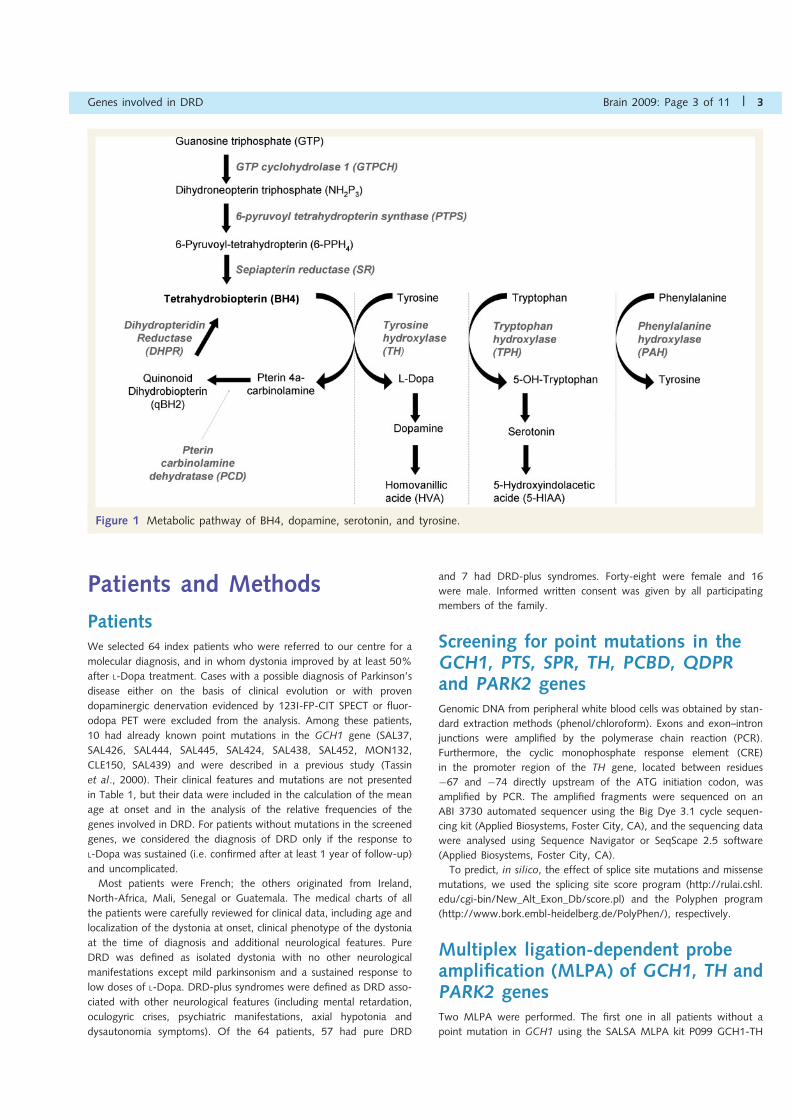
et al., 1994). GTPCH is involved in the first and rate-limiting
step of the de novo biosynthesis of tetrahydrobiopterin (BH4)
by catalysing the formation of dihydroneopterin triphosphate
from GTP (Fig. 1). BH4 is an essential cofactor required for the
activity of various enzymes such as the nitric oxide synthases
and phenylalanine-, tryptophane- and tyrosine- hydroxylases
(Thony et al., 2000). The second and last steps of the de novo
biosynthesis of BH4 are catalysed by the pyruvoyl tetrahydrop-
terin synthase (PTPS EC 4.6.1.10; gene symbol PTS) and the
sepiapterin reductase (SR EC 1.1.1.153; gene symbol SPR),
respectively. Furthermore, two additional enzymes, pterin-4a-
carbinolamine dehydratase (PCD EC 4.2.1.96; gene symbol
PCBD) and dihydropteridin reductase (DHPR EC 1.6.99.7; gene
symbol QDPR), are required for the regeneration of BH4
from intermediates formed during the hydroxylation of aromatic
amino acids.
Deficits in other enzymes involved in the BH4 biosynthesis may
be responsible for the DRD phenotypes associated with other
biochemical hallmarks such as hyperphenylalaninemia. Mutations
in PTS gene commonly induce hyperphenylalaninemia, but a DRD
phenotype has rarely been described (Hanihara et al., 1997) since
hyperphenylalaninemia is systematically detected through neonatal
screening, leading to early substitutive dopaminergic and BH4
treatment. Mutations in the SPR gene are responsible for
DRD with more complex clinical syndromes (Bonafe et al.,
2001; Neville et al., 2005, Abeling et al., 2006), without
hyperphenylalaninemia.
Thus, theoretically, all the enzymes involved in the biosynthesis
and recycling of BH4 could induce DRD, especially when hyper-
phenylalaninemia is mild and is not detected at birth. In addition,
mutations in two other genes also cause an autosomal recessive
form of DRD: the tyrosine hydroxylase gene (TH EC 1.14.16.2;
gene symbol TH) responsible for typical DRD (Ludecke et al.,
1995; Furukawa et al., 2001; Shiller et al., 2004) or L-Dopa-
responsive parkinsonism (Ludecke et al., 1996; van den Heuvel
et al., 1998) and the parkin gene (Parkin EC 6.3.2; gene symbol
PARK2) responsible for juvenile parkinsonism and more rarely for
typical DRD (Tassin et al., 2000).
However, to date, the relative frequency of the mutations in the
genes encoding the enzymes involved in the dopamine and
BH4 biosynthesis in DRD patients remains unknown. In order to
evaluate the respective contributions of these genes and the
associated clinical spectrum, we investigated a cohort of 64
index patients with DRD. All patients were screened for point
mutations and large rearrangements in the GCH1 gene, followed
by the sequencing of the TH, SPR, PTS, PCBD, QDPR, as well the
PARK2 genes in patients without mutation in GCH1.
2 | Brain 2009: Page 2 of 11 F. Clot et al.
Patients and Methods
PatientsWe selected 64 index patients who were referred to our centre for a
molecular diagnosis, and in whom dystonia improved by at least 50%
after L-Dopa treatment. Cases with a possible diagnosis of Parkinson’s
disease either on the basis of clinical evolution or with proven
dopaminergic denervation evidenced by 123I-FP-CIT SPECT or fluor-
odopa PET were excluded from the analysis. Among these patients,
10 had already known point mutations in the GCH1 gene (SAL37,
SAL426, SAL444, SAL445, SAL424, SAL438, SAL452, MON132,
CLE150, SAL439) and were described in a previous study (Tassin
et al., 2000). Their clinical features and mutations are not presented
in Table 1, but their data were included in the calculation of the mean
age at onset and in the analysis of the relative frequencies of the
genes involved in DRD. For patients without mutations in the screened
genes, we considered the diagnosis of DRD only if the response to
L-Dopa was sustained (i.e. confirmed after at least 1 year of follow-up)
and uncomplicated.
Most patients were French; the others originated from Ireland,
North-Africa, Mali, Senegal or Guatemala. The medical charts of all
the patients were carefully reviewed for clinical data, including age and
localization of the dystonia at onset, clinical phenotype of the dystonia
at the time of diagnosis and additional neurological features. Pure
DRD was defined as isolated dystonia with no other neurological
manifestations except mild parkinsonism and a sustained response to
low doses of L-Dopa. DRD-plus syndromes were defined as DRD asso-
ciated with other neurological features (including mental retardation,
oculogyric crises, psychiatric manifestations, axial hypotonia and
dysautonomia symptoms). Of the 64 patients, 57 had pure DRD
and 7 had DRD-plus syndromes. Forty-eight were female and 16
were male. Informed written consent was given by all participating
members of the family.
Screening for point mutations in theGCH1, PTS, SPR, TH, PCBD, QDPRand PARK2 genesGenomic DNA from peripheral white blood cells was obtained by stan-
dard extraction methods (phenol/chloroform). Exons and exon–intron
junctions were amplified by the polymerase chain reaction (PCR).
Furthermore, the cyclic monophosphate response element (CRE)
in the promoter region of the TH gene, located between residues
!67 and !74 directly upstream of the ATG initiation codon, was
amplified by PCR. The amplified fragments were sequenced on an
ABI 3730 automated sequencer using the Big Dye 3.1 cycle sequen-
cing kit (Applied Biosystems, Foster City, CA), and the sequencing data
were analysed using Sequence Navigator or SeqScape 2.5 software
(Applied Biosystems, Foster City, CA).
To predict, in silico, the effect of splice site mutations and missense
mutations, we used the splicing site score program (http://rulai.cshl.
edu/cgi-bin/New_Alt_Exon_Db/score.pl) and the Polyphen program
(http://www.bork.embl-heidelberg.de/PolyPhen/), respectively.
Multiplex ligation-dependent probeamplification (MLPA) of GCH1, TH andPARK2 genesTwo MLPA were performed. The first one in all patients without a
point mutation in GCH1 using the SALSA MLPA kit P099 GCH1-TH
Figure 1 Metabolic pathway of BH4, dopamine, serotonin, and tyrosine.
Genes involved in DRD Brain 2009: Page 3 of 11 | 3
(MRC-Holland, Amsterdam, The Netherlands) and the second one in
the patients without mutations in the genes involved in metabolic
pathways of BH4 and L-Dopa using the SALSA MLPA kit P052B
Parkinson.
All of the exons in GCH1, except the small exon 4 (32 bp), were
quantified. Only 6 of the 14 TH exons were quantified. One hun-
dred nanograms of DNA were used in the MLPA protocol, according
to the manufacturer’s instructions. Reactions were performed
on a GeneAmp PCR System 9700 (Applied Biosystems). One micro-
litre of the PCR products was analysed by capillary electrophoresis
on an ABI 3730 automated sequencer and MLPA data were
analysed using the ABI Prism Genemapper 4.0 software (Applied
Biosystems, Foster City, CA). Relative ratios were calculated for
each peak using the formula r=mean (peak area patient/
control area patient)/(peak area control individual/control area
control).
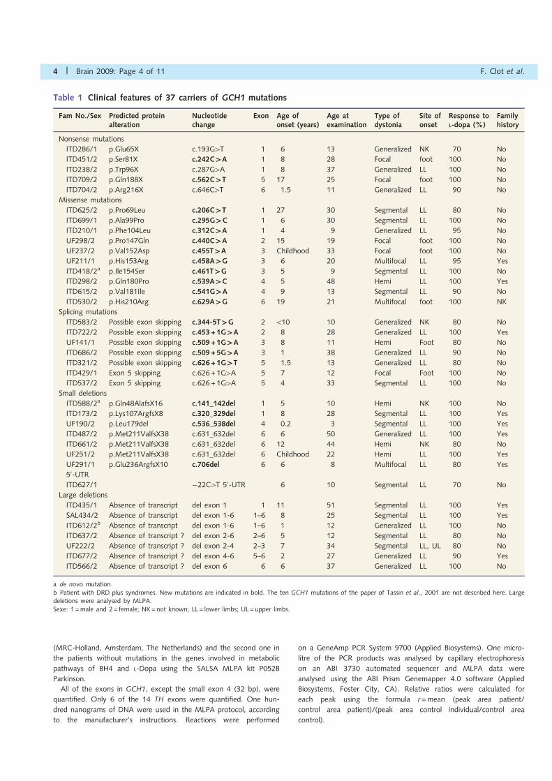
Table 1 Clinical features of 37 carriers of GCH1 mutations
Fam No./Sex Predicted proteinalteration
Nucleotidechange
Exon Age ofonset (years)
Age atexamination
Type ofdystonia
Site ofonset
Response toL-dopa (%)
Familyhistory
Nonsense mutations
ITD286/1 p.Glu65X c.193G4T 1 6 13 Generalized NK 70 No
ITD451/2 p.Ser81X c.242C`A 1 8 28 Focal foot 100 No
ITD238/2 p.Trp96X c.287G4A 1 8 37 Generalized LL 100 No
ITD709/2 p.Gln188X c.562C`T 5 17 25 Focal foot 100 No
ITD704/2 p.Arg216X c.646C4T 6 1.5 11 Generalized LL 90 No
Missense mutations
ITD625/2 p.Pro69Leu c.206C`T 1 27 30 Segmental LL 80 No
ITD699/1 p.Ala99Pro c.295G`C 1 6 30 Segmental LL 100 No
ITD210/1 p.Phe104Leu c.312C`A 1 4 9 Generalized LL 95 No
UF298/2 p.Pro147Gln c.440C`A 2 15 19 Focal foot 100 No
UF237/2 p.Val152Asp c.455T`A 3 Childhood 33 Focal foot 100 No
UF211/1 p.His153Arg c.458A`G 3 6 20 Multifocal LL 95 Yes
ITD418/2a p.Ile154Ser c.461T`G 3 5 9 Segmental LL 100 No
ITD298/2 p.Gln180Pro c.539A`C 4 5 48 Hemi LL 100 Yes
ITD615/2 p.Val181Ile c.541G`A 4 9 13 Segmental LL 90 No
ITD530/2 p.His210Arg c.629A`G 6 19 21 Multifocal foot 100 NK
Splicing mutations
ITD583/2 Possible exon skipping c.344-5T`G 2 510 10 Generalized NK 80 No
ITD722/2 Possible exon skipping c.453+1G`A 2 8 28 Generalized LL 100 Yes
UF141/1 Possible exon skipping c.509+1G`A 3 8 11 Hemi Foot 80 No
ITD686/2 Possible exon skipping c.509+5G`A 3 1 38 Generalized LL 90 No
ITD321/2 Possible exon skipping c.626+1G`T 5 1.5 13 Generalized LL 80 No
ITD429/1 Exon 5 skipping c.626+1G4A 5 7 12 Focal Foot 100 No
ITD537/2 Exon 5 skipping c.626+1G4A 5 4 33 Segmental LL 100 No
Small deletions
ITD588/2a p.Gln48AlafsX16 c.141_142del 1 5 10 Hemi NK 100 No
ITD173/2 p.Lys107ArgfsX8 c.320_329del 1 8 28 Segmental LL 100 Yes
UF190/2 p.Leu179del c.536_538del 4 0.2 3 Segmental LL 100 Yes
ITD487/2 p.Met211ValfsX38 c.631_632del 6 6 50 Generalized LL 100 Yes
ITD661/2 p.Met211ValfsX38 c.631_632del 6 12 44 Hemi NK 80 No
UF251/2 p.Met211ValfsX38 c.631_632del 6 Childhood 22 Hemi LL 100 Yes
UF291/1 p.Glu236ArgfsX10 c.706del 6 6 8 Multifocal LL 80 Yes
50-UTR
ITD627/1 !22C4T 50-UTR 6 10 Segmental LL 70 No
Large deletions
ITD435/1 Absence of transcript del exon 1 1 11 51 Segmental LL 100 Yes
SAL434/2 Absence of transcript del exon 1-6 1–6 8 25 Segmental LL 100 Yes
ITD612/2b Absence of transcript del exon 1-6 1–6 1 12 Generalized LL 100 No
ITD637/2 Absence of transcript ? del exon 2-6 2–6 5 12 Segmental LL 80 No
UF222/2 Absence of transcript ? del exon 2-4 2–3 7 34 Segmental LL, UL 80 No
ITD677/2 Absence of transcript ? del exon 4-6 5–6 2 27 Generalized LL 90 Yes
ITD566/2 Absence of transcript ? del exon 6 6 6 37 Generalized LL 100 No
a de novo mutation.b Patient with DRD plus syndromes. New mutations are indicated in bold. The ten GCH1 mutations of the paper of Tassin et al., 2001 are not described here. Largedeletions were analysed by MLPA.Sexe: 1 =male and 2= female; NK=not known; LL= lower limbs; UL= upper limbs.
4 | Brain 2009: Page 4 of 11 F. Clot et al.

Quantitative real-time PCRExon 4 of GCH1 was quantified by quantitative real-time PCR (q-PCR)
in all patients without point mutations. The primer pairs were designed
using Primer Express 1.5 software. The sequence of the forward and
reverse primers was TGATTTGTGATTAACTAAAACAATTCTTTC and
ACAGATTTTTAAAGCTTACCTTGTAGTCTTC, respectively. Real-time
PCR was performed on the ABI PRISM 7500 Sequence detection
system (Applied Biosystems), in a total volume of 25 ml, containing
10 ng of genomic DNA, 1" SYBR Green PCR master mix (Applied
Biosystems, Foster City, California, USA), and 250 nM of the forward
and reverse primers. The standard amplification protocol was used:
50#C for 2min, 95#C for 10min, followed by 40 cycles of 95#C for
15 s and 60#C for 1min. To normalize the amount of target DNA,
exon 8 of albumin gene was amplified in a separate reaction well
under identical thermal cycling conditions. Each reaction was run in
triplicate. Copy number values between 0.8 and 1.2 were considered
as normal; values 40.6 evidenced a deletion.
We also quantified exons 2 and 3 of the SPR gene by Q-PCR. The
sequences of the forward and reverse primers for exon 2 were
ACAGTCCTGGCCTCAACAGAA and TGCAGGGCACAGAGGGA,
respectively, and the sequences of the forward and reverse primers
for exon 3 were GTGAGCTCCCAGGTCATTGG and GCACAGCA
CAGACTCCTGACA, respectively.
PCR amplification was performed as above.
Deletion mapping by infinium HD DNAanalysis BeadChipPatient ITD612 was analysed using Illumina’s Human CNV 370-Quad
BeadChip (Illumina Inc, San Diego, CA, USA). We used 200 ng of
patient DNA and followed the protocol as described by the manufac-
turer (Illumina Inc., San Diego, CA, USA).
Statistical analysisAge at onset was compared using the Mann–Whitney test. Statistical
significance was defined as P50.05.
Results
GCH1 mutation carriers and theirclinical characteristicsDirect sequencing of the GCH1 gene was performed in 64
unrelated index patients. We identified 38 index patients with
heterozygous point mutations in GCH1, two index patients with
compound heterozygous mutations and seven index patients with
heterozygous large deletions. We found 34 different point muta-
tions (6 nonsense, 15 missense, 6 splice site mutations, 6 small
deletions and 1 mutation in the 50 untranslated region). Twenty of
them were new mutations (Table 1).
The two patients with compound heterozygous mutations
carried a common missense variant (c.68C4T, p.Pro23Leu) located
in exon 1. This substitution was previously described as a mutation
or a variant in 8 different families (Jarman et al., 1997; de la
Fuente-Fernandez 1997; Steinberger et al., 2000; Scola et al.,
2007; Zirn et al., 2008) and it was found in 1/210 control
chromosomes by Jarman et al. (1997). We found the
p.Pro23Leu variant in 1/174 control chromosomes. The two
mutations combined with the p.Pro23Leu variant were a 2-base
deletion (c.137_138delGC, p.Ser46SerfsX18) and a missense
mutation (c.206 C4T, p.Pro69Leu), both located in exon 1, in
patients ITD588 and ITD625, respectively. In patient ITD588,
the p.Pro23Leu variant was inherited from the mother and the
deletion (c.137_138delGC) occurred de novo (segregation analysis
of 8 informative polymorphic markers excluded non-paternity).
The p.Pro69Leu mutation was found in 1/174 control chromo-
somes in the same control individual that had the p.Pro23Leu
mutation. The parental DNA of the patient and the control were
not available, so we could not define whether the mutations were
in the cis- or in the trans- position. The p.Pro23Leu mutation
was considered by Polyphen to be benign and the p.Pro69Leu
mutation to be deleterious. Moreover, the proline residue at
position 23 is not well conserved in other species, in contrast to
the proline at position 69. These observations suggest that
p.Pro23Leu is a polymorphism and that p.Pro69Leu is a probably
causative mutation. All the other missense mutations identified
in the present study altered an amino acid that was conserved
across mammalian species and were not found on the control
chromosomes.
Four splice site mutations (c.453+ 1G4A, c.509+1G4A,
c.626 +1G4A and c.626 +1G4T) affected the invariant bases of
the consensus splice donor sequence. The previously reported
c.626 +1G4A splice site mutation (Hirano et al., 1998) resulted
in the skipping of exon 5 and the introduction of a premature stop
codon. The three other splice site mutations were novel; however,
since RNA from patients was not available, the transcripts could
not be analysed. It is highly probable that these mutations cause
exon skipping. We also identified two other splice site variants that
did not affect the invariant bases of the consensus splice site
sequence, but which were located at position –5 of intron 2
(c.344-5T4G) and at position +5 of intron 3 (c.509+5G4A).
The mutations reduced the splice site score from 7.2 to 4.4 for
the c.344-5T4G mutation and from 11.6 to 8.2 for the
c.509 +5G4A mutation. However, no RNA was available to
validate the in silico predictions that these variants are splice
site mutations.
Except for two in frame deletions (p.Leu179del and
p.Arg88_Gln89del), all small deletions caused a frameshift in the
coding region that introduced a premature stop codon, theoreti-
cally resulting in a truncated protein.
Two of the seven patients with large heterozygous deletions of
GCH1 (SAL434 and ITD612) had complete gene deletions and five
had partial deletions (exon 1, exons 2–4, exons 2–6, exons 4–6 or
exon 6). Four of the seven patients were sporadic cases, three
were familial. In family SAL434, four individuals (two males and
two females) had complete deletions. The index case presented a
pure DRD, whereas the other patients had only mild equinism.
Analysis of microsatellites and SNP in the region flanking the dele-
tion in this family demonstrated that the deletion encompassed
only the GCH1 gene. In patient ITD612 who also had a complete
deletion of GCH1, a mental retardation was observed in addition
to classical signs of DRD. Several male relatives of this patient had
a microphthalmia with mental retardation and several females had
Genes involved in DRD Brain 2009: Page 5 of 11 | 5
low intellectual levels variably associated with strabismus. The size
of the deletion was analysed by using Infinium High-Density
BeadChips (Human CNV 370-Quad). We identified a 2.3Mb
deletion ranging from rs4901534 to rs1189820. This region
contained 10 genes, one of which is the homeobox OTX2 gene,
involved in severe ocular malformations including anophthalmia-
microphthalmia and variable developmental delay (Wyatt et al.,
2008). In family ITD677, three females were affected (the mother
and her two daughters). They all had a partial deletion of GCH1
comprising exons 4–6, with ages at onset ranging from 5 to
8 years. In family ITD435, one female and two males were
affected, but DNA was available only for the index patient who
had an exon 1 deletion.
In the 47 families with a point mutation or a large deletion in
GCH1, there were 63 patients and 16 asymptomatic carriers.
There were 2.5 times more females than males (45 versus 18) in
the patients and 2.2 times more males than females (11 versus 5)
in the asymptomatic carriers. Thirty-five percent of the patients
had a family history of DRD and 65% were isolated (the one
patient for whom the family history was unknown was excluded).
Analysis of the DNA of the parents of the five index cases without
positive family histories of the disease revealed the existence of
two de novo mutations (one deletion and one missense mutation).
The clinical characteristics of patients with a mutation or a large
deletion in GCH1 were very stereotyped. The mean age of onset
was 7.5 years$ 4.9 (4 months to 27 years) and there was no
significant difference between carriers of point mutations com-
pared to carriers of large deletions (7.8$ 5.1 years versus
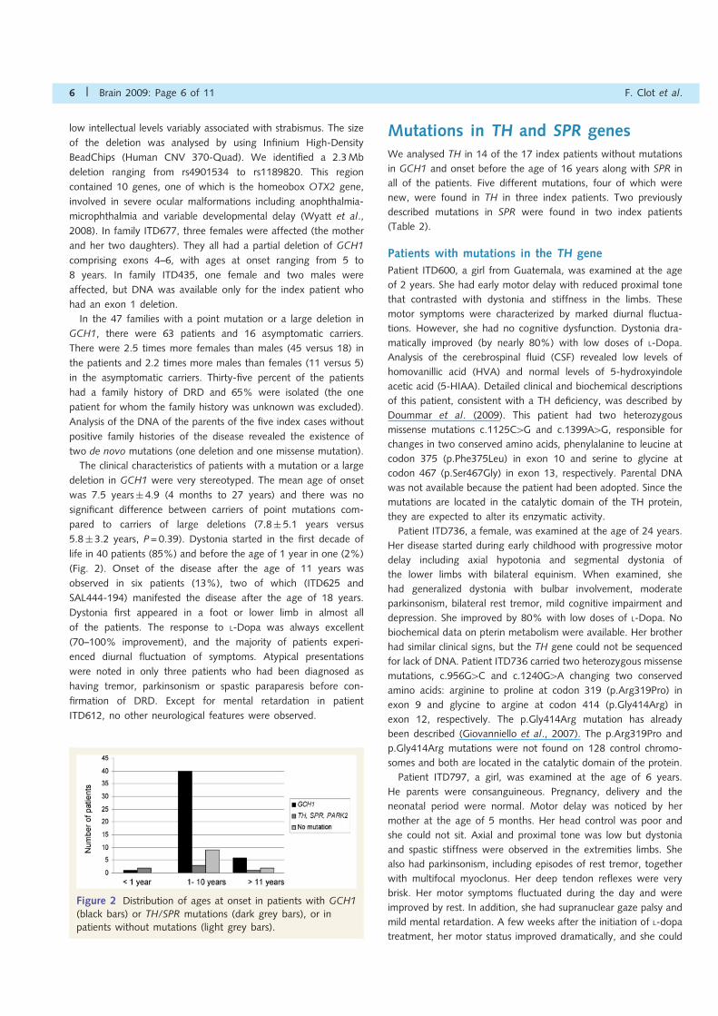
5.8$ 3.2 years, P=0.39). Dystonia started in the first decade of
life in 40 patients (85%) and before the age of 1 year in one (2%)
(Fig. 2). Onset of the disease after the age of 11 years was
observed in six patients (13%), two of which (ITD625 and
SAL444-194) manifested the disease after the age of 18 years.
Dystonia first appeared in a foot or lower limb in almost all
of the patients. The response to L-Dopa was always excellent
(70–100% improvement), and the majority of patients experi-
enced diurnal fluctuation of symptoms. Atypical presentations
were noted in only three patients who had been diagnosed as
having tremor, parkinsonism or spastic paraparesis before con-
firmation of DRD. Except for mental retardation in patient
ITD612, no other neurological features were observed.
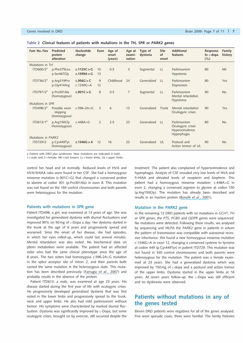
Mutations in TH and SPR genesWe analysed TH in 14 of the 17 index patients without mutations
in GCH1 and onset before the age of 16 years along with SPR in
all of the patients. Five different mutations, four of which were
new, were found in TH in three index patients. Two previously
described mutations in SPR were found in two index patients
(Table 2).
Patients with mutations in the TH gene
Patient ITD600, a girl from Guatemala, was examined at the age
of 2 years. She had early motor delay with reduced proximal tone
that contrasted with dystonia and stiffness in the limbs. These
motor symptoms were characterized by marked diurnal fluctua-
tions. However, she had no cognitive dysfunction. Dystonia dra-
matically improved (by nearly 80%) with low doses of L-Dopa.
Analysis of the cerebrospinal fluid (CSF) revealed low levels of
homovanillic acid (HVA) and normal levels of 5-hydroxyindole
acetic acid (5-HIAA). Detailed clinical and biochemical descriptions
of this patient, consistent with a TH deficiency, was described by
Doummar et al. (2009). This patient had two heterozygous
missense mutations c.1125C4G and c.1399A4G, responsible for
changes in two conserved amino acids, phenylalanine to leucine at
codon 375 (p.Phe375Leu) in exon 10 and serine to glycine at
codon 467 (p.Ser467Gly) in exon 13, respectively. Parental DNA
was not available because the patient had been adopted. Since the
mutations are located in the catalytic domain of the TH protein,
they are expected to alter its enzymatic activity.
Patient ITD736, a female, was examined at the age of 24 years.
Her disease started during early childhood with progressive motor
delay including axial hypotonia and segmental dystonia of
the lower limbs with bilateral equinism. When examined, she
had generalized dystonia with bulbar involvement, moderate
parkinsonism, bilateral rest tremor, mild cognitive impairment and
depression. She improved by 80% with low doses of L-Dopa. No
biochemical data on pterin metabolism were available. Her brother
had similar clinical signs, but the TH gene could not be sequenced
for lack of DNA. Patient ITD736 carried two heterozygous missense
mutations, c.956G4C and c.1240G4A changing two conserved
amino acids: arginine to proline at codon 319 (p.Arg319Pro) in
exon 9 and glycine to argine at codon 414 (p.Gly414Arg) in
exon 12, respectively. The p.Gly414Arg mutation has already
been described (Giovanniello et al., 2007). The p.Arg319Pro and
p.Gly414Arg mutations were not found on 128 control chromo-
somes and both are located in the catalytic domain of the protein.
Patient ITD797, a girl, was examined at the age of 6 years.
He parents were consanguineous. Pregnancy, delivery and the
neonatal period were normal. Motor delay was noticed by her
mother at the age of 5 months. Her head control was poor and
she could not sit. Axial and proximal tone was low but dystonia
and spastic stiffness were observed in the extremities limbs. She
also had parkinsonism, including episodes of rest tremor, together
with multifocal myoclonus. Her deep tendon reflexes were very
brisk. Her motor symptoms fluctuated during the day and were
improved by rest. In addition, she had supranuclear gaze palsy and
mild mental retardation. A few weeks after the initiation of L-dopa
treatment, her motor status improved dramatically, and she could
Figure 2 Distribution of ages at onset in patients with GCH1(black bars) or TH/SPR mutations (dark grey bars), or inpatients without mutations (light grey bars).
6 | Brain 2009: Page 6 of 11 F. Clot et al.
control her head and sit normally. Reduced levels of HVA and
HVA/5HIAA ratio were found in her CSF. She had a homozygous
missense mutation (c.901C4G) that changed a conserved proline
to alanine at codon 301 (p.Pro301Ala) in exon 8. This mutation
was not found on the 184 control chromosomes and both parents
were heterozygous for the mutation.
Patients with mutations in SPR gene
Patient ITD498, a girl, was examined at 13 years of age. She was
investigated for generalized dystonia with diurnal fluctuations and
improved 90% on 50mg of L-Dopa a day. Her dystonia started in
the trunk at the age of 6 years and progressively spread and
worsened. Since the onset of her disease, she had episodes,
in which her eyes rolled-up, which could last several minutes.
Mental retardation was also noted. No biochemical data on
pterin metabolism were available. The patient had an affected
sister who had the same clinical phenotype since the age of
8 years. The two sisters had homozygous c.596-2A4G mutation
in the splice acceptor site of intron 2, and their parents both
carried the same mutation in the heterozygous state. This muta-
tion has been described previously (Farrugia et al., 2007) and
probably results in the absence of the protein.
Patient ITD613, a male, was examined at age 23 years. His
disease started during the first year of life with oculogyric crises.
He progressively developed generalized dystonia that was first
noted in the lower limbs and progressively spread to the trunk,
neck and upper limbs. He also had mild parkinsonism without
tremor. His symptoms were characterized by marked diurnal fluc-
tuation. Dystonia was significantly improved by L-Dopa, but some
oculogyric crises, brought on by exercise, still occurred despite the
treatment. The patient also complained of hypersomnolence and
hyperphagia. Analysis of CSF revealed very low levels of HVA and
5-HIAA and elevated levels of neopterin and biopterin. This
patient had a homozygous missense mutation: c.448A4G in
exon 2, changing a conserved arginine to glycine at codon 150
(p.Arg150Gly). The mutation has already been described and
results in an inactive protein (Bonafe et al., 2001).
Mutation in the PARK2 gene
In the remaining 12 DRD patients with no mutations in GCH1, TH
or SPR genes, the PTS, PCBD and QDPR genes were sequenced.
No mutations were detected. Following these results, we analysed
by sequencing and MLPA the PARK2 gene in patients in whom
the pattern of transmission was compatible with autosomal reces-
sive inheritance. We found a new homozygous missense mutation
c.1346G4A in exon 12, changing a conserved cysteine to tyrosine
at codon 449 (p.Cys449Tyr) in patient ITD729. This mutation was
not found in 340 control chromosomes and both parents were
heterozygous for the mutation. The patient was a female exam-
ined at 23 years. She had a generalized dystonia which was
improved by 150mg of L-dopa and a postural and action tremor
of the upper limbs. Dystonia started in the upper limbs at 16
years. At seven years follow-up, the L-Dopa was still efficient
and no dyskinesia were observed.
Patients without mutations in any ofthe genes testedEleven DRD patients were negatives for all of the genes analysed.
Five were sporadic cases, three were familial. The family histories
Table 2 Clinical features of patients with mutations in the TH, SPR or PARK2 genes
Fam No./Sex Predictedproteinalteration
Nucleotidechange
Exon Age ofonset(years)
Age atexami-nation
Type ofdystonia
Siteofonset
Additionalfeatures
Responseto L-dopa(%)
Familyhistory
Mutations in TH
ITD600/2a p.Phe375Leu c.1125C`G 10 0.5 3 Segmental LL Parkinsonism 80 NK
p.Ser467Gly c.1399A`G 13 Hypotonia
ITD736/2a p.Arg319Pro c.956G`C 9 Childhood 24 Generalized LL Parkinsonism 80 Yes
p.Gly414Arg c.1240G4A 12 Depression
ITD797/2a p.Pro301Ala(homozygous)
c.901C`G 8 0.5 7 Segmental LL ParkinsonismMental retardationHypotonia
90 No
Mutations in SPR
ITD498/2a Possible exonskipping
(homozygous)
c.596–2A4G 3 6 13 Generalized Trunk Mental retardationOculogyric crises
90 Yes
ITD613/1a p.Arg150Gly(homozygous)
c.448A4G 2 2.5 23 Generalized LL ParkinsonismOculogyric crisesHypersomolenceHyperphagia
80 No
Mutations in PARK2
ITD729/2 p.Cys449Tyr(homozygous)
c.1346G`A 12 16 23 Generalized UL Postural andAction tremor of UL
90 No
a Patient with DRD plus syndromes. New mutations are indicated in bold.1 =male and 2= female; NK=not known; LL= lower limbs; UL =upper limbs.
Genes involved in DRD Brain 2009: Page 7 of 11 | 7
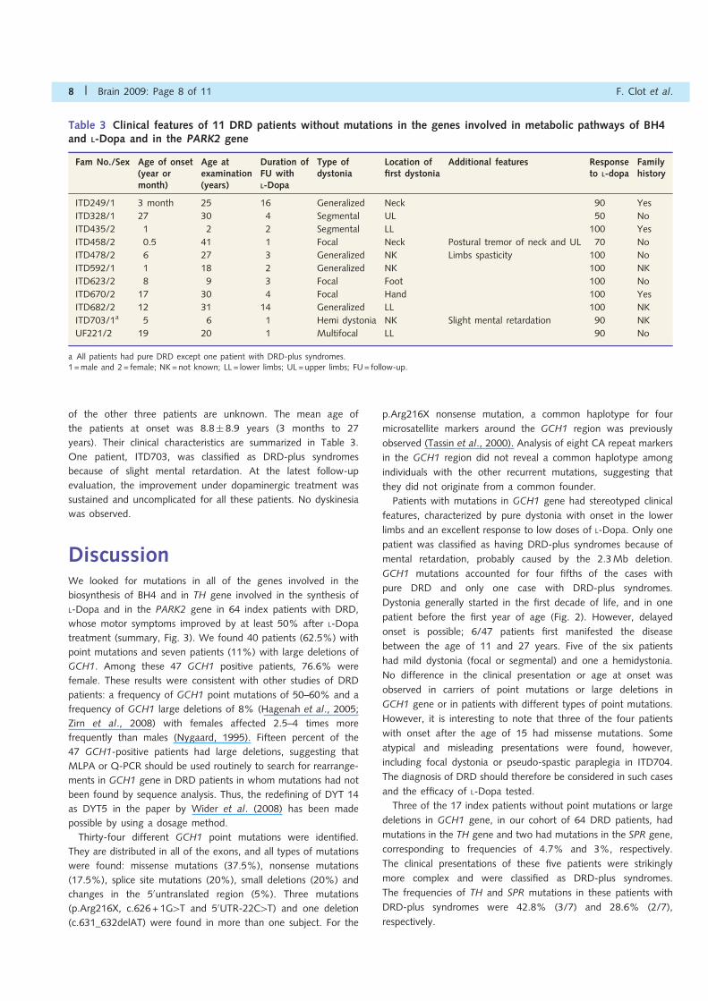
of the other three patients are unknown. The mean age of
the patients at onset was 8.8$ 8.9 years (3 months to 27
years). Their clinical characteristics are summarized in Table 3.
One patient, ITD703, was classified as DRD-plus syndromes
because of slight mental retardation. At the latest follow-up
evaluation, the improvement under dopaminergic treatment was
sustained and uncomplicated for all these patients. No dyskinesia
was observed.
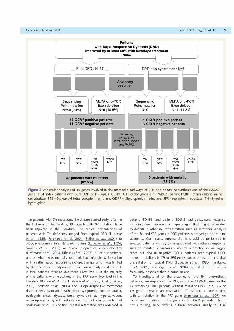
DiscussionWe looked for mutations in all of the genes involved in the
biosynthesis of BH4 and in TH gene involved in the synthesis of
L-Dopa and in the PARK2 gene in 64 index patients with DRD,
whose motor symptoms improved by at least 50% after L-Dopa
treatment (summary, Fig. 3). We found 40 patients (62.5%) with
point mutations and seven patients (11%) with large deletions of
GCH1. Among these 47 GCH1 positive patients, 76.6% were
female. These results were consistent with other studies of DRD
patients: a frequency of GCH1 point mutations of 50–60% and a
frequency of GCH1 large deletions of 8% (Hagenah et al., 2005;
Zirn et al., 2008) with females affected 2.5–4 times more
frequently than males (Nygaard, 1995). Fifteen percent of the
47 GCH1-positive patients had large deletions, suggesting that
MLPA or Q-PCR should be used routinely to search for rearrange-
ments in GCH1 gene in DRD patients in whom mutations had not
been found by sequence analysis. Thus, the redefining of DYT 14
as DYT5 in the paper by Wider et al. (2008) has been made
possible by using a dosage method.
Thirty-four different GCH1 point mutations were identified.
They are distributed in all of the exons, and all types of mutations
were found: missense mutations (37.5%), nonsense mutations
(17.5%), splice site mutations (20%), small deletions (20%) and
changes in the 50untranslated region (5%). Three mutations
(p.Arg216X, c.626 +1G4T and 50UTR-22C4T) and one deletion
(c.631_632delAT) were found in more than one subject. For the
p.Arg216X nonsense mutation, a common haplotype for four
microsatellite markers around the GCH1 region was previously
observed (Tassin et al., 2000). Analysis of eight CA repeat markers
in the GCH1 region did not reveal a common haplotype among
individuals with the other recurrent mutations, suggesting that
they did not originate from a common founder.
Patients with mutations in GCH1 gene had stereotyped clinical
features, characterized by pure dystonia with onset in the lower
limbs and an excellent response to low doses of L-Dopa. Only one
patient was classified as having DRD-plus syndromes because of
mental retardation, probably caused by the 2.3Mb deletion.
GCH1 mutations accounted for four fifths of the cases with
pure DRD and only one case with DRD-plus syndromes.
Dystonia generally started in the first decade of life, and in one
patient before the first year of age (Fig. 2). However, delayed
onset is possible; 6/47 patients first manifested the disease
between the age of 11 and 27 years. Five of the six patients
had mild dystonia (focal or segmental) and one a hemidystonia.
No difference in the clinical presentation or age at onset was
observed in carriers of point mutations or large deletions in
GCH1 gene or in patients with different types of point mutations.
However, it is interesting to note that three of the four patients
with onset after the age of 15 had missense mutations. Some
atypical and misleading presentations were found, however,
including focal dystonia or pseudo-spastic paraplegia in ITD704.
The diagnosis of DRD should therefore be considered in such cases
and the efficacy of L-Dopa tested.
Three of the 17 index patients without point mutations or large
deletions in GCH1 gene, in our cohort of 64 DRD patients, had
mutations in the TH gene and two had mutations in the SPR gene,
corresponding to frequencies of 4.7% and 3%, respectively.
The clinical presentations of these five patients were strikingly
more complex and were classified as DRD-plus syndromes.
The frequencies of TH and SPR mutations in these patients with
DRD-plus syndromes were 42.8% (3/7) and 28.6% (2/7),
respectively.
Table 3 Clinical features of 11 DRD patients without mutations in the genes involved in metabolic pathways of BH4and L-Dopa and in the PARK2 gene
Fam No./Sex Age of onset(year ormonth)
Age atexamination(years)
Duration ofFU withL-Dopa
Type ofdystonia
Location offirst dystonia
Additional features Responseto L-dopa
Familyhistory
ITD249/1 3 month 25 16 Generalized Neck 90 Yes
ITD328/1 27 30 4 Segmental UL 50 No
ITD435/2 1 2 2 Segmental LL 100 Yes
ITD458/2 0.5 41 1 Focal Neck Postural tremor of neck and UL 70 No
ITD478/2 6 27 3 Generalized NK Limbs spasticity 100 No
ITD592/1 1 18 2 Generalized NK 100 NK
ITD623/2 8 9 3 Focal Foot 100 No
ITD670/2 17 30 4 Focal Hand 100 Yes
ITD682/2 12 31 14 Generalized LL 100 NK
ITD703/1a 5 6 1 Hemi dystonia NK Slight mental retardation 90 NK
UF221/2 19 20 1 Multifocal LL 90 No
a All patients had pure DRD except one patient with DRD-plus syndromes.1 =male and 2= female; NK=not known; LL= lower limbs; UL =upper limbs; FU= follow-up.
8 | Brain 2009: Page 8 of 11 F. Clot et al.
In patients with TH mutations, the disease started early, often in
the first year of life. To date, 29 patients with TH mutations have
been reported in the literature. The clinical presentations of
patients with TH deficiency ranged from typical DRD (Ludecke
et al., 1995; Furukawa et al, 2001; Shiller et al., 2004) to
L-Dopa-responsive infantile parkinsonism (Ludecke et al., 1996,
Swaans et al., 2000) or severe progressive encephalopathy
(Hoffmann et al., 2004, Ribases et al., 2007). All of our patients,
one of whom was mentally retarded, had infantile parkinsonism
with a rather good response to L-Dopa therapy which was limited
by the occurrence of dyskinesia. Biochemical analyses of the CSF
in two patients revealed decreased HVA levels. In the majority
of the patients with mutations in the SPR gene described in the
literature (Bonafe et al., 2001; Neville et al., 2005, Abeling et al.,
2006, Friedman et al., 2006), the L-Dopa-responsive movement
disorder was associated with other symptoms, such as ataxia,
oculogyric crises, dysautonomia symptoms as hypersalivation,
microcephaly or growth retardation. Two of our patients had
oculogyric crises. In addition, mental retardation was observed in
patient ITD498, and patient ITD613 had behavioural features,
including sleep disorders or hyperphagia, that might be related
to deficits in other neurotransmitters such as serotonin. Analysis
of the TH and SPR genes in DRD patients is not yet part of routine
screening. Our results suggest that it should be performed in
selected patients with dystonia associated with others symptoms,
such as infantile parkinsonism, mental retardation or oculogyric
crises but also in negative GCH1 patients with typical DRD.
Indeed, mutations in TH or SPR genes can both result in a clinical
presentation of typical DRD (Ludecke et al., 1995; Furukawa
et al., 2001; Steinberger et al., 2004) even if this form is less
frequently observed than a complex one.
To investigate all of the enzymes of the BH4 biosynthesis
pathway, we sequenced the PTS, PCBD and QDPR genes in the
12 remaining DRD patients without mutations in GCH1, SPR or
TH genes. Despite an observation of dystonia in one patient
with a mutation in the PTS gene (Hanihara et al., 1997) we
found no mutations in this gene in our DRD patients. This is
not surprising, since deficits in these enzymes usually result in
Figure 3 Molecular analyses of six genes involved in the metabolic pathways of BH4 and dopamine synthesis and of the PARK2gene in 64 index patients with pure DRD or DRD-plus. GCH1=GTP cyclohydrolase 1; PARK2= parkin; PCBD=pterin carbinolaminedehydratase; PTS= 6-pyruvoyl tetrahydropterin synthase; QDPR=dihydropteridin reductase; SPR= sepiapterin reductase; TH= tyrosinehydroxylase.
Genes involved in DRD Brain 2009: Page 9 of 11 | 9
hyperphenylalaninemia, which is detected in newborn children and
treated by early administration of L-Dopa and BH4 which prevents
the development of dystonia. The identification of a PARK2
homozygous mutation in one patient of our cohort confirm that
defects in this gene can cause DRD similar to DRD secondary to
GCH1 mutations. In Parkinson’s disease (either genetic or not), the
DRD phenotype seems to be related with a juvenile onset (Lucking
et al., 2000). In such patients, only studies using 123I-FP-CIT
SPECT or [18F]dopa PET scans would allow to rule out dopami-
nergic cell loss and point to a BH4 or dopamine biosynthesis
defect.
Of the 11 patients without mutations, 10 had pure DRD with a
mean age at onset of 9.1 years$ 9.3 (range 3 months to
27 years), slightly higher than that of patients with GCH1 muta-
tions (7.5 years$ 4.8, range 4 months to 27 years). The inability
to detect GCH1 mutations in apparently typical phenotypes was
also reported in the papers of Hagenah et al. (2005) and Zirn
et al. (2008) and is still unexplained. Several hypotheses could
account for this observation, such as undetected mutations in
GCH1 promotor or regulatory regions leading to a decreased
expression of the gene. For these patients the hypothesis of
juvenile Parkinson’s disease not caused by PARK2 may also be
considered.
The value of measuring pterins and neurotransmitters levels in
the CSF was not systematically assessed in this study focused on
genetic aspects of DRD. However, these dosages help to guide
molecular analyses of TH and SPR genes in DRD-plus syndromes.
Finally, CSF study could be crucial for DRD patients without
mutations to formally discriminate between those with neurotrans-
mitters or pterins deficiencies.
In conclusion, patients with isolated DRD and with a good and
sustained response to low doses of L-Dopa should be tested, firstly
for point mutations, then for large deletions in GCH1. Molecular
analyses of other genes, including TH and SPR, should be
performed in patients without GCH1 mutation and in those with
DRD-plus syndromes, particularly when onset occurs during the
first year of life and before the age of 10. Ideally, the genetic
testing should be guided by biochemical analysis of the CSF.
Our study also suggests that molecular analyses of others genes
involved in hyperphenylalaninemia are likely to be negative. DRD
presentation of juvenile parkinsonism should be carefully ruled
out by testing patients for PARK2 mutations and by performing
123I-FP-CIT SPECT or [18F]dopa PET scans. Interestingly, the
yield of genetic analyses exceeds 4/5 in patients with pure
DRD or DRD-plus syndromes, but the genes involved are clearly
different in the two groups: GCH1 in pure DRD and TH or SPR in
DRD-plus syndromes.
AcknowledgementsWe are grateful for the contribution of the patients and their
relatives. We wish to thank the clinicians who referred patients
to us (Drs Yves Agid, Richard Baronnet, Patrick Berquin, Eric Bieth,
Thierry Billette de Villemeur, Emmanuel Broussole, Pierre Burbaud,
Patrick Calvas, Jean-Paul Carriere, Christian Confavreux, Pierre
Crost, Karin Dahan, Valerie Drouin Garraud, Franck Durif,
Bernard Echenne, Laurence Faivre, Annaik Feve, Pierre Genton,
Philippe Goas, Lucie Guyant-Marechal, Didier Hannequin,
Bertrand Isidor, Pierre Jedynak, Philippe Jonveaux, Alain
Lagueny, Caroline Moreau, Jacques Motte, Sylvie Odent,
Frederic Sedel, Marion Simonetta-Moreau, Clemence Simonin,
Mohnish Suri, Van Gaever, Francois Viallet, Jean-Sebastien Vidal,
Catherine Vincent-Delorme, Isabelle Vuillaume and Sandra
Vukusic). We thank Dr Merle Ruberg for her helpful suggestions
on the article (INSERM U679), Ms Lydia Guennec, Isabelle
Lagroua and Christelle Dussert (DNA and cell bank of IFR70) for
their technical assistance.
FundingINSERM, Reseau National des Dystonies and GIS-Maladies Rares.
ReferencesAbeling NG, Duran M, Bakker HD, Stroomer L, Thony B, Blau N, et al.
Poll-The BT. Sepiapterin reductase deficiency an autosomal recessiveDOPA-responsive dystonia. Mol Genet Metab 2006; 89: 116–20.
Bonafe L, Thony B, Penzien JM, Czarnecki B, Blau N. Mutations inthe sepiapterin reductase gene cause a novel tetrahydrobiopterin-dependent monoamine-neurotransmitter deficiency without hyperphe-nylalaninemia. Am J Hum Genet 2001; 69: 269–77.
Doummar D, Clot F, Vidailhet M, Afenjar A, Mignot C, Guet A, et al.Infantile hypokinetic-hypotonic syndrome due to two novel mutationsof the tyrosine hydroxylase gene. Mov Disord 2009 Fev 17.
Farrugia R, Scerri CA, Montalto SA, Parascandolo R, Neville BG,Felice AE. Molecular genetics of tetrahydrobiopterin (BH4) deficiencyin the Maltese population. Mol Genet Metab 2007; 90: 277–83.
Friedman J, Hyland K, Blau N, MacCollin M. Dopa-responsive hyper-somnia and mixed movement disorder due to sepiapterin reductasedeficiency. Neurology 2006; 67: 2032–5.
de la Fuente-Fernandez R. Mutations in GTP-cyclohydrolase I gene andvitiligo. Lancet 1997; 350: 640.
Furukawa Y, Graf WD, Wong H, Shimadzu M, Kish SJ. Dopa-responsivedystonia simulating spastic paraplegia due to tyrosine hydroxylase (TH)gene mutations. Neurology 2001; 56: 260–3.
Giovanniello T, Leuzzi V, Carducci C, Carducci C, Sabato ML, Artiola C,et al. Tyrosine hydroxylase deficiency presenting with a biphasicclinical course. Neuropediatrics 2007; 38: 213–5.
Hagenah J, Saunders-Pullman R, Hedrich K, Kabakci K, Habermann K,Wiegers K, et al. High mutation rate in dopa-responsive dystonia:detection with comprehensive GCHI screening. Neurology 2005; 64:908–11.
Hanihara T, Inoue K, Kawanishi C, Sugiyama N, Miyakawa T, Onishi H,et al. 6-Pyruvoyl-tetrahydropterin synthase deficiency with generalizeddystonia and diurnal fluctuation of symptoms: a clinical and molecularstudy. Mov Disord 1997; 12: 408–11.
van den Heuvel LP, Luiten B, Smeitink JA, de Rijk-van Andel JF,Hyland K, Steenbergen-Spanjers GC, et al. A common point mutationin the tyrosine hydroxylase gene in autosomal recessive L-DOPA-responsive dystonia in the Dutch population. Hum Genet 1998; 102:644–6.
Hirano M, Yanagihara T, Ueno S. Dominant negative effect of GTPcyclohydrolase I mutations in Dopa-responsive hereditary progressivedystonia. Ann Neurol 1998; 44: 365–71.
Hoffmann GF, Assmann B, Brautigam C, Dionisi-Vici C, Haussler M, deKlerk JB, et al. Tyrosine hydroxylase deficiency causes progressiveencephalopathy and dopa-nonresponsive dystonia. Ann Neurol 2004;55: 147–8.
10 | Brain 2009: Page 10 of 11 F. Clot et al.
Ichinose H, Ohye T, Takahashi E, Seki N, Hori T, Segawa M, et al.Hereditary progressive dystonia with marked diurnal fluctuationcaused by mutations in the GTP cyclohydrolase I gene. Nat Genet1994; 8: 236–42.
Jarman PR, Bandmann O, Marsden CD, Wood NW. GTP cyclohydrolaseI mutations in patients with dystonia responsive to anticholinergicdrugs. J Neurol Neurosurg Psychiatry 1997; 63: 304–8.
Lucking CB, Durr A, Bonifati V, Vaughan J, De Michele G, Gasser T,et al. French Parkinson’s Disease Genetics Study Group; EuropeanConsortium on Genetic Susceptibility in Parkinson’s Disease.Association between early-onset Parkinson’s disease and mutations inthe parkin gene. N Engl J Med 2000; 342: 1560–7.
Ludecke B, Dworniczak B, Bartholome K. A point mutation in thetyrosine hydroxylase gene associated with Segawa’s syndrome. HumGenet 1995; 95: 123–5.
Ludecke B, Knappskog PM, Clayton PT, Surtees RA, Clelland JD,Heales SJ, et al. Recessively inherited L-DOPA-responsive parkinsonismin infancy caused by a point mutation (L205P) in the tyrosinehydroxylase gene. Hum Mol Genet 1996; 5: 1023–8.
Neville BG, Parascandalo R, Farrugia R, Felice A. Sepiapterin reductasedeficiency: a congenital dopa-responsive motor and cognitive disorder.Brain 2005; 128: 2291–6.
Nygaard TG. Dopa-responsive dystonia. Curr Opin Neurol 1995; 8:310–3.
Ribases M, Serrano M, Fernandez-Alvarez E, Pahisa S, Ormazabal A,Garcıa-Cazorla A, et al. A homozygous tyrosine hydroxylase genepromoter mutation in a patient with dopa-responsive encephalopathy:clinical, biochemical and genetic analysis. Mol Genet Metab 2007; 92:274–7.
Shiller A, Wevers RA, Steenbergen GC, Blau N, Jung HH. Long-termcourse of L-dopa-responsive dystonia caused by tyrosine hydroxylasedeficiency. Neurology 2004; 63: 1524–6.
Scola RH, Carla Carducci C, Amaral VG, Lorenzoni PT, Teive HA,Giovanniello T, et al. A novel missense mutation pattern of the
GCH1 gene in dopa-responsive dystonia. Arq Neuropsiquiatr 2007;65: 1224–7.
Segawa M, Hosaka A, Miyagawa F, Nomura Y, Imai H. Hereditaryprogressive dystonia with marked diurnal fluctuation. Adv Neurol1976; 14: 215–33.
Steinberger D, Korinthenberg R, Topka H, Berghauser M, Wedde R,Muller U. Dopa-responsive dystonia: mutation analysis of GCH1 andanalysis of therapeutic doses of L-dopa. German Dystonia StudyGroup. Neurology 2000; 55: 1735–7.
Steinberger D, Blau N, Goriuonov D, Bitsch J, Zuker M, Hummel S, et al.Heterozygous mutation in 5’-untranslated region of sepiapterin reduc-tase gene (SPR) in a patient with dopa-responsive dystonia.Neurogenetics 2004; 5: 187–90.
Swaans RJ, Rondot P, Renier WO, Van Den Heuvel LP, Steenbergen-Spanjers GC, Wevers RA. Four novel mutations in the tyrosinehydroxylase gene in patients with infantile parkinsonism. Ann HumGenet 2000; 64: 25–31.
Tassin J, Durr A, Bonnet AM, Gil R, Vidailhet M, Lucking CB, et al.Levodopa-responsive dystonia. GTP cyclohydrolase I or parkinmutations? Brain 2000; 123: 1112–21.
Thony B, Auerbach G, Blau N. Tetrahydrobiopterin biosynthesis,regeneration and functions. Biochem J| 2000; 347: 1–16.
Wider C, Melquist S, Hauf M, Solida A, Cobb SA, Kachergus JM,et al. Study of a Swiss dopa-responsive dystonia family with a dele-tion in GCH1: redefining DYT14 as DYT5. Neurology 2008; 70:1373–4.
Wyatt A, Bakrania P, Bunyan DJ, Osborne RJ, Crolla JA, Salt A, et al.Novel heterozygous OTX2 mutations and whole gene deletions inanophthalmia, microphthalmia and coloboma. Hum Mutat 2008; 29:E278–83.
Zirn B, Steinberger D, Troidl C, Brockmann K, von der Hagen M,Feiner C, et al. Frequency of GCH1 deletions in Dopa-responsivedystonia. J Neurol Neurosurg Psychiatry 2008; 79: 183–6.
Genes involved in DRD Brain 2009: Page 11 of 11 | 11