New methods for the study of Primary Ciliary Dyskinesia - RUN
170
Andreia Lucia do Nascimento Pinto Master in Molecular Biology in Health July, 2021 New methods for the study of Primary Ciliary Dyskinesia Dissertação para obtenção do Grau de Doutor em Biologia Orientador: Susana Santos Lopes Investigadora Principal, Faculdade de Ciências Médicas | Nova Medical School da Universidade Nova de Lisboa Co-orientador: Thomas Burgoyne Investigador Senior, Royal Brompton Hospital, Londres Co-orientador: Jaime Mota Departamento de Ciências da Vida, Faculdade de Ciências e Tecnologia da Universidade Nova de Lisboa Júri: Presidente: Doutor Pedro Miguel Ribeiro Viana Batista, Professor Catedrático, Faculdade de Ciências e Tecnologia da Universidade Nova de Lisboa Arguentes: Doutor Kyriacos Kyriacou Professor in Biochemistry, Dean of Cyprus School of Molecular Medicine, Chipre Doutora Jane Sarah Anne Lucas Professor in Paediatric Respiratory Medicine, University of Southampton, Reino Unido Vogais: Doutor Pedro Miguel Ribeiro Viana Batista, Professor Catedrático, Faculdade de Ciências e Tecnologia da Universidade Nova de Lisboa Doutor Duarte Custal Ferreira Barral Professor Associado, Faculdade de Ciências Médicas | Nova Medical School da Universidade Nova de Lisboa Doutora Susana Santos Lopes Professor Associado, Faculdade de Ciências Médicas | Nova Medical School da Universidade Nova de Lisboa
-
Upload
khangminh22 -
Category
Documents
-
view
2 -
download
0
Transcript of New methods for the study of Primary Ciliary Dyskinesia - RUN

i
Andreia Lucia do Nascimento Pinto
[Nome completo do autor]
[Nome completo do autor]
[Nome completo do autor]
[Nome completo do autor]
[Nome completo do autor]
[Nome completo do autor]
[Nome completo do autor]
Master in Molecular Biology in Health
[Habilitações Académicas]
[Habilitações Académicas]
[Habilitações Académicas]
[Habilitações Académicas]
[Habilitações Académicas]
[Habilitações Académicas]
[Habilitações Académicas]
July, 2021
New methods for the study of Primary Ciliary
Dyskinesia
[Título da Tese]
Dissertação para obtenção do Grau de Doutor em
Biologia
Dissertação para obtenção do Grau de Mestre em
[Engenharia Informática]
Orientador: Susana Santos Lopes
Investigadora Principal, Faculdade de Ciências Médicas | Nova Medical School da Universidade Nova de Lisboa
Co-orientador: Thomas Burgoyne Investigador Senior, Royal Brompton Hospital, Londres
Co-orientador: Jaime Mota Departamento de Ciências da Vida, Faculdade de Ciências e Tecnologia da Universidade Nova de Lisboa
Júri: Presidente: Doutor Pedro Miguel Ribeiro Viana Batista,
Professor Catedrático, Faculdade de Ciências e Tecnologia da Universidade Nova de Lisboa
Arguentes: Doutor Kyriacos Kyriacou Professor in Biochemistry, Dean of Cyprus School of Molecular Medicine, Chipre
Doutora Jane Sarah Anne Lucas Professor in Paediatric Respiratory Medicine, University of Southampton, Reino Unido
Vogais: Doutor Pedro Miguel Ribeiro Viana Batista,
Professor Catedrático, Faculdade de Ciências e Tecnologia da Universidade Nova de Lisboa
Doutor Duarte Custal Ferreira Barral Professor Associado, Faculdade de Ciências Médicas | Nova Medical School da Universidade Nova de Lisboa
Doutora Susana Santos Lopes Professor Associado, Faculdade de Ciências Médicas | Nova Medical School da Universidade Nova de Lisboa

ii

Andreia Lucia do Nascimento Pinto
[Nome completo do autor]
[Nome completo do autor]
[Nome completo do autor]
[Nome completo do autor]
[Nome completo do autor]
[Nome completo do autor]
[Nome completo do autor]
Master in Molecular Biology in Health
[Habilitações Académicas]
[Habilitações Académicas]
[Habilitações Académicas]
[Habilitações Académicas]
[Habilitações Académicas]
[Habilitações Académicas]
[Habilitações Académicas]
Orientador: Susana Santos Lopes
Investigadora Principal, Faculdade de Ciências Médicas | Nova Medical School da Universidade Nova de Lisboa
Co-orientador: Thomas Burgoyne Investigador Senior, Royal Brompton Hospital, Londres
Co-orientador: Jaime Mota Departamento de Ciências da Vida, Faculdade de Ciências e Tecnologia da Universidade Nova de Lisboa
Júri: Presidente: Doutor Pedro Miguel Ribeiro Viana Batista,
Professor Catedrático, Faculdade de Ciências e Tecnologia da Universidade Nova de Lisboa
Arguentes: Doutor Kyriacos Kyriacou Professor in Biochemistry, Dean of Cyprus School of Molecular Medicine, Chipre
Doutora Jane Sarah Anne Lucas Professor in Paediatric Respiratory Medicine, University of Southampton, Reino Unido
Vogais: Doutor Pedro Miguel Ribeiro Viana Batista,
Professor Catedrático, Faculdade de Ciências e Tecnologia da Universidade Nova de Lisboa
Doutor Duarte Custal Ferreira Barral Professor Associado, Faculdade de Ciências Médicas | Nova Medical School da Universidade Nova de Lisboa
Doutora Susana Santos Lopes Professor Associado, Faculdade de Ciências Médicas | Nova Medical School da Universidade Nova de Lisboa
Dissertação para obtenção do Grau de Doutor em
Biologia
Dissertação para obtenção do Grau de Mestre em
[Engenharia Informática]
July, 2021
New methods for the study of Primary Ciliary
Dyskinesia
[Título da Tese]

iv

v
New methods for the study of Primary Ciliary Dyskinesia
Copyright © Andreia Lucia do Nascimento Pinto, Faculdade de Ciências e Tecnologia, Universidade
Nova de Lisboa.
A Faculdade de Ciências e Tecnologia e a Universidade Nova de Lisboa têm o direito, perpétuo e sem
limites geográficos, de arquivar e publicar esta dissertação através de exemplares impressos
reproduzidos em papel ou de forma digital, ou por qualquer outro meio conhecido ou que venha a ser
inventado, e de a divulgar através de repositórios científicos e de admitir a sua cópia e distribuição com
objectivos educacionais ou de investigação, não comerciais, desde que seja dado crédito ao autor e
editor.

vi

vii
ACKNOWLEDGMENTS
First and foremost I am extremely grateful to my supervisors, Dr. Susana Lopes and Dr. Thomas Burgoyne for their invaluable advice, continuous support, and patience during my PhD study. Their immense knowledge and plentiful experience have encouraged me in all the time of my academic research and daily life. Also want to thank NOVA University Lisbon, CEDOC, iMM-JLA and the Royal Brompton Hospital.
Two people essential on this journey were my oldest daughter Francisca, which gave me daily motivational speech, constantly checked on my progress and always said she believed in me, especially when she had no idea what was this or for what. And to Isabel, my youngster, for always giving her best not to disturb me, even when it was so, so tempting. They were the supporting team that made me crawl back to the surface every time I was in a bad place. And this work is for them. Not the words, but the meaning of it, the endurance and the perseverance behind it.
To my husband, Pedro, for never complaining, even during those times when I had to be left alone, when I was frustrated, grumpy and absolutely ‘insuportavel’. He was always a strong pilar of serenity and balance.
‘Mestre’ Moura Nunes and ‘Chefe’ Pedro Branco were the white rabbits that led me inside the rabbit hole and allowed me to feel like Alice and discover the wonderland of Electron Microscopy, which I have been exploring for the past 11 years, To them, I want to leave the most sincere thank you.
To all the friends (impossible to go through the names without forgetting someone), some more engaged than others, some far in Portugal, but still so close, and others, my safe places in London. To my friends, a significant sincere acknowledgement, we still made it, through a pandemic, through the biggest ordeal of our generation.
A special thank you to Lisbon PCD Team, especially to Carolina Constant and Susana Lopes. Susana as someone always present ever since I discovered science. To Leonor Saude, who introduced me to cilia and zebrafish, and to Tania Carvalho, who fully supported me when I decided to enrol in this work still at iMM-JLA. To the London and Southampton PCD diagnosis teams for always making me feel included and always so helpful.
None of the works in this dissertation could have been possible without Tom, Ranjit, Amelia and Claire. To them, I owe the closing of this chapter. A special thank you to Tom for accepting this challenge. It was like having suddenly a big unlimited oxygen bottle.
Finalmente um obrigada carregado de amor e afeto aos meus pais e ao meu avô. Por terem estado sempre seguros, por nunca me terem feito preocupar nem divergir numa altura tão importante da minha vida. Quando estivermos juntos, vamos chorar abraçados,comer e beber e falar todos aos mesmo tempo, e no fim vamos ficar ligeiramente irritados, mas não faz mal. Obrigada por serem felizes e fazerem o que vos faz feliz, estando longe, essa é a minha maior recompensa.

viii

ix
SUMÁRIO
Os cilios e flagelos são projeções celulares encontradas nas células eucariotas, são altamente
conservados entre espécies e envolvidos na locomoção e movimentação de fluídos. A Discinésia Ciliar
Primária (DCP) é uma doenca genética autossómica recessiva dos cílios móveis, que tem como
consequência várias manifestações clínicas. Estima-se que a DCP afete ~1 em cada 10.000 pessoas,
mas é mais prevalente em grupos com marcada consanguinidade. A DCP está associada até à data a
mais de 40 genes causadores de doença. O diagnóstico da DCP envolve a combinação de vários
testes, entre eles a microscopia electrónica (ME), teste determinante na classificação de anomalias
ciliares. Neste trabalho foquei-me nos cílios móveis e em como se classificam as derivações à estrutura
considerada normal. Este estudo levou ao desenvolvimento de feramentas e diretrizes que tornam o
diagnóstico de DCP por EM mais estandardizado, informativo e fidedigno. A DCP necessita de ser
modelada em organismos vertebrados como o ratinho, a rã e o peixe-zebra (PZ) para melhor
conhecimento dos seus mecanismos moleculares. O PZ é um bom modelo de DCP porque apresenta
diversos órgãos ciliados durante os estados larvares (cílios moveis e imoveis) e tem, até agora,
homólogos de todos os genes causadores da doença humana. Desta forma a utilização de peixes
mutantes tem sido um bom contributo para compreender esta doença humana. Neste trabalho
investiguei por ME dois tipos de cílios móveis do PZ concluindo que estes apresentam semelhanças
estruturais conservadas com os cílios móveis das vias aéreas do ser humano saudável e com DCP.
Palavras chave: A Discinesia Ciliar Primária (DCP), peixe-zebra (PZ), cílios móveis, microscopia
electrónica (ME), diagnóstico

x

xi
ABSTRACT
Cilia and flagella are cellular protrusions found in eucaryotic cells, highly conserved between
species and found in almost every cell type. Motile cilia are known for their motility properties and are
involved in propelling and moving fluids. Primary ciliary dyskinesia (PCD) is an inherited autosomal-
recessive disorder of motile cilia that results in several clinical manifestations. The estimated prevalence
of PCD is ∼1 per 10,000 births, but it is more prevalent in populations where consanguinity is common,
it is currently associated with mutations in more than 40 genes. To diagnose PCD it involves a
combination of tests, in particular, electron microscopy (EM) that is essential for determining the type
of ciliary ultrastructural defect. In this work I have focused on motile cilia ultrastructure and how the
differences in cilia can be identified and classified, through the development of tools and guidelines to
make the quantification and analysis of cilia more reliable and informative. The differential diagnosis of
PCD is complex but crucial, and the development of new potential targeted treatments is essential. For
better investigating the molecular mechanisms underlying PCD, it has been modelled in several
organisms like mice, frogs and Zebrafish (ZF). ZF is a teleost vertebrate used in many areas of
research, and a well-known animal model. ZF embryos develop quickly and allow unique advantages
for research studies owing to their transparency during larval stages. ZF has many ciliated organs and
presents primary cilia as well as motile cilia together with homologs for all the disease causing genes.
The use of mutant zebrafish has been contributing to the better understanding of PCD molecular
aetiology. Here, I investigated whether zebrafish cilia are ultrastructurally suitable for the study of PCD
and concluded that the motile cilia of zebrafish resemble the cilia in the human airway in healthy
conditions and in PCD.
Keywords: Primary Ciliary Dyskinesia (PCD), zebrafish, motile cilia, electron Microscopy (EM),
diagnosis

xii

xiii
TABLE OF CONTENTS
Sumário . . . . . . . . . . . . . . . . . . . . . . . . . . . . . . . . . . . . . . . . . . . . . . . . . . . . . . . . . . . . . . . . . . . . . . . . .ix
Abstract . . . . . . . . . . . . . . . . . . . . . . . . . . . . . . . . . . . . . . . . . . . . . . . . . . . . . . . . . . . . . . . . . . . . . . . . . xi
List of figures . . . . . . . . . . . . . . . . . . . . . . . . . . . . . . . . . . . . . . . . . . . . . . . . . . . . . . . . . . . . . . . . . . . . xv
List of tables . . . . . . . . . . . . . . . . . . . . . . . . . . . . . . . . . . . . . . . . . . . . . . . . . . . . . . . . . . . . . . . . . . . . xvii
List of abreviations . . . . . . . . . . . . . . . . . . . . . . . . . . . . . . . . . . . . . . . . . . . . . . . . . . . . . . . . . . . . . . . xix
CHAPTER 1 . . . . . . . . . . . . . . . . . . . . . . . . . . . . . . . . . . . . . . . . . . . . . . . . . . . . . . . . . . . . . . . . . . . . . . 1
Introduction
CHAPTER 2 . . . . . . . . . . . . . . . . . . . . . . . . . . . . . . . . . . . . . . . . . . . . . . . . . . . . . . . . . . . . . . . . . . . . . 45
Ciliary feature counter: a program for the quatitative assessment of cilia to diagnose PCD
CHAPTER 3 . . . . . . . . . . . . . . . . . . . . . . . . . . . . . . . . . . . . . . . . . . . . . . . . . . . . . . . . . . . . . . . . . . . . . 57
UA-zero as a uranyl acetate replacement when diagnosing Primary Ciliary Dyskinesia by TEM
CHAPTER 4 . . . . . . . . . . . . . . . . . . . . . . . . . . . . . . . . . . . . . . . . . . . . . . . . . . . . . . . . . . . . . . . . . . . . . 69
PCD detect: Enhancing ciliary features through image averaging and classification .
CHAPTER 5 . . . . . . . . . . . . . . . . . . . . . . . . . . . . . . . . . . . . . . . . . . . . . . . . . . . . . . . . . . . . . . . . . . . . . 89
New insights on the motile cilia of zebrafish
CHAPTER 6 . . . . . . . . . . . . . . . . . . . . . . . . . . . . . . . . . . . . . . . . . . . . . . . . . . . . . . . . . . . . . . . . . . . . 107
Discussion and Conclusion
BIBLIOGRAPHY . . . . . . . . . . . . . . . . . . . . . . . . . . . . . . . . . . . . . . . . . . . . . . . . . . . . . . . . . . . . . . . . .115
APPENDICES . . . . . . . . . . . . . . . . . . . . . . . . . . . . . . . . . . . . . . . . . . . . . . . . . . . . . . . . . . . . . . . . . . .137

xiv

xv
LIST OF FIGURES
1.1 Most common types of cilia found in vertebrates. . . . . . . . . . . . . . . . . . . . . . . . . . . . . . . . . . . . . 4
1.2 Schematic representation (not scaled) of a BB presented longitudinally and a
cross-section through the central core of the BB. . . . . . . . . . . . . . . . . . . . . . . . . . . . . . . . . . . . .13
1.3 Organisation of ODA and IDA, N-DRC and RS along the axonemal A-tubule. . . . . . . . . . . . . . 16
1.4 Simplified schematic model of a motile cilium cross-section and a detail of the CP. . . . . . . . . . 18
1.5 Anatomy of the respiratory conducting zone. . . . . . . . . . . . . . . . . . . . . . . . . . . . . . . . . . . . . . . . 27
1.6 Haematoxylin and Eosin (H&E) of the trachea epithelium . . . . . . . . . . . . . . . . . . . . . . . . . . . . . 28
1.7 Representation of the motile cilium components. . . . . . . . . . . . . . . . . . . . . . . . . . . . . . . . . . . . .30
1.8 Cross-sections through the cilium axoneme of normal and abnormal cilia . . . . . . . . . . . . . . . . 31
1.9 Chart showing two families where the gene of an AR disorder is mutated either30
homozygous or possibly heterozygous. . . . . . . . . . . . . . . . . . . . . . . . . . . . . . . . . . . . . . . . . . . . 32
1.10 Example of a PICADAR scoring chart. . . . . . . . . . . . . . . . . . . . . . . . . . . . . . . . . . . . . . . . . . . . . 36
1.11 Location of DNAH11 in the ODA of the microtubular doublet showed by
Shoemark et al. using ET. . . . . . . . . . . . . . . . . . . . . . . . . . . . . . . . . . . . . . . . . . . . . . . . . . . . . . .43
2.1 Normal healthy ciliary ultrastructure as well as common ciliary defects associated
with primary ciliary dyskinesia (PCD) and secondary to infection. . . . . . . . . . . . . . . . . . . . . . . . 48
2.2 Ultrastructure of the different regions of a respiratory cilium. . . . . . . . . . . . . . . . . . . . . . . . . . . . 49
2.3 Physical counters used to count cilia features to assist in the diagnosis of PCD. . . . . . . . . . . . .50
2.4 Basic digital counter to count cilia features to assist in the diagnosis of PCD. . . . . . . . . . . . . . 52
2.5 Advance counter that includes samples notes and an example of an Excel sheet that is
generated when saving the results. . . . . . . . . . . . . . . . . . . . . . . . . . . . . . . . . . . . . . . . . . . . . . . .53
3.1 UAZ provides a good alternative to UA when examining ciliary cross sections. . . . . . . . . . . . . .63
3.2 Key structural features for diagnosing PCD are visible when using UAZ to stain grids
or en bloc. . . . . . . . . . . . . . . . . . . . . . . . . . . . . . . . . . . . . . . . . . . . . . . . . . . . . . . . . . . . . . . . . . . 64
3.3 UAZ en bloc provides sample staining comparable to UA en bloc. . . . . . . . . . . . . . . . . . . . . . . 66
4.1 Cilia ultrastructure. . . . . . . . . . . . . . . . . . . . . . . . . . . . . . . . . . . . . . . . . . . . . . . . . . . . . . . . . . . . 72
4.2 PCD Detect workflow showing the function of the different programs in the tool
kit and the order in which to use them. . . . . . . . . . . . . . . . . . . . . . . . . . . . . . . . . . . . . . . . . . . . . 79
4.3 Image averaging shows a clear absence of the ODA in subjects with pathogenic
variants in DNAH5. . . . . . . . . . . . . . . . . . . . . . . . . . . . . . . . . . . . . . . . . . . . . . . . . . . . . . . . . . . .80
4.4 A subject with an atypical pathogenic variant in DNAH5 has a sporadic absence of
the ODA leading to the generation of two averaged classes. . . . . . . . . . . . . . . . . . . . . . . . . . . 81
4.5 Analysis of MTDs from a subject with pathogenic variants in DNAH9 gives two

xvi
class averages when using all cross-sectional images, but only single classes
when splitting the images into proximal or distal ciliary regions. . . . . . . . . . . . . . . . . . . . . . . . . 83
4.6 Feature averaging helps in the detection of the absence of the IDA in subjects
who have pathogenic variants in CCDC40 and CCDC103. . . . . . . . . . . . . . . . . . . . . . . . . . . . .84
4.7 Subjects carrying pathogenic variants in HYDIN have two distinct CP complex
Structure. . . . . . . . . . . . . . . . . . . . . . . . . . . . . . . . . . . . . . . . . . . . . . . . . . . . . . . . . . . . . . . . . . . .86
5.1 Presence of cilia in the LRO and the OP of wildtype (WT) and Ccdc40-/- mutant
zebrafish shown by confocal fluorescent microscopy. . . . . . . . . . . . . . . . . . . . . . . . . . . . . . . . .97
5.2 Schematic representation of 5-dpf Zebrafish head, structures of interest are
marked with arrows - olfactory placodes/pits (OP). . . . . . . . . . . . . . . . . . . . . . . . . . . . . . . . . . . .98
5.3 TEM features of cilia showing ultrastructural variations in the LRO of 9/10
somite stage WT zebrafish embryos and the OP of 5-dpf WT zebrafish larvae. . . . . . . . . . . . .101
5.4 The ratio of the outer dynein arm (ODA) volume compared to whole microtubule
doublet (MTD) volume. . . . . . . . . . . . . . . . . . . . . . . . . . . . . . . . . . . . . . . . . . . . . . . . . . . . . . . . .102
5.5 Tomogram z-projection (≥150 stacks) of 6 cilia CP observed in two different WT
zebrafish LROs, showing variation in the frequency of the CP. . . . . . . . . . . . . . . . . . . . . . . . .103
S3.1 Representative fast Fourier transform (FFT) used to set the defocus when images
were acquired for the survey. . . . . . . . . . . . . . . . . . . . . . . . . . . . . . . . . . . . . . . . . . . . . . . . . . . 137
S4.1 PCD Detect Crop and Average screenshots. . . . . . . . . . . . . . . . . . . . . . . . . . . . . . . . . . . . . . . .139
S4.2 Montages of some of the features (MTDs and CPs) of subjects carrying different
gene mutations extracted by PCD Average before performing a rotation and
translation search. . . . . . . . . . . . . . . . . . . . . . . . . . . . . . . . . . . . . . . . . . . . . . . . . . . . . . . . . . . . 140
S4.3 Screenshots of PCD Detect Classification and Analysis programs. . . . . . . . . . . . . . . . . . . . . . .141
S4.4 The outputs from hierarchy and PCA classification of MTDs. . . . . . . . . . . . . . . . . . . . . . . . . . . 142
S4.5 Test of selected images with added noise that have been put through PCD Detect. . . . . . . . . 143
S4.6 Examples of averaged structures generated from healthy controls. . . . . . . . . . . . . . . . . . . . . . 144
S4.7 Subjects predicted to have a potential HYDIN defect (PHD) were found to have
present C2b CP component. . . . . . . . . . . . . . . . . . . . . . . . . . . . . . . . . . . . . . . . . . . . . . . . . . . . 145
S5.1 5-dpf Zebrafish head and 9-10 somite stage embryo diagrams showing the
direction of section for obtaining thin sections for further Electron Microscopic evaluation. . . . 147
S5.2 Models of the MTD, product of the tomogram acquisition of both LRO cilia and OP
Cilia. . . . . . . . . . . . . . . . . . . . . . . . . . . . . . . . . . . . . . . . . . . . . . . . . . . . . . . . . . . . . . . . . . . . . .148

xvii
LIST OF TABLES
3.1 Genetic information of PCD subjects that had TEM samples stained in this study. . . . . . . . . . .60
3.2 The different methods of en bloc staining used. . . . . . . . . . . . . . . . . . . . . . . . . . . . . . . . . . . . . .61
4.1 Diagnostic, genetic, and image process information for the subjects involved in
this study. . . . . . . . . . . . . . . . . . . . . . . . . . . . . . . . . . . . . . . . . . . . . . . . . . . . . . . . . . . . . . . . . . .78
5.1 Genetic results of the three PCD patients presenting ultrastructural defects observed
in TEM. . . . . . . . . . . . . . . . . . . . . . . . . . . . . . . . . . . . . . . . . . . . . . . . . . . . . . . . . . . . . . . . . . . . .99
5.2 Full TEM assessment and defects quantification of cilia from the OP in wildtype
and ccdc40-/- zebrafish mutant, compared to the finding found in non-PCD human control
samples and CCDC40 PCD human sample. . . . . . . . . . . . . . . . . . . . . . . . . . . . . . . . . . . . . . . .100
S4.1 Three cases that were studied using PCD Detect due to previous testing indicating a
potential HYDIN defect (PHD). PHD [2] and PHD [3] are siblings. . . . . . . . . . . . . . . . . . . . . . . 146
S5.1 Human-Zebrafish homologue PCD genes as described in Ensemble.org. . . . . . . . . . . . . . . . . 149

xviii

xix
LIST OF ABREVIATIONS
-/- Homozygous +/- Heterozygous 2D Two-dimensional 3D Three-dimensional 3D-SIM Three-dimensional structured illumination microscopy AAA ATPases associated with diverse cellular activities aa Amino acid ALI Air-Liquid Interface ALMS1 Alström syndrome protein 1 AR Autosomal Recessive ARMC4 Armadillo repeat containing protein 4 ATP Adenosine triphosphate BB Basal bodies BBS Bardet-Biedl syndrome BFS Bovine foetal serum BMI Body mass index BMP Bone morphogenic protein BS Blocking solution C11orf70 Chromosome 11 open reading frame 70 C21orf59 Chromosome 21 open reading frame 59 CBF Cilia beat frequency CBP Cilia beat pattern Cep20 Centrosomal protein 20 CEP290 Centrosomal protein 290 CF Cystic Fibrosis CCDC39 Coiled-Coil Domain Containing protein 39 CCDC40 Coiled-Coil Domain Containing protein 40 CCDC65 Coiled-Coil Domain Containing protein 65 CCDC78 Coiled-Coil Domain Containing protein 78 CCDC103 Coiled-Coil Domain Containing protein 103 CCDC114 Coiled-Coil Domain Containing protein 114 CCDC151 Coiled-Coil Domain Containing protein 151 CCDC164 Coiled-Coil Domain Containing protein 164 CCNO Cyclin O CDC20B Cell division cycle 20B CFAP57 Cilia and flagella associated protein 57 CFAP221 Cilia and flagella associated protein 221 CFTR Cystic fibrosis transmembrane conductance regulator CP Central Pair CRISPR Clustered regularly inter-spaced short palindromic repeats crRNA CRISPR RNA Cryo-ET Cryo-Electron Tomography DAPI 4′,6-diamidino-2-phenylindole Dand5 DAN Domain BMP Antagonist Family Member 5
DC Docking complex
DCP Discinésia Ciliar Primária
DEUP1 Deuterossome assembly protein DHC Dynein heavy chain DIC Dynein intermediate chain DLC Dynein light chain DNA Desoxyribonucleic acid DNAAF1 Dynein axonemal assembly factor 1 DNAAF2 Dynein axonemal assembly factor 2 DNAAF3 Dynein axonemal assembly factor 3 DNAAF5 Dynein axonemal assembly factor 5 DNAH1 Dynein axonemal heavy chain 1 DNAH5 Dynein axonemal heavy chain 5 DNAH6 Dynein axonemal heavy chain 6 DNAH9 Dynein axonemal heavy chain 9 DNAH11 Dynein axonemal heavy chain 11 DNAI1 Dynein axonemal intermediate chain 1 DNAI2 Dynein axonemal intermediate chain 2 DNAJB13 DnaJ shock protein family member B13 DNAL1 Dynein axonemal light chain 1 Dpf Days post fertilization DRC1 Dynein regulatory complex 1 DRC2 Dynein regulatory complex 2 dsDNA Double strand DNA DYX1C1 Dyslexia susceptibility 1 candidate 1 EM Electron Microscopy EPR Electronic patient record ET Electron Tomography FGF Fibroblast growth factor FOXJ1 Forkhead Box protein 1 Fps Frames per second GAS2L2 Growth arrest specific 2 like 2 GAS8 Growth arrest specific 8 GEMC1 Geminin coiled-coil-domain containing protein 1 HDR Homologous Directed HH Hedgehog HSVM High-Speed Video Microscopy HYDIN Axonemal central pair apparatus protein Hz Hertz Icos Calcium oscillation IDA Inner Dynein Arm IF Immunofluorescence IFT Intraflagellar transport kD Kilodaltons KS Kartagener’ syndrome KV Kupffer’s Vesicle LC Lead Citrate Lefty1 Left-right determination factor 1 Lefty2 Left-right determination factor 2 LLE Locally linear embedding LPM Lateral Plate Mesoderm LM Light Microscopy L-R Left-Right LRRC6 Leucine reach repeat containing 6 LRRC56 Leucine reach repeat containing 56 MATLAB Matrix laboratory MC Mucociliary clearance

xx
MCC Multiciliated cells MCIDAS Coiled-coil domain-containing protein multicilin MDT Microtubular Doublet ME Microscopia Eletrónica MIP MT inner protein µm Micrometres MO Morpholino mRNA messenger ribonucleic acid MT Microtubule MV Microvilli MYB Myeloblastosis transcription factor N-DRC Nexin-Dynein Regulatory Complex NEK10 Never in mitosis A-related kinase 10 NGS Next generation sequencing nm Nanometres NME5 Non-metastatic cell 5 nNO Nasal Nitric Oxide NO Nitric Oxide Nodal Nodal growth differentiation factor NRDS Newborn Respiratory Distress Syndrome OB Olfactory bulb ODA Outer Dynein Arm ODAD2 Outer dynein docking complex subunit 2 ODAD4 Outer dynein docking complex subunit 4 OE Olfactory epithelium OFD1 Oral-facial-digital syndrome 1 protein OID Outer-inner dynein OMP Olfactory marker protein OOD Outer-outer dynein OP Olfactory pit or placode OR Odour receptor OSN Olfactory sensory neurons Otx2 Orthodenticle homeobox 2 PAM Protospacer adjacent motif Pax6 Paired box 6 PBS Phosphate buffer saline PCA Principle component analysis PCD Primary ciliary Dyskinesia PCP Planar cell polarity PF Protofilaments PFA Paraformaldehyde PHD Potential HYDIN defect
PICADAR Primary Ciliary Dyskinesia rule PIH1D3 PIH1 domain-containing protein 3 Pitx2 Paired like homeodomain 2 PKD Polycystic Kidney Disease PKD2 Polycystin 2 PZ Peixe-zebra qPCR Quantitative polymerase chain reaction RFX Regulatory factor X RFX1 Regulatory factor X1 RFX2 Regulatory factor X2 RFX3 Regulatory factor X2 RNA Ribonucleic Acid RPGR Retinitis pigmentosa GTPase regulator RS Radial spoke RSPH1 Radial spoke head component 1 RSPH3 Radial spoke head component 3 RSPH4A Radial spoke head component 4 homolog A RSPH9 Radial spoke head component 9 SBFSEM Serial block face scanning electron microscopy SEM Scanning Electron Microscopy SIM Structural illumination microscopy Six3 SIX homeobox 3 sgRNA Guided RNA SNP Single nucleotide polymorphism SPAG1 Sperm associated antigen 1 SPEF2 Sperm flagellar 2 Ss Somites STK36 Serine/Threonine kinase 36 TEM Transmission Electron Microscopy trcrRNA Trans-activating crRNA TTC12 Tetratricopeptide repeat domain 12 TTC25 Tetratricopeptide repeat domain 25 TXNDC3 Thioredoxin domain containing protein 3 TZ Transition zone UA Uranyl Acetate UAZ UA-zero VUS Variant of uncertain significant WNT Wingless-related integration site ZF Zebrafish ZMYND10 Zinc finger MYND-type containing 10

1
CHAPTER 1
Introduction
1

2

3
Cilia and Flagella
Cilia and flagella are finger-like organelles that protrude from the apical part of eukaryotic cells 1
2 3–7 and are present in nearly every cell type 8. For most ciliated cells, this happens in a stationary
phase of the cell cycle 9. The only exception is in insect gametes or unicellular ciliates or flagellates 9.
During cell division, the centrosome is recruited to form the mitotic spindle, and so, in most cells, the
cycle of ciliogenesis is coordinated with the cell cycle 9. There are two main types of cilia: motile cilia,
in this group, there are two main subtypes of cilia regarding their structure, cilia that have a 9+2
structure, meaning they have nine microtubule doublets (MTD) and a central pair complex (CP) that is
formed by the combination of two microtubules, these cilia or flagella beat in a specialised fashion, by
a series of bends, originating at the base and propagating towards the tip. Beating can be planar or
three-dimensional (3D), and it can be described according to its amplitude and wavelength (pattern)
and frequency 10. The other subgroup of motile cilia are those that have a 9+0 structure, nine MTDs but
no CP, characterised by a rotation planar movement 11,12,13 (panel on the left in Figure 1).
On the other hand, there are non-motile primary cilia in almost every cell, where several
ultrastructural configurations can be found (mainly 9+0 and 9+2 lacking dynein motor for movement)
depending on the organism, the tissue and the function, and these cilia are usually involved in sensory
functions 13 (panel on the right in Figure 1). The ultrastructure of the cilium has been widely studied by
researchers like Kozminski, who reported the importance of intraflagellar transport (IFT) in
Clamydomonas reinhardtii flagella 14. Bui and Ishikawa, showed in their work the benefits of cryo-
Electron Tomography (Cryo-ET) to study molecular structures of cilia and flagella, since it provides a
sub-atomic resolution of around 30 Å. Daniela Nicastro lab has been studying cilia and flagella in detail,
through the use of Cryo-ET. In Nicastro’s lab, they analysed in detail the ciliary axonemal components
of Chlamydomonas reinhardtii and sea urchin, they also described new important molecular
components of motile cilia with extra detail, structures like the CP and the dynein machinery 15,16,17.
Gaia Pigino lab, has been focusing on the studies of eukaryotic cilia of mammals using the same high
resolution techniques and Volume Electron Microscopy (EM) workflows 18. Through the same advance
techniques Stepanek and Gaia described in detail the process of Intraflagellar transport (IFT) using a
time resolved correlative fluorescence and 3D-electron microscopy (3D-EM) approach. They showed
the asymmetric dynamics of IFT, which carries ciliary components along the microtubules during the
assembly and disassembly of the cilium 19. Jordan et al. also showed the importance of cargo transport
along the microtubules in cilia and flagella. In recent work, they stressed the important role of
anterograde and retrograde Intraflagellar transport (IFT) by kinesin 2 and dynein 1b, respectively, and
the crucial role this coordinated movement plays in cilia assembly and in transporting specific
components back to the cell body 20.

4
Figure 1. Most common types of cilia found in vertebrates (mammals, fish, and frogs). Top left: Motile cilia showing 9+2, that can be found in airway or lungs, epidermis of some organisms, reproductive system, brain ventricles and spinal cord, laterality organ and kidney and vestibular organ of zebrafish 13; and motile 9+0 conformation present in spinal cord 13, and in the laterality organ in zebrafish 21; top right: non-motile primary cilia with 9+0 ultrastructure found in most signalling cilia and retina, and in the auditory/vestibular organ only described in mammals 22; and non-motile 9+2 organisation, that were already identified in auditory vestibular organ of mammals and in the rat olfactory neurons multiciliated cells 23 and adult zebrafish olfactory neuron multicilia 24; bottom panel: three main types of ciliated cells, left to right: multiciliated cells, depending on the organ, can nucleate dozens to hundreds of cilia protruding on the apical end of the cell 25 multiciliated cells are most commonly found in the airway, in the reproductive system and in the brain 13, but also found in the epidermis and kidney of some organisms (Xenopus laevis 26, and zebrafish 27); monociliated cells are associated with the laterality organ, and the signalling cilia 13, but were already described in brain and spinal cord of X. laevis and zebrafish respectively 2827, in kidney and in the auditory vestibular organ in both mammals and aquatic organisms 13; the sperm tail is part of the reproductive system of vertebrates (and some invertebrates such as Drosophila melanogaster 29), the flagellum moves in a whip-like motion and measures around 50-60 µm 13 .
Cilia are commonly classified as motile or non-motile, however, this is just the tip of the iceberg,
as each group accommodates numerous subtypes that need to be addressed and recognised in
different organs and organisms 13. Eukaryotic cilia evolution traces back in billions of years with the
existence of a known common ancestor 30. But the evolutionary path of cilia is not linear, an example is
the homology found between the algae Chlamydomonas reinhardtii and the human cilia (genes of the
central apparatus). Adding to that, IFT 30 proteins are also known to be present in distant branches of
phylogeny (insect, parasites and algae 31). Mitchell (2007) showed in his work 32, where he describes
the evolution of cilia in eukaryotic cells, evidence of a last common eukaryotic ancestor presenting a
9+2 flagellum anchored to a basal body or similar structure in the cell body and requiring IFT for
axonemal assembly 30. Evolutionarily, the different cilia types may arise from loss of ciliary components
no longer needed for the organism or cell type, as is the case of loss of rows of dyneins, loss of central

5
pair, radial spokes and some microtubule doublets. Given the case of completely non-motile sensory
cilia, all the components that are essential for movement or propelling may be absent, leaving only the
ciliary membrane and the microtubule doubles that will support IFT and receptor localisation 30,32.
It is becoming clear that the different morphology leads to specialised function. As examples of
complex ciliary evolution, function and structure, some parasitic flagellated do not require full flagellar
motility for propelling or locomotion as a consequence of simplified flagellar structures (3+0), with the
absence of central pair (CP) or radial spokes (RS), as what happens to the known simplest motile
eukaryotic parasitic protozoan Diplauxis hatti 33. The same features are observed in the embryonic left-
right organiser (LRO) 9+2 motile cilia in vertebrates, where a unidirectional fluid current is generated to
guarantee the correct establishment of left-right asymmetry 34. A Similar feature is also seen in Eel
spermatozoa. In this case, the 9+0 axoneme provides the organism a propelling rapid progression,
result of a left-handed helicoidal wave propagated distally towards the tip of the flagellum 35. Some
insect sperm axonemes (for instance, those from marine invertebrates that adapted to land environment
after having achieved internal fertilisation), that lack RS and CP, show instead multiple doublets
(hundreds), which might be a possible adaptation to a long dysfunctional axoneme 36. This example
shows how in evolution, the cilium was conserved to accommodate the RS, CP regulatory complex.
However, if any of these components are not needed, successful axonemes have evolved, showing
alternative structural patterns 36.
The evolution of cilia and its relation to function and variation in ultrastructure will remain a
fundamental question. It is accepted that the ancestral cilia are hybrids with both motility and sensory
functions as seen in Chlamydomonas and Tetrahymena, and some organs and organisms show this
feature today 37,38.
Nonetheless, in this work, the most relevant types of cilia will be described. The way they are
assembled and maintained and the link between their most conserved components and ciliary disease.
Cilia biogenesis
Despite the focus of this work on motile cilia, non-motile primary cilia play an important role in
vertebrates’ development and require introduction and summary for later understanding of ciliogenesis
6. Primary cilia, also called sensory cilia, are considered dynamic structures that assemble and
disassemble coordinated with the cell cycle. Cilia form when the cell becomes quiescent and begins to
disassemble when the cells re-enter the cell cycle 39. Primary cilia are generally short and lack some of
the ciliary components. Sensory or primary cilia have special characteristics that allow them to
successfully sense the environment around the cell, either fluid or light, odours or signalling 40. The
more extensive group of primary cilia are the solitary sensing cilia, with a 9+0 ultrastructure (containing
9 peripherical doublets and no CP) that are found on most quiescent or post-mitotic cells throughout
the vertebrate body (some differentiated cell lineages lack primary cilia such as lymphocytes,
hepatocytes, mature adipocytes and skeletal muscle) 40. Examples of non-motile cilia with 9+0

6
configuration are the monocilia that protrude from the mammalian kidney tubules epithelial cells, these
cilia have a mechanosensory role 41. Similar monocilia are found in the embryonic LRO of vertebrates.
These are believed to sense leftward fluid flow that is generated by the motile cilia in the same cavity
(more attention will be given to the motile cilia of the LRO, further in this chapter) 42. Non-motile cilia are
also known to be found in the retina nose and ears 43. In this group of cilia, the kinocilia stand out as a
particular type of cilia found in the inner ear. This cilium differs from the other sensory cilia not only
because it presents a 9+2 immotile profile (the kinocilium shows an organisation of 9 microtubular
doublets and a CP, with absent dynein arms), but also because it has a precise role as a generator of
accurate planar polarised arrangement of stereocilia, which are bundles of actin-based microvilli
involved in earing and balance 44,13. Primary cilia are crucial from the early stages of development and
known to be involved in embryonic patterning, where they are known to regulate the activity of important
morphogens, specially by those of the HH (hedgehog) family. Non-motile primary cilia have a huge
impact in the development and homeostasis of many tissues 6.
The IFT, an evolutionarily conserved mechanism first described in Chlamydomonas, is an
essential mechanism for the assembly and maintenance of cilia 40. Evidence came in the early nineties,
with the discovery of a mouse homologue of the Chlamydomonas IFT88 gene that was found mutated
in a model of Polycystic kidney disease (PKD). In this case, the cells lining the urinary epithelium failed
to assemble primary cilia completely. This was the first evidence of the importance of primary cilia 6.
However, IFT is not the only mechanism hypothesised to be involved in ciliogenesis, experiments
suggest that the Planar cell polarity pathway (PCP) signalling pathway might be involved in the
regulation of the formation of cilia and ciliary position 6. PCP signalling pathway is a powerful
development regulator of directional cell behaviours, and it is considered a non-canonical WNT
(wingless-related integration site) pathway due to the involvement of WNT ligands. An example of the
connection between primary cilia and the PCP, is again the kinocilium, in normal conditions the
kinocilium is always oriented on the lateral side of the developing cell. The position of the kinocilium
regulates the polarity of the chevron of stereocilia on the hair cell. The core components of the non-
canonical WNT pathway are required for the polarity of the hair cells 6,40.
Intraflagellar transport (IFT) is a shared mechanism between primary non-motile cilia and motile
cilia. IFT is crucial for the assembly and maintenance of the axoneme and mediation of the bidirectional
transport of both structural and signalling molecules along the microtubular axoneme 45. In this process
two significant complexes are involved, IFT-A (6 subunits) and IFT-B (10 subunits), that mediate
retrograde and anterograde trafficking, respectively, along the ciliary axoneme. These IFT complexes
form linear arrays or “trains” that are transported to the tip of the axoneme, in an anterograde IFT
powered by kinesin-2 motors 46. The retrograde IFT back to the base of the axoneme is driven by
cytoplasmic dynein-2 47. Focusing on the IFT-B complex, some of its subunits are essential for
ciliogenesis while others are dispensable for ciliogenesis but important for trafficking 45. As an example
of the importance of IFT, in the IFT-B complex, when specific subunits, like IFT20, IFT38 or IFT88 are
knocked out, this results in a lack of cilia, probably linked to deficient incorporation of tubulin 45,48. In

7
opposition, knock out of IFT B complex subunits IFT25, IFT27, IFT56, located in the periphery of the
axonemal shaft, seems only to impair the trafficking of certain ciliary proteins 49–51. This stresses the
importance of the IFT in the assembly of cilia their morphology and function 45,52,48. Another player in
the IFT mechanism is the BBSome. The BBSome consists of a third coat like complex and refers to
eight Bardet-Biedl Syndrome (BBS) proteins 53,54. BBS was the first-ever syndromic ciliopathy to be
described and is characterised by phenotypic heterogenicity. Some of its clinical manifestations are
retinal degeneration, polydactyly, cystic kidneys, obesity, anosmia and male infertility 55. Twenty-two
genes account for more than 80% of all known patient mutations, suggesting an enriched molecular
pathway that converges with the BBSome function 56. Unlike the IFT-A and IFT-B, which can be
separated into dissociated complexes, BBSome appears to remain octameric in all known conditions,
so the deletion of most of the subunits completely collapses the complex 53. BBSome seems to be
tightly regulating Hh pathway in many organisms, working as an essential contributor for the cilia
stability 53 and is required for the ciliary export of activated signalling receptors 57. Even though
mutations in BBS genes generally impair ciliary function, it does not interfere with ciliogenesis 54.
Developmental biology and extensive human genetics studies had been revealing much of the
function of cilia. Abnormalities, or lack of primary cilia, have been vastly associated with well-known
human cystic kidney diseases 58 and with other rare recessive human syndromic disorders (like BBS)
known as ciliopathies. Syndromic ciliopathies are caused by mutations in proteins co-localising with
cilia and basal bodies and cause clinical manifestations such as cystic kidneys, mental retardation,
blindness and various developmental malformations 6. Primary cilia are also known to be involved in
metabolic disorders like obesity or diabetes 59.
So far, it is estimated that there are 180 known ciliopathy-associated proteins and more than 250
ciliary proteins that might be good candidates as ciliopathy proteins since they can be in the pathway
to important cascades involved in ciliopathies 37. In addition, advances in genetics based on many
patients’ data allow the search and discovery of new genes and new mutations that might be involved
in the onset of a ciliopathy and help identify new ciliopathies 60.
The motile cilia, on which this work will focus, are usually long, finger-like structures with well-
described ultrastructure and functions 3,15,18. Motile cilia have a classical, but not exclusive 9+2
ultrastructure and the 9 microtubular doublets are outlined by chains of dynein motor proteins that use
energy from the ATP hydrolysis to generate the rhythmic movement of the axonemes 3. Among the
motile cilia, are the monocilia (existing as a single cilium per cell) such as the cilia of the proximal and
distal regions of the pronephric kidney in the zebrafish embryo and the flagellum of the sperm tail. The
sperm tail is determinant for the movement of the sperm. These cilia are characterised by wavelike or
corkscrew kinetics to generate fluid movement or cellular locomotion 27. Another type of motile monocilia
is those found in the LRO of vertebrate organisms, namely, the ventral node in mammals 61, the
gastrocoel roof plate (GRP) in frogs 62 and the Kupffer’s Vesicle (KV) in teleost fish 42. Both
ultrastructural presentations (9+2 and 9+0) have been described in teleost fish 63. Cilia from the LRO
structures show either a planar rotational movement or a wavy pattern and establish a counter

8
clockwise-directed fluid flow within the embryonic structure 64. Finally, the multiple motile cilia protruding
from multiciliated cells, which are known to harbour from 2 to hundreds of motile cilia 13. These cilia are
designed to move high viscosity fluid. As an example, multi motile cilia can be found projecting from
epithelial cells of the respiratory tract, reproductive system and those from ependymal cells in the central
nervous system of mammals. 9+2 arranged cilia beat metachronically with a planar stroke to clear
viscous fluid 65,66. Albeit the function of motile cilia is mainly mechanical, they can also display sensory
functions 67.
The leading recognised players in cilia biogenesis signalling pathways are the transcription
regulatory complex formed by the RFX (Regulatory Factor X) and the FOXJ1 (Forkhead box protein
J1), Hh, FGF (Fibroblast Growth Factor), WNT and NOTCH 13. RFX comprises a family of transcription
factors that were shown essential for directing the expression of core components of all types of cilia
(Rfx1-4) 68. Mutation in Rfx2 show different phenotypes depending on the cell type and organism, and
the abnormalities can range from mild to more severe (short and, or dysfunctional cilia) 69,68. RFX1 and
RFX2 had been described by Purvis et al. (2010) to regulate the transcription of ALMS1 (Alström
Syndrome Protein 1), a gene that encodes a basal body-associated protein found to be mutated in the
ciliopathy Alström Syndrome Mice with Rfx3 deficiency showed left-right asymmetry defects 70. FOXJ1
is an extremely important factor for the biogenesis of motile cilia 13. Knockdown experiments in different
organisms revealed complete loss of all motile cilia of the airway, choroid plexus, and left-right
asymmetry defects 71,72. Several studies convey that, based on loss of function phenotypes in multiple
model organisms, the RFX factor seems to be required to assemble both motile and immotile cilia, while
FOXJ1 is more likely to be involved in the assembly of motile cilia 28,68,77–79,69–76 80–83,84. However, both
RFX and FOXJ1 interact with each other and regulate signalling pathways towards ciliogenesis 69,74,80,85.
It was previously described that in zebrafish, mouse and chick, the expression of Foxj1 in the ciliated
floor of the spinal cord is induced by midline Hh 69,86. Additionally, Foxj1 and Rfx2 are induced by FGF
in zebrafish Kupffer’s vesicle 87. Furthermore, in many zebrafish tissues, WNT signalling seems to be
acting downstream of the FGF, directly controlling Foxj1 88. Other than Hh, FGF and WNT signalling,
NOTCH signalling has been linked to ciliogenesis and motile cilia differentiation, and it has been
implicated in the proper Foxj1 expression and ciliogenesis in the LRO of the zebrafish 89. NOTCH
signalling has a crucial role in specifying precursors of the motile multiciliated cells in the zebrafish,
mouse and Xenopus, repressing the multiciliated cell fate 73,76,90,91.
In summary, the RFX/FOXJ1 transcriptional cassette is essential for cilia biogenesis. The
cassette is modulated by different transcriptional factors and signalling pathways to generate different
cilia 13. NOTCH and WNT signalling induce the formation of rotational motile monocilia in the LRO of
zebrafish 89 and the GRP of the Xenopus 92. In addition, PCP genes independently regulate cilia polarity
in the mouse node 93. Cruz and colleagues (2010) described how Hh induces the generation of
rotational motile cilia in the floor plate of zebrafish and mouse 86. It was also previously reported that
NOTCH signalling acts as an inhibitor of the airway multicilia in mouse, the epidermal cilia in Xenopus
and the cilia from the zebrafish pronephric duct 76,90,91. The cascade is alternatively activated by

9
MCIDAS (coiled-coil domain-containing protein multicilin) and MYB (myeloblastosis transcription
factor), which activates the RFX and FOXJ1 factors 73,76,90,91. According to Stubbs (2012), MCIDAS and
MYB regulate genes required for basal body synthesis and docking, this happens independently from
the RFX/FOXJ1 cassette 73.
Ciliogenesis of multiciliated cells
Multiciliated cells (MCC) are epithelial cells with unique features. A single MCC projects n+ cilia
from its cell surface 25. These cilia, with a classical 9+2 motile profile, are indispensable for respiratory,
renal, brain and reproductive processes in vertebrates 25. In humans, these cells can be found in the
airway to clear the mucus and pathogens through a mechanism called mucociliary clearance (MC) 25.
These cells are around 20-µm high cylindrical or pyramidal cells that carry about 200-300 motile cilia
across the apical surface, measuring around 6 µm 94. Multiciliated cells can also be found in the choroid
plexus and the ependyma of the brain to move the cerebrospinal fluid and in the efferent ducts and
oviducts for gamete transport 25. Depending on the organ, a few dozens to hundreds of cilia beat in a
coordinated, directional fashion to generate fluid motion 25,95.
In order to make hundreds of motile cilia protruding from a single cell, it is first essential to
generate hundreds of basal bodies (BB) from which effective ciliary axonemes will protrude 96,97.
Biogenesis of BB in MCCs consists of a de novo process; BB are generated from procentrioles formed
from deuterossomes, which consist of globular electrodense structures that work as organising centres
96,97. It was already published that FOXJ1 is not required for the generation of multiple basal bodies but
rather involved in their docking to the cellular apical region, close to the membrane, observed by TEM
as a random distribution of the BB when Foxj1 is absent 71. Furthermore, other previous TEM studies
showed that loss of Foxj1 targets 9+2 motile cilia, not interfering with 9+0 non-motile cilia 71.
MCIDAS is a currently well-known protein required to form MCC in the frog epidermis and the
airway of mice 73. In both cases, MYB acts downstream of MCIDAS to generate multiciliated cells.
MCIDAS and MYB seem to be functionally redundant, however, isolated impairment of MYB does not
entirely disrupt ciliogenesis in the airway 91 but seems to strongly interfere in the formation of
multiciliated cells in the zebrafish kidney 98. Previous loss of function and overexpression experiments
provided evidence of MCIDAS and MYB acting in a single pathway where MCIDAS acts downstream
of NOTCH but upstream of MYB to i) activate genes essential for the BB formation and ii) to switch on
FOXJ1 and activate the cascade that will lead to BB docking, ciliary outgrow and motility 73,91,98. More
recently, another mediator of ciliogenesis was identified, the GEMC1 (geminin coiled-coil-domain
containing protein 1), which is believed to act upstream of MCIDAS, and be a critical regulator of
multiciliated cell differentiation program that is activated upon NOTCH inhibition 99–102 (part of the
pathway mentioned above). Lack of GEMC1 in the mouse shows absence of MCC in every tissue where
they usually are found, leading to hydrocephalus and infertility. The animals’ short life span might
explain the difficulty in accessing the phenotypical effects in the airway 99,101. On the other hand, ALI
cultures from Mcidas deficient mice showed no increased expression in genes involved in centriole
amplification namely CCNO (cyclin O), CDC20B (cell division cycle 20B), CCDC78 (coiled-coil domain

10
containing 78), DEUP1 (deuterossome assembly protein 1), indicating that GEMC1 alone is not enough
to activate those genes under its normal physiological regulation 103. Recently, Lu et al. (2019) proposed
a hypothesis that explains this process: GEMC1 activates MCIDAS (and other key transcription factors)
to promote MCC fate, and MCIDAS subsequentially activates the expression of genes required for
ciliogenesis 103.
There are still many unknown links regarding the ciliogenesis of MCCs, as reviewed very recently
by Lewis and Stracker 104. For instance, how the inhibition by NOTCH seems to activate the GEMC1
through an unknown mechanism; how CCNO mutants show elevated levels of MCIDAS expression,
suggesting perhaps there is a negative feedback role of CCNO, a mechanism that remains to be
determined. It is also not certain which proteins and processes precisely regulate the deuterossome
formation. In addition, the molecular crosstalk between the transcription factors known to be playing a
role in cilia biogenesis (such as FOXJ1, MYB and RFX) and the dynamic processes (BB and cilia
formation) and the interaction with the PCP, remains unclear 104.
Ciliogenesis is a complex multifactorial process, and most of it is still unclear, especially the way
the factors and processes interact and evolve. Important factors are certainly involved in the ciliogenesis
of both motile and non-motile cilia, such as IFT, and that the Hh signalling is actively involved in the
biogenesis of non-motile 13. In the motile cilia biogenesis FGF, FOXj1/RFX, GEMC1, CCNO and
MCIDAS are essential to produce functional motile cilia both in mono and multiciliated cells 21,88,100,101.

11
Motile Cilia components
The cilium projects from the cells as a structure called the ciliary axoneme 105. The cilium
attaches to the cell at the BB, has a transition zone (TZ) and finishes in the ciliary tip 106. BB are formed
by microtubules (MT) arranged in a ninefold symmetry 107. The BB is formed by the mother centriole
that approaches the cell membrane in a docking process. The BB will generate a cilium with a special
membrane that starts as a lipidic vesicle on top of the mother centriole 108. In multiciliated cells, the
centrioles (two in S-phase for cell division) replicate to increase the number of cilia projecting from the
cell membrane, and this occurs by autonomous replication, as in mitochondria and peroxisomes 106,105.
Ciliary base
The centrioles are microtubule (MT) based organelles framed with triplet arranged in nine-fold
symmetry. Centrioles are required to form the cilium and the centrosome 109. Centrosomes are crucial
for microtubule organisation and spindle formation in cell division and serve as a template of BB for
posterior cilia or flagella formation 109, which happens upon centrosome recruitment during the G0/G1
phase of the cell cycle 110.
The cilium can be segmented into two main regions, the ‘ciliary base’ and the ‘ciliary shaft’ 111.
The base is composed of the BB, TZ and ancillary structures that anchor the BB to the cytoskeleton
and cell membrane 112. The base nucleates into the protruding shaft provides stability and mediates
essential elements for trafficking along the axoneme 107,113,114.
Like the centriole, the BB has a nine-fold triplet conformation (organised in A-, B- and C-tubules
115), with an average diameter of 250 nm and an overall length of around 600 nm 116. The MT triplets
span longitudinally for about 400 nm (proximal to distal), where the C-tubule terminates (marked in the
longitudinal scheme of the BB in Figure 2). The A and B tubules continue as doublets for about 150 nm
until the TZ or plate, the area designated between the ciliary and BB MT 115,117,118. A molecular
mechanism was described by Rasi (2009) involving the severing of the MT in the TZ and katanin, a
candidate player for the deciliation in the TZ in Chlamydomonas reinhardtii 119. The proximal end of
each triplet microtubule is connected to a structure called the cartwheel (visible in the schematic cross-
section in Figure 2). Any missing part of the cartwheel structure will disassemble the nine-fold symmetry,
stressing the importance of the cartwheel for the stability of the BB 120. The cartwheel was also
connected to the TZ by Ishikawa as a star shape structure connected to the inside of the MTD in an
area close to the interface with the MT triplet 3.
Visible, protruding from the A-tubule, are electrodense structures known as A-tubule feet. These
feet are located along the wall of the triplets projecting towards the lumen of the BB barrel 115,118. The
A-tubule feet present characteristic 8 or 16 nm periodicity (visible in the schematic longitudinal view in
Figure 2, as triangles facing the lumen of the barrel, marked with arrows) 116. The region containing the
A-tubule feet starts at about 100 nm from the triplet proximal end (towards the cell body, bottom region
in Figure 2 longitudinal view), spans for about 250 nm and terminates at about 45 nm before the triplets
turn into doublets. This section altogether forms the central core of the BB 116.

12
Regarding the structure of the MT, the A-tubule is a complete MT arranged by 13 protofilaments
(PF). The organisation of the PF starts from the lumen of the BB and numbers clockwise (A1-A13) 121.
PF A10 is the location where the B-tubule joins with the A-tubule. PF A10 to A13 seemed to be shared
by both A- and B-tubule and are described as partition PF. The B-tubule contains 10 PF, again
numbered clockwise and starting from the outside surface of the MT triplet. The C-tubule has also 10
PF and PF1 starts from the PF B4 and is numbered clockwise. PF B5 to PF B8 are believed to partition
with the PF of the C-tubule. This detailed description of the BB triplet was done by Li and colleagues
(2012) using cryo-ET, and posterior pseudo-atomic model of the tubulins within the triplet, as a mask
to subtract its density from the 3D density map. Through these elaborate methods, they were able to
access a resolution of 33 Å 116. This allowed the visualisation and confirmation of non-tubulin
components associated with the triplet (previously shown by Keller (2005) the existence of nearly 50
non-tubulin components 122). Several filamentous and protein structures were segmented, protruding
from the MT triplet 116. For this section, I will only focus on the observation of a cone-shaped density
attached to the luminal side of the PF A5, with 8 nm periodicity, which is believed to work as a stabiliser
for the A-tubule and/or as part of the structures that connect to the neighbour triplet. This density is like
what was observed by Nicastro in the axonemal doublet 15,123,124.

13
Figure 2. Schematic representation (not scaled) of a BB presented longitudinally and a cross-section through the central core of the BB. The longitudinal scheme is across the centre of the BB barrel. The proximal end is at the bottom, and the distal end presents at the top. ‘The A-tubule feet’ are marked with the arrows and schematised as triangles. The cross-section scheme shows the nine-fold triplet MT organisation containing the PF, configuration of A- B- and C-tubules and the internal cartwheel 116.
The ‘Ciliary shaft’ - microtubules, motor dyneins and nexin-dynein regulatory complex
The ‘ciliary shaft’ is composed of the ciliary axoneme that protrudes from the cell measuring
around 7 µm in length and ~0.3 µm in diameter 94. Starting from the distal end of the TZ elongates to
the extracellular region. The axoneme is involved by the ciliary membrane and has enclosed a set of
polymerised tubulin molecules (α and β-tubulin) organised in PF 3,7,15. The PF are arranged in MT and
configured inside the ciliary axoneme as a nine-fold set of MT doublets 2. These MTs are composed of
two PF loops; A-tubule, containing 13 tubulin PF 15, parallel to the axis of the tubule without twisting 125.
The B-tubule is slightly larger than the A-tubules and is considered an incomplete tubule, containing
only 11 PF 15. Some authors refer to the B-tubule as containing 15 PF, numbering the partition zone
between A and B tubules 3,116. Cryo-ET studies describe PF B11 as being thinner, leading to some

14
confusion in the classification of the structure (the exact number of PF) 15. Decorating the MTD are
dynein motor protein subtypes, divided into outer and inner arms (ODA and IDA). These arms are
periodically repeated on each outer doublet MT (A-tubule 3). Dyneins are actively involved in the motion
of the axoneme, however, cytoplasmic dyneins are responsible for the transport of nucleotides, proteins
and organelles along the axoneme (plus to minus end 3)
Axonemal dyneins are organised in two rows along each MTD 15. All dyneins have one or more
Dynein Heavy Chains (DHC) of >500 kD each; one or more Dynein Intermediate Chains (DIC) ranging
from 45 kD to 14 kD; and 1 to 10 Dynein Light Chains (DLC) that can range from around 8 kD up to 28
kD 126. The dynein motor domains weighting around 350 kD, are located in the C-terminal region of the
DHC and have a direct relationship with the family of ATPases associated with diverse cellular activities
(AAA) 127. The AAA family is composed of 6 AAA domains consisting of ~3000 amino acids, making the
dynein motor a gigantic ATP (adenosine triphosphate) driven motor 128.
High-resolution ultrastructural studies (around 4 nm image resolution limit) conducted by Nicastro
et al. in Chlamydomonas and sea urchin gave great insight into axonemal components configuration 15.
Their studies confirmed the 24 nm axial periodicity of the ODA (~400kD 129) as well as IDA complex
and RS arrangement within the axoneme’s 96 nm longitudinal repeat 16,127,130 (Figure 3). It is believed
that the ODA link each other along the length of the axoneme through a group of protein subunits called
the outer-arm Docking Complex (DC) 131. The DC creates a small, pointed structure on the A-tubule at
the base of the ODA. The typical presentation of the ODA occurs due to the elongated DC polypeptides,
which are thought to join the ODA at every 24 nm interval 15. Another critical component of the dynein
motor is the Outer-Outer Dynein (OOD) linker 15. These structures are believed to be bridging from the
ODA motor domain closer to the A-tubule in the peripheral motor domain of the next ODA. The linkers
seem to be ideally positioned to control dynein activity along the outer doublets during the waveform
propagation 15. Another linker identified by cryo-ET was the Outer-Inner Dynein (OID) linker. The
presence of these linkers bridging the ODA and IDA (at the I1/f), suggests the existence of a mechanism
that coordinates IDA and ODA activities. For instance, signalling received from the CP and RS is
transmitted first to the I1/f IDA to generate movement. The signal can then be canalised through the
OID linkers to the ODA to control and shape beat frequency 15.
IDA, contrary to ODA, are very complex structures, and they are known to be essential to
generate effective movement 132. Chlamydomonas MTD show at least eight different DHC’s together
with several DIC’s and DLC’s into seven distinct complexes; one two-headed dynein referred to as I1/f
and six single-headed isoforms (a-e and g 133) (Figure 3). the IDA complexes are seen to repeat every
96 nm along the axoneme 127,129,134,135. The way IDA attaches to the A-tubule is by a long tail, and
several structures of the complex are in close contact with elements of the RS and/or the dynein
regulatory complex (DRC), potentially explaining the failure of certain IDA’s to assemble in DRC
mutants 135. Ishikawa made an in-depth analysis of MTD dyneins using advanced cryo-EM techniques
and mentioned the asymmetry of the IDA in respiratory cilia and related that feature with the asymmetric
profile of the cilia motion 3,136.

15
Nexin was early identified as a fundamental component for the stability and function of the
axoneme 137, measuring around 40 nm 15, is described as a flexible protein connecting neighbouring
MTD’s 127,138,139. Nexin is also thought to confer elastic resistance that converts doublet sliding into
axonemal bending 137,138 (the theory behind this hypothesis might be connected to the fact that the size
of the Nexin (~40 nm) is bigger than the gap between the MTD (~30 nm), forcing the protein to fold,
making it flexible and adaptable to bending and motion). Nexin seems to be part of the DRC (which
contacts both IDA and ODA and the RS-2). The data acquired by Nicastro supports the model that the
DRC and Nexin interact with and mediate regulatory signals between the dynein arms and the radial
spokes 15,134,135,138. More recently, Song et al. (2015) described the Nexin-DRC (N-DRC) to be
composed of at least 11 proteins, namely DRC1-DRC11 (including the Primary Ciliary Dyskinesia
(PCD) causing genes CCDC164 and CCDC65). Mutations observed in one or more subunits in the
Nexin-Dynein regulatory complex (N-DRC), are known to be involved in impairment of cilia movement
140.
Image averages of the MTD revealed the presence of densities in the lumen of the MT. These
were classified as microtubular inner proteins (MIP). These proteins differ in complexity, location and
size, and are consistent along the MT’s 15. The smallest is the MIP1, which lays next to the PF A5 and
repeats every 8 nm longitudinally (a similar density is found in the 9-fold triplet barrel shape organisation
of the BB 15,116). MIP2 is positioned between PF A9 and A10 and repeats every 16 nm, and finally, MIP3
is the largest and can be found between PF B9 and B10, with 16 nm periodicity 15. The theory proposed
by Nicastro et al. (2011) is that MIP may alter the stability of the MT PF. According to the authors, it is
notable that MIP1 is localised close to the DRC, and the MIP3 is located near the Nexin. Suggesting
these MIP provide structural reinforcement in sites more mechanically challenged 15.

16
Figure 3. Organisation of ODA and IDA, N-DRC and RS along the axonemal A-tubule. The electron micrograph shows a longitudinal view of the ciliary axoneme, prepared for TEM, scale bar: 100nm. The non-scaled scheme represents a 96 nm length fragment of the axoneme showing the organisation of the macro-complexes. ODA are in blue and occur every 24 nm; IDA appear in purple with a 96 nm axial periodicity; N-DRC can be seen in green, and the three RS contained in the 96 nm repeat in grey 15,133
Radial spoke, and Central Pair
The ciliary movement is not only regulated by the dynein proteins but also involves other
structures in the axoneme, the RS and the CP 141. T-shaped RS extend toward the CP from the A-
tubule and are known to be involved in mechanical and signal regulation of the dynein activity in
eukaryotic cells together with the CP 142. Using cryo-ET was possible to identify the three RS proteins
(RS1, RS2, RS3) distributed on the 96 nm repeat. RS2 is 32 nm apart from the RS1, and RS3 and RS2
are 24 nm apart 143. Mutations in the proteins of the RS head genes (RSPH4 or RSPH9, for instance)
were reported to be linked to respiratory abnormalities and fertility problems 144.
Work done in Chlamydomonas and Tetrahymena flagella demonstrated that the CP starts distal
of the TZ and extends to the tip of the axoneme, terminating in a cap structure 145–148. While in some
organisms, the CP has a fixed orientation in the axoneme kept during motion 35,149, in Chlamydomonas,
the CP is twisted and is thought to rotate during bending 150,151. Early results regarding the structure of
the CP remain until today as structural guidelines. The CP is a complex composed of two different

17
microtubules and their associated projections 152–154. Between the two tubules there is a bridge that
repeats every 16 nm and is believed to be involved in the CP stability 154,141. Each MT of the CP has its
own biochemical footprint, and they are typically distinguished as C1 and C2. Using unicellular
organisms, it was possible to characterise the CP projections 153. The MT with the longer projections is
C1, containing C1a and C1b projections that repeat every 16 nm along the length of the CP. C2 shows
two main projections, C2a and C2b, smaller in size and repeating every 16 nm. Two other projections
were described using rapid freeze/deep-etch EM, the C2c repeating every 6 nm and C1c and C1d with
32 nm repeat spaced along the CP.153,154
Advanced cryo-ET techniques using Chlamydomonas and Strongylocentrus flagella revealed
new structural evidence in CP ultrastructure 152. Adding to the already described set of CP projections
(C1 - 1a to 1d and C2 - 2a to 2c), 4 extra projections were identified: 1e and 1f from the C1 MT, and
the 2d and 2d projecting from the C2 MT 152. High-resolution 3D studies brought light to the structure
previously classified as sheath density (S) (grey structures connecting projection 1b to 1d, and 1a to 1c
in the schematic Figure 4). Through cryo-ET was possible to understand that the S densities are CP
projections, that due to their lack of contrast in 2D TEM are hard to assess and therefore not considered
projections of the CP complex 152. The bridge between the MT, previously considered to be composed
of three separated densities 155, is a more complex structure with organism-specific features 152. All the
findings around the CP support the theory that the CP is a complex, highly connected structure 152. The
continuous dense network surrounding the CP MT will eventually facilitate the communication of the
regulatory signals across the CP and to the RS. Although most of these detailed works were done in
invertebrate organisms, the highly conserved structure of the axoneme suggests most of the structures
will retain similarities, if not homology to mammals 152. Unfortunately, there is a lack of studies using
high-resolution techniques in the CP of more complex organisms, due probably to the methodology,
since the CP is a fragile structure that easily disassembles 156.

18
Figure 4. Simplified schematic model of a motile cilium cross-section (left) and a detail of the CP (right). Microtubule C1 shows densities 1a, b, c and d, and microtubule C2 shows densities 2a, b and c. Spanning the gap between the microtubules, there is a bridge (B) and a diagonal link (DL). Making an arc between subunit 1a and 1c, and between the tip of 1b and subunit 1d thinner arcs are observed, named sheath material (s) by Warner in 1976 155,157
Motile cilia disease
In human disease, mutations in motile cilia genes generate a range of phenotypes 158. Motile cilia
diseases are multisystemic, where many organs are affected, giving rise to several clinical
manifestations like heterotaxy, situs inversus, fertility problems, hydrocephaly and airway implications
60. The role of malformed cilia on these diseases is still seen as complex and multifactorial.
Nevertheless, with the upturn of genetic analysis and whole exome sequencing, it has been possible to
study and discover new target genes playing important roles in the phenotypical presentation of the
ciliary syndromes 159.
The hallmark disease of motile cilia is Primary Ciliary Dyskinesia (PCD), characterised either by
defects in the ultrastructure of cilia or by oligocilia or reduced generation of motile cilia 160. Motile cilia
in the airways have a prominent role in host-pathogen defence through ciliary movement (other
locations where motile cilia show important function is in the brain and the reproductive female system
158). Airway MC promoted by the ciliary movement, together with the mucus-producing epithelial goblet
cells, moves the mucus-containing trapped pathogens and pollutants either up or down the throat to be
ingested or expelled 161,162. Failure in this mechanism leads to neonatal respiratory distress, and
throughout life, there is a progressive accumulation of mucus and pathogens that will cause obstruction
and might lead to infection in the sinuses, ears and lungs 160. The persistent infections may ultimately
lead to severe lung damage 94,160.

19
Due to the highly conserved ultrastructure of motile cilia, it is possible to perform many in vitro
and in vivo experiments using cilia and flagella, to understand the cause of ciliopathies 28,163. Many of
the discoveries in cilia biology were made many years ago, using invertebrates like Chlamydomonas
reinhardtii and Caenorhabditis elegans (C. elegans), however, they lack the organs that are affected in
ciliopathies, and this made researchers use vertebrate models, such as mouse, frog/amphibian, and
fish. These models became popular to study ciliopathies, especially for studying diseases linked to
abnormal organogenesis or organ position, which are also observed in human patients diagnosed with
PCD 159,28.
Animal models used in the study of cilia and flagella
Motile cilia have an essential role in fluid generation and propulsion. Flagella are involved in
protists’ movement and locomotion; the same feature is recognised in the gametes of several species
159,164. First studies on flagellar motility were made in aquatic invertebrates with small similarity with
humans, this was possibly due to the conserved nature of cilia 165.
Throughout the history of cilia, many animal models were assessed for cilia structure, mainly
because cilia are conserved organelles between species 2 and between organs 8. Examples are the
adult drosophila antenna, which are specialised cilia containing three distinct morphological sensilla,
with singlet 9+0 configuration 166; and the outer segments of the photoreceptors in the retina, which are
well known specialised sensory cilia 167. Some animal models were already pinpointed as the pioneers
of cilia studies, unicellular organisms like the algae Chlamydomonas reinhardtii and the nematode C.
elegans, as well as Paramecium tetraurelia, which is outlined with cilia to promote motility 2. Growing in
complexity, parasites like protist Trypanosoma brucei and Leishmania donovani are still fashionable in
cilia research due to their long flagellum, which also assists in movement 2. Already in the vertebrate
family, larger and more complex organisms are widely used, like Mus musculus (mouse), Danio rerio
(zebrafish) and Xenopus (amphibian). These animals have complex organ morphogenesis and it is
possible to trace the effects of cilia disfunction 8,106,164,168–171.
Zebrafish | Danio rerio
Zebrafish was identified and described as a popular model for developmental biology for the first
time by a researcher in Oregon called George Streisinger 172,173. Since then, it became widely used in
research due to its unique embryonic and molecular characteristics, being easy to handle and maintain.
Zebrafish are more affordable to keep in a dedicated facility compared to mice 174. Zebrafish are known
to have rapid development from larvae to fully sexual mature adult fish in three months, and a single
female can lay hundreds of externally fertilised eggs per week 175. After three days of development, all
major organs are formed in larvae 175. Mice are considered mature when they reach three months of
age, but mice gestation is in utero and can last between 19-21 days. Mice generate on average 6-8
pups per litter 176,174,177. The accessibility and study of zebrafish embryos are also remarkable due to

20
their external fertilisation and transparent nature from egg to larval stages 178. It is also easy to
administrate drugs either to their environment (tank water) or by injection the egg at one-cell stage 179.
Zebrafish is a vertebrate, and so, an excellent model to study how mutations affect specific
organs in the body 173. In evolutionary terms, the human and zebrafish common ancestor is around 400
million years old 180. In 2001, the Sanger Institute initiated a study to sequence the zebrafish genome.
The study revealed that 71.4% of the human genes are found in zebrafish. Furthermore 84% of the
genes known to be associated with disease in humans have also a counterpart in the zebrafish 181.
Nowadays, it is known that some mutations in the early stages of zebrafish lead to a range of
perturbations in some of the most basic developmental processes, from complex organ morphogenesis
to the organisation of brain and vascular architecture, recognised to be similar to human disease
172,173,174.
Throughout the years, several genetic resources were created for zebrafish, and its genome has
been completely sequenced 182. Online platforms compare the genome of the zebrafish with other
animal models and with the human genome. However, there is still work being done using sequencing
techniques to improve zebrafish genome studies for research improvement and reliability. The Danio
rerio sequencing project is an online platform that facilitates the study and comprehension of the
disease-causing mutations 182.
Zebrafish is already recognised as a valuable model organism in gene function studies that can
be performed in embryos using transient instead of stable misexpression for reverse genetic analysis
183. These interventions are technically easier than those performed in mice for some of the reasons
already mentioned above. Microinjection of messenger Ribonucleic acid (mRNA) or antisense
morpholino oligonucleotides (MO) at one-cell stage are currently widely used in transient gene
overexpression or specific gene knockdown, respectively 184,183. In zebrafish, due to their easy
adaptation and conditioning, it is possible to apply specific conditions of compartmentalisation and
caging techniques, allowing tissue targeted experiments, temporal-specific expression and even
circadian cycle alterations 185. All these techniques make the zebrafish a valuable animal for disease
modelling. It is relatively quick to examine genetic interactions in zebrafish (severity or penetrance of a
mutation) and evaluate therapeutic effects when removing or adding specific drugs or gene products in
the context of a given mutation 182.
The cilia of the zebrafish
Zebrafish is a valuable animal model to study cilia and ciliogenesis since motile and sensory cilia
are found in many of its embryonic and larval structures 8,63. Motile cilia are found in the Kupffer’s vesicle
or LRO at 8-14 somite stage and in the Olfactory Pit or Placodes (OP) at 2-3 days of development 178.
Around 1-3 days of development, motile cilia are also observed in the spinal canal and the pronephros
76,186. It was previously shown that cilia motility in these organs is important for fluid movement,

21
morphogenesis, proper organ function and positioning. The generation of flow in the OP, is combined
with the ability of these cilia to sense odours in the environment 178159.
Following the nature of this work, I will focus only on two ciliated tissues of the zebrafish embryo,
the LRO and the OP.
Kupffer’s vesicle the Left-Right Organiser of the zebrafish
During the early stages of development, several pathways are used in the determination of axis
formation in zebrafish, namely, antero-posterior, dorso-ventral and Left-Right (L-R), the latter breaking
down the initial bilateral symmetry of the embryo 187. The L-R asymmetry of the internal organs of the
zebrafish is established through gene expression regulation occurring in an organ shaped as a vesicle
called the Kupffer’s vesicle (KV) consisting of a LRO 42. The LRO is a transient embryonic structure that
appears at the end of the notochord. It is already known that for the establishment of the L-R asymmetry,
a directional counter clockwise fluid flow inside the vesicle is required 188,189. This happens by the action
of rotating monocilia lining the lumen of the LRO, presented as 5 µm hair-like structures protruding from
the surface of cells (one cilium per cell) 190.
The classical asymmetry of the internal organs is called situs solitus and it is the normal
presentation of the organs, a feature conserved across many vertebrate species 191. In humans, the
heart sits on the left side together with the spleen, stomach, and pancreas, while the liver and the
gallbladder are on the right. The lungs are asymmetric organs specially configured in lobes to
accommodate the heart 191. These asymmetries are preceded by asymmetric gene expression that
starts in the LRO during the early stages of development 191. The exact mechanism leading to the
asymmetric placement of the internal organs is not yet fully understood, but the directional flow
occurring in the LRO is known to be central in this establishment 188. Several authors previously
described that leftward flow leads to asymmetric gene expression in the mice LRO surrounding cells
192–194.
Nodal (nodal growth differentiation factor) and Dand5 (DAN domain BMP antagonist family
member 5) were already described as critical factors in L-R, both initially symmetrically expressed in
the LRO 195. The asymmetric expression of Nodal, which will activate the Nodal-Lefty-Pitx2 pathway, is
dependent on the motile cilia rotation and generation of flow in a leftward way 196. Absence of cilia or
mutants showing movement impairment display similar phenotypes – alterations in gene expression
and organ asymmetry displacement 34. After being lateralised in the LRO, Nodal regulates its own
expression and Lefty1 (left-right determination factor 1), Lefty2 (left-right determination factor 2) and
Pitx2 (paired like homeodomain 2). Lefties occur only on the left side, and Lefty1 acts as a midline
barrier preventing the left side established factors to cross to the right side 197. Pitx2, on the other hand,
works as an informer of the cells on the left, that they are to adopt a leftward conformation 198. It is
acknowledged that the correct generation of flow in the LRO is the key point for the correct expression

22
and fate of the L-R genes 196. To better understand how dynamics and gene expression communicates,
two theories were generated.
The morphogen gradient model initially proposed that motile cilia create a unidirectional flow that
secretes morphogens to the left. Consequentially, leading to a more significant concentration of
morphogens on the left side of the LRO. However, in the loss of function experiments, mutants lacking
motile cilia seem to show a different phenotype from those only with motionless cilia (bilateral
expression of Nodal-Lefty-Pitx2 versus completely random to absence of Nodal-Lefty-Pitx2 respectively
199). This suggests that other factors are involved in how the flow is sensed, such as PKD2 (Polycystin
2), which is not required for the generation of cilia but seems to be important in sensing it 193.
On the other hand, the two cilia model attempts to justify the phenotype differences in the
previous model. It is recognised that the mice LRO has two types of cilia, i) motile cilia that generate
movement through a planar rotation like movement and ii) non-motile cilia that might show sensing
properties 42,63,200. The model suggests that the flow can bend the cilia, stimulating the mechanosensors
to induce an asymmetric signalling pathway leading, consequentially, to an asymmetric release of
signalling molecules 201. This mechanism may provide clarification since even in motionless cilia, the
signal can still be randomly activating mechanosensors, while lack of cilia inhibits both generation and
sense of flow 202. The explanation behind this model connects the Polycystin complex and PKD2 to the
L-R signalling, which was first connected to the Polycystic Kidney Disease and is known to be present
in the primary cilia of kidney cells 203.
The motile monocilia of the LRO generate directional fluid that will lead to the asymmetric calcium
(Ca2+) oscillations (icos) in the zebrafish LRO 204. These icos were described as being mainly on the
left side of the LRO and were shown to depend on PKD2 as caption channel 192–194. Consequently, the
presence of icos on the left side was correlated to a left-biased expression of genes in the lateral plate
mesoderm (LPM) (Nodal, Lefty and Pitx2). PKD2 has been pointed as an essential player in the
mechanosensor-channel complex, interchanging signalling information between the LRO flow and the
adjacent tissues. Impairment of Pkd2 alone (despite being part of a complex) was shown to lead to
randomisation of situs both in mice and zebrafish 205,206. Recently Lopes lab showed that, contrary to
what was published previously, Pkd2 loss of function studies also affects cilia length and flow dynamics
(reduced flow speed 207) and LRO architecture and fluid volume 208. The diminished LRO flow speed is
believed to be triggering early L-R abnormalities. Jacinto et al. (2021) 207 showed that short cilia length
impacts LRO flow speed, and therefore one cannot discern a Pkd2 sensory role from its role in flow
dynamics 207. This study indicates that the mechanosensory hypothesis cannot be studied solely based
on Pkd2 because to assess flow sensing, flow generation should be stabilized 207.

23
The Olfactory Pit or Placode
All peripheral sensory neurons in vertebrates are originated from the ectoderm 209. Placodes are
regions of condensed ectoderm that accumulate in the head of the organism to generate non-neural
and neural structures 210. The neurogenic placodes include the olfactory, trigeminal, profundal, lateral
line, epibranchial and otic placodes 210. Developmental of placode derived olfactory sensors start upon
subdivision of the embryonic ectoderm into epidermal ectoderm and neural ectoderm, and neural plate
border region. Olfactory placode cells emerge in the late gastrula and express Six3 (SIX homeobox 3),
Pax6 (paired box 6) and Otx2 (orthodenticle homeobox 3) 209,211. After induction of the olfactory
placodes in the early embryo, these structures cavitate (term used in zebrafish), transforming from a
thick sheet of ectoderm into a pit and then an epithelial cavity. This epithelial floor is initially outlined by
a single thick layer of cells and then later transforms into a multi-layered structure and undergoes
neurogenesis 212.
In vertebrates, the olfactory placodal cells are specified at a gastrula stage where cells that can
be either lens or olfactory. Later in development, olfactory placodal cells become spatially separated
from the lens 211. During olfactory placodal cells maturation, Fgf8 is expressed anteriorly and is linked
to the emergence of sensory epithelial cells 212. Furthermore, FGF signalling is dependent on the Bone
Morphogenic Protein (BMP), and this signalling is known to be involved in the generation of a
nonneurogenic olfactory domain, while FGF signalling is required to maintain the neurogenic region 212.
Diverse cell types mediate the functions of the multimodal sensory olfactory epithelium (OE). The
two main classes of sensory receptors in zebrafish are the ciliated and the microvillous 213. Olfactory
sensory neurons (OSN) give rise to smell and are bipolar neurons that link the OE to the olfactory bulb
(OB) 214. Six classes of OSN were identified so far 213 1) the ciliated neurons, equipped with odorant
receptions (OR), and expressing olfactory marker protein (OMP), extend from the OE up to the OB,
configuring as a slender dendrite that extends to the surface of the OP, where the dendritic knob bears
a cluster of primary cilia projecting into the olfactory cavity 24,215,216. 2) Microvillous OSN’s are
characterised by cell bodies lying in the intermediary layer of the OE. Those are also connected to the
OB and contain a tuft of short actin-rich microvilli (MV) 24,215,216. 3) Crypt neurons are less frequent and
are round shape cells sitting apically in the OE and may contain both cilia and MV 24,215,217–220. 4) Kappe
neurons show an actin enriched cap presumed to be composed of MV. Kappe neurons lie in the
superficial layers of the adult zebrafish OE 221. 5) Pear-shaped neurons are also characteristic of adult
zebrafish OE and express some common markers with ciliated neurons. 6) lastly, and only recently
described, are the olfactory rod cells. Their origin is still not clear, nonetheless, they are believed to
have a mechanosensory role in detecting ciliary movement or ciliary driven fluid flow 213. It is also
hypothesised that they have a similar function to microvillous cells that lack axons in mammals 222.
The OE is an exposed epithelium and contains neuronal tissue and non-sensorial material such
as basal cells, goblet cells, immune cells and multiciliated cells 24,60,220,223–225,226. Multiciliated cells were

24
described by Reiten (2017) as organising a rim around the OP in the fish, containing multiple motile
cilia beating at around 24 Hz, generating an asymmetric flow that draws water and odorants into the
olfactory cavity and then flushes them out again 227.
Visualisation and imaging in Zebrafish
Regarding methods to visualise cilia in these organs, the LRO is an internalised vesicle and
therefore hard to access and visualise. However, the zebrafish is transparent until the larval stage, and
this allows to perform imaging techniques, such as live and whole-mount imaging 42,178. To maximise
embryo transparency in late larval stages, the pigmentation can be delayed or blocked using
Phenylthiourea 178 or bleach. Zebrafish larvae are small enough to be screened in a confocal
microscope, and z-stacks of the whole animal can be easily acquired to visualise the internal organs
178. Cilia of the OP are the most accessible since they are at the surface and can be imaged easily,
either in fixed animals and live imaging 228,178.
Zebrafish embryos or larvae can be easily processed for histological studies, in which orientation
is critical. Early-stage embryos are extremely fragile to paraffin embedding so, cryostat sectioning of
gelatine embedded embryos is recommended, not only because the samples are fully hydrated,
preventing extreme damage from infiltration processes, but because gelatine embedding is better to
keep the embryo in a particular orientation. Cryostat sections are easy to label with fluorochromes and
visualise using different light microscopes 178.
Zebrafish as a model organism for disease
Based on what is described above, zebrafish is a good model to study PCD. Previous studies
used zebrafish for PCD investigation. Castleman et al. (2008) stressed the importance of the protein
RSPH9 undergoing knock-down/knock-out experiments using zebrafish orthologue rsph9. This gene
was found to be restricted to regions containing motile cilia in zebrafish. In this work, rsph9 was
described as essential to maintaining an effective movement of the axoneme (beating pattern). The
same was seen in human PCD patients 229. Later, Austin-Tse, et al. (2013) also identified PCD causing
disease genes in zebrafish (namely C21orf59 and coiled-coil domain containing protein 65 - CCDC65)
and used MO to knock them down. The authors screened the effects caused in the zebrafish and
compared those with the phenotypes in PCD patients with similar genetic profiles. The abnormalities
caused by the knockdown of these genes in the zebrafish were similar to those in humans, namely,
mutation in the C21orf59 caused loss of both dynein arm components, leading to lack of ciliary
movement 4.
The use of antisense MO was highly popular in the past 20 years as a general loss of function
approach; however, several concerns were raised, and their use started to be discouraged due to some
pitfalls in their specificity 4. However, well-controlled experiments were shown to be possible using MO

25
on zebrafish. Most of the studies supporting the correct use of MOs were done by Didier Stainier lab
230–232.
Zebrafish is considered a good organism for gene editing and brings many advantages as a
model organism 8,175,182,233. Ever since its discovery, CRISPR-Cas9 was considered a ground-breaking
technology and has quickly become a powerful method of choice for genome engineering, not only in
zebrafish but also in other model organisms. Knowledge on CRISPR dates back to the 1980’s when
the CRISPR (clustered regularly inter-spaced short palindromic repeats) mechanism was firstly
published by a group at the Osaka University 234. CRISPR-Cas9 is a mechanism that was based on a
defence system developed by bacteria to protect themselves from virus 235,236. The system works by
introducing a double-stranded break in DNA (desoxyribonucleic acid) at a targeted locus. There are at
least three known types of CRISPR systems 237, but the most widely used in gene editing is type II. This
system works in three distinct phases identified in bacteria: i) an adaptative phase where the bacteria
integrates small fragments of the invader; ii) the expression and interference phase when is given the
transcription of the spacer element into pre-CRISPR RNA (pre-crRNA) complementary to the invader;
iii) and finally the target and recognition phase, done by the crRNA, which will direct to silencing by
means of a Cas protein 238,239. Type II system constitutes a family of enzymes (Cas9) that require a
crosstalk complex between the activating trans-activation-CRISPR RNA (tracrRNA) and the targeting
crRNA (referred to as guided RNA (sgRNA)) to cleave the target double-strand DNA (dsDNA). The
specificity of the break is given by pair-base complementarity between the crRNA and the target
protospacer DNA, and a short motif called Protospacer Adjacent Motif (PAM), that is collated to the
complementary region in the target DNA 239. As a summary, the Cas9 family of endonucleases can be
programmed with sgRNA molecules to cleave specific DNA sites, and is hence considered as a
versatile gene editing tool that uses a RNA-directed system to generate dsDNA breaks in virtually any
site of the genome 239.
CRISPR-Cas9 has been used to study the effects of mutated PCD genes in mice with the
investigation of Wallmeier et al. (2016), where Ttc25 mutant mice generated by CRISPR-Cas9
technology presented laterality defects, and when confirmed by TEM, presented ODA absence. The
same was found in PCD patients with TTC25 biallelic mutation 240. Lewis et al. (2016) generated the
first Gas8 mouse mutant using the same method and reported it as the cause of severe PCD phenotype
241. Abbasi et al. (2018) observed impairment of male fertility in mice in the presence of an induced
rsph6 mutation 242. Application of CRISPR-Cas9 technology to zebrafish has also been published in
ALS (Amyotrophic Lateral Sclerosis) 243 and cardiovascular syndromes 244 245. Jaffe et al. (2016) used
CRISPR-Cas 9 and stressed the importance of C21orf59 gene (identified as a PCD gene in humans)
for cilia motility, causing dynein arm defects observed by TEM 246. More recently, Xie and colleagues
(2019) compared the effects of using MO and CRISPR on centrosomal protein Cep20 (centrosomal
protein 20), an important protein for the cilium-mediated processes during zebrafish embryogenesis.
Both procedures generated animals with defected cilia in the LRO and impaired organogenesis 247.
Rasteiro (2017) created a Ccdc40 -/- mutant in zebrafish to study the phenotype of this severe human
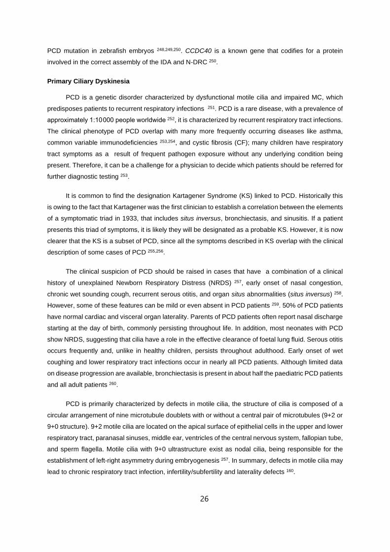
26
PCD mutation in zebrafish embryos 248,249,250. CCDC40 is a known gene that codifies for a protein
involved in the correct assembly of the IDA and N-DRC 250.
Primary Ciliary Dyskinesia
PCD is a genetic disorder characterized by dysfunctional motile cilia and impaired MC, which
predisposes patients to recurrent respiratory infections 251. PCD is a rare disease, with a prevalence of
approximately 1:10 000 people worldwide 252, it is characterized by recurrent respiratory tract infections.
The clinical phenotype of PCD overlap with many more frequently occurring diseases like asthma,
common variable immunodeficiencies 253,254, and cystic fibrosis (CF); many children have respiratory
tract symptoms as a result of frequent pathogen exposure without any underlying condition being
present. Therefore, it can be a challenge for a physician to decide which patients should be referred for
further diagnostic testing 253.
It is common to find the designation Kartagener Syndrome (KS) linked to PCD. Historically this
is owing to the fact that Kartagener was the first clinician to establish a correlation between the elements
of a symptomatic triad in 1933, that includes situs inversus, bronchiectasis, and sinusitis. If a patient
presents this triad of symptoms, it is likely they will be designated as a probable KS. However, it is now
clearer that the KS is a subset of PCD, since all the symptoms described in KS overlap with the clinical
description of some cases of PCD 255,256.
The clinical suspicion of PCD should be raised in cases that have a combination of a clinical
history of unexplained Newborn Respiratory Distress (NRDS) 257, early onset of nasal congestion,
chronic wet sounding cough, recurrent serous otitis, and organ situs abnormalities (situs inversus) 258.
However, some of these features can be mild or even absent in PCD patients 259. 50% of PCD patients
have normal cardiac and visceral organ laterality. Parents of PCD patients often report nasal discharge
starting at the day of birth, commonly persisting throughout life. In addition, most neonates with PCD
show NRDS, suggesting that cilia have a role in the effective clearance of foetal lung fluid. Serous otitis
occurs frequently and, unlike in healthy children, persists throughout adulthood. Early onset of wet
coughing and lower respiratory tract infections occur in nearly all PCD patients. Although limited data
on disease progression are available, bronchiectasis is present in about half the paediatric PCD patients
and all adult patients 260.
PCD is primarily characterized by defects in motile cilia, the structure of cilia is composed of a
circular arrangement of nine microtubule doublets with or without a central pair of microtubules (9+2 or
9+0 structure). 9+2 motile cilia are located on the apical surface of epithelial cells in the upper and lower
respiratory tract, paranasal sinuses, middle ear, ventricles of the central nervous system, fallopian tube,
and sperm flagella. Motile cilia with 9+0 ultrastructure exist as nodal cilia, being responsible for the
establishment of left-right asymmetry during embryogenesis 257. In summary, defects in motile cilia may
lead to chronic respiratory tract infection, infertility/subfertility and laterality defects 160.

27
The major organs of the respiratory system function primarily to provide oxygen to body tissues
for cellular respiration, remove the waste product carbon dioxide, and help to maintain acid-base
balance 261. Portions of the respiratory system are also used for non-vital functions, such as sensing
odours, speech production, and for straining, such as during childbirth or coughing. Functionally, the
respiratory system can be divided into a conducting zone and a respiratory zone. The conducting zone
of the respiratory system includes the organs and structures not directly involved in gas exchange. The
gas exchange occurs in the respiratory zone 261.
Focusing on the conducting zone, the major entrance and exit for the respiratory system is
through the nose (see nasal cavity/vestibule in Figure 5)
Figure 5. Anatomy of the respiratory conducting zone, adapted from Encyclopaedia Britannica © (2012) 262 263
The conchae, meatuses, and paranasal sinuses (maxillary and ethmoid sinus not shown in
Figure 5) are lined by respiratory epithelium composed of pseudostratified ciliated columnar epithelium
(Figure 6). The epithelium contains goblet cells, one of the specialized columnar epithelial cells that
produce mucus to trap debris. Other rarer cells found in the epithelium include ionocytes,
neuroendocrine and tuft cells 94. Cilia of the respiratory epithelium help remove the mucus and debris
from the nasal cavity with a constant beating motion, sweeping materials towards the oropharynx;
particles deposited on this film are eventually swallowed or expectorated, through MC 161,162. Capillaries
located just beneath the nasal epithelium warm the air by convection. Serous and mucus-producing
cells also secrete lysozyme and proteins called defensins, which have antibacterial properties. Immune
cells that patrol the connective tissue deep to the respiratory epithelium provide additional protection.
264,261

28
Figure 6. Haematoxylin and Eosin (H&E) of the trachea epithelium scale bar 10 µm
Inhaled particles are deposited largely according to their size. Thus, particles over 10 µm are
deposited largely in the turbulent air flow of the nose and upper airways; particles of 3-10 µm lodge in
the trachea and bronchi by impaction. Ineffective clearance of the particles from these sites is believed
to be crucial to the pathogenesis of respiratory infections 264.
Epithelial multiciliated cells bear approximately 200 motile cilia 25 . The beat pattern of 9+2 motile
cilia occurs in a waveform that includes a forward effective stroke followed by a return stroke 265. The
direction of stroke is a function of the directional orientation of the central microtubules. In addition to
moving in synchrony, individual cilia in normal healthy cells are very plastic and move fluidly, sometimes
deforming briefly upon encountering resistance and/or particles being transported over the mucosal
surface. Cilia are embedded in a watery periciliary fluid of low viscosity, which facilitates the rapid beat
cycle to move the more viscous overlying layer of mucus. Ciliary beat frequency ranges from
approximately 8-20 Hz under normal conditions, however exposure to irritants may increase the ciliary
beat frequency 257. It is also know that hyperactive cilia beat frequency can be related with specific
genetic defects like mutations such as in the DNAH11 gene 266. Overall, PCD causes an impaired MC
due to defective axonemal ultra-structure of the respiratory cilia and/or by oligocilia or reduced
generation of motile cilia 267,160,158.
As mentioned previously, not only respiratory cilia are affected, 50% of PCD patients also have
defects in visceral organ laterality, reflecting dysfunction of embryological nodal cilia. These cilia are
present during embryonic development in a transient monociliated organ called the ventral node and
are required for patterning the left-right axis in most vertebrates 268,42 .

29
Ultrastructure of motile cilia in PCD
The TEM analysis of cilia with the intention to study respiratory syndromes started with Afzelius,
when he first described in 1976 that male patients with infertility presented also respiratory distress 258.
Afzelius studied sperm flagella and cilia sampled from the nose of his male patients. He discovered that
the axonemal profile was similar in both, in some specific cases, he observed immotile sperm, showing
absence of the dynein motors. Afzelius, named this phenotype as immotile cilia disease 258,269. It was
only later that the complexity of this immotile cilia disease became apparent. Cilia were found to not
always be completely immotile when examined by light microscopy (LM), residual movement referred
to as dyskinetic movement was described that leads to impaired MC in the respiratory tract 270–273. A
new span of defects was unlocked, not only the initial dynein defects leading to immotility, described by
Afzelius in 1976 258, but a range of abnormalities from very mild disarrangements to a complete
transformation of the ciliary axoneme configuration. These finding lead to the designation of the disease
being changed to ciliary dyskinesia, due to the dyskinetic nature of the cilia in these patients and the
range of ultrastructural defects identified by TEM 274.
Advancements in LM techniques, leading to better understanding of PCD, showed healthy (non-
PCD) individuals could show signs of dyskinetic movement of cilia, and impaired MC. This has been
found to be a result of infections (the common cold for instance), that can lead to some ciliary
ultrastructural abnormalities when viewed by TEM 275. Due to existence of secondary factors causing
dyskinesia, the disease was renamed in the seventies to Primary Ciliary Dyskinesia.
TEM allows the ultrastructure of ciliary axonemes to be visualized in detail 3,267. Ultra-thin sections
are prepared for TEM from fixed cells or tissues using standard methods 276. Sections are examined
at magnification to visualize the ultrastructural features (Figure 7) 267, such as the accessory axonemal
components including the ODA and IDA, RS, CP and the N-DRC. IDA morphology is heterogeneous in
composition and repeats every 96 nm along the axoneme. ODA have a more homogeneous distribution
in the axoneme, with 24 nm periodicity 129. The most common ultrastructural defects are: ODA-defects
(25-50%) and combined IDA- and ODA-defects (25-50%), IDA-defects associated with microtubular
disorganization occur in 15% of PCD 277. Isolated IDA-defects as a cause of PCD are controversial
because, so far, no biallelic mutation has been clearly identified in IDA proteins, however, DNAH1 is a
potential candidate 278. IDA are difficult to identify due to their heterogenic structural profile of individual
IDA along the 96 nm repeats of the ciliary axoneme 267,267,279. CP defects occur less frequently (5-15%)
and are associated with a mixture of both normal and abnormal cilia. TEM of cell cultures showed CP
complex defects in only ~20% of the cilia so adequate numbers of cilia need to be viewed in longitudinal
and transversal section 267,267,280.
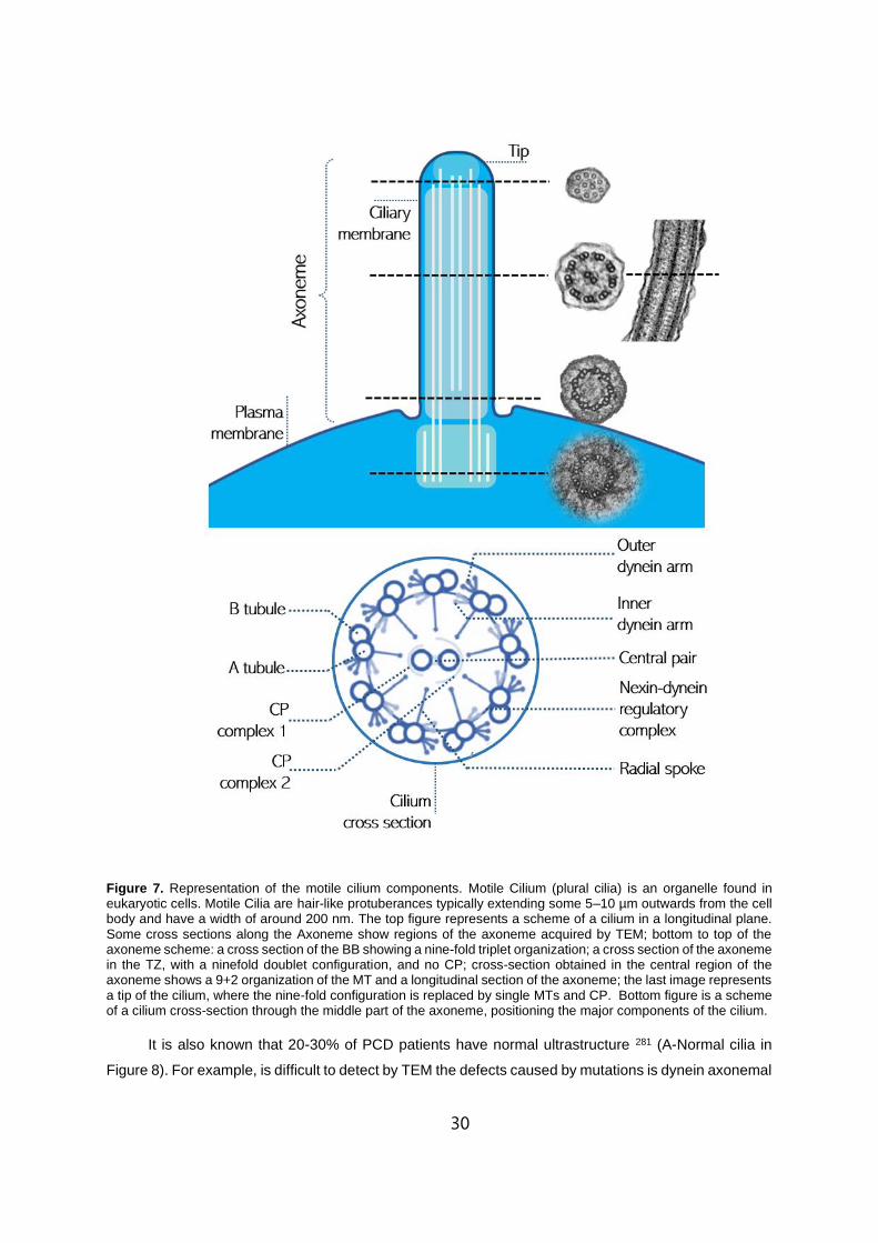
30
Figure 7. Representation of the motile cilium components. Motile Cilium (plural cilia) is an organelle found in eukaryotic cells. Motile Cilia are hair-like protuberances typically extending some 5–10 µm outwards from the cell body and have a width of around 200 nm. The top figure represents a scheme of a cilium in a longitudinal plane. Some cross sections along the Axoneme show regions of the axoneme acquired by TEM; bottom to top of the axoneme scheme: a cross section of the BB showing a nine-fold triplet organization; a cross section of the axoneme in the TZ, with a ninefold doublet configuration, and no CP; cross-section obtained in the central region of the axoneme shows a 9+2 organization of the MT and a longitudinal section of the axoneme; the last image represents a tip of the cilium, where the nine-fold configuration is replaced by single MTs and CP. Bottom figure is a scheme of a cilium cross-section through the middle part of the axoneme, positioning the major components of the cilium.
It is also known that 20-30% of PCD patients have normal ultrastructure 281 (A-Normal cilia in
Figure 8). For example, is difficult to detect by TEM the defects caused by mutations is dynein axonemal

31
heavy chains (such as DNAH11) 266 281, CP components (HYDIN or SPEF2) 282, ciliary biogenesis
defects (MCIDAS or CCNO) 283 and some defects of nexin link components (DRC1 for instance) 284.
Nonetheless, some genes are limited to a specific region of the axoneme (as is DNAH9) 285. Therefore,
normal ultrastructure cannot rule out PCD 260. Consequentially, the PCD diagnostic pathway need to
take into account this limitation and consider all other diagnostic evidence before making a diagnosis
251.
Figure 8. Cross-sections through the cilium axoneme of normal and abnormal cilia. (A) Normal cilium 9+2 ultrastructure and examples of a HYDIN and a DNAH11 mutation, showing apparent normal ultrastructure. (B) Left to right: ODA defect, ODA and IDA, IDA defect combined with MTD disorganization, CP defects and transposition defect (microtubular doublet resembles a central pair, when it translocated to the center). (C) Examples of secondary ciliary defects, left to right: duplicated CP, a cilium cross section showing lacking of MTD and presence of extra MT, one cross section showing extra tubules in the periphery of the cilium, one cilium with single tubules instead of MTD, and an example of a compound cilia (where a group of MT found by the same membrane) scale bars: 100 nm.
More recently, and to fill a gap of lack of standardization in analysing ciliary brushings by electron
microscopy, a consensus guideline was developed by PCD electron microscopy experts representing
18 centres in 14 countries 286. An initial meeting and discussion were followed using the Delphi method
to reach a consensus. The agreed guideline was tested and enhanced through exchange of samples
and electron micrographs between the 18 diagnostic centres.
The final guideline provides the following: a) Agreed terminology and a definition of class 1
defects which are diagnostic for PCD; b) Identifies class 2 defects which can indicate a diagnosis of
PCD in combination with other supporting evidence; c) Describes features which should be included in

32
a ciliary ultrastructure report to assist multidisciplinary diagnosis of PCD d) Defines adequacy of a
diagnostic sample 286.
Genetics of PCD
PCD is an autosomal recessive (AR) disease, however in rare cases other modes of inheritance
have been described (X-linked or autosomal dominance). Currently, more than 40 genes have been
identified, explaining approximately 70% of cases, where there seems to be a clear association between
genetic defects, ciliary ultrastructure and motion defects 260.
AR disorders are only clinically apparent when a patient has a homozygous mutation. The
general inheritance pattern might have the following characteristics i) patients parents appear to be
clinically normal; ii) a vertical transmission is not expected, the transmission pattern appears to be
horizontal; iii) male and female are affected equally iv) when both parents are heterozygous carriers of
the mutation, 25% of their offspring will be likely to be affected, 50% with be carriers and 25% will be
completely normal (Figure 9) 287.
Figure 9. Chart showing two families (squares are males, and circles females) where the gene of an AR disorder is mutated either homozygous (shadowed shapes) or possibly heterozygous (blank). Focusing on individual 6 that is a carrier of the affected gene (inherited from the parents), when he matches with a homozygous individual, their offspring have 50% likelihood of being homozygous. Individual 5 will always pass on an affected copy of the gene. Individuals eight and nine are carriers since their offspring shows a classical AR pattern, in which 25% are homozygous, 50% heterozygous, and 25% have no mutation. Individuals 10 and 11 are homozygous, and will produce affected offspring since they will both provide affected copies of the gene.287
Cystic fibrosis (CF) is also an AR disorder but only a gene is involved, the CFTR which was
discovered in 1989. CFTR – Cystic fibrosis transmembrane conductance regulator - encodes the
transmembrane conductance chloride channel, and, more than 2000 CFTR mutations have been
identified and described 288.
Compared to CF, PCD is a genetically heterogeneous disease. The first PCD associated gene,
DNAH5, was only discovered in the year 2000, and pathogenic mutations cause defects in the ODA 289.
Most of the disease-causing mutations identified to date involve the absence or dysfunction of
ciliary axonemal dyneins, but it is well-known, dozens of genes might be implicated in PCD caused by

33
mutations that are not linked to dynein motor proteins. Below is the most up to date list of genes that
are known to be related to PCD based on the work of M. Legendre, et al. (2020) 94. PCD-causing
mutations have been identified on genes that include DNAH5 (dynein axonemal heavy chain 5),
DNAH11 (dynein axonemal heavy chain 11), DNAI1 (dynein axonemal intermediate chain 1), DNAI2
(dynein axonemal intermediate chain 2), CCDC114 (coiled-coil domain containing protein 114), ARMC4
(armadillo repeat containing 4, or outer dynein arm dicking complex unit 2 – ODAD2), CCDC151 (coiled-
coil domain containing protein 151), TTC25 (tetratricopeptide repeat domain 25 or outer dynein arm
docking complex subunit 4 – ODAD4 ), DNAH9 (dynein axonemal heavy chain 9), TXNDC3 (thioredoxin
domain-containing protein 3), DNAL1 (dynein axonemal light chain 1) that are known to be linked to
absence or truncation of the ODA and are expressed in the ciliary axoneme 240,281,296,297,285,289–295.
CCDC103 (coiled-coil domain containing protein 103), ZMYND10 (zinc finger mynd-type containing 10),
C21orf59 (chromosome 21 open reading frame 59 or cilia and flagella associated protein 298), DNAAF5
(dynein axonemal assembly factor 5), LRRC6 (leucine rich repeat containing 6), RPGR (retinitis
pigmentosa gtpase regulator), SPAG1 (sperm associated antigen 1), PIH1D3 (PIH1 domain-containing
protein 3, also known as dynein assembly factor 6), DYX1C1 (dyslexia susceptibility 1 candidate 1),
DNAAF1 (dynein axonemal assembly factor 1), DNAAF2 (dynein axonemal assembly factor 2),
DNAAF3 (dynein axonemal assembly factor 3), C11orf70 (chromosome 11 open reading frame 70), is
the group of published genes connected to mutations in both the ODA and IDA. The expressed protein
is typically localised in the cytoplasm where they are involved in the cilia biogenesis, except for C11orf70
that is also found in the tip of the cilium and was found to be dependant of continuous IFT trafficking
246,298–301,302–309. Mutation in the inner-arm alone are not frequent but DNAH1 (dynein axonemal heavy
chain 1) has been described to be linked with the IDA in the ciliary axoneme 278 . Cases where there is
an absence of the inner dynein arm together with axonemal disorganization have been shown to result
from mutations in CCDC39 (coiled-coil domain containing protein 39), CCDC40 (coiled-coil domain
containing protein 40) and TTC12 (tetratricopeptide repeat domain) 94. CCDC39 and CCDC40 can be
found in the axoneme 310 311. However, TTC12, a cytoplasmic protein, has milder microtubular
disorganization and absent IDA and ODA observed in patient sperm 312 94. It is also known that DRC1
(dynein regulatory complex 1), DRC2 (dynein regulatory complex) and GAS8 (growth arrest specific 8)
are involved in the assembly and regulation of the N-DRC 284,313,314, and mutations within the genes can
lead to mis-aligned outer doublets in some cilia cross sections. Another large group of genes involved
in PCD defects, are those that when mutated show defects in the CP and RS, these include, RSPH1
(radial spoke head component 1), RSPH3 (radial spoke head component 3), RSPH9 (radial spoke head
component 9), RSPH4A (radial spoke head component 4 homolog A), HYDIN (axonemal central pair
apparatus protein), SPEF2 (sperm flagellar 2), STK36 (serine/threonine kinase 36), NME5 (non-
metastatic cells 5), DNAJB13 (DnaJ heat shock protein family member B13), and CFAP221 (cilia and
flagella associated protein 221). So far, cytoplasmic CFAP221 has shown no ultrastructural defects in
Transmission Electron Microscopy (TEM) sections, albeit, functional studies, reveal motility defects 315.
Regarding HYDIN and SPEF2, the CP complex defects might be missed during the TEM analysis.
There are genes that are not specifically linked with axonemal abnormalities but rather associated to

34
multiciliogenesis defects, those are CCNO, MCIDAS and FOXJ1 100,283,316,28. Patients with symptoms
consistent with PCD and marked reduction in the number of motile cilia in the airway cells have been
shown to have mutation in one of these two genes, CCNO and MCIDAS. Both genes code for proteins
required in centriole assembly and replication 283,316. Clinically these patients are unlikely to have situs
inversus (Constant et al. 2021 accepted for publication) because nodal cilia biogenesis in monociliated
cells do not require CCNO or MCIDAS 283. Thanks to cilia research using several animal models and in
vitro experiments, it has been possible to identify genes that are linked to PCD but with normal cilia
ultrastructure; namely OFD1 (oral-facial-digital syndrome 1 protein), NEK10 (never in mitosis A-related
kinase 10) associated with short cilia, CFAP57 (cilia and flagella associated protein 57) and LRRC56
(leucine rich repeat containing 56) known to cause motility defects in organisms like Chlamydomonas
and Trypanosoma and GAS2L2 (growth arrest specific 2 like 2) where cilia orientation seem to be
affected 317,318,319–321. Even with the exhaustive PCD genetics roadmap described here, mutations in
PCD genes account for only 70% of all known PCD cases 94,322.
Mutations in the ODA dynein axonemal heavy chain DNAH5, are the most common and underlie
28% of the cases in Europe 289. The second most common mutated gene is the DNAI1, accounting for
11% of the cases 289. Mutations in DNAH6 (dynein axonemal heavy chain 6) were described for the first
time in 2015 using zebrafish, describing a phenotype of heterotaxy and motility impairment, possibly
due to microtubular abnormalities and central pair defects in ~30% of the cilia observed in Dnah6
knocked down samples, the abundance of ODA was not affected 323. The authors provide evidence of
trans-heterozygous interaction between DNAH6 and other genes (in patients showing ultrastructural
abnormalities in the ODA), an event that can potentially cause a phenotype of PCD (which means that
this gene mutated in only one allele, when in combination with other mono-allelic mutation in different
PCD gene may case a phenotype) 323. Generally, mutations are associated with defects in ciliary ultra-
morphology, but there are other genes known to harbour PCD mutations, including DNAH11, in which
patients seem to have normal ciliary ultrastructure by normal TEM and cilia motility defects are the only
diagnostic features 324. These cases highlight the need for complex and multidisciplinary diagnostic
methods such as high-speed video microscopy (HSVM), genetics, immunofluorescence (IF), and
transmission electron tomography (ET) 325.
Genotype-Phenotype
The correlation between patient gene mutations and clinical manifestations are of great interest
in the field of PCD research and diagnostic testing 326. Groups of patients were analysed using standard
statistical analysis to compare the clinical characterization and the results from the laboratory tests and
radiology exams of patients with genetically diagnosed PCD, and subsequent correlation between
them. This approach led to the main source of knowledge about certain genotype-phenotype
relationships. For example, situs inversus has been found to be less likely in RSPH mutations and
multiciliogenesis impairment, since nodal cilia are not affected 327,328. Patients with mutation in the
DNAH11 gene show preserved lung function, seen when lung clearing 281, on the other hand , DNAH5

35
patients show the largest spectrum of phenotypes 289. The reasons behind the range of DNAH5
phenotypes is unclear but could be due to a large number of different mutations within this large gene,
so far, at least 100 different pathogenic mutation have been reported 289.
More recently Shoemark et al. (2021) described a tool called Topological Data Analysis (TDA)
clustering 326, to examine genotype-phenotype correlations in almost 400 individuals from three
European countries. TDA software by symphony AyasdiAI platform (www.ayasdi.com, v 2.0, Ayasdi
Inc., Menlo Park CA) was used to develop the topological models for the genotype-phenotype
assessment. Models were generated using automated analysis options. Highly complex data were
summarized and compressed into smaller representation of their variability using Locally Linear
embedding (LLE non-linear reduction technique). Using this methodology, the authors were able to
identify several previously unknown genotype-phenotype relationships. The benefit of TDA cluster
analysis for PCD is that many parameters (phenotypic data used was body mass index (BMI), lung
function tests, wet cough, rhinitis, glue ear, cardiac situs, congenital heart disease, nasal nitric oxide
(nNO), cilia beat pattern (CBP) and transmission electron microscopy (TEM)) can be correlated, and
therefore, having access to the genetic information, it was possible to explore the underlying patterns
in complex datasets by generating clusters of individuals with similar features. A further advantage of
TDA clustering is that it allows the observation and analysis of small clusters of interest in large
datasets. The most recent genotype-phenotype PCD data suggests that RS and N-DRC/molecular ruler
(CCDCs 39/40 and DRC), have worst prognosis regarding lung function, likelihood for ear glue, less
rhinitis and no situs inversus. Dynein heavy chain proteins, DNAH5 and DNAH11, show preserved lung
function and no major NRDS. This confirms an already known phenotype typical of DNAH11, whereas
DNAH5 appeared phenotypically diverse regarding lung function, with no clear cluster observed, this
also corroborates what was previously described by Hornef et al. 289,326,329.
Understanding the genotype-phenotype association, will ultimately improve the clinical
management of patients, identifying those that might need a more aggressive or personalized treatment
due to their underlying genetics. Patients with mutation in CCDC39 and CCDC40 might benefit from
more intense and targeted therapies 330,331. The link found using the topological analysis between a
certain genetic defect, ultrastructural and CBP results, indicated that TEM and HSVM tests play an
important role in the classification of novel gene variants and variants of uncertain significance (VUS).
These tests can also direct the genetic testing more towards a specific sub-set of genes 326.
Diagnosis of PCD
The first step in diagnosing PCD is the evaluation for clinical features of PCD. To determine the
PICADAR (primary ciliary dyskinesia rule), a questionnaire with seven simple questions are answered
and used to predict the likelihood of a patient to have PCD. It is used in many PCD specialist centres

36
as an initial evaluation to determine if diagnostic testing is required. PICADAR was developed and
validated using cohorts of known PCD patients and can be used in any patients with chronic respiratory
symptoms starting in early childhood. The total score is calculated and the individual probability of a
positive PCD diagnosis can be estimated. Patients with a score >10 have >90% probability of testing
positive for PCD, and those scoring <5 are more unlikely to test PCD-positive 332.
Figure 10. Example of a PICADAR scoring chart. PICADAR is a scoring system that uses seven questions to predict the likelihood of having PCD. It can be used in any patients with chronic respiratory symptoms starting in early childhood. The right-hand column show scores, and any above 5 indicate likelihood of the patient being diagnosed with PCD 332
Diagnosis of PCD is complex and patient clinical features play a major role in the initial triage. As
mentioned above, PICADAR score is a useful tool, however its only trustworthy if the patient shows
some or all of the symptoms like situs abnormalities, unexplained NRDS, persistent rhinitis from birth,
middle ear disease with complications and congenital heart disease 5. However, it still prioritizes the
presence of a wet cough, which is a poor indicator due to its low discriminative value as wet cough is
highly prevalent in both PCD positive and negative. The best approach for PCD referral, is to take into
consideration many factors of the patients medical history 5.
After referral for PCD, a patient will have assessment of their ciliary function by HSVM and nNO.
In the event of an abnormal or equivocal result the samples must be sent for TEM, IF, and cell culture.
Genetic testing is recommended either to confirm a positive diagnostic outcome (recommended in case
of ‘highly likely PCD’), or to clarify in case of a strong clinical history presenting with normal or unclear
test results 5.

37
Diagnostic tests
When patients are referred for diagnostic testing, PCD is one of several different possible
diagnoses. Therefore, investigations to exclude other more common conditions have often been
managed during clinical consultation by a referral centre. To exclude CF, a sweat test is administered,
whereas exclusion of gastro-oesophageal reflux, amongst others, requires immunological investigation.
These tests are complex and required specialised training and equipment; therefore, dedicated PCD
reference centres are required. The range of PCD phenotypes can be confounded by the presence of
secondary defects caused by airway infections, further emphasising the requirement for specialist
centres 333.
Listed below are the different diagnostic tests and the guidelines for the diagnosis of PCD
developed through a collaboration of multiple centres worldwide.
Screening tests
The most known screening tests are the saccharin test and the nNO measurement test.
Saccharin test is an outdated technique to test the MC, it consisted of placing saccharin approximately
1 mm behind the anterior end of the inferior turbinate. In the presence of normal mucociliary action, the
saccharin will be swept backwards to the nasopharynx and a sweet taste perceived. Failure to detected
sweetness within 10-20 minutes may be the sign of delayed MC 334,333. Nevertheless, the test has
limitations in children, if the child sniffs during the test saccharin may be tasted even in the absence of
MC, and the test does not differentiate between primary and secondary dyskinesia 333.
nNO measurement in the nasal airway, shows consistent high reliability with approximately 95%
sensitivity and specificity (when measure in a chemiluminescence analyser using velum closure
technique), as shown by two Independently conducted systematic reviews 335,336, the accuracy of the
test might differ depending on the type of analyser used, age of the patient and sampling method 337.
NO is an important cell-signalling molecule that is implicated in a range of important functions in the
organism, such as neurotransmission and neuromediation, relaxation of muscles, vasodilatation and
host defence actions 338. NO is present in the exhaled air of humans, and when is measured in the nose
at a rate of 250 ml/min during breath holds, a nNO level of >250 ppb is considered normal, nevertheless
there is no consensus on appropriate thresholds determined 340 . nNO is known to be abnormally low
in PCD patients when compared to controls, for unknown reasons 341. The test has limitation in young
children, asunder 6-year olds can be uncooperative, or struggle to breath hold. This stresses the need
for standardization of protocols and thresholds for tidal breathing, including for cases where a
differential diagnosis given between PCD and another respiratory disease (bronchiectasis, CF and even
rhinosinusitis) that can lead to low NO values 333.

38
Ciliary Beating pattern and Frequency
Defects in cilia motility associated with PCD can be screened ex vivo by assessment of the
activity of ciliated epithelial cells, biopsied from the nose or the bronchus 258. To estimate CBP and cilia
beat frequency (CBF) it is essential that cilia are kept in an enriched medium at optimal conditions
(controlled temperature). HSVM is typically performed using a microscope equipped with a heated
stage at 37°C, to mimic the human body temperature, but it has been showed that is not mandatory as
long as a record of the diagnostic temperature is kept 342. Cilia beating can be assessed from three
planes: sideways profile; beating towards the observer and from the top 343. To record beating cilia, a
digital high-speed camera can be adapted to a LM, to acquire a rate of around 400 frames per second
(fps). The camera allows the video sequences to be recorded and played back at a reduced frame rate
(slow motion 30-60 fps 340) or even frame by frame. The precise movement of the cilium may be
observed and evaluated during this beat cycle 343. This procedure grant access to the sample CBP and
CBF 340. The clips must be acquired from several patches of cells (10), to have a reliable test. It is
advised that each PCD service sets its own normal CBF ranges based on the population and HSVM
setup 344.
CBF and CBP offers the direct readout of the ciliary function. Previously, CBF was the primary
measure of ciliary motility dysfunction, but now that it is considered a limitative analysis, giving only the
perception of high or low CBFs, the isolated study of CBF is subjective and presents low specificity and
sensitivity, leading to a high number of false negatives 340,345. The limitations of measuring the CBF is
resolved by analysing CBP that has higher sensitivity in diagnosing PCD 346,277. Nevertheless, the
evaluation is still mostly done by visual assessment with no consensual metrics. Mostly due to the need
of highly skilled staff, there are services that are still not using HSVM as a diagnosis test 347.
Several centres are already using computer analysis for the assessment of cilia function 340. With
the aim of greater standardization, Sampaio et al. (2021) developed software created to evaluate CBF
in a semiautomated way, called CiliarMove 342. This tool allowed the authors to determine the CBF in a
cohort of Portuguese PCD patients within a genetic study and determine the normal CBF range for a
healthy volunteer target population. The authors assume the sampling may not represent the general
population, but the results go in line with what has been described previously 342.
HSVM is yet not sufficiently standardized to exclude PCD, and it is generically agreed that the
test needs to be done by experienced staff as part of the diagnosis work-out 340.

39
Primary Cell Culture
Primary airway cell cultures are developed from airway cells and can be obtained from biopsies,
brushings or explanted lungs 348. Airway cultures date back to the 1980s, where several growth
conditions were tested for bronchial cells 349. Since the introduction of epithelial cell cultures, improved
cell culture techniques have boosted the use of cultured airway cells for developing disease relevant
models, drug testing and inhalation toxicology 350.
Epithelial cells are usually dissociated from extracellular matrix by protease treatment and
unwanted cell type growth (fibroblasts) is inhibited by addition of selective medium. To allow cell
expansion, essential growth factors are added, as well as antibiotics that will prevent the microbial
overgrowth. In previous procedures, cells would be kept submerged in medium, attached to an
extracellular matrix (likely collagen) in a plastic dish. However, primary airway cell cultures that are kept
submerged result in loss of cell differentiation (into secretory and ciliated cells) 350. Primary airway cell
cultures became more physiologically relevant with the introduction of growing cells at the air-liquid
interface (ALI) 348. ALI greatly improves differentiation of the epithelial cells, matching an in vivo situation
351. Cells growing in ALI evolve to a pseudostratified epithelium consisting of goblet cells, ciliated cells
and basal cells 352. ALI is a cell culture method where epithelial cells are grown from a nasal biopsy and
the apical surface exposed to the air to simulate the human airway. It is frequent to start by seeding the
cells onto the permeable membrane of the insert, and initially the cells are fed both from the apical side
and the basal side. Once reaching confluence, the cells are fed only via the basal chamber, leaving the
apical side exposed. In the particular case of ciliated cells, the exposed apical region is advantageous
for reciliation 353. Ciliary movement and production of mucus can be assessed under a LM for control
and analysis.
This method is adequate for ciliated epithelium due to their ability to grow cilia. These ciliated
cells can be kept in ALI culture for many months and diagnostically assessed if any uncertainty is raised
during the examination of the primary sample.
In some European clinics, specific ALI cultures of patients’ nasal epithelia are performed to
confirm the first diagnosis 354. This is preferable for ambiguous cases where infection or inflammation
are present, or the sampling may damage the epithelium. It is assumed that in culture there will be no
secondary defects caused by infection or inflammation, and therefore only defects link to gene
mutations remain 265,340.
Immunofluorescence
Many PCD diagnostic centres now use IF antibody labelling to help determine the presence or
absence of key ciliary proteins in patient samples. This antibodies used are raised against axonemal
motor proteins or assembly factors.340,355.

40
IF has been paramount in the diagnosis and understanding of PCD in many ways, for instance
in the distribution of proteins like DNAH11 and DNAH9, which expression is not transversal along the
axoneme, but found in specific regions. In the case of DNAH11 its localisation is to the proximal region
of the cilium, which when mutated, by HSVM might be seen as reduced bending capacity of the
axoneme or static in patches, and classic hyperkinetic CBF (CBF > 11 Hertz (Hz)) 266. In contrast
DNAH9 has been showed by IF to be restricted to the apical region of the cilium 356. In regard to the
CBP in cases with DNAH9 loss-of-function, stiffness towards the tip is the most commonly observed
phenotype and varies depending on the severity of the mutation 285.
IF is a straight forward method to detect PCD proteins, however, as a diagnostic test it has limited
sensitivity for PCD due to the lacking of validated commercial antibodies, and low imaging resolution
356. Super resolution confocal microscopy is considered a future improvement for PCD diagnosis but is
not available to most PCD centres. Zhen Liu et al. (2020), described a tool using a super resolution
workflow to assist in the diagnosis of PCD 356. They showed in their work that super resolution imaging
is a powerful tool to investigate PCD proteins. For this study they used a panel of 21 antibodies that
recognize ciliary proteins liked to PCD, named by the authors as the PCD proteome. With high-
resolution 3D-SIM (three-dimensional structured illumination microscopy), it was possible to
characterize the distribution of the markers in the PCD proteome. PIH1D3, a recently described X-linked
PCD gene known to be a putative axonemal dynein assembly factor, showed punctate labelling found
in the cell body 304. They also presented a machine learning tool using IF 3D-SIM, based on a
quantitative analysis, designated as rotational polarity analysis 356. Consists of an automated image
analysis to quantify rotation polarity of BB and basal feet pairs in PCD cells. Through the creation of a
MATLAB (matrix laboratory) script, the authors could automatically identify BB and basal feet, and
calculate orientation of each pair in comparisson to the image axis 356. The authors consider IF 3D-SIM
workflow a promising tool for translational into clinical diagnostic that can be improved by the
implementation of high-throughput staining and data collection methods. Rotational polarity can be seen
as a quantitative alternative in cases that cannot be diagnosed by other methods 356
Currently, surveys are also being done to reach a consensus, not only in the analysis of IF but
also guidelines for the preparation of samples and which antibodies are more adequate to each
situation. This emphasises the important of exchange of knowledge in improving the diagnostic pathway
for PCD 357,358. Nevertheless, according to the most recent task force guideline (2017) 340, it is still
unfitted to determine the accuracy of IF testing for diagnosis. However, task force experts agree that IF
can be useful in clinical settings. IF is cheaper and easier than other diagnosis tests, and presents as
a potential test for resource-limiting centres 340.
Genetic testing
In PCD, to establish a genetic diagnosis, non-ambiguous biallelic mutations and hemizygous
mutation in X-linked genes, should be identified 340. Most of the reported mutations in PCD genes are:

41
nonsense, or a stop mutation, changes in the DNA that causes the protein to terminate leading to a
non-functional protein; frameshift mutation, in which there is an insertion or deletion of a nucleotide in
a way that disrupts the gene reading frame; or splice mutations, where an insertion or deletion of a
nucleotide occurs in a specific site at which splicing takes place. On the other hand, missense mutations
(incorporation of the wrong amino acid into a protein) are a minority 340. Most PCD gene mutations are
private, meaning they are found in a specific family or population, but, there is knowledge of founder
mutations (characteristic of groups geographically or culturally isolated, in which one ancestor was the
carrier of the altered gene and passed it on) like DNAH5 289 and DNAI1 359, and mutational hotspots,
such as CCNO 360. Mutation ranking in genetics should follow international guidelines: (1) pathogenic,
(2) likely pathogenic, (3) uncertain significance, (4) likely benign, or (5) benign 361.
It is expected that all DNA sequencing technologies can be used to test patients with confirmed
or high suspicion of PCD, those technologies are: i) sanger sequencing of all coding regions and
flanking intronic regions, ideally targeting to the genes responsible for a specific ultrastructural defect
(point mutations); ii) targeted Next Generation Sequencing (NGS) of all coding regions and flanking
intronic regions. Like sanger, this technology will detect point mutations; iii) whole exome sequencing
has usually a lower coverage and depth than targeted NGS (usually not sufficient to detect deletions
or duplications of more than one exon); iv) targeted copy number analysis as already been reported as
a semi-quantitative qPCR (quantitative polymerase chain reaction) to characterize large insert/deletions
362; v) whole genome copy number analysis (single nucleotide polymorphism - SNP array) suitable to
detect large rearrangements but shows low sensitivity for intragenic deletions; vi) transcript analysis on
airway epithelial cells from the patient, technique used in patients that showed a single heterozygous
mutation in a relevant gene. This technology can reveal deep intronic mutations that were missed by
any of the above-mentioned techniques 340.
Albeit the high number of technologies available to search for a specific PCD mutation, as
mentioned before, it is suggested that genetic testing identifies the gene in only approximately 70% of
the PCD cases 326,330,362. It is likely that this number increases as more genes and pathogenic variants
are identified 340. Nevertheless, it is still essential to incorporate the genetic results together with the
other PCD testing results.
Transmission Electron Microscopy (TEM)
TEM is one of most important tests to the diagnose PCD by determining ciliary ultrastructural
defects. However, standardization between experts and centres has been hard to reach. According to
Shapiro et al. (Department of Paediatrics, Montreal Children’s Hospital, Quebec, Canada) at least 20–
50 clear ciliary cross-sections are required for diagnostic TEM, and abnormalities should be consistently
demonstrated on cross sectional images from multiple different cilia to be considered disease causing
347. In contrast , PCD clinics in Southampton General Hospital and on the Royal Brompton Hospital
(RBH) (London), 50-100 ciliary cross-sections are analysed for each sample 286 . In Belgium, in the

42
department of paediatric pulmonology, University Hospital Leuven, Boon et al. also studied, per sample
at least 50 transversal cross-sections 363. Based on a questionnaire I made in 2018 between 3
independent hospitals using TEM for diagnosis of PCD (Oncology Institute – Lisbon; Fernando da
Fonseca Hospital – Amadora/Sintra and S. João Hospital - Oporto), I found that the services were
divided between assessing only up to ten ciliary cross-sections, or at least a thousand cilia before
submitting a valid report (unpublished data). This proves how complex and subjective the analyses of
cilia cross sections are amongst experts. However, the majority of experts work towards standardization
in many areas of PCD, while stressing the complexity of the disease 286.
Some cilia abnormalities are easier to assess than others and so, only a consistent number of
ciliary cross sections will contribute for a confident quantitative analysis. According to the most recent
guidelines (Shoemark et al. 2020 286), during an assessment the specialist should evaluate 50 or more
oriented cross sections of cilia, for an accurate analysis. The aim in TEM, is to push the use of the
nomenclature (class 1 and class 2 defects) suggested in this international consensus and therefore
make a more reliable and transferable analysis 286.
Advanced techniques for the confirmation of a PCD defect
Confidence in diagnosing patients with subtle defects is low, and patients often undergo several
repeat nasal biopsies in attempts to establish a diagnosis 364 Electron tomography (ET) can be used
to identify the structural defects in some of these cases.
In TEM, 3D objects are projected into two-dimensional (2D) images, but in many cases, 2D
projections are insufficient to understand the relationship between 3D structure and the function of
nanoscale objects 365. With ET it is possible to retrieve 3D structural information from a tilt series of 2D
projections 366. Retrieving the missing third dimension is required to determine the functionality of many
structures 365. ET provides a wide range of resolutions taking full advantage of the versatile capabilities
of the TEM 366. As an example, ET and genetics used in combination have identified the absence of
HYDIN as another cause of PCD 282. ET showed absence of the projection, C2b, associated with the
CP apparatus in patients with a HYDIN defect 282.
Pathogenic DNAH11 mutations, are difficult to assess using conventional TEM, being considered
as having a normal phenotype 266. The incidence of this mutation was previously shown to be around
20% in patients with PCD, without a defined ciliary ultrastructural defect 281. Biallelic mutation of the
gene causes an abnormality in ciliary ultrastructure undetectable by conventional TEM, but with
characteristic abnormal CBP detected by HSVM that includes reduced bending capacity and increased
CBF 266. DNAH11 mutations result in abnormality of the ciliary ultrastructure when visualized by ET.
The defect is specific to the “forearm” of the ODA and is detected at the proximal portion of the cilium
where DNAH11 is located. Advantageously, ET established a structural abnormality where TEM
showed previously normal. (Figure 11) 364.

43
Other methods have been created, such as the PCD detect software, to enhance TEM images
through image averaging and classification to help clarify the absence of ciliary structures, 367.
Figure 11. Location of DNAH11 in the ODA of the microtubular doublet showed by Shoemark et al. using ET 364. Image shows data from volumes from averaged transverse tomograms from the proximal portion of the axoneme of healthy controls ( b) i) ), and DNAH11 mutation patients showing two different mutation types ( b) ii) and iii) ). Red marks the predicted location of DNAH5 and the green triangles were added to the images on the right to indicate where DNAH11 is predicted to be missing. Image on the right ( c) i) ) represent a tomogram from a healthy control as visualized in Chimera 364.
PCD is a complex multi-systemic disease, and to diagnose it, specialist expertise is required
combined with well-established techniques. Therapeutic trials of new drugs and treatments in humans
are complex, so it is important to have good reliable animal models for the initial testing to help establish
the safety and effectiveness of different therapies.
My objectives are as follows; 1) contribute for the standardization of data analysis, appropriate
normative controls, and isolation and analysis techniques to facilitate comparison of results among PCD
diagnostic centres, and results regarding this aim can be found in chapters 2 to 4; and also 2) refine
and adapt EM techniques for a better classification of motile cilia in both human airway and zebrafish
major ciliated organs, which will be described in detail in chapter 5. This will play an important role in
establishing zebrafish as a model for ciliopathies, the same way, zebrafish are used as a model in other
diseases, such as carcinogenesis, infection, immunological diseases and psychological and
behavioural disorders, amongst others 182.

44

45
CHAPTER 2
Ciliary Feature Counter: A program for the Quantitative Assessment of Cilia to
Diagnose Primary Ciliary Dyskinesia
Published Article doi:10.3390/diagnostics10080524
Andreia L. Pinto1, Ranjit K. Rai1, Claire Hogg1,2 and Thomas Burgoyne1,3,* 1 Paediatric Respiratory Medicine, Department of Paediatrics, Royal Brompton & Harefield NHS Trust, London SW3 6NP, UK; [email protected] (A.L.P.); [email protected] (R.K.R.); [email protected] (C.H.) 2 Department of Paediatrics, Imperial College London, London SW3 6LY, UK 3 UCL Institute of Ophthalmology, University College London, London EC1V 9EL, UK * Correspondence: [email protected], Tel: + 44-(0)20-7352-8121
Personal contributions
My contribution to this manuscript was conceptualizing and investigation about the necessity of having
a digital counter for PCD, and also in the preparation, reviewing and editing of the manuscript.
2

46

47
Abstract: Primary ciliary dyskinesia (PCD) is a disorder that affects motile cilia in the airway that are
required for the removal of mucus, debris, and pathogens. It is important to diagnose PCD in early
childhood to preserve lung function. The confirmation of a diagnosis relies on the assessment of ciliary
ultrastructure by transmission electron microscopy (TEM). TEM involves the quantitative assessment
of the ciliary ultrastructure to identify PCD defects as well as abnormalities resulting from infection.
Many specialist diagnostic centres still rely on physical counters to tally results and paper notes to
summarise findings before transferring the results to computer databases/records. To speed up the
diagnostic data collection and increase the protection of patient information, we have developed digital
ciliary feature counters that conform to the PCD reporting international consensus guideline. These
counters can be used on a computer or tablet, and automatically generate notes regarding sample
observations. We show that the digital counters are easy to use and can generate TEM diagnostic
reports that will be useful for many PCD diagnostic centres.
Keywords: cilia; primary ciliary dyskinesia; digital counter.
Introduction
Primary Ciliary Dyskinesia (PCD) is a heterogeneous genetic disorder of the motile cilia. Cilia
are hair-like structures which play an essential role in the airway by moving and clearing mucus, debris,
and pathogens. PCD results in a failure to clear mucus from the airway, leading to chronic lung disease
and rhinosinusitis, as well as impacting on hearing and fertility. It is estimated that the prevalence of
PCD is approximately 1 per 10,000 births, but it is more common in populations where there is
consanguinity 368. A diagnosis of PCD at an early age is vital to implement appropriate treatment to
preserve lung function and prevent lung damage. Both the European and North American diagnostic
guidelines recommend that a diagnosis of PCD should be confirmed by transmission electron
microscopy (TEM) in combination with another diagnostic test 340,369.
PCD can present as a number of ultrastructural defects by TEM (Figure 1). These include the
complete or partial absence of the outer, inner, or both dynein arms, as well as microtubular
disorganisation or a central pair complex defect (Figure 1b). Due to infection, other secondary non-PCD
defects are often present and include compound cilia, extra tubules, and the absence of one of the
microtubules from the central pair complex (Figure 1c). As the dominant diagnostic assessment for
PCD, there are international consensus guidelines for reporting TEM results which involve classifying
PCD defects into two classes, as shown in Figure 1d 286.

48
Figure 1. Normal healthy ciliary ultrastructure as well as common ciliary defects associated with primary ciliary dyskinesia (PCD) and secondary to infection. (A) Electron microscopy image and diagram of a cross section through a healthy respiratory epithelial cilium, highlighting the main ciliary components. (B) Common PCD defects, including the loss of structures or disarrangement of the cilia. The blue arrows indicate absent outer dynein arms, the red arrows indicate absent inner dynein arms, the green arrow indicates a microtubule doublet that has transposed into the centre of the cilium, and the yellow arrow indicates the absence of the central pair microtubules. (C) Some of the ciliary defects associated with unhealthy respiratory epithelium are caused by bacterial or viral infections. The orange arrow indicates an extra microtubule within the cilium, and the purple arrow indicates the

49
loss of a single central pair microtubule. (D) The classification of primary PCD defects based on the international consensus guidelines for reporting TEM results 286.
Fifty or more ciliary cross-sections need to be evaluated to reach a diagnosis for PCD, as
outlined in the international consensus guidelines 286. These need to be well-orientated cross-sections,
and when assessing them it is vital to be aware of the ultrastructural differences at the base and tip
compared to the rest of the cilium (see Figure 2). This involves tallying the assessments of the
ultrastructure of each cilium as well as the dynein arms, which is often done using a physical counter
(see Figure 3) and paper notes, to score patient cilia and make notes about each diagnostic patient
sample. These notes are to be typed up onto a computer, which is time-consuming and poses a patient
information confidentially risk. To speed up the collection of results directly to a computer system and
to follow NHS (UK) and many international guidelines to phase out paper notes, we have developed a
digital PCD ciliary counter. This easy-to-use counter can be used on a computer or tablet and works
well with a touch screen, and it generates notes based on the simple selection of options that describe
the diagnostic sample.
Figure 2. Ultrastructure of the different regions of a respiratory cilium. Respiratory cilia have a “9 + 2” microtubule arrangement (9 microtubule doublets and 2 single microtubules that make up the central pair), as can be seen in the mid region of the cilium. This ultrastructural arrangement runs throughout the cilium apart from at the tip and base where there are different ultrastructures, which include single microtubules and Y-shaped linkers, respectively, and an absence of dynein arms in both regions. When diagnosing PCD by TEM, it is vital to be aware of these differences in ultrastructure. In patients that have a central pair complex defect (see Figure 1b), typically the translocation of a microtubule doublet is observed at the tip, which makes it an important region of the cilium to examine.

50
Figure 3. Physical counters used to count cilia features to assist in the diagnosis of PCD.
Materials and Methods
Preparation of Nasal Brushings for Ddiagnostic Electron Microscopy Aassessment
The ciliated respiratory epithelial cells were obtained using 3 mm bronchial cytology brushes
inserted into subjects’ noses and brushing the nasal turbinate. The biopsies were placed into media
199 (Thermo Fisher Scientific, Waltham, MA, US) before fixing by adding an equal volume of 2.5%
gluteraldehyde in 0.05 M sodium cacodylate buffer at pH 7.4 and left overnight or longer (up to 2 weeks)
at 4 °C. The biopsies were incubated in 1% osmium tetroxide in 0.05 M sodium cacodylate buffer at pH
7.4 for 1 h and subsequently embedded in a 2% agar aqueous solution. The samples were dehydrated
in increasing concentrations of methanol (70%, 90%, and 100%), followed by propylene oxide and
mixtures of propylene oxide and araldite (1:1 and 1:3, respectively). Finally, the samples were incubated
with araldite overnight to remove any propylene oxide and embedded in araldite at 65 °C for 72 hrs.
Then, 90 nm-thick sections were cut from the araldite-embedded nasal brushings onto TEM grids and
stained using 2% methanolic uranyl acetate for 7 min and Reynolds lead citrate solution for 5 min.
Diagnostic Electron Microscopy Assessment
Diagnostic sample sections were viewed on a TEM—in our case, a JEOL 1400+ TEM—and
images were acquired using an AMT 16X CCD camera. Strips of ciliated epithelial cells were examined,
and single cells were ignored (unless there were no sufficient strips of epithelia). For each strip of
epithelia, the number of cells were counted and the ciliary ultrastructure was examined and quantified
to determine if they were normal or had a defect (disarranged; extra tubule; single tubule; other defects,
including the total combined number of 8 + 2 (missing a microtubule doublet), 7 + 2 (missing two
microtubule doublets) and 8 + 1 (transposed cilia, where the central pair is absent and a microtubule
double is within the centre of the cilium) ciliary arrangements; missing tubule from central pair; missing

51
central pair; or compound cilia) as well as the assessment of dynein arms (both arms present, inner
dynein arm defect, outer dynein arm defect, or the absence of both arms). When the cilia were slightly
oblique, the microscope stage was tilted to provide better orientated ciliary cross-sections. While
examining the samples, additional notes were taken to determine the following: if the cilia have a normal
orientation and longitudinal profile; the number of cilia that have an 8 + 2, 7 + 2 and 8 + 1 ciliary
arrangement ; as well as the relative amount of blood, ciliated strips, mucus, single cells, amount of
cilia, inflammatory cells, and bacteria. Based on the counts and notes, a normal cilia or ciliary defect
option was chosen from one of the following: normal; normal\unhealthy when there are secondary
defects associated with infection and the presence of inflammatory cells and bacteria; class 1 defect
(see Figure 1); class 2 defect (see Figure 1); or inconclusive due to unclear result or a small count. The
counting and reporting follow the methods outlined in the international consensus guideline for reporting
TEM results in the diagnosis of Primary Ciliary Dyskinesia 286.
Software Development
Both 32- and 64-bit versions of a basic and advanced counter to quantify ciliary features which
assist in the diagnosis of PCD were designed using MATLAB 2020a and 2015b (Mathworks). The
programs were built using the MATLAB GUIDE environment, allowing the generation of a graphical
user interface and the programming of the underlying code. The two programs have been designed to
be used while examining samples on the TEM, and they can be downloaded from the Figshare public
repository.
The basic counter is a counter (Figure 4) to tally up ciliary features associated with PCD and
secondary infection defects (see Figure 1 for the different types of defects). This has been designed to
collect counts to subsequently input into the advanced counter. This approach may be popular with
some people due to the large buttons in the basic counter. The counter has been programmed to tally
(add one) when pressing the +1 button and to remove a value when pressing the −1 button. Values can
also be added by typing numbers directly into the counter interface.

52
Figure 4. Basic digital counter to count cilia features to assist in the diagnosis of PCD.
The advanced counter includes a counter to quantify cilia at different regions of the diagnostic
sample, with options to input sample information and notes (Figure 5). Similar to the basic counter,
numbers can be tallied up or down by a value of one by pressing the +1 or −1 buttons, or values can
be typed directly into the interface. The advance counter calculates the percentage of normal and
defective cilia by dividing by the total number of cilia examined. If the values are modified by pressing
+1, −1, or by entering a value, the percentages shown in the program are automatically updated. When
viewing the sample on the TEM, there are checkboxes that represent the descriptors that can be used
to provide additional notes about the sample. By saving the results, a Microsoft Excel sheet is generated
that consists of the quantification of ciliary features and the checkboxes (blue arrowhead in Figure 5a)
automatically populate a description of the sample (blue arrowhead in Figure 5b). The type of ciliary
defect when selected in the counter (red arrowhead in Figure 5a) provides the main result (red
arrowhead in Figure 5b), and comments are transferred to the Excel sheet (purple arrow in Figure 5a,b).
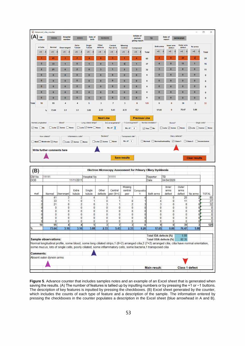
53
Figure 5. Advance counter that includes samples notes and an example of an Excel sheet that is generated when saving the results. (A) The number of features is tallied up by inputting numbers or by pressing the +1 or −1 buttons. The description of key features is inputted by pressing the checkboxes. (B) Excel sheet generated by the counter, which includes the counts of each type of feature and a description of the sample. The information entered by pressing the checkboxes in the counter populates a description in the Excel sheet (blue arrowhead in A and B).

54
The ciliary defect class and comments about the sample are automatically transferred from the counter to the Excel sheet (red and purple arrowheads in A and B respectively).
Results and Discussion
The counters provide an easy and efficient method to count ciliary features and make notes to
assist in the diagnosis of PCD. We tested the advance counter by simultaneously collecting paper
records of the diagnostic cilia feature counts and sample description when assessing 48 patient cases
(taken by three individuals). The paper notes were compared to the values and information generated
in the Excel sheets by the advance counter, and all the information was found to match without any
observed errors. The Excel sheet generated contains all the information required to upload to an
electronic patient record (EPR) system and to advise the consultant or clinician overseeing the referred
patient. The checkboxes within the advance counter automatically populates the description of the
sample observations in the Excel sheet (blue arrowhead in Figure 5a), which promotes the full
assessment of each diagnostic sample, removes the risk of spelling mistakes or typos, and leads to a
standardised terminology for each sample. This makes the results easier to interpret by the clinicians
and is highly beneficial when sharing results across different diagnostic centres for research, auditing,
and teaching purposes. By using the advance counter, the same diagnostic methods are the same as
the outline in the international diagnostic consensus guidelines 286. The images are assessed in the
same way, but the counter can help to improve the sensitivity and specificity by reducing errors in record
taking and maintaining standardised terminologies in the reports. Currently, it is difficult to calculate the
sensitivity and specificity of the TEM-based diagnosis of PCD, as previously there lacked agreement in
the reporting methods for TEM, and the consensus guidelines to overcome this have only recently been
implemented 286. Therefore, in the future it will be possible to accurately determine the sensitivity and
specificity, once a substantial number of cases have been assessed by TEM by multiple diagnostic
centres following these guidelines.
It is important to consider the results from other PCD diagnostic tests in combination with the
TEM analysis 340,369. These include genetic testing, measurements of cilia beat frequency, patterns from
high speed video microscopy, and the immunofluorescence labelling of ciliary proteins. The results from
these tests often correlate well with TEM—for instance, static cilia viewed by high-speed video
microscopy are frequently associated with the absence of the outer dynein arm by TEM (see Figure
1b), as found in patients that have pathogenic variants in the DNAH5 gene 370. A limitation of TEM
diagnostic analysis is that not all PCD defects are detectable by ultrastructural analysis. Approximately
25–30% of genetic mutations 251,259,363, including pathogenic variants in the DNAH11 gene, cause ciliary
defects that cannot be diagnosed by TEM 364. When a normal diagnosis is given by TEM, the other
diagnostic tests need to be considered to rule out PCD. Currently, there are approximately a quarter of
PCD patients that have no genetic cause yet established, and in these patients a ciliary function and
imaging assessment are required 369. TEM provides a valuable tool in validating genetic test results, as
there is a clear link between ultrastructural defects and many of the affected genes, and this can act as
a guide in advancing our knowledge of PCD-causing mutations.

55
Conclusions
We demonstrate a ciliary feature counting system that can be of great benefit to diagnostic centres
testing for PCD using TEM. This system follows international diagnostic consensus guidelines 286,
making it relevant to groups globally. Physical counters are costly and hard to come by, thus the digital
counter we demonstrate here provides a cost-free solution. As computer technology becomes an ever-
growing trend in the diagnosis of disease, the digital counter system removes the need for paper
records, and in the future it may be possible to integrate it into a machine learning system for the fully
automated diagnosis of PCD.
Author Contributions: Conceptualization, A.L.P. and T.B.; Software, T.B.; Resources, R.K.R.;
Investigation, A.L.P. and T.B.; Data curation, T.B.; Supervision, T.B.; Writing—Original Draft
Preparation, A.L.P., C.H., and T.B.; Writing—Review and Editing, A.L.P., R.K.R, C.H., and T.B.
Funding: This research received no external funding.
Acknowledgments: We would like to thank the members of the PCD diagnostic team including Paul
Griffin, Farheen Daudvohra, and Andrew Rogers for their support and feedback when using the digital
counter.
Conflicts of Interest: The authors declare no conflict of interest.

56

57
CHAPTER 3
UA-Zero as a uranyl acetate replacement when diagnosing primary ciliary
dyskinesia by transmission electron microscopy
Accepted for publication in Diagnostics
Andreia L Pinto1,5,6, Ranjit K Rai1, Amelia Shoemark1,2, Claire Hogg1,3, Thomas Burgoyne1,4*
1 Royal Brompton Hospital, Guy's and St Thomas' NHS Foundation Trust, London SW3 6NP, UK 2 School of Medicine, University of Dundee, Dundee DD1 9SY, UK 3 Department of Paediatrics, Imperial College, London, London SW3 6LY, UK 4 UCL Institute of Ophthalmology, University College London, London EC1V 9EL, UK 5 Department of Life Sciences, NOVA School of Science and Technology, Caparica, Portugal 6 CEDOC, NOVA Medical School, Rua Câmara Pestana nº 6, 6-A, Edifício CEDOC II, 1150-082 Lisboa
* Correspondence: [email protected]; Tel.: +44 (0)20 7608 4020
Personal contributions
My contribution to this manuscript was conceptualizing and investigation about suitable non-radioactive
alternatives for uranyl acetate in the diagnosis of PCD, and also in the preparation, reviewing and editing
of the manuscript.
3

58

59
Abstract: Primary ciliary dyskinesia (PCD) is a disorder affecting motile cilia and an early accurate
diagnosis is essential to help prevent lung damage and to preserve lung function. To make a diagnosis
an assessment of the ciliary ultrastructure is typically done by transmission electron microscopy (TEM).
This allows a quantitative assessment of ciliary components to identify defects associated with PCD.
Heavy metal staining is required to provide contrast when imaging cilia in the TEM. One of the most
commonly used stains is uranyl acetate (UA) that can be applied to cellular material before embedding
(en bloc) or to ultrathin sections of embedded samples (grid staining). UA is radioactive and due to
growing safety concerns and restrictions by government bodies, universities and hospitals, it is essential
to find a suitable alternative. We show UA-zero (UAZ) when used en bloc provides high contrast and
makes an excellent replacement for UA. PCD diagnostic experts, having reviewed ciliary cross sections
stained with UAZ en bloc are confident in the staining and PCD defects are readily detectable similar
to samples that have been stained with UA.
Keywords: primary ciliary dyskinesia; uranyl acetate; diagnosis; electron micrsocopy
Introduction
Primary Ciliary Dyskinesia (PCD) is a genetic disease affecting motile cilia. Symptoms include
chronic lung disease, rhinosinusitis, hearing impairment and subfertility. Diagnosis of PCD is confirmed
by identification of a ciliary ultrastructural defect by transmission elec-tron microscopy (TEM) or
identification of bi-allelic pathogenic mutations in a known PCD gene 286,340,369. The normal ultrastructure
of respiratory cilia is shown in Figure 1A. Hallmark defects diagnostic of PCD include outer dynein arm
defects, Outer and inner dynein arm defects, Inner dynein arm defects with microtubular disorganisation
and central complex defects. The dynein arms are small structures of approximately 10 nm in length 371
and in order to visualise these structures accurately at the resolution of diagnostic TEM, good contrast
(high signal to noise ratio) is essential. Contrast is usually achieved using a combination of heavy metal
stains including osmium tetroxide, lead citrate, and uranyl acetate (UA).
UA is derived from depleted uranium and is radioactive (alpha emitter). It is extensively used
as a TEM stain, either en bloc during sample preparation or staining ultrathin sections on electron
microscopy grids 372. Due to increasing concerns around using radioactive material for personal safety
and fears of its potential use in terrorism, many institutes have strict regulations on its use (e.g. United
Kingdom Government Ionising Radiation Regulations 2017). Justification for procurement and use of
UA for diagnostic TEM has become increasingly difficult and there is a global drive to find a safer, non-
radioactive replacement. Alternative reagents including platinum-blue 373, oolong tea extract 374 and
hafnium chloride 375, have been trialled as alternative stains for TEM but are generally inferior to UA
and their use is not widespread. More recently commercially available reagents have become available
that include UA-Zero (UAZ, Agar Scientific Ltd, Stansted, Essex, UK) and UAR-EMS (UAR, Electron
Microscopy Sciences, Hatfield, PA, USA). These are pre-mixed solutions which contain rare earth
elements (lanthanides) for contrast staining. UAZ includes ytterbium(III) chloride hexahydrate and UAR

60
has both samarium triacetate and gadolinium triacetate 376. It is im-portant to validate any changes to
diagnostic procedures to ensure that providing a safer contrast stain does not compromise the quality
of diagnostic ultrastructural studies. Here we compare two UA replacement stains to UA to determine
their suitability in the diagnosis of PCD by TEM.
Materials and Methods
Ethical approval
The study was approved by the ethics review board of the Institute of Child Health/Great
Ormond Street Hospital, London (UK) (08/H0713/82 15/10/2008). All subjects gave informed signed
consent for genetic testing and use of surplus diagnostic samples for the improvement and development
of new diagnostic methods.
Preparation of nasal brushing biopsies for transmission electron microscopy
3mm ConMed Cytology Brushes (ConMed, Largo, FL, USA) were used to take biopsies of the
respiratory epithelium from the inferior nasal turbinate of four patients (see table 1 for mutations) and a
healthy control.
Table 1. Genetic information of PCD subjects that had TEM samples stained in this study.
PCD Gene Axoneme structure affected
Genotype
DNAH5 ODAs Unsolved – Heterozygous DNAH5 NM_001369.2: c.7477G>C (p.Glu2493Arg)
CCDC40 IDAs and microtubular disorganisation
CCDC40 NM_017950.3: c.2712–1G>T + c.2712–1G>T
HYDIN CP C2b component HYDIN NM_001270974.1: c.8487del (p.Pro2830Hisfs*23) + c.8489C>A (p.Pro2830Gln)
LRRC6 ODAs and IDAs LRRC6 NM_012472.3: c630del (p.Trp210Cysfs*12) + c630del (p.Trp210Cysfs*12)
Translation termination codon is referred to by *
Nasal brushings were fixed with 2.5% glutaraldehyde (VWR International, Lutterworth,
Leicestershire, UK) in 0.05M sodium cacodylate buffer (Agar Scientific, Stansted, Essex, UK) at a pH
7.4 and stored at 4oC overnight or for up to 2 weeks. Samples were washed in 0.05M sodium
cacodylate buffer and embedded in 1% agar (Oxoid, Basingstoke, Hampshire, UK) before incubating
in 1% aqueous osmium tetroxide(TAAB Laboratory Equipment, Aldermaston, Berkshire, UK) for 1 hour
followed by washes in distilled water. At this point the healthy control nasal brushing biopsy was split
up into four separate samples. The specimens were prepared using one of the following en bloc stains;
(i) UA, (ii) UAZ, (iii) UAR, or (iv) no stain. The details of each of these stains is provided in Table 2. All
samples were dehydrated in increasing concentrations of ethanol (50%, 70%, 90%, 100%), followed by
propylene oxide and a mixture of propylene oxide and resin (1:1) before infiltration and embedding in
araldite resin (Agar Scientific, Stansted, Essex, UK). Polymerization was performed at 60°C for 48
hours, and ultrathin sections of 80 nm were obtained using a Reichert Ultracut E ultramicrotome and
collected on copper mesh grids (Agar Scientific, Stansted, Essex, UK). For the samples prepared with

61
no en bloc staining, sections were collected onto grids and stained with either; (i) UA (Agar Scientific,
Stansted, Essex, UK), (ii) UAZ (Agar Scientific, Stansted, Essex, UK), (iii) UAR (Electron Microscopy
Sciences, Hatfield, PA, USA) or (iv) left unstained. All except the unstained were counter stained with
Reynold’s lead citrate (Agar Scientific, Stansted, Essex, UK) and the details of the methods used for
each of these stains is provided in Table 3. Some of the sections collected from the UAZ en bloc were
further grid stained with UAZ. Images were acquired using a JEOL 1400plus (JEOL ltd, Tokyo, Japan)
TEM at an accelerating voltage of 120 kV. Digital images were obtained using an Advanced Microscopy
Technologies XR16 bottom mid mount digital camera (AMT Imaging Direct, Woburn, MA, USA). The
camera software has an autogain system that remaps the pixels values so that they extend across a
larger range making it easier to differentiate between lighter and darker pixels. When acquiring images
a fast Fourier transform was generate to make sure images were all taken at 0.7 μm from defocus and
that there was no drift or beam astigmatism (as shown in Supplementary Materials Figure S1).
Table 2. The different methods of en bloc staining used.
En bloc stain Method
No stain No stain added during sample preparation UA 1% aqueous UA (Agar Scientific Ltd) for 30 minutes
UAR UAR (Electron Microscopy Sciences) diluted 1:4 in distilled water and applied for 30 minutes
UAZ undiluted UAZ (Agar Scientific) for 30 minutes
Table 3. Types of grid stain and the methods used.
Grid stain Method
No stain No stain UA 2% aqueous UA for 7 minutes and Reynold’s lead citrate for 10minutes
UAR UAR (Electron Microscopy Sciences) diluted 1:4 in distilled water and applied for 30 minutes and Reynold’s lead citrate Reynold’s lead citrate
UAZ undiluted UAZ for 7 minutes and Reynold’s lead citrate for 10minutes
Analysis of diagnostic results
Diagnostic results from the assessment of nasal biopsy samples prepared for TEM with either
en bloc UAZ (N=58) or UA (N=58) grid stain were examined. The diagnostic assessment included
counting; cilia with a normal (9+2), abnormal microtubule arrange-ment (8+2, 8+1 and 7+2), extra
microtubules, missing central pair, compound cilia and dynein arm defect. The diagnostic results were
analysed using Microsoft Excel.
Image analysis
Cilia were cropped out of the images acquired from samples prepared with the different en bloc
and grid stains using Cilia Crop, a program that is part of the PCD Detect toolkit 367. The cropped images
were anonymised using an in-house program designed using MATLAB to allocate random number for
files names (DOI - 10.5522/04/14369258)

62
20 images per staining condition were analysed by 10 electron microscopists experienced in
PCD diagnosis and blinded to which stain was used. Images were ranked as ‘good’, ‘useable’ or
‘unusable’ for PCD diagnosis. These terms were converted into the corresponding scores; 100%, 50%
and 0% respectively. This allowed the assessment of the confidence the specialists had in the different
grid or en bloc stains.
To analyse the contrast range of images of cilia from the different staining methods the standard
deviation of the pixel gray value was calculated using ImageJ (NIH). All images are 8-bit greyscale
where 0 is black and 255 is white. Statistical significance was determined for the survey results and
standard deviation of the pixel gray value using Student’s t-tests.
Results
The results of a survey comparing the different staining methods to determine their suitability
in diagnosing PCD is shown in Figure 1. Representative images of the different methods are shown are
shown in Figure 1A and Supplementary Materials Figure S2, and it can be clearly seen that all staining
methods give better contrast than no stain. Assessment of ciliary cross sections by 10 electron
microscopists who routinely analyse cilia cross sections to diagnose PCD revealed similar confidence
when comparing UAZ to UA grid stain but lower confidence in UAR (Figure 1 B). UAZ en bloc provided
significantly higher confidence than both en bloc UA and UAR of over 80% (Figure 1 C). The standard
deviation (SD) of the pixel gray value was calculated from images of the ciliary cross-sections (Figure
1 D & E). All three stains when applied to sections gave similar standard deviations of the pixel gray
value (Figure 1 D). UAZ en bloc staining gave significantly higher SD of the pixel value compared to
UA, indicating greater image contrast (Figure 1 D). Whereas, UAR gave a significantly low standard
deviation of the pixel gray value compared UA.

63
Figure 1. UAZ provides a good alternative to UA when examining ciliary cross sections. (A) Images of ciliary cross sections from samples prepared with grid stained or en bloc using one of following stains; Lead citrate (LC) only, UA, UAR or UAZ. (B – C) Anonymised images were compared by 10 electron microscopy specialists and scored based on the confidence they had in the staining. (B) When staining sections on TEM grids, UA and UAZ gave the greater confidence in staining compared to UAR. No significant difference in the confidence in staining was observed between UA and UAZ. (C) UAZ on bloc provided staining that gave the highest confidence. (D – E) Standard deviation (SD) of the pixel gray value within images of ciliary cross sections was determined to examine the contrast range. (D) No difference in the SD of the pixel gray value was observed between UA, UAR and UAZ. (E) There was a significantly greater SD of pixel gray value when comparing UAZ to UA en bloc staining. A lower standard deviation of the pixel gray value was found for UAR when compared to UA en bloc staining. (B – E) Standard error of mean (SEM) shown and statistical significance was determined using Student’s t-test (n.s. P > 0.05 * P < 0.05 *** P <0.01 **** P < 0.0001).
The staining methods were further assessed by examining ciliary cross sections from PCD
patients that have known pathogen variants causing absence of ciliary structures as described in Table
1. The missing axoneme components that include ODA, IDA and C2b component of the CP were
detectable in patient samples when using UAZ grid staining (Figure 2 A – D and Supplementary

64
Materials Figure S3 ). UAZ en bloc staining that provides better confidence in staining quality over using
UA (Figure 1 C) provided high contrast allow the detection of absent inner and outer dynein arms (Figure
2 A3 & E1 and Supplementary Materials Figure S3). The addition of UAZ grid staining to samples that
had been prepared with UAZ en bloc did not improve the contrast further (Figure 2 A4 & E2 and
Supplementary Materials Figure S3).
Figure 2. Key structural features for diagnosing PCD are visible when using UAZ to stain grids or en bloc. (A) The control cilia shows a normal cilia ultrastructure that includes the presence of outer and inner dynein arms and a complete central pair complex. (B & C) Outer (blue arrowheads) or inner dynein arms (red arrowheads) can be seen to be absent in patients that have disease causing variants in DNAH5 or CCDC40 when staining with UA or UAZ on grid. (D) The loss of the C2b component (white arrowheads) of the central pair can be detect in a HYDIN case when staining grids with UAZ and UA. (E) The loss of both dynein arms (blue and red arrowheads) can seen in a LRRC6 case when staining with UAZ en bloc with and without an additional grid stain. The additional stain does not compromise image contrast. (F & G) Diagnostic results from a single UA-Z en bloc prepared samples compared to UA grid stained samples showing similar detection of dynein arm defects. (F) Diagnostic assessment

65
of patients that have pathogenic variants in CDCC40 (UAZ en bloc N=1 and UA en grid N=5). (G) Diagnostic results from patients with pathogenic variants in DNAH5 (UAZ en bloc N=1 and UA en grid N=6).
Following the results of our blinded survey we integrated UAZ staining into our clinical practice.
UAZ en bloc has been performed on 58 patient samples and out of these 11 have tested positive for
PCD. We audited our clinical practice where up to 100 cilia are counted per case to make a diagnosis.
Out of the total cilia cross section examined per case 52.60% ± 1.82% with UA and 53.48% ± 1.54%
with UAZ (shown as mean ± standard error of the mean) could be assessed for dynein arm defects.
This indicates that the UAZ stain change in protocol did not impact the quality of our clinical practice.
Furthermore 2 cases had been previously assessed with UA and were reassessed with UAZ staining.
These had gene mutations in DNAH5 and CCDC40. Quantification of these samples showed similar
detection of dynein arms defects to previous cases that were UA grid stained (without en bloc staining)
as shown in Figure 2 F & G.
In addition to ciliary ultrastructure it is important for PCD diagnosis to assess the health of the
cells and quality of the sample, therefore we assessed other cellular structures in the samples. UAZ en
bloc provides good contrast staining of other organelles and cellular membrane comparable to UA en
bloc (Figure 3). At lower magnification the contents of the cell can be clearly seen with both UAZ and
UA en bloc staining (Figure 3 A & D). At higher magnification, organelles such as mitochondria have
good contrast and membrane at cellular junctions are very clear and are of similar quality for both stains
(Figure 3 C – F).

66
Figure 3. UAZ en bloc provides sample staining comparable to UA en bloc. Images of respiratory epithelium prepared from a nasal brushing with (A – C) UA en bloc and (D – F) UAZ en bloc. (A & D) Low magnification view of ciliated respiratory epithelium show a similar level of contrast between the two stains. (B & E) There is clear staining of intercellular com-partments and organelles including mitochondria. (C – F) Both stains provide good membrane staining as demonstrated by the plasma membrane staining at junctions between cells.
Discussion
We show en bloc staining with UAZ provides a promising replacement for UA for the
assessment of ciliary ultrastructure. It gives comparable contrast to UA and allows imaging of the key
ciliary components that are linked to PCD.
UAZ en bloc staining applied during sample preparation resulted in TEM images with high
signal to noise that when assessed by PCD diagnostic specialists gave more confidence in the staining
and ability to detect ciliary features compared to UA. When using UAZ en bloc staining for a PCD patient
with a pathogenic variant in LRRC6, that encodes a protein required for transport of dynein arms 377,
the absence of the IDAs and ODAs was clear. Patients that have been diagnosed with ODA or/and IDA
defects associated with pathogenic variant in DNAH5 or CCDC40 show the detection of similar ratio of
defects to similar cases that were prepared with UA grid staining.

67
UAZ grid staining conferred similar confidence to UA and was found to be suitable for
diagnosing PCD, as absent ciliary structures were detectable in patient samples. This includes cilia
from patients with pathogenic variants in the DNAH5 associated with a loss of ODAs 370, CCDC40 that
leads to loss of IDAs and microtubular disarrangement 311 and HYDIN that causes loss of the C2b
component of the central pair 282. Based on our findings we suggest UAZ could be used en bloc as a
UA replacement in diagnosing PCD and UAZ grid staining can be used when samples have already
been embedded and UA is unavailable.
This is the first time UA replacement stains have been compared for the diagnosis of PCD. This
was done blinded and included PCD samples from genotyped patients. The assessment was done by
several experts from different centres that specialise in the diagnosis of PCD. We have prepared 58
diagnostic samples with en bloc staining and the assessment of absent ciliary structures and other
defects reflects the results found from our previous UA grid staining protocol.
Only a single control nasal brushing sample was assessed in the survey that was split up and
prepared using different stains methods. As centres use a range of sampling techniques and different
methodology for processing and TEMs, UAZ en bloc need to be validated locally before implementation
in other centres.
We predict that the sensitivity and specificity of TEM in the diagnosis of PCD could improve
when using UAZ compared to more traditional UA methods due to the greater confidence we found in
the staining of cilia. Due to the safety concerns, difficulties of purchasing, handling and storing of UA, it
makes UAZ a suitable replacement reagent for assisting in the diagnosis of PCD.
Supplementary Materials: Supplementary Materials: The following are available online at
https://www.mdpi.com/article/10.3390/diagnostics11061063/s1, Figure S1: Representative fast Fourier
transform (FFT) used to set the defocus when images were acquired for the survey.
Funding: This research received no external funding.
Institutional Review Board Statement: The study was conducted according to the guide-lines of the
Declaration of Helsinki, and approved by the Health Research Authority (protocol code HBD2008v1
15/10/2008).
Informed Consent Statement: Informed consent was obtained from all subjects involved in the study
Data Availability Statement: No new datasets were created or analyzed in this study. Data sharing
is not applicable to this article. The software used to randomize images names is publicly available and
can be found here [doi:10.5522/04/14369258].
Author Contributions: Conceptualization, A.L.P., A.S. and T.B.; Investigation, A.L.P., R.K.R. and T.B.;
Software, T.B.; Data curation, T.B.; Supervision, T.B.; Writing—Original Draft Preparation, R.K.R., and
T.B.; Writing—Review and Editing, A.L.P., R.K.R., A.S., C.H. and T.B. All authors have read and agreed
to the published version of the manuscript.

68
Acknowledgments: We would like to thank the PCD diagnostics at Royal Brompton Hos-pital (RBH)
and Southampton General Hospital (SGH) that include Paul Griffin (RBH), Farheen Daudvohra (RBH),
Andrew Rogers (RBH), James Thompson (SGH), Regan Doherty (SGH), Anton Page (SGH) and
Patricia Goggin (SGH) for their support completing the survey.
Conflicts of Interest: The authors declare no conflict of interest.

69
CHAPTER 4
PCD Detect: Enhancing ciliary features through image averaging and
classification
Published article Am J Physiol Lung Cell Mol Physiol doi:10.1152/ajplung.00264.2020
Amelia Shoemark1,2, Andreia L. Pinto1, Mitali P. Patel4, Farheen Daudvohra1, Claire Hogg1,3, Hannah
M. Mitchison4,5, Thomas Burgoyne1,6 #
1 Paediatric Respiratory Medicine, Primary Ciliary Dyskinesia Centre, Royal Brompton & Harefield NHS Trust, London, SW3 6NP, UK 2 School of Medicine, University of Dundee, Dundee, DD1 9SY, UK 3 Department of Paediatrics, Imperial College London, London, SW3 6LY, UK 4 Genetics and Genomic Medicine, University College London, UCL Great Ormond Street Institute of Child Health and NIHR Great Ormond Street Hospital Biomedical Research Centre, London, WC1N 1EH, UK 5 NIHR Great Ormond Street Hospital Biomedical Research Centre, London, WC1N 1EH, UK 6 UCL institute of Ophthalmology, London, EC1V 9EL, UK # All correspondence to [email protected], Paediatric Respiratory Medicine, Primary Ciliary Dyskinesia Centre, Royal Brompton & Harefield NHS Trust, London, SW3 6NP, UK Tel: 44-(0)20-7352-8121, ORCID: 0000-0002-8428-720X
Personal contributions
My contribution to this manuscript was analysing and interpreting the data generated by the PCD detect
software using control samples under unbiased experimental condition. I also tested the system and all
the possible outcomes. I also apply it when requested in the diagnostic workflow and assist in
troubleshooting.
4

70
71
Abstract
Primary ciliary dyskinesia (PCD) is an inherited disorder of the motile cilia. Early accurate
diagnosis is important to help prevent lung damage in childhood and to preserve lung function.
Confirmation of a diagnosis traditionally relied on assessment of ciliary ultrastructure by transmission
electron microscopy (TEM), however >50 known PCD genes has made the identification of bi-allelic
mutations a viable alternative to confirm diagnosis. TEM and genotyping lack sensitivity and research
to improve accuracy of both is required. TEM can be challenging when a subtle or partial ciliary defect
is present or affected cilia structures are difficult to identify due to poor contrast. Here we demonstrate
software to enhance TEM ciliary images and reduce background by averaging ciliary features. This
includes an option to classify features into groups based on their appearance, to generation multiple
averages when a nonhomogeneous abnormality is present. We validated this software on images taken
from subjects with well characterised PCD caused by variants in the outer dynein arm (ODA) heavy
chain gene DNAH5. Examining more difficult to diagnose cases, we detected (i) regionally restricted
absence of the ODAs away from the ciliary base, in a subject carrying mutations in DNAH9; (ii) loss of
the typically poorly contrasted inner dynein arms; (iii) sporadic absence of part of the central pair
complex in subjects carrying mutations in HYDIN, including one case with an unverified genetic
diagnosis. We show this easy to use software can assist in detailing relationships between genotype
and ultrastructural phenotype, improving diagnosis of PCD.
Keywords: primary ciliary dyskinesia, cilia ultrastructure, image averaging, image classification,
transmission electron microscopy
Introduction
Motile cilia are hair-like projections on the apical surface of human epithelial cells that have a
classical 9+2 microtubule arrangement of their core axoneme structure (Figure 1). Respiratory motile
cilia beat in a highly coordinated manner essential for the movement and clearance of mucus, bacteria
and debris from the airways. Primary ciliary dyskinesia (PCD) is an inherited disorder of motile cilia that
results in failure of mucus clearance, chronic lung disease, rhinosinusitis and hearing impairment, with
additional organ laterality defects, subfertility and occasional hydrocephalus. The estimated prevalence
is ∼1 per 10,000 births, but it is more prevalent in populations where consanguinity is common 368. An
early diagnosis of PCD is vital to implement appropriate treatment with the aim to preserve lung function
and prevent lung damage. Diagnosis is often complex, due to disease heterogeneity and incomplete
understanding of the molecular and genetic causes of PCD. Diagnosis currently involves combinations
of tests, including measurement of nasal nitric oxide (nNO) as well as acquiring cells by nasal brushings
for examination of cilia motility functions using high speed video microscopy (HSVM),
immunofluorescence microscopy (IF), transmission electron microscopy (TEM) and genotyping 340.
Confirmation of a diagnosis traditionally relied on assessment of ciliary ultrastructure by TEM, however
advancements in genetic testing has made the identification of bi-allelic mutations in a known PCD
gene a viable alternative to confirm diagnosis. Mutations in more than 50 different genes are known to
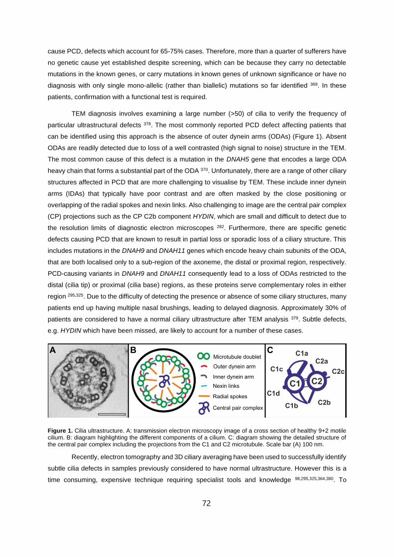
72
cause PCD, defects which account for 65-75% cases. Therefore, more than a quarter of sufferers have
no genetic cause yet established despite screening, which can be because they carry no detectable
mutations in the known genes, or carry mutations in known genes of unknown significance or have no
diagnosis with only single mono-allelic (rather than biallelic) mutations so far identified 369. In these
patients, confirmation with a functional test is required.
TEM diagnosis involves examining a large number (>50) of cilia to verify the frequency of
particular ultrastructural defects 378. The most commonly reported PCD defect affecting patients that
can be identified using this approach is the absence of outer dynein arms (ODAs) (Figure 1). Absent
ODAs are readily detected due to loss of a well contrasted (high signal to noise) structure in the TEM.
The most common cause of this defect is a mutation in the DNAH5 gene that encodes a large ODA
heavy chain that forms a substantial part of the ODA 370. Unfortunately, there are a range of other ciliary
structures affected in PCD that are more challenging to visualise by TEM. These include inner dynein
arms (IDAs) that typically have poor contrast and are often masked by the close positioning or
overlapping of the radial spokes and nexin links. Also challenging to image are the central pair complex
(CP) projections such as the CP C2b component HYDIN, which are small and difficult to detect due to
the resolution limits of diagnostic electron microscopes 282. Furthermore, there are specific genetic
defects causing PCD that are known to result in partial loss or sporadic loss of a ciliary structure. This
includes mutations in the DNAH9 and DNAH11 genes which encode heavy chain subunits of the ODA,
that are both localised only to a sub-region of the axoneme, the distal or proximal region, respectively.
PCD-causing variants in DNAH9 and DNAH11 consequently lead to a loss of ODAs restricted to the
distal (cilia tip) or proximal (cilia base) regions, as these proteins serve complementary roles in either
region 295,325. Due to the difficulty of detecting the presence or absence of some ciliary structures, many
patients end up having multiple nasal brushings, leading to delayed diagnosis. Approximately 30% of
patients are considered to have a normal ciliary ultrastructure after TEM analysis 379. Subtle defects,
e.g. HYDIN which have been missed, are likely to account for a number of these cases.
Figure 1. Cilia ultrastructure. A: transmission electron microscopy image of a cross section of healthy 9+2 motile cilium. B: diagram highlighting the different components of a cilium. C: diagram showing the detailed structure of the central pair complex including the projections from the C1 and C2 microtubule. Scale bar (A) 100 nm.
Recently, electron tomography and 3D ciliary averaging have been used to successfully identify
subtle cilia defects in samples previously considered to have normal ultrastructure. However this is a
time consuming, expensive technique requiring specialist tools and knowledge 98,295,325,364,380. To

73
address this clinical need to improve the detection of subtle or partial structural defects in cilia of patients
that are likely to have PCD, we have developed PCD detect, a package of programs (toolkit) that allows
the selection and averaging of ciliary features (microtubular doublets (MTDs) and CPs) from TEM
images. This assists in removing background while enhancing signal, allowing ciliary structures that are
critical for motility to be better structurally resolved and evaluated. Of importance for some PCD cases
where there is partial or sporadic loss of a ciliary structure, and/or genetic variants identified that are of
uncertain clinical significance, the image classification tool has been developed to provide multiple
(class) averaged outputs to better study and improve detection of these defects.
Materials and methods
Ethical approval
The study was approved by the ethics review board of the Institute of Child Health/Great
Ormond Street Hospital, London (UK) (08/H0713/82). All subjects gave informed signed consent for
genetic testing and use of surplus diagnostic samples for development of new diagnostic tools.
Preparation of nasal brushings for electron microscopy analysis
Respiratory epithelial cells were obtained from subjects’ nasal turbinate using 3-mm bronchial
cytology brushes and placing the biopsy into media 199 (Thermo Fisher Scientific). Each biopsy was
fixed and embedded following the protocol outlined in reference381. In brief, an equal volume of 2.5%
glutaraldehyde in 0.05M sodium cacodylate buffer pH 7.4 was added to the solution containing the
biopsies in 199 media and left overnight or longer (up to 2 weeks) at 4°C. The biopsies were incubated
in 1% osmium tetroxide in 0.05M sodium cacodylate buffer pH 7.4 for 1 hour before embedding in 2%
agar aqueous solution. The samples were then dehydrated and infused with araldite using a Lynx
Microscopy Tissue Processor. This involved incubating in increasing concentrations of methanol (70%,
90% and 100%) followed by propylene oxide, mixtures of propylene oxide:araldite (1:1 and 1:3
respectively) and araldite. Finally, the samples were embedded in araldite and incubated at 65°C for
72 hrs. 90 nm sections from archived araldite-embedded nasal brushings were cut onto electron
microscopy grids and stained using 2% methanolic uranyl acetate for 7 minutes and Reynolds lead
citrate solution for 5 minutes. The sections were viewed on a JEOL 1400+ TEM and when needed the
stage was tilted to provide ciliary cross-sections without obliqueness. Images were acquired at 40,000x
magnification using an AMT 16X CCD camera.
Image criteria
For optimal analysis of cilia using the PCD detect toolkit, it is important to use clear images
(strong signal to background), without stain artefacts, taken at a single magnification, also to include
non-oblique cross sections taken at the focus where it is possible to see the most ultrastructural detail.
Demonstration images are included along with the toolbox to demonstrate the kind of images that are
required. Blinded averaged images generated from control and patient cilia were examined to determine

74
sensitivity and specificity. It is recommended to pick MTD features from at least 5 cilia cross sections
(equivalent to 45 or more MTD features) from different cells for a sensitivity ≥75% and specificity of
100%. More than 10 cilia cross sections from different cells should be used in PCD detect when
examining the CP (equivalent to more than 10 CP features), for a sensitivity >63% and specificity >83%.
Software development
The toolkit includes programs to extract cilia from TEM images (Cilia Crop), perform averaging
of cilia structures (Cilia Averaging), group and generate class averages (Cilia Classification) and to
overlay results for analysis (Cilia Analysis). All programs were written in Matlab (Mathworks).
Cilia Crop: This is a tool to select multiple regions of interest from a larger image. (An example is shown
in Figure S1 A).
Cilia Averaging: This tool extracts manually chosen features (some of these are shown in Figure S2)
and performs a rotational and translational search (see Figure S1 B). By calculating the cross-
correlation, the best alignment of features is determined. The outputs from this tool include two
averaged images comprising (i) all features averaged together (ii) only features best matching the
reference based on the cross-correlation cut-off value. The latter average outputs are used in this study,
as these should contain less background by not including poor quality features (high background,
artefacts, tilted or distorted). All averaged images generated include a corresponding colour contour
map where dark blue indicates an electron dense feature and yellow indicates the absence of a feature.
When using a reference feature (rather than reference-free), it is important to select a well orientated
(non-oblique) feature (MTD or CP) that has good contrast and no stain artefacts.
Cilia Classification: The classification tool (Figure S3 A) was developed to allow two methods of
grouping features based on their similarity (image classification) as determined by (i) principle
component analysis (PCA) and (ii) hierarchical classification. When using PCA, histograms of the first
six coefficients are generated and the plots that indicate more than one feature class (multiple peaks)
are selected. Using these, k-means clustering then separates the features into two or more classes,
which is followed by averaging them together to generate the separate class averages. The hierarchical
classification approach was designed to analyse the feature alignment and similarity, using cross-
correlation results generated previously through the averaging tool. Using this data, the first class is
generated by averaging features aligned to the reference feature (has the most matching other features
based on cross-correlation). Subsequent classes are generated by averaging to the reference features
that are most dissimilar to the reference features used to generate the first class. The hierarchical
classification approach allows a feature to be included in more than one class if there is enough
similarity (based on cross-correlation) to more than one of the reference features used to generate the
classes. This is to allow any features that cannot be easily classed to contribute towards multiple class
averages. It is important to note that class averages can be generated when there is sporadic loss of
structure(s), but class averages can also be given by other types of variation between groups of
features, such as tilt (obliqueness). In this study, when using PCA (Figure S4), the number of classes

75
was set to 2 and the PCA and k-means calculated from an averaged version of each feature (each
feature acts as reference and all other features are averaged to it). By picking the plots that indicate
multiple classes (more than one peak), k-means are calculated to group features before generating
class averages.
Cilia Analysis: This tool allows the visualisation and comparison of averaged features (Figure S3B).
By opening two features, an overlay on both is provided. The features can be aligned automatically or
manually, to allow similarities and differences in the structures to be assessed (Figure S3 B2-4).
Toolkit validation
Image acquisition
The toolkit was initially tested using artificially generated images with added noise (see Figure
S5) before using ciliary images taken from patients with PCD. Details of the PCD subjects in this study
are provided in Table 1.
Input into PCD Detect
For the validation, the four programs were run sequentially. In cilia crop, cropping size is
selected by boxing an entire cross-sectional cilium, with cilia cropped out into a sub-directory by
pressing the Select ROI (region of interest) button and selecting the centre of each cilium within an
image. The folder containing the cropped images is then opened in the Cilia Averaging tool (Figure S1
B). Each image is checked manually to make sure all cilia are orientated in the same direction by flipping
images either horizontally or vertically. A box is drawn around the feature of interest to provide its size
before picking the features to be averaged together. For the validation, the feature of interest was either
the MTD (DNAH5, DNAH9, CCDC103 or CCDC40 cases) or the CP (HYDIN cases). Reference free
averaging was used, as well as a search range of 360° with increments of 1° and a translational search
in X and Y of 10 pixels with an increment size of 1 pixel.
After the Cilia Averaging tool performs the rotational and translational search aligning the
features, an average feature is generated. The data is opened and further processed within the Cilia
Classification tool (Figure S3 A), using both the PCA and hierarchy image classification schemes to
group features based on their structure and generate class averages. When using the hierarchy
classification, a cross-correlation cut-off of 0.6 (any features that have a cross-correlation when
compared to the reference below this value are not included in the class average) and class cut off of
10% (if a reference is found to have less than 10% of the total number of features matching it, no class
average is generated) were set. Finally, the analysis tool is used to overlay features between classes
as well as between PCD cases and control images. A workflow of PCD Detect is shown in Fig 2.
Results
Averaged MTDs show complete or sporadic absence of the ODAs in subjects with a pathogenic variant
in DNAH5

76
The utility of PCD Detect was first evaluated in the analysis of nasal epithelial cilia structures
from PCD patients carrying causal DNAH5 mutations, already established to have clear-cut defects in
conventional TEM analysis, involving loss of the ciliary ODAs. TEM images of transverse orientation
cilia were acquired from diagnostic nasal brushing samples taken from control subjects as well as the
DNAH5 PCD subjects (Figure 3 A, B and Table 1). Using the image averaging tool, the MTDs were
selected and reference-free alignment and averaging was performed. The averaged MTD generated
from a control and three DNAH5 subjects are shown in Figure 3 C, D. Averages from further controls
are given in Fig S6. In all three DNAH5 cases (DNAH5 [1-3]), there is a clearly visible absence of the
ODA, verifying the ability of PCD Detect to identify PCD. Interestingly in two of these cases, DNAH5 [1]
and DNAH5 [2], a small visible structure (white filled arrowheads in Figure 3 D) can be seen, indicating
that part of the ODA remains, whilst ODA are completely absent in the DNAH5 [3] case (denoted by
the white outline arrowheads in Figure 3 D). Of these cases, DNAH5 [1] carries two stop-gain mutations
(homozygosity confirmed by parental segregation analysis), while in DNAH5 [2] and the previously
reported case DNAH5 [3] (11) the genetic diagnosis is not complete since only one DNAH5 variant has
so far been identified in each, although it is a highly implicated rare stop-gain allele in DNAH5 [3];
DNAH5 [2] carries a single uncharacterised missense mutation (Table 1).
A further subject DNAH5 [4], with an uncharacterised missense mutation (p.Thr3410Met) plus
a previously reported DNAH5 stop-gain mutation p.Arg1761* (10), was examined using the
classification tool to group MTDs and form class averages. Many of the ciliary cross sections look
normal by TEM before using the enhancement tools (Figure 4 B). Class averages generated using the
hierarchy classification scheme are shown in Figure 4 C, D and plots from the hierarchy classification
as well as the PCA classification results are shown in Figure S4. When using this tool, subject DNAH5
[4] with the combined nonsense and missense mutation (Figure 4 C), had two visibly different MTD
classes, with and without the ODA. This reflects a sporadic absence of the ODAs that was not found in
the other three DNAH5 cases, as all classes generated had absent ODA (results for DNAH5 [1] shown
in Figure 4 D, E and other cases not shown).
Multiple MTD classes generated from a subject with pathogenic variants in DNAH9 due to loss of the
ODA at the distal region of the cilia
To further test the classification tool, we examined ciliary images from a subject that carried a
homozygous proven-pathogenic variant in DNAH9 (p.Asp4123Asn) 295 (Table 1). DNAH9 protein is a
component only of the ODA in the distal cilium, not the proximal region (determining these regions is
shown in Figure 5 A) and it is known that DNAH9 mutations cause a defect where the ODA are only
absent in the distal region of the ciliary axoneme 295,382. Images were acquired in both the proximal
(Figure 5 B) and distal regions (Figure 5 C) of the ciliary axoneme. By averaging the MTDs from these
images and using hierarchy classification, two distinct classes were generated when compared to
control cilia (Figure 5 D, E). When separating the ciliary images into two groups depending on the
region, the classification results in single distinct averaged structures for the distal and proximal regions,
indicating a clear structural difference (Figure 5 F, G). Images at the proximal region of the cilia resulted

77
in an averaged structure where the ODA is present, whereas the distal ciliary images resulted in an
averaged structure showing absence of the ODA.

78

79
Figure 2. PCD Detect workflow showing the function of the different programs in the tool kit and the order in which to use them. Information is given in regard to the type of ciliary images required for PCD Detect and more information about the DNAH5 [1], DNAH5 [4], HYDIN [2], and HYDIN [4] are provided in Figs. 3, 4, and 7 and Table 1. PCD, primary ciliary dyskinesia.

80
Figure 3. Image averaging shows a clear absence of the ODA in subjects with pathogenic variants in DNAH5. A: TEM image of a cilium from a control subject. B: cilia from three unrelated subjects with pathogenic variants in DNAH5. (C) average control MTD and (D) shows averaged MTD with absent ODAs from the subjects carrying pathogenic variants in DNAH5. The white filled arrowheads indicate MTD that have a projection emanating from the MTD, and the white outlined arrowheads indicate a complete absence of the ODA. Scale bars: 100 nm (A and B) and 10 nm (C and D). ODA, outer dynein arm; TEM, transmission electron microscopy; MTD, microtubular doublet.
The absence of IDAs can be detected in cases with pathogenic variants in CCDC40 and CCDC103
As the absence of IDAs can be difficult to detect by TEM, we determined whether averaging
MTDs was able to enhance this structure. We examined cilia from subjects with known pathogenic
variants in CCDC40 (subject homozygous for the splice site mutation c.2712-1G>T) 311 or CCDC103
(subject homozygous for the well-studied missense mutation p.His154Pro) 383 and compared them
against a control (Table 1, Figure 6 A-C). Mutations in both these genes confer a loss of IDAs in the
cilia of affected patients. When acquiring images, as the CCDC40 variant also results (as expected 311)
in a proportion of cilia with disarrangement MTDs, only normal 9+2 arranged cilia were selected for IDA
imaging. When comparing ciliary cross-section images from both CCDC40 and CCDC103 variants
against a control, it was clear in conventional TEM that IDA loss, in addition to their being of poor

81
contrast, can be masked by the nearby or overlapping presence of the electron dense radial spoke and
nexin structures as well as remnants of some IDA isoforms (Figure 6 A-C). By averaging the MTDs
together using PCD Detect, it became apparent that the IDAs were absent in the CCDC40 and less
dense in the CCDC103 variant cases when compared to a control (Figure 6 D-F).
Figure 4. A subject with an atypical pathogenic variant in DNAH5 has a sporadic absence of the ODA leading to the generation of two averaged classes. A: TEM image of a cilia from a control subject. B: a subject with a pathogenic variant of unknown significance in DNAH5 (compound heterozygous for a missense mutation p. Thr3410Met, plus a stop-gain mutation p.Arg1761*). C: two averaged classes generated for this subject shows the presence and absence (white filled arrowheads) of the ODA. D: in a subject with pathogenic variants in DNAH5 (homozygous for the stop-gain mutation p.Arg4429*), the class averages generated both show a loss of the ODA (white filled arrowheads), indicating complete absence of the ODAs. E: the difference, which is the subtraction of the two classes, is shown in yellow (white outlined arrowhead) and is overlaid and replaces part of the class 1MTD image (red). Scale bars: 100 nm (A and B) and 10 nm (C–E). ODA, outer dynein arm; TEM, transmission electron microscopy.
Confirmation of HYDIN mutations by visualisation of the central pair component C2b
To examine the absence of the C2b component that arises due to pathogenic variants in
HYDIN, cilia from three affected subjects (cases HYDIN [1-3]) carrying pathogenic variants and a
subject with variants of unknown significance in HYDIN were compared to control cilia (Figure 7 A, B).

82
HYDIN [1] and [2] carry compound heterozygous variants of pathogenic consequence while HYDIN [3]
carries only a single variant but one that was previously published 384. Subject HYDIN [4] is also less
well proven for genetic diagnosis, carrying two missense HYDIN variants that have not been previously
reported or functionally tested for their pathogenicity (Table 1). The CP complex from the ciliary images
were averaged together in each case and the hierarchy classification system used to generate class
averages (Figure 7 C-E). In the HYDIN mutation subjects, two main class averages were generated
from the classification tool, indicating a partial defect in the C2b component, where this component is
present in some cilia and absent in others (white filled arrowheads in Figure 7 D, E). As the subjects
with predicted null-effect causal variants (HYDIN [1-3]) generated the same result as the missense
mutation-carrying subject (HYDIN [4]), we could confirm that all these HYDIN [4] variants are highly
likely to be pathogenic in effect. To highlight the absence of the C2b component in the HYDIN [4]
subject, we overlaid the averaged CP complex on top of the control using the ‘cilia analysis’ tool (white
outlined arrowhead in Figure 7 E). While examining HYDIN [4], further cases were assessed that either
had previous diagnostic tests (semi-circling patches of cilia visualised by HSVM) indicating a potential
HYDIN defect (PHD) or had variants of unknown significance (VUS) in HYDIN (Table S1). These
individuals were all found to have a C2b component present in the averaged images generated (Figure
S7), suggesting that HYDIN variants are unlikely to be their disease cause.

83
Figure 5. Analysis of MTDs from a subject with pathogenic variants in DNAH9 gives two class averages when using all cross-sectional images, but only single classes when splitting the images into proximal or distal ciliary regions. A: DNAH11 instead of DNAH9 is localised to the base of healthy cilia close to the cell surface where microvilli are observed. Defects in DNAH9 do not affect the ODA structure within this region, making cilia cross sections surrounded by microvilli ideal internal controls when comparing to ODAs from the tip region. TEM images at the proximal ciliary regions determined by the presence of microvilli (black arrowhead) (B) and the distal ciliary region (C). AverageMTD from a healthy control (D) compared with a subject with pathogenic variants in DNAH9 (E–G). (E) All cross-sectional ciliary images generate two classes showing the presence and absence of the ODA (white filled arrowhead). Separating the ciliary images into two groups at the proximal and distal region of the cilium generates single classes showing the presence (F) and absence (G) of the ODA (white filled arrowhead). Scale bars: 100 nm (B and C) and 10 nm (D–G). ODA, outer dynein arm; TEM, transmission electron microscopy; MTD, microtubular doublet.
Discussion
To examine important structures in respiratory cilia that can be affected in PCD and other
conditions affecting the airway motile cilia 385, we have developed and implemented PCD Detect, a
user-friendly package of tools for image averaging and classification. Due to the uniquely conserved
structure and abundance of motile cilia in patient respiratory epithelial samples, these are ideal
structures for this type of image analysis. By averaging some of the structures that are repeated within
cilia cross-sections, background noise can be reduced and the signal and image resolution enhanced.
As some PCD defects are well known to be difficult to detect from TEM images due to their small size
as well as lack of contrast or resolution, the averaging technique this package employs can provide an

84
additional approach to assist in the diagnosis of PCD in problematic cases. It has become increasingly
recognised that diagnosis of PCD by TEM can be further complicated by the partial or sporadic absence
of a ciliary structure 282,295,325,383,386. The classification tool can assist in these cases by grouping cilia
axonemal structures (classes) based on their similarities.
The first samples we used to test PCD Detect were from subjects with pathogenic variants in
DNAH5, chosen because these typically present with a clear absence of the ODA. When averaging the
first three subjects’ MTDs, the ODAs were evidently absent, but in some cases a remnant of the ODA
was observed. The ODA is attached to the MTD via a docking complex 294,387. Depending on the type
of variant in DNAH5, PCD-causing mutations may result in the loss of a portion or the entire docking
complex, which may explain the remaining structure that is present in some cases 240.
When examining the MTDs in a subject (DNAH5 [4]) carrying a missense variant in DNAH5
that is uncharacterised for its likely pathogenic effect, using a combination of image averaging and
classification, two classes of structure were found, with and without the ODA present. A partial loss of
the ODA has been previously shown for a DNAH5 splice site mutation 388. Certain gene variants may
lead to reduced synthesis of DNAH5 or of DNAH5 protein that is transported and integrated into the
cilium but lacks stability, leading to loss of a portion of the ODAs. Our averaging and classification tool
allowed identification of this complex defect. The success of the average classification was further
demonstrated in a subject with pathogenic variants in DNAH9 which are known to lead to a loss of ODA
only in the distal region of the cilium, whereas the proximal cilium displayed a normal ultrastructure due
to the compensating presence of DNAH11 295,325. When using all images acquired from a DNAH9 PCD
Figure 6. Feature averaging helps in the detection of the absence of the IDA in subjects who have pathogenic variants in CCDC40 and CCDC103. TEM image of a cilium from a healthy control subject (A) and subjects with CCDC40 (B) and CCDC103 (C) mutations. D–F: averaged MTD from the different subjects. The pathogenic variants in CCDC40 (E) and CCDC103 (F) demonstrate a clear absence of the IDA when compared with the healthy control MTD (D). Scale bars: 100 nm (A–C) and 10 nm (D–F). TEM, transmission electron microscopy; MTD, microtubular doublet; IDA, inner dynein arm.

85
subject for the averaging and classification scheme, two class averages were generated, with and
without the ODA. By splitting the image input for PCD Detect into two groups, either proximal
(determined by the presence of epithelial cell apical microvilli) or distal, only single averages of distinct
structure were generated when using the classification tool. This shows that PCD Detect can
successfully group and average similar structures from a non-homogeneous dataset. The separation
of images into the distal and proximal was done manually, but we aim to develop a system that can
detect microvilli and segregate images automatically, which can be added to PCD Detect in the future.
The IDAs are typically difficult to detect in conventional ciliary TEM, linked to the fact that within
the 96 nm axonemal repeat there are four overlapping homologous double headed ODAs, whereas
there are six single-headed and one double-headed IDA of varying morphology 98. This results in a
lower contrast of the IDA than the ODA. Methods to enhance the contrast and reduce background can
be highly beneficial in TEM analysis when needing to determine if the IDAs are present or absent. The
cilia from subjects carrying pathogenic variants in CCDC40 and CCDC103 were both examined, as
mutations in both these genes give rise to loss of the IDAs, with the distinction that CCDC40 mutations
are also linked to microtubular disorganisation and CCDC103 mutations linked to ODA loss 298,311. The
averaged MTDs generated successfully showed a clear absence of the IDA for both mutation types.
To examine CP complex defects in PCD, the cilia of three subjects’ that carry pathogenic
variants in HYDIN were examined as well as cilia from a number of other subjects with less well
confirmed genetic results suggestive of HYDIN defects or no genetic tests yet performed. Using the
averaging and classification scheme, all the three well genetically defined cases were found to have
two class averages, as well as one individual (HYDIN [4]) carrying biallelic VUS, all of these cases
showing the presence and absence of the C2b projection known to be associated with pathogenic
variants in HYDIN. PCD Detect was also able to confirm the presence of the C2b structure in several
other analysed cases that either had suspected HYDIN defects based on previous clinical tests, or who
carried other HYDIN VUS, thus excluding HYDIN defects as their disease cause. This is the first time
two distinct CP structures have been shown to exist in the positively diagnosed HYDIN subjects. The
finding of the same two class-averaged structures in the less genetically well-unverified HYDIN [4]
confirmed the likely causal nature of their HYDIN variants, a conclusion that was supported by the
subject’s history and other diagnostic results.
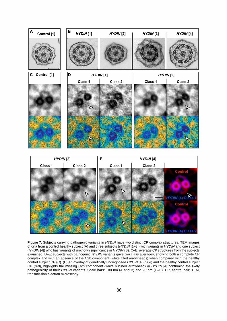
86
Figure 7. Subjects carrying pathogenic variants in HYDIN have two distinct CP complex structures. TEM images of cilia from a control healthy subject (A) and three subjects (HYDIN [1–3]) with variants in HYDIN and one subject (HYDIN [4]) who has variants of unknown significance in HYDIN (B). C–E: average CP structures from the subjects examined. D–E: subjects with pathogenic HYDIN variants gave two class averages, showing both a complete CP complex and with an absence of the C2b component (white filled arrowheads) when compared with the healthy control subject CP (C). (E) An overlay of genetically undiagnosed HYDIN [4] (blue) and the healthy control subject CP (red), highlights the missing C2b component (white outlined arrowhead) in HYDIN [4] confirming the likely pathogenicity of their HYDIN variants. Scale bars: 100 nm (A and B) and 20 nm (C–E). CP, central pair; TEM, transmission electron microscopy.

87
PCD Detect is a free toolkit that has advantages over other TEM image enhancing techniques
such as electron tomography as it requires less specialised knowledge, uses information from a larger
number of cilia and does not require prior information about which structure is affected. It does require
multiple TEM images of well orientated cilia, knowledge of how to interpret the final averaged images
and it has a lower resolution and lack of 3D data that is achievable through electron tomography. This
system package has built upon averaging and image enhancement methods previously developed to
assist in diagnosing PCD 389–393. The simple, automated angular and translational searches, cross
correlation and class averaging features present advantages over previously designed averaging
programs. Previous methods to enhance ciliary structures such as the IDAs have involved using the
cilium’s axonemal symmetry to average MTD 391. This involves manually rotating an image of a cilium
though steps of 40° and averaging each time. This is therefore limited to an average of only 9 MTD per
cilium. In PCD Detect, by allowing the toolkit to extract the features, there is no limit to the number of
structures that can be averaged together. The averaging scheme in PCD Detect has a user-friendly
interface along with a robust feature comparison and alignment system using cross-correlation, allowing
the exclusion of poor-quality features or artefacts. Furthermore, the addition of a classification tool
allows analysis of more complex, non-consistent PCD defects associated with non-homogenous
structural defects.
The averaging and classification scheme available within PCD Detect is particularly well suited
for difficult to diagnose cases that have atypical, poor characterised or difficult to detect ciliary structural
defects. Many of these patients have previously had to have multiple nasal brushings to make a
diagnosis (Table 1), as their initial TEM diagnostic results were either normal or inconclusive and only
after multiple inspections was a defect detected. PCD Detect has the potential to reduce the need for
additional nasal brushings, speeding up the diagnostic turnover times as well as reducing the use of
diagnostic resources.
In summary, PCD Detect is a toolkit to enhance diagnostic images of PCD patient cilia, through
an averaging and classification scheme. It has not been designed for routine use, but instead to assess
difficult cases that have had a suggestive HSVM result, TEM result or a variant of unknown significance
by genetics when following the conventional diagnostic pathway. PCD Detect should be used alongside
other test results as part of a multi-test diagnosis not as a standalone tool. Furthermore, it is important
that high quality TEM images are used to provide reliable averaged images. We were able to show
successful enhancing of structural detail in samples that have clear-cut as well as more subtle ciliary
structural defects and defects affecting only a portion of the axoneme or a minor structure such as the
C2b projection of the CP. PCD Detect can in this way form an associated technique that uplifts current
conventional PCD genetic diagnosis. This is something that can be helpful where genetic diagnosis is
more difficult, due to genetic variants of uncertain significance being identified. It has notable application
for the growing number of affected individuals in whom functional tests such as conventional TEM are
equivocal and they carry genetic variants that have been identified in known genes but are of less
certain causality, for example if they are not previously reported or where single amino acids are

88
affected 394, or in the specific case of HYDIN where variants are hard to call because of the HYDIN2
copy gene 282. This study shows PCD Detect has the potential to benefit global PCD diagnostic centres.
Furthermore, the principles of the toolkit could also be applied to image data beyond ciliary structure
analysis, including homogenous biological structures affected in diseases where diagnostics involves
TEM or for investigative structural biological studies.
Acknowledgements
We are very grateful to the families who participated in this study and thank the UK PCD Family
Support Group for their continued support, as well as Dr James Geraets (University of Helsinki) for his
image analysis advice. The views expressed are those of the author(s) and not necessarily those of the
NHS, the NIHR or the Department of Health. The authors participate in the COST Action BEAT-PCD:
Better Evidence to Advance Therapeutic options for PCD network (BM1407). H.M.M. is supported by
Great Ormond Street Children’s Charity (V1299, V2217) and the NIHR Great Ormond Street Hospital
Biomedical Research Centre (GOSH BRC).
Author Contributions: T.B.: wrote the programs; A.S. and T.B.: designed and performed experiments
as well as analysed and interpreted data; A.L.P. and F.D.: analysed and interpreted data; A.S., H.M.M.:
provided genetic results; A.S., T.B., H.M.M and C.H.: wrote the manuscript.
89
CHAPTER 5
New insights on the motile cilia of zebrafish
In preparation to be submited
Andreia L Pinto1,2,5, Margarida Rasteiro2, Catarina Bota2, Claire Hogg1,3, Thomas Burgoyne1,4, Susana
S. Lopes2
1 Paediatric Respiratory Medicine, Primary Ciliary Dyskinesia Centre, Royal Brompton & Harefield NHS Trust, London, SW3 6NP, UK 2 CEDOC, NOVA Medical School, Rua Câmara Pestana nº 6, 6-A, Edifício CEDOC II, 1150-082 Lisboa 3 Department of Paediatrics, Imperial College London, London, SW3 6LY, UK 4 UCL institute of Ophthalmology, London, EC1V 9EL, UK 5 Department of Life Sciences, NOVA School of Science and Technology, Caparica, Portugal
Personal contributions:
I did all the technicological conceptualization, experimental design, bench work and electron
microscopy analysis, and manuscript writing for this work. All images were obtained by me, except
Figure 1, which was obtained and analysed by my colleague Margarida Rasteiro, who is also
responsible for the Ccdc40-/- (homozygous) mutant generation, Immunofluorescence and confocal
microscopy.
5

90

91
Abstract
Zebrafish (ZF) is a vertebrate teleost widely used in many areas of research. As embryos, they develop
quickly and allow unique advantages for research studies owing to their transparency until later stages.
Zebrafish has many ciliated organs and presents primary cilia as well as motile cilia. This model
organism has two types of motile ciliated cells, mono and multiciliated, and at least two types of motile
cilia ultrastructure have been reported (9+2 and 9+0 microtubule arrangement). Using zebrafish as an
animal model helps better understanding human diseases such as, Primary Ciliary Dyskinesia (PCD),
a genetic disorder that affects cilia motility in an autosomal recessive manner, currently associated with
more than 40 genes. To validate zebrafish cilia as organelles to study PCD through the analysis of their
axonemal ciliary structure We used conventional Transmission Electron Microscopy (TEM) and
Transmission Electron Tomography (ET) for the analysis of zebrafish motile cilia to obtain high-
resolution data in both the olfactory pit (OP) and the Left right organiser (LRO). We characterised the
wild-type (WT) cilia in the OP and showed the presence of motile cilia (9+2) organised in the periphery
of the pit and the presence of immotile cilia (9+2) with absent outer dynein arms (ODA) in the centre of
the pit. We reported a variable pattern for the central pair (CP) in the LRO motile cilia. We confirmed
the presence of immotile cilia in the central region of the WT OP based on the presence of 9+2 cilia
lacking dynein arms. We proposed a new hypothesis for the distribution of cilia in the LRO. We showed
that the ODA of the microtubular doublet (MTD) of both OP and LRO cilia are structurally similar to the
human respiratory cilia at the resolution of EM and ET.
Keywords: Zebrafish, Primary Ciliary Dyskinesia (PCD), Cilia Ultrastructure, CCDC40, Transmission
Electron Microscopy (TEM)
Introduction
Motile Cilia are centriole derived organelles, surrounded by a membrane and containing
microtubules formed by protofilaments that can be longer or shorter, depending on the number of tubulin
molecules they contain2,3,15. This structure is called the ciliary axoneme or ciliary shaft 94, a transversal
term throughout species, considering how well the ciliary structure is conversed between them2.
Axonemal ultrastructure in eukaryotic cells segregates into two significant patterns: 9+2, in which nine
microtubule doublets organise around two microtubules in the centre known as the central pair (CP)
complex; and the 9+0 organisation, in which the CP is absent 12,395. Thus, there are two major types of
motile ciliated cells, cells that produce from dozens to several hundred 9+2 motile cilia and cells that
only generate one solitary cilium.
Monocilia lacking CP were described in the node of mice, hypothesising the presence of motile
9+0 cilia in the node of embryonic mouse11,34,196. However, non-motile primary cilia (9+0 configuration
and no dynein motor arms) can be found in several organs and tissues having solo sensory functions
and lacking movement and can be found in almost every cell type 396,40. On the other hand, Yu et al. in
2011 identified cilia that showed a 9+2 organisation but are immotile, called kinocilia found in the hair

92
cells of the inner ear that express Foxj1b 79 and in the ciliated receptor cells (primary sensory cells) in
the OP of zebrafish24.
Regarding motile cilia dynamics, multiciliated cell cilia with 9+2 configuration beat
metachronically with a planar stroke to clear fluid or promote locomotion 65,66. Motile cilia lacking CP
(9+0) have a specific movement pattern, described as planar rotation, circling or twisting. These
monocilia are known to exist in the LRO in vertebrates (called the Kupffer’s vesicle in the zebrafish
21,42,194). In case of disease, cilia wholly or partially lacking the CP are frequently accounted as one of
the phenotypes of Primary Ciliary Dyskinesia (PCD), an autosomal recessive disease of the motile cilia
144,229.
Cilia malfunction is linked to many diseases. Mutations in cilia genes may result in several
phenotypes (genetic heterogenicity), and mutations in a single gene lead to a wide range of phenotypic
manifestations (genetic pleiotropy) 8. An example is mutations in the CEP290 gene related to many
ciliopathies ranging from severe early-onset developmental disorders like Meckel-Gruber syndrome to
mild late-onset disorders like nephronophthisis. Bardet-Biedl and Joubert Syndromes are also well-
known disorders linked to CEP290 8,397.
Zebrafish started being popular as an animal model in the 1980s (Streisinger work) as a
genetically tractable organism, allowing a phenotypic characterisation of a large number of mutations
that cause defects in a variety of organ systems 174. As zebrafish are vertebrates, this gives them
leverage as a research tool, compared, for instance, to Chlamydomonas reinhardtii, which only major
defect from cilia impairment is lack of locomotion 398. Zebrafish helped shed light on the role of specific
genes in human diseases, as their genome has been widely studied, providing insight into their human
orthologues 181. Amongst the many advantages of zebrafish, we will stress the fact that zebrafish
embryos contain cilia in nearly every cell type, and their organogenesis defects can be easily
characterised using brightfield microscopy as the zebrafish embryo and larvae stays transparent until
all major organ systems are formed. Many of the organs and tissues of zebrafish are similar to those of
humans and 70% of genes are shared 181. Motile cilia have been well described in the Kupffer’s vesicle
or Left-Right Organiser between 3 and 14 somite (ss) stages and in the OP at 48-72 hpf, amongst other
locations 178. Moreover, genetic engineering methodologies like using the CRISPR/Cas9 system are
successful in editing the zebrafish genome and have greatly facilitated the generation of zebrafish
mutant models mimicking human ciliopathies 8,98,399
Primary ciliary dyskinesia (PCD) is a genetic disorder distinguished by recurrent infection in the
lower and upper respiratory tract 251, reduced fertility ad laterality problems (50% of PCD patients show
situs inversus) 160. Ultrastructural defects in motile cilia, or the reduced amount of cilia (in multiciliated
cells - oligocilia), are known to cause PCD 160 158.

93
Almost all identified PCD genes show homologue genes in zebrafish, as shown in
supplementary information table 1 400. For this work, and considering the high impact in PCD patients
401, we selected the zebrafish Ccdc40 (Coiled-Coiled Domain-Containing Protein 40) gene to mimic a
human hotspot mutation. The mutant line was generated using CRISPR-Cas9 technology, according
to Rasteiro Master Thesis work 250 and consisted in generating a truncated protein Ccdc40, at the
amino acid 116, excluding entirely the coiled-coil domain. The loss of function phenotype of the
zebrafish Ccdc40-/- mutant resembled that of the MO-injected embryos described by Rasteiro: situs
inversus in 70% of all cases, curved tails, lethargy and cardiac oedema 250. A previous study by Becker-
Heck in 2011 had also compared the effects of zebrafish Ccdc40 MO knockdown with the zebrafish
homozygous mutant locke, and showed these to be phenotypically indistinguishable324. In addition, the
ultrastructure of the defected cilia investigated by Becker et al. in the Kupffer’s vesicle (LRO) showed
some of the traits found in CCDC40 patients, namely decentralisation of the central pair 324,311. The
mutation in locke homozygous zebrafish35 is not the same as the one Rasteiro generated, while locke-
/- has a nonsense mutation at 778 aa. Rasteiro crispr/cas9 mutant has an early stop codon at 116 aa
excluding completely the coiled coil domain.
CCDC40 is a well-studied PCD gene 311,324. When mutated in humans, CCDC40 does not
completely render static cilia, but they were found to beat in a faster flickering way, with very reduced
beat amplitude 402.
Coiled-coil domains are frequent in proteins and consist of two to five amphipathic α-helices that
twist around each other to form a supercoil 403. Coiled-coil domains are essential in proteins involved in
intracellular transport, molecular recognition, signal transduction and movement regulation 403. In cilia,
CCDC40 is required for motility, as it is needed for the correct assembly of the Nexin-Dynein Regulatory
Complex (N-DRC) and IDA. CCDC40, together with CCDC39, form a complex identified as a molecular
ruler in cilia implicating the specific insertion of the radial spoke (RS) in a 96 nm periodicity 404.
Patients with N-DRC/molecular ruler abnormalities, phenotypically show as microtubular
disorganisation, central pair decentralisation and IDA absence, typical traits of CCDC39 and CCDC40
biallelic mutations, have a worst prognosis than other PCD cases with dynein defects 330,405. As
observed by Davis et al., these patients are more severely affected, showing a higher rate of neonatal
respiratory distress and lower FEV, situs inversus (in around 50% of the patients) 310,406, and poorest
overall condition.330,401. In PCD patients, CCDC40 is mutated in several called "hotspots". The most
frequent mutations are c.248delC and c.3129delC that modify the reading frame 324 and c.2440 C>T,
c.961 C>T and c.1345C>T that produce truncated proteins 311.
Despite previous research has been done on zebrafish cilia 24,215,21,227, no study fully
characterised the cilia ultrastructure of the OP and LRO. With this work, we aim to determine the
similarities and the significant differences in zebrafish cilia compared to human cilia, both for motile cilia
present in multiciliated cells and for embryonic LRO cilia (monociliated cells). Moreover, here we inspect
94
the results obtained from Ccdc40-/- mutant zebrafish and compare those with human patients (CCDC40
patients from the Royal Brompton Hospital, with genetic testing showing orthologous variants). For this,
we used wildtype zebrafish (WT) (AB fish lines) and mutant Ccdc40+/- zebrafish lines that were in
crossed to access the homozygous phenotype of both the LRO cilia in 9-10 ss stage embryos or the
OP in 5 day old larvae. Full TEM assessment was performed and sometimes for better understanding
of the phenotype presented, transmission Electron Tomography (ET) was performed.
Using Transmission Electron Microscopy (TEM), we confirmed the heterogenic configuration of
the zebrafish OP. We also showed by ET the variable presence of a CP in the monocilia of the LRO, a
feature already hypothesised in our previous publication by Tavares et al. 21.
Methods
Ethical Approval
The ethics review board approved the Institute of Child Health/Great Ormond Street Hospital study,
London (UK) (08/ H0713/82). All subjects gave informed signed consent for genetic testing and surplus
diagnostic samples to develop new diagnostic tools.
Animals
The zebrafish used in these experiments (AB WT line and Ccdc40+/- (heterozygous) mutant line) were
bred and kept in the CEDOC Nova Medical school fish facility under a controlled environment approved
by the Direção Geral de Alimentação e Veterinária (DGAV). Embryos and larvae stage 8 to 10 ss and
2 to 5 days post fertilization (dpf) respectively were removed from their chorion if needed and euthanised
by being placed in a bleach solution or fixative for TEM and ET analysis.
Ccdc40-/- mutants genetic engineering
Zebrafish mutants were obtained using the CRISPR-Cas9 method performed by the Lopes lab in
CEDOC Nova medical school according to what was described in the published work of Rasteiro 250.
The Ccdc40 zebrafish mutant line (in preparation for publication) expresses a truncated protein –
mutation at 116 aa – the truncated protein lacks the coiled-coil domain, making the homozygous animal
not viable after 7-dpf.
Immunofluorescence and image analysis
Embryos were fixed in 4% PFA (paraformaldehyde) in PBS (phosphate buffer solution)
overnight. The embryos were then removed from the PFA and washed five times with PBS,
dechorionated, permeabilized with Proteinase K and fixed again in 4% PFA-PBS for 20 minutes. The
samples were submitted to antigen retrieval using acetone for 7 minutes at -20oC, and blocking was
done using blocking solution (BS) (1% bovine foetal serum (BFS) in PBS) for 1h. The primary antibody

95
(mouse anti-acetylated alpha-tubulin) was diluted at 1:400 in BS and incubated with the embryos
overnight at 4oC with agitation. The embryos were incubated with secondary antibody overnight (anti-
mouse Alexa 546, 1:500) in a solution with equal amounts of PBS and DAPI (4′,6-diamidino-2-
phenylindole). Samples were fixed in 4% PFA in PBS and were mounted onto glass slides. Images
were taken using a two-photon microscope (40x water objective, NA 0.80, 2.5 optical zoom GaAsP
detector).
Transmission Electron Microscopy
Embedding for TEM was performed in zebrafish embryos (9-10 ss) and larvae (5-dpf) from both
WT AB and Ccdc40-/- mutants. The animals were whole-mount fixed with 2.5% glutaraldehyde (Sigma)
0.1M sodium cacodylate (Sigma) for at least 48 hours, and washed in the same buffer. After, were post-
fixed with 1% aqueous osmium tetroxide (EMS), and stained en bloc with 1% aqueous uranyl acetate
(Agar Scientific). Samples were dehydrated in increasing ethanol concentrations, infiltrated with
propylene oxide, and embedded in Epon 812 (EMS). Polymerisation of the Epon was performed at
60°C for 24 hours in bottleneck capsule beems (Agar Scientific). Under a stereoscope, the blocks
containing the embryos and larvae were analysed, and the best ones (based on their viability and
orientation) were selected for re-embedding in the desired orientation. The tip of the blocks containing
the samples was chopped, and the blocks containing the fragments were re-embed in coffin type silicon
moulds. For the larvae, only the head was dissected and re-embedded in coffin type silicon moulds in
an anterior-posterior position, with the OP facing the plane of sectioning. The 9-10 ss embryos were re-
embedded lateral to obtain longitudinal sections along the wider axis to fully access the LRO, the
somites, and the yolk sac. (Supplementary Figure 1). The resin was polymerised in an oven at 60°C for
at least 24 hours. Ultrathin sections (≥100nm to be able to get at least one complete repeat of the IDA)
were obtained in a Reichert Ultracut E ultramicrotome (Leica®) collected to 1% formvar coated copper
slot grids (Agar scientific), stained with 2% aqueous uranyl acetate (UA) and lead citrate (LC) (Reynold
recipe) and examined in a Jeol 1400 plus transmission electron microscope at an accelerating voltage
of 120 kV. Digital images were obtained using an AMT XR16 bottom mid-mount digital camera (AMT).
The sections were systematically analysed using AMT600 software, and several high and low
magnification images of cilia throughout the whole LRO and the OP of both mutant and WT were
acquired. Considering the high abundance of cilia found in the OP, these were counted using a custom-
made software developed to quantify cilia for PCD diagnosis 276. Patient samples were embedded for
TEM and analysed as previously described in Andreia L. Pinto et al. 276, following a standard clinical
diagnostic sample preparation and assessment procedure.
Electron Tomography
Sections from the same batch of animals were acquired for ET analysis, thick section of 100-
120 nm were collected into 1% formvar coated copper slot grids (Agar scientific), stained with 2%
aqueous UA, LC (Reynold recipe) and 10 nm colloidal gold (EMS), and examined in a Jeol 1400 plus

96
transmission electron microscope at an accelerating voltage of 120 kV. For dual-axis ET, selected areas
at a magnification of 25.000x were tilted from -60 to 60 degrees in the two axis (the grid was rotated 90
degrees between the collecting tilt series) using a high tilt specimen holder. SerialEM was used for the
automatic acquisition of the images. Cilia cross-sections of the LRO and the OP in both WT and Ccdc40-
/- mutants were assembled in a tomogram using IMOD 3Dmod 407, and both axis were combined for the
generation of an electron tomogram of the region of interest. Sub-tomographic averagings were
performed using IMOD PEET in both organs of WT and mutant fish. For the 3D analysis of the
structures, UCSF Chimera was used. Chimera is a program for the interactive visualisation and analysis
of molecular structures and related data. We open the data in Chimera with the averaged ciliary features
and produced a model showing the molecular anatomy of the structure, and we used Chimera to
measure the volumes of the ODA and MTD 408.
Data analysis
In order to better study the discrepancies between the experimental groups, Student’s t-test
was performed, and the significance was considered for values below 0.05.
Results
The cilia of zebrafish by light microscopy
Zebrafish shows motile cilia in some of its organs since the early stages of development 8.
Olstad et al. has shown the flow generated by the OP motile cilia before. Moreover, the motile monocilia
of the LRO, mainly 9+2 configured axonemes that beat with a rotation and wavy fashion were
extensively described and published by Sampaio et al. 2014 and Tavares et al. 2017. These motile
monocilia are essential to generate the flow for the determination of left-right asymmetry 21,42,189. The
presence and distribution of cilia in the WT LRO and Ccdc40-/- mutant zebrafish was again confirmed
by IF upon confocal microscopy image acquisition as shown in the A and B panels of Figure 1,
respectively. The monocilia found in the LRO of the mutant Ccdc40-/- fish seem to be shorter
(arrowheads pointing at cilia in both experimental conditions). The OP cilia of both WT and Ccdc40-/-
mutant zebrafish were also previously assessed by IF (panels C and D in Figure 1), and three regions
of the pit from each condition were selected to show the presence of cilia (labelled in red, marked with
an asterisk) throughout the OP. The results shown by IF confirmed the presence of cilia in the OP and
the LRO and revealed their 3D tomography which proved useful for future TEM embedding and
sectioning procedures. However, confocal microscopy is limited to 180 nm lateral and 500 nm axial
resolution and is not appropriate for ultrastuctural studies. We next used TEM and ET to access the
ultrastructure of the respective organ cilia.

97
Figure 1. Presence of cilia in the LRO and the OP of wildtype (WT) and Ccdc40-/- mutant zebrafish shown by
confocal fluorescent microscopy. Distribution of motile monocilia in the LRO of 8-9 ss WT (A) and mutant fish (B), shown in a z-projection of 145 images for the WT and 113 images for the mutant. Arrowheads pointing at cilia.
Regarding the OP, three image planes were selected through the pit of WT (C) and mutant Ccdc40-/- zebrafish
(D) revealing the consistent presence of cilia throughout the organ. Z-projection of confocal stacks (for the assessment of the LRO) and z-stack analysis (of the OP) were done using FIJI. Acetylated α-tubulin labelling was done by Immunofluorescence and appears in red and DAPI in blue in both panels; n – nucleus; * cilia; scale bar 20μm
Different cilia in the WT zebrafish OP
To maximise the ultrastructural characterisation of cilia analysed in the OP, a detailed
investigation was done by means of counting more than 50 cilia cross-sections when possible. This
was to provide a representative profile of features found in the OP. TEM assessment revealed that the

98
pit has a bowl shape morphology, and more towards the periphery of this concave structure, all cilia
appeared to have 9+2 and outer and inner dynein arms, (ODA and IDA) characteristic of motile cilia.
However, more into the centre of the pit, the cilia had absent outer dynein arms (IDA were hard to
access in some areas). Quantitative analysis of three different WT animals showed consistently that
close to 100 cilia had 60% of 9+2 cilia, and on average, 23% (±1) of cilia presented absent or incomplete
dynein arms (most notably in the ODA) (Figure 2 and Table 2). Thus, TEM analysis allowed the first-
time quantification of the heterogenicity of the cilia in a specific region of the OP. Cilia observed in the
OP thus have a 9+2 motile morphology in a more peripheral area and a 9+2 with ODA and IDA absence
towards the centre of the pit.
Figure 2. Schematic representation of 5-dpf Zebrafish head, structures of interest are marked with arrows - olfactory placodes/pits (OP). TEM low magnification image shows a cross-section across an OP of a WT zebrafish showing a bowl-shaped structure containing multiciliated cells and cilia in several orientations. In the periphery of this pit (dotted boxes a and inset), cilia containing classical motile structure 9+2 with dynein arms present (black arrowheads). However, in the most internal region of the OP (dotted box b and inset), cilia profile are more frequently 9+2 with absent dynein arms (red arrowheads); this pattern was visible in all WT embryos (n=3). n - nucleus, cb - cilia border, OP – olfactory /pit, thin bar 6µm, thick bar 100nm.
When compared, zebrafish Ccdc40-/- and WT samples revealed no significant differences in
the number of missing ODAs. However, when it comes to IDAs, the absence of IDA in the mutant is
significantly higher (**** P ≤ 0.0001).
Regarding CCDC40 PCD patients (table 1), these show an IDA absence combined with MT
disarrangement in cilia, with no abnormalities identified in the ODA. On the other hand, the zebrafish
OP has a characteristic pattern of absent ODA in about 25% of its cilia (Table 2), which explains the
~15% absent ODA seen in the Ccdc40-/- zebrafish mutants OP. We have accessed the number of
defects and normal cilia present in the Ccdc40-/- mutant zebrafish using the same method used for the
human patient samples, and the ODA defect was seen in addition to the CCDC40 human phenotype
that includes disarranged microtubules and complete absence of the (IDA) (table 2).

99
The percentages of missing ODA and IDA assessed by quantitative method 276 (as shown in
Table 2) were compared using Student’s t-test in both zebrafish and human samples (WT and Ccdc40-
/- mutants and with CCDC40 PCD and control patients) for significance analysis. The differences
between the WT zebrafish and the non-PCD patient concerning the presence of ODAs and IDAs is
significantly different (ODA *** P ≤ 0.001; IDA * P ≤ 0.05);
Between human samples (control non-PCD versus CCDC40 PCD patient data), there were no
significant differences regarding the consistency of the ODA (ODA were always present; ns P > 0.05).
On the other hand, there was a considerably higher number of IDA absent in the CCDC40 patient
samples (**** P ≤ 0.0001). Finally, when putting side-by-side, zebrafish Ccdc40-/- mutants and Human
CCDC40 PCD patients, differences in the presence of ODA were reported (absent ODA in the zebrafish
but not in the human sample; * P ≤ 0.05). Nevertheless, and in line with what was expected, IDA defects
were consistent between the two samples (human and zebrafish mutants show similar profile; ns P >
0.05).
The disarrangements found in WT zebrafish were considered similar to secondary defects and
fall more in the category of microtubular abnormalities that can be linked to mechanical stress inside
the pit (biofilm was identified in WT 2 and 3, and it might be suggestive of accumulation of medium
during development, data not shown). Human control samples showed no MT disarrangements in the
configuration of the ciliary axoneme.
Table 1 Genetic results of the three PCD patients presenting ultrastructural defects observed in TEM.
CCDC40 patient 1 CCDC40 patient 2 CCDC40 patient 3
CCDC40 homozygous for c.248del, p.Ala83Valfs84*
CCDC40 homozygous for c.248delC;
p.Ala83Valfs84*311
CCDC40 compound heterozygous for c.248del and
c.2712-1G>T
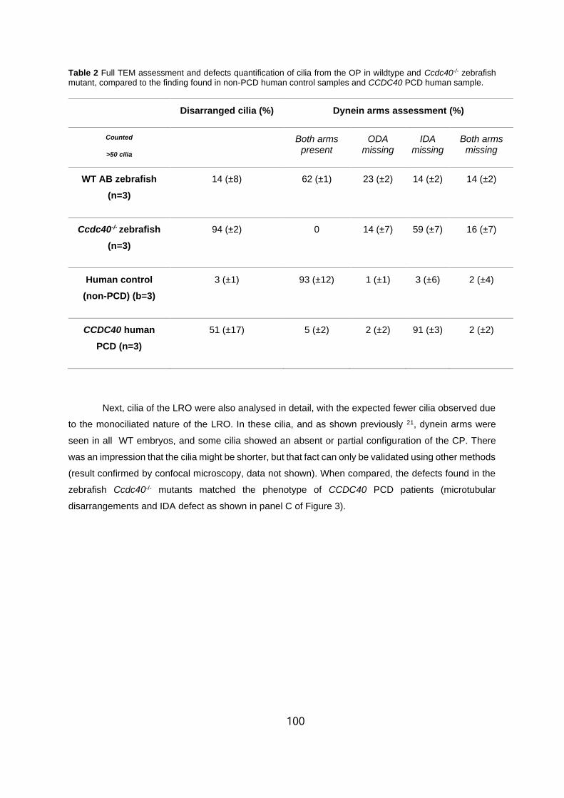
100
Table 2 Full TEM assessment and defects quantification of cilia from the OP in wildtype and Ccdc40-/- zebrafish mutant, compared to the finding found in non-PCD human control samples and CCDC40 PCD human sample.
Disarranged cilia (%) Dynein arms assessment (%)
Counted
>50 cilia
Both arms present
ODA missing
IDA missing
Both arms missing
WT AB zebrafish
(n=3)
14 (±8) 62 (±1) 23 (±2) 14 (±2) 14 (±2)
Ccdc40-/- zebrafish
(n=3)
94 (±2) 0 14 (±7) 59 (±7) 16 (±7)
Human control
(non-PCD) (b=3)
3 (±1) 93 (±12) 1 (±1) 3 (±6) 2 (±4)
CCDC40 human
PCD (n=3)
51 (±17) 5 (±2) 2 (±2) 91 (±3) 2 (±2)
Next, cilia of the LRO were also analysed in detail, with the expected fewer cilia observed due
to the monociliated nature of the LRO. In these cilia, and as shown previously 21, dynein arms were
seen in all WT embryos, and some cilia showed an absent or partial configuration of the CP. There
was an impression that the cilia might be shorter, but that fact can only be validated using other methods
(result confirmed by confocal microscopy, data not shown). When compared, the defects found in the
zebrafish Ccdc40-/- mutants matched the phenotype of CCDC40 PCD patients (microtubular
disarrangements and IDA defect as shown in panel C of Figure 3).

101
Figure 3. TEM features of cilia showing ultrastructural variations in the LRO of 9/10 somite stage WT zebrafish embryos (A) and the OP of 5-dpf WT zebrafish larvae (B). In the Ccdc40-/-, zebrafish mutants the LRO presented absence of CP in some cilia (C) and there was an IDA+ODA absence in the cilia of the OP (D). In human samples from PCD patients with CCDC40 mutations IDAs were absent and MTs disarrangements were present (E). White arrowheads – missing IDA; black arrowheads – missing ODA, dotted circle – abnormal position of/missing CP; scale bar 100nm.
Zebrafish and human have overall similar cilia ultrastructure in multiciliated cells
These results concluded that apart from the particular features of the zebrafish OP, namely the
absence of outer dynein arms in cilia from the central region of the pit, the ultrastructure of ciliary
components is similar to the cilia found in the human airway. Nevertheless, after assessing the cilia
from the LRO by ET, some concern regarding the size of the ODA was raised, as some variation in the
size of the outer dynein arms was visible, suggesting it might be smaller (Supplementary data figure 2).
To clarify, we used USFC Chimera software to measure the volume of the ODA compared to the total
volume of the microtubule doublet (MTD) to make sure the measurements were accurate since there
were variations in the size and thickness of the 5 MTD assembled by ET. Furthermore, we analysed
the volumetric ratio from averaged MTD from the LRO of 4 different WT zebrafish, and these
measurements were compared to the averaged MTD of 3 human control cilia (figure 4). No significant

102
differences was found between the two samples, indicating that LRO cilia and human airway cilia have
similar MTD (ns P>0.05). We also looked at the rendered data of MTD from two different WT zebrafish
OP. The ODA ratio showed no significant differences, but the numbers of animals analysed were not
enough for the statistical analysis.
Figure 4. The ratio of the outer dynein arm (ODA) volume compared to whole microtubule doublet (MTD) volume. Sub-tomographic averaging of the MTD of WT zebrafish cilia, showing ODA and IDA, tubules A and B and a portion of the radial spoke. The two sample groups showed no significant difference between them (ns P>0.05) performing a Student’s T-test.
The application of Transmission Electron Tomography for the study of the central pair
Despite the visible heterogenicity of the CP of the LRO, there was a need to provide more
evidence to validate these findings. The LRO is an organ well embedded in the embryo and hard to
access. It is spherical-like in shape, and depending on the plane of sectioning, the cilia will be in different
orientations. Additionally, LRO cells are monociliated 409. To acquire well-oriented cross-sections of
many cilia in the LRO is hard and produces sparse results, even in the event of finding an axoneme,
the probability of it being well orientated is low. ET offers the advantage of expanding the ability to view
structures that fall outside conventional image ranges and bridge the resolution gap 410. We were able
to successfully collect and assemble tilt series from six cilia from two different WT zebrafish LROs
(Figure 5, animal 1: cilium 1/4 to 4/4, and animal 2: cilium 1/2 and 2/2). We investigated the prevalence
of the CP using IMOD z-projection overlap, which consists in the combination of the slices of the
tomogram generating a single image. From this assessment, we could identify a heterogenic pattern
regarding the CP. Two cilia showed evidence of a CP; two cilia showed only one of the CP tubules
(incomplete phenotype), and two cilia presented no visible CP (Figure 5)

103
Figure 5. Tomogram z-projection (≥150 stacks) of 6 cilia CP observed in two different WT zebrafish LRO, showing variation in the frequency of the CP. Cilium 1/4 and 1/2 showing only one tubule present, cilium 2/4 and 4/4 showing both tubules present and cilium 3/4 and 2/2 showing no tubules in the central pair region. * missing tubule, incomplete CP; dotted circle missing CP. Bar 50 nm.
Discussion
Cilia are well-studied organelles; over 21.000 studies have been successfully published around
this topic, and a thousand only in the last year. Cilia are complex organelles, and the fact that they are
involved in many syndromes and diseases makes them essential targets in biology and translational
research 94,411. The difficulty of accessing human embryonic tissues provides the need to find model
organisms that resemble human organogenesis and have conserved evolutionary patterns.
Chlamydomonas reinhardtii, C. elegans, and Leishmania donovani, promastigote are good models,
easy to maintain and proliferate in captivity. However, they lack internal organs and organ to organ
communication, crucial for the study of ciliopathies in humans. Chlamydomonas, for instance, are
simple organisms equipped with a conserved flagellum that has already been widely used for
biochemical cilia characterization 8,17,404.
On the other hand, it was already shown using contemporary technologies like computer-
assisted image analysis and Cryo-EM that Chlamydomonas flagella have structural differences from
human cilia 17,392. Vertebrates like mouse and zebrafish are good models to study ciliopathies since
their genomes are well known and contain protein-coding genes with human orthologues [25], meaning,
when mutated, these genes may recapitulate the human phenotype. This feature has been shown for
PCD in many studies 4,164,412–416. PCD has already been associated with more than 40 genes known to
be causing disease 94, and all of those genes are present in the genome of zebrafish 400 (Supplementary
Table 1).
Technology in ultrastructural studies has improved in the last ten years and, higher resolution,
better image quality and improved sample preparation have allowed us to go further in studying
structures that were once less reachable 16. The zebrafish Kupffer’s vesicle is the fish LRO, equivalent
to the mouse node, therefore, an important structure to study in the zebrafish motile cilia research.
However, this vesicle is embedded at the end of the notochord and it is challenging to access 42,196.
Even though the zebrafish is transparent, and the LRO is accessible by light microscopy, a more
detailed analysis using conventional TEM can be problematic. The organ itself is not bigger than 70 µm
in diameter (measured by TEM) and is transient during the zebrafish development 42,194,190. In addition

104
to this, the fact that the LRO has a spherical shape, means that cilia will be in many different orientations,
making it harder to get a well-orientated cilia cross-section in the organ. Also, the LRO is composed of
monociliated cells, and the number of cells in the LRO is around 50 (assessed by light microscopy)
42,194. Altogether, these features make the LRO a challenging organ to study by conventional TEM. On
the other end, the OP is a surface organ that can be more easily reached 227, for this, the orientation of
the blocks for TEM is essential, and the sectioning must be done exceptionally carefully since the
structure is small (approximately 60 µm in length 227). The disadvantage is that being more superficial
also makes it more vulnerable to external mechanical aggressions like the salts on the embryonic
medium and even manipulating and handling the sample during the embedding protocol.
The findings that the OP has a heterogenic pattern of ciliary ultrastructure coincide with what
was already described by Reiten et al. 2016 227 functional tests. At the time, the authors described a
difference in the beat frequencies found on the OP, hypothesising that motile cilia in the nose of aquatic
vertebrates are specially organised and beat their cilia at distinct frequencies to attract particles,
showing no beating in the center of the pit 227. However, the authors showed no TEM analysis for the
cilia in the OP 227. The work presented here sheds light on the lack of flow detected in the center of the
pit, given the immotile profile of the cilia nested in that specific area (lacking dynein arm motor proteins).
Our findings and the work done by Reiten seem to agree with observations previously published by
Hansen (1998, 2005), in which a subgroup of ciliated sensory neurons are believed to cluster in the
centre of the OP and to have a 9+2 configuration with no dynein motors. However, these previous
studies were conducted in adult zebrafish, and they do not provide extensive TEM analysis to
characterize the ultrastructure of these cilia 24,215.
For this work, we chose to work with a zebrafish mutant Ccdc40-/- created in the Lopes lab 250.
The phenotype caused by this mutation in humans is well characterised and affects around 20% of
PCD patients 248,267. It also has a worst clinical prognosis compared to dynein structural gene
mutations326. In the case of a CCDC40 mutation, the findings are microtubular disarrangements,
acentric location of the CP and absence of the IDA due to disruption on the “nexin-dynein regulatory
complex” (N-DRC). In parallel, Ccdc40-/- zebrafish mutants show ultrastructural defects comparable to
those found in human PCD patients with mutations in the same region of the CCDC40 gene. These
homozygous zebrafish mutants have a very severe phenotype (curly down tail phenotype, situs
inversus in 50% of the cases 324, lethargy and cardiac oedema 417 and are only viable until
approximately 7-dpf. Detailed IDA assessment is vital to make a correct analysis of the Ccdc40 -/-
zebrafish mutants and CCDC40 patient samples. Cross-sections need to be analysed and, the
thickness of these cross-sections is very much essential to assess the IDA morphology.
IDA morphology is heterogeneous in composition, including several dynein heavy chains (DHC)
and light and intermediate chains, in an elaborate organisation that repeats every 96 nm. Differently,
ODA has a more homogeneous and frequent distribution along the axoneme 129,418. Therefore, thick
sections (≥100 nm), adequate microscope voltage (by applying a high-voltage driving force, electrons

105
will penetrate through tissue sections), and high signal to noise ratio (meaning more signal than noise,
leading to a more accurate assessment) needs to be secure to correctly assess IDAs, due to its
heterogeneous morphology. Failure in this can potentially lead the observer to conclude IDAs are
absent, leading to a false assessment 129,419,420.
Fortunately, to extract more detail and information from the samples, more sophisticated
techniques in EM are available, including dual-axis ET of plastic section to better assess components
in the cilium of both LRO and OP in zebrafish. The procedure involves tilting a thick target section and
acquiring images at angles of 1-degree increments (tilt range ± 60 degrees) and using these to
reconstruct a 3D image 421. Using ET, we were able to show that the MTD ODA from the LRO resembles
the structure of the MTD ODA of the cilia found in the human respiratory epithelium. Through ET was
possible to verify a previously hypothesised variation in the LRO cilia, 1) the unequivocal presence of
dynein motors proteins (IDA and ODA) in all cilia analysed, suggesting their ability to generate
movement, and 2) the apparent heterogenic distribution of the CP throughout the LRO cilia that
suggests cilia inside the LRO might be organised in a specialised fashion, which would explain some
observed beating patterns 42 194. This feature might be perhaps linked to their location in the vesicle,
but more evidence is needed to advocate for regional differences containing or not a viable central pair.
Another hypothesis is that the position of the CP in the axoneme might be partial in a specific area of
the axoneme. Due to technological limitations, this was not possible to confirm; however, serial analysis
of the LRO and its cilia is recommended. Large volume EM would allow a better understanding of the
distribution of the cilia within the LRO and map the heterogenic ciliary ultrastructure inside the vesicle.
We attempted SBFSEM 422 (serial-block face SEM) in a selection of well orientated LROs. SBFSEM is
a technology that combines ultramicrotomy and scanning electron microscopy, allowing images to be
taken in between thousands of thin sections (~30nm) of a specific region automatically. This sequential
image acquisition permits a 3D reconstruction of all the images acquired in the depth of the sample 422.
In our sample, the acquisition was unstable due to charging caused by bare resin and a hollow vesicle
(in vivo would be containing fluid). We stress the need to perform Volume EM in the LRO to better
describe the morphologic profile of the cilia inside. For future experiments, Array Tomography would be
a more appropriate technique 423 (another Volume EM technique) to resolve the caveats encountered
in SBFSEM.
Zebrafish motile cilia resemble the cilia found in the human airway and are accessible for
manipulation and analysis. Furthermore, the zebrafish is a suitable model to test the effect of many
PCD mutations since it has its whole genome sequenced and human PCD homologue genes identified.
Genetic engineering unlocks the potential to understand disease phenotypes and gene interactions,
and in that field, zebrafish already revealed to be a valuable tool 243,247,250. Lastly, Electron Microscopy
still represents the primary gold standard technique to analyse the ultrastructure of cilia, and more new
and sophisticated techniques will shed light on the ultrastructural details still to unravel. In summary,
this work provides strong evidence that zebrafish is a good animal model to study PCD.

106

107
CHAPTER 6
Discussion and Conclusion
6

108

109
DISCUSSION
The primary purpose of this research study was to improve the diagnosis of PCD. This involves
either using the data collected from PCD patients or model organisms like zebrafish (Danio rerio) to
better understand cilia and ciliopathies 8. it is possible to manipulate the genome of these organisms
and observe and study the effects of a single mutation and assess the phenotype caused in later stages
of development.
TEM is a complex analysis tool 424. However, it is still a critical diagnostic test for PCD 425.
Throughout the assessment of >50 ciliary cross-sections, it is possible to verify the presence, absence,
or consequences of the absence of many of the proteins involved in cilia biogenesis and function. TEM
sample preparation and data analysis can be done using different workflows, often leading to the same
results. However, despite some PCD reference centres recommending TEM as one of a combination
of tests to confirm the diagnosis of PCD, there were still concerns on how to designate a defect or a
standardised way to report the results, or even which terminology to use.
A crucial work published in 2020 that involved a group of experts from 18 different PCD centres
determined a TEM analysis guideline, which includes diagnostic findings being separated into two
classes of defects. Patients with a clinical history of symptoms should have at least 50 cross-sections
of the ciliary axoneme assessed between several different cells, and the analysis should be done in
both proximal and more distal regions of the axoneme. Class 1 defects diagnose PCD, as these have
‘hallmark’ defects, namely isolated outer dynein arm (ODA) defect, combined ODA and inner dynein
arm (IDA) defect, and microtubular disorganisation with IDA defect. Class 2 defects follow the same
assessment but need further confirmation with further diagnostic testing. Defects classified as class 2
involve the central pair (CP) abnormalities; mislocalisation of the basal bodies with few or no cilia;
microtubular disorganisation with IDA present; ODA absent from 25-50% of cross-sections, and ODA
and IDA absent from 25-50% of cross-sections 286.
Additionally, to segregating defects into two classes, the work by Shoemark et al. 286 describes
features that should be included in a TEM report to assist multidisciplinary diagnosis of PCD and ways
to determine the adequacy of the sample. This study provided the first-ever guideline for the diagnosis
of PCD by TEM, which can be used to standardise diagnosis internationally 286.
I was involved in the study mentioned above 286 and the development of the consensus since the
meeting at the BEAT-PCD training school and conference in 2017, as well as being involved in TEM
sample assessment, classification and reviewing of the manuscript. I was involved in implementing
these guidelines in Lisbon/Oporto PCD service and at the PCD reference centre in the Royal Brompton
Hospital. I was responsible for sharing the consensus guidelines information and displaying it on the
BEAT-PCD website.

110
As a consequence of the TEM consensus for diagnosis of PCD, as part of the Royal Brompton
Hospital PCD research group, I was involved in the development of software to assist in cilia counting
and classification, described in detail in Chapter 4. This tool makes it easier to bracket defects found in
isolated cilia, which can be distinguished in class 1 or 2 defects based on the percentage of a defect.
The Advance Counter for PCD diagnosis is a tool that replaces previous manual counters and
handwritten report dictated into a patient electronic record system. Paper notes containing patients
scores and diagnostic notes are considered time-consuming and a patient information confidential risk.
This counter not only speeds up the collection of results but automatically generates a spreadsheet that
can be uploaded to the patient record page. We hope, in the future, the advanced counter tool can be
integrated with a machine learning system for the complete automation of the assessment of PCD
defects. To validate this advanced counter compared to previous methods, we tested the advanced
counter by retrieving paper records of the diagnostic cilia feature counts and sample quality assessment
from 48 patients, gathered from three independent evaluations. The paper notes were compared to the
values and information generated in the excel spreadsheets by the advanced counter and, all the
information was found to match. The creation and implementation of this software counts as an
improvement in result delivery and standardisation and further automation reducing diagnosis/reporting
turnaround time 276.
The assessment of cilia ultrastructure requires good quality cross and longitudinal sections of
cilia. This can be only achieved if samples are treated and handled the best way possible, always
considering the final goal. For the analysis of PCD, the observation and detailed analysis of all the
identifiable components of the axoneme is essential for a successful diagnosis (IDA and ODA, radial-
spoke (RS) and CP). Preservation of the sample is an essential step in sample preparation but, at the
endpoint, sample contrast is a significant feature while analysing cilia under the electron microscope. A
good contrast (high signal to noise ratio) is essential for imaging. This contrast is achieved through
heavy metal stains that can be added en block during the embedding protocol or onto a grid to stain
ultra-thin sections. The most common staining used in TEM is uranyl acetate (UA) 372, which is
sometimes used in combination with Reynolds’s lead citrate. UA has given excellent results throughout
the years and is widely used 372. However, it is radioactive that has led to concerns around safety and
its potential use in terrorism 426. These concerns have forced institutions to regulate the use, handling
and storage of UA, and its commercialisation is subject to many restrictions, including when it is shipped
overseas. Motivated by this, many alternatives to UA surged in the last years, namely Oolong tea
extract, hafnium chloride, platinum blue, UranyLess from EMS, and more recently, Neodymium, UAR
from EMS and UA-zero from Agar Scientific 373–376,427,428. We decided to test only UAR and UZ-zero
against UA to determine their suitability for the diagnosis of PCD by TEM. It Is described in detail in
chapter 3 how the stains were applied either after fixation (en bloc) during sample preparation or on
ultrathin sections on the embedded sample (en grid). Our goal was to find a stain equivalent to UA that
provided good sample contrast. UA-zero proved to be a suitable non-radioactive replacement when
assessing subtle structures like dynein arms and showed excellent results when used en bloc and

111
combined with en grid post staining. This has allowed us to identify a safer alternative for assessing
ciliary components that will lead to a reliable diagnosis of PCD by TEM.
It is widely discussed and generally agreed that TEM is a demanding, not standardised
technique. There are still some variations in protocols and image analysis, which is why there is so
much combined effort to write guidelines and find standardisation methods. Comparison of material and
constant interinstitutional auditing is commonly used to validate results, the quality of the sample and
sample preparation. During a PCD sample assessment, TEM can be challenging when a partial or
subtle defect is present. Subtle defects identification is important mainly because PCD is a disease in
which more than 40 genes have been identified 94. It is known that around 25% of the defects caused
by mutations in PCD genes 5,424 do not show detectable abnormalities in TEM. Specific genetic
mutations causing PCD are known to cause partial or sporadic loss of a ciliary structure, such as the
case of DNAH9 and DNAH11 gene mutations, that leads to defects in a restrictive region of the
axoneme 285,364. These abnormalities are difficult to detect using conventional TEM, and the patients
end up having multiple nasal brushings 364. This stresses the need for better tools to analyse cilia in
detail. To improve the diagnosis of PCD using TEM, we created software to enhance ciliary features
assessed by averaging and classifying TEM image data. We develop a package of programs, the PCD
detect, that permits selecting and averaging ciliary features from TEM images. Through PCD detect, it
is possible to enhance the signal allowing a better assessment of ciliary structures critical for motility.
We tested our toolkit using several known cases with known genetic results. We chose mutations in
genes that result in subtle phenotypes hard to report in a conventional TEM assessment to test our
system. Testing several known cases with genetic mutation classified as subtle and difficult to assess
by conventional TEM, we believe we have developed an easy-to-use tool that can help associate a
specific genetic mutation with an ultrastructural phenotype. The creation of this PCD detect software
contributes to the diagnosis of PCD by TEM by hopefully reducing the number of false-negative
undiagnosed cases with apparently normal TEM.
To better understand how genetic mutations affect the ultrastructure of cilia and flagella, animal
models such as Chlamydomonas reinhardtii and C. elegans have been widely used to study the
complex genetic basis of ciliopathies. However, vertebrate models like zebrafish can be seen as more
advantageous because, similarly to a human, their development involves the organogenesis of complex
systems, in which motile and immotile cilia play an essential role 8. We have been able to do a complete
characterisation of zebrafish cilia in the OP, the anterior structure in the zebrafish head confided
between the eyes, and in the LRO, embryonic structure homologous to the embryonic node in the
mouse. The 9+2 cilia observed in the OP multiciliated cells shows similarities to the respiratory cilia in
humans, saving some unique characteristics of normal cilia in both locations. Both cilia from the OP
and the LRO show a heterogeneous ultrastructural pattern. Cilia in more internal regions of the WT OP
lack dynein arms compared to what is found in the pit periphery, where cilia have dynein arms. In the
LRO, two main types of cilia were identified, containing a central complex (two central MT) or showing
absence of one or both MT in the CP complex.

112
It is also known that homologous PCD genes in zebrafish, when mutated, show a disease
phenotype, and in some cases, similar to human patients of PCD, heterotaxy disorders. Furthermore,
the similarity between human cilia and zebrafish cilia makes zebrafish a suitable animal model to study
ciliopathies like PCD. Zebrafish are also easy to maintain and has fast external development.
This work aimed to improve the diagnosis of PCD by TEM and support zebrafish as a good
vertebrate model of PCD. It was hypothesised that zebrafish motile cilia share ultrastructural features
with human airway motile cilia, and we were able to confirm this. The variations specific to the zebrafish,
namely the absence of dynein motor proteins in a specific conserved region of the OP, and the presence
and absence of MT in the CP of the monocilia in the LRO, were described in detail. This research
agrees in part with what was described previously by Reiten et al. 227. Regarding a heterogenic beating
pattern in the OP that would correlate with our ultrastructure analysis of zebrafish motile cilia.
Furthermore, the ultrastructural characterisation of the OP fits with results previously described by
Hansen in 1998 and 2005 regarding a type of ciliated sensory neurons (olfactory sensory neurons) that
protrude tufts of sensory cilia, 9+2 configuration, but immotile kinetic profile. These ciliated sensory cells
were described to locate in the centre of the pit. However, these studies were conducted in adult
zebrafish and had no TEM characterisation of the cilia ultrastructure within the different cilia distributions
215.
Previous work referred to zebrafish as a good model of PCD in their studies of the effects of PCD
genes mutations in zebrafish cilia and early development 4,229,246. Adding to this, published and
unpublished data from Rasteiro (2017) from Lopes lab shows that CRISPR-cas9 mutation in the Ccdc40
gene in zebrafish produces ciliary defects comparable to those found in humans. Furthermore,
homozygous animals show physical signs of disease and have a poor prognosis 250.
It would be ideal for future work to access more specialised equipment to further assess cilia in
zebrafish and human airway. For this work, we attempted to use SBFSEM, but due to the characteristics
of the LRO in a WT zebrafish, this generated no results, and unfortunately, we had no other available
option to do volume EM of the zebrafish LRO. Complex techniques can be used to analyse the
axoneme of the zebrafish motile cilium in more considerable detail. Through serial sectioning and high
throughput methods of image acquisition (either with array tomography or manual serial sectioning)
could be possible to assess the contents of the LRO, the ultrastructure of the embryonic cilia and the
way they are organised inside the organ, and subsequentially how this organisation relates to the
generation of flow. Furthermore, with Cryo-ET, for instance, it is possible to confirm at a molecular
structural level the similarity between human airway cilia and zebrafish motile cilia.,
PCD is a multisystemic genetic disease that affects the motility of cilia 160. Depending on the
mutated gene and the mutation, PCD translates into a spectrum of ciliary structure and function
phenotypes and patient symptoms. This makes PCD hard to diagnose using only a single technique.
The workflow to make a diagnosis of PCD involves several techniques and several specialists in many
areas: GPs, specialist doctors, geneticists, clinical scientists, laboratory technicians, physiotherapists,
and physiologists. TEM is still at the forefront of PCD diagnosis, despite being a time-consuming

113
technique that requires a highly specialised scientist 5,340. The latest ERS Taskforce guideline (2017)
determines that TEM is a highly specific test to confirm the diagnosis of PCD and play an important key
role in the diagnostic workflow and should always be performed when PCD is suspected. Nevertheless,
it states that further investigation should be made in patients with normal ultrastructure (HSVM, IF)
since around 25% of patients with normal ultrastructure have a genetic mutation in a PCD gene.
Patients with ‘hallmark’ ciliary ultrastructural defects (since 2020 classified as class 1 defects according
to the TEM PCD assessment guideline) do not need any other confirmation method 425.
CONCLUSION
TEM is not expected to become obsolete in the diagnostic of PCD, so better practices are
essential to maintain high-quality sample assessment and reporting. Throughout this work, new tools
and protocol optimisation were undertaken to improve the quality of the diagnosis of PCD using TEM.
In addition to this, it was reinforced the importance and relevance of zebrafish as an animal model to
study PCD. EM analysis, made more user-friendly, reproducible and efficient, in diagnosis and research
are priorities that I will continue to work to improve

114

115
BIBLIOGRAPHY
1. Praveen, K., Davis, E. E. & Katsanis, N. Unique among ciliopathies: Primary ciliary dyskinesia, a motile cilia disorder. F1000Prime Rep. 7, (2015).
2. Vincensini, L., Blisnick, T. & Bastin, P. 1001 Model Organisms To Study Cilia and Flagella. Biol. Cell 103, 109–130 (2011).
3. Ishikawa, T. Axoneme structure from motile cilia. Cold Spring Harb. Perspect. Biol. 9, (2017).
4. Austin-Tse, C. et al. Zebrafish ciliopathy screen plus human mutational analysis identifies C21orf59 and CCDC65 defects as causing primary ciliary dyskinesia. Am. J. Hum. Genet. 93, 672–686 (2013).
5. Bhatt, R. & Hogg, C. Primary ciliary Dyskinesia: A major player in a bigger game. Breathe 16, 1–12 (2020).
6. Goetz, S. C. & Anderson, K. V. The primary cilium: A signalling centre during vertebrate development. Nature Reviews Genetics vol. 11 331–344 (2010).
7. Ishikawa, T., Ueno, H., Omori, T. & Kikuchi, K. Cilia and centrosomes: Ultrastructural and mechanical perspectives. Seminars in Cell and Developmental Biology vol. 110 61–69 (2021).
8. Song, Z., Zhang, X., Jia, S., Yelick, P. C. & Zhao, C. Zebrafish as a Model for Human Ciliopathies. J. Genet. Genomics 43, 107–120 (2016).
9. Quarmby, L. M. & Parker, J. D. K. Cilia and the cell cycle? Journal of Cell Biology vol. 169 707–710 (2005).
10. Lodish, H. et al. Cilia and Flagella: Structure and Movement. (2000).
11. Caspary, T., Larkins, C. E. & Anderson, K. V. The Graded Response to Sonic Hedgehog Depends on Cilia Architecture. Dev. Cell 12, 767–778 (2007).
12. Porter, K. R. The submicroscopic morphology of protoplasm. 1956. Anat. Rec. A. Discov. Mol. Cell. Evol. Biol. 287, 1186–1204 (2005).
13. Choksi, S. P., Lauter, G., Swoboda, P. & Roy, S. Switching on cilia: Transcriptional networks regulating ciliogenesis. Development (Cambridge) vol. 141 1427–1441 (2014).
14. Kozminski, K. G., Johnson, K. A., Forscher, P. & Rosenbaum, J. L. A motility in the eukaryotic flagellum unrelated to flagellar beating. Proc. Natl. Acad. Sci. U. S. A. 90, 5519–5523 (1993).
15. Nicastro, D. et al. The molecular architecture of axonemes revealed by cryoelectron tomography. Science (80-. ). 313, 944–948 (2006).
16. Nicastro, D. Cryo-electron microscope tomography to study axonemal organization. Methods Cell Biol. 91, 1–39 (2009).
17. Nicastro, D. et al. Cryo-electron tomography reveals conserved features of doublet microtubules in flagella. Proc. Natl. Acad. Sci. U. S. A. 108, (2011).
18. Bui, K. H., Pigino, G. & Ishikawa, T. Three-dimensional structural analysis of eukaryotic flagella/cilia by electron cryo-tomography. in Journal of Synchrotron Radiation vol. 18 2–5 (International Union of Crystallography, 2011).
19. Stepanek, L. & Pigino, G. Microtubule doublets are double-track railways for intraflagellar transport trains. Science (80-. ). 352, 721–724 (2016).
20. Jordan, M. A., Diener, D. R., Stepanek, L. & Pigino, G. The cryo-EM structure of intraflagellar transport trains reveals how dynein is inactivated to ensure unidirectional anterograde movement in cilia. Nature Cell Biology vol. 20 1250–1255 (2018).

116
21. Tavares, B. et al. Notch/Her12 signalling modulates, motile/immotile cilia ratio downstream of Foxj1a in zebrafish left-right organizer. Elife 6, (2017).
22. Sobkowicz, H. M., Slapnick, S. M. & August, B. K. The kinocilium of auditory hair cells and evidence for its morphogenetic role during the regeneration of stereocilia and cuticular plates. J. Neurocytol. 24, 633–653 (1995).
23. Lidow, M. S. & Menco, B. P. M. Observations on axonemes and membranes of olfactory and respiratory cilia in frogs and rats using tannic acid-supplemented fixation and photographic rotation. J. Ultrasructure Res. 86, 18–30 (1984).
24. Hansen, A. & Zeiske, E. The Peripheral Olfactory Organ of the Zebrafish, Danio rerio: an Ultrastructural Study. Chem. Senses vol. 23 https://academic.oup.com/chemse/article/23/1/39/360603 (1998).
25. Spassky, N. & Meunier, A. The development and functions of multiciliated epithelia. Nature Reviews Molecular Cell Biology vol. 18 423–436 (2017).
26. Steinman, R. M. An electron microscopic study of ciliogenesis in developing epidermis and trachea in the embryo of Xenopus laevis. Am. J. Anat. 122, 19–55 (1968).
27. Kramer-Zucker, A. G. et al. Cilia-driven fluid flow in the zebrafish pronephros, brain and Kupffer’s vesicle is required for normal organogenesis. Development 132, 1907–1921 (2005).
28. Hagenlocher, C., Walentek, P., Müller, C., Thumberger, T. & Feistel, K. Ciliogenesis and cerebrospinal fluid flow in the developing Xenopus brain are regulated by foxj1. Cilia 2, (2013).
29. Carvalho-Santos, Z. et al. BLD10/CEP135 Is a Microtubule-Associated Protein that Controls the Formation of the Flagellum Central Microtubule Pair. Dev. Cell 23, 412–424 (2012).
30. Mitchell, D. R. Evolution of cilia. Cold Spring Harb. Perspect. Biol. 9, (2017).
31. Avidor-Reiss, T. et al. Decoding cilia function: Defining specialized genes required for compartmentalized cilia biogenesis. Cell 117, 527–539 (2004).
32. Mitchell, D. R. The evolution of eukaryotic cilia and flagella as motile and sensory organelles. Advances in Experimental Medicine and Biology vol. 607 130–140 (2007).
33. Prensier, G., Vivier, E., Goldstein, S. & Schrével, J. Motile flagellum with a ‘3+0’ ultrastructure. Science (80-. ). 207, 1493–1494 (1980).
34. Nonaka, S. et al. Randomization of left-right asymmetry due to loss of nodal cilia generating leftward flow of extraembryonic fluid in mice lacking KIF3B motor protein. Cell 95, 829–837 (1998).
35. Gibbons, I. R. Cilia and Flagella of Eukaryotes. http://rupress.org/jcb/article-pdf/91/3/107s/1075481/107s.pdf.
36. Baccetti, B. Evolutionary trends in sperm structure. Comparative Biochemistry and Physiology -- Part A: Physiology vol. 85 29–36 (1986).
37. Silverman, M. A. & Leroux, M. R. Intraflagellar transport and the generation of dynamic, structurally and functionally diverse cilia. Trends in Cell Biology vol. 19 306–316 (2009).
38. Takeda, S. & Narita, K. Structure and function of vertebrate cilia, towards a new taxonomy. Differentiation 83, S4–S11 (2012).
39. Nigg, E. A. & Stearns, T. The centrosome cycle: Centriole biogenesis, duplication and inherent asymmetries. Nature Cell Biology vol. 13 1154–1160 (2011).
40. Sánchez, I. & Dynlacht, B. D. Cilium assembly and disassembly. Nature Cell Biology vol. 18 711–717 (2016).
41. Praetorius, H. A. & Leipziger, J. Primary cilium-dependent sensing of urinary flow and paracrine

117
purinergic signaling. Seminars in Cell and Developmental Biology vol. 24 3–10 (2013).
42. Sampaio, P. et al. Article Left-Right Organizer Flow Dynamics: How Much Cilia Activity Reliably Yields Laterality? Dev. Cell 29, 716–728 (2014).
43. Jenkins, P. M., Mcewen, D. P. & Martens, J. R. Olfactory cilia: Linking sensory cilia function and human disease. Chem. Senses 34, 451–464 (2009).
44. Schwander, M., Kachar, B. & Müller, U. The cell biology of hearing. Journal of Cell Biology vol. 190 9–20 (2010).
45. Katoh, Y. et al. Overall architecture of the intraflagellar transport (IFT)-B complex containing cluap1/IFT38 as an essential component of the IFT-B peripheral subcomplex. J. Biol. Chem. 291, 10962–10975 (2016).
46. Cole, D. G. et al. Chlamydomonas kinesin-II-dependent intraflagellar transport (IFT): IFT particles contain proteins required for ciliary assembly in Caenorhabditis elegans sensory neurons. J. Cell Biol. 141, 993–1008 (1998).
47. Pazour, G. J., Dickert, B. L. & Witman, G. B. The DHC1b (DHC2) isoform of cytoplasmic dynein is required for flagellar assembly. J. Cell Biol. 144, 473–481 (1999).
48. Katoh, Y. et al. Practical method for targeted disruption of cilia-related genes by using CRISPR/Cas9-mediated, homology-independent knock-in system. Mol. Biol. Cell 28, 898–906 (2017).
49. Eguether, T. et al. IFT27 links the bbsome to ift for maintenance of the ciliary signaling compartment. Dev. Cell 31, 279–290 (2014).
50. Funabashi, T. et al. Ciliary entry of KIF17 is dependent on its binding to the IFT-B complex via IFT46-IFT56 as well as on its nuclear localization signal. Mol. Biol. Cell 28, 624–633 (2017).
51. Keady, B. T. et al. IFT25 Links the Signal-Dependent Movement of Hedgehog Components to Intraflagellar Transport. Dev. Cell 22, 940–951 (2012).
52. Takei, R., Katoh, Y. & Nakayama, K. Robust interaction of IFT70 with IFT52-IFT88 in the IFT-B complex is required for ciliogenesis. (2018) doi:10.1242/bio.033241.
53. Tayeh, M. K. et al. Genetic interaction between Bardet-Biedl syndrome genes and implications for limb patterning. Hum. Mol. Genet. 17, 1956–1967 (2008).
54. Lechtreck, K. F. et al. The Chlamydomonas reinhardtii BBSome is an IFT cargo required for export of specific signaling proteins from flagella. J. Cell Biol. 187, 1117–1132 (2009).
55. Forsythe, E. & Beales, P. L. Bardet-Biedl syndrome. Eur. J. Hum. Genet. 21, 8–13 (2013).
56. Weihbrecht, K. et al. Keeping an Eye on Bardet-Biedl Syndrome: A Comprehensive Review of the Role of Bardet-Biedl Syndrome Genes in the Eye. Med. Res. Arch. 5, (2017).
57. Nachury, M. V. The molecular machines that traffic signaling receptors into and out of cilia. Current Opinion in Cell Biology vol. 51 124–131 (2018).
58. Bergmann, C. et al. Polycystic kidney disease. Nature Reviews Disease Primers vol. 4 50 (2018).
59. Lee, H., Song, J., Jung, J. H. & Ko, H. W. Primary cilia in energy balance signaling and metabolic disorder. BMB Reports vol. 48 647–654 (2015).
60. Reiter, J. F. & Leroux, M. R. Genes and molecular pathways underpinning ciliopathies. Nature Reviews Molecular Cell Biology vol. 18 533–547 (2017).
61. Marques, S. et al. The activity of the Nodal antagonist Cerl-2 in the mouse node is required for correct L/R body axis. Genes Dev. 18, 2342–2347 (2004).

118
62. Stubbs, J. L., Oishi, I., Izpisúa Belmonte, J. C. & Kintner, C. The forkhead protein Foxj1 specifies node-like cilia in Xenopus and zebrafish embryos. Nat. Genet. 40, 1454–1460 (2008).
63. Tavares, B. & Santos Lopes, S. The importance of Zebrafish in biomedical research. Acta Med. Port. 26, 583–92 (2013).
64. Babu, D. & Roy, S. Left-right asymmetry: cilia stir up new surprises in the node. Open biology vol. 3 130052 (2013).
65. Del Bigio, M. R. Ependymal cells: Biology and pathology. Acta Neuropathologica vol. 119 55–73 (2010).
66. Satir, P. & Sleigh, M. A. The physiology of cilia and mucociliary interactions. Annu. Rev. Physiol. 52, 137–155 (1990).
67. Bloodgood, R. A. Sensory reception is an attribute of both primary cilia and motile cilia. Journal of Cell Science vol. 123 505–509 (2010).
68. Chung, M. I. et al. RFX2 is broadly required for ciliogenesis during vertebrate development. Dev. Biol. 363, 155–165 (2012).
69. Yu, X., Ng, C. P., Habacher, H. & Roy, S. Foxj1 transcription factors are master regulators of the motile ciliogenic program. Nat. Genet. 40, 1445–1453 (2008).
70. Bonnafe, E. et al. The Transcription Factor RFX3 Directs Nodal Cilium Development and Left-Right Asymmetry Specification. Mol. Cell. Biol. 24, 4417–4427 (2004).
71. Brody, S. L., Yan, X. H., Wuerffel, M. K., Song, S. K. & Shapiro, S. D. Ciliogenesis and left-right axis defects in forkhead factor HFH-4-null mice. Am. J. Respir. Cell Mol. Biol. 23, 45–51 (2000).
72. Chen, J., Knowles, H. J., Hebert, J. L. & Hackett, B. P. Mutation of the mouse hepatocyte nuclear factor/forkhead homologue 4 gene results in an absence of cilia and random left-right asymmetry. J. Clin. Invest. 102, 1077–1082 (1998).
73. Stubbs, J. L., Vladar, E. K., Axelrod, J. D. & Kintner, C. Multicilin promotes centriole assembly and ciliogenesis during multiciliate cell differentiation. Nat. Cell Biol. 14, 140–147 (2012).
74. Alten, L. et al. Differential regulation of node formation, nodal ciliogenesis and cilia positioning by Noto and Foxj1. Development 139, 1276–1284 (2012).
75. Bisgrove, B. W., Makova, S., Yost, H. J. & Brueckner, M. RFX2 is essential in the ciliated organ of asymmetry and an RFX2 transgene identifies a population of ciliated cells sufficient for fluid flow. Dev. Biol. 363, 166–178 (2012).
76. Liu, Y., Pathak, N., Kramer-Zucker, A. & Drummond, I. A. Notch signaling controls the differentiation of transporting epithelia and multiciliated cells in the zebrafish pronephros. Development 134, 1111–1122 (2007).
77. Hellman, N. E. et al. The zebrafish foxj1a transcription factor regulates cilia function in response to injury and epithelial stretch. Proc. Natl. Acad. Sci. U. S. A. 107, 18499–18504 (2010).
78. Ashique, A. M. et al. The Rfx4 transcription factor modulates Shh signaling by regional control of ciliogenesis. Sci. Signal. 2, (2009).
79. Yu, X., Lau, D., Ng, C. P. & Roy, S. Cilia-driven fluid flow as an epigenetic cue for otolith biomineralization on sensory hair cells of the inner ear. Development 138, 487–494 (2011).
80. El Zein, L. et al. RFX3 governs growth and beating efficiency of motile cilia in mouse and controls the expression of genes involved in human ciliopathies. J. Cell Sci. 122, 3180–3189 (2009).
81. Cachero, S. et al. The gene regulatory cascade linking proneural specification with differentiation in Drosophila sensory neurons. PLoS Biol. 9, (2011).
82. Newton, F. G. et al. Forkhead Transcription Factor Fd3F Cooperates with Rfx to Regulate a

119
Gene Expression Program for Mechanosensory Cilia Specialization. Dev. Cell 22, 1221–1233 (2012).
83. Swoboda, P., Adler, H. T. & Thomas, J. H. The RFX-type transcription factor DAF-19 regulates sensory neuron cilium formation in C. Elegans. Mol. Cell 5, 411–421 (2000).
84. Dubruille, R. et al. Drosophila regulatory factor X is necessary for ciliated sensory neuron differentiation. Development 129, 5487–5498 (2002).
85. Didon, L. et al. RFX3 Modulation of FOXJ1 regulation of cilia genes in the human airway epithelium. Respir. Res. 14, (2013).
86. Cruz, C. et al. Foxj1 regulates floor plate cilia architecture and modifies the response of cells to sonic hedgehog signalling. Development 137, 4271–4282 (2010).
87. Neugebauer, J. M., Amack, J. D., Peterson, A. G., Bisgrove, B. W. & Yost, H. J. FGF signalling during embryo development regulates cilia length in diverse epithelia. Nature 458, 651–654 (2009).
88. Caron, A., Xu, X. & Lin, X. Wnt/β-catenin signaling directly regulates Foxj1 expression and ciliogenesis in zebrafish Kupffer’s vesicle. Development 139, 514–524 (2012).
89. Lopes, S. S. et al. Notch signalling regulates left-right asymmetry through ciliary length control. Development 137, 3625–3632 (2010).
90. Ma, M. & Jiang, Y. J. Jagged2a-notch signaling mediates cell fate choice in the zebrafish pronephric duct. PLoS Genet. 3, 0133–0145 (2007).
91. Tan, F. E. et al. Myb promotes centriole amplification and later steps of the multiciliogenesis program. Dev. 140, 4277–4286 (2013).
92. Boskovski, M. T. et al. The heterotaxy gene GALNT11 glycosylates Notch to orchestrate cilia type and laterality. Nature 504, 456–459 (2013).
93. Hashimoto, M. et al. Planar polarization of node cells determines the rotational axis of node cilia. Nat. Cell Biol. 12, 170–176 (2010).
94. Legendre, M., Zaragosi, L. E. & Mitchison, H. M. Motile cilia and airway disease. Semin. Cell Dev. Biol. (2020) doi:10.1016/j.semcdb.2020.11.007.
95. Yuan, S. et al. Motile cilia of the male reproductive system require miR-34/miR-449 for development and function to generate luminal turbulence. Proc. Natl. Acad. Sci. U. S. A. 116, 3584–3593 (2019).
96. SOROKIN, S. P. Reconstructions of Centriole Formation and Ciliogenesis in Mammalian Lungs. J. Cell Sci. 3, (1968).
97. Anderson, R. G. W. & Brenner, R. M. The formation of basal bodies (centrioles) in the rhesus monkey oviduct. J. Cell Biol. 50, 10–34 (1971).
98. Liu, D. et al. Efficient Gene Targeting in Zebrafish Mediated by a Zebrafish-Codon-Optimized Cas9 and Evaluation of Off-Targeting Effect. Journal of Genetics and Genomics vol. 41 43–46 (2014).
99. Terré, B. et al. GEMC 1 is a critical regulator of multiciliated cell differentiation . EMBO J. 35, 942–960 (2016).
100. Kyrousi, C. et al. Mcidas and gemc1 are key regulators for the generation of multiciliated ependymal cells in the adult neurogenic niche. Dev. 142, 3661–3674 (2015).
101. Arbi, M. et al. GemC1 controls multiciliogenesis in the airway epithelium. EMBO Rep. 17, 400–413 (2016).
102. Zhou, F. et al. Gmnc Is a Master Regulator of the Multiciliated Cell Differentiation Program. Curr.

120
Biol. 25, 3267–3273 (2015).
103. Lu, H. et al. Mcidas mutant mice reveal a two-step process for the specification and differentiation of multiciliated cells in mammals. Dev. 146, (2019).
104. Lewis, M. & Stracker, T. H. Transcriptional regulation of multiciliated cell differentiation. Seminars in Cell and Developmental Biology vol. 110 51–60 (2021).
105. Cilia, flagella, and centrioles | Celebrate Cytochemistry | Gwen V. Childs, Ph.D. http://cytochemistry.net/cell-biology/cilia.htm.
106. Rao, V. G. & Kulkarni, S. S. Xenopus to the rescue: A model to validate and characterize candidate ciliopathy genes . genesis 59, e23414 (2021).
107. Kobayashi, T. & Dynlacht, B. D. Regulating the transition from centriole to basal body. Journal of Cell Biology vol. 193 435–444 (2011).
108. Shakya, S. & Westlake, C. J. Recent advances in understanding assembly of the primary cilium membrane. Fac. Rev. 10, (2021).
109. Jana, S. C., Mendonça, S., Werner, S. & Bettencourt-Dias, M. Methods to study centrosomes and cilia in drosophila. in Methods in Molecular Biology vol. 1454 215–236 (Humana Press Inc., 2016).
110. Izawa, I., Goto, H., Kasahara, K. & Inagaki, M. Current topics of functional links between primary cilia and cell cycle. Cilia vol. 4 12 (2015).
111. Jana, S. C. et al. Differential regulation of transition zone and centriole proteins contributes to ciliary base diversity. Nat. Cell Biol. 20, 928–941 (2018).
112. Reiter, J. F., Blacque, O. E. & Leroux, M. R. The base of the cilium: Roles for transition fibres and the transition zone in ciliary formation, maintenance and compartmentalization. EMBO Reports vol. 13 608–618 (2012).
113. Bayless, B. A., Giddings, T. H., Winey, M. & Pearson, C. G. Bld10/Cep135 stabilizes basal bodies to resist cilia-generated forces. Mol. Biol. Cell 23, 4820–4832 (2012).
114. Garcia-Gonzalo, F. R. et al. A transition zone complex regulates mammalian ciliogenesis and ciliary membrane composition. Nat. Genet. 43, 776–784 (2011).
115. Cavalier-Smith, T. Basal body and flagellar development during the vegetative cell cycle and the sexual cycle of Chlamydomonas reinhardii. J. Cell Sci. 16, 529–556 (1974).
116. Li, S., Fernandez, J. J., Marshall, W. F. & Agard, D. A. Three-dimensional structure of basal body triplet revealed by electron cryo-tomography. EMBO J. 31, 552–562 (2012).
117. O’Toole, E. T., Giddings, T. H., McIntosh, J. R. & Dutcher, S. K. Three-dimensional organization of basal bodies from wild-type and δ-tubulin deletion strains of Chlamydomonas reinhardtii. Mol. Biol. Cell 14, 2999–3012 (2003).
118. Geimer, S. & Melkonian, M. The ultrastructure of the Chlamydomonas reinhardtii basal apparatus: Identification of an early marker of radial asymmetry inherent in the basal body. J. Cell Sci. 117, 2663–2674 (2004).
119. Rasi, M. Q., Parker, J. D. K., Feldman, J. L., Marshall, W. F. & Quarmby, L. M. Katanin knockdown supports a role for microtubule severing in release of basal bodies before mitosis in Chlamydomonas. Mol. Biol. Cell 20, 379–388 (2009).
120. Nakazawa, Y., Hiraki, M., Kamiya, R. & Hirono, M. SAS-6 is a Cartwheel Protein that Establishes the 9-Fold Symmetry of the Centriole. Curr. Biol. 17, 2169–2174 (2007).
121. Linck, R. W. & Stephens, R. E. Functional protofilament numbering of ciliary, flagellar, and centriolar microtubules. Cell Motility and the Cytoskeleton vol. 64 489–495 (2007).

121
122. Keller, L. C., Romijn, E. P., Zamora, I., Yates, J. R. & Marshall, W. F. Proteomic analysis of isolated Chlamydomonas centrioles reveals orthologs of ciliary-disease genes. Curr. Biol. 15, 1090–1098 (2005).
123. Sui, H. & Downing, K. H. Molecular architecture of axonemal microtubule doublets revealed by cryo-electron tomography. Nature 442, 475–478 (2006).
124. Movassagh, T., Bui, K. H., Sakakibara, H., Oiwa, K. & Ishikawa, T. Nucleotide-induced global conformational changes of flagellar dynein arms revealed by in situ analysis. Nat. Struct. Mol. Biol. 17, 761–767 (2010).
125. Chrétien, D. & Wade, R. H. New data on the microtubule surface lattice. Biol. Cell 71, 161–174 (1991).
126. Sakato, M. & King, S. M. Design and regulation of the AAA+ microtubule motor dynein. in Journal of Structural Biology vol. 146 58–71 (J Struct Biol, 2004).
127. Goodenough, U. W. Cilia, flagella and the basal apparatus. Curr. Opin. Cell Biol. 1, 58–62 (1989).
128. Neuwald, A. F. The hexamerization domain of N-ethylmaleimide-sensitive factor: Structural clues to chaperone function. Structure vol. 7 (1999).
129. Porter, M. E. Axonemal dyneins: Assembly, organization, and regulation. Curr. Opin. Cell Biol. 8, 10–17 (1996).
130. Smith, E. F. & Yang, P. The Radial Spokes and Central Apparatus: Mechano-Chemical Transducers That Regulate Flagellar Motility. Cell Motility and the Cytoskeleton vol. 57 8–17 (2004).
131. Koutoulis, A. et al. The Chlamydomonas reinhardtii ODA3 gene encodes a protein of the outer dynein arm docking complex. J. Cell Biol. 137, 1069–1080 (1997).
132. Kamiya, R. Functional diversity of axonemal dyneins as studied in Chlamydomonas mutants. Int. Rev. Cytol. 219, 115–155 (2002).
133. Osinka, A. et al. Ciliary Proteins: Filling the Gaps. Recent Advances in Deciphering the Protein Composition of Motile Ciliary Complexes. Cells 8, 730 (2019).
134. Mastronarde, D. N., O’Toole, E. T., McDonald, K. L., McIntosh, J. R. & Porter, M. E. Arrangement of inner dynein arms in wild-type and mutant flagella of Chlamydomonas. J. Cell Biol. 118, 1145–1162 (1992).
135. Gardner, L. C., O’Toole, E., Perrone, C. A., Giddings, T. & Porter, M. E. Components of a ‘dynein regulatory complex’ are located at the junction between the radial spokes and the dynein arms in Chlamydomonas flagella. J. Cell Biol. 127, 1311–1325 (1994).
136. Ueno, H. et al. Mouse respiratory cilia with the asymmetric axonemal structure on sparsely distributed ciliary cells can generate overall directional flow. Nanomedicine Nanotechnology, Biol. Med. 8, 1081–1087 (2012).
137. Witman, G. B., Plummer, J. & Sander, G. Chlamydomonas flagellar mutants lacking radial spokes and central tubules. Structure, composition, and function of specific axonemal components. J. Cell Biol. 76, 729–747 (1978).
138. Woolley, D. M. Studies on the eel sperm flagellum. I. The structure of the inner dynein arm complex. J. Cell Sci. 110, 85–94 (1997).
139. Lindemann, C. B., Macauley, L. J. & Lesich, K. A. The counterbend phenomenon in dynein-disabled rat sperm flagella and what it reveals about the interdoublet elasticity. Biophys. J. 89, 1165–1174 (2005).
140. Song, K. et al. In situ localization of N and C termini of subunits of the flagellar nexin-dynein regulatory complex (N-DRC) using SNAP tag and cryo-electron tomography. J. Biol. Chem. 290,

122
5341–5353 (2015).
141. Smith, E. F. & Lefebvre, P. A. The role of central apparatus components in flagellar motility and microtubule assembly. Cell Motility and the Cytoskeleton vol. 38 1–8 (1997).
142. Oda, T., Yanagisawa, H., Yagi, T. & Kikkawa, M. Mechanosignaling between central apparatus and radial spokes controls axonemal dynein activity. J. Cell Biol. 204, 807–819 (2014).
143. Pigino, G. & Ishikawa, T. Axonemal radial spokes. Bioarchitecture 2, 50–58 (2012).
144. Yoke, H. et al. Rsph4a is essential for the triplet radial spoke head assembly of the mouse motile cilia. PLoS Genet. 16, (2020).
145. Ringo, D. L. Flagellar motion and fine structure of the flagellar apparatus in Chlamydomonas. J. Cell Biol. 33, 543–571 (1967).
146. Dentler, W. L. & Rosenbaum, J. L. Flagellar elongation and shortening in chlamydomonas. III. Structures attached to the tips of flagellar microtubules and their relationship to the directionality of flagellar microtubule assembly. J. Cell Biol. 74, 747–759 (1977).
147. Dentler, W. L. Attachment of the cap to the central microtubules of Tetrahymena cilia. J. Cell Sci. 66, 167–173 (1984).
148. Rosenbaum, J. L. & Carlson, K. Cilia regeneration in Tetrahymena and its inhibition by colchicine. J. Cell Biol. 40, 415–425 (1969).
149. Sale, W. S. The axonemal axis and Ca2+-induced asymmetry of active microtubule sliding in sea urchin sperm tails. J. Cell Biol. 102, 2042–2052 (1986).
150. Omoto, C. K. & Kung, C. Rotation and twist of the central-pair microtubules in the cilia of paramecium. J. Cell Biol. 87, 33–46 (1980).
151. Mitchell, D. R. Orientation of the central pair complex during flagellar bend formation in Chlamydomonas. Cell Motil. Cytoskeleton 56, 120–129 (2003).
152. Carbajal-González, B. I. et al. Conserved structural motifs in the central pair complex of eukaryotic flagella. Cytoskeleton 70, 101–120 (2013).
153. Mitchell, D. R. Reconstruction of the projection periodicity and surface architecture of the flagellar central pair complex. Cell Motil. Cytoskeleton 55, 188–199 (2003).
154. Loreng, T. D. & Smith, E. F. The central apparatus of cilia and eukaryotic flagella. Cold Spring Harb. Perspect. Biol. 9, (2017).
155. Warner, F. D. CILIARY INTER-MICROTUBULE BRIDGES. J. Cell Sci. ao (1976).
156. Kiesel, P. et al. The molecular structure of mammalian primary cilia revealed by cryo-electron tomography. Nat. Struct. Mol. Biol. 27, 1115–1124 (2020).
157. Mitchell, D. R. & Sale, W. S. Characterization of a Chlamydomonas insertional mutant that disrupts flagellar central pair microtubule-associated structures. J. Cell Biol. 144, 293–304 (1999).
158. Mitchison, H. M. & Valente, E. M. Motile and non-motile cilia in human pathology: from function to phenotypes. J. Pathol. 241, 294–309 (2017).
159. Corkins, M. E., Krneta-Stankic, V., Kloc, M. & Miller, R. K. Aquatic models of human ciliary diseases. Genesis (2021) doi:10.1002/dvg.23410.
160. Lucas, J. S. et al. Diagnosis and management of primary ciliary dyskinesia. Archives of Disease in Childhood vol. 99 850–856 (2014).
161. Mall, M. A. Role of cilia, mucus, and airway surface liquid in mucociliary dysfunction: lessons from mouse models. Journal of aerosol medicine and pulmonary drug delivery vol. 21 13–24

123
(2008).
162. Wanner, A., Salathe, M. & O’Riordan, T. G. Mucociliary clearance in the airways. American Journal of Respiratory and Critical Care Medicine vol. 154 1868–1902 (1996).
163. Hirst, R. A. et al. Culture of Primary Ciliary Dyskinesia Epithelial Cells at Air-Liquid Interface Can Alter Ciliary Phenotype but Remains a Robust and Informative Diagnostic Aid. doi:10.1371/journal.pone.0089675.
164. Oura, S. et al. Cfap97d1 is important for flagellar axoneme maintenance and male mouse fertility. PLoS Genet. 16, 1–22 (2020).
165. Shingyoji, C., Murakami, A. & Takahashi, K. Local reactivation of Triton-extracted flagella by iontophoretic application of ATP [30]. Nature vol. 265 269–270 (1977).
166. Jana, S. C., Girotra, M. & Ray, K. Heterotrimeric kinesin-II is necessary and sufficient to promote different stepwise assembly of morphologically distinct bipartite cilia in Drosophila antenna. Mol. Biol. Cell 22, 769–781 (2011).
167. Bujakowska, K. M., Liu, Q. & Pierce, E. A. Photoreceptor cilia and retinal ciliopathies. Cold Spring Harbor Perspectives in Biology vol. 9 (2017).
168. Yano, J., Valentine, M. & Van Houten, J. Novel Insights into the Development and Function of Cilia Using the Advantages of the Paramecium Cell and Its Many Cilia. Cells 4, 297–314 (2015).
169. Langousis, G. & Hill, K. L. Motility and more: The flagellum of Trypanosoma brucei. Nature Reviews Microbiology vol. 12 505–518 (2014).
170. Mukhopadhyay, A. G. & Dey, C. S. Reactivation of flagellar motility in demembranated Leishmania reveals role of cAMP in flagellar wave reversal to ciliary waveform. Sci. Rep. 6, 1–14 (2016).
171. Jana, S. C., Bettencourt-Dias, M., Durand, B. & Megraw, T. L. Drosophila melanogaster as a model for basal body research. Cilia vol. 5 22 (2016).
172. Li, H. H., Huang, P., Dong, W., Zhu, Z. Y. & Liu, D. [A brief history of zebrafish research--toward biomedicine]. Yi chuan = Hereditas / Zhongguo yi chuan xue hui bian ji vol. 35 410–420 (2013).
173. Rosenthal, N. & Ashburner, M. Taking stock of our models: The function and future of stock centres. Nat. Rev. Genet. 3, 711–717 (2002).
174. Driever, W. et al. A genetic screen for mutations affecting embryogenesis in zebrafish. Development 123, 37–46 (1996).
175. Driever, W., Stemple, D., Schier, A. & Solnica-Krezel, L. Zebrafish: genetic tools for studying vertebrate development. Trends in Genetics vol. 10 152–159 (1994).
176. TMF - Mouse Husbandry, Breeding and Development. https://web.archive.org/web/20070704035747/http://www.research.uci.edu/tmf/husbandry.htm#guidelines.
177. Ackerman, S. D. & Monk, K. R. The scales and tales of myelination: using zebrafish and mouse to study myelinating glia. Brain Research vol. 1641 79–91 (2016).
178. Drummond, I. & Austin-Tse, C. Zebrafish cilia. in Methods in Enzymology vol. 525 219–244 (Academic Press Inc., 2013).
179. Rihel, J. & Ghosh, M. Zebrafish. in Drug Discovery and Evaluation: Pharmacological Assay, Fourth Edition 4071–4155 (Springer International Publishing, 2015). doi:10.1007/978-3-319-05392-9_135.
180. Jung, H. et al. The Ancient Origins of Neural Substrates for Land Walking. Cell 172, 667-682.e15 (2018).

124
181. Howe, K. et al. The zebrafish reference genome sequence and its relationship to the human genome. Nature 496, 498–503 (2013).
182. Lieschke, G. J. & Currie, P. D. Animal models of human disease: Zebrafish swim into view. Nat. Rev. Genet. 8, 353–367 (2007).
183. Nasevicius, A. & Ekker, S. C. Effective targeted gene ‘knockdown’ in zebrafish. Nat. Genet. 26, 216–220 (2000).
184. Hammerschmidt, M., Blader, P. & Strähle, U. Chapter 7: Strategies to Perturb Zebrafish Development. Methods Cell Biol. 59, 87–115 (1998).
185. Ando, H., Furuta, T., Tsien, R. Y. & Okamoto, H. Photo-mediated gene activation using caged RNA/DNA in zebrafish embryos. Nat. Genet. 28, 317–325 (2001).
186. Jurisch-Yaksi, N. et al. Rer1p maintains ciliary length and signaling by regulating γ-secretase activity and Foxj1a levels. J. Cell Biol. 200, 709–720 (2013).
187. Collignon, J., Varlet, I. & Robertson, E. J. Relationship between asymmetric nodal expression and the direction of embryonic turning. Nature 381, 155–158 (1996).
188. Levin, M. & Palmer, A. R. Left-right patterning from the inside out: Widespread evidence for intracellular control. BioEssays vol. 29 271–287 (2007).
189. Dasgupta, A. & Amack, J. D. Cilia in vertebrate left - Right patterning. Philosophical Transactions of the Royal Society B: Biological Sciences vol. 371 (2016).
190. Essner, J. J., Amack, J. D., Nyholm, M. K., Harris, E. B. & Yost, H. J. Kupffer’s vesicle is a ciliated organ of asymmetry in the zebrafish embryo that initiates left-right development of the brain, heart and gut. Development 132, 1247–1260 (2005).
191. Lohr, J. L., Danos, M. C. & Yost, H. J. Left-right asymmetry of a nodal-related gene is regulated by dorsoanterior midline structures during Xenopus development. Development 124, 1465–1472 (1997).
192. Field, S. et al. Pkd1l1 establishes left-right asymmetry and physically interacts with Pkd2. Development 138, 1131–1142 (2011).
193. Kamura, K. et al. Pkd1l1 complexes with Pkd2 on motile cilia and functions to establish the left-right axis. Development 138, 1121–1129 (2011).
194. Roxo-Rosa, M. & Santos Lopes, S. The Zebrafish Kupffer’s Vesicle: A Special Organ in a Model Organism to Study Human Diseases. in Zebrafish in Biomedical Research (IntechOpen, 2020). doi:10.5772/intechopen.88266.
195. Nakamura, T. et al. Fluid flow and interlinked feedback loops establish left-right asymmetric decay of Cerl2 mRNA. Nat. Commun. 3, (2012).
196. Nonaka, S., Shiratori, H., Saijoh, Y. & Hamada, H. Determination of left-right patterning of the mouse embryo by artificial nodal flow. Nature 418, 96–99 (2002).
197. Tsukui, T. et al. Multiple left-right asymmetry defects in Shh-/- mutant mice unveil a convergence of the Shh and retinoic acid pathways in the control of Lefty-1. Proc. Natl. Acad. Sci. U. S. A. 96, 11376–11381 (1999).
198. Hamada, H. Molecular Mechanisms of Left-Right Development. in Heart Development and Regeneration 297–306 (Elsevier Inc., 2010). doi:10.1016/B978-0-12-381332-9.00014-1.
199. Lowe, L. A. et al. Conserved left-right asymmetry of nodal expression and alterations in murine situs inversus. Nature 381, 158–161 (1996).
200. McGrath, J., Somlo, S., Makova, S., Tian, X. & Brueckner, M. Two populations of node monocilia initiate left-right asymmetry in the mouse. Cell 114, 61–73 (2003).

125
201. Okada, Y. et al. Abnormal nodal flow precedes situs inversus in iv and inv mice. Mol. Cell 4, 459–468 (1999).
202. Tabin, C. J. & Vogan, K. J. A two-cilia model for vertebrate left-right axis specification. Genes and Development vol. 17 1–6 (2003).
203. Pazour, G. J., San Agustin, J. T., Follit, J. A., Rosenbaum, J. L. & Witman, G. B. Polycystin-2 localizes to kidney cilia and the ciliary level is elevated in orpk mice with polycystic kidney disease. Current Biology vol. 12 R378–R380 (2002).
204. Yuan, S., Zhao, L., Brueckner, M. & Sun, Z. Intraciliary calcium oscillations initiate vertebrate left-right asymmetry. Curr. Biol. 25, 556–567 (2015).
205. Pennekamp, P., Menchen, T., Dworniczak, B. & Hamada, H. Situs inversus and ciliary abnormalities: 20 years later, what is the connection? Cilia vol. 4 (2015).
206. Schottenfeld, J., Sullivan-Brown, J. & Burdine, R. D. Zebrafish curly up encodes a Pkd2 ortholog that restricts left-side-specific expression of southpaw. Development 134, 1605–1615 (2007).
207. Jacinto, R., Sampaio, P., Roxo-Rosa, M., Pestana, S. & Lopes, S. S. Pkd2 Affects Cilia Length and Impacts LR Flow Dynamics and Dand5. Front. Cell Dev. Biol. 9, 756 (2021).
208. Roxo-Rosa, M., Jacinto, R., Sampaio, P. & Lopes, S. S. The zebrafish Kupffer’s vesicle as a model system for the molecular mechanisms by which the lack of Polycystin-2 leads to stimulation of CFTR. Biol. Open 4, 1356–1366 (2015).
209. Ahrens, K. & Schlosser, G. Tissues and signals involved in the induction of placodal Six1 expression in Xenopus laevis. Dev. Biol. 288, 40–59 (2005).
210. Schlosser, G. Making Senses. Development of Vertebrate Cranial Placodes. in International Review of Cell and Molecular Biology vol. 283 129–234 (Elsevier Inc., 2010).
211. Sjödal, M., Edlund, T. & Gunhaga, L. Time of Exposure to BMP Signals Plays a Key Role in the Specification of the Olfactory and Lens Placodes Ex Vivo. Dev. Cell 13, 141–149 (2007).
212. Maier, E. C., Saxena, A., Alsina, B., Bronner, M. E. & Whitfield, T. T. Sensational placodes: Neurogenesis in the otic and olfactory systems. Dev. Biol. 389, 50–67 (2014).
213. Cheung, K. Y. et al. Olfactory Rod Cells: A Rare Cell Type in the Larval Zebrafish Olfactory Epithelium With a Large Actin-Rich Apical Projection. Front. Physiol. 12, (2021).
214. Axel, R. The molecular logic of smell. Scientific American vol. 273 154–159 (1995).
215. Hansen, A. & Zielinski, B. S. Diversity in the olfactory epithelium of bony fishes: Development, lamellar arrangement, sensory neuron cell types and transduction components. J. Neurocytol. 34, 183–208 (2005).
216. Sato, Y., Miyasaka, N. & Yoshihara, Y. Mutually exclusive glomerular innervation by two distinct types of olfactory sensory neurons revealed in transgenic zebrafish. J. Neurosci. 25, 4889–4897 (2005).
217. Parisi, V. et al. Immunohistochemical characterization of the crypt neurons in the olfactory epithelium of adult zebrafish. Ann. Anat. 196, 178–182 (2014).
218. Biechl, D., Tietje, K., Gerlach, G. & Wullimann, M. F. Crypt cells are involved in kin recognition in larval zebrafish. Sci. Rep. 6, (2016).
219. Bettini, S., Milani, L., Lazzari, M., Maurizii, M. G. & Franceschini, V. Crypt cell markers in the olfactory organ of Poecilia reticulata: analysis and comparison with the fish model Danio rerio. Brain Struct. Funct. 222, 3063–3074 (2017).
220. Sepahi, A. et al. Olfactory sensory neurons mediate ultrarapid antiviral immune responses in a TrkA-dependent manner. Proc. Natl. Acad. Sci. U. S. A. 116, 12428–12436 (2019).

126
221. Ahuja, G. et al. Kappe neurons, a novel population of olfactory sensory neurons. Sci. Rep. 4, (2015).
222. Genovese, F. & Tizzano, M. Microvillous cells in the olfactory epithelium express elements of the solitary chemosensory cell transduction signaling cascade. PLoS One 13, (2018).
223. Hansen, A. & Zeiske, E. Development of the olfactory organ in the zebrafish, Brachydanio rerio. J. Comp. Neurol. 333, 289–300 (1993).
224. Byrd, C. A. & Brunjes, P. C. Organization of the olfactory system in the adult zebrafish: Histological, immunohistochemical, and quantitative analysis. J. Comp. Neurol. 358, 247–259 (1995).
225. Demirler, M. C. et al. Purinergic signalling selectively modulates maintenance but not repair neurogenesis in the zebrafish olfactory epithelium. FEBS J. 287, 2699–2722 (2020).
226. Kraus, A. & Salinas, I. Olfactory sensory neurons regulate mucosal antiviral immune responses. J. Immunol. 204, (2020).
227. Reiten, I. et al. Motile-Cilia-Mediated Flow Improves Sensitivity and Temporal Resolution of Olfactory Computations. Curr. Biol. 27, 166–174 (2017).
228. Vasilyev, A. & Drummond, I. A. Live imaging kidney development in Zebrafish. Methods Mol. Biol. 886, 55–70 (2012).
229. Castleman, V. H. et al. Mutations in radial spoke head protein genes RSPH9 and RSPH4A cause primary ciliary dyskinesia with central-microtubular-pair abnormalities. Am. J. Hum. Genet. 84, 197–209 (2008).
230. Grewal, S. An interview with didier stainier. Development (Cambridge) vol. 142 2861–2863 (2015).
231. Stainier, D. Y. R., Kontarakis, Z. & Rossi, A. Making sense of anti-sense data. Developmental Cell vol. 32 7–8 (2015).
232. Lai, J. K. H., Gagalova, K. K., Kuenne, C., El-Brolosy, M. A. & Stainier, D. Y. R. Induction of interferon-stimulated genes and cellular stress pathways by morpholinos in zebrafish. Dev. Biol. 454, 21–28 (2019).
233. Laale, H. W. The biology and use of zebrafish, Brachydanio rerio in fisheries research.. A literature review. J. Fish Biol. 10, 121–173 (1977).
234. Ishino, Y., Shinagawa, H., Makino, K., Amemura, M. & Nakatura, A. Nucleotide sequence of the iap gene, responsible for alkaline phosphatase isoenzyme conversion in Escherichia coli, and identification of the gene product. J. Bacteriol. 169, 5429–5433 (1987).
235. Bolotin, A., Quinquis, B., Sorokin, A. & Dusko Ehrlich, S. Clustered regularly interspaced short palindrome repeats (CRISPRs) have spacers of extrachromosomal origin. Microbiology 151, 2551–2561 (2005).
236. Mojica, F. J. M., Díez-Villaseñor, C., García-Martínez, J. & Soria, E. Intervening sequences of regularly spaced prokaryotic repeats derive from foreign genetic elements. J. Mol. Evol. 60, 174–182 (2005).
237. Makarova, K. S. et al. Evolution and classification of the CRISPR-Cas systems. Nature Reviews Microbiology vol. 9 467–477 (2011).
238. Wiedenheft, B., Sternberg, S. H. & Doudna, J. A. RNA-guided genetic silencing systems in bacteria and archaea. Nature vol. 482 331–338 (2012).
239. Jinek, M. et al. A programmable dual-RNA-guided DNA endonuclease in adaptive bacterial immunity. Science (80-. ). 337, 816–821 (2012).
240. Wallmeier, J. et al. TTC25 Deficiency Results in Defects of the Outer Dynein Arm Docking

127
Machinery and Primary Ciliary Dyskinesia with Left-Right Body Asymmetry Randomization. Am. J. Hum. Genet. 99, 460–469 (2016).
241. Lewis, W. R. et al. Mutation of Growth Arrest Specific 8 Reveals a Role in Motile Cilia Function and Human Disease. PLoS Genet. 12, (2016).
242. Abbasi, F. et al. RSPH6A is required for sperm flagellum formation and male fertility in mice. J. Cell Sci. 131, (2018).
243. Lebedeva, S., de Jesus Domingues, A. M., Butter, F. & Ketting, R. F. Characterization of genetic loss-of-function of Fus in zebrafish. RNA Biol. 14, 29–35 (2017).
244. Yang, J. et al. Deletion of Pr130 interrupts cardiac development in zebrafish. Int. J. Mol. Sci. 17, (2016).
245. Zhu, X. et al. An efficient genotyping method for genome-modified animals and human cells generated with CRISPR/Cas9 system. Sci Rep 4, 6420 (2014).
246. Jaffe, K. M. et al. C21orf59/kurly Controls Both Cilia Motility and Polarization. Cell Rep. 14, 1841–1849 (2016).
247. Xie, S. et al. Centrosomal protein FOR20 is essential for cilia-dependent development in zebrafish embryos. FASEB J. 33, 3613–3622 (2019).
248. Blanchon, S. et al. Delineation of CCDC39/CCDC40 mutation spectrum and associated phenotypes in primary: Ciliary dyskinesia. J. Med. Genet. 49, 410–416 (2012).
249. Davis, S. D. et al. Clinical features of childhood primary ciliary dyskinesia by genotype and ultrastructural phenotype. Am. J. Respir. Crit. Care Med. 191, 316–324 (2015).
250. Rasteiro, M. O. A. Creation of a Danio rerio mutant using CRISPR-Cas9 as a model system to study Primary Ciliary Dyskinesia ( PCD ). 9, (2017).
251. Kouis, P. et al. Prevalence of primary ciliary dyskinesia in consecutive referrals of suspect cases and the transmission electron microscopy detection rate: a systematic review and meta-analysis. Pediatr. Res. N.PAG-N.PAG (2016) doi:10.1038/pr.2016.263.
252. Behan, L., Rubbo, B., Lucas, J. S. & Dunn Galvin, A. The patient’s experience of primary ciliary dyskinesia: a systematic review. Qual. Life Res. 26, 2265–2285 (2017).
253. Shapiro, A. J. et al. Diagnosis, monitoring, and treatment of primary ciliary dyskinesia: PCD foundation consensus recommendations based on state of the art review. Pediatr. Pulmonol. 51, 115–132 (2016).
254. Kesson, A. M. & Kakakios, A. Immunocompromised children: conditions and infectious agents. Paediatric Respiratory Reviews vol. 8 231–239 (2007).
255. Mishra, M., Kumar, N., Jaiswal, A., Verma, A. K. & Kant, S. Kartagener’s syndrome: A case series. Lung India 29, 366–369 (2012).
256. Dixit, R., Dixit, K., Jindal, S. & Shah, K. An unusual presentation of immotile-cilia syndrome with azoospermia: Case report and literature review. Lung India 26, 142–145 (2009).
257. Leigh, M. W. et al. Clinical and genetic aspect of Primary Ciliary Dyskinesia / Kartagener syndrome. 11, 473–487 (2013).
258. Afzelius, B. A. A human syndrome caused by immotile cilia. Science (80-. ). 193, 317–319 (1976).
259. Knowles, M. R., Daniels, L. A., Davis, S. D., Zariwala, M. A. & Leigh, M. W. Primary ciliary dyskinesia: Recent advances in diagnostics, genetics, and characterization of clinical disease. Am. J. Respir. Crit. Care Med. 188, 913–922 (2013).
260. Lucas, J. S., Paff, T., Goggin, P. & Haarman, E. Diagnostic Methods in Primary Ciliary

128
Dyskinesia. Paediatr. Respir. Rev. 18, 8–17 (2016).
261. Young, K. Anatomy and Phisiology. (2016).
262. Vestibule | of the nose | Britannica. https://www.britannica.com/science/vestibule-of-the-nose.
263. Lateral Wall of Nasal Cavity. https://www.netterimages.com/lateral-wall-of-nasal-cavity-labeled-multiple-publications-general-anatomy-frank-h-netter-4877.html.
264. L., R. Pathologic Basic Disease. (W.B. Saunders Company, 1989).
265. Hirst, R. A., Rutman, A., Williams, G. & O’Callaghan, C. Ciliated air-liquid cultures as an aid to diagnostic testing of primary ciliary dyskinesia. Chest 138, 1441–1447 (2010).
266. Schwabe, G. C. et al. Primary ciliary dyskinesia associated with normal axoneme ultrastructure is caused by DNAH11 mutations. Hum. Mutat. 29, 289–298 (2008).
267. Papon, J. F. et al. A 20-year experience of electron microscopy in the diagnosis of primary ciliary dyskinesia. Eur. Respir. J. 35, 1057–1063 (2010).
268. Amack, J. D. Salient features of the ciliated organ of asymmetry. Bioarchitecture 4, 6–15 (2014).
269. Afzelius, B. A. The Immotile-Cilia syndrome and other ciliary diseases. Int. Rev. Exp. Pathol. VOL. 19, 1–43 (1979).
270. Edwards, D. F., Patton, C. S. & Kennedy, J. R. Primary ciliary dyskinesia in the dog. Problems in veterinary medicine vol. 4 291–319 (1992).
271. Stannard, W. A., Chilvers, M. A., Rutman, A. R. & Williams, C. D. Diagnostic Testing of Patients Suspected of Primary Ciliary Dyskinesia. doi:10.1164/rccm.200903-0459OC.
272. Kuek, L. E. & Lee, R. J. First contact: The role of respiratory cilia in host-pathogen interactions in the airways. American Journal of Physiology - Lung Cellular and Molecular Physiology vol. 319 L603–L619 (2020).
273. Amirav, I. et al. A reach-out system for video microscopy analysis of ciliary motions aiding PCD diagnosis. BMC Res. Notes 8, 71 (2015).
274. Sturgess, J. M., Chao, J. & Turner, J. A. P. Cilia with defective radial spokes: A cause of human respiratory disease. Bull. Int. Union Tuberc. 54, 397–398 (1979).
275. Mygind, N., Pedersen, M. & Nielsen, M. H. Acta Oto-Laryngologica Primary and Secundary Ciliary Dyskinesia. (2009) doi:10.3109/00016488309139463.
276. Pinto, A. L., Rai, R. K., Hogg, C. & Burgoyne, T. Ciliary feature counter: A program for the quantitative assessment of cilia to diagnose primary ciliary dyskinesia. Diagnostics 10, 1–8 (2020).
277. Chilvers, M. A., Rutman, A. & O’Callaghan, C. Ciliary beat pattern is associated with specific ultrastructural defects in primary ciliary dyskinesia. J. Allergy Clin. Immunol. 112, 518–24 (2003).
278. Ben Khelifa, M. et al. Mutations in DNAH1, which encodes an inner arm heavy chain dynein, lead to male infertility from multiple morphological abnormalities of the sperm flagella. Am. J. Hum. Genet. 94, 95–104 (2014).
279. O'Callaghan, C., Rutman, A., Williams, G. M. & Hirst, R. A. Inner dynein arm defects causing primary ciliary dyskinesia: Repeat testing required. Eur. Respir. J. 38, 603–607 (2011).
280. Stannard, W., Rutman, A., Wallis, C. & O’Callaghan, C. Central microtubular agenesis causing primary ciliary dyskinesia. Am. J. Respir. Crit. Care Med. 169, 634–637 (2004).
281. Knowles, M., Leigh, M., Carson, J. & Davis, S. Mutations of DNAH11 in Primary Ciliary Dyskinesia Patients with Normal Ciliary Ultrastructure. Thorax 67, 433–441 (2012).
282. Olbrich, H. et al. Recessive HYDIN mutations cause primary ciliary dyskinesia without

129
randomization of left-right body asymmetry. Am. J. Hum. Genet. 91, 672–684 (2012).
283. Boon, M. et al. MCIDAS mutations result in a mucociliary clearance disorder with reduced generation of multiple motile cilia. Nat. Commun. 5, 4418 (2014).
284. Wirschell, M. The nexin-dynein regulatory complex subunit DRC1 is essential for motile cilia function in algae and humans. Nat. Publ. Gr. (2013).
285. Loges, N. T. et al. Recessive DNAH9 Loss-of-Function Mutations Cause Laterality Defects and Subtle Respiratory Ciliary-Beating Defects. Am. J. Hum. Genet. 103, 995–1008 (2018).
286. Shoemark, A. et al. International consensus guideline for reporting transmission electron microscopy results in the diagnosis of Primary Ciliary Dyskinesia (BEAT PCD TEM Criteria). Eur. Respir. J. 55, (2020).
287. Autosomal Recessive Disorder - an overview | ScienceDirect Topics. https://www.sciencedirect.com/topics/medicine-and-dentistry/autosomal-recessive-disorder.
288. Riordan, J. R. et al. Identification of the cystic fibrosis gene: Cloning and characterization of complementary DNA. Science (80-. ). 245, 1066–1073 (1989).
289. Hornef, N. et al. DNAH5 mutations are a common cause of primary ciliary dyskinesia with outer dynein arm defects. Am. J. Respir. Crit. Care Med. 174, 120–126 (2006).
290. Lucas, J. S. et al. Static respiratory cilia associated with mutations in Dnahc11/DNAH11: A mouse model of PCD. Hum. Mutat. 33, 495–503 (2012).
291. Kennedy, M. P. et al. Congenital heart disease and other heterotaxic defects in a large cohort of patients with primary ciliary dyskinesia. Circulation 115, 2814–2821 (2007).
292. Onoufriadis, A. et al. Combined exome and whole-genome sequencing identifies mutations in ARMC4 as a cause of primary ciliary dyskinesia with defects in the outer dynein arm. J. Med. Genet. 51, 61–7 (2014).
293. Knowles, M. R. et al. Exome sequencing identifies mutations in CCDC114 as a cause of primary ciliary dyskinesia. Am. J. Hum. Genet. 92, 99–106 (2013).
294. Hjeij, R. et al. CCDC151 mutations cause primary ciliary dyskinesia by disruption of the outer dynein arm docking complex formation. Am. J. Hum. Genet. 95, 257–274 (2014).
295. Fassad, M. R. et al. Mutations in Outer Dynein Arm Heavy Chain DNAH9 Cause Motile Cilia Defects and Situs Inversus. Am. J. Hum. Genet. 103, 984–994 (2018).
296. Pazour, G. J., Agrin, N., Walker, B. L. & Witman, G. B. Identification of predicted human outer dynein arm genes: Candidates for primary ciliary dyskinesia genes. J. Med. Genet. 43, 62–73 (2006).
297. Horváth, J. et al. Identification and analysis of axonemal dynein light chain 1 in primary ciliary dyskinesia patients. Am. J. Respir. Cell Mol. Biol. 33, 41–47 (2005).
298. Panizzi, J. R. et al. CCDC103 mutations cause primary ciliary dyskinesia by disrupting assembly of ciliary dynein arms. Nat. Genet. 44, 714–719 (2012).
299. Zariwala, M. A. et al. ZMYND10 is mutated in primary ciliary dyskinesia and interacts with LRRC6. Am. J. Hum. Genet. 93, 336–345 (2013).
300. Diggle, C. P. et al. HEATR2 Plays a Conserved Role in Assembly of the Ciliary Motile Apparatus. PLoS Genet. 10, (2014).
301. Z. Bukowy-Bieryłło. RPGR mutations might cause reduced orientation of respiratory cilia. Pediatr. Pulmonol. (2012).
302. Gakovic, M. et al. The role of RPGR in cilia formation and actin stability. Hum. Mol. Genet. 20, 4840–4850 (2011).

130
303. Chagot, M. E. et al. Binding properties of the quaternary assembly protein SPAG1. Biochem. J. 476, 1679–1694 (2019).
304. Olcese, C. et al. X-linked primary ciliary dyskinesia due to mutations in the cytoplasmic axonemal dynein assembly factor PIH1D3. Nat. Commun. 8, 1–15 (2017).
305. Hartill, V. L. et al. DNAAF1 links heart laterality with the AAA+ ATPase RUVBL1 and ciliary intraflagellar transport. Hum. Mol. Genet. 27, 529–545 (2018).
306. Tarkar, A. et al. DYX1C1 is required for axonemal dynein assembly and ciliary motility. Nat. Genet. 45, 995–1003 (2013).
307. Manuscript, A. Ktu/PF13 is required for cytoplasmic pre-assembly of axonemal dyneins. 456, 611–616 (2012).
308. Mitchison, H. M. et al. Mutations in axonemal dynein assembly factor DNAAF3 cause primary ciliary dyskinesia. Nat. Genet. 44, 381–389 (2012).
309. Fassad, M. R. et al. C11orf70 Mutations Disrupting the Intraflagellar Transport-Dependent Assembly of Multiple Axonemal Dyneins Cause Primary Ciliary Dyskinesia. Am. J. Hum. Genet. 102, 956–972 (2018).
310. Merveille, A. C. et al. CCDC39 is required for assembly of inner dynein arms and the dynein regulatory complex and for normal ciliary motility in humans and dogs. Nat. Genet. 43, 72–78 (2011).
311. Antony, D. et al. Mutations in CCDC39 and CCDC40 are the Major Cause of Primary Ciliary Dyskinesia with Axonemal Disorganization and Absent Inner Dynein Arms. Hum. Mutat. 34, 462–472 (2013).
312. Thomas, L. et al. TTC12 Loss-of-Function Mutations Cause Primary Ciliary Dyskinesia and Unveil Distinct Dynein Assembly Mechanisms in Motile Cilia Versus Flagella. Am. J. Hum. Genet. 106, 153–169 (2020).
313. Jeanson, L. et al. Mutations in GAS8, a Gene Encoding a Nexin-Dynein Regulatory Complex Subunit, Cause Primary Ciliary Dyskinesia with Axonemal Disorganization. Hum. Mutat. 37, 776–785 (2016).
314. Bower, R. et al. DRC2/CCDC65 is a central hub for assembly of the nexin–dynein regulatory complex and other regulators of ciliary and flagellar motility. Mol. Biol. Cell 29, 137–153 (2018).
315. Bustamante-Marin, X. M. et al. Identification of genetic variants in CFAP221 as a cause of primary ciliary dyskinesia. J. Hum. Genet. 65, 175–180 (2020).
316. Wallmeier J, Al-Mutairi DA, Chen CT, Loges NT, Pennekamp P, Menchen T, Ma L, Shamseldin HE, Olbrich H, Dougherty GW, Werner C, Alsabah BH, Köhler G, Jaspers M, Boon M, Griese M, Schmitt-Grohé S, Zimmermann T, Koerner-Rettberg C, Horak E, Kintner C, Alkura, O. H. Mutations in CCNO result in congenital mucociliary clearance disorder with reduced generation of multiple motile cilia. Nat. Genet. (2014).
317. Bukowy-Bieryllo, Z. et al. Truncating mutations in exons 20 and 21 of OFD1 can cause primary ciliary dyskinesia without associated syndromic symptoms. J. Med. Genet. 56, 769–777 (2019).
318. Chivukula, R. R. et al. A human ciliopathy reveals essential functions for NEK10 in airway mucociliary clearance. Nature Medicine vol. 26 244–251 (2020).
319. Bustamante-Marin, X. M. et al. Mutation of CFAP57, a protein required for the asymmetric targeting of a subset of inner dynein arms in Chlamydomonas, causes primary ciliary dyskinesia. PLoS Genet. 16, (2020).
320. Bonnefoy, S. et al. Biallelic Mutations in LRRC56, Encoding a Protein Associated with Intraflagellar Transport, Cause Mucociliary Clearance and Laterality Defects. Am. J. Hum. Genet. 103, 727–739 (2018).
131
321. Bustamante-Marin, X. M. et al. Lack of GAS2L2 Causes PCD by Impairing Cilia Orientation and Mucociliary Clearance. Am. J. Hum. Genet. 104, 229–245 (2019).
322. Horani, A. & Ferkol, T. W. Advances in the Genetics of Primary Ciliary Dyskinesia: Clinical Implications. Chest vol. 154 645–652 (2018).
323. Li, Y. et al. DNAH6 and Its Interactions with PCD Genes in Heterotaxy and Primary Ciliary Dyskinesia. PLoS Genet. 12, (2016).
324. Becker-heck, A. et al. The coiled-coil domain containing protein CCDC40 is essential for motile cilia function and left-right axis formation. 43, 79–84 (2011).
325. Dougherty, G. W. et al. DNAH11 localization in the proximal region of respiratory cilia defines distinct outer dynein arm complexes. Am. J. Respir. Cell Mol. Biol. 55, 213–224 (2016).
326. Shoemark, A. et al. Topological data analysis reveals genotype-phenotype relationships in primary ciliary dyskinesia. Eur. Respir. J. 2002359 (2021) doi:10.1183/13993003.02359-2020.
327. Knowles, M. R. et al. Mutations in RSPH1 cause primary ciliary dyskinesia with a unique clinical and ciliary phenotype. Am. J. Respir. Crit. Care Med. 189, 707–717 (2014).
328. Best, S. et al. Risk factors for situs defects and congenital heart disease in primary ciliary dyskinesia. Thorax 74, 203–205 (2019).
329. Irving, S. et al. Primary Ciliary Dyskinesia Due to Microtubular Defects is Associated with Worse Lung Clearance Index. Lung 196, 231–238 (2018).
330. Davis, S. D. et al. Clinical features of childhood primary ciliary dyskinesia by genotype and ultrastructural phenotype. Am. J. Respir. Crit. Care Med. 191, 316–324 (2015).
331. Ghandourah, H. & Dell, S. D. Severe disease due to CCDC40 gene variants and the perils of late diagnosis in primary ciliary dyskinesia. BMJ Case Rep. 2018, (2018).
332. Behan, L. et al. PICADAR: A diagnostic predictive tool for primary ciliary dyskinesia. Eur. Respir. J. 47, 1103–1112 (2016).
333. Hogg, C. Primary ciliary dyskinesia: when to suspect the diagnosis and how to confirm it. Paediatr. Respir. Rev. 10, 44–50 (2009).
334. Olm, M. A. K., Caldini, E. G. & Mauad, T. Diagnóstico de discinesia ciliar primária. J. Bras. Pneumol. 41, 251–263 (2015).
335. Kouis, P., Evriviadou, A. & Yiallouros, P. K. Nasal nitric oxide measurement for primary ciliary dyskinesia diagnosis: The impact of underlying genetic defects on diagnostic accuracy. Pediatr. Investig. 3, 214–216 (2019).
336. Shapiro, A. J. et al. Accuracy of nasal nitric oxide measurement as a diagnostic test for primary ciliary dyskinesia a systematic review and meta-analysis. Annals of the American Thoracic Society vol. 14 1184–1196 (2017).
337. Collins, S. A., Gove, K., Walker, W. & Lucas, J. S. A. Nasal nitric oxide screening for primary ciliary dyskinesia: Systematic review and meta-analysis. Eur. Respir. J. 44, 1589–1599 (2014).
338. Selimoglu, E. Nitric Oxide in Health and Disease from the Point of View of the Otorhinolaryngologist. Curr. Pharm. Des. 11, 3051–3060 (2005).
339. Li, D., Shirakami, G., Zhan, X. & Johns, R. A. Regulation of ciliary beat frequency by the nitric oxide-cyclic guanosine monophosphate signaling pathway in rat airway epithelial cells. Am. J. Respir. Cell Mol. Biol. 23, 175–181 (2000).
340. Lucas, J. S. et al. European Respiratory Society guidelines for the diagnosis of primary ciliary dyskinesia. Eur. Respir. J. ERJ-01090-2016 (2016) doi:10.1183/13993003.01090-2016.
341. Walker, W. T., Jackson, C. L., Lackie, P. M., Hogg, C. & Lucas, J. S. Nitric oxide in primary

132
ciliary dyskinesia. European Respiratory Journal vol. 40 1024–1032 (2012).
342. Sampaio, P. et al. CiliarMove: new software for evaluating ciliary beat frequency helps find novel mutations by a Portuguese multidisciplinary team on primary ciliary dyskinesia. ERJ Open Res. 7, 00792–02020 (2021).
343. Chilvers, M. A. & O’callaghan, C. Analysis of ciliary beat pattern and beat frequency using digital high speed imaging: comparison with the photomultiplier and photodiode methods. Thorax vol. 55 (2000).
344. Bush, A. et al. Primary ciliary dyskinesia: Diagnosis and standards of care. European Respiratory Journal vol. 12 982–988 (1998).
345. Raidt, J. et al. Ciliary beat pattern and frequency in genetic variants of primary ciliary dyskinesia. doi:10.1183/09031936.00052014.
346. Stannard, W. & O’Callaghan, C. Ciliary function and the role of cilia in clearance. Journal of Aerosol Medicine: Deposition, Clearance, and Effects in the Lung vol. 19 110–115 (2006).
347. Shapiro, A. J. et al. Diagnosis, monitoring, and treatment of primary ciliary dyskinesia: PCD foundation consensus recommendations based on state of the art review. Pediatr. Pulmonol. 51, 115–132 (2016).
348. Saint-Criq, V. et al. Choice of Differentiation Media Significantly Impacts Cell Lineage and Response to CFTR Modulators in Fully Differentiated Primary Cultures of Cystic Fibrosis Human Airway Epithelial Cells. Cells 9, (2020).
349. Lechner, J. F. et al. Clonal Growth of Epithelial Cells from Normal Adult Human Bronchus. (1981).
350. Hiemstra, P. S., Tetley, T. D. & Janes, S. M. Airway and alveolar epithelial cells in culture. Eur. Respir. J. 54, (2019).
351. Gruenert, D. C., Finkbeiner, W. E. & Widdicombe, J. H. Culture and transformation of human airway epithelial cells. American Journal of Physiology - Lung Cellular and Molecular Physiology vol. 268 (1995).
352. Dvorak, A., Tilley, A. E., Shaykhiev, R., Wang, R. & Crystal, R. G. Do airway epithelium air-liquid cultures represent the in vivo airway epithelium transcriptome? Am. J. Respir. Cell Mol. Biol. 44, 465–473 (2011).
353. BéruBé, K., Prytherch, Z., Job, C. & Hughes, T. Human primary bronchial lung cell constructs: The new respiratory models. Toxicology vol. 278 311–318 (2010).
354. de Jong, P. M. et al. Ciliogenesis in human bronchial epithelial cells cultured at the air-liquid interface. Am. J. Respir. Cell Mol. Biol. 10, 271–277 (1994).
355. Fan, Q. et al. Page 1 of 39 1. 1–39 (2017).
356. Liu, Z. et al. A quantitative super-resolution imaging toolbox for diagnosis of motile ciliopathies. Sci. Transl. Med. 12, 71 (2020).
357. Gardner, L. E. et al. Proceedings of the 4th BEAT-PCD Conference and 5th PCD Training School. BMC Proc. 14, 7 (2020).
358. Shoemark, A. et al. Accuracy of Immunofluorescence in the Diagnosis of Primary Ciliary Dyskinesia. Am. J. Respir. Crit. Care Med. 196, 94–101 (2017).
359. Zariwala, M. A. et al. Mutations of DNAI1 in primary ciliary dyskinesia: Evidence of founder effect in a common mutation. Am. J. Respir. Crit. Care Med. 174, 858–866 (2006).
360. Amirav, I. et al. Systematic Analysis of CCNO Variants in a Defined Population: Implications for Clinical Phenotype and Differential Diagnosis. Hum. Mutat. 37, 396–405 (2016).

133
361. Richards, S. et al. Standards and guidelines for the interpretation of sequence variants: A joint consensus recommendation of the American College of Medical Genetics and Genomics and the Association for Molecular Pathology. Genet. Med. 17, 405–424 (2015).
362. Marshall, C. R. et al. Whole-exome sequencing and targeted copy number analysis in primary ciliary dyskinesia. G3 Genes, Genomes, Genet. 5, 1775–1781 (2015).
363. Boon, M. et al. Primary ciliary dyskinesia: critical evaluation of clinical symptoms and diagnosis in patients with normal and abnormal ultrastructure. Orphanet J. Rare Dis. 9, 11 (2014).
364. Shoemark, A. et al. Primary ciliary dyskinesia with normal ultrastructure: Three-dimensional tomography detects absence of DNAH11. Eur. Respir. J. 51, (2018).
365. De Rosier, D. J. & Klug, A. Reconstruction of three dimensional structures from electron micrographs. Nature 217, 130–134 (1968).
366. Ercius, P., Alaidi, O., Rames, M. J. & Ren, G. Electron Tomography: A Three-Dimensional Analytic Tool for Hard and Soft Materials Research. Advanced Materials vol. 27 5638–5663 (2015).
367. Shoemark, A. et al. PCD Detect: enhancing ciliary features through image averaging and classification. Am. J. Physiol. Cell. Mol. Physiol. 319, L1048–L1060 (2020).
368. Dehlink, E., Hogg, C., Carr, S. B. & Bush, A. Clinical phenotype and current diagnostic criteria for primary ciliary dyskinesia. Expert Review of Respiratory Medicine vol. 10 1163–1175 (2016).
369. Shapiro, A. J. et al. Diagnosis of primary ciliary dyskinesia: An official American thoracic society clinical practice guideline. Am. J. Respir. Crit. Care Med. 197, e24–e39 (2018).
370. Olbrich, H. et al. Mutations in DNAH5 cause primary ciliary dyskinesia and randomization of left-right asymmetry. Nat. Genet. 30, 143–144 (2002).
371. Burgoyne, T., Dixon, M., Luther, P., Hogg, C. & Shoemark, A. Generation of a three-dimensional ultrastructural model of human respiratory cilia. Am. J. Respir. Cell Mol. Biol. 47, 800–806 (2012).
372. Watson, M. L. Staining of tissue sections for electron microscopy with heavy metals. J. Cell Biol. 4, 475–478 (1958).
373. Inaga, S. et al. Platinum blue as an alternative to uranyl acetate for staining in transmission electron microscopy. Arch. Histol. Cytol. 70, 43–49 (2007).
374. Sato, S., Adachi, A., Sasaki, Y. & Ghazizadeh, M. Oolong tea extract as a substitute for uranyl acetate in staining of ultrathin sections. J. Microsc. 229, 17–20 (2008).
375. Ikeda, K. I., Inoue, K., Kanematsu, S., Horiuchi, Y. & Park, P. Enhanced effects of nonisotopic hafnium chloride in methanol as a substitute for uranyl acetate in TEM contrast of ultrastructure of fungal and plant cells. Microsc. Res. Tech. 74, 825–830 (2011).
376. Nakakoshi, M., Nishioka, H. & Katayama, E. New versatile staining reagents for biological transmission electron microscopy that substitute for uranyl acetate. J. Electron Microsc. (Tokyo). 60, 401–407 (2011).
377. Inaba, Y. et al. Transport of the outer dynein arm complex to cilia requires a cytoplasmic protein Lrrc6. Genes to Cells 21, 728–739 (2016).
378. Shoemark, A., Dixon, M., Corrin, B. & Dewar, A. Twenty-year review of quantitative transmission electron microscopy for the diagnosis of primary ciliary dyskinesia. Journal of Clinical Pathology vol. 65 267–271 (2012).
379. Goutaki, M. et al. The international primary ciliary dyskinesia cohort (iPCD cohort): Methods and first results. Eur. Respir. J. 49, (2017).
380. Burgoyne, T. et al. Characterizing the ultrastructure of primary ciliary dyskinesia transposition

134
defect using electron tomography. Cytoskeleton 71, 294–301 (2014).
381. Rutland, J., Griffin, W. M. & Cole, P. J. Human ciliary beat frequency in epithelium from intrathoracic and extrathoracic airways. Am. Rev. Respir. Dis. 125, 100–105 (1982).
382. Fliegauf, M. et al. Mislocalization of DNAH5 and DNAH9 in respiratory cells from patients with primary ciliary dyskinesia. Am. J. Respir. Crit. Care Med. 171, 1343–1349 (2005).
383. Shoemark, A. et al. High prevalence of CCDC103 p.His154Pro mutation causing primary ciliary dyskinesia disrupts protein oligomerisation and is associated with normal diagnostic investigations. Thorax 73, 157–166 (2018).
384. Davidson, A. E. et al. Mutations in ARL2BP, encoding ADP-ribosylation-factor-like 2 binding protein, cause autosomal-recessive retinitis pigmentosa. Am. J. Hum. Genet. 93, 321–329 (2013).
385. Mitchison, H. M. & Shoemark, A. Motile cilia defects in diseases other than primary ciliary dyskinesia: The contemporary diagnostic and research role for transmission electron microscopy. Ultrastructural Pathology vol. 41 415–427 (2017).
386. Zariwala, M. A., Omran, H. & Ferkol, T. W. The emerging genetics of primary ciliary dyskinesia. in Proceedings of the American Thoracic Society vol. 8 430–433 (Proc Am Thorac Soc, 2011).
387. Takada, S., Wilkerson, C. G., Wakabayashi, K. I., Kamiya, R. & Witman, G. B. The outer dynein arm-docking complex: Composition and characterization of a subunit (Oda1) necessary for outer arm assembly. Mol. Biol. Cell 13, 1015–1029 (2002).
388. Kispert, A. Genotype-phenotype correlations in PCD patients carrying DNAH5 mutations. Thorax 58, 552-b-554 (2003).
389. Afzelius, B. A., Dallai, R., Lanzavecchia, S. & Bellon, P. L. Flagellar structure in normal human spermatozoa and in spermatozoa that lack dynein arms. Tissue Cell 27, 241–247 (1995).
390. Carson, J. L., Hu, S. C. S. & Collier, A. M. Computer-assisted analysis of radial symmetry in human airway epithelial cilia: Assessment of congenital ciliary defects in primary ciliary dyskinesia. Ultrastruct. Pathol. 24, 169–174 (2000).
391. Escudier, E. et al. Computer-assisted analysis helps detect inner dynein arm abnormalities. Am. J. Respir. Crit. Care Med. 166, 1257–1262 (2002).
392. O’Toole, E. T., Giddings, T. H., Porter, M. E. & Ostrowski, L. E. Computer-assisted image analysis of human cilia and chlamydomonas flagella reveals both similarities and differences in axoneme structure. Cytoskeleton 69, 577–590 (2012).
393. Schroeder, J. A. Application of laboratory and digital techniques for visual enhancement during the ultrastructural assessment of cilia. Ultrastruct. Pathol. 41, 399–407 (2017).
394. Wheway, G. et al. Opportunities and challenges for molecular understanding of ciliopathies–the 100,000 genomes project. Frontiers in Genetics vol. 10 (2019).
395. Satir, P. & Christensen, S. T. Overview of Structure and Function of Mammalian Cilia. Annu. Rev. Physiol. 69, 377–400 (2007).
396. Gluenz, E. et al. Beyond 9+0: noncanonical axoneme structures characterize sensory cilia from protists to humans. FASEB J. 24, 3117–3121 (2010).
397. Coppieters, F., Lefever, S., Leroy, B. P. & De Baere, E. CEP290, a gene with many faces: Mutation overview and presentation of CEP290base. Hum. Mutat. 31, 1097–1108 (2010).
398. Liu, G., Wang, L., Pan, J. & Yao, X. Chlamydomonas WDR92 in association with R2TP-like complex and multiple DNAAFs to regulate ciliary dynein preassembly. J. Mol. Cell Biol. 11, 770–780 (2019).
399. Chang, N. et al. Genome editing with RNA-guided Cas9 nuclease in Zebrafish embryos. Cell
135
Res. 23, 465–472 (2013).
400. Yates, A. D. et al. Ensembl 2020. Nucleic Acids Res. 48, D682–D688 (2020).
401. Sui, W. et al. CCDC40 mutation as a cause of primary ciliary dyskinesia: a case report and review of literature. Clin. Respir. J. 10, 614–621 (2016).
402. Casey, J. P. et al. Unexpected genetic heterogeneity for primary ciliary dyskinesia in the Irish Traveller population. Eur. J. Hum. Genet. 23, 210–217 (2015).
403. Burkhard, P., Stetefeld, J. & Strelkov, S. V. Coiled coils: A highly versatile protein folding motif. Trends in Cell Biology vol. 11 82–88 (2001).
404. Oda, T., Yanagisawa, H., Kamiya, R. & Kikkawa, M. A molecular ruler determines the repeat length in eukaryotic cilia and flagella. Science (80-. ). 346, 857–860 (2014).
405. Davis, S. D. et al. Primary ciliary dyskinesia: Longitudinal study of lung disease by ultrastructure defect and genotype. Am. J. Respir. Crit. Care Med. 199, 190–198 (2019).
406. Cannarella, R. et al. Ultrastructural Sperm Flagellum Defects in a Patient With CCDC39 Compound Heterozygous Mutations and Primary Ciliary Dyskinesia/Situs Viscerum Inversus. Front. Genet. 11, (2020).
407. Computer Visualization of Three-Dimensional Image Data Using IMOD. https://bio3d.colorado.edu/imod/paper/.
408. UCSF Chimera Home Page. https://www.rbvi.ucsf.edu/chimera/.
409. Montenegro-Johnson, T. D., Baker, D. I., Smith, D. J. & Lopes, S. S. Three-dimensional flow in Kupffer’s Vesicle. J. Math. Biol. 73, 705–725 (2016).
410. Zheng, S. Q., Sedat, J. W. & Agard, D. A. Automated data collection for electron microscopic tomography. in Methods in Enzymology vol. 481 283–315 (Academic Press Inc., 2010).
411. Waters, A. M. & Beales, P. L. Ciliopathies: An expanding disease spectrum. Pediatric Nephrology vol. 26 1039–1056 (2011).
412. Mangos, S. et al. The ADPKD genes pkd1a/b and pkd2 regulate extracellular matrix formation. DMM Dis. Model. Mech. 3, 354–365 (2010).
413. Obara, T. et al. Polycystin-2 immunolocalization and function in zebrafish. J. Am. Soc. Nephrol. 17, 2706–2718 (2006).
414. Bataille, S. et al. Association of PKD2 (Polycystin 2) mutations with left-right laterality defects. Am. J. Kidney Dis. 58, 456–460 (2011).
415. Vick, P. et al. An Early Function of Polycystin-2 for Left-Right Organizer Induction in Xenopus. iScience 2, 76–85 (2018).
416. Rachev, E. et al. CFAP43 modulates ciliary beating in mouse and Xenopus. Dev. Biol. 459, 109–125 (2020).
417. Zhao, C. & Malicki, J. Genetic defects of pronephric cilia in zebrafish. Mech. Dev. 124, 605–616 (2007).
418. Bui, K. H., Sakakibara, H., Movassagh, T., Oiwa, K. & Ishikawa, T. Molecular architecture of inner dynein arms in situ in Chlamydomonas reinhardtii flagella. J. Cell Biol. 183, 923–932 (2008).
419. Burgess, S. A., Carter, D. A., Dover, S. D. & Woolley, D. M. The inner dynein arm complex: compatible images from freeze-etch and thin section methods of microscopy. J. Cell Sci. 100, (1991).
420. King, S. J. & Dutcher, S. K. Phosphoregulation of an inner dynein arm complex in

136
Chlamydomonas reinhardtii is altered in phototactic mutant strains. J. Cell Biol. 136, 177–191 (1997).
421. Neumüller, J. main topic Electron tomography-a tool for ultrastructural 3D visualization in cell biology and histology Elektronentomografie-Ein Verfahren zur 3D-Visualisierung von ultrastrukturellen Details in Zellbiologie und Histologie. Wiener Medizinische Wochenschrift 168, 322–329 (2018).
422. Paredes, A. M. Microscopy: Scanning Electron Microscopy. in Encyclopedia of Food Microbiology: Second Edition 693–701 (Elsevier Inc., 2014). doi:10.1016/B978-0-12-384730-0.00215-9.
423. Smith, S. J. Q&A: Array tomography. BMC Biology vol. 16 98 (2018).
424. Jackson, C. L. et al. Accuracy of diagnostic testing in primary ciliary dyskinesia. Eur. Respir. J. 47, 837–848 (2016).
425. Lucas, J. et al. ERS Task Force guideline for the diagnosis of primary ciliary dyskinesia. Eur. Respir. J. 49, 1–54 (2016).
426. Tindall, R. Use and Disposal of Uranyl Acetate in the Electron Microscope Laboratory: Glow in the Dark or Walk in the Park? Micros. Today 12, 40–41 (2004).
427. Kuipers, J. & Giepmans, B. N. G. Neodymium as an alternative contrast for uranium in electron microscopy. Histochem. Cell Biol. 153, 271–277 (2020).
428. Benmeradi, N., Payre, B. & Goodman, S. L. Easier and Safer Biological Staining: High Contrast UranyLess Staining of TEM Grids using mPrep/g Capsules. Microsc. Microanal. 21, 721–722 (2015).

137
APPENDICES
Supplementary information of Chapter 3
UA-Zero as a uranyl acetate replacement when diagnosing primary ciliary dyskinesia by
transmission electron microscopy
Supplementary Figure 1. Representative fast Fourier transform (FFT) used to set the defocus when images were acquired for the survey. All images were acquired with a defocus of 0.7μm avoiding beam astigmatism and sample drift.

138
Supplementary information of Chapter 4
PCD Detect: Enhancing ciliary features though image averaging and classification
The PCD Detect programs have been built for both 32- and 64-bit windows machines and are
include in the supplementary zip files. The toolkit includes a guide (PCD Detect Guide.pdf) within the
zip files and there are also compressed test images. These files are as following; PCD Detect 32-bit
part 1.zip, PCD Detect 32-bit part 2.zip, PCD Detect 64-bit.zip, Test images for PCD Detect.zip.
Supplementary figures, table and programs are available to download from Figshare;
https://figshare.com/s/bc7920e94ad14fba2ee8.

139
Supplementary Figure 1. PCD Detect Crop and Average screenshots. (A) Cilia Crop that has been designed to extract ciliary cross-sections from transmission electron microscopy images. (B) Cilia Average program used to pick cilia features (in this case MTDs) by performing a rotational and translational search before averaging them together.
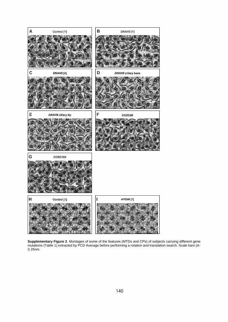
140
Supplementary Figure 2. Montages of some of the features (MTDs and CPs) of subjects carrying different gene mutations (Table 1) extracted by PCD Average before performing a rotation and translation search. Scale bars (A-I) 25nm.

141
Supplementary Figure 3. Screenshots of PCD Detect Classification and Analysis programs. (A) Cilia Classification has been designed for subject samples that have a non-homogeneous defect in order to group images based on structural similarity, before generating a number of averaged outputs. (B) Analysis program designed to overlay images and highlight ciliary structural differences. The hollow arrowheads indicate the presence of the C1a and C2a CP complexes (see Figure 1). Whereas, the white arrow shows the position of the C2b CP component that is absent in the HYDIN case (in blue) compared to the control (in red). (B1) The program interface showing an overlay of two average images of the CP, control in red and a HYDIN defect in blue. (B1-B4) Shows the different images that can be generated by the program when comparing two averaged images. (B2) A subtraction image showing the difference between the red and blue images. (B3) The subtraction image overlaid (in yellow) onto the red image (image 1). (B4) Image showing overlapping detail from the two images.

142
Supplementary Figure 4. The outputs from hierarchy and PCA classification of MTDs. (A-B) Classification outputs for a subject that has an atypical pathogenic variant in DNAH5 and (C-D) pathogenic variants in DNAH5. (A, C) Graphs generated for hierarchy classification show the feature that has been picked as the reference and the corresponding features used to generate the first class average. (B, D) Graphs of the first six PCA coefficients calculated from MTD features, as well as cluster analysis using k-means to group the features into two classes. The white filled arrowheads indicate absence of the ODA. Scale bar (B, D) 10nm.

143
Supplementary Figure 5. Test of selected images with added noise that have been put through PCD Detect.

144
Supplementary Figure 6. Examples of averaged structures generated from healthy controls. Scale bars (A) 100nm (B-C) 10nm.

145
Supplementary Figure 7. Subjects predicted to have a potential HYDIN defect (PHD) were found to have present C2b CP component. (A) Cilia from a control healthy subject and (B) three subjects predicted to have a PHD. (C-D) Average CP structures from the subjects examined. When generating class averages using the same settings only one class is given for Control [1] and PHD [1], whereas multiple classes are given for PHD [2] and PHD [3]. This is likely to be due to a larger number of features input into PCD detect for PHD [2-3] leading to the detection of minor variations of the features including image tilt. Even so, there is no visible defect of the CP when comparing to the control. The white filled arrowheads indicate the position of the C2b CP component. Scale bars (A – B) 100nm and (C – E) 20nm.

146
Supplementary Table 1. Three cases that were studied using PCD Detect due to previous testing indicating a potential HYDIN defect (PHD). PHD [2] and PHD [3] are siblings.
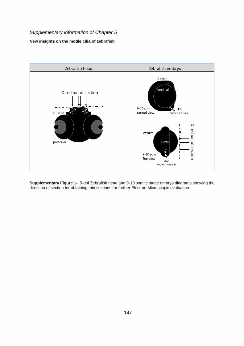
147
Supplementary information of Chapter 5
New insights on the motile cilia of zebrafish
Supplementary Figure 1- 5-dpf Zebrafish head and 9-10 somite stage embryo diagrams showing the direction of section for obtaining thin sections for further Electron Microscopic evaluation.

148
Supplementary Figure 2 - Models of the MTD, product of the tomogram acquisition of both LRO cilia and OP cilia. due to the fact the OP have multiciliated cells, It was possible to aquire tomograms containing several cilia, and so, these were averages together (9+ MTD per field of view), resulting In 2 sub-tomogram averagings assembled using Chimera. On the other hand, in the LRO, there was only one cilia per field of view, and subsequentially, only one cilia to average from (only 9 MTD were averaged per cilium) these too, were assembled in Chimera and models were generated. In Chimera the volume of the ODA was measured in comparisson to the total volume of the MTD using volume measurement tool. Black arrow heads – ODA; white arrow heads – IDA; dotted square – not visible IDA, maybe due to the low resolution of the image, or a location in the axoneme where the IDA was not repeating.

149
Supplementary data Table 1 - Human-Zebrafish homologue PCD genes as described in Ensemble.org 400
PCD gene Zebrafish transcript name Zebrafish transcript ID
DNAH5 dnah5-201 ENSDART00000123150.4
dnah5-202 ENSDART00000191818.1
CCDC114 (ODAD1) ccdc114-201 ENSDART00000023745.8
ARMC4 (ODAD2)
cr847789.1-201 ENSDART00000186851.1
armc4-201 ENSDART00000077453.5
armc4-204 ENSDART00000170018.2
armc4-203 ENSDART00000153115.2
armc4-202 ENSDART00000152887.2
TTC25 (ODAD4) ttc25-201 ENSDART00000080946.5
DNAH9 dnah9-201 ENSDART00000160926.2
DNAH11 dnah11-201 ENSDART00000148294.4
dnah11-202 ENSDART00000020821.10
dnah11-203 ENSDART00000138744.4
DNAI1
dnai1.2-201 ENSDART00000080431.5
dnai1.2-202 ENSDART00000142468.3
dnai1.1-205 ENSDART00000170205.2
dnai1.1-201 ENSDART00000160163.2
dnai1.1-204 ENSDART00000169676.2
dnai1.1-202 ENSDART00000163063.2
dnai1.1-203 ENSDART00000165798.2
DNAI2
dnai2a-201 ENSDART00000162579.2
dnai2a-202 ENSDART00000164199.2
dnai2b-203 ENSDART00000188726.1
dnai2b-201 ENSDART00000003339.9
dnai2b-202 ENSDART00000188648.1
DNAL1 dnal1-203 ENSDART00000188500.1
dnal1-202 ENSDART00000156182.2
dnal1-201 ENSDART00000043651.7
TXNDC3 (NME8) nme8-201 ENSDART00000163684.2
CCDC103 ccdc103-201 ENSDART00000075493.4
ccdc103-202 ENSDART00000132293.2
CFAP298 (C21orf59) cfap298-201 ENSDART00000051197.6
cfap298-202 ENSDART00000130093.3
cfap298-203 ENSDART00000181950.1
CFAP300 (c11orf70) cfap300-201 ENSDART00000151109.2
cfap300-202 ENSDART00000192737.1
DNAAF1 (LRRC50) dnaaf1-201 ENSDART00000145762.4
dnaaf1-203 ENSDART00000173909.2
dnaaf1-202 ENSDART00000173853.2
DNAAF2 (KTU) dnaaf2-201 ENSDART00000167840.2
DNAAF3 dnaaf3l-201 ENSDART00000079233.5
DNAAF4 (DYX1C1) dnaaf4-201 ENSDART00000165855.2
DNAAF5 (HEATR2) lo018183.1-201 ENSDART00000194031.1
DNAAF6 (PIH1D3)
pih1d3-201 ENSDART00000056375.5
pih1d3-203 ENSDART00000145388.3
pih1d3-202 ENSDART00000136858.2
pih1d3-204 ENSDART00000183524.1
pih1d3-205 ENSDART00000191761.1
LRRC6 lrrc6-203 ENSDART00000188883.1
lrrc6-202 ENSDART00000132346.3
lrrc6-201 ENSDART00000075347.5
RPGR
rpgrb-201 ENSDART00000088624.5
rpgrb-202 ENSDART00000124471.3
rpgrip-201 ENSDART00000138541.3
rpgrip-203 ENSDART00000190953.1
rpgrip-202 ENSDART00000179003.2
rpgrip1l-202 ENSDART00000185324.1

150
rpgrip1l-201 ENSDART00000126326.5
SPAG1 spag1b-201 ENSDART00000101207.5
spag1a-202 ENSDART00000185960.1
spag1a-201 ENSDART00000130537.3
ZMYND10 zmynd10-201 ENSDART00000017413.10
zmynd10-202 ENSDART00000189261.1
zmynd10-203 ENSDART00000183251.1
CCDC39 ccdc39-202 ENSDART00000190769.1
ccdc39-201 ENSDART00000169709.2
CCDC40 ccdc40-202 ENSDART00000169752.2
Ccdc40-201 ENSDART00000164275.2
Ccdc40-203 ENSDART00000182267.1
TTC12 ttc12-201 ENSDART00000156234.2
ttc12-202 ENSDART00000157380.2
CCDC65 (DRC2) ccdc65-201 ENSDART00000043946.8
ccdc65-202 ENSDART00000177219.2
CCDC164 (DRC1) drc1-201 ENSDART00000061829.5
GAS8 gas8-202 ENSDART00000170982.2
gas8-201 ENSDART00000165126.2
CFAP221 not found in ZF
DNAJB13
dnajb13-204 ENSDART00000148093.3
dnajb13-201 ENSDART00000063365.6
dnajb13-203 ENSDART00000139097.2
dnajb13-202 ENSDART00000133505.2
HYDIN
hydin-201 ENSDART00000143265.4
hydin-202 ENSDART00000145701.2
hydin-203 ENSDART00000169861.2
bx571975.1-201 ENSDART00000185269.1
NME5 nme5-201 ENSDART00000060998.6
RSPH1 rsph1-201 ENSDART00000160273.3
ct573248.3-201 ENSDART00000181186.1
RSPH3 rsph3-202 ENSDART00000128823.5
rsph3-201 ENSDART00000103394.3
RSPH4a rsph4a-201 ENSDART00000097340.5
RSPH9 rsph9-201 ENSDART00000010903.8
STK36 stk36-201 ENSDART00000086765.5
stk36-202 ENSDART00000139065.2
SPEF2 spef2-201 ENSDART00000159718.2
spef2-202 ENSDART00000168984.2
CFAP57 cfap57-201 ENSDART00000080900.6
cfap57-202 ENSDART00000149309.3
LRRC56 lrrc56-202 ENSDART00000161369.2
lrrc56-201 ENSDART00000150364.2
GAS2L2 gas2l2-201 ENSDART00000112744.4
NEK10 nek10-201 ENSDART00000155162.2
OFD1 ofd1-201 ENSDART00000000552.12
CCNO fq311924.1-201 ENSDART00000158096.2
FOXJ1
foxj1b-201 ENSDART00000126676.2
foxj1b-203 ENSDART00000181942.1
foxj1b-202 ENSDART00000153327.2
foxj1a-201 ENSDART00000157772.2
foxj1a-202 ENSDART00000168280.2
MCIDAS cu633857.1-201 ENSDART00000192716.1