Axonal Dynamics of Excitatory and Inhibitory Neurons in Somatosensory Cortex

Pergamon 0306-4522(95)00375-4
Neuroscience Vol. 70, No. 1, pp. 79-92, 1996 Elsevier Science Ltd
Copyright © 1995 IBRO Printed in Great Britain. All rights reserved
0306-4522/96 $9.50 + 0.00
DIFFERENTIAL EFFECT OF WHISKER TRIMMING ON EXCITATORY AND INHIBITORY TRANSMISSION IN
PRIMARY SOMATOSENSORY CORTEX OF THE ADULT RAT IN VIVO
S. DOLAN* and P. M. B. CAHUSAC Department of Psychology, University of Stirling, Stirling FK9 4LA, Scotland, U.K.
Abstract--The effects of sensory deprivation on excitatory and inhibitory activity in the primary somatosensory cortex were studied in the adult rat. Excitatory and inhibitory transmission generated by whisker stimulation, and neuronal responsiveness to iontophoretically applied excitatory amino acids were recorded. Whisker input deprivation, through whisker trimming for a median of 24 days, resulted in a significant decrease in excitatory transmission to surround whisker stimulation. In contrast, the response magnitude to principal whisker stimulation remained unchanged. However, the response latencies to principal whisker and surround whisker stimulation were significantly reduced, which led to altered temporal response distributions in deprived cells. Neurons deprived of sensory input were significantly less responsive to glutamate, N-methyl-D-aspartate, ct-amino-3-hydroxy-5-methyl-4-isoxazol, epropionate and kainate. Following deprivation, no change was observed in cortical inhibitory transmiss~n measured 30-200 ms post-stimulus.
These results show that excitatory transmission (including excitatory amino acid receptor function) is altered by adult whisker deprivation.
Key words: barrel cortex, sensory deprivation, excitatory amino acids, synaptic transmission, glutamate.
The fine structure of the adult primary somatosen- sory cortex (SI) is continually being modified by afferent activity, and altering levels of sensory input can lead to functional and structural reorganiza- tion. 22'37 The rodent whisker system provides a useful model for studying the effects of peripheral sensory alterations on neuronal plasticity. The mystacial whiskers are arranged in five distinct rows (labelled A-E), and each whisker projects to a discrete aggre- gate of neurons in layer IV of the SI, known as a barrel. The barrels are arranged in a series of rows and arcs which preserve the pattern of the facial whiskers. 68 Each barrel receives rapid excitatory input from its corresponding facial whisker, the principal whisker (PW), by direct input from the ventral pos- terior medial thalamic nucleus. 7 Excitatory responses of longer latency also occur, which are mediated intracortically via polysynaptic relay from surround- ing barrels. 8 The excitatory amino acid (EAA) gluta- mate is believed to be a major neurotransmitter in cortex. 63 The glutamate ionotropic receptor subtypes
*To whom correspondence should be addressed. Abbreviations: AMPA, ct-amino-3-hydroxy-5-methyl-4-
isoxazolepropionate; EAA, excitatory amino acid; GAD, glutamate decarboxylase; I-S, inter-stimulus; NMDA, N-methyl-D-aspartate; PSTH, peristimulus time histograms; PW, principal whisker; S1, stimulus 1; $2, stimulus 2; SI, primary somatosensory cortex; SW, surround whisker.
N-methyl-D-aspartate (NMDA) and non-NMDA are known to mediate sensory transmission in the neo- cortex. 6'25 Within the non-NMDA subtype, ~-amino- 3-hydroxy- 5-methyl-4-isoxazolepropionate (AMPA) and kainate are known selective agonists. Large numbers of glutamate-positive staining neurons are found in the SI? 1 In addition, high densities of NMDA and AMPA reactivity have been character- ized within the barrel field. 3° The barrel field also possesses a large population of GABA-immuno- positive neurons 18,33 which mediate synaptic inhi- bition, and therefore play an important role in regulating excitatory activity within the barrel cortex. 43
The effect of altering sensory input on the normal cytoarchitecture, and functional activity of the adult SI barrel cortex, has been well documented. For example, a reduction in energy metabolism in the barrel cortex follows whisker removal, 67 and an in- crease occurs after stimulation of a single spared whisker. 35'41 Additionally, the expression of gluta- mate, 6° the GABA biosynthetic enzyme glutamate decarboxylase (GAD), 4,64 and the expression of NMDA ar, d AMPA receptorsF have all been found to be regulated by the recent history of whisker activity. Electrophysiological studies have provided evidence for changes in SI cortical neuronal receptive fields after manipulation of whisker input in adult- hood. Alterations in neuronal response properties
79

80 S. Dolan and P. M. B. Cahusac
were found following a br ief per iod of "whisker pa i r ing" (where all whiskers are removed except one pair of adjacent whiskers), such tha t the response magni tudes of the paired whiskers increased, while the responses to cut su r round whiskers decreased) 1'22 A n explana t ion for this cort ical reorganiza t ion has focussed on changes in pos tsynapt ic receptor activity in in t racor t ical pathways. A dependence on E A A receptor funct ion in ac t iv i ty-dependent synaptic plas- ticity has been demons t r a t ed in developmenta l stud- ies on the visual system, 26,34 and tO a lesser degree in work on the barrel cortex. 52 The present s tudy exam- ined the effect of whisker depr iva t ion on exci tatory and inhib i tory cortical synaptic activity by recording extracellular neurona l responses to na tu ra l whisker s t imulat ion. The sensitivity of neurons to the excit- a tory effects of iontophore t ica l ly applied glutamate, N M D A , A M P A and kainate was also studied.
EXPERIMENTAL PROCEDURES
Whisker trimming
Male Wistar rats (Charles River U.K. Ltd) (n = 34) ranging in weight from 350 to 500 g had a single row of whiskers trimmed back to the level of the fur, on one side of the face. For this procedure some animals were anaes- thetized with isoflurane, but most were simply hand held. Animals were then returned to their normal environment. Whiskers were checked every second day for regrowth, and if necessary were retrimmed. Alternate rats either had all whiskers in row E trimmed, or in row C trimmed.
Electrophysiology The median duration of whisker deprivation was 24 days
(range 10451 days). Whiskers were allowed to regrow for two to three days before the experiment to allow for stimulation. Rats were anaesthetized with isoflurane, fol- lowed by an intraperitoneal injection of urethane (1.5 g/kg, 25% in water). Top-up doses were given if necessary during the experiment. Anaesthesia was maintained at a depth such that hindlimb withdrawal reflex to firm pinching was abol- ished. Tracheal and jugular cannulations were performed. Body temperature was maintained at 37°C under rectal thermistor control and the heart rate continuously moni- tored on an oscilloscope.
A rectangular area of skull was removed to expose the posteromedial barrel subfield area of the SI (0°5 mm pos- terior to bregma, 4-7 mm lateral to the midline). The dura was removed and the exposed area was covered with warm 2% agar in phosphate-buffered saline. Recording sessions lasted for periods of approximately 8-10 h.
A total of 130 cells were recorded from barrels deprived of whisker input. Control data (n = 160) were collected from both untrimmed control animals and from trimmed animals (control neurons came from barrels receiving input from vibrissae situated two rows away from the trimmed row). Extracellular neuronal recordings were made from layer IV (500~00 #m) of the SI barrel cortex, using multi-barrelled carbon-fibre microelectrodes (tip diameter=10-15gm). The electrode was orientated normal to the pial surface, and advanced through the cortex using a microdrive. Extracellu- larly recorded electrical activity was amplified and filtered using Neurolog equipment, and triggered spikes transferred to two oscilloscopes for visual inspection, one of which was fed from a delay-line. Individual spikes were isolated using a window discriminator. In addition, activity was monitored through a loud speaker. The signal was captured by com- puter using the CED 1401plus interface and Spike 2 data
capture program. Details of all piezo stimulator activity and iontophoretic applications, i.e. duration and magnitude of current, were also recorded on computer disk.
Individual barrels in the contralateral cortex were located during the experiment using the following criteria. First, the depth was provided by the microdrive reading, giving a rough guide to the anatomical location of the cell. Secondly, and more accurately, cells in layer IV have greater response magnitudes and shorter modal latencies (6-10 ms) to deflec- tions of the PW. s To verify the location of recording sites, electrolytic lesions were made at the end of each track by passing a 2.5 #A current (tip negative) through the carbon fibre electrode for 10 s. This was followed by dye ejection (500 nA for 20 min), on the last recording site, providing additional information on the specific location of the recording site.
Excitatory synaptic transmission The postsynaptic activation of both control and deprived
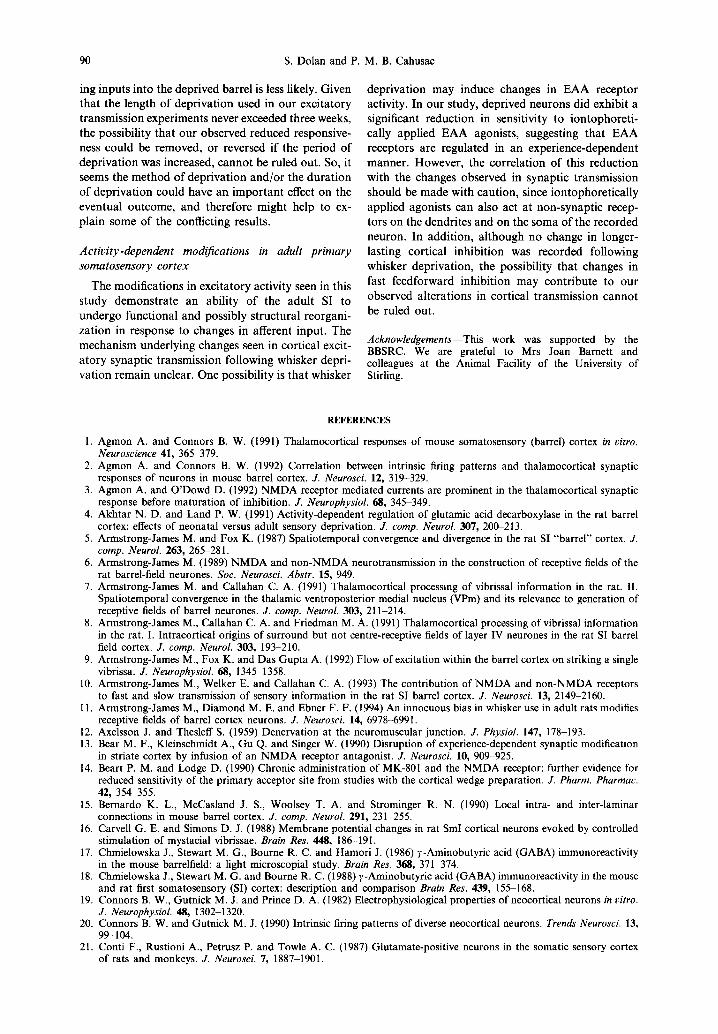
neurons after deflection of appropriate whiskers was recorded. The mean modal response latencies, and the mean number of spikes per stimulus generated in 5-100 ms post- stimulus, were determined from peristimulus time his- tograms (PSTHs). Deflection of whiskers was achieved using a specially constructed stimulator: a piezoelectric bimorph slab with a borosilicate glass capillary tube attached, into which the appropriate whisker was placed. Stimulation consisted of at least 40 deflections of the selected whisker for 3 ms durations, followed by a 3 s delay. Deflections of approximately 200 #m were produced from an application of 25 V. All responses were corrected for spontaneous activity, which was calculated from the number of spikes arising 1 s prior to each trial. The PW was that whisker which evoked the largest response to the stimulus. The response magnitude (spikes/stimulus) and response latency to deflection of the PW were compared in cells from both control and deprived barrels. Responses to deflection of four neighbouring surround whiskers (SWs) were also recorded. Two were located within the same row as the PW (Within- rows): WLARGE, which represents the SW which evoked the largest response magnitude, and WSMALL, which represents the SW which produced the smaller response magnitude. The other SWs were recorded from adjacent rows (Between- rows): and were labelled BLARG E and BSMAL L (see Fig. 1). The mean number of spikes evoked after SW stimulation were represented as a percentage of the PW response magnitude.
lontophoresis
Iontophoresis experiments were carried out on control neurons and neurons in deprived barrels. Iontophoresis barrels contained a combination of the following receptor agonists (all drugs purchased from Tocris): L-glutamate (0.5 M, pH 8.5), NMDA (0.1 M, pH8), AMPA (0.01M in 0.075 M NaC1, pH 8) and kainate (0.1 M, pH 8). The re- maining barrels contained Pontamine Sky Blue dye and 1 M NaC1 for current balancing. Positive retaining currents were used, and ranged from 12 to 14 nA. Cells were studied using cycled applications of each drug. At least two cycles, but usually three or more cycles, of each agonist were applied. Glutamate and NMDA were ejected for 20 s, and AMPA and kainate were ejected for 25 s. Each of these applications was followed by 20-60 s of retention within the cycle period. Ejection currents were adjusted to achieve an obvious increase in firing above the spontaneous level. Those cells with agonist-evoked activity of less than 5 Hz above spon- taneous were discarded from analysis.
The firing rate of control and deprived neurons was recorded for each EAA agonist, and the mean number of spikes per second calculated. The spontaneous firing rate of each neuron was also recorded for a minimum of 3.5 s before the application of each drug. In order to compare response magnitudes to each particular agonist, an Acti- vation Index was constructed which divided the response

Whisker deprivation in ttie adult rat 8
A
_ 7 " 0 0 0 0 ~ A
• @0 ~ c \ sh 0 0 0 .-.,~ G D
\ ~ 0 0 0 0 0 0 w
~ 3 2 1
--....
B
Fig. 1. A schematic representation of the facial whiskers is shown in A. The whiskers are labelled rows A - E and numbered rostro-caudally. Examples of the whiskers studied in a given experiment are represented by the filled circles. The PW is C3 (black circle) and the four SWs are C2, C4, B3 and D3 (light shaded circles). The spatial arrangements of the facial whiskers are replicated in the contralateral cortex, as shown in B. Rounded outlines represent barrel boundaries in layer IV. The barrels labelled in B correspond to the shaded whiskers in A. The SWs to either side of the PW within row C are labelled WLArt~E and WSMALL, and SWs to either side of the PW in adjacent rows are labelled BLARG E and BSMAL L . In a given experiment the position of the PW and SWs varied depending upon the particular row of
whiskers that had been trimmed, and upon the recording position.

82 S. Dolan and P. M. B. Cahusac
rate, corrected for spontaneous activity, by the ejection current used:
Activation Index
Firing rate - Spontaneous activity
Ejection current × 100.
Inhibitory synaptic transmission To evaluate the magnitude of inhibitory input and to
assess the influence of whisker deprivation on barrel neuron inhibition, a condition-test paradigm was devised. First, the inhibition within a single whisker barrel was examined using a paradigm which consisted of a single deflection of the PW (stimulus 1, S1), followed directly by a second stimulus of the PW (stimulus 2, $2), of identical duration and intensity, and delivered at a range of inter-stimulus (I-S) intervals. These were 30, 50, 100, 150 and 200ms I-S intervals. Not all cells were tested using all the I-S intervals. Each cell was subjected to a minimum of 40 such trials. The mean number of spikes per stimulus generated and the mean response latencies were calculated for SI and $2. From these results, the reduction in the spike count, the percentage inhibition and the latency difference were calculated at each I-S interval.
To assess the magnitude of between-barrel inhibition, a second paradigm was devised. A neighbouring SW within the same row as the PW was initially deflected in isolation (S1). After a 3 s interval, stimulation of the PW preceded stimulation of the SW ($2) by 50 ms. The percentage inhibition and the latency difference were calculated from the response to S1 and from $2 (the inhibited response). The magnitudes of the inhibitory input and latency changes were compared in control and in deprived neurons.
Histology At the end of the experiment, rats were perfused with
75 ml 0.9% saline with 0.25 ml 2% lignocaine and 0.2 ml 2% heparin added. This was followed with 100ml of 1% paraformaldehyde and 2% glutaraldehyde in phosphate- buffered saline, 60 ml 10% sucrose and 60 ml 20% sucrose in 0.1 M phosphate buffer. Brains were removed and stored in 20% sucrose at 4°C until they sank. Serial tangential sections were cut at 50 #m on a freezing microtome, and then underwent cytochrome C reaction to identify lesion sites. The incubation medium used was that described by Wong-Riley: ~ 50 mg diaminobenzidine, 90 ml 0.1 M phos- phate buffer, pH 7.4, 15.30 mg cytochrome C and 4 g su- crose made up fresh before use. Sections were incubated at 37°C for 30~o0 min and checked for light to dark reaction. Sections were then rinsed in three changes of 0.1 M phos- phate buffer, mounted, air dried and coverslipped.
DEPRIVED
100 1 W~atcz
~ I • B~o, ~ /
w , . . Imrmlm I
100
CONTROL
w . . . . I • l m I •
J ,. 0 " " "' """ "" ""
Fig. 2. Three-dimensional bar diagrams showing both the spatial arrangement and the response magnitude to the whiskers studied. The mean responses to deflection of each SW (two located within the same row as the PW and two in adjacent rows) are represented here as percentages of the PW response. The response magnitude to PW deflection is equal in control and deprived neurons, and was therefore assigned the value of 100°/0. Control data are shown in the bottom diagram and deprived data in the top graph. In the deprived condition the response magnitude to SW deflection was significantly lower for three of the whiskers: WLARGE,
BLARG E and BSMAL L.
RESULTS
Excitatory synaptic transmission
Magnitude of response to principal whisker and surround whisker stimulation. Exci ta tory postsynapt ic responses to P W and SW s t imula t ion were recorded f rom a to ta l of 74 neurons in 14 rats. The mean n u m b e r of spikes produced to deflection of the P W was 3.04 spikes/s t imulus (n = 36) by cells in deprived barrels and 2.83 spikes/st imulus (n = 38) by cells in cont ro l barrels. The small difference was no t statisti- cally significant (t = 1.13, 69 d.f., P = 0.26).
The responses evoked after deflection of four se- lected SWs were also recorded for each neuron . Less spikes were p roduced by SW st imula t ion compared with P W st imula t ion (see Fig. 2). In depr ived neurons
the mean n u m b e r of spikes generated in response to all four SWs (0.96 spikes/st imulus) was significantly lower than the mean n u m b e r of spikes produced in cont ro l neurons (1.44 spike/st imulus) [F(1,112) = 8.58, P = 0.004.]. The results of t-tests carr ied out on cor responding whiskers f rom bo th condi t ions are displayed in Table 1, and show the response magni-
tudes to WLARGE, BLARG E and BSMAL L to be signifi- cant ly lower in deprived neurons.
Latency of response to principal whisker and sur- round whisker stimulation. The m e a n modal latency was recorded after P W and SW deflection for all cells. S t imula t ion of the P W tended to evoke the fastest response latency in b o t h condi t ions (range 6-12 ms). However, cells in deprived barrels were found to respond faster to deflection of the P W than control

Whisker deprivation in the adult rat 83
Table 1. The response magnitudes to stimulation of the four selected surround whiskers represented as a percentage of
the principal whisker response
SW response magnitude (% of PW response)
P value Whisker Control Deprived (t-test)
WLARG E 6 8 + 4 (n =31) 5 4 + 4 (n = 35) 0.009* BLARG E 53 + 5 (n = 28) 36 + 4 (n = 26) 0.008* WSMAL L 31+3 (n =30) 27+5 (n = 15) 0.54 BSMAL L 28 __+ 4 (n = 20) 16 +_ 2 (n = 16) 0.02*
For each whisker the percentage response + S.E.M. and the number of cells studied (in parentheses) are shown for control and deprived neurons. The results of independent t-tests are shown in the last column. Those SW responses significantly reduced are denoted by an asterisk.
cells. A Mann-Whi tney test showed the latency of deprived cells (median = 7 ms, n = 34) to be signifi- cantly faster than the response latency of control cells (median = 8 ms, n = 38) (W = 1623, P = 0.007) (see Table 2). This result was supported by an analysis of the temporal distribution of the PW response. The PW response was divided into two epochs: 5-8 and 8 -100ms post-stimulus. In the control condition, spikes evoked in the 8-100 ms epoch accounted for 75% of the total response magnitude. Hence, only 25% of spikes occurred in the first 8 ms. An altered response distribution was produced by cells in de- prived barrels, as shown in Fig. 3. The spikes pro- duced in the 8-100 ms epoch accounted for only 48% of the total response. As a result, just over 50% of the PW response occurred in the first 8 ms post-stimulus. The mean number of spikes generated by deprived neurons in the first epoch was significantly higher than spikes evoked by control cells (t = 4.8, 66 d.f., P = 0.001).
Stimulation of SWs produced responses with longer latencies than PW responses. In control neur- ons the mean modal latency varied considerably (range 8-36 ms); the collective mean latency for all four SWs was 14ms. Shorter response latencies to SW stimulation were more common in deprived neurons, where the collective mean latency following SW stimulation was 10.4 ms, and unlike control cells
Table 2. The median response latencies to stimulation of the principal whisker and the four surround whiskers in control
and deprived neurons
Whisker
Response latency (ms) P value (Mann-
Control Deprived Whitney)
PW 8 (n = 38) 7 (n = 34) 0.01" WLA~ E 9 (n = 31) 8 (n = 26) 0.02* BLARG E 11 (n =28) 10 (n =21) 0.14 WSMAL L 11 (n = 27) 10 (n = 22) 0.25 BSMAL L 15 (n = 19) 10 (n = 15) 0.01"
The number of ceils studied are given in parentheses. The results of Mann-Whitney tests are shown in the last column. The response latencies significantly reduced are denoted by an asterisk.
the latency never exceeded 20 ms. A two-way analysis of variance was performed on data from all four SWs and response latencies were found to be significantly faster in deprived cells [F(1,112) = 20.38, P < 0.001]. Individual corresponding SW latencies were com- pared and a Mann-Whi tney test showed that stimu- lation of the whiskers WLARG E and BSMAL L produced significantly faster response latencies in the deprived condition (see Table 2). The shift in SW response latency (e.g., in Fig. 3) became obvious after analysis of the response distribution to SW stimulation. In control neurons, approximately 90% of the total response occurred after 8 ms post-stimulus. However, deprived neurons displayed an altered response pat- tern in which only 68% of the total response occurred after 8 ms. Thus, a significantly larger number of spikes occurred in the 5-8 ms epoch (32% of total SW response) than occurred in this epoch in control neurons (14% of total response) ( t - -2 .75 , 53 d.f., P = 0.01).
Changes in response magnitude and latency were observed after nine to 21 days of whisker trimming, but there was no relationship between the durat ion of whisker tr imming and the size of the effects.
Responses to excitatory amino acid receptor agonists
Extracellular recordings during iontophoretic ap- plication of EAA agonists were obtained from 81 cells in layer IV of the SI in 12 whisker-deprived animals, of which 39 were located in deprived barrels and 42 were located in control barrels. Addit ional data from two untrimmed animals were recorded from cells in rows C and E (n = 17). No difference was found between trimmed and untr immed control animal neuronal Activation Indexes, for each EAA agonist (all t-tests: P > 0.3).
The mean response rates, corrected for spon- taneous activity, and the mean ejection currents recorded for each E A A are listed in Table 3. Deprived neurons required larger ejection currents of each agonist to achieve a firing rate comparable with control cells. As a result, the Activat ion Indexes calculated for each glutamate, N M D A , A M P A and kainate were significantly lower in cells deprived of whisker input. The differences in Activation Indexes are displayed in Fig. 4.
A paired t-test was also done on control and deprived data collected from the same animals (the same electrode was used on control and deprived neurons for each animal). Sufficient data were col- lected on responses to N M D A (t = 3.27, 5d.f., P = 0.02) and A M P A (t = 3.15, 4 d.f., P = 0.035) in five animals, and this showed that the Activation Indexes for deprived cells were again significantly lower than the Activat ion Indexes for control cells.
Cells deprived of whisker input displayed a significantly lower median spontaneous firing rate (0.623 spikes/s) compared to control cells (1.81 spikes/s) (W = 1278, P = 0.02). The relationship between spontaneous activity and the sensitivity to

84 S. Dolan and P. M. B. Cahusac
C O N T R O L D E P R I V E D
WLARCE
I I I I I I I
B LARGE
i O
WSMALL
-10 0 10 30 50 -10 0 10 30 50 Time (ms) Time (ms)
Fig. 3. Typical differences in the response profile between control and deprived neurons. PSTHs generated from deflection of the principal and three selected SWs. The histograms on the left are from a control cell and those on the right are from a deprived cell. The top histograms show the response to PW stimulation, while those below are responses to three SWs, as labelled. Each histogram displays the response over 50 ms post-stimulus for 40 trials. The vertical axis represents the number of spikes and the horizontal axis the time in milliseconds. The deprived cell PSTHs clearly show the reduction in the number of spikes generated in response to SW deflection. The change in response latency is also clear, with few
spikes occurring after 10 ms.
the E A A agonists was further analysed. Cells were divided into two groups: high spontaneous firing rate ( > 1 spike/s) and low spontaneous rate ( < 1 spike/s). Control cells had equal mean Activat ion Indexes, irrespective of their level of spontaneous activity. However, deprived neurons with high spontaneous firing rates produced significantly lower mean Acti- vat ion Indexes than those deprived cells with low spontaneous firing rates (t = 3.71, 88 d.f., P = 0.001). Thus, those neurons with zero or minimal spon- taneous activity, which one might expect to be the least sensitive to E A A agonists, were actually more sensitive and possessed higher Activat ion Indexes.
Changes in Activat ion Indexes were observed after 10-21 days of whisker trimming, but there was no relationship between the duration of whisker t r imming and the magnitude of the effect.
Inhibitory synaptic transmission in the primary somatosensory cortex
The magnitude of inhibitory input in response to natural whisker stimulation was recorded from a total of 101 neurons in layer IV of the SI barrel cortex, in eight adult male Wistar rats. Data were collected from 55 deprived cells and 46 control cells,

Whisker deprivation in the adult rat
Table 3. Changes in effectiveness of iontophoretically applied excitatory amino acid agonists
85
Glutamate NMDA AMPA Kainate
Firing Ejection Firing Ejection Firing Ejection Firing Ejection rate current rate current rate current rate current (nz) (hA) n (Hz) (nA) n (Hz) (nA) n (nz) (nA) n
Control 6__+0.4 27-1- 1.5 15 12__ 1.3 224-2.0 38 18__+2.1 224- 1.8 34 18_+2.3 21 4- 1.8 39 Deprived 54-0.5 344- 1.2 7 74-0.7 374-2.0 27 134- 1.9 344-2.2 30 144- 1.6 344-2.2 31
The mean firing rate in Hz (spikes/s) and the mean ejection current in nA _ S.E.M. recorded for each EAA agonist are listed for both control and deprived neurons. The firing rates have been corrected for spontaneous activity. The number of neurons (n) studied during the application of each EAA agonist are also given.
17 of which came from untr immed control animals and 29 from tr immed controls.
The mean number of spikes per stimulus generated after stimulation of the PW was similar in untr immed (3.22, n = 17) and t r immed (3.80, n = 29) controls, and inhibition after the condition-test on the PW was unchanged (t = 1.06, 37 d.f., P = 0.3). As a result, data from both control conditions were pooled for all subsequent analysis.
Spontaneous activity was again lower in deprived neurons (0.78 spikes/s, n = 55) than in control neur- ons (1.35 spikes/s, n = 46), al though this difference was not statistically significant. As expected, there was a relationship between the level of spontaneous activity and the strength of the inhibitory input, such that cells with low spontaneous activity were more strongly inhibited.
Within-barrel inhibition. Within-barrel inhibition measured by the condition-test protocol on the PW, at the standard I-S intervals of 50 ms, showed a significant reduction in the number of spikes elicited to $2. In control neurons the mean number of spikes per stimulus produced to the first deflection of the PW (S1) was 3.59 spikes/stimulus. This response was reduced to 1.27 spikes/stimulus after $2 (mean de- crease of 63.4%). Neurons deprived of whisker input had a mean of 4.13 spikes/stimulus to S1, which was not significantly different from control cell responses to S1 (as reported earlier). The number of spikes produced to $2 was reduced to 1.81 spikes/stimulus (mean decrease of 56.8%). The level of inhibition in response to $2 was similar in both control and deprived conditions. An independent t-test (t = 1.09, 97 d.f., P = 0.28) and a paired t-test on related data
100--
80-
,~ 60- .=.
e ~
..~ "~ 40- <
Ii
m _ m
glutamate NMDA AMPA kainate
T T • Control [ ] Deprived
Fig. 4. Changes in sensitivity to EAA agonists following whisker trimming. Bar diagram illustrating the Activation Indexes in 100 x spikes/(s x nA), calculated for glutamate, NMDA, AMPA and kainate. The filled bar represents the mean Activation Index for control cells, and the open bars those for deprived cells. The Activation Index is represented on the vertical axis. S.E.M. values are indicated on each bar. The group comparisons for each agonist revealed statistically significant differences: *P <0.05, **P < 0.01, ***P < 0.001. Numbers of ceils in each of the groups were as follows: glutamate control, n = 15; glutamate deprived, n = 7; NMDA control, n = 35; NMDA deprived, n = 24; AMPA control,
n = 34; AMPA deprived, n = 30; kainate control, n = 39; kainate deprived, n = 31.

86
80
70
60
~ so e~
N 4O
~ 3 0
2O
10
0
S. Dolan and P. M. B. Cahusac
Cond i t i ons
~ _ - t l - oo~ol ' l ~ - - - ~ " " deprived
. . . . . . . . . . . . . . . . . . . . . . . . . . . . . " ' , . . . . .
30 50 100 150 200
I-S I n t e r v a l ( m s )
Fig. 5. Inhibition using a condition-test paradigm on the PW for control and deprived neurons. Two stimuli of equal strength and duration were applied at different I-S intervals (30-200 ms), and the mean percentage decrease to the second stimulus are plotted. Control results at each I-S interval are represented by a square and deprived results are represented by a triangle. The vertical axis displays the percentage inhibition and the horizontal axis displays the I-S
intervals, in milliseconds.
f rom six animals (t = 0.43, 7 d.f., P = 0.68) failed to show any indication of statistically significant differences.
The level of inhibition was compared in both conditions using a range of I-S intervals (see Fig. 5). A similar pattern was found in both control and deprived conditions. Inhibition of the PW response was maximum at 30 ms I-S, where few spikes could be elicited. Responses only began to reach baseline levels when the I-S interval reached 200 ms. The inhibition recorded in neurons deprived of whisker input was indistinguishable from control cell results at all I-S intervals studied. A two-way analysis of variance failed to find any differences in the inhibition
30
cond i t i ons
- ~ control - - -~t ' - - deprived
200
4
~3
50 100 150
I-S I n t e r v a l ( m s )
Fig. 6. Changes in response latency following a condition- test paradigm on the PW are shown in the graph, for control and deprived neurons. The stimuli were applied at different I-S intervals (30-200 ms), shown on the horizontal axis. The changes in response latency are shown on the vertical axis for each I-S interval. Control results are represented by a
square and deprived results by a triangle.
to $2 for control and deprived neurons at each interval studied [F(3,32)= 0.58, P = 0.63].
At each I-S interval the mean response latency to stimulation of the PW was determined from PSTHs, and the difference in latency between responses to S1 and $2 was calculated for each cell. For PW stimu- lation there was a significant increase in the response latency to $2 in both control and deprived neurons, at all I-S intervals studied (all t-tests: P < 0:03). The shift in latency was greatest at 30 ms I-S, where the response latency to $2 was increased by 4.29ms (n = 17) in control neurons and by 3.85 ms (n = 13) in deprived neurons. As the I-S time increased, the latency shift (and the degree of inhibition) to $2 decreased, until at 200 ms I-S intervals no latency shift occurred. A similar pattern of results was col- lected from control and deprived neurons for each I-S time; these results are plotted in Fig. 6. No difference in the latency change was detected between the two conditions by a two-way analysis of variance [F(3,127) = 4.49, P = 0.42].
Between-barrel inhibition. Inhibition mediated be- tween barrels by stimulation of the PW was also studied. Responses to stimulation of the nearest SW (S1) within the PW row were recorded in each cell. The response to stimulation of the SW was then recorded 50 ms directly after stimulation of the PW ($2). Control and deprived cells displayed matching levels of inhibition to $2. Control cells (n = 41) had a mean of 1.24 spikes/stimulus to $2 (a mean decrease of 63.5%) and deprived cells (n = 40) had a mean of 1.59spikes/stimulus to $2 (a mean decrease of 67.3%).
A similar mean modal response latency of 9 ms to S1 was recorded from control and deprived neurons. A significant increase in the response latency was recorded after $2 of 1.1 ms in the control, and 0.73 ms in the deprived condition (both t-tests: P <0.01). The change in latency was not statistically signifi- cantly different between control and deprived con- ditions.
Inhibitory transmission in different whisker rows. As alternate animals had either row C or row E tr immed (see Experimental Procedures), the level of inhibition in each row was compared. Within-barrel inhibition showed no row-specific differences in inhibition. However, when assessing the inhibitory transmission between barrels, row E displayed higher inhibition than row C (t =2.16, 75d.f., P =0.05). This is in agreement with Simons et al .: 5 who reported that the ventral and caudal whiskers evoked the greatest inhibition within the barrel cortex.
D I S C U S S I O N
Altering afferent activity to the adult SI barrel cortex, by tr imming whiskers, was found to have significant effects on intracortical and thalamocorti- cal synaptic transmission. The response magnitude to SW stimuli was reduced. The response latencies to

Whisker deprivation in the adult rat 87
both PW and SW stimuli were also significantly reduced. Deprivation of whisker input also resulted in reduced sensitivity of neurons to iontophoreticaUy applied EAA receptor agonists: glutamate, NMDA, AMPA and kainate. However, neither within-barrel nor between-barrel inhibition measured 30-200 ms post-stimulus was affected by this brief period of sensory deprivation.
Modifications in exci tatory synaptic responses to whisker stimulation
Whisker deprivation induced some significant changes in excitatory synaptic transmission. Synaptic responses to SW stimulation were significantly re- duced. There was also an increase in the number of early latency discharges (i.e. < 8 ms) to both PW and SW stimulation. The reduction in excitatory response magnitude from the SWs, but not from the PW, suggests changes in intracortical transmission be- tween barrels. This is supported by the fact that inputs from the untrimmed SWs were also signifi- cantly reduced relative to their corresponding control SWs, which suggests that it is primarily the intra- cortical pathway which undergoes modification rather than the thalamocortical pathway.
The total response evoked after stimulation of the PW was equal in control and deprived neurons. However, the alteration in the temporal distribution of the PW response in deprived neurons suggests that some functional modification does take place within the PW barrel. In a series of studies by Armstrong- James and coUeagues, 7,8 it was reported that the early component of the PW response, i.e. 5-8 ms post- stimulus, is almost exclusively generated by input from thalamocortical afferents, which mainly utilize non-NMDA receptors. 1° Those responses occurring during 8-10 ms were said to be a combination of NMDA and non-NMDA receptor activity, while later spike discharges were thought to be mediated exclusively by N M D A receptors. Intracellular record- ings from corticocortical neurons have shown that the excitatory postsynaptic potential consists of an early non-NMDA and a later N M D A receptor com- ponent. 62 What proportion of NMDA receptor re- sponses are mediated via thalamocortical input or via within-barrel intracortical relay 9 is not clear. During development, Agmon and O'Dowd 3 found the thala- mocortical response to be mediated by a combination of N M D A and non-NMDA receptors. However, later in development, as the inhibitory component increased, N M D A receptor-mediated activity was lost. A similar loss of NMDA-mediated activity with maturation of the cortex has been reported previously by Luhmann and Prince. 42 It seems that in the adult cortex, activation of N M D A receptors requires re- moval of a voltage-dependent block. D'Angelo et al. 2~a reported that postsynaptic currents, which were both glycine- and voltage-dependent, could be elicited exclusively through N M D A receptor chan- nels. Inputs which come from non-NMDA receptor
action and from the high levels of spontaneous activity in vivo (see discussion on "Reduced respon- siveness to excitatory amino acid receptor agonists" for more detail) have been suggested as sources of activity which may bring the neuron closer to threshold, and thus enable removal of the NMDA receptor voltage-dependent block. This idea is sup- ported from studies showing apparent poor selectiv- ity of non-NMDA receptors antagonists, where the N M D A response is also reducedJ °,51 For example, non-NMDA antagonists prevent non-NMDA- mediated depolarization of the postsynaptic neuron, which in turn could disable removal of the voltage- dependent block, preventing discharge through N M D A receptors. Indeed, it is this process which has been suggested as a reason for the fast activation of NMDA receptors following PW stimulation) ° NMDA receptor-mediated responses were found to be dependent on the latency of the first responses from the thalamocortical volley. This dependence on the latency of the initial input could underlie our observed changes in response distribution following whisker deprivation; for example, in our study early responses mediated at < 8 ms post-stimulus appeared to potentiate, while responses arising >8 ms de- creased. Faster early non-NMDA receptor-mediated thalamocortical input may lead to faster NMDA receptor activation and hence produce the increase in early spike discharge which was seen. Potentiation of the thalamic input in itself has been found to evoke a faster response latency, which consequently pro- duced an increased number of short-latency spikes. A study by Armstrong-James et al. H reported that potentiation of the thalamocortical relay to spared "paired whisker" stimulation resulted in a corre- sponding reduction in response latency. The latencies of responses to trimmed whiskers were also reduced in that study, in agreement with the present work.
An alternative explanation for our observed shift in response distribution could be that there is a reorga- nization in the relative contribution of NMDA and non-NMDA receptors to the PW evoked response; for example, if the early response mediated via non- N M D A receptors was enhanced, while the later NMDA receptor component was reduced. If indeed transmission mediated via N M D A receptors is re- duced, this would fit well with our observed decrease in SW transmission, which is also thought to be mediated via N M D A receptors. 6 A further possible explanation for the reduced latencies of responses following deprivation could be due to a decrease in the excitatory input to cells mediating feedforward inhibition. Such a decrease in the excitatory drive would tend to curtail the short-latency inhibitory action of these circuits on excitatory throughput and thus allow the manifestation of earlier excitatory responses to occur.

88 S. Dolan and P. M. B. Cahusac
Reduced responsiveness to excitatory amino acid receptor agonists
Neurons deprived of whisker input displayed a clear reduction in sensitivity to each of the EAAs studied. The underlying mechanisms involved in these changes are unclear. They could involve neuronal functional reorganization or modifications at the molecular level. The neocortex receives spontaneous afferent input from various sources, and one main glutamatergic source comes from the thalamus. 24 The high level of spontaneous activity is reflected by the large proportion of control cells which were spon- taneously active (>90%). The diminished spon- taneous activity of neurons deprived of whisker input in this study (only 74% of neurons were spon- taneous), and also a similar reduction in spontaneous activity recorded from a spared whisker barrel, fol- lowing removal of all but two whiskers, H may reflect a general decrease in cortical excitatory activity following deprivation. The binding of EAA neuro- transmitters, generated by spontaneous discharges within the PW barrel, to NMDA receptors has been suggested as a reason for shorter latency NMDA- mediated activity. ~° When the cortex is deprived of whisker input, the level of postsynaptic receptor activation is reduced, since EAA neurotransmitter liberated via spontaneous activity has decreased. As a result, the potential for normal receptor activation may be impaired. Initially, it was thought that this phenomeno n might explain the reduction in respon- siveness to EAA receptor agonists recorded in this study. Simply, as the average neuronal activity and therefore the spiking threshold is reduced, the neuron will be more difficult to depolarize. For instance, levels of spontaneous activity tend to be low in vitro, and as a result the average membrane potential tends to be further away from thresholdfl However, fur- ther analysis of neuronal spontaneous activity re- vealed that this cannot be the only explanation for the reduction in sensitivity to EAA agonists. Unexpect- edly, deprived neurons with a zero or minimal spon- taneous firing rate were significantly more responsive to EAA agonists than those deprived cells with higher spontaneous firing rates. This is the opposite to what would be expected if the cells were further away from spiking threshold, as suggested.
An alternative explanation for the observed re- duction in sensitivity could be that a morphological reorganization of cortical pathways occurs. Previous studies on the adult CNS found removal of or diminished EAA receptor activity induced reorgani- zation of EAA receptor sites. Application of the NMDA antagonist 3-(-2-carboxypiperazin-4-yl)- propyl-l-phosphonic acid for 21 days to adult mice decreased NMDA receptor mRNA in the frontal cortex and hippocampus. 4° Similarly, treatment of adult rats for seven days with the non-competitive NMDA channel blocker MK-801 resulted in de- creased cortical binding to NMDA receptors, 4s and
subsequent depolarizing responses to NMDA recorded from cortical wedge preparations were re- duced relative to control. 14 The down-regulation of N M D A receptor sites after treatment with antagon- ists suggests that receptor expression may be regu- lated by their level of activation. A related study looked at the expression of specific receptor types following four days (10 min/day) of tactile stimu- lation, and found increased binding to both [3 H]MK- 801 and [3H]AMPA, representing an increase in NMDA and AMPA receptors. 57 Related work look- ing at neuronal gene expression has found specific genes to be regulated by sensory input. Increased transcription of immediate early genes following tac- tile stimulation 44 and a reduction in immediate early gene expression following sensory deprivation 6~ has been reported in the barrel cortex. Immediate early gene activation is thought to reflect short-term changes in neuronal activity, and may also be import- ant in neuronal plasticity and long-term poten- tiation. 23 This regulation of gene transcription suggests a molecular basis for the capacity of the adult barrel cortex to adapt to changes in afferent input.
There is abundant evidence from developmental studies showing the expression of specific receptors to be dependent on their level of activation. Many of these studies have used the visual system to analyse the effects of altered sensory input on synaptic organ- ization. The NMDA receptor has been shown to play a crucial role in cortical plasticity, t3'34'56 Elevated levels of both NMDA 26 and non-NMDA 49 receptors occur in the cortex during early development, and control of expression seems to be activity-dependent. For instance, monocular deprivation was found to decrease the expression and distribution of NMDA, kainate and various AMPA receptor subtypes in the visual cortex. 39'5° These results, combined with those from studies on adult sensory manipulation, suggest that, both during development and in adulthood, expression of EAA receptors may be regulated by the level of postsynaptic activation. However, whether a down-regulation of EAA receptors is the actual reason for our observed reduction in sensitivity is unclear from this experiment.
Inhibitory synaptic transmission
The condition-test paradigms used in this study were designed to look at cortical inhibitory inter- actions within a single whisker barrel and between adjacent whisker barrels. Both types of inhibition appeared to be unaffected by whisker removal. How- ever, the design had some limitations in that it only allowed measurement of longer lasting inhibitory activity, i.e. inhibitory responses lasting greater than 30 ms. Previous studies have shown that the first responses in layer IV barrel neurons are a combi- nation of both excitatory and inhibitory activity. 2'65 The barrel cortex contains a rich population of GABAergic, 17.32 non-spiny inhibitory interneurons: 6

Whisker deprivation in the adult rat
which have been identified as fast spiking units. 54 Regular spiking units have been identified with pyra- midal and spiny stellate cells. 2° Both excitatory and inhibitory cells receive direct input from thalamocor- tical afferents, ~ but inhibitory/GABAergic neurons receive more powerful input on their somata and proximal dendrites. 33 In vivo extracellular studies have shown fast spiking barrel neurons to produce powerful, fast-latency responses to sensory stimu- lation. 10.65 These fast GABAergic neurons are thought to generate a wave of feedforward inhibition, inhibit- ing regular spiking cells within the same barrel. For example, Welker et al. 65 found that the firing rate of regular spiking units in layer IV to whisker stimu- lation only became maximal after fast spiking units had decreased their firing rate. This idea is also supported by light microscopic evidence, which shows that smooth cell fast spiking units are substan- tially interconnected within individual barrels, 28 and make GABAergic synapses on both smooth and spiny cells. 33 Thus, fast feedforward inhibitory inter- actions are believed to occur at less than 30 ms post-stimulus, and therefore would remain unde- tected using the paradigm designed for our study. This form of inhibition may be reduced as a conse- quence of a general decrease in excitatory synaptic effectiveness (see above in "Modifications in excit- atory synaptic responses to whisker stimulation").
Longer lasting inhibition seems to depend on intra- cortical transmission between barrels.16 For example, results from the present study and from a previous study by Simons 53 showed that neurons in one barrel can be inhibited by stimulation of an adjacent whisker. A number of possible pathways exist which might mediate between barrel inhibition. One route is tangential relay within layer IV, via local axons which send horizontal excitatory projections to neighbour- ing barrelsJ 5 Alternatively, transmission could be through layer III and layer V neurons, which are known to have extensive horizontal connections ~5 which synapse on layer IV neuronal dendrites, and thus may generate a wave of feedforward inhibition in the neighbouring barrel. Another possible sub- strate for between-barrel inhibition is from GABA- ergic neurons, which have been found to possess dendrites which span across to adjacent barrels and synapse with thalamocortical afferents. 33 Thus, as whisker deprivation was found to produce changes in intracortical excitatory transmission, there is the possibility that inhibition mediated between adjacent barrels could also be affected. However, from our observations cross-whisker-mediated inhibition measured at 50 ms post-stimulus was unaffected by whisker deprivation.
The effects of adult whisker deprivation on cortical inhibition have been examined previously in various binding studies. Removal of whisker follicles in adult- hood resulted in a decrease in GABAA receptor binding in the barrel field. 58 In addition, a reversible decrease in immunostaining of GAD, a GABA
NSC 70/I D
89
biosynthetic enzyme, has been reported, 4'64 which presumably represents a decrease in GABA neuro- transmission. Activity-dependent down-regulation of GABAA receptor subunits has also been shown in the visual cortex following monocular deprivation, both in the developing rat 38 and in the adult monkey. 29 However, Kossut et al. 36 failed to detect any change in GABA immunostaining in the barrel cortex fol- lowing adult denervation. While our experimental observations failed to detect any change in inhibition between 30 and 200 ms post-stimulus, the main body of evidence suggests that GABAergic neurotrans- mission is regulated in an activity-dependent manner. However, the finding that deprived neurons exhibited significantly reduced spontaneous firing rates is con- trary to what would be expected if inhibition and GABA transmission were reduced. Interestingly, in the studies reported above, 36'64 changes in GAD and GABA immunoreactivity corresponded only with those barrels deprived of whisker input. However, other studies using the paired whisker paradigm have found the firing of deprived neurons to whisker stimulation to be decreased, while the firing of spared barrel neurons increased, H'22 which is opposite to what would be expected if the changes in inhibition predicted above did occur.
M e t h o d s o f deprivat ion
The reduction in sensitivity seen in the present study is in contrast to some previous work, where receptor supersensitivity has been observed. For example, at the neuromuscular junction denervation leads to a redistribution of receptors and an increased responsiveness to acetylcholine. ~2 In the rat cortex, destruction of noradrenergic nerve terminals led to a supersensitivity to catecholamines. 59 The differences observed could be attributable to the different trans- mitter systems being studied or alternatively the method of deprivation used. Denervation or nerve transection removes all input to the receptive area in the cortex. Merzenich et al. 48 reported an immediate elimination of all responses in the cortex following nerve transection, whereas after whisker trimming some activity remains, albeit much reduced. Thus, cortical plasticity may be dependent on the mode of deprivation. Alternatively, the length of deprivation could be an important contributing factor. A com- mon feature seen following denervation is the exten- sion of neighbouring connections into the deprived area of cortex. 31'47 This remapping is thought to be due partly to the unmasking of previously ineffective connections. For instance, Welker et al. 64 found a decrease in levels of GAD immunostaining shortly after nerve transection but not after several months, demonstrating the potential for recovery after long periods of denervation. It was suggested that this return to normal could be a result of barrels eventu- ally being taken over by neighbouring inputs. How- ever, as the neuronal connections to our trimmed whisker barrel remain intact, expansion of surround-
90 S. Dolan and P. M. B. Cahusac
ing inputs into the deprived barrel is less likely. Given that the length of deprivation used in our excitatory transmission experiments never exceeded three weeks, the possibility that our observed reduced responsive- ness could be removed, or reversed if the period of deprivation was increased, cannot be ruled out. So, it seems the method of deprivation and/or the durat ion of deprivation could have an important effect on the eventual outcome, and therefore might help to ex- plain some of the conflicting results.
Activity-dependent modifications in adult primary
somatosensory cortex
The modifications in excitatory activity seen in this study demonstrate an ability of the adult SI to undergo functional and possibly structural reorgani- zation in response to changes in afferent input. The mechanism underlying changes seen in cortical excit- atory synaptic transmission following whisker depri- vation remain unclear. One possibility is that whisker
deprivation may induce changes in EAA receptor activity. In our study, deprived neurons did exhibit a significant reduction in sensitivity to iontophoreti- cally applied E A A agonists, suggesting that EAA receptors are regulated in an experience-dependent manner. However, the correlation of this reduction with the changes observed in synaptic transmission should be made with caution, since iontophoretically applied agonists can also act at non-synaptic recep- tors on the dendrites and on the soma of the recorded neuron. In addition, although no change in longer- lasting cortical inhibition was recorded following whisker deprivation, the possibility that changes in fast feedforward inhibition may contribute to our observed alterations in cortical transmission cannot be ruled out.
Acknowledgements--This work was supported by the BBSRC. We are grateful to Mrs Joan Barnett and colleagues at the Animal Facility of the University of Stirling.
REFERENCES
1. Agmon A. and Connors B. W. (1991) Thalamocortical responses of mouse somatosensory (barrel) cortex in vitro. Neuroscience 41, 365-379.
2. Agmon A. and Connors B. W. (1992) Correlation between intrinsic firing patterns and thalamocortical synaptic responses of neurons in mouse barrel cortex. J. Neurosci. 12, 319-329.
3. Agrnon A. and O'Dowd D. (1992) NMDA receptor mediated currents are prominent in the thalamocortical synaptic response before maturation of inhibition. J. Neurophysiol. 68, 345-349.
4. Akhtar N. D. and Land P. W. (1991) Activity-dependent regulation of glutamic acid decarboxylase in the rat barrel cortex: effects of neonatal versus adult sensory deprivation. J. comp. Neurol. 307, 200-213.
5. Armstrong-James M. and Fox K. (1987) Spatiotemporal convergence and divergence in the rat SI "barrel" cortex. J. comp. Neurol. 263, 265-281.
6. Armstrong-James M. (1989) NMDA and non-NMDA neurotransmission in the construction of receptive fields of the rat barrel-field neurones. Soc. Neurosci. Abstr. 15, 949.
7. Armstrong-James M. and Callahan C. A. (1991) Thalamocortical processing of vibrissal information in the rat. II. Spatiotemporal convergence in the thalamic ventroposterior medial nucleus (VPm) and its relevance to generation of receptive fields of barrel neurones. J. comp. Neurol. 303, 211-214.
8. Armstrong-James M., Callahan C. A. and Friedman M. A. (1991) Thalamocortical processing of vibrissal information in the rat. I. Intracortical origins of surround but not centre-receptive fields of layer IV neurones in the rat SI barrel field cortex. J. comp. Neurol. 303, 193-210.
9. Armstrong-James M., Fox K. and Das Gupta A. (1992) Flow of excitation within the barrel cortex on striking a single vibrissa. J. Neurophysiol. 68, 1345 1358.
10. Armstrong-James M., Welker E. and Callahan C. A. (1993) The contribution of NMDA and non-NMDA receptors to fast and slow transmission of sensory information in the rat SI barrel cortex. J. Neurosci. 13, 2149 2160.
I 1. Armstrong-James M., Diamond M. E. and Ebner F. F. (1994) An innocuous bias in whisker use in adult rats modifies receptive fields of barrel cortex neurons. J. Neurosci. 14, 6978~6991.
12. Axelsson J. and Thesleff S. (1959) Denervation at the neuromuscular junction. J. Physiol. 147, 178-193. 13. Bear M. F., Kleinschmidt A., Gu Q. and Singer W. (1990) Disruption of experience-dependent synaptic modification
in striate cortex by infusion of an NMDA receptor antagonist. J. Neurosci. 10, 909-925. 14. Beart P. M. and Lodge D. (1990) Chronic administration of MK-801 and the NMDA receptor: further evidence for
reduced sensitivity of the primary acceptor site from studies with the cortical wedge preparation. J. Pharm. Pharmac. 42, 354-355.
15. Bernardo K. L., McCasland J. S., Woolsey T. A. and Strominger R. N. (1990) Local intra- and inter-laminar connections in mouse barrel cortex. J. comp. Neurol. 291, 231-255.
16. Carvell G. E. and Simons D. J. (1988) Membrane potential changes in rat SmI cortical neurons evoked by controlled stimulation of mystacial vibrissae. Brain Res. 448, 186-191.
17. Chmielowska J., Stewart M. G., Bourne R. C. and Hamori J. (1986) 7-Aminobutyric acid (GABA) immunoreactivity in the mouse barrelfield: a light microscopial study. Brain Res. 368, 371-374.
18. Chmielowska J., Stewart M. G. and Bourne R. C. (1988) 7-Aminobutyric acid (GABA) immunoreactivity in the mouse and rat first somatosensory (SI) cortex: description and comparison Brain Res. 439, 155-168.
19. Connors B. W., Gutnick M. J. and Prince D. A. (1982) Electrophysiological properties of neocortical neurons in vitro. J. Neurophysiol. 48, 1302 1320.
20. Connors B. W. and Gutnick M. J. (1990) Intrinsic firing patterns of diverse neocortical neurons. Trends Neurosci. 13, 99-104.
21. Conti F., Rustioni A., Petrusz P. and Towle A. C. (1987) Glutamate-positive neurons in the somatic sensory cortex of rats and monkeys. J. Neurosci. 7, 1887 190l.

Whisker deprivation in the adult rat 91
21a. D'Angelo E., Rossi P. and Garthwaite J. (1990) Dual component NMDA receptor currents at a single central synapse. Nature 346, 467-470.
22. Diamond M. E., Armstrong-James M. and Ebner F. F. (1993) Experience-dependent plasticity in adult rat barrel cortex. Proc. natn. Acad. Sci. U.S.A. 90, 2082-2086.
23. Dragunow M., Currie R. W., Faull R. L. M., Robertson H. A. and Jansen K. (1989) Immediate-early genes, kindling and long-term potentiation. Neurosci. Biobehav. Rev. 13, 301-313.
24. Fox K. and Armstrong-James M. (1986) The role of anterior intra-laminar nuclei and N-methyl-D-aspartate receptors in the generation of spontaneous bursts in rat neocortical neurons. Expl Brain Res. 63, 505-518.
25. Fox K., Sato H. and Daw N. (1989) The localisation and function of NMDA receptors in cat and kitten visual cortex. J. Neurosci. 9, 2443-2454.
26. Fox K., Daw N., Sato H. and Czepita D. (1991) Dark-rearing delays the loss of NMDA-receptor function in kitten visual cortex. Nature 350, 342-344.
28. Harris R. M. and Woolsey T. A. (1983) Computer assisted analysis of barrel neuron axons and there putative synaptic contacts. J. comp. Neurol. 22,0, 63~9.
29. Hendry S. H. C., Huntsman M. M., Vifiuela A., Mrhler H., de Bias A. L. and Jones E. G. (1994) GABA A receptor subunit immunoreactivity in primate visual cortex: distribution in macaques and humans and regulation by visual input in adulthood. J. Neurosci. 14, 2383-2401.
30. Jaarsma R., Sebens J. B. and Korf J. (1991) Localization of NMDA and AMPA receptors in rat barrel field. Neurosci. Lett. 133, 233-236.
31. Kaas J. H., Merzenich M. M. and Killackey H. P. (1983) The reorganisation of somatosensory cortex following peripheral nerve damage in the adult and developing mammals. A. Rev. Neurosci. 6, 325-356.
32. Keller A. and White E. L. (1986) Distribution of glutamic acid decarboxylase-immunoreactive structures in the barrel region of mouse somatosensory cortex. Neurosci. Lett. 66, 245-250.
33. Keller A. and White E. L. (1987) Synaptic organisation of GABAergic neurons in the mouse Sml cortex. J. comp. Neurol. 262, 1-12.
34. Klienschmidt A., Bear M. F. and Singer W. (1987) Blockade of NMDA receptors disrupts experience dependent plasticity of kitten striate cortex. Science 238, 335-357.
35. Kossut M., Hand P. J., Greenberg J. and Hand C. L. (1988) Single vibrissal column in the SI cortex of rat and its alterations in neonatal and adult vibrissa-deafferented animals: a quantitative 2DQ study. J. Neurophysiol. 60, 829-852.
36. Kossut M., Stewart M. G., Suicinska E., Bourne R. C. and Gabbott P. L. A. (1991) Loss of y-aminobutyric acid (GABA) immunoreactivity from the mouse first somatosensory (SI) cortex following neonatal, but not adult denervation. Brain Res. 538, 165-170.
37. Kossut M. (1992) Plasticity of the barrel cortex neurons. Prog. Neurobiol. 39, 389-422. 38. Kumar A. and Schliebs R. (1993) Post-natal ontogeny of GABA A and benzodiazepine receptors in individual layers
of the rat visual cortex and the effects of visual deprivation. Neurochem. Int. 23, 99 106. 39. Kumar A., Schliebs R. and Bigl V. (1994) Post-natal development of NMDA, AMPA and kainate receptors in
individual layers of the rat visual cortex and the effects of monocular deprivation. Int. J. devl Neurosci. 12, 31-41. 40. Lesch K. P., Aulakh C. S., Wolozin B. L., Hill J. L. and Murphy D. L. (1992) 3-(2-Carboxypiperazin-4-yl)propyl-l-
phosphonic acid decreases NMDA receptor mRNA. Eur. J. Pharmac. 227, 109 I l l . 41. Levin B. E. and Dunn-Meynell A. (1991) Adult rat barrel cortex plasticity occurs at 1 week but not at 1 day after
vibressectomy as demonstrated by the 2-deoxyglucose method. Expl Neurol. 113, 237248. 42. Luhmann H. J. and Prince D. A. (1990) Transient expression of NMDA receptor-mediated activity during neocortical
development. Neurosci. Lett. 111, 109 115. 43. Luhmann H. J. and Prince D. A. (1991) Postnatal maturation of the GABAergic system in rat neocortex. J.
Neurophysiol. 65, 247-263. 44. Mack K. J. and Mack P. A. (1992) Induction of transmission factors in somatosensory cortex after tactile stimulation.
Molec. Brain Res. 12, 141 147. 45. Manallack D. T., Lodge D. and Beart P. M. (1989) Subchronic administration of MK-801 in the rat decreases cortical
binding of [3 H]D-AP5, suggesting down-regulation of the cortical N-methyl-D-aspartate receptors. Neuroscience 30, 87~4.
46. McCormick D. A., Connors B. W., Lighthall J. W. and Prince D. A. (1985) Comparative electrophysiology of pyramidal and spiny stellate neurons of the neocortex. J. Neurophysiol. 54, 782 806.
47. Merzenich M. M., Kaas J. H., Wall J., Nelson R. J., Sur M. and Felleman D. (1983) Topographic reorganisation of somatosensory cortical areas 3b and 1 in adult monkey following restricted deafferentation. Neuroscience 8, 33-55.
48. Merzenich M. M., Kass J. H., Wall R. J., Sur J., Nelson M. and Felleman D. (1983) Progression of change following median nerve transection in the cortical representation of the hand in areas 3b and 1 in adult owl and squirrel monkeys. Neuroscience 10, 639~65.
49. Pellegrini-Giampietro D. E., Bennett V. L. and Zukin R. S. (1991) Differential expression of three glutamate receptor genes in developing rat brain: an in situ hybridization study. Proc. natn. Acad. Sci. U.S.A. 88, 4157-4161.
50. RoBner S., Kumar A., Kues W., Witzemann V. and Schliebs R. (1993) Differential laminar expression of AMPA receptor genes in the developing visual cortex using in situ hybridisation histochemistry. Effect of visual deprivation. Int. J. devl. Neurosci. 11, 411-425.
51. Salt T. E. and Eaton S. A. (1989) Function of non-NMDA receptors and NMDA receptors in synaptic responses to natural somatosensory stimulation in the ventrobasal thalamus. Expl Brain Res. 77, 646~552.
52. Schlagger B. L., Fox K. and O'Leary D. D. M. (1993) Post-synaptic control of plasticity in developing somatosensory cortex. Nature 364, 623~26.
53. Simons D. J. (1983) Multi-whisker stimulation and its effects on vibrissa units in rat SmI barrel cortex. Brain Res. 276, 178 182.
54. Simons D. J. and Carvell G. E. (1989) Thalamocortical response transformation in the rat vibrissa/barrel system. J. Neurophysiol. 61, 311-330.
55. Simons D. J., Land P. W. and Suter K. (1993) Neonatal whisker trimming reduces surround inhibition in adult barrel cortex. Soc. Neurosci. Abstr. 29.10, p. 46.

92 S. Dolan and P. M. B. Cahusac
56. Simons D. J., Prusky G. L., O'Leary D. D. M. and Constantine-Paton M. (1992) N-Methyl-o-aspartate receptor antagonists disrupt the formation of a mammalian neural map. Proc. natn Acad. Sci. U.S.A. 89, 10,593-10,597.
57. Skangiel-Kramska J., Jablonska B. and Kossut M. (1993) Increase of NMDA and AMPA receptor sites in cortical representation of whiskers after peripheral tactile stimulation. Soc. Neurol. Abstr. 69.5, p. 63.
58. Skangiel-Kramska J., Gla2ewski S., Jabtofiska B., Siucifiska E. and Kossut M. (1994) Reduction of GABA A receptor binding of [3 H]muscimol in the barrel field of mice after peripheral denervation: transient and long-lasting effects. Expl Brain Res. 100, 39-46.
59. Sporn J. R., Wolfe B. B., Kendall-Hardon T. and Molinoff P. B. (1977) Supersensitivity in the rat cerebral cortex: pre and post-synaptic effects of 6-hydoxydopamine at noradrenergic synapses. Molec. Pharmac. 13, 1170 1180.
60. Stewart M. G., Suicinska E., Kossut M. and Davies H. (1993) Loss of glutamate immunoreactivity from the mouse first somatosensory (SI) cortex following neonatal vibrissal lesion. Brain Res. 621, 331 338.
61. Stiener H. and Gerfen C. R. (1994) Tactile sensory input regulates basal and apomorphine-induced immediate early gene expression in the rat barrel cortex. J. comp. Neurol. 344, 297 304.
62. Thomson A. M., Girdlestone D. and West D. C. (1989) A local circuit neocortical synapse that operates via both NMDA and non-NMDA receptors. Br. J. Pharmac. 96, 406-408.
63. Tsumoto T. (1990) Excitatory amino acid transmitters and their receptors in neural circuits or the cerebral cortex. Neurosci. Res. 9, 79 102.
64. Welker E., Soriano E. and Van der Loos J. (1989) Plasticity in the barrel cortex of the adult mouse: effects of peripheral deprivation of GAD-immunoreactivity. Expl Brain Res. 74, 441-452.
65. Welker E., Armstrong-James M. and Van Der Loose H. (1993) The mode of activation of a barrel column: response properties of single units in the somatosensory cortex of the mouse to whisker deflection. Eur. J. Neurosei 5, 691-712.
66. Wong-Riley M. (1979) Changes in the visual system of monocularly sutured or enucleated cats demonstrable with cytochrome oxidase histochemistry. Brain Res. 171, 11 28.
67. Wong-Riley M. T. T. and Welt C. (1980) Histochemical changes in cytochrome oxidase of cortical barrels after vibrissal removal in neonatal and adult mice. Proc. natn. Acad. Sci. U.S.A. 77, 2333-2337.
68. Woolsey T. A. and Van der Loos J. (1970) The structural organization of layer 1V in the somatosensory region (SI) of mouse cerebral cortex. Brain Res. 17, 205 244.
(Accepted 22 August 1995)
Copyright © 2022 FDOKUMEN