Selective overexpression of excitatory amino acid transporter 2 (EAAT2) in astrocytes enhances...
14
Selective Over Expression Of EAAT2 In Astrocytes Enhances Neuroprotection From Moderate But Not Severe Hypoxia- Ischemia Melodie L. Weller 1 , Ida M. Stone, Amber Goss, Thomas Rau, Cherokee Rova, and David J. Poulsen * NIH COBRE Center for Structural and Functional Neuroscience, Department of Biomedical and Pharmaceutical Sciences, University of Montana, Missoula, MT 59812 Abstract Attempts have been made to elevate EAAT2 expression in effort to compensate for loss of function and expression associated with disease or pathology. Increased EAAT2 expression has been noted following treatment with β-lactam antibiotics, and during ischemic preconditioning (IPC). However, both of these conditions induce multiple changes in addition to alterations in EAAT2 expression that could potentially contribute to neuroprotection. Therefore, the aim of this study was to selectively overexpress EAAT2 in astrocytes and characterize the cell type specific contribution of this transporter to neuroprotection. To accomplish this we used a recombinant Adeno-associated virus vector, AAV1-GFAP-EAAT2, designed to selectively drive the overexpression of EAAT2 within astrocytes. Both viral mediated gene delivery and β-lactam antibiotic (penicillin-G) treatment of rat hippocampal slice cultures resulted in a significant increase in both the expression of EAAT2, and dihydrokainate (DHK) sensitive glutamate uptake. Penicillin-G provided significant neuroprotection in rat hippocampal slice cultures under conditions of both moderate and severe oxygen glucose deprivation (OGD). In contrast, the overexpression of EAAT2 in astrocytes provided enhanced neuroprotection only following a moderate OGD insult. These results indicate that functional EAAT2 can be selectively overexpressed in astrocytes, leading to enhanced neuroprotection. However, this cell type specific-increase in EAAT2 expression offers only limited protection compared to treatment with penicillin-G. INTRODUCTION Glutamate is the primary excitatory amino acid neurotransmitter in the mammalian central nervous system. When released from presynaptic terminals, glutamate can activate ionotropic receptors such as AMPA and KA to mediate standard fast excitatory signaling, or contribute to the higher order processing required in development, plasticity, learning and memory by activating the NMDA and metabotropic glutamate receptors. Extracellular enzymes do not metabolize free glutamate. Instead, high affinity, Na-dependant excitatory amino acid transport proteins (EAATs), located in the plasma membranes of both neurons and surrounding © 2008 IBRO. Published by Elsevier Ltd. All rights reserved. *Corresponding Author: David J. Poulsen, PhD, University of Montana, Dept Biomedical and Pharmaceutical Sciences,, 32 Campus Dr., #1552, Missoula, MT 59812, 406-329-5702, [email protected]. 1 current address: NIH/NIDCR, Building 10 - Magnuson CC, 1N103, 10 Center Dr, Bethesda, MD, [email protected] Publisher's Disclaimer: This is a PDF file of an unedited manuscript that has been accepted for publication. As a service to our customers we are providing this early version of the manuscript. The manuscript will undergo copyediting, typesetting, and review of the resulting proof before it is published in its final citable form. Please note that during the production process errors may be discovered which could affect the content, and all legal disclaimers that apply to the journal pertain. NIH Public Access Author Manuscript Neuroscience. Author manuscript; available in PMC 2009 September 9. Published in final edited form as: Neuroscience. 2008 September 9; 155(4): 1204–1211. doi:10.1016/j.neuroscience.2008.05.059. NIH-PA Author Manuscript NIH-PA Author Manuscript NIH-PA Author Manuscript
Transcript of Selective overexpression of excitatory amino acid transporter 2 (EAAT2) in astrocytes enhances...
Selective Over Expression Of EAAT2 In Astrocytes EnhancesNeuroprotection From Moderate But Not Severe Hypoxia-Ischemia
Melodie L. Weller1, Ida M. Stone, Amber Goss, Thomas Rau, Cherokee Rova, and David J.Poulsen*NIH COBRE Center for Structural and Functional Neuroscience, Department of Biomedical andPharmaceutical Sciences, University of Montana, Missoula, MT 59812
AbstractAttempts have been made to elevate EAAT2 expression in effort to compensate for loss of functionand expression associated with disease or pathology. Increased EAAT2 expression has been notedfollowing treatment with β-lactam antibiotics, and during ischemic preconditioning (IPC). However,both of these conditions induce multiple changes in addition to alterations in EAAT2 expression thatcould potentially contribute to neuroprotection. Therefore, the aim of this study was to selectivelyoverexpress EAAT2 in astrocytes and characterize the cell type specific contribution of thistransporter to neuroprotection. To accomplish this we used a recombinant Adeno-associated virusvector, AAV1-GFAP-EAAT2, designed to selectively drive the overexpression of EAAT2 withinastrocytes. Both viral mediated gene delivery and β-lactam antibiotic (penicillin-G) treatment of rathippocampal slice cultures resulted in a significant increase in both the expression of EAAT2, anddihydrokainate (DHK) sensitive glutamate uptake. Penicillin-G provided significant neuroprotectionin rat hippocampal slice cultures under conditions of both moderate and severe oxygen glucosedeprivation (OGD). In contrast, the overexpression of EAAT2 in astrocytes provided enhancedneuroprotection only following a moderate OGD insult. These results indicate that functional EAAT2can be selectively overexpressed in astrocytes, leading to enhanced neuroprotection. However, thiscell type specific-increase in EAAT2 expression offers only limited protection compared to treatmentwith penicillin-G.
INTRODUCTIONGlutamate is the primary excitatory amino acid neurotransmitter in the mammalian centralnervous system. When released from presynaptic terminals, glutamate can activate ionotropicreceptors such as AMPA and KA to mediate standard fast excitatory signaling, or contributeto the higher order processing required in development, plasticity, learning and memory byactivating the NMDA and metabotropic glutamate receptors. Extracellular enzymes do notmetabolize free glutamate. Instead, high affinity, Na-dependant excitatory amino acid transportproteins (EAATs), located in the plasma membranes of both neurons and surrounding
© 2008 IBRO. Published by Elsevier Ltd. All rights reserved.*Corresponding Author: David J. Poulsen, PhD, University of Montana, Dept Biomedical and Pharmaceutical Sciences,, 32 CampusDr., #1552, Missoula, MT 59812, 406-329-5702, [email protected] address: NIH/NIDCR, Building 10 - Magnuson CC, 1N103, 10 Center Dr, Bethesda, MD, [email protected]'s Disclaimer: This is a PDF file of an unedited manuscript that has been accepted for publication. As a service to our customerswe are providing this early version of the manuscript. The manuscript will undergo copyediting, typesetting, and review of the resultingproof before it is published in its final citable form. Please note that during the production process errors may be discovered which couldaffect the content, and all legal disclaimers that apply to the journal pertain.
NIH Public AccessAuthor ManuscriptNeuroscience. Author manuscript; available in PMC 2009 September 9.
Published in final edited form as:Neuroscience. 2008 September 9; 155(4): 1204–1211. doi:10.1016/j.neuroscience.2008.05.059.
NIH
-PA Author Manuscript
NIH
-PA Author Manuscript
NIH
-PA Author Manuscript

astrocytes, facilitate cellular uptake (for review see Billups et al., 1998, Bridges et al., 1999;Danbolt., 2001; Seal and Amara, 1999; Takahashi et al., 1997). The regulation of glutamatewithin the synaptic cleft is critical to limit the over stimulation of excitatory amino acidreceptors. Excitatory amino acid transporter 2 (EAAT2) is responsible for up to 90% of allglutamate uptake activity in the brain and is primarily localized on astrocytes (Chen, 2004;Maragakis, 2004).
Given the extensive role of EAAT2 in regulating extracellular glutamate concentrations,alterations in EAAT2 expression and activity can have profound effects on neuroprotectionand neuropathology. Multiple neurodegenerative diseases have been associated with reducedEAAT2 expression and function (Boston-Howes, 2006; Cross, 1987; Guo, 2002; Li et al.,1997; Rao, 2001; Rothstein, 1996; Rothstein et al., 1996). In contrast, increased astrocyticEAAT2 expression appears to afford greater neuroprotection under excitotoxic conditions.Rosenberg and Aizenman (1989) demonstrated that cortical neuron cultures were significantlyless vulnerable to glutamate when cultured in an astrocyte-rich environment compared tocultures grown with few astrocytes. Recent studies have demonstrated that treatment ofcultured neurons with β-lactam antibiotics results in increased expression and activity ofEAAT2, which was hypothesized to enhance neuroprotection against hypoxia/ischemia(Rothstein et al., 2005; Lipski et al., 2007). However, an earlier study by Mitani and Tanaka(2003) reported higher extracellular concentrations of glutamate in the brains of wild type micecompared to EAAT2(GLT1) knock out mice following ischemia, suggesting that EAAT2 mayactually contribute to neuropathology following ischemia. In addition, Bonde et al (2003)observed a 181% increase in EAAT2(GLT1) expression following the treatment ofhippocampal slice cultures with GDNF. This increase in EAAT2 was not associated withgreater neuronal survival, but rather with increased neuronal loss following hypoxia/ischemia.The picture is further complicated by the observation that EAAT2(GLT1) expression may shiftfrom astrocytes to neurons following ischemic insult (Danbolt, 2001; Martin et al., 1997, Bondeet al., 2003; Rao et al., 2001a; Fukamachi et al., 2001, Xu et al., 2003), and we have previouslyshown that over expression of EAAT2 in neurons increases neuronal sensitivity to glutamatemediated excitotoxicity (Selkirk, et al., 2005).
Clearly EAAT2 activity has a profound impact on the regulation of normal glutamatergicneurotransmission, and can profoundly influence neuroprotection or neurodegeneration.However, the inability to selectively study the expression and function of EAAT2 in a cell typespecific manner has made it difficult to clearly determine the exact contribution of astrocyticEAAT2 towards neuroprotection or neurodegeneration. Therefore, we used recombinantAdeno-associated viral vectors to transduce rat hippocampal slice cultures and limit theoverexpression of human EAAT2 to astrocytes using the GFAP promoter. Under theseconditions we demonstrated that functional EAAT2 expression and activity could beselectively increased in astrocytes, leading to a significant increase in neuroprotectionfollowing moderate hypoxia/ischemia but not following a more sever insult.
MethodsVirus Preparation
The AAV1-GFAP-hrGRP and AAV1-GFAP-EAAT2 viruses were packaged in HEK293Tcells cultures grown in standard growth media (DMEM, 10% heat inactivated FBS, 0.05%penicillin/streptomycin (5000U/ml), 0.1 mM MEM nonessential amino acids, 1 mM MEMsodium pyruvate, and gentamicin (25 mg/ml). Cells were transfected with three plasmids usingPolyfect Transfection Reagent (Qiagen, Valencia, CA). The three plasmids used in thetransfection were: 1) adeno helper plasmid (pFD6), AAV helper (H21) and the AAV packagingvector containing the glial fibrillary acidic protein (GFAP) promoter followed by either theenhanced green fluorescent protein (eGFP) gene or the human EAAT2 (GLT1a) gene sequence
Weller et al. Page 2
Neuroscience. Author manuscript; available in PMC 2009 September 9.
NIH
-PA Author Manuscript
NIH
-PA Author Manuscript
NIH
-PA Author Manuscript

(obtained from J. Rothstein, Johns Hopkins), flanked by AAV2 inverted terminal repeats. Viruswas isolated from HEK293T cells through repeated freeze-thaw cycles, incubation for 30minutes at 37°C with 50U benzonase (Novagen, Madison, WI) and 0.5% sodium deoxycholate,briefly sonicated and further purified by iodixonol density gradient centrifugation as previouslyreported (Zolotukhin, 1999). The titer (genomic particles/ml (gp/ml)) of final virus isolate wasdetermined by quantitative real time-polymerase chain reaction (RT-PCR) using an ABI Prism7700 with primer and probe sets specific for the EAAT2 sequence or the WPRE sequence.
Rat hippocampal slice cultures (RHSC)RHSC were prepared using a modified method of Noraberg (1999). Hippocampal tissue wasisolated from 7-day-old Sprague-Dawley rats. Tissue was cut into 400µm slices using aMcIlwain tissue chopper and transferred to ice-cold dissection media (Hanks balanced saltsolution, 20mM HEPES, 25mM D-glucose, pH 7.3, filter sterilized) and incubated for 30minutes on ice. Slices presenting clear hippocampal architecture were transferred to dissectionmedia alone or media containing 1 × 10¹¹ gp/ml of AAV1-GFAP-EAAT2, AAV1-GFAP-nullor AAV1-GFAP-hrGFP, oxygenated for 30 minutes and placed on inserts in 6-well platescontaining 1 ml of primary RHSC media (50% Optimem (Invitrogen, Carlsbad, CA), 25%HBSS, 25% heat inactivated horse serum, 25mM D-Glucose, (+/−) 100µM penicillin G, pH7.3, filter sterilized). On day three, media was changed to a secondary culture media(Neurobasal-A media, B-27 supplement, 1mM Gluta-max (Invitrogen, Carlsbad, CA), 25mMD-glucose, 2.7 +/− 100µM penicillin G, pH 7.3, filter sterilized). Half of the secondary mediawas changed every other day. Cultures were maintained at 37°C, 5% CO2 for 10 days.
Oxygen and glucose deprivation (OGD)OGD studies were performed using a modified method of Bonde, et al. (2003). Propidiumiodide (PI) is a polar compound that gains entry into dead and dying neurons and binds tonucleic acid. Binding of PI to DNA results in a red maximum fluorescence emission at 630nmupon excitation at 495nm. At least 6 hours prior to OGD, PI (Molecular Probes, Eugene, OR)was added to the media at a concentration of 2µM (Noraberg, 1999). At this concentration,staining is specific for damaged neurons. OGD was established by transferring inserts todeoxygenated, glucose-free balanced salt solution (BSS) (120 mM NaCl, 5mM KCl, 1.25 mMNa2HPO4, 2mM CaCl2, 25mM NaHCO3, 20mM HEPES, 25mM Sucrose, pH 7.3, filtersterilized). Cultures were placed directly into an oxygen deprivation chamber (37°C, 5%CO2 and 95% N2, Biospheric, PRO-OX 110) in glucose free media for 60 minutes to establisha moderate insult, or incubated under normal oxygen conditions in glucose-free buffer for 15minutes then placed into the oxygen and glucose free conditions for 60 minutes to establish asevere insult. Non-OGD control slices were transferred to BSS with glucose and incubated innormal O2 for 1 hour. After OGD, inserts were transferred back into wells with 1ml secondarymedia containing 25 mM D-glucose, B-27 without antioxidants and with 2µM PI then returnedto normal oxygen conditions. Fluorescent images were taken of the hippocampal slices priorto OGD and at 18, and 24 hours post-OGD on an Olympus IX71 inverted fluorescentmicroscope (Melville, NY) attached to an Hamamatsu ORCA-ER digital camera using Image-Pro Plus software package (Media Cybernetics, Bethesda, MD). Four independent experimentswere conducted with 8–12 slices/experiment.
Western Blot AnalysisWestern blot analysis of rat hippocampal slice cultures were performed to evaluate EAAT2expression levels in control slices, penicillin G treated slices and cultures transduced withAAV1-GFAP-EAAT2. The RHSC were homogenized in lysis solution (2.5% sodiumdeoxycholate, 0.1% protease inhibitor cocktail set III (Calbiochem, San Diego, CA), and 0.05%benzonase (EMD Biosciences, San Diego, CA) in PBS). Protein concentrations of lysate
Weller et al. Page 3
Neuroscience. Author manuscript; available in PMC 2009 September 9.
NIH
-PA Author Manuscript
NIH
-PA Author Manuscript
NIH
-PA Author Manuscript
samples were determined using the Bio-Rad DC protein assay (Hercules, CA). Aliquots ofhomogenized RHSC (30µg) were loaded onto NuPAGE 4–12% Bis-Tris gels (Invitrogen,Carlsbad, CA). Proteins were transferred to Immuno-Blot PVDF membrane (Bio-Rad,Hercules, CA) and blocked in Tris buffer containing Tween-20 and 0.5% non-fat milk.Membranes were probed with antibodies to GLT-1 to label EAAT2 (1:500, ABR) and actin(1:1000, Sigma, St. Louis, MO). Proteins were visualized using ECL (Pierce, Rockford, IL)with species-specific HRP conjugated secondary antibodies. Blots were imaged on a KodakImage Station 440 CF. Dosimetry analysis of regions of interest were used to calculate foldchange in EAAT2 protein expression corrected to actin controls. Similar experiments wereperformed using the anti-EAAC1 (1:100 kindly provided by Dr Jeffery Rothstein) and anti-GLAST (1:100, ABR) to label EAAT3 and EAAT1 respectively.
ImmunohistochemistryCell type specific expression obtained with the AAV1-GFAP vector was evaluated in RHSCtransduced with AAV1-GFAP-GFP. Slice cultures were transduced with AAV1-GFAP-GFPand fixed after 10 days in culture. Slices were fixed with 4% paraformaldehyde for 30 minutesat room temperature, and stored at 4°C in PBS. Slices were cryo-protected by incubation in asucrose solution at 4°C and cryo-sectioned into 10µM thick sections. Slices were thenincubated in blocking buffer (1% normal goat serum, 0.3% Triton-X 100) for 1 hour at roomtemperature. After initial blocking, neurons were stained with the fluorescent Nissl stain,NeuroTrace Blue (1:400; Invitrogen). Astrocytes were stained with a primary rabbit anti-GFAPantibody (1:500, Chemicon, Temecula, CA) overnight at 4°C, then rinsed in blocking buffer(1% normal goat serum) and incubated for 1 hour in secondary anti-rabbit antibody conjugatedto Alexa 546 (1:500, Molecular Probes, Eugene, OR). Fluorescent images were obtained on aBio-Rad Radiance 2000 MP laser scanning confocal microscope (Molecular Histology Core-University of Montana) and an Olympus IX71 inverted fluorescent microscope attached to aHamamatsu ORCA-ER camera using Image-Pro plus software.
Glutamate uptake assaysUptake assays were performed using a modified method of Selkirk (Selkirk, 2005). Briefly,whole slices were homogenized on ice in tissue buffer (50mM Tris, 0.3M sucrose, pH 7.3) andcentrifuged at 14,000 × g for 10 minutes at 4°C. The pellet was resuspended in 250µL of eithersodium containing Krebs buffer or sodium-free Krebs containing equimolar amount of choline.Hippocampal homogenates were incubated with aspartic acid solution (200nM [3H] D-asparticacid (PerkinElmer, Boston, MA) and 2.0µM cold D-aspartic acid (Novabiochem, San Diego,CA) in the presence and absence of 500µM dihydrokainic acid (DHK), (Tocris, Ellisville, MO).DHK is an EAAT2 selective uptake inhibitor and allows for assessment of non-EAAT2mediated aspartic acid uptake. Reactions were incubated at 37°C for 4 minutes and promptlyterminated by filtration through Whatman GC/F filter paper (Whatman, Brentford, Middlesex,UK) via a Brandel Cell Harvester (Brandel, Gaithersburg, MD). Filtrate was incubatedovernight in scintillation cocktail and counted on a Beckman LS 6500 Scintillation System.Specific EAAT2 mediated uptake of [3H]-D-aspartic acid was calculated as total sodium-dependent uptake (pmol Asp/mg protein/min) less the uptake in the presence of DHK.
Data AnalysisOne-way ANOVA was performed to determine statistical significance of western blotdosimetry analysis, functional [3H]D-aspartic acid uptake and level of neuroprotection offeredbetween the control tissue and those treated with either penicillin G or transduced with AAV-GFAP-EAAT2. Statistical analysis was performed using Prism Software (GraphPad Software,Inc., San Diego, CA).
Weller et al. Page 4
Neuroscience. Author manuscript; available in PMC 2009 September 9.
NIH
-PA Author Manuscript
NIH
-PA Author Manuscript
NIH
-PA Author Manuscript
ResultsAstrocyte targeted transgene expression following AAV-mediated gene delivery
The GFAP promoter sequence identified by Brenner et. al. (1994) has been used in multiplestudies to selectively drive protein expression in astrocytes. Feng et. al. (2004) demonstratedstable, long-term astrocyte specific expression of apolipoprotein E (ApoE) using rAAVcontaining the GFAP promoter. Guo et. al. (2003) utilized the GFAP promoter sequence todrive EAAT2 expression in a transgenic mouse model and noted a high degree of astrocytespecific transgene expression.
We validated the astrocyte specificity of the AAV1-GFAP viral construct by transducing rathippocampal slice cultures (RHSC) with AAV1-GFAP-GFP. Extensive GFP expression wasobserved throughout the slices, with a greater density of GFP positive cells present within thestratum radiatum, stratum oriens, hilus and dentate (Figure 1B & C). Neurons within transducedslices were labeled with NeuroTrace Blue and showed a distinct pattern separate from GFPpositive cells (Figure 1A & C). Immunohistochemical analysis was performed on cryosectionsprepared from RHSCs to confirm astrocyte specificity of transgene expression and to establishthe extent to which AAV could penetrate slices and transduce cells within the core of thecultures. Neurons were labeled with NeuroTrace Blue (Figure 1F & G) and astrocytes werelabeled with anti-GFAP antibody (Figure 1E & G). Co-labeling of green GFP positive cellswith anti-GFAP antibody (Figure 1 G & J) indicated that transgene expression was effectivelytargeted to astrocytes, with no GFP expression observed in neurons (Figure 1G). Extensiveastrocytes-specific GFP expression was seen within all cryosections examined indicating thatAAV was capable of efficient astrocytes transduction throughout the entire slice culture.
AAV-mediated delivery of the EAAT2 gene results in increased expression of functionalEAAT2 within astrocytes
Western blot analysis was performed to determine the level of EAAT2 overexpression thatcould be achieved within astrocytes of AAV transduced RHSC compared to cultures treatedwith penicillin-G. Immunoblot analysis indicated that EAAT2 expression was significantlyincreased to 229% of controls following transduction with AAV1-GFAP-EAAT2 (Figure 2A&B). In comparison, treatment of RHSC with 100µM penicillin-G rendered a somewhat lower,but still statistically significant increase of 163% in EAAT2 expression over that of controls.Total intensity of all three bands characteristic of EAAT2, consisting of a monomer, dimer andmultimeric aggregated proteins, were used to calculate the total level of EAAT2 expressionnormalized against β actin.
We further wanted to determine if the overexpression of exogenous EAAT2 caused alterationsin the expression of other glutamate transporters. EAAC1(EAAT3) and GLAST(EAAT1) areglutamate transporters also expressed with in the hippocampus. EAAC1 is predominantlyexpressed in neurons and GLAST is expressed in astrocytes. Western blot analysis indicatedno changes in the expression of either of these transporters under any of the conditionsexamined (Figure 2C & D).
To determine if exogenous EAAT2 transporters were trafficked to the cell surface andcontributed to functional uptake activity, we measured [3H]D-aspartate uptake in control,penicillin-G treated and AAV1-GFAP-EAAT2 transduced RHSCs. Crude homogenatepreparations from AAV1-GFAP-EAAT2 and penicillin-G treated slices showed a 162% and128% increase in DHK sensitive glutamate uptake respectively, over that of controls (Figure3). The disparity observed between the level of EAAT2 expression in western blots to thatnoted in EAAT2 specific uptake activity could be attributed to the fact that western blot analysis
Weller et al. Page 5
Neuroscience. Author manuscript; available in PMC 2009 September 9.
NIH
-PA Author Manuscript
NIH
-PA Author Manuscript
NIH
-PA Author Manuscript

quantifies total EAAT2 levels, which would include both immature and fully processed orfunctional transporters.
Selective over expression of EAAT2 in astrocytes provides limited neuroprotection followingoxygen-glucose deprivation
Rothstein et al (2005) suggested that β-lactam antibiotics mediate neuroprotection as aconsequence of increased EAAT2 expression and activity. To test this hypothesis, we exposedRHSCs to oxygen-glucose deprivation (OGD) and compared neuronal survival in control slicesto that observed in cultures transduced with AAV1-GFAP-EAAT2 or treated with penicillin-G. Neuronal loss following OGD was determined by staining slices with propidium iodide(PI). Significant accumulation of PI staining was not observed until after 12 hours post OGD.Therefore, fluorescent images were collected at 18 and 24 hours post OGD. Treatment witheither penicillin G or AAV-GFAP-EAAT2 resulted in equally robust neuroprotectionfollowing moderate OGD at both the 18 and 24-hour time points (Figure 4A). We wanted toconfirm that the protection observed with AAV-GFAP-EAAT2 was actually due to EAAT2overexpression in astrocytes rather than a nonspecific consequence of AAV transduction.Therefore, RHSCs were transduced with a control null virus, which contained the GFAPexpression cassette minus the EAAT2 sequence. Transduction of slice cultures with the nullcontrol virus did not provide any degree of neuroprotection following OGD (Figure 4A).
We further examined the neuroprotective potential of penicillin G and AAV-GFAP-EAAT2treatment under the conditions of a more stringent insult by incubating slice cultures in glucosefree buffer but under normal oxygen conditions for 15 prior to the 60-minute OGD to morethoroughly deplete glucose reserves. This approach results in approximately three times moreneuronal loss following the same 60 minute OGD. Under these conditions, penicillin Gprovided similarly high levels of neuroprotection as were observed under moderate OGDconditions. In contrast, cultures transduced with AAV-GFAP-EAAT2 showed absolutely noneuroprotection under the more stringent conditions and had neuronal loss similar to controlOGD slices and slices treated with the null virus (Figure 4B).
DiscussionPrior attempts to modulate EAAT2 function have been limited to non-specific or indirectapproaches. This is due to the fact that only a limited number of inhibitory compounds exhibittransporter isotype specificity. In addition, all currently available inhibitors are unable to targettransporters in a cell type specific manner. Likewise, enhancers of EAAT2 function, such asthe β-lactam antibiotics, also lack cell type specificity.
In an effort to circumvent this limitation, Guo et. al., (2003) developed a transgenic mousemodel using the GFAP promoter to drive EAAT2 expression in astrocytes. Unfortunately, theover expression of EAAT2 during development in these transgenic animals reduced lifespan,litter size and overall growth. Therefore, a true measure of EAAT2 mediated neuroprotectioncould not be conclusively defined due to the potential compensatory alterations occurringduring CNS development. In contrast, the use of AAV to mediate specific changes in EAAT2expression limits complications due to altering essential glutamate signaling during thedevelopmental processes (Lujan et al., 2005). In contrast, viral mediated gene delivery in postnatal animals circumvents results events. Furthermore, results presented here clearlydemonstrate that functional EAAT2 protein can be selectively overexpressed in astrocytesthrough the use of recombinant AAV viral constructs.
Here we have shown that specific overexpression of EAAT2 in astrocytes is neuroprotectiveunder conditions of moderate oxygen glucose deprivation. Prior studies have shown upregulation of EAAT2 expression correlates with neuroprotection but were unable to
Weller et al. Page 6
Neuroscience. Author manuscript; available in PMC 2009 September 9.
NIH
-PA Author Manuscript
NIH
-PA Author Manuscript
NIH
-PA Author Manuscript
conclusively state the direct impact of EAAT2 due to the simultaneous activation of otherneuroprotective pathways (Rothstein et al., 2005; Ganel et al., 2006; Labrande et al., 2006). Inthis study we were able to successfully isolate astrocytic EAAT2, and demonstrated itsneuroprotective potential. However, our results indicate that increased expression of EAAT2in astrocytes is not the sole mechanism of β-lactam antibiotic mediated neuroprotection.
Our data suggest that increased astrocyte expression of EAAT2 represents only one ofpotentially many events that contribute to β-lactam antibiotic mediated neuroprotection.Presumably, transduction of slices with AAV-GFAP-EAAT2 and treatment with penicillin-Ginvoke similar changes that occur as a result of increased EAAT2 expression and through theheightened removal of glutamate from the synaptic cleft under excitotoxic conditions. Ischemicpreconditioning, a plausible mechanism behind neuroprotection noted with β-lactam antibiotictreatment, may be a response to an acute, sub-lethal excitotoxic event. Penicillin-G is anantagonist at the GABA-A receptor (Fujimoto, 1995). Inhibition of this receptor is thought toinvoke a dose dependent excitotoxic event through continued glutamate release. Penicillin-Gcauses a spike in glutamate release and is commonly used to induce seizure at higher doses(Shen and Lai, 2002). Moderate dosing of penicillin-G may elicit the required acute, sub-lethalexcitatory event needed to initiate ischemic preconditioning. This initial excitotoxic eventcould cause numerous changes to protein expression and function (Dirnagl et al., 2003; Stenzel-Poore et al., 2003; Carmel et al., 2004; Romera et al., 2004). Up regulation of GABA-Areceptor, hypoxia inducible factor-1 (HIF-1), glucose transporters, and tumor necrosis factoralpha (TNFa), and down regulation of glutamate receptors, transcriptional activators, andvesicular docking proteins are just a few of the proteins changes associated with ischemicpreconditioning (Liu et al., 2000; Ruscher et al., 2002; Sommer et al., 2002; Dirnagl et al.,2003; Stenzel-Poore et al., 2003; Carmel et al., 2004; Romera et al., 2004; Dave et al., 2005;Lu et al., 2005). The direct overexpression of EAAT2 in astrocytes through the use of AAV1-GFAP-EAAT2 circumvents the activation of additional pathways associated with ischemicpreconditioning.
Although the exact mechanisms mediating neuroprotection in AAV1-GFAP-EAAT2transduced tissue remain to be defined, a number of possibilities exist. The most obviousmechanism would be that increased EAAT2 membrane expression limits excitotoxicitythrough the enhanced removal of glutamate and termination of post-synaptic excitatorysignaling. Under low glutamate signaling conditions, glutamate receptors have been shown toshift in subunit composition and signaling potential (Molnar and Isaac, 2002). Therefore,increasing the ability of astrocytes to remove glutamate from the synaptic cleft may producea refined or hypoactive glutamate-signaling environment.
Alternatively, increased supply of glutamate to astrocytes may facilitate an increasedproduction of nutrients and antioxidants to neurons (Benarroch, 2005). Astrocytes possess acystine-glutamate exchanger that facilitates accumulation of cystine within the astrocyte.Inside the astrocyte, cystine is converted to cysteine and used in the astrocytic production ofglutathione precursor for use by neurons. The glutathione precursor, L-cysteinyl-glycine isconverted to glutathione in the neuron by interaction with the g-glutamyl transpeptidasecoenzyme. Additionally, astrocytes are able to regenerate ascorbic acid to ascorbate for reuseby neurons (Kim et al., 2005). Ascorbic acid recycling is positively coupled to increasedglutamate uptake, glucose utilization and glutathione production. Neurons are incapable ofproducing these potent antioxidants alone. Increased glutamate uptake capacity of astrocytescould enhance the supply of precursors and energy production required to synthesize thesepotent antioxidants essential for neuronal health.
Increasing glutamate uptake into astrocytes may also create a shift in energy production andutilization that could alter the metabolic coupling between astrocytes and neurons. Glutamate
Weller et al. Page 7
Neuroscience. Author manuscript; available in PMC 2009 September 9.
NIH
-PA Author Manuscript
NIH
-PA Author Manuscript
NIH
-PA Author Manuscript
uptake is coupled to a concomitant sodium influx. The Na+/K+ ATPases maintains anintracellular sodium concentration (10–20 mM) (Kimelberg and Goderie, 1993). Increases inintracellular sodium concentrations results in the activation of energy consuming Na+/K+ATPase and stimulates glucose uptake and glycolytic activity. Accordingly, activation of thesepathways results in an increase in astrocytic lactate and pyruvate production and release. Low-level increases in the supply of lactate to neurons up regulates pathways involved in the use oflactate as an alternative energy source and promotes enhanced ability to utilize lactate undersuccessive anaerobic conditions (Dienel and Hertz, 2005). Thus an increase in glutamate uptakecould initiate these changes, resulting in increased energy supply to neurons in the form oflactate and to a lesser extent pyruvate. This may promote a more neuroprotective environmentwith the accumulation of an energy reserve and enhanced lactate utilization capacity to be usedduring subsequent oxygen glucose deprivation. These three proposed mechanisms of EAAT2mediated neuroprotection together may promote the overall health of tissue through increasedproduction of anti-oxidants, energy supplies, balanced extracellular environment and induce ageneral energy depletion tolerance.
AcknowledgementsThis publication was supported by grants from NCRR and NINDS (P20 RR15583, P20 RR017670, R21 NS058541-01)
ReferencesBenarroch EE. Neuron-astrocyte interactions: partnership for normal function and disease in the central
nervous system. Mayo Clin Proc 2005;80:1326–1338. [PubMed: 16212146]Billups B, Rossi D, Oshima T, Warr O, Takahashi M, Sarantis M, Szatkowski M, Attwell D. Physiological
and pathological operation of glutamate transporters. Prog Brain Res 1998;116:45–57. [PubMed:9932369]
Bonde C, Sarup A, Schousboe A, Gegelashvili G, Zimmer J, Noraberg J. Neurotoxic and neuroprotectiveeffects of the glutamate transporter inhibitor DL-threo-beta-benzyloxyaspartate (DL-TBOA) duringphysiological and ischemia-like conditions. Neurochem Int 2003;43:371–380. [PubMed: 12742081]
Boston-Howes W, Gibb SL, Williams EO, Pasinelli P, Brown RH Jr, Trotti D. Caspase-3 cleaves andinactivates the glutamate transporter EAAT2. J Biol Chem. 2006
Bridges RJ, Kavanaugh MP, Chamberlin AR. A pharmacological review of competitive inhibitors andsubstrates of high-affinity, sodium-dependent glutamate transport in the central nervous system. CurrPharm Des 1999 May 5;(5):363–379. [PubMed: 10213800]
Carmel JB, Kakinohana O, Mestril R, Young W, Marsala M, Hart RP. Mediators of ischemicpreconditioning identified by microarray analysis of rat spinal cord. Exp Neurol 2004;185:81–96.[PubMed: 14697320]
Chen W, Mahadomrongkul V, Berger UV, Bassan M, DeSilva T, Tanaka K, Irwin N, Aoki C, RosenbergPA. The glutamate transporter GLT1a is expressed in excitatory axon terminals of mature hippocampalneurons. J Neurosci 2004;24:1136–1148. [PubMed: 14762132]
Cross AJ, Slater P, Simpson M, Royston C, Deakin JF, Perry RH, Perry EK. Sodium dependent D-[3H]aspartate binding in cerebral cortex in patients with Alzheimer’s and Parkinson’s diseases. NeurosciLett 1987;79:213–217. [PubMed: 2823192]
Danbolt NC. Glutamate uptake. Prog Neurobiol 2001;65:1–105. [PubMed: 11369436]Danbolt NC, Storm-Mathisen J, Kanner BI. An [Na+ + K+]coupled L-glutamate transporter purified from
rat brain is located in glial cell processes. Neuroscience 1992;51:295–310. [PubMed: 1465194]Dave KR, Lange-Asschenfeldt C, Raval AP, Prado R, Busto R, Saul I, Perez-Pinzon MA. Ischemic
preconditioning ameliorates excitotoxicity by shifting glutamate/gamma-aminobutyric acid releaseand biosynthesis. J Neurosci Res 2005;82:665–673. [PubMed: 16247804]
Dienel GA, Hertz L. Astrocytic contributions to bioenergetics of cerebral ischemia. Glia 2005 Jun;50(4):362–388. [PubMed: 15846808]
Dirnagl U, Simon RP, Hallenbeck JM. Ischemic tolerance and endogenous neuroprotection. TrendsNeurosci 2003;26:248–254. [PubMed: 12744841]
Weller et al. Page 8
Neuroscience. Author manuscript; available in PMC 2009 September 9.
NIH
-PA Author Manuscript
NIH
-PA Author Manuscript
NIH
-PA Author Manuscript

Fujimoto M, Munakata M, Akaike N. Dual mechanisms of GABAA response inhibition by beta-lactamantibiotics in the pyramidal neurones of the rat cerebral cortex. Br J Pharmacol 1995;116:3014–3020.[PubMed: 8680737]
Fukamachi S, Furuta A, Ikeda T, Ikenoue T, Kaneoka T, Rothstein JD, Iwaki T. Altered expressions ofglutamate transporter subtypes in rat model of neonatal cerebral hypoxia-ischemia. Brain Res DevBrain Res 2001 Dec 31;132(2):131–139.
Ganel R, Ho T, Maragakis NJ, Jackson M, Steiner JP, Rothstein JD. Selective up-regulation of the glialNa+-dependent glutamate transporter GLT1 by a neuroimmunophilin ligand results inneuroprotection. Neurobiol Dis 2006;21:556–567. [PubMed: 16274998]
Guo H, Lai L, Butchbach ME, Lin CL. Human glioma cells and undifferentiated primary astrocytes thatexpress aberrant EAAT2 mRNA inhibit normal EAAT2 protein expression and prevent cell death.Mol Cell Neurosci 2002;21:546–560. [PubMed: 12504589]
Guo H, Lai L, Butchbach ME, Stockinger MP, Shan X, Bishop GA, Lin CL. Increased expression of theglial glutamate transporter EAAT2 modulates excitotoxicity and delays the onset but not the outcomeof ALS in mice. Hum Mol Genet 2003;12:2519–2532. [PubMed: 12915461]
Kim EJ, Park YG, Baik EJ, Jung SJ, Won R, Nahm TS, Lee BH. Dehydroascorbic acid prevents oxidativecell death through a glutathione pathway in primary astrocytes. J Neurosci Res 2005;79:670–679.[PubMed: 15668957]
Kimelberg HK, Goderie SK. Effect of ascorbate on Na(+)-independent and Na(+)-dependent uptake of[3H]norepinephrine by rat primary astrocyte cultures from neonatal rat cerebral cortex. Brain Res1993;602:41–44. [PubMed: 8448657]
Labrande C, Velly L, Canolle B, Guillet B, Masmejean F, Nieoullon A, Pisano P. Neuroprotective effectsof tacrolimus (FK506) in a model of ischemic cortical cell cultures: role of glutamate uptake andFK506 binding protein 12 kDa. Neuroscience 2006;137:231–239. [PubMed: 16289353]
Lipski J, Wan CK, Bai JZ, Pi R, Li D, Donnelly D. Neuroprotective potential of ceftriaxone in in vitromodels of stroke. Neuroscience 2007;146:617–629. [PubMed: 17363173]
Li S, Mallory M, Alford M, Tanaka S, Masliah E. Glutamate transporter alterations in Alzheimer diseaseare possibly associated with abnormal APP expression. J Neuropathol Exp Neurol 1997;56(8):901–911. [PubMed: 9258260]
Liu J, Ginis I, Spatz M, Hallenbeck JM. Hypoxic preconditioning protects cultured neurons againsthypoxic stress via TNF-alpha and ceramide. Am J Physiol Cell Physiol 2000;278:C144–C153.[PubMed: 10644522]
Lu GW, Yu S, Li RH, Cui XY, Gao CY. Hypoxic preconditioning: a novel intrinsic cytoprotectivestrategy. Mol Neurobiol 2005;31:255–271. [PubMed: 15953826]
Lujan R, Shigemoto R, Lopez-Bendito G. Glutamate and GABA receptor signalling in the developingbrain. Neuroscience 2005;130:567–580. [PubMed: 15590141]
Maragakis NJ, Dykes-Hoberg M, Rothstein JD. Altered expression of the glutamate transporter EAAT2bin neurological disease. Ann Neurol 2004;55:469–477. [PubMed: 15048885]
Martin LJ, Brambrink AM, Lehmann C, Portera-Cailliau C, Koehler R, Rothstein J, Traystman RJ.Hypoxia-ischemia causes abnormalities in glutamate transporters and death of astroglia and neuronsin newborn striatum. Ann Neurol 1997 Sep;42(3):335–348. [PubMed: 9307255]
Mitani A, Tanaka K. Functional changes of glial glutamate transporter GLT-1 during ischemia: an invivo study in the hippocampal CA1 of normal mice and mutant mice lacking GLT-1. J Neurosci2003;23(18):7176–7182. [PubMed: 12904478]
Molnar E, Isaac JT. Developmental and activity dependent regulation of ionotropic glutamate receptorsat synapses. ScientificWorldJournal 2002;2:27–47. [PubMed: 12806037]
Rao VL, Bowen KK, Dempsey RJ. Transient focal cerebral ischemia down-regulates glutamatetransporters GLT-1 and EAAC1 expression in rat brain. Neurochem Res 2001a May;26(5):497–502.[PubMed: 11513475]
Rao VL, Dogan A, Todd KG, Bowen KK, Kim BT, Rothstein JD, Dempsey RJ. Antisense knockdownof the glial glutamate transporter GLT-1, but not the neuronal glutamate transporter EAAC1,exacerbates transient focal cerebral ischemia-induced neuronal damage in rat brain. J Neurosci2001;21:1876–1883. [PubMed: 11245672]
Weller et al. Page 9
Neuroscience. Author manuscript; available in PMC 2009 September 9.
NIH
-PA Author Manuscript
NIH
-PA Author Manuscript
NIH
-PA Author Manuscript
Romera C, Hurtado O, Botella SH, Lizasoain I, Cardenas A, Fernandez-Tome P, Leza JC, Lorenzo P,Moro MA. In vitro ischemic tolerance involves upregulation of glutamate transport partly mediatedby the TACE/ADAM17-tumor necrosis factor-alpha pathway. J Neurosci 2004;24:1350–1357.[PubMed: 14960606]
Rosenberg PA, Aizenman E. Hundred-fold increase in neuronal vulnerability to glutamate toxicity inastrocyte-poor cultures of rat cerebral cortex. Neurosci Lett 1989;103(2):162–168. [PubMed:2570387]
Rothstein JD, Dykes-Hoberg M, Pardo CA, Bristol LA, Jin L, Kuncl RW, Kanai Y, Hediger MA, WangY, Schielke JP, Welty DF. Knockout of glutamate transporters reveals a major role for astroglialtransport in excitotoxicity and clearance of glutamate. Neuron 1996;16:675–686. [PubMed:8785064]
Rothstein JD, Patel S, Regan MR, Haenggeli C, Huang YH, Bergles DE, Jin L, Dykes Hoberg M,Vidensky S, Chung DS, Toan SV, Bruijn LI, Su ZZ, Gupta P, Fisher PB. Beta-lactam antibioticsoffer neuroprotection by increasing glutamate transporter expression. Nature 2005;433:73–77.[PubMed: 15635412]
Ruscher K, Freyer D, Karsch M, Isaev N, Megow D, Sawitzki B, Priller J, Dirnagl U, Meisel A.Erythropoietin is a paracrine mediator of ischemic tolerance in the brain: evidence from an in vitromodel. J Neurosci 2002;22:10291–10301. [PubMed: 12451129]
Seal RP, Amara SG. Excitatory amino acid transporters: a family in flux. Annu Rev Pharmacol Toxicol1999;39:431–456. [PubMed: 10331091]
Selkirk JV, Stiefel TH, Stone IM, Naeve GS, Foster AC, Poulsen DJ. Over-expression of the humanEAAT2 glutamate transporter within neurons of mouse organotypic hippocampal slice cultures leadsto increased vulnerability of CA1 pyramidal cells. Eur J Neurosci 2005;21:2291–2296. [PubMed:15869527]
Shen EY, Lai YJ. In vivo microdialysis study of excitatory and inhibitory amino acid levels in thehippocampus following penicillin-induced seizures in mature rats. Acta Paediatr Taiwan2002;43:313–318. [PubMed: 12632783]
Sommer C, Fahrner A, Kiessling M. [3H]muscimol binding to gamma-aminobutyric acid(A) receptorsis upregulated in CA1 neurons of the gerbil hippocampus in the ischemia-tolerant state. Stroke2002;33:1698–1705. [PubMed: 12053014]
Stenzel-Poore MP, Stevens SL, Xiong Z, Lessov NS, Harrington CA, Mori M, Meller R, RosenzweigHL, Tobar E, Shaw TE, Chu X, Simon RP. Effect of ischaemic preconditioning on genomic responseto cerebral ischaemia: similarity to neuroprotective strategies in hibernation and hypoxia-tolerantstates. Lancet 2003;362:1028–1037. [PubMed: 14522533]
Takahashi M, Billups B, Rossi D, Sarantis M, Hamann M, Attwell D. The role of glutamate transportersin glutamate homeostasis in the brain. J Exp Biol 1997;200(Pt 2):401–409. [PubMed: 9050249]
Xu NJ, Bao L, Fan HP, Bao GB, Pu L, Lu YJ, Wu CF, Zhang X, Pei G. Morphine withdrawal increasesglutamate uptake and surface expression of glutamate transporter GLT1 at hippocampal synapses. JNeurosci 2003 Jun 1;23(11):4775–4784. [PubMed: 12805317]
Zolotukhin S, Byrne BJ, Mason E, Zolotukhin I, Potter M, Chesnut K, Summerford C, Samulski RJ,Muzyczka N. Recombinant adeno-associated virus purification using novel methods improvesinfectious titer and yield. Gene Ther 1999;6:973–985. [PubMed: 10455399]
Weller et al. Page 10
Neuroscience. Author manuscript; available in PMC 2009 September 9.
NIH
-PA Author Manuscript
NIH
-PA Author Manuscript
NIH
-PA Author Manuscript
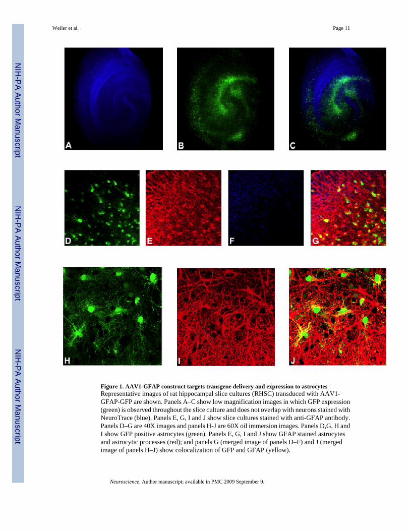
Figure 1. AAV1-GFAP construct targets transgene delivery and expression to astrocytesRepresentative images of rat hippocampal slice cultures (RHSC) transduced with AAV1-GFAP-GFP are shown. Panels A–C show low magnification images in which GFP expression(green) is observed throughout the slice culture and does not overlap with neurons stained withNeuroTrace (blue). Panels E, G, I and J show slice cultures stained with anti-GFAP antibody.Panels D–G are 40X images and panels H-J are 60X oil immersion images. Panels D,G, H andI show GFP positive astrocytes (green). Panels E, G, I and J show GFAP stained astrocytesand astrocytic processes (red); and panels G (merged image of panels D–F) and J (mergedimage of panels H–J) show colocalization of GFP and GFAP (yellow).
Weller et al. Page 11
Neuroscience. Author manuscript; available in PMC 2009 September 9.
NIH
-PA Author Manuscript
NIH
-PA Author Manuscript
NIH
-PA Author Manuscript
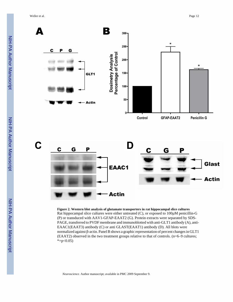
Figure 2. Western blot analysis of glutamate transporters in rat hippocampal slice culturesRat hippocampal slice cultures were either untreated (C), or exposed to 100µM penicillin-G(P) or transduced with AAV1-GFAP-EAAT2 (G). Protein extracts were separated by SDS-PAGE, transferred to PVDF membrane and immunoblotted with anti-GLT1 antibody (A), anti-EAAC1(EAAT3) antibody (C) or anti GLAST(EAAT1) antibody (D). All blots werenormalized against β-actin. Panel B shows a graphic representation of percent changes in GLT1(EAAT2) observed in the two treatment groups relative to that of controls. (n=6–9 cultures;*=p<0.05)
Weller et al. Page 12
Neuroscience. Author manuscript; available in PMC 2009 September 9.
NIH
-PA Author Manuscript
NIH
-PA Author Manuscript
NIH
-PA Author Manuscript

Figure 3. [3H]D-Asp uptake activityTotal uptake activity within crude preparations of RHSCs were compared between controls,AAV-GFAP-EAAT2 transduced (GFAP-EAAT2) or penicillin G (PenG) treated slices.EAAT2 specific uptake was determined by measuring activity in the presence and absence ofthe EAAT2 selective inhibitor, DHK.
Weller et al. Page 13
Neuroscience. Author manuscript; available in PMC 2009 September 9.
NIH
-PA Author Manuscript
NIH
-PA Author Manuscript
NIH
-PA Author Manuscript
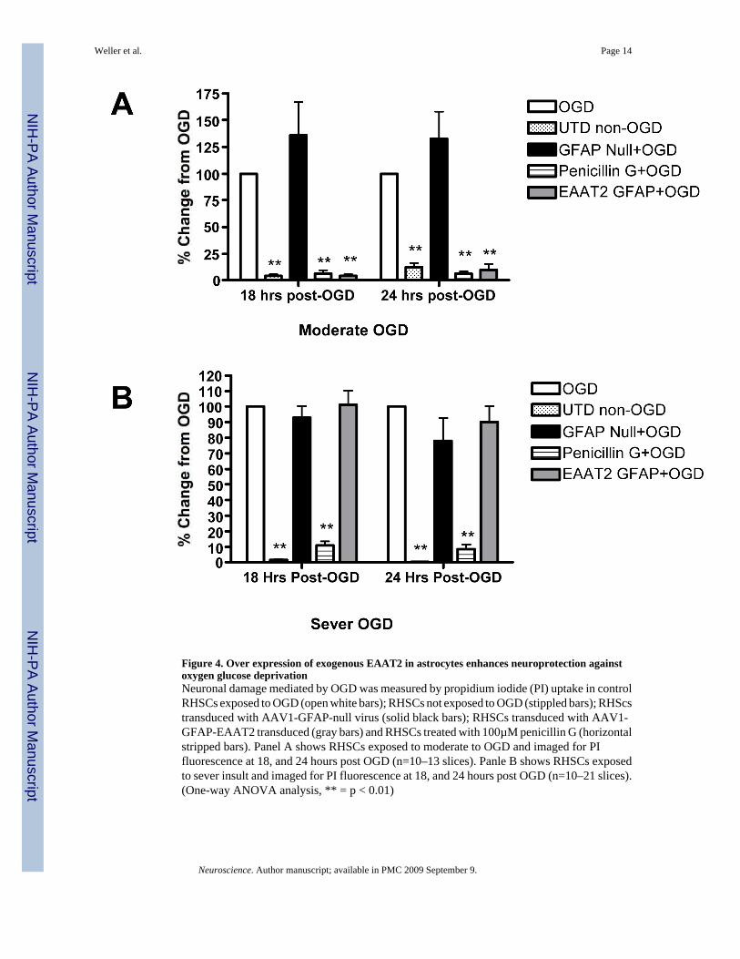
Figure 4. Over expression of exogenous EAAT2 in astrocytes enhances neuroprotection againstoxygen glucose deprivationNeuronal damage mediated by OGD was measured by propidium iodide (PI) uptake in controlRHSCs exposed to OGD (open white bars); RHSCs not exposed to OGD (stippled bars); RHScstransduced with AAV1-GFAP-null virus (solid black bars); RHSCs transduced with AAV1-GFAP-EAAT2 transduced (gray bars) and RHSCs treated with 100µM penicillin G (horizontalstripped bars). Panel A shows RHSCs exposed to moderate to OGD and imaged for PIfluorescence at 18, and 24 hours post OGD (n=10–13 slices). Panle B shows RHSCs exposedto sever insult and imaged for PI fluorescence at 18, and 24 hours post OGD (n=10–21 slices).(One-way ANOVA analysis, ** = p < 0.01)
Weller et al. Page 14
Neuroscience. Author manuscript; available in PMC 2009 September 9.
NIH
-PA Author Manuscript
NIH
-PA Author Manuscript
NIH
-PA Author Manuscript