Cocaine-induced alterations in prostaglandin production in rabbit aorta
Upload
independentCategory
view
0download
0
BioMed CentralJournal of Neuroinflammation
ss
Open AcceResearchMicroglial inhibition of neuroprotection by antagonists of the EP1 prostaglandin E2 receptorNoel G Carlson*1,2,3,4,5,6, Monica A Rojas2,3, John-David Black2,3, Jonathan W Redd2,3, John Hille2,3, Kenneth E Hill2,4 and John W Rose2,4,5Address: 1Geriatric Research Education and Clinical Center (GRECC), VASLCHCS, Salt Lake City, UT 84148, USA, 2Neurovirology Research Laboratory, VASLCHCS, Salt Lake City, UT 84148, USA, 3Department of Neurobiology and Anatomy, University of Utah, Salt Lake City, UT 84112, USA, 4Department of Neurology, University of Utah, Salt Lake City, UT 84112, USA, 5Brain Institute, University of Utah, Salt Lake City, UT 84112, USA and 6Center on Aging, University of Utah, Salt Lake City, UT 84112, USA
Email: Noel G Carlson* - [email protected]; Monica A Rojas - [email protected]; John-David Black - [email protected]; Jonathan W Redd - [email protected]; John Hille - [email protected]; Kenneth E Hill - [email protected]; John W Rose - [email protected]
* Corresponding author
AbstractBackground: The EP1 receptor for the prostanoid PGE2 is a G-protein coupled receptor that hasbeen shown to contribute to excitotoxic neuronal death. In this study we examined the influenceof non-neuronal cells on neuroprotective properties of EP1 receptor antagonists (Ono 8711 andSC 51089).
Methods: Primary neuronal cultures systems with or without non-neuronal cells were used toexamine how the neuroprotective properties of EP1 antagonists were influenced by non-neuronalcells. The influence of astrocytes or microglia were individually tested in excitotoxicity assays usinga co-culture system with these cells grown on permeable transwell inserts above the neuronal-enriched cultures. The influence of microglia on PGE2 synthesis and EP1 receptor expression wasexamined.
Results: EP1 antagonists were neuroprotective in neuronal-enriched cultures (> 90% neurons) butnot in mixed cultures (30% neurons plus other non-neuronal cells). Co-cultures of microglia onpermeable transwell inserts above neuronal-enriched cultures blocked neuroprotection by EP1antagonists. Incubation of microglia with neuronal-enriched cultures for 48 hours prior to NMDAchallenge was sufficient to block neuroprotection by EP1 antagonists. The loss of neuroprotectionby EP1 antagonists was accompanied by a decrease of neuronal EP1 expression in the nucleus incultures with microglia present.
Conclusion: These findings demonstrate microglial modulation of neuronal excitotoxicity throughinteraction with the EP1 receptor and may have important implications in vivo where microglia areassociated with neuronal injury.
Published: 17 February 2009
Journal of Neuroinflammation 2009, 6:5 doi:10.1186/1742-2094-6-5
Received: 8 December 2008Accepted: 17 February 2009
This article is available from: http://www.jneuroinflammation.com/content/6/1/5
© 2009 Carlson et al; licensee BioMed Central Ltd. This is an Open Access article distributed under the terms of the Creative Commons Attribution License (http://creativecommons.org/licenses/by/2.0), which permits unrestricted use, distribution, and reproduction in any medium, provided the original work is properly cited.
Page 1 of 16(page number not for citation purposes)

Journal of Neuroinflammation 2009, 6:5 http://www.jneuroinflammation.com/content/6/1/5
BackgroundCyclooxygenase-2 (COX-2), the enzyme that catalyzes therate limiting step in the synthesis of prostanoids, contrib-utes to neuronal death. Inhibitors of COX, termed non-steroidal anti-inflammatory drugs (NSAIDs) [1], can pro-tect neurons following an assault with toxic stimuli thatpromote excitotoxic death; both in vitro [2,3] and in vivo[4-7]. COX-2 knockout mice are also less susceptible toexcitotoxicity following exposure to the glutamate recep-tor agonist N-methyl D-aspartate (NMDA) [8]. Therefore,a loss of COX-2 activity either by inhibition of the enzymeor loss of expression is associated with increased neuronalviability. Conversely, increased COX-2 activity appears toaugment neuronal death. The increased COX-2 expressionin neurons observed in vivo in animal models of stroke[4], following stimulation with the glutamate receptoragonist kainic acid [6], and in vitro following NMDAstimulation [2,3] is coincident with loss of neurons. Con-stitutive expression of COX-2 in neurons at high amountsin transgenic mice results in a greater loss of neurons instroke models [9] and age-associated loss of neurons [10].In addition, constitutive COX-2 expression renders neu-rons more susceptible to NMDA-stimulated death [11].
There are two COX genes, COX-1 and COX-2 [1]. COXcatalyzes the initial steps in the conversion of arachidonicacid (AA) to one of the five prostanoids, prostacyclin(PGI2), thromboxane (TxA2), prostaglandin D2 (PGD2),prostaglandin F2α (PGF2α) and prostaglandin E2 (PGE2)[1,12]. In addition to the generation of prostanoids, reac-tive oxygen species (ROS) are also generated by COX-2 inthe reaction of prostanoids [1]. It was demonstrated thatthe COX-2-generated prostanoids (and not ROS), are themajor contributors by COX-2 towards excitotoxicity fol-lowing administration of NMDA to animals [13].
Each of the prostanoids synthesized by COX activates atleast one specific prostanoid receptor. These receptors arecoupled to G-proteins and are designated IP (for PGI2),TP (for TXA2), DP1 or DP2 (for PGD2), FP (for PGF2α)and EP1-4 (for PGE2) [12]. Recent investigations havefocused on understanding how activation of specific pros-tanoids affects neuronal viability. In our earlier studies weidentified that PGF2α and PGE2 were made in primaryneuronal cortical cultures in response to stimulation withNMDA [3,14]. An analog of PGE2, 17-phenol trinor PGE2(17-pt-PGE2), but not PGF2α, could reverse the neuro-protective effect of a COX-2-specific inhibitor in vitro [3]and in vivo [13] following NMDA administration. Thesestudies indicate that PGE2 production by COX-2 can con-tribute to the deleterious actions of COX-2 in NMDA-mediated excitotoxicity of neurons. However, in vitrostudies investigating the role of PGE2 and its analogs haveyielded contradictory results. PGE2 or its analogs havebeen reported to both increase neuronal survival follow-
ing NMDA stimulation [15-19] and in some cases be neu-rotoxic [20,21]. These opposing effects or PGE2 onneuronal viability are due to activation of specific EPreceptors that exert either pro survival or pro death effects.In general, activation of EP1 contributes to neuronaldeath [21-24], while activation of EP2 [17-19] and EP4[24] promote neuroprotection. EP1 has been shown tocontribute to NMDA-mediated neuronal death in vivo[24]. Decreased EP1 activation by a pharmacologic antag-onist or genetic knockout of the EP1 receptor decreasedNMDA-stimulated neuronal death, whereas a specific EP1receptor agonist augmented death [22-24].
Significant progress has been made in understanding howprostanoids contribute to neuronal death [25]. EP1 recep-tor activation in neurons has been linked to two differentintracellular mechanisms tied to excitotoxic cell death.EP1 receptor activation was initially shown to impair theNa+-Ca2+ exchanger (NCX) which subsequently causesgreater increases in intracellular calcium leading to neuro-nal death [23]. More recently, EP1 receptor activation hasbeen linked to the AKT signaling pathway that affects neu-ronal viability [26].
However, the interaction of EP1 with other cell types inthe central nervous system (CNS) is not well understood.In this study, we examined whether inhibition of neuro-nal EP1 contributes to neuronal viability in primary cul-tures with differing compositions of non neuronal CNScells. We investigated the neuroprotective properties oftwo specific EP1 receptor antagonists, Ono 8711 [27] andSC51089 [28] along with a COX-2 inhibitor NS398. Thisstudy demonstrates that the presence of microglia in cul-ture can offset the neuroprotective effects of EP1 antago-nists. Our findings have important implications toconditions where microglia may accumulate followingneuronal injury and potentially limit neuroprotectionconferred by EP1 antagonists.
MethodsReagents and antibodiesOno 8711 was generously provided by Takayuki Maru-yama Ph.D., Ono Pharmaceutical Company, (Osaka,Japan). N-[2-(cyclohexyloxy)-4-nitrophenyl]-meth-anesulfonamide (NS398) was obtained from CaymanChemical Company, (Ann Arbor MI). SC51089 wasobtained from Biomol, (Plymouth Meeting, PA). NMDA,MK801 and Arabinose cytosine (AraC) were obtainedfrom Sigma Chemical Company (St. Louis, MO). Tissueculture media minimum essential media (MEM) wasobtained from Sigma Chemical Company (St. Louis,MO), glutamine and trypsin were from Gibco Life Sci-ences, Invitrogen (Carlsbad, CA). Serum (defined fetalbovine serum and defined horse serum) was purchasedfrom Hyclone (Logan, UT). Primary antibodies used in
Page 2 of 16(page number not for citation purposes)

Journal of Neuroinflammation 2009, 6:5 http://www.jneuroinflammation.com/content/6/1/5
this study were, mouse monoclonal anti microtubuleassociated protein (MAP)-2 (BD Pharmingen, San Diego,CA), rabbit polyclonal EP1 (Cayman Chemical Com-pany), rat monoclonal CD11b (Abcam, Cambridge, MA),rat monoclonal anti glial fibrillary acidic protein(GFAP)(Calbiochem, San Diego, CA) and rat monoclonalanti mouse F4/80 was produced by our laboratory. Sec-ondary antibodies used included FITC-coupled goat anti-mouse, Cy5-coupled anti-rat and Cy5-coupled goat anti-rabbit. Secondary antibodies were obtained from JacksonImmuno-Research Laboratories Inc. (West Grove, PA).Propidium iodide (PI) was obtained from MolecularProbes (Eugene, OR).
Primary cortical culturesCortical cultures were prepared essentially as describedpreviously [3,29-31]. Cortices from E15 embryos (mousestrain CD1, from Charles River Laboratories, WilmingtonMA) were removed and the cells were dissociated by diges-tion with trypsin (0.025%) for 30 min at 37°C, followedby physical dissociation resulting from pipeting the cells(15–20 times) through a 5-ml pipette placed in a 15-mlconical tube with a loose seal between the bottom of thetube and the pipette. The cells were then plated at a den-sity of 1.1 × 10⁶ trypan blue-excluding cells per well in a12-well plate (Corning dishes pre coated with poly-D-lysine). Cells were initially plated in 1-ml of MEM withEarle's salts supplemented with 5% horse serum (heatinactivated), 5% fetal bovine serum (heat inactivated), 30mM glucose, 2 mM glutamine and grown at 37°C inhumidified chambers with 5% CO2. The cultures were fedthe next day by replenishing about half the volume withfresh growth media (MEM with Earle's salts supplementedwith 10% horse serum, 30 mM glucose, 2 mM glutamine)and fed every 2–3 days after that to generate mixed corti-cal cultures. After nine days in culture, these mixed cul-tures were exposed to AraC (4–8 μM) for a 24 hour periodto limit the growth of mitotic cells. Each preparation ofAraC differed with respect to its anti-mitotic efficacy andwas tested for optimal inhibition of cell growth with min-imal toxicity. These mixed culture preparations containedapproximately 30% of the cells as neurons [3]. All excito-toxicity experiments with mixed cultures were started 2days after the last feeding and were used 15 days after plat-ing. At this stage, the neurons exhibited the most consist-ent response to NMDA while still having low backgrounddeath. Neuronal-enriched cultures were prepared as aboveexcept that, on the second day after plating, the cells weretreated with 2–4 μM AraC for a 24-hour period and fedonly once more with feeding media (MEM with Earle'ssalts supplemented with 10% horse serum, 30 mM glu-cose, 2 mM glutamine). Neuronal-enriched cultureswhich were incubated for 8 days in vitro (DIV), containedbetween 90–95% neurons as determined by immunoflu-orescent microscopy as the number of PI stained nuclei
that were associated with cells labeled with the neuronal-specific marker MAP-2 (data not shown). Excitotoxicityexperiments with these cultures were started after the cul-tures were maintained for 8 DIV, a time at which there islow background neuronal death and the neurons exhibita consistent response to NMDA stimulated excitotoxicity.Excitotoxicity experiments were performed in growthmedia that contained 10% horse serum unless stated oth-erwise.
Cortical cultures were also examined in serum-free media.Cortical cultures were prepared as described and allowedto incubate in plating media (MEM with Earle's salts sup-plemented with 5% horse serum, 5% fetal bovine serum,30 mM glucose, 2 mM glutamine) for 24 hours. Themedia was then replaced with neurobasal media contain-ing 2% B27 supplement (Invitrogen) and, 24 hours later,the cultures were then exposed to AraC (4–8 μM). The cul-tures were fed 24 hours after this by replacing one half ofthe media with fresh Neurobasal media with 2% B27 sup-plement. These cultures were used in excitotoxicity exper-iments 8 days after the initial plating.
Excitotoxicity assayExcitotoxicity was measured using a modified protocol ofestablished methods as described in detail elsewhere[3,29-31]. Neuronal cell death was assessed by photo-graphing the same four fields in each well, before andafter addition of NMDA. Each field contained 150 to 300neurons prior to treatment with NMDA. The numbers ofintact neurons were counted before NMDA treatment asthe phase bright cells (see Figure 1). Twenty hours aftertreatment with NMDA (in the presence or absence of neu-roprotective agents) one ml of trypan blue (0.4%) wasadded to the media (final concentration of 0.2%) and thevolume of this mixture was adjusted to leave minimalsolution to cover the cells. The cells were incubated withthe trypan solution for 20 minutes. Plates were markedwith reference so that the same fields could be photo-graphed as shown in Figure 1. Cells that did not stain withtrypan blue were counted as live cells. These trypan blue-excluding cells also did not stain by the necrotic dead cellstain ethidium homodimer (Molecular Probes), but didstain with the live cell stain Calcein AM green (MolecularProbes) (data not shown). All excitotoxicity experimentswere repeated at least three times. The percentages of sur-viving neurons were determined in each field by dividingthe total of trypan-excluding cells after NMDA treatmentby the total number of cells in that field counted prior toNMDA. A total of three fields were examined in each treat-ment per plate. The average percent neuronal survival wasassessed for each treatment from at least three independ-ent experiments and included the average from a mini-mum of nine fields for each condition reported in eachfigure. In mixed cultures, neurons were identifiable as
Page 3 of 16(page number not for citation purposes)

Journal of Neuroinflammation 2009, 6:5 http://www.jneuroinflammation.com/content/6/1/5
phase-bright cells that were observed in a focal planeabove the glial monolayer. This was confirmed in sistercultures by immunofluorescence showing that these cellswere stained with the neuronal marker MAP-2 [3,29-31].
The cell counting method for determining viable cells canbe used when cultures are grown in the presence of serum(as in most of our current experiments) or when testingagents that may have antioxidant properties which inter-fere with assays that measure release of the enzyme lactatedehydrogenase (LDH) into the media from dead cells. Foreach experiment, the concentration of NMDA required to
give about 80% neuronal death was pre-determined andvaried between different lots of NMDA and cell prepara-tions. Typical concentrations of NMDA required for 80%neuronal death ranged between 10–40 μM for neuronal-enriched cultures and 15–80 μM for mixed cultures. Allexperiments were conducted using this high amount ofNMDA-induced neuronal death for convenience and reli-ability in scoring the survival after NMDA treatment. Forneuroprotective experiments, the neuroprotective agent(NS398, Ono 8711 or SC51089) was added directly to thegrowth media coincident with the addition of NMDA. Inall cases, the effect of each neuroprotectant treatment (or
Excitotoxicity assay and neuroprotection by Ono 8711Figure 1Excitotoxicity assay and neuroprotection by Ono 8711. Fields of neuronal-enriched cultures were photographed before treatment with vehicle (A) or the EP1 receptor antagonist Ono 8711 (B). These cultures were then treated with NMDA (15 μM) and photographed twenty hours later after staining with trypan blue dead cell stain for vehicle (C) and Ono 8711 (D). Examples of cells that survived NMDA treatment and excluded trypan blue are indicated by a black arrow in each panel. An example of a cell in each field that was killed by NMDA (as indicated by staining with trypan blue) is indicated by a green arrow. The numbers of neurons were counted for each of the cultures before and after NMDA (as non Trypan-stained cells). In this example, the addition of 30 nM Ono 8711 increased the percent of surviving neurons from 20% to 50%. Images were captured at 20 × magnification.
Page 4 of 16(page number not for citation purposes)

Journal of Neuroinflammation 2009, 6:5 http://www.jneuroinflammation.com/content/6/1/5
combination of treatments) on neuronal viability in theabsence of NMDA was also determined by photographingthe cells before and 20 hours after treatment with theagent indicated. In each experiment, 500 to 2000 neuronswere counted for each treatment.
Microglial and astrocyte culturesMicroglial cultures were prepared according to the meth-ods described [32]. Brains were removed from 1 day oldCD1 mice and the cerebral cortices were obtained. Themeninges were removed and the tissue was minced anddigested with trypsin (0.025%) for 30 min at 37°C, fol-lowed by physical dissociation as described earlier forneuronal cortical cultures. The cells were plated in MEMcontaining 10% fetal bovine serum into 25 cm2 tissue cul-ture flasks (1 brain per flask) that were pretreated withpoly-d-lysine. Cultures were grown for 7–9 days at 37°Cand the microglial cells were removed by shaking on anorbital shaker at 250 rpm for 30 min. Microglial cells werethen centrifuged for 5 min at 1500 g and resuspended inneurobasal medium with 2% B27 supplement and platedonto either poly-D-lysine coated12-well plates or tran-swells (12 mm diameter) at a density ranging from 1.5 ×10⁵ to 3 × 10⁵ cells/mm2. Lower cell density microglial cul-tures were more difficult to grow. After 8 days in culture,microglial transwells were transferred onto 5 day old neu-ronal-enriched cultures (in 12 well plates) and co-cul-tured another 48 hours prior to the start of excitotoxicityexperiments. Microglial cultures contained greater than95% microglial cells as determined by staining withmicroglial markers, F4/80 or CD11b.
Astrocyte cultures were prepared as described [32]. Cellswere isolated from cerebral cortices from 1 day old miceas described above and the cells were suspended in MEMcontaining 10% fetal bovine serum and plated onto eithertranswells or tissue cultures plates (1 × 10⁵ cells/mm).These cultures were grown at 37°C for about 1–2 weeksuntil the cells reached confluency. Transwell astrocyte cul-tures were transferred to neuronal-enriched cultures (5day old cultures), 48 hours before the start of excitotoxic-ity experiments.
Conditioned media experimentsMedia was harvested from mixed cultures (15 DIV) andneuronal-enriched cultures (8 DIV) and centrifuged for 5minutes (1000 × g) to eliminate any residual cells. Freshlyharvested media was subsequently exchanged betweenthe mixed and neuronal-enriched culture preparations.Since the neuronal-enriched cultures (8 DIV) did not sur-vive a direct media exchange, the mixed conditionedmedia was exchanged in the 12-well plates by seven two-fold dilutions. Neuronal-enriched media was directlyexchanged on the mixed cultures (15 DIV) in one
exchange. Excitotoxicity experiments were performed asdescribed above.
Transwell experimentsMixed cortical cultures, microglia or astrocytes wereplated onto the top side of the transwell inserts (0.4 μmpore size polycarbonate membrane coated with poly-D-lysine, Corning catalog number 3401, Corning, NY) at thecell density as described above. The mixed cultures ontranswells were treated with AraC (on day 9) and fed asdescribed for mixed cultures. These mixed culture tran-swells (15 DIV) were transferred to neuronal-enrichedcultures 48 hours prior to treatment with NMDA. Neuro-nal-depleted mixed cultures were obtained by exposure ofthe mixed cultures (11 DIV) to 25 μM NMDA for 48 hoursprior to transfer of the transwells to plates containingfresh media and three exchanges of media to removeNMDA. The transwell cultures were positioned approxi-mately 2 mm above the neuronal-enriched cultures andthe cells grown on the transwells were separated from theneuronal-enriched cultures by the permeable transwellmembrane. The neuronal-enriched cultures were photo-graphed prior to addition of the transwells and treatmentwith NMDA. The mixed culture transwells were thenreturned to the same wells of the neuronal-enriched cul-tures and subsequently treated with vehicle, SC51089,and then NMDA at a concentration that yielded approxi-mately 20% neuronal survival. All drugs were added to themedia below the transwells. After 24 hours, the mixed cul-ture transwells were removed and the neuronal-enrichedcultures were stained with trypan blue, photographed andscored for neuronal survival as described above. Viabilityof the mixed cultures, microglia cultures and astrocytecoultures on the transwells prior to NMDA treatment wasverified using dead cell stain ethidium homodimer andthe live cell stain Calcein AM green.
Statistical analysesThe data from the NMDA excitotoxicity experiments wereanalyzed using InStat software (GraphPad software, SanDiego, CA). Data from each treatment group passed themethod of Kolmogorov and Smirnov for normal distribu-tions making it appropriate for parametric tests to assesssignificance between treatment groups. One way ANOVAwith post-test using the Tukey-Kramer multiple compari-son tests was performed as to determine significancebetween treatment groups at the confidence intervals indi-cated. Pair-wise comparisons were performed usingunpaired students t-test.
Immunofluorescence assayCortical cultures were prepared for microscopy by firstrinsing the cultures with phosphate-buffered saline (PBS),followed by fixing the cells with freshly prepared 2% para-formaldehyde in PBS. Following five rinses with PBS
Page 5 of 16(page number not for citation purposes)

Journal of Neuroinflammation 2009, 6:5 http://www.jneuroinflammation.com/content/6/1/5
(minimum of 2 minutes per rinse), cells were permeabi-lized with 0.2% triton-X 100 in PBS, rinsed (3 rinses withPBS, minimum of 5 minutes per rinse) and blocked withImage-it FX signal enhancer solution (Molecular Probes).Primary antibodies were added at dilutions of (1:500) forrabbit polyclonal antibody for EP1 (final concentration 1μg/ml), (1:400) mouse monoclonal antibody for MAP-2(final concentration 1 μg/ml), (1:500) rat monoclonalGFAP (final concentration of 1 μg/ml), (1:100) rat poly-clonal for F4/80 (final concentration 10 μg/ml) and(1:100) rat monoclonal antibody for CD11b (final con-centration 5 μg/ml). Incubation of the anti-EP1 antibod-ies with the EP1 epitope blocking peptide (10:1 molarratio of peptide: antibody) (Cayman Chemical Company)blocked binding (data not shown). Secondary antibodieswere diluted (1:400) for FITC-conjugated anti-mouse(IgG, H&L) (final concentration 3 μg/ml), (1:400) FITC-conjugated anti-rat (final concentration 3 μg/ml), and(1:800) Cy5-conjugated anti-rabbit (IgG, H&L) (finalconcentration 1.5 μg/ml). The fluorochrome-conjugatedantibodies were cross-adsorbed to immunoglobulinsfrom other species to ensure specific multiple labeling. Allantibodies were diluted in PBS with 0.025% Tween 20.Combinations of the selective primary antibodies wereadded to the sections and incubated overnight in ahumidified chamber at 4°C. Conjugated secondary anti-bodies were added for 1 hour at room temperature (RT)followed by staining with PI to visualize cell nuclei. PBSwashes were done (3 rinses with PBS, minimum of 5 min-utes per rinse) between measures prior to mounting. Sec-tions were mounted with ProLong Gold anti-fadepermanent mounting media (Molecular Probes). Toaccount for any background non-specific staining, nega-tive controls with normal mouse and normal rabbit serumat concentrations comparable to primary antibodies wereconducted in parallel.
Confocal laser scanning microscopyPersonal Confocal Microscope PCM-2000 (NIKON,Melville, NY) with Argon, HeNe green and HeNe redlasers were used to acquire the images. Simple PersonalConfocal Image program (Simple PCI, Compix, Cran-berry Township, PA) was used to acquire digital imagesand construct compilations. The fluorochromes wereresolved into three different image channels with respec-tive emission filters. The FITC label was detected with theArgon laser at 488 nm, Cy5 with the red HeNe laser at 633nm and PI was visualized with the green HeNe laser at605/32 nm narrow band pass width filter. The stained cul-tures were individually scanned for each respective fluor-ochrome and the multi-focal program (z-focus) was usedto create a stereopsis image. The three separate imageswere merged together to acquire the final triple-coloredimage; the PI image was converted to blue color duringmerge. This allowed for comprehensive visualization of
labeling throughout the entire depth of the sample.Images were stored as digital files and the final figureswere created using Photoshop (Adobe, San Jose, CA). Thetotal magnification of each image presented is equal to(magnification of the objective) × (width of the imagepanel in cm) × (0.93). This scale of image size was basedon size calibration using an American Optical improvedNeubauer hemacytometer and simple PCI linear calibra-tion scales.
Quantification of neuronal nuclear EP1 expression in different culture preparationsThe amount of neuronal expression in the nucleus wasquantified from digital images of immunofluorescenceconfocal microscopy of cortical cultures stained with PI(for nuclei) and immuno-stained for EP1, and MAP-2.Each image was captured with the same sensitivity settingson the confocal microscope and all images were capturedwith a 60× objective with 640 × 480 pixels. A blindedobserver (unaware of the culture preparation) counted thenumber of pixels over the nuclear regions of the neuronsthat were stained for EP1 with an enlarged image withAdobe Photoshop. Each culture preparation was exam-ined in four independent fields and the number of neu-rons analyzed varied from 50–70 for each culturepreparation.
Prostaglandin measurementsPGE2 was measured using a prostaglandin enzyme-linkedimmunosorbent assay (ELISA) detection kit (CaymanChemical Company) that uses a PGE2-specific antibody.For both the mixed cultures (15 DIV) and neuronal-enriched cultures (8 DIV), the media was exchanged fromthe standard growth media (MEM with Earle's salts/10%horse serum, 30 mM glucose, 2 mM glutamine) to HBSS+(Mg2+-free Hanks buffered salt solution, 3 mM CaCl2, 20mM HEPES (pH 7.55), 13.4 mM NaHCO3, 30 mM glu-cose and 25 μM glycine). Since the neuronal-enriched cul-tures did not survive a direct media exchange, the mediawas exchanged in the 12-well plates by seven two-folddilutions (repeated series of addition of 1 ml of HBSS+ to1 ml of media, followed by removal of 1 ml). This resultedin 0.78% of the original growth media remaining whichhad no detectable amounts of PGE2. After the media wasexchanged, the cultures were exposed to a treatment ofNMDA that yielded < 10% neuronal death over the 90min of the experiment. The mixed cultures were treatedwith 100 μM NMDA for 15 min followed by treatmentwith 3 μM of the NMDA antagonist MK801. The neuro-nal-enriched cultures were treated with 200 μM NMDAfor 20 min, followed by treatment with 3 μM MK801. Themedia was removed from the cultures 90 minutes after theinitial NMDA treatment and immediately frozen (-80°C)for analyses at a later time. The amount of PGE2 in themedia was determined by enzyme immunoassay as per
Page 6 of 16(page number not for citation purposes)

Journal of Neuroinflammation 2009, 6:5 http://www.jneuroinflammation.com/content/6/1/5
the instructions from the manufacturer. This assay candetect as little as 8 pg/ml of PGE2.
ResultsNeuroprotection by an EP1 receptor antagonistThe neuroprotective effect of the EP1 receptor antagonistOno 8711 was examined using the assay for excitotoxicityshown in Figure 1. The same field of the neuronal culturewas photographed before treatments, and twenty hoursafter continuous exposure with NMDA. In this assay thefate of each neuron can then be followed over time. In Fig-ure 1A, the neuronal-enriched cultures were treated with aconcentration of NMDA that leaves only about 20% of theneurons viable as determined by trypan blue exclusion(Figure 1C). However, when 30 nM Ono 8711 was addedto a sister culture prior to treatment with the same concen-tration of NMDA, there was a dramatic increase in thenumber of surviving neurons (Figure 1B and 1D). A moreextensive analysis using this assay was performed withtreatments of Ono 8711 at concentrations of 0.3 nM, 1nM, 3 nM, 10 nM, 30 nM and 100 nM added just prior totreatment with NMDA. As seen in Figure 2, treatment withas little as 3 nM of Ono 8711 increased the percent of sur-viving neurons to nearly 60%, compared with 20% asseen with NMDA alone. Similar amounts of neuroprotec-tion were observed with treatments of 10 nM and 30 nMOno 8711 and were not significantly different than treat-ments with 30 μM of the COX-2 inhibitor NS398. Thisconcentration of NS398 is the optimal concentration forprotection with this drug [2,3]. However, the highest con-centration of Ono 8711 tested (100 nM) yielded no signif-icant neuroprotection and resulted in significantly loweramounts of surviving neurons than observed with 3, 10and 30 nM Ono 8711. Treatment of cultures with 100 nMOno 8711 in the absence of NMDA did not result in anydetectable toxicity of neurons (data not shown). AnotherEP1 receptor antagonist, SC51809 [28] at a concentrationof 10 μM, produced the same amount of neuroprotectionin neuronal-enriched cultures as seen with Ono 8711 and30 μM NS398 (Figure 2).
Neuroprotection by EP1 antagonists is sensitive to modulation by non-neuronal cellsIn the preceding experiments, the neuroprotective effectsof Ono 8711 and SC51089 were assessed using neuronal-enriched cultures where neurons account for about 90–95% of the cells in the culture (see methods). Neuropro-tection mediated by the EP1 antagonists in these neuro-nal-enriched cultures, is consistent with EP1 antagonistsdirectly blocking EP1 receptor activation on neurons. Inorder to assess whether this effect is modulated by othercell types, we examined whether EP1 receptor antagonistswere neuroprotective in a mixed culture preparation,where approximately 30% of the cells present in the cul-ture were neurons (see methods). In sharp contrast to the
results obtained with neuronal-enriched cultures, therewas no appreciable neuroprotective effect elicited by treat-ment with any concentration of the EP1 antagonist Ono8711, 10 μM SC51089 or with 30 μM NS398 (see Figure3). A similar lack of neuroprotection by the EP1 antago-nists was also observed when neuronal-enriched cultureswere prepared with insufficient amounts of the anti-
Neuroprotection by EP1 receptor antagonists and a COX-2 inhibitor in neuronal-enriched culturesFigure 2Neuroprotection by EP1 receptor antagonists and a COX-2 inhibitor in neuronal-enriched cultures. The effect of varying concentrations of Ono 8711 on neuronal survival to NMDA treatment was assessed using the assay outlined in Figure 1 (see methods). Cultures were either pre-treated with the drug vehicle DMSO (veh), or the concentra-tion of the drug indicated below each bar. The cultures were then treated with a concentration of NMDA that leaves only about 20% survival in the non-drug treated cultures (average [NMDA] = 15 μM). The percent survival for the treatments indicated was determined and plotted as percent surviving neurons. The shaded region of the bar graph shows the sur-vival obtained with NMDA alone and the unshaded portion of the bar indicates the additional neuronal survival resulting from co-treatment with the drug indicated. In each experi-ment, the neuronal cell numbers were counted from four fields (similar to that shown in Figure 1) from each treatment and averaged. The results shown were then average from at least four independent experiments. (* indicates P < 0.05 and ** indicates P < 0.01 in comparison to vehicle treatment, # indicates a < 0.05 in comparison to the concentrations (3,10 and 30 nM) of Ono 8711, by ANOVA, Tukey-Kramer multi-ple comparison test). The 100 nM Ono 8711 was not signifi-cantly different from the vehicle control. Error bars are standard error of the mean (SEM).
Page 7 of 16(page number not for citation purposes)

Journal of Neuroinflammation 2009, 6:5 http://www.jneuroinflammation.com/content/6/1/5
mitotic agent AraC (see methods) to limit the growth ofnon-neuronal cells (data not shown).
Media exchange between culturesTo assess whether the difference in response to the EP1antagonists was due to differences in cell-derived compo-nents in the culture media, experiments were done wherethe media was exchanged between the two cultures. Asseen in Figure 4, EP1 antagonist SC51089 was still neuro-protective in the neuronal-enriched cultures with mediatransferred from mixed cultures. Conversely, SC51089still did not confer neuroprotection in mixed cultures con-taining media from neuronal-enriched cultures. There-fore, the neuroprotective response to SC51089 remainedthe same, irrespective to the source of media. These resultsillustrate that the cellular environment in the mixed cul-tures is important to determine whether the EP1 antago-nist is neuroprotective. Alternatively, components in themedia from the mixed cell cultures are labile and lose
activity during the media transfer procedure or requirelonger exposure time to elicit an effect.
Cellular composition of mixed and neuronal-enriched culturesCellular composition in both mixed and neuronal-enriched cultures was examined by confocal immunoflu-orescence microscopy to identify possible candidate non-neuronal cell types that influence EP1 neuronal response.Mixed cultures contained an abundance of cells stainedwith the astrocyte-specific marker GFAP (Figure 5a) incontrast to the neuronal-enriched cultures which con-tained a rare GFAP-positive cell (Figure 5b). Microgliawere also present in the mixed cultures as indicated by thepresence of cells that contain the monocyte/macrophagelineage marker, F4/80 (Figure 5c). F4/80 positive cellswere not detected in the neuronal-enriched cultures (Fig-ure 5d). Similar results were obtained with the microglialmarker, CD11b (not shown). These results identify bothastrocytes and microglia as potential cell types in mixed
Lack of neuroprotection by EP1 receptor antagonists or NS398 in mixed culturesFigure 3Lack of neuroprotection by EP1 receptor antagonists or NS398 in mixed cultures. Ono 8711, SC51089 and NS398 did not confer neuroprotection in mixed cultures. Mixed cortical cultures were treated with the concentrations of Ono 8711 indicated, 10 μM SC51089 or 30 μM NS398 fol-lowed by treatment with NMDA (average [NMDA] = 20 μM). These cultures were treated in parallel with the neuro-nal-enriched cultures shown in Figure 2. Neuroprotection was not observed with any concentration of Ono 8711, SC51089, or NS398. The results shown were the average (+SEM) from four independent experiments.
Neuroprotection following media exchange between mixed and neuronal-enriched culturesFigure 4Neuroprotection following media exchange between mixed and neuronal-enriched cultures. Media from mixed and neuronal-enriched (NE) cultures was exchanged (see methods) prior to challenge with NMDA (average [NMDA] = 15 μM for NE cultures and 20 μM for mixed cul-tures) in the presence or absence of the EP1 antagonist SC51089 (shown as SC). The source of the media is shown above the bars and the culture below the bars. This is the average of four independent experiments. The protection by SC in the neuronal-enriched cultures with mixed media was significant (P < 0.01) by ANOVA, Tukey-Kramer multiple comparison test. The results shown were then averaged from four independent experiments.
Page 8 of 16(page number not for citation purposes)

Journal of Neuroinflammation 2009, 6:5 http://www.jneuroinflammation.com/content/6/1/5
cultures that could negate the neuroprotective effects ofEP1 antagonists.
Transwell co-cultures with neuronal-enriched culturesIn order to assess which of these different cell types couldaffect neuroprotection by an EP1 antagonist, a series ofexperiments were performed where different types of cul-tures were grown on permeable transwell filters andplaced above neuronal-enriched cultures. When tran-swells containing mixed cultures were placed above the
neuronal-enriched cultures (during and 48 hours prior toNMDA challenge), the EP1 antagonist was not neuropro-tective in the neuronal-enriched cultures grown below(Figure 6). This experimental system with the mixed cul-ture transwells now made the neurons in the neuronal-enriched cultures respond similarly to EP1 antagonist asthe neurons contained in the mixed cultures (Figure 3).This illustrates that soluble factors from the mixed cul-tures influenced how the neurons in the neuronal-enriched cultures responded to EP1 antagonists. Mixed
Cellular composition of mixed and neuronal-enriched culturesFigure 5Cellular composition of mixed and neuronal-enriched cultures. Confocal immunofluorescence microscopy was per-formed on mixed and neuronal-enriched cultures that were stained with neuronal specific antibody MAP-2 (green) and either the astrocyte-specific marker GFAP (red) (A and B) or the microglia marker F4/80 (red) (C and D), nuclei were stained with PI and appear blue. Prominent GFAP staining in mixed cultures is indicated with an arrow (A) and the rare astrocyte in the neu-ronal-enriched culture is shown with an arrow (B). Microglia were prominent in mixed cultures (C) and two examples were marked with an arrow. Microglial staining could not be detected in the neuronal-enriched cultures (D). Images were captured at 10× magnification for A and B, 60× for C and 40× for D. Magnification bars are 100 μm for A and B, 20 μm for C and D.
Page 9 of 16(page number not for citation purposes)

Journal of Neuroinflammation 2009, 6:5 http://www.jneuroinflammation.com/content/6/1/5
cultures devoid of neurons also produced the same effectwhen placed on transwells above neuronal-enriched cul-tures rendering the neurons in the neuronal-enriched cul-tures insensitive to the neuroprotective effects of the EP1antagonist (Figure 6). This suggests that non neuronalcells in the mixed cultures were negating the neuroprotec-tive effect of the EP1 antagonist.
The effects of astrocytes and microglia were then individ-ually examined for their potential influence on the neuro-protective effect of the EP1 antagonist. Astrocyte transwellcultures appeared to have little effect on the neuroprotec-tion conferred by SC51089. However, when cultures ofmicroglia (90% of the cells were positive for the micro-glial marker, F4/80) grown on transwells were placedabove the neuronal-enriched cultures, SC51089 was notneuroprotective (Figure 6). These same trends for the dif-ferent combinations of transwell cultures on neuronal-enriched cultures were seen at two different levels ofNMDA-induced excitotoxicity (20% survival, Figure 6aand 10% survival, Figure, 6b). None of the transwell cul-
tures were toxic to the neurons in the neuronal-enrichedcultures in the absence of NMDA (data not shown).Therefore, these experiments showed that the presence ofmicroglia blocks neuroprotection by an EP1 antagonist.The same effects of microglia on neuroprotection by anEP1 antagonist was also observed in cultures grown indefined serum-free media (see methods). In these cul-tures, microglial transwells also abrogated the neuropro-tective effects of SC51089 on neuronal-enriched cultures(data not shown). This indicates that the effect of micro-glia on neuroprotection by an EP1 antagonist was not dueto serum-derived components in the growth media.
Effects of microglia on sensitivity to NMDA-induced excitotoxicityIn the preceding experiments, the relative neuroprotectiveeffects of the EP1 antagonists with and without microgliawere compared under conditions where NMDA wasadjusted to produce equivalent amounts of neuronaldeath in each culture condition. However, when microgliawere present in transwells above the neuronal-enriched
Effects of transwell co-cultures on EP1 antagonist-mediated neuroprotectionFigure 6Effects of transwell co-cultures on EP1 antagonist-mediated neuroprotection. Transwells containing either mixed cultures, neuron-depleted mixed cultures, astrocytes or microglia cultures were co-cultured with neuronal-enriched cultures for 48 hours prior to treatment with vehicle (veh) or the EP1 antagonist SC51089 (SC) and subsequent challenge with NMDA (average [NMDA] = 15 μM for neuronal-enriched cultures and astrocyte-containing cultures and 16 μM for cultures with tran-swells containing mixed cultures, neuron-depleated mixed cultures and microglia). Neuronal viability was assessed 20 hours after NMDA in each culture paradigm (as outlined in Figure 1) and compared to neuronal-enriched cultures without transwells (first two bars in each set). The composition of the transwell cultures are marked above the bars. The relative amount of neu-roprotection was assessed with two different amounts of NMDA-induced excitotoxicity 20% survival (A) and 10% survival for (B). For 10% survival the average [NMDA] = 12 μM for neuronal-enriched cultures and astrocyte-containing cultures and 18 μM for cultures with transwells containing mixed cultures, neuron-depleated mixed cultures and microglia. Each set is the aver-age (+ SEM) of four independent experiments. Protection by SC in neuronal-enriched cultures with and without astrocytes was highly significant (P < 0.001 by ANOVA Tukey-Kramer multiple comparison test). A line is included in the bar to show the additional amount of survival conferred by addition of the EP1 antagonist.
Page 10 of 16(page number not for citation purposes)

Journal of Neuroinflammation 2009, 6:5 http://www.jneuroinflammation.com/content/6/1/5
cultures, the neurons were more resistant to NMDA-medi-ated excitotoxicity as indicated in the concentration-response curves shown in Figure 7. Therefore, in this con-text, microglia conferred a neuroprotective effect on thecultures. However, despite this neuroprotective effect elic-ited by microglia, EP1 antagonists lacked any additionalneuroprotective effect in cultures containing microglia.
Microglia alter neuronal response to EP1 antagonistsThe preceding experiments demonstrated that the pres-ence of microglia in the culture alter how the EP1 receptorantagonists confer neuroprotection following an excito-toxic challenge with NMDA. However, those experimentsdo not distinguish whether these differences are due to
microglial-induced changes in neuronal EP1 response orare an indirect consequence of EP1 signaling in the micro-glia. In order to test whether microglia can induce changesin neuronal response to EP1 antagonists, neuronal-enriched cultures were co-cultured with microglial tran-swells for 48 hours, then the microglial transwells wereremoved prior to challenge with NMDA. As seen in Figure8, this pre-incubation with microglia was sufficient toabolish the neuroprotective affect of the EP1 antagonist inthe neuronal-enriched cultures. Notably, the microglialpreconditioned neuronal-enriched cultures were alsomore resistant to NMDA similar to that seen when micro-glia are present during the excitotoxic challenge (data notshown). Since microglia need not be present during the
Microglial influence on NMDA-induced neuronal deathFigure 7Microglial influence on NMDA-induced neuronal death. Neuronal-enriched cultures were incubated with or without microglial transwell cultures for 48 hours and were exposed to varying concentrations of NMDA. Neuronal death was assessed as depicted in Figure 1 after 20 hours of continuous NMDA exposure. The results are the average of two independ-ent experiments. Error bars for each concentration of NMDA represent the SEM.
Page 11 of 16(page number not for citation purposes)

Journal of Neuroinflammation 2009, 6:5 http://www.jneuroinflammation.com/content/6/1/5
challenge with NMDA to alter the neuroprotective effectsof EP1 antagonist, this would suggest that microglia areable to alter the EP1 response in neurons. The potentialeffect of microglia on EP1 expression and NMDA inducedPGE2 production was subsequently examined.
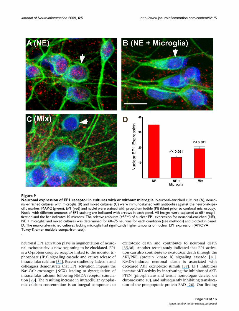
Neuronal expression of EP1 in mixed cultures and neuronal-enriched cultures with and without microglial transwellsEP1 receptor antagonists may have diverse neuroprotec-tive properties with different culture preparations becauseof potential microglial-induced changes in neuronal EP1receptor expression. In order to address this possibility,the neuronal expression of EP1 receptor was examined byimmunofluorescence confocal microcopy for mixed cul-tures and neuronal-enriched cultures in the presence orabsence of microglial transwells. As seen in Figure 9, therewas no appreciable net reduction in the amount of EP1receptor expressed in microglia-containing cultures. How-
ever, there was a significant, two-fold decrease of EP1expression in the nucleus in the neuronal-enriched cul-tures containing microglial transwells (Figure 9b) and inthe mixed cultures (Figure 9c) compared to neuronal-enriched cultures (Figure 9a). Therefore, higher amountsof nuclear EP1 were seen in the cultures in which EP1antagonists were neuroprotective.
NMDA stimulation of PGE2 in mixed and neuronal-enriched culturesWe have shown previously that NMDA stimulates the syn-thesis and release of PGE2 in cortical cultures [3,14]. Acomparison of the NMDA-stimulated release of PGE2 byneuronal-enriched and mixed cultures was done to assesswhether differences in neuroprotection by COX-2 inhibi-tors and EP1 receptor antagonists could be due to changesin PGE2 production. Both culture preparations showedabout a six to nine-fold increase in PGE2, 90 minutes aftertreatment with NMDA (Figure 10A and 10B) and thisinduction was blocked by the NMDA receptor antagonistMK801 (3 μM) (data not shown). The net amount ofPGE2 in the mixed cultures was about five-fold higherthan in the neuronal-enriched cultures. In both cases theconcentration of NMDA-induced PGE2 (425 pM for mixand 85 pM for neuronal-enriched cultures) was below theKi reported for the EP1 receptor of 22 nM [33]. However,as noted previously [14], the concentration of PGE2 in themedia may not reflect higher local concentrations at thesite of synthesis near the neurons. These findings indicatethat a lack of neuroprotection with the EP1 antagonists incultures containing microglia was not due to an inabilityto synthesize PGE2.
DiscussionIn this study we found that two structurally distinct EP1receptor antagonists (Ono 8711 and SC51089) protect asignificant percentage of neurons against a sustainedexposure to NMDA in primary neuronal cultures devoidof microglia. The neuroprotective effect of these agents ismediated by specifically blocking the EP1 receptor sincemaximal neuroprotection with Ono 8711 occurred withconcentrations as low at 3 nM, which is just above the Kifor the EP1 receptor (1.7 nM), (Watanabe et al., 1999).Concentrations of Ono 8711 greater than 100 nM werenot directly toxic to neurons, but had no significant neu-roprotective effect agonist NMDA-mediated toxicity. It isnot known what accounts for this effect, but higher con-centrations of Ono 8711 may have off target actionswhich may alter the contribution of EP1 to neuronal via-bility or neuronal sensitivity to excitotoxic stimuli.
The pharmacological evidence that we demonstrated withEP1 receptor antagonists is consistent with recent in vitro[21,22] and in vivo studies demonstrating that EP1 activa-tion contributes to neuronal death [23,24]. The role that
Microglial influence on EP1 antagonist-mediated neuropro-tectionFigure 8Microglial influence on EP1 antagonist-mediated neu-roprotection. Neuronal-enriched cultures were cultured in the absence or presence of microglia-containing transwells for 48 hours after which the microglial transwells were removed (pre-conditioning with microglia). The cultures were then treated with NMDA (average [NMDA] = 25 μM for neuronal-enriched cultures and 100 μM for microglia pre-conditioned media) in the presence or absence of the EP1 antagonist SC51089 (10 μM) and scored for neuronal sur-vival 20 hours later. Neuroprotection is only seen in the neu-ronal-enriched cultures not exposed to microglia (P < 0.001 ANOVA Tukey-Kramer multiple comparisons test). A line is included in the bar to show the additional amount of survival conferred by addition of the EP1 antagonist. These results show that preconditioning with microglia for 48 hours prior to NMDA treatment is sufficient to inhibit the neuroprotec-tive effect of the EP1 antagonist. This is the average of three independent experiments.
Page 12 of 16(page number not for citation purposes)
Journal of Neuroinflammation 2009, 6:5 http://www.jneuroinflammation.com/content/6/1/5
neuronal EP1 activation plays in augmentation of neuro-nal excitotoxicity is now beginning to be elucidated. EP1is a G-protein coupled receptor linked to the inositol tri-phosphate (IP3) signaling cascade and causes release ofintracellular calcium [34]. Recent studies by Iadecola andcolleagues demonstrate that EP1 activation impairs theNa+-Ca2+ exchanger (NCX) leading to dysregulation ofintracellular calcium following NMDA receptor stimula-tion [23]. The resulting increase in intracellular cytoplas-mic calcium concentration is an integral component to
excitotoxic death and contributes to neuronal death[35,36]. Another recent study indicated that EP1 activa-tion can also contribute to excitotoxic death through theAKT/PKB (protein kinase B) signaling cascade [26].NMDA-induced neuronal death is associated withdecreased AKT excitotoxic stimuli [37]. EP1 inhibitorsincrease AKT activity by inactivating the inhibitor of AKT,PTEN (phosphatase and tensin homologue deleted onchromosome 10), and subsequently inhibiting transloca-tion of the proapoptotic protein BAD [26]. Our finding
Neuronal expression of EP1 receptor in cultures with or without microgliaFigure 9Neuronal expression of EP1 receptor in cultures with or without microglia. Neuronal-enriched cultures (A), neuro-nal-enriched cultures with microglia (B) and mixed cultures (C) were immunostained with antibodies against the neuronal-spe-cific marker, MAP-2 (green), EP1 (red) and nuclei were stained with propidium iodide (PI) (blue) prior to confocal microscopy. Nuclei with different amounts of EP1 staining are indicated with arrows in each panel. All images were captured at 60× magni-fication and the bar indicates 10 microns. The relative amounts (+SEM) of nuclear EP1 expression for neuronal-enriched (NE), NE + microglia, and mixed cultures was determined for 60–75 neurons for each condition (see methods) and plotted in panel D. The neuronal-enriched cultures lacking microglia had significantly higher amounts of nuclear EP1 expression (ANOVA Tukey-Kramer multiple comparison test).
Page 13 of 16(page number not for citation purposes)

Journal of Neuroinflammation 2009, 6:5 http://www.jneuroinflammation.com/content/6/1/5
that EP1 antagonists protected neurons in neuronal-enriched cultures is also consistent with a mechanism inwhich neuronal activation of EP1 leads to cell death.
Our results indicated that when microglia were present inthe cultures, the neuroprotective properties of EP1 antag-onists were lost. Gendron et al. demonstrated that EP1antagonists conferred neuroprotection against excitotox-icity induced by oxygen-glucose-deprivation in mixedneuronal cultures [22]. However, they did not character-ize the cellular composition with respect to microglia. Intheir culture system anti-mitotic agents were added totheir mixed cultures on day 4 after plating compared toour mixed cultures in which the anti-mitotic agents wereadded on day 9 (see methods). This difference may havelimited the amounts of microglia in their system.
In our culture system, the loss of neuroprotection with theEP1 antagonist occurred when the microglia were eithercultured with the neurons (as in the mixed cultures) orwhen the microglia were co-cultured in transwells abovethe neuronal-enriched cultures. Co-culturing of neuronal-enriched cultures with microglia for 48 hours was also suf-ficient to alter the neuroprotective effects of EP1 antago-nists. This indicates that the microglial cultures were ableto produce soluble factors that could diffuse through the0.4 μm pores in the transwell membrane and influenceEP1 response in the neurons in the neuronal-enriched cul-tures. Simple transfer of media from the mixed cultures tothe neuronal-enriched cultures could not abolish neuro-
protection. This indicates that the soluble component(s)from the mixed cultures that were able to affect EP1response in the transwell cultures are either labile, notincubated for sufficient times before NMDA challenge orare produced in the mixed cultures during NMDA stimu-lation. Identification of these components produced bymicroglia and how they modulate neuronal EP1 responsewill require further investigation.
The mechanism by which microglia inhibit the neuropro-tective effects of EP1 antagonists appeared to be morecomplex than just downregulation of neuronal EP1expression or decreased NMDA-induced synthesis ofPGE2. However, there was a significant difference in cellu-lar distribution of EP1 to the nuclear region in neuronsfrom cultures that lacked microglia. Additional studieswill be required to determine whether this change in EP1localization is important to EP1 signaling and the viabilityof neurons following NMDA stimulation. Nuclear locali-zation of EP1 has been observed in neurons and brain-derived endothelial cells, where stimulation of EP1 recep-tors can induce changes in nuclear calcium concentrationsand modulate gene expression of genes such as c-fos [38].
In our culture systems, neurons were more resistant toNMDA induced excitotoxicity when microglia werepresent. Decreased neuronal nuclear expression of EP1was also observed in microglia-containing cultures. It ispossible that the neuroprotective effect of microglia couldbe in part due to a redistribution of EP1 expression.
NMDA induces prostaglandin E2 production in mixed and neuronal-enriched culturesFigure 10NMDA induces prostaglandin E2 production in mixed and neuronal-enriched cultures. Media from mixed (A) and neuronal-enriched (B) cultures were analyzed by ELISA for the presence of PGE2 from cultures either treated with NMDA or vehicle (control). The amount of PGE2 detected (pg/ml) is plotted for the average (+SEM) from 4 independent experiments. Note that the scales of PGE2 differ between preparations. (**indicates P = 0.01 in comparison to vehicle treatment by ANOVA, Tukey-Kramer multiple comparison test).
Page 14 of 16(page number not for citation purposes)

Journal of Neuroinflammation 2009, 6:5 http://www.jneuroinflammation.com/content/6/1/5
Our data support a role for microglia in altering the neu-ronal EP1 response. However, we cannot rule out whetherEP1 signaling in microglia can alter neuronal viability.Further studies will be needed to assess the role that EP1signaling in microglia play in modulating neuronal viabil-ity. EP1 receptors were abundantly expressed in microgliain our cultures (data not shown) and could play some rolein the interaction with neurons.
This study identified a new role for microglia as an impor-tant modulator of the neuroprotective properties of EP1antagonists in vitro. If the same interactions were to occurin vivo, this could have important consequences withrespect to microglial-mediated effects on neuronal viabil-ity. In vivo, microglia migrate to the site of neuronalinjury and have been implicated to play a role in strokeand neurodegenerative diseases such as Alzheimer's dis-ease, Parkinson's disease, multiple sclerosis, and HIV-dementia [39]. It is presently controversial whether micro-glia play a harmful or beneficial role in neuronal death[39-41]. The role played by microglia in neuronal deathmay differ depending on the stimuli used to induce death,the cellular milieu and the presence of various factorsalong with the specific intracellular pathways that are acti-vated [39].
There are two examples in vitro where microglia havebeen shown to be detrimental towards neurotoxin-induced neuronal death that is associated with COX-2. Inan in vitro model of Parkinson's disease, COX-2 inhibi-tors showed a dramatically reduced level of neuroprotec-tion towards dopaminergic neurons in cultures that lackmicroglia following MPP+ (1-methyl-4-phenylpyridin-ium)-induced death [42]. Microglia were shown to be themajor source of MPP+-induced production of PGE2 as wellas contribute to increasing neuronal COX-2 induction. Inanother example, microglia are also required for lipopol-ysaccharide (LPS) – induced neuronal death in culture[43]. In this case, stimulation of the EP2 receptor onmicroglia induced expression of COX-2 and iNOS whichwere required for neuronal death. These examples differfrom our studies where microglia appeared to have neuro-protective effects and diminished the contribution of neu-ronal EP1 receptor activation to excitotoxic death.
In light of the recently identified complications of cardio-vascular disease associated with COX-2 inhibitors([25,44,45], it is important to identify new targets down-stream of COX-2 that contribute to neuronal death [46].Inhibition of EP1 represents an attractive alternative toCOX-2 inhibition for neuroprotective strategies that couldbe used for treating stroke and other neurodegenerativediseases [23,24,47,48].
ConclusionThis study demonstrates that microglia may limit the con-tribution of EP1 receptor activation towards neuronalexcitotoxicity. Understanding how microglia could limitthe contribution of EP1 to neuronal excitotoxicity is notonly an important consideration for designing neuropro-tective treatments with EP1 antagonists but may also gen-erate new approaches in the treatment ofneurodegenerative disease.
Competing interestsThe authors declare that they have no competing interests.
Authors' contributionsNGC is the major contributor in drafting the manuscriptand revising it critically for important intellectual content,conception and design, acquisition of data, analysis andinterpretation of data. MAR has played a major role inrevising the manuscript critically for important intellec-tual content conception and design, acquisition of data,analysis and interpretation of data. J-DB has also madesignificant contribution to design, acquisition of data,analysis and interpretation of data. JWR, JH and KEHplayed important roles in design and acquisition of data,analysis and interpretation of data. JWR has have beeninvolved in drafting the manuscript and revising it criti-cally for important intellectual content, conception anddesign. All authors read and approved the final manu-script.
AcknowledgementsThis work is supported by VA Merit grants (NGC and JWR) and NIH grant RO1 AG20650 (NGC). Partial support was also provided by a grant from the National Multiple Sclerosis Society (to JWR and NGC) and the Val A. Browning foundation of Utah and the Cumming Foundation for support of MAR. The authors thank Jacob Snow, Blair Wood and Philip Tang for there technical contributions, and to Angelynn Long for both technical contribu-tions and assistance in manuscript preparation. We are very grateful for the comments on the manuscript by Dr Ikuo Tsunoda. We thank Dr Thomas Green for help with statistical analyses.
References1. Smith WL, DeWitt DL: Prostaglandin endoperoxide H syn-
thases (cyclooxygenases)-1 and -2. Adv Immunol 1996,62:167-215.
2. Hewett SJ, Uliasz TF, Vidwans AS, Hewett JA: Cyclooxygenase-2contributes to N-methyl-D-aspartate-mediated neuronalcell death in primary cortical cell culture. J Pharmacol Exp Ther2000, 293:417-425.
3. Carlson NG: Neuroprotection of cultured cortical neuronsmediated by the cyclooxygenase-2 inhibitor APHS can bereversed by a prostanoid. J Neurosci Res 2003, 71:79-88.
4. Nogawa S, Zhang F, Ross ME, Iadecola C: Cyclo-oxygenase-2 geneexpression in neurons contributes to ischemic brain damage.J Neurosci 1997, 17:2746-2755.
5. Nagayama M, Niwa K, Nagayama T, Ross ME, Iadecola C: Thecyclooxygenase-2 inhibitor NS-398 ameliorates ischemicbrain injury in wild-type mice but not in mice with deletionof the inducible nitric oxide synthase gene. J Cereb Blood FlowMetab 1999, 19:1213-1219.
Page 15 of 16(page number not for citation purposes)
Journal of Neuroinflammation 2009, 6:5 http://www.jneuroinflammation.com/content/6/1/5
6. Kunz T, Oliw E: The selective cyclooxygenase-2 inhibitorrofecoxib reduces kainate-induced cell death in the rat hip-pocampus. Eur J Neurosci 2001, 13:569-575.
7. Kawaguchi K, Hickey RW, Rose ME, Zhu L, Chen J, Graham S:Cyclooxygenase-2 expression is induced in rat brain afterkainate-induced seizures and promotes neuronal death inCA3 hippocampus. Brain Res 2005, 1050:130-137.
8. Iadecola C, Niwa K, Nogawa S, Zhao X, Nagayama M, Araki E,Morham S, Ross ME: Reduced susceptibility to ischemic braininjury and N-methyl-D-aspartate-mediated neurotoxicity incyclooxygenase-2-deficient mice. Proc Natl Acad Sci USA 2001,98:1294-1299.
9. Dore S, Otsuka T, Mito T, Sugo N, Hand T, Wu L, Hurn PD: Neuro-nal overexpression of cyclooxygenase-2 increases cerebralinfarction. Ann Neurol 2003, 54:155-162.
10. Andreasson KI, Savonenko A, Vidensky S, Goellner JJ, Shang Y, ShafferA, Kaufmann WE, Worley PF, Isakson P, Markowska AL: Age-dependent cognitive deficits and neuronal apoptosis incyclooxygenase-2 transgenic mice. J Neurosci 2001,21:8198-8209.
11. Kelley KA, Ho L, Winger D, Freire-Moar J, Borelli CB, Aisen PS, Pasi-netti GM: Potentiation of excitotoxicity in transgenic miceoverexpressing neuronal cyclooxygenase-2. Am J Pathol 1999,155:995-1004.
12. Hata AN, Breyer RM: Pharmacology and signaling of prostag-landin receptors: multiple roles in inflammation andimmune modulation. Pharmacol Ther 2004, 103:147-166.
13. Manabe Y, Anrather J, Kawano T, Niwa K, Zhou P, Ross ME, IadecolaC: Prostanoids, not reactive oxygen species, mediate COX-2-dependent neurotoxicity. Ann Neurol 2004, 55:668-675.
14. Lerea LS, Carlson NG, Simonato M, Morrow JD, Roberts JL, McNa-mara JO: Prostaglandin F2α is required for NMDA receptor-mediated induction of c-fos mRNA in dentate gyrus neurons.J Neurosci 1997, 17:117-124.
15. Cazevieille C, Muller A, Meynier F, Dutrait N, Bonne C: Protectionby prostaglandins from glutamate toxicity in cortical neu-rons. Neurochem Int 1994, 24:395-398.
16. Akaike A, Kaneko S, Tamura Y, Nakata N, Shiomi H, Ushikubi F,Narumiya S: Prostaglandin E2 protects cultured cortical neu-rons against N-methyl-D-aspartate receptor-mediatedglutamate cytotoxicity. Brain Res 1994, 663:237-243.
17. McCullough L, Wu L, Haughey N, Liang X, Hand T, Wang Q, BreyerRM, Andreasson K: Neuroprotective function of PGE2 EP2receptor in cerebral ischemia. J Neurosci 2004, 24(1):257-68.
18. Ahmad AS, Ahmad M, de Brum-Fernandes AJ, Dore S: ProstaglandEP4 receptor agonist protects against acute neurotoxicity.Brain Res 2005, 1066:71-77.
19. Ahmad AS, Zhuang H, Echeveriia V, Dore S: Stimulation of pros-taglandin EP2 receptors prevents NMDA-induced excitotox-icity. J Neurotrauma 2006, 23:1895-1903.
20. Takadera T, Shiraishi Y, Ohyashiki T: Prostaglandin E2 inducedcaspase-dependent apoptosis possibly through activation ofEP2 receptors in cultured hippocampal neurons. NeurochemInt 2004, 45:713-719.
21. Carrasco E, Casper C, Werner P: PGE2 receptor EP1 rendersdopaminergic neurons selectively vulnerable to low-leveloxidative stress and direct PGE2 neurotoxicity. J Neurosci Res2007, 85:3109-3117.
22. Gendron TF, Brunette E, Tauskela JS, Morley P: The dual role ofprostaglandin E2 in excitotoxicity and preconditioning-induced neuroprotection. Eur J Pharm 2005, 517:12-27.
23. Kawano T, Anrather J, Zhou P, Park L, Wang G, Frys KA, Kunz A,Cho S, Orio M, Iadecola C: Prostaglandin E(2) EP1 receptors:downstream effectors of COX-2 neurotoxicity. Nat Med 2006,12:225-229.
24. Ahmad AS, Saleem S, Ahmad M, Dore S: Prostaglandin EP1 recep-tor contributes to excitotoxicity and focal ischemic braindamage. Toxicological Sci 2006, 89:265-270.
25. Iadecola C, Gorelick PB: The Janus face of cyclooxygenase-2 inischemic stroke: shifting toward downstream targets. Stroke2005, 36:182-185.
26. Zhou P, Qian L, Chou T, Iadecola C: Neuroprotection by PGE2receptor EP1 inhibition involves the PTEN/AKT pathway.Neurobiol Dis 2008, 29:543-551.
27. Watanabe Y, Matsumura K, Takechi H, Kato K, Morii H, Bjorkman M,Langstrom B, Noyori R, Suzuki M, Watanabe Y: A novel subtype of
prostacyclin receptor in the central nervous system. J Neuro-chem 1999, 72:2583-2592.
28. Hallinan EA, Hagen TJ, Tsymbalov S, Husa RK, Lee AC, Stapelfeld A,Savage MA: Aminoacetyl moiety as a potential surrogate fordiacylhydrazine group of SC-5 a potent PGE2 antagonist,and its analogs. J Med Chem 1996, 39(2):609-13.
29. Carlson NG, Bacchi A, Rogers SW, Gahring LC: Nicotine blocksTNF-α-mediated neuroprotection to NMDA by an α-bunga-rotoxin-sensitive pathway. J Neurobiol 1998, 35:29-36.
30. Carlson NG, Wieggel WA, Chen J, Bacchi A, Rogers SW, Gahring LC:Inflammatory cytokines IL-1 alpha, IL-1 beta, IL-6, and TNF-alpha impart neuroprotection to an excitotoxin through dis-tinct pathways. J Immunol 1999, 163:3963-3968.
31. Gahring LC, Carlson NG, Wieggel WA, Howard J, Rogers SW: Alco-hol blocks TNFalpha but not other cytokine-mediated neu-roprotection to NMDA. Alcohol Clin Exp Res 1999, 23:1571-1579.
32. Bruce-Keller AJ, Geddes JW, Knapp PE, McFall RW, Keller JN, Holts-berg FW, Parthasarathy S, Steiner SM, Mattson MP: Anti-deathproperties of TNF against metabolic poisoning: mitochon-drial stabilization by MnSOD. J Neuroimmunol 1999, 93:53-71.
33. Boie Y, Stocco R, Sawyer N, Slipetz DM, Ungrin MD, Neuschäfer-Rube F, Püschel GP, Metters KM, Abramovitz M: Molecular cloningand characterization of the four rat prostaglandin E2 prosta-noid receptor subtypes. Eur J Pharmacol 1997, 340:227-241.
34. Katoh H, Watabe A, Sugimoto Y, Ichikawa A, Negishi M: Character-ization of the signal transduction of prostaglandin E receptorEP1 subtype in cDNA-transfected Chinese hamster ovarycells. Biochim Biophys Acta 1995, 1244:41-48.
35. Choi DW: Cellular defences destroyed. Nature 2005,433:696-698.
36. Sattler R, Tymianski M: Molecular mechanisms of calcium-dependent excitotoxicity. J Mol Med 2000, 78:3-13.
37. Luo HR, Hattori H, Hossanin MA, Hester L, Huang Y, Lee-Kwon W,Donowitz M, Nagata E, Snyder SH: Akt as a mediator of celldeath. Proc Natl Acad Sci USA 2003, 100:11712-11717.
38. Bhattacharya M, Peri KG, Almazan G, Ribeiro-da-Silva A, Shichi H,Durocher Y, Abramovitz M, Hou X, Varma DR, Chemtob S: Nuclearlocalization of prostaglandin E2 receptors. Proc Natl Acad SciUSA 1998, 95:15792-15797.
39. Dheen ST, Kaur C, Ling EA: Microglial activation and its impli-cations in the brain diseases. Curr Med Chem 2007,14:1189-1197.
40. del Zoppo GJ, Becker KJ, Hallenbeck JM: Inflammation afterstroke: is it harmful? Arch Neurol 2001, 58:669-672.
41. Feuerstein GZ, Wang X: Inflammation and stroke: benefitswithout harm? Arch Neurol 2001, 58:672-674.
42. Wang T, Pei Z, Zhang W, Liu B, Langenbach R, Lee C, Wilson B,Reece JM, Miller DS, Hong JS: MPP+-induced COX-2 activationand subsequent dopaminergic neurodegeneration. FASEB J2005, 19:1134-1136.
43. Shie FS, Montine KS, Breyer RM, Montine TJ: Microglial EP2 is crit-ical to neurotoxicity from activated cerebral innate immu-nity. Glia 2005, 52:70-77.
44. Fitzgerald GA: Coxibs and cardiovascular disease. N Engl J Med2004, 351:1709-1711.
45. Topol EJ: Failing the public health-rofecoxib, Merck, and theFDA. N Engl J Med 2004, 351:1707-1709.
46. Hewett SJ, Bell SC, Hewett JA: Contributions of cyclooxygenase-2 to neuroplasticity and neuropathology of the central nerv-ous system. Pharmacol Ther 2006, 112:335-357.
47. Dore S: GPCR antagonists as an alternative to COX-2 inhibi-tors: a case for PGE2 EP1 receptor. Trends Pharmacol Sci 2006,27(9):458-60.
48. Saleem S, Li RC, Wei G, Dore S: Effects of EP1 receptor on cer-ebral blood flow in the middle cerebral artery occlusionmodel of stroke in mice. J Neurosci Res 2007, 85:2433-2440.
Page 16 of 16(page number not for citation purposes)
Copyright © 2022 FDOKUMEN