Prostaglandin synthesis in the male and female reproductive ...
12
Prostaglandin synthesis in the male and female reproductive tract R. W. Kelly M.R.C. Unit of Reproductive Biology, 37 Chalmers St, Edinburgh EH3 9EW, U.K. Introduction Prostaglandins (PGs) and closely related compounds (prostanoids) are a family of lipid substances derived from arachidonic acid which have potent and diverse pharmacological actions. The result of much prostaglandin research over the past 50 years has been the demonstration of an ever increasing number of prostanoid structures. Some of these have actions which suggest a physiological role but there are also many prostanoid compounds without a clearly identified action and many are without obvious biological significance. In particular there has been little success to date in attributing a role to the major prostaglandins (PGs A to F). This is particularly apparent in reproductive physiology in which early work established high concentrations of PGE and PGF in human semen (von Euler, 1936) and in menstrual fluid (Pickles, 1957). The concentrations in semen and menstrual fluid probably represent the highest concentrations of PGs found in the human body and yet today we can do little but theorize on the role of PGs in the male and female reproductive tract. The problem lies in the complexity of PG mixtures found in the male and female reproductive tracts and may be because prostaglandin biosynthesis forms part of the arachidonic acid cascade (Text-fig. 1) which contains many strained and labile structures and there is consequent extensive non-enzymic breakdown of many of the PGs and intermediates. The reports of large quantities of PGs of the A, B, 19-hydroxy A and 19-hydroxy series in semen (Hamberg & Samuelsson, 1966) are, therefore, probably due to non-enzymic activity during storage. Because of the existence of these non-enzymic pathways, it would be prudent to consider which of them could participate in arachidonic acid catabolism to give the prostaglandins we observe. The PGE and PGF observed in human semen and human endometrium can be formed non-enzymically from the endoperoxides PGG and PGH (Hamberg & Samuelsson, 1974), and there may therefore be a pivotal role for the endoperoxides in both the male and female reproductive tract. This communication examines this possibility with particular emphasis on the secretions and content of the male accessory glands and the uterus. Male reproductive tract Human semen has 4 major components (Text-fig. 2), PGE-1 and PGE-2 (Samuelsson, 1963) and 19-hydroxy PGE-1 and 19-hydroxy PGE-2 (Taylor & Kelly, 1974; Jonsson, Middleditch & Desiderio, 1975). The artefactual nature of the PGA, PGB, 19-hydroxy PG-A and 19-hydroxy PG-B prostaglandins has already been mentioned and it is now generally accepted that all PGs of the A and series are probably artefacts (Middleditch, 1975). Apart from the 4 major prostaglandins other compounds have been found: PGF-la, PGF-2a and PGE-3 (Samuelsson, 1963), 19-hydroxy PGFs and 8-iso 19-hydroxy PGFs (Taylor & Kelly, 1975) and a range of 8-iso PGs corresponding to all the major prostaglandin components of semen (Taylor, 1979). 0022-4251/81/O30293-12S02.00/0 © 1981 Journals of Reproduction & Fertility Ltd
-
Upload
khangminh22 -
Category
Documents
-
view
0 -
download
0
Transcript of Prostaglandin synthesis in the male and female reproductive ...
Prostaglandin synthesis in the male and femalereproductive tract
R. W. KellyM.R.C. Unit ofReproductive Biology, 37 Chalmers St, Edinburgh EH3 9EW, U.K.
Introduction
Prostaglandins (PGs) and closely related compounds (prostanoids) are a family of lipidsubstances derived from arachidonic acid which have potent and diverse pharmacologicalactions. The result of much prostaglandin research over the past 50 years has been thedemonstration of an ever increasing number of prostanoid structures. Some of these have actionswhich suggest a physiological role but there are also many prostanoid compounds without a
clearly identified action and many are without obvious biological significance. In particular therehas been little success to date in attributing a role to the major prostaglandins (PGs A to F). Thisis particularly apparent in reproductive physiology in which early work established highconcentrations of PGE and PGF in human semen (von Euler, 1936) and in menstrual fluid(Pickles, 1957). The concentrations in semen and menstrual fluid probably represent the highestconcentrations of PGs found in the human body and yet today we can do little but theorize on
the role of PGs in the male and female reproductive tract.The problem lies in the complexity of PG mixtures found in the male and female
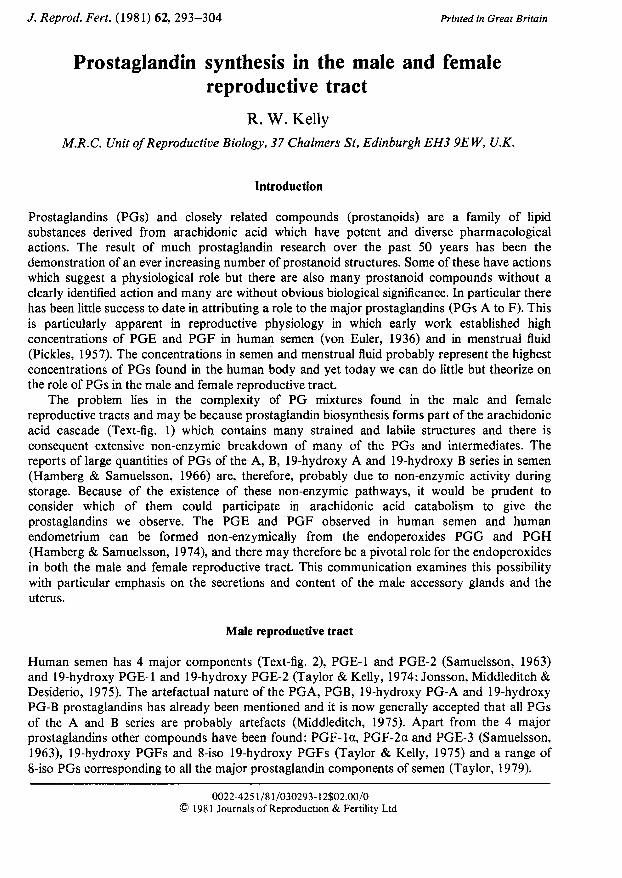
reproductive tracts and may be because prostaglandin biosynthesis forms part of the arachidonicacid cascade (Text-fig. 1) which contains many strained and labile structures and there isconsequent extensive non-enzymic breakdown of many of the PGs and intermediates. Thereports of large quantities of PGs of the A, B, 19-hydroxy A and 19-hydroxy series in semen
(Hamberg & Samuelsson, 1966) are, therefore, probably due to non-enzymic activity duringstorage. Because of the existence of these non-enzymic pathways, it would be prudent toconsider which of them could participate in arachidonic acid catabolism to give theprostaglandins we observe. The PGE and PGF observed in human semen and humanendometrium can be formed non-enzymically from the endoperoxides PGG and PGH(Hamberg & Samuelsson, 1974), and there may therefore be a pivotal role for the endoperoxidesin both the male and female reproductive tract. This communication examines this possibilitywith particular emphasis on the secretions and content of the male accessory glands and theuterus.
Male reproductive tract
Human semen has 4 major components (Text-fig. 2), PGE-1 and PGE-2 (Samuelsson, 1963)and 19-hydroxy PGE-1 and 19-hydroxy PGE-2 (Taylor & Kelly, 1974; Jonsson, Middleditch &Desiderio, 1975). The artefactual nature of the PGA, PGB, 19-hydroxy PG-A and 19-hydroxyPG-B prostaglandins has already been mentioned and it is now generally accepted that all PGsof the A and series are probably artefacts (Middleditch, 1975). Apart from the 4 majorprostaglandins other compounds have been found: PGF-la, PGF-2a and PGE-3 (Samuelsson,1963), 19-hydroxy PGFs and 8-iso 19-hydroxy PGFs (Taylor & Kelly, 1975) and a range of8-iso PGs corresponding to all the major prostaglandin components of semen (Taylor, 1979).
0022-4251/81/O30293-12S02.00/0© 1981 Journals of Reproduction & Fertility Ltd
TO «J5 -
S
UJ
t
• to-
o.oa
o»as
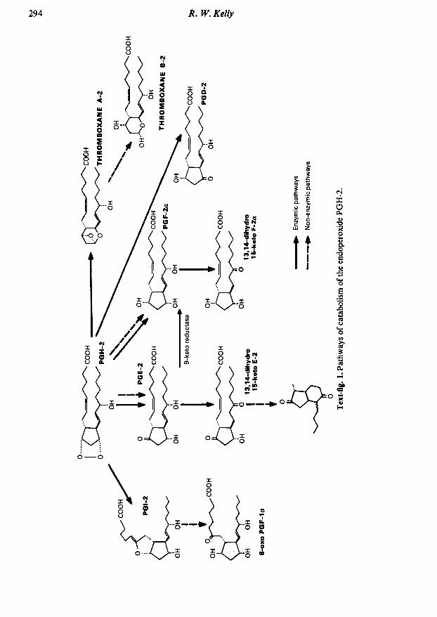
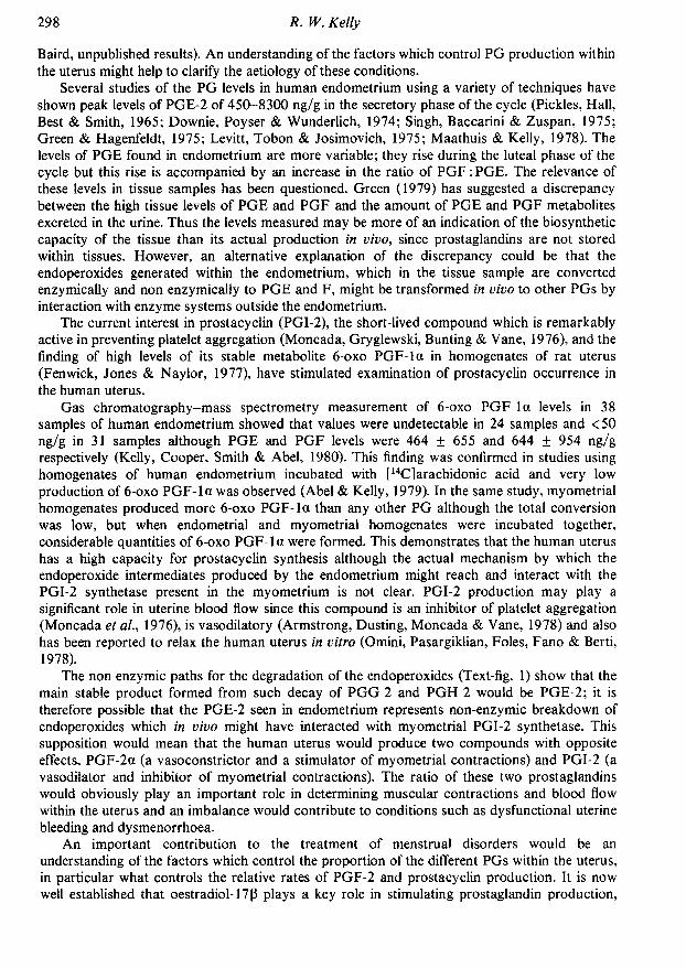
Because of the complexity of human semen it is perhaps unlikely that the minor PG productswill play a physiological role. Inspection of a gas chromatogram of an extract of semen (Text-fig.2) demonstrates the preponderance of the 4 PGs of the E series prostaglandins and a usefulapproach may be to regard these as the functional PGs and examine the minor components togain insight into the manner of production of the seminal PGs.
The prostaglandins were so named because early workers had detected smooth musclestimulating activity in the prostate gland. Today it seems certain that the seminal vesicle is thesource of the majority of PGs in semen; this has been shown by the correlation of PG andfructose concentrations in split ejaculates (Eliasson, 1959) and confirmed by the demonstrationof PGE production from [14C]eicosatrienoic acid incubated with homogenates of human seminalvesicles (Hamberg, 1976). Despite this there is still evidence of prostatic production of PGs(Cavanaugh, Farnsworth, Greizerstein & Wojtowicz, 1980) and we have shown a highproduction of PGF-2a by benign hyperplastic prostatic tissue (F. K. Habib & R. W. Kelly,unpublished) which suggests that the prostate may well contribute some of the F series PGs to
Cholesterolacetate (internal
standard)
(a) Chimpanzee19-OH PGE-1
Isomer of
Retention time (min)Text-fig. 2. Gas chromatogram of the prostaglandins in (a) chimpanzee and (b) human semen.The prostaglandins are derivatized by in-situ methyl oxime formation, extraction, methylationand trimethylsilyl ether formation. The formation of oxime isomers is an inevitableaccompaniment of this process. Chimpanzee semen has a high concentration (~500 pg/ml) of19-hydroxy PGE-1 with small quantities of 19-hydroxy PGE-2 and virtually no PGE. Incontrast, human semen has roughly equal concentrations of 19-hydroxy PGE-1 and 19-hydroxyPGE-2 with substantial amounts of the E prostaglandins.
semen. One anomaly of PG production seen in the bull is that the seminal vesicles have a highcapacity to synthesize PGs (Yamamoto et al, 1977) and yet the semen of this species containsvery little PG (Voglmayr, 1973; Kelly et al, 1976).
In general, the species distribution of seminal PGs is difficult to reconcile with their possiblefunctions. Man is the only anima' with substantial quantities of PGE and 19-hydroxy PGE in thesemen; the apes and some old world monkeys have high concentrations of 19-hydroxy PGE andvery little PGE (Kelly et al, 1976) (Text-fig. 2) and sheep and goats have PGE and PGF(Eliasson, 1959). One species of marsupial, the brush-tailed possum (Trichosurus vulpécula) has19-hydroxy PGF in the semen (Marley, Selley, Duffield, Roger & White, 1977) but theconcentrations in the semen of eutherian species studied are all < 1 pg/ml and it is possible thatthe PGs present in smaller amounts are generated by organs other than the seminal vesicles.There are 3 possible roles for the seminal prostaglandins—(1) participation in the ejaculatoryprocess, (2) stimulation of the female reproductive tract after ejaculation, and (3) action on
spermatozoa around the time of ejaculation; however, no clear-cut effects can be identified.Much evidence is available on the action of PGs on the female reproductive tract. Early
work showed that crude extracts of seminal prostaglandins have a relaxing action on humanmyometrium in vitro (Eliasson, 1959); this can be accounted for by a relaxing action of PGEand also of the 19-hydroxy PGEs, which are the most abundant PGs in human semen (Russell,Taylor & Kelly, 1979). Relaxation of any part of the female reproductive tract after depositionof semen in the vagina is not so easily demonstrated and attempts to investigate this have givenequivocal results (Eliasson & Posse, 1960). Undoubtedly the vagina is a good route foradministration of drugs, but since we know of no direct vascular route from vagina to uterus,PGs absorbed from the vagina would have to traverse the lungs, with their high capacity for PGmetabolism, and an uncertain quantity of PG would reach the uterus or oviducts. A secondquestion which remains unanswered is how relaxation of the uterus could facilitate transport ofspermatozoa. Although evidence for accelerated transport of spermatozoa after PG ad¬ministration is available (Mandi, 1972; Chang, Hunt & Polge, 1973; Spilman, Finn & Norland,1973), this is for species with very low levels of PGs in the semen (Poyser, 1974).
The second possibility, that seminal PGs can enable or facilitate ejaculation has obviousattractions. PGs have been shown to affect neural transmission and PGs can affect thecontraction of the smooth muscle of the male reproductive tract (for a review see Cenedella,1975); moreover, the co-ordination of ejaculation might demand exceptionally high PG levels.But why are these high levels restricted to primates and sheep? These questions cannot beanswered easily and therefore such a role for seminal PGs must remain only a possibility.
The third potential role for seminal PGs is an action on the spermatozoa. This idea has beentested on several occasions and in general no good evidence is available for an action of PGs on
spermatozoa. However, several recent findings suggest that further investigations are justified.PGs can affect calcium flux within cells and across membranes; PGE and PGF are believed toact on the sarcoplasmic reticulum to release into the cytoplasm calcium ions which in turntrigger contractions of the muscle fibres (Carsten & Miller, 1977). Preliminary evidence on themechanism by which PGF-2a exerts its luteolytic effect suggests that in the first instance thismay be by an increase of Ca2+ influx into the luteal cell (or an intracellular release of boundcalcium) which is associated with an inhibition of adenylate cyclase activation (Behrman, 1979).These observations encourage a reappraisal of the possible effects of prostaglandins on calciumflux in the spermatozoa. Such a flux is important since Peterson, Seyler, Bundman & Freund(1979) have shown that the treatment of human spermatozoa with dibutyryl cyclic AMP andtheophylline, a combination designed to raise cAMP levels and of which each component isknown to increase sperm motility (for a review see Hoskins & Casillas, 1975), inhibits the entryof Ca2+ into the spermatozoa by an action which is probably at the plasma membrane.
Neither dibutyryl cAMP nor caffeine are natural components of semen but theprostaglandins are present in huge amounts and the above evidence for their action as
modulators of intracellular calcium levels, together with evidence that prostaglandins might actdirectly as ionophores to facilitate the movement of Ca2+ through lipoprotein membranes(Kirtland & Baum, 1972), suggests that the role of PGs in semen may be to alter Ca2+ flux andthereby control intracellular cAMP levels. If PGs act in this way to regulate sperm metabolismthen this should be detectable; one reason why it may not have been is that previousinvestigations have used Ca2+-free buffer in which to suspend the washed spermatozoa(Eliasson, Murdoch & White, 1968; Pento, Cenedella & Inskeep, 1970) and if the primary effectis on Ca2+ transport into the cell, this medium is obviously inappropriate. An effect of19-hydroxy PGEs in depressing oxidative metabolism in human spermatozoa has been shown(Kelly, 1977) and this explains previous findings of higher respiration rates in washedspermatozoa (Eliasson, 1971). With a Ca2+-containing buffer, an increase in fructose utilizationwas observed as well as a drop in 14C02 production. However, since respiration is low in humanspermatozoa the significance of such an effect may be that it indicates more fundamentalmetabolic changes.
Any PG action on calcium transport might be expected to result in an association betweenspermatozoa and PGs. This may be apparent as binding or by an incorporation of PG into thesperm cell. There is some direct evidence for sperm binding of PGs (Bartoszewitcz, Dandekar,Glass & Gordon, 1975; Mercado, Villalobos, Domínguez & Rosado, 1978). Moreover,azoospermic men have higher seminal PG levels (Sturde, 1968), and after vasectomy, PG levelsin semen tend to rise (Brummer, 1973). The PG concentration in the semen of polyzoospermicmen is lower than that in corresponding groups of men with normal sperm counts (Kelly,Cooper & Templeton, 1979). These findings indicate an inverse relationship between spermdensity and PG content of semen which could be interpreted as a removal or binding of PGs bythe spermatozoa. Although such spermatozoon-PG interactions may be with the E series PGs,we have been unable to demonstrate any such effect with labelled PGs and therefore interactionmight be with the endoperoxides. Because of the non-enzymic routes for PG formation, the PGcontent of semen could be explained almost entirely by the initial presence of endoperoxideswhich would break down to give many of the minor PG products found in semen. Moreover, a
requirement of different endoperoxide concentrations for different species might result in a
qualitative change in the PG content of the semen, since the relatively poor solubility ofendoperoxides in semen would be improved by the addition of a 19-hydroxy group. We do infact observe 19-hydroxy PGEs only in species with very high PG concentrations in the semen. Arecent theory of the symmetry of PG receptors (Beddell & Goodford, 1977) proposes aconvenient channel in the receptor opposite the 19 position of the PG skeleton which suggeststhat a 19-hydroxy group might modify solubility without affecting PGE-like action. If theendoperoxides were the active agents, the apparent removal of PGs by spermatozoa would beexplained since there is strong evidence that the endoperoxides and other active intermediatesbind covalently to protein (Anderson, Crutchley, Chaudhari, Wilson & Elring, 1979). Thehalf-life of the endoperoxides in aqueous solution is approximately 5 min (Hamberg &Samuelsson, 1974) and this would accord with the relatively short time available for interactionwith spermatozoa around the time of ejaculation. We should, therefore, be aware of thepossibility that the PGs found in semen are the breakdown products of active species which havefulfilled their function.
Female reproductive tract
Prostaglandin production by the uterus
Study of the synthesis of PGs by the uterus is particularly important since an imbalance ofPG production is implicated in disorders of menstruation such as dysmenorrhoea (Lundstrom &Green, 1978) and dysfunctional uterine bleeding (S. K. Smith, M. H. Abel, R. W. Kelly & D. T.
Baird, unpublished results). An understanding of the factors which control PG production withinthe uterus might help to clarify the aetiology of these conditions.
Several studies of the PG levels in human endometrium using a variety of techniques haveshown peak levels of PGE-2 of 450-8300 ng/g in the secretory phase of the cycle (Pickles, Hall,Best & Smith, 1965; Downie, Poyser & Wunderlich, 1974; Singh, Baccarini & Zuspan, 1975;Green & Hagenfeldt, 1975; Levitt, Tobon & Josimovich, 1975; Maathuis & Kelly, 1978). Thelevels of PGE found in endometrium are more variable; they rise during the luteal phase of thecycle but this rise is accompanied by an increase in the ratio of PGF:PGE. The relevance ofthese levels in tissue samples has been questioned. Green (1979) has suggested a discrepancybetween the high tissue levels of PGE and PGF and the amount of PGE and PGF metabolitesexcreted in the urine. Thus the levels measured may be more of an indication of the biosyntheticcapacity of the tissue than its actual production in vivo, since prostaglandins are not storedwithin tissues. However, an alternative explanation of the discrepancy could be that theendoperoxides generated within the endometrium, which in the tissue sample are convertedenzymically and non enzymically to PGE and F, might be transformed in vivo to other PGs byinteraction with enzyme systems outside the endometrium.
The current interest in prostacyclin (PGI-2), the short-lived compound which is remarkablyactive in preventing platelet aggregation (Moneada, Gryglewski, Bunting & Vane, 1976), and thefinding of high levels of its stable metabolite 6-oxo PGF-la in homogenates of rat uterus(Fenwick, Jones & Naylor, 1977), have stimulated examination of prostacyclin occurrence inthe human uterus.
Gas chromatography-mass spectrometry measurement of 6-oxo PGF-la levels in 38samples of human endometrium showed that values were undetectable in 24 samples and <50ng/g in 31 samples although PGE and PGF levels were 464 ±655 and 644 ± 954 ng/grespectively (Kelly, Cooper, Smith & Abel, 1980). This finding was confirmed in studies usinghomogenates of human endometrium incubated with [14C] arachidonic acid and very lowproduction of 6-oxo PGF-la was observed (Abel & Kelly, 1979). In the same study, myometrialhomogenates produced more 6-oxo PGF-la than any other PG although the total conversionwas low, but when endometrial and myometrial homogenates were incubated together,considerable quantities of 6-oxo PGF-la were formed. This demonstrates that the human uterushas a high capacity for prostacyclin synthesis although the actual mechanism by which theendoperoxide intermediates produced by the endometrium might reach and interact with thePGI-2 synthetase present in the myometrium is not clear. PGI-2 production may play a
significant role in uterine blood flow since this compound is an inhibitor of platelet aggregation(Moneada et al, 1976), is vasodilatory (Armstrong, Dusting, Moneada & Vane, 1978) and alsohas been reported to relax the human uterus in vitro (Omini, Pasargiklian, Foles, Fano & Berti,1978).
The non-enzymic paths for the degradation of the endoperoxides (Text-fig. 1) show that themain stable product formed from such decay of PGG-2 and PGH-2 would be PGE-2; it istherefore possible that the PGE-2 seen in endometrium represents non-enzymic breakdown ofendoperoxides which in vivo might have interacted with myometrial PGI-2 synthetase. Thissupposition would mean that the human uterus would produce two compounds with oppositeeffects, PGF-2a (a vasoconstrictor and a stimulator of myometrial contractions) and PGI-2 (avasodilator and inhibitor of myometrial contractions). The ratio of these two prostaglandinswould obviously play an important role in determining muscular contractions and blood flowwithin the uterus and an imbalance would contribute to conditions such as dysfunctional uterinebleeding and dysmenorrhoea.
An important contribution to the treatment of menstrual disorders would be an
understanding of the factors which control the proportion of the different PGs within the uterus,in particular what controls the relative rates of PGF-2 and prostacyclin production. It is now
well established that oestradiol-17 ß plays a key role in stimulating prostaglandin production,
many experiments in vivo with laboratory animals have demonstrated that the optimumconditions for PG production are the action of oestradiol-17ß on a progesterone-primed uterus(Ramwell, Leovey & Sintetos, 1977); studies on human endometrium maintained in organculture have shown that oestradiol-17ß is a powerful stimulant of PGF production (Abel &Baird, 1980). However, the action of oestradiol-17ß in stimulating PG synthesis appears to be bya mechanism different from that of the classical receptor pathway which expresses itsuterotrophic action.
The mechanism by which oestradiol-17ß exerts uterotrophic action is well understood; itinvolves the binding of oestradiol-17ß to a cytoso lie receptor and the complex enters the nucleusto initiate messenger RNA production which in turn induces general protein synthesis (Jensen &Jacobson, 1962). This mechanism could explain increased PG synthesis by the uterus inresponse to oestradiol-17 ß since the PG synthetase could be the protein synthesized, but thereare several reasons for suggesting that this does not wholly account for the mechanism of actionof oestradiol. Horton & Poyser (1976) have suggested that there is a discrepancy between thetime taken for general protein synthesis to occur (detectable after 4 h) and the 60-90 min whichelapse before PGF-2a levels rise in response to an oestradiol-17ß infusion into the sheep uterus.Castracane & Jordan (1976) have shown that co-administration of protein synthesis inhibitorsor an anti-oestrogen to rats did not affect the action of oestradiol-17ß on PG production by theuterus. Moreover the reported effects of oestradiol-17ß on the stimulation of PGF-2a production
Control
+0estradiol-17ß (0-2 mM)
2-OH Oestradiol- 1 7ß (0-2 mM)
w
îî î t|PGF-2a | PGD-2
PGE-26-keto PGF-1(i
Arachidonicacid
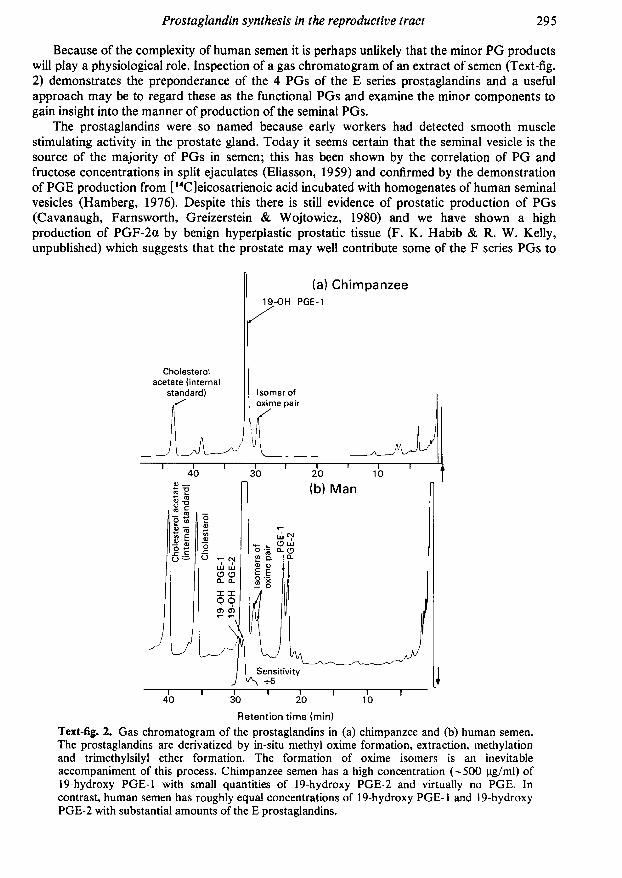
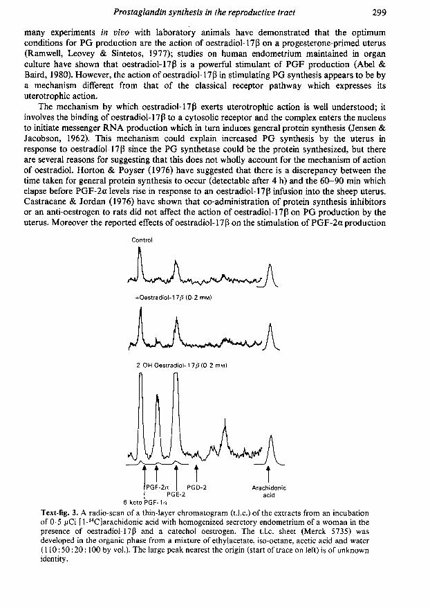
Text-fig. 3. A radio-scan of a thin-layer chromatogram (t.l.c.) of the extracts from an incubationof 0-5 pCi [l-14C|arachidonic acid with homogenized secretory endometrium of a woman in thepresence of oestradiol-17ß and a catechol oestrogen. The t.l.c. sheet (Merck 5735) was
developed in the organic phase from a mixture of ethylacetate, iso-octane, acetic acid and water(110:50:20:100 by vol.). The large peak nearest the origin (start of trace on left) is of unknownidentity.
by homogenates of guinea-pig uterus were small compared to the action of oestradiol-17ß invivo (Naylor & Poyser, 1975). We have therefore examined the possibility that catecholmetabolites of oestradiol-17 ß may be active in stimulating PG synthesis within the uterus. Abroken-cell preparation of whole rat uterus or human endometrium was used, and it was foundthat human (Text-fig. 3) and rat uterus can be stimulated greatly by 2,3,17ß-trihydroxyoestra-l,3,5(10)-triene (2-hydroxy oestradiol) which is several times as effective as oestradiol-17ß itself (Kelly & Abel, 1980a, b).
Although the stimulation of PG synthesis by 2-hydroxy oestradiol is also seen with adrenalinthis effect is not shared with many other cofactors or even catechols. Tryptophan (200 pm),glutathione (200 pM) and catechol (O-hydroxyphenol; 100 pM) are inactive and the effect of2-hydroxy oestradiol-17 ß-acetate, a compound similar in all respects to 2-hydroxy oestradiolexcept in the derivative at the 17ß-position, is minimal (Kelly & Abel, 1980a). Moreover, 100µ -2-hydroxy oestriol and 2-methoxy oestrone (a metabolite of 2-hydroxy oestrone) are alsoinactive, and there is therefore an apparent high specificity for the cofactor requirements of theuterine PG synthetase. Although doses of 100 pM-adrenalin produce little effect, the rat uterusoxidizes adrenalin rapidly as well as oxidizing catechol oestrogens (R. L. Kennedy, P. L. Taylor,M. H. Abel, & R. W. Kelly unpublished observations). This is in accordance with the findingthat such oxidations often accompany PG synthetase (Takeguchi & Sih, 1972) and we shouldtherefore interpret apparent inactivity of low levels of adrenalin with caution.
The levels of catechol oestrogens used in these studies may appear high but the catecholoestrogens are liable to oxidation both enzymically (Nelson, Mitchell, Dybing & Sasame, 1976)and non-enzymically (Gelbke & Knuppen, 1972), with binding of the oxidized forms to proteinsand peptides. This problem can be overcome by the addition of ascorbate to protect thecatechols but we have found that ascorbate inhibits the action of catechol oestrogen andtherefore cannot be used in experiments of the present type.
We have tested the action of other catechol oestrogens on rat and human uterine tissue; the4-hydroxylated oestrogens share the stimulatory action of 2-hydroxy oestrogens but do notinhibit 6-oxo PGF-la formation. We have therefore demonstrated that catechol oestrogens,which are relatively abundant in human blood (Ball, Evans, Haupt, Hoppen & Knuppen, 1978),might play a key role both in stimulating PG production and influencing the PGF-2a toprostacyclin ratio.
Prostaglandin metabolism within the human uterus
Since, in general, prostaglandins are generated and exert their action in the same tissue or
organ, the rate of removal of the active PGs by metabolism is important in controlling theiraction. There is a marked cyclic variation in the activity in human endometrial cytosol of an
NAD-dependent 15-hydroxy prostaglandin dehydrogenase (PGDH; EC 1.1.1.141) (Casey,Hemsell, MacDonald & Johnston, 1980), which has been shown to act on PGE. This enzymeconverts prostaglandins to their relatively inactive 15-keto metabolites. The activity of thisenzyme is maximal between Days 15 and 25 of the menstrual cycle and falls to very low levels on
Days 26-28. Since the maximum activity of the PGDH coincides with maximum progesteroneproduction by the corpus luteum, this finding is in accord with earlier observations that PGDHmay be regulated by steroid hormones and in particular shows a positive correlation withprogesterone concentration (Alam, Russell, Tabor & Moulton, 1976; Falkay & Sas, 1978).
Apart from the degradation of PGs to relatively inactive metabolites, they can also beinterconverted. The most significant conversion would be by the PG 9-keto reducíase enzymewhich converts PGE-2 to PGF-2a. Such an enzyme, which is NADPH-dependent, has beenfound in the ovary (Watson, Sheppherd & Dodson, 1979) although there are no reports of itsoccurrence in the human uterus.

A possible rolefor PGs in the human uterus
If it is assumed that it is the synthetic capacity of the tissue which has been measured inprevious studies, and with the knowledge that PGDH activity is high in the mid-luteal phase butfalls markedly towards the end of the menstrual cycle, then PG concentrations might besignificant only around the time of menstruation. The events immediately precedingmenstruation are regression of the endometrium followed by a period of stasis and constrictionof the spiral arterioles, which characterize the menstruating species (Markee, 1940). Theseevents suggest the production of one or more vasoactive compounds within the uterus. Referringto the vasoactive agent in endometrium which had been transplanted into the eye of rhesusmonkeys, Markee (1940) wrote:
'The stimulus which initiates the constriction of the part of the artery outside the area ofnecrosis is unknown. However the dilation of the iridai vessels immediately outside thetransplant at the time of the contraction of the arteries in the basal zone suggests thatsome agent may be elaborated by the degenerating endometrium and cause changes inthe vessels with which it comes into contact."
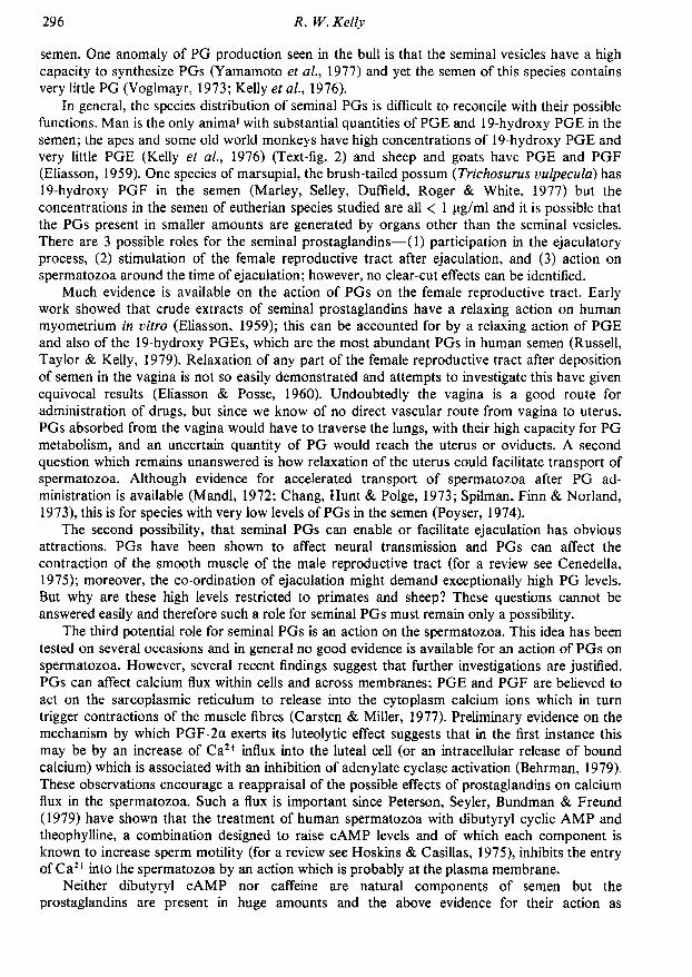
In the light of today's knowledge it is not unreasonable to assume that this agent is aprostaglandin. Initial vasoconstriction might have several effects. First, it is known that freearachidonic acid is re-incorporated into phospholipids by acyl-coenzyme A ligase and acetyltransferase in an ATP-dependent process (Lands & Crawford, 1976). These actions result in afree arachidonic acid concentration that is normally low but the ischaemia associated withvasoconstriction would reduce the ATP supply and interrupt the removal of arachidonic acidwithout effect on its production, thereby increasing the free arachidonic acid concentration.Second, the observed stasis would restrict the removal of any endoperoxide intermediates,thereby increasing the likelihood of their conversion to PGF within the endometrium. Third, thereduced transfer of endoperoxide to the myometrium would restrict the synthesis of thevasodilatory PGI-2 (Text-fig. 4). Thus a positive-feedback situation might exist which would
Venous lake
COOH
OR
PGE-2—»1 5-ketoPGF-2ii—»IS-keto PG
Constriction
PGI-2—»Dilatation
FUNCTIONALLAYER
BASALLAYER
MYOMETRIUM
Text-fig. 4. Diagram of the vasculature of the human endometrium showing spiral arterioles(A), veins (V) and lymph drainage (L). Vasospasm which occurs during the early stages ofmenstruation would restrict any supply of endoperoxide to the myometrium reducing PGI-2production. At this stage of the cycle, formation of the 15-keto metabolites is reduced. Thiscombination of factors leads to increased PGF-2a formation which increases myometrialcontractions and vasoconstriction.
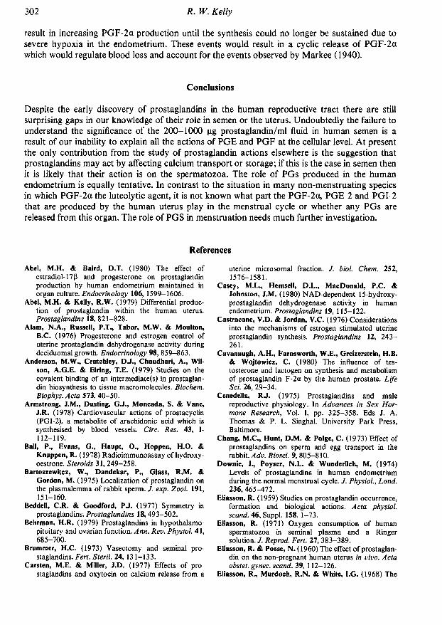
result in increasing PGF-2a production until the synthesis could no longer be sustained due tosevere hypoxia in the endometrium. These events would result in a cyclic release of PGF-2awhich would regulate blood loss and account for the events observed by Markee (1940).
Conclusions
Despite the early discovery of prostaglandins in the human reproductive tract there are stillsurprising gaps in our knowledge of their role in semen or the uterus. Undoubtedly the failure tounderstand the significance of the 200-1000 pg prostaglandin/ml fluid in human semen is aresult of our inability to explain all the actions of PGE and PGF at the cellular level. At presentthe only contribution from the study of prostaglandin actions elsewhere is the suggestion thatprostaglandins may act by affecting calcium transport or storage; if this is the case in semen thenit is likely that their action is on the spermatozoa. The role of PGs produced in the humanendometrium is equally tentative. In contrast to the situation in many non-menstruating speciesin which PGF-2a the luteolytic agent, it is not known what part the PGF-2ot, PGE-2 and PGI-2that are produced by the human uterus play in the menstrual cycle or whether any PGs arereleased from this organ. The role of PGS in menstruation needs much further investigation.
References
Abel, M.H. & Baird, D.T. (1980) The effect ofestradiol-17ß and progesterone on prostaglandinproduction by human endometrium maintained inorgan culture. Endocrinology 106, 1599-1606.
Abel, M.H. & Kelly, R.W. (1979) Differential produc¬tion of prostaglandin within the human uterus.Prostaglandins 18, 821-828.
Alam, N.A., Russell, P. I'.. Tabor, M.W. & Moulton,B.C. (1976) Progesterone and estrogen control ofuterine prostaglandin dehydrogenase activity duringdeciduomal growth. Endocrinology 98, 859-863.
Anderson, M.W., Crutchley, DJ., Chaudhari, ., Wil¬son, A.G.E. & Elring, T.E. (1979) Studies on thecovalent binding of an intermediate(s) in prostaglan¬din biosynthesis to tissue macromolecules. Biochem.Biophys. Acta 573, 40-50.
Armstrong, J.M., Dusting, G.J., Moneada, S. & Vane,J.R. (1978) Cardiovascular actions of prostacyclin(PGI-2), a metabolite of arachidonic acid which issynthesised by blood vessels. Circ. Res. 43, I-112-119.
Ball, P., Evans, G„ Haupt, O., Hoppen, H.O. &Knuppen, R. (1978) Radioimmunoassay of hydroxy-oestrone. Steroids 31, 249-258.
Bartoszewitcz, W., Dandekar, P., Glass, R.M. &Gordon, M. (1975) Localization of prostaglandin onthe plasmalemma of rabbit sperm. /. exp. Zool. 191,151-160.
Beddell, C.R. & Goodford, PJ. (1977) Symmetry inprostaglandins. Prostaglandins 18,493-502.
Behrman, H.R. (1979) Prostaglandins in hypothalamo-pituitary and ovarian function. Ann. Rev. Physiol. 41,685-700.
Brummer, H.C. (1973) Vasectomy and seminal pro¬staglandins. Fert. Steril. 24, 131-133.
Carsten, M.E. & Miller, J.D. (1977) Effects of pro¬staglandins and oxytocin on calcium release from a
uterine microsomal fraction. /. biol. Chem. 252,1576-1581.
Casey, M.L., Hemsell, D.L., MacDonald, P.C. &Johnston, J.M. (1980) NAD-dependent 15-hydroxy-prostaglandin dehydrogenase activity in humanendometrium. Prostaglandins 19, 115-122.
Castracene, V.D. & Jordan, V.C. (1976) Considerationsinto the mechanisms of estrogen stimulated uterineprostaglandin synthesis. Prostaglandins 12, 243-261.
Cavanaugh, A.H., Farnsworth, W.E., Greizerstein, H.B.& Wojtowicz, C. (1980) The influence of tes¬tosterone and lactogen on synthesis and metabolismof prostaglandin F-2a by the human prostate. LifeSci. 26, 29-34.
Cenedella, RJ. (1975) Prostaglandins and malereproductive physiology. In Advances in Sex Hor¬mone Research, Vol. I, pp. 325-358. Eds J. A.Thomas & P. L. Singhal. University Park Press,Baltimore.
Chang, M.C., Hunt, D.M. & Polge, C. (1973) Effect ofprostaglandins on sperm and egg transport in therabbit. Adv. Biosci. 9, 805-810.
Dow nie, J„ Poyser, N.L. & Wunderlich, M. (1974)Levels of prostaglandins in human endometriumduring the normal menstrual cycle. /. Physiol, Lond.236, 465-472.
Eliasson, R. (1959) Studies on prostaglandin occurrence,formation and biological actions. Acta physiol.scand. 46, Suppl. 158, 1-73.
Eliasson, R. (1971) Oxygen consumption of humanspermatozoa in seminal plasma and a Ringersolution. J. Reprod. Fert. 27, 383-389.
Eliasson, R. & Posse, N. (1960) The effect of prostaglan¬din on the non-pregnant human uterus in vivo. Actaobstet, gynec. scand. 39, 112-126.
Eliasson, R., Murdoch, R.N. & White, I.G. (1968) The

metabolism of human spermatozoa in the presence ofprostaglandin El. Acta physiol. scand. 73, 379-382.
Falkay, G. & Sas, M. (1978) Correlation between theconcentrations of prostaglandin dehydrogenase andprogesterone in the early human placenta. J. Endocr.76, 173-174.
Fenwick, L., Jones, R.L. & Naylor, B. (1977) Productionof prostaglandins by the pseudopregnant rat uterus invitro, and the effect of tamoxifen with the iden¬tification of 6-keto prostaglandin F-l as a mjaorproduct. Br. J. Pharmac. 59, 191-199.
Gelbke, H.P. & Knuppen, R. (1972) A new method forpreventing oxidative decomposition of catecholoestrogens during chromatography. J. Chromatog.71,465-471.
Green, K. (1979) Determination of prostaglandins inbody fluids and tissues. Acta obstet, gynec. scand.,Suppl. 87, 15-20.
Green, K. & Hagenfeldt, K. (1975) Prostaglandins in thehuman endometrium. Gas chromatographic-massspectrometric quantitation before and after I.U.D.insertion. Am.J. Obstet. Gynec. 122,611-614.
Hamberg, M. (1976) Biosynthesis of prostaglandin E byhuman seminal vesicles. Lipids 11, 249-250.
Hamberg, M. & Samuelsson, B. (1966) Prostaglandins inhuman seminal plasma. J. Biol. Chem. 241,257-263.
Hamberg, M. & Samuelsson, B. (1974) Prostaglandinendoperoxides. Novel transformations of arachidonicacid in human platelets. Proc. natn. Acad. Sci.U.SA. 71, 3400-3404.
Horton, E.W. & Poyser, N.L. (1976) Uterine luteolytichormone: a physiological role for prostaglandin F-2.Physiol. Rev. 56, 595-651.
Hoskins, D.D. & Casillas, E.R. (1975) Hormones,second messengers and the mammalian sperm¬atozoon. In Advances in Sex Hormone Research.Vol. 1, pp. 283-24. Eds J. A. Thomas & R. L.Singhal. University Park Press, Baltimore.
Jensen, E.V. & Jacobson, H.I. (1962) Basic guide to themechanism of estrogen action. Recent Prog. Horm.Res. 18, 387-408.
Jonsson, H.T., Middleditch, B.S. & Desiderio, D.M.(1975) Prostaglandin in human seminal fluid: twonovel compounds. Science, N.Y. 187, 1093-1094.
Kelly, R.W. (1977) Effect of seminal prostaglandins on
the metabolism of human spermatozoa. J. Reprod.Fert. 50,211-222.
Kelly, R.W. & Abel, M.H. (1980a) Catechol oestrogensstimulate uterine prostaglandin production. Adv.Prostaglandins Thromboxane Res. 8, 1369-1370.
Kelly, R.W. & Abel, M.H. (1980b) Catechol oestrogensstimulate and direct prostaglandin synthesis.Prostaglandins 20, 613-626.
Kelly, R.W., Taylor, P.L., Hearn, J.P., Short, R.V.,Martin, D.E. & Marston, J.H. (1976) 19-Hydroxyprostaglandin El as a major component of thesemen of primates. Nature, Lond. 260, 544-545.
Kelly, R.W., Cooper, I. & Templeton, A.A. (1979)Reduced prostaglandin levels in the semen of menwith very high sperm concentrations. J. Reprod. Fert.56, 195-199.
Kelly, R.W., Cooper, I., Smith, S.K. & Abel, M.H.(1980) The measurement of the production of 6-oxoprostaglandin F-l by the uterus. In 7V¡e Role of
Prostaglandins, Prostacyclin and ThromboxanesMeasurement, pp.142—154. Eds J. M. Boeynaems &A. G. Herman. Martinus Nijhoff. The Hague.
Kirtland, SJ. & Baum, H. (1972) Prostaglandin E mayact as a "calcium ionophore". Nature, New Biol.236,47-49.
Lands, W.E.M. & Crawford, CG. (1976) Enzymes ofmembrane phospholipid metabolism in animals. InThe Enzymes of Biological Membranes Biosynthesisof Cell Components, Vol. 2. pp. 3-85. Ed. A.Martonosi. Plenum, New York.
Levitt, T.M., Tobon, H. & Josimovich, J.B. (1975)Prostaglandin content of human endometrium. Fert.Steril. 26, 296-300.
Lundstrom, V. & Green, K. (1978) Endogenous levels ofprostaglandin F-2a and its main metabolites inplasma and endometrium of normal and dys-menorrhoic women. Am. J. Obstet. Gynec. 130,640-646.
Maathuis, J.B. & Kelly, R.W. (1978) Concentrations ofprostaglandins F-2a and E-2 in the endometriumthroughout the menstrual cycle, after the ad¬ministration of clomiphene or an oestrogen-progestogen pill and in early pregnancy. /. Endocr.77,361-371.
Mandi, J.P. (1972) The effect of prostaglandin E-l onrabbit sperm transport in vivo. J. Reprod. Fert. 31,263-269.
Markee, J.E. (1940) Menstruation in intraocular en¬dometrial transplants in the rhesus monkey. Contr.Embryol. Carnegie Instn. 177, 211-308.
Marley, P.G., Selley, M.L., Duffield. A.M., Roger, J.C. &White, I.G. (1977) 19-OH prostaglandins F in thesemen and prostate gland of marsupials.Theriogenology 8, 207-210.
Mercado, E., Villalobos, M., Domínguez, R. & Rosado, . (1978) Differential binding of PGE-1 and PGF-2ato the human spermatozoa membrane. Life Sci. 22,429-436.
Middleditch, B.S. (1975) PGA: fact or artifact?Prostaglandins 9, 409^111.
Moneada, S., Gryglewski, R., Bunting, S. & Vane, J.R.(1976) An enzyme isolated from arteries transformsprostaglandin endoperoxides to an unstablesubstance that inhibits platelet aggregation. Nature,Lond. 263, 663-665.
Naylor, B. & Poyser, N.L. (1975) Effect of oestradioland progesterone on the in vitro production ofprostaglandin F-2a by the guinea pig uterus. Br. J.Pharmac. 55, 229-232.
Nelson, S.D.. Mitchell, J.R., Dybing, E. & Sasame. H.A.(1976) Cytochrome P-450 mediated oxidation of2-hydroxy estrogens to reactive intermediates.Biochem. Biophys. Res. Commun. 70, 1157-1165.
Omini, C, Pasargiklian, R., Foies, G.C., Fano, M. &Berti, F. (1978) Pharmacological activity of PGI andits metabolite 6-oxo PGF-la on human uterus andfallopian tubes. Prostaglandins 15, 1045-1054.
Pento, J.T., Cenedella, RJ. & Inskeep, E.K. (1970)Effects of prostaglandins E and F upon carbohydratemetabolism of ejaculated and epididymal sperm¬atozoa in vitro. J. Anim. Sci. 30, 409-411.
Peterson, R.N., Seyler, D., Bundman, D. & Freund, M.(1979) The effect of theophylline and dibutyrylcAMP on the uptake of radioactive calcium and

phosphate ions by boar and human spermatozoa. J.Reprod. Fert. 55, 385-390.
Pickles, V.R. (1957) A plain muscle stimulant in themenstruum. Nature, Lond. 180, 1198-1199.
Pickles, V.R., Hall, W.J., Best, F.A. & Smith, G.N.(1965) Prostaglandins in endometrium and menstrualfluid from normal and dysmenorrheic subjects. J.Obstet. Gynaec.Br. Commonw. 72, 185-192.
Poyser, N.L. (1974) Some aspects of prostaglandins inreproduction. Biochem. Soc. Trans. 2, 1196-1200.
Ram well, P.W., Leovey, E.M.K. & Sintetos, A.L. (1977)Regulation of the arachidonic acid cascade. Biol.Reprod. 16, 70-87.
Russell, J.A., Taylor, P.L. & Kelly, R.W. (1979)Preliminary observations on the effects of 19-hydroxy prostaglandin E-l on the activity of thehuman myometrium in vitro. J. Reprod. Fert. 56,33-36.
Samuelsson, B. (1963) Isolation and identification ofprostaglandins from human seminal plasma. J. biol.Chem. 238, 3229-3234.
Singh, HJ.. Baccarini, I.M. & Zuspan, F.P. (1975)Levels of prostaglandins F and E in human en¬dometrium during the menstrual cycle. Am. J. Obstet.Gynec. 121, 1003-1006.
Spilman, C.H., Finn, A.E. & Norland, J.F. (1973) Effectof prostaglandins on sperm transport and fertilizationin the rabbit. Prostaglandins 4, 57-64.
Sturde, H.C. (1968) Experimentelle Untersuchungen zur
Frage der Prostaglandine und ihrer Beziehungen zur
mannlichen Fertilitat. Arzneimittel-Forsch. 18, 895-900, 1158-1163,1298-1310.
Takeguchi, C. & Sih, CJ. (1972) A rapid photometricassay for prostaglandin synthetase: application to thestudy in non-steroidal anti-inflammatory agents.Prostaglandins 2, 169-184.
Taylor, P.L. (1979) The 8-iso prostaglandins, evidencefor eight compounds in human semen.
Prostaglandins 175, 259-267.Taylor, P.L. & Kelly, R.W. (1974) 19-Hydroxylated
prostaglandins as the major prostaglandins of humansemen. Nature, Lond. 250, 665-667.
Taylor, P.L. & Kelly, R.W. (1975) The occurrence of19-hydroxy F prostaglandins in human semen.FEBS Letters 57, 22-25.
Voglmayr, J.K. (1973) Prostaglandin F-2a concen¬tration in genital tract secretions of dairy bulls.Prostaglandins 4, 673-678.
von Euler, U.S. (1936) On the specific vaso-dilating andplain muscle stimulating substances from accessorygenital glands in man and certain animals (prosta¬glandin and vesiglandin). J. Physiol., Lond. 88,213-234.
Watson, J., Sheppherd, T.S. & Dodson, K.S. (1979)Prostaglandin E-2-9-ketoreductase in ovarian tissues.J. Reprod. Fert. 57,489-496.
Yamamoto, S., Ogino, N., Ohki, S., Yoshimoto, T., Bhat,S.G., Oka, J. & Hayaishi, O. (197.7) Enzymologicalstudies on PG biosynthesis. In Biochemical Aspectsof Prostaglandins and Thromboxanes, pp. 1-13. EdsN. Kharash & J. Fried. Academic Press, New York.