Antibody selection for immunocytochemical characterization of the male reproductive system in...
12
Antibody selection for immunocytochemical characterization of the male reproductive system in Psittaciformes Susanne Reitemeier a , Maria Hänse b , Anke Hahn a , Volker Schmidt b , Katrin Steinbach-Sobiraj b , Susanne Tätzner a , Maria-Elisabeth Krautwald-Junghanns b , Almuth Einspanier a, * a Institute of Physiological Chemistry, Veterinary Faculty, University of Leipzig, Leipzig, Germany b Clinic for Birds and Reptiles, Veterinary Faculty, University of Leipzig, Leipzig, Germany article info Article history: Received 17 April 2013 Received in revised form 28 May 2013 Accepted 29 May 2013 Keywords: Immunocytochemistry Testes Psittaciformes Steroidogenesis Relaxin family Proliferation abstract The success of breeding programs is limited by the sparse knowledge about endocrine regulation and biochemical reactions in the psittacine male tract. The immunocyto- chemical analysis of parrots’ testicular tissues provides an insight into their reproductive system but is often hampered by the lack of reliable antibodies. In the present study, we tested a large panel of antibodies raised against steroid receptors, steroidogenic en- zymes, relaxin peptides including their receptors, and proliferation markers on paraffin sections of testicular tissue from eight psittacine genera representing three continents. All investigated species displayed the tested markers in somatic and germ cells of testis and epididymis, even though cell distribution was partly heterogenous and in species-specific patterns. The 17b-hydroxysteroid-dehydrogenase-2, 3b-hydroxysteroid- dehydrogenase, and smooth muscle actin allowed the cross-species differentiation between active and nonactive gonads. The remaining steroidogenic enzymes, steroid receptors, relaxin peptides, and Ki67 proved to be suitable to define reproductive activity depending on the parrot species. Adapting immunocytochemical methods to different psittacines was successful, though various cellular expression patterns do not allow the transfer of results among different parrot species. However, the availability of a reliable repertory of sexual markers is important to examine reproductive biology of psittacine birds. Ó 2013 Elsevier Inc. All rights reserved. 1. Introduction According to the International Union for Conservation of Nature [1] and Birdlife International [2], more than 130 out of approximately 370 living parrot species are listed as near-threatened or worse. In addition to numerous projects targeting the conservation of wild parrots, breeding pro- grams of psittacine birds in captivity are maintained to increase their respective populations. Irrespective of various factors like constricted genetic diversity and difficult pair bonding, the success of these projects also depends on substantiated knowledge about psittacine procreation. Our previous study concentrated on the immunocyto- chemical detection of steroid-producing cells within the testis and epididymis in conjunction with gonadal histologyd a reliable method to ascertain the actual reproductive status of male budgerigars (Buds) (Melopsittacus undulatus) [3]. We were able to provide valuable information about local ste- roidogenesis in male psittacine gonads. Nevertheless, addi- tional special markers are required for these birds to provide a more thorough documentation describing gonadal function and hormonal control by initially demonstrating the local synthesis of various reproductive factors. This is fundamental for the peripheral detection of these factors in psittacine * Corresponding author. Tel.: 0049 341 97 38 100; fax: 0049 341 97 38 119. E-mail address: [email protected] (A. Einspanier). Contents lists available at SciVerse ScienceDirect Theriogenology journal homepage: www.theriojournal.com 0093-691X/$ – see front matter Ó 2013 Elsevier Inc. All rights reserved. http://dx.doi.org/10.1016/j.theriogenology.2013.05.027 Theriogenology 80 (2013) 597–608
-
Upload
uni-leipzig -
Category
Documents
-
view
0 -
download
0
Transcript of Antibody selection for immunocytochemical characterization of the male reproductive system in...

at SciVerse ScienceDirect
Theriogenology 80 (2013) 597–608
Contents lists available
Theriogenology
journal homepage: www.ther io journal .com
Antibody selection for immunocytochemical characterization of the malereproductive system in Psittaciformes
Susanne Reitemeier a, Maria Hänse b, Anke Hahn a, Volker Schmidt b, Katrin Steinbach-Sobiraj b,Susanne Tätzner a, Maria-Elisabeth Krautwald-Junghanns b, Almuth Einspanier a,*a Institute of Physiological Chemistry, Veterinary Faculty, University of Leipzig, Leipzig, GermanybClinic for Birds and Reptiles, Veterinary Faculty, University of Leipzig, Leipzig, Germany
a r t i c l e i n f o
Article history:Received 17 April 2013Received in revised form 28 May 2013Accepted 29 May 2013
Keywords:ImmunocytochemistryTestesPsittaciformesSteroidogenesisRelaxin familyProliferation
* Corresponding author. Tel.: 0049 341 97 38 100119.
E-mail address: [email protected]
0093-691X/$ – see front matter � 2013 Elsevier Inchttp://dx.doi.org/10.1016/j.theriogenology.2013.05.0
a b s t r a c t
The success of breeding programs is limited by the sparse knowledge about endocrineregulation and biochemical reactions in the psittacine male tract. The immunocyto-chemical analysis of parrots’ testicular tissues provides an insight into their reproductivesystem but is often hampered by the lack of reliable antibodies. In the present study, wetested a large panel of antibodies raised against steroid receptors, steroidogenic en-zymes, relaxin peptides including their receptors, and proliferation markers on paraffinsections of testicular tissue from eight psittacine genera representing three continents.All investigated species displayed the tested markers in somatic and germ cells oftestis and epididymis, even though cell distribution was partly heterogenous and inspecies-specific patterns. The 17b-hydroxysteroid-dehydrogenase-2, 3b-hydroxysteroid-dehydrogenase, and smooth muscle actin allowed the cross-species differentiationbetween active and nonactive gonads. The remaining steroidogenic enzymes, steroidreceptors, relaxin peptides, and Ki67 proved to be suitable to define reproductiveactivity depending on the parrot species. Adapting immunocytochemical methods todifferent psittacines was successful, though various cellular expression patterns do notallow the transfer of results among different parrot species. However, the availability ofa reliable repertory of sexual markers is important to examine reproductive biology ofpsittacine birds.
� 2013 Elsevier Inc. All rights reserved.
1. Introduction
According to the International Union for Conservation ofNature [1] and Birdlife International [2], more than 130 outof approximately 370 living parrot species are listed asnear-threatened or worse. In addition to numerous projectstargeting the conservation of wild parrots, breeding pro-grams of psittacine birds in captivity are maintained toincrease their respective populations. Irrespective ofvarious factors like constricted genetic diversity and
; fax: 0049 341 97 38
e (A. Einspanier).
. All rights reserved.27
difficult pair bonding, the success of these projects alsodepends on substantiated knowledge about psittacineprocreation.
Our previous study concentrated on the immunocyto-chemical detectionof steroid-producing cellswithin the testisand epididymis in conjunction with gonadal histologyda reliable method to ascertain the actual reproductive statusof male budgerigars (Buds) (Melopsittacus undulatus) [3]. Wewere able to provide valuable information about local ste-roidogenesis in male psittacine gonads. Nevertheless, addi-tional specialmarkers are required for thesebirds to provide amore thorough documentation describing gonadal functionand hormonal control by initially demonstrating the localsynthesis of various reproductive factors. This is fundamentalfor the peripheral detection of these factors in psittacine

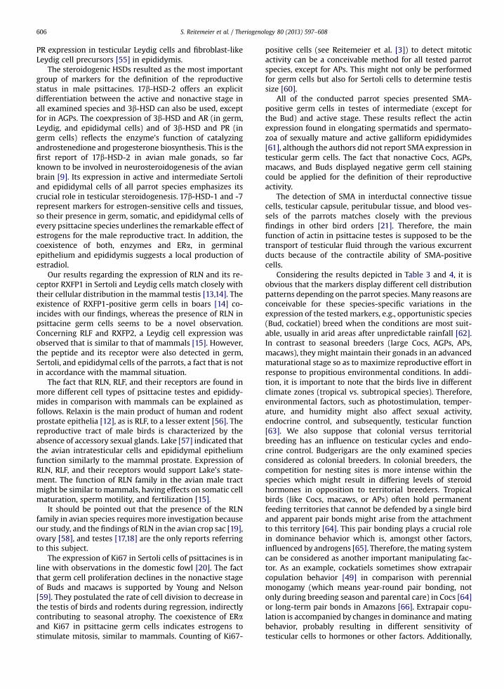
S. Reitemeier et al. / Theriogenology 80 (2013) 597–608598
blood or feces, a less invasive method for fertility assessmentcompared with endoscopy or testicular biopsy.
Numerous immunocytochemical markers successfullyused for the characterization of the mammalian repro-ductive tract are not established in avian species; especiallypsittacines are still scarcely investigated, e.g., the ste-roidogenic enzyme P450 aromatase represents mainly amarker for mammalian Leydig cells but also (subject tospecies and reproductive stage) for Sertoli, germ, andepididymal cells [4]. The only immunocytochemicaldemonstration in gonads of the rooster [5] requires acomparison with the localization of the enzyme in otheravian species. The role of aromatase in the parrot’s testes, incorrelation with the previous detection of estrogen recep-tor (ERa) in spermatocytes [3], has still to be clarified.
Different isoenzymes of 17b-hydroxysteroid dehydro-genase (HSD) were isolated in reproductive tissues ofvarious mammal species [6–8]. Considering the avian sit-uation, quail [9] and starling [10] brain and chicken ovary[11] displayed 17b-HSD expression. Whereas the firstreport about the upregulation of 17b-HSD type 2 in Sertolicells of Buds [3] supports the thesis of androgen biosyn-thesis in avian Sertoli cells, the role of other 17b-HSD iso-enzymes in the psittacine male tract remains unexplained.
The distribution of the relaxin (RLN) family peptides andtheir receptors in cells and tissues of the mammalian maletract is well-studied [12]; RLN represents a marker forSertoli cells [13] and in combination with the RLN familypeptide receptor (RXFP) 1 for Leydig cells [14]. Relaxin-likefactor (RLF) and RXFP2 are basically expressed inmammalian Leydig cells [15]. References to the RLN pep-tides in aves are limited to the chicken ovary [16], testis[17,18], and the pigeon crop sac [19].
Morphological changes in the testes and the epididy-mides occur because of proliferation and apoptosis duringreproductive activity and inactivity. Proliferation processesin the avian male tract were demonstrated in galliformSertoli cells [20] and in psittacine germ cells [3]. Within thescope of mitotic activity of somatic and germ cell types, thesituation in other parrot species requires more investiga-tion. This also applies to the expression of contractilecytoskeletal proteins, especially the microfilament smoothmuscle actin (SMA) found in the testicular tissues of galli-form and anseriform birds [21–23]. Its function in psitta-cine male reproduction continues to remain a matter ofinterest.
The present study focused on two special objectives;first, to evaluate a spectrum of reproductive markers for theBud as a model species for endangered Psittaciformes andsecond, to test the transferability of these establishedmethods to other psittacine species.
2. Materials and methods
2.1. Animals and tissue preparation
For the detection of reliable markers to evaluate thereproductive status, testis samples of different psittacinespecies were examined. Budgerigars (Melopsittacus undu-latus, N ¼ 45) and cockatoos (Coc; Cacatuidae; N ¼ 12,including the genera Nymphicus, Eolophus and Cacatua)
were chosen as representatives for Australian species. TheCongo African gray parrot (AGP, Psittacus erithacus eritha-cus, N ¼ 6) was selected as Old World species and Amazonparrots (AP; genus Amazona, N ¼ 6) and macaws (N ¼ 6,including the genera Ara and Cyanopsitta) likewise as rep-resentatives of the New World species. Tissue sampleswere collectedwithin the scope of routine necropsies at theClinic for Birds and Reptiles in Leipzig. The experimentalprocedure of sample retrieving, tissue preparation andhistochemical staining was performed as explicitlydescribed in our previous publication [3].
2.2. Histological characterization of the reproductive status
Using the Axioskop 2mot plus with magnification�400and the pictorial documentation systemAxioVision (AxioVs40 V 4.5.0.0, Carl Zeiss Imaging Solutions, Jena, Germany),the hematoxylin and eosin-stained gonadal samples wereassigned to three different reproductive phases; nonactive,intermediate, and active stage. The criteria for the definitionof a nonactive, intermediate, and active status were thequantity of germ cells (spermatids and spermatozoa) plusthe interstitial and seminiferous tubular dimensions. Thedistance measurement of seminiferous tubules and inter-stitium in micrometers was conducted by scaling 10 fieldsof view per slide with magnification �200. Furthermore,the presence of melanin in the gonadal tissue was reportedto examine a potential relation between testicular mela-nogenesis, seasonal changes in testicular morphology, andendocrine control of the gonads.
2.3. Immunocytochemistry
The immunocytochemical analyses were conducted byadhering to the established protocol [3]. To demask thenuclear markers (androgen receptor ([AR]-1, AR-2), ERa,and progesterone receptor [PR]), the slides were pretreatedin citrate buffer (pH 6.0) at 120 �C using a steamer. Theincubation with 0.0125% trypsin solution at 37 �C for 15minutes and the endogenous peroxidase inhibitionwith 3%hydrogen peroxide was followed by staining the sectionswith a 2% alcoholic solution of Sudan black B (SigmaAldrich Chemie, Deisenhofen, Germany) for 20 minutes.Because Sudan black B colors phospholipids, neutral fats,and sterols dark blue to black [24], this step was designedto mask an unspecific reaction of lipid accumulation in theseminiferous tubules (described in Reitemeier et al. 2011[3]) with the primary antibody. To avoid nonspecific bind-ing of the primary antibody, the tissue samples wereincubated with pigeon plasma (1:100 dilution of Aquabidest). Subsequently, the slides were reacted with thespecific antibody (all specifities, dilutions, clone names, andproviders are listed in Table 1) at room temperature for 2hours. Washing in PBS was followed by incubation withDako EnVisionþ Dual Link System Peroxidase (DakoCyto-mation, Carpinteria, CA, USA) for 45 minutes. The enzymeactivity was visualized using the chromogenic substrate 3-amino-9-ethylcarbazol (AEC) (AEC Substrate Kit forPeroxidase, Vector Laboratories, Burlingame, CA, USA).Finally, the tissue sections were coveredwith Immu-Mount(Thermo-Scientific, Pittsburgh, PA, USA).

Table 1List of markers used for the investigation of the reproductive status in male Psittaciformes.
Marker Publishedliterature
Providera Specificity/clone Dilution Antigenretrieval
Steroid receptorsAR-1 [25,26] A pc (rabbit IgG) 1:50 (Bud 1:150) h, tAR-2 [27,28] B mc (mouse IgG); Clone 2F12 1:100 h, tERa [29] C mc (mouse anti-human IgG);
Clone B10-A1:100 (Bud 1:50) h, t
PR [30,31] D mc (mouse IgG); Clone PR10A9 1:50 h, tSteroidogenic enzymesAromatase [32] E pc (rabbit anti-Callithrix jacchus IgG) 1:50 t3b-HSD [33,34] E pc (rabbit anti-H3BHSDP-5C) 1:4000 (AGP 1:1000; Ma 1:3000;
AP 1:5000)t
17b-HSD-1 [32] E pc (rabbit anti-Callithrix jacchus IgG) 1:50 (Bud 1:100) t17b-HSD-2 [9,35] E pc (rabbit anti-Callithrix jacchus IgG) 1:500 (AP 1:800; Bud 1:1250) t17b-HSD-7 [36,37] E pc (rabbit anti-Callithrix jacchus IgG) 1:1000 (Bud 1:250) t
Relaxin familyRXFP1 [29,36] F pc (rabbit IgG) 1:800 tRelaxin [29,36] G pc (rabbit anti-porcine) 1:1000 (Bud 1:3000) tRXFP2 [38,39] F pc (rabbit IgG) 1:800 tRelaxin-like factor [38,39] E pc (rabbit IgG) 1:600 (Bud: 1:400) t
Proliferation markersKi67 [40,41] H pc (rabbit IgG) 1:1000 (Bud 1:4000) hSmooth muscle actin [42,43] I mc (mouse anti-human IgG);
Clone 1A41:100 (Bud 1:500) t
Particular dilutions are shown in parentheses.Abbreviations: AGP, African gray parrot; AP, Amazon parrot; AR, androgen receptor; Bud, Budgerigar; ER, estrogen receptor; h, heat pretreatment; HSD,hydroxysteroid dehydrogenase; Ma, Macaw; mc, monoclonal; pc, polyclonal; PR, progesterone receptor; RXFP, relaxin family peptide receptor; t, trypsinpretreatment.
a Providers: A, Thermo Fisher Scientific (Rockford, IL, USA); B, Medac (Hamburg, Germany); C, Euromedex (Souffelweyersheim, France); D, Immunotech(Marseille, France); E, Pineda (Berlin, Germany); F, Prof. Dr. A. Einspanier (Institute of Physiological Chemistry, University of Leipzig, Leipzig, Germany);G, Prof. Dr. O.D. Sherwood (Department of Molecular and Integrative Physiology, University of Illinois, Urbana, IL, USA); H, Antibodies-online (Aachen,Germany); I, DakoCytomation (Carpinteria, CA, USA).
Table 2Interstitial and seminiferous tubular dimensions in gonads of differentpsittacine species.
Species NA INT A
Cockatooa (N ¼ 12) T: 65.2 � 10.3 T: 142.0 � 26.0 T: 192.4 � 43.5I: 23.6 � 5.7 I: 12.9 � 5.3 I: 4.3 � 1.6
African gray parrot(N ¼ 6)
T: 72.1 � 15.3 T: 130.7 � 24.3 T: 199.9 � 27.0I: 19.0 � 5.7 I: 9.2 � 2.1 I: 2.8 � 0.7
Macaw (N ¼ 6) T: 49.3 � 10.1 T: 120.7 � 16.4 T: 151.0 � 14.2I: 17.7 � 4.0 I: 8.8 � 1.7 I: 2.8 � 1.0
Amazon parrot(N ¼ 6)
T: 56.5 � 10.6 T: 114.7 � 17.8 T: 166.1 � 36.9I: 18.0 � 2.2 I: 9.9 � 2.5 I: 4.6 � 1.7
Budgerigarb
(N ¼ 30)T: 57.6 � 10.8 T: 110.9 � 28.2 T: 151.7 � 30.2I: 11.8 � 4.1 I: 6.8 � 3.7 I: 3.8 � 1.2
I and seminiferous T dimensions (mm; mean � SEM) in NA, INT, and Agonads of different psittacine species (distance measurement by scaling10 fields per slide with magnification �200). P values � 0.05 are listed inthe footnotes. The remaining P values exceeded the level of significance(data not shown).Abbreviations: A, active; I, interstitial; INT, intermediate; NA, nonactive;T, tubular.
a T: INT versus A (P ¼ 0.024); I: INT vs. NA (P ¼ 0.024).b T: INT versus A (P � 0.001); INT versus NA (P � 0.001); A versus NA(P � 0.001); I: INT versus A (P ¼ 0.004); INT versus NA (P ¼ 0.001); Aversus NA (P � 0.001).
S. Reitemeier et al. / Theriogenology 80 (2013) 597–608 599
The detection of cytoplasmic steroidogenic enzymes(3b-HSD, 17b-HSD type 1, 2, and 7), RLN family members(Relaxin, Relaxin-like-factor, RXFP-1, and RXFP-2), and SMAwas performed similarly as for the nuclear markers, exceptfor the heat pretreatment. Additionally, the visualization ofthe enzyme with AEC was followed by a counterstainingwith Mayer’s hematoxylin.
A slightly different method was used for the localiza-tion of aromatase. After an overnight reaction with pri-mary antibody (see Table 1) at 4 �C, the slides wereincubated with secondary antibody (polyclonal horse anti-mouse/rabbit IgG; HþL Biotin, Vector Laboratories) atroom temperature for 30 minutes. Subsequently, thevisualization was carried out using alkaline phosphatasestandard (AK-5000; Vector Laboratories) and the chro-mogen Histo Red (AP-Substrate Kit; Linaris, Wertheim-Bettingen, Germany).
The double-indirect method for obtaining proliferationmarker Ki67 (based on Scholzen et al. [40]) was previouslyspecified [3]. Psittacine kidney, adrenal, ovary, and oviduct(steroid receptors, steroidogenic enzymes, and RLN familymarkers) and mice tumors (proliferation markers) wereused as positive control tissue. The omission of the primaryantibody and the incubation with IgG antibodies of irrele-vant specificity served as negative controls.
The immunocytochemistrywas interpreted using a lightmicroscope (Axioskop 2 mot plus) with magnification�100, �200, and �400. The distribution of all testedmarkers in the testicular and epididymal tissues wasrecorded.
2.4. Statistical analysis
The results of the interstitial and seminiferous tubularmeasurement are presented in terms ofmean values� SEMand statistically compared using the Mann-Whitney U Test.The level of significance was defined at P � 0.05 (investi-gated via IBM SPSS Statistics 20).

Fig. 1. Histological and immunocytochemical analysis of testes or epididymides of various psittacine species in different reproductive activity stages. (A) He-matoxylin and eosin staining, Yellow-Crested cockatoo (Cacatua sulphurea), nonactive testis: massive accumulation of dark brown melanocytes in testicularinterstitium and tunica albuginea. (B) Hematoxylin and eosin staining, Galah cockatoo (Eolophus roseicapilla), intermediate testis: moderate accumulation of dark
S. Reitemeier et al. / Theriogenology 80 (2013) 597–608600

S. Reitemeier et al. / Theriogenology 80 (2013) 597–608 601
2.5. Ethics
All psittacine tissues used for this study were obtainedwithin the scope of routine necropsies at the Clinic for Birdsand Reptiles. The birds either deceased or were euthanizedbecause of various diseases, which did not affect thereproductive tract. No birds were sacrificed only for thepurpose of this study. All study protocols were arrangedand permitted according to the German law on animalexperimentation.
3. Results
3.1. Histological analysis
3.1.1. Seminiferous tubules and testicular interstitiumThe quantity of germ cells (spermatids and spermato-
zoa) was similar to the results determined for the Bud [3],so detailed data are not considered at this point. We re-ported significant variations (P values in Table 2) in tubularand interstitial dimensions between the three reproductivestates of the Bud. In the other investigated species (Cocs,AGPs, macaws, and APs) the tubular dimensions alsodeclined from active to nonactive status whereas theinterstitial diameters did the opposite (see Table 2). Onlythe Cocs displayed significant dimension differences intubules of intermediate versus active stage (P ¼ 0.024) andin interstitium of nonactive versus intermediate stage (P ¼0.024). Both the largest testicular tubules (192.4� 43.5 mm)in the active and the largest interstitium (23.6 � 5.7 mm) inthe nonactive status were presented by the Coc. The non-active macaw’s tubular (49.3 � 10.1 mm) and the activeAGPs interstitial (2.8 � 0.7 mm) dimensions were thesmallest observed.
3.1.2. Interstitial melaninMelanin was observed in testicular interstitium and
tunica albuginea of Cocs (nonactive and intermediatestage), but only distributed in form of pigment-containingcells or melanocytes in the genera Eolophus and Cacatua.The testes of genus Nymphicus (intermediate and activestage) displayed no melanin.
The Cocs, however, showed that the degree of melaninaccumulationwas higher in nonactive than in intermediatestage (Fig. 1A, B). One nonactive macaw (Ara ararauna) alsoexhibited black to brown pigmented cells in the inter-stitium, but to a lesser degree than in the Cocs’ testes. The
brown melanocytes in testicular interstitium. (C) Progesterone receptor, Scarlet Macspermatogonia. (D) Aromatase, Yellow-Crested cockatoo, nonactive testis: red cytoplmelanocytes. (E) 17b-hydroxysteroid dehydrogenase (HSD)-1, Spix’s Macaw (Cyamatogonia and Sertoli cells). (F) 17b-HSD-1, Spix’s Macaw, active testis: positive Sertno positive signal, interstitium with massive accumulation of dark brown melanocyreaction (red) of Sertoli cells, interstitium with moderate accumulation dark browntestis: positive cytoplasmic reaction of Sertoli cells. (J) 17b-HSD-7, Blue-and-Yellowmoderate lipid accumulation in the tubular centers (visualized with Sudan black B)signal in germ and Leydig cells, detached lipids (visualized with Sudan black B) in thtestis: positive spermatocytes and Sertoli cells. (M) Relaxin family peptide receptospermatogonia, interstitium with dark brown melanocytes. (N) RXFP1, Congo Africcytoplasmic reaction in epithelium and connective tissue cells of the efferent duthe efferent ductal epithelium. Bar, 50 mm. Ctc, interductal connective tissue; Ed,M, melanocytes; S, Sertoli cells; Sc, spermatocytes; Sg, spermatogonia; St, spermat
absence or presence of melanin in the tunica albuginea wasalso visible macroscopically, because only the testes ofgenus Eolophus and Cacatua appeared brown to black, theremaining ones white-yellowish. No melanin was detect-able in the gonads of the other parrot species.
3.2. Immunocytochemistry
The detailed results of the immunocytochemical ana-lyses, reporting all positive cell types according to repro-ductive stage and psittacine species are summarized inTable 3. The main results are listed in the followingparagraphs.
3.2.1. Steroid receptors
3.2.1.1. Androgen receptor. The steroid receptor AR-2 wasundetectable in all species throughout all reproductivestages. Concerning the AR-1, intermediate and active ma-caws merely displayed positive testicular capsules. Non-active macaws did not express the receptor. Apart fromthat, all species presented AR–1-positive germ cells inde-pendent of their sexual status. Remarkably, only the Cocshowed no positive Leydig cell staining throughout allstages and its nonactive testis expressed AR-1 in Sertolicells. Androgen receptor–1-positive efferent ductalepithelium (ciliated and nonciliated cells) and interductalconnective tissue cells were detected in epididymides ofactive and intermediate Buds, Amazon, and African grayparrots (the latter also in the nonactive stage).
3.2.1.2. Estrogen receptor. The ERa was presented by germcells of every species in all three phases. Leydig andepididymal cells were ERa-positive depending on speciesand reproductive status. Only nonactive Cocs, Buds, andAPs displayed ERa-expressing Sertoli cells.
3.2.1.3. Progesterone receptor. Apart from nonactive macawsand APs, in which no positive staining for PR was noticed,all species showed PR-positive germ cells (Fig. 1C) in allreproductive stages. Intermediate Cocs and nonactive Budsand African gray parrots presented PR in Sertoli cells. Pro-gesterone receptor-positive Leydig cells were merelyobserved in active Cocs and nonactive AGPs. The Bud wasthe only species in which PR was detected in epididymalepithelium and interductal connective tissue cellsthroughout all reproductive phases.
aw (Ara macao), intermediate testis: progesterone receptor-positive nuclei ofasmic signal in spermatogonia and Sertoli cells, interstitiumwith dark brownnopsitta spixii), active testis: positive staining of basal tubular cells (sper-oli cell cytoplasm. (G) 17b-HSD-2, Yellow-Crested cockatoo, nonactive testis:tes. (H) 17b-HSD-2, Galah cockatoo, intermediate testis: positive cytoplasmicmelanocytes. (I) 17b-HSD-2, Cuban Amazon (Amazona leucocephala), activeMacaw (Ara ararauna), nonactive testis: positive signal (red) in Leydig cells,. (K) 17b-HSD-7, cockatiel (Nymphicus hollandicus), active testis: cytoplasmice tubular centers. (L) 17b-HSD-7, budgerigar (Melopsittacus undulatus), activer (RXFP) 1, Galah cockatoo, nonactive testis: moderate positive staining ofan gray parrot (Psittacus erithacus erithacus), nonactive epididymis: positivects. (O) RXFP1, budgerigar, active epididymis: red staining in cytoplasm ofefferent duct; Ep, epithelium; I, interstitial tissue; L, Leydig cells; Lp, lipids;ids; Sz, spermatozoa; T, tubule; Tc, testicular capsule.

S. Reitemeier et al. / Theriogenology 80 (2013) 597–608602
3.2.2. Steroidogenic enzymes
3.2.2.1. Aromatase. The steroidogenic enzyme aromatasewas present in germ cells (Fig. 1D) and epididymalepithelium of every species in all sexual states (except nopositive germ cells in nonactive AGPs). Whereas macawsdisplayed aromatase-expressing Sertoli cells in all stages,no positive staining was detected in Sertoli cells of APs. Thevisualization of the enzymewas successful in Leydig cells ofalmost every species and reproductive stage, with onlyintermediate and nonactive Cocs and active macawsexhibiting negative ones. All species expressed aromatasein epididymal tissue throughout all reproductive states,most of them in epididymal epithelium and in interductalconnective tissuedwith the exception of the Coc, whichpresented only aromatase-positive interductal connectivetissue in all three stages.
3.2.2.2. 3b-HSD. The active and intermediate testis sam-ples of every parrot species revealed 3b-HSD in Leydigcells and epididymal epithelium, and additionally in thenonactive ones of AGPs. Conspicuously, all nonactive go-nads (except AGPs) displayed no reaction on the enzyme.Only active and intermediate macaws expressed 3b-HSDin Sertoli cells. Apart from the Coc, all active andintermediate species presented germ cells positive for3b-HSD.
3.2.2.3. 17b-HSD-1. Irrespective of the fact that 17b-HSD-1was not detectable in active Cocs and nonactive macawsand APs, all species displayed this enzyme in epididymalepithelium, independent of the reproductive status. TheAGP was the only species with positive reactions in Sertoliand Leydig cells throughout all reproductive states. Inter-mediate and active AGPs, macaws, APs, and Buds showed17b-HSD-1 in germ cells. On the contrary, we observed 17b-HSD-1-positive germ cells in intermediate and nonactiveCocs (Fig. 1E, F).
3.2.2.4. 17b-HSD-2. All nonactive testes were negative for17b-HSD-2 (Fig. 1G), whereas all active and intermediatespecies expressed the enzyme in Sertoli (Fig. 1H, I) andepididymal epithelial cells. Only active Cocs showed anadditional staining in germ cells and interductal connectivetissue cells.
3.2.2.5. 17b-HSD-7. Nonactive Buds were negative for 17b-HSD-7. However, all species displayed enzyme-positivegerm cells (except nonactive macaws and active APs) andLeydig cells in every reproductive status (Fig. 1J, L).
3.2.3. Relaxin family
3.2.3.1. Relaxin receptor RXFP1. The RLN receptor RXFP1was not expressed in nonactive APs, whereas other spe-cies of this reproductive status demonstrated positivegerm (Fig. 1M), Sertoli, and Leydig cells and epididymalcells (for species-specific differences, see Table 3). On thecontrary, all active and intermediate stages showed across-species staining of germ and efferent duct epithelial
cells (Fig. 1N, O). Considering every species on its own,a different expression of RXFP1 was remarkable in inter-mediate and active stages versus the nonactive stage. Forexample, nonactive APs did not express the receptor at all,and active and intermediate ones displayed RXFP1 in so-matic, germ, and epididymal cells. The same cellular dis-tribution was demonstrated by Cocs of intermediate andactive stage, whereas RXFP1-positive germ cells onlyoccurred in the nonactive stage.
3.2.3.2. Relaxin. A comparable phenomenon was verifiablefor RLN in macaws (Fig. 2A) and APs. Positive Sertoli cellswere detected in active and intermediate macaws, whereasno staining occurred in nonactive ones. Amazon parrots ofactive and intermediate stage demonstrated positive germand epididymal cells, whereas RLN was not verifiable at allin the nonactive stage. Cockatoos, AGPs, and Buds did notshow such noticeable varieties of RLN expression betweenthe reproductive states.
3.2.3.3. Relaxin receptor RXFP2. Concerning the main re-ceptor for RLF, RXFP2, all active and intermediate phaseswere dominated by positive Leydig and efferent ductepithelial cells (Fig. 2B, C). Only Buds of intermediate stagedid not express RXFP2 staining in Leydig cells. NonactiveMacaws and APs were conspicuous considering the lack ofRXFP2-positive epididymal epithelium, whereas nonactivestages of Cocs had no RXFP2-expression in Leydig, and noexpression in AGPs in germ cells.
3.2.3.4. Relaxin-like factor. A remarkable result wasdemonstrated in the analysis of RLF in Buds. This was theonly species showing a correlation of positive stained cellsfor RLF and its receptor RXFP2 throughout all reproductivestages. Other species displayed this coherency merely inone stage (Cocs and AGPs in intermediate, APs in activestage). Another remarkable fact is that nonactive Cocs,AGPs, macaws, and APs expressed RLF in more different celltypes than RXFP2. Figure 2D, E represent the positivecytoplasmic reaction of RLF in spermatogonia of a non-active Coc and in spermatocytes, Sertoli, and Leydig cells ofan active macaw.
3.2.4. Proliferation markers
3.2.4.1. Ki67. The proliferation marker Ki67 did not occur innonactive macaws and Buds. On the contrary, a cross-species positive staining of germ cells was verifiable in in-termediate and active stage (Fig. 2F, G). Most species(except the Bud) demonstrated Sertoli cell expression inthe intermediate stage (AGP also in the nonactive stage).Referring to the type of positive stained cells, an explicitdifference between active and nonactive stage was provenin macaws, APs, and Buds.
3.2.4.2. Smooth muscle actin. The SMA expression isdemonstrated in Figure 2H, I. Active and intermediate Cocs,AGPs, and macaws displayed SMA in germ cells and inter-ductal connective tissue cells and in the testicular capsule,the peritubular tissue, and in blood vessels. In contrast,

Table 3Reaction of different antibodies on paraffin sections of psittacine testicular and epididymal tissue.
Psittacine species marker Reproductive state Cockatoo (N ¼ 12) African gray parrot (N ¼ 7) Macaw (N ¼ 6) Amazon parrot (N ¼ 7) Budgerigar (N ¼ 45)
Steroid receptorsAR-1 Active GC GC, L, Ed-Ep, Ed-Ctc, BV Tc GC, L, Ed-Ep, Ed-Ctc, BV GC, L, Ed-Ep, Ed-Ctc
Intermediate GC GC, L, Ed-Ep, Ed-Ctc, BV Tc GC, L, Ed-Ep, Ed-Ctc, BV GC, Ed-Ep, Ed-CtcNonactive S, GC GC, L, Ed-Ep, Ed-Ctc, BV ( - ) GC ( - )
AR-2 ( - )ERa Active GC, L, Ed-Ctc, BV GC, Ed-Ctc, BV GC, L, Ed-Ep GC, Ed-Ep, Ed-Ctc, BV GC, Ed-Ep
Intermediate GC, L, Ed-Ep, Ed-Ctc, BV GC, Ed-Ep, Ed-Ctc, BV GC, Ed-Ep GC, L, Ed-Ep, Ed-Ctc, BV GC, Ed-EpNonactive S, GC, Ed-Ep, Ed-Ctc GC, L, Ed-Ep, Ed-Ctc, BV GC, L S, GC, L S, GC, L, Ed-Ctc
PR Active GC, L, Ed-Ctc GC, BV GC, Ed-Ep GC,Ed-Ep, Tc GC, Ed-Ep, Ed-CtcIntermediate S, GC GC, Ed-Ep, Ed-Ctc, BV GC, Ed-Ep GC, Ed-Ep, Ed-Ctc GC, Ed-Ep, Ed-CtcNonactive GC S, GC, L, Ed-Ep, BV ( - ) ( - ) S, GC, Ed-Ep, Ed-Ctc
Steroidogenic enzymesAromatase Active GC, L, Ed-Ep GC, L, Ed-Ep, Ed-Ctc S, GC, Ed-Ep GC, L, Ed-Ep GC, L, Ed-Ep, Ed-Ctc
Intermediate S, GC, Ed-Ep S, GC, L, Ed-Ep, Ed-Ctc S, GC, L, Ed-Ep GC, L, Ed-Ep, Ed-Ctc GC, L, Ed-EpNonactive S, GC, Ed-Ep S, L, Ed-Ep, Ed-Ctc S, GC, L, Ed-Ep, Ed-Ctc GC, L, Ed-Ep, Ed-Ctc S, GC, L, Ed-Ep
3b-HSD Active L, Ed-Ep, Ed-Ctc GC, L, Ed-Ep, Ed-Ctc S, GC, L, Ed-Ep GC, L, Ed-Ep GC, L, Ed-EpIntermediate L, Ed-Ep, Ed-Ctc GC, L, Ed-Ep, Ed-Ctc S, GC, L, Ed-Ep GC, L, Ed-Ep GC, L, Ed-EpNonactive ( - ) GC, Tbl, L, Ed-Ep, Ed-Ctc ( - ) ( - ) ( - )
17b-HSD-1 Active ( - ) S, GC, L, Ed-Ep S, GC, L, Ed-Ep GC, Ed-Ep GC, Ed-EpIntermediate GC, Ed-Ep S, GC, L, Ed-Ep, Ed-Ctc GC, Ed-Ep S, GC, L, Ed-Ep S, GC, L, Ed-EpNonactive GC, Ed-Ep S, L, Ed-Ep ( - ) ( - ) S, L, Ed-Ep
17b-HSD-2 Active S, GC, Ed-Ep, Ed-Ctc S, Ed-Ep S, Ed-Ep S, Ed-Ep S, Ed-EpIntermediate S, Ed-Ep S, Ed-Ep S, Ed-Ep S, Ed-Ep S, Ed-EpNonactive ( - )
17b-HSD-7 Active GC, L, Ed-Ep S, GC, L, Ed-Ep, Ed-Ctc GC, L L, Ed-Ep, Ed-Ctc S, GC, L, Ed-EpIntermediate S, GC, L, Ed-Ep S, GC, Tbl, L, Ed-Ep, Ed-Ctc S, GC, L, Ed-Ep, Ed-Ctc GC, L, Ed-Ep, Ed-Ctc S, GC, L, Ed-EpNonactive S, GC, L, Ed-Ep, Ed-Ctc GC, Tbl, L, Ed-Ep, Ed-Ctc L S, GC, L ( - )
Relaxin familyRXFP1 Active GC, L, Ed-Ep S, GC, L, Ed-Ep S, GC, Ed-Ep S, GC, L,
Ed-Ep, Ed-Ctc, BVGC, L, Ed-Ep
Intermediate S, GC, L, Ed-Ep GC, L, Ed-Ep S, GC, Ed-Ep S, GC, L, Ed-Ep, Ed-Ctc, BV GC, Tbl, L, Ed-Ep, Ed-Ctc, TcNonactive GC S, GC, Ed-Ep, Ed-Ctc, BV S, GC, L ( - ) Ed-Ep
Relaxin Active L, Ed-Ep S, GC, L, Ed-Ep S, L, Ed-Ep GC, Ed-Ep, Ed-Ctc GC, L, Ed-Ep, BVIntermediate S, GC, L, Ed-Ep, Ed-Ctc GC, L, Ed-Ep S, GC, L, Ed-Ep S, GC, Ed-Ep, Ed-Ctc S, GC, L, Ed-Ep, BVNonactive S, GC, L, Ed-Ep, Tc S, GC, L, Ed-Ep, Ed-Ctc, BV L, Ed-Ep ( - ) S, GC, L, Ed-Ep, BV
RXFP2 Active GC, L, Ed-Ep S, GC, L, Ed-Ep S, L, Ed-Ep S, GC, L, Ed-Ep L, Ed-EpIntermediate S, GC, L, Ed-Ep S, GC, L, Ed-Ep S, L, Ed-Ep S, L, Ed-Ep S, Ed-EpNonactive S, GC, Ed-Ep S, L, Ed-Ep S, GC, L S, L S, Ed-Ep
Relaxin-like factor Active S, GC, L, Ed-Ep, Ed-Ctc S, GC, L, Ed-Ep, Ed-Ctc S, GC, L, Ed-Ep S, GC, L, Ed-Ep L, Ed-EpIntermediate S, GC, L, Ed-Ep S, GC, L, Ed-Ep S, GC, L, Ed-Ep S, GC, L, Ed-Ep, Ed-Ctc S, Ed-EpNonactive S, GC, L, Ed-Ep, Tc S, GC, L, Ed-Ep S, L, Ed-Ep S, GC, L, Ed-Ep S, Ed-Ep
Proliferation markersKi67 Active GC GC GC GC GC
Intermediate S, GC S, GC S, GC S, GC, Ed-Ctc GCNonactive GC S, GC ( - ) L ( - )
Smooth muscle actin Active GC, Tbl, Ed-Ctc, BV, Tc GC, Tbl, Ed-Ctc, BV, Tc GC, Tbl, Ed-Ctc, BV, Tc GC, Tbl, Ed-Ctc, BV, Tc GC, Tbl, L, Ed-Ctc, BV, TcIntermediate GC, Tbl, Ed-Ctc, BV, Tc GC, Tbl, Ed-Ctc, BV, Tc GC, Tbl, Ed-Ctc, BV, Tc GC, Tbl, Ed-Ep, Ed-Ctc, BV, Tc Tbl, L, Ed-Ctc, BV, TcNonactive Tbl, Ed-Ctc, BV, Tc Tbl, Ed-Ctc, BV, Tc Tbl, Ed-Ctc, BV, Tc S, GC, Tbl, Ed-Ctc, BV, Tc Tbl, L, BV, Tc
Abbreviations: ( - ), no specific staining verifiable; BV, blood vessels; Ed-Ctc, efferent duct-connective tissue cells; Ed-Ep, efferent duct-epithelium cells; GC, germ cells (spermatogonia, spermatocytes, spermatids,spermatozoa); L, Leydig cells; S, Sertoli cells; Tbl, tubular basal lamina; Tc, testicular capsule.
S.Reitemeier
etal./
Theriogenology80
(2013)597
–608603

Fig. 2. Immunocytochemical analysis of testes or epididymides of various psittacine species in different reproductive activity stages. (A) Relaxin, Blue-and-YellowMacaw, nonactive testis: cytoplasmic signal in Leydig cells, moderate lipid accumulation in the tubular centers (visualized with Sudan black B). (B) Relaxin familypeptide receptor 2, Scarlet Macaw, intermediate testis: positive Sertoli and Leydig cell staining. (C) Relaxin family peptide receptor 2, Scarlet Macaw, intermediateepididymis: red staining in cytoplasm of the efferent ductal epithelium, detached lipids in the epithelial cells (visualized by Sudan black B). (D) Relaxin-like factor,Galah Cockatoo, nonactive testis: positive reaction of spermatogonia (red), interstitiumwith dark brown melanocytes. (E) Relaxin-like factor, Spix’s Macaw, activetestis: red cytoplasmic staining of spermatocytes, Sertoli, and Leydig cells. (F) Ki67, Scarlet Macaw, intermediate testis: nuclear signal in spermatocytes and Sertolicells. (G) Ki67, OrangeWinged Amazon (Amazona amazonica), active testis: nuclear signal in spermatogonia and spermatocytes. (H) Smooth muscle actin, Panamayellow-fronted Amazon (Amazona ochrocephala panamensis), intermediate epididymis: positive reaction of interductal connective tissue cells, blood vessels withSudan black-stained erythrocytes. (I) Smooth muscle actin, budgerigar, active testis: strongly positive peritubular tissue. Bar, 50 mm. Bv, blood vessel; Ctc,interductal connective tissue; Ed, efferent duct; Ep, epithelium; I, interstitial tissue; L, Leydig cells; Lp, lipids; M, melanocytes; S, Sertoli cells; Sc, spermatocytes;Sg, spermatogonia; Sz, spermatozoa; T, tubule.
S. Reitemeier et al. / Theriogenology 80 (2013) 597–608604
nonactive testes of the same species consistently had nopositive germ cell staining. The remarkable findings in Budswere negatively-reacting germ and interductal connectivetissue cells in the nonactive stage. The nonactive APwas theonly parrot with actin-positive Sertoli cells.
4. Discussion
A large panel of antibodies was tested for the immu-nocytochemical analysis of testicular tissues from eightdifferent psittacine genera to provide repeatable tech-niques for the support of breeding programs in captivity. Asshown in mammals, histological and immunocytochemicalinvestigations of testicular tissue are also reliable tech-niques to evaluate the sexual activity of budgerigars [3]. Inthe following paragraphs, we will discuss our results
obtained for other psittacine species, sorted accordingto morphological and immunocytochemical aspects andalways in comparisonwith our previous observations in theBud.
4.1. Histological criteria
Reitemeier et al. [3] provided measurements of tubularand interstitial dimensions that define active, intermediate,and nonactive reproductive states of the Bud males. On thecontrary, the other parrot species did not display significantdifferences within the three testicular stages. A conceivablereason for this findingmight be species-specific variation intestis size based on different sperm competition, extrapaircopulation behavior [44], breeding density, and clutch size[45].We suggested that the enormous increase in testis size

Table 4Applicability of various markers for the immunocytochemical differenti-ation of active and nonactive gonads in male Psittaciformes.
Psittacine speciesmarker
Cockatoo Africangrayparrot
Macaw Amazonparrot
Budgerigar
Steroid receptorsAR-1 (-) (-) D (-) D
ER-a (-) (-) (-) (-) (-)PR (-) (-) D D (-)
Steroidogenicenzymes
Aromatase (-) (-) (-) (-) (-)3b-HSD D (-) D D D
17b-HSD-1 (-) (-) D D (-)17b-HSD-2 D D D D D
17b-HSD-7 (-) (-) (-) (-) D
Relaxin familyRXFP1 (-) (-) (-) D (-)Relaxin (-) (-) (-) D (-)RXFP2 (-) (-) (-) (-) (-)Relaxin-like factor (-) (-) (-) (-) (-)
Proliferation markersKi67 D D D (-) D
Smooth muscleactin
D D D (-) D
Abbreviations: D, suitable; (-) unsuitable; AR, androgen receptor; ER,estrogen receptor; HSD, hydroxysteroid dehydrogenase; PR, progesteronereceptor; RXFP, relaxin family peptide receptor.
S. Reitemeier et al. / Theriogenology 80 (2013) 597–608 605
of the Bud during the breeding season (influenced byhigher sperm competition because of colonial breeding,extrapair copulation behavior, and clutch size [44,45]),especially in relation to the body mass, might be the reasonfor the significant differences in interstitial and seminifer-ous tubular dimensions. In contrast, testes of larger psit-tacines (solitarily breeders, strictly monogamous, smallerclutch size) do not enlarge that much during the breedingseason, so the variation in seminiferous tubular volume andinterstitial diameters might be less conspicuous. Hence, forpsittacine species other than the Bud, the method ofmeasuring tubular and interstitial dimensions is not reli-able to characterize the reproductive status.
All examined large Cocs (Cacatua alba, Cacatua sul-phurea sulphurea, Eolophus roseicapilla) presented mela-nocytes distributed in the testicular capsule andinterstitium, a finding that is consistent with previousstudies in various avian species [46–49]. Many of thesereports discuss a correlation between testicular melano-genesis and gonadal regression. Marshall [47] suggestedthat the presence or absence of melanin was influenced byvolumetric changes of the testes and the dispersion ofmelanocytes during the breeding season. The increasedseminiferous tubules displace the melanocytes and themelanistic testis appears paler when reproductive activitybegins. This matches closely with our observation that in-termediate testes displayed less melanin than nonactiveones. Other authors hypothesize hormones of reproduc-tion, such as FSH, LH, testosterone, and estrogen to haveinhibitory effects on melanogenesis in the testis [46]. So itmight be expected that gonads with melanin depositionshow a downregulation of AR, ERa, and the activity ofsteroidogenic enzymes. However, we cannot support thisview because nonactive and intermediate Cocs expressedthe aforementioned hormone receptors and enzymes. Ourdetection of melanin accumulation in the nonactive testisof a macaw is in line with the observations of O’Malley [48].Interestingly, melanin was not found either in testes ofNymphicus hollandicus, also a genus relative to Cacatuidae,or in the Buds, Amazon, or African gray parrots. Therefore,the testicular melanization seems to be species-specific,probably favored by natural selection in males of specieswith high mutation rates in the mitochondrial genome, aspostulated by Galván et al. [50]. They also supposed thatmelanin protects male germ cells from oxidative stress, afact that might be more important for large Cocs than forother parrot species.
4.2. Immunocytochemical markers: applicability, cellulardistribution, and species-specificity
Wewere able to expand the repertory of markers for theimmunocytochemical investigation of psittacine testeswith the present study. This was achieved by the additionof further steroidogenic enzymes (aromatase, 17b-HSD-1and -7), peptides of the RLN family (RLN, RXFP1 and 2, RLF)and the proliferation marker SMA to the spectrum estab-lished for the Bud [3]. Moreover, these markers were alsotested on gonads of seven further psittacine genera toprove species-transferability of our methods. Their
applicability concerning the assessment of the actualreproductive stage of a parrot species is depicted in Table 4.
Data on AR expression in avian testes are particularlyconflicting. Most studies in poultry and canaries reportrestricted receptor localization on somatic cells [51,52].This matches with our detection of AR in Leydig cells ofBuds, African gray, and Amazon parrots, in Coc Sertoli cellsand macaw smooth muscle cells (testicular capsule). So far,no correlating data exist for other avian species regardingAR-positive germ cells. Therefore, the regulation of sper-matogenesis is supposed to be similar to that in mammalsbecause androgens influence testicular somatic cells ratherthan act directly on germ cells [51]. Thanks to the expres-sion of AR and androgen-synthesizing enzymes (HSDs) ingerm cells (Table 3), this study provides evidence about adirect influence of androgens on the germinal epithelium ofparrots. The cellular-specific differences in AR-expressionin the three reproductive stages indicate different sensi-tivity to androgens.
The coexpression of ERa and aromatase in the avianmale tract represents a novel finding. Other authors re-ported the detection of both, ERa and aromatase, in brainregions of passerines [53]. Every conducted parrot speciesdisplayed ERa and aromatase in germinal epitheliumcoinciding with the observations of Kwon et al. [5] inWhite Leghorn roosters. They postulated germ cells to besites for estrogen synthesis because of strong aromataseimmunostaining.
As aforementioned for the Bud [3], this is still the firstevidence of PR in gonads of male birds because so far, it hasonly been described in the reproductive tract of femalebirds, e.g., ostriches [54]. Similar to mammals, progester-one is supposed to be a stimulator of Leydig cell steroido-genesis. This could be confirmed by our results concerning
S. Reitemeier et al. / Theriogenology 80 (2013) 597–608606
PR expression in testicular Leydig cells and fibroblast-likeLeydig cell precursors [55] in epididymis.
The steroidogenic HSDs resulted as the most importantgroup of markers for the definition of the reproductivestatus in male psittacines. 17b-HSD-2 offers an explicitdifferentiation between the active and nonactive stage inall examined species and 3b-HSD can also be used, exceptfor in AGPs. The coexpression of 3b-HSD and AR (in germ,Leydig, and epididymal cells) and of 3b-HSD and PR (ingerm cells) reflects the enzyme’s function of catalyzingandrostenedione and progesterone biosynthesis. This is thefirst report of 17b-HSD-2 in avian male gonads, so farknown to be involved in neurosteroidogenesis of the avianbrain [9]. Its expression in active and intermediate Sertoliand epididymal cells of all parrot species emphasizes itscrucial role in testicular steroidogenesis. 17b-HSD-1 and -7represent markers for estrogen-sensitive cells and tissues,so their presence in germ, somatic, and epididymal cells ofevery psittacine species underlines the remarkable effect ofestrogens for the male reproductive tract. In addition, thecoexistence of both, enzymes and ERa, in germinalepithelium and epididymis suggests a local production ofestradiol.
Our results regarding the expression of RLN and its re-ceptor RXFP1 in Sertoli and Leydig cells match closely withtheir cellular distribution in the mammal testis [13,14]. Theexistence of RXFP1-positive germ cells in boars [14] co-incides with our findings, whereas the presence of RLN inpsittacine germ cells seems to be a novel observation.Concerning RLF and RXFP2, a Leydig cell expression wasobserved that is similar to that of mammals [15]. However,the peptide and its receptor were also detected in germ,Sertoli, and epididymal cells of the parrots, a fact that is notin accordance with the mammal situation.
The fact that RLN, RLF, and their receptors are found inmore different cell types of psittacine testes and epididy-mides in comparison with mammals can be explained asfollows. Relaxin is the main product of human and rodentprostate epithelia [12], as is RLF, to a lesser extent [56]. Thereproductive tract of male birds is characterized by theabsence of accessory sexual glands. Lake [57] indicated thatthe avian intratesticular cells and epididymal epitheliumfunction similarly to the mammal prostate. Expression ofRLN, RLF, and their receptors would support Lake’s state-ment. The function of RLN family in the avian male tractmight be similar tomammals, having effects on somatic cellmaturation, sperm motility, and fertilization [15].
It should be pointed out that the presence of the RLNfamily in avian species requires more investigation becauseour study, and the findings of RLN in the avian crop sac [19],ovary [58], and testes [17,18] are the only reports referringto this subject.
The expression of Ki67 in Sertoli cells of psittacines is inline with observations in the domestic fowl [20]. The factthat germ cell proliferation declines in the nonactive stageof Buds and macaws is supported by Young and Nelson[59]. They postulated the rate of cell division to decrease inthe testis of birds and rodents during regression, indirectlycontributing to seasonal atrophy. The coexistence of ERaand Ki67 in psittacine germ cells indicates estrogens tostimulate mitosis, similar to mammals. Counting of Ki67-
positive cells (see Reitemeier et al. [3]) to detect mitoticactivity can be a conceivable method for all tested parrotspecies, except for APs. This might not only be performedfor germ cells but also for Sertoli cells to determine testissize [60].
All of the conducted parrot species presented SMA-positive germ cells in testes of intermediate (except forthe Bud) and active stage. These results reflect the actinexpression found in elongating spermatids and spermato-zoa of sexually mature and active galliform epididymides[61], although the authors did not report SMA expression intesticular germ cells. The fact that nonactive Cocs, AGPs,macaws, and Buds displayed negative germ cell stainingcould be applied for the definition of their reproductiveactivity.
The detection of SMA in interductal connective tissuecells, testicular capsule, peritubular tissue, and blood ves-sels of the parrots matches closely with the previousfindings in other bird orders [21]. Therefore, the mainfunction of actin in psittacine testes is supposed to be thetransport of testicular fluid through the various excurrentducts because of the contractile ability of SMA-positivecells.
Considering the results depicted in Table 3 and 4, it isobvious that the markers display different cell distributionpatterns depending on the parrot species. Many reasons areconceivable for these species-specific variations in theexpression of the tested markers, e.g., opportunistic species(Bud, cockatiel) breed when the conditions are most suit-able, usually in arid areas after unpredictable rainfall [62].In contrast to seasonal breeders (large Cocs, AGPs, APs,macaws), they might maintain their gonads in an advancedmaturational stage so as to maximize reproductive effort inresponse to propitious environmental conditions. In addi-tion, it is important to note that the birds live in differentclimate zones (tropical vs. subtropical species). Therefore,environmental factors, such as photostimulation, temper-ature, and humidity might also affect sexual activity,endocrine control, and subsequently, testicular function[63]. We also suppose that colonial versus territorialbreeding has an influence on testicular cycles and endo-crine control. Budgerigars are the only examined speciesconsidered as colonial breeders. In colonial breeders, thecompetition for nesting sites is more intense within thespecies which might result in differing levels of steroidhormones in opposition to territorial breeders. Tropicalbirds (like Cocs, macaws, or APs) often hold permanentfeeding territories that cannot be defended by a single birdand apparent pair bonds might arise from the attachmentto this territory [64]. This pair bonding plays a crucial rolein dominance behavior which is, amongst other factors,influenced by androgens [65]. Therefore, themating systemcan be considered as another important manipulating fac-tor. As an example, cockatiels sometimes show extrapaircopulation behavior [49] in comparison with perennialmonogamy (which means year-round pair bonding, notonly during breeding season and parental care) in Cocs [64]or long-term pair bonds in Amazons [66]. Extrapair copu-lation is accompanied by changes in dominance andmatingbehavior, probably resulting in different sensitivity oftesticular cells to hormones or other factors. Additionally,

S. Reitemeier et al. / Theriogenology 80 (2013) 597–608 607
pair mating depends on migration behavior, so variationsmight appear between sedentary [67] and irregularmigrating birds [68]. In sedentary birds, mated pairs func-tion as social alliances in resource competition. Most of all,especially with regard to RLN [12], evolutionary aspectsmight influence the species-specific cellular expression ofthe RLN family peptides and receptors. Within the scope ofdivergency of parrots, we hypothesize the development ofdifferent expression patterns.
4.3. Conclusions
In conclusion, we were able to detect 14 of the 15 testedmarkers in the testes of all investigated parrot species, eventhough they displayed partly heterogeneous, species-specific expression patterns. In general, 17b-HSD-2, 3b-HSD, and SMA allowed a precise definition of reproductiveactivity, whereas the other steroidogenic enzymes, steroidreceptors, RLN peptides, and Ki67 turned out to be appli-cable depending on the species. The immunocytochemicalmethods established within the framework of our previousstudy in the Bud [3], were successfully adapted to otherpsittacines. It is important to note that different cellulardistribution patterns do not allow a transfer of the resultsfrom one parrot species to another. Therefore, we recom-mend using a combination of markers, always in consid-eration of the investigated species. Our study delivers areliable repertoire of sexual markers, an essential basis forfurther analyses, for instance, the peripheral detection ofthese factors in psittacine blood or feces.
Acknowledgments
The authors thank Dr. Thomas Scholzen (ResearchCentre Borstel) for his generous gift of the Ki67 antibody.
This study was supported by the European Union (Eu-ropean Social Fund; ESF) and the German Research Foun-dation (DFG), DFG EI 333/12-2.
The European Social Fund (ESF) is a structural fund setup by the European Union. It was established in 1957 in aneffort to scale down the differences in living standardsbetween people living in the various regions of the Euro-pean Union. Saxony also benefits from this structural fund.The benefits are felt in particular by businesses that arecreating more permanent jobs, by unemployed individualsseeking to achieve commercial autonomy, and by qualifiedspecialists who wish to initiate well-grounded researchprojects.
The DFG is the self-governing organization for scienceand research in Germany. It serves all branches of scienceand the humanities. In organizational terms, the DFG is anassociation under private law. Its membership consists ofGerman research universities, nonuniversity research in-stitutions, scientific associations, and the Academies ofScience and the Humanities. The DFG receives the largemajority of its funds from the states and the Federal Gov-ernment, which are represented in all Grants Committees.At the same time, the voting system and procedural regu-lations guarantee science-driven decisions.
The role of ESF and DFG is confined to financial supportof this study; both funding sources were neither involved
in study design, nor in the collection, analysis, and inter-pretation of data, in the writing of the report, and in thedecision to submit the report for publication.
References
[1] IUCN Red List of Threatened Species, Version 2012.2, <http://www.iucnredlist.org/>; 2013.
[2] BirdLife International. BirdLife checklist of the birds of the world,with conservation status and taxonomic sources, <http://www.birdlife.org/datazone/species/search>; 2013.
[3] Reitemeier S, Hänse M, Hahn A, Schmidt V, Steinbach-Sobiraj K,Krautwald-Junghanns ME, et al. Evaluating the reproductive statusof the male budgerigar (Melopsittacus undulatus). Gen Comp Endo-crinol 2011;171:350–8.
[4] Carreau S, Lambard S, Delalande C, Denis-Galeraud I, Bilinska B,Bourguiba S. Aromatase expression and role of estrogens in malegonad: a review. Reprod Biol Endocrinol 2003;1:35–40.
[5] Kwon S, Hess RA, Bunick D, Nitta H, Janulis L, Osawa Y, et al. Roostertesticular germ cells and epididymal sperm contain P450 aromatase.Biol Reprod 1995;53:1259–64.
[6] Lemazurier E, Séralini GE. Evidence for sulfatase and 17beta-hydroxysteroid dehydrogenase type 1 activities in equine epidid-ymis and uterus. Theriogenology 2002;58:113–21.
[7] Pelletier G, Luu-The V, Li S, Ren L, Labrie F. Localization of 17beta-hydroxysteroid dehydrogenase type 1 mRNA in mouse tissues. J MolEndocrinol 2004;33:459–65.
[8] Penning TM. Molecular endocrinology of hydroxysteroid de-hydrogenases. Endocr Rev 1997;18:281–305.
[9] Matsunaga M, Ukena K, Tsutsui K. Androgen biosynthesis in thequail brain. Brain Res 2002;948:180–5.
[10] Riters LV, Baillien M, Eens M, Pinxten R, Foidart A, Ball GF, et al.Seasonal variation in androgen-metabolizing enzymes in the dien-cephalon and telencephalon of the male European starling (Sturnusvulgaris). J Neuroendocrinol 2001;13:985–97.
[11] Wajima Y, Furusawa T, Kawauchi S, Wakabayashi N, Najabayashi O,Nishimori K, et al. The cDNA cloning and transient expression of anovary-specific 17beta-hydroxysteroid dehydrogenase of chickens.Gene 1999;233:75–82.
[12] Ivell R, Kotula-Balak M, Glynn D, Heng K, Anand-Ivell R. Relaxinfamily peptides in the male reproductive system - a criticalappraisal. Mol Hum Reprod 2011;17:71–84.
[13] Cardoso LC, Nascimento AR, Royer C, Porto CS, Lazari MFM. Locallyproduced relaxin may affect testis and vas deferens function in rats.Reproduction 2010;139:185–96.
[14] Kohsaka T, Kato S, Qin S, Minagawa I, Yogo K, Kawarasaki T, et al.Identification of boar testis as a source and target tissue of relaxin.Ann N Y Acad Sci 2009;1160:194–6.
[15] Agoulnik AI. Relaxin and related peptides in male reproduction. AdvExp Med Biol 2007;612:49–64.
[16] Hisaw FL, Zarrow MX. The physiology of relaxin. Vitam Horm 1950;8:151–78.
[17] Steinetz BG, Beach VL, Kroc RL. The physiology of relaxin in laboratoryanimals. In: Lloyd CH, editor. Recent progress in the endocrinology ofreproduction. New York: Academic Press; 1959. p. 389–427.
[18] Steinetz BG, Beach VL, Tripp LV, Defalco RJ. Reactions of antisera toporcine relaxin with relaxin-containing tissues of other speciesin vivo and in vitro. Acta Endocrinol 1964;47:371–84.
[19] Bani G, Sacchi TB, Bigazzi M. Response of the pigeon crop sac tomammotrophic hormones: comparison between relaxin and pro-lactin. Gen Comp Endocrinol 1990;80:16–23.
[20] Bozkurt HH, Aktas A, Ulkay MB, Firat UB. Sertoli cell proliferationduring the post hatching period in domestic fowl. J Vet Sci 2007;8:219–22.
[21] Aire TA, Ozegbe PC. The testicular capsule and peritubular tissue ofbirds: morphometry, histology, ultrastructure and immunohisto-chemistry. J Anat 2007;210:731–40.
[22] Maretta M, Marettová E. Immunohistochemical demonstration ofmyoid cells in the testis and its excurrent ducts in the domesticfowl. Br Poult Sci 2004;45:585–9.
[23] Van Nassauw L, Harrisson F, Callebaut M. Smooth muscle cells inthe peritubular tissue of the quail testis. Eur J Morphol 1993;31:60–4.
[24] Romeis B. Paraplasmatische Substanzen - Färbung mit Sudan undScharlach. In: Böck P, editor. Mikroskopische technik, auflage 17.München, Wien, Baltimore: Urban and Schwarzenberg; 1989. p.379–82.

S. Reitemeier et al. / Theriogenology 80 (2013) 597–608608
[25] Clancy AN, Whitman C, Michael AP, Albers HE. Distribution ofandrogen receptor-like immunoreactivity in the brains of intact andcastrated male hamsters. Brain Res Bull 1994;33:325–32.
[26] Takeda H, Nakamoto T, Kokontis J, Chodak GW, Chang C. Autor-egulation of androgen receptor expression in rodent prostate:immunohistochemical and in situ hybridization analysis. BiochemBiophys Res Comm 1991;177:488–96.
[27] Bläuer M, Vaalasti A, Pauli SL, Ylikomi T, Joensuu T, Tuohimaa P.Location of androgen receptor in human skin. J Invest Dermatol1991;97:264–8.
[28] Nevalainen MT, Valve EM, Mäkelä SI, Bläuer M, Tuohimaa PJ,Härkönen PL. Estrogen and prolactin regulation of rat dorsal andlateral prostate in organ culture. Endocrinology 1991;129:612–22.
[29] Simon C, Einspanier A. The hormonal induction of cervical remod-eling in the common marmoset monkey (Callithrix jacchus).Reproduction 2009;137:517–25.
[30] Miller K, Auld J, Jessup E, Rhodes A, Ashton-Key M. Antigenunmasking in formalin-fixed routinely processed paraffin wax-embedded sections by pressure cooking: a comparison with a mi-crowave oven heating and traditional methods. Adv Anat Pathol1995;2:60–4.
[31] Shi SR, Key ME, Kalra KL. Antigen retrieval in formalin-fixed,paraffin-embedded tissues: an enhancement method for immuno-histochemical staining based on microwave oven heating of tissuesections. J Histochem Cytochem 1991;39:741–8.
[32] Einspanier A, Lieder K, Brüns A, Husen B, Thole H, Simon C. Induc-tion of endometriosis in the marmoset monkey (Callithrix jacchus).Mol Hum Reprod 2006;12:291–9.
[33] Beindorff N, Einspanier A. Luteotrophic effects of relaxin, chorionicgonadotrophin and FSH in common marmoset monkeys (Callithrixjacchus). Reproduction 2010;139:923–30.
[34] Purohit VD, Basrur PK, Bhatnagar MK. Histochemical localization of3beta-hydroxysteroid dehydrogenase in the testes of chicken-pheasant hybrids. Histochem J 1977;9:293–9.
[35] Tsutsui K, Matsunaga M, Miyabara H, Ukena K. Neurosteroidbiosynthesis in the quail brain: a review. J Exp Zool Part A Comp ExpBiol 2006;305:733–42.
[36] Einspanier A, Lieder K, Husen B, Ebert K, Lier S, Einspanier R, et al.Relaxin supports implantation and early pregnancy in themarmoset monkey. Ann N Y Acad Sci 2009;1160:140–6.
[37] Husen B, Adamski J, Brüns A, Deluca D, Fuhrmann K, Möller G, et al.Characterization of 17beta-hydroxysteroid dehydrogenase type 7 inreproductive tissues of the marmoset monkey. Biol Reprod 2003;68:2092–9.
[38] Anand-Ivell RJ, Relan V, Balvers M, Coiffec-Dorval I, Fritsch M,Bathgate RA, et al. Expression of the insulin-like peptide 3 (INSL3)hormone-receptor (LGR8) system in the testis. Biol Reprod 2006;74:945–53.
[39] Ivell R, Bathgate RA. Reproductive biology of the relaxin-like factor(RLF/INSL3). Biol Reprod 2002;67:699–705.
[40] Scholzen T, Endl E, Wohlenberg C, van der Sar S, Cowell IG, Gerdes J,et al. The Ki-67 protein interacts with members of the hetero-chromatin protein 1 (HP1) family: a potential role in the regulationof higher-order chromatin structure. J Pathol 2002;196:135–44.
[41] Traut W, Endl E, Scholzen T, Gerdes J, Winking H. The temporal andspatial distribution of the proliferation associated Ki-67 proteinduring female and male meiosis. Chromosoma 2002;111:156–64.
[42] Brennan PA, Umar T, Zaki GA, Langdon JD, Spedding A, Buckley J, et al.Are myoepithelial cells responsible for the widespread expression ofinducible nitric oxide synthase in pleomorphic adenoma? Animmunohistochemical study. J Oral Pathol Med 2000;29:279–83.
[43] Roholl PJ, Elbers HR, Prinsen I, Claessens JA, van Unnik JA. Distri-bution of actin isoforms in sarcomas: an immunohistochemicalstudy. Hum Pathol 1990;21:1269–74.
[44] Garamszegi LZ, Eens M, Hurtrez-Boussès S, Møller AP. Testosterone,testes size, and mating success in birds: a comparative study. HormBehav 2005;47:389–409.
[45] Pitcher TE, Dunn PO, Whittingham LA. Sperm competition and theevolution of testes size in birds. J Evol Biol 2005;18:557–67.
[46] Anthony RG, Buss IO. Relationship of gonadal recrudescence andtesticular melanogenesis in California quail. Condor 1974;76:452–7.
[47] Marshall AJ. Breeding seasons and migration. In: Marshall AJ, editor.Biology and comparative physiology of birds. New York: AcademicPress; 1961. p. 307–39.
[48] O’Malley B. Avian anatomy and physiology. In: O’Malley B, editor.Clinical anatomy and physiology of exotic species. Structure andfunction of mammals, birds, reptiles and amphibians. Edinburgh,London: Elsevier Saunders; 2005. p. 97–161.
[49] Spoon TR, Millam JR, Owings DH. Behavioural compatibility,extrapair copulation and mate switching in a socially monogamousparrot. Anim Behav 2007;73:815–24.
[50] Galván I, Møller AP, Erritzøe J. Testicular melanization has evolvedin birds with high mtDNA mutation rates. J Evol Biol 2011;24:988–98.
[51] Dornas RA, Oliveira AG, Dias MO, Mahecha GA, Oliveira CA.Comparative expression of androgen receptor in the testis andepididymal region of roosters (Gallus domesticus) and drakes(Anas platyrhynchos). Gen Comp Endocrinol 2008;155:773–9.
[52] Nastiuk KL, Clayton DF. Seasonal and tissue specific regulation ofcanary androgen receptor messenger ribonucleic acid. Endocri-nology 1994;134:640–9.
[53] Canoine V, Fusani L, Schlinger B, Hau M. Low sex steroids, highsteroid receptors: increasing the sensitivity of the nonreproductivebrain. Dev Neurobiol 2007;67:57–67.
[54] Madekurozwa MC. A study of the immunohistochemical localiza-tion of the progesterone, oestrogen receptors in the magnum of theimmature ostrich, Struthio camelus. Anat Histol Embryol 2002;31:317–20.
[55] Madekurozwa MC, Chapvepi TS, Matema S, Teerds KJ. Relationshipbetween seasonal changes in spermatogenesis in the juvenileostrich (Struthio camelus) and the presence of the LH receptor and3beta-hydroxysteroid dehydrogenase. Reproduction 2002;123:735–42.
[56] Klonisch T, Müller-Huesmann H, Riedel M, Kehlen A, Bialek J,Radestock Y, et al. INSL3 in the benign hyperplastic and neoplastichuman prostate gland. Int J Onc 2005;27:307–15.
[57] Lake PE. Fowl semen as collected by the massage method. J Agric Sci1957;49:120–6.
[58] Brackett KH, Fields PA, Dubois W, Chang SM, Mather FB, Fields MJ.Relaxin: an ovarian hormone in an avian species (Gallus domesticus).Gen Comp Endocrinol 1997;105:155–63.
[59] Young KA, Nelson RJ. Mediation of seasonal testicular regression byapoptosis. Reproduction 2001;122:677–85.
[60] Orth JM. The role of follicle-stimulating hormone in controllingSertoli cell proliferation in the testes of fetal rats. Endocrinology1984;115:1248–55.
[61] Aire TA, Ozegbe PC. Immunohistochemistry of the cytoskeleton oftestis in birds of the Galloanserae monophyly. Cell Tissue Res 2008;333:311–21.
[62] Forshaw JM. Australian parrots. Syndney: Landsdowne Editions;1981.
[63] Dawson A, Sharp PJ. Photorefractoriness in birds – photoperiodicand non-photoperiodic control. Gen Comp Endocrinol 2007;153:378–84.
[64] Oring LW. Avian mating systems. In: Farner J, King J, editors. AvianBiology, Volume 6. New York: Academic Press; 1982. p. 1–92.
[65] Ardia DR, Broughton DR, Gleicher MJ. Short-term exposure totestosterone propionate leads to rapid bill color and dominancechanges in zebra finches. Horm Behav 2010;58:526–32.
[66] Rodríguez Castillo AM, Eberhard JR. Reproductive behavior of theYellow-crowned Parrot (Amazona ochrocephala) in Western Pan-ama. Wilson J Ornithol 2006;118:225–36.
[67] Pepper JW. The behavioral ecology of the glossy black-cockatooCalyptorhynchus lathami halmaturinus. PhD thesis. Biology Depart-ment, The University of Michigan; 1996.
[68] Myers SA, Millam JR, El Halawani ME. Plasma LH and prolactinlevels during the reproductive cycle of the cockatiel (Nymphicushollandicus). Gen Comp Endocrinol 1989;73:85–91.