Immunocytochemical localization of d-amino acid oxidase in rat brain
17
Journal of Neurocytology 28, 169–185 (1999) Immunocytochemical localization of D -amino acid oxidase in rat brain SANDRA MORENO 1* , ROBERTA NARDACCI 2 , ANNAMARIA CIMINI 3 andMARIA PAOLA CER ` U 3 1 Dipartimento di Biologia, Universit` a Roma Tre, Roma 00146, Italy; 2 Dipartimento di Biologia Cellulare e dello Sviluppo, Universit` a di Roma La Sapienza, Roma 00185, Italy; 3 Dipartimento di Biologia di Base ed Applicata, Universit` a dell’Aquila, Coppito 67010 L’Aquila, Italy Received 18 August 1998; revised 7 January and 14 May 1999; accepted 21 May 1999 Summary D-amino acid oxidase (D-AAO) is a peroxisomal flavoenzyme, the physiological substrate and the precise function of which are still unclear. We have investigated D-AAO distribution in rat brain, by immunocytochemistry, with an affinity-purified poly- clonal antibody. Immunoreactivity occurred in both neuronal and glial cells, albeit at different densities. Glial immunostaning was strongest in the caudal brainstem and cerebellar cortex, particularly in astrocytes, Golgi-Bergmann glia, and tanycytes. Hindbrain neurons were generally more immunoreactive than those in the forebrain. Immunopositive forebrain cell popu- lations included mitral cells in the olfactory bulb, cortical and hippocampal neurons, ventral pallidum, and septal, reticular thalamic, and paraventricular hypothalamic nuclei. Within the positive regions, not all the neuronal populations were equally immunoreactive; for example, in the thalamus, only the reticular and anterodorsal nuclei showed intense labelling. In the hindbrain, immunopositivity was virtually ubiquitous, and was especially strong in the reticular formation, pontine, ventral and dorsal cochlear, vestibular, cranial motor nuclei, deep cerebellar nuclei, and the cerebellar cortex, especially in Golgi and Purkinje cells. Introduction D-amino acid oxidase (D-AAO) is a flavoenzyme first characterized in the early 1930s, that catalyzes the oxidative deamination of D-amino acids (Krebs, 1935). This enzyme is widely present in prokaryotes and eukaryotes, including mammals, where it is mainly localized in the kidney and liver. Although many stud- ies in vitro have led to detailed clarification of the struc- ture, amino acid sequence, substrate specificity (also in relation to different species), and kinetic mechanism of D-AAO (Dixon & Kleppe, 1965; Bright & Porter, 1975; Ronchi et al., 1982; Konno et al., 1982; D’Aniello et al., 1993a), the function of this enzyme in vivo re- mains obscure, as its physiological substrate is still unknown. In fact, in higher animals, D-amino acids were believed to be present only in trace amounts. This raised the possibility that the natural substrate might not be D-amino acids (Hamilton, 1985). However, sev- eral lines of evidence obtained by using mutant mice lacking D-AAO (Konno & Yasumura, 1983), suggest that the enzyme might be involved in neutral D-amino acid metabolism (Konno & Yasumura, 1992). Moreover, * To whom correspondence should be addressed. the administration of free D-amino acids to pregnant and lactating rats induces a specific increase of liver and kidney D-AAO in their offspring (D’Aniello et al., 1993). Recently, considerable concentration of endogenous D-serine has been detected in mouse and rat brain (Nagata, 1992; Hashimoto et al., 1992) and an inverse correlation between the amount of this D-amino acid and D-AAO activity has been shown in various re- gions, at different developmental ages (Horiike et al., 1994; Nagata et al., 1994; Hashimoto et al., 1995; Schell et al., 1995). These observations led to the proposal that the oxidase in the brain may specifically deaminate D-serine, which is supposed to act as a neuromodu- lator, by activating the N-methyl-D-aspartate (NMDA) receptor complex, through binding to the strychnine- insensitive glycine-binding site (Hashimoto et al., 1993; Fedele et al., 1997). D-AAO is localized in peroxisomes, ubiquitous cy- toplasmic organelles involved in a number of anabolic and catabolic processes, such as acyl-CoA ß-oxidation, 0300–4864 C 1999 Kluwer Academic Publishers
-
Upload
independent -
Category
Documents
-
view
0 -
download
0
Transcript of Immunocytochemical localization of d-amino acid oxidase in rat brain

Journal of Neurocytology 28, 169–185 (1999)
Immunocytochemical localization of D-amino acidoxidase in rat brainS A N D R A M O R E N O 1∗ , R O B E R T A N A R D A C C I 2,A N N A M A R I A C I M I N I 3 a n d M A R I A P A O L A C E R U 3
1 Dipartimento di Biologia, Universita Roma Tre, Roma 00146, Italy; 2 Dipartimento di Biologia Cellulare e dello Sviluppo, Universita diRoma La Sapienza, Roma 00185, Italy; 3 Dipartimento di Biologia di Base ed Applicata, Universita dell’Aquila,Coppito 67010 L’Aquila, Italy
Received 18 August 1998; revised 7 January and 14 May 1999; accepted 21 May 1999
Summary
D-amino acid oxidase (D-AAO) is a peroxisomal flavoenzyme, the physiological substrate and the precise function of which arestill unclear. We have investigated D-AAO distribution in rat brain, by immunocytochemistry, with an affinity-purified poly-clonal antibody. Immunoreactivity occurred in both neuronal and glial cells, albeit at different densities. Glial immunostaningwas strongest in the caudal brainstem and cerebellar cortex, particularly in astrocytes, Golgi-Bergmann glia, and tanycytes.Hindbrain neurons were generally more immunoreactive than those in the forebrain. Immunopositive forebrain cell popu-lations included mitral cells in the olfactory bulb, cortical and hippocampal neurons, ventral pallidum, and septal, reticularthalamic, and paraventricular hypothalamic nuclei. Within the positive regions, not all the neuronal populations were equallyimmunoreactive; for example, in the thalamus, only the reticular and anterodorsal nuclei showed intense labelling. In thehindbrain, immunopositivity was virtually ubiquitous, and was especially strong in the reticular formation, pontine, ventraland dorsal cochlear, vestibular, cranial motor nuclei, deep cerebellar nuclei, and the cerebellar cortex, especially in Golgi andPurkinje cells.
Introduction
D-amino acid oxidase (D-AAO) is a flavoenzyme firstcharacterized in the early 1930s, that catalyzes theoxidative deamination of D-amino acids (Krebs, 1935).This enzyme is widely present in prokaryotes andeukaryotes, including mammals, where it is mainlylocalized in the kidney and liver. Although many stud-ies in vitro have led to detailed clarification of the struc-ture, amino acid sequence, substrate specificity (also inrelation to different species), and kinetic mechanismof D-AAO (Dixon & Kleppe, 1965; Bright & Porter,1975; Ronchi et al., 1982; Konno et al., 1982; D’Anielloet al., 1993a), the function of this enzyme in vivo re-mains obscure, as its physiological substrate is stillunknown. In fact, in higher animals, D-amino acidswere believed to be present only in trace amounts. Thisraised the possibility that the natural substrate mightnot be D-amino acids (Hamilton, 1985). However, sev-eral lines of evidence obtained by using mutant micelacking D-AAO (Konno & Yasumura, 1983), suggestthat the enzyme might be involved in neutral D-aminoacid metabolism (Konno & Yasumura, 1992). Moreover,
∗ To whom correspondence should be addressed.
the administration of free D-amino acids to pregnantand lactating rats induces a specific increase of liverand kidney D-AAO in their offspring (D’Aniello et al.,1993).
Recently, considerable concentration of endogenousD-serine has been detected in mouse and rat brain(Nagata, 1992; Hashimoto et al., 1992) and an inversecorrelation between the amount of this D-amino acidand D-AAO activity has been shown in various re-gions, at different developmental ages (Horiike et al.,1994; Nagata et al., 1994; Hashimoto et al., 1995; Schellet al., 1995). These observations led to the proposal thatthe oxidase in the brain may specifically deaminateD-serine, which is supposed to act as a neuromodu-lator, by activating the N-methyl-D-aspartate (NMDA)receptor complex, through binding to the strychnine-insensitive glycine-binding site (Hashimoto et al., 1993;Fedele et al., 1997).
D-AAO is localized in peroxisomes, ubiquitous cy-toplasmic organelles involved in a number of anabolicand catabolic processes, such as acyl-CoA ß-oxidation,
0300–4864 C© 1999 Kluwer Academic Publishers

170 MORENO ET AL.
biosynthesis of plasmalogens, bile acids, cholesteroland dolichol, amino acid metabolism, glyoxilatemetabolism and catabolism of purines, phytanate andpipecolate (Van den Bosch et al., 1992). While the dis-tribution, morphological features, protein composition,and functions of peroxisomes have been extensivelyinvestigated in liver and kidney, their precise role innervous tissue is still unclear, as they are especiallydifficult to study due to their relative paucity andsmall size (Holtzman, 1982). Important functions ofperoxisomes in the CNS are suggested by the exis-tence of congenital peroxisomal diseases, most of whichinvolve severe neurological disorders, originating byboth developmental and degenerative disturbances(Moser, 1987).
A complete map of the distribution in the rat CNSof catalase, considered as a peroxisomal marker en-zyme, was previously obtained by our group. Thiswas achieved by immunocytochemistry utilizing an ex-tremely sensitive intensification procedure based onthe deposition of biotinylated tyramine on the tissue,through the action of peroxidase (Adams, 1992; Morenoet al., 1995). We reported the presence of catalase im-munoreactivity both in neuronal and glial cells, albeitat different densities in different brain regions and indifferent cell types.
In the CNS, D-AAO localization has been investi-gated by means of cytochemical methods, based on theability of D-AAO-produced H2O2 either to directly ox-idize cerium ions (Arnold et al., 1979) or to peroxidize3,3′-diaminobenzidine in a coupled peroxidase cat-alyzed reaction (Horiike et al., 1987, 1994). These reportsagree in finding enzymatic activity in astrocytes, but notin other glial cell types or neurons. Moreover, a regionalinhomogeneity in D-AAO distribution and, specifi-cally, its abundance in the cerebellum and brainstem,as compared to the forebrain, was described (Horiikeet al., 1994).
To date, immunocytochemical methods have beensuccessfully applied to investigate D-AAO localizationin kidney and liver, also at the ultrastructural level(Usuda et al., 1986, 1991; Perotti et al., 1987). How-ever, to our knowledge, the only paper dealing withthe immunolocalization of D-AAO in nervous tissuedescribes the immunofluorescence pattern in the cere-bellum (Weimar & Neims, 1977).
The aim of our study was to investigate the immuno-cytochemical distribution of D-AAO in rat brain, re-vealed by means of the tyramine intensification method(Adams, 1992), using an affinity-purified polyclonal an-tibody prepared in our laboratory (Cimini et al., 1998).The specificity of the antibody was assayed both by im-munoblotting and by immunoelectron microscopy onrat kidney and brain.
Materials and methods
ANTIBODY
Polyclonal antiserum was raised in rabbit, using conventionalprocedures, against hog kidney D-AAO (Sigma, St. Louis,MO). The antiserum was affinity purified (Cimini et al., 1998)and its specificity was tested by immunoblotting.
IMMUNOBLOTTING
Hog kidney D-AAO (20 ng), rat brain and liver totalhomogenate (50 and 25µg, respectively) were resolvedby SDS-PAGE on 10% polyacrylamide gel according toLaemmli (1979). The following prestained proteins (Sigma)were used as molecular weight (Mr) standards: β-galacto-sidase (116 000), fructose-6-phosphate kinase (84 000), pyru-vate kinase (58 000), lactic dehydrogenase (36 500), trypsininhibitor (20 100), α-lactalbumin (14 200). Proteins werethen electrically transferred onto nitrocellulose sheet (0.45µm, Sigma) according to Towbin et al. (1979). Sheets wereincubated with 3% BSA in PBS to block aspecific binding,overnight at 4◦C, then with anti-D-AAO (1 : 500 in PBScontaining 0.1% Tween 20) for 3 h at room temperature (RT),and finally with horseradish peroxidase (HRPO)-conjugatedgoat anti-rabbit antibody (Amersham, Buckinghamshire,UK) (1 : 3000 in PBS-Tween), for 1 h at RT. Immunoreactivebands were visualized by enhanced chemiluminescence(ECL Western blotting detection set, Amersham). Extensivewashings with PBS-Tween were carried out between eachstep. Controls were performed on the same samples, byusing the primary antibody after incubation with excessantigen (1 : 50) overnight at 4◦C.
ANIMALS
Twelve adult Fischer rats of either sex, weighing about250 g, kept on a standard laboratory diet and waterad libitum, were used for this study. All animals were housedand handled according to guidelines proposed by the Societyfor Neuroscience and the Italian National Research Council,and were deeply anesthetized with pentobarbital (40 mg/kgb.w., injected intraperitoneally) before rapid killing by tran-scardial perfusion with the fixative solution.
LIGHT MICROSCOPIC IMMUNOCYTOCHEMISTRY
Rats were perfused at room temperature with physiologi-cal saline followed by a zinc-formol fixative consisting of4% commercial formaldehyde, 0.5% zinc salicylate and 0.9%NaCl, pH 6.2 (Mugnaini & Dahl, 1983). Other rats wereperfused with a calcium-free Ringer’s variant saturated topH 7.3 with 95% O2 and 5% CO2, followed by 4% freshly de-polymerized paraformaldehyde in 0.12 M phosphate buffer(PB), pH 7.3. The brains were removed one hour after per-fusion and left 48 hours at 4◦C in a cryoprotectant so-lution (30% sucrose in physiological saline). Serial, sagit-tal, or coronal brain sections were cut on a freezing stagemicrotome at 25 µm, and collected in phosphate bufferedsaline (PBS), pH 7.3. The free floating sections were incu-bated for 1 hour in 2% non-fat dry milk and 0.1% Triton-X

D-AAO in rat brain 171
100, under gentle agitation. After rinsing in PBS, sectionswere incubated in the primary antibody diluted 1 : 600 inPBS containing 1% non-fat dry milk and 0.05% Triton-X 100and then processed according to the amplification proce-dure of the avidin-biotin-HRPO system localized with DAB(Adams, 1992). Biotinylated secondary antiserum, normalgoat serum, and avidin-biotin-HRPO (Standard ABC kit)were purchased from Vector (Burlingame, CA, USA); NHS-biotin from Pierce Chemical Co. (Rockford, IL, USA), tyra-mine and DAB (3,3′ -diaminobenzidine) from Sigma. Eachstep was followed by extensive wash with PBS. The sec-tions were mounted in 3 : 1 glycerol-0.4 M phosphate buffer,pH 7.4, and sealed with nail polish. For controls, (a) pre-immune serum was substituted for the primary antiserum,or (b) the primary antiserum was pre-incubated with excessantigen, or (c) the primary antiserum was omitted. Controlsections were free of immunoreaction product.
PRE-EMBEDDING IMMUNOELECTRON MICROSCOPY
Rats were perfused with a calcium-free Ringer’s variant sat-urated to pH 7.3 with 95% O2 and 5% CO2, followed by4% freshly depolymerized paraformaldehyde in 0.12 M PB,pH 7.3. One hour after perfusion, the brains were dissectedout and cut coronally on a Vibratome at 30–40 µm. Sectionswere collected in PB and then transferred to PBS containing2% non-fat dry milk and 0.1% Triton X-100 for 1 hour. Sec-tions were incubated in the primary antibody diluted 1 : 600in PBS containing 1% non-fat dry milk and 0.05% Triton-X 100for 24 hours at 4◦C, under gentle agitation, and finally theywere processed as specified above. After the immunoreac-tion, slices were post-fixed in OsO4 and uranyl acetate, dehy-drated, flat-embedded in Epon, remounted on Epon blanks,and sectioned on an ultramicrotome. Ultra-thin sections werephotographed in a Philips CM120 electron microscope.
POST-EMBEDDING IMMUNOELECTRON MICROSCOPY
Two rats were killed by decapitation and kidneys werequickly removed. Kidney cortices were cut in smaller piecesand immersed in 1% freshly depolymerized paraformalde-hyde with 0.2% glutaraldehyde in 0.1 M cacodylate pH7.4, containing 0.05% calcium chloride, for 1 hour at 4◦C.Samples were rinsed in buffer, partially dehydrated (upto 95% alcohol), and embedded in London Resin White(LR White, Agar Scientific Ltd., Stanted, UK) at 4◦C for48 hours. Tissue blocks were cut on an ultramicrotome,and ultrathin sections were processed for immunogold tech-nique. Grids were pre-incubated with 10% normal goatserum in 10 mM PBS containing 1% bovine serum albu-min (BSA) and 0.13% NaN3 (medium A), for 15 minutesat RT; sections were then incubated with primary antibodydiluted 1 : 10 in medium A, overnight at 4◦C. After rinsingin medium A containing 0.01% Tween 20 (Merck, Darm-stadt, Germany), sections were incubated in goat anti-rabbitIgG conjugated to 15 nm colloidal gold (British BioCell Int.,Cardiff, UK), diluted 1 : 20 in medium A containing fishgelatin, for 1 hour at RT. Grids were thoroughly rinsed in dis-tilled water, contrasted with aqueous 2% uranyl acetate for20 minutes, and photographed in a Philips CM120 electronmicroscope.
Results
SPECIFICITY OF THE ANTISERUM
The specificity of our affinity-purified anti-D-AAO an-tibody was tested by western blotting (Fig. 1A) andimmunoelectron microscopy (Fig. 1B–D). Western blot-ting shows one band of about 40 kDa recognized by theanti-D-AAO antibody, when a suspension of purifiedhog kidney D-AAO is used (lane 1). One band of similarmolecular weight is recognized also in brain and livertotal homogenates (lanes 2 and 3, respectively). The spe-cific D-AAO band is absent when the primary antibodyis preadsorbed with the antigen (lanes 1′-3′). In thekidney cortex, D-AAO immunoreactivity revealed bypost-embedding EM immunocytochemistry is exclu-sively localized to peroxisomes, and is especially highin proximal convoluted tubule epithelial cells (Zaar,1992) (Fig. 1B). In the brain, D-AAO immunoreactiv-ity revealed by pre-embedding EM immunocytochem-istry is present both in neurons (Fig. 1C) and astrocytes(Fig. 1D). Reaction product is localized to cytoplas-mic organelles morphologically recognizable as perox-isomes.
BRAIN
Comparable results were obtained with both fixationprocedures used. D-AAO was detected in both neu-ronal and glial cells. Reaction product was confined tothe cytoplasm of labelled cells, with a somewhat dif-fuse appearance. Immunoreactive cells were present tovariable extents in both the forebrain and the hindbrain,as shown semi-quantitatively in Table 1 (nomenclaturefollows that of Paxinos & Watson, 1986).
NEURONS
In the rhinencephalon, the most intensely labelled el-ements are mitral cells of the olfactory bulb (Fig. 2A).Other positive cells include numerous medium to largeneurons in the external plexiform layer, interpreted astufted cells, and few medium-sized elements in the in-ternal granular layer (Fig. 2A). Immunoreactive neu-rons are also found in layers 1a and 3 of the anteriorolfactory nucleus (Fig. 2B). In the olfactory cortex,pyramidal cells of the piriform cortex and cells of theolfactory tubercle show distinct positivity (Figs. 2Cand 3C).
The telencephalic cortex contains numerous stronglyimmunopositive neurons (Fig. 3A). Although regionaldifferences are not marked, a slightly larger number ofstained neurons is seen in the frontal area. The D-AAOpositive neurons appear evenly distributed in the dif-ferent layers. These include pyramidal cells, such as thegiant neurons of layer V, and other cells with a distri-bution similar to GABAergic interneurons (Mugnaini

172 MORENO ET AL.
Fig. 1. (A) Immunoblotting with the affinity-purified anti-D-AAO polyclonal antibody. Lane 1, hog kidney D-AAO (20 ngprotein). Lane 2, rat brain total homogenate (50 µg protein). Lane 3, rat liver total homogenate (25 µg protein). Only oneimmunoreactive band at 40 kDa is recognized by our antibody. Lanes 1′ -3′ , same as in lanes 1-3, but incubated with primaryantibody preadsorbed with excess antigen. In these conditions no bands are visible. (B) Post-embedding immunoelectron mi-croscopy of rat kidney with the anti-D-AAO antibody. Portion of a proximal convoluted tubule cell containing two immunogoldlabelled peroxisomes (arrows). Label is absent from other cellular compartments. m, mitochondria. (C), (D) Pre-embeddingimmunoelectron microscopy of rat brain with anti-D-AAO antibody, revealed with DAB. Portions of a neocortical neuron (C)and of a cerebellar astrocyte (D) showing immunostained peroxisomes (arrows). Other cellular compartments, as the nucleus,or mitochondria, are devoid of immunoprecipitate. Scale bars = 1 µm.
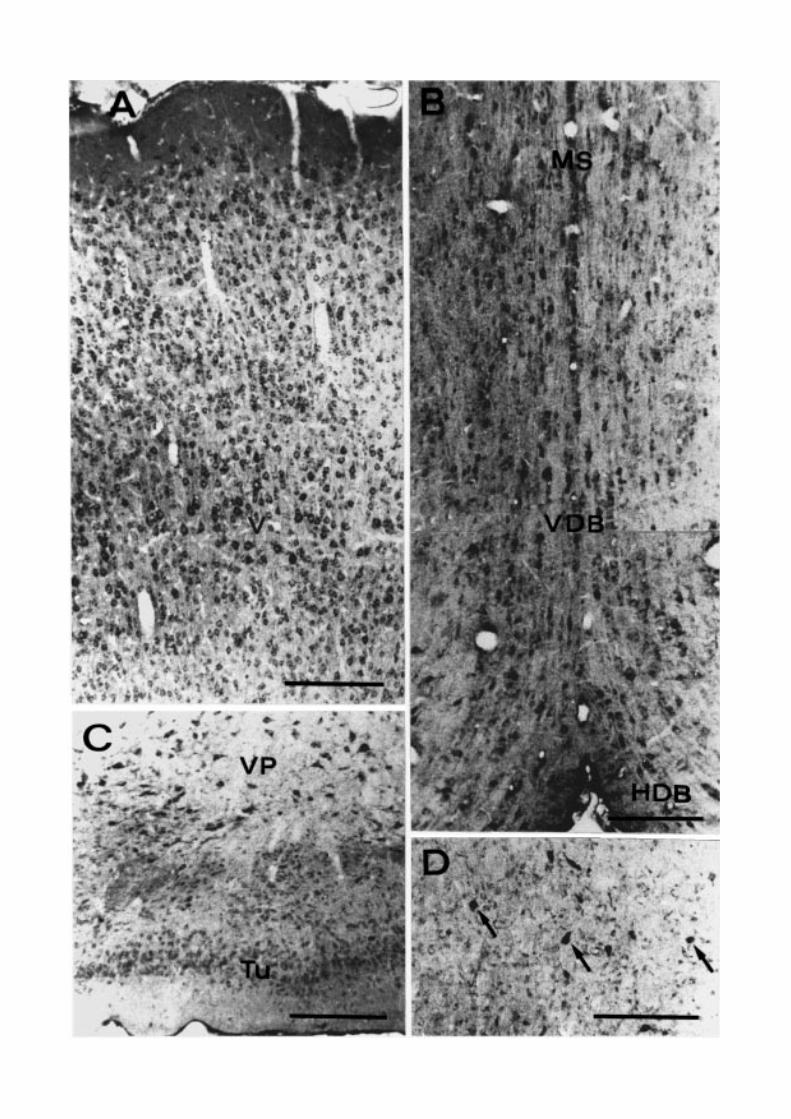
& Oertel, 1985) (Fig. 3A). In the remainder of the te-lencephalon, the septal nuclei and the basal ganglia,especially the ventral pallidum, are among the rich-est in D-AAO positive neurons (Fig. 3B–D). Concern-ing the caudate-putamen, only scattered medium- tolarge-sized cells probably corresponding to the cholin-ergic interneurons, show dense immunoreaction prod-uct, while the medium spiny neurons are only faintlystained (Fig. 3D). Other immunoreactive centers in-clude the medial and anteromedial preoptic nuclei andthe supraoptic nucleus (Fig. 4A). In the hippocampalformation, pyramidal neurons of fields CA1 and CA3,granule cells of the dentate gyrus, and many interneu-rons scattered in all the layers and in the subiculum areimmunopositive (Fig. 4B–D).
In the diencephalon, neurons of the reticular thala-mic nucleus are strongly immunoreactive, while otherventral and dorsal thalamic nuclei are weakly positive(Fig. 4E). Intensely stained neurons are also presentin the zona incerta and subthalamic nucleus (Fig. 4F).The hypothalamus also displays differentiated levelsof D-AAO immunoreactivity, with the strongest stain-ing occurring in the paraventricular nucleus (Fig. 5A),periventricular nucleus, and bed nucleus of the anteriorcommissure. Additionally, a small group of medium-sized cells, peculiarly located in a parafornical posi-tion, shows intense immunoreactivity (Fig. 5B). Thesecells, which do not precisely correspond to any ofthe nuclei classically described in this area, appearinstead very similar in location to those previously
D-AAO in rat brain 173
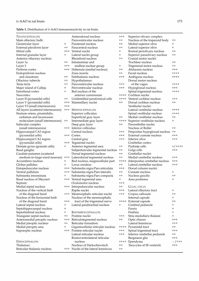
Table 1. Distribution of D-AAO immunoreactivity in rat brain.
TELENCEPHALON Anterodorsal nucleus +++ Superior olivary complex:Main olfactory bulb: Paraventricular nucleus ++ Nucleus of the trapezoid body ++Glomerular layer + Paratenial nucleus ++ Medial superior olive +External plexiform layer ++ Paracentral nucleus ++ Lateral superior olive +Mitral cells +++ Ventral nuclei + Rostral periolivary nucleus ++Internal granular layer + Lateral nuclei group + Superior paraolivary nucleus ++Anterior olfactory nucleus: Rhomboid nucleus ++ Cranial motor nuclei:Layer 1a ++ Intralaminar and Trochlear nucleus ++Layer 3 +++ midline nuclear group + Trigeminal motor nucleus ++Piriform cortex +++ Habenula (medial nucleus) ++ Abducens nucleus +++Endopiriform nucleus Zona incerta ++ Facial nucleus ++++
and claustrum ++ Subthalamic nucleus +++ Ambiguus nucleus ++++Olfactory tubercle ++ Hypothalamus: Dorsal motor nucleusTenia tecta ++ Paraventricular nucleus +++ of the vagus ++++Major island of Calleja + Periventricular nucleus +++ Hypoglossal nucleus +++Entorhinal cortex + Bed nucleus of the Spinal trigeminal nucleus ++++Neocortex: anterior commissure ++++ Cochlear nuclei:Layer II (pyramidal cells) ++ Unidentified parafornical cells ++++ Ventral cochlear nucleus +++Layer V (pyramidal cells) +++ Mammillary nuclei ++ Dorsal cochlear nucleus ++Layer VI (small interneurons) +++ Vestibular nuclei:All layers (scattered interneurons) +++ MESENCEPHALON Lateral vestibular nucleus ++++Stratum oriens, piramidalis, Superior colliculus: Spinal vestibular nucleus ++++
radiatum and lacunosum- Superficial gray layer ++ Medial vestibular nucleus ++moleculare (small interneurons) ++ Intermediate gray layer ++++ Superior vestibular nucleus +
Subicular complex Deep gray layer ++ Precerebellar nuclei:(small interneurons) +++ Inferior colliculus: Nucleus of Roller ++
Hippocampal CA3 region Central nucleus +++ Prepositus hypoglossal nucleus ++(pyramidal cells) +++ Cortex +++ External cuneate nucleus +++
Hippocampal CA1 region Central gray +++ Inferior olive +++(pyramidal cells) +++ Tegmental nuclei: Cerebellar cortex:
Dentate gyrus (granule cells) + Anterior tegmental area ++ Purkinje cells +/++++Basal ganglia: Subpeduncular tegmental nucleus ++ Golgi cells +++Caudate-putamen (scattered Dorsal tegmental nucleus ++ Cerebellar nuclei:
medium-to-large-sized neurons) +++ Laterodorsal tegmental nucleus ++ Medial cerebellar nucleus +++Accumbens nucleus + Red nucleus, magnocellular part ++++ Interpositus cerebellar nucleus +++Globus pallidus + Locus ceruleus ++ Lateral cerebellar nucleus +++Entopeduncular nucleus +++ Substantia nigra:Pars reticulata +++ Dorsal column nuclei:Ventral pallidum +++ Substantia nigra:Pars lateralis ++ Cuneate nucleus +Substantia innominata + Substantia nigra:Pars compacta ++ Nucleus gracilis ++Basal nucleus of Meynert +++ Ventral tegmental area + Area postrema +Septum: Oculomotor nucleus +++Medial septal nucleus +++ Interpeduncular nucleus ++ GLIAL CELLSNucleus of the vertical limb Raphe nuclei +++ Lateral olfactory tract +
of the diagonal band +++ Mesencephalic reticular nuclei +++ Corpus callosum ++Nucleus of the horizontal limb Nucleus of the mesencephalic Internal capsule +
of the diagonal band +++ tract of the trigeminal nerve ++++ External capsule ++Lateral septal nucleus + Lateral parabrachial nucleus + Cerebral peduncle +Septohippocampal nucleus + Fornix +Septofimbrial nucleus + RHOMBENCEPHALON Fimbria +Triangular septal nucleus ++ Pontine nuclei +++ Stria medullaris thalami +Anteromedial preoptic nucleus +++ Reticulotegmental nucleus ++ Optic chiasm +++Medial preoptic nucleus ++ Reticular formation: Lateral lemniscus +++Medial preoptic area + Gigantocellular reticular nucleus ++++ Pyramidal tract ++Supraoptic nucleus +++ Pontine reticular nuclei +++ Spinal trigeminal tract +++
Lateral reticular nucleus +++ Inferior cerebellar peduncle ++Rostroventrolateral reticular Bergmann glia +++
DIENCEPHALON nucleus ++++ Ependyma −/+++Thalamus: Nucleus of Darkschewitsch ++ Tanycytes of III ventricle +++Reticular thalamic nucleus +++ Nuclei of the lateral lemniscus ++

174 MORENO ET AL.
Fig. 2. Coronal sections of rat telencephalon. D-AAO immunocytochemistry. (A) Main olfactory bulb. Positive neurons includemitral cells (ML) and cells (arrows) in the external plexiform (EPL) and internal granular (IGrL) layers. GL, glomerular layer. (B)Anterior olfactory nucleus. Arrows indicate immunoreactive cells of layers 1a and 3. LOT, lateral olfactory tract. (C) Piriformcortex. LOT, lateral olfactory tract. Scale bars = 200 µm.
identified by catalase immunocytochemistry (Morenoet al., 1995).
In the mesencephalon, neurons located in both thesuperficial and intermediate divisions of the supe-rior colliculus show dense immunoreaction product(Fig. 5C). The central nucleus and the external cortex ofthe inferior colliculus, as well as the central gray, are alsopositive (Fig. 5D and E). The red nucleus represents oneof the most intensely stained centers of the whole brain(Fig. 6A). In the substantia nigra, the pars compacta
Fig. 3. Coronal sections of rat telencephalon. D-AAO immunocytochemistry. (A) Cerebral cortex. Numerous intenselyimmunostained neurons are visible throughout the layers. Note the immunonegative nuclei, particularly evident in the giantpyramidal cells of the fifth layer (V). (B) Septal complex. MS, medial septal nucleus; VDB and HDB, vertical and horizontal nu-cleus of the diagonal band of Broca. (C) VP, Ventral pallidum; Tu, olfactory tubercle. (D) Caudate putamen. The immunoreactionproduct is found in medium-to-large sized neurons (arrows). Scale bars = 200 µm.
and particularly the pars reticulata, are D-AAO posi-tive neurons (Fig. 6B). Other mesencephalic immuno-labelled areas include the raphe nuclei (Fig. 6C).
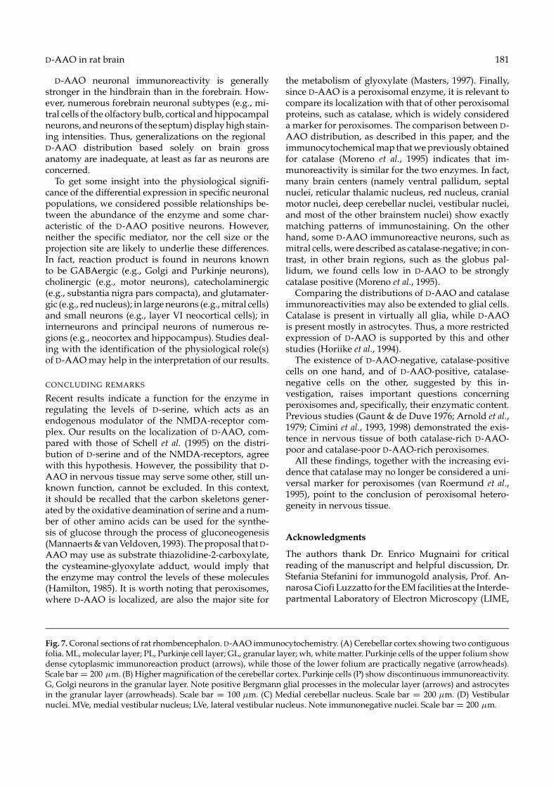
High immunoreactivity levels occur in numerousregions in the rhombencephalon. The cerebellumshows strong positivity in several neuronal subtypes,including Golgi neurons of the granular layer (Fig. 7Aand B). Purkinje cells are also immunoreactive, al-though their positivity is not uniform throughout thecortex (Fig. 7A). They show higher staining intensities

176 MORENO ET AL.

D-AAO in rat brain 177
in some folia, or parts of them, and much weaker—ifnot absent—staining in others (Fig. 7A). Granule, bas-ket, and stellate neurons have almost negligible im-munoreactivity. Moderate to high concentrations of im-munoreaction product are found in neurons belongingto the deep cerebellar nuclei and vestibular nuclei, withthe lateral vestibular nucleus displaying the most in-tense staining (Fig. 7C and D). Neurons of the reticularformation are strongly immunopositive, especially thegigantocellular nucleus (Fig. 8A and C). Cranial motornuclei, especially the ambiguous, facial, and dorsal mo-tor nuclei, show very strong staining (Fig. 8A–D). Otherpositive centers include pontine nuclei, cochlear nuclei,inferior olive, sensory trigeminal nuclei, and nuclei ofthe trapezoid body (Fig. 8E and F).
GLIA
Among glia, the astrocyte is the most intensely im-munoreactive cell type. Positive astrocytes are foundthroughout the brain, including gray and white matter.In the forebrain, the corpus callosum, the optic chiasm,and the external capsule show strong immunolabellingof astrocytes (Fig. 9A, B). The largest numbers of pos-itive cells and the strongest immunostaining are ob-served in the hindbrain, especially in the cerebellum(Figs. 7B, 8E and F and 9C).
The Golgi-Bergmann glia of the cerebellar cortex ex-hibit intense positivity which extends from the cell bodyto the radial processes (Fig. 7B). Other radial glia, e.g.tanycytes bordering the third ventricle and the medianeminence, show strong and specific staining (Fig. 9D).
Immunoreactivity is also observed in ependymalcells (Figs. 5A and 8D), but no immunopositive oligo-dendrocytes are evident.
Discussion
In the present paper we report the first complete mapof D-AAO in rat brain, obtained by means of an in-tensification immunocytochemical procedure (Adams,1992), which proved highly sensitive in detecting per-oxisomal enzymes (Moreno et al., 1995, 1997). We showthat D-AAO is localized in both neurons and glia, al-beit at different concentrations depending on the celltype and regional location. The diffuse appearanceof the cytoplasmic staining could be either due tothe small size of peroxisomes in neural cells, whichare hardly recognized at the LM level in relatively
Fig. 4. Coronal sections of rat telencephalon (A–D) and diencephalon (E, F). D-AAO immunocytochemistry. (A) SO, supraopticnucleus; ox, optic chiasm. (B) CA1 region of the hippocampus. Pyramidal cells (Pyr) are highly immunoreactive. Arrowsindicate positive neurons distributed in all the layers. (C) CA3 region of the hippocampus, showing positive pyramidal cells(Pyr) and scattered neurons (arrows). (D) Subiculum. Arrows indicate immunostained neurons. (E) Thalamus. RT, reticularthalamic nucleus; VL, ventrolateral thalamic nucleus. Note also immunorective glial cells of the internal capsule (ic). (F) STh,subthalamic nucleus; ZI, zona incerta; cp, cerebral peduncle. Scale bars = 200 µm.
thick sections, or, most probably, to the fact that D-AAO protein easily leaks out from peroxisomes, asalso biochemically described (Gaunt & De Duve, 1976;Cimini et al., 1998).
DISTRIBUTION OF D-AAO IMMUNOREACTIVITYIN GLIAL CELLS
Astrocytes and radial glia are the glial cell types rich-est in D-AAO. Ependymal cells also contain moderateamounts of the enzyme, while other glia are appar-ently immunonegative. Regional differences have alsobeen detected. Astroglial cells of the caudal brainstemand the cerebellum are generally more immunoreactivethan those in the forebrain. These results are compati-ble with previous studies utilizing histochemical tech-niques (Gaunt & De Duve, 1976; Arnold et al., 1979;Horiike et al., 1994; Schell et al., 1995). Regarding radialglia, Golgi-Bergmann cells of the cerebellar cortex andtanycytes bordering the third ventricle and median em-inence show distinct immunoreactivity. This result is inagreement with previous histochemical findings on ratBergmann glia (Weimar & Neims, 1977; Arnold et al.,1979; Horiike et al., 1987) and on frog retinal Mullercells (Beard et al., 1988).
DISTRIBUTION OF D-AAO IMMUNOREACTIVITYIN NEURONS
Many neurons were observed to contain D-AAO. Thisfinding is novel, as the enzyme was previously reportedto be absent from neurons, based on histochemical stud-ies (Arnold et al., 1979; Horiike et al., 1994; Schell et al.,1995). A possible explanation of this difference is thathistochemical techniques are based on enzymatic ac-tivity, while immunohistochemistry allows detection ofthe protein even if it is not active. Although the histo-chemical method may be extremely sensitive, fixationand subsequent procedures may impair some of theD-AAO activity. Furthermore, the natural substrate forD-AAO has not yet been identified; therefore, D-prolineused for histochemical localization (Arnold et al., 1979;Horiike et al., 1985; Beard et al., 1988) may not be themost suitable substrate to reveal D-AAO activity in cellsother than astrocytes. Actually, other substrates, suchas thiazolidine-2-carboxylic acid, proved effective in re-vealing D-AAO activity both biochemically (Fitzpatrick& Massey, 1982; Hamilton, 1985) and cytochemically(Angermuller, 1989; Jules et al., 1991).

178 MORENO ET AL.

D-AAO in rat brain 179
Fig. 6. Coronal sections of rat mesencephalon. D-AAO immunocytochemistry. (A) The magnocellular part of the red nucleus(R) is extraordinarily positive. 3, oculomotor nucleus; IP, interpeduncular nucleus. (B) Substantia nigra. SNR, pars reticulata;SNC, pars compacta. (C) Raphe nuclei. RMg, magnus nucleus; RPa, pallidus nucleus. Scale bars = 200 µm.
Fig. 5. Coronal sections of rat diencephalon (A, B) and mesencephalon (C–E). D-AAO immunocytochemistry. (A) Hypothalamus.Pa, paraventricular hypothalamic nucleus. Note the intense staining in ependymal cells bordering the third ventricle. (B)Hypothalamus. Small group of immunopositive neurons close to the fornix (f). (C) Superior colliculus. Note immunone-gative nuclei. (D) Inferior colliculus. EC, external cortex; CIC, central nucleus. (E) Central gray (CG) neurons are intenselyimmunopositive. Scale bars = 200 µm.

D-AAO in rat brain 181
D-AAO neuronal immunoreactivity is generallystronger in the hindbrain than in the forebrain. How-ever, numerous forebrain neuronal subtypes (e.g., mi-tral cells of the olfactory bulb, cortical and hippocampalneurons, and neurons of the septum) display high stain-ing intensities. Thus, generalizations on the regionalD-AAO distribution based solely on brain grossanatomy are inadequate, at least as far as neurons areconcerned.
To get some insight into the physiological signifi-cance of the differential expression in specific neuronalpopulations, we considered possible relationships be-tween the abundance of the enzyme and some char-acteristic of the D-AAO positive neurons. However,neither the specific mediator, nor the cell size or theprojection site are likely to underlie these differences.In fact, reaction product is found in neurons knownto be GABAergic (e.g., Golgi and Purkinje neurons),cholinergic (e.g., motor neurons), catecholaminergic(e.g., substantia nigra pars compacta), and glutamater-gic (e.g., red nucleus); in large neurons (e.g., mitral cells)and small neurons (e.g., layer VI neocortical cells); ininterneurons and principal neurons of numerous re-gions (e.g., neocortex and hippocampus). Studies deal-ing with the identification of the physiological role(s)of D-AAO may help in the interpretation of our results.
CONCLUDING REMARKS
Recent results indicate a function for the enzyme inregulating the levels of D-serine, which acts as anendogenous modulator of the NMDA-receptor com-plex. Our results on the localization of D-AAO, com-pared with those of Schell et al. (1995) on the distri-bution of D-serine and of the NMDA-receptors, agreewith this hypothesis. However, the possibility that D-AAO in nervous tissue may serve some other, still un-known function, cannot be excluded. In this context,it should be recalled that the carbon skeletons gener-ated by the oxidative deamination of serine and a num-ber of other amino acids can be used for the synthe-sis of glucose through the process of gluconeogenesis(Mannaerts & van Veldoven, 1993). The proposal that D-AAO may use as substrate thiazolidine-2-carboxylate,the cysteamine-glyoxylate adduct, would imply thatthe enzyme may control the levels of these molecules(Hamilton, 1985). It is worth noting that peroxisomes,where D-AAO is localized, are also the major site for
Fig. 7. Coronal sections of rat rhombencephalon. D-AAO immunocytochemistry. (A) Cerebellar cortex showing two contiguousfolia. ML, molecular layer; PL, Purkinje cell layer; GL, granular layer; wh, white matter. Purkinje cells of the upper folium showdense cytoplasmic immunoreaction product (arrows), while those of the lower folium are practically negative (arrowheads).Scale bar = 200 µm. (B) Higher magnification of the cerebellar cortex. Purkinje cells (P) show discontinuous immunoreactivity.G, Golgi neurons in the granular layer. Note positive Bergmann glial processes in the molecular layer (arrows) and astrocytesin the granular layer (arrowheads). Scale bar = 100 µm. (C) Medial cerebellar nucleus. Scale bar = 200 µm. (D) Vestibularnuclei. MVe, medial vestibular nucleus; LVe, lateral vestibular nucleus. Note immunonegative nuclei. Scale bar = 200 µm.
the metabolism of glyoxylate (Masters, 1997). Finally,since D-AAO is a peroxisomal enzyme, it is relevant tocompare its localization with that of other peroxisomalproteins, such as catalase, which is widely considereda marker for peroxisomes. The comparison between D-AAO distribution, as described in this paper, and theimmunocytochemical map that we previously obtainedfor catalase (Moreno et al., 1995) indicates that im-munoreactivity is similar for the two enzymes. In fact,many brain centers (namely ventral pallidum, septalnuclei, reticular thalamic nucleus, red nucleus, cranialmotor nuclei, deep cerebellar nuclei, vestibular nuclei,and most of the other brainstem nuclei) show exactlymatching patterns of immunostaining. On the otherhand, some D-AAO immunoreactive neurons, such asmitral cells, were described as catalase-negative; in con-trast, in other brain regions, such as the globus pal-lidum, we found cells low in D-AAO to be stronglycatalase positive (Moreno et al., 1995).
Comparing the distributions of D-AAO and catalaseimmunoreactivities may also be extended to glial cells.Catalase is present in virtually all glia, while D-AAOis present mostly in astrocytes. Thus, a more restrictedexpression of D-AAO is supported by this and otherstudies (Horiike et al., 1994).
The existence of D-AAO-negative, catalase-positivecells on one hand, and of D-AAO-positive, catalase-negative cells on the other, suggested by this in-vestigation, raises important questions concerningperoxisomes and, specifically, their enzymatic content.Previous studies (Gaunt & de Duve 1976; Arnold et al.,1979; Cimini et al., 1993, 1998) demonstrated the exis-tence in nervous tissue of both catalase-rich D-AAO-poor and catalase-poor D-AAO-rich peroxisomes.
All these findings, together with the increasing evi-dence that catalase may no longer be considered a uni-versal marker for peroxisomes (van Roermund et al.,1995), point to the conclusion of peroxisomal hetero-geneity in nervous tissue.
Acknowledgments
The authors thank Dr. Enrico Mugnaini for criticalreading of the manuscript and helpful discussion, Dr.Stefania Stefanini for immunogold analysis, Prof. An-narosa Ciofi Luzzatto for the EM facilities at the Interde-partmental Laboratory of Electron Microscopy (LIME,

182 MORENO ET AL.
Fig. 8. Coronal sections of rat rhombencephalon. D-AAO immunocytochemistry. (A) Amb, ambiguous nucleus; RVL, rostro-ventrolateral reticular nucleus. (B) Facial nucleus. (C) Arrowheads indicate neurons of the gigantocellular reticular nucleus,which show evident positivity in their somata and processes, but not in their nuclei. Note the numerous positive axons. (D) 10,dorsal motor nucleus of the vagus nerve; 12, hypoglossal nucleus. (E) Inferior olive (IO). Note also immunoreactive glial cellsof the pyramidal tract (py). (F) Neuronal and glial elements of the spinal trigeminal tract (sp5) and nucleus (Sp5) show strongimmunostaining. Scale bars = 200 µm.

D-AAO in rat brain 183
Fig. 9. Coronal sections of rat brain. D-AAO immunocytochemistry. (A) Corpus callosum. Many immunoreactive astroglialcells are visible (arrows). (B) Optic chiasm. Note that the immunoreaction product extends from the cell body to the processesof astrocytes (arrows). (C) icp, inferior cerebellar peduncle; sp5, spinal trigeminal tract. (D) Tanycytes of the third ventricle,showing positive staining in their long processes (arrows). Scale bars = 100 µm.
University ROMA TRE-Rome, Italy), and Dr. PierpaoloAimola for technical assistance. This research was sup-ported by CNR grant No. 97.04447.CT04, and par-tially by the European Concerted Action Peroxisomalleukodystrophy BMH4-CT96.1621.
References
ADAMS, J. C. (1992) Biotin amplification of biotin andhorseradish peroxidase signals in histochemical stains.Journal of Histochemistry and Cytochemistry 40, 1457–1463.
ANGERMULLER, S. (1989) Peroxisomal oxidases: cyto-chemical localization and biological relevance. Progressin Histochemistry and Cytochemistry 20, 1–63.
ARNOLD, G., LISCUM, L. & HOLTZMAN, E. (1979)Ultrastructural localization of D-amino acid oxidase in
microperoxisomes in the rat nervous system. Journal ofHistochemistry and Cytochemistry 27, 735– 745.
BEARD, M. E., DAVIES, T., HOLLOWAY, M. &HOLTZMAN, E. (1988) Peroxisomes in pigment epithe-lium and Muller cells of amphibian retina possess D-amino acid oxidase as well as catalase. Experimental EyeResearch 47, 795–806.
BRIGHT, H. J. & PORTER, D. J. T. (1975) Flavoprotein oxi-dases. In The Enzymes, Vol. 12: (edited by BOYER, P. D.),pp. 421–505. New York: Academic Press.
CIMINI, A. M., MORENO, S., SERAFINI, B. & CERU, M.P. (1993) Purification of peroxisomal fraction from ratbrain. Neurochemistry International 23, 249–260.
CIMINI, A. M., SINGH, I., FARIOLI VECCHIOLI, S.,CRISTIANO, L. & CERU, M. P. (1998) Presence of het-erogeneous peroxisomal populations in the rat nervoustissue. Biochimica et Biophysica Acta 1425, 13–26.

184 MORENO ET AL.
D’ANIELLO, A., D’ONOFRIO, G., PESCHETOLA, M.,D’ANIELLO, G., VETERE, A., PETRUCELLI, L. &FISCHER, G. H. (1993) Biological role of D-amino acidoxidase and D-aspartate oxidase: effects of D-aminoacids. Journal of Biological Chemistry 268, 26941–26949.
D’ANIELLO, A., VETERE, A. & PETRUCELLI, L. (1993a)Further study on the specificity of D-amino acid oxidaseand of D-aspartate oxidase and time course for completeoxidation of D-amino acids. Comparative Biochemistry andPhysiology 105 B, 731–734.
DIXON, M. & KLEPPE, K. (1965) D-amino acid oxidase: II.Specificity, competitive inhibition and reaction sequence.Biochimica et Biophysica Acta 96, 368–382.
FEDELE, E., BISAGLIA, M. & RAITERI, M. (1997) D-serinemodulates the NMDA receptor/nitric oxide/cGMPpathway in the rat cerebellum during in vivo microdial-ysis. Naunyn-Schmiedeberg’s Archives Pharmacology 355,43–47.
FITZPATRICK, P. F. & MASSEY, V. (1982) Thiazolidine-2-carboxylic acid, an adduct of cysteamine and glyoxilate,as a substrate for D-amino acid oxidase. Journal of Biolog-ical Chemistry 257, 1166–1171.
GAUNT, G. L. & DE DUVE, C. (1976) Subcellular distribu-tion of D-amino acid oxidase and catalase in rat brain.Journal of Neurochemistry 26, 749–759.
HAMILTON, G. A. (1985) Peroxisomal oxidases and sug-gestions for the mechanism of action of insulin and otherhormones. Advances in Enzymology 57, 85–178.
HASHIMOTO, A., NISHIKAWA, T., HAYASHI, T., FUJI,N., HARADA, K., OKA, T. & TAKAHASHI, K. (1992)The presence of free D-serine in rat brain. FEBS Letters296, 33–36.
HASHIMOTO, A., NISHIKAWA, T., OKA, T. &TAKAHASHI, K. (1993) Endogenous D-serine inrat brain: N-methyl-D-aspartate receptor-related dis-tribution and aging. Journal of Neurochemistry 60,783–786.
HASHIMOTO, A., OKA, T. & NISHIKAWA, T. (1995)Anatomical distribution and post-natal changes in en-dogenous free D-aspartate and D-serine in rat brain andperiphery. European Journal of Neuroscience 7, 1657–1663.
HOLTZMAN, E. (1982) Peroxisomes in nervous tissue.Annals of the New York Academy of Sciences 386, 523–525.
HORIIKE, K., ARAI, R., TOJO, H., YAMANO, T.,NOZAKI, M. & MAEDA, T. (1985) Histochemical stain-ing of cells containing flavoenzyme D-amino acid oxidasebased on its enzymatic activity: application of a coupledperoxidation method. Acta Histochemica et Cytochemica 18,539–550.
HORIIKE, K., TOJO, H., ARAI, R., YAMANO, T.,NOZAKI, M. & MAEDA, T. (1987) Localization of D-amino acid oxidase in Bergmann glial cells and astrocytesof rat cerebellum. Brain Research Bullettin 19, 587–596.
HORIIKE, K., TOJO, H., ARAI, R., NOZAKI, M. &MAEDA, T. (1994) D-Aminoacid oxidase is confined tothe lower brain stem and cerebellum in rat brain: regionaldifferentiation of astrocytes. Brain Research 652, 297–303.
JULES, R. S., KENNARD, J., SETLIK, W. & HOLTZMAN,E. (1991) Peroxisomal oxidation of thiazolidine carboxy-lates in firefly fat body, frog retina, and rat liver and kid-ney. European Journal of Cell Biology 55, 94–103.
KONNO, R., SASAKI, M., ASAKURA, S., FUKUI, K.,
ENAMI, J. & NIWA, A. (1997) D-Amino-acid oxidaseis not present in the mouse liver. Biochimica et BiophysicaActa 1335, 173–181.
KONNO, R., UCHYAMA, S. & YASUMURA, Y. (1982)Intraspecies and interspecies variations in the substratespecificity of D-amino acid oxidase. Comparative Biochem-istry and Physiology 71B, 735–738.
KONNO, R. & YASUMURA, Y. (1983) Mouse mutant de-ficient in D-aminoacid oxidase activity. Genetica 103,277–284.
KONNO, R. & YASUMURA, Y. (1992) D-amino-acid oxi-dase and its physiological function. International Journalof Biochemistry 24, 519–524.
KREBS, H. A. (1935) Metabolism of amino-acids. III. Deami-nation of amino-acids. Biochemical Journal 29, 1620–1644.
LAEMMLI, E. K. (1970) Cleavage of structural proteins dur-ing the assembly of the head of the bacteriophage T4.Nature 227, 680–685.
MANNAERTS, G. P. & VAN VELDHOVEN, P. P. (1993)Metabolic role of mammalian peroxisome. In Peroxisomes.Biology and Importance in Toxicology and Medicine (editedby GIBSON, G. & LAKE, B.), pp. 19–62. London: Taylor& Francis.
MASTERS, C. (1997) Gluconeogenesis and the peroxisome.Molecular and Cellular Biochemistry 166, 159–168.
MORENO, S., MUGNAINI, E. & CERU, M. P. (1995) Im-munocytochemical localization of catalase in the centralnervous system of the rat. Journal of Histochemistry andCytochemistry 43, 1253–1267.
MORENO, S., NARDACCI, R. & CERU, M. P. (1997)Regional and ultrastructural immunolocalization ofcopper-zinc superoxide dismutase in rat central nervoussystem. Journal of Histochemistry and Cytochemistry 45,1611–1622.
MOSER, H. W. (1987) New approaches in peroxisomal dis-orders. Developmental Neuroscience 9, 1–18.
MUGNAINI, E. & DAHL, A. L. (1983) Zinc-aldehyde fixa-tion for light microscopic immunocytochemistry of ner-vous tissues. Journal of Histochemistry and Cytochemistry31, 1435.
MUGNAINI, E. & OERTEL, W. H. (1985) An atlas ofthe distribution GABAergic neurons and terminals inthe rat CNS as revealed by GAD immunohistochem-istry. In Handbook of Chemical Neuroanatomy (edited byBJORKLUND, A. & HOKFELT, T.) Vol. 4. Part I:pp. 436–608. New York: Elsevier.
NAGATA, Y. (1992) Involvement of D-amino acid oxidasein elimination of D-serine in mouse brain. Experientia 48,753–755.
NAGATA, Y., HORIIKE, K. & MAEDA, T. (1994) Distribu-tion of free D-serine in vertebrate brains. Brain Research634, 291–295.
PAXINOS, G. & WATSON, C. (1986) The Rat Brain inStereotaxic Coordinates, 2nd ed. San Diego: AcademicPress.
PEROTTI, M.E., GAVAZZI, E., TRUSSRDO, L.,MALGARETTI, N. & CURTI, B. (1987) Immuno-electron microscopic localization of D-amino acidoxidase in rat kidney and liver. Histochemical Journal 19,157–169.
RONCHI, S., MINCHIOTTI, L., GALLIANO, M.,CURTI, B., SWENSON, R. P., WILLIAMS, C. H. &

D-AAO in rat brain 185
MASSEY, V. (1982) The primary structure of D-aminoacid oxidase from pig kidney. Journal of BiologicalChemistry 257, 8824–8834.
SCHELL, M. J., MOLLIVER, M. E. & SNYDER, S. H.(1995) D-serine, an endogenous synaptic modulator: lo-calization to astrocytes and glutamate-stimulated re-lease. Proceedings of the National Academy of Sciences USA92, 3948–3952.
TOWBIN, H., STAHELIN, T. & GORDON., J. (1979) Elec-trophoretic transfer of proteins from polyacrylamide gelsto nitrocellulose sheets: procedure and some applica-tions. Proceedings of the National Academy of Sciences USA76, 4350–4354.
USUDA, N., YOKOTA, S., HASHIMOTO, T. & NAGATA,T. (1986) Immunocytochemical localization of D-aminoacid oxidase in the central clear matrix of rat kidney per-oxisomes. Journal of Histochemistry and Cytochemistry 34,1709–1718.
USUDA, N., YOKOTA, S., ICHIKAWA, R.,HASHIMOTO, T. & NAGATA, T. (1991) Immuno-
electron microscopic study of a new D-aminoacidoxidase-immunoreactive subcompartment in rat liverperoxisomes. Journal of Histochemistry and Cytochemistry39, 95–102.
VAN DEN BOSCH, H., SCHUTGENS, R. B. H.,WANDERS, R. J. A. & TAGER, J. M. (1992) Biochem-istry of peroxisomes. Annual Reviews in Biochemistry 61,157–197.
VAN ROERMUND, C. W. T., VAN DEN BERG, M. &WANDERS, R. J. A. (1995) Localization of 3-oxoacyl-CoA thiolase in particles of varied density in rat liver:implications for peroxisome biogenesis. Biochimica et Bio-physica Acta 1245, 348–358.
WEIMAR, W. R. & NEIMS, A. H. (1977) Hog cerebel-lar D-amino acid oxidase and its histochemical and im-munofluorescence localization. Journal of Neurochemistry28, 559–572.
ZAAR, K. (1992) Structure and function of peroxisomes inthe mammalian kidney. European Journal of Cell Biology59, 233–254.