Decreased cytochrome c oxidase subunit VIIa in aged rat heart mitochondria: immunocytochemistry
Upload
khangminh22Category
view
4download
0
Citation: Hederstedt, L. Diversity of
Cytochrome c Oxidase Assembly
Proteins in Bacteria. Microorganisms
2022, 10, 926. https://doi.org/
10.3390/microorganisms10050926
Academic Editor: Carla C. C. R. de
Carvalho
Received: 5 April 2022
Accepted: 27 April 2022
Published: 28 April 2022
Publisher’s Note: MDPI stays neutral
with regard to jurisdictional claims in
published maps and institutional affil-
iations.
Copyright: © 2022 by the author.
Licensee MDPI, Basel, Switzerland.
This article is an open access article
distributed under the terms and
conditions of the Creative Commons
Attribution (CC BY) license (https://
creativecommons.org/licenses/by/
4.0/).
microorganisms
Review
Diversity of Cytochrome c Oxidase Assembly Proteinsin BacteriaLars Hederstedt
The Microbiology Group, Department of Biology, Lund University, Sölvegatan 35, SE-223 62 Lund, Sweden;[email protected]
Abstract: Cytochrome c oxidase in animals, plants and many aerobic bacteria functions as theterminal enzyme of the respiratory chain where it reduces molecular oxygen to form water in areaction coupled to energy conservation. The three-subunit core of the enzyme is conserved, whereasseveral proteins identified to function in the biosynthesis of the common family A1 cytochromec oxidase show diversity in bacteria. Using the model organisms Bacillus subtilis, Corynebacteriumglutamicum, Paracoccus denitrificans, and Rhodobacter sphaeroides, the present review focuses on proteinsfor assembly of the heme a, heme a3, CuB, and CuA metal centers. The known biosynthesis proteinsare, in most cases, discovered through the analysis of mutants. All proteins directly involved incytochrome c oxidase assembly have likely not been identified in any organism. Limitations in theuse of mutants to identify and functionally analyze biosynthesis proteins are discussed in the review.Comparative biochemistry helps to determine the role of assembly factors. This information can, forexample, explain the cause of some human mitochondrion-based diseases and be used to find targetsfor new antimicrobial drugs. It also provides information regarding the evolution of aerobic bacteria.
Keywords: cytochrome oxidase; heme protein; copper protein; enzyme biosynthesis; bioenergetics;enzyme assembly factors
1. Introduction
The biosynthesis of enzymes containing metal prosthetic groups, such as heme, iron-sulfur clusters, and copper centers, generally requires one or more assisting proteins.Cytochrome c oxidases of the heme–copper oxygen reductase family A (present in mito-chondria and many aerobic bacteria [1]) is, with respect to the core, a conserved enzyme, asdemonstrated by three-dimensional structures, functional features, and the gene sequenceinformation available from a large number of different types of organisms [1–5]. Not longago, it was assumed that the proteins that function in the biosynthesis of cytochrome coxidase are also conserved [6,7]. However, and intriguingly, there seems to be considerablediversity of some of the assembly proteins and their occurrence in bacteria. This reviewaddresses this diversity and raises questions about how it relates to the evolution of cy-tochrome c oxidase and its specific assembly factors. It also presents an overview of thecurrent knowledge concerning biosynthesis proteins for family A cytochrome c oxidasein bacteria.
Research in the field of enzyme biosynthesis can reveal hitherto undiscovered mech-anisms in cells and, for example, explain how certain mutations cause disease. Fromthe applied perspective, the diversity of proteins with key functions in the assembly ofheme–copper oxygen reductases provide the possibility to find new drugs that specificallyaffect oxidase function in only certain selected bacterial species and not in eukaryotes.
Cytochrome oxidases in mitochondria and aerobic bacteria function to reduce molecu-lar oxygen to yield water and generate energy conservation by the formation of a transmem-brane electrochemical gradient that drives ATP synthesis and other energy-demandingprocesses in cells. The family A cytochrome c oxidases contain three different transmem-brane polypeptides, subunits I, II, and III (plus, most often, a small fourth polypeptide),
Microorganisms 2022, 10, 926. https://doi.org/10.3390/microorganisms10050926 https://www.mdpi.com/journal/microorganisms
Microorganisms 2022, 10, 926 2 of 17
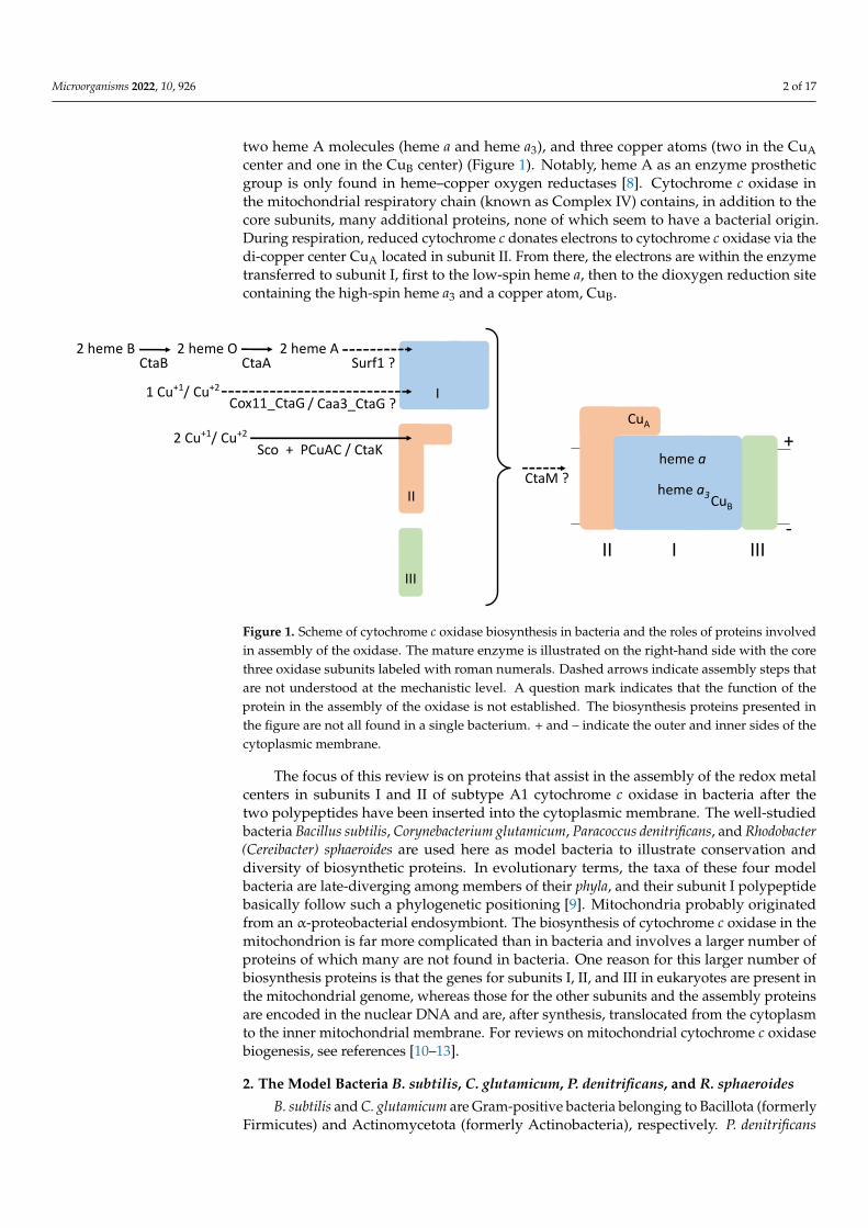
two heme A molecules (heme a and heme a3), and three copper atoms (two in the CuAcenter and one in the CuB center) (Figure 1). Notably, heme A as an enzyme prostheticgroup is only found in heme–copper oxygen reductases [8]. Cytochrome c oxidase inthe mitochondrial respiratory chain (known as Complex IV) contains, in addition to thecore subunits, many additional proteins, none of which seem to have a bacterial origin.During respiration, reduced cytochrome c donates electrons to cytochrome c oxidase via thedi-copper center CuA located in subunit II. From there, the electrons are within the enzymetransferred to subunit I, first to the low-spin heme a, then to the dioxygen reduction sitecontaining the high-spin heme a3 and a copper atom, CuB.
Microorganisms 2022, 10, x FOR PEER REVIEW 2 of 17
transmembrane electrochemical gradient that drives ATP synthesis and other energy-de-manding processes in cells. The family A cytochrome c oxidases contain three different transmembrane polypeptides, subunits I, II, and III (plus, most often, a small fourth pol-ypeptide), two heme A molecules (heme a and heme a3), and three copper atoms (two in the CuA center and one in the CuB center) (Figure 1). Notably, heme A as an enzyme pros-thetic group is only found in heme–copper oxygen reductases [8]. Cytochrome c oxidase in the mitochondrial respiratory chain (known as Complex IV) contains, in addition to the core subunits, many additional proteins, none of which seem to have a bacterial origin. During respiration, reduced cytochrome c donates electrons to cytochrome c oxidase via the di-copper center CuA located in subunit II. From there, the electrons are within the enzyme transferred to subunit I, first to the low-spin heme a, then to the dioxygen reduc-tion site containing the high-spin heme a3 and a copper atom, CuB.
Figure 1. Scheme of cytochrome c oxidase biosynthesis in bacteria and the roles of proteins involved in assembly of the oxidase. The mature enzyme is illustrated on the right-hand side with the core three oxidase subunits labeled with roman numerals. Dashed arrows indicate assembly steps that are not understood at the mechanistic level. A question mark indicates that the function of the pro-tein in the assembly of the oxidase is not established. The biosynthesis proteins presented in the figure are not all found in a single bacterium. + and – indicate the outer and inner sides of the cyto-plasmic membrane.
The focus of this review is on proteins that assist in the assembly of the redox metal centers in subunits I and II of subtype A1 cytochrome c oxidase in bacteria after the two polypeptides have been inserted into the cytoplasmic membrane. The well-studied bacte-ria Bacillus subtilis, Corynebacterium glutamicum, Paracoccus denitrificans, and Rhodobacter (Cereibacter) sphaeroides are used here as model bacteria to illustrate conservation and di-versity of biosynthetic proteins. In evolutionary terms, the taxa of these four model bacte-ria are late-diverging among members of their phyla, and their subunit I polypeptide ba-sically follow such a phylogenetic positioning [9]. Mitochondria probably originated from an α-proteobacterial endosymbiont. The biosynthesis of cytochrome c oxidase in the mi-tochondrion is far more complicated than in bacteria and involves a larger number of pro-teins of which many are not found in bacteria. One reason for this larger number of bio-synthesis proteins is that the genes for subunits I, II, and III in eukaryotes are present in the mitochondrial genome, whereas those for the other subunits and the assembly pro-teins are encoded in the nuclear DNA and are, after synthesis, translocated from the cyto-plasm to the inner mitochondrial membrane. For reviews on mitochondrial cytochrome c oxidase biogenesis, see references [10–13].
CtaM ?
III
II I III
CuA
+
-
2 Cu+1/ Cu+2
II
Sco + PCuAC / CtaK
Cox11_CtaG / Caa3_CtaG ?I1 Cu+1/ Cu+2
2 heme B 2 heme O 2 heme ACtaB CtaA Surf1 ?
CuB
heme a
heme a3
Figure 1. Scheme of cytochrome c oxidase biosynthesis in bacteria and the roles of proteins involvedin assembly of the oxidase. The mature enzyme is illustrated on the right-hand side with the corethree oxidase subunits labeled with roman numerals. Dashed arrows indicate assembly steps thatare not understood at the mechanistic level. A question mark indicates that the function of theprotein in the assembly of the oxidase is not established. The biosynthesis proteins presented inthe figure are not all found in a single bacterium. + and – indicate the outer and inner sides of thecytoplasmic membrane.
The focus of this review is on proteins that assist in the assembly of the redox metalcenters in subunits I and II of subtype A1 cytochrome c oxidase in bacteria after thetwo polypeptides have been inserted into the cytoplasmic membrane. The well-studiedbacteria Bacillus subtilis, Corynebacterium glutamicum, Paracoccus denitrificans, and Rhodobacter(Cereibacter) sphaeroides are used here as model bacteria to illustrate conservation anddiversity of biosynthetic proteins. In evolutionary terms, the taxa of these four modelbacteria are late-diverging among members of their phyla, and their subunit I polypeptidebasically follow such a phylogenetic positioning [9]. Mitochondria probably originatedfrom an α-proteobacterial endosymbiont. The biosynthesis of cytochrome c oxidase in themitochondrion is far more complicated than in bacteria and involves a larger number ofproteins of which many are not found in bacteria. One reason for this larger number ofbiosynthesis proteins is that the genes for subunits I, II, and III in eukaryotes are present inthe mitochondrial genome, whereas those for the other subunits and the assembly proteinsare encoded in the nuclear DNA and are, after synthesis, translocated from the cytoplasmto the inner mitochondrial membrane. For reviews on mitochondrial cytochrome c oxidasebiogenesis, see references [10–13].
2. The Model Bacteria B. subtilis, C. glutamicum, P. denitrificans, and R. sphaeroides
B. subtilis and C. glutamicum are Gram-positive bacteria belonging to Bacillota (formerlyFirmicutes) and Actinomycetota (formerly Actinobacteria), respectively. P. denitrificans

Microorganisms 2022, 10, 926 3 of 17
and R. sphaeroides are Gram-negative α-Proteobacteria. These four model bacteria do notcause disease in animals or plants and contribute to the degradation of organic materialin nature. A high G+C content of genomic DNA is a common feature of C. glutamicumand P. denitrificans. B. subtilis, C. glutamicum, and P. denitrificans are under all conditionsorganoheterotrophs with mainly a respiratory metabolism [14–16]. R. sphaeroides is a purplenon-sulfur photoheterotroph that, in the dark, is a respiring organoheterotroph [17]. Allfour bacterial species can also respire anaerobically by nitrate reductase. The cytochrome aa3oxidases from B. subtilis, C. glutamicum, P. denitrificans, and R. sphaeroides are characterizedin the details, including the atomic level structural data [18–21]. A major experimentaladvantage of the four model bacteria is that their family A oxidases are not required forgrowth, which is essential for mutant studies.
B. subtilis contains two heme A-containing oxidases: the menaquinol oxidase cy-tochrome aa3 and the cytochrome c oxidase caa3. Both enzymes belong to the subtype A1 ofheme–copper oxygen reductases. The cytochrome aa3 has no CuA center (it was apparentlylost during evolution and replaced by a menaquinone binding site [18]). The situation withtwo variants of the cytochrome aa3-type in the same bacterial species is useful for exper-imental identification of proteins specifically required for CuA assembly and those thatfunction to incorporate heme A or have a role in the assembly of the heme a3–CuB center.The electron donating cytochrome c in B. subtilis cytochrome caa3 is a domain of subunitII sitting adjacent to the CuA domain at the C-terminal end of the protein. Cytochromecaa3, the cytochrome bc complex (QcrABC), and a small membrane-anchored cytochrome c(CccA or CccB) are in the cytoplasmic membrane found as a supercomplex that can oxidizemenaquinol coupled to the reduction of molecular oxygen [22,23].
Cytochrome aa3 and cytochrome bc form a supercomplex also in C. glutamicum [24] andother Actinomycetota, and this complex is important for the stability of each constituentenzyme [25,26]. The cta and qcr genes encoding the supercomplex polypeptides are orga-nized in one operon (Figure 2). The QcrC subunit of the bc complex in Actinomycetota hasa diheme cytochrome c domain that functions as the electron donor to the cytochrome aa3in the supercomplex [21,24]. There seems to be no other cytochrome c in C. glutamicum [26].
Microorganisms 2022, 10, x FOR PEER REVIEW 3 of 17
2. The Model Bacteria B. subtilis, C. glutamicum, P. denitrificans, and R. sphaeroides B. subtilis and C. glutamicum are Gram-positive bacteria belonging to Bacillota (for-
merly Firmicutes) and Actinomycetota (formerly Actinobacteria), respectively. P. denitrif-icans and R. sphaeroides are Gram-negative α-Proteobacteria. These four model bacteria do not cause disease in animals or plants and contribute to the degradation of organic mate-rial in nature. A high G+C content of genomic DNA is a common feature of C. glutamicum and P. denitrificans. B. subtilis, C. glutamicum, and P. denitrificans are under all conditions organoheterotrophs with mainly a respiratory metabolism [14–16]. R. sphaeroides is a pur-ple non-sulfur photoheterotroph that, in the dark, is a respiring organoheterotroph [17]. All four bacterial species can also respire anaerobically by nitrate reductase. The cyto-chrome aa3 oxidases from B. subtilis, C. glutamicum, P. denitrificans, and R. sphaeroides are characterized in the details, including the atomic level structural data [18–21]. A major experimental advantage of the four model bacteria is that their family A oxidases are not required for growth, which is essential for mutant studies.
B. subtilis contains two heme A-containing oxidases: the menaquinol oxidase cyto-chrome aa3 and the cytochrome c oxidase caa3. Both enzymes belong to the subtype A1 of heme–copper oxygen reductases. The cytochrome aa3 has no CuA center (it was apparently lost during evolution and replaced by a menaquinone binding site [18]). The situation with two variants of the cytochrome aa3-type in the same bacterial species is useful for experimental identification of proteins specifically required for CuA assembly and those that function to incorporate heme A or have a role in the assembly of the heme a3–CuB center. The electron donating cytochrome c in B. subtilis cytochrome caa3 is a domain of subunit II sitting adjacent to the CuA domain at the C-terminal end of the protein. Cyto-chrome caa3, the cytochrome bc complex (QcrABC), and a small membrane-anchored cy-tochrome c (CccA or CccB) are in the cytoplasmic membrane found as a supercomplex that can oxidize menaquinol coupled to the reduction of molecular oxygen [22,23].
Cytochrome aa3 and cytochrome bc form a supercomplex also in C. glutamicum [24] and other Actinomycetota, and this complex is important for the stability of each constit-uent enzyme [25,26]. The cta and qcr genes encoding the supercomplex polypeptides are organized in one operon (Figure 2). The QcrC subunit of the bc complex in Actinomycetota has a diheme cytochrome c domain that functions as the electron donor to the cytochrome aa3 in the supercomplex [21,24]. There seems to be no other cytochrome c in C. glutamicum [26].
Figure 2. Organization of genes for family A1 cytochrome c oxidase in the chromosome of four model bacteria. Arrows show the transcription units. The genes for oxidase polypeptides are indi-cated I, II, III, and IV. The qcr genes encode the polypeptides of the cytochrome bc complex. The ctaG
C. glutamicum ctaC ctaF ctaE qcrC qcrA qcrB ctaDIII IV III
P. denitrificans
B. subtilis ctaA ctaB ctaC ctaD ctaE ctaF ctaGIII III IV
R. sphaeroides
ctaC ctaB ctaG ctaE surf1cII III
ctaDI
ctaC ctaB ctaG ctaEII III
ctaDI
Figure 2. Organization of genes for family A1 cytochrome c oxidase in the chromosome of four modelbacteria. Arrows show the transcription units. The genes for oxidase polypeptides are indicated I,II, III, and IV. The qcr genes encode the polypeptides of the cytochrome bc complex. The ctaG genesof B. subtilis and P. denitrificans encode different types of proteins, i.e., Caa3_CtaG and Cox11_CtaG,respectively (see text).
P. denitrificans and R. sphaeroides belong to the order Rhodobacterales and contain,under high oxygen conditions, one mitochondrial-type cytochrome aa3 and a water-soluble

Microorganisms 2022, 10, 926 4 of 17
periplasmic cytochrome c as the electron donor to the oxidase. The cytochrome aa3 andcytochrome bc1 complex form a supercomplex [27,28]. P. denitrificans contains in addition afamily B heme–copper oxidase, cytochrome ba3, whereas R. sphaeroides, under low oxygenconditions, contains a family C heme–copper oxidase, cytochrome cbb3. Cytochromes ba3and cbb3 do not contain the CuA center. Due to the different cofactor compositions and otherdifferences among cytochromes aa3, ba3, caa3, and cbb3, they are useful for comparativestudies to determine roles of heme–copper oxygen reductase biosynthesis proteins.
3. Assembly of Cytochrome c Oxidase
The experimental data available, to date, strongly suggests that, in the biosynthesis offamily A cytochrome c oxidase, subunits I and II mature separately before they associate toform the enzymatically active enzyme [11,29,30]. Subunit I normally has 12 transmembranesegments. After this polypeptide has been inserted into the membrane and folded (to someextent), it is hemylated, to create the hemes a and a3, and one copper ion is inserted toform CuB. Subsequently, a covalent bond essential for proton pumping forms between theε-N of a His ligand to CuB and C6 of a Tyr residue at the dioxygen-binding site [31–34].It is not clear if any accessory factor is required for this polypeptide modification tooccur. Subunit II, with two transmembrane segments, incorporates two copper ions inthe extracellular domain to create the CuA center and the subunit then associates withcofactor-loaded subunit I. The subunit I+II complex binds subunit III, which stabilizes thenascent enzyme [35]. The mature oxidase also contains a tightly bound magnesium ionand bound lipid molecules. Depending on the organism, there are additional modificationsof subunit II. In the case of B. subtilis cytochrome caa3, subunit II is a lipoprotein with adiacylglyceride moiety at the N-terminal end (modification achieved by the activities ofleader sequence peptidase II and lipoprotein diacylglyceride transferase) and a cytochromec domain (with one covalently bound heme) in the C-terminal end of the protein. Thus, thematuration of the subunit II polypeptide in B. subtilis comprises three post-translationalmodifications. In the final assembled cytochrome c oxidase, the heme a and heme a3–CuBdioxygen-binding center are positioned deep within the membrane, only accessible to smallmolecules. This feature implies that the processes of insertion of the two heme A moleculesand folding of the subunit I polypeptide are integrated. The CuA and cytochrome c (whenpresent) domains of subunit II are exposed on the outer side of the cytoplasmic membranein bacteria to interact with periplasmic proteins and are assembled at this location.
Heme molecules and copper ions are toxic to cells: when in the reduced state anddioxygen is present, reactive oxygen species can be generated. Reduced heme A is moreprone for oxidation than heme B due to its higher mid-point redox potential. Chaperoneproteins provide protection against the toxicity of heme and metal ions during transport inthe cell, from the site of synthesis or acquisition to the final destination in a protein [36].Heme and copper ion homeostasis in the cell can, in addition, comprise excretion of excessamounts of the metal factors out of the cell. Most aerobic bacteria contain multiple hemeproteins. As an example, B. subtilis has more than 24 different heme proteins [15]. Themechanisms of heme transport in bacterial cells and how heme is incorporated into proteinsto form cytochromes are generally not understood [37,38]. More is known about copperion trafficking and homeostasis, but the picture is not complete for any bacterium [39,40].
4. Difficulties in the Identification of Biosynthesis Proteins
It is often a challenge to identify and study proteins that function in the biosynthesisof enzymes because: (i) these proteins are, in most cases, not associated with the mature en-zyme, (ii) assembly processes are transient and mechanistically not apparent in the enzymeproduct, and (iii) biosynthesis proteins are generally quantitatively minor constituents incells and their activities problematic to assay. The large majority of the proteins found tofunction in cytochrome c oxidase biosynthesis have been identified through mutations thataffect respiration in microbes or cause disease [41–43]. A mutant approach to detect thepresence and function of proteins is powerful. However, for the design of experiments and

Microorganisms 2022, 10, 926 5 of 17
the interpretation of data, it is essential to acknowledge the limitations and caveats in thatapproach. Proteins essential for cell viability and those with a function covered also byanother protein are generally missed when relying on single mutant data. Furthermore, amutation in a biosynthesis protein for an enzyme might cause indirect effects, which candiffer from those resulting from mutations in the actual enzyme. For example, a defect in abiosynthesis protein may result in the accumulation of a toxic intermediate or degradationproduct that does not appear when the enzyme itself is inactivated. In a mutant approach,we, in addition, need to consider the difference between the complete absence of a protein(due to interruption or deletion of the corresponding gene) compared to the inactivation ofa protein by a point mutation (causing an amino acid residue substitution). The completelack of a certain protein in the cell may disturb the assembly or stability of other proteinsand thereby affect several processes. Thus, mutations in a gene can readily cause indirector pleiotropic effects, leading to the wrong interpretation of data and incorrect suggestedfunction for the protein encoded by that gene. Desired amino acid substitutions in theprotein of interest are those that affect the specific activity without gross changes in thestructure of the protein [44]. Ideally, to be very informative, a mutant protein in the cellshould result in a phenotype and retain most of its properties, such as stability, the abilityto bind cofactors, and the interaction with other proteins.
Table 1. Diversity and occurrence of proteins identified to function in cytochrome c oxidase biosyn-thesis in four bacteria. The color background quickly brings an overview on the occurence of thedifferent proteins among the four bacteria. The UniProt identification numbers bring a second layer(more detailed) of information.
Function ProteinBacterium and UniProt Identification Numbers
Bacillus subtilis Corynebacteriumglutamicum
Paracoccusdenitrificans
Rhodobactersphaeroides
CuAsynthesis
Sco P54178(YpmQ)
A1BAG3/A1B5S0(ScoA/ScoB) 1
Q3J6C2(PrrC)
CtaK P40768
PCuAC Q8NPY8 A1BAG4/A1AZD7(PCu1/PCu2) A0A2W5SGC1
CuBinsertion
Cox11_CtaG A1BA38 Q3J5F7
Caa3_CtaG 2 O34329 Q8NMV8(CtiP) 3
Heme Asynthesis and
insertion
Heme Osynthase CtaB 4 P24009 Q8NQ66 A1BA40 Q3J5F9
Heme Asynthase CtaA P12946 Q8NQ70 A1B8C2 Q3IXW9
Surf1 Q8NNG3 A1BA36(Surf1c) 5 Q3J5F5
Other role CtaM O31845
Cytochrome csynthesis
Disulfide bondreduction
CcdA P45706 Q8NT70 A0A533I729 Q3J4J7ResA P35160 Q8NT71CcmG P52236 Q3J512CcmH A1B950 Q9ANS4
Heme transportand ligase
System I: CcmACcmBCcmCCcmDCcmE
P52218P52219P52220P52221A1B946
O33570Q3J515Q3J514Q3J513Q3J278
System II:CcsB/ResBCcsA/ResC
P35161 (ResB)P35162 (ResC)
Q8NT69Q8NT68
1 The two P. denitrificans Sco paralogs seemingly overlap in functions. 2 The role of this protein in CuB assembly istentative. 3 CtiP is a fusion protein, including a Caa3_CtaG domain, and might function in CuA rather than CuBassembly [45]. 4 B. subtilis contains also a CtaB paralog, CtaO [46]. 5 P. denitrificans contains two Surf1 proteins,Surf1c and Surf1q, which are specific for the biosynthesis of cytochrome aa3 and ba3, respectively [47].
Bona fide cytochrome c oxidase biosynthesis proteins function directly and specificallyin the assembly of the enzyme by interacting with subunit polypeptides to insert cofactors

Microorganisms 2022, 10, 926 6 of 17
or to chaperone folding or oligomerization of subunits in the membrane. Other proteins,in oxidase assembly, play a more peripheral role by performing tasks, such as catalyzingthe synthesis of a cofactor or the delivery of a cofactor in the appropriate redox state tothe proper subcellular compartment. To establish the function at the molecular level of abiosynthesis protein, detailed characterizations of the protein and its activity in vitro withpurified components are required. This ultimate goal has essentially been reached for someCuA center assembly proteins [48], but is remote for many other cytochrome c oxidasebiosynthesis proteins [49].
In a recent genome-wide approach to find genes important for the biosynthesis offamily A heme–copper oxygen reductase, a library of 3966 B. subtilis strains, systematicallydeleted for all non-essential genes, was screened for cytochrome c oxidase activity [50].Additionally, two collections of strains containing random point mutations induced bychemical mutagenesis were screened. The screens revealed two new biosynthesis proteins,making a total of twelve proteins presently known to function in cytochrome caa3 biosyn-thesis in B. subtilis (Table 1). Three of the proteins (Sco, Caa3_CtaG, CtaK) specificallyfunction in the assembly of cytochrome caa3; two are the enzymes for heme A synthesis(CtaA, CtaB); four are for the cytochrome c synthesis (CcdA, ResA, ResB, ResC); one (CtaM)is required for both cytochrome caa3 and cytochrome aa3 activity; and two act in lipoproteinmodification (LspA, Lgt) [50]. As discussed earlier in this section, there are probably addi-tional biosynthesis genes in B. subtilis to be discovered, because the used screens would notidentify genes encoding proteins that overlap in function nor genes that are required forgrowth or not necessary for cytochrome caa3 assembly under the growth conditions used.
5. Heme Synthesis
Aerobic organisms, with only a few exceptions, can synthesize protoheme IX (heme B).In the bacteria, the tetrapyrrole universal precursor 5-aminolevulinic acid is synthesizedeither from glycine and succinyl-CoA (called the Shemin pathway) or from glutamyl-tRNA(called the C5 pathway). The synthesis of uroporphyrinogen III from 5-aminolevulinic acidis catalyzed by a conserved set of three enzymes [51]. The subsequent biosynthesis of hemeB from uroporphyrinogen III occurs via at least three different pathways (i to iii), dependingon the organism [52]. (i) Eukaryotes and Pseudomonadota (formerly Proteobacteria) havethe so called classical pathway comprising three enzyme-catalyzed steps and where theinsertion of one ferrous ion into protoporphyrin IX occurs in the last step. (ii) In Bacillotaand Actinomycetota, the iron atom is inserted into uroporphyrin III forming coprohemethat, in the last step of the pathway, is oxidized to yield heme B. (iii) In some bacteria, hemeB is synthesized via siroheme in the so called alternate pathway. Notably, in the contextand perspective of this review, the three pathways for heme-B synthesis are mosaic, in thatthey rely on some enzymes that are the same for all pathways and some that are uniquefor the respective pathway [53]. For a discussion on the possible origin and evolution ofenzymes for heme synthesis and heme-containing proteins, see [54].
Heme A is synthesized from heme B in two enzyme-catalyzed steps, with heme O as astable intermediate [55,56]. The first step is catalyzed by heme O synthase (CtaB/Cox10),a membrane protein that farnesylates heme B [57]. Heme O is then converted to heme Aby the activity of heme A synthase (CtaA/Cox15) [8]. Both enzymes have multiple trans-membrane segments and are conserved among organisms, although there is considerablesequence variation and the two enzyme polypeptides are fused in some bacteria [9]. Fur-thermore, in certain archaea, the CtaA polypeptide is only about half the length, comparedto what is normal, and forms a homodimer [58,59]. Heme A synthase activity seems basedon radical chemistry and requires molecular oxygen, but the oxygen atom incorporated toform the characteristic aldehyde group of heme A is derived from water [60]. The X-raycrystal structure of B. subtilis CtaA is known [61]. CtaA and CtaB can form a complexin the membrane and this presumably facilitates the tunneling of heme O between thetwo enzymes. The complex formation with CtaA and CtaB of B. subtilis and R. sphaeroidesindicates the conservation of the protein structure [62]. Possibly, CtaA binds the oxidase

Microorganisms 2022, 10, 926 7 of 17
apo-subunit I to transfer newly synthesized heme A for the assembly of hemes a and a3(Figure 1) [47,55,63].
E. coli cells can synthesize heme O, but not heme A. The E. coli cyoABCDE operonencodes the four polypeptides of cytochrome bo3 (a heme O-containing family A heme–copper oxidase). The expression of the cloned E. coli cyoABCD in P. denitrificans results inassembled functional cytochrome ba3, i.e., oxidase with heme A in the high-spin site, insteadof heme O [64]. In Bacillus species, heme O can substitute for heme A in oxidases [65,66].The findings show flexibility in heme bindings sites in subunit I, and indicate that theavailability and stoichiometry of different hemes in the cell influence the heme compositionof cytochrome oxidase and thereby enzyme activity.
6. Insertion of Heme A into Subunit I
Patients with Leigh’s syndrome show decreased cytochrome c oxidase activity inthe mitochondria and have a gene for heme A synthesis (CtaB/Cox10 or CtaA/Cox15)or the protein Surf1 (from SURFEIT locus) mutated [42]. This suggests that Surf1 hasa role related to heme A. Surf1 is a membrane protein with a large extracytoplasmicdomain flanked by one transmembrane segment on either side [47]. Surf1 is present inbacteria belonging to the phylum Actinomycetota, including the species C. glutamicum,Mycobacterium tuberculosis, and Streptomyces coelicolor, and Pseudomonadota, for example,P. denitrificans and R. sphaeroides, but is not found in bacteria of the phylum Bacillota, forexample, B. subtilis and Staphylococcus aureus (Table 1) [47,67,68]. The effect from mutationsin the Surf1 gene varies from moderately decreased cytochrome c oxidase activity to anentire lack of this activity, depending on the bacterial species.
P. denitrificans contains two Surf1 paralogs, Surf1c and Surf1q, whose respective role isspecific for the cytochrome aa3 and the cytochrome ba3, respectively [69]. The surf1c geneis in the chromosome genetically linked to genes for subunits II and III of cytochrome coxidase (Figure 2). The properties and function of the P. denitrificans Surf1 proteins havebeen analyzed in great detail in the laboratory of Ludwig [6,29,69,70]. For a review see [47].Surf1c and Surfq can both bind heme A, specifically, and conserved residues important forthis function are positioned in the regions where the extracellular domain is attached to thetransmembrane segments and includes a His residue, which probably functions as an axialligand to the heme. It has been suggested that P. denitrificans Surf1c acts to modulate hemeA synthase activity, store heme A transiently, and to chaperone the insertion of heme A intosubunit I [29,47]. Research with C. glutamicum has demonstrated that Surf1 is required forthe assembly of the cytochrome bc–aa3 supercomplex [68]. The deletion of the surf1 gene inR. sphaeroides, in contrast, has a less drastic effect. It results in three variants of cytochromeaa3 in the cells: 40% are active enzymes with normal composition, 50% lack the heme a3,and 10–15% contain the heme a3 but lack CuB [67].
Thus, the functional role(s) of Surf1 remains unclear, and it is not known how the twoheme A groups are inserted into subunit I after synthesis on CtaA, as discussed in recentreviews [38,55]. In the absence of Surf1, sufficient heme A is in certain cases apparently notavailable for assembly of the cytochrome c oxidase, resulting in a total block in assembly ofactive enzyme or the accumulation of oxidase assembly intermediates. The composition ofthe intermediates indicates the order of events in the maturation of subunit I: the low-spinheme a forms first, followed by heme a3 and then the insertion of CuB (this order is notindicated in Figure 1) and association of subunits I and II containing their cofactors.
7. Proteins for Assembly of the CuA Center
Residues in the conserved sequence HX34CXEXCX3HX2M (X is different amino acids)are directly involved in coordinating the CuA center. The assembly of CuA can occur in theabsence of protein factors, provided that ligating Cys residues in the site are in the thiolstate and copper ions are available [71]. The biosynthesis of CuA has been elucidated inmolecular detail by Hennecke, Glockshuber, and their coworkers using Bradyrhizobiumdiazoefficiens and experiments with mutant cells and isolated components [48]. Based on

Microorganisms 2022, 10, 926 8 of 17
their findings and many more from other laboratories, the assembly of CuA involves twoperiplasmic proteins, Sco and PCuAC (or another small copper-binding protein) [48,72].
Sco (from the synthesis of cytochrome oxidase) is a small copper ion-binding proteinwith a thioredoxin fold and a CXXXCXnH sequence motif [73]. The protein is anchoredto the membrane by a single transmembrane segment or by a diacylglycerol moiety (bac-terial lipoprotein anchor). Sco is present in many aerobic organisms (other names forSco are YpmQ, PrrC, and SenC) (Table 1) and its gene is sometimes genetically linkedto genes for cytochrome c oxidase [74]. PCuAC (from the periplasmic CuA chaperone)family members are small water-soluble periplasmic copper ion-binding proteins, with acupredoxin-like fold with an inserted solvent-exposed β-hairpin and His and Met residuesin a H(M)X10MX21HXM sequence motif. A C-terminal unstructured extension rich inHis and Met residues is often present [75,76]. For the assembly of CuA, Sco and PCuACcooperate. First Sco, with one bound Cu2+, associates with reduced subunit II. The formedcomplex triggers the binding of PCuAC loaded with one Cu1+ (bound to the structuredcupredoxin-like domain) and one Cu2+ (bound to the C-terminal part of the protein). Next,the Cu2+ is transferred from PCuAC to the Sco/Cu2+- subunit-II complex and Sco/Cu2+ isreleased. Finally, a second molecule of copper-loaded PCuAC binds the subunit-II/Cu2+
intermediate and one Cu1+ is delivered, resulting in the mature CuA center. There arevariations in the mechanism depending on the bacterium [48].
P. denitrificans contains two Sco-family and two PCuAC-family proteins (ScoA/ScoBand PCu1/PCu2) [77]. The paralogs seem to overlap in function: mutants lacking ScoAor ScoB show no oxidase defect, and the deletion of both Sco proteins (or growth of ascoB deletion mutant in medium with a low copper content) results in oxidase deficiency.In R. sphaeroides, the predominant role of PCuAC is to deliver copper ions to Sco (PrrC),which has been shown to be important for maturation of CuA in cytochrome aa3 and CuB incytochrome cbb3, and to the Cox11_CtaG protein, which is important for CuB assembly [78].C. glutamicum apparently has no Sco homolog, but the Cg1883 protein is of the PCuAC-typeand encoded by the same operon as CopC (Cg1884), and therefore it probably functions inthe assembly of the CuA center of the cytochrome bc–aa3 supercomplex [45].
Sco (YpmQ) was originally discovered in B. subtilis [79] and the protein has beenstudied in molecular detail [73,80,81]. It is a lipoprotein required for cytochrome c oxidaseassembly, if the copper ion concentration of the growth medium is≤1 µM [79,82]. B. subtilisdoes not contain a PCuAC homolog (Table 1) and, instead, has the recently discoveredlipoprotein CtaK [50]. CtaK contains conserved His and Met residues, putatively involvedin copper ion binding, and has, at the C-terminal end, a peculiar sequence (-EEEHSHHH)which also might contribute to copper ion binding. Strains with the ctaK gene deleted, orwith a point mutation in the gene (His118→Tyr), show the same phenotype as mutantsdeficient in Sco: they are defective in cytochrome c oxidase activity, contain decreasedamounts of full-length subunit II polypeptide relative to subunit I (indicating that subunitII is prone to degradation in the mutants), and are complemented by copper ions added tothe growth medium. Compared to Sco-deficient mutants, much lower concentrations ofcopper ions suppress the phenotype of CtaK-deficient mutants [50].
TlpA is a thiol-disulfide oxidoreductase originally identified in Bradyrhiozobium japon-icum as being required for cytochrome c oxidase activity [83]. The protein keeps the Cysresidues in the CuA-binding site in apo-subunit II reduced and also reduces Sco [84]. ATlpA homolog is not found in many bacteria, for example, B. subtilis. Other disulfideoxidoreductases, therefore, presumably reduce apo-subunit II or the demand for reduc-tant varies, depending on the redox environment on the outer side of the cytoplasmicmembrane [48]. Sco might be a bifunctional protein acting both as a reductant for the Cysresidues at the CuA-binding site and as a copper ion-binding chaperone [85]. In B. subtilis,the extracytoplasmic BdbD (together with BdbC) catalyzes disulfide bond formation insecreted proteins and in membrane protein domains exposed on the outer side of thecytoplasmic membrane [86,87]. The CcdA protein transfers reducing equivalents fromthioredoxin (TrxA) in the cytoplasm to the outer side of the cytoplasmic membrane to

Microorganisms 2022, 10, 926 9 of 17
reduce ResA and StoA [88] and might also reduce Sco. Among the extracytoplasmaticthiol-disulfide oxidoreductases in B. subtilis (BdbD, ResA, StoA, and YneN), only ResA isrequired for assembly of active cytochrome c oxidase [89,90]. ResA and CcdA functiontogether in cytochrome c synthesis, by keeping the two Cys residues in apo-cytochrome creduced. The absence of either of these two proteins can, however, be suppressed by theinactivation of BdbD (or BdbC) or by adding reductant (but not by adding copper ions) tothe growth medium [90,91]. Based on the available data, it cannot be ruled out that reducedResA can break a disulfide bond between the two Cys residues in the CuA-binding site ofapo-subunit II.
8. Proteins for the Assembly of the Heme a3–CuB Center
The copper ion of the heme a3–CuB center is ligated by three His residues in thesubunit I polypeptide. Two of these residues are adjacent and the third His is the onecovalently linked to a Tyr residue in the mature oxidase. Depending on the bacterium,either of two very different proteins, Cox11_CtaG or Caa3_CtaG, seems required for theassembly of CuB. Cox11_CtaG is only found in α-, β-, and γ-Proteobacteria [74], includingP. denitrificans, R. sphaeroides (Table 1), and in eukaryots [49]. Caa3_CtaG is present in B.subtilis [82], C. glutamicum (as part of the CtiP protein) [45] and, notably, also in manyα-Proteobacteria [9]. In contrast to the feature of Sco-defective mutants, those deficient inCox11_CtaG or Caa3_CtaG cannot be complemented by the addition of copper ions to thegrowth medium [82,92].
As a cytochrome c oxidase assembly factor, the Cox11_CtaG protein was first discov-ered by mutations in the COX11 gene of yeast [93]. Its role in CuB biogenesis in bacteriawas demonstrated by studies with R. sphaeroides [94]. The relatively small Cox11_CtaGis a copper-binding protein exposed on the outer side of the cytoplasmic membrane andanchored by a single N-terminal transmembrane segment [95]. Studies with the wild-typeR. sphaeroides Cox11_CtaG and mutant variants [92], combined with analyses of the yeastCOX11p [96] and of other orthologues, show that the protein functions as a dimer and theCys residues in a conserved CXC sequence bind two copper ions in a Cu2S4 cluster bridgingthe two polypeptides in the dimer [95]. In biochemical experiments with P. denitrificans,dimeric Cox11_CtaG was found to bind subunit I early in oxidase assembly and to remainbound far into the process, suggesting a chaperone function of Cox11_CtaG for insertion ofheme A and the copper ion [29,97].
Caa3_CtaG proteins have multiple transmembrane segments [9]. The B.subtilis proteinhas seven predicted transmembrane segments and the gene (ctaG) is the terminal one inthe cta locus (Figure 2). In Bacillus halodurans, the genes for Caa3_ctaG and Sco are adjacenton the chromosome, consistent with roles in biosynthesis of cytochrome caa3. ConservedHis residues and one Asp residue in Caa3_CtaG might function to ligate a metal ion [9].The deletion of B. subtilis ctaG, or certain point mutations in this gene, results in the lack ofcytochrome caa3 activity, but does not affect cytochrome aa3 activity [50,82]. The inactivecytochrome c oxidase contains heme a and heme a3 and the α-band light absorption peakfor reduced heme a is blue-shifted. Based on these observations, it was suggested thatCaa3_CtaG mediates the insertion of CuB in cytochrome caa3 [82], but this needs to beconfirmed by, for example, molecular analysis of the purified oxidase from a ctaG-negativemutant. If Caa3_CtaG is a CuB assembly factor, the experimental data suggest the existenceof another protein for assembly of the CuB in cytochrome aa3 or no that no factor is requiredin that case. No specific assembly proteins are known for the E. coli ubiquinol oxidasecytochrome bo3 (which contains CuB), except for the heme O synthase [98,99].
C. glutamicum Caa3_CtaG is the C-terminal part of the CtiP protein, which has 16 pre-dicted transmembrane segments, and the region corresponding to segments 7–9 is a ho-molog of CopD [45]. The function of the N-terminal part of CtiP (including segments1–6) is unknown. The lack of CtiP destabilizes the bc1–aa3 supercomplex and causes in-active oxidase activity [45]. This effect is very similar to that of Surf1 deficiency in C.glutamicum [68]. It has been proposed that the CopD domain of CtiP in cooperation with

Microorganisms 2022, 10, 926 10 of 17
the putative copper-binding protein CopC (cg1884) imports copper ions [45]. CopC isexposed on the outer side of the cytoplasmic membrane and anchored to the membrane bya C-terminal transmembrane segment. According to this model, the copper ion for CuBis provided from the cytoplasm. The protein Cg1883 is of the PCuAC-type and encodedby the same operon as CopC. It probably functions in the assembly of the CuA center onthe outer side of the cytoplasmic membrane [45]. Thus, CtiP, with its Caa3_CtaG domain,might function in CuA synthesis and is not (or also) a CuB assembly factor [45].
The structure of subunit I of cytochrome cbb3 (family C heme–copper oxidase) issimilar to that of family A cytochrome oxidase [100]. Subunit II does not contain CuA.In some bacteria, cytochrome cbb3 biosynthesis depends on Sco and PCuAC, which mayindicate that these periplasmic proteins can function in CuB assembly [39,101] and that thecopper ion for CuB assembly is obtained from the outer side of the cytoplasmic membrane.In other bacteria, cytochrome cbb3 synthesis, in contrast to cytochrome aa3 synthesis, is notdependent on Sco [102]. Whether Sco and PCuAC-type proteins have a direct role in CuBsynthesis in some bacteria remains an open question, as discussed in [101].
9. Proteins with an Essential Unassigned Function in the Biosynthesis of Cytochrome cOxidase in Bacteria
The CtaM protein is required for cytochrome aa3 activity in Staphylococcus aureus [103]and was recently shown in B. subtilis to be essential for the biosynthesis of both cytochromeaa3 and caa3 [50]. CtaM is a DUF420 domain integral membrane protein with four or fivetransmembrane segments, depending on the bacterium. Genes for the protein are notwidely distributed in bacteria (not found in C. glutamicum, P. denitrificans and R. sphaeroides)and their presence is correlated with genes for cytochrome oxidase and hemeA synthe-sis [103]. The ctaM gene in S. aureus is adjacent to ctaB, and in Bacillus firmus, it is theterminal gene in the ctaCDEFM operon. In B. subtilis, however, the gene is not co-localizedwith a gene relevant for heme–copper enzymes.
Heme a and heme a3 form in subunit I in the absence of CtaM in B. subtilis. Notably, aredox-active chromophore, with the absorption maximum at about 585 nm when reducedand of unknown identity, occurs in membranes from mutants deleted for the ctaM gene [50].The chromophore might correspond to heme A bound to CtaA or some other protein.The experimental findings indicate that the CtaM protein is important for the assembly ofCuB or for some other process that occurs late in the assembly of family A heme–copperoxidases [50].
10. Cytochrome c Synthesis
In c-type cytochromes, heme B is covalently linked to protein by two thioether bonds(in a few cases by only one bond) formed by the reaction of the heme vinyl groups, with theCys residues in a CXXCH sequence motif in the apo-cytochrome polypeptide. The stereo-specific ligation occurs in bacteria on the outer side of the cytoplasmic membrane andrequires cytochrome c synthase activity, heme, and that the Cys residues are in the reducedstate. Extracytoplasmic thiol-disulfide oxidoreductases maintain apo-cytochrome reduced.
Significant for the context of this review, there are at least four different machineries(Systems I–IV) for cytochrome c synthesis, and the system in operation depends on the or-ganism and also on the cytochrome c variant. For reviews see [104–111]. System I comprisesseveral membrane-bound Ccm proteins and accessory thiol-disulfide oxidoreductases. Thissystem is present in, for example, α- and γ-Proteobacteria. System II is composed of twointegral membrane proteins, CcsB/ResB and CcsA/ResC (the two polypeptides are fusedin some bacteria), and a module of proteins (CcdA, ResA/CcsX) to keep Cys residuesreduced. System II is present in, for example, bacteria belonging to the Bacillota (e.g., B.subtilis), cyanobacteria, and δ-Proteobacteria (e.g., Wolinella succinogenes). Yeasts, such asSaccharomyces cerevisiae, contain in the mitochondrial inter-membrane compartment twohomologous cytochrome c synthases (System III) specific for the water-soluble cytochromec and the membrane-bound cytochrome c of the cytochrome bc1 complex, respectively. The

Microorganisms 2022, 10, 926 11 of 17
covalent binding of heme to a Cys residue in the cytochrome b subunit of the cytochromeb6f complex of chloroplasts depends on System IV. For certain protists that contain cy-tochrome c genes, the assembly proteins have not been identified. Thus, at least fivedifferent machineries apparently operate in nature to synthesize cytochrome c. SystemIII is thought to have evolved during or after the mitochondrion originated, as the resultof endosymbiosis with an α-Proteobacterium (that contained System I) [110,111]. TheCcmC and CcmF proteins of System I and ResC/CcsA of System II might have a commonevolutionary origin [112]. The low availability of heme gives preference for System I overSystem II, but how the two systems have evolved and been distributed among bacteria isunexplained [110].
11. How Diverse Are the Machineries for the Assembly of Cytochrome c OxidaseRedox Metal Centers?
To summarize the available findings concerning assembly of the redox metal centers infamily A1 cytochrome c oxidase in bacteria, proteins for heme A synthesis (CtaA and CtaB)are conserved and present in the bacteria that have heme A-containing oxidase. Providedthat the environmental copper ion concentration is low Sco and a PCuAC family, or a CtaK-type protein, is generally required for the assembly of the CuA center in subunit II. Proteinsthat function in the insertion of metal centers in subunit I or in other aspects of oxidasebiosynthesis, however, are poorly understood and appear diverse (Table 1). The Surf1protein promotes heme A trafficking/insertion and is found in α-Proteobacteria, eukaryotes,and Actinomycetota, but is not found in Bacillota. Cox11_CtaG, important for the assemblyof CuB, is present in α-Proteobacteria and eukaryotes, but is not found among Bacillotanor Actinomycetota. The Caa3_CtaG protein, present in Bacillota and Actinomycetota,but not in α-Proteobacteria, is required for the assembly of active cytochrome c oxidaseand is only tentatively identified as a CuB assembly factor. The CtaM protein, found inBacillota, is required for heme A-containing family A1 heme–copper oxygen reductases,but not necessary for the insertion of heme a and heme a3. Additional proteins in bacteriaare expected to function in the assembly process, but have not yet been identified due tovarious experimental limitations.
The observed diversity of cytochrome c oxidase biosynthesis proteins among bacteriamight reflect the existence of different biosynthetic machineries built upon different pro-teins, and perhaps of variable complexity, which complete the same task. Alternatively,evolutionary unrelated proteins essentially play the same role (for example, PCuAC andCtaK). A confusing factor can be that similar proteins function in different processes (forexample, the Sco-family proteins are important for CuA synthesis in some bacteria and forCuB synthesis in other bacteria [72]). Another difficulty for the interpretation of experi-mental findings is the existence of gaps in our knowledge about cell biochemistry, and thatthe biosynthesis of oxidases is intertwined with heme and copper homeostasis of the cell.Cytochrome c oxidase biosynthesis in bacteria reasonably relies on proteins with broadfunctional roles in the cell (e.g., in membrane protein maturation and in copper and hememetabolism), in addition to those that are oxidase-specific assembly factors. Proteins withan essential role in the cell can potentially be studied using a biochemical approach, basedon in vitro protein synthesis procedures or conditional mutants (e.g., temperature-sensitivemutants or cells in which the concentration of the protein of interest can be drasticallyincreased or decreased during growth).
Thus, the question remains open whether there exist different machineries in naturefor the assembly of some of the redox metal centers in family A cytochrome c oxidase. Prece-dence for diversity of machineries is the different systems for cytochrome c biosynthesis(Section 10) and by the variation in the pathways for heme B biosynthesis (Section 5).
12. On the Evolutionary Origin of Family A Heme–Copper Oxidases and TheirAssembly Factors
When, how, and where did cytochrome c oxidase originate? Judged from the aminoacid sequence, structure, and the prosthetic groups, the core of family A cytochrome c
Microorganisms 2022, 10, 926 12 of 17
oxidase is conserved. The family A enzymes have a low affinity for dioxygen, comparedto other respiratory oxidases [113], and originated more than 2 billion years ago, i.e., ac-companying the drastic increase in atmospheric oxygen content, a revolutionary changefor primordial earth, which occurred about 2.3 billon years (Ga) ago [114,115]. The accom-panying consequential decrease in the availability of soluble copper ions put new demandson cell metabolism and probably challenged copper-containing enzyme biosynthesis. Theancestral form of family A heme–copper oxidase has diverged in various phylogeneticbranches of life and, apparently, the oxidase-encoding genes have been distributed withinand across kingdoms by multiple horizontal gene transfers. Because of the ancient originand the extensive horizontal gene transfers in the past, it is hard to trace the evolutionaryorigin of genes for cytochrome c oxidase and the topic is unsettled with different interpreta-tions of data prevailing, see [116] and the references therein. Homologs of the cytochromec oxidase biosynthesis proteins found in α-Proteobacteria (CtaA, CtaB, Cox11_CtaG, Sco,and Surf1) are present in eukaryotes, which is consistent with the origin of mitochondriafrom an ancestor of such a bacterium [49].
The demand for proteins for biosynthesis in a bacterium is arguably influenced bythe structure and biochemistry of the cell, combined with the prevailing cell environment.Gram-negative bacteria have a membrane-enclosed periplasmic compartment with a rel-atively constant environment and protein composition, compared to the correspondingouter surface of the cytoplasmic membrane in Gram-positive bacteria, which essentially isexposed to the bulk of the cell external environment. This difference is reflected in featuresof various proteins that function on the outer side of the cytoplasmic membrane. Forexample, the c-type cytochromes and the extracytoplasmic thiol-disulfide oxidoreductasesare membrane-anchored in Gram-positive bacteria, but mostly water-soluble proteins inthe periplasm in Gram-negative bacteria. In view of the four model bacteria in this review,the presence of some (but not all) oxidase biosynthesis proteins is correlated with whetherthe bacterium is Gram-positive or Gram–negative (Table 1). Notably, as mentioned earlier,no specific proteins have been identified for the assembly of E. coli cytochrome bo3 (a familyA heme–copper oxygen reductase). This may suggest that no enzyme-specific factors areabsolutely required for assembly of the heme centers and CuB, and that E. coli, duringevolution, lost factors that were present in the ancestor.
Heme A synthesis must have been present when the heme A-containing oxidase firstappeared. The oxidase probably originated from an oxidase with heme O as the prostheticgroup(s) similar to cytochrome bo3. Phylogenetic analysis suggests that subunit I, CtaA, andCaa3_CtaG have coevolved and that heme A-containing cytochrome c oxidase originatedin an ancestor of extant iron-oxidizing Pseudomonadota [9]. The origin and evolution ofgenes for assembly factors reasonably depends on whether the biosynthesis proteins areabsolutely required. It is conceivable that some initially not essential biosynthesis proteinsarouse later, from the need to assemble oxidase more accurately (for example, to avoid theincorporation of heme O or heme B instead of heme A) and at rates compatible with thephysiology of the organism and to match copper and heme availability. The acquisitionof a new biosynthesis protein can be accomplished by gene duplication (of an entire geneor part of a gene), followed by adaptive evolution of one copy to encode the biosynthesisfactor. The evolutionary adaptation of the function of the protein is generally a slowprocess that occurs before and/or after horizontal gene transfer of the corresponding genebetween organisms. In bacteria, the genes for some of the identified biosynthesis proteinsare frequently found clustered with those for polypeptides of the oxidase, for example,Sco, Surf1, CtaA, CtaB, Cox11_CtaG, and Caa3_CtaG [74,116] (Figure 2). A clusteringof genes promotes the likelihood of their co-transfer and its presence in genomes canreflect past horizontal gene-transfer events, but can also have other explanations. Theclustering of genes encoding proteins with associated functions might be beneficial forthe co-regulation of transcription and translation of the genes, which is important for theassembly of complex enzymes, such as cytochrome c oxidase.

Microorganisms 2022, 10, 926 13 of 17
In conclusion, to date, the in bacteria identified heme A-containing family A1 cy-tochrome c oxidase biosynthesis proteins show an apparent mosaic distribution pattern:some proteins are present in most cases, others have a limited distribution and are found indifferent combinations (Table 1). The mosaic pattern presumably reflects phylogeny as wellas variable demands for specific assembly factors and, perhaps, the existence of differentprocesses or types of machineries for the assembly of the same type of redox metal center.Some biosynthesis factors have seemingly coevolved with subunit I of heme A-containingcytochrome c oxidase [9], but the evolutionary origins and the dispersion and loss in thepast of the factors among bacteria are enigmas.
Funding: This research received no external funding.
Acknowledgments: I thank Courtney Stairs for the critical reading of the manuscript, Mauro DegliEsposti for discussions and comments on an early version of the manuscript, and apologize to authorswhose work could also been cited.
Conflicts of Interest: The author declares no conflict of interest.
References1. Sousa, F.L.; Alves, R.J.; Ribeiro, M.A.; Pereira-Leal, J.B.; Teixeira, M.; Pereira, M.M. The superfamily of heme-copper oxygen
reductases: Types and evolutionary considerations. Biochim. Biophys. Acta 2012, 1817, 629–637. [CrossRef] [PubMed]2. Wikström, M.; Krab, K.; Sharma, V. Oxygen Activation and Energy Conservation by Cytochrome c Oxidase. Chem. Rev. 2018, 118,
2469–2490. [CrossRef]3. Popovic, D.M.; Leontyev, I.V.; Beech, D.G.; Stuchebrukhov, A.A. Similarity of cytochromes c oxidases in different organisms.
Proteins 2010, 78, 2691–2698. [CrossRef] [PubMed]4. Michel, H.; Behr, J.; Harrenga, A.; Kannt, A. Cytochrome c oxidase: Structure and spectroscopy. Annu. Rev. Biophys. Biomol. Struct.
1998, 27, 329–356. [CrossRef]5. Lyons, J.A.; Hilbers, F.; Caffrey, M. Structure and Function of Bacterial Cytochrome c Oxidases. In Cytochrome Complexes: Evolution,
Structures, Energy Transduction, and Signaling; Cramer, W.A., Kallas, T., Eds.; Springer Science+Business Media: Diordrecht, TheNetherland, 2016; pp. 307–329.
6. Hannappel, A.; Bundschuh, F.A.; Greiner, P.; Alles, M.; Werner, C.; Richter, O.M.; Ludwig, B. Bacterial model systems forcytochrome c oxidase biogenesis. Indian J. Chem. 2011, 50A, 374–382.
7. Cao, J.; Hosler, J.; Shapleigh, J.; Revzin, A.; Ferguson-Miller, S. Cytochrome aa3 of Rhodobacter sphaeroides as a model formitochondrial cytochrome c oxidase. The coxII/coxIII operon codes for structural and assembly proteins homologous to those inyeast. J. Biol. Chem. 1992, 267, 24273–24278. [CrossRef]
8. Hederstedt, L. Heme A biosynthesis. Biochim. Biophys. Acta 2012, 1817, 920–927. [CrossRef]9. Degli Esposti, M.; Moya-Beltrán, A.; Quatrini, R.; Hederstedt, L. Respiratory Heme A-Containing Oxidases Originated in the
Ancestors of Iron-Oxidizing Bacteria. Front. Microbiol. 2021, 12, 644216. [CrossRef]10. Soto, I.C.; Fontanesi, F.; Liu, J.S.; Barrientos, A. Biogenesis and assembly of eukaryotic cytochrome c oxidase catalytic core. Biochim.
Biophys. Acta 2012, 1817, 883–897. [CrossRef]11. Timón-Gómez, A.; Nývltová, E.; Abriata, L.A.; Vila, A.J.; Hosler, J.; Barrientos, A. Mitochondrial Cytochrome c Oxidase Biogenesis:
Recent Developments. Semin. Cell Dev. Biol. 2018, 76, 163–178. [CrossRef]12. Watson, S.A.; McStay, G.P. Functions of cytochrome c oxidase assembly factors. Int. J. Mol. Sci. 2020, 21, 7254. [CrossRef]
[PubMed]13. Jett, K.A.; Leary, S.C. Building the CuA site of cytochrome c oxidase: A complicated, redox-dependent process driven by a
surprisingly large compliment of accessory proteins. J. Biol. Chem. 2018, 293, 4644–4652. [CrossRef] [PubMed]14. Bott, M.; Niebisch, A. The respiratory chain of Corynebacterium glutamicum. J. Biotechnol. 2003, 104, 129–153. [CrossRef]15. Hederstedt, L. Molecular Biology of Bacillus subtilis Cytochromes anno 2020. Biochemistry 2021, 86, 8–21. [CrossRef]16. Baker, S.C.; Ferguson, S.J.; Ludwig, B.; Page, M.D.; Richter, O.-M.H.; van Spanning, R.J.M. Molecular genetics of the genus
Paracoccus: Metabolically versatile bacteria with bioenergetic flexibility. Microbiol. Mol. Biol. Rev. 1998, 62, 1046–1078. [CrossRef]17. Mackenzie, C.; Eraso, J.M.; Choudhary, M.; Roh, J.H.; Zeng, X.; Bruscella, P.; Pusk, A.; Kaplan, S. Postgenomic adventures with
Rhodobacter sphaeroides. Annu. Rev. Microbiol. 2007, 61, 283–307. [CrossRef]18. Xu, J.; Ding, Z.; Liu, B.; Yi, S.M.; Li, J.; Zhang, Z.; Liu, Y.; Li, J.; Liu, L.; Zhou, A.; et al. Structure of the cytochrome aa3-600
heme-copper menaquinol oxidase bound to inhibitor HQNO shows TM0 is part of the quinol binding site. Proc. Natl. Avad. Sci.USA 2019, 117, 872–876. [CrossRef]
19. Iwata, S.; Ostermeier, C.; Ludwig, B.; Michel, H. Structure at 2.8 Å resolution of cytochrome c oxidase from Paracoccus denitrificans.Nature 1995, 376, 660–669. [CrossRef]
20. Svensson-Ek, M.; Abramson, J.; Larsson, G.; Törnroth, S.; Brzezinski, P.; Iwata, S. The X-ray crystal structures of wild-type andEQ(I-286) mutant cytochrome c oxidases from Rhodobacter sphaeroides. J. Mol. Biol. 2002, 321, 329–339. [CrossRef]

Microorganisms 2022, 10, 926 14 of 17
21. Moe, A.; Kovalova, T.; Król, S.; Yanofsky, D.J.; Bott, M.; Sjöstrand, M.; Rubenstein, J.L.; Högbom, M.; Brzezinski, P. The respiratorysupercomplex from C. glutamicum. Structure 2022, 30, 338–349.e3. [CrossRef]
22. Montes de Oca, L.Y.J.G.; Chagolla-Lopez, A.; de la Vara, L.; Cabellos-Alevar, T.; Gomez-Lojero, C.; Gutierrez Cirlos, E.B. Thecomposition of the Bacillus subtilis aerobic respiratory chain supercomplexes. J. Bioenerg. Biomembr. 2012, 44, 473–486. [CrossRef][PubMed]
23. Sousa, P.M.F.; Videira, M.A.M.; Santos, F.A.S.; Hood, B.L.; Conrads, T.P.; Melo, A.M.P. The bc:caa3 supercomplexes from the Grampositive bacterium Bacillus subtilis respiratory chain: A megacomplex organization? Arch. Biochem. Biophys. 2013, 537, 153–160.[CrossRef] [PubMed]
24. Niebisch, A.; Bott, M. Purification of a cytochrome bc1-aa3 supercomplex with qquinol oxidase activity from Corynebacteriumglutamicum. J. Biol. Chem. 2003, 278, 4339–4346. [CrossRef] [PubMed]
25. Falke, D.; Fischer, M.; Biefel, B.; Ihling, C.; Hammerschmidt, C.; Reinefeld, K.; Haase, A.; Sinz, A.; Sawers, R.G. Cytochromebcc-aa3 oxidase supercomplexes in the aerobic respiratory chain of Streptomyces coelicolor A3(2). J. Mol. Microbiol. Biotechnol. 2018,28, 255–268. [CrossRef] [PubMed]
26. Kao, W.-C.; Kleinschroth, T.; Nitschke, W.; Baymann, F.; Neehaul, Y.; Hellwig, P.; Richers, S.; Vonck, J.; Bott, M.; Hunte, C. Theobligate respiratory supercomplex from Actinobacteria. Biochem. Biophys. Acta 2016, 1857, 1705–1714. [CrossRef]
27. Fedotovskaya, O.; Albertsson, I.; Nordlund, G.; Hong, S.; Gennis, R.B.; Brzezinski, P.; Ädelroth, P. Identification of a cytochromebc1-aa3 supercomplex in Rhodobacter sphaeroides. Biochem. Biophys. Acta Bioenerg. 2021, 1862, 148433. [CrossRef]
28. Stroh, A.; Anderka, O.; Pfeiffer, K.; Yagi, T.; Finel, M.; Ludwig, B.; Schägger, H. Assmebly of respiratory complexes I, III, and IVinto NADH oxidase supercomplex stabilises complex I in Paracoccus denitrificans. J. Biol. Chem. 2004, 279, 5000–5007. [CrossRef]
29. Schimo, S.; Wittig, I.; Pos, K.M.; Ludwig, B. Cytochrome c oxidase biogenesis and metallochaperone interactions: Steps in theassembly pathway of a bacterial complex. PLoS ONE 2017, 12, e0170037. [CrossRef]
30. McStay, G.P.; Su, C.H.; Tzagoloff, A. Modular assembly of yeast cytochrome oxidase. Mol. Biol. Cell 2013, 24, 440–452. [CrossRef]31. Blomberg, M.R.A. The redox-active tyrosine is essential for proton pumping in cytochrome c oxidase. Front. Chem. 2021, 9, 640155.
[CrossRef]32. Das, T.K.; Pecoraro, C.; Tomson, F.L.; Gennis, R.B.; Rousseau, D.L. The post-translational modification in cytochrome c oxidase is
required to establish a functional environment of the catalytic site. Biochemistry 1998, 37, 14471–14476. [CrossRef] [PubMed]33. Pinakoulaki, E.; Pfitzner, U.; Ludwig, B.; Varotsis, C. The role of the cross-link His-Tyr in the functional propertyies of the binuclar
center in cytochrome c oxidase. J. Biol. Chem. 2002, 277, 13563–13568. [CrossRef] [PubMed]34. Shimada, A.; Hara, F.; Shinzawa-Itoh, K.; Kanehisa, N.; Yamashita, E.; Muramoto, K.; Tsukihara, T.; Yoshikawa, S. Critical
roles of the CuB site in efficient proton pumping as revealed by crystal structures of mammalian cytochrome c oxidase catalyticintermediates. J. Biol. Chem. 2021, 297, 100967. [CrossRef]
35. Hosler, J. The influence of subunit III of cytochrome c oxidase on the D pathway, the proton exit pathway and mechanism-basedinactivation in subunit I. Biochem. Biophys. Acta 2004, 1655, 332–339. [CrossRef]
36. Luk, E.; Jensen, L.T.; Culotta, V.C. The many highways for intracellular trafficking of metals. J. Biol Inorg Chem 2003, 8, 803–809.[CrossRef] [PubMed]
37. Chambers, I.G.; Willoughby, M.M.; Hamza, I.; Reddi, A.R. One ring to bring them all and in the darkness bind them: Thetrafficking of heme without deliverers. Biochem. Biophys. Acta Mol. Cell Res. 2021, 1868, 118881. [CrossRef]
38. Swenson, S.A.; Moore, C.A.; Marcero, J.R.; Medlock, A.E.; Reddi, A.R.; Khalimonchuk, O. From synthesis to utilization: The insand outs of mitochondrial heme. Cells 2020, 9, 579. [CrossRef]
39. Garg, N.; Taylor, A.J.; Pastorelli, F.; Flannery, S.E.; Jackson, P.J.; Johnson, M.P.; Kelly, D.J. Genes Linking Copper Trafficking andHomeostasis to the Biogenesis and Activity of the cbb3-Type Cytochrome c Oxidase in the Enteric Pathogen Campylobacter jejuni.Front. Microbiol. 2021, 12, 683260. [CrossRef]
40. Ekici, S.; Turkarslanb, S.; Pawlikc, G.; Dancisd, A.; Baligab, N.S.; Koch, H.G.; Daldal, F. Intracytoplasmic Copper HomeostasisControls Cytochrome c Oxidase Production. mBio 2014, 5, e01055-13. [CrossRef]
41. Fernández-Vizarra, E.; Tiranti, V.; Zeviani, M. Assembly of the oxidative phosphorylation system in humans: What we havelearned by studying its defects. Biochim. Biophys. Acta 2009, 1793, 200–211. [CrossRef]
42. Brischigliaro, M.; Zeviani, M. Cytochrome c oxidase deficiency. Biochem. Biophys. Acta Bioenerg. 2021, 1862, 148335. [CrossRef][PubMed]
43. Punter, A.; Glerum, D.M. Defects in assembly of cytochrome oxidase: Roles in mitochondrial disease. Top. Curr. Genet. 2004, 8,123–148.
44. Schultz, L.; Sendker, F.L.; Hochberg, G.K.A. Non-adaptive complexity and biochemical function. Curr. Opin. Struct. Biol. 2022, 73,102339. [CrossRef]
45. Morosov, X.; Davoudi, C.-F.; Baumgart, M.; Brocker, M.; Bott, M. The copper-deprivation stimulon of Corynebacterium glutamicumcomprises proteins for biogenesis of the actinobacterial cytochrome bc1-aa3 supercomplex. J. Biol. Chem. 2018, 293, 15628–15640.[CrossRef] [PubMed]
46. Throne-Holst, M.; Hederstedt, L. The Bacillus subtilis ctaB paralogue, yjdK, can complement the heme A synthesis deficiency of aCtaB-deficient mutant. FEMS Microbiol. Lett. 2000, 183, 247–251. [CrossRef]
47. Hannappel, A.; Bundschuh, F.A.; Ludwig, B. Role of Surf1 in heme recruitment for bacterial COX biogenesis. Biochim. Biophys.Acta 2012, 1817, 928–937. [CrossRef]

Microorganisms 2022, 10, 926 15 of 17
48. Canonica, F.; Hennecke, H.; Glockshuber, R. Biochemical pathway for the biosynthesis of the CuA center in bacterial cytochrome coxidase. FEBS Lett. 2019, 593, 2977–2989. [CrossRef]
49. Greiner, P.; Hannappel, A.; Werner, C.; Ludwig, B. Biogenesis of cytochrome c oxidase—In vitro approaches to study cofactorinsertion into a bacterial subunuit I. Biochem. Biophys. Acta 2008, 1777, 904–911. [CrossRef]
50. Von Wachenfeldt, C.; Hallgren, J.; Hederstedt, L. Cytochrome c oxidase biosynthesis factors in Bacillus subtilis: Discovery of YtkA(CtaK) and YozB (CtaM). Mol. Microbiol. 2020, 116, 184–199. [CrossRef]
51. Layer, G. Heme biosynthesis in prokaryotes. Biochem. Biophys. Acta Mol. Cell Res. 2021, 1868, 118861. [CrossRef]52. Dailey, H.A.; Dailey, T.A.; Gerdes, S.; Jahn, D.; Jahn, M.; O’Brian, M.R.; Warren, M.J. Prokaryotic Heme Biosynthesis: Multiple
Pathways to a Common Essential Product. Microbiol. Mol. Biol. Rev. 2017, 81, e00048-16. [CrossRef] [PubMed]53. Koreny, L.; Obornik, M.; Horakova, E.; Waller, R.F.; Lukes, J. The convoluted history of haem biosynthesis. Biol. Rev. 2022, 97,
141–162. [CrossRef] [PubMed]54. Ducluzeau, A.-L.; Nitschke, W. When did hemes enter the scene of life? On the natural histrory of heme cofactors and heme-
containing enzymes. In Cytochrome Complexes: Evolution, Structures, Energy Transduction and Signaling. Advances in Photosynthesisand Respiration. 41; Cramer, W.A., Kallas, T., Eds.; Springer Science+Business Media: Dordrecht, The Netherland, 2016; Volume 41,pp. 13–24.
55. Rivett, E.D.; Heo, L.; Feig, M.; Hegg, E.L. Biosynthesis and trafficking of heme o and heme a: New structural insights and theirimplications for reaction mechanisms and prenylated heme transfer. Crit. Rev. Biochem. Mol. Biol. 2021, 56, 640–668. [CrossRef]
56. Mogi, T.; Saiki, K.; Anraku, Y. Biosynthesis and functional role of haem O and haem A. Mol. Microbiol. 1994, 14, 391–398.[CrossRef]
57. Saiki, K.; Mogi, T.; Hori, H.; Tsubaki, M.; Anraku, Y. Identification of the functional domains in heme O synthase. Site-directedmutagenesis studies on the cyoE gene of the cytochrome bo operon in Escherichia coli. J. Biol. Chem. 1993, 268, 26927–26934.[CrossRef]
58. Lewin, A.; Hederstedt, L. Compact archaeal variant of heme A synthase. FEBS Lett. 2006, 580, 5351–5356. [CrossRef] [PubMed]59. Lewin, A.; Hederstedt, L. Heme A synthase in bacteria depends on one pair of cysteinyls for activity. Biochim. Biophys. Acta 2016,
1857, 160–168. [CrossRef] [PubMed]60. Brown, K.R.; Brown, B.M.; Hoagland, E.; Mayne, C.L.; Hegg, E.L. Heme A synthase does not incorporate molecular oxygen into
the formyl group of heme A. Biochemistry 2004, 43, 8616–8624. [CrossRef]61. Niwa, S.; Takeda, K.; Kosugi, M.; Tsutsumi, E.; Mogi, T.; Miki, K. Crystal structure of heme A synthase from Bacillus subtilis. Proc.
Natl. Acad. Sci. USA 2018, 115, 11953–11957. [CrossRef]62. Brown, B.M.; Wang, Z.; Brown, K.R.; Cricco, J.A.; Hegg, E.L. Heme O synthase and heme A synthase from Bacillus subtilis and
Rhodobacter sphaeroides interact in Escherichia coli. Biochemistry 2004, 43, 13541–13548. [CrossRef]63. Bareth, B.; Dennerlein, S.; Mick, D.U.; Nikolov, M.; Urlaub, H.; Rehling, P. The heme a synthase Cox15 associates with cytochrome
c oxidase assembly intermediates during Cox1 maturation. Mol. Cell Biol. 2013, 33, 4128–4137. [CrossRef] [PubMed]64. Schröter, T.; Winterstein, C.; Ludwig, B.; Richter, O.-M.H. Expression of the Escherichia coli cyo operon in Paracoccus denitrificans
results in a fully active quinol oxidase of unexpected heme composition. FEBS Lett. 1998, 432, 109–112. [CrossRef]65. Sone, N.; Fujiwara, Y. Haem O can replace haem A in the active site of cytochrome c oxidase from thermophilic bacterium PS3.
FEBS Lett. 1991, 288, 154–158. [CrossRef]66. Contreras-Zentella, M.; Mendoza, G.; HMembrillo-Hernández, J.; Escamilla, J.E. A novel double heme substitution produces a
functional bo3 variant of the quinol oxidase aa3 of Bacillus cereus. J. Biol. Chem. 2003, 278, 31473–31478. [CrossRef] [PubMed]67. Smith, D.; Gray, J.; Mitchell, L.; Antholine, W.E.; Hosler, J.P. Assembly of cytochrome-c oxidase in the absence of assembly protein
Surf1p leaads to loss of the active site heme. J. Biol. Chem. 2005, 280, 17652–17656. [CrossRef]68. Davoudi, C.-F.; Ramp, P.; Baumgart, M.; Bott, M. Identification of Surf1 as an assembly factor of the cytochrome bc1—aa3
supercomplex of Actinobacteria. Biochem. Biophys. Acta Bioenerg. 2019, 1860, 148033. [CrossRef]69. Bundschuh, F.A.; Hoffmeier, K.; Ludwig, B. Two variants of the assembly factor Surf1 target specific terminal oxidases in
Paracoccus denitrificans. Biochem. Biophys. Acta 2008, 1777, 1336–1343. [CrossRef]70. Hannappel, A.; Bundschuh, F.A.; Ludwig, B. Characterisation of heme binding properties of Paracoccus denitrificans Surf1 proteins.
FEBS J. 2011, 278, 1769–1778. [CrossRef]71. Von Wachenfeldt, C.; de Vries, S.; van der Oost, J. The CuA site of the caa3-type oxidase of Bacillus subtilis is a mixed-valence
binuclear copper centre. FEBS Lett. 1994, 340, 109–113. [CrossRef]72. Llases, M.-E.; Morgada, M.N.; Vila, A.J. Biochemistry of copper site assembly in heme-copper oxidases: A theme with variations.
Int. J. Mol. Sci. 2019, 20, 3830. [CrossRef]73. Balatri, E.; Banci, L.; Bertini, I.; Cantini, F.; Ciofi-Baffoni, S. Solution structure of Sco1: A thioredoxin-like protein involved in
cytochrome c oxidase assembly. Structure 2003, 11, 1431–1443. [CrossRef] [PubMed]74. Banci, L.; Bertini, I.; Cavallaro, G.; Rosato, A. The functrion of Sco proteins from genome-based analysis. J. Proteome Res. 2007, 6,
1568–1579. [CrossRef] [PubMed]75. Banci, L.; Bertini, I.; Ciofi-Baffoni, S.; Katsaros, N.; Kubicek, K.; Mangani, S. A copper(I) protein possibly involved in the assembly
of CuA center of bacterial cytochrome c oxidase. Proc. Natl. Acad. Sci. USA 2005, 102, 3994–3999. [CrossRef] [PubMed]76. Blundell, K.L.; Hough, M.A.; Vijgenboom, E.; Worall, J.A. Structural and mechanistic insights into an extracytoplasmic copper
trafficking pathway in Streptomyces lividans. Biochem. J. 2014, 459, 525–538. [CrossRef] [PubMed]

Microorganisms 2022, 10, 926 16 of 17
77. Dash, B.P.; Alles, M.; Bundschuh, F.A.; Richter, O.M.H.; Ludwig, B. Protein chaperones mediating copper insertion into the CuAsite of the aa3-type cytochrome c oxidase of Paracoccus denitrificans. Biochem. Biophys. Acta 2015, 1847, 202–211.
78. Thompson, A.K.; Gray, J.; Liu, A.; Hosler, J. The roles of Rhodobacter sphaeroides copper chaperones PCuAC and Sco (PrrC) inthe assembly of the copper centers of the aa3-ttype and the cbb3-type cytochrome c oxidases. Biochem. Biophys. Acta 2012, 1817,955–964. [CrossRef]
79. Mattatall, N.R.; Jazairi, J.; Hill, B.C. Characterization of YpmQ, an accessory protein required for the expression of cytochrome coxidase in Bacillus subtilis. J. Biol. Chem. 2000, 275, 28802–28809. [CrossRef]
80. Hussain, S.; Andrews, D.; Hill, B.C. Using Tryptophan Mutants To Probe the Structural and Functional Satus of BsSCO, a CopperBinding, Cytochrome c Oxidase Assembly Protein from Bacillus subtilis. Biochemistry 2017, 56, 6355–6367. [CrossRef]
81. Siluvai, G.S.; Nakano, M.; Mayfioeld, M.; Blackburn, N.J. The essential role of the Cu(II) state of Sco in the maturation of the Cu-Acenter of cytochrome oxidase: Evidence from H135Met and H135SeM variants of the Bacillus subtilis Sco. J. Biol. Inorg. Chem. 2011,16, 285–297. [CrossRef]
82. Bengtsson, J.; von Wachenfeldt, C.; Winstedt, L.; Nygaard, P.; Hederstedt, L. CtaG is required for formation of active cytochrome coxidase in Bacillus subtilis. Microbiology 2004, 150, 415–425. [CrossRef]
83. Loferer, H.; Bott, M.; Hennecke, H. Bradyrhizobium japonicum TlpA, a novel membrane-anchored thioredoxin-like protein involvedin the biogenesis of cytochrome aa3 and development of symbiosis. EMBO J. 1993, 12, 3373–3383. [CrossRef] [PubMed]
84. Abicht, H.K.; Schärer, M.A.; Quade, N.; Ledermann, R.; Mohorko, E.; Capitani, G.; Hennecke, H.; Glockshuber, R. Howperiplasmic thioredoxin TlpA reduces bacterial copper chaperone Sco1 and cytochrome oxidase subunit II (CoxB) prior tometallation. J. Biol. Chem. 2014, 289, 32431–32444. [CrossRef] [PubMed]
85. Hill, B.C.; Andrews, D. Differential affinity of BsSCO for Cu(II) and Cu(I) suggests a redox role in copper trasfer to the Cu(A)center of cytochrome c oxidase. Biochem. Biophys. Acta 2012, 1817, 948–954. [PubMed]
86. Kouwen, T.R.H.M.; van Dijl, J.M. Interchangeable modules in bacterial thiol-disulfide exchange pathways. Trends Microbiol. 2009,17, 6–12. [CrossRef]
87. Crow, A.; Lewin, A.; Hecht, O.; Carlsson Moller, M.; Moore, G.R.; Hederstedt, L.; Le Brun, N.E. Crystal structure and biophysicalproperties of Bacillus subtilis BdbD. An oxidizing thiol:disulfide oxidoreductase containing a novel metal site. J. Biol. Chem. 2009,284, 23719–23733. [CrossRef]
88. Moller, M.C.; Hederstedt, L. Extracytoplasmic processes impaired by inactivation of trxA (thioredoxin gene) in Bacillus subtilis. J.Bacteriol. 2008, 190, 4660–4665. [CrossRef]
89. Erlendsson, L.S.; Moller, M.; Hederstedt, L. Bacillus subtilis StoA Is a thiol-disulfide oxidoreductase important for spore cortexsynthesis. J. Bacteriol. 2004, 186, 6230–6238. [CrossRef]
90. Erlendsson, L.S.; Hederstedt, L. Mutations in the thiol-disulfide oxidoreductases BdbC and BdbD can suppress cytochrome cdeficiency of CcdA-defective Bacillus subtilis cells. J. Bacteriol. 2002, 184, 1423–1429. [CrossRef]
91. Erlendsson, L.S.; Acheson, R.M.; Hederstedt, L.; Le Brun, N.E. Bacillus subtilis ResA is a thiol-disulfide oxidoreductase involved incytochrome c synthesis. J. Biol. Chem. 2003, 278, 17852–17858. [CrossRef]
92. Thompson, A.K.; Smith, D.; Gray, J.; Carr, H.S.; Liu, A.; Winge, D.R.; Hosler, J.P. Mutagenic analysis of Cox11 of Rhodobactersphaeroides: Insights into the assembly of CuB of cytochrome c oxidase. Biochemistry 2010, 49, 5651–5661. [CrossRef]
93. Tzagoloff, A.; Nobrega, M.; Gorman, N.; Sinclair, P. On the function of yeast COX10 and COX11 gene products. Biochem. Mol. Biol.Int. 1993, 31, 593–598. [PubMed]
94. Hiser, L.; Di Valentin, M.; Hamer, A.G.; Hosler, J.P. Cox11p is required for stable formation of the Cu(B) and magnesium centersof cytochrome c oxidase. J. Biol. Chem. 2000, 275, 619–623. [CrossRef] [PubMed]
95. Banci, L.; Bertini, I.; Cantini, F.; Ciofi-Baffoni, S.; Gonnelli, L.; Mangani, S. Solution structure of Cox11, a novel type of b-immunoglobin-like fold involved in CuB site formation of cytochrome c oxidase. J. Biol. Chem. 2004, 279, 34833–34839. [CrossRef]
96. Banting, G.S.; Glerum, D.M. Mutational analysis of the Saccharomyces cerevisiae cytochrome c oxidase assembly protein Cox11p.Eukaryot Cell 2006, 5, 568–578. [CrossRef]
97. Gurumoorthy, P.; Ludwig, B. Deciphering protein-protein interactions during the biogenesis of cytochrome c oxidase fromParacoccus denitrificans. FEBS J. 2015, 282, 537–539. [CrossRef] [PubMed]
98. Saiki, K.; Mogi, T.; Ogura, K.; Anraku, Y. In vitro heme O synthesis by the cyoE gene product from Escherichia coli. J. Biol. Chem.1993, 268, 26041–26044. [CrossRef]
99. Stenberg, F.; von Heijne, G.; Daley, D.O. Assembly of the cytochrome bo3 complex. J. Mol. Biol. 2007, 371, 765–773. [CrossRef]100. Buschmann, S.; Warkentin, E.; Xie, H.; Langer, J.D.; Ermler, U.; Michel, H. The structure of cbb3 cytochrome oxidase provides
insights into proton pumping. Science 2010, 329, 327–330. [CrossRef]101. Khalfaoui-Hassani, B.; Verissimo, A.F.; Shroff, N.P.; Ekicia, S.; Trasnea, P.-I.; Utz, M.; Koch, H.-G.; Daldal, F. Biogenesis of
Cytochrome c Complexes: From Insertion of Redox Cofactors to Assembly of Different Subunits. In Cytochrome Complexes:Evolution, Structures, Energy Transduction, and Signaling; Cramer, W.A., Kallas, T., Eds.; Springer Science+Business Media:Dordrecht, The Netherland, 2016; Volume 41, pp. 527–554.
102. Bühler, D.; Rossman, R.; Landolt, S.; Balsinger, S.; Fischer, H.-M.; Hennecke, H. Disparate pathways for the biogenesis ofcytochrome oxidases in Bradyrhizobium japonicum. J. Biol. Chem. 2010, 285, 15704–15713. [CrossRef]
103. Hammer, N.D.; Schurig-Bricco, L.A.; Gerdes, S.Y.; Gennis, R.B.; Skaar, E.P. CtaM is required for menaquinol oxidase aa3 functionin Staphylococcus aureus. mBio 2016, 7, e00823-16. [CrossRef]

Microorganisms 2022, 10, 926 17 of 17
104. Stevens, J.M.; Mavridou, D.A.; Hammer, R.; Kritsiligkou, P.; Goddard, A.D.; Ferguson, C. Cytochrome c biogenesis System I.FEBS J. 2011, 278, 4170–4178. [CrossRef] [PubMed]
105. Ferguson, S.J. Remarkable diversity in biosynthesis of c-type cytochromes in eukaryotes and prokaryotes. FEBS J. 2011, 278, 4169.[CrossRef] [PubMed]
106. Simon, J.; Hederstedt, L. Composition and function of cytochrome c biogenesis System II. FEBS J. 2011, 278, 4179–4188. [CrossRef][PubMed]
107. Kranz, R.; Lill, R.; Goldman, B.; Bonnard, G.; Merchant, S. Molecular mechanisms of cytochrome c biogenesis: Three distinctsystems. Mol. Microbiol. 1998, 29, 383–396. [CrossRef] [PubMed]
108. Das, A.; Hamel, P.P. Bioenergetics Theory and Components|Cytochrome c assembly. In Encyclopedia of Biological Chemistry III, 3rded.; Academic Press: Cambridge, MA, USA, 2021; Volume 2, pp. 94–106.
109. De Vitry, C. Cytochrome c maturation system on the negative side of bioenergetic membranes: CCB or System IV. FEBS J. 2011,278, 4189–4197. [CrossRef] [PubMed]
110. Kranz, R.G.; Richard-Fogal, C.; Taylor, J.-S.; Frawley, E.R. Cytochrome c biogenesis: Mechanisms for covalent modification andtrafficking of heme and for heme-iron rdox control. Microbiol. Mol. Biol. Rev. 2009, 73, 510–528. [CrossRef]
111. Babbitt, S.E.; Sutherland, M.C.; San Francisco, B.; Mendez, D.L.; Kranz, R.G. Mitochondrial cytochrome c biogenesis: No longeran enigma. Trends Biochem. Sci. 2015, 40, 446–455. [CrossRef]
112. Lee, J.H.; Harvat, E.M.; Stevens, J.M.; Ferguson, S.J.; Saier, M.H., Jr. Evolutionary origins of members of a superfamily of integralmembrane cytochrome c biogenesis proteins. Biochim. Biophys. Acta 2007, 1768, 2164–2181. [CrossRef]
113. Degli Esposti, M.; Mentel, M.; Martin, W.; Sousa, F.L. Oxygen Reductases in Alphaproteobacterial Genomes: PhysiologicalEvolution From Low to High Oxygen Environments. Front. Microbiol. 1919, 10, 499. [CrossRef]
114. Cloud, P.E., Jr. Atmospheric and hydrospheric evolution on the primitive earth. Science 1968, 160, 729–736. [CrossRef]115. Slotznick, S.P.; Johnson, J.E.; Raub, R.D.; Webb, M.; Zi, J.-V.; Kirschvink, L.; Fischer, W.W. Reexamination of 2.5-Ga “whiff” of
oxygen interval points to anoxic ocean before GOE. Sci. Adv. 2022, 8, eabj7190. [CrossRef] [PubMed]116. Degli Esposti, M. On the evolution of cytochrome oxidases consuming oxygen. Biochem. Biophys. Acta Bioenerg. 2020, 1861, 148304.
[CrossRef] [PubMed]
Copyright © 2022 FDOKUMEN