Identification of monoamine oxidase and cytochrome P450 ...
11
Identification of monoamine oxidase and cytochrome P450 isoenzymes involved in the deamination of phenethylamine-derived designer drugs (2C-series) Denis S. Theobald, Hans H. Maurer * Department of Experimental and Clinical Toxicology, Institute of Experimental and Clinical Pharmacology and Toxicology, University of Saarland, D-66421 Homburg (Saar), Germany 1. Introduction The members of the so-called 2C-series belong to a class of designer drugs that are all phenethylamine derivatives. Their chemical structures comprise a primary amine functionality separated from the phenyl ring by two carbon atoms (‘‘2C’’), the presence of methoxy groups in positions 2 and 5 of the aromatic ring, and a lipophilic substituent in position 4 of the aromatic ring (alkyl, halogen, alkylthio, etc.) [1]. Typical 2Cs are 4-bromo-2,5-dimethoxy-b-phenethylamine (2C-B), 4-iodo- 2,5-dimethoxy-b-phenethylamine (2C-I), 2,5-dimethoxy-4- methyl-b-phenethylamine (2C-D), 4-ethyl-2,5-dimethoxy-b- phenethylamine (2C-E), 4-ethylthio-2,5-dimethoxy-b-phe- nethylamine (2C-T-2), and 2,5-dimethoxy-4-propylthio-b- biochemical pharmacology 73 (2007) 287–297 article info Article history: Received 3 August 2006 Accepted 20 September 2006 Keywords: 2C-series Designer drug Metabolism Cytochrome P450 Monoamine oxidase abstract In recent years, several compounds of the phenethylamine-type (2C-series) have entered the illicit drug market as designer drugs. In former studies, the qualitative metabolism of frequently abused 2Cs (2C-B, 2C-I, 2C-D, 2C-E, 2C-T-2, 2C-T-7) was studied using a rat model. Major phase I metabolic steps were deamination and O-demethylation. Deamination to the corresponding aldehyde was the reaction, which was observed for all studied compounds. Such reactions could in principal be catalyzed by two enzyme systems: monoamine oxidase (MAO) and cytochrome P450 (CYP). The aim of this study was to determine the human MAO and CYP isoenzymes involved in this major metabolic step and to measure the Michaelis– Menten kinetics of the deamination reactions. For these studies, cDNA-expressed CYPs and MAOs were used. The formation of the aldehyde metabolite was measured using GC–MS after extraction. For all compounds studied, MAO-A and MAO-B were the major enzymes involved in the deamination. For 2C-D, 2C-E, 2C-T-2 and 2C-T-7, CYP2D6 was also involved, but only to a very small extent. Because of the isoenzymes involved, the 2Cs are likely to be susceptible for drug–drug interactions with MAO inhibitors. # 2006 Elsevier Inc. All rights reserved. * Corresponding author. Tel.: +49 6841 1626050; fax: +49 6841 1626051. E-mail address: [email protected] (H.H. Maurer). Abbreviations: 2C-B, 4-bromo-2,5-dimethoxy-b-phenethylamine; 2C-I, 4-iodo-2,5-dimethoxy-b-phenethylamine; 2C-D, 2,5-dimethoxy-4-methyl- b-phenethylamine; 2C-E, 4-ethyl-2,5-dimethoxy-b-phenethylamine; 2C-T-2, 4-ethylthio-2,5-dimethoxy-b-phenethylamine; 2C-T-7, 2,5-dimethoxy-4- propylthio-b-phenethylamine; 5-HT, 5-hydroxy tryptamine (serotonin); MAO, monoamine oxidase; CYP, cytochrome P450; K m , Michaelis–Menten constant; V max , maximal turnover rate; PAR, peak area ratio; SIM, selected-ion monitoring; EI, electron ionization; IS, internal standard; LC–MS, liquid chromatography–mass spectrometry; APCI, atmospheric pressure chemical ionization; HPLC-UV, high performance liquid chromatography ultra violet detection available at www.sciencedirect.com journal homepage: www.elsevier.com/locate/biochempharm 0006-2952/$ – see front matter # 2006 Elsevier Inc. All rights reserved. doi:10.1016/j.bcp.2006.09.022
-
Upload
khangminh22 -
Category
Documents
-
view
0 -
download
0
Transcript of Identification of monoamine oxidase and cytochrome P450 ...
b i o c h e m i c a l p h a r m a c o l o g y 7 3 ( 2 0 0 7 ) 2 8 7 – 2 9 7
Identification of monoamine oxidase and cytochromeP450 isoenzymes involved in the deamination ofphenethylamine-derived designer drugs (2C-series)
Denis S. Theobald, Hans H. Maurer *
Department of Experimental and Clinical Toxicology, Institute of Experimental and Clinical Pharmacology and Toxicology,
University of Saarland, D-66421 Homburg (Saar), Germany
a r t i c l e i n f o
Article history:
Received 3 August 2006
Accepted 20 September 2006
Keywords:
2C-series
Designer drug
Metabolism
Cytochrome P450
Monoamine oxidase
a b s t r a c t
In recent years, several compounds of the phenethylamine-type (2C-series) have entered
the illicit drug market as designer drugs. In former studies, the qualitative metabolism of
frequently abused 2Cs (2C-B, 2C-I, 2C-D, 2C-E, 2C-T-2, 2C-T-7) was studied using a rat model.
Major phase I metabolic steps were deamination and O-demethylation. Deamination to the
corresponding aldehyde was the reaction, which was observed for all studied compounds.
Such reactions could in principal be catalyzed by two enzyme systems: monoamine oxidase
(MAO) and cytochrome P450 (CYP). The aim of this study was to determine the human MAO
and CYP isoenzymes involved in this major metabolic step and to measure the Michaelis–
Menten kinetics of the deamination reactions. For these studies, cDNA-expressed CYPs and
MAOs were used. The formation of the aldehyde metabolite was measured using GC–MS
after extraction. For all compounds studied, MAO-A and MAO-B were the major enzymes
involved in the deamination. For 2C-D, 2C-E, 2C-T-2 and 2C-T-7, CYP2D6 was also involved,
but only to a very small extent. Because of the isoenzymes involved, the 2Cs are likely to be
susceptible for drug–drug interactions with MAO inhibitors.
# 2006 Elsevier Inc. All rights reserved.
avai lab le at www.sc iencedi rec t .com
journal homepage: www.e lsev ier .com/ locate /b iochempharm
1. Introduction
The members of the so-called 2C-series belong to a class of
designer drugs that are all phenethylamine derivatives. Their
chemical structures comprise a primary amine functionality
separated from the phenyl ring by two carbon atoms (‘‘2C’’),
the presence of methoxy groups in positions 2 and 5 of the
* Corresponding author. Tel.: +49 6841 1626050; fax: +49 6841 1626051.E-mail address: [email protected] (H.H. Mau
Abbreviations: 2C-B, 4-bromo-2,5-dimethoxy-b-phenethylamine; 2C-I, 4b-phenethylamine; 2C-E, 4-ethyl-2,5-dimethoxy-b-phenethylamine; 2C-T-2,propylthio-b-phenethylamine; 5-HT, 5-hydroxy tryptamine (serotonin); MAconstant; Vmax, maximal turnover rate; PAR, peak area ratio; SIM, selected-iochromatography–mass spectrometry; APCI, atmospheric pressure chemical iodetection0006-2952/$ – see front matter # 2006 Elsevier Inc. All rights reserveddoi:10.1016/j.bcp.2006.09.022
aromatic ring, and a lipophilic substituent in position 4 of the
aromatic ring (alkyl, halogen, alkylthio, etc.) [1]. Typical 2Cs
are 4-bromo-2,5-dimethoxy-b-phenethylamine (2C-B), 4-iodo-
2,5-dimethoxy-b-phenethylamine (2C-I), 2,5-dimethoxy-4-
methyl-b-phenethylamine (2C-D), 4-ethyl-2,5-dimethoxy-b-
phenethylamine (2C-E), 4-ethylthio-2,5-dimethoxy-b-phe-
nethylamine (2C-T-2), and 2,5-dimethoxy-4-propylthio-b-
rer).
-iodo-2,5-dimethoxy-b-phenethylamine; 2C-D, 2,5-dimethoxy-4-methyl-4-ethylthio-2,5-dimethoxy-b-phenethylamine; 2C-T-7, 2,5-dimethoxy-4-O, monoamine oxidase; CYP, cytochrome P450; Km, Michaelis–Mentenn monitoring; EI, electron ionization; IS, internal standard; LC–MS, liquidnization; HPLC-UV, high performance liquid chromatography ultra violet
.

b i o c h e m i c a l p h a r m a c o l o g y 7 3 ( 2 0 0 7 ) 2 8 7 – 2 9 7288
Fig. 1 – Chemical structures of the studied members of the
2C-series.
phenethylamine (2C-T-7) [2–5]. Their chemical structures are
depicted in Fig. 1.
Most of known members of the 2C-series were synthesized
and described by Shulgin during the 1970s and 1980s [1]. Since
the 1990s, they have entered the illicit drug market as
recreational drugs [3]. Later the 2Cs were sold in so-called
‘‘smart shops’’ and were mentioned in scene books and on so-
called drug information web sites (http://www.erowid.org,
http://www.lycaeum.org June 2006) [3]. Furthermore, seizures
by the police of tablets containing 2Cs or combinations of
them with other drugs were reported in recent years [6–11]. As
a consequence, several 2Cs have been scheduled in many
countries [12–14].
Only little information is available on pharmacological
properties of the 2Cs, but it is known, that the compounds of
the 2C-series show affinity to 5-HT2 receptors, acting as
agonists or antagonists at different receptor subtypes [15–23].
For 2C-B, partial agonism at the a1-adrenergic receptor was
described [24,25]. Little is known about the toxicology of these
compounds, but at least for 2C-T-7 fatal intoxications have
been reported during 2000/2001 [4,12,26].
In recent studies, the metabolism of several 2Cs was
studied mainly in rats [27–33], but also in humans [34], mice
[35], and hepatocytes of different species [36,37]. One major
metabolic step was the deamination of the parent compound
to the corresponding aldehyde. These aldehydes could not be
detected in urine, most probably because they were rapidly
reduced or oxidized to the respective alcohols and carboxylic
acids, which were present in urine.
The involvement of particular isoenzymes in the biotrans-
formation of a new therapeutic drug has to be thoroughly
investigated before it can be marketed. Such investigations
allow to predict possible drug–drug-interactions, inter-indivi-
dual variations in pharmacokinetic profiles and increased
appearance of side effects and serious poisonings [38]. Such
risk assessment is typically performed for substances
intended for therapeutic use, but not for drugs of the illicit
market. In addition, there is good evidence that genetic
variations in drug metabolism have important behavioral
consequences that can alter the risk of drug abuse and
dependence [39].
Regarding the above mentioned deamination reaction,
isoenzymes of the monoamine oxidase (MAO) and cyto-
chrome P450 (CYP) type might be able to catalyze this reaction.
MAO enzymes A and B are outer mitochondrial membrane-
bound flavoenzymes that can be found mainly in neuronal
and glia cells, but also in the liver. They catalyze the oxidation
of primary, secondary, and some tertiary amines to their
corresponding protonated imines with further non-enzymatic
hydrolysis of the imine products to the corresponding
aldehyde [40]. Their physiological substrates are neurotrans-
mitters such as dopamine or noradrenaline, which show
structural similarity to the 2Cs [41]. Consistently, phenethy-
lamine is a specific substrate for MAO-B [41]. CYP enzymes are
located in membranes, mainly the endoplasmic reticulum,
and can be found mainly in the liver. They are also able to
catalyze deamination via oxidation of the a-carbon atom next
to the nitrogen [42].
Therefore, isoenzymes of the MAO- and CYP-type were
studied concerning their ability to catalyze deamination of the
2Cs. Furthermore, the enzyme kinetics of these reactions was
measured and the kinetic data like Michaelis–Menten con-
stants (Km) and the maximal turnover rates (Vmax) were
determined.
2. Materials and methods
2.1. Materials
For research purposes, hydrochlorides of 2C-D and 2C-E
were provided by Dejachem (Schwendi, Germany), 2C-B
tartrate by Hessisches Landeskriminalamt (Wiesbaden,
Germany), 2C-I hydrochloride by Landeskriminalamt
Baden-Wurttemberg (Stuttgart, Germany), 2C-T-2 hydro-
chloride by Bundeskriminalamt (Wiesbaden, Germany), and
2C-T-7 hydrochloride by Bayerisches Landeskriminalamt
(Munich, Germany).
NADP+ was obtained from Biomol, isocitrate and isocitrate
dehydrogenase from Sigma, all other chemicals and reagents
from Merck. The following microsomes were from Gentest
and delivered by NatuTec: baculovirus-infected insect cell
microsomes containing 1 nmol/mL human cDNA-expressed
CYP1A2, CYP2A6, CYP2B6, CYP2C8, CYP2C9, CYP2C19,
CYP2D6, CYP2E1, or CYP3A4 (supersomes), baculovirus-
infected insect cell microsomes containing 5 mg/mL human
cDNA-expressed MAO-A or MAO-B (supersomes), wild-type
baculovirus-infected insect cell microsomes (control super-
somes). After delivery, the microsomes were thawed at 37 8C,
aliquoted, snap-frozen in liquid nitrogen and stored at �80 8C
until use.
2.2. Microsomal incubations
For the CYP enzymes, typical incubation mixtures (final
volume: 50 mL) consisted of 90 mM phosphate buffer (pH
7.4), 5 mM Mg2+, 5 mM isocitrate, 1.2 mM NADP+, 2 U/mL
isocitrate dehydrogenase, 200 U/mL superoxide dismutase,
and various concentrations of substrate at 37 8C. For the MAO
enzymes, typical incubation mixtures (final volume: 50 mL)
consisted of 100 mM phosphate buffer (pH 7.4), and various
concentrations of substrate at 37 8C. The substrate was
added after dilution of a 25 mM aqueous stock solution in
buffer. Reactions were started by addition of the ice-cold
microsomes and terminated with 5 mL of perchloric acid
60% (w/w).

b i o c h e m i c a l p h a r m a c o l o g y 7 3 ( 2 0 0 7 ) 2 8 7 – 2 9 7 289
2.3. Initial screening studies
In order to investigate the involvement of particular MAOs or
CYPs in metabolism of the 2Cs, 250 mM of the respective 2C
compound (2C-B, 2C-I, 2C-D, 2C-E, 2C-T-2, or 2C-T-7) and
50 pmol/mL CYP1A2, CYP2A6, CYP2B6, CYP2C8, CYP2C9,
CYP2C19, CYP2D6, CYP2E1, CYP3A4, 0.2 mg/mL MAO-A, or
0.2 mg/mL MAO-B were incubated for 30 min. For incubations
with CYP2A6 or CYP2C9, phosphate buffer was replaced with
45 mM or 90 mM Tris buffer, according to the Gentest
manuals.
2.4. Enzyme kinetic studies
Duration of and protein content for all incubations were in the
linear range of metabolite formation (data not shown). Kinetic
constants were derived from incubations (n = 2 each) with the
following 2C concentration ranges, incubation times and
protein concentrations: 5–600 mM 2C-B with 0.05 mg MAO-A/
mL for 30 min, 2–600 mM 2C-B with 0.05 mg MAO-B/mL for
30 min, 5–600 mM 2C-I with 0.05 mg MAO-A/mL for 30 min, 5–
600 mM 2C-I with 0.05 mg MAO-B/mL for 30 min, 10–600 mM 2C-
D with 0.05 mg MAO-A/mL for 30 min, 10–600 mM 2C-D with
0.05 mg MAO-B/mL for 30 min, 5–600 mM 2C-E with 0.1 mg
MAO-A/mL for 25 min, 5–1000 mM 2C-E with 0.05 mg MAO-B/
mL for 30 min, 5–600 mM 2C-T-2 with 0.05 mg MAO-A/mL for
30 min, 5–600 mM 2C-T-2 with 0.05 mg MAO-B/mL for 30 min,
1–600 mM 2C-T-7 with 0.05 mg MAO-A/mL for 30 min, 5–
600 mM 2C-T-7 with 0.03 mg MAO-B/mL for 30 min.
Apparent Km and Vmax values for single isoenzymes were
estimated by nonlinear curve fit according to the Michaelis–
Menten equation:
V ¼ Vmax � ½S�Km þ ½S�
(1)
Unfortunately, no reference substances of the metabolites
were available. Therefore, only relative estimations of Vmax
values, expressed as dimensionless peak area ratios (PAR) per
minute and mg protein could be obtained.
2.5. Extraction of the metabolites
After termination of the incubation, the samples were
extracted with 50 mL cyclohexane containing 0.01 mM 2,5-
dimethoxybenzaldehyde as internal standard. The samples
were shaken for 2 min on a rotary shaker and centrifuged for
1 min. After centrifugation, the organic phases were trans-
ferred to autosampler vials. A 1 mL aliquot was directly
injected into the GC–MS apparatus and analyzed in the full
scan and selected-ion monitoring (SIM) mode.
2.6. Identification of the metabolites
The extracted aldehyde metabolites of the respective 2C
compounds were separated by GC and identified by electron
ionization (EI) mass spectrometry in the full scan mode by
their recorded mass spectra. The postulated structures of the
metabolites were deduced from the fragments detected in the
EI mode, which were interpreted in correlation to those of
other metabolites detected in previous studies [27,29–31,43,44].
The interpretations were according to the rules described by,
e.g. McLafferty and Turecek [45] and Smith and Busch [46].
2.7. Statistical analysis
All statistics were calculated using GraphPad Prism 3.02
software (San Diego, CA) designed for nonlinear curve fit
analysis. The Michaelis–Menten parameters Km and Vmax
were calculated by fitting kinetic data to a one-site binding
model.
2.8. GC–MS conditions and quantification in microsomalincubation extracts
2.8.1. Apparatus
The samples were analyzed using a Hewlett Packard
(Agilent) HP 6890 Series GC system combined with an HP
5972 Series mass selective detector, an HP 6890 Series
injector and an HP Chem Station software G1701AA Version
A.03.00.
2.8.2. GC–MS conditionsGC conditions were as follows: splitless injection mode;
column, HP-5MS capillary (30 m � 0.25 mm i.d.), 5% phenyl
methyl siloxane, 250 nm film thickness; injection port tem-
perature, 280 8C; carrier gas, helium; flow rate, 0.6 mL/min;
column temperature, 50 8C for 3 min, then increased to 310 8C
at 40 8C/min and was held at this temperature for 1 min. MS
conditions were as follows: transfer line heater, 280 8C; source
temperature, 140 8C; EI mode; ionization energy, 70 eV;
selected-ion monitoring with the following program: solvent
delay, 4 min; m/z 166 for the internal standard 2,5-dimethox-
ybenzaldehyde, m/z 229 for 2C-B aldehyde, m/z 277 for 2C-I
aldehyde, m/z 165 for 2C-D aldehyde, m/z 179 for 2C-E
aldehyde, m/z 211 for 2C-T-2 aldehyde and m/z 225 for 2C-T-
7 aldehyde. For full-scan mode a range of m/z 50–800 was
detected. The PARs between the respective 2C compound and
2,5-dimethoxybenzaldehyde (IS) were determined.
3. Results
3.1. GC–MS procedures
The aldehyde metabolites were identified by their MS
fragmentation pattern in the full-scan mode. The EI mass
spectra, the structures and predominant fragmentation
patterns of them are shown in Fig. 2. As observed for many
other metabolites of the 2Cs [27–31], the benzyl cleavage
was the major fragmentation step, and the resulting m/z value
was chosen as target ion in the SIM procedure. Since the
extraction was done at acidic pH, the parent compounds
were not extracted and are therefore not present in the GC–MS
runs.
The applied GC–MS conditions provided baseline separa-
tion of all aldehydes and the internal standard. The mass
fragmentograms in Fig. 3 show exemplarily the separation for
2C-T-7. The chosen target ions were selective for the analytes
under these conditions as proven with blank samples (control
microsomes without substrate and IS; data not shown).
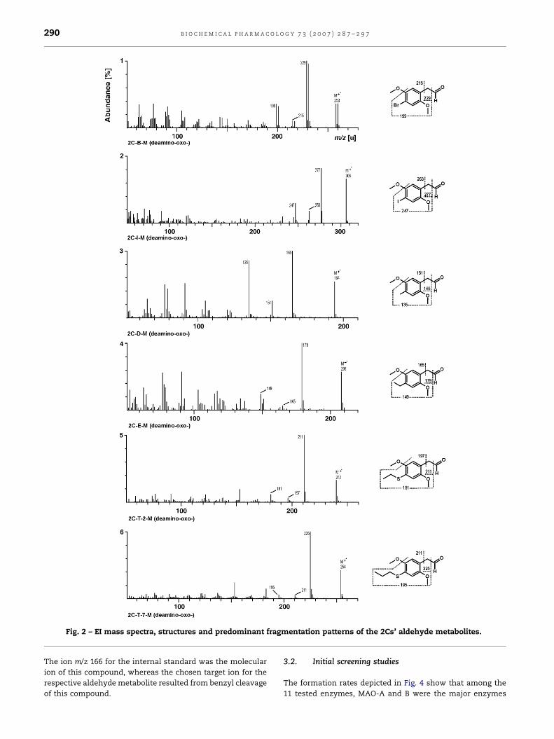
b i o c h e m i c a l p h a r m a c o l o g y 7 3 ( 2 0 0 7 ) 2 8 7 – 2 9 7290
Fig. 2 – EI mass spectra, structures and predominant fragmentation patterns of the 2Cs’ aldehyde metabolites.
The ion m/z 166 for the internal standard was the molecular
ion of this compound, whereas the chosen target ion for the
respective aldehyde metabolite resulted from benzyl cleavage
of this compound.
3.2. Initial screening studies
The formation rates depicted in Fig. 4 show that among the
11 tested enzymes, MAO-A and B were the major enzymes
b i o c h e m i c a l p h a r m a c o l o g y 7 3 ( 2 0 0 7 ) 2 8 7 – 2 9 7 291
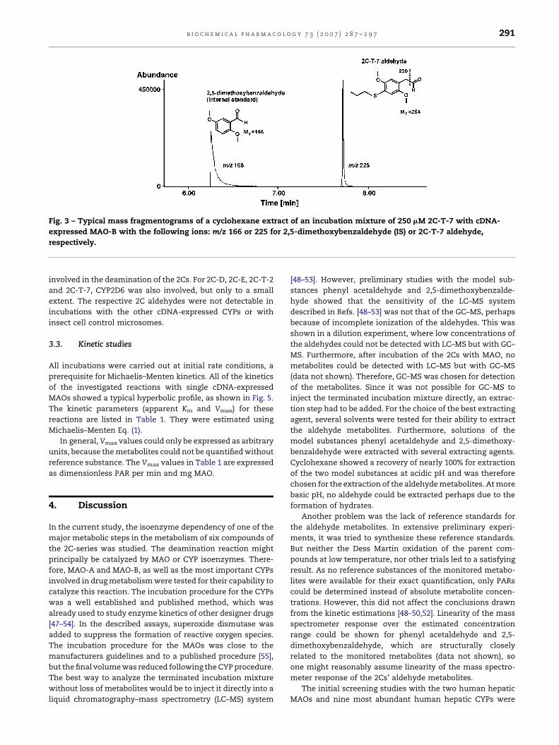
Fig. 3 – Typical mass fragmentograms of a cyclohexane extract of an incubation mixture of 250 mM 2C-T-7 with cDNA-
expressed MAO-B with the following ions: m/z 166 or 225 for 2,5-dimethoxybenzaldehyde (IS) or 2C-T-7 aldehyde,
respectively.
involved in the deamination of the 2Cs. For 2C-D, 2C-E, 2C-T-2
and 2C-T-7, CYP2D6 was also involved, but only to a small
extent. The respective 2C aldehydes were not detectable in
incubations with the other cDNA-expressed CYPs or with
insect cell control microsomes.
3.3. Kinetic studies
All incubations were carried out at initial rate conditions, a
prerequisite for Michaelis–Menten kinetics. All of the kinetics
of the investigated reactions with single cDNA-expressed
MAOs showed a typical hyperbolic profile, as shown in Fig. 5.
The kinetic parameters (apparent Km and Vmax) for these
reactions are listed in Table 1. They were estimated using
Michaelis–Menten Eq. (1).
In general, Vmax values could only be expressed as arbitrary
units, because the metabolites could not be quantified without
reference substance. The Vmax values in Table 1 are expressed
as dimensionless PAR per min and mg MAO.
4. Discussion
In the current study, the isoenzyme dependency of one of the
major metabolic steps in the metabolism of six compounds of
the 2C-series was studied. The deamination reaction might
principally be catalyzed by MAO or CYP isoenzymes. There-
fore, MAO-A and MAO-B, as well as the most important CYPs
involved in drug metabolism were tested for their capability to
catalyze this reaction. The incubation procedure for the CYPs
was a well established and published method, which was
already used to study enzyme kinetics of other designer drugs
[47–54]. In the described assays, superoxide dismutase was
added to suppress the formation of reactive oxygen species.
The incubation procedure for the MAOs was close to the
manufacturers guidelines and to a published procedure [55],
but the final volume was reduced following the CYP procedure.
The best way to analyze the terminated incubation mixture
without loss of metabolites would be to inject it directly into a
liquid chromatography–mass spectrometry (LC–MS) system
[48–53]. However, preliminary studies with the model sub-
stances phenyl acetaldehyde and 2,5-dimethoxybenzalde-
hyde showed that the sensitivity of the LC–MS system
described in Refs. [48–53] was not that of the GC–MS, perhaps
because of incomplete ionization of the aldehydes. This was
shown in a dilution experiment, where low concentrations of
the aldehydes could not be detected with LC–MS but with GC–
MS. Furthermore, after incubation of the 2Cs with MAO, no
metabolites could be detected with LC–MS but with GC–MS
(data not shown). Therefore, GC–MS was chosen for detection
of the metabolites. Since it was not possible for GC–MS to
inject the terminated incubation mixture directly, an extrac-
tion step had to be added. For the choice of the best extracting
agent, several solvents were tested for their ability to extract
the aldehyde metabolites. Furthermore, solutions of the
model substances phenyl acetaldehyde and 2,5-dimethoxy-
benzaldehyde were extracted with several extracting agents.
Cyclohexane showed a recovery of nearly 100% for extraction
of the two model substances at acidic pH and was therefore
chosen for the extraction of the aldehyde metabolites. At more
basic pH, no aldehyde could be extracted perhaps due to the
formation of hydrates.
Another problem was the lack of reference standards for
the aldehyde metabolites. In extensive preliminary experi-
ments, it was tried to synthesize these reference standards.
But neither the Dess Martin oxidation of the parent com-
pounds at low temperature, nor other trials led to a satisfying
result. As no reference substances of the monitored metabo-
lites were available for their exact quantification, only PARs
could be determined instead of absolute metabolite concen-
trations. However, this did not affect the conclusions drawn
from the kinetic estimations [48–50,52]. Linearity of the mass
spectrometer response over the estimated concentration
range could be shown for phenyl acetaldehyde and 2,5-
dimethoxybenzaldehyde, which are structurally closely
related to the monitored metabolites (data not shown), so
one might reasonably assume linearity of the mass spectro-
meter response of the 2Cs’ aldehyde metabolites.
The initial screening studies with the two human hepatic
MAOs and nine most abundant human hepatic CYPs were

b i o c h e m i c a l p h a r m a c o l o g y 7 3 ( 2 0 0 7 ) 2 8 7 – 2 9 7292
Fig. 4 – Formation rates (V) of 2C deamination (250 mM 2C compound each) with 0.2 mg/mL MAO-A or MAO-B or 50 pmol/mL of
the given individual CYPs (V given as dimensionless PAR per min and mg protein) and with insect cell control microsomes.
performed to identify their possible role in 2C deamination.
According to the supplier’s recommendations, the incubation
conditions chosen were adequate to make a statement on a
general involvement of a particular MAO or CYP. The data
revealed that MAO-A and MAO-B were capable of catalyzing
the monitored reaction. For 2C-D, 2C-E, 2C-T-2 and 2C-T-7,
also CYP2D6 was involved, but with low formation rate. Only
the kinetic profiles of the reactions by MAO-A and MAO-B were

b i o c h e m i c a l p h a r m a c o l o g y 7 3 ( 2 0 0 7 ) 2 8 7 – 2 9 7 293
Fig. 5 – Michaelis–Menten plots for 2C deamination catalyzed by MAO-A or MAO-B. Values represent the mean of duplicate
incubations. V given as dimensionless PAR per min and mg protein. Curves were calculated by nonlinear curve fit
according to Eq. (1) (one-site binding model).

b i o c h e m i c a l p h a r m a c o l o g y 7 3 ( 2 0 0 7 ) 2 8 7 – 2 9 7294
Fig. 5. (Continued ).
further investigated. Kinetic assays with these enzymes were
performed under initial rate conditions, a prerequisite for
Michaelis–Menten kinetics [56]. These conditions were chosen
according to previous experiments concerning the enzyme
concentration and time linearity. Furthermore, less than 20%
of substrate was metabolized in all incubations, as determined
with HPLC-UV after direct injection of the incubation mixtures
(data not shown). This method was used since the parent
compounds were not extracted at the acidic pH of the
incubation mixture and therefore were not present in the
GC–MS runs.
As expected, classical hyperbolic Michaelis–Menten plots
(Fig. 5) were found using cDNA-expressed MAOs. The
Table 1 – Kinetic data of 2C deamination catalyzed by MAO-A
Apparent Km (best fitvalue � standarderror) for MAO-A
Apparent Km (bevalue � standerror) for MA
2C-B 43.8 � 8.7 63.8 � 7.7
2C-I 31.1 � 4.1 88.3 � 7.2
2C-D 41.3 � 3.6 96.9 � 9.7
2C-E 49.6 � 3.3 187.8 � 19.1
2C-T-2 38.8 � 2.7 146.0 � 13.0
2C-T-7 14.4 � 2.1 108.5 � 19.2
Units are: apparent Km in mM, Vmax in dimensionless PAR/min and mg p
apparent Km and Vmax values of the investigated MAOs were
calculated by nonlinear curve fit according to Eq. (1). The
apparent Km values listed in Table 1 show that all studied 2Cs
have a slightly higher affinity for MAO-A than for MAO-B.
Furthermore, the differences of the Km values between MAO-
A and B are getting greater by an increasing 4-substituent size.
These facts might be explained by the size of the binding
pockets of both, MAO-A and B. Miller et al. reported for several
4-substituted benzylamines, that increasing the 4-substitu-
ent size resulted in tighter binding to MAO-A [40]. For 4-
substituted phenethylamines, Nandigama et al. reported
similar results [57]. The reason for this might be a large
binding pocket for 4-substituents in the case of MAO-A,
and MAO-B
st fitardO-B
Vmax (best fitvalue � standarderror) for MAO-A
Vmax (best fitvalue � standarderror) for MAO-B
2.3 � 0.1 1.7 � 0.1
2.5 � 0.1 4.6 � 0.1
1.7 � 0.04 2.3 � 0.1
0.7 � 0.01 4.3 � 0.2
1.5 � 0.03 4.3 � 0.2
3.4 � 0.1 4.5 � 0.3
rotein.

b i o c h e m i c a l p h a r m a c o l o g y 7 3 ( 2 0 0 7 ) 2 8 7 – 2 9 7 295
whereas MAO-B should contain a small hydrophobic binding
pocket for 4-substituents [58]. Furthermore, MAO-B showed
in general with exception of 2C-B, increased Vmax value
compared to MAO-A for a single 2C compound. One might
speculate, that MAO-A has a higher affinity for the 2Cs than
MAO-B, but MAO-B has the higher capacity for the 2Cs
concerning the deamination reaction. However, statements
concerning the measured Vmax values are difficult, because
quantification of metabolites was not possible, as mentioned
before. As MAO-A and MAO-B are involved in one of the
major metabolic steps of the 2Cs, the 2Cs might be
susceptible for drug-drug interactions with MAO inhibitors
possibly leading to elevated plasma concentrations of the
2Cs, and therefore increasing the probability of toxic side
effects. Such inhibitors are used as antidepressants such as
tranylcypromine and moclobemide or as antiparkinsonians
such as selegiline. Amphetamine derivatives, which are
often abused together with the 2Cs are also known to be
potent MAO inhibitors [59,60]. Beside this, due to the
relatively high apparent Km values of the 2Cs, further studies
on their MAO inhibitory potential are required. Such
inhibition would lead to further interactions for example
with indirect sympathomimetics such as cocaine, or with
food ingredients such as tyramine. However, the question
whether drug interactions are of relevance for 2C pharma-
cokinetics and/or clinical outcome of intoxications cannot be
answered at the moment due to lack of sufficient authentic
human data.
Acknowledgements
The authors would like to thank Peter Roesner, Liane D. Paul
and Roland F. Staack, Frank T. Peters, Andreas H. Ewald,
Markus R. Meyer, and Armin A. Weber for their assistance and
helpful discussions.
r e f e r e n c e s
[1] Shulgin A, Pihkal A. Chemical love story Berkley (CA):Transform Press; 1991.
[2] Shulgin A. #142 PEA; phenethylamine. In: Dan J, Pihkal A,editors. Chemical love story. Berkley (CA): Transform Press;1991. p. 815–8.
[3] de Boer D, Bosman I. A new trend in drugs-of-abuse; the 2C-series of phenethylamine designer drugs. Pharm World Sci2004;26:110–3.
[4] European Monitoring Centre for Drugs and Drug Addiction(EMCDDA). Report on the risk assessment of 2C-I, 2C-T-2and 2C-T-7 in the framework of the joint action on newsynthetic drugs. http://www.emcdda.eu.int/multimedia/publications/risk_assessments/2Cs_2003_1636.pdf, 2004.
[5] Schifano F, Deluca P, Agosti L, Martinotti G, Corkery JM,Alex B, et al. New trends in the cyber and street market ofrecreational drugs? The case of 2C-T-7 (‘Blue Mystic’) JPsychopharmacol 2005;19:675–9.
[6] Drug Enforcement Administration—Office of ForensicSciences. BZP and Nexus tablets. Microgram 2001;34:3.
[7] Drug Enforcement Administration—Office of ForensicSciences. 2,5-Dimethoxy-4-(n)-propylthiophenethylamine(2C-T-7). Microgram 2001;34:193.
[8] Drug Enforcement Administration—Office of ForensicSciences. Dipropyltryptamine and 2C-I in portland, oregon.Microgram 2004;37:113–4.
[9] Drug Enforcement Administration—Office of ForensicSciences. 2C-I tablets in the Balearic Islands (spain).Microgram 2004;37:48–9.
[10] Drug Enforcement Administration—Office of ForensicSciences. 2,5-Dimethoxy-4-ethylphenethylamine (2C-E)encountered in FT. Pierce, Florida and Royal Oak, Michigan.Microgram 2004;37:193–4.
[11] Drug Enforcement Administration—Office of ForensicSciences. 2,5-Dimethoxy-4-ethylphenethylamine(2C-E) capsules in Bettendorf, Iowa. Microgram 2005;38:59.
[12] Drug Enforcement Administration - Department of Justice.Schedules of controlled substances; placement of 2,5-dimethoxy-4-(n)-propylthiophenethylamine and N-benzylpiperazine into schedule i of the controlledsubstances act. Fed Register 2004;69:12794–7.
[13] European Communities. Council decision 2003/847/JHA of27 November 2003 concerning control measures andcriminal sanctions in respect of the new synthetic drugs2C-I, 2C-T-2, 2C-T-7 and TMA-2. Off J Eur Commun2003;L321:64–5.
[14] WHO expert committee on drug dependence. Evaluationof dependence-producing drugs—32nd report of theWHO expert committee on drug dependence. EB 109/33,2001.
[15] Johnson MP, Mathis CA, Shulgin AT, Hoffman AJ, NicholsDE. [125I]-2-(2,5-dimethoxy-4-iodophenyl)aminoethane([125I]-2C-I) as a label for the 5-HT2 receptor in rat frontalcortex. Pharmacol Biochem Behav 1990;35:211–7.
[16] Glennon RA, Raghupathi R, Bartyzel P, Teitler M, LeonhardtS. Binding of phenylalkylamine derivatives at 5-HT1C and5-HT2 serotonin receptors: evidence for a lack of selectivity.J Med Chem 1992;35:734–40.
[17] Glennon RA, Titeler M, Lyon RA. A preliminaryinvestigation of the psychoactive agent 4-bromo-2,5-dimethoxyphenethylamine: a potential drug of abuse.Pharmacol Biochem Behav 1988;30:597–601.
[18] Nichols DE, Frescas S, Marona-Lewicka D, Huang X, RothBL, Gudelsky GA, et al. 1-(2,5-Dimethoxy-4-(trifluoromethyl)phenyl)-2-aminopropane: a potentserotonin 5-HT2A/2C agonist. J Med Chem 1994;37:4346–51.
[19] Cozzi NV, Nichols DE. 5-HT2A receptor antagonists inhibitpotassium-stimulated gamma-aminobutyric acid release inrat frontal cortex. Eur J Pharmacol 1996;309:25–31.
[20] Acuna-Castillo C, Villalobos C, Moya PR, Saez P, Cassels BK,Huidobro-Toro JP. Differences in potency and efficacy of aseries of phenylisopropylamine/phenylethylamine pairs at5-HT(2A) and 5-HT(2C) receptors. Br J Pharmacol2002;136:510–9.
[21] Villalobos CA, Bull P, Saez P, Cassels BK, Huidobro-Toro JP.4-Bromo-2,5-dimethoxyphenethylamine (2C-B) andstructurally related phenylethylamines are potent 5-HT2Areceptor antagonists in Xenopus laevis oocytes. Br JPharmacol 2004;141:1167–74.
[22] Khorana N, Pullagurla MR, Dukat M, Young R, Glennon RA.Stimulus effects of three sulfur-containing psychoactiveagents. Pharmacol Biochem Behav 2004;78:821–6.
[23] Fantegrossi WE, Harrington AW, Eckler JR, Arshad S, RabinRA, Winter JC, et al. Hallucinogen-like actions of 2,5-dimethoxy-4-(n)-propylthiophenethylamine (2C-T-7) inmice and rats. Psychopharmacology (Berl) 2005;181:496–503.
[24] Lobos M, Borges Y, Gonzalez E, Cassels BK. The action ofthe psychoactive drug 2C-B on isolated rat thoracic aorta.Gen Pharmacol 1992;23:1139–42.

b i o c h e m i c a l p h a r m a c o l o g y 7 3 ( 2 0 0 7 ) 2 8 7 – 2 9 7296
[25] Saez P, Borges Y, Gonzalez E, Cassels BK. Alpha-adrenergicand 5-HT2-serotonergic effects of some beta-phenylethylamines on isolated rat thoracic aorta. GenPharmacol 1994;25:211–6.
[26] Curtis B, Kemp P, Harty L, Choi C, Christensen D.Postmortem identification and quantitation of 2,5-dimethoxy-4-n-propylthiophenethylamine using GC-MSDand GC-NPD. J Anal Toxicol 2003;27:493–8.
[27] Theobald DS, Fehn S, Maurer HH. New designer drug 2,5-dimethoxy-4-propylthiophenethylamine (2C-T-7): studieson its metabolism and toxicological detection in rat urineusing gas chromatography/mass spectrometry. J MassSpectrom 2005;40:105–16.
[28] Theobald DS, Staack RF, Puetz M, Maurer HH. New designerdrug 2,5-dimethoxy-4-ethylthio-beta-phenethylamine (2C-T-2): studies on its metabolism and toxicological detectionin rat urine using gas chromatography/mass spectrometry.J Mass Spectrom 2005;40:1157–72.
[29] Theobald DS, Puetz M, Schneider E, Maurer HH. Newdesigner drug 4-iodo-2,5-dimethoxy-beta-phenethylamine(2C-I): studies on its metabolism and toxicological detectionin rat urine using gas chromatographic/mass spectrometricand capillary electrophoretic/mass spectrometrictechniques. J Mass Spectrom 2006;41:872–86.
[30] Theobald DS, Maurer HH. Studies on the metabolism andtoxicological detection of the designer drug 4-ethyl-2,5-dimethoxy-b-phenethylamine (2C-E) in rat urine using gaschromatographic-mass spectrometric techniques. JChromatogr B Anal Technol Biomed Life Sci 2006 [Epubahead of print].
[31] Theobald DS, Maurer HH. Studies on the metabolism andtoxicological detection of the designer drug 2,5-dimethoxy-4-methyl-b-phenethylamine (2C-D) in rat urine usinggaschromatographic-massspectrometric techniques. JMass Spectrom 2006. in revision.
[32] Kanamori T, Inoue H, Iwata Y, Ohmae Y, Kishi T. In vivometabolism of 4-bromo-2,5-dimethoxyphenethylamine(2C-B) in the rat: identification of urinary metabolites. JAnal Toxicol 2002;26:61–6.
[33] Kanamori T, Tsujikawa K, Ohmae Y, Iwata Y, Inoue H,Inouye Y, et al. Excretory profile of 4-bromo-2,5-dimethoxy-phenethylamine (2C-B) in rat. J Health Sci 2003;49:166–9.
[34] de Boer D, dos Reys LA, Pylon N, Gijzels M, Bosman IJ, MaesRAA. Preliminary results on the urinary excretion of 2C-B(4-bromo-2,5-dimethoxyphenethylamine) and itsmetabolites In humans. Br J Pharmacol 1999;127:41P.
[35] Carmo H, Boer D, Remiao F, Carvalho F, Reys LA, Bastos ML.Metabolism of the designer drug 4-bromo-2,5-dimethoxyphenethylamine (2C-B) in mice, after acuteadministration. J Chromatogr B Anal Technol Biomed LifeSci 2004;811:143–52.
[36] Carmo H, Hengstler JG, de Boer D, Ringel M, Remiao F,Carvalho F, et al. Metabolic pathways of 4-bromo-2,5-dimethoxyphenethylamine (2C-B): analysis of phase Imetabolism with hepatocytes of six species includinghuman. Toxicology 2005;206:75–89.
[37] Kanamori T, Tsujikawa K, Ohmae Y, Iwata YT, Inoue H,Kishi T, et al. A study of the metabolism ofmethamphetamine and 4-bromo-2,5-dimethoxyphenethylamine (2C-B) in isolated rathepatocytes. Forensic Sci Int 2005;148:131–7.
[38] Evans WE, McLeod HL. Pharmacogenomics-drugdisposition, drug targets, and side effects. N Engl J Med2003;348:538–49.
[39] Howard LA, Sellers EM, Tyndale RF. The role ofpharmacogenetically-variable cytochrome P450 enzymesin drug abuse and dependence. Pharmacogenomics2002;3:185–99.
[40] Miller JR, Edmondson DE. Structure-activityrelationships in the oxidation of para-substitutedbenzylamine analogues by recombinant human livermonoamine oxidase A. Biochemistry 1999;38:13670–83.
[41] Kalgutkar AS, Dalvie DK, Castagnoli Jr N, Taylor TJ.Interactions of nitrogen-containing xenobiotics withmonoamine oxidase (MAO) isozymes A and B: SAR studieson MAO substrates and inhibitors. Chem Res Toxicol2001;14:1139–62.
[42] Ortiz-de-Montellano PR, de-Voss JJ. Substrate oxidation bycytochrome P450 enzymes. In: Ortiz-de-Montellano PR,editor. Cytochrome P450—structure, mechanism, andbiochemistry. New York: Kluwer Academic/PlenumPublishers; 2005. p. 183–245.
[43] Theobald DS, Fehn S, Maurer HH. New designer drug 2,5-dimethoxy-4-propylthiophenethylamine (2C-T-7): studieson its metabolism in rat urine using GC/MS. Medimond2005;143–6 (Ed. Oesch F).
[44] Theobald DS, Fritschi G, Maurer HH. Studies on thetoxicological detection of the designer drug 4-bromo-2,5-dimethoxy-b-phenethylamine (2C-B) in rat urine usinggaschromatography–massspectrometry. J Chromatogr B:Anal Technol Biomed Life Sci; 2006 September 13 [Epubahead of print].
[45] McLafferty FW, Turecek F. Interpretation of mass spectraMill Valley, CA: University Science Books; 1993.
[46] Smith RM, Busch KL. Understanding mass spectra—a basicapproach New York (NY): Wiley; 1999.
[47] Kraemer T, Pflugmann T, Bossmann M, Kneller NM, PetersFT, Paul LD, et al. Fenproporex N-dealkylation toamphetamine-enantioselective in vitro studies in humanliver microsomes as well as enantioselective in vivo studiesin Wistar and Dark Agouti rats. Biochem Pharmacol2004;68:947–57.
[48] Springer D, Paul LD, Staack RF, Kraemer T, Maurer HH.Identification of the cytochrome P450 enzymes involved inthe metabolism of 40-methyl-(alpha)-pyrrolidinopropiophenone, a novel scheduled designerdrug, in human liver microsomes. Drug Metab Dispos2003;31:979–82.
[49] Springer D, Staack RF, Paul LD, Kraemer T, Maurer HH.Identification of cytochrome P450 enzymes involved in themetabolism of 4’-methoxy-pyrrolidinopropiophenone(MOPPP), a designer drug, in human liver microsomes.Xenobiotica 2003;33:989–98.
[50] Springer D, Paul LD, Staack RF, Kraemer T, Maurer HH.Identification of cytochrome P450 enzymes involved in themetabolism of 30,40-methylenedioxy-a�pyrrolidinopropiophenone (MDPPP), a designer drug,in human liver microsomes. Xenobiotica 2005;35:227–37.
[51] Staack RF, Theobald DS, Paul LD, Springer D, Kraemer T,Maurer HH. In vivo metabolism of the new designer drug 1-(4-methoxyphenyl)piperazine (MeOPP) in rat andidentification of the human cytochrome P450 enzymesresponsible for the major metabolic step. Xenobiotica2004;34:179–92.
[52] Staack RF, Paul LD, Springer D, Kraemer T, Maurer HH.Cytochrome P450 dependent metabolism of the newdesigner drug 1-(3-trifluoromethylphenyl)piperazine(TFMPP). In vivo studies in Wistar and Dark Agouti rats aswell as in vitro studies in human liver microsomes.Biochem Pharmacol 2004;67:235–44.
[53] Staack RF, Theobald DS, Paul LD, Springer D, Kraemer T,Maurer HH. Identification of human cytochrome p450 2D6as major enzyme involved in the o-demethylation of thedesigner drug p-methoxymethamphetamine. Drug MetabDispos 2004;32:379–81.

b i o c h e m i c a l p h a r m a c o l o g y 7 3 ( 2 0 0 7 ) 2 8 7 – 2 9 7 297
[54] Staack RF, Maurer HH. Metabolism of designer drugs ofabuse [review]. Curr Drug Metab 2005;6:259–74.
[55] Yu AM, Granvil CP, Haining RL, Krausz KW, Corchero J,Kupfer A, et al. The relative contribution of monoamineoxidase and cytochrome p450 isozymes to the metabolicdeamination of the trace amine tryptamine. J PharmacolExp Ther 2003;304:539–46.
[56] Clarke SE. In vitro assessment of human cytochrome P450.Xenobiotica 1998;28:1167–202.
[57] Nandigama RK, Edmondson DE. Structure-activity relationsin the oxidation of phenethylamine analogues byrecombinant human liver monoamine oxidase A.Biochemistry 2000;39:15258–65.
[58] Miller LG, Friedman H, Greenblatt DJ. Measurement ofclonazepam by electron-capture gas–liquidchromatography with application to single-dosepharmacokinetics. J Anal Toxicol 1987;11:55–7.
[59] Green AL, El Hait MA. p-Methoxyamphetamine, a potentreversible inhibitor of type-A monoamine oxidase in vitroand in vivo. J Pharm Pharmacol 1980;32:262–6.
[60] Scorza MC, Carrau C, Silveira R, Zapata-Torres G, CasselsBK, Reyes-Parada M. Monoamine oxidase inhibitoryproperties of some methoxylated and alkylthioamphetamine derivatives: structure-activity relationships.Biochem Pharmacol 1997;54:1361–9.