Variations on a (t)heme?novel mechanisms, redox partners and catalytic functions in the cytochrome...
25
REVIEW www.rsc.org/npr | Natural Product Reports Variations on a (t)heme—novel mechanisms, redox partners and catalytic functions in the cytochrome P450 superfamily† Andrew W. Munro,* Hazel M. Girvan and Kirsty J. McLean Received (in Cambridge, UK) 15th December 2006 First published as an Advance Article on the web 4th May 2007 DOI: 10.1039/b604190f Covering: up to the end of 2006 The catalytic potential of the P450s in organic biotransformations is the subject of this review—with emphasis on the breadth of P450 redox systems now recognised and the catalytic versatility of these biotechnologically important enzymes. 1 Introduction 2 Cytochromes P450: a brief history 3 The P450 catalytic cycle Faculty of Life Sciences, Manchester Interdisciplinary Biocentre, University of Manchester, 131 Princess Street, Manchester, M1 7DN, UK. E-mail: [email protected]; Fax: +44 161 306 8919; Tel: +44 161 306 5151 † This paper was published as a part of a special issue on the chemistry and biochemistry of heme proteins. Andrew Munro is Professor of Molecular Enzymology in the Faculty of Life Sciences at the University of Manchester. He received his PhD from the University of Aberdeen in 1991 and undertook Postdoctoral work at the University of Glasgow (1992–1995) prior to securing a Royal Society of Edinburgh fellowship (1996) allowing him to research into cytochrome P450 enzyme mechanism at Edinburgh University. In 1999 he took up a Lectureship in Pure and Applied Chemistry at the University of Strathclyde, followed by a Senior Lectureship appointment in the Biochemistry Department at the University of Leicester (2001), where he was promoted to a chair in 2003. Andrew’s group relocated to the University of Manchester in 2005 and are based at the new Manchester Interdisciplinary Biocentre (MIB). Hazel Girvan received her first degree in Biochemistry and Microbiology from the University of Strathclyde in 2001, and completed her PhD studies in the Biochemistry Department at the University of Leicester in 2005, where she characterised P450 enzymes with novel heme iron ligation states. She is now employed as a BBSRC Postdoctoral Researcher in Professor Munro’s laboratory, and is investigating the properties of the flavocytochrome P450 BM3 system and of other cytochrome P450 enzymes. She has particular interests in transient kinetic analysis and spectroscopic characterisation of P450 enzymes and their partner proteins. Kirsty McLean received her first degree in Biochemistry from the University of Stirling in 1998, followed by a PhD degree in the Biochemistry Department at the University of Leicester (2003). Following her PhD, she began Postdoctoral studies on cytochrome P450 systems from the human pathogen Mycobacterium tuberculosis, and is currently working as part of EU-funded integrated project “New Medicines for Tuberculosis” (NM4TB) involving the identification of new drug target enzymes in M. tuberculosis and the development of novel antitubercular drugs. She has particular interests in the structural and mechanistic characterisation of P450 enzymes. Andrew Munro Hazel Girvan Kirsty McLean 4 The biological diversity of P450 redox systems 5 The major reactions of cytochromes P450 6 Unorthodox P450 systems and reactions 7 Insights from P450 redox system atomic structure 8 P450cam and P450 BM3—paradigms in the P450 superfamily 9 Technological and biotechnological advances 10 Conclusions and future prospects 11 References This journal is © The Royal Society of Chemistry 2007 Nat. Prod. Rep., 2007, 24, 585–609 | 585
-
Upload
manchester -
Category
Documents
-
view
2 -
download
0
Transcript of Variations on a (t)heme?novel mechanisms, redox partners and catalytic functions in the cytochrome...

REVIEW www.rsc.org/npr | Natural Product Reports
Variations on a (t)heme—novel mechanisms, redox partners and catalyticfunctions in the cytochrome P450 superfamily†
Andrew W. Munro,* Hazel M. Girvan and Kirsty J. McLean
Received (in Cambridge, UK) 15th December 2006First published as an Advance Article on the web 4th May 2007DOI: 10.1039/b604190f
Covering: up to the end of 2006
The catalytic potential of the P450s in organic biotransformations is the subject of this review—withemphasis on the breadth of P450 redox systems now recognised and the catalytic versatility of thesebiotechnologically important enzymes.
1 Introduction2 Cytochromes P450: a brief history3 The P450 catalytic cycle
Faculty of Life Sciences, Manchester Interdisciplinary Biocentre, Universityof Manchester, 131 Princess Street, Manchester, M1 7DN, UK. E-mail:[email protected]; Fax: +44 161 306 8919; Tel: +44 161306 5151† This paper was published as a part of a special issue on the chemistryand biochemistry of heme proteins.
Andrew Munro is Professor of Molecular Enzymology in the Faculty of Life Sciences at the University of Manchester. He received his PhDfrom the University of Aberdeen in 1991 and undertook Postdoctoral work at the University of Glasgow (1992–1995) prior to securing aRoyal Society of Edinburgh fellowship (1996) allowing him to research into cytochrome P450 enzyme mechanism at Edinburgh University. In1999 he took up a Lectureship in Pure and Applied Chemistry at the University of Strathclyde, followed by a Senior Lectureship appointmentin the Biochemistry Department at the University of Leicester (2001), where he was promoted to a chair in 2003. Andrew’s group relocatedto the University of Manchester in 2005 and are based at the new Manchester Interdisciplinary Biocentre (MIB).
Hazel Girvan received her first degree in Biochemistry and Microbiology from the University of Strathclyde in 2001, and completed herPhD studies in the Biochemistry Department at the University of Leicester in 2005, where she characterised P450 enzymes with novel hemeiron ligation states. She is now employed as a BBSRC Postdoctoral Researcher in Professor Munro’s laboratory, and is investigating theproperties of the flavocytochrome P450 BM3 system and of other cytochrome P450 enzymes. She has particular interests in transient kineticanalysis and spectroscopic characterisation of P450 enzymes and their partner proteins.
Kirsty McLean received her first degree in Biochemistry from the University of Stirling in 1998, followed by a PhD degree in the BiochemistryDepartment at the University of Leicester (2003). Following her PhD, she began Postdoctoral studies on cytochrome P450 systems fromthe human pathogen Mycobacterium tuberculosis, and is currently working as part of EU-funded integrated project “New Medicinesfor Tuberculosis” (NM4TB) involving the identification of new drug target enzymes in M. tuberculosis and the development of novelantitubercular drugs. She has particular interests in the structural and mechanistic characterisation of P450 enzymes.
Andrew Munro Hazel Girvan Kirsty McLean
4 The biological diversity of P450 redox systems5 The major reactions of cytochromes P4506 Unorthodox P450 systems and reactions7 Insights from P450 redox system atomic structure8 P450cam and P450 BM3—paradigms in the P450
superfamily9 Technological and biotechnological advances10 Conclusions and future prospects11 References
This journal is © The Royal Society of Chemistry 2007 Nat. Prod. Rep., 2007, 24, 585–609 | 585

1 Introduction
The regio- and stereoselective oxygenation of organic moleculesis notoriously difficult to achieve by standard organic synthesisapproaches (e.g. using Sharpless asymmetric hydroxylation orJacobsen’s salen catalyst for asymmetric epoxidation) and thesemethods often do not lend themselves to large scale synthesis.1,2
Enzyme-based approaches have the advantages of being “cleaner”and usually safer (i.e. involving use of less hazardous reagents,fewer reaction steps and requiring less fractionation of productmixtures) and usually produce a smaller spectrum of oxygenatedproducts. Indeed, a single product may be feasible—as seene.g. in the exclusive 5-exo hydroxylation of camphor by thePseudomonas putida cytochrome P450cam enzyme (CYP101A1).3
That said, there are relatively few types of enzyme catalystknown to catalyse oxygenation reactions. These include the flavin-containing mono-oxygenases (FMOs) and dioxygenases, whichare involved in diverse reactions such as xenobiotic metabolismand detoxification, collagen and antibiotic biosynthesis.4,5 Themost prominent class of oxygenases is the cytochrome P450 (P450)enzyme superfamily, members of which are found widespreadin nature and which perform a wide array of oxygenationand other biochemical transformations on countless endogenousbiochemicals and xenobiotics.6–9 The P450s are heme-containingbiocatalysts that typically have a relatively hydrophobic activesite cavity for substrate binding, as revealed by atomic structuralstudies.10–14 There are several examples of the mutagenesis (bothby rational design and forced evolution methods) of different P450enzymes in order to facilitate alteration of substrate selectivity, andthe position of oxygenation of the substrate e.g.15–18 The catalyticpotential of the P450s in organic biotransformations is the subjectof this review—with emphasis on the breadth of P450 systems nowrecognised and the catalytic versatility of these biotechnologicallyimportant enzymes.
2 Cytochromes P450: a brief history
The P450s were first recognised as unusual pigments in mam-malian liver microsome samples (i.e. the membranous materialfrom the liver endoplasmic reticulum). Their ability to form acomplex with carbon monoxide (CO) led to their recognition asa distinctive type of cytochrome (i.e. a heme-binding electron-transferring protein), with absorption maximum of the ferrous–CO complex shifted to ∼450 nm (hence Pigment 450 or P450)19,20
(Fig. 1). This characteristic shift of the main (Soret) band ofthe P450 heme is a consequence of the nature of the proximalaxial ligand to the heme iron (i.e. the ligand trans to CO in thecomplex), which is a phylogenetically conserved cysteine thiolate.The electron donating character of this bond is critical for theP450’s catalytic activity.21 The ligand trans to the cysteinate istypically a water molecule (as opposed to another amino acidfrom the protein), and the water is readily displaced during theenzyme’s catalytic cycle in favour of dioxygen (vide infra).22
From the earliest stages of research into the P450s, theirinvolvement with oxidation of organic molecules was recognised.In particular, carbon monoxide (or other ligands) was found toinhibit the oxidative transformation and detoxification of variousdrugs and other organic compounds by the hepatic P450s (e.g.references 23, 24). This stimulated intensive research into the
Fig. 1 Absorption spectra for cytochrome P450 and its ferrous–carbonmonoxide complex. A typical absorption spectrum for a P450 enzyme(CYP51B1 from Mycobacterium tuberculosis, ∼4 lM enzyme) is shownin its oxidised (ferric) state (thin solid line, Amax = 418 nm) and in itsdithionite-reduced Fe(II)–CO complex (thick solid line, Amax = 450 nm).The major absorption (Soret) band shift to ∼450 nm in the CO complexis a hallmark of the cysteinate-coordinated heme iron of P450 enzymes.A small shoulder originating from the inactive (thiol-coordinated) P420form of CYP51B1 is seen at ∼420 nm in the spectrum of the Fe(II)–COcomplex.
properties of these enzymes, and a huge amount of progress hasbeen made over the last 50 years into the understanding of thestructure, mechanism, biological function and diversity of theP450s.25 It was soon established that P450s catalysed the reductiveactivation and scission of molecular oxygen (dioxygen) that bindsto their heme iron. This leads to insertion of an atom of oxygeninto the substrate (frequently resulting in hydroxylation) andproduction of a molecule of water from the other oxygen atom.26
This typical P450 reaction requires delivery of two electrons andtwo protons to the P450 heme iron, as shown in eqn (1).
RH + O2 + 2e− + 2H+ → ROH + H2O (1)
However, as will be discussed later in the article, reactions farmore diverse than hydroxylations are catalysed by P450s, andtheir means of sourcing protons and electrons are also variable27,28
(Fig. 2).
3 The P450 catalytic cycle
To facilitate P450 catalysis, the vast majority of these enzymesrequire the delivery of two electrons ultimately derived frompyridine nucleotide coenzymes (i.e. NADPH or NADH). Theelectrons are transported to the P450 heme via one or more redoxpartner proteins. Electrons are delivered at discrete points in theP450 catalytic cycle, as illustrated in Fig. 2. The first electronreduces the ferric heme iron to ferrous, which can then binddioxygen. As a prelude to reduction, substrate binding usuallyfacilitates dissociation of a water ligand bound weakly to the ironand trans to the cysteinate, which in turn favours an electronicreorganisation in the heme iron d-orbitals. This results in a shiftin ferric heme iron spin state equilibrium from low-spin (S = 1/2)towards high-spin (S = 5/2).29,30 In turn, this leads to an increasein the heme iron reduction potential that favours electron transferto the heme iron from the redox partner (e.g. references 31, 32).
586 | Nat. Prod. Rep., 2007, 24, 585–609 This journal is © The Royal Society of Chemistry 2007

Fig. 2 The catalytic cycle of cytochrome P450. The intermediate stages of a P450 catalytic cycle leading to substrate (R–H) hydroxylation are shown.The heme macrocycle is depicted to represent heme, with the oxidation state of the heme iron indicated. The proximal heme ligand (cysteine thiolate,indicated as a S-atom linked to the iron) and distal ligand (a water molecule, changing to dioxygen as the cycle progresses) are also indicated. In the firststep, substrate binding leads to displacement of the distal water ligand. This effects a shift in ferric heme iron spin-state equilibrium from low-spin (S =1/2) towards high-spin (S = 5/2). This leads to a more positive potential of the iron and favours electron transfer from the redox partner to reduce theheme iron to the ferrous state. Ferrous heme iron binds oxygen to form the ferrous–oxy intermediate (which is isoelectronic with the ferric–superoxyform). Delivery of a second electron from the redox partner reduces heme iron to the ferric peroxy state. Protonation produces the ferric hydroperoxoform (also known as compound 0). A further protonation leads to scission of the bound dioxygen and the production of a water molecule. The remnantintermediate on the heme is a ferryl–oxo species (compound I) with a porphyrin p radical cation. This is likely to be the catalytically relevant substrateoxidant in most P450 reactions. Compound I attacks the nearby substrate and effects its hydroxylation. The departure of product (R–OH) allows waterto rebind the ferric iron and complete the cycle. Non-productive pathways leading to collapse of oxy intermediates in the cycle are also indicated. Theferrous–oxy species can decay to reform ferric P450 with production of superoxide. Compound 0 collapses with production of peroxide, and the reactioncan be driven (productively) in the reverse direction (albeit inefficiently in most cases) by mixing H2O2 (or organic peroxides) with ferric, substrate-boundP450. Compound I collapses with production of water. Collapse of these species may occur if e.g. electron/proton delivery is not timely or if substrate isinappropriately positioned or otherwise resistant to oxidative attack.
This may be an elegant mechanism to ensure that electron transferto the P450 and subsequent formation of reactive oxygen speciesoccurs only when substrate is available for oxygenation. While thisphenomenon is observed for many “well-regulated” P450 enzymes(e.g. the P. putida camphor hydroxylase P450cam [CYP101A1]and the Bacillus megaterium fatty acid hydroxylase P450 BM3[CYP102A1]) which also often have relatively restricted substrateselectivity, it clearly does not hold for all P450s (e.g. references33, 34). The ferrous oxy species formed on binding of dioxygenis isoelectronic with a ferric superoxy state, and the equilibriummay be in favour of the latter form. The delivery of a secondelectron leads to the production of the ferric peroxy form, whichis rapidly protonated to the ferric hydroperoxy state (compound0). A further protonation leads to collapse of this species with theproduction of a ferryl–oxo form (compound I) considered to be theprimary species responsible for the attack on, and oxygenation of,the substrate. A molecule of water is also produced at this stage inthe cycle. Compound I oxygenates the substrate, and dissociationof the product results in the re-binding of a water molecule as the6th ligand to the restored ferric heme iron, completing the cycle.8,35
The apparent simplicity of the cycle hides the fact that later P450intermediates (particularly compounds 0 and I) are transient anddifficult to characterise. In particular, compound I is extremelyelusive and it has proven almost impossible to obtain convincingstructural or spectroscopic evidence for its formation. However,recent studies have provided evidence for the existence of theferryl–oxo state.36 In addition, there is some proof that boththe ferryl intermediate and its predecessor compound 0 may beviable substrate oxidants.37,38 The source of protons in the cycle isultimately bulk solvent, although active site amino acid residuesor substrate can clearly participate in relay pathways to facilitateproton delivery to ferrous oxy intermediates,12,39,40 as discussed inmore detail later.
The nature of electron donor proteins for P450s was, untilrelatively recently, considered to be quite well understood—withonly two major classes of protein systems thought to be responsiblefor transport of NAD(P)H-derived electrons to members of theP450 superfamily. However, studies in recent years have revealeda diversity of redox partner systems that almost matches thediversity of reactions performed by the P450s themselves.
This journal is © The Royal Society of Chemistry 2007 Nat. Prod. Rep., 2007, 24, 585–609 | 587

4 The biological diversity of P450 redox systems
As detailed above, the stereotypical P450 requires consecutivedelivery of two electrons to its heme iron at distinct points inits catalytic cycle (Fig. 2). However, the means by which thisprocess is achieved in nature is considerably more diverse thanwas first thought. In early years of P450 research, two distincttypes of redox partners came to prominence and for several yearswere considered to reflect the only major classes of P450 redoxsystems. These were the mammalian hepatic type which consistsof two protein components (a P450 and a NADPH-dependentreductase, both membrane bound) and the bacterial-type system,which (on basis of studies with the P. putida CYP101A1) consistsof a soluble P450 to which electron transfer is mediated by aniron–sulfur protein, which in turn is reduced by a flavin-containingreductase.41–43 More detailed kinetic and product formation studiesof the systems revealed that both were dependent on NADPH orNADH, with the mammalian system favouring NADPH, andthe CYP101A1 system favouring NADH. The redox partnerin mammalian liver (and subsequently shown to participate inseveral P450-dependent reactions in other mammalian organs andeukaryotic cells) is a FAD- and FMN-containing enzyme termedcytochrome P450 reductase (CPR).44,45 The FAD is the entry pointfor electrons derived (by hydride ion transfer) from NADPH, withFMN receiving single electrons from the FAD and passing theseto the P450.46–49 In the bacterial system, detailed analyses of theCYP101A1 redox partners demonstrated that these were a FAD-containing reductase (putidaredoxin reductase) that transferredelectrons from NADH to a 2Fe–2S ferredoxin (putidaredoxin).Putidaredoxin (a one electron carrier) then mediated singleelectron transfer to CYP101A1.50–52
Over the years, these types of P450 redox systems becamereferred to as class I (for the CYP101A1-like 3-component redoxsystem) and class II (for the eukaryotic CPR/P450 2-componentsystem) (Fig. 3). The class II system components were shown to bemembrane anchored as a consequence of hydrophobic N-terminalpeptide sections that embedded them into the microsomal mem-branes. Large numbers of eukaryotic P450s belong to class II. Theclass I components of the CYP101A1 system were all cytosolicproteins encoded on a transmissible plasmid that conferred onPseudomonas putida the ability to grow on camphor as a solecarbon source.53 However, early studies on P450s from mammalianadrenal mitochondria revealed them also to have a redox systemdiverse from liver-type class II systems. These mitochondrialP450s are critical for conversion of cholesterol into pregnenolone(catalysed by CYP11A1; the committed step in synthesis of steroidhormones) and for other steroid transformations (notably thetransformations of deoxycorticosterone to corticosterone, and of11-deoxycortisol to cortisol catalysed by CYP11B1, and oxygena-tions of corticosterone to aldosterone catalysed by CYP11B2).54–56
It was established that these mitochondrial reactions are supportedby the FAD-binding NADPH-dependent adrenodoxin reductase(ADR) and the 2Fe–2S protein adrenodoxin (AD), and detailedstudies of the molecular structures of these redox partner proteins(in isolation and in complex) have been done,57–60 as have analysesof their catalytic and spectroscopic characteristics, and molecularinteractions (e.g. references 61–67). The mitochondrial system hasparallels with the CYP101A1 system (although ADR and themitochondrial P450s are membrane anchored proteins), perhaps
Fig. 3 Biodiversity of P450 systems and redox partners. Recent years haveseen the discovery of several new types of natural redox systems for drivingP450 catalysis. These are illustrated schematically, using the appropriateatomic structures to represent the individual P450s and their partnerproteins as available. A, The soluble class I P450cam camphor hydroxylasesystem, shown (left to right) as the structure of P450cam, putidaredoxinand putidaredoxin reductase (PDB codes 2CPP, 1PDX and 1Q1R,respectively).22,228,229 B, the membrane associated class II (liver-type) redoxsystem illustrated as the structures of rabbit CYP2C5 and rat CPR (1DT6and 1AMO).114,196 C, The B. megaterium flavocytochrome P450 BM3 fattyacid hydroxylase represented as the substrate (palmitoleate)-bound BM3heme domain (1FAG) and the rat CPR structure (1AMO).114,197 D, TheP450-phthalate dioxygenase reductase (PDOR)-type fusion protein asexemplified by the system from Rhodococcus sp. NCIMB 9784.101 Thestructure is represented as that for Saccharopolyspora erythraea P450eryF(CYP107A1) linked to the PDOR enzyme from Burkholderia cepacia(1OXA and 2PIA).98,205 E, The Methylococcus capsulatus CYP51-Fdxfusion protein, illustrated as the linked M. tuberculosis CYP51B1 andthe Pyrococcus furiosus 3Fe–4S ferredoxin (1X8V and 1SJ1).11,117,306 F,The Rhodococcus rhodocrous P450/flavodoxin fusion protein (XplA),illustrated as the structures of rabbit CYP2B4 and E. coli flavodoxin(1PO5 and 1AHN).125,214,307 G, The pyruvate-dependent P450 system fromSulfolobus solfataricus, in which a soluble P450 is reduced by a pyru-vate-ferredoxin oxidoreductase and a 3Fe–4S- and 4Fe–4S-cluster-bindingferredoxin (7-Fe ferredoxin).126 The system is represented by S. solfataricusCYP119A1, a Desulfovibrio africanus pyruvate ferredoxin oxidoreduc-tase and an Azotobacter vinelandii 7-Fe ferredoxin (1F4U, 2C3M and1FD2).216,308,309 H, The hypothetical Pseudomonas fluorescens P450-acylCoA dehydrogenase (P450-ACAD) fusion protein, represented by thePolyangium cellulosum P450epoK (CYP167A1) fused to the Sus scrofa (pigliver) medium chain acyl coA dehydrogenase (1Q5E and 3MDE).10,310 Ineach case, bound protein cofactors (heme: red, FMN: orange, FAD: yellow,iron–sulfur clusters: orange and blue) are shown in spacefill representation.
pointing to the evolutionary relationships between the organelleand bacterial cells. While the mitochondrial-type P450 redoxapparatus can be embraced into a “wider” class I system, the1980’s brought in a revolution in our recognition of the broader
588 | Nat. Prod. Rep., 2007, 24, 585–609 This journal is © The Royal Society of Chemistry 2007

biodiversity of P450 redox systems. The expansion of types of P450redox systems continues today, and is continually fuelled by noveldata from genome sequencing projects.
The first major “outlier” in the neat class I and class II systemarose through elegant studies of bacterial lipid oxidation done inthe laboratory of Armand Fulco at UCLA.68 The presence of fattyacid hydroxylation activity in the soil bacterium B. megateriumwas established, and the positions of hydroxylation on the fattyacid chains were shown to be sub-terminal (i.e. at the x-1 tox-3 positions), rather than at the x-terminal, as seen for manyeukaryotic family 4 P450s.69,70 When the relevant gene (classified asCYP102A1) was isolated, its sequence indicated that the encodedprotein (P450 BM3) was a fusion enzyme consisting of a fatty acidhydroxylase P450 (at the N-terminal end) covalently attached to aCPR by a short peptide linker71,72 (Fig. 3). There was no evidence ofany membrane anchor region for either of the two major domainsof P450 BM3, and subsequent expression studies demonstratedthat P450 BM3 is a soluble multidomain flavocytochrome.72–74
P450 BM3 catalyses oxygenation (epoxidation and hydroxylation)of a wide range of saturated and unsaturated fatty acids, favouringlipids of carbon chain length ∼12–20, and having the highestreported rate of any P450 oxygenase (e.g. approx. 17000 min−1 witharachidonic acid as substrate).72,75–78 The key factor underlyingthis efficient oxygenase activity is the rapid rate of electrontransfer through the P450 BM3 system. The coenzyme favoured isNADPH, and the apparent rate of electron (hydride ion) transferfrom NADPH to the FAD in the CPR domain of P450 BM3 is>700 s−1 (the fast phase from biphasic stopped-flow absorptiontransients) at 25 ◦C, substantially faster than respective rates ine.g. human CPR.79,80 Internal electron transfer between FAD andFMN cofactors in the P450 BM3 CPR domain is also much fasterthan in human CPR. Cytochrome c reduction (which occurs viaelectron transfer from the FMN cofactor, and thus requires FAD-to-FMN electron transfer) by P450 BM3 has a kcat value of ∼44 s−1
for P450 BM3 at 25 ◦C, whereas a similar rate for human CPRis achieved only at 37 ◦C in a similar buffer system, and rat CPRis also much slower.75,81,82 Temperature jump equilibrium pertur-bation studies of 2-electron reduced human CPR also indicatedthat FAD-to-FMN electron transfer rate is only ∼11 s−1 in sodiumdithionite reduced enzyme, with stimulation of the rate observedin NADPH-reduced CPR (55 s−1).83 The stimulation of internalelectron transfer achieved with NADPH as electron donor wasassigned to conformational effects in the coenzyme-bound form,perhaps particularly in the region of a “gatekeeper” tryptophanresidue, the side chain of which shields the CPR FAD in theoxidised enzyme and must be displaced to enable electron transferfrom the NADPH nicotinamide.83 Stopped-flow measurementsof FMN-to-heme electron transfer in P450 BM3 (determinedby following rate of formation of the ferrous–carbon monoxidecomplex at 450 nm) indicated that the limiting rate for the firstFMN-to-heme electron transfer reaction was ∼223 s−1 at 25 ◦C (formyristic acid-bound enzyme), and that this is likely a major rate-limiting step in the entire substrate oxygenation catalytic cycle ofthe flavocytochrome.79,84 Comparable rates of CPR FMN-to-P450heme iron in eukaryotic P450s were reported as e.g. ∼80 s−1 for thefast phase of heme reduction in benzphetamine-bound CYP2B4and only 0.85 s−1 for substrate-free CYP1A1 (e.g. references 85, 86).Due to its relative ease of expression and purification, its solubleand single component nature, and to the fact that it was the first
prokaryotic system to mimic the mammalian P450/CPR redoxsystem—P450 BM3 has been adopted as an experimental modelof the eukaryotic class II redox system. It has been extensivelyengineered to explore such aspects as substrate selectivity, cofactorbinding and electron transfer (e.g. references 87–89). Atomicstructural studies have also provided critical information for thegeneral understanding of P450 mechanism, as detailed in theP450cam and P450 BM3—paradigms in the P450 superfamilysection below.
The advent of genome sequencing in the 1990’s led to therecognition that P450 BM3-like enzymes exist in several otherbacteria, including various Bacilli, Ralstonia metallidurans andBradyrhizobium japonicum (e.g. references 90, 91). Two BM3homologues (CYP102A2 and CYP102A3) are present in Bacillussubtilis and both catalyse hydroxylation of fatty acids at thex-1 to x-3 positions.92 BM3-type enzymes are also present inlower eukaryotes. In the fungus Fusarium oxysporum (a ricepathogen) a fatty acid hydroxylase activity (oxygenation at x-1to x-3 positions, as for BM3) was discovered in the 1980’s andshown to be due to a BM3-like P450-CPR fusion protein.93,94
The enzyme (P450 foxy or CYP505A1) was strongly NADPH-specific and membrane associated (although apparently devoid ofa true membrane anchor region).95,96 Similar enzymes are evidentin the genomes of other eukaryotes, including Neurospora crassaand Aspergillus oryzae. In Fusarium verticilloides, CYP505B1likely functions as a hydroxylase in production of the polyketidemycotoxin fumonisin.97
Genome analysis also led to identification of another typeof bacterial P450–redox partner fusion involving the fusion ofa soluble P450 (N-terminal) to a FMN- and 2Fe–2S cluster-containing reductase resembling phthalate dioxygenase reductase(PDOR). The original enzyme originated from Rhodococcussp. NCIMB 9784.98 This type of enzyme is also observed invarious species of the genera Burkholderia and in Ralstoniametallidurans99 (Fig. 3). The isoforms from Rhodococcus sp.NCIMB 9784 (CYP116B2), Ralstonia metallidurans (CYP116B1)and Rhodococcus ruber strain DSM 44319 (CYP116B3) havebeen expressed and characterised, and constructs expressing therespective P450 and PDOR domains of the Rhodococcus enzymehave also been reported. All these enzymes have been shownto be NAD(P)H-dependent electron transferases.100–103 Weak 7-ethoxycoumarin deethylase activity of the CYP116B2 protein wasdemonstrated, and CYP116B3 hydroxylates polycyclic aromatichydrocarbons (including fluorene, indene and naphthalene) atlow rates.101,103 The physiologically-relevant enzymatic functionsof these enzymes remain to be resolved, but it is of note that a non-fused P450 (ThcB or CYP116A1 from Rhodococcus erythropolis)has extensive amino acid identity (>50%) to the heme domainsof these P450-PDOR enzymes, and catalyses oxidative attack anddegradation of atrazine and thiocarbamate herbicides (e.g. EPTC:S-ethyl dipropylthiocarbamate).104
The BM3-type and P450-PDOR type fusion proteins are bonafide fusions of P450s with NAD(P)H-dependent reductases andare clearly catalytically self-sufficient P450s (i.e. requiring no otherprotein partner: simply coenzyme, dioxygen and substrate foroxygenation).27 However, other types of P450 redox systems andfusion proteins have also been uncovered in recent years. Studiesof the P450 BioI (CYP107H1) enzyme from B. subtilis confirmedits involvement in the biotin (vitamin H) synthesis pathway, and
This journal is © The Royal Society of Chemistry 2007 Nat. Prod. Rep., 2007, 24, 585–609 | 589

showed that the P450 communicated productively with the twoflavodoxin proteins from B. subtilis (YkuN and YkuP), as wellas with the sole ferredoxin in the genome—the 4Fe–4S proteinFer.105–110 In Citrobacter braakii, P450cin (CYP176A1) is involvedin oxygenation of the monoterpene 1,8-cineole to facilitate itsutilisation as a carbon source for growth, and also uses a flavodoxinredox partner (cindoxin) (Fig. 3).111
Given the ability of ferredoxin and flavodoxin proteins to act assingle electron shuttles in various important microbial processes(e.g. reductive activation of pyruvate-formate lyase and biotinsynthase), the ability of flavodoxins to participate in bacterial P450electron transport chains was not entirely unexpected.27,28,112,113 Thefact that eukaryotic CPRs and the reductase domain of P450BM3 have a flavodoxin-like domain responsible for mediatingelectron transport between the FAD/NADPH-binding domainand the P450 also demonstrates that the flavodoxin module iswidely exploited in P450 systems.84,114 The fact that flavodoxinscan supplant ferredoxins in a class I-like system is now well es-tablished, with NAD(P)H-dependent FAD-containing reductasesacting to reduce the flavodoxins. Indeed, the E. coli flavodoxinreductase–flavodoxin system has been shown to support activityof mammalian, as well as bacterial, P450s (e.g. progesterone 17a-hydroxylase activity of bovine P450c17).115,116
P450s have now been discovered in which flavodoxin orferredoxin modules are fused to the hemoprotein (Fig. 3). Inthe bacterium Methylococcus capsulatus, a soluble P450 (N-terminal) fused to a ferredoxin occurs, with the two majorstructural domains attached via an alanine-rich linker.117 Theprotein (named McCYP51FX) is a member of the CYP51P450 family. The CYP51s are sterol demethylases which playessential roles in eukaryotes.7 They are perhaps best known fortheir roles in yeasts and fungi, where the 3-step oxidation oflanosterol (resulting in cleavage of the 14a-methyl group) producesthe membrane sterol ergosterol, essential for maintenance ofmembrane fluidity. This enzyme is the target for azole antifungals(e.g. fluconazole), and loss of membrane fluidity and integrityis a consequence of accumulation of methylated sterols.118 Thediscovery of prokaryotic CYP51s was unexpected, since it wasconsidered that sterol biosynthetic pathways did not exist inthese organisms. The first prokaryotic CYP51 (CYP51B1) wascharacterised in the pathogen Mycobacterium tuberculosis, and hasbeen structurally and biochemically analysed.11,119–121 CYP51B1catalyses demethylation of dihydrolanosterol and the plant sterolobtusifoliol, but there does not appear to be a true sterolbiosynthetic pathway in M. tuberculosis (or in M. bovis or othermycobacterial or actinobacterial strains in which the enzymealso occurs).122,123 However, in Methylococcus capsulatus thereis compelling evidence for cholesterol biosynthesis and hence aphysiological role for McCYP51FX. The fusion of the CYP51to a ferredoxin (notionally a 3Fe–4S ferredoxin from amino acidsequence, although firm spectroscopic evidence for its identityhas yet to be presented) likely offers advantages of coordinateexpression of the P450 and its redox partner, and may (as inBM3) afford improved electron transfer kinetics as a consequenceof enhanced domain interactions, optimised protein geometriesand inter-cofactor (iron sulfur-to-heme) distances. However, thistype of fusion enzyme must still source electrons from anotherredox partner, likely a NAD(P)H-dependent FAD-containingreductase.27,124 Recently, a fusion between a soluble P450 and a
flavodoxin was identified in the bacterium Rhodococcus rhodocrous(strain Y-11) (Fig. 3). The enzyme, XplA, has a flavodoxin domainat the N-terminal of the protein and participates in degradationof the explosive RDX (Royal Demolition eXplosive, hexahydro-1,3,5-trinitro-1,3,5-triazine). The host strain was isolated fromRDX-contaminated soil, and plants expressing XplA may offerthe possibility of degrading RDX in explosive contaminatedsoil.125 At present, a physiological role for XplA is not clear,but it may be the case that the degradative reaction catalysedis reductive and thus that the reaction occurs efficiently only atlow environmental oxygen concentrations (see below).
In studies from the Ortiz de Montellano group, a completelynovel electron source for P450 catalysis was identified, neces-sitating operation of a further different type of redox partnerchain. CYP119A1 from the archaeon Sulfolobus solfataricus wasshown to be reduced in a non-pyridine nucleotide coenzyme-dependent manner by a 2-oxoacid-ferredoxin oxidoreductase anda ferredoxin from the related organism Sulfolobus tokodaii126
(Fig. 3). The ferredoxin is a 7Fe protein (binding 3Fe–4S and4Fe–4S clusters) and the 2-oxoacid-ferredoxin oxidoreductasecatalysed pyruvate dehydrogenation and was reported to bindTPP, 4Fe–4S and CoA cofactors. The system supports lauricacid hydroxylation by CYP119A1 with a temperature optimumclose to 70 ◦C. Subsequently, the comparable redox partners werecloned from S. solfataricus itself, and were also shown to supportlaurate hydroxylation by CYP119A1.126,127 While the “traditional”P450 redox chain depends on NAD(P)H oxidation, the fact thatthe P450s do not interact directly with the dehydrogenase (butinstead with an intermediate electron donor protein or domain,e.g. a ferredoxin) suggests that P450 reducing equivalents maybe sourced from substrates much more diverse than pyridinenucleotide coenzymes in certain systems, as in the Sulfolobussystem discussed above.
Recent developments in the characterisation of novel P450redox partner systems and fusions clearly indicate that simplisticviews of class I and II P450 redox systems are outmoded and thata wide variety of distinctive P450 redox systems likely remain tobe characterised in nature. In this respect, the apparent presenceof an acyl CoA dehydrogenase-P450 fusion in the genome of thebacterium Pseudomonas fluorescens may indicate a further novelclass of P450 enzyme128 (Fig. 3).
5 The major reactions of cytochromes P450
The P450s are oxygenases, and the classical reaction catalysedis the hydroxylation of organic substrates on carbon atoms—for example the hydroxylation of saturated fatty acids at the x-carbon (as catalysed by eukaryotic CYP4 family P450s), or atx-1 to x-3 positions (by P450 BM3).77,129 However, P450s areconsiderably more versatile in their reactivity and perform severalother forms of chemical transformations.130,131 Several differentmolecular conversions known to be catalysed by various P450isoforms are shown in Fig. 4, along with examples of the P450s thatperform these reactions. One of the most famous P450 reactionsis the demethylation of sterols, with the 14a-demethylation oflanosterol catalysed by fungal CYP51 enzymes being the targetreaction inhibited by the azole and triazole classes of antifungaldrugs (including clotrimazole, fluconazole and ketoconazole).132
In addition to dealkylation at carbon atoms, N-, S- and
590 | Nat. Prod. Rep., 2007, 24, 585–609 This journal is © The Royal Society of Chemistry 2007

Fig. 4 Reaction classes catalysed by cytochromes P450. Typical reactions representing different major classes of P450-dependent oxidations are shown.A, Demethylation: the 3-step oxidation of the fungal sterol lanosterol catalysed by the sterol demethylase (CYP51). The product is the 14a-demethylatedergosterol.7 B, S-oxidation: the stereoselective S-oxidation of the heart failure drug flosequinan by CYP3A2 in rat and human liver microsomes.311
C, C–C bond cleavage: the 3-step oxidation of cholesterol resulting in its dihydroxylation, followed by side chain cleavage and the formation ofpregnenolone, catalysed by P450scc (CYP11A1).312 D, Polycyclic aromatic hydroxylation and epoxidation: the successive hydroxylation and epoxidationreactions catalysed by mammalian CYP1A1 with benzo[a]pyrene, leading to formation of genotoxic and carcinogenic epoxide products (the (+) and(−) benzo[a]pyrene-7,8-diol-9,10-epoxides).313 E, Dehydrogenation: the P450-mediated dehydrogenation of acetaminophen to its iminoquinone, likelycatalysed by CYP2E1 and other human P450s.151,314 F, C–C bond cleavage: the successive hydroxylation and scission of tetradecanoic acid (or oftetradecanoyl coenzyme A) by B. subtilis P450 BioI (CYP107H1).106 G, C–H bond hydroxylation: the hydroxylation of tetradecanoic acid at the x-1,x-2 or x-3 position (by Bacillus megaterium P450 BM3 [CYP102A1]), at the x position (by eukaryotic CYP4 enzymes), and at the a/b positions (byBacillus subtilis P450 BSb [CYP152A1]).69,129,162 H, N-deethylation: the N-deethylation of lidocaine by rat liver CYP2B1 and CYP2B2.315 I, deamination:deamination of amphetamine by rabbit CYP2C3.316
O-dealkylations are also catalysed by P450s; for example theN3-demethylation of caffeine catalysed by human CYP1A2 andthe O-demethylation of 5-methoxytryptamine by CYP2D6.133,134
The oxidative cleavage of carbon–carbon bonds is also catalysedby P450s. Perhaps the most famous conversion being the 3-stepreaction catalysed by the mammalian P450scc (P450 side chaincleavage, CYP11A1), which results in removal of the cholesterolside chain and formation of pregnenolone and 4-methylpentanalas products.135,136 This reaction occurs in mitochondria of theadrenal glands, and is the committed step in steroid hormonesynthesis.137 A similar reaction scheme is thought to be catalysedby B. subtilis P450 BioI (CYP107H1) in its conversion of longchain fatty acyl CoA esters (and/or fatty acids) to pimeloyl CoA(and/or pimelic acid) in the biotin synthesis pathway.104
Dehydrogenation reactions include the well-studied oxidationand desaturation of the anticonvulsant valproate to 2-n-propyl-4-pentenoic acid catalysed by various eukaryotic P450s, including
CYP2B1 and CYP2C9.138–140 Epoxidations of alkenes and ofcarbon–carbon double bonds in e.g. unsaturated fatty acids arewell known P450 reactions.141,142 A concerted mechanism of olefinepoxidation is inferred from the retention of stereochemistryobserved in certain P450-mediated reactions (e.g. epoxidationof cis-stilbene and oleic acid).138,143,144 However, a non-concertedpathway may also occur, since alkylation of heme at one of itspyrrole nitrogens can occur during P450-catalysed oxidation ofterminal olefins to their epoxides.145 Epoxides themselves do notalkylate the heme, and alkylation must occur due to formationof a reactive intermediate produced during the oxidative attackon the olefin.138 Further evidence in favour of an alternative non-concerted pathway comes from the observation that carbonyl-containing products are formed during oxidation of some olefins,e.g. the formation of both oxide and aldehyde products fromoxidation of trichloroethylene.146 Again, such products do notappear to be due to rearrangement of epoxide products, but would
This journal is © The Royal Society of Chemistry 2007 Nat. Prod. Rep., 2007, 24, 585–609 | 591

be consistent with the formation of a carbocation intermediatein an alternative reaction pathway to the olefin epoxidationroute.138,146,147 The presence of alternative pathways might beconsistent with the two-state reactivity model of de Visser et al.,in which the ferryl porphyrin radical intermediate in the P450catalytic cycle (i.e. compound I) exists in either doublet or quartetstates. Different energy barriers to the closure of the epoxide occuraccording to nature of the intermediate, with the higher barrierfrom the quartet state transition enabling alternative reactionsto compete with epoxidation (including heme alkylation).148–150
The oxidation of acetylenes is also performed by P450s, althoughoxidation of terminal acetylenes is prone to result in covalentmodification of the heme macrocycle if oxygen addition is to theinternal carbon of the acetylenic bond. Addition to the externalcarbon leads to a ketene product.138
A particularly important P450 reaction in human physiology isthat of aromatase (CYP19A1). Aromatase creates an aromaticsteroid A ring in the oxidative conversion of the androgensandrostanedione and testosterone to the estrogens estrone and17b-estradiol, respectively.151 This is another 3-step reaction whichultimately leads to removal of the substrate C19 methyl group. Aninteresting aspect of the reactions catalysed is the possibility ofinvolvement of compound 0 (i.e. the ferric hydroperoxy speciesin Fig. 2) in the final step of side chain removal.152,153 Otherreactions of physiological and biotechnological relevance includethe oxygenation of aromatic molecules, as exemplified by theepoxidation and hydroxylation of the cigarette smoke componentbenzo[a]pyrene to a carcinogenic diol epoxide, catalysed by humanCYP1A1.151 Human CYP1B1 also oxygenates and activates a spec-trum of carcinogens, including polycyclic aromatic hydrocarbons(PAH’s), heterocyclic and aromatic amines (e.g. benz[a]anthraceneand 2-aminoanthracene).151,154,155 Active site mutations also en-hanced the capacity of P. putida CYP101A1 to oxygenate a rangeof PAH’s, including phenanthrene and pyrene.156,157 Oxidationat nitrogen atoms is another well known P450 reaction, and isoften competitive with N-dealkylation. Examples include the N-oxygenation of N,N-dimethylalanine and N,N-dialkylarylaminesby mammalian CYP2B1 and CYP2B4.158,159 Other types of P450-mediated reactions include oxidative deamination and dehalo-genation, reduction of epoxides and reductive b-scission of alkylperoxides.130,131 Detailed descriptions of the multiple reactions ofhuman and other P450 enzymes, and of their mechanisms, aregiven in the excellent reviews by de Voss and Ortiz de Montellano,and by Guengerich.138,151
6 Unorthodox P450 systems and reactions
The reactions above all involve a relatively “traditional” P450pathway whereby oxygen is reductively activated. The reactionsare thus oxygen-dependent and also require interaction of theP450 with a redox partner and (ultimately) electron transfer fromNAD(P)H. However, a series of other P450-dependent reactionsare now recognised which do not involve the “orthodox” P450pathway. As shown in Fig. 2, the pathway of collapse of theferric hydroperoxy intermediate in the cycle generates hydrogenperoxide (H2O2) as a by-product as the ferric form of P450 isregenerated. The reaction can be forced in the productive directionby addition of peroxide to ferric substrate-bound P450, in the so-called “peroxide shunt” pathway.160 This can be done with H2O2
itself or with organic peroxides (e.g. cumene hydroperoxide), butis often inefficient and usually results in at least partial oxidativedestruction of the heme and in oxidative damage to the protein.161
The discovery that nature had adopted this method of drivingP450s came as a surprise to the field. P450 BSb (CYP152A1)from B. subtilis catalyses fatty acid hydroxylation at the b-carbon,using H2O2 as a source of electrons/proton to drive the P450from the ferric form directly to compound 0.162 The enzyme iswell adapted as a peroxygenase, and negligible heme destructionis observed in turnover (Fig. 5). CYP152’s are not confined toB. subtilis. Homologues are found in other organisms, e.g. thebacterium Sphingomonas paucimobilis (CYP152B1).163 Anothernovel P450 reaction that deviates from requirement for dioxygen isthe reduction of two molecules of nitric oxide (nitrogen monoxide,
Fig. 5 Atomic structures of P450nor and P450BSb. Atomic structuresare shown for B. subtilis P450 BSb (CYP152A1) and Fusarium oxysporumP450nor (CYP55A1). Panel A shows the topology of P450 BSb and panel Bthat for P450nor, with helices and b sheets represented in blue and green,respectively. The heme cofactor is shown in red spacefill at the centreof both structures. The fatty acid substrate (palmitic acid) is shown inyellow spacefill bound to P450 BSb, while the NADH analogue NAAD(nicotinic acid pyridine dinucleotide) is shown in atom coloured spacefillon the distal face of the heme in P450nor. Panels C and D show atomicdetail from the active sites of P450 BSb and P450nor, respectively. Thealkyl chain of palmitic acid is indicated in yellow stick format in panel C,while the NAAD is shown as atom coloured sticks in panel D. The axialheme ligands for P450 BSb (Cys 363 and the oxygen of a water molecule)and P450nor (Cys 352) are shown. Key amino acids in the active siteat the distal face of the heme iron are also indicated in both cases. InP450 BSb (panel C), the I helix is distorted by Pro 243, which is locatedclose to the 6th coordination (water) position on the heme iron. Palmiticacid is stabilised by both hydrophobic and electrostatic interactions withArg 242. The Phe 289 side chain also makes hydrophobic contacts withpalmitic acid. The side chains of Asn 239 and Arg 242 (and that of Gln85) create a polar environment to accommodate the fatty acid carboxylateand the H2O2 substrate.162 The fatty acid carboxylate is proposed to assistin cleavage of the peroxide O–O bond. In P450nor (panel D), Arg 64and Arg 174 bind and stabilise the coenzyme analogue, the latter forminga strong interaction with the dinucleotide pyrophosphate group. A saltbridge network involving Glu 71, Arg 64 and Asp 88 is disrupted onNAAD binding. Thr 243 and Glu 240 also make stabilising hydrogenbonds to the NAAD.169
592 | Nat. Prod. Rep., 2007, 24, 585–609 This journal is © The Royal Society of Chemistry 2007

NO) to dinitrogen oxide (N2O) catalysed by the fungal P450nor(CYP55A1). The original P450nor was characterised from the ricepathogen Fusarium oxysporum, and there appear to be separatecytosolic and mitochondrial isoforms.164
CYP55A1 is nitrite/nitrate inducible and participates in anenergy generating pathway for reduction of these molecules toN2O. This pathway could be important to the host under oxygenlimiting conditions, perhaps explaining CYP55A1’s presence inmitochondria and how toxic effects of oxidative decay of NOand NO complexes of CYP55A1 can be minimised. CYP55A1’sreductive removal of NO from mitochondria also prevents itsaction as an inhibitor of the fungal respiratory chain.165 Otherfungal isoforms have been analysed in Trichosporon cutaneumand Cylindrocarpon tonkinese.166 In the latter case there are twoseparate mitochondrial and cytosolic isoforms (CYP55A2 andA3), and the cytosolic form has higher affinity for NADPH, sug-gesting a NO detoxification role in the cytoplasm.167,168 P450nor,like P450 BSb, bypasses requirement for an accessory redoxpartner, and the enzyme reacts with pyridine nucleotide coenzymedirectly (NADH is preferred over NADPH for CYP55A1) tosource reducing equivalents. Atomic structural data for a pyridinenucleotide (PAAD: 3-pyridine aldehyde adenine dinucleotide)complex provided insights into the mode of coenzyme binding(in the active site cavity above the heme plane) and the mechanismof hydride transfer. It was concluded from structural data thatmost PAAD molecules were oxidised to form NAAD (nicotinicacid pyridine dinucleotide) and that a large structural changeaccompanied ligand binding. Two arginines (Arg 64 and Arg174) interacted with the ligand pyrophosphate, and stabilisingprotein side chain interactions with the nicotinic acid ring werealso observed.169 Stereo-selective hydride transfer from NADHto the NO-bound heme was suggested from the structure, withthe pro-R side of the ligand C4 atom facing the heme-bound NOmolecule (Fig. 5). The presence of an unusual active site salt-bridgenetwork appears to destabilise the ligand-free structure, but itsdisruption on binding the coenzyme should also enhance rate ofrelease of product (NAD(P)+), thus helping to explain the enzyme’shigh catalytic rate (∼1200 s−1 at 10 ◦C).169,170 This is obviously afascinating P450 class, but there is as yet no indication whetherthis type of P450 could be evolved (rationally or otherwise) from areductase into a substrate-specific oxygenase. While the P450norreaction is novel in terms of the direct interaction of P450 withcoenzyme, the capacity of P450s to catalyse substrate reductionsis well recognised. For instance, reductive dehalogenation of CCl4
and halogenated anaesthetics such as halothane is catalysed byP450s under low oxygen tension.171–173 In addition, degradation ofthe explosive RDX by the P450-flavodoxin enzyme XplA from R.rhodocrous (discussed above) also likely results from a reductivereaction catalysed in anoxic environments.125
Also mentioned above, CYP119A1 from the archaeon Sul-folobus solfataricus can be driven by a class I-like redox systemthat exploits pyruvate instead of NAD(P)H.126,127 Thus, non-standard P450 reactions are now recognised which do not requiredioxygen, exogenous redox partners or NAD(P)H. Other P450sthat have dispensed with all three of these reaction components arethose that catalyse either dehydration or molecular rearrangementreactions. Plant allene oxide synthases (CYP74A family, firstcharacterised as the flax seed isoform) are involved in the pathwayfor synthesis of the plant growth regulator jasmonic acid. Flax
CYP74A1 catalyses dehydration of fatty acid hydroperoxidesto the respective allene oxides, which are reactive epoxides andtransient intermediates in pathways to more stable end products(Fig. 6). The flax seed enzyme catalysed dehydration of 13(S)-hydroperoxylinolenic acid to the respective allene oxide at ∼70–80000 min−1 at 25 ◦C, much faster than the rate for any oxygenaseP450.174 The product formed is the substrate of allene oxide cyclase,and is a precursor in the pathway to jasmonic acid. The plantallene oxide synthase P450 is the first enzyme of the octadecanoidpathway leading to synthesis of jasmonates.175 The enzyme thusalso has pivotal roles in e.g. plant development and defence.176
Fig. 6 Non-oxygen-dependent cytochrome P450 reactions. The reac-tions catalysed by the mammalian P450 enzymes thromboxane synthase(CYP5A1) and prostacyclin synthase (CYP8A1), and by the plant enzymeallene oxide synthase (CYP74A1), do not require molecular oxygen orinteractions with (and electron delivery from) exogenous redox partnersor cofactors. Instead, these enzymes catalyse molecular rearrangements oftheir respective substrates. In the upper reaction scheme the transforma-tions of prostaglandin H2 catalysed by thromboxane synthase (conversionto thromboxane A2) and prostacyclin synthase (conversion to prostacyclin,also known as prostaglandin I2) are shown. Reactions are hypothe-sised to progress via homolytic cleavage of the endoperoxide.151,178–180 Inthe lower scheme, the allene oxide synthase-catalysed dehydration of13(S)-hydroperoxylinolenic acid to the respective allene oxide is shown.174
The mammalian thromboxane synthase (CYP5A1) catalysesconversion of prostaglandin (PG) H2 to thromboxane A2. Theproduct induces platelet aggregation and vasoconstriction.177,178
The CYP5A1-dependent conversions of prostaglandins H1, H2,H3 and G2 into various products have in common the fact thatall are molecular rearrangements for which neither dioxygen orelectron transfer from NAD(P)H are required151,178 (Fig. 6). Thereaction is hypothesised to take place via homolytic cleavage ofthe endoperoxide in the PG substrate, with a ferryl heme ironbonded to one of the oxygens and a radical located on the otheroxygen atom. This rearranges with transfer of the radical to acarbon atom, followed by electron transfer to the heme iron (whichdissociates from the intermediate product and is restored to theferric form), and then the collapse of the carbocation intermediateto yield the final stable product in the physiological reaction.151,179
This journal is © The Royal Society of Chemistry 2007 Nat. Prod. Rep., 2007, 24, 585–609 | 593

A very similar reaction scheme is envisaged for prostacyclinsynthase (CYP8A1). This has been purified from human andbovine cells and catalyses prostaglandin transformation (includingPGH2 and PGG2) into the corresponding prostacyclins151,179,180
(Fig. 6). Prostacyclin (PGI2) is the CYP8A1 product derived fromPGH2 rearrangement, and has strong platelet anti-aggregation andvasodilation effects. The two products, derived from the actionsof CYP5A1 and CYP8A1 on PGH2, are antagonistic in cellularfunction, and the balance of products is clearly important inhuman health.151 Thus, as our knowledge of P450s and P450reactions continues to expand, we become increasingly aware oftheir diversity, of their differing reactivity and mechanisms, andthe differing requirements (or lack of these) for redox partners.There is, in fact, no longer an “orthodox” P450 system. However,hydroxylation reactions remain the most frequently observed and“stereotypical” P450 reactions, and oxygenations catalysed byP450s are frequently preliminary reactions that ultimately leadto end points such as cleavage of C–C bonds.
7 Insights from P450 redox system atomic structure
The age of structural biology has truly arrived for the P450superfamily, with several distinct P450 isoforms now structurallyresolved by X-ray crystallography, as well as substrate- andinhibitor/ligand-bound structures determined for certain P450s.Structural features general to the P450s are exemplified in theP450cam and P450 BM3 atomic structures. These P450s havethe general shape of a triangular prism and two major structuraldomains that sandwich the heme b.22,181 The so-called a and bdomains are named according to their prominent secondary struc-tural elements. The general fold (as shown in Fig. 7 for P450cam,CYP101A1) is conserved across the P450 superfamily, and therehas yet to be any non-P450 enzyme shown to adopt this structuralarrangement.182 Thus, while eukaryotic and prokaryotic nitricoxide synthases perform similar reaction chemistry to P450s andhave cysteinate-coordinated heme iron, their structural topologyis dramatically different e.g.183,184 At the time of preparation ofthis article, there were structures of 27 distinct P450s available (orpending release) on the PDB database (Table 1). While a helicaland b sheet elements and their three dimensional arrangementare broadly conserved in the P450s (Fig. 7), there are substantialdifferences in the relative organisation of these elements and thusin the structural organisation of the P450 active site and other keyregions of the enzymes.182 While several amino acids are highlyconserved within P450 families (families formally being P450swith ≥40% amino sequence identity and generally showing similarsubstrate selectivity185), there are few amino acids completelyconserved across the P450 superfamily. The cysteine ligand to theheme iron is absolutely conserved and critical to P450 oxygenasefunction, and resides in a loop region (the b-bulge) preceding the L-helix (Fig. 7). Structural organisation of this segment protects theCys ligand and enables it to accept hydrogen bonds from peptideNH groups. The structural arrangement of the Cys ligand is sharedby two other heme b-containing monooxygenases (NOS andchloroperoxidase), which otherwise have protein folding patternsdistinctive from P450s.182,186,187 This points to the importance of thecysteine loop region in facilitating oxygenase catalysis in the P450s,and it appears clear that proximal coordination of the heme iron isby cysteinate anion in active P450s.188 Protonation of the thiolate
Fig. 7 General topology of cytochrome P450. The general fold for aP450 enzyme is shown, using the P. putida camphor hydroxylase P450cam(CYP101A1) as an example. The major secondary structural elements areshown and selected helices labelled according to standard nomenclature.22
The a helices are represented as blue cylinders, with b sheet componentsas brown arrows. Interconnecting loop regions are shown in cyan stringrepresentation. The P450s adopt an overall structural fold resembling atrigonal prism, with the heme cofactor (shown in red) buried at the centreof the molecule. There are two major domains of a P450: the larger a(helix-rich) domain and the smaller b (sheet-rich) domain. The b domainis evident at the top left of the P450cam structure as represented here.Major structural elements include the long I helix, which runs acrossthe distal face of the heme and contains several residues important forcatalysis (including Thr 252 and Val 247, with important roles in couplingof electron transfer to substrate oxygenation and in substrate specificity,respectively).201,202,250 The positions of structural elements close to the heme(including the I and L helices) are generally well conserved in the P450s,while there is considerable deviation in structural organisation of elementsthat control substrate specificity, including the B′ helix.182
to a thiol likely underlies the Soret optical transition from ∼450 nmto ∼420 nm for the ferrous–CO adduct of the heme iron, and thisP450-to-P420 transition is associated with inactivation of P450oxygenase function.189 Substrate-binding stabilises the thiolate-coordinated (P450) form in certain P450s, and can even re-convertinactive P420 into active P450 enzyme.121,190
A heavily conserved residue located seven amino acids priorto the absolutely conserved cysteine is a phenylanine (Phe 393 inP450 BM3) which interacts with the Cys–Fe bond and plays animportant role in tuning the reduction potential of the heme ironand its reactivity with dioxygen in P450 BM3.191 Studies of BM3F393A/H variants revealed a more positive heme iron potentialand a concomitant acceleration of rate of electron transfer fromthe fused CPR redox partner. However, this led to a more stableferrous–oxy intermediate and a substantial decrease in steady-staterate of fatty acid hydroxylation.87 Thus, the conserved Phe playsa key role in poise of heme iron redox potential to facilitate bothefficient electron transfer to the heme iron from the redox partner,
594 | Nat. Prod. Rep., 2007, 24, 585–609 This journal is © The Royal Society of Chemistry 2007

Tab
le1
Ato
mic
stru
ctur
esof
cyto
chro
me
P45
0en
zym
esA
tth
eti
me
ofpr
epar
atio
nof
this
arti
cle,
the
stru
ctur
esof
27di
stin
ctP
450
enzy
mes
had
been
repo
rted
.All
stru
ctur
esw
ere
solv
edby
X-r
aycr
ysta
llogr
aphy
.In
cert
ain
case
s(e
.g.f
orP
450c
aman
dP
450
BM
3)th
ere
are
anu
mbe
rof
stru
ctur
esav
aila
ble
for
indi
vidu
alP
450
isof
orm
son
the
PD
B(w
ww
.pdb
.org
)in
vari
ous
subs
trat
e/lig
and
boun
dfo
rms.
The
tabl
epr
ovid
esda
tafo
rth
efir
stat
omic
stru
ctur
eso
lved
inea
chca
sefo
ral
loft
heP
450
enzy
mes
.The
tabl
esh
ows
the
curr
entl
isti
nch
rono
logi
calo
rder
ofth
eda
teof
the
first
repo
rted
stru
ctur
efo
rea
chP
450
isof
orm
P45
0P
DB
code
Org
anis
mF
unct
ion
CY
P10
1A122
(P45
0cam
)2C
PP
Pse
udom
onas
puti
daC
amph
or5-
hydr
oxyl
ase
CY
P10
2A118
1(P
450
BM
3)2H
PD
Bac
illus
meg
ater
ium
Fat
tyac
idm
onoo
xyge
nase
e.g.
arac
hido
nic
acid
hydr
oxyl
ase
CY
P10
8A129
2(P
450
terp
)1C
PT
Pse
udom
onas
sp.
a-T
erpi
neol
hydr
oxyl
ase
CY
P10
7A129
3(P
450e
ryF
)1O
XA
Sac
char
opol
yspo
raer
ythr
aea
6-D
eoxy
eryt
hron
olid
eB
hydr
oxyl
ase
CY
P55
A129
4(P
450n
or)
1RO
MF
usar
ium
oxys
poru
mN
itri
cox
ide
redu
ctas
eC
YP
2C519
61D
T6
Ory
ctol
agus
cuni
culu
sU
nspe
cific
oxid
ored
ucta
see.
g.pr
oges
tero
ne21
-hyd
roxy
lase
CY
P11
9A129
51F
4TS
ulfo
lobu
sso
lfact
aric
usM
onoo
xyge
nase
e.g.
laur
icac
idhy
drox
ylas
eC
YP
51B
111(C
YP
51)
1EA
1M
ycob
acte
rium
tube
rcul
osis
Ster
olde
met
hyla
see.
g.ob
tusi
folio
l14a
-dem
ethy
lase
CY
P16
5B329
6(O
xyB
)1L
FK
Am
ycol
atop
sis
orie
ntal
isO
xida
tive
phen
olco
uplin
gof
CD
-rin
gdu
ring
vanc
omyc
inbi
osyn
thes
isC
YP
154C
1297
1GW
IS
trep
tom
yces
coel
icol
orA
3(2)
12-
and
14-C
arbo
nm
acro
lact
one
mon
ooxy
gena
see.
g.na
rbom
ycin
hydr
oxyl
ase
CY
P12
1121N
4OM
ycob
acte
rium
tube
rcul
osis
Pote
ntia
llyin
volv
edin
poly
keti
dem
etab
olis
mC
YP
175A
1218
1N97
The
rmus
ther
mop
hilu
sb-
Car
oten
ehy
drox
ylas
eC
YP
152A
1162
(P45
0B
sb)
1IZ
OB
acill
ussu
btili
sF
atty
acid
mon
ooxy
gena
see.
g.m
yris
tic
acid
hydr
oxyl
ase
CY
P2C
9298
1OG
2H
omo
sapi
ens
Uns
peci
ficox
idor
educ
tase
e.g.
war
fari
nhy
drox
ylas
eC
YP
2B421
31P
O5
Ory
ctol
agus
cuni
culu
sU
nspe
cific
oxid
ored
ucta
see.
g.ar
yl-4
-mon
ooxy
gena
seC
YP
167A
110(P
450e
poK
)1Q
5DP
olya
ngiu
mce
llulo
sum
Epo
thilo
neC
and
Dep
oxid
atio
nC
YP
165C
4299
(Oxy
C)
1UE
DA
myc
olat
opsi
sor
ient
alis
Oxi
dati
veph
enol
coup
ling
ofA
B-r
ing
duri
ngva
ncom
ycin
bios
ynth
esis
CY
P15
4A130
01O
DO
Str
epto
myc
esco
elic
olor
A3(
2)Po
ssib
lyin
volv
edin
poly
keti
dem
etab
olis
mC
YP
2C830
11P
Q2
Hom
osa
pien
sU
nspe
cific
oxid
ored
ucta
see.
g.ce
riva
stat
inm
onoo
xyge
nase
CY
P17
6A111
1(P
450c
in)
1T2B
Cit
roba
cter
braa
kii
1,8-
Cin
eole
mon
ooxy
gena
seC
YP
119A
2302
(P45
0st)
1UE
8S
ulfo
lobu
sto
koda
iiPo
ssib
lefa
tty
acid
mon
ooxy
gena
seC
YP
3A420
81W
0EH
omo
sapi
ens
Uns
peci
ficox
idor
educ
tase
e.g.
prog
este
rone
21-h
ydro
xyla
seC
YP
158A
2303
1SE
6S
trep
tom
yces
coel
icol
orA
3(2)
Oxi
dati
veph
enol
icco
uplin
gin
volv
edin
flavi
olin
poly
mer
isat
ion
CY
P2A
6131Z
10H
omo
sapi
ens
Uns
peci
ficsm
allm
olec
ule
mon
ooxy
gena
see.
g.7-
etho
xyco
umar
inhy
drox
ylat
ion
CY
P2D
6304
2F9Q
Hom
osa
pien
sU
nspe
cific
oxid
ored
ucta
see.
g.de
bris
oqui
ne4-
hydr
oxyl
ase
CY
P10
7L129
1(P
ikC
)2B
VJ
Str
epto
myc
esve
nezu
elae
12-
and
14-C
arbo
nm
acro
lact
one
mon
ooxy
gena
see.
g.na
rbom
ycin
hydr
oxyl
ase
CY
P8A
1305
(Pro
stac
yclin
synt
hase
)2I
AG
Hom
osa
pien
sP
rost
agla
ndin
H2
isom
eris
atio
n
This journal is © The Royal Society of Chemistry 2007 Nat. Prod. Rep., 2007, 24, 585–609 | 595

and the P450-mediated reduction of dioxygen.87,191 The Phe isabsent from a small number of P450s, including CYP10 from thepond snail Lymnaea stagnalis and members of the CYP74 (alleneoxide synthase) and CYP8A (prostacyclin synthase) families. Inthe case of the CYP74/CYP8A enzymes (at least), these P450s donot perform oxygenation chemistry.175
Glycines in the b-bulge heme-binding region immediatelyaround the conserved cysteine are also heavily, but not absolutely,conserved in P450s. The only other residues considered absolutelyconserved in the P450 superfamily are a glutamate and an argininein a EXXR motif in the K helix region (Glu 320 and Arg 323in P450 BM3). These residues appear important for hydrogenbonding and maintaining a “meander” region of peptide ∼10–15 residues to the N-terminal side of the heme binding loop andon the proximal side of the heme. These conserved residues arethought important for maintenance of P450 tertiary structure, andlikely contribute to heme binding.192,193 However, recent expressionstudies of the Streptomyces coelicolor CYP157C1 showed thatthis P450 binds heme and forms a normal (P450) ferrous–carbonmonoxide complex, despite absence of the conserved argininein this sequence. Mutagenesis to restore the arginine resulted inproteins that bound heme, but formed P420 CO complexes. OtherCYP157 family members and S. coelicolor CYP156A1 also do nothave the EXXR motif, and it may now be the case that only thecysteine iron ligand (or possibly the cysteine and the glutamatefrom the EXXR motif) is conserved across the P450 superfamily(which currently has >6500 members).194
The bacterial P450s are usually soluble enzymes, and X-ray crystal structures for the prokaryotic P450s are typicallysolved for proteins expressed from unmodified versions of therespective genes. However, eukaryotic P450s are membranous, andsuccesses with structural characterisation of many of these P450sin recent years has resulted from protein engineering to removethe transmembrane domain at the N-terminus of the P450, andsometimes with secondary mutagenesis to prevent aggregationof soluble domains produced.182,195,196 The structures solved todate confirm the typical topology of the P450s shown in Fig. 7.However, intriguing structural variations exist that give rise tothe differing e.g. substrate selectivity and binding, redox partnerinteractions and conformational heterogeneity within the P450s.Examples include the dramatically different active site architectureobserved in the first two P450s for which atomic structures wereresolved. In P450 BM3 the active site is ∼1 nm in diameter at themouth of the entrance, with the active site extending ∼2 nm downto the heme. Atomic structures of fatty acid-bound forms (bothpalmitoleic acid and the esterified substrate N-palmitoylglycine)show the long chain substrates extending down the active site chan-nel towards the heme.197,198 By contrast the active site in P450cam ismore compact and substantial conformational rearrangements ofthe P450 do not occur on binding camphor (see P450cam and P450BM3—paradigms in the P450 superfamily section below).22,199 Thehigh resolution atomic structure of Mycobacterium tuberculosisCYP121 shows a large active site cavity which is highly constrictedin the immediate vicinity of the heme iron, suggesting a veryspecific type of substrate recognition.12
The M. tuberculosis sterol demethylase (CYP51B1) remains theonly representative of this enzyme family to be structurally charac-terised, and catalyses eukaryotic CYP51-like 14a-demethylation ofvarious sterol substrates (including the plant sterol obtusifoliol).122
CYP51B1 exhibits a disordered I helix region in the ligand-freeform, and a profound distortion of the I helix is observed in estriol-and ligand (4-phenylimidazole or fluconazole)-bound forms. Thisdeformation, combined with the extended conformation of theBC loop region, creates a wide access channel to the active site.Comparisons of atomic structures of ligand-free and estriol-boundforms of CYP51B1 show that the C helix undergoes a helix-to-coiltransition on estriol dissociation and that loss of C helix structureresults in a more extensive opening to the heme pocket. Binding ofazoles or estriol triggers substantial (4-phenylimidazole) or partial(fluconazole or estriol) C helix ordering. Azole binding does notaffect conformation of the BC loop, but estriol binding releasesthe loop from its surface position and enables it to adopt a moreclosed conformation.11,119,200
In P. putida P450cam, a combination of atomic structuralanalysis and mutagenesis has been used to address the issue ofproton delivery to heme iron–oxy species. Thr 252 on the distal(substrate) side of the heme plane is pivotal to a distortion of the Ihelix, with its side chain making a hydrogen bond to a peptidecarbonyl oxygen atom, where the latter atom would normallybe involved in hydrogen bonding to maintain the I helix.182 Itwas hypothesised that this residue had a key role in protonrelay and/or oxygen binding to P450cam. Mutagenesis of Thr252 resulted in P450cam mutants in which NADH oxidationwas substantially uncoupled from formation of product (5-exo-hydroxy-camphor).201,202 An analogous mutant of B. megateriumP450 BM3 (T268A) also demonstrated considerable uncouplingof NADPH oxidation from oxygenation of saturated fatty acids.203
In the atomic structure of the substrate (6-deoxyerythronolide Bor 6-DEB)-bound form of Saccharopolyspora erythraea P450eryF(CYP107A1, involved in the production of the antibioticerythromycin204), an alanine replaces the threonine residue foundin P450cam/P450 BM3 (and widely conserved as Thr or Ser innumerous P450s). However, a similar distortion of the P450eryF Ihelix is observed as in P450cam, with a water molecule occupyingthe same position as would be occupied by the Thr 252 sidechain of P450cam.205 Structural data for both the P450eryF–6DEB complex and the ferrous–oxy complex of P450cam areconsistent with a model in which ordered solvent molecules atP450 active sites are likely proton donors to the heme-bounddioxygen to facilitate O–O bond cleavage and P450 catalysis.205,206
In the P450cam oxy complex a new water molecule is positionedclose to the oxygen and is poised for proton delivery. In theP450eryF complex, a substrate hydroxyl appears to anchor awater molecule in a similarly catalytically relevant position closeto the iron.182,205,206 Thus, both protein residues and substratesappear to be relevant to organising the active site solvent networkto facilitate proton donation.182 In M. tuberculosis CYP121, anintricate network of hydrogen bonding interactions between aminoacid side chains and active site water molecules may also define aproton relay system.12
Important features from atomic structures of eukaryotic P450sinclude the fact that these are larger proteins than their prokaryoticcounterparts, and there are typically insertions including addi-tional helical elements between F and G helices (F′ and G′) andan extended peptide between J and K helices.207 CYP3A4 is themajor human P450 enzyme involved in clearance of xenobiotics,and atomic structures were solved for ligand-free enzyme andfor complexes with metyrapone and progesterone.208,209 Given
596 | Nat. Prod. Rep., 2007, 24, 585–609 This journal is © The Royal Society of Chemistry 2007

the large size of some known substrates for human CYP3A4(including cyclosporine with a molecular weight of 1203 Da)and the atypical kinetics and ligand binding properties of theP450 (suggestive of two or more ligand–substrate binding sites),the finding that the active site of CYP3A4 was relatively small(ca. 950 A3) was unexpected. For instance CYP3A4 exhibitshomotropic cooperativity in the oxidation of aflatoxin B1, andparallel studies are consistent with three distinct ligand bindingsites in the enzyme.210 Also, fluorescence resonance energy transfer(FRET) analysis was used to demonstrate that there were twobinding sites for the fluorescent substrate 1-pyrenebutanol, andtitration data were consistent with a sequential binding mechanismfor the ligand.211 Also unexpected was the fact that the ligand-bound structures did not demonstrate considerable changes inprotein conformation, even despite the fact that the two ligandsoccupied different sites—either coordinating the heme iron in theactive site (metyrapone) or bound peripherally in a postulatedeffector site (progesterone).208 In view of the relatively small size ofthe ligands used in earlier structural studies of CYP3A4, Ekroosand Sjogren determined structures of CYP3A4 complexes withthe larger inhibitory ligands ketoconazole and erythromycin.212
Substantial conformational rearrangements were seen in theCYP3A4–ketoconazole complex, including the disruption of alarge, ordered cluster of phenylalanine residues located above theactive site and major changes in the organisation of the F andG helices and their interconnecting loops.212 Importantly, twomolecules of ketoconazole were bound in the CYP3A4 activesite, with one coordinating to the heme iron and the secondstacked above the first ketoconazole molecule. This is the firststructural description of simultaneous binding of multiple ligandsin a P450 enzyme, and is consistent with models of multipleligand occupancy based on CYP3A4 kinetics. Conformationalalterations in the CYP3A4–erythromycin complex were not asextensive as those in the ketoconazole complex, and the natureof the rearrangement in the F/G helix region was different, withresidues moving in opposite directions. There are large increases inactive site volume in both complexes, and possibly this increasesto >2000 A3 in the erythromycin-bound form.212 Thus, ligand-associated structural rearrangements in CYP3A4 are extremelydifficult to predict, and the atypical (i.e. non-hyperbolic) kineticand equilibrium binding properties of this enzyme may have theirorigins in multiple binding events and/or conformational statesof the enzyme. Even more extensive conformational changes wereobserved in structures of CYP2B4, one of the most extensivelystudied mammalian P450s. CYP2B4’s structure was determinedin the ligand-free form and in complex with two azole drugs (4-(4-chlorophenyl)imidazole [CPI] and bifonazole). The ligand-freecrystal form is a dimer which adopts a conformation in whichthe active site is wide open, with a cleft formed by F/G helicesat one side of the molecule and the B′/C loop region and Chelix on the other side.213 Intriguingly, a histidine residue (His226) from one of the monomers coordinates the heme iron inthe other monomer. In the CPI inhibitor-bound crystals of aH226Y point mutant, a far more closed structure was observedwith the B′/C loop region and the N-terminal portion of the Ihelix moving towards the active site and contacting the ligand,and with F/G helices also migrating towards the B′ helix to capthe active site.214 However, when bound to the bulkier bifonazoleligand a distinctive open conformation of the P450 was observed,
in which the I helix was distorted to accommodate bifonazole andunexpected ligand interactions with residues in the P450 C helixwere observed.215 Dramatic conformational changes are thus also afeature of CYP2B4 and are likely to be a hallmark of many of themammalian drug metabolising P450s that have broad substrateselectivity. Conformational flexibility is also inferred from map-ping positions of azole drug resistance-conferring mutations foundin Candida albicans CYP51 onto the atomic structure of the M.tuberculosis enzyme. These mutations map mainly to flexible P450regions (including the F/G and B/C helical sections and theirintervening loops) rather than to the CYP51 active site, andsuggest that resistance may be achieved by altered conformationaldynamics in CYP51, as opposed to disruption of the active site.11,27
With respect to redox partner interactions, the importance ofa positively charged region surrounding the proximal face of theP450 heme is well established, and several basic residues wereidentified in the structure of rabbit CYP2C5 (the first mammalianP450 crystal structure) that could form electrostatic interactionswith an acidic face on the CPR partner.114,196 In the case of P450nor(CYP55A1), however, the redox partner has been dispensed within favour of direct interaction with NADH, which binds withinthe active site cavity. In P450nor there is a distinct lack of positivecharge on the heme distal surface, as might be expected.169
Atomic structures have also been solved for two P450s fromthermophilic organisms (CYP119A1 from Sulfolobus solfataricusand CYP175A1 from Thermus thermophilus). Obvious differencesbetween these P450s and those from mesostable organisms aretheir shorter overall amino acid sequences and smaller surfaceloops with fewer labile residues at peptide turns and on theprotein surface.216,217 CYP119A1 also has a clustering of aromaticamino acids on one side of the P450 that has been validated(by characterisation of a site-directed mutant) as important forthermostability. An extensive network of salt bridges appearsimportant in thermostability of both CYP119A1 and CYP175A1,with charged residues in the latter arranged into networks(rather than ion pairs) for added stabilisation.218 Thermophilicadaptations clearly hold lessons important for rational engineeringof mesostable P450s, to achieve greater thermal resistance.
8 P450cam and P450 BM3—paradigms in the P450superfamily
The field of P450 enzymology is large and complex and thescientific focus on multiple isoforms has inevitably led to thediscovery of vast variations in structural, catalytic and mechanisticfeatures in the P450 superfamily. In the search for commonalitythat binds the P450s together, the detailed characterisation ofmodel P450 systems has been of enormous use in understandingbasic principles relating to structure and function in CYP enzymes.Due to the more experimentally amenable nature of solubleprokaryotic P450s, it is not surprising that the systems moststudied, and from which most detail on P450 characteristicshave been gleaned, are both bacterial. These systems are the P.putida camphor hydroxylase P450cam, (CYP101A1) and the B.megaterium fatty acid hydroxylase P450 BM3 (CYP102A1).22,84
P450cam was the first bacterial P450 studied in detail from the1960s onwards, and the first P450 for which an atomic structurewas resolved.199 It is a representative of the class I redox system.
This journal is © The Royal Society of Chemistry 2007 Nat. Prod. Rep., 2007, 24, 585–609 | 597

P450cam catalyses 5-exo hydroxylation of D-camphor, the first stepin catabolism of the compound. P450 BM3 catalyses oxygenationof long chain saturated and unsaturated fatty acids (∼C12 to C20).It hydroxylates saturated substrates near the x terminus (at x-1to x-3 positions). The physiological role of P450 BM3 remainsuncertain, although it was postulated that BM3-type enzymescould have roles in detoxification of polyunsaturated fatty acids(e.g. linolenic acid).219 P450 BM3 was the first prokaryotic class IIredox system characterised from the early 1980s onwards, and is anatural fusion protein between soluble P450 and CPR proteins.220
Its heme (P450) domain was the second P450 enzyme structurallyresolved by X-ray crystallography.181 Due to its eukaryotic-likeredox system and its convenient fusion arrangement, together withthe facts that its heme domain has strong similarity to eukaryoticfatty acid oxygenases and that is inducible by barbiturates in B.megaterium (as seen for many mammalian P450s), it was soonadopted as a tractable model for the mammalian class II systems.221
Several defining features of P450s in general have been establishedthrough studies of P450cam and (latterly) P450 BM3.
For both P450cam and P450 BM3, substrate binding (camphorand fatty acids, respectively) induces a shift in heme iron spin-state equilibrium from low-spin towards high-spin (i.e. from S =1/2 to S = 5/2) due to displacement of the distal axial aqualigand and the creation of a pentacoordinate ferric heme ironspecies.31 This results in an increase in the heme iron reductionpotential (by ∼130–140 mV).32,222 This thermodynamic switch isan important regulatory feature in both P450s (and doubtless inseveral other P450 systems) and serves to control electron transferbetween the P450 and the redox partner. If electron flow occursin substrate-free P450, non-productive reduction of molecularoxygen occurs and reducing equivalents are wasted in formationof superoxide, H2O2 or water. Substrate-dependent regulation ofheme iron potential ensures that electron transport to the hemeiron is dramatically accelerated when substrate is available foroxygenation. While these data point to elegant thermodynamiccontrol over electron transfer in the bacterial enzymes, it shouldbe noted that many, particularly eukaryotic, P450s exist in a partialor predominant high spin-state in their natural environment, andthat thermodynamic control of catalysis may be less important inthese cases. However, conformational changes in P450 enzymesare now well established (see Insights from P450 redox systematomic structure section) and these are also clearly relevant tocatalysis. For P450cam, there are no dramatic alterations in overallstructure on substrate binding, according to atomic structuraldata for substrate-free and camphor-bound forms.22,199 However,in P450 BM3 the structures of substrate-free and palmitoleic acid-bound forms of the heme domain are quite different, and a majorstructural rearrangement was considered to be a consequence ofbinding fatty acids.197,198 In the palmitoleate-bound structure, fattyacid is bound at a considerable distance from the heme iron, withits carboxylate group interacting with residues Arg 47 and Tyr 51at the active site mouth.75,197 NMR studies, based on measurementof the relaxation effects of the unpaired electrons of the heme ironon protons of water and of the bound substrates, were consistentwith this finding, suggesting a distance of ∼7.6–7.8 A from theterminal methyl of lauric acid substrate to the P450 heme iron inits ferric form.223 Reduction of the complex suggested movementof the fatty acid closer to the heme iron (by ∼6 A), but as yet there isno crystal structure of a reduced fatty acid complex of the P450.224
Recent studies of mutant forms of P450 BM3 and otherCYP102 family members cast some doubts on the general modelof substrate-binding mode and substrate-induced conformationalchange, as developed by the aforementioned studies on P450 BM3.First, amino acid sequences available for several other CYP102enzymes show that the Arg/Tyr motif is absent in all cases.27
Studies of the CYP102A2 and CYP102A3 isoforms from B.subtilis indicate that catalytic properties (i.e. fatty acid oxygenationwith similar regioselectivity) are like those of P450 BM3, andthese data thus suggest that the fatty acid binding mode identifiedin the P450 BM3 palmitoleic acid-bound atomic structure islikely not that observed across the CYP102 family.92 Obviously,structures for other CYP102 P450s in substrate-bound forms willbe essential to resolve the issue. However, it is possible that thebinding of palmitoleate identified in the ferric P450 BM3 complexstructure reflects an unproductive mode stabilised fortuitously byinteractions with substrate carboxylate, and one that would not befeasible in other CYP102 enzymes. In structural studies of mutantforms of P450 BM3, the “substrate-bound” conformation of theprotein (i.e. as seen for wild-type P450 BM3) was also observedin atomic structures of both substrate-free (SF) and palmitoleate-bound (SB) forms of an A264E BM3 heme domain.225 In the SFstructure, the Glu-264 side chain adopts two positions—either as aligand to the iron or interacting with the phenyl side chain of activesite residue Phe 87.226 In the SB structure, palmitoleate interactswith Phe 87 and the Glu 264 side chain is forced towards coor-dination with the heme iron. An interesting observation was thatfatty acid substrates bound substantially tighter to A264E hemedomain than to wild-type P450 BM3.226 An obvious question thenbecomes whether binding of fatty acids genuinely induce the con-formational rearrangement observed in the wild-type P450 BM3(i.e. SF-to-SB), or whether different conformers exist naturally insolution and that substrate binds more tightly to, and stabilises, theSB conformer. Data for the A264E variant are consistent with thelatter conclusion, as is the more recent structural identification of athird conformer (in both wild-type BM3 and an A264H mutant) inwhich there is more open access to the active site, likely reflectinga state primed for substrate entry.227 Thus, structural studies ofP450cam and P450 BM3 point towards different P450 types inwhich conformational rearrangements in substrate-bound formsare less or more important, respectively, to catalysis.
Abiding questions in P450 catalysis relate to mechanisms bywhich proton and electron transfer to the heme iron is achieved.As seen in the catalytic cycle shown in Fig. 2, delivery of twoprotons is required for formation of the ferryl–oxo compound Ispecies. Seminal studies on P450cam provided key informationon the mechanism of proton relay in P450s, and (as discussedabove) conserved amino acids with hydroxyl side chains (Thr 252in P450cam, Thr 268 in P450 BM3) are critical for organisationof a solvent network to facilitate proton delivery.182,203 Electrontransfer from redox partners has also been studied extensively inboth systems. In P450cam, interactions with the 2Fe–2S proteinputidaredoxin (Pd) have been analysed extensively by mutagenesisand by analysis of electron transfer reactions between the partnerenzymes. While there is no atomic structure of a P450cam–Pdcomplex, there are atomic structures available for both Pd andits electron donor protein putidaredoxin reductase (Pdr).228,229
Structural analysis of Pd indicates that it has a much lowerasymmetric charge distribution compared to homologues from
598 | Nat. Prod. Rep., 2007, 24, 585–609 This journal is © The Royal Society of Chemistry 2007
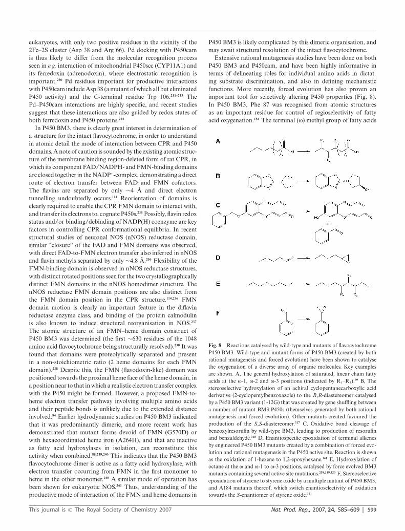
eukaryotes, with only two positive residues in the vicinity of the2Fe–2S cluster (Asp 38 and Arg 66). Pd docking with P450camis thus likely to differ from the molecular recognition processseen in e.g. interaction of mitochondrial P450scc (CYP11A1) andits ferredoxin (adrenodoxin), where electrostatic recognition isimportant.230 Pd residues important for productive interactionswith P450cam include Asp 38 (a mutant of which all but eliminatedP450 activity) and the C-terminal residue Trp 106.231–233 ThePd–P450cam interactions are highly specific, and recent studiessuggest that these interactions are also guided by redox states ofboth ferredoxin and P450 proteins.234
In P450 BM3, there is clearly great interest in determination ofa structure for the intact flavocytochrome, in order to understandin atomic detail the mode of interaction between CPR and P450domains. A note of caution is sounded by the existing atomic struc-ture of the membrane binding region-deleted form of rat CPR, inwhich its component FAD/NADPH- and FMN-binding domainsare closed together in the NADP+-complex, demonstrating a directroute of electron transfer between FAD and FMN cofactors.The flavins are separated by only ∼4 A and direct electrontunnelling undoubtedly occurs.114 Reorientation of domains isclearly required to enable the CPR FMN domain to interact with,and transfer its electrons to, cognate P450s.235 Possibly, flavin redoxstatus and/or binding/debinding of NADP(H) coenzyme are keyfactors in controlling CPR conformational equilibria. In recentstructural studies of neuronal NOS (nNOS) reductase domain,similar “closure” of the FAD and FMN domains was observed,with direct FAD-to-FMN electron transfer also inferred in nNOSand flavin methyls separated by only ∼4.8 A.236 Flexibility of theFMN-binding domain is observed in nNOS reductase structures,with distinct rotated positions seen for the two crystallographicallydistinct FMN domains in the nNOS homodimer structure. ThenNOS reductase FMN domain positions are also distinct fromthe FMN domain position in the CPR structure.114,236 FMNdomain motion is clearly an important feature in the diflavinreductase enzyme class, and binding of the protein calmodulinis also known to induce structural reorganisation in NOS.237
The atomic structure of an FMN–heme domain construct ofP450 BM3 was determined (the first ∼630 residues of the 1048amino acid flavocytochrome being structurally resolved).238 It wasfound that domains were proteolytically separated and presentin a non-stoichiometric ratio (2 heme domains for each FMNdomain).238 Despite this, the FMN (flavodoxin-like) domain waspositioned towards the proximal heme face of the heme domain, ina position near to that in which a realistic electron transfer complexwith the P450 might be formed. However, a proposed FMN-to-heme electron transfer pathway involving multiple amino acidsand their peptide bonds is unlikely due to the extended distanceinvolved.84 Earlier hydrodynamic studies on P450 BM3 indicatedthat it was predominantly dimeric, and more recent work hasdemonstrated that mutant forms devoid of FMN (G570D) orwith hexacoordinated heme iron (A264H), and that are inactiveas fatty acid hydroxylases in isolation, can reconstitute thisactivity when combined.88,239,240 This indicates that the P450 BM3flavocytochrome dimer is active as a fatty acid hydroxylase, withelectron transfer occurring from FMN in the first monomer toheme in the other monomer.240 A similar mode of operation hasbeen shown for eukaryotic NOS.241 Thus, understanding of theproductive mode of interaction of the FMN and heme domains in
P450 BM3 is likely complicated by this dimeric organisation, andmay await structural resolution of the intact flavocytochrome.
Extensive rational mutagenesis studies have been done on bothP450 BM3 and P450cam, and have been highly informative interms of delineating roles for individual amino acids in dictat-ing substrate discrimination, and also in defining mechanisticfunctions. More recently, forced evolution has also proven animportant tool for selectively altering P450 properties (Fig. 8).In P450 BM3, Phe 87 was recognised from atomic structuresas an important residue for control of regioselectivity of fattyacid oxygenation.181 The terminal (x) methyl group of fatty acids
Fig. 8 Reactions catalysed by wild-type and mutants of flavocytochromeP450 BM3. Wild-type and mutant forms of P450 BM3 (created by bothrational mutagenesis and forced evolution) have been shown to catalysethe oxygenation of a diverse array of organic molecules. Key examplesare shown. A, The general hydroxylation of saturated, linear chain fattyacids at the x-1, x-2 and x-3 positions (indicated by R1–R3).69 B, Thestereoselective hydroxylation of an achiral cyclopentanecarboxylic acidderivative (2-cyclopentylbenzoxazole) to the R,R-diastereomer catalysedby a P450 BM3 variant (1-12G) that was created by gene shuffling betweena number of mutant BM3 P450s (themselves generated by both rationalmutagenesis and forced evolution). Other mutants created favoured theproduction of the S,S-diastereomer.317 C, Oxidative bond cleavage ofbenzoxylresorufin by wild-type BM3, leading to production of resorufinand benzaldehyde.318 D, Enantiospecific epoxidation of terminal alkenesby engineered P450 BM3 mutants created by a combination of forced evo-lution and rational mutagenesis in the P450 active site. Reaction is shownas the oxidation of 1-hexene to 1,2-epoxyhexane.141 E, Hydroxylation ofoctane at the x and x-1 to x-3 positions, catalysed by force evolved BM3mutants containing several active site mutations.258,319,320 F, Stereoselectiveepoxidation of styrene to styrene oxide by a multiple mutant of P450 BM3,and A184 mutants thereof, which switch enantioselectivity of oxidationtowards the S-enantiomer of styrene oxide.321
This journal is © The Royal Society of Chemistry 2007 Nat. Prod. Rep., 2007, 24, 585–609 | 599

interacts with the Phe 87 side chain above the plane of the hemeiron, and is thus protected from oxidative attack. Saturated fattyacids are instead oxygenated at x-1 to x-3 positions.69 An F87Amutant was reported to catalyse fatty acid hydroxylation at the x-carbon, using NMR to identify the product of laurate oxidation.242
However, other studies on a F87V mutant suggested thatoxygenation position of arachidonic acid moved away from thex-end.15 In wild-type P450 BM3 there was conversion of arachi-donic acid to an almost enantiomerically pure 18(R)-hydroxy-eicosatetraenoic acid and 14(S),15(R)-epoxyeicosatetraenoic acidmixture (ca. 80:20 ratio of products). In the F87V mutant thelatter product accumulated as ∼99% of the overall productwith negligible 18(R)-hydroxyeicosatetraenoic acid detected.15,78
Other P450 BM3 Phe 87 mutants were also shown to improveoxidation of certain aromatic compounds, as discussed furtherbelow. Elsewhere in the P450 BM3 heme domain, mutation ofArg 47 at the active site mouth (a likely interaction site for thecarboxylate group of fatty acid substrates) resulted in weakenedaffinity for fatty acids (increased Km values for lauric acid andarachidonic acid with R47A and R47G mutants) and decreasedkcat values.75 A R47E mutant bound alkyltrimethylammoniumderivatives of C12–C16 saturated fatty acids much tighter thanwild-type BM3 (lower Km’s and much higher kcat values), againconsistent with a role for Arg 47 in interaction with the fatty acidcarboxylate group.243
In the P450 BM3 reductase domain (as with CPR and othereukaryotic diflavin reductases, including NOS and methioninesynthase reductase), a “catalytic triad” of residues is conservedaround the NADPH binding site and is implicated in catalysisof hydride transfer from the coenzyme to the enzyme FAD.244 InP450 BM3 these residues are Cys 999, Asp 1044 and Ser 830. Fromstructural and catalytic studies of wild-type and mutant forms ofrat CPR, these residues are considered essential for binding andorientation of the coenzyme nicotinamide ring, and in promotinghydride transfer by the stabilisation of a transient carbocation onthe nicotinamide following hydride transfer to the FAD.244 TheP450 BM3 C999A mutant catalysed ferricyanide and cytochromec reduction much slower than wild-type enzyme, largely due to aconsiderably decreased rate of hydride transfer from the coenzyme,consistent with the postulates of Kasper and co-workers.89,244
A stable charge transfer species was formed between reduced(hydroquinone) FAD cofactor and oxidised coenzyme (NADP+) inthe wild-type BM3 FAD domain, but not in the C999A variant—suggesting that the conserved triad cysteine is also importantfor stabilising the FADH2–NADP+ complex following electrontransfer from the coenzyme.89 Elsewhere in the BM3 reductase,the aromatic side chain of W1064 covers the isoalloxazine ring ofthe FAD, and must be displaced on binding NADPH to enableflavin reduction.235 The comparable residue in human CPR isTrp 676, and a W676A mutant demonstrated a 1000-fold switchin specificity from NADPH towards NADH for CPR-catalysedcytochrome c reduction.81 In the comparable W1064A/H mutantsof P450 BM3 reductase domain, the mutations effected an∼100 mV increase in reduction potential of the FAD, and aneven more substantial switch in coenzyme selectivity towardsNADH was observed (∼6000-fold for BM3 reductase-catalysedferricyanide reduction).245 Given the much lower cost of NADH,these substrate specificity conversions may be of significance forthe biotechnological exploitation of P450 BM3 (see below).
In studies of P450cam, there is a long history of rational muta-genesis aimed at exploration of structure–function relationships.Among the most interesting mutants is T252A, where substantialuncoupling of NADH oxidation from camphor hydroxylationwas observed.201,202 The majority of reducing equivalents wastedwere diverted into H2O2 production, consistent with collapseof the catalytic cycle at the compound 0 stage (Fig. 2). Asdiscussed above, it is thought that this amino acid is essentialin hydrogen bonding to a water molecule that acts as protondonor for scission of dioxygen bound to the heme iron.182
Structural detail demonstrated that the hydroxyl side chain ofactive site residue Tyr 96 forms a hydrogen bond with the ketonegroup of D-camphor, and regioselectivity was altered in a Y96Fmutant, with hydroxylation occurring at the 3 and 6 positionsin addition to the 5-exo hydroxylation observed with wild-type P450cam.246,247 These data were consistent with less specificsubstrate interactions and, possibly, greater mobility of camphorin the mutant active site. Y96A/F mutants were also shown tosubstantially enhance rate of production of styrene oxide fromstyrene, and to increase coupling of NADH oxidation to styreneoxide formation by up to ∼5-fold.248 Turnover of naphthaleneto 1-naphthol and 2-naphthol was also enhanced in various Tyr96 mutants.249 Mutations of other active site residues proximal tobound camphor (V295I and V247A) also resulted in perturbationsto product profiles with camphor analogues, again consistent withthe importance of several active site contacts in maintaining thebound substrate in a position appropriate for attack at the 5-exo position by the P450 compound I.250 Also apparent frommutagenic studies on redox partner (Pd) interactions was the factthat the Pd C-terminal tryptophan was important for its bindingto P450cam.233 Mutations at basic residues on the proximal faceof the P450 (Arg72, Lys344 and Arg112) diminished binding toPd, consistent with the importance of electrostatic interactionsguiding productive redox partner interactions.251,252 Combinationsof mutations in the active site and heme vicinity of P450camhave also been shown to effect considerable changes in substrateselectivity (Fig. 9). For instance, variants containing 8 or 9point mutations (including the L358P mutation that removes ahydrogen bond between the main chain amide of the leucineside chain and the heme iron cysteine thiolate) were shown tocatalyse oxidation of ethane to ethanol, albeit with substantialuncoupling of NADH oxidation from product formation.253 Arational and progressive process of decreasing active site volume(by mutations to introduce bulky amino acid side chains) wasalso successful in creating P450cam variants able to oxygenatethe gaseous alkanes butane and propane.254 Earlier studies useda similar approach to improve reaction efficiency and couplingwith ethylbenzene as the substrate.255,256 P450cam has also beenrationally evolved to oxygenate polychlorinated benzenes (1,3-dichlorobenzene and 1,3,5-trichlorobenzene),157 and mutations toengineer out the P450 BM3 carboxylate binding motif (Arg 47and Tyr 51) in combination with active site mutations (includingmutations to Leu 437) were also shown to enable efficient oxidationof both polycyclic aromatic hydrocarbons (phenanthrene andfluoranthrene) and short chain alkanoic acids.17,257
Recent years have seen the application of directed (forced)evolution in attempts to alter the properties of P450s, and thegreatest successes have undoubtedly come with studies of P450BM3. The enzyme has been evolved to produce large increases in
600 | Nat. Prod. Rep., 2007, 24, 585–609 This journal is © The Royal Society of Chemistry 2007

Fig. 9 Reactions catalysed by wild-type and mutants of P450cam.P450cam, the most intensively studied P450, has been shown to catalysea range of oxidative reactions, and mutated forms further enhance thesubstrate specificity range of the enzyme. Key examples are shown. A,The natural reaction of wild-type P450cam: the 5-exo hydroxylation ofD-camphor.3 B, Conversion of diphenylmethane to 4-hydroxydiphenyl-methane by variants isolated from active site mutant P450 libraries.322 C,Oxygenation of (+)-valencene to (+)-trans-nootkatol (R: -OH) and the finefragrance (+)-nootkatone (R: =O) by variants of P450cam containing theF87A/L and Y96F mutations (with (+)-nootkatone production enhancedby the L244A mutation).268 D, Conversion of 1,3,5-trichlorobenzeneto 2,4,6-trichlorophenol; catalysed poorly by wild-type P450cam anddramatically improved in a F87W, Y96F, V247L triple mutant.157 E,Conversion of ethane to ethanol catalysed by a multiple mutant ofP450cam (incorporating the L358P mutation adjacent to the Cys 357 hemeligand).253 F, Oxidation of ethylhexanol to ethylhexanoic acid by wild-typeand active site mutants of P450cam.323
the rate of oxidation of alkanes. In a BM3 variant named 139-3with 11 individual mutations (only two of which are in the activesite), turnover of pentane, hexane, cyclohexane and octane wasincreased to levels comparable with those for wild-type BM3 andsome of its slower fatty acid substrates (i.e. from <200 min−1 up to3–4000 min−1).258 Butane and propane were also oxygenated, andthe 1- and 2-hydroxylation of propane was further improved insubsequent rounds of evolution of the 139-3 mutant to produce the9-10A mutant with thirteen individual mutations.18 It is difficultto rationalise clearly the structural reasons underlying enhancedaffinity and activity for alkanes in the 139-3 variant, althoughdisruption of a salt bridge (that might facilitate conformationalchange to enable alkane binding) and the fact that a number
of mutations are located in the flexible F/G helix region arepossible explanations.258 Later studies used active site saturationmutagenesis to generate other derivatives of mutant 9-10A thatfurther enhanced propane oxidation and facilitated conversion ofethane to ethanol.259 Oxidation of methane to methanol, however,has yet to be achieved by any P450 mutant. In other directedevolution studies, BM3 variants have been created that enhancethermostability and peroxide shunt activity of the enzyme.260,261
The peroxygenase variant was further evolved to enable produc-tion of human-like metabolites of the b-adrenergic blocker drugpropranolol using H2O2.262 Enzyme tolerance to solvents (dimethylsulfoxide and tetrahydrofuran) for its lipid substrates was alsoimproved by directed evolution.263 The era of forced evolution willobviously usher in production of mutant P450 (and other) enzymeswith novel properties and biotechnological applications. However,there are new challenges involved for structural biologists andenzymologists with respect to rationalising (for P450s containingmultiple mutations) how many of the mutations identified actuallycontribute to producing the desired biochemical effects, and alsoin sorting the “wheat from the chaff” in terms of establishing whichindividual mutations compound the desirable effects of others, andwhich make negligible overall difference.
9 Technological and biotechnological advances
With their critical roles in eukaryotic and bacterial physiology,and their capacity to introduce oxygen atoms regio- and stereo-selectively into organic substrates, it is no surprise that P450s havereceived immense attention from both academic and industrialconsortia aiming to e.g. exploit their reaction chemistry forproduction of high value biomolecules, enhance their catalyticperformance or alter substrate selectivity, enzyme stability or othercharacteristics by rational mutagenesis or forced evolution. Therehas been a long history of rational engineering of P450 structure–function, with a major breakthrough being conversion of substrateselectivity of murine P450coh (CYP2A5) from coumarin totestosterone hydroxylation by a single F209L mutation.264 ThePhe 209 residue is located in the F/G helix region (possibly in theinterconnecting loop between helices), an important region forsubstrate recognition and active site entry.182,265 A further excellentexample of conversion of P450 specificity by point mutation wasthe switch in regioselectivity of steroid (androstanedione andtestosterone) hydroxylation from the 16b to the 15a position bytwo point mutations (I114A/G478S) in CYP2B1.266
Rational approaches to P450 engineering have been aidedby availability of atomic structures for several P450s (Table 1),as well as by the huge (and increasing) database of gene andtranslated amino acid sequences for P450s from all domains oflife. This has enabled a systematic approach to investigating rolesof phylogenetically conserved amino acids, and to establishinghow single mutations can accomplish substantial changes toP450 properties. Examples include the demonstration that theinvariant P450 cysteine is the thiolate ligand to heme iron(and is essential for oxygenase catalysis), and that the stronglyconserved phenylalanine residue in the heme-binding motif ispivotal in modulating thermodynamic properties and reactivityof the heme.87,267 In the P450cam system, structural data enabledrational active site mutagenesis that introduced catalytic activity
This journal is © The Royal Society of Chemistry 2007 Nat. Prod. Rep., 2007, 24, 585–609 | 601

towards the sesquiterpene (+)-valencene, and the ability to producethe fine fragrance (+)-nootkatone.268
In human CYP2A6, a L160H mutation is a natural poly-morphism that diminishes catalytic activity of nicotine oxi-dation, and leads to a switch from nicotine 7-hydroxylationto 3-hydroxylation.151,269 It is plausible that defects in nicotinemetabolism lead to reduction in an individual’s smoking levelsand to a lowered lung cancer risk, particularly since CYP2A6 ac-tivates tobacco carcinogens (including nornitrosonicotine [NNN]and 4-(methylnitrosamino)-1-(3-pyridyl)-1-butanone [NNK]).151
However, links between CYP2A6 genotype and smoking-relatedcancer remain speculative. A genetic test involving CYP2A6 hasbeen marketed (http://www.nicotest.com), but is controversial.The basis of the NicoTestTM genetic blood test is the establish-ment of the usefulness and extent of requirement for nicotinereplacement for individuals wishing to give up smoking. Thetest checks for polymorphisms in the dopamine D2 receptor(DRD2) gene, which are claimed to affect relative usefulness ofnicotine replacement therapy (patches) and the drug ZybanTM
(bupropion, which reduces craving for nicotine) for the subject.Simultaneous assessment of the CYP2A6 gene is designed toinform on ability of the subject’s P450 to metabolise nicotine, andhence on the level of nicotine replacement to be used. However,the organisation GeneWatch UK (http://www.genewatch.org)has expressed concerns relating to validity of the approach sincethey consider it unlikely to provide useful information for malesubjects. Notwithstanding the controversy over the relevanceof genetic testing for CYP2A6 polymorphisms with respectto smoking cessation, there have been other notable successeswith commercialisation of expressed P450s. Systems availablefor analysis of P450 catalytic properties, inhibition profiles etc.include P450 and redox partner enzymes overexpressed in insectmicrosomes using the Baculovirus system (BD Biosciences) andhuman P450s co-expressed with CPR in E. coli membranes (CypexLtd). Assays have also been commercialised for measurement ofactivities of several important individual human P450 isoforms(e.g. from Promega), and these are of particular relevance fordetermining specific inhibition of key P450s involved in drugmetabolism.
With respect to developing P450 enzymes with desirable featuresand enhanced catalytic properties, there have been great advancesin recent years in scientists’ ability to produce soluble, functionaldomains of eukaryotic P450s.270 As discussed, these breakthroughshave also facilitated crystallisation of several important mam-malian drug-metabolising P450s, leading to determination of theirthree dimensional structures and more detailed understanding oftheir mode of interaction with substrates and inhibitors.182 Thecoming years will doubtless see the determination of several moreP450 crystal structures, and further development of our under-standing of interactions of P450s with important biomolecules.There have also been substantial efforts towards creation ofP450–redox partner fusion enzymes as a means of enhancingelectron transfer to P450s (and hence P450 activity) and generatingcatalytically self-sufficient P450s similar to P450 BM3. Moststudies have involved genetic fusion of eukaryotic P450s to CPRenzymes, often resulting in moderate increases in turnover numberwith respect to the rates obtained by “natural” reconstitution ofindividual P450 and CPR enzymes.271 However, no such fusionyet reported has an oxygenase activity at a level even 10% of that
for P450 BM3, although this is at least partially explained bythe much faster electron transport process within the BM3 CPRcompared to those for eukaryotic CPR enzymes.79,83 Examples ofcatalytically functional and stable mammalian P450/CPR fusionenzymes include CYP3A4 and CYP17A chimeras.272,273 Anotherinteresting P450 fusion enzyme was a “triple” fusion of P450camto both Pdr and Pd (i.e. all components of this class I P450 system).While (as with P450 BM3 mimics) the camphor hydroxylaseactivity of this fusion was much less than that of the mixture of theindividual proteins (∼ 30 min−1), the single component system hasclear advantages in simplicity of expression and purification.274
Although the hiatus of activity in research on P450/redox partnerfusions was in the 1990’s, there are obvious applications for suchsystems in e.g. diagnostics and fine oxychemical production.
Another sphere of interest in P450s, with ramifications forbiotechnological applications, is the mode by which these enzymescan be supplied with electrons for catalysis. Rational engineeringhas provided routes to altering CPR and P450 BM3 coenzymeselectivity from NADPH towards NADH.246,247 This has thepotential to decrease operation costs substantially (possibly byup to 90%), although inclusion of a NADPH regeneratingsystem (e.g. glucose-6-phosphate dehydrogenase and glucose-6-phosphate) can also enhance the cost efficiency of NADPH-dependent P450 catalytic processes. The recent studies of Ortiz deMontellano and co-workers in defining a P450 system reliant on 2-oxo-acids (pyruvate rather than NAD(P)H) as electron donor alsopoints the way to development of novel, cheaper redox apparatusto drive P450 function.126 Alternatives to costly coenzymes includethe use of peroxides and the peroxide shunt pathway (Fig. 2).While some advances have been made in this area by generatingP450s with greater stability to oxidative destruction by H2O2,260
substantial work remains to be done in order to establishhow oxidative destruction of the heme macrocycle and proteinmatrix can be minimised, and productive interactions with H2O2
maximised. In this case, the “natural” peroxygenase enzymes,typified by the B. subtilis CYP152A1 fatty acid b-hydroxylase, maybe ideal model systems.162 Electrocatalysis is another attractivemethod for driving P450 catalysis, since it can bypass altogetherrequirements for redox partners and deliver electrons directly toa P450 from an electrode. There have been successes in this area,although there are issues with respect to avoiding loss of P450activity (aggregation, unfolding, heme dissociation etc.) at theelectrode surface. Estabrook and co-workers reported fatty acid x-hydroxylation by rat CYP4A1 in presence of rat CPR, or as a P450fusion to rat CPR, using a silver electrode and cobalt sepulchrateas a mediator.275 12-Hydroxylauric acid was produced from lauricacid at rates comparable to those achieved by driving the reactionwith NADPH. Activity was maintained for a much longer periodin the presence of catalase, pointing to protein-damaging effectsof oxygen radicals produced at the electrode surface, or by non-productive oxidation of the mediator. The requirement for CPRalso suggests that electrons are still delivered to the P450 by thisroute, following CPR reduction by the mediator. This raises theimportant issue of requirement for temporally distinct deliveryof the two electrons required for productive oxygenase catalysis(either side of the binding of dioxygen, see Fig. 2) and suggeststhat this could only be achieved by the CPR in the electrocatalyticsystem. A similar electrocatalytic approach was used by the samegroup to drive progesterone hydroxylation at ∼25 min−1 using a
602 | Nat. Prod. Rep., 2007, 24, 585–609 This journal is © The Royal Society of Chemistry 2007

bovine CYP17A-CPR fusion protein.276 Thus, electrocatalysis is aviable method for driving P450 oxygenations, but efficient electrontransport may rely on mediators and natural redox partners todeliver electrons to a P450 whose heme may be quite well insulatedat the centre of its protein matrix.
Finally, an important recent advance relevant to analysis ofP450s (and several other membrane proteins) has been the devel-opment of NanodiscTM technology by Sligar.277,278 Nanodiscs areself-assembling, monodisperse discoidal molecules, comprised of alipid (dipalmitoylphosphatidylcholine, DPPC) bilayer surroundedby a helical peptide “belt” of variable length, termed the membranescaffold protein (MSP). These self-assembling nanoparticles weredesigned as a tool for solubilisation and isolation of membrane-bound enzymes, transporters and channels, and to provide a novelroute to their biochemical and structural characterisation.278 Thetechnology has already been used to incorporate arabidopsis P450(CYP73A5) and CPR into Nanodiscs, with the assemblies isolatedusing a hexahistidine tag attached to the MSP and by nickelaffinity chromatography.279 The Nanodisc-encapsulated P450 wasshown to have native-like substrate (trans-cinammic acid) bindingproperties, and substrate hydroxylation was reconstituted whenCYP73A5 and CPR enzymes were reconstituted in the sameNanodisc.279 Other studies have focused on human CYP3A4and showed that encapsulated P450 exhibits homotropic coop-erativity in binding its substrate testosterone (using absorptionspectroscopy to monitor association of substrate with P450) andthat substrate hydroxylation can be driven in CYP3A4 Nanodiscsusing H2O2.280 More recent studies exploited Nanodisc technologyto explore conformational equilibria and reduction kinetics in theCYP3A4 system.281 Nanodiscs clearly have enormous potentialfor studying isolated, membranous P450s (with Nanodiscs conve-niently suspended in aqueous media), and for analysis of molecularinteractions between P450s and ligands/redox partners.
10 Conclusions and future prospects
The last 20 years has seen a vast increase in our understandingof structure and mechanism of P450s, and of their biologicaland catalytic diversity. It will undoubtedly be the case that moreatomic structures of eukaryotic P450s (particularly hepatic drug-metabolising isoforms) will be solved in the near future, given theenormous pharmacological importance of these enzymes. Recentstructural data for the major human P450 isoform CYP3A4indicates that detailed atomic structures may be essential forany predictive drug metabolism to be undertaken, given theconformational flexibility of certain P450 isoforms.212 Anotherkey question relating to P450 structure–function is the mode ofinteraction of P450s with their various redox partners. This willrequire determination of an atomic structure of a stoichiometriccomplex of a P450 with the partner protein, or of one of the naturalP450–redox partner fusion proteins (e.g. a CYP102, CYP505 orCYP116B enzyme). However, a full appreciation of modes ofbinding of P450s with CPR and other redox partners is also likelyto require structural elucidation of crystal complexes solved indifferent redox states and substrate-bound forms. This is a majorchallenge and will require several years’ effort.
Despite huge progress in recent years, numerous key questionsremain relating to the definitive (spectroscopic or structural)identification of the major oxidising species (i.e. compound I)
in the P450 cycle, and also regarding the relevance of otherintermediates (primarily compound 0) in P450-mediated reactions.A substantial step towards identifying the transient compound Iwas made recently in studies of Sulfolobus solfataricus CYP119A1,in which peroxynitrite was used to oxidise the P450 heme toa moderately stable species with spectral properties consistentwith that of a Fe(IV)–oxo neutral porphyrin (compound II), asreported for the heme thiolate protein choloroperoxidase.282 Laserphotoexcitation of the CYP119A1 species at 355 nm resultedin partial conversion to a new, transient spectral species withabsorption characteristics similar to known compound I species inmodels and enzyme systems.36 Photooxidation of compound II tocompound I has been reported in other proteins (equine myoglobinand horseradish peroxidase) and thus the method used here forCYP119A1 may be generally applicable.283 However, the relativelylong half life of the compound I-like species in CYP119A1, andlack of influence of P450 substrate molecules on its decay, raisesome questions as to the nature of the intermediate, and furtherstudies are obviously needed in this area.36 In an alternativeapproach to isolating reactive P450 cycle intermediates, Hoffmanand co-workers used c irradiation of a substrate-bound P450camferrous–oxy complex (at 77 K) to introduce the 2nd electron.EPR and 1H ENDOR studies provided compelling evidence forformation of the ferric peroxy intermediate, which then abstracteda proton to form substantial amounts (up to 40% yield) of theferric hydroperoxy form.37 Elevating temperature (“annealing”)to ∼200 K led to hydroxycamphor product formation in the wild-type enzyme, but no direct evidence for compound I formationwas obtained. Cutting-edge studies of this type have brought uscloser to identification of compound I and appear to rule out theinvolvement of compound 0 in most P450 oxidations. However,there are experimental data suggesting that compound 0 is aviable oxidant for e.g. epoxidation of carbon–carbon double bondsand for sulfur oxidations.284–287 These contrast with computationalstudies indicating that different spin states of compound I act asdifferent oxidants, and that compound 0 may have no significantrole in oxygenation chemistry.288 The definitive identification of theultimate oxidant species in P450 catalysis (nominally compound I)and the possibility of secondary oxidant(s) clearly remains a hotlycontested and controversial area and will obviously be the subjectof numerous studies in the next few years.
Other areas of interest revolve around biotechnological ex-ploitation of P450s, and particularly their usefulness in productionof chiral oxyfunctionalised compounds as fine chemicals andintermediates in organic synthetic processes. For instance, P450sare already involved naturally in the synthesis of high valuelipids (e.g. steroids, eicosanoids) and of numerous antibiotics(see Fig. 10). There are certainly opportunities to engineerP450 variants with altered activities that could, for example,produce novel polyketide antibiotics as a result of alterationin the position of substrate oxygenation. Possibilities in thisarea are highlighted by the Streptomyces venezuelae PikC P450(CYP107L1), which catalyses hydroxylation of two different typesof macrolactone at two different positions (or both positions),creating a range of different macrolide antibiotics (methymycin,neomethymycin, novamethymycin, pikromycin, neopikromycinand novapikromycin).289–291 Rational and forced evolution ap-proaches have other roles to play in producing P450s that are morethermostable, pressure stable, protease and solvent resistant. This
This journal is © The Royal Society of Chemistry 2007 Nat. Prod. Rep., 2007, 24, 585–609 | 603

Fig. 10 Involvement of P450 enzymes in antibiotic synthesis. P450 enzymes are widely exploited for oxidative modifications of polyketide and otherantibiotics. Their activity is usually essential for the biological activities of these molecules. Selected examples are shown, with the position(s) of oxidativemodification performed by P450 enzymes indicated by arrows. A, nystatin;324 B, nocardicin A;325 C, vancomycin;326 D, erythromycin;204 E, rifamycinB;327 F, amphotericin.328 For rifamycin, an orf0 (P450-encoding) gene deletion led to the lack of methyl hydroxylation of proansamycin X (a rifamycin Bprecursor) and failure of the host organism (Amycolatopsis mediterranei) to transform this precursor to rifamycin B via the intermediate rifamycin W.327
In the case of amphotericin, gene inactivation studies indicated that two distinct P450s from Streptomyces nodosus (amphL and amphN) are responsiblefor C8 hydroxylation and for C16 methyl oxidation to form the carboxylate, as indicated in the relevant panel.329,330
will be complemented by isolation and characterisation of natural(likely archaeal) P450 enzymes from unusual environments thatalready possess characteristics that make them robust and suitablefor industrial applications. In the race for exploitation of P450s forbiotechnological purposes, it should not be forgotten that there is avast amount we do not understand regarding normal physiologicalroles of P450s in primary and secondary metabolism in numerousorganisms. For instance, very little is known about the roles ofmost of the (>270) P450s in Arabidopsis thaliana, and even severalof the 57 human P450s have yet to be characterised in detail. InMycobacterium tuberculosis, there are 20 P450s, and azole and
triazole compounds (known P450 inhibitors) have been shown tobe effective antimycobacterial drugs.123 Evidently a vast amountof research is required on these and many other P450 systems touncover further novel activities and the place of these enzymes inthe web of metabolism in their respective host organisms.
The P450 field is vibrant and its research community is thrivingon novel structural and bioinformatics data. This informationwill provide the fuel for research in diverse areas on theseoxygenases and their redox partners, in order to further developour understanding of their biological functions and their potentialas environmentally friendly biocatalysts in the chemist’s tool box.
604 | Nat. Prod. Rep., 2007, 24, 585–609 This journal is © The Royal Society of Chemistry 2007

11 References
1 E. N. Jacobsen, M. Istvan, W. S. Mungall, G. Schroeder and K. B.Sharpless, J. Am. Chem. Soc., 1988, 110, 1968.
2 S. E. Schaus, B. S. Brandes, J. F. Larrow, M. Tokunaga, K. B. Hansen,A. E. Gould, M. E. Furrow and E. N. Jacobsen, J. Am. Chem. Soc.,2002, 124, 1307.
3 I. C. Gunsalus, T. C. Pederson and S. G. Sligar, Annu. Rev. Biochem.,1975, 44, 377.
4 S. Eswaramoorthy, J. B. Bonanno, S. K. Burley and S. Swaminathan,Proc. Natl. Acad. Sci. U. S. A., 2006, 103, 9832.
5 J. C. Price, E. W. Barr, L. M. Hoffart, C. Krebs and J. M. Bollinger,Jr., Biochemistry, 2005, 44, 8138.
6 V. B. Urlacher and S. Eiben, Trends Biotechnol., 2006, 24, 324.7 M. R. Waterman and G. I. Lepesheva, Biochem. Biophys. Res.
Commun., 2005, 338, 418.8 I. G. Denisov, T. M. Makris, S. G. Sligar and I. Schlichting, Chem.
Rev., 2005, 105, 2253.9 F. P. Guengerich, Drug Metab. Rev., 2004, 36, 159.
10 S. Nagano, H. Li, H. Shimizu, C. Nishida, H. Ogura, P. R.Ortiz de Montellano and T. L. Poulos, J. Biol. Chem., 2003, 278,44886.
11 L. M. Podust, T. L. Poulos and M. R. Waterman, Proc. Natl. Acad.Sci. U. S. A., 2001, 98, 3068.
12 D. Leys, C. G. Mowat, K. J. McLean, A. Richmond, S. K. Chapman,M. D. Walkinshaw and A. W. Munro, J. Biol. Chem., 2003, 278,5141.
13 J. K. Yano, M. H. Hsu, K. J. Griffin, C. D. Stout and E. F. Johnson,Nat. Struct. Mol. Biol., 2005, 12, 822.
14 M. R. Wester, J. K. Yano, G. A. Schoch, C. Yang, K. J. Griffin andE. F. Johnson, J. Biol. Chem., 2004, 279, 35630.
15 S. Graham-Lorence, G. Truan, J. A. Peterson, J. R. Falck, S. Wei, C.Helvig and J. H. Capdevila, J. Biol. Chem., 1997, 272, 1127.
16 S. G. Bell, J. A. Stevenson, H. D. Boyd, S. Campbell, A. D. Riddle,E. L. Orton and L. L. Wong, Chem. Commun., 2002, 490.
17 T. W. Ost, C. S. Miles, J. Murdoch, Y. Cheung, G. A. Reid, S. K.Chapman and A. W. Munro, FEBS Lett., 2000, 486, 173.
18 M. W. Peters, P. Meinhold, A. Glieder and F. H. Arnold, J. Am. Chem.Soc., 2003, 125, 13442.
19 M. Klingenberg, Arch. Biochem. Biophys., 1958, 75, 376.20 T. Omura and R. Sato, J. Biol. Chem., 1962, 237, 1375.21 T. M. Makris, R. Davydov, I. G. Denisov, B. M. Hoffman and S. G.
Sligar, Drug Metab. Rev., 2002, 34, 691.22 T. L. Poulos, B. C. Finzel and A. J. Howard, J. Mol. Biol., 1987, 195,
687.23 R. W. Estabrook, R. W. Cooper and O. Rosenthal, Biochem. Z., 1963,
338, 741.24 R. Kahl, D. E. Friederici, G. F. Kahl, W. Ritter and R. Krebs, Drug
Metab. Dispos., 1980, 8, 191.25 S. G. Sligar, Essays Biochem., 1999, 34, 71.26 M. Katagiri, B. N. Ganguli and I. C. Gunsalus, J. Biol. Chem., 1968,
243, 3543.27 A. W. Munro, H. M. Girvan and K. J. McLean, Biochim. Biophys.
Acta, 2006, 1770, 345.28 A. W. Munro and J. G. Lindsay, Mol. Microbiol., 1996, 20, 1115.29 R. Tsai, C. A. Yu, I. C. Gunsalus, J. Peisach, W. Blumberg, W. H.
Orme-Johnson and H. Beinert, Proc. Natl. Acad. Sci. U. S. A., 1970,66, 1157.
30 C. R. Jefcoate, W. H. Orme-Johnson and H. Beinert, J. Biol. Chem.,1976, 251, 3706.
31 S. G. Sligar, Biochemistry, 1976, 15, 5399.32 S. N. Daff, S. K. Chapman, K. L. Turner, R. A. Holt, S. Govindaraj,
T. L. Poulos and A. W. Munro, Biochemistry, 1997, 36, 13816.33 M. Murray and A. J. Ryan, Chem.-Biol. Interact., 1983, 43, 341.34 C. Hashimoto-Yutsudo, Y. Imai and R. Sato, J. Biochem., 1980, 88,
505.35 S. G. Sligar, T. M. Makris and I. G. Denisov, Biochem. Biophys. Res.
Commun., 2005, 338, 346.36 M. Newcomb, R. Zhang, R. E. Chandrasena, J. A. Halgrimson, J. H.
Horner, T. M. Makris and S. G. Sligar, J. Am. Chem. Soc., 2006, 128,4580.
37 R. Davydov, T. M. Makris, V. Kofman, D. E. Werst, S. G. Sligar andB. M. Hoffman, J. Am. Chem. Soc., 2001, 123, 1403.
38 M. Newcomb, P. F. Hollenberg and M. J. Coon, Arch. Biochem.Biophys., 2003, 409, 72.
39 T. Hishiki, H. Shimada, S. Nagano, T. Egawa, Y. Kanamori, R.Makino, S. Y. Park, S. Adachi, Y. Shiro and Y. Ishimura, J. Biochem.,2000, 128, 965.
40 H. Xiang, R. A. Tschirret-Guth and P. R. Ortiz de Montellano, J. Biol.Chem., 2000, 275, 35999.
41 A. Y. H Lu, K. W. Junk and M. J. Coon, J. Biol. Chem., 1969, 244,3714.
42 H. E. Conrad, K. Lieb and I. C. Gunsalus, J. Biol. Chem., 1965, 240,4029.
43 M. Katagiri, B. N. Ganguli and I. C. Gunsalus, J. Biol. Chem., 1968,243, 3543.
44 T. Iyanagi and H. S. Mason, Biochemistry, 1973, 12, 2297.45 J. L. Vermillion and M. J. Coon, Biochem. Biophys. Res. Commun.,
1974, 60, 1315.46 J. L. Vermillion and M. J. Coon, J. Biol. Chem., 1978, 253, 2594.47 J. L. Vermillion and M. J. Coon, J. Biol. Chem., 1978, 253, 8812.48 B. S. S Masters, H. Kamin, Q. H. Gibson and C. H. Williams, J. Biol.
Chem., 1965, 240, 921.49 J. L. Vermillion, D. P. Ballou, V. Massey and M. J. Coon, J. Biol.
Chem., 1981, 256, 266.50 C. A. Tyson, J. D. Lipscomb and M. J. Coon, J. Biol. Chem., 1972,
247, 5777.51 I. C. Gunsalus and G. C. Wagner, Methods Enzymol., 1978, 52, 166.52 P. W. Roome, Jr., J. C. Philley and J. A. Peterson, J. Biol. Chem., 1983,
258, 2593.53 J. G. Rheinwald, A. M. Chakrabarty and I. C. Gunsalus, Proc. Natl.
Acad. Sci. U. S. A., 1973, 70, 885.54 M. C. Hu, H. J. Hsu, I. C. Guo and B. C. Chung, Mol. Cell Endocrinol.,
2004, 215, 95.55 T. Hakki and R. Bernhardt, Pharmacol. Ther., 2006, 111, 27.56 M. Bureik, M. Lisurek and R. Bernhardt, Biol. Chem., 2002, 383,
1537.57 J. J. Muller, A. Lapko, G. Bourenkov, K. Ruckpaul and U. Heinemann,
J. Biol. Chem., 2001, 276, 2786.58 G. A. Ziegler and G. E. Schulz, Biochemistry, 2000, 39, 10986.59 A. V. Grinberg, F. Hannemann, B. Schiffler, J. Muller, U. Heinemann
and R. Bernhardt, Proteins, 2000, 40, 590.60 I. A. Pikuleva, K. Tesh, M. R. Waterman and Y. Kim, Arch. Biochem.
Biophys., 2000, 373, 44.61 T. Kimura and K. Suzuki, J. Biol. Chem., 1967, 242, 485.62 K. Suhara, Y. Ikeda, S. Takemori and M. Katagiri, FEBS Lett., 1972,
28, 45.63 J.-W. Chu and T. Kimura, J. Biol. Chem., 1973, 248, 2089.64 D. Lambeth and H. Kamin, J. Biol. Chem., 1976, 251, 4299.65 A. J. Thomson, R. Cammack, D. O. Hall, K. K. Rao, B. Briat, J. C.
Rivoal and J. Badoz, Biochim. Biophys. Acta, 1977, 493, 132.66 D. Lambeth and H. Kamin, J. Biol. Chem., 1979, 254, 2766.67 K. Takeuchi, M. Tsubaki, J. Fuatgawa and H. Hori, J. Biochem., 2001,
130, 789.68 R. T. Ruettinger and A. J. Fulco, J. Biol. Chem., 1981, 256, 5728.69 L. O. Narhi and A. J. Fulco, J. Biol. Chem., 1986, 261, 7160.70 R. T. Okita and J. R. Okita, Curr. Drug Metab., 2001, 2, 265.71 S. Govindaraj and T. L. Poulos, Protein Sci., 1996, 5, 1389.72 J. S. Miles, A. W. Munro, B. N. Rospendowski, W. E. Smith, J. E.
McKnight and A. J. Thomson, Biochem. J., 1992, 288, 503.73 R. T. Ruettinger, L. P. Wen and A. J. Fulco, J. Biol. Chem., 1989, 264,
10987.74 H.-Y. Li, K. Darwish and T. L. Poulos, J. Biol. Chem., 1991, 366,
11909.75 M. A. Noble, C. S. Miles, S. K. Chapman, D. A. Lysek, A. C. MacKay,
G. A. Reid, R. P. Hanzlik and A. W. Munro, Biochem. J., 1999, 339,371.
76 L. O. Narhi, L. P. Wen and A. J. Fulco, Mol. Cell. Biochem., 1988, 79,63.
77 R. S. Matson, R. A. Stein and A. J. Fulco, Biochem. Biophys. Res.Commun., 1980, 97, 955.
78 J. H. Capdevila, S. Wei, C. Helvig, J. R. Falck, Y. Belosludtsev, G.Truan, S. E. Graham-Lorence and J. A. Peterson, J. Biol. Chem.,1996, 271, 22663.
79 A. W. Munro, S. Daff, J. R. Coggins, J. G. Lindsay and S. K. Chapman,Eur. J. Biochem., 1996, 239, 403.
80 A. Gutierrez, M. J. Paine, C. R. Wolf, N. S. Scrutton and G. C. KRoberts, Biochemistry, 2002, 41, 4626.
81 O. Dohr, M. J. Paine, T. Friedberg, G. C. Roberts and C. R. Wolf,Proc. Natl. Acad. Sci. U. S. A., 2001, 98, 81.
This journal is © The Royal Society of Chemistry 2007 Nat. Prod. Rep., 2007, 24, 585–609 | 605

82 D. S. Sem and C. B. Kasper, Biochemistry, 1994, 33, 12012.83 A. Gutierrez, A. W. Munro, A. Grunau, C. R. Wolf, N. S. Scrutton
and G. C. K Roberts, Eur. J. Biochem., 2003, 270, 2612.84 A. W. Munro, D. Leys, K. J. McLean, K. R. Marshall, T. W. Ost, S.
Daff, C. S. Miles, S. K. Chapman, D. A. Lysek, C. C. Moser, C. C.Page and P. L. Dutton, Trends Biochem. Sci., 2002, 27, 250.
85 U. M. Kent, L. Pascual, R. A. Roof, D. P. Ballou and P. F. Hollenberg,Arch. Biochem. Biophys., 2004, 423, 277.
86 T. Cvrk and H. W. Strobel, Arch. Biochem. Biophys., 2001, 385, 290.87 T. W. Ost, C. S. Miles, A. W. Munro, J. Murdoch, G. A. Reid and S. K.
Chapman, Biochemistry, 2001, 40, 13421.88 M. L. Klein and A. J. Fulco, J. Biol. Chem., 1993, 268, 7553.89 O. Roitel, N. S. Scrutton and A. W. Munro, Biochemistry, 2003, 42,
10809.90 M. Mergeay, S. Monchy, T. Vallaeys, V. Auguier, A. Benotmane, P.
Bertin, S. Taghavi, J. Dunn, D. van der Lelie and R. Wattiez, FEMSMicrobiol. Rev., 2003, 27, 385.
91 F. Kunst, N. Ogasawara, I. Moszer, A. M. Albertini and G. Alloni,et al., Nature, 1997, 390, 237.
92 M. C. Gustafsson, O. Roitel, K. R. Marshall, M. A. Noble, S. K.Chapman, A. Pessegueiro, A. J. Fulco, M. R. Cheesman, C. vonWachenfeldt and A. W. Munro, Biochemistry, 2004, 43, 5474.
93 N. Nakayama and H. Shoun, J. Biochem., 1985, 97, 755.94 T. Kitazume, N. Takaya, N. Nakayama and H. Shoun, J. Biol. Chem.,
2000, 275, 39734.95 N. Nakayame, A. Takemae and H. Shoun, J. Biochem., 1996, 119, 435.96 L. O. Narhi and A. J. Fulco, J. Biol. Chem., 1987, 262, 6683.97 J. A. Seo, R. H. Proctor and R. D. Plattner, Fungal Genet. Biol., 2001,
34, 155.98 G. T. Gassner, M. L. Ludwig, D. L. Gatti, C. C. Correll and D. P.
Ballou, FASEB J., 1995, 1995, 1411.99 R. De Mot and A. H. A Parret, Trends Microbiol., 2002, 10, 502.
100 G. A. Roberts, A. Celik, D. J. Hunter, T. W. Ost, J. H. White, S. K.Chapman, N. J. Turner and S. L. Flitsch, J. Biol. Chem., 2003, 278,48914.
101 D. J. Hunter, G. A. Roberts, T. W. Ost, J. H. White, S. Muller, N. J.Turner, S. L. Flitsch and S. K. Chapman, FEBS Lett., 2005, 579, 2215.
102 A. J. Warman, et al., unpublished data.103 L. Liu, R. D. Schmid and V. B. Urlacher, Appl. Microbiol. Biotechnol.,
2006, 72, 876.104 I. Nagy, F. Compernolle, K. Ghys, J. Vanderleyden and R. De Mot,
Appl. Environ. Microbiol., 1995, 61, 2056.105 M. J. Cryle, N. J. Matovic and J. J. De Voss, Org. Lett., 2003, 5, 3341.106 M. J. Cryle and J. J. De Voss, Chem. Commun., 2004, 86.107 A. J. Green, S. L. Rivers, M. Cheesman, G. A. Reid, L. G. Quaroni,
I. D. Macdonald, S. K. Chapman and A. W. Munro, J. Biol. Inorg.Chem., 2001, 6, 523.
108 A. J. Green, A. W. Munro, M. R. Cheesman, G. A. Reid, C. vonWachenfeldt and S. K. Chapman, J. Inorg. Biochem., 2003, 93, 92.
109 R. J. Lawson, D. Leys, M. J. Sutcliffe, C. A. Kemp, M. R. Cheesman,S. J. Smith, J. Clarkson, W. E. Smith, I. Haq, J. B. Perkins and A. W.Munro, Biochemistry, 2004, 43, 12410.
110 R. J. Lawson, C. von Wachenfeldt, I. Haq, J. Perkins and A. W. Munro,Biochemistry, 2004, 43, 12390.
111 Y. T. Meharenna, H. Y. Li, D. B. Hawkes, A. G. Pearson, J. De Vossand T. L. Poulos, Biochemistry, 2004, 43, 9487.
112 S. G. Mayhew, and G. Tollin, General properties of flavodoxins in:ed. F. Muller, Chemistry and Biochemistry of Flavodoxins, CRC Press,Boca Raton, FL, 1992, vol. 3, pp. 389–426.
113 H. P. Blaschkowski, G. Neuer, M. Ludwig-Festl and J. Knappe,Eur. J. Biochem., 1982, 123, 563.
114 M. Wang, D. L. Roberts, R. Paschke, T. M. Shea, B. S. S Masters andJ.-J. P. Kim, Proc. Natl. Acad. Sci. U. S. A., 1997, 94, 9411.
115 C. M. Jenkins and M. R. Waterman, J. Biol. Chem., 1994, 269, 27401.116 C. M. Jenkins and M. R. Waterman, Biochemistry, 1998, 37, 6106.117 C. J. Jackson, D. C. Lamb, T. M. Marczylo, A. G. S Warrilow, N. J.
Manning, D. J. Lowe, D. E. Kelly and S. L. Kelly, J. Biol. Chem., 2002,277, 46959.
118 F. C. Odds, A. J. Brown and N. A. Gow, Trends Microbiol., 2003, 11,272.
119 L. M. Podust, L. V. Yermalitskya, G. I. Lepesheva, V. N. Podust,E. A. Dalmasso and M. R. Waterman, Structure (London), 2004, 12,1937.
120 Y. Aoyama, T. Horiuchi, O. Gotoh, M. Noshiro and Y. Yoshida,J. Biochem., 1998, 124, 694.
121 K. J. McLean, A. J. Warman, H. E. Seward, K. R. Marshall, M. R.Cheesman, M. A. Waterman and A. W. Munro, Biochemistry, 2006,45, 8427.
122 A. Bellamine, A. T. Mangla, W. D. Nes and M. R. Waterman, Proc.Natl. Acad. Sci. U. S. A., 1999, 96, 8937.
123 K. J. McLean, A. J. Dunford, M. Sabri, R. Neeli, H. M. Girvan, P. R.Balding, D. Leys, H. E. Seward, K. R. Marshall and A. W. Munro,Biochem. Soc. Trans., 2006, 34, 1178.
124 S. L. Kelly, D. C. Lamb, C. J. Jackson, A. G. Warrilow and D. E. Kelly,Adv. Microb. Physiol., 2003, 47, 131.
125 E. L. Rylott, R. G. Jackson, J. Edwards, G. L. Womack, H. M. B Seth-Smith, D. A. Rathbone, S. E. Strand and N. C. Bruce, Nat. Biotechnol.,2006, 24, 216.
126 A. V. Puchkaev, T. Wakagi and P. R. Ortiz de Montellano, J. Am.Chem. Soc., 2002, 124, 12682.
127 A. V. Puchkaev and P. R. Ortiz de Montellano, Arch. Biochem.Biophys., 2005, 434, 169.
128 S. Ghisla and C. Thorpe, Eur. J. Biochem., 2004, 271, 494.129 R. T. Okita and J. R. Okita, Curr. Drug Metab., 2001, 2, 265.130 M. Sono, M. P. Roach, E. D. Coulter and J. H. Dawson, Chem. Rev.,
1996, 96, 2841.131 R. Bernhardt, J. Biotechnol., 2006, 124, 128.132 G. I. Lepesheva and M. R. Waterman, Mol. Cell Endocrinol., 2004,
215, 165.133 M. A. Butler, M. Iwasaki, F. P. Guengerich and F. F. Kadlubar, Proc.
Natl. Acad. Sci. U. S. A., 1989, 86, 7696.134 A. M. Yu, J. R. Idle, T. Herraiz, A. Kupfer and F. J. Gonzalez,
Pharmacogenetics, 2003, 13, 307.135 R. C. Tuckey and K. J. Cameron, Steroids, 1993, 58, 230.136 R. I. Murray and S. G. Sligar, J. Am. Chem. Soc., 1985, 107, 2186.137 W. L. Miller, Mol. Cell Endocrinol., 2002, 198, 7.138 P. R. Ortiz de Montellano and J. J. de Voss. Substrate oxidation by
cytochrome P450 enzymes, in Cytochrome P450: Structure, Mech-anism and Biochemistry, ed. P. R. Ortiz de Montellano, KluwerAcademic/Plenum Publishers, New York, 3rd edn, 2005, pp. 183–245.
139 A. E. Rettie, M. Boberg, A. W. Rettenmeier and T. A. Baillie, J. Biol.Chem., 1988, 263, 13733.
140 A. J. M Sadeque, M. B. Fisher, K. R. Korzekwa, F. J. Gonzalez andA. E. Rettie, J. Pharmacol. Exp. Ther., 1997, 283, 698.
141 T. Kubo, M. W. Peters, P. Meinhold and F. H. Arnold, Chem.–Eur. J.,2006, 12, 1216.
142 J. F. Buchanan and A. J. Fulco, Biochem. Biophys. Res. Commun.,1978, 85, 1254.
143 T. Watabe and K. Akamatsu, Biochem. Pharmacol., 1974, 23, 1079.144 T. Watabe, Y. Ueno and J. Imazumi, Biochem. Pharmacol., 1971, 20,
912.145 P. R. Ortiz de Montellano and M. A. Correia, Inhibition of cyto-
chrome P450 enzymes, in Cytochrome P450: Structure, Mechanismand Biochemistry, ed. P. R. Ortiz de Montellano, Plenum Publishers,New York, 2nd edn, 1995, pp. 305–364.
146 R. E. Miller and F. P. Guengerich, Biochemistry, 1982, 21, 1090.147 D. C. Liebler and F. P. Guengerich, Biochemistry, 1983, 22, 5482.148 S. P. de Visser, F. Ogliaro, N. Harris and S. Shaik, J. Am. Chem. Soc.,
2001, 123, 3037.149 S. P. de Visser, F. Ogliaro and S. Shaik, Chem. Commun., 2001, 2322.150 S. P. de Visser, F. Ogliaro and S. Shaik, Angew. Chem., Int. Ed., 2001,
40, 2871.151 F. P. Guengerich, Human cytochrome P450 enzymes, in Cytochrome
P450: Structure, Mechanism and Biochemistry, ed. P. R. Ortizde Montellano, Kluwer Academic/Plenum Publishers, New York,3rd edn., 2005, pp. 377–530.
152 P. A. Cole and C. H. Robinson, J. Am. Chem. Soc., 1988, 110, 1284.153 M. Akhtar, D. Corina, S. Miller, A. Z. Shyadehi and J. N. Wright,
Biochemistry, 1994, 33, 4410.154 T. Shimada, C. L. Hayes, H. Yamazaki, S. Amin, S. S. Hecht, F. P.
Guengerich and T. R. Sutter, Cancer Res., 1996, 56, 2979.155 T. Shimada, H. Yamazaki, M. Foroozesch, N. E. Hopkins, W. L.
Alworth and F. P. Guengerich, Chem. Res. Toxicol., 1998, 11, 1048.156 C. F. Harford-Cross, A. B. Carmichael, F. K. Allan, P. A. England,
D. A. Rouch and L. L. Wong, Protein Eng., 2000, 13, 121.157 J. P. Jones, E. J. O’Hare and L. L. Wong, Eur. J. Biochem., 2001, 268,
1460.158 Y. Seto and F. P. Guengerich, J. Biol. Chem., 1993, 268, 9986.159 P. Hlavica and U. Kunzel-Mulas, Biochim. Biophys. Acta, 1993, 1158,
83.
606 | Nat. Prod. Rep., 2007, 24, 585–609 This journal is © The Royal Society of Chemistry 2007

160 H. Joo, Z. Lin and F. H. Arnold, Nature, 1999, 399, 670.161 Q. S. Li, J. Ogawa and S. Shimizu, Biochem. Biophys. Res. Commun.,
2001, 280, 1258.162 D. S. Lee, A. Yamada, H. Sugimoto, I. Matsunaga, H. Ogura, K.
Ichihara, S. Adachi, S. Y. Park and Y. Shiro, J. Biol. Chem., 2003, 278,9761.
163 Y. Imai, I. Matsunaga, E. Kusunose and K. Ichihara, J. Biochem.,2000, 128, 189.
164 K. Nakahara and H. Shoun, J. Biochem., 1996, 120, 1082.165 H. Shoun and T. Tanimoto, J. Biol. Chem., 1991, 266, 11078.166 A. Daiber, H. Shoun and V. Ullrich, J. Inorg. Biochem., 2005, 99, 185.167 T. O. Watsuji, N. Takaya, A. Nakamura and H. Shoun, Biosci.,
Biotechnol., Biochem., 2003, 67, 1109.168 L. Zhang, N. Takaya, T. Kitazume, T. Kondo and H. Shoun,
Eur. J. Biochem., 2001, 268, 3198.169 R. Oshima, S. Fushinobu, F. Su, L. Zhang, N. Takaya and H. Shoun,
J. Mol. Biol., 2004, 342, 207.170 Y. Shiro, M. Fujii, T. Iizuka, S. Adachi, K. Tsukamoto, K. Nakahara
and H. Shoun, J. Biol. Chem., 1995, 270, 1617.171 F. P. Guengerich, Curr. Drug Metab., 2001, 2, 93.172 B. A. Mico and L. R. Pohl, Arch. Biochem. Biophys., 1983, 225, 596.173 C. E. Castro, R. S. Wade and N. O. Belser, Biochemistry, 1985, 24,
204.174 W. C. Song and A. R. Brash, Science, 1991, 253, 781.175 D. L. Laudert, U. Pfannschmidt, F. Lottspeich, H. Hollander-Czytko
and E. W. Weiler, Plant Mol. Biol., 1996, 31, 323.176 I. Feussner and C. Wasternack, Annu. Rev. Plant Biol., 2002, 53, 275.177 C. Yokoyama, A. Miyata, H. Ihara, V. Ullrich and T. Tanabe, Biochem.
Biophys. Res. Commun., 1991, 178, 1479.178 M. Hecker, M. Haurand, V. Ullrich, U. Diczfalusy and S.
Hammarstrom, Arch. Biochem. Biophys., 1987, 254, 124.179 M. Hecker and V. Ullrich, J. Biol. Chem., 1989, 264, 141.180 D. L. DeWitt and W. L. Smith, J. Biol. Chem., 1983, 258, 3285.181 K. G. Ravichandran, S. S. Boddupalli, C. A. Hasemann, J. A. Peterson
and J. Deisenhofer, Science, 1993, 261, 731.182 T. L. Poulos, and E. F. Johnson, Structures of cytochrome P450 en-
zymes, in Cytochrome P450: Structure, Mechanism and Biochemistry,ed. P. R. Ortiz de Montellano, Kluwer Academic/Plenum Publishers,New York, 3rd edn, 2005, pp. 87–114.
183 C. S. Raman, H. Li, P. Martasek, V. Kral, B. S. Masters and T. L.Poulos, Cell, 1998, 95, 939.
184 K. Pant, A. M. Bilwes, S. Adak, D. J. Stuehr and B. R. Crane,Biochemistry, 2002, 41, 11071.
185 D. R. Nelson, L. Koymans, T. Kamataki, J. J. Stegeman, R. Feyereisen,D. J. Waxman, M. R. Waterman, O. Gotoh, M. J. Coon, R. W.Estabrook, I. C. Gunsalus and D. W. Nebert, Pharmacogenetics, 1996,6, 142.
186 M. Sundaramoorthy, J. Terner and T. L. Poulos, Structure (London),1995, 3, 1367.
187 B. R. Crane, A. S. Arvai, R. Gachhui, C. Wu, D. K. Ghosh, E. D.Getzoff, D. J. Stuehr and J. A. Tainer, Science, 1997, 278, 425.
188 R. Perera, M. Sono, J. A. Sigman, T. D. Pfister, Y. Lu and J. H. Dawson,Proc. Natl. Acad. Sci. U. S. A., 2003, 100, 3641.
189 A. W. Munro, J. G. Lindsay, J. R. Coggins, S. M. Kelly and N. C. Price,Biochim. Biophys. Acta, 1996, 1296, 127.
190 H. Ogura, C. R. Nishida, U. R. Hoch, R. Perera, J. H. Dawson andP. R. Ortiz de Montellano, Biochemistry, 2004, 43, 14712.
191 T. W. Ost, A. W. Munro, C. G. Mowat, P. R. Taylor, A. Pesseguiero,A. J. Fulco, A. K. Cho, M. R. Cheesman, M. D. Walkinshaw andS. K. Chapman, Biochemistry, 2001, 40, 13430.
192 J. A. Peterson, and S. E. Graham-Lorence, Bacterial P450s, in Cyto-chrome P450: Structure, Mechanism and Biochemistry, ed. P. R. Ortizde Montellano, Plenum Press, New York, 2nd edn, 2005, pp. 151–180.
193 C. A. Hasemann, R. G. Kurumbail, S. S. Boddupalli, J. A. Petersonand J. Deisenhofer, Structure (London), 1995, 3, 41.
194 S. Rupasinghe, M. A. Schuler, N. Kagawa, H. Yuan, L. Lei, B. Zhao,S. L. Kelly, M. R. Waterman and D. C. Lamb, FEBS Lett., 2006, 580,6338.
195 J. Cosme and E. F. Johnson, J. Biol. Chem., 2000, 28, 2545.196 P. A. Williams, J. Cosme, V. Sridhar, E. F. Johnson and D. E. McRee,
Mol. Cell, 2000, 5, 121.197 H. Li and T. L. Poulos, Nat. Struct. Biol., 1997, 4, 140.198 D. C. Haines, D. R. Tomchick, M. Machius and J. A. Peterson,
Biochemistry, 2001, 40, 13456.
199 T. L. Poulos, B. C. Finzel and A. J. Howard, Biochemistry, 1986, 25,5314.
200 G. I. Lepesheva, L. M. Podust, A. Bellamine and M. R. Waterman,J. Biol. Chem., 2001, 276, 28413.
201 M. Imai, H. Shimada, Y. Watanabe, Y. Matsushima-Hibiya, R.Makino, H. Koga, T. Horiuchi and Y. Ishimura, Proc. Natl. Acad.Sci. U. S. A., 1989, 86, 7823.
202 S. A. Martinis, W. M. Atkins, P. S. Stayton and S. G. Sligar, J. Am.Chem. Soc., 1989, 111, 9252.
203 H. Yeom, S. G. Sligar, H. Li, T. L. Poulos and A. J. Fulco, Biochemistry,1995, 34, 14733.
204 J. F. Andersen, K. Tatsuta, H. Gunji, T. Ishiyama and C. R.Hutchinson, Biochemistry, 1993, 32, 1905.
205 J. R. Cupp-Vickery, O. Han, C. R. Hutchinson and T. L. Poulos, Nat.Struct. Biol., 1996, 3, 632.
206 I. Schlichting, J. Berendzen, K. Chu, A. M. Stock, S. A. Maves, D. E.Benson, R. M. Sweet, D. Ringe, G. A. Petsko and S. G. Sligar, Science,2000, 287, 1615.
207 E. F. Johnson and C. D. Stout, Biochem. Biophys. Res. Commun., 2005,338, 331.
208 P. A. Williams, J. Cosme, D. M. Vinkovic, A. Ward, H. C. Angove,P. J. Day, C. Vonrhein, I. J. Tickle and H. Jhoti, Science, 2004, 305,683.
209 J. K. Yano, M. R. Wester, G. A. Schoch, K. J. Griffin, C. D. Stout andE. F. Johnson, J. Biol. Chem., 2004, 279, 38091.
210 N. A. Hosea, G. P. Miller and F. P. Guengerich, Biochemistry, 2000,39, 5929.
211 D. R. Davydov, A. E. Botchkareva, N. E. Davydova and J. R. Halpert,Biophys. J., 2005, 89, 418.
212 M. Ekroos and T. Sjogren, Proc. Natl. Acad. Sci. U. S. A., 2006, 103,13682.
213 E. E. Scott, Y. A. He, M. R. Wester, M. A. White, C. C. Chin, J. R.Halpert, E. F. Johnson and C. D. Stout, Proc. Natl. Acad. Sci. U. S. A.,2003, 100, 13196.
214 E. E. Scott, M. A. White, Y. A. He, E. F. Johnson, C. D. Stout andJ. R. Halpert, J. Biol. Chem., 2004, 279, 27294.
215 Y. Zhao, M. A. White, B. K. Muralidhara, L. Sun, J. R. Halpert andC. D. Stout, J. Biol. Chem., 2006, 281, 5973.
216 S. Y. Park, K. Yamane, S. Adachi, Y. Shiro, S. A. Maves, K. E. Weiss,S. A. Maves and S. G. Sligar, J. Inorg. Biochem., 2002, 91, 491.
217 A. V. Puchkaev, L. S. Koo and P. R. Ortiz de Montellano, Arch.Biochem. Biophys., 2003, 409, 52.
218 J. K. Yano, F. Blasco, H. Li, R. D. Schmid, A. Henne and T. L. Poulos,J. Biol. Chem., 2003, 278, 608.
219 C. N. Palmer, M. C. Gustafsson, H. Dobson, C. von Wachenfeldt andC. R. Wolf, Biochem. Soc. Trans., 1999, 27, 374.
220 A. J. Fulco, Annu. Rev. Pharmacol. Toxicol., 1991, 31, 177.221 G. C. Shaw and A. J. Fulco, J. Biol. Chem., 1992, 267, 5515.222 S. G. Sligar and I. C. Gunsalus, Proc. Natl. Acad. Sci. U. S. A., 1976,
73, 1078.223 S. Modi, W. U. Primrose, J. M. Boyle, C. F. Gibson, L.-Y. Lian and
G. C. K Roberts, Biochemistry, 1995, 34, 8982.224 S. Modi, M. J. Sutcliffe, W. U. Primrose, L.-Y. Lian and G. C. K
Roberts, Nat. Struct. Biol., 1996, 3, 401.225 H. M. Girvan, K. R. Marshall, R. J. Lawson, D. Leys, M. G. Joyce, J.
Clarkson, W. E. Smith, M. R. Cheesman and A. W. Munro, J. Biol.Chem., 2004, 279, 23274.
226 M. G. Joyce, H. M. Girvan, A. W. Munro and D. Leys, J. Biol. Chem.,2004, 279, 23287.
227 H. M. Girvan, H. E. Seward, H. S. Toogood, M. R. Cheesman, D.Leys and A. W. Munro, J. Biol. Chem., 2007, 282, 564.
228 I. F. Sevrioukova, H. Li and T. L. Poulos, J. Mol. Biol., 2004, 336, 889.229 I. F. Sevrioukova, C. Garcia, H. Li, B. Bhaskar and T. L. Poulos,
J. Mol. Biol., 2003, 333, 377.230 L. E. Vickery, Steroids, 1997, 62, 124.231 M. J. Holden, M. P. Mayhew, D. Bunk, A. E. Roitberg and V. L. Vilker,
J. Biol. Chem., 1997, 272, 21720.232 A. E. Roitberg, M. J. Holden, M. P. Mayhew, I. V. Kurnikov, D. N.
Beratan and V. L. Vilker, J. Am. Chem. Soc., 1998, 120, 8927.233 M. D. Davies and S. G. Sligar, Biochemistry, 1992, 31, 11383.234 V. Y. Kuznetsov, T. L. Poulos and I. F. Sevrioukova, Biochemistry,
2006, 45, 11934.235 M. J. I Paine, N. S. Scrutton, A. W. Munro, A. Gutierrez, G. C. K
Roberts, and C. R. Wolf, Electron transfer partners of cytochromeP450, in Cytochrome P450: Structure, Mechanism and Biochemistry,
This journal is © The Royal Society of Chemistry 2007 Nat. Prod. Rep., 2007, 24, 585–609 | 607

ed. P. R. Ortiz de Montellano, Kluwer Academic/Plenum Publishers,New York, 3rd edn., 2005, pp. 115–148.
236 E. D. Garcin, C. M. Bruns, S. J. Lloyd, D. J. Hosfeld, M. Tiso, R.Gachhui, D. J. Stuehr, J. A. Tainer and E. D. Getzoff, J. Biol. Chem.,2004, 36, 37918.
237 A. Adak, S. Ghosh, H. M. Abu-Soud and D. J. Stuehr, J. Biol. Chem.,1999, 274, 22313.
238 I. F. Sevrioukova, H. Li, J. A. Peterson and T. L. Poulos, Proc. Natl.Acad. Sci. U. S. A., 1999, 96, 1863.
239 S. D. Black and S. T. Martin, Biochemistry, 1994, 33, 12056.240 R. Neeli, H. M. Girvan, A. Lawrence, M. J. Warren, D. Leys, N. S.
Scrutton and A. W. Munro, FEBS Lett., 2005, 579, 5582.241 U. Siddhanta, A. Presta, B. Fan, D. Wolan, D. L. Rousseau and D. J.
Stuehr, J. Biol. Chem., 1998, 273, 18950.242 C. F. Oliver, S. Modi, M. J. Sutcliffe, W. U. Primrose, L.-Y. Lian and
G. C. Roberts, Biochemistry, 1997, 36, 1567.243 C. F. Oliver, S. Modi, W. U. Primrose, L.-Y. Lian and G. C. Roberts,
Biochem. J., 1997, 327, 537.244 P. A. Hubbard, A. L. Shen, R. Paschke, C. B. Kasper and J.-J. Kim,
J. Biol. Chem., 2001, 276, 29163.245 R. Neeli, O. Roitel, N. S. Scrutton and A. W. Munro, J. Biol. Chem.,
2005, 280, 17634.246 W. M. Atkins and S. G. Sligar, J. Biol. Chem., 1989, 263, 18842.247 W. M. Atkins and S. G. Sligar, J. Am. Chem. Soc., 1989, 111, 2715.248 D. P. Nickerson, C. F. Harford-Cross, S. R. Fulcher and L.-L. Wong,
FEBS Lett., 1997, 405, 153.249 P. A. England, C. F. Harford-Cross, J. A. Stevenson, D. A. Rouch and
L.-L. Wong, FEBS Lett., 1998, 424, 271.250 E. J. Mueller, P. J. Loida, and S. G. Sligar, Twenty-five years of
P450cam research, in Cytochrome P450: Structure, Mechanism andBiochemistry, ed. P. R. Ortiz de Montellano, Plenum Press, New York,2nd edn, 2005, pp. 83–124.
251 P. S. Stayton and S. G. Sligar, Biochemistry, 1990, 29, 7381.252 H. Koga, Y. Sagara, T. Yaoi, M. Tsujimara, K. Nakamura, K.
Sekimizu, R. Makino, H. Shimada, Y. Ishimura, K. Yura, M. Go,M. Ikeguchi and T. Horiuchi, FEBS Lett., 1993, 331, 109.
253 F. Xu, S. G. Bell, J. Lednik, A. Insley, Z. Rao and L.-L. Wong, Angew.Chem., Int. Ed., 2005, 44, 4029.
254 S. G. Bell, E. Orton, H. Boyd, J.-A. Stevenson, A. Riddle, S. Campbelland L.-L. Wong, Dalton Trans., 2003, 2133.
255 P. J. Loida and S. G. Sligar, Protein Eng., 1993, 2, 207.256 P. J. Loida and S. G. Sligar, Biochemistry, 1993, 32, 11530.257 A. B. Carmichael and L.-L. Wong, Eur. J. Biochem., 2001, 268, 3117.258 A. Glieder, E. T. Farinas and F. H. Arnold, Nat. Biotechnol., 2002, 20,
1135.259 P. Meinhold, M. W. Peters, M. M. Y. Chen, K. Takahashi and F. H.
Arnold, ChemBioChem, 2005, 6, 1765.260 P. C. Cirino and F. H. Arnold, Angew. Chem., Int. Ed., 2003, 42, 3299.261 J. D. Bloom, S. T. Labthavikul, C. R. Otey and F. H. Arnold, Proc.
Natl. Acad. Sci. U. S. A., 2006, 103, 5869.262 C. R. Otey, G. Bandara, J. Lalonde, K. Takahashi and F. H. Arnold,
Biotechnol. Bioeng., 2006, 93, 494.263 T. S. Wong, F. H. Arnold and U. Schwanenberg, Biotechnol. Bioeng.,
2004, 85, 351.264 R. L. Lindberg and M. Negishi, Nature, 1989, 339, 632.265 M. Iwasaki, T. A. Darden, L. G. Peterson, D. G. Davis, R. O. Juvonen,
T. Sueyoshi and M. Negishi, J. Biol. Chem., 1993, 268, 759.266 J. R. Halpert and Y. A. He, J. Biol. Chem., 1993, 268, 4453.267 S. Yoshioka, S. Takhashi, H. Hori, K. Ishimori and I. Morishima,
Eur. J. Biochem., 2001, 268, 252.268 R. J. Sowden, S. Yasmin, N. H. Rees, S. G. Bell and L.-L. Wong, Org.
Biomol. Chem., 2005, 3, 57.269 H. Hadidi, K. Zahlsen, J. R. Idle and S. Cholerton, Food Chem.
Toxicol., 1997, 35, 903.270 J. Cosme and E. F. Johnson, J. Biol. Chem., 2000, 275, 2545.271 S. Sukumaran, W. M. Atkins and R. Shanker, Appl. Biochem.
Biotechnol., 2002, 102-103, 291.272 C. W. Fisher, M. S. Shet, D. L. Caudle, C. A. Martin-Wixtrom and
R. W. Estabrook, Proc. Natl. Acad. Sci. U. S. A., 1992, 89, 10817.273 M. S. Shet, C. W. Fisher, P. L. Holmans and R. W. Estabrook, Proc.
Natl. Acad. Sci. U. S. A., 1993, 90, 11748.274 O. Sibbesen, J. J. de Voss and P. R. Ortiz de Montellano, J. Biol. Chem.,
1996, 271, 22462.275 K. M. Faulkner, M. S. Shet, C. W. Fisher and R. W. Estabrook, Proc.
Natl. Acad. Sci. U. S. A., 1995, 92, 7705.
276 R. W. Estabrook, M. S. Shet, K. M. Faulkner and C. W. Fisher, Endocr.Res., 1996, 22, 665.
277 T. H. Bayburt, Y. V. Grinkova and S. G. Sligar, Nano Lett., 2002, 2,853.
278 I. G. Denisov, Y. V. Grinkova, A. A. Lazarides and S. G. Sligar, J. Am.Chem. Soc., 2004, 126, 3477.
279 H. Duan, N. R. Civjan, S. G. Sligar and M. A. Schuler, Arch. Biochem.Biophys., 2004, 424, 141.
280 B. J. Baas, I. G. Denisov and S. G. Sligar, Arch. Biochem. Biophys.,2004, 430, 218.
281 D. R. Davydov, H. Fernando, B. J. Baas, S. G. Sligar and J. R. Halpert,Biochemistry, 2005, 44, 13902.
282 A. Daiber, S. Herold, C. Schoneich, D. Namgaladze, J. A. Petersonand V. Ullrich, Eur. J. Biochem., 2000, 267, 6729.
283 R. Zhang, R. E. Chandrasena, E. Martinez, 2nd, J. H. Horner and M.Newcomb, Org. Lett., 2005, 7, 1193.
284 M. Newcomb, R. Shen, S.-Y. Choi, P. H. Toy, P. F. Hollenberg, A. D. NVaz and M. J. Coon, J. Am. Chem. Soc., 2000, 122, 2677.
285 R. E. Chandrasena, K. P. Vatsis, M. J. Coon, P. F. Hollenberg and M.Newcomb, J. Am. Chem. Soc., 2004, 126, 115.
286 A. D. N Vaz, D. F. McGinnity and M. J. Coon, Proc. Natl. Acad. Sci.U. S. A., 1998, 95, 3555.
287 M. J. Cryle and J. J. De Voss, Angew. Chem., Int. Ed., 2006, 45,8221.
288 F. Ogliaro, S. P. de Visser, S. Cohen, P. K. Sharma and S. Shaik, J. Am.Chem. Soc., 2002, 124, 2806.
289 Q. Zhang and D. H. Sherman, J. Nat. Prod., 2001, 64, 1447.290 S. K. Lee, J. W. Park, J. W. Kim, W. S. Jung, S. R. Park, C. Y. Choi,
E. S. Kim, B. S. Kim, J. S. Ahn, D. H. Sherman and Y. J. Yoon, J. Nat.Prod., 2006, 69, 847.
291 D. H. Sherman, S. Li, L. V. Yermalitskaya, L. V. Kim, J. A. Smith,M. R. Waterman and L. M. Podust, J. Biol. Chem., 2006, 281, 26289.
292 C. A. Hasemann, K. G. Ravichandran, J. A. Peterson and J.Deisenhofer, J. Mol. Biol., 1994, 236, 1169.
293 J. R. Cupp-Vickery, H. Y. Li and T. L. Poulos, Proteins: Struct., Funct.,Genet., 1994, 20, 197.
294 S. Y. Park, H. Shimizu, S. Adachi, A. Nakagawa, I. Tanaka, K.Nakahara, H. Shoun, E. Obayashi, H. Nakamura, T. Iizuka and Y.Shiro, Nat. Struct. Biol., 1997, 4, 827.
295 J. K. Yano, L. S. Koo, D. J. Schuller, H. Li, P. R. Ortiz de Montellanoand T. L. Poulos, J. Biol. Chem., 2000, 275, 31086.
296 K. Zerbe, O. Pylypenko, F. Vitali, W. W. Zhang, S. Rouse, M. Heck,J. W. Vrijbloed, D. Bischoff, B. Bister, R. D. Sussmuth, S. Pelzer, W.Wohlleben, J. A. Robinson and I. Schlichting, J. Biol. Chem., 2002,277, 47476.
297 L. M. Podust, Y. Kim, M. Arase, B. A. Neely, B. J. Beck, H. Bach,D. H. Sherman, D. C. Lamb, S. L. Kelly and M. R. Waterman, J. Biol.Chem., 2003, 278, 12214.
298 P. A. Williams, J. Cosme, A. Ward, H. C. Angova, D. M. Vinkovic andH. Jhoti, Nature, 2003, 424, 464.
299 O. Pylypenko, F. Vitali, K. Zerbe, J. A. Robinson and I. Schlichting,J. Biol. Chem., 2003, 278, 46727.
300 L. M. Podust, H. Bach, Y. Kim, D. C. Lamb, M. Arase, D. H. Sherman,S. L. Kelly and M. R. Waterman, Protein Sci., 2004, 13, 255.
301 G. A. Schoch, J. K. Yano, M. R. Wester, K. J. Griffin, C. D. Stout andE. F. Johnson, J. Biol. Chem., 2004, 279, 9497.
302 Y. Oku, A. Ohtaki, S. Kamitori, N. Nakamura, M. Yohda, H. Ohnoand Y. Kawarabayasi, J. Inorg. Biochem., 2004, 98, 1194.
303 B. Zhao, F. P. Guengerich, A. Bellamine, D. C. Lamb, M. Izumikawa,L. Lei, L. M. Podust, M. Sundaramoorthy, J. A. Kalaitzis, L. M.Reddy, S. L. Kelly, B. S. Moore, D. Stec, M. Voehler, J. R. Falck, T.Shimada and M. R. Waterman, J. Biol. Chem., 2005, 280, 11599.
304 P. Rowland, F. E. Blaney, M. G. Smyth, J. J. Jones, V. R. Leydon, A. K.Oxbrow, C. J. Lewis, M. G. Tennant, S. Modi, D. S. Eggleston, R. J.Chenery and A. M. Bridges, J. Biol. Chem., 2006, 281, 7614.
305 H.-C. Y. Chia-Wang Chiang, L.-H. Wang and N.-L. Chan, J. Mol.Biol., 2006, 364, 266.
306 M. S. Nielsen, P. Harris, B. L. Ooi and H. E. Christensen, Biochemistry,2004, 43, 5188.
307 D. M. Hoover and M. L. Ludwig, Protein Sci., 1997, 6, 2525.308 E. Chabriere, M. H. Charon, A. Volbeda, L. Pieulle, E. C. Hatchikian
and J. C. Fontecilla-Camps, Nat. Struct. Biol., 1999, 6, 182.309 A. E. Martin, B. K. Burgess, C. D. Stout, V. L. Cash, D. R. Dean,
G. M. Jensen and P. J. Stephens, Proc. Natl. Acad. Sci. U. S. A., 1990,87, 598.
608 | Nat. Prod. Rep., 2007, 24, 585–609 This journal is © The Royal Society of Chemistry 2007

310 J.-J. Kim, M. Wang and R. Paschke, Proc. Natl. Acad. Sci. U. S. A.,1993, 90, 7523.
311 E. Kashiyama, T. Yokoi, M. Odomi, Y. Funae, K. Inoue and T.Kamataki, Drug Metab. Dispos., 1997, 25, 716.
312 I. Pikuleva, Drug Metab. Dispos., 2006, 34, 513.313 T. Shimada and Y. Fujii-Kuriyama, Cancer Sci., 2004, 95, 1.314 L. Koymans, G. M. Donne-Op den Kelder, J. M. te Koppele and
N. P. E Vermeulen, Xenobiotica, 1993, 23, 633.315 I. H. Hanna, E. S. Roberts and P. F. Hollenberg, Biochemistry, 1998,
37, 311.316 S. Shiiyama, T. Soejima Ohkuma, S. Honda, Y. Kumagai, A. K. Cho,
H. Yamada, K. Oguri and H. Yoshimura, Xenobiotica, 1997, 27, 379.317 D. F. Munzer, P. Meinhold, M. W. Peters, S. Feichtenhofer, H. Griengl,
F. H. Arnold, A. Glieder and A. de Raadt, Chem. Commun., 2005, 20,2597.
318 B. M. A Lussenburg, L. C. Babel, N. P. E Vermeulen and J. N. MCommandeur, Anal. Biochem., 2005, 341, 148.
319 P. Meinhold, M. W. Peters, A. Hartwick, A. R. Hernandez and F. H.Arnold, Adv. Synth. Catal., 2006, 348, 763.
320 D. Appel, S. Lutz-Wahl, P. Fischer, U. Schwaneberg and R. D. Schmid,J. Biotechnol., 2001, 88, 167.
321 K. L. Tee and U. Schwaneberg, Angew. Chem., Int. Ed., 2006, 45,5380.
322 R. E. Speight, F. E. Hancock, C. Winkel, H. S. Bevinakatti, M. Sarkar,S. L. Flitsch and N. J. Turner, Tetrahedron: Asymmetry, 2004, 15,2829.
323 K. J. French, D. A. Rock, D. A. Rock, J. I. Manchester, B. M. Goldsteinand J. P. Jones, Arch. Biochem. Biophys., 2002, 398, 188.
324 O. Volokhan, H. Sletta, T. E. Ellingsen and S. B. Zotchev, Appl.Environ. Microbiol., 2006, 72, 2514.
325 W. L. Kelly and C. A. Townsend, J. Am. Chem. Soc., 2002, 124, 8186.326 D. Bischoff, B. Bister, M. Bertazzo, V. Pfeifer, E. Stegmann, G. J.
Nicholson, S. Keller, S. Pelzer, W. Wohlleben and R. D. Sussmuth,ChemBioChem, 2005, 6, 267.
327 S. K. Lee, C. Y. Choi, J. S. Ahn, J.-Y. Cho, C. S. Park and Y. J. Yoon,J. Microbiol. Biotechnol., 2004, 14, 356.
328 P. Caffrey, S. Lynch, E. Flood, S. Finnan and M. Oliynyk, Chem. Biol.,2001, 8, 713.
329 B. Byrne, M. Carmody, E. Gibson, B. Rawlings and P. Caffrey, Chem.Biol., 2003, 10, 1215.
330 M. Carmody, B. Murphy, B. Byrne, P. Power, D. Rai, B. Rawlings andP. Caffrey, J. Biol. Chem., 2005, 280, 34420.
This journal is © The Royal Society of Chemistry 2007 Nat. Prod. Rep., 2007, 24, 585–609 | 609