Tramadol as a new probe for cytochrome P450 2D6 phenotyping: A population study
Cytochromes P450 in Nanodiscs
Ilia G. Denisov and Stephen G. SligarDepartment of Biochemistry, School of Molecular and Cellular Biology, The University of Illinois,505 South Goodwin Avenue Urbana, IL 61801 USA
AbstractNanodiscs have proven to be a versatile tool for the study all types of membrane proteins,including receptors, transporters, enzymes and viral antigens. The self-assembled Nanodisc systemprovides a robust and common means for rendering these targets soluble in aqueous media whileproviding a native like bilayer environment that maintains functional activity. This system hasthus provided a means for studying the extensive collection of membrane bound cytochromesP450 with the same biochemical and biophysical tools that have been previously limited to usewith the soluble P450s. These include a plethora of spectroscopic, kinetic and surface basedmethods. Significant improvements in homogeneity and stability of these preparations open newpossibilities for detailed analysis of equilibrium and steady-state kinetic characteristics of catalyticmechanisms of human cytochromes P450 involved in xenobiotic metabolism and in steroidbiosynthesis. The experimental methods developed for physico-chemical and functional studies ofmembrane cytochromes P450 incorporated in Nanodiscs allow for more detailed understanding ofthe scientific questions along the lines pioneered by Professor Klaus Ruckpaul and his array ofcolleagues and collaborators.
KeywordsCytochrome P450; Monooxygenase; Membrane protein; Nanodisc; Reaction Intermediates
1. IntroductionThis review is dedicated to Professor Dr. Klaus Ruckpaul, whose works on cytochromesP450 constitute an indispensable part of the fundamental knowledge base of this field. Hiswork, reaching a pinnacle through the “Berlin Buch Group” in the 1970s and 1980s, isbriefly described in an autobiographical historical review [1]. Results obtained through theefforts of his group provide a classic paradigm forming studies during “Biophysical andBiochemical Period” [2] which mapped the landscape and paths for future generationsinvestigating this class of enzymes that play a central role in human health and disease [3].These include application of spectroscopic methods for substrate binding and spin-shiftthermodynamics and kinetics [4-6], studies of interaction with redox partners for hepatic andmitochondrial cytochromes P450 [7-8], cryogenic spectroscopy of binding intermediates andnon-equilibrium states obtained by cryoradiolytic reduction and cryogenic photolysis [9-10],magneto-optical methods [11], novel spin labeled and fluorescent dye labeled components
Contact Information: Stephen G. Sligar, School of Molecular and Cellular Biology, The University of Illinois, 505 South GoodwinAvenue, Urbana, IL 61801 USA, [email protected], Telephone: 217 244-7395.Publisher's Disclaimer: This is a PDF file of an unedited manuscript that has been accepted for publication. As a service to ourcustomers we are providing this early version of the manuscript. The manuscript will undergo copyediting, typesetting, and review ofthe resulting proof before it is published in its final citable form. Please note that during the production process errors may bediscovered which could affect the content, and all legal disclaimers that apply to the journal pertain.
NIH Public AccessAuthor ManuscriptBiochim Biophys Acta. Author manuscript; available in PMC 2012 January 1.
Published in final edited form as:Biochim Biophys Acta. 2011 January ; 1814(1): 223–229. doi:10.1016/j.bbapap.2010.05.017.
NIH
-PA Author Manuscript
NIH
-PA Author Manuscript
NIH
-PA Author Manuscript

[12], and other methods of modern physical chemistry which have often led in developingnew tools for biochemistry of membrane proteins.
The current work in our laboratory continues and extends the application of novelbiophysical methods for mechanistic studies of P450 catalysis and biomolecular recognitionin soluble and membrane bound monooxygenase systems. During the last decade we havedeveloped a new and versatile tool for the field of membrane biophysics and biochemistry,the soluble nanoscale lipid bilayers termed Nanodiscs. Through a simple self-assemblyprocess, membrane proteins can be incorporated into a nanometer scale lipid bilayer thatplaces the target protein in its native-like membrane environment yet renders the complexsoluble via an encircling amphipathic helical “belt”. To date (March, 2010) numerousmembrane associated cytochromes P450 have been successfully incorporated in Nanodiscs,including in chronological order: CYP2B4 [13], CYP6B1 [14], CYP3A4 [15-25], CYP73A5[26], CYP19 [27-28], CYP86A8 [29]. Incorporation of the broad spectrum of membraneproteins into Nanodiscs has been recently reviewed by Duan et al. [29], Boldog et al. [30],Borch et al. [31], Nath et al. [32], Ritchie et al. [33] and Bayburt et al. [34]. The generalapproach to functional reconstitution of membrane proteins into Nanodiscs from cholatesolubilized mixture of components was originally developed using cytochrome P450reductase (CPR) [35-36].
Incorporation of cytochromes P450 into Nanodiscs results in stable soluble, homogeneousand monomeric preparations in native-like environment of lipid bilayer. As a brief exampleof this approach, Nanodisc preparations showed that it is possible to achieve full conversionof the spin-state in Type I substrate binding of CYP3A4 with testosterone and bromocriptine[15,20-22,37], preparation and stabilization at low temperature of oxy-complex in CYP3A4and CYP19 [27], measurement of the activation parameters of autoxidation in these enzymeswith and without substrates [21,28], immobilization of CYP3A4 on silver nanoparticles forultrasensitive plasmon resonance monitoring of Type I and Type II binding of substrates andinhibitors [38], measurement of midpoint potential of CYP3A4 and CPR in bilayers andevaluation of effects of substrate binding and presence of anionic lipids [37,39]. In abeautiful application of single molecule biophysical techniques, Nanodisc incorporatedCYP3A4 were used to monitor substrate binding dynamics [25]. In addition, Nanodiscs canbe formed with different compositions of synthetic lipids, as well as with lipids from nativemembranes [14] to explore the role of lipids in ligand binding, catalytic mechanism, andmacromolecular recognition.
2. Novel methods for P450 investigations enabled by Nanodiscincorporation
Historically, the first functional incorporation of CPR and CYP2B4 into prototype ofNanodiscs, i.e. reconstituted high density lipoproteins (HDL) have been described in[13,35-36] and used for the atomic force microscopy (AFM) characterization on the surface.CPR was assembled in HDL using DPPC and full length human apolipoprotein A (ApoA) at37° C by dialysis of cholate solubilized reconstitution mixture of all components [35]. Afterdialysis at 37° C, CPR incorporated in HDL were purified from heterogeneous mixture ofHDL particles having multiple sizes by size exclusion chromatography on Sephadex 200HR10/30 column at room temperature. Successful incorporation of CPR in HDL wasconfirmed by the release of soluble CPR by tryptic hydrolysis of the membrane binding N-terminal domain and SDS electrophoresis. HDL containing CPR have been absorbed on theflat surface of freshly cleaved mica in a fluid cell and characterized by atomic forcemicroscopy (AFM) in the tapping mode. Lipoprotein particles with CPR have been detectedas objects protruding ∼3.5 nm higher above surface as compared with flat HDL particleswith no CPR. Subsequent analysis of force-distance measurements at each pixel image gave
Denisov and Sligar Page 2
Biochim Biophys Acta. Author manuscript; available in PMC 2012 January 1.
NIH
-PA Author Manuscript
NIH
-PA Author Manuscript
NIH
-PA Author Manuscript

better statistics and, by allowing extrapolation to zero force, generated precise measurementof the height of CPR above bilayer as 5.6 nm [36]. Similar treatment by trypsin caused lossof these particles from the image as well as loss of activity of CPR activity as measured bycytochrome c reduction. Subsequent incorporation of the full length CYP 2B4 purified fromrabbit liver into DPPC – ApoA lipoprotein particles was accomplished using the samemethod of cholate dialysis [13]. AFM measurements on a mica surface provided the firstsingle-molecule evaluation of the topology of membrane incorporation of the mammalianhepatic cytochrome P450, envisioned as the insertion of the N-terminal tip of the triangularprism of the protein into the lipid bilayer, as shown schematically in the Figure 6, ref [13].
In addition to the approach based on self-assembly of the purified membrane proteins, lipidsand the membrane scaffold protein MSP [15,20,30,34,40-43], incorporation of thecytochromes P450 into Nanodiscs can be accomplished by direct solubilization of themembrane fraction of heterologous expression system in the mixture of detergent andmembrane scaffold protein MSP with subsequent dialysis of detergent [14,26]. CYP6B1[14], CYP73A5 and housefly reductase have been expressed separately in Sf9 cells andtransfected by baculovirus expression vectors [14,26]. Microsomal membranes solubilizedwith cholate are mixed with MSP, dialyzed to remove detergent, and the Nanodiscincorporated proteins are separated by affinity chromatography on a Ni-chelated columnusing a His6-tag on the MSP construct [14]. Subsequent size-exclusion chromatographyallows for separation of fractions containing cytochrome P450 [14] or complex ofcytochrome P450 and reductase [26].
In a more recent review [29] the general strategy of using Nanodiscs for the high-throughputapproach to the analysis and screening of heterologously expressed cytochromes P450 andreductases has been outlined. Here the application of Nanodiscs provides a breakthrough dueto the possibility of using different affinity tags to separate multiple target proteins fromnon-labeled membrane proteins in one pass, yielding a much higher purity and stability ofcytochrome P450 incorporated in Nanodiscs and significantly better signal/noise ratio thanis usually obtained in liposomal or microsomal samples.
The opportunity to incorporate additional functionalities provided by Nanodiscs and theencircling MSP has been used in several studies where the goal is to obtain stable andfunctional preparations of membrane cytochromes P450 and other membrane proteinsinserted into Nanodiscs while immobilized on surfaces [25,38,44]. For example, affinity tagsengineered on the scaffold protein or on the lipid molecules in Nanodiscs provide forspecific tight binding on the metal or other activated surface eliminate the need ofmodification of cytochrome P450 for direct immobilization on solid substrate, whichpotentially can perturb the structure and binding properties of the heme enzyme. In addition,incorporation of monomeric cytochromes P450 in Nanodiscs greatly improves their stabilityagainst inactivation and formation of P420.
In the first single-molecule fluorescence study of the human cytochrome P450 [25], theCYP3A4 in Nanodiscs have been attached to the avidin treated glass slides by using 5%fraction of biotinylated lipids in disc preparation. Binding and dissociation of the modelsubstrate fluorescent dye Nile Red was monitored in a single-molecule mode and comparedto the equilibrium titration measurements in solution. Single molecule detection allowed forseparation of the dissociation events from the Nanodisc lipid bilayer (fast, ∼ 30 s-1) andfrom the active site of the heme enzyme (slow, ∼1.5 s-1). Allosteric effect of alpha-naphthoflavone (ANF) on the monomeric CYP3A4 in Nanodisc was revealed through thechanges in dissociation rate of Nile Red in the presence of effector.
Denisov and Sligar Page 3
Biochim Biophys Acta. Author manuscript; available in PMC 2012 January 1.
NIH
-PA Author Manuscript
NIH
-PA Author Manuscript
NIH
-PA Author Manuscript

Another current method for label-free ultra-sensitive detection and screening of smallmolecule binding to the membrane cytochromes P450 based on localized surface plasmonresonance (LSPR) was also used Nanodiscs for immobilization and stabilization of theenzyme on the surface of the gold or silver nanoparticles [38]. The surface of silvernanoparticles on a glass slide was modified by a monolayer of 11-mercaptoundecanoic acid(11-MUA), and Nanodiscs with incorporated human hepatic cytochrome CYP3A4 werecovalently attached by EDC coupling between the terminal carboxylic groups of 11-MUAand protein amino groups. Detection of Type I or Type II binding was accomplished bymonitoring the blue- or red-shift of the LSPR band of the nanoparticles. A much highersensitivity of this method, as compared to standard SPR, for quantitating the binding of low-molecular weight substrates and inhibitors of mammalian cytochromes P450 incorporated inthe native-like lipid bilayer is realized. This is due to an electronic coupling of the hemetransition moment with the resonant wavelength of the plasmon resonance. Approximately100 Nanodiscs with cytochrome P450 can be adsorbed to one nanoparticle and measurementof LSPR in a single particle mode is possible before and after substrate binding extendingassays to the attomolar detection level of small molecule binding. LSPR response can beoptimized by the choice of nanoparticles with the maximal overlap with the absorptionspectrum of the protein. As a result, such methods can be considered as prototype formonitoring of drug interactions with membrane bound cytochromes P450 on the nanoscaleand for design of new high-throughput screening methods of chemical libraries.
Incorporation of membrane cytochromes P450 into Nanodiscs opens a straightforward pathto creation of micro-arrays for simultaneous screening of multiple isozymes and/or multipledrugs using functional groups on lipids or scaffold protein for selective binding with highaffinity on nanoparticles (LPSR) or surfaces and for creating of microarrays [45]. Detectionof binding events can be accomplished either by monitoring an LSPR optical signal, asdescribed above, or by direct observation of binding or release of fluorescent substrate orsubstrate analog [16,24-25].
Our recent works describe additional advantages provided by homogeneous andmonodisperse preparations of CYP3A4 in Nanodiscs for various modern spectroscopicmethods. One example is the first solid-state NMR study of CYP3A4 in POPC Nanodiscs[46] which demonstrated that high resolution spectra can be obtained for these largemetalloenzyme oxygenases. In these procedures, the P450 enzyme retains full functionalactivity after resuspension in buffer. In a subsequent solid state NMR experiment (Kijac etal., in press) protein-lipid interactions were documented by monitoring magnetizationtransfer. As a final example, resonance Raman spectra of CYP3A4 in Nanodiscs wereobtained which allowed determining the packing of different substrates such as testosterone,bromocriptine and erythromycin into the active site and the subsequent proteinconformational changes as monitored by the heme and Fe-CO vibrational modes (Mak et al.,in preparation).
3. Equilibrium and steady-state kinetic properties of monomeric CYP3A4 inNanodiscs
Stable homogeneous preparations of CYP3A4 and other cytochromes P450 incorporatedinto the lipid bilayer provide a vitally important basis for quantitative mechanistic studies ofequilibrium and steady-state biophysical and biochemical properties of these hemeoxygenases. A detailed understanding of essential functional properties of monomericcytochromes P450 in the native membrane environment is necessary to distinguish betweenthe characteristics of monomeric enzyme and the role of homo- and hetero-oligomers whichcan be formed in vivo. Nanodiscs provide an ideal tool for such studies because they makepossible the isolation of stable and homogeneous populations of fully functional monomeric
Denisov and Sligar Page 4
Biochim Biophys Acta. Author manuscript; available in PMC 2012 January 1.
NIH
-PA Author Manuscript
NIH
-PA Author Manuscript
NIH
-PA Author Manuscript

cytochrome P450 embedded into a lipid bilayer with a controlled composition. Thisapproach eliminates ambiguities caused by the presence of high concentrations ofdetergents, or by coexistence of multiple forms of the enzyme with different propertiescaused by oligomerization [47].
In the first study of homotropic cooperativity of substrate binding by human cytochromeP450 in Nanodiscs [15] we have shown that CYP3A4, when purified and incorporated intoPOPC bilayer in a monomeric form, can bind up to three testosterone (TS) moleculeseliciting an almost complete shift in the ferric spin state equilibrium (∼92%) with a highapparent cooperativity (Hill coefficient >2). Analysis of these binding data with the Adairequation for three binding events revealed a very low spectral signal from the first bindingevent. As we discussed in our original publication [15], this could be a result of either veryhigh binding cooperativity, or substantially different spectral response for sequential bindingevents, with the first occupancy being spectrally silent. The latter possibility was suggestedto be the case, although the answer could not be proved based solely on the results ofspectral titration. This led to the use of Nanodiscs to co-incorporate reductase with the P450to allow deconvolution of the activities of each substrate bound state yielding anunderstanding of the functional activity of xenobiotic metabolizing cytochromes P450 andthe role of homotropic and heterotropic cooperativity in their function [20,48-49].
Thus we developed the method of co-incorporation of the functional complex of CYP3A4and CPR into Nanodiscs in 1:1 ratio [20,50]. The mechanism of functional homotropiccooperativity of TS metabolism by CYP3A4 was resolved using simultaneous analysis ofthe results of several different experiments (spectral titration, steady-state kinetics ofNADPH consumption, and kinetics of the product formation, all as a function of TSconcentration) performed under identical conditions [20]. Such an approach allows applyingthe same set of binding constants to all experiments to resolve the equilibrium populationsof binding intermediates, i.e. the fraction of the enzyme with one, two, or three molecules ofsubstrate bound. At any given substrate concentration, the overall response of the system isdetermined by fractional contributions from all these intermediates. Results of the globalanalysis revealed that the high homotropic cooperativity of TS hydroxylation by monomericCYP3A4 is due to essentially “all-or-none” dependence of the rate of product formation onthe number of substrate molecules bound [20,49]. While no product was formed byCYP3A4 with only one TS molecule bound, the binding of the second TS moleculeswitched the CPR-CYP3A4-substrate complex to a catalytically active state. Importantly,the actual microscopic dissociation constants for all three binding events are very similar,and thus there is no binding cooperativity present in this system despite the highlycooperative Type I spectral titration curves. The optical signature is simply due to a verysmall spectral response caused by the first binding event, as compared to the almost 100%spin shift caused by the second substrate association [20]. These results are consistent withmany similar observations of the presence of the spectrally silent and/or non-productivebinding in several other cytochromes P450 which are capable of binding more than onemolecule of substrate. This approach, which builds on the work of Professor Ruckpaul, hasbeen recently reviewed [48]. It is thus clear that the mechanisms of functional cooperativitymay be common for all xenobiotic metabolizing cytochromes P450, most of which areknown for their extremely broad range of substrates efficiently metabolized, as there is norequirement for the presence of highly selective binding and specific interaction betweenbinding sites.
This same global analysis approach can be applied to the case of heterotropic cooperativity,when different substrates can bind simultaneously to one cytochrome P450. The associationof an additional substrate or effector can trigger the same response as binding of a secondmolecule of the same substrate and thus result in heterotropic, or allosteric, positive effect
Denisov and Sligar Page 5
Biochim Biophys Acta. Author manuscript; available in PMC 2012 January 1.
NIH
-PA Author Manuscript
NIH
-PA Author Manuscript
NIH
-PA Author Manuscript

on the substrate metabolism. This could be erroneously attributed to an interaction freeenergy between these two compounds. However, like in the case of homotropic functionalcooperativity of CYP3A4 with TS, where a lack of cooperative interactions on the level ofbinding is compatible with a heterotropic functional cooperativity, a specially designed setof experiments is needed to resolve the mechanistic basis of this interaction. Working on thegeneral problem of drug-drug interactions mediated by human cytochromes P450, wedeveloped a new method for the detection of heterotropic cooperativity in CYP3A4, termeda “mixed titration approach” [23]. This method is based on the comparison of the titrationwith a mixture of two substrates at a constant concentration ratio with the results of titrationswith the pure substrates. For example to resolve the effect of ANF, which activates TShydroxylation by CYP3A4 with elimination of cooperativity of this reaction, we comparedthe results of mixed titration experiments with different TS:ANF ratios with the results ofsimilar titrations with pure substrates. In all cases, the mixed titration data can be describedby the simple additive effect of two substrates without any synergistic cooperativeinteraction between them. Thus, the allosteric effect of ANF on TS binding is simply due tothe presence of an additional substrate in the system and does not demand that there be anyspecific interactions between the two entities.
A detailed study of binding of several substrates to empty Nanodiscs and to CYP3A4incorporated in Nanodiscs have addressed an important question as to the distribution ofsmall hydrophobic molecules between the membrane lipid bilayer and active center ofcytochrome P450 [24]. Because of the well known composition and stoichiometry, Nanodiscsystem provides an ideal tool for simultaneous measurement of partition coefficient for agiven drug between bilayer and aqueous phase and of determination of the actual affinity ofthe P450 embedded into this bilayer. As described in the original publication [24] and in arecent Ph.D. theses [51], different substrates can bind preferentially to CYP3A4 or partitioninto the membrane, and the overall distribution depends on the volume of the lipid phase andconcentration of the heme enzyme.
Other benefits accrue when using Nanodiscs to investigate the cytochrome P450s. Workingin the Ruckpaul laboratory in 1980s for a two week lecture visit, it was noticed that oneinvestigated human P450s by conducting experiments in ice buckets. Why, when theenzyme normally lives at 37° C? Most eukaryotic cytochromes P450 are unstable indetergent solubilized mixtures and loose activity within 1 – 2 days. However, CYP3A4incorporated in POPC Nanodiscs is stabilized against temperature inactivation as well asP420 formation induced by high pressure [17]. In addition, characteristics of Type I titrationcurves of monomeric CYP3A4 in Nanodiscs with testosterone do not change at highpressure, contrary to the behavior of this enzyme in oligomeric state in solution.
An important result has been obtained by applying the method of spectropotentiometrictitration for the measurement of the midpoint potential E°′ of the CYP3A4 in Nanodiscs inthe substrate free and substrate bound states. Previous attempts to measure the effect of thesubstrate binding on redox properties of the membrane bound cytochromes P450 have beeninconclusive and reported either the presence of up-shift of the midpoint Fe3+/Fe2+ potential,or the lack of any real effect, and yet for CYP3A4 a dramatic acceleration of the rate ofreduction by the presence of some substrates including TS is well established. The problemcan be traced to aggregates of P450, and their inability to interact productively with anelectrode surface or mediator. We measured an upshift of the midpoint potential by +80 mVin the presence of the Type I substrates as TS and bromocriptine (BC) when CYP3A4 isself-assembled into Nanodiscs [37] and a lack of such change in redox potential in thepresence of erythromycin. This placed the membrane bound P450 on the same footing as thesoluble CYP101 in the correlation of the presence or absence of a ferric spin shift with redoxpotential. Recall that binding of TS and BC induce an almost complete spin-shift in
Denisov and Sligar Page 6
Biochim Biophys Acta. Author manuscript; available in PMC 2012 January 1.
NIH
-PA Author Manuscript
NIH
-PA Author Manuscript
NIH
-PA Author Manuscript
CYP3A4 embedded in Nanodiscs, while significantly lower effects (∼50 – 60%) have beenreported in earlier works [52-53]. Thus the origin of differing conclusions on the coupling ofspin state and redox free energies seem due to the non-native state of the monooxygenase,with a common mechanism being present in both soluble and membrane bound P450s.
An additional opportunity provided by Nanodiscs is the ability to systematically control thelipid composition in the immediate vicinity of incorporated membrane protein. This is ofcrucial importance in documenting the effect of the membrane bilayer on the variousfunctional properties of membrane bound proteins and enzymes. For example, changes inthe redox potential of CPR induced by the lipid bilayer have been measured usingspectropotentiometric titration in solution [39]. In pure POPC Nanodiscs, the midpointpotentials of both FMN and FAD undergo a shift to more positive values as compared to thesoluble CPR. However, in the presence of the negatively charged POPS, the redox potentialsof both flavins become more negative, thus favoring the electron transfer to the cytochromeP450. These changes are much more pronounced for the semiquinone/hydroquinone couplethan for oxidized/semiquinone couple [39].
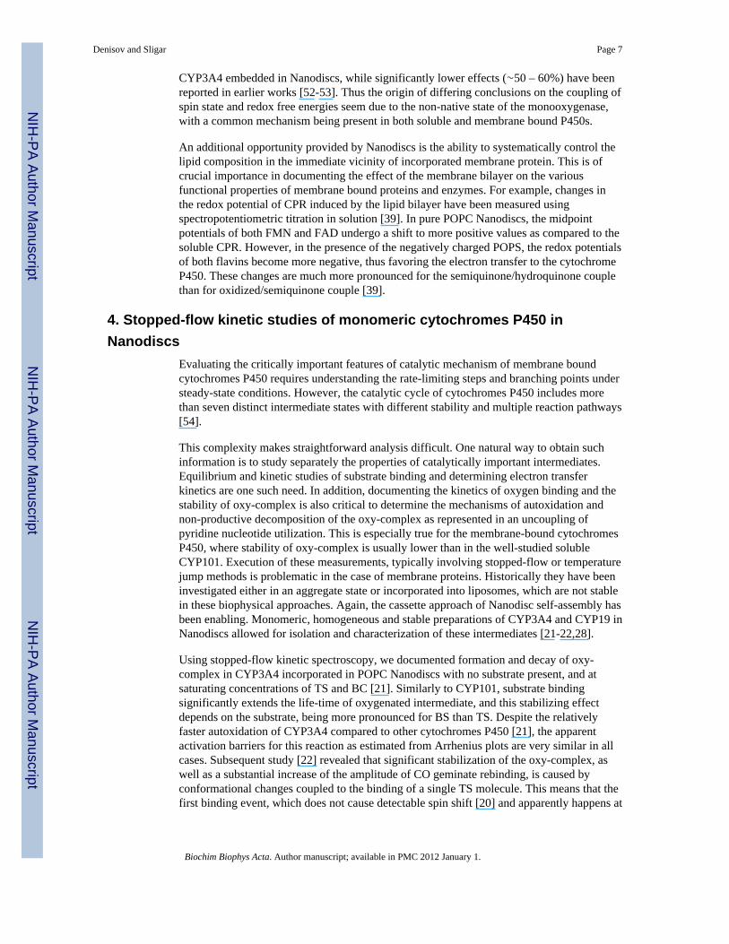
4. Stopped-flow kinetic studies of monomeric cytochromes P450 inNanodiscs
Evaluating the critically important features of catalytic mechanism of membrane boundcytochromes P450 requires understanding the rate-limiting steps and branching points understeady-state conditions. However, the catalytic cycle of cytochromes P450 includes morethan seven distinct intermediate states with different stability and multiple reaction pathways[54].
This complexity makes straightforward analysis difficult. One natural way to obtain suchinformation is to study separately the properties of catalytically important intermediates.Equilibrium and kinetic studies of substrate binding and determining electron transferkinetics are one such need. In addition, documenting the kinetics of oxygen binding and thestability of oxy-complex is also critical to determine the mechanisms of autoxidation andnon-productive decomposition of the oxy-complex as represented in an uncoupling ofpyridine nucleotide utilization. This is especially true for the membrane-bound cytochromesP450, where stability of oxy-complex is usually lower than in the well-studied solubleCYP101. Execution of these measurements, typically involving stopped-flow or temperaturejump methods is problematic in the case of membrane proteins. Historically they have beeninvestigated either in an aggregate state or incorporated into liposomes, which are not stablein these biophysical approaches. Again, the cassette approach of Nanodisc self-assembly hasbeen enabling. Monomeric, homogeneous and stable preparations of CYP3A4 and CYP19 inNanodiscs allowed for isolation and characterization of these intermediates [21-22,28].
Using stopped-flow kinetic spectroscopy, we documented formation and decay of oxy-complex in CYP3A4 incorporated in POPC Nanodiscs with no substrate present, and atsaturating concentrations of TS and BC [21]. Similarly to CYP101, substrate bindingsignificantly extends the life-time of oxygenated intermediate, and this stabilizing effectdepends on the substrate, being more pronounced for BS than TS. Despite the relativelyfaster autoxidation of CYP3A4 compared to other cytochromes P450 [21], the apparentactivation barriers for this reaction as estimated from Arrhenius plots are very similar in allcases. Subsequent study [22] revealed that significant stabilization of the oxy-complex, aswell as a substantial increase of the amplitude of CO geminate rebinding, is caused byconformational changes coupled to the binding of a single TS molecule. This means that thefirst binding event, which does not cause detectable spin shift [20] and apparently happens at
Denisov and Sligar Page 7
Biochim Biophys Acta. Author manuscript; available in PMC 2012 January 1.
NIH
-PA Author Manuscript
NIH
-PA Author Manuscript
NIH
-PA Author Manuscript
the remote site far from the heme iron, plays an important functional allosteric role servingas a modulator of the first uncoupling channel of CYP3A4.
A stopped-flow study of autoxidation of CYP19 saturated with the substrateandrostenedione in Nanodisc revealed a similar fast autoxidation rate (0.7 s-1 at 37° C) andhigh activation energy (18 kcal/mol) [28]. These finding provided a guideline to thepreparation of oxy-complexes of these and other human cytochromes P450 forcryoradiolytic experiments, as reviewed in the next section.
The kinetics of CYP3A4 reduction by dithionite was studied using stopped flow [18] toevaluate the role of oligomerization of the enzyme and conformational heterogeneity withrespect to the observed allosteric properties of cytochromes P450. Monomeric CYP3A4 inNanodiscs was reduced in a single exponential reaction, both in the presence and in theabsence of substrate bromocriptine. Analogous effects have been reported recently forreduction of CYP3A4 by the flavin domain of cytochrome P450 CYP102 [19]. As withdithionite, the reduction of monomeric CYP3A4 in Nanodiscs was almost complete,contrary to only 50% reduction of oligomeric CYP3A4 in liposomes observed with a lowlipid:protein ratio (2:1:0.6 phosphatidylcholine, phosphatidylethanolamine and phosphatidicacid). Direct comparison of results obtained with low (76 lipids per CYP3A4) and high (910lipids per CYP3A4) ratios in liposomes revealed the definitive role of oligomerization ofCYP3A4 in incomplete reduction by flavoprotein reductase. These works demonstrate theimportance of studying the equilibrium and kinetic properties of monomeric cytochromesP450 and to separate effects caused by aggregation and / or oligomerization.
5. Cryogenic radiolytic reduction and the spectroscopy of unstableintermediates in membrane cytochromes P450
The incorporation of human cytochromes P450 into Nanodiscs provided an opportunity forthe isolation and entrapment of unstable catalytic intermediates in P450 catalysis using thesame cryogenic methods developed recently for mechanistic studies of oxygen activation inthe soluble P450 heme enzymes [55-63]. The overall stability of heme oxy-complexesincreases dramatically at low temperatures due to the high activation barriers for thisreaction (15 – 25 kcal/mol [21,28,64-65]). For example, the half-life of the oxy-complex inCYP3A4 in the presence of testosterone increases from ∼10 ms at 37° C to hundreds ofseconds at -30° C, making possible the oxygenation of anaerobically reduced heme enzymeby manual mixing. The advantage of working at subzero temperatures is especiallyimportant for the mammalian cytochromes P450, which usually are autoxidized very quicklyat ambient conditions. However, the work in solution at < -30° C requires the use of aqueouscryosolvents. The most popular solvents used in cryoenzymology are glycerol and ethyleneglycol, which are preferable due to their inability to dissolve lipids and to disrupt Nanodiscs,as opposed to such alternatives as methanol, ethanol or dimethyl sulfoxide. In separatecontrol experiments we confirmed that CYP3A4 or CYP19 Nanodiscs remain intact after theaddition of 75% glycerol, and the enzymes still show full activity and lack of P420formation. The usage of high glycerol or ethylene glycol concentration thus allows theformation of a transparent glassy frozen sample suitable for optical absorption spectroscopyat cryogenic temperatures [59].
The oxy-complex in CYP19 incorporated in Nanodiscs was prepared by mixing of reducedenzyme with an excess of oxygenated cold 90% glycerol/phosphate buffer solution at -30° Cand cooled to liquid nitrogen temperatures as described [27]. Absorption spectra of thesesamples measured at cryogenic temperatures are very similar to those resolved fromstopped-flow kinetic experiments [28]. Samples for EPR spectroscopy were preparedsimilarly by mixing directly in quartz tubes and frozen in liquid nitrogen. Radiolytic
Denisov and Sligar Page 8
Biochim Biophys Acta. Author manuscript; available in PMC 2012 January 1.
NIH
-PA Author Manuscript
NIH
-PA Author Manuscript
NIH
-PA Author Manuscript
reduction was accomplished by γ-irradiation from 60Co source at liquid nitrogentemperature as described [59-60]. As a result of one-electron reduction of the oxy-complexin CYP19, the unprotonated peroxo-ferric complex was formed with characteristic g-valuesg1 = 2.254 and g2 = 2.163 (g3 could not be determined since it overlaps with radicalsgenerated in frozen glycerol matrix). This initial peroxo-anion complex was stable attemperatures up to 180 K, and then gradually converted to the low-spin ferric complex withg1 = 2.39 at 190 – 220 K. Stabilization of unprotonated peroxo-ferric complex in CYP19indicates potentially important difference in the proton delivery mechanism between thisenzyme and other heme enzymes such as CYP101 and CYP2B4 [56-57]. As the activeintermediate in the aromatization reaction catalyzed by CYP19 is suggested to be theperoxo-anion complex, the active site of aromatase may be optimized to maintain thiscomplex in an unprotonated state. Further investigation of these intermediates in CYP19using ENDOR and Q-band EPR is in progress through collaboration with Professor BrianHoffman and Dr. Roman Davydov at Northwestern University.
Using the same methods, we have also prepared and measured the UV-VIS spectra of theoxy- and peroxo- complexes in CYP3A4 self-assembled into POPC Nanodiscs. The spectraof oxy-complex of CYP3A4 saturated with TS and BC and measured in 65% glycerol at T =100 K are shown in Figure 2. Similar to the spectra measured in stopped-flow experiments[21], the position of the main Soret band is slightly different for the CYP3A4-TS andCYP3A4-BC complexes. The different shape of Soret and Q-bands is better resolved at lowtemperatures and may indicate the presence of multiple conformers of oxygen moleculecoordinated to iron atom, similarly to the conformational heterogeneity of the oxy-complexof CYP101 documented by resonance Raman spectroscopy [66-67].
After cryoradiolytic reduction, a (hydro)peroxo-ferric complex is formed with the estimatedyield ∼50%. The optical spectrum of this intermediate is shown in Figure 3 together with thespectrum of the same sample before irradiation. The shape and positions of split Soret bandsat 439 nm and at ∼367 nm are similar to the spectrum of the same intermediate in CYP101[59].
6. Conclusions and SummaryWe have attempted in this brief review to link the pioneering investigations of ProfessorRuckpaul on the cytochromes P450 with future efforts at unraveling the deep secrets of themembrane bound form of these important monoxygenases. Early mechanistic work in the1970-1990 time period utilized the soluble microbial P450s as a model of the importantmembrane bound forms which compose the vast majority of the ∼11,000 P450 genes so faridentified. When these membrane bound P450s are removed from their native environment,aggregation and inactivation often occur and the sophisticated biochemical and biophysicaltechniques used with their soluble brothers were often not applicable. What was needed is ameans for converting these aggregating and often inactive forms into a solublemonodisperse entity that would allow mechanistic details to be revealed. The self-assembledNanodisc system has proven to open this door to advanced quantitative analysis. Highlypurified homogeneous fully functional and stable preparations of membrane bound P450shave now been used by many laboratories in their quest to understand the workings of oneof the most prevalent enzymes in Nature's basket of biotransformations.
AcknowledgmentsThe works of many members of the Sligar laboratory cited in this review is gratefully acknowledged. Our researchon cytochromes P450 has been supported for nearly thirty years by two grants from the National Institute of HealthGM31756 and GM33775.
Denisov and Sligar Page 9
Biochim Biophys Acta. Author manuscript; available in PMC 2012 January 1.
NIH
-PA Author Manuscript
NIH
-PA Author Manuscript
NIH
-PA Author Manuscript
References1. Ruckpaul K. Early years of cytochrome P450 research in Berlin-Buch: its present state and origin of
the biochemical and biophysical conferences. Biochem Biophys Res Commun 2003;312:65–74.[PubMed: 14630020]
2. Ruckpaul K, Renneberg R. Ron Estabrook--a catalyst of scientific interactions between East andWest. Drug Metab Rev 2007;39:267–270. [PubMed: 17786617]
3. Ruckpaul, K.; Rein, H., editors. Cytochrome P450. Akademie Verlag; Berlin: 1984. p. 4054. Rein H, Ristau O, Friedrich J, Janig GR, Ruckpaul K. Evidence for the existence of a high spin-low
spin equilibrium in liver microsomal cytochrome P-450. FEBS Lett 1977;75:19–22. [PubMed:852581]
5. Ristau O, Rein H, Janig GR, Ruckpaul K. Quantitative analysis of the spin equilibrium ofcytochrome P-450 LM-2 fraction from rabbit liver microsomes. Biochim Biophys Acta1978;536:226–34. [PubMed: 708762]
6. Ziegler M, Blanck J, Greschner S, Lenz K, Lau A, Ruckpaul K. Kinetics of elementary steps in thecytochrome P-450 reaction sequence. V. Laser temperature-jump investigation of the spinrelaxation kinetics of cytochrome P-450 LM2. Biomed Biochim Acta 1983;42:641–9. [PubMed:6639642]
7. Bernhardt R, Pommerening K, Ruckpaul K. Modification of carboxyl groups on NADPH-cytochrome P-450 reductase involved in binding of cytochromes c and P-450 LM2. Biochem Int1987;14:823–832. [PubMed: 2841939]
8. Blanck J, Janig GR, Schwarz D, Ruckpaul K. Role of lipid in the electron transfer betweenNADPH-cytochrome P-450 reductase and cytochrome P-450 from mammalian liver cells.Xenobiotica 1989;19:1231–1246. [PubMed: 2515662]
9. Davydov RM, Greshner S, Magonov SN, Ruckpaul K, Blyumenfel'd LA. Absorption spectra ofcytochrome P-450 nonequilibrium states formed during the low-temperature reduction of protein.Doklady Akad Nauk SSSR 1978;241:707–709.
10. Sharonov Yu A, Pismensky VF, Greschner S, Ruckpaul K. Low- and ultralow-temperaturemagnetic circular dichroism studies of reduced cytochromes P-450-LM2 and P-420-LM2 and ofphoto-products of their co-complexes. The spin-state and axial ligation of heme iron. BiochemBiophys Res Commun 1987;146:165–172. [PubMed: 3606614]
11. Davydov RM, Greschner S, Ruckpaul K. Study of absorption and magnetic circular dichroismspectra of hemoproteins in conformationally nonequilibrium states. V. Cytochrome P 450 and itscomplexes with substrates. Molek Biol 1979;13:1397–1406.
12. Schwarz D, Pirrwitz J, Meyer HW, Coon MJ, Ruckpaul K. Membrane topology of microsomalcytochrome P-450: saturation transfer EPR and freeze-fracture electron microscopy studies.Biochem Biophys Res Commun 1990;171:175–181. [PubMed: 2168169]
13. Bayburt TH, Sligar SG. Single-molecule height measurements on microsomal cytochrome P450 innanometer-scale phospholipid bilayer disks. Proc Natl Acad Sci USA 2002;99:6725–6730.[PubMed: 11997441]
14. Civjan NR, Bayburt TH, Schuler MA, Sligar SG. Direct solubilization of heterologously expressedmembrane proteins by incorporation into nanoscale lipid bilayers. BioTechniques 2003;35:556–563. [PubMed: 14513561]
15. Baas BJ, Denisov IG, Sligar SG. Homotropic cooperativity of monomeric cytochrome P450 3A4 ina nanoscale native bilayer environment. Arch Biochem Biophys 2004;430:218–228. [PubMed:15369821]
16. Chougnet A, Grinkova Y, Ricard D, Sligar S, Woggon WD. Fluorescent probes for rapid screeningof potential drug-drug interactions at the CYP3A4 level. ChemMedChem 2007;2:717–724.[PubMed: 17357170]
17. Davydov DR, Baas BJ, Sligar SG, Halpert JR. Allosteric mechanisms in cytochrome P450 3A4studied by high-pressure spectroscopy: pivotal role of substrate-induced changes in theaccessibility and degree of hydration of the heme pocket. Biochemistry 2007;46:7852–7864.[PubMed: 17555301]
Denisov and Sligar Page 10
Biochim Biophys Acta. Author manuscript; available in PMC 2012 January 1.
NIH
-PA Author Manuscript
NIH
-PA Author Manuscript
NIH
-PA Author Manuscript
18. Davydov DR, Fernando H, Baas BJ, Sligar SG, Halpert JR. Kinetics of dithionite-dependentreduction of cytochrome P450 3A4: heterogeneity of the enzyme caused by its oligomerization.Biochemistry 2005;44:13902–13913. [PubMed: 16229479]
19. Davydov DR, Sineva EV, Sistla S, Davydova NY, Frank DJ, Sligar SG, Halpert JR. Electrontransfer in the complex of membrane-bound human cytochrome P450 3A4 with the flavin domainof P450BM-3: The effect of oligomerization of the heme protein and intermittent modulation ofthe spin equilibrium. Biochim Biophys Acta 2010;1797:378–390. [PubMed: 20026040]
20. Denisov IG, Baas BJ, Grinkova YV, Sligar SG. Cooperativity in cytochrome P450 3A4: Linkagesin substrate binding, spin state, uncoupling, and product formation. J Biol Chem 2007;282:7066–7076. [PubMed: 17213193]
21. Denisov IG, Grinkova YV, Baas BJ, Sligar SG. The ferrous-dioxygen intermediate in humancytochrome P450 3A4: Substrate dependence of formation of decay kinetics. J Biol Chem2006;281:23313–23318. [PubMed: 16762915]
22. Denisov IG, Grinkova YV, McLean MA, Sligar SG. The one-electron autoxidation of humancytochrome P450 3A4. J Biol Chem 2007;282:26865–26873. [PubMed: 17650504]
23. Frank DJ, Denisov IG, Sligar SG. Mixing apples and oranges: Analysis of heterotropiccooperativity in cytochrome P450 3A4. Arch Biochem Biophys 2009;488:146–152. [PubMed:19560436]
24. Nath A, Grinkova YV, Sligar SG, Atkins WM. Ligand binding to cytochrome P450 3A4 inphospholipid bilayer nanodiscs: the effect of model membranes. J Biol Chem 2007;282:28309–28320. [PubMed: 17573349]
25. Nath A, Koo PK, Rhoades E, Atkins WM. Allosteric Effects on substrate dissociation fromcytochrome P450 3A4 in Nanodiscs observed by ensemble and single-molecule fluorescencespectroscopy. J Am Chem Soc 2008;130:15746–15747. [PubMed: 18980315]
26. Duan H, Civjan NR, Sligar SG, Schuler MA. Co-incorporation of heterologously expressedArabidopsis cytochrome P450 and P450 reductase into soluble nanoscale lipid bilayers. ArchBiochem Biophys 2004;424:141–153. [PubMed: 15047186]
27. Gantt SL, Denisov IG, Grinkova YV, Sligar SG. The critical iron-oxygen intermediate in humanaromatase. Biochem Biophys Res Commun 2009;387:169–173. [PubMed: 19591804]
28. Grinkova YV, Denisov IG, Waterman MR, Arase M, Kagawa N, Sligar SG. The ferrous-oxycomplex of human aromatase. Biochem Biophys Res Commun 2008;372:379–382. [PubMed:18482580]
29. Duan H, Schuler MA. Heterologous expression and strategies for encapsulation of membrane-localized plant P450s. Phytochem Rev 2006;5:507–523.
30. Boldog T, Li M, Hazelbauer GL. Using Nanodiscs to create water-soluble transmembranechemoreceptors inserted in lipid bilayers. Methods Enzymol 2007;423:317–335. [PubMed:17609138]
31. Borch J, Hamann T. The Nanodisc: a novel tool for membrane protein studies. Biol Chem2009;390:805–814. [PubMed: 19453280]
32. Nath A, Atkins WM, Sligar SG. Applications of phospholipid bilayer nanodiscs in the study ofmembranes and membrane proteins. Biochemistry 2007;46:2059–69. [PubMed: 17263563]
33. Ritchie TK, Grinkova YV, Bayburt TH, Denisov IG, Zolnerciks JK, Atkins WM, Sligar SG.Reconstitution of membrane proteins in phospholipid bilayer nanodiscs. Methods Enzymol2009;464:211–231. [PubMed: 19903557]
34. Bayburt TH, Sligar SG. Membrane protein assembly into Nanodiscs. FEBS letters. 201035. Bayburt TH, Carlson JW, Sligar SG. Reconstitution and imaging of a membrane protein in a
nanometer-size phospholipid bilayer. J Struct Biol 1998;123:37–44. [PubMed: 9774543]36. Bayburt TH, Carlson JW, Sligar SG. Single molecule height measurements on a membrane protein
in nanometer-scale phospholipid bilayer disks. Langmuir 2000;16:5993–5997.37. Das A, Grinkova YV, Sligar SG. Redox potential control by drug binding to cytochrome P 450
3A4. J Am Chem Soc 2007;129:13778–13779. [PubMed: 17948999]38. Das A, Zhao J, Schatz GC, Sligar SG, Van Duyne RP. Screening of Type I and II drug binding to
human cytochrome P450-3A4 in Nanodiscs by localized surface plasmon resonance spectroscopy.Anal Chem 2009;81:3754–3759. [PubMed: 19364136]
Denisov and Sligar Page 11
Biochim Biophys Acta. Author manuscript; available in PMC 2012 January 1.
NIH
-PA Author Manuscript
NIH
-PA Author Manuscript
NIH
-PA Author Manuscript

39. Das A, Sligar SG. Modulation of the cytochrome P450 reductase redox potential by thephospholipid bilayer. Biochemistry 2009;48:12104–12. [PubMed: 19908820]
40. Bayburt TH, Grinkova YV, Sligar SG. Assembly of single bacteriorhodopsin trimers in bilayernanodiscs. Arch Biochem Biophys 2006;450:215–222. [PubMed: 16620766]
41. Bayburt TH, Leitz AJ, Xie G, Oprian DD, Sligar SG. Transducin activation by nanoscale lipidbilayers containing one and two rhodopsins. J Biol Chem 2007;282:14875–14881. [PubMed:17395586]
42. Bayburt TH, Sligar SG. Self-assembly of single integral membrane proteins into soluble nanoscalephospholipid bilayers. Protein Sci 2003;12:2476–2481. [PubMed: 14573860]
43. Boldog T, Grimme S, Li M, Sligar SG, Hazelbauer GL. Nanodiscs separate chemoreceptoroligomeric states and reveal their signaling properties. Proc Natl Acad Sci U S A2006;103:11509–11514. [PubMed: 16864771]
44. Marin VL, Bayburt TH, Sligar SG, Mrksich M. Functional assays of membrane-bound proteinswith SAMDI-TOF mass spectrometry. Angew Chem, Int Ed 2007;46:8796–8798.
45. Goluch ED, Shaw AW, Sligar SG, Liu C. Microfluidic patterning of nanodisc lipid bilayers andmultiplexed analysis of protein interaction. Lab Chip 2008;8:1723–1728. [PubMed: 18813396]
46. Kijac AZ, Li Y, Sligar SG, Rienstra CM. Magic-angle spinning solid-state NMR spectroscopy ofNanodisc-embedded human CYP3A4. Biochemistry 2007;46:13696–13703. [PubMed: 17985934]
47. Davydov DR, Halpert JR. Allosteric P450 mechanisms: multiple binding sites, multipleconformers or both? Expert Opin Drug Metab Toxicol 2008;4:1523–1535. [PubMed: 19040328]
48. Denisov IG, Frank DJ, Sligar SG. Cooperative properties of cytochromes P450. Pharmacol Ther2009;124:151–167. [PubMed: 19555717]
49. Sligar SG, Denisov IG. Understanding cooperativity in human P450 mediated drug-druginteractions. Drug Metab Rev 2007;39:567–579. [PubMed: 17786639]
50. Baas, BJ. Ph D Thes. University of Illinois; Urbana: 2006. Characterization of monomeric humancytochrome P450 3A4 and cytochrome P450 reductase in nanoscale phospholipid bilayer discs; p.180
51. Nath, A. Ph D Thes. University of Washington; Seattle: 2008. Thermodynamics and kinetics ofligand binding to cytochrome P450 3A4 in membrane nanodiscs; p. 120
52. Hosea NA, Miller GP, Guengerich FP. Elucidation of distinct ligand binding sites for cytochromeP450 3A4. Biochemistry 2000;39:5929–5939. [PubMed: 10821664]
53. Isin EM, Guengerich FP. Kinetics and thermodynamics of ligand binding by cytochrome P4503A4. J Biol Chem 2006;281:9127–9136. [PubMed: 16467307]
54. Denisov IG, Makris TM, Sligar SG, Schlichting I. Structure and chemistry of cytochrome P 450.Chem Rev 2005;105:2253–2277. [PubMed: 15941214]
55. Davydov R, Kappl R, Huettermann J, Peterson JA. EPR-spectroscopy of reduced oxyferrous-P450cam. FEBS Lett 1991;295:113–115. [PubMed: 1662641]
56. Davydov R, Makris TM, Kofman V, Werst DE, Sligar SG, Hoffman BM. Hydroxylation ofcamphor by reduced oxy-cytochrome P450cam: Mechanistic implications of EPR and ENDORstudies of catalytic intermediates in native and mutant enzymes. J Am Chem Soc 2001;123:1403–1415. [PubMed: 11456714]
57. Davydov R, Razeghifard R, Im SC, Waskell L, Hoffman BM. Characterization of the microsomalcytochrome P450 2B4 O2 activation intermediates by cryoreduction and electron paramagneticresonance. Biochemistry 2008;47:9661–9666. [PubMed: 18700729]
58. Denisov IG, Dawson JH, Hager LP, Sligar SG. The ferric-hydroperoxo complex ofchloroperoxidase. Biochem Biophys Res Commun 2007;363:954–958. [PubMed: 17920039]
59. Denisov IG, Makris TM, Sligar SG. Cryoradiolysis for the study of P450 reaction intermediates.Meth Enzymol 2002;357:103–115. [PubMed: 12424902]
60. Denisov IG, Victoria DC, Sligar SG. Cryoradiolytic reduction of heme proteins: Maximizing dosedependent yield. Radiat Phys Chem 2007;76:714–721.
61. Ibrahim M, Denisov IG, Makris TM, Kincaid JR, Sligar SG. Resonance Raman spectroscopicstudies of hydroperoxo-myoglobin at cryogenic temperatures. J Am Chem Soc 2003;125:13714–13718. [PubMed: 14599210]
Denisov and Sligar Page 12
Biochim Biophys Acta. Author manuscript; available in PMC 2012 January 1.
NIH
-PA Author Manuscript
NIH
-PA Author Manuscript
NIH
-PA Author Manuscript
62. Mak PJ, Denisov IG, Victoria D, Makris TM, Deng T, Sligar SG, Kincaid JR. Resonance Ramandetection of the hydroperoxo intermediate in the cytochrome P450 enzymatic cycle. J Am ChemSoc 2007;129:6382–6383. [PubMed: 17461587]
63. Mak PJ, Kincaid JR. Resonance Raman spectroscopic studies of hydroperoxo derivatives of cobalt-substituted myoglobin. J Inorg Biochem 2008;102:1952–1957. [PubMed: 18723225]
64. Lipscomb JD, Sligar SG, Namtvedt MJ, Gunsalus IC. Autooxidation and hydroxylation reactionsof oxygenated cytochrome P-450cam. J Biol Chem 1976;251:1116–1124. [PubMed: 2601]
65. Tuckey RC, Kamin H. The oxyferro complex of adrenal cytochrome P-450scc. Effect ofcholesterol and intermediates on its stability and optical characteristics. J Biol Chem1982;257:9309–9314. [PubMed: 7107571]
66. Denisov IG, Mak PJ, Makris TM, Sligar SG, Kincaid JR. Resonance Raman characterization of theperoxo and hydroperoxo intermediates in cytochrome P450. J Phys Chem A 2008;112:13172–13179. [PubMed: 18630867]
67. Sjodin T, Christian JF, Macdonald IDG, Davydov R, Unno M, Sligar SG, Hoffman BM, ChampionPM. Resonance Raman and EPR investigations of the D251N oxycytochrome P450cam/putidaredoxin complex. Biochemistry 2001;40:6852–6859. [PubMed: 11389599]
68. Heller H, Schaefer M, Schulten K. Molecular dynamics simulation of a bilayer of 200 lipids in thegel and in the liquid-crystal phases. J Phys Chem 1993;97:83434–8360.
69. Denisov IG, Grinkova YV, Lazarides AA, Sligar SG. Directed self-assembly of monodispersephospholipid bilayer Nanodiscs with controlled size. J Am Chem Soc 2004;126:3477–3487.[PubMed: 15025475]
70. Yano JK, Wester MR, Schoch GA, Griffin KJ, Stout CD, Johnson EF. The structure of humanmicrosomal cytochrome P450 3A4 determined by X-ray crystallography to 2.05-A resolution. JBiol Chem 2004;279:38091–38094. [PubMed: 15258162]
71. Wang M, Roberts DL, Paschke R, Shea TM, Masters BS, Kim JJ. Three-dimensional structure ofNADPH-cytochrome P450 reductase: prototype for FMN- and FAD-containing enzymes. ProcNatl Acad Sci USA 1997;94:8411–8416. [PubMed: 9237990]
Abbreviations
AFM Atomic force microscopy
ANF alpha-napthoflavone
BC bromocriptine
CPR Cytochrome P450 reductase
DPPC 1,2-dipalmitoyl-sn-glycero-3-phosphocholine
EDC 1-ethyl-3-(3-dimethylaminopropyl) carbodiimide
HDL High-density lipoprotein
LSPR localized surface plasmon resonance
MSP Membrane scaffold protein
POPC 1-palmitoyl-2-oleoyl-sn-glycero-3-phosphocholine
SPR surface plasmon resonance
TS testosterone
Denisov and Sligar Page 13
Biochim Biophys Acta. Author manuscript; available in PMC 2012 January 1.
NIH
-PA Author Manuscript
NIH
-PA Author Manuscript
NIH
-PA Author Manuscript

Figure 1.A molecular model of a 1:1 functional complex of CYP3A4 and CPR incorporated in aNanodisc. The coordinates of the POPC bilayer in a liquid crystalline state are based onmolecular dynamics simulations available at:http://www.lrzmuenchen.de/∼heller/membrane/membrane.html [68]. The membranescaffold protein (MSP1E3) [69] (blue) is modeled as an ideal α-helix. CYP3A4 (red), fromthe Protein Data Bank file 1TQN.pdb [70] and CPR (yellow) from Protein Data Bank file1AMO.pdb [71] are pictured in a close contact in their docked conformation for illustrativepurposes.
Denisov and Sligar Page 14
Biochim Biophys Acta. Author manuscript; available in PMC 2012 January 1.
NIH
-PA Author Manuscript
NIH
-PA Author Manuscript
NIH
-PA Author Manuscript

Figure 2.The absorption spectra of the oxy-ferrous complex of CYP3A4 in Nanodiscs both withtestosterone (red) and bromocriptine (blue) bound at the active site. Potassium phosphatebuffer /65% glycerol, T=100 K.
Denisov and Sligar Page 15
Biochim Biophys Acta. Author manuscript; available in PMC 2012 January 1.
NIH
-PA Author Manuscript
NIH
-PA Author Manuscript
NIH
-PA Author Manuscript

Figure 3.The absorption spectra of the oxy-ferrous (red) and hydroperoxo-ferric (blue) complexes ofCYP3A4 in Nanodiscs with bromocriptine bound.
Denisov and Sligar Page 16
Biochim Biophys Acta. Author manuscript; available in PMC 2012 January 1.
NIH
-PA Author Manuscript
NIH
-PA Author Manuscript
NIH
-PA Author Manuscript
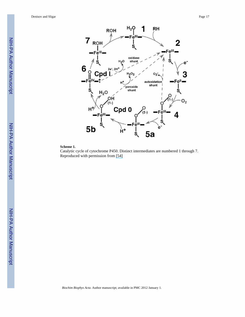
Scheme 1.Catalytic cycle of cytochrome P450. Distinct intermediates are numbered 1 through 7.Reproduced with permission from [54]
Denisov and Sligar Page 17
Biochim Biophys Acta. Author manuscript; available in PMC 2012 January 1.
NIH
-PA Author Manuscript
NIH
-PA Author Manuscript
NIH
-PA Author Manuscript
Copyright © 2022 FDOKUMEN