QSAR modeling of aromatase inhibitory activity of 1-substituted 1,2,3-triazole analogs of letrozole
To cite this article: Neuroendocrinol Lett 2010; 31(5):101–109
OR
IG
IN
AL
A
RT
IC
LE
Neuroendocrinology Letters Volume 31 No. 5 2010
Expression and distribution of P450-aromatase in the ovine hypothalamus at different stages of fetal development Arcadia Mura, Sergio Gadau, Gianluca Lepore, Francesca Balzano, Marco Zedda, Emilio Mura, Vittorio FarinaDepartment of Animal Biology, University of Sassari, Italy
Correspondence to: Arcadia Mura, PhD.Department of Animal Biology University of Sassari, Via Vienna, 2, 07100 Sassari, Italy.tel: +39 079229462; fax: +39 079229432; e-mail: [email protected]
Submitted: 2010-09-13 Accepted: 2010-09-21 Published online: 2010-00-00
Key words: aromatase; brain sexual differentiation; hypothalamus; fetal sheep
Neuroendocrinol Lett 2010; 31(5):101–109 PMID: ----- NEL310510AXX © 2010 Neuroendocrinology Letters • www.nel.edu
Abstract OBJECTIVES: An important step of sexual differentiation is the conversion of testosterone to estrogen by aromatase leading to masculinization and defeminiza-tion of the fetal brain areas crucial for normal sexual behavior and reproduction. Brain sexual differentiation occurs throughout a critical period starting from different prenatal stages depending on the species. Such period goes on from gestation day (GD) 30 to 100GD in the sheep. The fetal sheep brain is reported to aromatize androgens to estrogens at 64GD. The main goal of this work was to evaluate aromatase expression in sheep hypothalami during the whole period of sexual differentiation (35GD, 55GD, 80GD, 115GD) and whether differences may be observed depending on gestational stage and sex.METHODS: Sections at the hypothalamic level underwent immunoperoxidase technique employing anti-aromatase and anti-androgen receptor antibodies. Samples from 35GD and 55GD were also processed with in situ hybridization using aromatase cDNA probe. Blot analyses were performed to quantify possible aromatase immunoexpression differences between sexes. For sexing, samples at 35GD and 55GD underwent DNA extraction and SRY amplification.RESULTS: Our results revealed aromatase and androgen receptor immunoreactiv-ity along the whole period of sexual differentiation. Both molecules were detected in many brain regions and markedly in the periventricular area. The highest aromatase and androgen receptor amounts were observed at 35GD and 55GD, when aromatase was more abundant in females than in males.CONCLUSIONS: In conclusion, the sheep can be included among the species where aromatase is highly expressed in the hypothalamus during the whole period of sexual differentiation.
IntroductIon
The brain is an important site of estrogen synthe-sis (Roselli, 2007), as demonstrated in mammals by Naftolin et al. (1971), who ascertained that the central neuroendocrine tissues are capable of
converting androgens into estrogens by means of cytochrome P450-aromatase. In the last years, it has been shown that many of the androgen effects on neural functions are mediated by aromatase

102 Copyright © 2010 Neuroendocrinology Letters ISSN 0172–780X • www.nel.edu
Arcadia Mura, Sergio Gadau, Gianluca Lepore, Francesca Balzano, Marco Zedda, Emilio Mura, Vittorio Farina
through a multistep enzymatic pathway (Roselli and Resko, 2001; Simpson et al. 1994). Two proteins are required for aromatase activity: cytochrome P450, that is the CYP19 gene product, and NADP-cytochrome P450-reductase, a ubiquitous flavoprotein (Garcia-Segura, 2008; Negri-Cesi et al. 1992). The enzyme com-plex is located in the smooth endoplasmic reticulum of estrogen producing cells. Aromatase is expressed in many peripheral tissues and organs, such as placenta, fetal tissues, adipose tissue, skin, bones, cartilage, muscle, ovary, testis, etc. (Negri-Cesi et al. 1992; Roselli & Resko 2001).
A large number of studies have demonstrated that brain aromatase is predominantly distributed in areas involved in the control of reproductive functions such as the hypothalamus, limbic system and preoptic area (Negri-Cesi et al. 2008). These are indeed essential regions for sex-specific control of the reproductive hormone secretion and sex behavior. More precisely, the hypothalamic neurons of the periventricular region and the arcuate nucleus produce reproductive hor-mones, whereas the medial preoptic, ventromedial, and ventral premammillary nuclei influence sex behavior. Aromatase is also expressed in non-reproductive brain areas such as cerebral cortex, midbrain and spinal cord (Simerly 2002). This enzyme plays an essential role in several neural processes. For instance, aromatase is involved in synaptic plasticity of brain regions related to cognition such as the hippocampus (Von Schassen et al. 2006) and it may also be responsible for the early stages of neural cell differentiation (Martínez-Cerdeño et al. 2006).
Aromatase is expressed by neurons in baseline con-dition (Balthazart & Ball 1998; Lephart 1996; Naftolin et al. 1971; Negri-Cesi et al. 1992), whereas several studies have suggested that it is expressed by astrocytes only after brain injury (Garcia-Segura et al. 1999). This indicates that astrocytes have the potential to produce aromatase and therefore to express neuroprotective estrogens (Garcia-Segura 2008).
In addition, aromatase is essential for sexual dif-ferentiation of the brain in many mammals and avian species (Herbosa & Foster 1996) leading to the produc-tion of estrogens that act permanently in the developing brain. Indeed, during prenatal development, “sex brain” is feminine by default, unless some specific stimuli drive it to a male phenotype (Negri-Cesi et al. 2008). Therefore, to achieve male-specific brain it is necessary to activate two independent processes: masculinization and defeminization. The former establishes male-typi-cal copulatory behavior and partner preference, while the latter entails the estrogen-mediated “elimination” of GnRH surge center (Senger 2003). In contrast, female brain is not exposed to testosterone and develops the hypothalamic GnRH surge center. Such processes occur in the brain during prenatal and perinatal development, dependently on the length of gestation. Indeed, in long-gestation species like monkeys masculinization and
defeminization mainly take place prenatally, whereas in short-gestation species like rats, they occur postnatally (Herbosa and Foster,1996; Roselli et al. 2003).
Recent studies have suggested that these develop-mental phenomena may also be applied to the sheep, which is a long-gestation species, where masculiniza-tion occurs prenatally from day 30 to 100 (Short 1974). Lines of evidence have shown that the fetal sheep brain is able to aromatize androgens to estrogens, but this process has been documented in the literature at day 64 of gestation only (Roselli et al. 2003), when no signifi-cant sex differences in aromatase activity were found, similarly to what demonstrated for other long-gestation species like monkeys (George & Ojeda 1982; Roselli & Resko 1986; Roselli et al. 2003). In contrast, sex differ-ences in hypothalamic aromatase activity were detected in fetuses from short-gestation species like ferrets and rats (Negri-Cesi et al. 2008; Tobet et al. 1985).
Thus, aim of the present investigation was to detect aromatase hypothalamic immunoexpression through-out the period of sexual differentiation in the fetal sheep brain. Moreover, aromatase expression in male and female fetuses was estimated at early stages of sexual differentiation in order to assess whether sex dif-ferences in aromatase activity exist during that crucial period of brain development.
MAtErIAL And MEtHodS
Sheep fetuses from different gestation days (GD) i.e. 35±2GD (n=13 for immunocytochemistry and in situ hybridization, n=6 for Western blot), 55±2GD (n=14 for immunocytochemistry and in situ hybridization, n=6 for Western blot), 80±2GD (n=6 for immunocy-tochemistry), 115±2GD (n=6 for immunocytochem-istry) were obtained at local abattoirs, when pregnant sheep were slaughtered. The period of gestation was established measuring fetal crown-rump length, fol-lowing the tables reported by McGeady et al. (2006). Fetal heads from 35GD and 55GD were completely processed, whereas fetal heads from 80GD and 115GD were removed and brains dissected using the ven-tral surface as landmark. Hypothalamic tissue blocks extending caudally from the anterior border of the optic chiasm to the posterior border of the mammillary body and dorsally to the roof of the third ventricle were obtained. Blocks were fixed with ice-cold 4% parafor-maldehyde for 24 hrs, then postfixed with 30% sucrose/PBS as cryoprotectant. Then, they were stored at –80 °C until cutting. Sections, 10 μm thick, were mounted onto microscope slides and underwent immunoperoxidase or in situ hybridization techniques. In addition, blocks were homogenised and frozen at –80 °C to be processed by Western blot analysis.
Immunoperoxidase technique (ABC method, Vector, Burlingame, CA, USA) was performed employing the following antibodies: polyclonal anti-P450-aromatase

103Neuroendocrinology Letters Vol. 31 No. 5 2010 • Article available online: http://node.nel.edu
Aromatase expression in fetal sheep brain
antibody (Acris, Hiddenhausen, Germany) diluted 1:200, monoclonal anti-androgen receptor antibody (Sigma, St. Louis, MO, USA) diluted 1:100 and mono-clonal anti-tubulin βIII (Sigma) antibody, a neuronal cell marker (Moody et al. 1996). In details, protease digestion (0.1% trypsin, 0.1% calcium chloride, TRIS/HCl 0.005 M, pH 7.6 at 37 °C for 20 min) was made to counteract the possible antigen masking effects of paraformaldehyde fixation. Then, coronal sections were blocked for 20 min with normal goat serum and incubated overnight at 4 °C with the primary antibod-ies, then for 1 hr in biotinylated goat anti-rabbit or anti-mouse antibodies diluted 1:250. Sections were incubated with avidin-biotin complex for 30 min, then with diaminobenzidine for 1–3 min and finally dehy-drated and coverslipped. The anti-P450-aromatase anti-body was previously tested by Western blot analysis in order to establish its cross-reactivity to the sheep. Other two anti-P450-aromatase antibodies were used from Sigma and Novus Biological (Littleton, CO, USA) to further confirm the effectiveness of the antibody used.
In order to evaluate aromatase gene expression, in situ hybridization technique was performed. A melt-ing curve ranging 37 °C to 42 °C was calculated in a preliminary study carried out at our labs, indicating 38 °C as the optimal temperature (Lepore et al. 2009). A biotinylated cDNA probe with 48-nucleotide sequence from Sigma (Roselli et al. 2000) was used on samples from 35GD and 55GD, i.e. the earliest stages examined. Then, sections were incubated with streptavidin per-oxidase complex and diaminobenzidine as chromogen. A sense probe was used to verify biotinylated cDNA probe and displayed no signal.
In addition, possible differences in aromatase expression between sexes were monitored at 35GD and 55GD by immunoperoxidase and Western blot. For sexing, fetal sample tissues underwent DNA extraction and PCR amplification of the sex determining region (SRY) of the Y chromosome following the protocol of Mara et al. (2004) specific for the sheep (Figure 1).
As to Western blot, samples were homogenized in lysis buffer with protease inhibitor (Roche Com-plete Mini, Basel, Switzerland) according to Tabori et al. (2005). In order to rule out possible differences in protein estimation and loading, protein concentrations were determined by a colorimetric assay (DC Protein Assay, Bio-Rad, Hercules, CA, USA) setting the spec-trophotometer to 750 nm. The lysate was centrifuged at 15,000 rpm for 15 min. Aliquots (40 μg of protein) were prepared in sample buffer (62.5 mM Tris-HCl pH 6.8, 20% glycerol, 2% SDS, 5% β-mercaptoethanol, 0.5% bromophenol blue). Proteins were separated on SDS-PAGE in 10% gel at 200 V for 1hr and then transferred onto nitrocellulose membranes (Trans-Blot transfer medium, 0.45 µm, Bio-Rad) using a semidry transfer unit at a constant current of 20 volts for 20 min. The nitrocellulose membranes were blocked for 1 hr in 5% skim milk PBS and incubated overnight with the same
anti-P450-aromatase antibody employed in the immu-nocytochemical study, diluted 1:500. The membranes were then incubated with a 1:30,000 IgG anti-rabbit alkaline phosphatase-conjugated antibody (Sigma) for 1 hr and the reaction developed using nitro blue tet-razolium chloride/5-bromo-4-chloro-3-indolyl-phos-phate, toluidine salt (NBT/BCIP, Roche) as chromogen. Densitometric values of protein bands were quantified using the computer software Scion Image and statisti-cally processed by analysis of variance (ANOVA).
Quantitative analysis of immunocytochemical results was performed by means of Image J 1.42q soft-ware (NIH, Bethesda MD, USA), which detected aro-matase and androgen receptor immunopositive cells in all gestation stages examined.
rESuLtS
Aromatase immunoexpression in fetal sheep hypo-thalamus was detected along the whole period of sexual differentiation. Aromatase was widely distrib-uted in different regions, especially in the diencephalic periventricular area, where a high number of immu-noreactive cells was detected in dorso- and ventro medial regions. In the early stages (35GD and 55GD), aromatase immunopositivity was more appreciable
Fig. 1. Differences in aromatase expression between sexes monitored at 35GD and 55GD by immunoperoxidase and Western blot..

104 Copyright © 2010 Neuroendocrinology Letters ISSN 0172–780X • www.nel.edu
Arcadia Mura, Sergio Gadau, Gianluca Lepore, Francesca Balzano, Marco Zedda, Emilio Mura, Vittorio Farina
Fig. 2. Differences in aromatase immunoexpression between early and late stages of brain sexual differentiation.
Fig. 3. immunopositive cells as apolar and unipolar neuroblasts. Fig. 4. In situ hybridization technique performed at the earliest stages examined (35GD and 55GD).
105Neuroendocrinology Letters Vol. 31 No. 5 2010 • Article available online: http://node.nel.edu
Aromatase expression in fetal sheep brain
than in the late stages (80GD and 115GD), as shown in Figure 2. These remarkable differences in aromatase immunoexpression between early and late stages of brain sexual differentiation were highlighted by quanti-tative analyses (Figure 2). The neuronal specific marker tubulin βIII let identify the immunopositive cells as apolar and unipolar neuroblasts (Figure 3). Indeed, at those early stages of sexual differentiation they had no
cytoplasmatic processes and showed globular shape with a large cell body. In contrast, at the later stages neuroblasts were mostly piriform- or oval-shaped and showed one or two processes respectively.
In situ hybridization technique performed at the earliest stages examined (35GD and 55GD), i.e. those showing the strongest aromatase immunoexpression, pointed out that aromatase gene expression occurred at
35GD Female 35GD Male
% o
f pos
itiv
e ce
lls
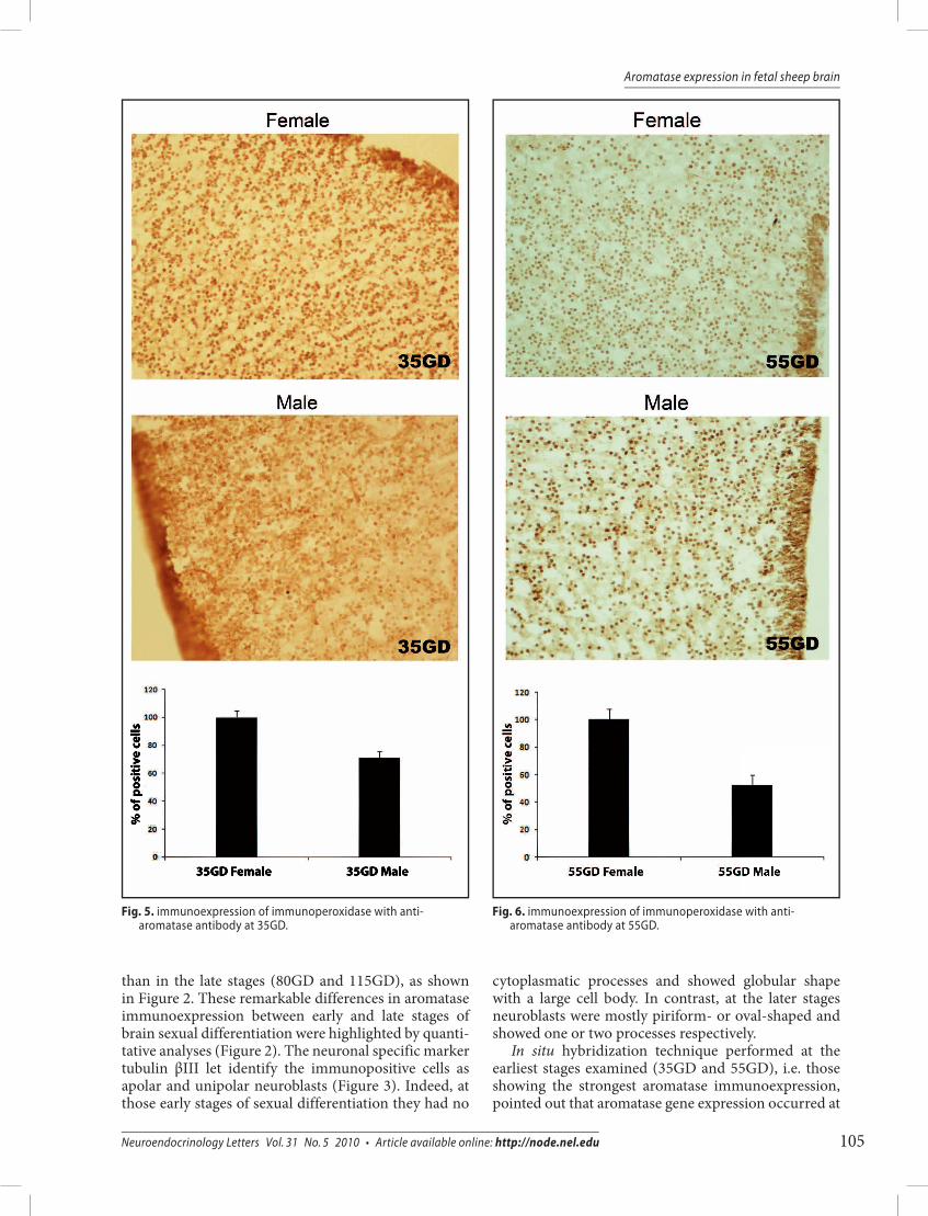
Fig. 5. immunoexpression of immunoperoxidase with anti-aromatase antibody at 35GD.
Fig. 6. immunoexpression of immunoperoxidase with anti-aromatase antibody at 55GD.

106 Copyright © 2010 Neuroendocrinology Letters ISSN 0172–780X • www.nel.edu
Arcadia Mura, Sergio Gadau, Gianluca Lepore, Francesca Balzano, Marco Zedda, Emilio Mura, Vittorio Farina
the hypothalamic level ever since the beginning of the crucial period of brain sexual differentiation (Figure 4).
Moreover, immuperoxidase with anti-aromatase antibody at 35GD and 55GD showed more noticeable cell immunoexpression in females than in males, this datum being illustrated in the diagram of Figures 5 and 6.
Western blot analysis revealed a band at 55 kD cor-responding to the molecular weight of brain aromatase. In fact, blot analysis confirmed the results of the immu-nocytochemical studies. Indeed, the densitometric analysis of the bands revealed a small but significant increase in aromatase amount in female than in male sheep fetuses (Figure 7).
Androgen receptor immunoexpression in the dien-cephalic periventricular area displayed a marked immu-noexpression at 35GD which gradually declined at the following stages, showing a trend similar to that found for aromatase immunoexpression (Figure 8).
dIScuSSIon
As initially determined by Short (1974) and later by Clarke et al. (1976), brain sexual differentiation in fetal sheep occurs prenatally, approximately from 30GD to 100GD. Aromatase is a fundamental enzyme involved in brain sexual differentiation and studies on the sheep reported that its activity can be detected at 64GD (Roselli et al. 2003). Our work pointed out for the first time that aromatase immunoexpression can be detected
along the whole period of sexual differentiation in the diencephalic periventricular area of fetal sheep brain. The higher aromatase expression in the early stages (35GD and 55GD) than in the late stages (80GD and 115GD) is in agreement with what reported by Con-nolly et al. (1994), who demonstrated aromatase activ-ity in guinea pig at early stages of sexual differentiation (35GD–40GD) showing a steady decline through devel-opment. Such higher amount in the early stages was also found in the present investigation on the sheep, suggest-ing that this enzyme is essential in the first half of the sexual differentiation period representing an input for the masculinization processes of the brain. Testosterone exposure of female sheep fetuses from 30GD to 86GD led to the masculinization of the external genitalia and defeminization with the elimination of LH surge (Woodet al. 1991). In addition, ewes showed a modification of LH surge mechanism leading to sporadic ovulation. In contrast, exposure to testosterone between 89GD and 135GD was without effects (Clarke & Scaramuzzi 1978; Short 1974). Variability in phenotypes was observed in female lambs from mothers undergone 20-day andro-gen treatment at the early (G30D to 50GD) or late (65GD to 85GD) stages of the critical period. The first group possessed a penis and an empty scrotum, whereas the second one was phenotypically normal (Wood et al. 1995). In addition, female fetuses exposed to androgens at early stage of brain sexual differentiation showed a delayed puberty timing (Wood & Foster 1998). A con-tinued exposure to estrogens during gestation is needed
Fig. 7. Western blot analysis revealed a band at 55 kD corresponding to the molecular weight of brain aromatase.

107Neuroendocrinology Letters Vol. 31 No. 5 2010 • Article available online: http://node.nel.edu
Aromatase expression in fetal sheep brain
Fig. 8. Androgen receptor immunoexpression in the diencephalic periventricular area.
to complete masculinization and defeminization pro-cesses in the fetal sheep brain (Foster et al. 2006), and the high aromatase immunoexpression detected here at the early stages (35GD and 55GD) likely suggests that this enzyme is involved in the onset of ovine brain sexual differentiation.
In our work aromatase expression was widely dis-tributed in different brain regions, especially in the
diencephalic periventricular area. The distribution pat-tern of aromatase in ovine fetal brain is consistent with the idea that the enzyme takes part in the crucial neu-ronal mechanisms involved in the central regulation of reproductive behavior and neuroendocrine functions.
Androgen receptor immunoreactivity revealed the same trend as aromatase immunoexpression, being marked in the early (35GD) and decreased in the late

108 Copyright © 2010 Neuroendocrinology Letters ISSN 0172–780X • www.nel.edu
Arcadia Mura, Sergio Gadau, Gianluca Lepore, Francesca Balzano, Marco Zedda, Emilio Mura, Vittorio Farina
(115GD) period of brain sexual differentiation. Thus, a correlation may be suggested between androgen and aromatase activity, as documented in rodents (Abdel-gadir et al. 1994; Connolly et al. 1990). However, data about the relationship between testosterone and brain aromatase are conflicting since some authors reported that in the rat testosterone can inhibit the expression and activity of aromatase (Lephart et al. 1992; Negri-Cesi et al. 2001). In details, Lephart et al. showed a sig-nificant dose-dependent decrease in aromatase activity caused by testosterone and dihydrotestosterone. In contrast, it has been demonstrated that androgens have a stimulatory role on aromatase expression both in rat and mouse brain (Karolczak et al. 1998; MacLusky et al. 1985). Moreover, studies on monkeys suggested that aromatization is not essential for androgen regulation during the development (Roselli & Resko 1986; Resko & Elliwood 1985). This mismatching evidence could be due to interspecific differences in the regulatory mecha-nisms of sex behavior.
In the present investigation higher aromatase immu-noexpression was detected in female than in male sheep fetal brain. The existence of differences in aromatase expression between sexes is however still matter of debate. Indeed, Roselli et al. (2003) demonstrated that sex differences were not present in fetal sheep hypotha-lamic areas at 64GD. This finding is consistent with other studies on the rat (George & Ojeda 1982; Lauber et al. 1997; Weisz et al. 1982), rabbit (George et al. 1978), and guinea pig (Connolly et al. 1994), all performed during the critical period of sexual differentiation. In contrast, Roselli and Resko (1986) observed in the monkey a little but significant sex difference in aromatase activ-ity in the amygdala and cerebral cortex but not in the hypothalamus and preoptic areas. In addition, Tobet et al. (1985) showed sex differences in the ferret fetal brain where aromatase activity was high in the preoptic area, but there were no sex differences at any postnatal stage examined. Moreover, age-dependent sex differences were demonstrated in aromatase immunoreactivity during the critical period of sexual differentiation in the rat (from 18GD to post-natal day 10) and shortly after (Lauber et al. 1997; Negri-Cesi et al. 2008). The rats did not display sex differences prenatally, but at days 2 and 6 after birth, males expressed significantly higher levels of aromatase mRNA than females, although at wean-ing (post-natal day 21) aromatase activity was higher in females than in males. This trend could be due to the fact that rat aromatase is involved in the onset of female puberty and is required for the normal expression of female sexual behavior (Negri-Cesi et al. 2008).
In conclusion, our study showed changes in aroma-tase immunoexpression in the diencephalic periventric-ular area throughout the whole period of brain sexual differentiation of fetal sheep. Aromatase expression was higher in the early stages (35GD and 55GD) than in the late stages (80GD and 115GD). Sex differences in aromatase immunoexpression at early stages were also
detected, more precisely females showed higher aro-matase expression than males. Androgen receptor and aromatase expression were similar as to their occur-rence during the whole period of brain sexual differ-entiation and their upregulation at early stages (35GD and 55GD). The correlation between androgen receptor and aromatase suggests a possible synergy of these two molecules in the neuronal tuning of reproduction and sex behavior.
REfERENcEs
1 Abdelgadir sE, Resko JA, Ojeda sR, Lephart ED, McPhaul MJ, Roselli cE (1994). Androgens regulate aromatase cytochrome P450 messenger ribonucleic acid in rat brain. Endocrinology. 135: 395–401.
2 Balthazart J, Ball Gf (1998). New insights into the regulation and function of brain estrogen synthase (aromatase). Trends Neuro-sci. 21: 243–249.
3 clarke IJ, scaramuzzi RJ, short RV (1976). Effects of testosterone implants in pregnant ewes on their female offspring. J Embryol Exp Morphol. 36: 87–99.
4 clarke IJ, scaramuzzi RJ (1978). sexual behaviour and LH secre-tion in spayed androgenized ewes after a single injection of testosterone or oestradiol-17beta. J Reprod fertil. 52: 313–320.
5 connolly PB, Roselli cE, Resko JA (1990). Aromatase activity in adult guinea pig brain is androgen dependent. Biol Reprod. 43: 698–703.
6 connolly PB, Roselli cE, Resko JA (1994). Aromatase activity in developing guinea pig brain: ontogeny and effects of exogenous androgens. Biol Reprod. 50: 436–441.
7 foster DL, Jackson LM, Padmanabhan V (2006). Programming of GnRH feedback controls timing puberty and adult reproductive activity. Mol cell Endocrinol. 25: 109–119.
8 Garcia-segura LM, Wozniak A, Azcoitia I, Rodriguez JR, Hutchison RE, Hutchison JB (1999). Aromatase expression by astrocytes after brain injury: implications for local estrogen formation in brain repair. Neuroscience. 89: 567–578.
9 Garcia-segura LM (2008). Aromatase in the brain: not just for reproduction anymore. J Neuroendocrinol. 20: 705–712.
10 George fW, Tobleman WT, Milewich L, Wilson JD (1978). Aroma-tase activity in the developing rabbit brain. Endocrinology. 102: 86–91.
11 George fW, Ojeda sR (1982). changes in aromatase activity in the rat brain during embryonic, neonatal, and infantile develop-ment. Endocrinology. 111: 522–529.
12 Herbosa cG, foster DL (1996). Defeminization of the reproduc-tive response to photoperiod occurs early in prenatal develop-ment in the sheep. Biol Reprod. 54: 420–428.
13 Karolczak M, Küppers E, Beyer c (1998). Developmental expres-sion and regulation of aromatase and 5alpha-reductase type I mRNA in the male and female mouse hypothalamus. J Neuroen-docrinol. 10: 267–274.
14 Lauber ME, sarasin A, Lichtensteiger W (1997). Transient sex dif-ferences of aromatase (cYP19) mRNA expression in the develop-ing rat brain. Neuroendocrinology. 66: 173–180.
15 Lephart ED, simpson ER, McPhaul MJ, Kilgore MW, Wilson JD, Ojeda sR (1992). Brain aromatase cytochrome P-450 messenger RNA levels and enzyme activity during prenatal and perinatal development in the rat. Brain Res Mol Brain Res. 16: 187–192.
16 Lephart ED (1996). A review of brain aromatase cytochrome P450. Brain Res. 22: 1–26.
17 Lepore G, Gadau s, Mura A, Zedda M, farina V (2009). Aromatase immunoreactivity in fetal ovine neuronal cell cultures exposed to oxidative injury. Eur J Histochem. 53: 233–238.
18 MacLusky NJ, Philip A, Hurlburt c, Naftolin f (1985). Estrogen for-mation in the developing rat brain: sex differences in aromatase activity during early post-natal life. Psychoneuroendocrinology. 10: 355–361.

109Neuroendocrinology Letters Vol. 31 No. 5 2010 • Article available online: http://node.nel.edu
Aromatase expression in fetal sheep brain
19 Mara L, Pilichi s, sanna A, Accardo c, chessa B, chessa f, Dattena M, Bomboi G, cappai P (2004). sexing of in vitro produced ovine embryos by duplex PcR. Mol Reprod Dev. 69: 35–42.
20 Martínez-cerdeño V, Noctor sc, Kriegstein AR (2006). Estradiol stimulates progenitor cell division in the ventricular and subven-tricular zones of the embryonic neocortex. Eur J Neurosci. 24: 3475–3488.
21 McGeady TA, Quinn PJ, fitzPatrick Es, Ryan MT (2006). Veterinary Embryology. In: chapter 23: Age determination of the embryo and foetus. Oxford: Blackwell Publishing. p. 331–336.
22 Moody sA, Miller V, spanos A, frankfurter A (1996). Develop-mental expression of a neuron-specific beta-tubulin in frog (Xenopus laevis): a marker for growing axons during the embry-onic period. J comp Neurol. 364: 219–230.
23 Naftolin f, Ryan KJ, Petro Z, (1971). Aromatization of andro-stenedione by the diencephalon. J clin Endocrinol Metab. 33: 368–370.
24 Negri-cesi P, Melcangi Rc, celotti f, Martini L (1992). Aromatase activity in cultured brain cells: difference between neurons and glia. Brain Res. 589: 327–332.
25 Negri-cesi P, colciago A, Motta M, Martini L, celotti f (2001). Aro-matase expression and activity in male and female cultured rat hypothalamic neurons: effect of androgens. Mol cell Endocrinol. 178: 1–10.
26 Negri-cesi P, colciago A, Pravettoni A, casati L, conti L, celotti f (2008). sexual differentiation of the rodent hypothalamus: hormonal and environmental influences. J steroid Biochem Mol Biol. 109: 294–299.
27 Resko JA, Ellinwood WE (1985). Negative feedback regulation of gonadotropin secretion by androgens in fetal rhesus macaques. Biol Reprod. 33: 346–352.
28 Roselli cE, Resko JA (1986). Effects of gonadectomy and andro-gen treatment on aromatase activity in the fetal monkey brain. Biol Reprod. 35: 106–112.
29 Roselli cE, stormshak f, Resko JA (2000). Distribution of aroma-tase mRNA in the ram hypothalamus: an in situ hybridization study. J Neuroendocrinol. 12: 656–664.
30 Roselli cE, Resko JA (2001). cytochrome P450 aromatase (cYP19) in the non-human primate brain: distribution, regulation, and functional significance. J steroid Biochem Mol Biol. 79: 247–253.
31 Roselli cE, Resko JA, stormshak f (2003). Estrogen synthesis in fetal sheep brain: effect of maternal treatment with an aroma-tase inhibitor. Biol Reprod. 68: 370–374.
32 Roselli cf (2007). Brain aromatase: roles in reproduction and neuroprotection. J steroid Biochem Mol Biol. 106: 143–150.
33 senger PL (2003). Pathway to pregnancy and parturition. In: chapter 6: Puberty. 2nd rev. ed. Washington: current conception, Inc. p. 128–143.
34 simerly RB (2002). Wired for reproduction: organization and development of sexually dimorphic circuits in the mammalian forebrain. Annu Rev Neurosci. 25: 507–536.
35 simpson ER, Mahendroo Ms, Means GD, Kilgore MW, Hin-shelwood MM, Graham-Lorence s, et al (1994). Aromatase cyto-chrome P450, the enzyme responsible for estrogen biosynthesis. Endocr Rev.15: 342–355.
36 short RV (1974). sexual differentiation of the brain of the sheep. INsERM. 32: 121–142.
37 Tabori NE, stewart Ls, Znamensky V, Romeo RD, Alves sE, McEwen Bs, et al (2005). Ultrastructural evidence that androgen receptors are located at extranuclear sites in the rat hippocam-pal formation. Neuroscience. 130: 151–163.
38 Tobet sA, shim JH, Osiecki sT, Baum MJ, canick JA (1985). Andro-gen aromatization and 5 alpha-reduction in ferret brain during perinatal development: effects of sex and testosterone manipu-lation. Endocrinology. 116: 1869–1877.
39 Von schassen c, fester L, Prange-Kiel J, Lohse c, Huber c, Böttner M, Rune GM (2006). Oestrogen synthesis in the hippocampus: role in axon outgrowth. J Neuroendocrinol. 18: 847–856.
40 Weisz J, Brown BL, Ward IL, (1982). Maternal stress decreases ste-roid aromatase activity in brains of male and female rat fetuses. Neuroendocrinology. 35: 374–379.
41 Wood RI, Ebling fJ, I’Anson H, Bucholtz Dc, Yellon sM, foster DL (1991). Prenatal androgens time neuroendocrine sexual matura-tion. Endocrinology. 128: 2457–2468.
42 Wood RI, Mehta V, Herbosa cG, foster DL (1995). Prenatal tes-tosterone differentially masculinizes tonic and surge modes of luteinizing hormone secretion in the developing sheep. Neuro-endocrinology. 62: 238–247.
43 Wood RI, foster DL (1998). sexual differentiation of reproductive neuroendocrine function in sheep. Rev Reprod. 3: 30–40.
Copyright © 2022 FDOKUMEN