Modulation of human CYP19A1 activity by mutant NADPH P450 oxidoreductase
17
Modulation of Human CYP19A1 Activity by Mutant NADPH P450 Oxidoreductase Amit V. Pandey,* Petra Kempna ´ ,* Gaby Hofer, Primus E. Mullis, and Christa E. Flu ¨ ck Pediatric Endocrinology, Diabetology & Metabolism, and Department of Clinical Research, University of Bern, CH-3010 Bern, Switzerland Mutations in NADPH P450 oxidoreductase (POR) cause a broad spectrum of human disease with abnormalities in steroidogenesis. We have studied the impact of P450 reductase mutations on the activity of CYP19A1. POR supported CYP19A1 ac- tivity with a calculated K m of 126 nM for andro- stenedione and a V max of 1.7 pmol/min. Mutations R457H and V492E located in the FAD domain of POR that disrupt electron transfer caused a com- plete loss of CYP19A1 activity. The A287P mutation of POR decreased the activities of CYP17A1 by 60–80% but had normal CYP19A1 activity. Molec- ular modeling and protein docking studies sug- gested that A287P is involved in the interaction of POR:CYP17A1 but not in the POR:CYP19A1 inter- action. Mutations C569Y and V608F in the NADPH binding domain of POR had 49 and 28% of activity of CYP19A1 compared with normal reductase and were more sensitive to the amount of NADPH avail- able for supporting CYP19A1 activity. Substitution of NADH for NADPH had a higher impact on C569Y and V608F mutants of POR. Similar effects were obtained at low/high (5.5/8.5) pH, but using octanol to limit the flux of electrons from POR to CYP19A1 inhibited activity supported by all variants. High molar ratios of KCl also reduced the CYP19A1 sup- porting activities of C569Y and V608F mutants of POR to a greater extent compared to normal POR and A287P mutant. Because POR supports many P450s involved in steroidogenesis, bone formation, and drug metabolism, variations in the effects of POR mutations on specific enzyme activities may explain the broad clinical spectrum of POR deficiency. (Molecular Endocrinology 21: 2579–2595, 2007) N ADPH P450 OXIDOREDUCTASE (POR) (EC 1.6.2.4) transfers electrons from nicotinamide ad- enine dinucleotide phosphate (NADPH) to all microso- mal type II P450 enzymes. The 32-kb gene (GI 94721356, NM_000941.2) for POR is located on chro- mosome 7q11.2 (1), has one untranslated exon (exon 1U) and 15 coding exons (2), and encodes a 82-kDa membrane-bound protein with 680 amino acids (NP_000932.3) (3). POR was initially identified by Horecker (4, 5) in 1950 as a cytochrome c reductase. Later studies by Williams and Kamin (6) and Phillips and Langdon (7) demonstrated this flavoprotein to be present in the endoplasmic reticulum (microsomes); subsequent studies in the 1960s and 1970s linked POR to the microsomal electron transport chains cy- tochromes P450 and b 5 involved in drug and steroid hydroxylations (8). The definitive evidence for the re- quirement of POR in cytochrome P450-mediated re- actions came from the work of Lu et al. (8) who dis- sected the P450 containing mixed function oxidase system into three constituent components, POR, cy- tochrome P450, and lipids. The structure of POR is well understood from the x-ray crystal structures of the soluble, N-terminally deleted (N-63) form of rat POR (9) and FMN binding domain of human POR (10). This structure shows that POR has two distinct domains, one containing the NADPH-binding site and the FAD- binding domain, and the other containing the FMN domain that interacts with the redox-partner binding site of the P450s. The FMN domain is located at the N terminus of the POR and is structurally similar to fla- vodoxins, whereas the FAD/NADPH domain is located at the C terminus and is similar to ferredoxin reducta- ses. The N terminus of POR has a 25- to 30-amino- acid-long hydrophobic sequence that serves as an anchor to position the POR toward the cytosolic side of the endoplasmic reticulum and is important for in- teraction with cytochrome P450s. Deletion of this N terminal leads to loss of P450 reductase activity of POR (8, 11). The domains that bind the FAD and FMN moieties are separated by a flexible hinge region. The electron transfer in POR from NADPH occurs in the form of two equivalents of hydride ion transfer to FAD moiety (12, 13). There are 50 genes encoding micro- somal P450s that have been identified from the human genome (14); of these, 20 are involved in the biosyn- thesis of cholesterol, steroid hormones, fatty acids, and eicosanoids; 15 are involved in hepatic drug me- First Published Online June 26, 2007 * A.V.P. and P.K. contributed equally to this work. Abbreviations: ABS, Antley Bixler syndrome; 3D, three- dimensional; K m , Michaelis constant; MD, molecular dynam- ics; NADPH, nicotinamide adenine dinucleotide phosphate; POR, NADPH P450 oxidoreductase; V max , maximum velocity; WT, wild type. Molecular Endocrinology is published monthly by The Endocrine Society (http://www.endo-society.org), the foremost professional society serving the endocrine community. 0888-8809/07/$15.00/0 Molecular Endocrinology 21(10):2579–2595 Printed in U.S.A. Copyright © 2007 by The Endocrine Society doi: 10.1210/me.2007-0245 2579
Transcript of Modulation of human CYP19A1 activity by mutant NADPH P450 oxidoreductase

Modulation of Human CYP19A1 Activity by MutantNADPH P450 Oxidoreductase
Amit V. Pandey,* Petra Kempna,* Gaby Hofer, Primus E. Mullis, and Christa E. Fluck
Pediatric Endocrinology, Diabetology & Metabolism, and Department of Clinical Research, Universityof Bern, CH-3010 Bern, Switzerland
Mutations in NADPH P450 oxidoreductase (POR)cause a broad spectrum of human disease withabnormalities in steroidogenesis. We have studiedthe impact of P450 reductase mutations on theactivity of CYP19A1. POR supported CYP19A1 ac-tivity with a calculated Km of 126 nM for andro-stenedione and a Vmax of 1.7 pmol/min. MutationsR457H and V492E located in the FAD domain ofPOR that disrupt electron transfer caused a com-plete loss of CYP19A1 activity. The A287P mutationof POR decreased the activities of CYP17A1 by60–80% but had normal CYP19A1 activity. Molec-ular modeling and protein docking studies sug-gested that A287P is involved in the interaction ofPOR:CYP17A1 but not in the POR:CYP19A1 inter-action. Mutations C569Y and V608F in the NADPHbinding domain of POR had 49 and 28% of activityof CYP19A1 compared with normal reductase and
were more sensitive to the amount of NADPH avail-able for supporting CYP19A1 activity. Substitutionof NADH for NADPH had a higher impact on C569Yand V608F mutants of POR. Similar effects wereobtained at low/high (5.5/8.5) pH, but using octanolto limit the flux of electrons from POR to CYP19A1inhibited activity supported by all variants. Highmolar ratios of KCl also reduced the CYP19A1 sup-porting activities of C569Y and V608F mutants ofPOR to a greater extent compared to normal PORand A287P mutant. Because POR supports manyP450s involved in steroidogenesis, bone formation,and drug metabolism, variations in the effects ofPOR mutations on specific enzyme activities mayexplain the broad clinical spectrum of PORdeficiency. (Molecular Endocrinology 21:2579–2595, 2007)
NADPH P450 OXIDOREDUCTASE (POR) (EC1.6.2.4) transfers electrons from nicotinamide ad-
enine dinucleotide phosphate (NADPH) to all microso-mal type II P450 enzymes. The 32-kb gene (GI94721356, NM_000941.2) for POR is located on chro-mosome 7q11.2 (1), has one untranslated exon (exon1U) and 15 coding exons (2), and encodes a 82-kDamembrane-bound protein with 680 amino acids(NP_000932.3) (3). POR was initially identified byHorecker (4, 5) in 1950 as a cytochrome c reductase.Later studies by Williams and Kamin (6) and Phillipsand Langdon (7) demonstrated this flavoprotein to bepresent in the endoplasmic reticulum (microsomes);subsequent studies in the 1960s and 1970s linkedPOR to the microsomal electron transport chains cy-tochromes P450 and b5 involved in drug and steroidhydroxylations (8). The definitive evidence for the re-quirement of POR in cytochrome P450-mediated re-actions came from the work of Lu et al. (8) who dis-
sected the P450 containing mixed function oxidasesystem into three constituent components, POR, cy-tochrome P450, and lipids. The structure of POR iswell understood from the x-ray crystal structures of thesoluble, N-terminally deleted (N-63) form of rat POR (9)and FMN binding domain of human POR (10). Thisstructure shows that POR has two distinct domains,one containing the NADPH-binding site and the FAD-binding domain, and the other containing the FMNdomain that interacts with the redox-partner bindingsite of the P450s. The FMN domain is located at the Nterminus of the POR and is structurally similar to fla-vodoxins, whereas the FAD/NADPH domain is locatedat the C terminus and is similar to ferredoxin reducta-ses. The N terminus of POR has a 25- to 30-amino-acid-long hydrophobic sequence that serves as ananchor to position the POR toward the cytosolic sideof the endoplasmic reticulum and is important for in-teraction with cytochrome P450s. Deletion of this Nterminal leads to loss of P450 reductase activity ofPOR (8, 11). The domains that bind the FAD and FMNmoieties are separated by a flexible hinge region. Theelectron transfer in POR from NADPH occurs in theform of two equivalents of hydride ion transfer to FADmoiety (12, 13). There are 50 genes encoding micro-somal P450s that have been identified from the humangenome (14); of these, 20 are involved in the biosyn-thesis of cholesterol, steroid hormones, fatty acids,and eicosanoids; 15 are involved in hepatic drug me-
First Published Online June 26, 2007* A.V.P. and P.K. contributed equally to this work.Abbreviations: ABS, Antley Bixler syndrome; 3D, three-
dimensional; Km, Michaelis constant; MD, molecular dynam-ics; NADPH, nicotinamide adenine dinucleotide phosphate;POR, NADPH P450 oxidoreductase; Vmax, maximum velocity;WT, wild type.
Molecular Endocrinology is published monthly by TheEndocrine Society (http://www.endo-society.org), theforemost professional society serving the endocrinecommunity.
0888-8809/07/$15.00/0 Molecular Endocrinology 21(10):2579–2595Printed in U.S.A. Copyright © 2007 by The Endocrine Society
doi: 10.1210/me.2007-0245
2579

tabolism; and 15 are “orphans” with unknown catalyticactivities (14, 15). Because all these enzymes dependon POR for electron supply, disruption of POR mayaffect all microsomal P450 enzyme activities with di-sastrous consequences. Consistent with this, the PORknockout mouse results in early embryonic lethality(16, 17). Recently, we have identified more than 20different POR mutations in over 34 patients with ap-parent combined 17�-hydroxylase, 21-hydroxylasedeficiency (OMIM 201750) and a broad spectrum ofclinical characteristics ranging from severe neonatalskeletal malformations with genital ambiguity [knownas Antley Bixler syndrome (ABS); OMIM 207410], tophenotypically minor polycystic ovary syndrome-likefeatures (18–24). Subsequent to our initial report, Arltet al. (25) identified another POR missense mutation,Y181D (reported as Y178D), and also reported three ofthe POR mutations (A287P, R457H, and C569Y) thatwe had originally described. In a larger follow-upstudy, we have examined the POR genes in 19 addi-tional patients (19). Fifteen of 19 patients having ab-normal genitalia and disordered steroidogenesis werehomozygous or apparent compound heterozygous forPOR mutations that destroyed or dramatically inhib-ited POR activity. Most missense mutations werefound once, but A287P was found on 10 alleles, andR457H was found on seven alleles. Thus, these twomissense mutations accounted for 17 of 34 (50%) ofthe identified POR missense mutations. The A287Pmutation was found only in samples from subjectsdescribed as “white,” “Caucasian,” or “European.”This same mutation was also found on two (of eight)alleles in our initial report (18) in individuals of Euro-pean heritage. Thus, A287P is a frequent mutationcausing POR deficiency in this group. The R457H mu-
tation was found in four of eight alleles from Japanesepatients. This same mutation was also found on one(of two) Japanese alleles in our initial report (18) and ontwo of four alleles (26) and 10 of 16 alleles (27) in twosubsequent reports of Japanese patients. Thus,R457H is strongly associated with alleles of Japaneseheritage (28). In the recent reports from other labora-tories, several new POR variants have been described(28–30). A G5G variant of POR has recently been re-ported to be associated with increased breast cancerrisk in African-American women (31).
POR-dependent, steroidogenic type II P450 en-zymes include CYP17A1 (17�-hydroxylase/17,20lyase), CYP21B1 (21-hydroxylase), and CYP19A1(aromatase). CYP17A1 is essential for the synthesis ofglucocorticoids through its 17�-hydroxylase activityand for the synthesis of all sex steroids through its17�-hydroxylase and 17,20 lyase activities (32).CYP21B1 catalyzes 21-hydroxylation of mineralocor-ticoids and glucocorticoids, and CYP19A1 convertsandrogens to estrogens, specifically androstenedioneto estrone, testosterone to estradiol, and 16�-hy-droxytestosterone to estriol (33). CYP19A1 is encodedby CYP19 that belongs to the family 19 of the P450proteins (14, 34). It is expressed in many differenttissues including the ovaries, testes, placenta, adiposetissue, and bone osteoblasts (33). CYP19A1 is local-ized in the endoplasmatic reticulum of the cell andcatalyzes a unique three-step reaction (Fig. 1). The firsttwo oxidative steps are cytochrome P450 hydroxyla-tions, and the third step is a ferric peroxide removal ofthe aldehyde moiety, followed by aromatization of thesteroid A-ring. Each step of the reaction requires a pairof electrons that are supplied from NADPH throughPOR.
Fig. 1. Schematic Diagram of Reactions Catalyzed by CYP19A1The reaction catalyzed by aromatase involves conversion of the ring A of the steroid structure to an aromatic ring with the loss
of the angular C-19 methyl group and the cis-eliminaion of the 1� and 2� hydrogens to yield estrogen and formic acid. Note: thisthree-step reaction needs three pairs of electrons provided by NADPH.
2580 Mol Endocrinol, October 2007, 21(10):2579–2595 Pandey et al. • Interaction of POR and CYP19A1

Functional testing of our initial POR mutations usingthe classical cytochrome c assay and CYP17A1 (17�-hydroxylase/17,20 lyase) activity assays revealed par-tially conflicting results indicating that different PORvariants may impact supported enzymatic reactions tovariable degrees (18, 19). There were three distinctfeatures of the POR mutations that we have originallydescribed in our first report and that we are now ad-dressing in more detail. First, mutations in the FADregion that destroy the binding of FAD to POR resultedin total loss of activity in both CYP17A1 and cyto-chrome c-based assays and therefore, should be al-most inactive with all substrates. Second, the mutationA287P, found mostly in European patients, is in alocation in POR that is not directly involved in thefunction of the electron transport and has close towild-type (WT) activity with cytochrome c assay butlower activity with CYP17A1 assays. So it is possiblethat A287 is involved in the interaction of POR toP450s and other electron acceptors, and dependingon its role in interacting with a particular acceptorprotein, it may have variable effects on the activity ofparticular proteins. Third, the mutations in the NADPHbinding domain of POR showed a higher Michaelisconstant (Km) for NADPH in cytochrome c-based as-say. However, CYP17A1 was assayed with fixedNADPH concentration of 1 mM, which is more than5000 times higher than the apparent Km of POR forNADPH in cytochrome c-based assays. Therefore it isconceivable that the variations in the NADPH concen-tration might play a role in activity of these mutantsand that a P450 with a faster reaction or requiringhigher amounts of NADPH may be affected to agreater extent by mutations in the NADPH bindingdomain of POR than most other POR-dependentproteins.
To test these three hypotheses concerning differentPOR mutants, we coexpressed WT human CYP19A1,which requires three pairs of electrons for its activity,together with WT or mutant POR in yeast to obtainrecombinant proteins for performing enzyme kineticstudies. We then compared kinetic data for POR-sup-ported CYP19A1 activity with CYP17A1 activities. Weused variations in assay conditions that limit the elec-tron flux between POR and CYP19A1 to assess theimpact on different mutations of POR. Finally, we builtthree-dimensional (3D) protein models for CYP19A1,CYP17A1, and POR and performed computationalprotein docking studies to understand better thestructure-function relationship of various POR mu-tants and their impact on P450 activities.
RESULTS AND DISCUSSION
CYP19A1 Activity Supported by WT orMutant POR
To assess the effect of WT or mutant POR onCYP19A1 activity we coexpressed both proteins in
yeast W(B) (35), prepared microsomes, and testedthem for the ability to convert [3H]androstenedione toestrone, thereby releasing tritiated water (36). We de-cided to use microsomal preparations containingP450 and POR in our reactions instead of purifiedPOR-P450-reconstituted systems for several differentreasons. First, in our humanized yeast system we wereable to express full-length aromatase in a similar fash-ion as we have expressed P450c17 for enzyme kineticanalysis in previous studies (18, 37). Western blotanalysis confirmed that engineered yeast microsomescontained similar amounts of both WT or mutant PORas well as WT aromatase. Second, various methodsavailable to reconstitute POR with P450 proteins givevariable results in different laboratories. The reconsti-tuted systems using phospholipids give best activity ata POR:P450 ratio of 1:5, and activity drops once thisratio is exceeded (38). This ratio and resulting activitiesvary depending on the type of P450 used and may notprovide a standard value to compare P450 activities.Another concern was that these methods were opti-mized with normal POR in mind and changes broughtby purification processes that may affect differentPOR variants to different degrees may add anotherlevel of complication that needs to be resolved sepa-rately for each POR variant before purified prepara-tions of variant POR could routinely be used in all P450reactions.
Kinetic studies of CYP19A1 reaction converting an-drostenedione to estrone revealed a Km of 126.3 nM
and a maximum velocity (Vmax) of 1.70 pmol/min�mgprotein (Table 1). Previously published enzyme kineticstudies found Km values for the conversion of andro-stenedione by human CYP19A1 ranging from 100 to260 nM when assaying N-terminally modified recom-binant human CYP19A1 expressed either in insectcells (39) or in E. coli (40). Thus our novel, yeast-basedfunctional assay seemed a reasonable system forstudying the influence of POR variants on CYP19A1activity.
In our previous report of the first POR mutations, wehad identified the POR missense mutations A287P,R457H, and V492E in patients with the severe ABS skel-
Table 1. Calculated Kinetic Constants for CYP19A1Activity with POR Variants Using Androstenedione asSubstrate
Apparent Km(nM)
Vmax(pmol/min) Vmax/Km
WT 126.3 � 10.4 1.70 � 0.28 13.45A287P 74.6 � 15.2 1.04 � 0.23 13.96R457H 60.2 � 18.4 0.01 � 0.002 0.17V492E n.d. n.d. n.d.C569Y 171.2 � 31.8 1.18 � 0.17 6.88V608F 78.1 � 22.9 0.25 � 0.08 3.24
Values are mean � SD of triplicate data sets. Experimentalconditions are as described in Materials and Methods. n.d.,Conversion activity too small (�1%); therefore, kinetic con-stants were not determined.
Pandey et al. • Interaction of POR and CYP19A1 Mol Endocrinol, October 2007, 21(10):2579–2595 2581

etal malformation phenotype, genital ambiguities, anddisordered steroidogenesis, whereas the POR mutationsC569Y and V608F were found in a young woman withnormal breast development who was infertile and hadpolycystic ovaries and mild arterial hypertension (18, 41).Testing the effect of those POR variants on CYP17A1activities in vitro, we found a loss of both 17�-hydroxy-lase and 17,20 lyase activities of CYP17A1 for the PORmutations R457H and V492E, a severe decrease of bothactivities for A287P, but only a mild inhibition for C569Yand V608F. Thus, our in vitro assay for CYP17A1:PORreflected the clinical findings in those patients. For POR-supported CYP19A1 activity, we found that comparedwith WT POR, A287P and C569Y inhibited CYP19A1activity moderately and V608F significantly; however,when CYP19A1 activity was tested with POR R457H andV492E, enzymatic activity was completely lost (Fig. 2A).Calculations for enzyme kinetic constants revealed val-ues that are summarized in Table 1. The catalytic effi-ciency (Vmax/Km) of CYP19A1 supported by POR variantA287P was similar to WT POR, but catalytic efficiency ofPOR mutants C569Y and V608F was significantly lowerand was almost unmeasurable for POR R457H andV492E (Table 1).
POR Mutants A287P, C569Y, and V608F HaveVariable Effects on CYP19A1 and CYP17A1 Activities
POR is the obligate electron transfer partner for bothsteroidogenic enzymes CYP19A1 and CYP17A1. Wehave tested the influence of different POR variants onthe activities of CYP17A1 (18, 19) by identical methodsas we have now used to test their influence onCYP19A1 activity; therefore, we were able to comparethe impact of specific POR mutants on both enzymesdirectly (Fig. 2B). For POR mutants R457H and V492E,which are located in a highly conserved FAD bindingdomain (Fig. 2, C and D), enzymatic activities of boththe CYP17A1 and CYP19A1 were lost. In contrast,POR mutants A287P, C569Y, and V608F were foundto affect the activities of CYP17A1 and CYP19A1 todifferent degrees (Fig. 2B). Mutation A287P exerted noinhibitory effect on CYP19A1 activity compared withWT POR but was found to decrease 17�-hydroxylaseto 20% and 17,20 lyase to 10% of WT activity (18).Conversely, we found a more prominent inhibitory ef-fect of POR mutants C569Y and V608F, which areboth located in the NADPH binding domain (Fig. 2C),on CYP19A1 activity than on CYP17A1 activities (25–50% vs. 50–80%; Fig. 2B and Ref. 18). Thus, differentPOR variants may affect diverse P450 activities todifferent degrees, explaining the broad spectrum ofclinical phenotypes.
POR Mutants R457H and V492E Are Located inthe FAD Domain and Disrupt Electron Transfer toAny Interacting P450
POR mutations R457H and V492E, which were foundto cause a loss of both CYP19A1 and CYP17A1 ac-
tivities, are located in the FAD domain of the PORprotein (Fig. 2D). The FAD binding domain (residues450–492) is highly conserved, and the sequence RYYSI(457–461) is invariant among human, rat, and yeast POR.The x-ray crystal structure of rat POR suggests that R457forms a hydrogen bond with the pyrophosphate group ofFAD, and Y459 contacts the FAD isoalloxazine ring andhydrogen bonds with the ribityl 3�-hydroxyl group (9, 42).Mutation R457H abolished all measurable activity ofCYP19A1 similar to CYP17A1 activities (Fig. 2B). Similarresults were obtained when these mutations were stud-ied in rat POR (42–44). Residues 488–494, which lie inhelix N, form hydrogen bonds with the FAD pyrophos-phate. The mutation V492E, which is predicted to disrupthydrogen bonds with FAD, has no activity. Thus, weconclude that mutations in the FAD domain of POR thatdirectly interfere with the binding of FAD may affect alldepending P450 enzyme activities severely because mu-tations in this location will hamper the ability of POR totransfer electrons to the FMN group of POR that ulti-mately provides electrons to P450s. Diminished FADbinding to R457H and V492E mutants of POR has re-cently been demonstrated (45).
3D Models of CYP19A1 and CYP17A1
Our CYP17A1 and CYP19A1 models were based onstructures of mammalian P450 that have becomeavailable in the past couple of years. Both modelsshowed that typical P450 fold around I helix and hemebinding sites are in agreement with the structures ofother mammalian P450 structures. CYP19A1 modelhad a fold typical of mammalian P450 structures withwell-defined C, D, E, G, H, I, J, K, and L helices formingthe structure. The helix F was in two distinct parts evenafter extensive MD refinement and could be a featurefor providing access to different substrates ofCYP19A1 because it was found to be quite flexible(Fig. 3). Models had energy values and structure pa-rameters close to experimental structures (Table 2).Structural models of CYP19A1 based on bacterialP450 structures have been described previously (46,47). In the later effort a consensus core structurebased on structures of soluble bacterial P450s wasemployed to build the initial model, and less con-served residues and loops were added separately (47).Although the major features of the P450 structure likeheme binding region and I-helix that constitutes thewidth of the molecule were found to be similar in thebacterial P450-based model, the substrate accesschannel and N-terminal helices were found to beslightly different from our model. A kink in the I-helix ofCYP19A1 that is caused by sequence A-A-P was pre-dicted in the earlier model and was also present in ourmodel structure. More recently, structural models ofCYP19A1 have been built based on mammalian P450structures (48, 49). A model of CYP19A1 publishedrecently that is based on the structure of rabbit P4502C5 has similar features as our model (49). In thebacterial structure-based model, H105 was proposed
2582 Mol Endocrinol, October 2007, 21(10):2579–2595 Pandey et al. • Interaction of POR and CYP19A1

Fig. 2. Localization of POR Mutations and Their Effect on CYP19A1 ActivityA, Human CYP19A1 activity supported by WT or mutant POR. Yeast strain W(B) lacking endogenous CPR was transfected with
both human WT CYP19 and WT or mutant POR expression vectors. Microsomes containing CYP19A1 and POR proteins wereprepared and their activity to convert [3H] labeled androstenedione to estrone was tested by the tritiated water release assay (36).Kinetic data are presented in a fitted plot. The calculated Km and Vmax values are summarized in Table 1. F, Red line, WT; f, blueline, A287P; light green line, R457H; �, yellow line, V492E; �, green line, C569Y; Œ, black line, V608F. B, Catalytic efficiency ofCYP19A1 activity supported by WT or mutant POR compared with both 17�-hydroxylase and 17,20 lyase activities of CYP17A1.Catalytic efficiency was calculated as Vmax/Km (Table 1) and plotted as percentage of WT. To compare the effect of POR variantson different P450s, we have included the equivalent values calculated for the 17�-hydroxylase and 17,20 lyase activities ofCYP17A1, which we have tested in a similar fashion previously (18). White columns, 17�-Hydroxylase activity; gray, 17,20 lyaseactivity; black, CYP19A1 activity. C, POR mutations in the model structure of human P450 oxidoreductase. A 3D model of humanPOR showing the position of amino acids A287, R457, V492, C569, and V608. The POR model is based on the crystal structureof rat POR (PDB no. 1AMO) that shares 96% amino acid similarity with human POR. Protein is shown as a ribbon model andcolored light blue. Arginine 457 and valine 492 are shown in red, alanine 287 is shown in magenta, and cysteine 569 and valine608 are in cyan. FAD and FMN are shown in yellow, and NADPH is shown in blue. D, Localization and impact of POR mutationsR457H and V492E. A closeup view of the FAD binding domain of POR showing the positions of amino acids R457 and V492. PORis shown as a blue ribbon model, FAD and FMN groups are shown in yellow spheres, and NADPH is shown as a blue spheremodel. Amnio acids R457 and V492 are shown in red sphere models. The van der Walls surface of amino acids R457 and V492is shown as a mesh model to emphasize the location and interaction with FAD group. POR mutations R457H and V492E arelocated in the FAD domain of the POR protein, and R457 forms a hydrogen bond with the pyrophosphate group of FAD. MutationR457H results in complete loss of POR enzymatic activity. Amino acids V492 in POR is involved in stabilization of FAD group byforming hydrogen bonds. A change to glutamic acid results in disruption of FAD binding that leads to total loss of enzymaticactivity of POR. The mutations in the FAD domain of POR, which destabilize the binding of FAD, may severely damage the electrontransfer process because in the absence of FAD group in the POR the electrons from NADPH could not be transferred to the FMNgroup of POR or to its partner proteins. Protein model was edited with the programs Pymol and DeepView and rendered as aray-traced image with POVRAY.
Pandey et al. • Interaction of POR and CYP19A1 Mol Endocrinol, October 2007, 21(10):2579–2595 2583

to be pointing toward solvent and had similar positionin our (CYP3A4-based) model and the CYP2C5-basedmodel. However, K150 present on helix C that wasproposed to interact with heme propionate was faraway from heme in mammalian P450-based models,and this position is taken by R145 in our model andR130 in the CYP3A4 structure. One of the heme pro-pionates in our CYP19A1 model is stabilized by K119and R375, whereas R435 and R145 stabilize the other
propionate; these positions are R105, R375, R130,and R440 in CYP3A4 structure. A cysteine at position437 that provides the fifth ligand for heme iron inCYP19A1 is conserved across all P450s and wasmodeled similarly in the bacterial as well as mamma-lian P450-based models. A 3D model for CYP17A1based on a bacterial P450 structure has been de-scribed previously (50). In our CYP17A1 model, R96,H373, R125, and R440 provide propionate ligands,
Fig. 3. Construction of the 3D Protein Model of Human CYP19A1Model of CYP19A1 was based on crystal structure of human P450 3A4. Several rounds of refinements and energy minimization
steps were performed to achieve the final model of CYP19A1. Model building is described under 3D protein models. A,Refinement of CYP19A1 model by MD simulations. For MD refinement a simulation cell was constructed around the model witha cubic area extending 7.9 Å around each side of the model protein structure. Simulation cell was filled with water (B) and cellneutralization was performed to bring the pH to 7.0 by placing sodium and chloride ions as necessary (C). Sodium and chlorideions used to neutralize the cell are depicted as purple and green spheres. An initial 5-psec energy minimization step was carriedout to distribute the solvents around the molecule, and final MD simulation was carried out using AMBER99 force-field for 500psec at 298 K. After simulation, MD trajectories were analyzed, and snapshots obtained at different time intervals were evaluatedfor model quality parameters (D). After final model was selected among various candidates, a 1000-psec MD simulation was runfor refinement. E, A plot of MD simulation trajectory is shown with total energy plotted against simulation time. F, Final model ofCYP19A1. Model shows typical fold of P450 proteins with a dominant I helix across the molecule.
2584 Mol Endocrinol, October 2007, 21(10):2579–2595 Pandey et al. • Interaction of POR and CYP19A1

whereas C442 is linked to heme iron. In the CYP17A1model from Auchus and Miller (50), neither R96 norR125 is pointing toward heme propionate, and onlyR441 was identified as the correct ligand for hemepropionate. A mutation in H373 has been reported tohave complete loss of CYP17A1 activities (51), butH373 does not bind heme propionate in the model ofAuchus and Miller (50) but was a ligand of heme in ourmodel. In the CYP21B1 model described by us previ-ously, R91, R124, H365, and R426 were found toassociate with heme propionates (52). These positivelycharged amino acids are conserved across all mam-malian P450s (H369 and H368 in CYP2C5 andCYP2B4 structures), and use of a bacterial P450 tem-plate for model seems to have caused this discrep-ancy in the model of Auchus and Miller. Other majorstructural features like the presence of a T group at thecatalytic center in I helix, an R group in K helix, and Win C helix were in the same positions in both our modeland the model of Auchus and Miller. CytochromeP450s have quite variable N-terminal regions as com-pared with C-terminal region containing the meanderregion/heme binding site and I, J, K, and L helices thatare well conserved. Earlier modeling efforts that werebased on bacterial P450 structures had to rely onextensive refinement of the variable region, which of-ten had to be modeled separately and led to incorrectassignment for some critical residues. Another advan-tage of using P450 3A4 structure as template is thattestosterone, a substrate of CYP19A1, is also metab-olized by CYP3A4 and, therefore, may have the regionaround substrate access and binding that is likely tobe closer to the CYP19A1 structure.
POR Mutant A287P Interferes with the Interactionof POR:CYP17A1 But Not of POR:CYP19A1
To investigate why POR mutant A287P is able to sup-port CYP19A1 as well as WT POR but exerts an in-hibitory effect on CYP17A1 activities, we performeddetailed protein structure–function analysis. In the 3Dmodel of human POR as well as in crystal structure ofrat POR, the amino acid alanine 287 is located in theback side of the POR molecule, below the FAD bindingregion, and does not seem to play any direct role in the
activity of POR (Fig. 2C). But because A287 may playa role in the interaction with P450s, we analyzed theinteraction of POR with CYP17A1 and CYP19A1 bymolecular docking studies. Models of CYP19A1 andCYP17A1 were built based on 3D crystal structures ofCYP3A4 and CYP2B4, respectively, and docking ofPOR with either CYP17A1 or CYP19A1 was per-formed. Model evaluation parameters are given in Ta-ble 2 and Fig. 3. Interaction of CYP17A1 with PORinvolves location of CYP17A1 behind the FMN andFAD domains of POR so that C-terminal arginines ofCYP17A1 are in close proximity to negatively chargedamino acids in the FMN domain of POR (Fig. 4A).Amino acid alanine 287 is located at the start of a loopin POR that is interacting with the C-terminus loop ofCYP17A1, and possibly the interaction of these loopsplays a role in correct positioning of CYP17A1 withPOR (Fig. 4B). A change from alanine to proline willbreak the loop in POR at the beginning, and as aconsequence the interacting loop in CYP17A1 will nolonger be in close proximity with POR. In Fig. 4C, aclose-up of the interface of the POR:CYP17A1 inter-action is shown where the loop involving A287 and theC-terminal loop on CYP17A1 are visible. In contrast,interaction of POR with CYP19A1 does not seem toinvolve amino acid A287 of POR because CYP19A1interacts with POR mostly from the front side of theFMN domain and the space between FMN andNADPH domain (Fig. 4D). This rules out any damagingeffect of the POR A287P mutant on the activity ofCYP19A1.
POR Mutants C569Y and V608F, Located in theNADPH Binding Domain, Are More Susceptible toCofactor Concentrations
The NADPH binding domain of POR is highly con-served, especially the sequence GTGVAP (residues534–539), which contains the consensus GXG se-quence typical of NADP�-binding proteins (53, 54).However, the details of NADPH binding to POR are notclearly understood because the electron density of thenicotinamide ring is not well defined in the crystalstructure of rat POR, and subsequently there is signif-icant spatial disorder in this region of the structure.
Table 2. Parameters for Evaluation of Model Quality for CYP19A1 and CYP17A1 Model Structures
Evaluation CYP3A4 CYP19A1 CYP2B4 CYP17A1Parameter (PDB id 1tqn) Model (PDB id 1suo) Model
RMS deviation 1.4 1.7RMSZ bond length 0.469 0.560 0.689 0.963RMSZ bond angle 0.738 0.716 0.836 1.027RMSZ improper dihedrals 0.506 0.627 0.424 0.784Inside/outside distribution 1.073 1.152 1.006 1.115Ramachandran plot score �2.643 �2.625 0.4 �0.871Total energy (kJ/mol) �13,347 �19,255 �19,027 �18,573
Parameters for the crystal structures are taken from PDBreport (78) database from WHATIF server. RMSD calculations wereperformed with DALI (83).
Pandey et al. • Interaction of POR and CYP19A1 Mol Endocrinol, October 2007, 21(10):2579–2595 2585

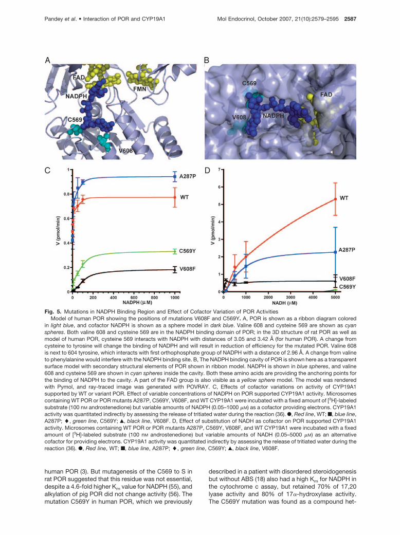
Several residues have been proposed to bind theNADPH in the POR that include Ser599, Arg600, andLys605. These residues are believed to provide spec-ificity for NADPH binding in POR as compared withNADH. Both cysteine 569 and valine 608 are located inthe NADPH binding domain of POR (Fig. 5). In the 3Dstructure of rat POR as well as model of human POR,
cysteine 569 interacts with NADPH with distances of3.05 and 3.42 Å (for human POR). A change fromcysteine to tyrosine will change the binding of NADPHand will result in reduction of efficiency for the mutatedPOR. Studies in which C569 of human POR was al-kylated with iodoacetic acid eliminated activity, impli-cating a role for cysteine in binding of NADPH to
Fig. 4. Interaction of Amino Acid A287 of POR with CYP19A1 and CYP17A1Models of CYP19A1 and CYP17A1 were based on 3D crystal structures of P450 3A4 and P450 2B4, respectively, and docking
of POR with either CYP17A1 or CYP19A1 was performed using the programs Hex4.5 and Patchdock. A, Docking model of PORinteracting with CYP17A1. Amino acid alanine 287 is located at the start of a loop in POR that is interacting with the C-terminalloop of CYP17A1, and change from alanine to proline will break the loop at the beginning so that the loop in CYP17A1 will nolonger be in close proximity with POR. One of the docking solutions is shown here with POR in blue ribbon and CYP17A1 in redribbon. B, The surface of CYP17A1 is shown with 60% transparency to emphasize the position of amino acid 287 (shown inmagenta). In POR FAD and FMN groups are shown as yellow spheres and NADPH is shown as a blue sphere model. C, A close-upof the POR:CYP17A1 interaction interface is shown where the loop involving A287 and the loop on CYP17A1 interacting with PORare visible. D, Protein docking model of POR interacting with CYP19A1. CYP19A1 is shown as green ribbon and POR as blueribbon. CYP19A1 does not seem to be in contact with the loop harboring POR A287. E, Superimposition of normal and A287Pvariant POR structure. The A287P mutation was performed in silico and MD simulation was performed to calculate the structuralimplications of this mutation. Normal POR is shown in light blue and altered (A287P) structure is shown in red. Changes to�-sheets and loops in the POR structure after A287P mutations are probably responsible for altered activity profile. Protein modelswere edited with Pymol and rendered as ray-traced images with POVRAY.
2586 Mol Endocrinol, October 2007, 21(10):2579–2595 Pandey et al. • Interaction of POR and CYP19A1
human POR (3). But mutagenesis of the C569 to S inrat POR suggested that this residue was not essential,despite a 4.6-fold higher Km value for NADPH (55), andalkylation of pig POR did not change activity (56). Themutation C569Y in human POR, which we previously
described in a patient with disordered steroidogenesisbut without ABS (18) also had a high Km for NADPH inthe cytochrome c assay, but retained 70% of 17,20lyase activity and 80% of 17�-hydroxylase activity.The C569Y mutation was found as a compound het-
Fig. 5. Mutations in NADPH Binding Region and Effect of Cofactor Variation of POR ActivitiesModel of human POR showing the positions of mutations V608F and C569Y. A, POR is shown as a ribbon diagram colored
in light blue, and cofactor NADPH is shown as a sphere model in dark blue. Valine 608 and cysteine 569 are shown as cyanspheres. Both valine 608 and cysteine 569 are in the NADPH binding domain of POR; in the 3D structure of rat POR as well asmodel of human POR, cysteine 569 interacts with NADPH with distances of 3.05 and 3.42 Å (for human POR). A change fromcysteine to tyrosine will change the binding of NADPH and will result in reduction of efficiency for the mutated POR. Valine 608is next to 604 tyrosine, which interacts with first orthophosphate group of NADPH with a distance of 2.96 Å. A change from valineto phenylalanine would interfere with the NADPH binding site. B, The NADPH binding cavity of POR is shown here as a transparentsurface model with secondary structural elements of POR shown in ribbon model. NADPH is shown in blue spheres, and valine608 and cysteine 569 are shown in cyan spheres inside the cavity. Both these amino acids are providing the anchoring points forthe binding of NADPH to the cavity. A part of the FAD group is also visible as a yellow sphere model. The model was renderedwith Pymol, and ray-traced image was generated with POVRAY. C, Effects of cofactor variations on activity of CYP19A1supported by WT or variant POR. Effect of variable concentrations of NADPH on POR supported CYP19A1 activity. Microsomescontaining WT POR or POR mutants A287P, C569Y, V608F, and WT CYP19A1 were incubated with a fixed amount of [3H]-labeledsubstrate (100 nM androstenedione) but variable amounts of NADPH (0.05–1000 �M) as a cofactor providing electrons. CYP19A1activity was quantitated indirectly by assessing the release of tritiated water during the reaction (36). F, Red line, WT; f, blue line,A287P; �, green line, C569Y; Œ, black line, V608F. D, Effect of substitution of NADH as cofactor on POR supported CYP19A1activity. Microsomes containing WT POR or POR mutants A287P, C569Y, V608F, and WT CYP19A1 were incubated with a fixedamount of [3H]-labeled substrate (100 nM androstenedione) but variable amounts of NADH (0.05–5000 �M) as an alternativecofactor for providing electrons. CYP19A1 activity was quantitated indirectly by assessing the release of tritiated water during thereaction (36). F, Red line, WT; f, blue line, A287P; �, green line, C569Y; Œ, black line, V608F.
Pandey et al. • Interaction of POR and CYP19A1 Mol Endocrinol, October 2007, 21(10):2579–2595 2587

Fig. 6. Perturbation of Reaction Conditions and Altered POR ActivitiesEffect of pH on POR supported CYP19A1 activity. A, pH response curve of CYP19A1 activity supported by WT and mutant POR.
Microsomes containing either WT POR or POR mutants A287P, C569Y, V608F, and WT CYP19A1 were incubated with a fixed amount of[3H] labeled substrate (100 nM androstenedione) and NADPH (1 mM) as the cofactor providing electrons. Reactions were carried out atdifferent pH values to assess the effect of pH on interaction of POR with CYP19A1. CYP19A1 activity was quantitated indirectly by assessingthe release of tritiated water during the reaction (36). F, Red line, WT; f, blue line, A287P; �, green line, C569Y; Œ, black line, V608F. WTCYP19A1 follows a bell-shaped curve with change in pH that is typical of enzymatic reactions under variable pH conditions. An optimum pHof 7–7.4 is obtained for WT POR. CYP19A1 activity supported by mutant POR also had 7.4 as pH optima. B, Comparison of POR supportedCYP19A1 activity as percentage of optimum value. Activity at pH 7.4 was optimal for all POR variants and was taken as 100% for calculationof activities at different pH values. Normal and A287P variants of POR were relatively stable at low pH values, whereas POR variant C569Yand V608F lost almost all activity at pH 5.5. At higher pH values, all CYP19A1 reactions with mutant POR lost almost 80% of their pH 7.4activity, whereas activity with WT POR retained 40% of its optimal value. Red bars, WT; blue, A287P; green, C569Y; and black, V608F. C andD, Effect of KCl on interaction of normal and mutant POR with CYP19A1. Microsomes containing WT POR or POR mutants A287P, C569Y,V608F, and WT CYP19A1 were incubated with a fixed amount of [3H]-labeled substrate (100 nM androstenedione) and NADPH (1 mM) as acofactor providing electrons. Potassium chloride was included at a concentration of 0 to 500 mM to perturb the interaction of POR withCYP19A1. CYP19A1 activity was quantitated indirectly by assessing the release of tritiated water during the reaction (36). F, Red line, WT;f, blue line, A287P; �, green line, C569Y; Œ, black line, V608F. Small amount of KCl has no adverse effect on P450 activities and is oftenincluded in preparation of homogenates and microsomes during P450 assays. However, high concentrations of KCl can disturb theelectrostatic interaction between POR and P450 and lead to reduction of activities. Up to 200 mM concentration of KCl had only a mild effecton POR-supported activity, but increasing the KCl concentration to 500 mM resulted in loss of activity for C569Y and V608F, and binding ofNADPH to POR seems to be affected by higher KCl concentrations, because inclusion of octanol as described earlier did not result in anyspecific effect for these POR variants. POR variant A287P did not lose as much activity, suggesting that general disturbances in POR-P450interaction are not the only effects of higher KCl concentration. C, Total activity plotted against increasing KCl concentration. D, Percentageof activity remaining compared with activity in the absence of KCl, plotted against increasing KCl concentration.
2588 Mol Endocrinol, October 2007, 21(10):2579–2595 Pandey et al. • Interaction of POR and CYP19A1

erozygote with V608F (18) which retained 52% of17,20 lyase activity and 53% of 17�-hydroxylase ac-tivity. Valine 608 is next to tyrosine 607 and lysine 605which have several contacts with NADPH, especiallybetween OH group of tyrosine and first orthophos-phate group of NADPH with a distance of 2.96 Å andsecond orthophosphate group of NADPH is at a dis-tance of 2.49 Å from lysine 605. A change from valineto phenylalaline will disturb the NADPH binding site.However, an excess of NADPH may be able to over-come some of the binding defect because bindingdoes not seem to be completely destroyed.
Conversion of androgens to estrogens by humanCYP19A1 depends on the transfer of three pairs ofelectrons from NADPH through POR (Fig. 1). In con-trast, CYP17A1 activities for the conversion of bothsteps, first pregnenolone to 17�-hydroxypreg-nenolone (by 17�-hydroxylase), and second to dehy-droepiandrosterone (by 17,20 lyase) require only onepair of electrons per reaction (15). Therefore, wethought that the differential inhibition of POR mutantsC569Y and V608F on CYP17A1 and CYP19A1 mightbe due to a difference in cofactor requirement. So weassayed CYP19A1 activity supported by constantamounts of WT, A287P, C569Y, and V608F POR pro-tein but different concentrations of NADPH (Fig. 5C).WT and A287P POR needed approximately 5 nM
NADPH for half maximal CYP19A1 activity (calculatedEC50). By contrast, C569Y and V608F required 76–108nM NADPH. Consistent with that, mutations in theNADPH binding domain also show a higher Km forNADPH in cytochrome c assays (18, 19). Thus, PORmutations in the NADPH binding domain seem to im-pair CYP19A1 activity more when cofactor availabilityis limited. In line with these in vitro findings, the singlereported patient harboring two POR mutations in theNADPH binding domain manifested with a very mild,polycystic ovary syndrome-like phenotype consistentwith only CYP19A1 deficiency (18).
POR Mutants C569Y and V608F Have DiminishedCapacity for NADH as Electron Donor Comparedwith WT POR
A change from cysteine to tyrosine seems to havehigher impact on binding of NADH and results in re-duced binding efficiency for the POR mutation C569Y.The mutation C569Y, which has a higher Km forNADPH in the cytochrome c assays, has almost noactivity when NADH was substituted for NADPH as theelectron donor (Fig. 5D). Valine 608 on the other handis not directly involved in the binding of NADPH andhas some activity with NADH as substrate, implying aless critical role for V608 than C569 in the cofactorbinding. In general, both C569Y and V608F mutationsthat lie in the NADPH binding site of POR resulted inseverely diminished activities when NADH is used as asubstrate compared with NADPH. Normal and A287Pmutant of POR also showed reduced activities with
NADH as substrate, but the effects were much lesscompared with C569Y and V608F mutants of POR.
Effect of Changes in pH on Activity of CYP19A1Supported by Normal and Mutant POR
We have used several different methods to vary theinteraction between POR and CYP19A1 to assess theimpact of variable electron flux on changes in the activityof CYP19A1 supported by different POR mutants. Onesuch method is to change the pH of the reaction in theassay of CYP19A1 activity (57). The pH profile ofCYP19A1 activity supported by normal POR follows abell-shaped curve with optimum pH between 7.0 and7.4, and mutant enzymes also show optimum pH around7.4 (Fig. 6A). However, there are remarkable variations inthe responses of different POR variants at pH valueshigher or lower than optimum pH. At pH 5.5, up to 35%of optimal CYP19A1 activity supported by normal POR(obtained at pH 7.4) is retained (Fig. 6B). However,CYP19A1 activity supported by C569Y mutant drops toless than 10% and mutant V608F has almost no activityat pH 5.5. At higher pH values, the effects are a little lessremarkable but the same pattern is observed. At pH 8.5,normal POR retained more than 40% of its optimal ac-tivity, and activities of A287P, C569Y, and V608F mu-tants fell below 20% of their optimal values (Fig. 6B).Higher pH seems to destabilize A287P variant of POR toa greater extent than low pH conditions because at pH5.5 and 6.0, the A287P mutant of POR retains greaterthan 50 and 80% activities, respectively.
Changes in Membrane Order by Addition ofOctanol in CYP19A1 Reaction Reduce ActivityBut Are Not Specific for Any POR Mutant
Addition of n-alkanols has been used previously to alterthe membrane order in CYP19A1 activity assays (57). Weused variable concentrations of octanol in the reactionmixture during assay of CYP19A1 activity to change themembrane order of microsomes containing CYP19A1and POR and perturb the interaction of POR withCYP19A1. We used 0 to 50 mM octanol in our reactionsto alter the membrane conditions based on previousresults and found that normal and A287P mutant of PORretained only 25% of their activity in the presence of 50mM octanol (Table 3). Effects on C569Y and V608F mu-tants were also in the same range. This suggests thatchanges in general conditions that limit the POR inter-action with P450 do not differentiate between the variantPOR studied by us. However, any mutations on thesurface of POR that might be involved in interaction withP450 might show a different response to changes inmembrane order that affect the POR-P450 interaction.
Effect of KCl on CYP19A1 Activity Supported byNormal and Mutant POR
Generally KCl has only a mild effect on P450 activities,and only at KCl concentrations of 250 mM or higher is
Pandey et al. • Interaction of POR and CYP19A1 Mol Endocrinol, October 2007, 21(10):2579–2595 2589

a significant decrease in activity observed. We ob-served this pattern for the normal and A287P variant ofPOR (Fig. 6C). However, C569Y and V608F variantsshowed much reduced activities around 200 mM KCland lost more than 60% of their activities at 500 mM
KCl (Fig. 6D). In comparison, WT POR retained morethan 80% activity, and the A287P mutant retainedmore than 60% of its activity, suggesting a mildereffect by higher KCl concentrations. Some destabili-zation in the A287P mutant may be responsible forgreater reduction in activity compared with normalPOR. Increased loss of activities by C569Y and V608Fmutants suggests alterations in NADPH binding underthese conditions that have greater impact on POR withmutations in the NADPH binding site. Reduction inactivity of P450 at high KCl concentrations is attrib-uted to disruption of charged pair electrostatic inter-actions between POR and P450, but the higher impacton mutations C569Y and V608F is probably due todiminished binding of NADPH or slower release ofNADP in the cofactor binding pocket of POR.
In summary, our detailed studies of POR-supportedCYP17A1 and CYP19A1 activities suggest that differ-ent POR mutations can affect dependent P450s dif-ferently. Mutations in the FAD domain, which disruptelectron transfer, will affect all microsomal P450s. Mu-tations in the NADPH domain may affect P450s withhigh electron requirement more severely. Cofactorspecificity of POR seems to be affected even more bymutations in the NADPH binding site. Normal POR haspreference for NADPH over NADH by several orders ofmagnitude in the Km values, which are in millimolarrange for NADH but lie in nanomolar to submicromolarrange for NADPH. Similar results were observed in thisstudy for CYP19A1 reaction. However, C569Y muta-tion seems to lose almost all capacity to bind and useNADH as an alternate electron donor. Limiting thecofactor availability by other methods of perturbationlike inclusion of alcohol or salt enabled us to differen-tiate the effects in general POR-P450 interactionscompared with specific changes in cofactor utilization.POR mutations C569Y and V608F can interact withP450s like normal POR as evidenced by relativelyminor changes in apparent Km of P450 substrates forthe P450s in both CYP17A1 and CYP19A1 reactionsstudied by us. Changing the POR-P450 interaction bymembrane perturbation induced by octanol results ina general reduction of CYP19A1 activities. However,
some changes by inclusion of octanol/KCl or changein pH could be due to altered protein conformation.Some indication of this is suggested from relativelyhigher loss of activity supported by A287P mutant ofPOR under low pH and high KCl concentrations. Fur-ther studies on folding and conformation of differentPOR mutants will shed more light on these differences.
Finally, some POR mutations may be located in theinteraction face of some POR:P450 complexes, ham-pering the interaction between some partners but notaffecting others. In reconstituted systems, P450 pro-teins form a complex with POR with an apparent Km ofaround 0.2 �M. The P450:POR interaction is influencedby many factors including type of P450, availability ofsubstrates, and ionic strength of the system (58). Theinteraction of POR with cytochrome P450 and otherelectron acceptor proteins is based primarily on elec-trostatic charge pairing, although there is evidence foran additional hydrophobic component. The surface ofthe electron-donating (FMN) domain of POR is char-acterized by acidic residues (9, 59, 60), whereas theredox-partner binding site of microsomal P450 en-zymes is typically characterized by basic residues (61–63). Chemical cross-linking and modification studieshave shown that POR contains multiple carboxylategroups, presumably contributed by the acidic aminoacids aspartate and glutamate (59, 64). These chargegroups pair with basic amino acids (lysines, arginines)on the various electron acceptor proteins (65–67).Thus electrostatic interactions that pair charges areobviously important in governing the association ofPOR with a P450 enzyme. Chemical modification ofacidic residues in the FMN site of POR reduces theactivity of P450 (59, 64, 68). Perturbation in the mem-brane and electrostatic environment of POR-P450 re-actions by changes in pH/KCl and inclusion of n-alcohols will be able to differentiate these mutations.
In addition, cytochrome P450 forms a dipole acrossthe molecule, with the positive charge at the proximalface of the protein where the heme makes its closestapproach to the surface. This is thought to be thesurface most suitable for electron transfer from POR.Although electrostatic forces may serve to connectand orient the partners, hydrophobic forces contrib-uted by nonpolar amino acids (leucine, tryptophan,valine, etc.) may be responsible for bringing the twoproteins close enough together for electron transfer(69). Other electron acceptor proteins, such as cyto-
Table 3. Effect of Increasing Octanol Concentration on the Activity of CYP19A1 Supported by WT or Variant POR
Octanol (mM) POR WT POR A287P POR C569Y POR V608F
0 0.775 � 0.003 0.672 � 0.013 0.36 � 0.0123 0.224 � 0.0115 0.538 � 0.093 0.473 � 0.067 0.216 � 0.016 0.139 � 0.003
15 0.382 � 0.072 0.488 � 0.013 0.127 � 0.008 0.079 � 0.00830 0.247 � 0.021 0.205 � 0.010 0.092 � 0.002 0.068 � 0.00550 0.172 � 0.009 0.237 � 0.017 0.094�0.008 0.092 � 0.003
Data are expressed as mean � SD for CYP19A1 activity (pmol/min) in triplicate experiments. Reactions were carried out asdescribed in CYP19A1 activity assay. A fixed substrate concentration of 100 nM androstenedione and 1 mM NADPH was used.
2590 Mol Endocrinol, October 2007, 21(10):2579–2595 Pandey et al. • Interaction of POR and CYP19A1

chrome b5, heme oxygenase, and squalene monoox-ygenase, probably interact by the same mechanism.POR interacts with and supports the enzymatic activ-ities of all 50 human microsomal P450 enzymes. Theactivities assessed in different P450 studies are notonly a function of the specific P450 enzyme used asthe read-out of the POR activity, but also a function ofthe form of POR used. Both the full-length POR usedin our first report and the N-27 human POR used in ourbacterial expression system remain associated withmembranes and are able to support the reduction of acytochrome P450 (41). By contrast, when rat or humanPOR is rendered wholly soluble by deleting 57 N-terminal residues, it can still reduce cytochrome c butcannot reduce a P450 (8, 11). Because the interactionsof POR with different P450 enzymes will vary with thegeometry of the redox-partner binding site of theP450, no assay based on a single P450 enzyme willreliably forecast all the consequences of a specificPOR mutant. Only the residues that severely affect theFAD or FMN binding may lose all enzymatic activityand could be tested by employing the cytochromec-based assays along with a single P450-based assay(41). By contrast, those POR mutations that are in-volved in POR:P450 interactions may have to betested with many different P450 assays to establishthe exact nature of their impact on individual P450reactions. Because all 50 microsomal P450 enzymesdepend on POR for electron supply, we are puttingforward the hypothesis that defects in POR may causedisorders in hepatic drug and xenobiotic metabolismas well as affect the functions of all steroid metabo-lizing microsomal P450 enzymes.
MATERIALS AND METHODS
Plasmid Constructs
Yeast expression vector pYeSF1-pgk was built by insertingthe constitutively active pgk (phosphoglycerate kinase) pro-moter in the yeast vector pYeSF1 (37). In brief, pgk promoterwas cleaved from yeast vector V10 using BglII and BamHIrestriction sites. The fragment was then cloned into BamHIsite of pYeSF1 resulting in pYeSF1-pgk vector containing anade2 selection marker. CYP19A1 cDNA was amplified withLong Template Polymerase (Roche, Basel, Switzerland) usingplasmid pcDNA3-ARO (70) as template and the followingprimers AroF: 5�TCCCCCGGGAGCCATGGTTTTGGAAAT-GCTGAACCCGA; and AroR: 5�TCCCCCGGGCTAGTGTTC-CAGACACCTGTCTGAGTTTC (restriction sites are under-lined, start and stop codons are shown in bold). The PCRproduct was cloned into the pYeSF1-pgk vector through theSmaI restriction site. Correctness of the resulting constructpYeSF1-pgk-ARO was confirmed by direct sequencing (Mi-crosynth, Basel, Switzerland). Construction of human WT andmutant POR cDNAs A287P, R457H, V492E, C569Y, andV608F into pYcDE2 has been described previously (18).Numbering of the amino acids of the POR protein is based onNCBI NP_000932.3 and corresponds to the full length, 680amino acid human POR sequence.
Expression of Human CYP19A1 and POR
Yeast strain W(B) lacking the endogenous yeast CPR1 gene(35, 71, 72) was propagated and transiently cotransfectedwith pYcDE2 expressing full-length WT or mutant PORcDNAs and pYeSF1-pgk-ARO expressing human WTCYP19A1. Yeast microsomes were prepared as described(37), and microsomal proteins were quantitated colorimetri-cally (Protein Assay Dye Reagent, Bio-Rad, Hercules, CA). Tocontrol for equal expression, Western blot analyses of micro-somal proteins were performed using a rabbit polyclonalantibody against human placental CYP19A1 kindly providedby Dr. Nobuhiro Harada (73), and a rabbit polyclonal antibodyagainst POR commercially available from Stressgen Biore-agents (LuBioScience GmbH, Lucerne, Switzerland).
CYP19A1 Activity Assay
The ability of POR to support the activity of CYP19A1 wasassayed with yeast microsomal proteins containing humanCYP19A1 and WT or mutant human POR as electron donor.CYP19A1 activity was measured by the release of tritiatedwater from 1�-3H substrates during aromatization as de-scribed by Lephart and Simpson (36). Briefly, reactions wereperformed in 15-ml Falcon tubes in a final volume of 200 �l at37 C. Yeast microsomes (40 �g microsomal protein/reaction)were incubated with 0.01–3 �M cold androstenedione, and aconstant amount of [1�-3H(N)]-androstene-3,17-dione(40,000 cpm/reaction) in 50 mM K-phosphate buffer (pH 7.4).Reactions were initiated by adding 1 mM NADPH (unlessindicated differently). Reactions were stopped by adding 1 mlof chloroform. The mixture was vortexed for 30 sec, then 1 mlof water was added, and the mix was centrifuged at 1000 rpmfor 5 min. Aliquots of the water phase (0.5 ml) were taken andmixed with an equal volume of 5% charcoal/0.5% dextran.After 40 sec of extraction, samples were centrifuged at12,000 rpm for 15 min, and 0.5-ml aliquots of supernatantswere collected for counting of 3H radioactivity. The measuredcpm values were corrected for background radioactivity andmultiplied by a factor of 4 because actual counted aliquotsrepresented only one fourth of the total reaction volume.Once percentage conversion of androstenedione to estronewas assessed, further calculations were performed to de-scribe the kinetic characteristics of WT CYP19A1 when sup-ported by either WT or mutant POR. Nonlinear regressionwas used for this analysis. Curve fitting and calculation ofVmax and apparent Km values were performed using PRISM3.02 (GraphPad Software Inc., San Diego, CA). Data repre-sent the mean � SD of two to four independent experiments,each performed in duplicate.
Buffers involving different pH values for measuring theeffect of pH on CYP19A1 activities were prepared separatelyat twice the working concentration and substituted for normalbuffer in the reactions involving measurements at alternatepH values. Experiments involving the presence of octanolwere carried out by addition of octanol in the reaction mixturebefore addition of substrate and NADPH. Total alcohol vol-ume did not exceed 5% of the reaction volume, and after anincubation of 5 min at room temperature the reaction wasstarted by addition of substrate and NADPH. Similarly KClwas added from a stock solution of 5.0 M in the reactionsinvolving measurement of CYP19A1 activity at different con-centrations of KCl. In one set of experiments NADH wassubstituted for NADPH as the source of electrons.
3D Protein Models
3D structural models of CYP19A1, POR, and CYP17A1 weregenerated to study the potential impact of POR mutations oninteraction with CYP19A1 and CYP17A1. A phi BLAST searchof the protein structure database was performed against theCYP19A1 (NP_000094) and CYP17A1 (NP_000093) amino
Pandey et al. • Interaction of POR and CYP19A1 Mol Endocrinol, October 2007, 21(10):2579–2595 2591

acid sequences, which revealed the recently solved structureof human P450 3A4 (74) (PDB no. 1tqn) (NP_059488) as theclosest match for CYP19A1, and rabbit P450 2B4 structure(PDB no. 1suo) (75) was identified as the template for mod-eling human CYP17A1. We first performed a multiple se-quence alignment of CYP19A1 with the human P450 se-quences from PDB database including steroidogenicCYP17A1 and CYP21B1 by CLUSTALW (http://www.ebi.ac.uk/clustalw) (76) to analyze the structural features of theCYP19A1 protein sequence. We then performed a structuralalignment of P4503A4 and CYP19A1 and used that for modelbuilding with the program MODELLER 8 (77). For theCYP17A1 model, we have used our previously describedalignment for P450 2B4 structure and CYP21B1 andCYP17A1 sequences (52). Basic protocols for model buildingfor CYP19A1 and CYP17A1 models were as described pre-viously (52). The model was subjected to energy minimizationand then checked by the programs WHATCHECK (http://swift.cmbi.ru.nl/gv/whatcheck) (78), Verify3D (79) (http://nihserver.mbi.ucla.edu/Verify_3D), and PROCHECK (80)(http://biotech.embl-ebi.ac.uk:8400/) and by Ramachandranplot analysis (81, 82) for abnormalities in the structure. Cal-culations for RMSD values were performed with DALI (83).Several rounds of model building and optimizations wereperformed to finally obtain a model that was energeticallystable and passed the tests for the quality of the structure.CYP17A1 model was also generated in similar fashion basedon 3D structure of rabbit P4502B4. For human POR, we usedour previously described 3D protein model, which is basedon the structure of rat POR (9) (PDB id 1AMO) (NP_113764)that shares 96% sequence similarity with human POR(NP_000932). The WT protein model was used to createmutants of POR with the program DEEPVIEW (84). Alteredstructures were subjected to energy minimization using mo-lecular dynamics (MD) simulation and checked by WHATIF(http://swift.cmbi.kun.nl/whatif/) (85) and WHATCHECK (78)to remove bumps in the sidechains with respect to neighbor-ing amino acids that were caused by in silico mutations.Coordinates of computer models are available as PDB filesupon request. Models were depicted with Pymol (www.pymol.org) and rendered as ray-traced images with POVRAY(www.povray.org).
MD Simulation for Model Refinement
The MD simulations were performed using YASARA dynam-ics (86). A simulation cell (91 � 72 � 83 Å) was constructedaround the CYP19A1 homology model with a 7.9 Å cutoff forLennard-Jones forces and the direct space portion of theelectrostatic forces, which were calculated using the ParticleMesh Ewald method. The pKa values of the ionizable groupsin the model were predicted and assigned the protonationstates based on pH 7.0. The cell was filled with water, and theAMBER99 (87) electrostatic potential was evaluated at allwater molecules; the one with the lowest or highest potentialwas turned into a sodium or chloride counter ion until the cellwas neutral. A short steepest descent minimization of allatoms was done to remove severe bumps in the protein andto achieve convergence until the maximum atom speeddropped below 2200 m/sec. Then a start-up simulation wasrun for 5 psec, using a multiple time step of 1 fsec forintramolecular and 2 fsec for intermolecular forces, with allheavy protein atoms fixed, so that the solvent moleculescould smoothly cover the protein surface. Simulated anneal-ing minimizations were started at 298 K, and velocities werescaled down every 10 steps for a total time of 5 psec in 500steps. We then ran MD simulations with AMBER99 force fieldat 298 K and 0.9% NaCl in the simulation cell for 1000 psecto refine the models. All MD computations were run locally ona Dell computer server containing 4 intel CPUs running underSUSE Linux 10.1/Windows server 2003. A local installation ofWHAT IF program was used to check the quality of models incombination with Verify3D analysis to check for abnormali-
ties. An analysis of MD trajectories showed a significantimprovement, and final model snapshot has a free energy of�19255 kJ/mol for CYP19A1 model. The C�-RMSD droppedfrom an initial value of 2.7 Å, which is typical for a decenthomology model to 1.4 Å, which corresponds to a medium-resolution x-ray structure. Using the MD simulation, we couldrefine the homology model close to an experimental-likestructure. Modeling of P450 proteins has recently been quitesuccessful, and some of the models of P450 show very highsimilarity when compared with the actual structures whichhave recently become available (88). An increase in the num-ber of mammalian P450 structures that have become avail-able recently combined with powerful computational hard-ware and software for modeling side-chains and loops in thevariable regions of homology models could be attributed tothis achievement.
Analysis of the Interaction of POR with CYP19A1 andCYP17A1 by 3D Docking
We used the programs Patchdock (89) and Hex 4.5 (90) foranalysis of the interaction of WT and A287P mutant of PORwith CYP19A1 and CYP17A1. We performed the 3D protein-protein interaction analysis of the interaction of POR witheither CYP19A1 or CYP17A1 to understand the role of ala-nine 287 in P450:POR interactions and whether a change toproline would affect such interactions. Electrostatic interac-tions are known to be involved between positively chargedgroups on P450s and negatively charged acidic residues onPOR and were taken into consideration while calculating thedockings. In the first step, a molecular shape representationwas performed by computing the molecular surface of themolecule. Next, a segmentation algorithm was applied fordetection of geometric patches, which were filtered, and thena geometric hashing and pose-clustering matching was per-formed to match the patches on two structures. Concavepatches were matched with convex and flat patches with anytype of patches. The complexes obtained were then exam-ined, and all complexes with unacceptable penetrations ofthe atoms of the receptor to the atoms of the ligand werediscarded. Finally, the remaining candidates were rankedaccording to a geometric shape complementarity score. Intotal, 50,000 calculations were performed per pair, and so-lutions were filtered for energetically stable conformations.The top 20 solutions from each round of calculations weremanually checked for interaction sites and orientation of themolecules. Structures were edited with the program Pymoland depicted and ray-traced images were generated byPOVRAY.
Acknowledgments
We thank Prof. Walter L. Miller, University of California SanFrancisco (San Francisco, CA), and Dr. Nobuhiro Harada,Fujita Health University (Toyoake, Aichi, Japan) for support.
Received May 10, 2007. Accepted June 21, 2007.Address all correspondence and requests for reprints to:
Christa E. Fluck, M.D., Pediatric Endocrinology and Diabe-tology, University of Bern, Freiburgstrasse 15, G3 812, CH-3010 Bern, Switzerland. E-mail: [email protected].
Address requests for materials and structure model filesto: Amit V. Pandey, Ph.D., Department of Clinical Research,University of Bern, Tiefenaustrasse 120, CH-3004 Bern, Swit-zerland. E-mail: [email protected].
This work was supported by Swiss National Science Foun-dation Grants 3232B0103178/179 (to C.E.F.) and 3100A0-113719 (to A.V.P.).
The nucleotide sequence of the human NADPH P450 ox-idoreductase gene is available in the NCBI nucleotide data-
2592 Mol Endocrinol, October 2007, 21(10):2579–2595 Pandey et al. • Interaction of POR and CYP19A1

base under NCBI accession no. NM_000941. The amino acidsequence of human POR can be accessed through NCBIProtein Database under NCBI accession no. NP_000932. Theatomic coordinates for the crystal structure of rat POR areavailable in the Research Collaboratory for Structural Bioin-formatics Protein Databank PDB no. 1AMO. The atomic co-ordinates for the crystal structure of FMN binding domain ofhuman POR are available in the Research Collaboratory forStructural Bioinformatics Protein Databank PDB no. 1B1C.Online Mendelian Inheritance in Man, OMIM no. 124015,201750. Enzyme collection number, EC 1.6.2.4.
Disclosure Statement: A.V.P., P.K., G.H., P.E.M., andC.E.F. have nothing to declare.
REFERENCES
1. Shephard EA, Phillips IR, Santisteban I, West LF, PalmerCN, Ashworth A, Povey S 1989 Isolation of a humancytochrome P-450 reductase cDNA clone and localiza-tion of the corresponding gene to chromosome 7q11.2.Ann Hum Genet 53:291–301
2. Scott RR, Gomes LG, Huang N, Van Vliet G, Miller WL2007 Apparent manifesting heterozygosity in p450 oxi-doreductase deficiency and its effect on coexisting 21-hydroxylase deficiency. J Clin Endocrinol Metab 92:2318–2322
3. Haniu M, McManus ME, Birkett DJ, Lee TD, Shively JE1989 Structural and functional analysis of NADPH-cyto-chrome P-450 reductase from human liver: completesequence of human enzyme and NADPH-binding sites.Biochemistry 28:8639–8645
4. Horecker BL, Heppel LA 1949 The reduction of cyto-chrome c by xanthine oxidase. J Biol Chem 178:683–690
5. Horecker BL 1950 Triphosphate nucleotide-cytochromec reductase in liver. J Biol Chem 183:593–605
6. Williams Jr CH, Kamin H 1962 Microsomal triphosphopy-ridine nucleotide-cytochrome c reductase of liver. J BiolChem 237:587–595
7. Phillips AH, Langdon RG 1962 Hepatic triphosphopyri-dine nucleotide-cytochrome c reductase: isolation, char-acterization, and kinetic studies. J Biol Chem 237:2652–2660
8. Lu AY, Junk KW, Coon MJ 1969 Resolution of the cyto-chrome P-450-containing �-hydroxylation system of livermicrosomes into three components. J Biol Chem 244:3714–3721
9. Wang M, Roberts DL, Paschke R, Shea TM, MastersBSS, Kim JJ 1997 Three-dimensional structure ofNADPH-cytochrome P450 reductase: prototype forFMN- and FAD-containing enzymes. Proc Natl Acad SciUSA 94:8411–8416
10. Zhao Q, Modi S, Smith G, Paine M, McDonagh PD, WolfCR, Tew D, Lian LY, Roberts GC, Driessen HP 1999Crystal structure of the FMN-binding domain of humancytochrome P450 reductase at 1.93 A resolution. ProteinSci 8:298–306
11. Roman LJ, McLain J, Masters BSS 2003 Chimeric en-zymes of cytochrome P450 oxidoreductase and neuronalnitric-oxide synthase reductase domain reveal structuraland functional differences. J Biol Chem 278:25700–25707
12. Munro AW, Noble MA, Robledo L, Daff SN, Chapman SK2001 Determination of the redox properties of humanNADPH-cytochrome P450 reductase. Biochemistry 40:1956–1963
13. Hubbard PA, Shen AL, Paschke R, Kasper CB, Kim JJ2001 NADPH-cytochrome P450 oxidoreductase. Struc-tural basis for hydride and electron transfer. J Biol Chem276:29163–29170
14. Nelson DR, Zeldin DC, Hoffman SM, Maltais LJ, WainHM, Nebert DW 2004 Comparison of cytochrome P450
(CYP) genes from the mouse and human genomes, in-cluding nomenclature recommendations for genes,pseudogenes and alternative-splice variants. Pharmaco-genetics 14:1–18
15. Miller WL 2005 Minireview: regulation of steroidogenesisby electron transfer. Endocrinology 146:2544–2550
16. Shen AL, O’Leary KA, Kasper CB 2002 Association ofmultiple developmental defects and embryonic lethalitywith loss of microsomal NADPH-cytochrome P450 oxi-doreductase. J Biol Chem 277:6536–6541
17. Otto DM, Henderson CJ, Carrie D, Davey M, GundersenTE, Blomhoff R, Adams RH, Tickle C, Wolf CR 2003Identification of novel roles of the cytochrome P450 sys-tem in early embryogenesis: effects on vasculogenesisand retinoic acid homeostasis. Mol Cell Biol 23:6103–6116
18. Fluck CE, Tajima T, Pandey AV, Arlt W, Okuhara K, VergeCF, Jabs EW, Mendonca BB, Fujieda K, Miller WL 2004Mutant P450 oxidoreductase causes disordered steroi-dogenesis with and without Antley-Bixler syndrome. NatGenet 36:228–230
19. Huang N, Pandey AV, Agrawal V, Reardon W, LapunzinaPD, Mowat D, Jabs EW, Van Vliet G, Sack J, Fluck CE,Miller WL 2005 Diversity and function of mutations inp450 oxidoreductase in patients with Antley-Bixler syn-drome and disordered steroidogenesis. Am J Hum Genet76:729–749
20. Pandey AV, Fluck CE, Huang N, Tajima T, Fujieda K,Miller WL 2004 P450 oxidoreductase deficiency: a newdisorder of steroidogenesis affecting all microsomalP450 enzymes. Endocr Res 30:881–888
21. Pandey AV 2006 Biochemical analysis of mutations inP450 oxidoreductase. Biochem Soc Trans 34:1186–1191
22. Miller WL 2004 P450 oxidoreductase deficiency: a newdisorder of steroidogenesis with multiple clinical mani-festations. Trends Endocrinol Metab 15:311–315
23. Miller WL, Huang N, Fluck CE, Pandey AV 2004 P450oxidoreductase deficiency. Lancet 364:1663
24. Miller WL, Huang N, Pandey AV, Fluck CE, Agrawal V2005 P450 oxidoreductase deficiency: a new disorder ofsteroidogenesis. Ann NY Acad Sci 1061:100–108
25. Arlt W, Walker EA, Draper N, Ivison HE, Ride JP, HammerF, Chalder SM, Borucka-Mankiewicz M, Hauffa BP, Mal-unowicz EM, Stewart PM, Shackleton CH 2004 Congen-ital adrenal hyperplasia caused by mutant P450 oxi-doreductase and human androgen synthesis: analyticalstudy. Lancet 363:2128–2135
26. Adachi M, Tachibana K, Asakura Y, Yamamoto T, HanakiK, Oka A 2004 Compound heterozygous mutations ofcytochrome P450 oxidoreductase gene (POR) in two pa-tients with Antley-Bixler syndrome. Am J Med Genet128A:333–339
27. Fukami M, Horikawa R, Nagai T, Tanaka T, Naiki Y, SatoN, Okuyama T, Nakai H, Soneda S, Tachibana K, MatsuoN, Sato S, Homma K, Nishimura G, Hasegawa T, OgataT 2005 Cytochrome P450 oxidoreductase gene muta-tions and Antley-Bixler syndrome with abnormal genitaliaand/or impaired steroidogenesis: molecular and clinicalstudies in 10 patients. J Clin Endocrinol Metab 90:414–426
28. Adachi M, Asakura Y, Matsuo M, Yamamoto T, Hanaki K,Arlt W 2006 POR R457H is a global founder mutationcausing Antley-Bixler syndrome with autosomal reces-sive trait. Am J Med Genet A 140:633–635
29. Homma K, Hasegawa T, Nagai T, Adachi M, Horikawa R,Fujiwara I, Tajima T, Takeda R, Fukami M, Ogata T 2006Urine steroid hormone profile analysis in cytochromeP450 oxidoreductase deficiency: implication for thebackdoor pathway to dihydrotestosterone. J Clin Endo-crinol Metab 91:2643–2649
30. Fukami M, Hasegawa T, Horikawa R, Ohashi T, Nish-imura G, Homma K, Ogata T 2006 Cytochrome P450
Pandey et al. • Interaction of POR and CYP19A1 Mol Endocrinol, October 2007, 21(10):2579–2595 2593

oxidoreductase deficiency in three patients initially re-garded as having 21-hydroxylase deficiency and/or aro-matase deficiency: diagnostic value of urine steroid hor-mone analysis. Pediatr Res 59:276–280
31. Haiman CA, Setiawan VW, Xia LY, Le Marchand L, InglesSA, Ursin G, Press MF, Bernstein L, John EM, HendersonBE 2007 A variant in the cytochrome p450 oxidoreduc-tase gene is associated with breast cancer risk in AfricanAmericans. Cancer Res 67:3565–3568
32. Miller WL 1988 Molecular biology of steroid hormonesynthesis. Endocr Rev 9:295–318
33. Simpson ER 2004 Aromatase: biologic relevance of tis-sue-specific expression. Semin Reprod Med 22:11–23
34. Nebert DW, Nelson DR, Feyereisen R 1989 Evolution ofthe cytochrome P450 genes. Xenobiotica 19:1149–1160
35. Pompon D, Perret A, Bellamine A, Laine R, Gautier JC,Urban P 1995 Genetically engineered yeast cells andtheir applications. Toxicol Lett 82–83:815–822
36. Lephart ED, Simpson ER 1991 Assay of aromatase ac-tivity. In: Waterman MR, Johnson EF, eds. Methods inenzymology. San Diego: Academic Press; 477–483
37. Auchus RJ, Lee TC, Miller WL 1998 Cytochrome b5augments the 17,20-lyase activity of human P450c17without direct electron transfer. J Biol Chem 273:3158–3165
38. Reed JR, Kelley RW, Backes WL 2006 An evaluation ofmethods for the reconstitution of cytochromes P450 andNADPH P450 reductase into lipid vesicles. Drug MetabDispos 34:660–666
39. Amarneh B, Simpson ER 1995 Expression of a recombi-nant derivative of human aromatase P450 in insect cellsutilizing the baculovirus vector system. Mol Cell Endo-crinol 109:R1–R5
40. Kagawa N, Hori H, Waterman MR, Yoshioka S 2004Characterization of stable human aromatase expressedin E. coli. Steroids 69:235–243
41. Fluck CE, Nicolo C, Pandey AV 2007 Clinical, structuraland functional implications of mutations and polymor-phisms in human NADPH P450 oxidoreductase. FundamClin Pharmacol 21:399–410
42. Shen AL, Kasper CB 2000 Differential contributions ofNADPH-cytochrome P450 oxidoreductase FAD bindingsite residues to flavin binding and catalysis. J Biol Chem275:41087–41091
43. Shen AL, Kasper CB 1996 Role of Ser457 of NADPH-cytochrome P450 oxidoreductase in catalysis and con-trol of FAD oxidation-reduction potential. Biochemistry35:9451–9459
44. Shen AL, Sem DS, Kasper CB 1999 Mechanistic studieson the reductive half-reaction of NADPH-cytochromeP450 oxidoreductase. J Biol Chem 274:5391–5398
45. Marohnic CC, Panda SP, Martasek P, Masters BS 2006Diminished FAD binding in the Y459H and V492E Antley-Bixler syndrome mutants of human cytochrome P450reductase. J Biol Chem 281:35975–35982
46. Graham-Lorence S, Khalil MW, Lorence MC, MendelsonCR, Simpson ER 1991 Structure-function relationships ofhuman aromatase cytochrome P-450 using molecularmodeling and site-directed mutagenesis. J Biol Chem266:11939–11946
47. Graham-Lorence S, Amarneh B, White RE, Peterson JA,Simpson ER 1995 A three-dimensional model of aro-matase cytochrome P450. Protein Sci 4:1065–1080
48. Loge C, Le Borgne M, Marchand P, Robert JM, Le BautG, Palzer M, Hartmann RW 2005 Three-dimensionalmodel of cytochrome P450 human aromatase. J EnzymeInhib Med Chem 20:581–585
49. Favia AD, Cavalli A, Masetti M, Carotti A, Recanatini M2006 Three-dimensional model of the human aromataseenzyme and density functional parameterization of theiron-containing protoporphyrin IX for a molecular dynam-ics study of heme-cysteinato cytochromes. Proteins 62:1074–1087
50. Auchus RJ, Miller WL 1999 Molecular modeling of humanP450c17 (17�-hydroxylase/17,20-lyase): insights into re-action mechanisms and effects of mutations. Mol Endo-crinol 13:1169–1182
51. Monno S, Ogawa H, Date T, Fujioka M, Miller WL, Koba-yashi M 1993 Mutation of histidine 373 to leucine incytochrome P450c17 causes 17 �-hydroxylase defi-ciency. J Biol Chem 268:25811–25817
52. Janner M, Pandey AV, Mullis PE, Fluck CE 2006 Clinicaland biochemical description of a novel CYP21A2 genemutation 962_963insA using a new 3D model for theP450c21 protein. Eur J Endocrinol 155:143–151
53. Hanukoglu I, Gutfinger T 1989 cDNA sequence of ad-renodoxin reductase. Identification of NADP-bindingsites in oxidoreductases. Eur J Biochem 180:479–484
54. Scrutton NS, Berry A, Perham RN 1990 Redesign of thecoenzyme specificity of a dehydrogenase by protein en-gineering. Nature 343:38–43
55. Shen AL, Christensen MJ, Kasper CB 1991 NADPH-cytochrome P-450 oxidoreductase. The role of cysteine566 in catalysis and cofactor binding. J Biol Chem 266:19976–19980
56. Haniu M, Iyanagi T, Legesse K, Shively JE 1984 Struc-tural analysis of NADPH-cytochrome P-450 reductasefrom porcine hepatic microsomes. Sequences of proteo-lytic fragments, cysteine-containing peptides, and aNADPH-protected cysteine peptide. J Biol Chem 259:13703–13711
57. Grogan J, Shou M, Zhou D, Chen S, Korzekwa KR 1993Use of aromatase (CYP19) metabolite ratios to charac-terize electron transfer from NADPH-cytochrome P450reductase. Biochemistry 32:12007–12012
58. Backes WL, Kelley RW 2003 Organization of multiplecytochrome P450s with NADPH-cytochrome P450 re-ductase in membranes. Pharmacol Ther 98:221–233
59. Shen AL, Kasper CB 1995 Role of acidic residues in theinteraction of NADPH-cytochrome P450 oxidoreductasewith cytochrome P450 and cytochrome c. J Biol Chem270:27475–27480
60. Estabrook RW, Shet MS, Fisher CW, Jenkins CM, Wa-terman MR 1996 The interaction of NADPH-P450 reduc-tase with P450: an electrochemical study of the role ofthe flavin mononucleotide-binding domain. Arch Bio-chem Biophys 333:308–315
61. Hasemann CA, Kurumbail RG, Boddupalli SS, PetersonJA, Deisenhofer J 1995 Structure and function of cyto-chromes P450: a comparative analysis of three crystalstructures. Structure 3:41–62
62. Fisher CW, Shet MS, Estabrook RW 1996 Constructionof plasmids and expression in Escherichia coli of enzy-matically active fusion proteins containing the heme-domain of a P450 linked to NADPH-P450 reductase.Methods Enzymol 272:15–25
63. Davydov DR, Kariakin AA, Petushkova NA, Peterson JA2000 Association of cytochromes P450 with theirreductases: opposite sign of the electrostatic interac-tions in P450BM-3 as compared with the microsomal2B4 system. Biochemistry 39:6489–6497
64. Nadler SG, Strobel HW 1991 Identification and charac-terization of an NADPH-cytochrome P450 reductase de-rived peptide involved in binding to cytochrome P450.Arch Biochem Biophys 290:277–284
65. Shen S, Strobel HW 1993 Role of lysine and arginineresidues of cytochrome P450 in the interaction betweencytochrome P4502B1 and NADPH-cytochrome P450 re-ductase. Arch Biochem Biophys 304:257–265
66. Shen S, Strobel HW 1992 The role of cytochrome P450lysine residues in the interaction between cytochromeP450IA1 and NADPH-cytochrome P450 reductase. ArchBiochem Biophys 294:83–90
67. Shimizu T, Tateishi T, Hatano M, Fujii-Kuriyama Y 1991Probing the role of lysines and arginines in the catalyticfunction of cytochrome P450d by site-directed mutagen-
2594 Mol Endocrinol, October 2007, 21(10):2579–2595 Pandey et al. • Interaction of POR and CYP19A1

esis. Interaction with NADPH-cytochrome P450 reduc-tase. J Biol Chem 266:3372–3375
68. Nadler SG, Strobel HW 1988 Role of electrostatic inter-actions in the reaction of NADPH-cytochrome P-450 re-ductase with cytochromes P-450. Arch Biochem Bio-phys 261:418–429
69. Strobel HW, Nadler SG, Nelson DR 1989 CytochromeP-450: cytochrome P-450 reductase interactions. DrugMetab Rev 20:519–533
70. Maffei L, Murata Y, Rochira V, Tubert G, Aranda C,Vazquez M, Clyne CD, Davis S, Simpson ER, Carani C2004 Dysmetabolic syndrome in a man with a novelmutation of the aromatase gene: effects of testosterone,alendronate, and estradiol treatment. J Clin EndocrinolMetab 89:61–70
71. Pompon D, Louerat B, Bronine A, Urban P 1996 Yeastexpression of animal and plant P450s in optimized redoxenvironments. Methods Enzymol 272:51–64
72. Urban P, Truan G, Gautier JC, Pompon D 1993 Xenobi-otic metabolism in humanized yeast: engineered yeastcells producing human NADPH-cytochrome P-450 re-ductase, cytochrome b5, epoxide hydrolase and P-450s.Biochem Soc Trans 21:1028–1034
73. Harada N 1988 Novel properties of human placentalaromatase as cytochrome P-450: purification and char-acterization of a unique form of aromatase. J Biochem(Tokyo) 103:106–133
74. Yano JK, Wester MR, Schoch GA, Griffin KJ, Stout CD,Johnson EF 2004 The structure of human microsomalcytochrome P450 3A4 determined by x-ray crystallogra-phy to 2.05-A resolution. J Biol Chem 279:38091–38094
75. Scott EE, White MA, He YA, Johnson EF, Stout CD,Halpert JR 2004 Structure of mammalian cytochromeP450 2B4 complexed with 4-(4-chlorophenyl)imidazoleat 1.9-A resolution: insight into the range of P450 con-formations and the coordination of redox partner bind-ing. J Biol Chem 279:27294–27301
76. Chenna R, Sugawara H, Koike T, Lopez R, Gibson TJ,Higgins DG, Thompson JD 2003 Multiple sequencealignment with the Clustal series of programs. NucleicAcids Res 31:3497–3500
77. Sali A, Blundell TL 1993 Comparative protein modellingby satisfaction of spatial restraints. J Mol Biol 234:779–815
78. Hooft RW, Vriend G, Sander C, Abola EE 1996 Errors inprotein structures. Nature 381:272
79. Eisenberg D, Luthy R, Bowie JU 1997 VERIFY3D: as-sessment of protein models with three-dimensional pro-files. Methods Enzymol 277:396–404
80. Morris AL, MacArthur MW, Hutchinson EG, Thornton JM1992 Stereochemical quality of protein structure coordi-nates. Proteins 12:345–364
81. Ramachandran GN, Ramakrishnan C, Sasisekharan V1963 Stereochemistry of polypeptide chain configura-tions. J Mol Biol 7:95–99
82. Hooft RW, Sander C, Vriend G 1997 Objectively judgingthe quality of a protein structure from a Ramachandranplot. Comput Appl Biosci 13:425–430
83. Holm L, Park J 2000 DaliLite workbench for protein struc-ture comparison. Bioinformatics 16:566–567
84. Guex N, Peitsch MC 1997 SWISS-MODEL and theSwiss-PdbViewer: an environment for comparative pro-tein modeling. Electrophoresis 18:2714–2723
85. Vriend G 1990 WHAT IF: a molecular modeling and drugdesign program. J Mol Graph 8:52- 56:29
86. Krieger E, Darden T, Nabuurs SB, Finkelstein A, Vriend G2004 Making optimal use of empirical energy functions:force-field parameterization in crystal space. Proteins57:678–683
87. Liu H, Elstner M, Kaxiras E, Frauenheim T, Hermans J,Yang W 2001 Quantum mechanics simulation of proteindynamics on long timescale. Proteins 44:484–489
88. Rowland P, Blaney FE, Smyth MG, Jones JJ, Leydon VR,Oxbrow AK, Lewis CJ, Tennant MG, Modi S, EgglestonDS, Chenery RJ, Bridges AM 2006 Crystal structure ofhuman cytochrome P450 2D6. J Biol Chem 281:7614–7622
89. Schneidman-Duhovny D, Inbar Y, Polak V, Shatsky M,Halperin I, Benyamini H, Barzilai A, Dror O, Haspel N,Nussinov R, Wolfson HJ 2003 Taking geometry to itsedge: fast unbound rigid (and hinge-bent) docking. Pro-teins 52:107–112
90. Mustard D, Ritchie DW 2005 Docking essential dynamicseigenstructures. Proteins 60:269–274
Molecular Endocrinology is published monthly by The Endocrine Society (http://www.endo-society.org), the foremostprofessional society serving the endocrine community.
Pandey et al. • Interaction of POR and CYP19A1 Mol Endocrinol, October 2007, 21(10):2579–2595 2595