Effect of glutathione on homo- and heterotropic cooperativity in cytochrome P450 3A4
25
Effect of Glutathione on Homo- and Heterotropic Cooperativity in Cytochrome P450 3A4 Dmitri R. Davydov * , Nadezhda Y. Davydova, Tamara N. Tsalkova, and James R. Halpert Department of Pharmacology and Toxicology, The University of Texas Medical Branch, 301 University Blvd., Galveston, Texas 77555-1031 Abstract Glutathione (GSH) exerted a profound effect on the oxidation of 7-benzyloxy-4-(trifluoromethyl) coumarin (BFC) and 7-benzyloxyquinoline (BQ) by human liver microsomes as well as by CYP3A4- containing insect cell microsomes (Baculosomes). The cooperativity in O-debenzylation of both substrates is eliminated in the presence of 1–4 mM GSH. Addition of GSH also increased the amplitude of the 1-PB induced spin shift with purified CYP3A4 and abolished the cooperativity of 1-PB or BFC binding. Changes in fluorescence of 6-bromoacetyl-2-dimethylaminonaphthalene attached to the cysteine-depleted mutant CYP3A4(C58,C64) suggest a GSH-induced conformational changes in proximity of α-helix A. Importantly, the K S value for formation of the GSH complex and the concentrations in which GSH decreases CYP3A4 cooperativity are consistent with the physiological concentrations of GSH in hepatocytes. Therefore, the allosteric effect of GSH on CYP3A4 may play an important role in regulation of microsomal monooxygenase activity in vivo. Keywords cytochrome P450 3A4; glutathione; regulatory effect; allostery; human liver microsomes; 7- benzyloxy-4-(trifluoromethyl)coumarin; 7-benzyloxyquinoline; testosterone; 1-pyrenebutanol; α- naphthoflavone Introduction Homo- and heterotropic cooperativity in cytochromes P450 has attracted considerable attention in the past decade. Initial “space-filling” models proposed that the large substrate-binding pocket of microsomal drug metabolizing cytochromes P450 sometimes requires more than one substrate molecule to assure a productive binding orientation of at least one of them [1–9]. However, this hypothesis fails to explain the entire body of observations obtained with cytochrome P450 3A4 (CYP3A4), the principal human drug-metabolizing enzyme [10]. Although the presence of at least two substrates in the same binding pocket is well established [7,11–16]; the additive effects of different effectors and lack of competition between certain substrates possessing cooperativity [4,7–9,17–22] have given rise to a set of complex and mutually incompatible models suggesting the presence of three or even more substrate- selective binding sites per molecule of the enzyme [18,23–28]. * Corresponding author: E-mail: [email protected], Tel.: +1 (409) 7729658, Fax: +1 (409) 7729642. Publisher's Disclaimer: This is a PDF file of an unedited manuscript that has been accepted for publication. As a service to our customers we are providing this early version of the manuscript. The manuscript will undergo copyediting, typesetting, and review of the resulting proof before it is published in its final citable form. Please note that during the production process errors may be discovered which could affect the content, and all legal disclaimers that apply to the journal pertain. NIH Public Access Author Manuscript Arch Biochem Biophys. Author manuscript; available in PMC 2009 March 15. Published in final edited form as: Arch Biochem Biophys. 2008 March 15; 471(2): 134–145. NIH-PA Author Manuscript NIH-PA Author Manuscript NIH-PA Author Manuscript
-
Upload
independent -
Category
Documents
-
view
1 -
download
0
Transcript of Effect of glutathione on homo- and heterotropic cooperativity in cytochrome P450 3A4

Effect of Glutathione on Homo- and Heterotropic Cooperativity inCytochrome P450 3A4
Dmitri R. Davydov*, Nadezhda Y. Davydova, Tamara N. Tsalkova, and James R. HalpertDepartment of Pharmacology and Toxicology, The University of Texas Medical Branch, 301University Blvd., Galveston, Texas 77555-1031
AbstractGlutathione (GSH) exerted a profound effect on the oxidation of 7-benzyloxy-4-(trifluoromethyl)coumarin (BFC) and 7-benzyloxyquinoline (BQ) by human liver microsomes as well as by CYP3A4-containing insect cell microsomes (Baculosomes). The cooperativity in O-debenzylation of bothsubstrates is eliminated in the presence of 1–4 mM GSH. Addition of GSH also increased theamplitude of the 1-PB induced spin shift with purified CYP3A4 and abolished the cooperativity of1-PB or BFC binding. Changes in fluorescence of 6-bromoacetyl-2-dimethylaminonaphthaleneattached to the cysteine-depleted mutant CYP3A4(C58,C64) suggest a GSH-induced conformationalchanges in proximity of α-helix A. Importantly, the KS value for formation of the GSH complex andthe concentrations in which GSH decreases CYP3A4 cooperativity are consistent with thephysiological concentrations of GSH in hepatocytes. Therefore, the allosteric effect of GSH onCYP3A4 may play an important role in regulation of microsomal monooxygenase activity in vivo.
Keywordscytochrome P450 3A4; glutathione; regulatory effect; allostery; human liver microsomes; 7-benzyloxy-4-(trifluoromethyl)coumarin; 7-benzyloxyquinoline; testosterone; 1-pyrenebutanol; α-naphthoflavone
IntroductionHomo- and heterotropic cooperativity in cytochromes P450 has attracted considerable attentionin the past decade. Initial “space-filling” models proposed that the large substrate-bindingpocket of microsomal drug metabolizing cytochromes P450 sometimes requires more than onesubstrate molecule to assure a productive binding orientation of at least one of them [1–9].However, this hypothesis fails to explain the entire body of observations obtained withcytochrome P450 3A4 (CYP3A4), the principal human drug-metabolizing enzyme [10].Although the presence of at least two substrates in the same binding pocket is well established[7,11–16]; the additive effects of different effectors and lack of competition between certainsubstrates possessing cooperativity [4,7–9,17–22] have given rise to a set of complex andmutually incompatible models suggesting the presence of three or even more substrate-selective binding sites per molecule of the enzyme [18,23–28].
* Corresponding author: E-mail: [email protected], Tel.: +1 (409) 7729658, Fax: +1 (409) 7729642.Publisher's Disclaimer: This is a PDF file of an unedited manuscript that has been accepted for publication. As a service to our customerswe are providing this early version of the manuscript. The manuscript will undergo copyediting, typesetting, and review of the resultingproof before it is published in its final citable form. Please note that during the production process errors may be discovered which couldaffect the content, and all legal disclaimers that apply to the journal pertain.
NIH Public AccessAuthor ManuscriptArch Biochem Biophys. Author manuscript; available in PMC 2009 March 15.
Published in final edited form as:Arch Biochem Biophys. 2008 March 15; 471(2): 134–145.
NIH
-PA Author Manuscript
NIH
-PA Author Manuscript
NIH
-PA Author Manuscript

The major trend in much current research on CYP3A4 is to assess whether the “non-Michaelis-Menten” behavior represents a manifestation of true allosteric regulation based on an effector-induced conformational transition in the enzyme [29–35]. One of the first pieces of evidencefor the involvement of such changes was obtained in our studies with P450eryF, the best-studied bacterial enzyme known to exhibit cooperativity [32,33]. An evidence of aconformational transition in CYP3A4 caused by α-naphthoflavone (ANF), a prototypicheterotropic activator, was obtained in our recent work using site-directed incorporation of afluorescent probe into a cysteine-depleted mutant bearing a fluorescent probe [34]. Morerecently, the use of pressure-perturbation spectroscopy allowed us to demonstrate unusualstabilization of the substrate-bound high-spin state and a prominent increase in thecooperativity of CYP3A4 at elevated pressure, suggesting that allosteric mechanisms involvedecreased protein hydration [35]. Decisive support for substantial ligand-inducedconformational changes has been provided by a recently resolved crystal structure of aketoconazole complex of CYP3A4, which revealed two ligand molecules in the active site[36].
An alternative to the multiple binding sites model is the concept of persistent heterogeneity ofCYP3A4 proposed by Koley and co-authors. Heterotropic cooperativity was hypothesized toreflect modulation of the partitioning of CYP3A4 between two functionally differentconformers, which are otherwise static. This concept is supported by our studies on thepressure-induced P450-to-P420 transition in solution and in microsomes [37] and ourinvestigation of the kinetics of dithionite-dependent reduction of CYP3A4 [38]. The resultssuggest that the functional heterogeneity of the CYP3A4 pool may be caused by diverseconformations and/or orientation of the subunits in the enzyme oligomer.
This high level of complexity may indicate that this finely tuned allosteric mechanism isdesigned to play an important regulatory function in the cell. Therefore, the question of apossible physiological role of CYP3A4 cooperativity is equally intriguing as the molecularmechanisms of cooperativity. One attractive possibility is that the cooperativity of xenobioticoxidation by microsomal P450 enzymes may simply represent "incidental manifestations" ofan allosteric regulatory mechanism that is triggered by some physiological effector(s). Withtypical turnover numbers of 5–50 min−1 the microsomal monooxygenase is fairly uncoupledand characterized by a high rate of futile cycling and production of reactive oxygen species(see [39,40] for review). Allosteric regulation may reflect the need to minimize uncouplingand to coordinate P450 function with the oxidative status of the cell and the function ofmetabolically related enzymes. One of possible candidates for the role of allosteric effector ofcytochromes P450 is reduced glutathione (GSH). Besides the metabolic connections betweenP450 and GSH based on the role of the latter in the antioxidant system and redox signaling[41–43], another obvious junction between P450 and GSH arises from irreversibleconsumption of GSH upon the formation by glutathione S-transferases of conjugates of theproducts of P450-catalysed reactions.
There are several reports that demonstrate activation of CYP3A4-dependent reactions by GSHin reconstituted systems in vitro [44–46]. Since the maximal activation is observed immediatelyafter addition of GSH, GSH-dependent reduction of protein thiols does not appear to beinvolved [44,46]. The effect of GSH, which apparently involves a strengthening of theinteractions of CYP3A4 with NADPH-cytochrome P450 reductase (CPR) [46], is amplifiedconsiderably in the presence of cytochrome b5 [46] and increased at high ionic strength [45].The fact that the activating effect of GSH is eliminated in reconstituted systems containing 0.5mg/ml CHAPS [47] also suggests an involvement of protein-protein interactions in themechanism.
Davydov et al. Page 2
Arch Biochem Biophys. Author manuscript; available in PMC 2009 March 15.
NIH
-PA Author Manuscript
NIH
-PA Author Manuscript
NIH
-PA Author Manuscript

Although the physiological regulation of cytochromes P450 by GSH appears to be quite logical,it has never been demonstrated or hypothesized to our knowledge. Quite the opposite, it hasbeen suggested that the activation of CYP3A4 by GSH may cause a disruption of regulationof cellular GSH level due to increased production of reactive oxygen species by P450 [48].Furthermore, the effect of GSH on CYP3A4 observed in reconstituted systems has never beenreproduced in microsomes. The only known attempt to probe such an effect showed nomodulation of the rate of oxidation 200 µM nifedipine by human liver microsomes in thepresence of 0.5 – 5 mM GSH [44].
Here we undertook a rigorous investigation of the effect of GSH on activity and cooperativityof CYP3A4 with 7-benzyloxy-4-(trifluoromethyl)coumarin (BFC) and 7-benzyloxyquinoline(BQ) in model microsomes (Baculosomes) and in human liver microsomes. We also probedthe impact of GSH on the activation of CYP3A4-dependent O-debenzylation of BFC by α-naphthoflavone (ANF). Our results provide the first evidence of an effect of GSH on CYP3A4in the microsomal membrane and show that in the presence of GSH the homotropiccooperativity with BFC and BQ is eliminated. In contrast, GSH amplifies considerably theactivation by ANF of BFC oxidation by human liver microsomes or Baculosomes enrichedwith cytochromes P450 3A4 and b5. These experiments were complemented by studies of theeffect of GSH on cooperativity of substrate binding to purified CYP3A4 in solution, theformation of spectral complexes of GSH with CYP3A4, and the effect of GSH on thefluorescence of a BADAN fluorophore introduced at Cys-64. Our results reveal GSH as aprominent heterotropic effector of CYP3A4 that exhibits at least two different modes ofbinding, one of which involves coordination of the glutathione SH-group to the heme iron.
Materials and MethodsMaterials
Water-soluble testosterone (HPCD-encapsulated testosterone), glucose-6-phosphate,glucose-6-phosphate dehydrogenase, glutathione, NADPH, protocatechuic acid, andprotocatechuate-3,4-dioxygenase were the products of Sigma Chemicals (St. Louis, MO). 1-Pyrenebutanol (1-PB), 7-benzyloxy-4-(trifluoromethyl)coumarin (BFC), 7-hydroxy-4-(trifluoromethyl)coumarin (HFC), 7-benzyloxyquinoline (BQ), 7- hydroxyquinoline (HQ) and6-bromoacetyl-2-dimethylaminonaphthalene (BADAN) were from Invitrogen/MolecularProbes Inc. (Eugene, CA). All other chemicals were of ACS grade and were used withoutfurther purification. Rat liver microsomal cytochrome b5 purified from recombinant E. coliwas a generous gift of Prof. Sergei Usanov (Institute of Bioorganic Chemistry, Minsk, Belarus).Pooled human liver microsomes (pool H0620) were obtained from XenoTech LLC (Lenexa,KS). CYP3A4 microsomes prepared from insect cells infected with recombinant baculoviruscontaining CYP3A4 and rabbit NADPH-P450 reductase (Baculosomes) were obtained fromInvitrogen (Carlsbad, CA).
Expression, purification, and chemical modification of CYP3A4Wild-type CYP3A4 and its cysteine-depleted mutant CYP3A4 (C58,C64) [34] were expressedas His-tagged proteins in E. coli TOPP3 cells and purified by chromatography on Ni-NTAresin followed by ion-exchange chromatography on Macro-Prep CM Support resin (Bio-RadLaboratories, Hercules, CA) [34]. Modification of CYP3A4 (C58,C64) by BADAN wasperformed as described earlier, except for the final step where the removal of detergent andunreacted label was performed on Ni-NTA Agarose as opposed to CM Sepharose CL-6B[34].
Davydov et al. Page 3
Arch Biochem Biophys. Author manuscript; available in PMC 2009 March 15.
NIH
-PA Author Manuscript
NIH
-PA Author Manuscript
NIH
-PA Author Manuscript

Incorporation of purified CYP3A4 and cytochrome b5 into BaculosomesIn order to better approximate the composition of the liver microsomes we incorporatedcytochrome b5 and additional CYP3A4 into baculosomal membranes by incubation ofBaculosomes with the purified proteins. Stock solutions of the purified CYP3A4 (100 – 200µM) and cytochrome b5 (300 µM) were added directly to undiluted commercial preparationof Baculosomes (9.3 mg protein/ml, 0.1 nmol P450 per mg of protein) to achieve 20 µMconcentration of each protein in solution. The incubation media was supplemented with anoxygen scavenging system (5 mM protocatechuic acid and 0.3 unit/ml protocatechuate-3,4-dioxygenase) to prevent oxidative destruction of CYP3A4 during the incubation. After 3 hoursof incubation at room temperature (22 °C) under continuous stirring in a tightly closed flaskflushed with argon gas the sample was diluted 10 times with 0.1 M Na-Hepes buffer, pH 7.4,containing 1 mM DTT, 1 mM EDTA, and 1.15% KCl. The pellet of enriched Baculosomesobtained by 90 min of centrifugation at 35,000 rpm was resuspended in 0.1 M Na-Hepes buffer,pH 7.4, containing 1 mM DTT, 1 mM EDTA, and 10% glycerol. The concentration ofBaculosomes was standardized based on the activity of CPR in a cytochrome c reduction assay[49]. This procedure resulted in preparations containing 0.7–1 nmol of each of CYP3A4 andcytochrome b5 per milligram of protein.
Activity measurementsThe measurements of the rate of BFC and BQ O-debenzylation were performed by a real-timecontinuous fluorometric assay. Three to six µl of the suspension of Baculosomes (1 nmolCYP3A4 per ml) was added to 300 µl of 0.1 M Na-HEPES buffer, pH 7.4, containing 1 – 8mM of GSH (if indicated), placed into a 5 × 5 mm quartz cell under continuous stirring, andthermostated at 25 °C. The cocktail (9.6 µl) of NADPH, glucose-6-phosphate, and glucose-6-phosphate dehydrogenase was added to attain the concentration of these ingredients of 200µM, 10 mM, and 2 units/ml, respectively. After a short pre-incubation (15–20 s) the reactionwas initiated by the addition of an aliquot of a 15–20 mM stock solution of BFC or BQ inacetone to attain a desired concentration in the range of 1 – 100 µM. This order of addition ofthe reagents was chosen to avoid a pre-stationary (lag) phase observed in the case of initiationof the reaction of BFC debenzylation by NADPH. Our control experiment showed that themaximal stationary rate of debenzylation of both BQ and BFC was not dependent on the wayof initiating the reaction. The registration of the increase in the concentration of the product(HFC) was performed with a computerized Hitachi F2000 spectrofluorometer equipped witha thermostated cell holder and a magnetic stirrer [50] by monitoring the increase in the emissionat 500 nm or 516 nm (for BFC and BQ respectively) with excitation at 404 nm. The slit bandwithof both excitation and emission monochromators was set to 10 and 20 nm, respectively. In theassays with BFC the excitation light beam was passed through a long-pass 400 nm cut-off filterto decrease photobleaching of HFC. The rate of the formation of a product was estimated bydetermining the slope of the linear part of the kinetic curve recorded over a period of 2 – 5 min.Calibration of the assay was performed at the end of each day by measuring the intensity offluorescence in a series of 4–5 samples of the same reaction mixture containing HFC or HQat concentrations increasing from 0.5 to 3 µM.
In the activity measurements presented here we used two batches of Baculosomes (Lots39195B and 283776A) characterized by an NADPH-cytochrome c reductase activity of 1.1and 3.3 µmol/min*mg and the CYP3A4 content of 82 and 100 pmol/mg protein respectively.Assuming the turnover number of cyt. c reduction for pure rat CPR to be around 4000 min−1
[51,52] we may estimate the content of reductase in these batches to be around 280 and 800pmol/mg, which corresponds to the CPR:P450 ratio of 3.4:1 and 8:1 respectively. Because ofthe considerable molar excess of reductase over CYP3A4, the latter is anticipated to becompletely complexed with the flavoprotein and the functional properties of themonooxygenase are expected to be largely insensitive to possible variations in CPR content
Davydov et al. Page 4
Arch Biochem Biophys. Author manuscript; available in PMC 2009 March 15.
NIH
-PA Author Manuscript
NIH
-PA Author Manuscript
NIH
-PA Author Manuscript

between the batches. This conclusion is supported by the observation of Yamazaki et al. thatthe incorporation of exogenous CPR into CYP3A4 Baculosomes does not affect the rate oftestosterone 6β-hydroxylation [53]. It should be noted also that all experiments on BFCdebenzylation with intact Baculosomes were done with the first batch (39195B), while BQexperiments and the experiments with BFC and enriched baculosomes were done with thesecond batch.
Absorbance and fluorescence measurementsAbsorbance spectra were recorded with a S2000 CCD rapid scanning spectrometer (OceanOptics, Inc., Dunedin, FL, USA) using an L7893 UV/VIS fiber-optics light source(Hammamatsu Photonics K.K., Shizuoka, Japan). Fluorescence measurements were performedwith an F900 spectrofluorometer (Edinburgh Instruments Ltd., Edinburgh, UK) equipped witha thermostated cell holder and a magnetic stirrer. In the fluorometric titration experiments theexcitation of fluorescence was performed at 402 nm with a bandwidth of 5 nm. The emissionspectra in the 420 nm – 650 nm wavelength region were recorded with a bandwidth of 2 nm.All experiments were performed at 25 °C in 100 mM HEPES buffer, pH 7.4. 1-PB, BQ, BFC,and ANF were added as 10–20 mM stock solutions in acetone. Testosterone was added as a600 µM solution of water-soluble testosterone-HPCD complex in 0.1M Na-Hepes buffer, pH7.4. The HPCD complex of testosterone was used to increase the solubility of this low-affinitysubstrate in water and diminish increase of light scattering at high testosterone concentrations.Control studies presented in our recent publication showed that replacement of the stocksolution of testosterone in acetone with its water-soluble complex with HPCD has virtually noeffect on the parameters of its interactions with CYP3A4 [35].
Data ProcessingThe analysis of the series of absorbance and fluorescence spectra was done with our SpectraLabprogram using a principal component analysis (PCA) method (also known as singular valuedecomposition (SVD) technique), as described earlier [12,54,55]. To interpret the changes inabsorbance spectra in terms of the changes in the concentration of P450 species, we used aleast-squares fitting of the spectra of principal components by the set of the spectral standardsof pure high- and low-spin of CYP3A4 [37] complemented, when appropriate, with theextinction spectra of thiol- and thiolate complexes of P450cam taken from [56]. Fitting of thetitration curves to the Hill and Michaelis-Menten equations was made by non-linear regressionusing a combination of Nelder-Mead and Marquardt algorithms as implemented in ourSpectraLab program [54].
ResultsEffect of GSH on homotropic cooperativity of O-debenzylation of BFC and BQ by CYP3A4-containing Baculosomes
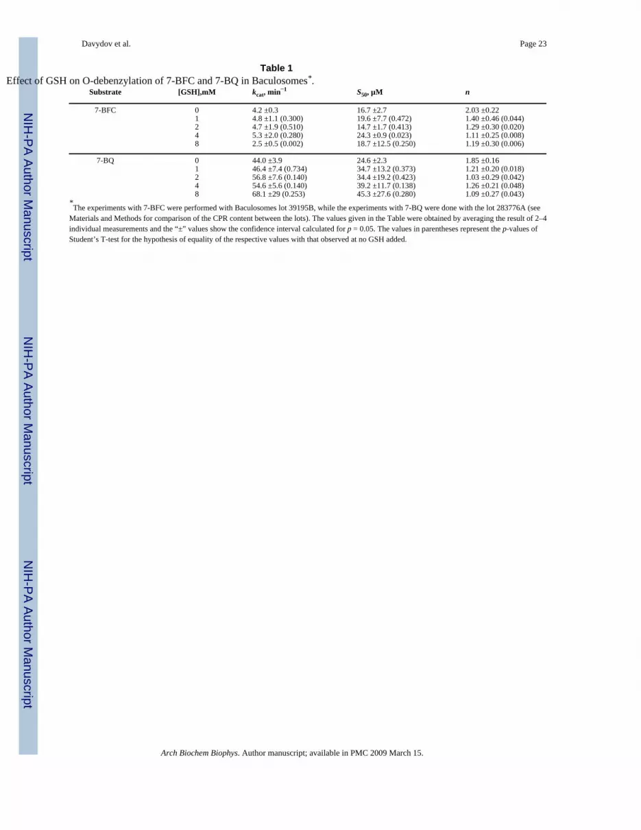
As shown in Fig. 1 and Table 1, addition of 1 – 4 mM GSH results in a significant decrease inthe cooperativity of CYP3A4 with BFC as a substrate. For example, the Hill coefficient (n) of2.0 ± 0.2 in the absence of GSH decreases to 1.1 ± 0.2 at 4 mM GSH (Table 1). There is nofurther decrease in n at 8 mM GSH, although kcat begins to decrease at that concentration.Similar results were obtained using BQ as a substrate (Table 1). The dependencies of the Hillcoefficient on the GSH concentration can be fitted to a hyperbolic (Michaelis-Menten) equationthat gives the effective constant of dissociation of the GSH-CYP3A4 complex of 0.5 ± 0.2 and0.24 ± 0.2 mM for BFC and BQ debenzylation, respectively (Fig 1b).
Davydov et al. Page 5
Arch Biochem Biophys. Author manuscript; available in PMC 2009 March 15.
NIH
-PA Author Manuscript
NIH
-PA Author Manuscript
NIH
-PA Author Manuscript

Effect of GSH on homotropic cooperativity of O-debenzylation of BFC by enrichedBaculosomes and human liver microsomes
It is important to note that the CYP3A4 Baculosomes used in our initial experiments differconsiderably from the membrane of the endoplasmic reticulum of human liver in the contentof the components of the monooxygenase system. As described in Matherials and Methods,the molar ratio of CPR to CYP3A4 in the two preparations of Baculosomes used in this studywas estimated to be around 3.2:1 and 8:1, while the ratio of CPR to total P450 in livermicrosomes is known to be 1:10 [57] or even 1:50 [58, 59]. Another important difference isthat the Baculosomes do not contain cytochrome b5. In order to probe the effect of GSH onBFC debenzylation in a system closer to the in vivo conditions we incorporated into theBaculosomal membrane cytochrome b5 and increased the concentration of CYP3A4 byincubating Baculosomes with purified recombinant proteins. As described in Materials andMethods, our treatment increased the concentration of CYP3A4 from 7 to 10 times, so that themolar ratio of CYP3A4 to CPR may be estimated to be around 2:1. The molar ratio ofcytochrome b5 to CYP3A4 in these preparations, which are designated here as “EnrichedBaculosomes”, was close to 1:1.
As seen from Table 2, incorporation of additional cytochrome 3A4 decreases the kcat calculatedper mol of the heme protein, while the rate calculated per mole of CPR remained almostunchanged (not shown). The effect of GSH on BFC metabolism in enriched Baculosomes wasqualitatively similar to that observed in the initial preparation, although the maximal effect ofGSH on cooperativity was observed at considerably lower concentrations of GSH. An increasein kcat was also revealed in enriched Baculosomes at 4 mM GSH(Table 2).
Elimination of cooperativity of BFC debenzylation in the presence of GSH was also observedwith human liver microsomes (Table 2). Similar to enriched Baculosomes, the maximal effecton cooperativity and kcat in liver microsomes was observed at 0.5–2 mM GSH, which isconsiderably lower than the concentration of 4 mM necessary for the maximal GSH effect inunenriched Baculosomes.
Effect of GSH on heterotropic cooperativity of CYP3A4 with ANFThe studies shown above demonstrate that addition of GSH eliminates homotropiccooperativity with BFC and BQ both in human liver microsomes and in model Baculosomalmembranes containing recombinant CYP3A4. To examine a possible influence of GSH onheterotropic cooperativity we studied the effect of ANF on O-debenzylation of BFC. Earlierstudies have shown activation of this CYP3A4-catalyzed reaction in the presence of ANF inboth a reconstituted system with DOPC [4] and in human liver microsomes [60]. Interestingly,our pilot experiments showed no measurable activating effect of 1 – 75 µM ANF on O-debenzylation of 30 µM BFC by CYP3A4-Baculosomes but rather a well-pronouncedinhibition of this reaction at ANF concentrations >5 µM (data not shown).
However, in both enriched Baculosomes and in human liver microsomes Fig. 2 the additionof 25 µM ANF results in an important decrease in S50 while greatly decreasing the homotropiccooperativity with this substrate. Importantly, addition of ANF to both these systems does notaffect the value of kcat for BFC O-debenzylation (Table 2), in contrast to that observed in thereconstituted micellar system [4]. Addition of GSH to human liver microsomes in the presenceof ANF, while preserving these effects, results in an increase in the kcat value, similar to thatobserved in the absence of ANF, suggesting that ANF and GSH act independently of eachother.
Davydov et al. Page 6
Arch Biochem Biophys. Author manuscript; available in PMC 2009 March 15.
NIH
-PA Author Manuscript
NIH
-PA Author Manuscript
NIH
-PA Author Manuscript

Interactions of GSH with CYP3A4 monitored by changes in the heme protein absorbance inthe Soret region
The above results suggest that the effect of GSH on cooperativity and kcat of BFC and BQoxidation by CYP3A4 is attained through formation of a complex with the enzyme. Earlierstudies showed that sulfur donor ligands, such as mercaptoethanol, 1-propanethiol, or p-chlorothiophenol, are capable of binding to the sixth coordination position of the heme iron[56,61,62]. The binding of the deprotonated SH-compounds (thiolates) results in uniquehyperporphyrin UV-visible spectrum with the Soret band split into two bands located around370–380 and 455–465 nm, respectively [56]. The complexes with SH-containing compoundsin the protonated state (thiols) have absorbance spectra quite similar to those of low-spin ligand-free enzyme but with a broader and approximately 20% less extensive Soret absorbance band(λmax=418 nm) [56]. Since GSH exhibits a pK value of 8.7 [63], its SH-group is almostcompletely protonated in the conditions of our experiments (pH 7.4). As seen from Fig. 3a, thespectral changes observed upon addition of GSH to CYP3A4 are completely consistent withbinding of a thiol to the heme iron. The titration results exhibit weakening and broadening ofthe Soret band, which is reflected in the appearance of a trough at 414 nm accompanied by twopeaks at the left and right sides (at 378 and 460 nm, respectively) of the difference spectrashown in the inset. To interpret these changes quantitatively we used least-squares fitting ofthe P450 spectra by a linear combination of spectral standards of CYP3A4 low- and high-spinstates [11] and the extinction spectra of thiol- and thiolate complexes of P450cam taken from[56]. The consistency of this approach is demonstrated by the fact that the correlationcoefficient for the fitting of the spectra was always higher than 0.99, and the total concentrationof the heme protein derived from such fitting does not exhibit any changes during the titrationwith GSH. The application of this approach shows that the addition of GSH results in a decreasein the concentration of the low-spin ligand-free CYP3A4 due to its conversion to the thiol-ligated state concomitant with some increase in the concentration of both high spin and thiolate-ligated states of the enzyme (Fig. 3b). As shown in Fig. 3c at saturation of CYP3A4 with GSHthe fraction of the enzyme represented by thiol- and thiolate-complexes approaches 40%. Thedependence of the fraction of GSH-ligated CYP3A4 on the concentration of GSH may beapproximated by the Hill equation with an S50 of 8.6 mM and Hill coeffici7ent of 2.2. Positivecooperativity in GSH binding observed in these experiments indicates the presence of anadditional high-affinity effector binding site, saturation of which with GSH promotes theinteractions of this thiol with the heme iron.
At very high concentrations of GSH (>30 mM) the spectral changes indicating the binding ofGSH by coordination to the heme iron were followed by an extensive low-to-high spin shift(Fig. 3a, dashed line). This GSH-induced spin shift, which was completely reversible upondilution of the sample, appears to signify an additional low-affinity Type-I binding event witha very strong cooperativity characterized by a Hill coefficient of 2.5 – 3 and S50 value higherthan 70 mM (Fig. 3d). This additional low-affinity interaction of CYP3A4 with GSH may betherefore responsible for some decrease in kcat of BFC debenzylation observed at 8 mM GSH(Table 1).
Effect of GSH on homotropic cooperativy of substrate binding to CYP3A4 in solutionA series of spectra obtained upon titration of CYP3A4 with 1-PB in the presence of GSH areshown in Fig. 4a. Although the addition of 1-PB results in spectral changes characteristic of asubstrate-induced low-to-high spin shift, there is an important difference from the prototypicalType I binding. As shown in the inset to Fig. 3a, the major trough of the difference spectra,which is located at 418 nm, has a distinct shoulder at 452 nm that is apparently indicative ofthe dissociation of the thiolate-ligated hyporporphyrin complex of CYP3A4 discussed above.This observation confirms the above conclusion on the binding of GSH to CYP3A4 bycoordination of the SH-group of GSH to the heme iron. Similar spectral changes were also
Davydov et al. Page 7
Arch Biochem Biophys. Author manuscript; available in PMC 2009 March 15.
NIH
-PA Author Manuscript
NIH
-PA Author Manuscript
NIH
-PA Author Manuscript

observed in the titrations of CYP3A4 with testosterone, BFC, or ANF in the presence of GSH(data not shown). The changes in the concentration of the low-spin, high-spin, and thiol(ate)-ligated forms of the enzyme shown in Fig. 4b confirm that the binding of 1-PB in the presenceof GSH results in the conversion of both low-spin ligand-free and thiol(ate)-ligated states ofthe enzyme to the high-spin 1-PB-bound form.
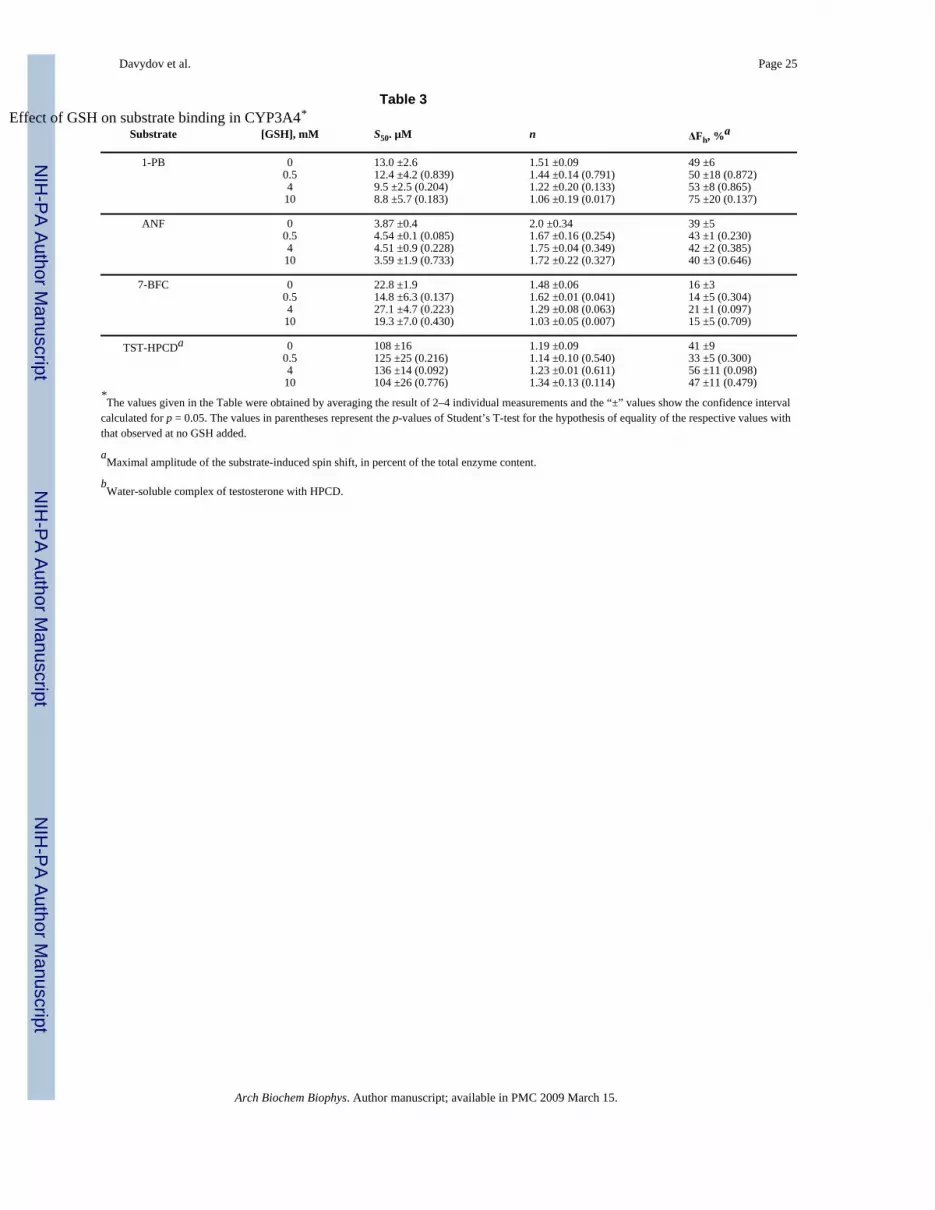
Concentration dependencies of the high spin fraction of CYP3A4 obtained in the titrations ofthe enzyme with 1-PB at different concentrations of GSH are shown in Fig 5a. Addition ofGSH amplifies the 1-PB-induced spin shift considerably. In addition, GSH decreases the S50from 12.4 ± 4.2 to 8.8 ± 5.0 µM (Fig. 5b). The most important observation is that GSHeliminates the homotropic cooperativity in the binding of 1-PB (Fig 5b) as evidenced by adecrease in the Hill coefficient from 1.5 ± 0.1 to 1.05 ± 0.2 upon addition of 8 mM GSH.Results of these experiments as well as those of the similar studies with three other substrates(ANF, BFC, and testosterone) are summarized in Table 3. Similar to the results with 1-PB,addition of 10 mM GSH completely eliminates the cooperativity of BFC binding. However,there is no effect on the S50 value. Most strikingly, addition of GSH has no significant effecton the interactions of CYP3A4 with ANF or testosterone.
Effect of GSH on the fluorescence of CYP3A4(C58,C64) labeled with BADANIn order to probe whether the interactions of CYP3A4 with GSH result in a conformationaltransition similar to that described for the binding of ANF [34], we studied the effect of GSHon the fluorescence of BADAN attached to Cys-64 in our CYP3A4(C58,C64) construct. Asshown in Fig. 6a addition of GSH results in a decrease in the intensity of fluorescence similarto that observed earlier upon addition of ANF [34]. The dependence of the relative intensityof fluorescence on the concentration of GSH (Fig. 6b) may be adequately approximated by ahyperbolic (Michaelis-Menten) equation. The values of the maximal amplitude of thefluorescence decrease and the apparent KD of the CYP3A4-GSH complex found by averagingthe results of three individual experiments are 20 ± 14% and 1.2 ± 0.5 mM, respectively. Thelatter value is reasonably close to the estimates of 0.5 and 1.3 mM obtained for KD app for theeffect of GSH on the Hill coefficient for O-debenzylation of BFC and BQ by CYP3A4-containing Baculosomes. We may conclude therefore that the CYP3A4 transition revealed inthe effect of GSH on the fluorescence of CYP3A4(C58,C64)-BADAN is similar or directlyrelated to the GSH-dependent transition that abolishes cooperativity of BFC and BQ oxidation.
In order to probe the relationship between ANF and GSH binding we studied the effect of ANFon the fluorescence of the labeled protein in the presence of 3 mM GSH (Fig. 7). Similar toprevious results in the absence of GSH [34], addition of ANF to CYP3A4(C58,C64)-BADANin the presence of GSH results in a decrease in the fluorescence of the probe (Fig. 7a). Thedependence of the relative intensity of fluorescence on the concentration of ANF (Fig. 7b)obeys the Hill equation. The values of S50, n, and the maximal amplitude of the fluorescencedecrease obtained by averaging the results of three individual experiments are equal to 14.2 ±4.6 µM, 1.8 ± 0.3, and 51.5 ± 3.1% respectively. Since these estimates are similar to the valuesobtained earlier in the absence of GSH (18.2 ± 0.7 µM, 1.7 ± 0.1 and 49 ± 8% respectively),we may conclude that the effects of ANF and GSH on CYP3A4 are additive, and the bindingsite for GSH appears to be distinct from the low-affinity ANF-binding site. However, thesimilarity of GSH- and ANF-induced changes in the fluorescence of the BADAN probesuggests that the mechanisms of action of these effectors are closely interrelated.
DiscussionAlthough the activating effect of GSH on CYP3A4 catalyzed reactions in model systems hasbeen documented previously, the present study provides the first evidence of a role ofglutathione as an allosteric effector of CYP3A4 in microsomal membranes. We demonstrated
Davydov et al. Page 8
Arch Biochem Biophys. Author manuscript; available in PMC 2009 March 15.
NIH
-PA Author Manuscript
NIH
-PA Author Manuscript
NIH
-PA Author Manuscript

here that the homotropic cooperativity of human liver microsomes with the CYP3A4 markersubstrate BFC is eliminated at low milimolar concentrations of GSH. As this effect is wellreproduced in CYP3A4 Baculosomes with BFC as well as with another CYP3A4 markersubstrate, BQ, we may conclude that the loss of cooperativity caused by GSH in livermicrosomes is determined primarily by the interactions with CYP3A4. The effect of GSH isnot limited to suppression of CYP3A4 cooperativity but also involves some increase in kcat.for BQ oxidation in Baculosomes and BFC metabolism in enriched Baculosomes and humanliver microsomes.
The loss of cooperativity of CYP3A4 in the presence of GSH was also revealed in our studiesof interactions with BFC monitored by a substrate-induced spin shift in the purified enzymein solution. This effect was found to be even more pronounced with 1-PB, another substratepossessing homotropic cooperativity. In the case of 1-PB there was also a remarkable increaseof the amplitude of the 1-PB-induced spin shift in the presence of GSH, which may indicatean effect of the latter on the degree of hydration and the openness of the heme pocket of the 1-PB-bound enzyme. It seems therefore likely that the binding of GSH displaces the equilibriumbetween the recently postulated “P” and “R” conformations of the enzyme [35] towards the“P” state, where water flux into the heme pocket is hampered and the high-spin state of thesubstrate-bound CYP3A4 is stabilized.
In contrast to 1-PB and BFC, interactions of CYP3A4 with GSH have virtually no effect onthe binding of testosterone or ANF, suggesting differences in the mechanisms of binding ofthe substrates. This conclusion agrees with the fact that in our recent study of CYP3A4(C58,C64) labeled with fluorescent probes, only ANF but not 1-PB or testosterone had apronounced effect on the fluorescence, and only testosterone but not 1-PB affected thefluorescence in the presence of ANF.
Titration of CYP3A4 showed that GSH is capable of binding to the sixth coordination positionof the heme iron. The positive cooperativity in GSH binding observed in these experimentsindicates the presence of an additional high-affinity effector binding site, saturation of whichby GSH promotes the interactions of this thiol with the heme iron. This is also supported bythe results of our experiments with CYP3A4(C58,C64)-BADAN, which revealed a GSHbinding site with KD
app of 1.2 ± 0.5 mM, which is consistent with its role as an effectorpromoting the coordination of the second molecule of GSH to the heme iron characterized byan S50 of 8.6 mM and Hill coefficient of 2.2. The above KD is also consistent with the estimatesof 0.8 – 1.2 mM for the apparent dissociation constant obtained from the dependencies of theHill coefficient in BFC and BQ debenzylation on GSH concentration. Therefore, we maypostulate a presence of a high-affinity effector binding site for GSH characterized with a lowmillimolar KD. The interaction of GSH with this site is likely to be responsible for the effecton CYP3A4 cooperativity with BFC, BQ and 1-PB and also promote the binding of GSH tothe heme iron as a thiol ligand.
Importantly, the effect of ANF, a prototype heterotropic activator of CYP3A4, was found tobe additive with the effect of GSH. The effects of ANF and GSH on the fluorescence ofBADAN attached to Cys-64 were also additive. Therefore, the high affinity binding site forGSH appears to be distinct from the effector binding site for ANF described in our recent studywith CYP3A4(C58,C64) construct [34].
Coordination of GSH to the CYP3A4 heme iron is expected to compete with the interactionsof CYP3A4 with substrates. However, the addition of 1 – 10 mM GSH results in an increasedaffinity of CYP3A4 for 1-PB and does not affect the S50 values for the binding of BFC,testosterone, or ANF, suggesting that there is no competition between coordination of GSH tothe heme iron and the interactions of CYP3A4 with these substrates. A possible explanation
Davydov et al. Page 9
Arch Biochem Biophys. Author manuscript; available in PMC 2009 March 15.
NIH
-PA Author Manuscript
NIH
-PA Author Manuscript
NIH
-PA Author Manuscript

for this observation may be provided by the concept of persistent heterogeneity of CYP3A4[37,38,64–66] caused by dissimilarity of the orientation and/or conformation of the subunitsin the oligomers (aggregates) in solution [38,66]. Lack of competition between GSH ligationto the heme and substrate binding may be caused by heterogeneity of oligomeric P450 insolution, similar to that revealed in barotropic behavior of CYP3A4 [37], kinetics of the enzymereduction by dithionite [38], and CO-binding kinetics [64]. A suggestion that only some of theP450 molecules constituting the oligomer in solution have an orientation permitting them tobind GSH by ligation to the heme iron is consistent with our observation that, upon saturationof CYP3A4 with GSH the fraction of the enzyme represented by its thiol- and thiolate-ligatedheme complexes does not exceed 40%. This is in contrast to soluble monomeric P450cam,where the binding of thiols and thiolates was shown to be competitive with substrate binding,and the complete conversion of P450cam to the thiol(ate)-bound forms was observed uponsaturation of this enzyme with thiols and thiolates [56]. Since the binding of GSH binding bycoordination to the heme iron reveals no competition with substrate binding in CYP3A4 inoligomers in solution, we may infer that the CYP3A4 molecules interacting with GSH do notparticipate in the interactions with BFC, 1-PB, ANF, or testosterone and/or do not respond tosubstrate binding by the low-to-high spin shift.
A similar mechanism may also take place in the membrane, where microsomal cytochromesP450 are also known to exist in equilibrium between monomers and oligomers [67–72].However, the reversibility of oligomerization in the membrane, as opposed to virtualirreversibility in solution, would blunt the manifestations of the proposed conformationalheterogeneity in the membrane-bound enzyme. Consistent with this inference, increasingconcentrations of GSH in Baculosomes and human liver microsomes caused an increase inS50 with BQ and BFC, respectively, possibly resulting from competition of GSH ligation tothe heme with substrate binding (Table 1).
Regardless of the mechanisms of GSH action, our study provides convincing evidence for therole of GSH as a heteroropic activator and a modulator of homotropic cooperativity inCYP3A4. This finding may indicate that the abundant evidence of CYP3A4 homo- andheterotropic cooperativity represent a manifestation of an allosteric mechanism designed to betriggered by physiological effectors, such as GSH, the concentration of which in tissues isconsistent with this role. For instance, the average concentration of GSH in rat liver was shownto be in the range of 4 – 8 mM [73]. Moreover, the GSH level in primary cultures of rathepatocytes is reported to be 40 – 60 nmol/106 cells [74,75], which is equivalent to aconcentration of 8 – 12 mM (calculated assuming the volume of a single hepatocyte cell equalto 4940 µm3 [76]). This concentration is subject to substantial changes in response to variousstimuli [41,48,73,77,78]. In particular, exposure of human hepatocarcinoma Hep2 cells toorganochlorine insecticides, such as DDT, decreases the GSH concentration from 4 – 5 downto 1.2 – 1.5 mM (calculated from [48] assuming the protein content in hepatocytes to be of 220mg/g [79]). Therefore, the estimate of 0.5 – 1.3 mM for the concentration of GSH exhibitinga half-maximal effect on the Hill coefficient for O-debenzylation of BFC and BQ is certainlyconsistent with the physiological relevance of this newly-discovered regulatory interaction.Moreover, an inhibition of CYP3A4 at high GSH concentrations observed in our experimentsmay also be physiologically significant under certain conditions. The fact that the GSHconcentration necessary for its maximal effect on cooperativity is decreased upon incorporationof cytochrome b5 and increase in CYP3A4 concentration in Baculosomes shows that themechanism of action of GSH may also involve an effect on protein-protein interactions, suchas CYP3A4 interactions with cytochrome b5, CPR and/or CYP3A4 oligomerization. Therefore,the response of the microsomal monooxygenase to GSH appears to be sensitive to thecomposition of the microsomal electron transfer chain, and a regulatory action of GSH invivo may change upon variation of the content of cytochromes b5 and P450 in the membrane.
Davydov et al. Page 10
Arch Biochem Biophys. Author manuscript; available in PMC 2009 March 15.
NIH
-PA Author Manuscript
NIH
-PA Author Manuscript
NIH
-PA Author Manuscript

In view of possible physiological role of observed effect, further detailed study of themechanism of binding of GSH to CYP3A4 and its effect on the structure and functionalproperties of the enzyme appear to be important. Of particular interest is the nature of the high-affinity GSH binding site and its possible overlap with the effector binding site for substratesexhibiting homotropic cooperativity. Another important question is whether the interactionsof CYP3A4 with GSH affect the stoichiometry of monooxygenation. The functional role ofCYP3A4 modulation by glutathione may be especially important if in addition to the effect oncooperativity and turnover rate interactions with GSH modulate the coupling of NADPHoxidation to substrate monooxygenation. A possible effect of glutathione on coupling issupported by the observation that the addition of GSH results in a remarkable activation oftestosterone hydroxylation by cytochrome b5, [46], which is known to increase the couplingof microsomal P450 enzymes (see [39,40] for review). Furthermore, an important practicalinference that may be derived from our study is that the effects of GSH on CYP3A4cooperativity and the rate of monooxygenation appear to be important factors that, which mustbe taken into account when extrapolating the results of in vitro studies of drug metabolism anddrug-drug interactions to the situation in vivo.
Aknowledgements
The authors are grateful to Dr. Santosh Kumar (Department of Pharmacology and Toxicology, UTMB) for his helpwith the experiments on the effect of GSH on BQ metabolism by Baculosomes. This research was supported by NIHgrant GM54995, Center grant ES06676, and Robert A. Welch Foundation grant H1458
References1. Shou M, Grogan J, Mancewicz JA, Krausz KW, Gonzalez FJ, Gelboin HV, Korzekwa KR.
Biochemistry 1994;33:6450–6455. [PubMed: 8204577]2. Korzekwa KR, Krishnamachary N, Shou M, Ogai A, Parise RA, Rettie AE, Gonzalez FJ, Tracy TS.
Biochemistry 1998;37:4137–4147. [PubMed: 9521735]3. Ueng YF, Kuwabara T, Chun YJ, Guengerich FP. Biochemistry 1997;36:370–381. [PubMed: 9003190]4. Domanski TL, He YA, Khan KK, Roussel F, Wang Q, Halpert JR. Biochemistry 2001;40:10150–
10160. [PubMed: 11513592]5. Ekins S, Stresser DM, Williams JA. Trends Pharm. Sci 2003;24:161–166. [PubMed: 12707001]6. Yoon MY, Campbell AP, Atkins WM. Drug Metab. Rev 2004;36:219–230. [PubMed: 15237852]7. He YA, Roussel F, Halpert JR. Arch. Biochem. Biophys 2003;409:92–101. [PubMed: 12464248]8. Hosea NA, Miller GP, Guengerich FP. Biochemistry 2000;39:5929–5939. [PubMed: 10821664]9. Ngui JS, Chen Q, Shou MG, Wang RW, Stearns RA, Baillie TA, Tang W. Drug Metab. Disp
2001;29:877–886.10. Guengerich FP. Annu. Rev. Pharmacol. Toxicol 1999;39:1–17. [PubMed: 10331074]11. Fernando H, Halpert JR, Davydov DR. Biochemistry 2006;45:4199–4209. [PubMed: 16566594]12. Davydov DR, Femando H, Halpert JR. Biophys. Chem 2006;123:95–101. [PubMed: 16701937]13. Cameron MD, Wen B, Allen KE, Roberts AG, Schuman JT, Campbell AP, Kunze KL, Nelson SD.
Biochemistry 2005;44:14143–14151. [PubMed: 16245930]14. Roberts AG, Campbell AP, Atkins WM. Biochemistry 2005;44:1353–1366. [PubMed: 15667229]15. Baas BJ, Denisov IG, Sligar SG. Arch. Biochem. Biophys 2004;430:218–228. [PubMed: 15369821]16. Denisov IG, Baas BJ, Grinkova YV, Sligar SG. J. Biol. Chem 2007;282:7066–7076. [PubMed:
17213193]17. Wang RW, Newton DJ, Liu N, Atkins WM, Lu AYH. Drug Metab. Disp 2000;28:360–366.18. Galetin A, Clarke SE, Houston JB. Drug Metab. Disp 2002;30:1512–1522.19. Galetin A, Clarke SE, Houston JB. Drug Metab. Disp 2003;31:1108–1116.20. Funahashi T, Tanaka Y, Yamaori S, Kimura T, Matsunaga T, Ohmori S, Kageyama T, Yamamoto I,
Watanabe K. Drug Metab. Pharmacokinetics 2005;20:358–367.
Davydov et al. Page 11
Arch Biochem Biophys. Author manuscript; available in PMC 2009 March 15.
NIH
-PA Author Manuscript
NIH
-PA Author Manuscript
NIH
-PA Author Manuscript

21. Lu P, Lin Y, Rodrigues AD, Rushmore TH, Baillie TA, Shou MG. Drug Metab. Disp 2001;29:1473–1479.
22. Ngui JS, Tang W, Stearns R, A, Shou MG, Miller RR, Zhang Y, Lin JH, Baillie T. Drug Metab. Disp2000;28:1043–1050.
22. Houston B, Galetin A. Drug Metab. Rev 2003;35:393–415. [PubMed: 14705868]24. Houston JB, Galetin A. Arch. Biochem. Biophys 2005;433:351–360. [PubMed: 15581591]25. Schrag ML, Wienkers LC. Arch. Biochem. Biophys 2001;391:49–55. [PubMed: 11414684]26. Egnell AC, Houston JB, Boyer CS. J. Pharm. Exp. Ther 2005;312:926–937.27. Kenworthy KE, Clarke SE, Andrews J, Houston JB. Drug Metab. Disp 2001;29:1644–1651.28. Kenworthy KE, Bloomer JC, Clarke SE, Houston JB. Brit. J. Clin. Pharm 1999;48:716–727.29. Atkins WM, Wang RW, Lu AYH. Chem. Res. Toxicol 2001;14:338–347. [PubMed: 11304120]30. Atkins WM. Expert Opinion Drug Metab. Toxicol 2006;2:573–579.31. Lampe JN, Atkins WM. Biochemistry 2006;45:12204–12215. [PubMed: 17014074]32. Davydov DR, Botchkareva AE, Kumar S, He YQ, Halpert JR. Biochemistry 2004;43:6475–6485.
[PubMed: 15157081]33. Davydov DR, Botchkareva AE, Davydova NE, Halpert JR. Biophys. J 2005;89:418–432. [PubMed:
15834000]34. Tsalkova TN, Davydova NE, Halpert JR, Davydov DR. Biochemistry 2007;46:106–119. [PubMed:
17198380]35. Davydov DR, Baas BJ, Sligar SG, Halpert JR. Biochemistry 2007;46:7852–7864. [PubMed:
17555301]36. Ekroos M, Sjögren T. Proc. Natl. Acad. Sci. USA 2006;103:13684–13687.37. Davydov DR, Halpert JR, Renaud JP, Hui Bon Hoa G. Biochem. Biophys. Res. Commun
2003;312:121–130. [PubMed: 14630029]38. Davydov DR, Fernando H, Baas BJ, Sligar SG, Halpert JR. Biochemistry 2005;44:13902–13913.
[PubMed: 16229479]39. Davydov DR. Trends Biochem. Sci 2001;26:155–160. [PubMed: 11246020]40. Zangar RC, Davydov DR, Verma S. Toxicol. Appl. Pharm 2004;199:316–331.41. Lu SC. FASEB J 1999;13:1169–1183. [PubMed: 10385608]42. Sies H. Free Radical Biol. Med 1999;27:916–921. [PubMed: 10569624]43. Meyer AJ, Hell R. Photosynth. Res 2005;86:435–457. [PubMed: 16315075]44. Gillam EM, Baba T, Kim BR, Ohmori S, Guengerich FP. Arch. Biochem. Biophys 1993;305:123–
131. [PubMed: 8342945]45. Ingelman-Sundberg M, Hagbjork A, L, Ueng Y, Yamazaki FH, Guengerich FP. Biochem. Biophys.
Res. Commun 1996;221:318–322. [PubMed: 8619853]46. Kim BR, Kim DH. Biochem. Biophys. Res. Commun 1998;242:209–212. [PubMed: 9439637]47. Shaw PM, Hosea NA, Thompson DV, Lenius JM, Guengerich FP. Arch. Biochem. Biophys
1997;348:107–115. [PubMed: 9390180]48. Dehn PF, Allen-Mocherie S, Karek J, Thenappan A. Toxicol. in vitro 2005;19:261–273. [PubMed:
15649640]49. French JS, Coon MJ. Arch. Biochem. Biophys 1979;195:565–577. [PubMed: 112928]50. Davydov DR, Kumar S, Halpert JR. Biochem. Biophys. Res. Commun 2002;294:806–812. [PubMed:
12061778]51. Masters BS, Okita RT. Pharm. Ther 1980;9:227–244.52. Yim SK, Yun SJ, CH Y. J. Biochem. Mol. Biol 2004;37:629–633. [PubMed: 15479629]53. Yamazaki H, Nakajima M, Nakamura M, Asahi S, Shimada N, Gillam EMJ, Guengerich FP, Shimada
T, Yokoi T. Drug Metab. Disp 1999;27:999–1004.54. Davydov DR, Deprez E, Hui Bon Hoa G, Knyushko TV, Kuznetsova GP, Koen YM, Archakov AI.
Arch. Biochem. Biophys 1995;320:330–344. [PubMed: 7625841]55. Renaud JP, Davydov DR, Heirwegh KPM, Mansuy D, Hui Bon Hoa G. Biochem. J 1996;319:675–
681. [PubMed: 8920966]
Davydov et al. Page 12
Arch Biochem Biophys. Author manuscript; available in PMC 2009 March 15.
NIH
-PA Author Manuscript
NIH
-PA Author Manuscript
NIH
-PA Author Manuscript

56. Sono M, Andersson LA, Dawson JH. J. Biol. Chem 1982;257:8308–8320. [PubMed: 6282878]57. Estabrook RW, Franklin MR, Cohen B, Shigamatzu A, Hildebrandt A. Metabolism 1971;20:187–
199. [PubMed: 4395592]58. Karyakin AV, Davydov DR. Vestnik Akad. Med. Nauk SSSR 1988;1988(1):53–62.59. Watanabe J, Asaka Y, Fujimoto S, Kanamura S. J. Histochem. Cytochem 1993;41:43–49. [PubMed:
8417111]60. Stresser DM, Turner SD, Blanchard AP, Miller VP, Crespi CL. Drug Metab. Disp 2002;30:845–852.61. Ullrich V, Nastainczyk W, Ruf HH. Biochemi. Soc. Trans 1975;3:803–807.62. Nastainzcyk W, Ruf H, Ullrich V. Eur. J. Biochem 1975;60:615–620. [PubMed: 1204657]63. Gough JD, Williams RH, Donofrio AE, Lees WJ. J. Am. Chem. Soc 2002;124:3885–3892. [PubMed:
11942825]64. Koley AP, Buters JT, Robinson RC, Markowitz A, Friedman FK. J. Biol, Chem 1995;270:5014–
5018. [PubMed: 7890608]65. Koley AP, Buters JTM, Robinson RC, Markowitz A, Friedman FK. J. Biol. Chem 1997;272:3149–
3152. [PubMed: 9013547]66. Fernando H, Davydov DR, Chin CC, Halpert JR. Arch. Biochem. Biophys 2007;460:129–140.
[PubMed: 17274942]67. Greinert R, Finch SA, Stier A. Bioscience Reports 1982;2:991–994. [PubMed: 7165794]68. Kawato S, Gut J, Cherry RJ, Winterhalter KH, Richter C. J. Biol. Chem 1982;257:7023–7029.
[PubMed: 7085615]69. Schwarz D, Pirrwitz J, Meyer HW, Coon MJ, Ruckpaul K. Biochem. Biophys. Res. Commun
1990;171:175–181. [PubMed: 2168169]70. Alston K, Robinson RC, Park SS, Gelboin HV, Friedman FK. J. Biol. Chem 1991;266:735–739.
[PubMed: 1985961]71. Szczesna-Skorupa E, Mallah B, Kemper B. J. Biol. Chem 2003;278:31269–31276. [PubMed:
12766165]72. Schwarz D, Chernogolov L, Kisselev P. Biochemistry 1999;38:9456–9464. [PubMed: 10413522]73. Huang ZZ, Li HY, Cai JX, Kuhlenkamp J, Kaplowitz N, Lu SC. Hepatology 1998;27:147–153.
[PubMed: 9425930]74. Lu SC, Sun WM, Yi J, Ookhtens M, Sze G, Kaplowitz N. J. Clin. Inv 1996;97:1488–1496.75. Eisenbrand GS, Golzer P. Chem. Res. Toxicol 1995;8:40–46. [PubMed: 7703365]76. Weibel ER, Staubli W, Gnagi HR, Hess FA. J. Cell Biol 1969;42:68–91. [PubMed: 4891915]77. Voehringer DW. Free Radical Biol. Med 1999;27:945–950. [PubMed: 10569627]78. Mceligot AJ, Yang S, Meyskens FL. Ann. Rev. Nutr 2005;25:261–295. [PubMed: 16011468]79. Wibo M, Amar-Costesec A, Berthet J, Beayfay H. J. Cell Biol 1971;51:52–71. [PubMed: 4329524]
Abbreviations usedCYP3A4, cytochrome P450 3A4; Hepes, N-[2-hydroxyethylpiperazine-N"-[2-ethanesulfonicacid]; 1-PB, 1-pyrenebutanol; BFC, 7-benzyloxy-4-(trifluoromethyl)coumarin; HFC, 7-hydroxy-4-(trifluoromethyl)coumarin; BQ, 7-benzyloxyquinoline; HPCD, 2-hydroxypropyl-β-cyclodextrin; BADAN, 6-bromoacetyl-2-dimethylaminonaphthalene; GSH, reducedglutathione.
Davydov et al. Page 13
Arch Biochem Biophys. Author manuscript; available in PMC 2009 March 15.
NIH
-PA Author Manuscript
NIH
-PA Author Manuscript
NIH
-PA Author Manuscript

Figure 1.Effect of GSH on O-debenzylation of BFC and BQ by CYP3A4 in Baculosomes. Panel a showsthe dependence of the rate of oxidation of BFC on the concentration of this substrate at noglutathione added (circles, solid line) and in the presence of 1 mM (inverted triangles, longdash), 2 mM (squares, medium dash), 4 mM (diamonds, short dash), and 8 mM (triangles,dash-dot) GSH. The lines represent the result of fitting of the data sets to the Hill equation.Panel b shows the dependencies of the Hill coefficient on the glutathione concentration forO-debenzylation of BFC (closed circles, solid line) and BQ (open circles dashed line). Thepoints shown on this plot represent the averages of from two to three individual measurements.
Davydov et al. Page 14
Arch Biochem Biophys. Author manuscript; available in PMC 2009 March 15.
NIH
-PA Author Manuscript
NIH
-PA Author Manuscript
NIH
-PA Author Manuscript

The lines on this plot show the results of the fitting of these data sets to a hyperbolic (Michaelis-Menten) equation with KD
app values of 0.5 ± 0.2 and 0.24 ± 0.2 mM, respectively.
Davydov et al. Page 15
Arch Biochem Biophys. Author manuscript; available in PMC 2009 March 15.
NIH
-PA Author Manuscript
NIH
-PA Author Manuscript
NIH
-PA Author Manuscript

Figure 2.Effect of ANF on O-debenzylation of 7-BFC by human liver microsomes and effect of GSH.The titration curve obtained at no effector added is shown with circles, while the data obtainedat 25 µM ANF in the absence or in the presence of 1mM GSH are represented by triangles andsquares, respectively. Solid lines represent the results of the fitting of the data sets to the Hillequation.
Davydov et al. Page 16
Arch Biochem Biophys. Author manuscript; available in PMC 2009 March 15.
NIH
-PA Author Manuscript
NIH
-PA Author Manuscript
NIH
-PA Author Manuscript

Davydov et al. Page 17
Arch Biochem Biophys. Author manuscript; available in PMC 2009 March 15.
NIH
-PA Author Manuscript
NIH
-PA Author Manuscript
NIH
-PA Author Manuscript

Figure 3.GSH-induced spectral transitions in CYP3A4 in solution. Panel a shows a series of spectra of1 µM CYP3A4 in 0.1 M Na-Hepes buffer, pH 7.4 (25 °C) recorded at no GSH added and inthe presence of GSH at the concentrations of 1.8, 4.7, 8.1, 14, 22, 32 mM (solid lines) and 84mM (dashed line). The inset shows the difference spectra obtained by subtraction of the firstspectrum of the series (no GSH added) from all subsequent spectra. Panel b illustrates the GSH-induced changes in the concentrations of the water-coordinated low spin (circles),pentacoordinated high-spin (inverted triangles), thiolate- (squares) and thiol-coordinated(diamonds) states of the heme protein. The changes in the fraction of the enzyme representedby the total of its thiolate- and thiol-coordinated states are shown in panel c. Here the solid linerepresent the results of the fitting of the data set by the Hill equation (S50 = 8.6 mM, n = 2.2,ΔFmax = 40 %). Changes in the high-spin fraction of the enzyme are illustrated in panel d,where the line represent the results of the fitting of the data set by the Hill equation (S50 = 76.5mM, n = 2.5, ΔFmax = 50.5 %).
Davydov et al. Page 18
Arch Biochem Biophys. Author manuscript; available in PMC 2009 March 15.
NIH
-PA Author Manuscript
NIH
-PA Author Manuscript
NIH
-PA Author Manuscript
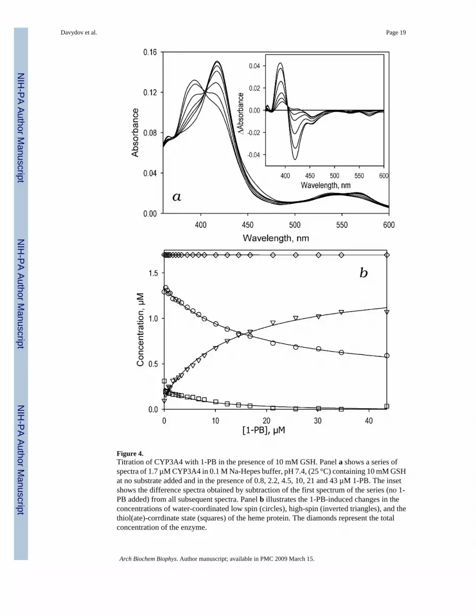
Figure 4.Titration of CYP3A4 with 1-PB in the presence of 10 mM GSH. Panel a shows a series ofspectra of 1.7 µM CYP3A4 in 0.1 M Na-Hepes buffer, pH 7.4, (25 °C) containing 10 mM GSHat no substrate added and in the presence of 0.8, 2.2, 4.5, 10, 21 and 43 µM 1-PB. The insetshows the difference spectra obtained by subtraction of the first spectrum of the series (no 1-PB added) from all subsequent spectra. Panel b illustrates the 1-PB-induced changes in theconcentrations of water-coordinated low spin (circles), high-spin (inverted triangles), and thethiol(ate)-corrdinate state (squares) of the heme protein. The diamonds represent the totalconcentration of the enzyme.
Davydov et al. Page 19
Arch Biochem Biophys. Author manuscript; available in PMC 2009 March 15.
NIH
-PA Author Manuscript
NIH
-PA Author Manuscript
NIH
-PA Author Manuscript

Figure 5.Effect of GSH on 1-PB-induced spin shift in CYP3A4. Panel a shows the changes in the highspin fraction of CYP3A4 upon addition of 1-PB at no glutathione added (circles) and in thepresence of 0.5 mM (inverted triangles), 4 mM (squares), and 10 mM (diamonds) GSH. Thelines represent the result of fitting of the data sets to the Hill equation. Panel b shows thedependence of the S50 value (circles) and the Hill coefficient (squares) on the glutathioneconcentration. The points on this plot represent the averages of two to three individualmeasurements. The solid lines show the fitting of these data sets with a hyperbolic (Michaelis-Menten) equation with KD app values of 1.4 ± 0.9 and 1.6 ± 1.4 mM, respectively.
Davydov et al. Page 20
Arch Biochem Biophys. Author manuscript; available in PMC 2009 March 15.
NIH
-PA Author Manuscript
NIH
-PA Author Manuscript
NIH
-PA Author Manuscript

Figure 6.Interaction of CYP3A4(C58,C64)-BADAN with GSH monitored by the changes in thefluorescence of the probe. (a) A series of emission spectra of 1.5 µM enzyme recorded at 0,0.17, 0.33, 0.66, 1.0, 1.5, and 2.6 mM GSH. (b) Relative fluorescence intensity versus GSHconcentration. The line shows the fitting of the data sets to a hyperbolic (Michaelis-Menten)equation with KD
app = 1.6 mM and the maximal amplitude of the fluorescence decrease of17%.
Davydov et al. Page 21
Arch Biochem Biophys. Author manuscript; available in PMC 2009 March 15.
NIH
-PA Author Manuscript
NIH
-PA Author Manuscript
NIH
-PA Author Manuscript

Figure 7.Effect of ANF on fluorescence of CYP3A4(C58,C64)BADAN in the presence of 3 mM GSH.(a) A series of emission spectra of 1.5 µM protein recorded at 0, 2, 5, 10, 15, 20, 25, 30, 35,and 50 µM ANF. (b) Relative fluorescence intensity versus substrate concentration. The lineshows the fitting of the data sets to the Hill equation with S50 = 16.6 µM, n=1.7, and the maximalamplitude of the fluorescence decrease of 56%.
Davydov et al. Page 22
Arch Biochem Biophys. Author manuscript; available in PMC 2009 March 15.
NIH
-PA Author Manuscript
NIH
-PA Author Manuscript
NIH
-PA Author Manuscript
NIH
-PA Author Manuscript
NIH
-PA Author Manuscript
NIH
-PA Author Manuscript
Davydov et al. Page 23
Table 1Effect of GSH on O-debenzylation of 7-BFC and 7-BQ in Baculosomes*.
Substrate [GSH],mM kcat, min−1 S50, µM n
7-BFC 0 4.2 ±0.3 16.7 ±2.7 2.03 ±0.221 4.8 ±1.1 (0.300) 19.6 ±7.7 (0.472) 1.40 ±0.46 (0.044)2 4.7 ±1.9 (0.510) 14.7 ±1.7 (0.413) 1.29 ±0.30 (0.020)4 5.3 ±2.0 (0.280) 24.3 ±0.9 (0.023) 1.11 ±0.25 (0.008)8 2.5 ±0.5 (0.002) 18.7 ±12.5 (0.250) 1.19 ±0.30 (0.006)
7-BQ 0 44.0 ±3.9 24.6 ±2.3 1.85 ±0.161 46.4 ±7.4 (0.734) 34.7 ±13.2 (0.373) 1.21 ±0.20 (0.018)2 56.8 ±7.6 (0.140) 34.4 ±19.2 (0.423) 1.03 ±0.29 (0.042)4 54.6 ±5.6 (0.140) 39.2 ±11.7 (0.138) 1.26 ±0.21 (0.048)8 68.1 ±29 (0.253) 45.3 ±27.6 (0.280) 1.09 ±0.27 (0.043)
*The experiments with 7-BFC were performed with Baculosomes lot 39195B, while the experiments with 7-BQ were done with the lot 283776A (see
Materials and Methods for comparison of the CPR content between the lots). The values given in the Table were obtained by averaging the result of 2–4individual measurements and the “±” values show the confidence interval calculated for p = 0.05. The values in parentheses represent the p-values ofStudent’s T-test for the hypothesis of equality of the respective values with that observed at no GSH added.
Arch Biochem Biophys. Author manuscript; available in PMC 2009 March 15.

NIH
-PA Author Manuscript
NIH
-PA Author Manuscript
NIH
-PA Author Manuscript
Davydov et al. Page 24Ta
ble
2Ef
fect
s of G
SH a
nd A
NF
on O
-deb
enzy
latio
n of
7-B
FC in
enr
iche
d B
acul
osom
es a
nd h
uman
live
r mic
roso
mes
* .Sy
stem
[AN
F], µ
M[G
SH],
mM
k cat
, min−1
S 50,
µMN
Enric
hed
Bac
ulos
omes
a0
00.
65 ±
0.09
30.6
±1.
21.
47 ±
0.13
00.
50.
64 ±
0.05
(0.9
09)
14.6
±2.
8 (0
.013
)0.
96 ±
0.06
(0.0
22)
01
0.72
±0.
23 (0
.668
)36
.9 ±
2.6
(0.0
48)
1.06
±0.
19 (0
.071
)0
41.
32 ±
0.19
(0.0
14)
47.8
±5.
6 (0
.011
)1.
19 ±
0.42
(6.1
0−4 )
250
0.68
±0.
03 (0
.583
)5.
82 ±
0.17
(6.1
0−4 )
0.98
±0.
03 (0
.019
)25
40.
91 ±
0.24
(0.3
23)
16.7
±3.
0 (0
.030
)1.
0 ±0
.08
(0.0
11)
Hum
an li
ver m
icro
som
esb
00
0.42
±0.
0359
.4 ±
17.4
2.00
±0.
340
0.5
0.61
±0.
09 (0
.011
)64
.8 ±
17.5
(0.6
80)
1.17
±0.
20 (0
.040
)0
10.
47 ±
0.07
(0.3
25)
56.0
±11
.3 (0
.762
)1.
24 ±
0.05
(0.0
44)
02
0.46
±0.
04 (0
.253
)58
.4 ±
18.6
(0.9
51)
0.96
±0.
09 (0
.063
)0
40.
54 ±
0.10
(0.0
65)
116
±42
(0.0
41)
1.00
±0.
02 (0
.027
)0
80.
40 ±
0.09
(0.4
90)
200
±47
(0.0
02)
1.13
±0.
34 (0
.072
)25
00.
54 ±
0.18
(0.2
20)
16.7
±1.
1 (0
.001
)1.
34 ±
0.36
(0.1
90)
251
0.69
±0.
04 (1
.10−
4 )23
.2 ±
9.0
(0.0
23)
1.23
±0.
22 (0
.092
)* Th
e va
lues
giv
en in
the
Tabl
e w
ere
obta
ined
by
aver
agin
g th
e re
sult
of 2
–4 in
divi
dual
mea
sure
men
ts a
nd th
e “±
” va
lues
show
the
conf
iden
ce in
terv
al c
alcu
late
d fo
r p =
0.0
5. T
he v
alue
s in
pare
nthe
ses
repr
esen
t the
p-v
alue
s of S
tude
nt’s
T-te
st fo
r the
hyp
othe
sis o
f equ
ality
of t
he re
spec
tive
valu
es w
ith th
at o
bser
ved
at n
o ef
fect
or (G
SH a
nd/o
r AN
F) a
dded
.
a Enric
hed
prep
arat
ions
wer
e pr
epar
ed u
sing
the
Bac
ulos
omes
lot 2
8377
6A.
b In th
e ca
se o
f hum
an li
ver m
icro
som
es k
cat v
alue
s are
app
aren
t and
cal
cula
ted
per t
otal
cyt
ochr
ome
P450
con
tent
.
Arch Biochem Biophys. Author manuscript; available in PMC 2009 March 15.
NIH
-PA Author Manuscript
NIH
-PA Author Manuscript
NIH
-PA Author Manuscript
Davydov et al. Page 25
Table 3Effect of GSH on substrate binding in CYP3A4*
Substrate [GSH], mM S50. µM n ΔFh, %a
1-PB 0 13.0 ±2.6 1.51 ±0.09 49 ±60.5 12.4 ±4.2 (0.839) 1.44 ±0.14 (0.791) 50 ±18 (0.872)4 9.5 ±2.5 (0.204) 1.22 ±0.20 (0.133) 53 ±8 (0.865)10 8.8 ±5.7 (0.183) 1.06 ±0.19 (0.017) 75 ±20 (0.137)
ANF 0 3.87 ±0.4 2.0 ±0.34 39 ±50.5 4.54 ±0.1 (0.085) 1.67 ±0.16 (0.254) 43 ±1 (0.230)4 4.51 ±0.9 (0.228) 1.75 ±0.04 (0.349) 42 ±2 (0.385)10 3.59 ±1.9 (0.733) 1.72 ±0.22 (0.327) 40 ±3 (0.646)
7-BFC 0 22.8 ±1.9 1.48 ±0.06 16 ±30.5 14.8 ±6.3 (0.137) 1.62 ±0.01 (0.041) 14 ±5 (0.304)4 27.1 ±4.7 (0.223) 1.29 ±0.08 (0.063) 21 ±1 (0.097)10 19.3 ±7.0 (0.430) 1.03 ±0.05 (0.007) 15 ±5 (0.709)
TST-HPCDa 0 108 ±16 1.19 ±0.09 41 ±90.5 125 ±25 (0.216) 1.14 ±0.10 (0.540) 33 ±5 (0.300)4 136 ±14 (0.092) 1.23 ±0.01 (0.611) 56 ±11 (0.098)10 104 ±26 (0.776) 1.34 ±0.13 (0.114) 47 ±11 (0.479)
*The values given in the Table were obtained by averaging the result of 2–4 individual measurements and the “±” values show the confidence interval
calculated for p = 0.05. The values in parentheses represent the p-values of Student’s T-test for the hypothesis of equality of the respective values withthat observed at no GSH added.
aMaximal amplitude of the substrate-induced spin shift, in percent of the total enzyme content.
bWater-soluble complex of testosterone with HPCD.
Arch Biochem Biophys. Author manuscript; available in PMC 2009 March 15.