The Central Role of Cytochrome P450 in Xenobiotic ... - MDPI
21
Review The Central Role of Cytochrome P450 in Xenobiotic Metabolism—A Brief Review on a Fascinating Enzyme Family Francisco Esteves * , José Rueff and Michel Kranendonk Citation: Esteves, F.; Rueff, J.; Kranendonk, M. The Central Role of Cytochrome P450 in Xenobiotic Metabolism—A Brief Review on a Fascinating Enzyme Family. J. Xenobiot. 2021, 11, 94–114. https:// doi.org/10.3390/jox11030007 Academic Editor: François Gagné Received: 10 May 2021 Accepted: 16 June 2021 Published: 22 June 2021 Publisher’s Note: MDPI stays neutral with regard to jurisdictional claims in published maps and institutional affil- iations. Copyright: © 2021 by the authors. Licensee MDPI, Basel, Switzerland. This article is an open access article distributed under the terms and conditions of the Creative Commons Attribution (CC BY) license (https:// creativecommons.org/licenses/by/ 4.0/). Center for Toxicogenomics and Human Health (ToxOmics), Genetics, Oncology and Huma Toxicology, NOVA Medical School/Faculty of Medical Sciences, Universidade NOVA de Lisboa, 1169-056 Lisboa, Portugal; [email protected] (J.R.); [email protected] (M.K.) * Correspondence: [email protected] Abstract: Human Cytochrome P450 (CYP) enzymes constitute a superfamily of membrane-bound hemoproteins that are responsible for the metabolism of a wide variety of clinically, physiologically, and toxicologically important compounds. These heme-thiolate monooxygenases play a pivotal role in the detoxification of xenobiotics, participating in the metabolism of many structurally diverge compounds. This short-review is intended to provide a summary on the major roles of CYPs in Phase I xenobiotic metabolism. The manuscript is focused on eight main topics that include the most relevant aspects of past and current CYP research. Initially, (I) a general overview of the main aspects of absorption, distribution, metabolism, and excretion (ADME) of xenobiotics are presented. This is followed by (II) a background overview on major achievements in the past of the CYP research field. (III) Classification and nomenclature of CYPs is briefly reviewed, followed by (IV) a summary description on CYP’s location and function in mammals. Subsequently, (V) the physiological relevance of CYP as the cornerstone of Phase I xenobiotic metabolism is highlighted, followed by (VI) reviewing both genetic determinants and (VI) nongenetic factors in CYP function and activity. The last topic of the review (VIII) is focused on the current challenges of the CYP research field. Keywords: Cytochrome P450 (CYP); drug-metabolizing enzymes (DMEs); xenobiotic; metabolism; toxicology; carcinogens; adverse drug reactions (ADRs) 1. Xenobiotics Disposition and Excretion Humans are continuously exposed to a wide variety of chemicals. An important portion of these compounds is not essential for maintenance of normal homeostasis. They are neither nutrients nor intermediate metabolites, produced from nutritional metabolism. Drugs, environmental pollutants, cosmetics, and even components present in our diet, such as food additives, form this extended group of xenobiotics in general harmless, but potentially toxic [1–4]. In a human lifetime, one might be exposed to 1–3 million different foreign compounds, which can accumulate within a variety of different organs and tissues [4]. Storage of xenobiotics can function as either a protective mechanism or as a mean by which bioaccumulation can trigger toxic effects. This potential toxic route depends on the physiologic relationship between the storage depot and the target tissues for a specific toxicant [1,3]. Xenobiotics are metabolized and ultimately eliminated through the urine, bile, and feces, with minor routes through expiration and sweat. However, without effective detoxi- fication and subsequent excretion, many compounds may reach toxic levels and interfere with cellular homeostasis, leading to cellular and tissue damage, with detrimental effects on health [1,2,4]. Harmful cellular and tissue concentrations can be prevented by direct elimination of xenobiotics, whose mechanisms are dependent on the physico-chemical proprieties of the compounds. Yet, the vast majority undergo biotransformation by com- plex metabolic mechanisms, resulting in the formation of numerous metabolites, some of J. Xenobiot. 2021, 11, 94–114. https://doi.org/10.3390/jox11030007 https://www.mdpi.com/journal/jox
-
Upload
khangminh22 -
Category
Documents
-
view
1 -
download
0
Transcript of The Central Role of Cytochrome P450 in Xenobiotic ... - MDPI

Review
The Central Role of Cytochrome P450 in XenobioticMetabolism—A Brief Review on a Fascinating Enzyme Family
Francisco Esteves * , José Rueff and Michel Kranendonk
�����������������
Citation: Esteves, F.; Rueff, J.;
Kranendonk, M. The Central Role of
Cytochrome P450 in Xenobiotic
Metabolism—A Brief Review on a
Fascinating Enzyme Family. J.
Xenobiot. 2021, 11, 94–114. https://
doi.org/10.3390/jox11030007
Academic Editor: François Gagné
Received: 10 May 2021
Accepted: 16 June 2021
Published: 22 June 2021
Publisher’s Note: MDPI stays neutral
with regard to jurisdictional claims in
published maps and institutional affil-
iations.
Copyright: © 2021 by the authors.
Licensee MDPI, Basel, Switzerland.
This article is an open access article
distributed under the terms and
conditions of the Creative Commons
Attribution (CC BY) license (https://
creativecommons.org/licenses/by/
4.0/).
Center for Toxicogenomics and Human Health (ToxOmics), Genetics, Oncology and Huma Toxicology,NOVA Medical School/Faculty of Medical Sciences, Universidade NOVA de Lisboa, 1169-056 Lisboa, Portugal;[email protected] (J.R.); [email protected] (M.K.)* Correspondence: [email protected]
Abstract: Human Cytochrome P450 (CYP) enzymes constitute a superfamily of membrane-boundhemoproteins that are responsible for the metabolism of a wide variety of clinically, physiologically,and toxicologically important compounds. These heme-thiolate monooxygenases play a pivotal rolein the detoxification of xenobiotics, participating in the metabolism of many structurally divergecompounds. This short-review is intended to provide a summary on the major roles of CYPs inPhase I xenobiotic metabolism. The manuscript is focused on eight main topics that include themost relevant aspects of past and current CYP research. Initially, (I) a general overview of themain aspects of absorption, distribution, metabolism, and excretion (ADME) of xenobiotics arepresented. This is followed by (II) a background overview on major achievements in the past ofthe CYP research field. (III) Classification and nomenclature of CYPs is briefly reviewed, followedby (IV) a summary description on CYP’s location and function in mammals. Subsequently, (V) thephysiological relevance of CYP as the cornerstone of Phase I xenobiotic metabolism is highlighted,followed by (VI) reviewing both genetic determinants and (VI) nongenetic factors in CYP functionand activity. The last topic of the review (VIII) is focused on the current challenges of the CYPresearch field.
Keywords: Cytochrome P450 (CYP); drug-metabolizing enzymes (DMEs); xenobiotic; metabolism;toxicology; carcinogens; adverse drug reactions (ADRs)
1. Xenobiotics Disposition and Excretion
Humans are continuously exposed to a wide variety of chemicals. An importantportion of these compounds is not essential for maintenance of normal homeostasis. Theyare neither nutrients nor intermediate metabolites, produced from nutritional metabolism.Drugs, environmental pollutants, cosmetics, and even components present in our diet,such as food additives, form this extended group of xenobiotics in general harmless,but potentially toxic [1–4]. In a human lifetime, one might be exposed to 1–3 milliondifferent foreign compounds, which can accumulate within a variety of different organsand tissues [4]. Storage of xenobiotics can function as either a protective mechanism oras a mean by which bioaccumulation can trigger toxic effects. This potential toxic routedepends on the physiologic relationship between the storage depot and the target tissuesfor a specific toxicant [1,3].
Xenobiotics are metabolized and ultimately eliminated through the urine, bile, andfeces, with minor routes through expiration and sweat. However, without effective detoxi-fication and subsequent excretion, many compounds may reach toxic levels and interferewith cellular homeostasis, leading to cellular and tissue damage, with detrimental effectson health [1,2,4]. Harmful cellular and tissue concentrations can be prevented by directelimination of xenobiotics, whose mechanisms are dependent on the physico-chemicalproprieties of the compounds. Yet, the vast majority undergo biotransformation by com-plex metabolic mechanisms, resulting in the formation of numerous metabolites, some of
J. Xenobiot. 2021, 11, 94–114. https://doi.org/10.3390/jox11030007 https://www.mdpi.com/journal/jox

J. Xenobiot. 2021, 11 95
which have the potential to cause unintendedly, more toxic effects, i.e., bioactivation [1,4–6].Detoxification routes comprise enzymatic functionalization and/or conjugation reactionsthat facilitate elimination and excretion. All combined, these pathways act jointly todetoxify xenobiotics and remove them from cells and tissues [1,3,4,6–9]. In chemical toxi-cology, it is therefore of great interest to have comprehensive and integrated knowledge ofin vivo xenobiotic metabolism, to understand, predict and prevent potential health hazardsthrough bioavailability, bioaccumulation, or generation of harmful reactive metabolites,after chemical exposure.
Four stages can be distinguished in the processes of absortion, metabolism and cellu-lar excretion of xenobiotics, namely (i) influx by transporter enzymes, biotransformationin (ii) Phases I and (iii) II, mediated by drug-metabolizing enzymes (DMEs), followedby (iv) Phase III, the excretion mediated mostly through transporter enzymes [1,6,9–12](Figure 1). Organic anion transporting polypeptides (OATP), organic anion transporters(OAT) and sodium taurocholate cotransporting polypeptide (NTCP), are involved in the in-flux of the xenobiotics (reviewed in Murray M, Zhou F, 2017) [12]. Phase I enzymes, whichinclude the cytochrome P450s (CYPs) superfamily—the major contributer— but also flavin-containing monooxygenases (FMOs), NAD(P)H:quinone oxidoreductases (NQOs), amineoxidases, alcohol dehydrogenases, esterases and peroxidases (reviewed in Gan J, et al.,2016) catalyze the oxidation, reduction or hydrolyses of primarily lipophilic xenobioticsinto more polar molecules [7,13–16]. The introduction of polar groups by Phase I reactionsprovides sites that enable conjugation reactions, mediated by Phase II enzymes [5,7,17,18].Although Phase II enzymes can directly act on the parent compound [19], typically PhaseI-metabolites are conjugated with charged species, such as glucuronic acid, glutathione,sulfate, amino acids (glycine, taurine, glutamic acid), methyl or acetyl groups. Addition ofthese large anionic groups, which may detoxify reactive electrophiles (either parent com-pound or Phase I metabolite), produce Phase II metabolites, with increased hydrophilicityand molecular weight, which in larger part are not able to diffuse across phospholipid mem-brane barrier (reviewed in Jancová P, Šiller M, 2012) [6,10,11,13,19,20]. Phase III xenobiotictransporters excrete hydrophilic conjugates, with the anionic groups acting as affinity tagsfor a variety of membrane carriers belonging to two main clusters: ATP binding cassette(ABC), including the multidrug resistance protein (MRP) family, and solute carrier (SLC)transporters (reviewed in Döring B, Petzinger E, 2014) [21–23].
There are major inter- and intra-individual variations in the capacity to metabolize,detoxify and extrude xenobiotics (see below). These are of genetic, epigenetic, environ-mental, and physio- or pathophysiological origin, and vary during lifetime [9,13,21,24–29].Most xenobiotics are detoxified and cleared through an intricate network of multiple en-zymes and pathways. The relationship between xenobiotic local/cellular concentration,specific enzymes affinity, tissue specific enzyme expression, stability, and cofactors avail-ability, often determine which metabolic reactions dominate in a given individual, at oneprecise moment [1,7,30].

J. Xenobiot. 2021, 11 96J. Xenobiot. 2021, 11, FOR PEER REVIEW 3
Figure 1. Xenobiotic metabolism in the hepatocyte and the central role of CYPs in biotransformation. Besides hydroxylation, exemplified here, CYPs catalyze a variety of other biotransformation reac-tions (e.g., epoxidation, dealkylation, oxygenation, dehydrogenation, dehalogenation, among oth-ers). Non-CYP mediated metabolism may also occur in Phase I via flavin-containing monooxygen-ases (FMOs), NAD(P)H:quinone oxidoreductases (NQOs), amine oxidases, alcohol dehydrogen-ases, esterases and peroxidases. ABC: ATP binding cassette (e.g., multidrug resistance protein fam-ily—MRP); GA: glucuronic acid; GSH: glutathione; NTCP: sodium taurocholate cotransporting pol-ypeptide; OAT: organic anion transporters; OATP: organic anion transporting polypeptides; OH: hydroxyl; PAPS: phosphoadenosine-phosphosulfate; SLC: solute carrier transporters; UDPGA: uri-dine diphosphate-glucuronic acid. Transferases: glutathione S-transferases (GST), methyltransferases, glycine N-acyltransferase (GLYAT), N-acetyltransferases (NAT), sulfotransferases (SULT), UDP-glucuronosyltransferases (UGT).
There are major inter- and intra-individual variations in the capacity to metabolize, detoxify and extrude xenobiotics (see below). These are of genetic, epigenetic, environ-mental, and physio- or pathophysiological origin, and vary during lifetime [9,13,21,24–29]. Most xenobiotics are detoxified and cleared through an intricate network of multiple enzymes and pathways. The relationship between xenobiotic local/cellular concentration, specific enzymes affinity, tissue specific enzyme expression, stability, and cofactors avail-ability, often determine which metabolic reactions dominate in a given individual, at one precise moment [1,7,30].
2. Historical Aspects of Cytochrome P450s Research Cytochrome P450s comprise the major Phase I family of enzymes capable of catalyz-
ing the oxidative biotransformation of a vast majority of lipophilic xenobiotics and are the focus of research in areas such as clinical pharmacology and toxicology [5,18,25,31–33]. The early reports dealing with CYP, dated back to the 1940s and were related with in vitro studies on the metabolism of steroids and xenobiotics, including drugs and carcinogens [34].
Figure 1. Xenobiotic metabolism in the hepatocyte and the central role of CYPs in biotransformation. Besides hydroxylation,exemplified here, CYPs catalyze a variety of other biotransformation reactions (e.g., epoxidation, dealkylation, oxygenation,dehydrogenation, dehalogenation, among others). Non-CYP mediated metabolism may also occur in Phase I via flavin-containing monooxygenases (FMOs), NAD(P)H:quinone oxidoreductases (NQOs), amine oxidases, alcohol dehydrogenases,esterases and peroxidases. ABC: ATP binding cassette (e.g., multidrug resistance protein family—MRP); GA: glucuronic acid;GSH: glutathione; NTCP: sodium taurocholate cotransporting polypeptide; OAT: organic anion transporters; OATP: organicanion transporting polypeptides; OH: hydroxyl; PAPS: phosphoadenosine-phosphosulfate; SLC: solute carrier transporters;UDPGA: uridine diphosphate-glucuronic acid. Transferases: glutathione S-transferases (GST), methyltransferases, glycineN-acyltransferase (GLYAT), N-acetyltransferases (NAT), sulfotransferases (SULT), UDP-glucuronosyltransferases (UGT).
2. Historical Aspects of Cytochrome P450s Research
Cytochrome P450s comprise the major Phase I family of enzymes capable of catalyzingthe oxidative biotransformation of a vast majority of lipophilic xenobiotics and are the focusof research in areas such as clinical pharmacology and toxicology [5,18,25,31–33]. The earlyreports dealing with CYP, dated back to the 1940s and were related with in vitro studies onthe metabolism of steroids and xenobiotics, including drugs and carcinogens [34].
The establishment of the CYP research field goes back to the late 50s/early 60s, whenseveral groups focused on a particular pigment in animal liver microsomes, which seemedto be directly related with specific hepatic functions [35,36]. In 1962, a major breakthroughwas obtained by Drs. Tsuneo Omura and Ryo Sato, describing for the first time, the spectralobservation of CYP in liver microsomes [37]. In fact, the name of CYP (P450) is derivedfrom the characteristic absorption maximum at 450 nm, observed in differential spectraof the reduced CO-bond enzyme complex. Initially, CYP was thought to be a single en-zymatic entity, which was believed to exist almost exclusively in the liver, responsiblefor the metabolism of drugs and other foreign chemicals [38–40]. Several seminal reportsdescribed the inducibility of CYP activities, of clinical relevance in pharmacology and thera-peutics [40–42]. Subsequently, other remarkable discoveries were made, namely: the role ofCYPs in steroid and fatty acid hydroxylation, both in adrenal cortex and liver (1963) [43,44];protein separation of CYPs [45] and their purification [46,47]; identification and biochemicalcharacterization of multiple CYP forms—evidencing the existence of numerous isoenzymes

J. Xenobiot. 2021, 11 97
(late 60s/early 70s) [39,47–50]; biophysical studies and biochemical characterization ofbacterial CYP (P450cam or CYP101A1)—of importance to establish template protein struc-tures to be used in homology modeling (1968) [51]; and data supporting models on oxygenactivation, evidencing a stepwise process involving C-H bond breaking (1978) [52]. In1983, the first complete analysis of a CYP gene (rat) was accomplished [53]. Thereafter, theevolution of molecular genetic techniques led researchers to uncover the extensive inter-and intra-species variability of the CYP genes superfamily. Genetic studies have shown theconsiderable inter-individual differences in expression of CYP isoenzymes in the humanpopulation (since the 80s) [30,54–59]. In the waking of the new millennium, 57 humanCYP genes (plus multiple pseudogenes) were identified by the human genome project(2003) [26,60,61]. During this period other significant contributions were the develop-ment of mammalian (including human) heterologous CYP expression, using recombinantDNA technology (early 90s)—critical for crystallography and functional studies [62–65];application of engineered bacterial strains expressing human CYP enzymes complex inbiotransformation and genotoxicity studies of xenobiotics (late 90s) [66,67]; the first CYPproteins structures solved (bacterial P450cam and P450BM3) (early 80s and 90s) [68–70],followed by the crystal structures of human CYPs—particularly high-resolution structures(2000s) [71–74]; and the description of determinant protein-protein interactions within CYPenzymes complex—impacting CYP activity (2000s and early 10s) [75–81].
3. Classification and Nomenclature of Human Cytochrome P450s
Back in the 80s, studies enabled by the big boom in molecular biology demonstratedthat CYP genes are ubiquitously present in almost all life forms, from prokaryotes tohumans, adding a new dimension to the complex repertoire of functions catalyzed by thissuper enzyme family [53,54]. The genes encoding these heme-thiolate monooxygenasescapable of catalyzing the oxidative biotransformation of endogenous compounds andxenobiotics, diverged from a single ancestor in an evolutionary process started 3 billionyears ago [82,83]. In contrast with “conventional” enzymes, which normally demonstratehigh substrate specificity and turnover rates, human CYP isoenzymes involved in drugmetabolism, evolved favoring low substrate specificity and turnover rates, characteristicsof DMEs in general [30].
Although the outcome of the CYP-mediated metabolism prevents bioaccumulationby chemically transforming lipophilic compounds (readily absorbed) into hydrophilicmetabolites (readily excreted) (Figure 2), CYP mediated biotransformation as well asother DMEs, evolved in a species-specific manner [54]. Historically, species of rodents,dogs, rabbits and pigs were considered to be suitable organisms for comparative drugmetabolism studies. However, the variability in xenobiotic metabolism among species, inparticular CYP metabolism, is evidenced by the fact that mutagenicity profiles of chemicalsdetermined by the Ames test [84,85] when using human liver S9 fraction is significantlydifferent from the ones observed with the rat liver S9 fraction [86]. This raises an importantissue regarding the utility and reliability of results obtained from toxicological studies,using animal materials and models for risk assessments of human exposure [87]. Severalfactors must be carefully considered for the appropriate selection of an animal model whichadequately mimics the human metabolism of a particular xenobiotic. Species-specific ex-pression, regulation and function of CYPs have to be taken into account to avoid significantdeviations. This inter-species variability is harshly exemplified by the case of thalidomide,in the late 50s/early 60s. This drug was commercialized as a non-addictive, non-barbituratehypnotic and anti-emetic, and used for treatment of morning sickness of pregnant women.Thalidomide was tested for potential teratogenicity in mice and considered to be safe,however its use led to severe birth defects involving limb malformation. Teratogenicity isthought to occur through a reactive metabolite of thalidomide (arene oxide) (Figure 2A),formed in significantly higher proportions in human (and rabbit) liver than in mouse liver.After this occurrence, U.S. Food and Drug Administration (FDA), and medicine agenciesworldwide, implemented obligatory full multi-species (in vivo) teratogenicity testing as

J. Xenobiot. 2021, 11 98
a requisite for drug approval [88,89]. Another example is the metabolism and effect ofaflatoxin B1 in trout. Aflatoxin B1, the most potent hepatic chemical carcinogen knowntoday is activated via a CYP-dependent reaction (Figure 2G). Interestingly, and in contrastwith the rat model, trout exposure to aflatoxin B1 may result in a hepatocellular carcinomawith relevant similarity to the one found in humans [90,91].
J. Xenobiot. 2021, 11, FOR PEER REVIEW 5
termined by the Ames test [84,85] when using human liver S9 fraction is significantly dif-ferent from the ones observed with the rat liver S9 fraction [86]. This raises an important issue regarding the utility and reliability of results obtained from toxicological studies, using animal materials and models for risk assessments of human exposure [87]. Several factors must be carefully considered for the appropriate selection of an animal model which adequately mimics the human metabolism of a particular xenobiotic. Species-spe-cific expression, regulation and function of CYPs have to be taken into account to avoid significant deviations. This inter-species variability is harshly exemplified by the case of thalidomide, in the late 50s/early 60s. This drug was commercialized as a non-addictive, non-barbiturate hypnotic and anti-emetic, and used for treatment of morning sickness of pregnant women. Thalidomide was tested for potential teratogenicity in mice and consid-ered to be safe, however its use led to severe birth defects involving limb malformation. Teratogenicity is thought to occur through a reactive metabolite of thalidomide (arene oxide) (Figure 2A), formed in significantly higher proportions in human (and rabbit) liver than in mouse liver. After this occurrence, U.S. Food and Drug Administration (FDA), and medicine agencies worldwide, implemented obligatory full multi-species (in vivo) terato-genicity testing as a requisite for drug approval [88,89]. Another example is the metabo-lism and effect of aflatoxin B1 in trout. Aflatoxin B1, the most potent hepatic chemical car-cinogen known today is activated via a CYP-dependent reaction (Figure 2G). Interest-ingly, and in contrast with the rat model, trout exposure to aflatoxin B1 may result in a hepatocellular carcinoma with relevant similarity to the one found in humans [90,91].
(B)
(A)
Figure 2. Cont.
J. Xenobiot. 2021, 11 99J. Xenobiot. 2021, 11, FOR PEER REVIEW 6
(C)
(D)
(E)
Figure 2. Cont.

J. Xenobiot. 2021, 11 100J. Xenobiot. 2021, 11, FOR PEER REVIEW 7
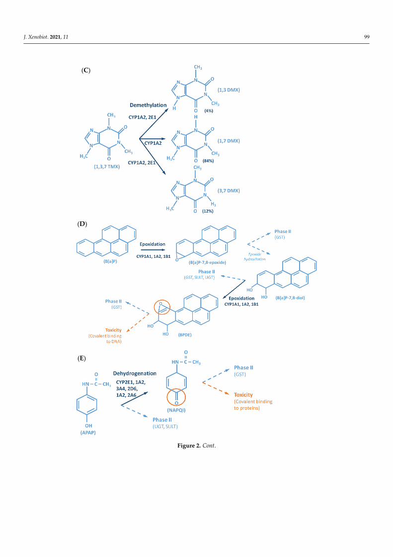
Figure 2. Examples of reactions catalyzed by CYPs. (A) CYP-mediated bioactivation of thalidomide. (B) CYP-mediated hydroxylation of phenol. (C) CYP-mediated metabolism of caffeine, with multi-ple metabolites. (D) Bioactivation of benzo[a]pyrene mediated by CYP enzymes. (E) CYP-mediated metabolism of paracetamol and its potential toxicity. (F) CYP-mediated bioactivation of NNK. (G) Bioactivation of aflatoxin B1 by CYP enzymes. AFB1: Aflatoxin B1. APAP: acetaminophen; B[a]P: benzo[a]pyrene; BPDE: benzo[a]pyrene diol epoxide; DMX: dimethylxanthine; GST: glutathione S-transferases; NAPQI: N-acetyl-p-benzoquinone imine; NNAL: 4-(methylnitrosamino)-1-(3-pyridyl)-1-butanol; NNK: nitrosamine 4-(methylnitrosamino)-1-(3-pyridyl)-1-butanone; PST: phe-nol sulfotransferase; SULT: sulfotransferases; TMX: trimethylxanthine; UGT: UDP-glucuronosyl-transferases.
In 1987, the gene superfamily nomenclature system, based on evolutionary diver-gence of CYP, was proposed [54,83]. Since then, CYPs are organized into families and subfamilies, based on the percentage of amino acid sequence identity (Figure 3) [61,92,93]. Cytochrome P450s forms sharing an identity of ≥40% constitute a particular family desig-nated by an Arabic numeral, whereas enzymes with ≥55% identity are assigned to a par-ticular subfamily designated by a letter. Finally, the gene coding the isoenzyme is desig-nated by an Arabic numeral (https://drnelson.uthsc.edu/) (accessed on 20 June 2021). In humans, the CYP superfamily consists of 57 genes (and 58 pseudo-genes) divided into 18 families and 44 sub-families and can be classified based on major substrate classes (Table 1) (https://cyped.biocatnet.de/) (accessed on 20 June 2021) [5,25,82,94].
Figure 3. Schematic representation of the nomenclature system of CYP genes (example of CYP3A4*1).
Root symbol
for CYP genes
Superfamily
≥40% identity
with other
CYP3 proteins
Family
≥55% identity
with other
CYP3A
Subfamily
Gene
identifier
Isoenzyme
"Wild-type"
most common
Allele
(F)
(G)
Figure 2. Examples of reactions catalyzed by CYPs. (A) CYP-mediated bioactivation of thalidomide. (B) CYP-mediatedhydroxylation of phenol. (C) CYP-mediated metabolism of caffeine, with multiple metabolites. (D) Bioactivation ofbenzo[a]pyrene mediated by CYP enzymes. (E) CYP-mediated metabolism of paracetamol and its potential toxicity.(F) CYP-mediated bioactivation of NNK. (G) Bioactivation of aflatoxin B1 by CYP enzymes. AFB1: Aflatoxin B1. APAP:acetaminophen; B[a]P: benzo[a]pyrene; BPDE: benzo[a]pyrene diol epoxide; DMX: dimethylxanthine; GST: glutathioneS-transferases; NAPQI: N-acetyl-p-benzoquinone imine; NNAL: 4-(methylnitrosamino)-1-(3-pyridyl)-1-butanol; NNK:nitrosamine 4-(methylnitrosamino)-1-(3-pyridyl)-1-butanone; PST: phenol sulfotransferase; SULT: sulfotransferases; TMX:trimethylxanthine; UGT: UDP-glucuronosyltransferases.
In 1987, the gene superfamily nomenclature system, based on evolutionary diver-gence of CYP, was proposed [54,83]. Since then, CYPs are organized into families andsubfamilies, based on the percentage of amino acid sequence identity (Figure 3) [61,92,93].Cytochrome P450s forms sharing an identity of ≥40% constitute a particular family des-ignated by an Arabic numeral, whereas enzymes with ≥55% identity are assigned to aparticular subfamily designated by a letter. Finally, the gene coding the isoenzyme isdesignated by an Arabic numeral (https://drnelson.uthsc.edu/) (accessed on 20 June2021). In humans, the CYP superfamily consists of 57 genes (and 58 pseudo-genes) dividedinto 18 families and 44 sub-families and can be classified based on major substrate classes(Table 1) (https://cyped.biocatnet.de/) (accessed on 20 June 2021) [5,25,82,94].
J. Xenobiot. 2021, 11, FOR PEER REVIEW 7
Figure 2. Examples of reactions catalyzed by CYPs. (A) CYP-mediated bioactivation of thalidomide. (B) CYP-mediated hydroxylation of phenol. (C) CYP-mediated metabolism of caffeine, with multi-ple metabolites. (D) Bioactivation of benzo[a]pyrene mediated by CYP enzymes. (E) CYP-mediated metabolism of paracetamol and its potential toxicity. (F) CYP-mediated bioactivation of NNK. (G) Bioactivation of aflatoxin B1 by CYP enzymes. AFB1: Aflatoxin B1. APAP: acetaminophen; B[a]P: benzo[a]pyrene; BPDE: benzo[a]pyrene diol epoxide; DMX: dimethylxanthine; GST: glutathione S-transferases; NAPQI: N-acetyl-p-benzoquinone imine; NNAL: 4-(methylnitrosamino)-1-(3-pyridyl)-1-butanol; NNK: nitrosamine 4-(methylnitrosamino)-1-(3-pyridyl)-1-butanone; PST: phe-nol sulfotransferase; SULT: sulfotransferases; TMX: trimethylxanthine; UGT: UDP-glucuronosyl-transferases.
In 1987, the gene superfamily nomenclature system, based on evolutionary diver-gence of CYP, was proposed [54,83]. Since then, CYPs are organized into families and subfamilies, based on the percentage of amino acid sequence identity (Figure 3) [61,92,93]. Cytochrome P450s forms sharing an identity of ≥40% constitute a particular family desig-nated by an Arabic numeral, whereas enzymes with ≥55% identity are assigned to a par-ticular subfamily designated by a letter. Finally, the gene coding the isoenzyme is desig-nated by an Arabic numeral (https://drnelson.uthsc.edu/) (accessed on 20 June 2021). In humans, the CYP superfamily consists of 57 genes (and 58 pseudo-genes) divided into 18 families and 44 sub-families and can be classified based on major substrate classes (Table 1) (https://cyped.biocatnet.de/) (accessed on 20 June 2021) [5,25,82,94].
Figure 3. Schematic representation of the nomenclature system of CYP genes (example of CYP3A4*1).
Root symbol
for CYP genes
Superfamily
≥40% identity
with other
CYP3 proteins
Family
≥55% identity
with other
CYP3A
Subfamily
Gene
identifier
Isoenzyme
"Wild-type"
most common
Allele
(F)
(G)
Figure 3. Schematic representation of the nomenclature system of CYP genes (example of CYP3A4*1).

J. Xenobiot. 2021, 11 101
Table 1. Main classes of compounds metabolized by CYPs and major isoenzymes involved theirbiotransformation.
Classes of Compounds CYP Isoenzymes
Sterols 1B1, 7A1, 7B1, 8B1, 11A1, 11B1, 11B2, 17A1, 19A1, 21A2,27A1, 39A1, 46A1, 51A1
Xenobiotics 1A1, 1A2, 2A6, 2A13, 2B6, 2C8, 2C9, 2C18, 2C19, 2D6, 2E1,2F1, 3A4, 3A5, 3A7
Fatty acids 2J2, 2U1, 4A11, 4B1, 4F11, 4F12, 4F22, 4V2, 4X1, 4Z1Eicosanoids 4F2, 4F3, 4F8, 5A1, 8A1Vitamins 2R1, 24A1, 26A1, 26B1, 26C1, 27B1, 27C1Unknown 2A7, 2S1, 2W1, 4A22, 20A1
Microsomal CYPs (CPR as obligatory electron donor) in black and mitochondrial CYPs (adreno-doxin/adrenodoxin reductase as obligatory electron donor) in green.
4. Location and Function of Cytochrome P450s in Mammals
Cytochrome P450s are expressed virtually in all tissues, with highest concentrationsfound in the small intestine, but particularly in the liver. These membrane-bound heme-proteins contain more than 500 amino acid residues and a single heme prosthetic groupin the active site [14,25,26,32]. CYPs are abundant in the microsomal fraction of the liver,playing a central role in bile acid biosynthesis and in the metabolism of foreign com-pounds [5,25,94]. CYPs are also involved in the homeostasis of steroid hormones, withrelevant CYP forms present in the inner membrane of mitochondria of steroidogenic tis-sues, such as adrenal cortex, testis, ovary, breasts, and placenta [43,44]. Additionally, CYPsare of importance in vitamin metabolism, metabolism of unsaturated fatty acids, and incholesterol biosynthesis [54,82,95] (Table 1).
The human gut microbiome represents a site for xenobiotic metabolism, altering thepharmacokinetics outcome of drugs, environmental toxicants, and heavy metals. Increasedmetabolism or biactivation of xenobiotics by the gut microbiome may occur, either throughthe intestinal tract or re-entering the gut via enterohepatic circulation. This is dependenton the enzymatic activity within the microbial niche [96,97]. CYP host expression in miceshowed to be modulated by the collection of microorganisms in the gastrointestinal tractvia altered xenobiotic nuclear receptors activity [98,99]. In a dual effect, the microbiotamodulates the pharmacokinetics of the xenobiotics, while reciprocally xenobiotics caninfluence the viability and metabolism of the microbiota.
Cytochrome P450s catalyze a variety of oxidation and reduction reactions involvingbroad, and in many cases overlapping substrate specificity (Figure 2) [8,18,26,100–106].In this context, a single compound can be metabolized by different CYP isoenzymes,in a complex biotransformation enabled by multiple pathways, resulting in numerousmetabolites. On the other hand, a unique compound can be metabolized by a single CYPoriginating different metabolites. The typical CYP-mediated monooxygenation consistsof the incorporation of one oxygen atom into the substrate (RH + O2 + 2e− + 2H+ →
ROH + H2O), although the activated oxygen atom may not necessarily be incorporatedbut used in different types of reactions. Reactions catalyzed by CYPs include: aliphatic,aromatic and N-hydroxylation; epoxidation; N-, O- and S-dealkylation; N- and S-oxidation;oxidative deamination; dehalogenation; dehydrogenation; dehydration; C-C bond cleavage;isomerization; reduction; and esterase [107].
The energy required to activate oxygen is supplied to microsomal CYPs (includingthose of families 1–4, involved in xenobiotic metabolism, see below) by cytochrome P450oxidoreductase (CPR) [108]. This reductase obtains two electron equivalents in the form ofa hydride (H−) from NADPH, which is received by its flavin adenine dinucleotide (FAD)moiety (reductase) and subsequently donated to CPR’s second flavin prosthetic group,flavin mononucleotide (FMN) (transporter). Through an extensive open/close proteindynamics, the FMN is reduced (inter-flavin electron transfer, closed conformation) andsubsequently, CPR undergoes a large rearrangement, allowing the interaction of CYPswith the FMN domain of CPR (open conformation), with electron equivalents transferred,

J. Xenobiot. 2021, 11 102
one at a time, to the heme group of CYPs (inter-protein electron transfer) [80,81,109–113].Additionally, cytochrome b5 may play an auxiliary role in sustaining CYPs in their activity,by donating the second electron, which is facultative and dependent on the CYP isoenzymeand/or substrate [75–79].
5. The Central Role of Cytochrome P450s as Xenobiotic-Metabolizing Enzymes
In humans, the CYP enzyme family represents the most important enzymatic systeminvolved in Phase I drug metabolism [5,10,13,20,25]. A survey on literature databases ofhuman oxidoreductases and CYP enzymes implicated in the Phase I metabolism evidencedthat CYPs are involved in the vast majority (approx. 90–95%) of enzymatic reactions in themetabolism of xenobiotics [7]. Additional enzyme systems may play a role in hepatic PhaseI metabolism, albeit to a lesser extent than CYPs, as mentioned above. These include FMOs,NQOs, amine oxidases, alcohol dehydrogenases, esterases and peroxidases [7,13–16].
Although the majority of the isoenzymes of the 18 human CYP families have specificfunctions in the metabolism of endobiotics, about 15 isoforms belonging to CYP families 1,2 and 3 (Table 2) are accountable for 70–80% of all Phase I metabolism of clinically useddrugs [25] and are involved in the biotransformation of a vast diversity of environmentalchemicals (approx. 90%), including 66% metabolism reactions of chemical carcinogens [114].From these, CYP1A2, CYP2C9, CYP2D6 and CYP3A4/5, are responsible for about 72% of allCYP-mediated metabolism of clinically marketed drugs [25]. Although typically contribut-ing to theω-oxidation of endogenous fatty acids and eicosanoids, members of the CYP4family are additionally involved in xenobiotic metabolism (e.g., CYP4A11, CYP4F2 andCYP4F12), albeit to a much lower extend than isoforms of the CYP1–3 families [115–118].
The CYP-enzymes involved in xenobiotic metabolism have evolved to protect humansagainst potential toxic agents. However, CYP-mediated biotransformation may resultin metabolic activation of environmental chemicals to reactive carcinogenic products, aprocess known as “lethal synthesis” or bioactivation [14,25,82,94,114,119,120]. While CYPsmay catalyze the activation of procarcinogens to electrophilic ultimate carcinogens, thePhase II enzymes in general detoxify electrophilic intermediates into non-toxic substrates,with few exceptions of non-canonical bioactivation through conjugation reactions [19]. Thepotentially toxic reactive metabolites that “escape” from Phase II metabolism are able tocovalently interact with nucleophilic structures such as those of nucleic acids, proteins,or lipids, causing cell damage and potentially triggering carcinogenesis, teratogenicity,and/or adverse drug reactions (ADRs) [1,3,20,58,103–105,114,120] (Figure 1). SeveralCYP-mediated oxidations contribute to the synthesis of more toxic metabolites, after ex-posure to specific compounds such as: (i) polycyclic aromatic hydrocarbons (PAHs) (e.g.,benzo[a]pyrene, Figure 2D), including nitro-polycyclic aromatic hydrocarbons, derivedfrom incomplete combustion; (ii) heterocyclic aromatic amines (HAAs) from charbroiledmeats; (iii) aromatic amines as dyes, or present in pesticides, tobacco smoke and phar-maceuticals (e.g., paracetamol, Figure 2E); (iv) nitrosamines present in tobacco smoke(e.g., nitrosamine 4-(methylnitrosamino)-1-(3-pyridyl)-1-butanone, NNK, Figure 2F) anddiet, formed from nitrites and nitrates; (v) toxins present in food products (e.g., grains orcereals) contaminated with pathogenic microorganisms (e.g., aflatoxin B1 produced byAspergillus flavus and A. parasiticus, Figure 2G). These xenobiotics are transformed to theirtoxic forms—usually electrophiles such as epoxides, hydroxylamides, acyl halides, amongothers—through CYP-mediated activity [5,14,106,107,121].

J. Xenobiot. 2021, 11 103
Table 2. Genetic variability and importance of the main CYP isoenzymes involved the metabolism of xenobiotics.
CYCYP Isoenzyme Polymorphism Frequency Functional Effects Allelic Variants Participation in the Metabolism of Xenobiotics
1A1 Relatively high Rare 13
J. Xenobiot. 2021, 11, FOR PEER REVIEW 10
Table 2. Genetic variability and importance of the main CYP isoenzymes involved the metabolism of xenobiotics.
CYP Isoenzyme Polymorphism Frequency Functional Effects Allelic Variants Participation in the Metabolism of Xenobiotics
1A1 Relatively high Rare 13
1A2 High Rare 21
1B1 Frequent missense mutations Rare 26
2A6 Higher in Orientals than in Caucasians Significant 45
2B6 High Significant 38
2C8 High Significant 14
2C9 Relatively rare in Caucasians Very significant 70
2C19 High Very significant 38
2D6 Very High Highly significant 145
2E1 Low No significance 7
3A4 Low Low significance 32
3A5 High Significant 9
Other (a) - - - (a) Other CYP isoenzymes involved in smaller fraction of xenobiotic metabolism (2A13, 2C18, 2F1, 2J2, 3A7 and some members of the CYP4 family). Due to substrate overlapping specificity, CYP1B1 (typical sterols metabolizer) and 2J2 (typical fatty acids metabolizer) were also included in the CYP xenobiotic metabolizer analysis.
1A2 High Rare 211B1 Frequent missense mutations Rare 262A6 Higher in Orientals than in Caucasians Significant 452B6 High Significant 382C8 High Significant 142C9 Relatively rare in Caucasians Very significant 70
2C19 High Very significant 382D6 Very High Highly significant 1452E1 Low No significance 73A4 Low Low significance 323A5 High Significant 9
Other (a) - - -(a) Other CYP isoenzymes involved in smaller fraction of xenobiotic metabolism (2A13, 2C18, 2F1, 2J2, 3A7 and some members of the CYP4 family). Due to substrate overlapping specificity, CYP1B1 (typicalsterols metabolizer) and 2J2 (typical fatty acids metabolizer) were also included in the CYP xenobiotic metabolizer analysis.

J. Xenobiot. 2021, 11 104
6. Genetic Determinants in Cytochrome P450s Expression and Activity
With the advances in research during the last 30 years, it became clear that the ef-fects of genetic variability of DMEs, particularly those of the CYP enzymes complex, arehighly relevant in terms of drug response and detoxification or bioactivation of xeno-biotics in general [24,26,57,59,114]. Cytochrome P450s exhibit genetic polymorphismswith multiple allelic variants, demonstrating frequencies varying between different pop-ulations and ethnicities [57–59,82,106,122] (Figure 4). These include single nucleotidevariants, small deletions or insertions, and copy number variants (gene deletion or du-plication/amplification, the later more frequent) [55–57]. These genetic variants can alterstructurally the CYP enzyme or its expression, resulting in normal, reduced, increasedor absence of activity [58,59,82,122,123]. A nomenclature system has been set up for theCYP alleles (using the suffixes *1, *2, *3 . . . ; where *1 designates the “wild type,” or mostcommon gene-variant) (Figure 3). Allelic variants are summarized and described on thehome page of the human CYP allele nomenclature committee, at Pharmacogene Variation(PharmVar) Consortium (https://www.pharmvar.org/htdocs/archive/index_original.htm)(accessed on 20 June 2021).
J. Xenobiot. 2021, 11, FOR PEER REVIEW 12
Figure 4. Factors influencing inter- and intra-individual variability in CYP activity and expression, and their impact on xenobiotic metabolism (CYP circles: size indicate the importance of the differ-ent inter- and intra-individual levels of expression and function of CYP enzymes).
Isoforms CYP2C9, 2C19 and 2D6 present the highest genetic variability in the human population, with so far 70, 38 and 145 allelic variants being identified, respectively (Table 2). These three CYPs have been estimated to account for approximately 35–40% of oxida-tive drug metabolism and a quarter of biotransformation of xenobiotics in general, includ-ing environmental and industrial pollutants [7,17,25,114]. The high frequencies of genetic polymorphism of these three CYPs were demonstrated to cause significant functional ef-fects and high penetrance in individual susceptibility in the human population [4,5,56–58,82]. Conversely, severe loss-of-function alleles or functional gene duplications are rare in genes encoding CYP1A2 and 3A4 (21 and 32 allelic variants, respectively) [7,14,25,57,122]. These two CYPs together are responsible for 35% and 30% of drug- and of general chemical metabolism, respectively.
This extensive genetic variability, causing inter-individual differences in expression and activity of human CYPs is considered to be one of the major causes in the lack of efficacy and in ADRs of therapeutic drugs, as well as in the variability of toxic outcome after exposure to environmental compounds [55,56,58,124,125]. Pharmacogenetic testing of CYPs is gaining attention due to the possibility of developing safer drugs and patient-tailored drug therapy in precision medicine [5,25,26]. The occurrence of specific gene var-iants translates into four major metabolizer phenotypes: (i) ultrarapid metabolizers (UM), involving two or more active gene copies or allelic variants, encoding more efficient en-zymes or over-expressed CYP isoforms; (ii) extensive metabolizers (EM), carriers of two functional CYP alleles; (iii) intermediate metabolizers (IM), heterozygous for a defect al-lele or carrying two alleles causing combined decreased activity of a CYP; (iv) poor me-tabolizers (PM), carrying two defective alleles, producing CYPs with very low or without activity/function [55,56,122,125]. A potential additional genetic variability in CYP medi-ated xenobiotic metabolism has recently been revealed. This regards the genetic variabil-ity of CPR (encoded by the POR gene), the obligatory redox partner of microsomal CYPs for the reception of electron equivalents to sustain their activity. Recent data suggest that
Figure 4. Factors influencing inter- and intra-individual variability in CYP activity and expression,and their impact on xenobiotic metabolism (CYP circles: size indicate the importance of the differentinter- and intra-individual levels of expression and function of CYP enzymes).
Isoforms CYP2C9, 2C19 and 2D6 present the highest genetic variability in the humanpopulation, with so far 70, 38 and 145 allelic variants being identified, respectively (Table 2).These three CYPs have been estimated to account for approximately 35–40% of oxidativedrug metabolism and a quarter of biotransformation of xenobiotics in general, includingenvironmental and industrial pollutants [7,17,25,114]. The high frequencies of geneticpolymorphism of these three CYPs were demonstrated to cause significant functional effectsand high penetrance in individual susceptibility in the human population [4,5,56–58,82].Conversely, severe loss-of-function alleles or functional gene duplications are rare in genesencoding CYP1A2 and 3A4 (21 and 32 allelic variants, respectively) [7,14,25,57,122]. Thesetwo CYPs together are responsible for 35% and 30% of drug- and of general chemicalmetabolism, respectively.

J. Xenobiot. 2021, 11 105
This extensive genetic variability, causing inter-individual differences in expressionand activity of human CYPs is considered to be one of the major causes in the lack ofefficacy and in ADRs of therapeutic drugs, as well as in the variability of toxic outcomeafter exposure to environmental compounds [55,56,58,124,125]. Pharmacogenetic testingof CYPs is gaining attention due to the possibility of developing safer drugs and patient-tailored drug therapy in precision medicine [5,25,26]. The occurrence of specific genevariants translates into four major metabolizer phenotypes: (i) ultrarapid metabolizers(UM), involving two or more active gene copies or allelic variants, encoding more efficientenzymes or over-expressed CYP isoforms; (ii) extensive metabolizers (EM), carriers oftwo functional CYP alleles; (iii) intermediate metabolizers (IM), heterozygous for a defectallele or carrying two alleles causing combined decreased activity of a CYP; (iv) poormetabolizers (PM), carrying two defective alleles, producing CYPs with very low or with-out activity/function [55,56,122,125]. A potential additional genetic variability in CYPmediated xenobiotic metabolism has recently been revealed. This regards the geneticvariability of CPR (encoded by the POR gene), the obligatory redox partner of microsomalCYPs for the reception of electron equivalents to sustain their activity. Recent data suggestthat natural occurring genetic variants of POR may lead to altered CYP mediated drugmetabolism [126–129].
Genotyping provides sequence data allowing the estimation of the expectable CYPmetabolizer phenotypes. However, additional determinants have been shown to play arole in modulating CYP enzymes function, such as those of the environment and physio-pathological conditions (discussed below) [25,130,131]. Of relevance also to mention twomain epigenetic mechanisms affecting CYP gene expression: (i) altered DNA methylation —involved in biased cellular control of gene expression; and (ii) microRNA (miR) regulation—affecting expression levels of target CYPs [5,132,133]. The inhibition of methylation inhepatic cell lines has been reported to induce CYP genes expression, particularly CYP3Agenes [134]. Additionally, methylation patterns in the promoters of CYP genes seem to bedifferent in distinct physiological conditions or environmental exposures (e.g., decreasedinducibility of CYP1B1 due to promoter methylation at multiple CpG sites; lower methyla-tion in the CYP1A1 promoter found in heavy smokers) [132,135]. Direct regulation of CYPsby miRs was evidenced for CYP1B1 (miR-27b), CYP2C9 (miR-130b), CYP2C9 (miR-34a),CYP2E1 (miR-378), and CYP3A4 (miR-27b, miR148a, and miR34a) [133,136–138]. Addi-tionally, histone protein modification, an epigenetic mechanism that may affect chromatinstructure, impacting accessibility, have been indicated to be involved in transcriptionalregulation of CYP expression. Epigenetic patterns leading to divergent CYP-mediatedmetabolism are normally reversible, tissue-specific and highly dependent on environmentaland individual physio-pathological conditions [25,56,139].
7. Nongenetic Factors Influencing Cytochrome P450s Expression and Activity
Factors such as age, sex, hormone levels, and environment, as well as pathologicalconditions such as infection, inflammation, cholestasis, and cancer are aspects demon-strated to influence CYP expression and activity [25,130,131] (Figure 4). Other biochemicalfactors such as protein-protein interaction—involving CYP interaction with redox partnersand other proteins with allosteric regulatory effect [76,77,81,129], or substrate-substrateinteraction—consisting in CYP activity inhibition due to substrate interference/competitionmechanism, are also implicated in CYP function [25,100,140–142].
Transcriptional activation is described as the main process of induction of CYP genesand protein levels [143]. Yet, mRNA and protein stabilization, or inhibition of proteindegradation pathways will also lead to altered levels of CYP activity [144]. Proteasome-mediated degradation, phosphorylation, and long non-coding RNA (lncRNAs)-relatedmechanisms, are among the non-canonical post-transcriptional regulation pathways ofCYPs [145–147]. Induction of expression of CYP enzymes, and of DMEs in general,involves a complex expression-regulation network dependent on cell-membrane- andnuclear-receptors, promoter-regulation sequences of the cis-acting elements, and of trans-

J. Xenobiot. 2021, 11 106
acting activators and repressors, which may be shared among the same enzyme familyand between different DME families. Expression of genes encoding CYPs involved inxenobiotic metabolism (mainly of CYP families 1–4) is highly inducible and can be tran-scriptionally activated by xenobiotics through xenobiotic receptor-dependent mechanisms(Figure 5) [26,148]. Several receptors mediate the induction of these CYPs, such as: the arylhydrocarbon receptor (AhR)—CYP1 genes; the pregnane nuclear receptor (PXR)—CYP2A6,2B, 2C, and 3A genes; the constitutive androstane receptor (CAR)—CYP1A, 2A6, 2B, 2C8,2C9 and 3A4 genes [5,144,148–153]. Typically, CYP1A1 and 1A2 are highly inducible bynumerous xenobiotics that act as AhR ligands. Genes of these two CYP1A members arearranged in a head-to-head orientation, sharing a common bi-directional promoter withat least 13 AhR response elements [154,155]. Additionally, transactivation of both CYP1Apromoters by CAR is also possible through a common cis-regulatory estrogen receptorelement (ER8) in the 5′-flanking region [149]. Transcriptional regulation of CYP2A6 geneinvolves PXR and CAR activators via direct repeat 4 (DR4) elements [150]. In vivo andin vitro studies are indicative that transcriptional upregulation of CYP2A6 may also oc-curs through an estrogen receptor-dependent pathway [156,157]. CYP2B6 expression ismajorly regulated by an orphan CAR (NR113) via a phenobarbital-responsive enhancermodule (PBREM), while PXR (NR112) contributes to smaller fraction in CYP2B6 inductionthrough a distal xenobiotics-responsive enhancer module (XREM) [151,152]. The CYP2Cgenes are variable in their relative inducibility, which is dependent on ligands of the PXRand CAR, glucocorticoid (GR), and vitamin D nuclear receptor (VDR) pathways [158,159].CYP2C9 is the highest expressed member of this subfamily in the liver, requiring cross-talkbetween distal PXR and CAR sites and proximal hepatocyte nuclear factor 4α (HNF4α)binding sites in its promoter [160,161]. The proximal PXR responsive element (prPXRE),REM, and the constitutive liver enhancer module 4 (CLEM4) are cis-regulatory elementsresponsible for inducible transcriptional regulation of CYP3A genes via xenosensors PXRand CAR [153,160,162]. Peroxisome proliferator-activated receptor (PPAR) also contributesto inducible and constitutive regulation of CYP3A4, the major expressed member of thesubfamily [163]. Constitutive transcriptional regulators of CYP3A genes include membersof CCAAT/enhancer-binding proteins (C/EBP) and HNFs [161,164,165]. Although most ofthe studies on inducibility of expression of CYP4A subfamily genes have been performedin mouse and rat models, accumulated data are indicative that PPAR mediates inductionof expression of members of this subfamily in humans [116]. Expression of the CYP4F2gene seems to be transactivated by the sterol regulatory element-binding protein (SREBP)and AMP-activated protein kinase (AMPK) activators [118,166].
Inhibition of CYP enzymes impairs the biotransformation of clinically used drugs orenvironmental compounds, resulting in higher plasma concentrations of these xenobiotics,which may lead to toxicity (Figure 4) [130,144]. On the other end, if the compound isa prodrug, the efficacy of the therapeutic regimen could be decreased as concentrationsof the metabolite (the active drug), may fall below effective levels. Inhibitors of CYPenzymes can be classified into three mechanistically distinct groups, namely agents thatform (i) reversible complexes (competitive or noncompetitive), (ii) quasi-irreversible com-plexes with the heme-iron atom, (iii) irreversible complexes through covalent binding toparticular residues of the CYP protein. The latter disrupt critical interactions with its redoxpartners, i.e., CPR or b5, or of the heme moiety, accelerating degradation and/or oxidativefragmentation of the prosthetic heme [81,140–142]. In competitive reversible inhibitiontwo structurally different molecules transiently compete for the same CYP isoenzymeirrespective of whether these are substrates for the enzyme. In noncompetitive reversibleinhibition, a molecule binds to a site other than the active site, causing allosteric modulationof CYP function [5,140].

J. Xenobiot. 2021, 11 107
J. Xenobiot. 2021, 11, FOR PEER REVIEW 14
through an estrogen receptor-dependent pathway [156,157]. CYP2B6 expression is ma-jorly regulated by an orphan CAR (NR113) via a phenobarbital-responsive enhancer mod-ule (PBREM), while PXR (NR112) contributes to smaller fraction in CYP2B6 induction through a distal xenobiotics-responsive enhancer module (XREM) [151,152]. The CYP2C genes are variable in their relative inducibility, which is dependent on ligands of the PXR and CAR, glucocorticoid (GR), and vitamin D nuclear receptor (VDR) pathways [158,159]. CYP2C9 is the highest expressed member of this subfamily in the liver, requiring cross-talk between distal PXR and CAR sites and proximal hepatocyte nuclear factor 4α (HNF4α) binding sites in its promoter [160,161]. The proximal PXR responsive element (prPXRE), REM, and the constitutive liver enhancer module 4 (CLEM4) are cis-regulatory elements responsible for inducible transcriptional regulation of CYP3A genes via xenosensors PXR and CAR [153,160,162]. Peroxisome proliferator-activated receptor (PPAR) also contributes to inducible and constitutive regulation of CYP3A4, the major expressed member of the subfamily [163]. Constitutive transcriptional regulators of CYP3A genes include members of CCAAT/enhancer-binding proteins (C/EBP) and HNFs [161,164,165]. Although most of the studies on inducibility of expression of CYP4A sub-family genes have been performed in mouse and rat models, accumulated data are indic-ative that PPAR mediates induction of expression of members of this subfamily in humans [116]. Expression of the CYP4F2 gene seems to be transactivated by the sterol regulatory element-binding protein (SREBP) and AMP-activated protein kinase (AMPK) activators [118,166].
Figure 5. Main mechanisms of CYP induction. AhR: aryl hydrocarbon receptor; CAR: constitutive androstane receptor; CLEM4: constitutive liver enhancer module 4; DR4: direct repeat 4; ER8: es-trogen receptor 8; HNF4α: hepatocyte nuclear factor 4α; PBREM: phenobarbital-responsive en-hancer module; PPAR: proliferator-activated receptor; prPXRE: proximal PXR responsive element; PXR: pregnane nuclear receptor; XNR: xenobiotic-nuclear receptors; XNR-RE: xenobiotic-nuclear receptors-responsive elements; XREM: xenobiotics-responsive enhancer module. (Note: other reg-ulatory factors and elements not referred may be involved in CYP expression induction).
Inhibition of CYP enzymes impairs the biotransformation of clinically used drugs or environmental compounds, resulting in higher plasma concentrations of these xenobiot-ics, which may lead to toxicity (Figure 4) [130,144]. On the other end, if the compound is a prodrug, the efficacy of the therapeutic regimen could be decreased as concentrations of the metabolite (the active drug), may fall below effective levels. Inhibitors of CYP enzymes can be classified into three mechanistically distinct groups, namely agents that form (i)
Figure 5. Main mechanisms of CYP induction. AhR: aryl hydrocarbon receptor; CAR: constitutive androstane receptor;CLEM4: constitutive liver enhancer module 4; DR4: direct repeat 4; ER8: estrogen receptor 8; HNF4α: hepatocyte nuclearfactor 4α; PBREM: phenobarbital-responsive enhancer module; PPAR: proliferator-activated receptor; prPXRE: proximalPXR responsive element; PXR: pregnane nuclear receptor; XNR: xenobiotic-nuclear receptors; XNR-RE: xenobiotic-nuclearreceptors-responsive elements; XREM: xenobiotics-responsive enhancer module. (Note: other regulatory factors andelements not referred may be involved in CYP expression induction).
Dietary factors such as phytochemicals affect CYP expression and activity whichmay be of importance in diet-drug interactions. Several studies evidenced the inhibitoryproperties of flavonoids by structural interference with CYP proteins [167]. Additionally,soy components seem to promiscuously modulate several nuclear receptors includingAhR, PXR, PPAR and liver X receptor (LXR), altering drug pharmacokinetics and thera-peutic efficacy [168] (Figure 5). Other factors have been described to be involved in theinduction or inhibition of CYP enzymes, in particularly conditions implying underlyingchronic inflammation, such as bacterial, parasitic or viral infections (HIV, hepatitis C),sepsis, rheumatoid arthritis, liver transplant, multiple myeloma, chronic liver disease andcancer [130,131]. Proinflammatory states with secretion of large amounts of cytokines seemto be implicated in divergent xenobiotic metabolism. Cytokines such as interleukins IL-1β,IL-2, IL-4, IL-6, IL-10, and IL-23, interferon gamma (IFNγ), transforming growth factorbeta (TGFβ), tumor necrosis factor alpha (TNFα), but also factors involved in infectionsuch as lipopolysaccharides (LPS), have been reported to directly or indirectly modulateCYP expression, as demonstrated in hepatic cell lines, animal models and humans (e.g.,patients with cancer undergoing immunotherapy) [130,131,142,169–174]. These inflam-mation factors generally exert a down-regulation of CYP expression, although in somecases, the reverse was noted. The importance of a particular cytokine in CYP regulationwill depend on many factors, including its concentration in the liver, particularly in thevicinity of the hepatocytes, time course of its production, modulation by other cytokines,and concentrations of natural antagonists (e.g., IL-1-ra) and facilitators (e.g., specific sol-uble receptors) [25,131,174]. Cross-talks between cytokine levels and xenobiotic nuclearreceptors (i.e., PXR and CAR) have been reported to contribute to the modulation of theCYP activity. During inflammation, nuclear factor κB (NF-κB) represses both PXR and CYPexpression through protein-protein interactions with the PXR pathway. Upregulation ofprotein kinase C (PKC) and cAMP-dependent protein kinase A (PKA) seem to be involvedin the repression of CYP expression associated with liver inflammation [146,175]. Moreover,extra-hepatic conditions (e.g., infection, tumors) involving inflammation also reduce thecapacity of hepatic drug metabolism due to downregulation of hepatic CYP expression,

J. Xenobiot. 2021, 11 108
probably mediated via inflammatory cytokines released by remotely inflamed organs,reaching the liver via systemic circulation [176,177].
8. Final Remarks
This short review is intended to highlight main aspects of the central role of CYP en-zymes in xenobiotic metabolism, of high significance in clinical pharmacology and toxicol-ogy. Although with over seven decades of research and major scientific breakthroughs, sev-eral questions and challenges still maintain regarding CYP-mediated metabolism. Currentresearch is mainly focused on obtaining more detailed and precise knowledge regarding:(i) functional properties and differences of CYP isoenzymes; (ii) their interspecies func-tional variance; (iii) tissue distribution and cellular location; (iv) regulatory mechanismsof gene expression; (v) population genetic and epigenetic determinants and variability;(vi) physio-pathological and environmental factors influencing expression and activity;(vii) genotype–phenotype correlation; (viii) and overall clinical impact. The scientificliterature cited in this short-review, and many more studies not referred, are evidence ofthe remarkable efforts and achievements in understanding CYPs as the central Phase Ienzyme family responsible for the oxidative biotransformation of xenobiotics, integratedin a vast and complex physiological detoxification network.
Author Contributions: Bibliographic analysis, F.E. and M.K.; writing—original draft preparation, F.E.and M.K.; writing—review and editing, all authors; funding acquisition, M.K. and J.R. All authorshave read and agreed to the published version of the manuscript.
Funding: The elaboration of this manuscript was in part funded by grant UID/BIM/0009/2020 of thePortuguese Fundação para a Ciência e a Tecnologia (FCT) for the Research Center for Toxicogenomicsand Human Health (ToXomics).
Institutional Review Board Statement: Not applicable.
Informed Consent Statement: Not applicable.
Data Availability Statement: Not applicable.
Conflicts of Interest: The authors declare no conflict of interest.
References1. Croom, E. Metabolism of Xenobiotics of Human Environments. In Progress in Molecular Biology and Translational Science;
Ernest, H., Ed.; Elsevier: Amsterdam, The Netherlands, 2012; Volume 112, pp. 31–88. ISBN 978-0-12-415813-9.2. Juchau, M.R.; Chen, H. Developmental Enzymology. In Handbook of Developmental Neurotoxicology; William, S., Jr.,
Louis, W.C., Eds.; Elsevier: Amsterdam, The Netherlands, 1998; pp. 321–337. ISBN 978-0-12-648860-9.3. Evans, T.J. Chapter 2—Toxicokinetics and Toxicodynamics. In Small Animal Toxicology, 3rd ed.; Peterson, M.E., Talcott, P.A., Eds.;
W.B. Saunders: Saint Louis, MO, USA, 2013; pp. 13–19. ISBN 978-1-4557-0717-1.4. Johnson, C.H.; Patterson, A.D.; Idle, J.R.; Gonzalez, F.J. Xenobiotic Metabolomics: Major Impact on the Metabolome. Annu. Rev.
Pharmacol. Toxicol. 2012, 52, 37–56. [CrossRef] [PubMed]5. Manikandan, P.; Nagini, S. Cytochrome P450 Structure, Function and Clinical Significance: A Review. Curr. Drug Targets 2018, 19.
[CrossRef] [PubMed]6. Stanley, L.A. Drug Metabolism. In Pharmacognosy; Simone, B., Rupika, D., Eds.; Elsevier: Amsterdam, The Netherlands, 2017;
pp. 527–545. ISBN 978-0-12-802104-0.7. Rendic, S.; Guengerich, F.P. Survey of Human Oxidoreductases and Cytochrome P450 Enzymes Involved in the Metabolism of
Xenobiotic and Natural Chemicals. Chem. Res. Toxicol. 2015, 28, 38–42. [CrossRef] [PubMed]8. Lewis, D.F.; Ioannides, C.; Parke, D.V. Cytochromes P450 and Species Differences in Xenobiotic Metabolism and Activation of
Carcinogen. Environ. Health Perspect. 1998, 106, 633–641. [CrossRef]9. Almazroo, O.A.; Miah, M.K.; Venkataramanan, R. Drug Metabolism in the Liver. Clin. Liver Dis. 2017, 21, 1–20. [CrossRef]10. Bachmann, K. Drug Metabolism. In Pharmacology; Miles, H., William, M., Kenneth, B., Eds.; Elsevier: Amsterdam,
The Netherlands, 2009; pp. 131–173. ISBN 978-0-12-369521-5.11. Tillement, J.-P.; Tremblay, D. Clinical Pharmacokinetic Criteria for Drug Research. In Comprehensive Medicinal Chemistry II;
Taylor, J.B., Triggle, D.J., Eds.; Elsevier: Amsterdam, The Netherlands, 2007; pp. 11–30. ISBN 978-0-08-045044-5.12. Murray, M.; Zhou, F. Trafficking and Other Regulatory Mechanisms for Organic Anion Transporting Polypeptides and Organic
Anion Transporters That Modulate Cellular Drug and Xenobiotic Influx and That Are Dysregulated in Disease. Br. J. Pharmacol.2017, 174, 1908–1924. [CrossRef]

J. Xenobiot. 2021, 11 109
13. Janov, P.; Iller, M. Phase II Drug Metabolism. In Topics on Drug Metabolism; Paxton, J., Ed.; InTech: London, UK, 2012; pp. 35–60.ISBN 978-953-51-0099-7.
14. Guengerich, F.P. Cytochrome P450 and Chemical Toxicology. Chem. Res. Toxicol. 2008, 21, 70–83. [CrossRef] [PubMed]15. Strolin Benedetti, M.; Tipton, K.F.; Whomsley, R. Amine Oxidases and Monooxygenases in the in Vivo Metabolism of Xenobiotic
Amines in Humans: Has the Involvement of Amine Oxidases Been Neglected? Amine Oxidases Monooxygenases Amine Metab.Hum. Fundam. Clin. Pharmacol. 2007, 21, 467–480. [CrossRef]
16. Gan, J.; Ma, S.; Zhang, D. Non-Cytochrome P450-Mediated Bioactivation and Its Toxicological Relevance. Drug Metab. Rev. 2016,48, 473–501. [CrossRef] [PubMed]
17. Furge, L.L.; Guengerich, F.P. Cytochrome P450 Enzymes in Drug Metabolism and Chemical Toxicology: An Introduction. Biochem.Mol. Biol. Educ. 2006, 34, 66–74. [CrossRef]
18. Bernhardt, R. Cytochromes P450 as Versatile Biocatalysts. J. Biotechnol. 2006, 124, 128–145. [CrossRef]19. Grillo, M.P. Bioactivation by Phase-II-Enzyme-Catalyzed Conjugation of Xenobiotics. In Encyclopedia of Drug Metabolism and
Interactions; Lyubimov, A.V., Ed.; John Wiley & Sons, Inc.: Hoboken, NJ, USA, 2012; ISBN 978-0-470-92192-0. [CrossRef]20. Iyanagi, T. Molecular Mechanism of Phase I and Phase II Drug-Metabolizing Enzymes: Implications for Detoxification. In
International Review of Cytology; Elsevier: Amsterdam, The Netherlands, 2007; Volume 260, pp. 35–112. ISBN 978-0-12-374114-1.21. Roy, U.; Barber, P.; Tse-Dinh, Y.-C.; Batrakova, E.V.; Mondal, D.; Nair, M. Role of MRP Transporters in Regulating Antimicrobial
Drug Inefficacy and Oxidative Stress-Induced Pathogenesis during HIV-1 and TB Infections. Front. Microbiol. 2015, 6. [CrossRef]22. Döring, B.; Petzinger, E. Phase 0 and Phase III Transport in Various Organs: Combined Concept of Phases in Xenobiotic Transport
and Metabolism. Drug Metab. Rev. 2014, 46, 261–282. [CrossRef]23. Petzinger, E.; Geyer, J. Drug Transporters in Pharmacokinetics. Naunyn. Schmiedebergs Arch. Pharmacol. 2006, 372, 465–475.
[CrossRef] [PubMed]24. Fujita, K. Cytochrome P450 and Anticancer Drugs. Curr. Drug Metab. 2006, 7, 23–37. [CrossRef]25. Zanger, U.M.; Schwab, M. Cytochrome P450 Enzymes in Drug Metabolism: Regulation of Gene Expression, Enzyme Activities,
and Impact of Genetic Variation. Pharmacol. Ther. 2013, 138, 103–141. [CrossRef] [PubMed]26. Guengerich, F.P. Human Cytochrome P450 Enzymes. In Cytochrome P450: Structure, Mechanism, and Biochemistry; Ortiz de
Montellano, P.R., Ed.; Springer International Publishing: Cham, Switzerland, 2015; pp. 523–785. ISBN 978-3-319-12108-6.27. Frederiks, C.N.; Lam, S.W.; Guchelaar, H.J.; Boven, E. Genetic Polymorphisms and Paclitaxel- or Docetaxel-Induced Toxicities: A
Systematic Review. Cancer Treat. Rev. 2015, 41, 935–950. [CrossRef]28. Stingl, J.C.; Brockmöller, J.; Viviani, R. Genetic Variability of Drug-Metabolizing Enzymes: The Dual Impact on Psychiatric
Therapy and Regulation of Brain Function. Mol. Psychiatry 2013, 18, 273–287. [CrossRef]29. Annalora, A.J.; Marcus, C.B.; Iversen, P.L. Alternative Splicing in the Cytochrome P450 Superfamily Expands Protein Diversity to
Augment Gene Function and Redirect Human Drug Metabolism. Drug Metab. Dispos. 2017, 45, 375–389. [CrossRef] [PubMed]30. Nebert, D.W.; Nelson, D.R.; Feyereisen, R. Evolution of the Cytochrome P450 Genes. Xenobiotica Fate Foreign Compd. Biol. Syst.
1989, 19, 1149–1160. [CrossRef] [PubMed]31. Omura, T. Future Perception in P450 Research. J. Inorg. Biochem. 2018, 186, 264–266. [CrossRef] [PubMed]32. Ortiz de Montellano, P.R. Substrate Oxidation by Cytochrome P450 Enzymes. In Cytochrome P450; Ortiz de Montellano, P.R., Ed.;
Springer International Publishing: Cham, Switzerland, 2015; pp. 111–176. ISBN 978-3-319-12107-9.33. Estabrook, R.W. A Passion for P450s (Rememberances of the Early History of Research on Cytochrome P450). Drug Metab. Dispos.
Biol. Fate Chem. 2003, 31, 1461–1473. [CrossRef] [PubMed]34. Mueller, G.C.; Miller, J.A. The Metabolism of 4-Dimethylaminoazobenzene by Rat Liver Homogenates. J. Biol. Chem. 1948, 176,
535–544. [CrossRef]35. Klingenberg, M. Pigments of Rat Liver Microsomes. Arch. Biochem. Biophys. 1958, 75, 376–386. [CrossRef]36. Garfinkel, D. Studies on Pig Liver Microsomes. I. Enzymic and Pigment Composition of Different Microsomal Fractions. Arch.
Biochem. Biophys. 1958, 77, 493–509. [CrossRef]37. Omura, T.; Sato, R. A New Cytochrome in Liver Microsomes. J. Biol. Chem. 1962, 237, 1375–1376. [CrossRef]38. Conney, A.H. Pharmacological Implications of Microsomal Enzyme Induction. Pharmacol. Rev. 1967, 19, 317–366.39. Hildebrandt, A.; Remmer, H.; Estabrook, R.W. Cytochrome P-450 of Liver Microsomes—One Pigment or Many. Biochem. Biophys.
Res. Commun. 1968, 30, 607–612. [CrossRef]40. Gillette, J.R.; Davis, D.C.; Sasame, H.A. Cytochrome P-450 and Its Role in Drug Metabolism. Annu. Rev. Pharmacol. 1972, 12,
57–84. [CrossRef]41. Wada, O.; Yano, Y. Adaptive Responses of the Liver to Foreign Compounds, with Special Reference to Microsomal Drug-
Matabolizing Enzymes. Rev. Environ. Health 1974, 1, 261–282. [PubMed]42. Parke, D.V. Induction of the Drug-Metabolizing Enzymes. Basic Life Sci. 1975, 6, 207–271. [CrossRef] [PubMed]43. Estabrook, R.W.; Cooper, D.Y.; Rosenthal, O. The Light Reversible Carbon Monoxide Inhibition of the Steroid C21-Hydroxylase
System of the Adrenal Cortex. Biochem. Z. 1963, 338, 741–755.44. Cooper, D.Y.; Estabrook, R.W.; Rosenthal, O. The Stoichiometry of C21 Hydroxylation of Steroids by Adrenocortical Microsomes.
J. Biol. Chem. 1963, 238, 1320–1323. [CrossRef]45. Lu, A.Y.; Coon, M.J. Role of Hemoprotein P-450 in Fatty Acid Omega-Hydroxylation in a Soluble Enzyme System from Liver
Microsomes. J. Biol. Chem. 1968, 243, 1331–1332. [CrossRef]

J. Xenobiot. 2021, 11 110
46. Imai, Y.; Sato, R. A Gel-Electrophoretically Homogeneous Preparation of Cytochrome P-450 from Liver Microsomes ofPhenobarbital-Pretreated Rabbits. Biochem. Biophys. Res. Commun. 1974, 60, 8–14. [CrossRef]
47. Haugen, D.A.; van der Hoeven, T.A.; Coon, M.J. Purified Liver Microsomal Cytochrome P-450. Separation and Characterizationof Multiple Forms. J. Biol. Chem. 1975, 250, 3567–3570. [CrossRef]
48. Alvares, A.P.; Schilling, G.; Levin, W.; Kuntzman, R. Studies on the Induction of CO-Binding Pigments in Liver Microsomes byPhenobarbital and 3-Methylcholanthrene. Biochem. Biophys. Res. Commun. 1967, 29, 521–526. [CrossRef]
49. Sladek, N.E.; Mannering, G.J. Induction of Drug Metabolism. II. Qualitative Differences in the Microsomal N-DemethylatingSystems Stimulated by Polycyclic Hydrocarbons and by Phenobarbital. Mol. Pharmacol. 1969, 5, 186–199. [PubMed]
50. Schenkman, J.B.; Remmer, H.; Estabrook, R.W. Spectral Studies of Drug Interaction with Hepatic Microsomal Cytochrome. Mol.Pharmacol. 1967, 3, 113–123. [PubMed]
51. Katagiri, M.; Ganguli, B.N.; Gunsalus, I.C. A Soluble Cytochrome P-450 Functional in Methylene Hydroxylation. J. Biol. Chem.1968, 243, 3543–3546. [CrossRef]
52. Groves, J.T.; McClusky, G.A. Aliphatic Hydroxylation by Highly Purified Liver Microsomal Cytochrome P-450. Evidence for aCarbon Radical Intermediate. Biochem. Biophys. Res. Commun. 1978, 81, 154–160. [CrossRef]
53. Mizukami, Y.; Sogawa, K.; Suwa, Y.; Muramatsu, M.; Fujii-Kuriyama, Y. Gene Structure of a Phenobarbital-Inducible CytochromeP-450 in Rat Liver. Proc. Natl. Acad. Sci. USA 1983, 80, 3958–3962. [CrossRef] [PubMed]
54. Nebert, D.W.; Gonzalez, F.J. P450 Genes: Structure, Evolution, and Regulation. Annu. Rev. Biochem. 1987, 56, 945–993. [CrossRef][PubMed]
55. Eichelbaum, M.; Ingelman-Sundberg, M.; Evans, W.E. Pharmacogenomics and Individualized Drug Therapy. Annu. Rev. Med.2006, 57, 119–137. [CrossRef]
56. Johansson, I.; Ingelman-Sundberg, M. Genetic Polymorphism and Toxicology—With Emphasis on Cytochrome P450. Toxicol. Sci.2011, 120, 1–13. [CrossRef] [PubMed]
57. Solus, J.F.; Arietta, B.J.; Harris, J.R.; Sexton, D.P.; Steward, J.Q.; McMunn, C.; Ihrie, P.; Mehall, J.M.; Edwards, T.L.; Dawson, E.P.Genetic Variation in Eleven Phase I Drug Metabolism Genes in an Ethnically Diverse Population. Pharmacogenomics 2004, 5,895–931. [CrossRef] [PubMed]
58. He, X.; Feng, S. Role of Metabolic Enzymes P450 (CYP) on Activating Procarcinogen and Their Polymorphisms on the Risk ofCancers. Curr. Drug Metab. 2015, 16, 850–863. [CrossRef] [PubMed]
59. Stranger, B.E.; Forrest, M.S.; Dunning, M.; Ingle, C.E.; Beazley, C.; Thorne, N.; Redon, R.; Bird, C.P.; de Grassi, A.; Lee, C.;et al. Relative Impact of Nucleotide and Copy Number Variation on Gene Expression Phenotypes. Science 2007, 315, 848–853.[CrossRef]
60. Ingelman-Sundberg, M. The Human Genome Project and Novel Aspects of Cytochrome P450 Research. Toxicol. Appl. Pharmacol.2005, 207, 52–56. [CrossRef]
61. Nelson, D.R. The Cytochrome P450 Homepage. Hum. Genomics 2009, 4, 59–65. [CrossRef]62. Barnes, H.J.; Arlotto, M.P.; Waterman, M.R. Expression and Enzymatic Activity of Recombinant Cytochrome P450 17 Alpha-
Hydroxylase in Escherichia Coli. Proc. Natl. Acad. Sci. USA 1991, 88, 5597–5601. [CrossRef] [PubMed]63. Larson, J.R.; Coon, M.J.; Porter, T.D. Alcohol-Inducible Cytochrome P-450IIE1 Lacking the Hydrophobic NH2-Terminal Segment
Retains Catalytic Activity and Is Membrane-Bound When Expressed in Escherichia Coli. J. Biol. Chem. 1991, 266, 7321–7324.[CrossRef]
64. Li, Y.C.; Chiang, J.Y. The Expression of a Catalytically Active Cholesterol 7 Alpha-Hydroxylase Cytochrome P450 in EscherichiaColi. J. Biol. Chem. 1991, 266, 19186–19191. [CrossRef]
65. Fisher, C.W.; Caudle, D.L.; Martin-Wixtrom, C.; Quattrochi, L.C.; Tukey, R.H.; Waterman, M.R.; Estabrook, R.W. High-LevelExpression of Functional Human Cytochrome P450 1A2 in Escherichia Coli. FASEB J. Off. Publ. Fed. Am. Soc. Exp. Biol. 1992, 6,759–764. [CrossRef] [PubMed]
66. Kranendonk, M.; Mesquita, P.; Laires, A.; Vermeulen, N.P.; Rueff, J. Expression of Human Cytochrome P450 1A2 in Escherichia Coli:A System for Biotransformation and Genotoxicity Studies of Chemical Carcinogens. Mutagenesis 1998, 13, 263–269. [CrossRef][PubMed]
67. Kranendonk, M.; Carreira, F.; Theisen, P.; Laires, A.; Fisher, C.W.; Rueff, J.; Estabrook, R.W.; Vermeulen, N.P. Escherichia ColiMTC, a Human NADPH P450 Reductase Competent Mutagenicity Tester Strain for the Expression of Human Cytochrome P450Isoforms 1A1, 1A2, 2A6, 3A4, or 3A5: Catalytic Activities and Mutagenicity Studies. Mutat. Res. 1999, 441, 73–83. [CrossRef]
68. Poulos, T.L.; Finzel, B.C.; Gunsalus, I.C.; Wagner, G.C.; Kraut, J. The 2.6-A Crystal Structure of Pseudomonas Putida CytochromeP-450. J. Biol. Chem. 1985, 260, 16122–16130. [CrossRef]
69. Poulos, T.L.; Finzel, B.C.; Howard, A.J. High-Resolution Crystal Structure of Cytochrome P450cam. J. Mol. Biol. 1987, 195, 687–700.[CrossRef]
70. Ravichandran, K.G.; Boddupalli, S.S.; Hasermann, C.A.; Peterson, J.A.; Deisenhofer, J. Crystal Structure of Hemoprotein Domainof P450BM-3, a Prototype for Microsomal P450’s. Science 1993, 261, 731–736. [CrossRef]
71. Williams, P.A.; Cosme, J.; Ward, A.; Angove, H.C.; Matak Vinkovic, D.; Jhoti, H. Crystal Structure of Human Cytochrome P4502C9 with Bound Warfarin. Nature 2003, 424, 464–468. [CrossRef] [PubMed]
72. Yano, J.K.; Wester, M.R.; Schoch, G.A.; Griffin, K.J.; Stout, C.D.; Johnson, E.F. The Structure of Human Microsomal CytochromeP450 3A4 Determined by X-Ray Crystallography to 2.05-A Resolution. J. Biol. Chem. 2004, 279, 38091–38094. [CrossRef]

J. Xenobiot. 2021, 11 111
73. Rowland, P.; Blaney, F.E.; Smyth, M.G.; Jones, J.J.; Leydon, V.R.; Oxbrow, A.K.; Lewis, C.J.; Tennant, M.G.; Modi, S.;Eggleston, D.S.; et al. Crystal Structure of Human Cytochrome P450 2D6. J. Biol. Chem. 2006, 281, 7614–7622. [CrossRef]
74. Sansen, S.; Yano, J.K.; Reynald, R.L.; Schoch, G.A.; Griffin, K.J.; Stout, C.D.; Johnson, E.F. Adaptations for the Oxidation ofPolycyclic Aromatic Hydrocarbons Exhibited by the Structure of Human P450 1A2. J. Biol. Chem. 2007, 282, 14348–14355.[CrossRef] [PubMed]
75. Duarte, M.P.; Palma, B.B.; Gilep, A.A.; Laires, A.; Oliveira, J.S.; Usanov, S.A.; Rueff, J.; Kranendonk, M. The Stimulatory Roleof Human Cytochrome b5 in the Bioactivation Activities of Human CYP1A2, 2A6 and 2E1: A New Cell Expression System toStudy Cytochrome P450-Mediated Biotransformation (a Corrigendum Report on Duarte et al. (2005) Mutagenesis 20, 93–100).Mutagenesis 2007, 22, 75–81. [CrossRef]
76. Palma, B.B.; Silva, E.; Sousa, M.; Urban, P.; Rueff, J.; Kranendonk, M. Functional Characterization of Eight Human CYP1A2Variants: The Role of Cytochrome b5. Pharmacogenet. Genomics 2013, 23, 41–52. [CrossRef] [PubMed]
77. Yamazaki, H.; Nakamura, M.; Komatsu, T.; Ohyama, K.; Hatanaka, N.; Asahi, S.; Shimada, N.; Guengerich, F.P.; Shimada, T.;Nakajima, M.; et al. Roles of NADPH-P450 Reductase and Apo- and Holo-Cytochrome b5 on Xenobiotic Oxidations Catalyzed by12 Recombinant Human Cytochrome P450s Expressed in Membranes of Escherichia Coli. Protein Expr. Purif. 2002, 24, 329–337.[CrossRef]
78. Yamaori, S.; Yamazaki, H.; Suzuki, A.; Yamada, A.; Tani, H.; Kamidate, T.; Fujita, K.; Kamataki, T. Effects of Cytochrome b(5)on Drug Oxidation Activities of Human Cytochrome P450 (CYP) 3As: Similarity of CYP3A5 with CYP3A4 but Not CYP3A7.Biochem. Pharmacol. 2003, 66, 2333–2340. [CrossRef] [PubMed]
79. Storbeck, K.-H.; Swart, A.C.; Fox, C.L.; Swart, P. Cytochrome b5 Modulates Multiple Reactions in Steroidogenesis by DiverseMechanisms. J. Steroid Biochem. Mol. Biol. 2015, 151, 66–73. [CrossRef] [PubMed]
80. Hlavica, P. Mechanistic Basis of Electron Transfer to Cytochromes P450 by Natural Redox Partners and Artificial Donor Constructs.Adv. Exp. Med. Biol. 2015, 851, 247–297. [CrossRef] [PubMed]
81. Waskell, L.; Kim, J.-J.P. Electron Transfer Partners of Cytochrome P450. In Cytochrome P450: Structure, Mechanism, and Biochemistry;Ortiz de Montellano, P.R., Ed.; Springer International Publishing: Cham, Switzerland, 2015; pp. 33–68. ISBN 978-3-319-12108-6.
82. Nebert, D.W.; Wikvall, K.; Miller, W.L. Human Cytochromes P450 in Health and Disease. Philos. Trans. R. Soc. B Biol. Sci. 2013,368, 20120431. [CrossRef] [PubMed]
83. Nebert, D.W.; Adesnik, M.; Coon, M.J.; Estabrook, R.W.; Gonzalez, F.J.; Guengerich, F.P.; Gunsalus, I.C.; Johnson, E.F.; Kemper, B.;Levin, W. The P450 Gene Superfamily: Recommended Nomenclature. DNA 1987, 6, 1–11. [CrossRef]
84. Maron, D.M.; Ames, B.N. Revised Methods for the Salmonella Mutagenicity Test. Mutat. Res. 1983, 113, 173–215. [CrossRef]85. Rueff, J.; Rodrigues, A.S.; Kranendonk, M. A Personally Guided Tour on Some of Our Data with the Ames Assay-A Tribute to
Professor Bruce Ames. Mutat. Res. 2019, 846, 503094. [CrossRef]86. Hakura, A.; Shimada, H.; Nakajima, M.; Sui, H.; Kitamoto, S.; Suzuki, S.; Satoh, T. Salmonella/Human S9 Mutagenicity Test: A
Collaborative Study with 58 Compounds. Mutagenesis 2005, 20, 217–228. [CrossRef]87. Baillie, T.A.; Rettie, A.E. Role of Biotransformation in Drug-Induced Toxicity: Influence of Intra- and Inter-Species Differences in
Drug Metabolism. Drug Metab. Pharmacokinet. 2011, 26, 15–29. [CrossRef]88. Lu, J.; Helsby, N.; Palmer, B.D.; Tingle, M.; Baguley, B.C.; Kestell, P.; Ching, L.-M. Metabolism of Thalidomide in Liver Microsomes
of Mice, Rabbits, and Humans. J. Pharmacol. Exp. Ther. 2004, 310, 571–577. [CrossRef]89. Gordon, G.B.; Spielberg, S.P.; Blake, D.A.; Balasubramanian, V. Thalidomide Teratogenesis: Evidence for a Toxic Arene Oxide
Metabolite. Proc. Natl. Acad. Sci. USA 1981, 78, 2545–2548. [CrossRef] [PubMed]90. Williams, D.E.; Buhler, D.R. Purified Form of Cytochrome P-450 from Rainbow Trout with High Activity toward Conversion of
Aflatoxin B1 to Aflatoxin B1-2,3-Epoxide. Cancer Res. 1983, 43, 4752–4756.91. Eaton, D.L.; Gallagher, E.P. Mechanisms of Aflatoxin Carcinogenesis. Annu. Rev. Pharmacol. Toxicol. 1994, 34, 135–172. [CrossRef]92. Nelson, D.R. Cytochrome P450 Nomenclature. Methods Mol. Biol. Clifton N. J. 1998, 107, 15–24. [CrossRef]93. Nelson, D.R. Progress in Tracing the Evolutionary Paths of Cytochrome P450. Biochim. Biophys. Acta 2011, 1814, 14–18. [CrossRef]94. Guengerich, F.P.; Waterman, M.R.; Egli, M. Recent Structural Insights into Cytochrome P450 Function. Trends Pharmacol. Sci. 2016,
37, 625–640. [CrossRef]95. Omura, T. Forty Years of Cytochrome P450. Biochem. Biophys. Res. Commun. 1999, 266, 690–698. [CrossRef]96. Collins, S.L.; Patterson, A.D. The Gut Microbiome: An Orchestrator of Xenobiotic Metabolism. Acta Pharm. Sin. B 2020, 10, 19–32.
[CrossRef]97. Zimmermann, M.; Zimmermann-Kogadeeva, M.; Wegmann, R.; Goodman, A.L. Mapping Human Microbiome Drug Metabolism
by Gut Bacteria and Their Genes. Nature 2019, 570, 462–467. [CrossRef]98. Selwyn, F.P.; Cheng, S.L.; Klaassen, C.D.; Cui, J.Y. Regulation of Hepatic Drug-Metabolizing Enzymes in Germ-Free Mice by
Conventionalization and Probiotics. Drug Metab. Dispos. Biol. Fate Chem. 2016, 44, 262–274. [CrossRef] [PubMed]99. Aleksunes, L.M.; Klaassen, C.D. Coordinated Regulation of Hepatic Phase I and II Drug-Metabolizing Genes and Transporters
Using AhR-, CAR-, PXR-, PPARα-, and Nrf2-Null Mice. Drug Metab. Dispos. Biol. Fate Chem. 2012, 40, 1366–1379. [CrossRef][PubMed]
100. Lewis, D.F. On the Recognition of Mammalian Microsomal Cytochrome P450 Substrates and Their Characteristics: Towards thePrediction of Human P450 Substrate Specificity and Metabolism. Biochem. Pharmacol. 2000, 60, 293–306. [CrossRef]
J. Xenobiot. 2021, 11 112
101. Urban, P.; Truan, G.; Pompon, D. Access Channels to the Buried Active Site Control Substrate Specificity in CYP1A P450 Enzymes.Biochim. Biophys. Acta 2015, 1850, 696–707. [CrossRef] [PubMed]
102. Nehlig, A. Interindividual Differences in Caffeine Metabolism and Factors Driving Caffeine Consumption. Pharmacol. Rev. 2018,70, 384–411. [CrossRef]
103. Song, M.-K.; Yoon, J.-S.; Song, M.; Choi, H.-S.; Shin, C.-Y.; Kim, Y.-J.; Ryu, W.-I.; Lee, H.-S.; Ryu, J.-C. Gene Expression AnalysisIdentifies DNA Damage-Related Markers of Benzo[a]Pyrene Exposure in HepG2 Human Hepatocytes. Toxicol. Environ. HealthSci. 2012, 4, 19–29. [CrossRef]
104. Barnes, J.L.; Zubair, M.; John, K.; Poirier, M.C.; Martin, F.L. Carcinogens and DNA Damage. Biochem. Soc. Trans. 2018, 46,1213–1224. [CrossRef]
105. Mazaleuskaya, L.L.; Sangkuhl, K.; Thorn, C.F.; FitzGerald, G.A.; Altman, R.B.; Klein, T.E. PharmGKB Summary: Pathways ofAcetaminophen Metabolism at the Therapeutic versus Toxic Doses. Pharmacogenet. Genomics 2015, 25, 416–426. [CrossRef]
106. Wu, J.; Guan, X.; Dai, Z.; He, R.; Ding, X.; Yang, L.; Ge, G. Molecular Probes for Human Cytochrome P450 Enzymes: RecentProgress and Future Perspectives. Coord. Chem. Rev. 2021, 427, 213600. [CrossRef]
107. Isin, E.M.; Guengerich, F.P. Complex Reactions Catalyzed by Cytochrome P450 Enzymes. Biochim. Biophys. Acta 2007, 1770,314–329. [CrossRef] [PubMed]
108. Masters, B.S.S. The Journey from NADPH-Cytochrome P450 Oxidoreductase to Nitric Oxide Synthases. Biochem. Biophys. Res.Commun. 2005, 338, 507–519. [CrossRef]
109. Hamdane, D.; Xia, C.; Im, S.-C.; Zhang, H.; Kim, J.-J.P.; Waskell, L. Structure and Function of an NADPH-Cytochrome P450Oxidoreductase in an Open Conformation Capable of Reducing Cytochrome P450. J. Biol. Chem. 2009, 284, 11374–11384.[CrossRef] [PubMed]
110. Aigrain, L.; Pompon, D.; Moréra, S.; Truan, G. Structure of the Open Conformation of a Functional Chimeric NADPH CytochromeP450 Reductase. EMBO Rep. 2009, 10, 742–747. [CrossRef]
111. Ellis, J.; Gutierrez, A.; Barsukov, I.L.; Huang, W.-C.; Grossmann, J.G.; Roberts, G.C.K. Domain Motion in Cytochrome P450Reductase: Conformational Equilibria Revealed by NMR and Small-Angle x-Ray Scattering. J. Biol. Chem. 2009, 284, 36628–36637.[CrossRef] [PubMed]
112. Campelo, D.; Lautier, T.; Urban, P.; Esteves, F.; Bozonnet, S.; Truan, G.; Kranendonk, M. The Hinge Segment of Human NADPH-Cytochrome P450 Reductase in Conformational Switching: The Critical Role of Ionic Strength. Front. Pharmacol. 2017, 8, 755.[CrossRef]
113. Campelo, D.; Esteves, F.; Brito Palma, B.; Costa Gomes, B.; Rueff, J.; Lautier, T.; Urban, P.; Truan, G.; Kranendonk, M. Probing theRole of the Hinge Segment of Cytochrome P450 Oxidoreductase in the Interaction with Cytochrome P450. Int. J. Mol. Sci. 2018,19, 3914. [CrossRef]
114. Rendic, S.; Guengerich, F.P. Contributions of Human Enzymes in Carcinogen Metabolism. Chem. Res. Toxicol. 2012, 25, 1316–1383.[CrossRef]
115. Jin, Y.; Zollinger, M.; Borell, H.; Zimmerlin, A.; Patten, C.J. CYP4F Enzymes Are Responsible for the Elimination of Fingolimod(FTY720), a Novel Treatment of Relapsing Multiple Sclerosis. Drug Metab. Dispos. Biol. Fate Chem. 2011, 39, 191–198. [CrossRef][PubMed]
116. Edson, K.Z.; Rettie, A.E. CYP4 Enzymes as Potential Drug Targets: Focus on Enzyme Multiplicity, Inducers and Inhibitors, andTherapeutic Modulation of 20-Hydroxyeicosatetraenoic Acid (20-HETE) Synthase and Fatty Acidω-Hydroxylase Activities. Curr.Top. Med. Chem. 2013, 13, 1429–1440. [CrossRef]
117. Seguin, R.P.; Herron, J.M.; Lopez, V.A.; Dempsey, J.L.; Xu, L. Metabolism of Benzalkonium Chlorides by Human HepaticCytochromes P450. Chem. Res. Toxicol. 2019, 32, 2466–2478. [CrossRef]
118. Hsu, M.-H.; Savas, U.; Lasker, J.M.; Johnson, E.F. Genistein, Resveratrol, and 5-Aminoimidazole-4-Carboxamide-1-β-D-Ribofuranoside Induce Cytochrome P450 4F2 Expression through an AMP-Activated Protein Kinase-Dependent Pathway.J. Pharmacol. Exp. Ther. 2011, 337, 125–136. [CrossRef]
119. Wahlang, B.; Falkner, K.C.; Cave, M.C.; Prough, R.A. Role of Cytochrome P450 Monooxygenase in Carcinogen and Chemothera-peutic Drug Metabolism. Adv. Pharmacol. San Diego Calif 2015, 74, 1–33. [CrossRef]
120. Stepanov, I.; Upadhyaya, P.; Carmella, S.G.; Feuer, R.; Jensen, J.; Hatsukami, D.K.; Hecht, S.S. Extensive Metabolic Activation ofthe Tobacco-Specific Carcinogen 4-(Methylnitrosamino)-1-(3-Pyridyl)-1-Butanone in Smokers. Cancer Epidemiol. Biomark. Prev.2008, 17, 1764–1773. [CrossRef] [PubMed]
121. Guengerich, F.P. Principles of Covalent Binding of Reactive Metabolites and Examples of Activation of Bis-Electrophiles byConjugation. Arch. Biochem. Biophys. 2005, 433, 369–378. [CrossRef]
122. Mizutani, T. PM Frequencies of Major CYPs in Asians and Caucasians. Drug Metab. Rev. 2003, 35, 99–106. [CrossRef]123. Distlerath, L.M.; Reilly, P.E.; Martin, M.V.; Davis, G.G.; Wilkinson, G.R.; Guengerich, F.P. Purification and Characterization of the
Human Liver Cytochromes P-450 Involved in Debrisoquine 4-Hydroxylation and Phenacetin O-Deethylation, Two Prototypes forGenetic Polymorphism in Oxidative Drug Metabolism. J. Biol. Chem. 1985, 260, 9057–9067. [CrossRef]
124. Chun, Y.-J.; Kim, D. Cancer Activation and Polymorphisms of Human Cytochrome P450 1B1. Toxicol. Res. 2016, 32, 89–93.[CrossRef]
125. Ingelman-Sundberg, M.; Sim, S.C.; Gomez, A.; Rodriguez-Antona, C. Influence of Cytochrome P450 Polymorphisms on DrugTherapies: Pharmacogenetic, Pharmacoepigenetic and Clinical Aspects. Pharmacol. Ther. 2007, 116, 496–526. [CrossRef]

J. Xenobiot. 2021, 11 113
126. Burkhard, F.Z.; Parween, S.; Udhane, S.S.; Flück, C.E.; Pandey, A.V. P450 Oxidoreductase Deficiency: Analysis of Mutations andPolymorphisms. J. Steroid Biochem. Mol. Biol. 2017, 165, 38–50. [CrossRef] [PubMed]
127. Udhane, S.S.; Parween, S.; Kagawa, N.; Pandey, A.V. Altered CYP19A1 and CYP3A4 Activities Due to Mutations A115V, T142A,Q153R and P284L in the Human P450 Oxidoreductase. Front. Pharmacol. 2017, 8, 580. [CrossRef] [PubMed]
128. Esteves, F.; Campelo, D.; Gomes, B.C.; Urban, P.; Bozonnet, S.; Lautier, T.; Rueff, J.; Truan, G.; Kranendonk, M. The Role of theFMN-Domain of Human Cytochrome P450 Oxidoreductase in Its Promiscuous Interactions With Structurally Diverse RedoxPartners. Front. Pharmacol. 2020, 11, 299. [CrossRef]
129. Esteves, F.; Urban, P.; Rueff, J.; Truan, G.; Kranendonk, M. Interaction Modes of Microsomal Cytochrome P450s with Its Reductaseand the Role of Substrate Binding. Int. J. Mol. Sci. 2020, 21, 6669. [CrossRef]
130. Morgan, E. Impact of Infectious and Inflammatory Disease on Cytochrome P450–Mediated Drug Metabolism and Pharmacokinet-ics. Clin. Pharmacol. Ther. 2009, 85, 434–438. [CrossRef] [PubMed]
131. Morgan, E.T. Regulation of Drug-Metabolizing Enzymes and Drug Metabolism by Inflammatory Responses. In Drug Metabolismin Diseases; Wen, X., Ed.; Elsevier: Amsterdam, The Netherlands, 2017; pp. 21–58. ISBN 978-0-12-802949-7.
132. Beedanagari, S.R.; Taylor, R.T.; Bui, P.; Wang, F.; Nickerson, D.W.; Hankinson, O. Role of Epigenetic Mechanisms in DifferentialRegulation of the Dioxin-Inducible Human CYP1A1 and CYP1B1 Genes. Mol. Pharmacol. 2010, 78, 608–616. [CrossRef]
133. Pan, Y.-Z.; Gao, W.; Yu, A.-M. MicroRNAs Regulate CYP3A4 Expression via Direct and Indirect Targeting. Drug Metab. Dispos.Biol. Fate Chem. 2009, 37, 2112–2117. [CrossRef] [PubMed]
134. Dannenberg, L.O.; Edenberg, H.J. Epigenetics of Gene Expression in Human Hepatoma Cells: Expression Profiling the Responseto Inhibition of DNA Methylation and Histone Deacetylation. BMC Genomics 2006, 7, 181. [CrossRef]
135. Anttila, S.; Hakkola, J.; Tuominen, P.; Elovaara, E.; Husgafvel-Pursiainen, K.; Karjalainen, A.; Hirvonen, A.; Nurminen, T.Methylation of Cytochrome P4501A1 Promoter in the Lung Is Associated with Tobacco Smoking. Cancer Res. 2003, 63, 8623–8628.
136. Tsuchiya, Y.; Nakajima, M.; Takagi, S.; Taniya, T.; Yokoi, T. MicroRNA Regulates the Expression of Human Cytochrome P450 1B1.Cancer Res. 2006, 66, 9090–9098. [CrossRef] [PubMed]
137. Mohri, T.; Nakajima, M.; Fukami, T.; Takamiya, M.; Aoki, Y.; Yokoi, T. Human CYP2E1 Is Regulated by MiR-378. Biochem.Pharmacol. 2010, 79, 1045–1052. [CrossRef] [PubMed]
138. Lamba, V.; Ghodke, Y.; Guan, W.; Tracy, T.S. MicroRNA-34a Is Associated with Expression of Key Hepatic Transcription Factorsand Cytochromes P450. Biochem. Biophys. Res. Commun. 2014, 445, 404–411. [CrossRef]
139. Ingelman-Sundberg, M.; Gomez, A. The Past, Present and Future of Pharmacoepigenomics. Pharmacogenomics 2010, 11, 625–627.[CrossRef] [PubMed]
140. Correia, M.A.; Hollenberg, P.F. Inhibition of Cytochrome P450 Enzymes. In Cytochrome P450; Ortiz de Montellano, P.R., Ed.;Springer International Publishing: Cham, Switzerland, 2015; pp. 177–259. ISBN 978-3-319-12107-9.
141. Gentry, K.A.; Anantharamaiah, G.M.; Ramamoorthy, A. Probing Protein-Protein and Protein-Substrate Interactions in theDynamic Membrane-Associated Ternary Complex of Cytochromes P450, b5, and Reductase. Chem. Commun. Camb. Engl. 2019, 55,13422–13425. [CrossRef] [PubMed]
142. Ling, S.; Jamali, F. The Effect of Infliximab on Hepatic Cytochrome P450 and Pharmacokinetics of Verapamil in Rats withPre-Adjuvant Arthritis: A Drug-Disease and Drug-Drug Interaction. Basic Clin. Pharmacol. Toxicol. 2009, 105, 24–29. [CrossRef]
143. Tompkins, L.M.; Wallace, A.D. Mechanisms of Cytochrome P450 Induction. J. Biochem. Mol. Toxicol. 2007, 21, 176–181. [CrossRef][PubMed]
144. Pelkonen, O.; Turpeinen, M.; Hakkola, J.; Honkakoski, P.; Hukkanen, J.; Raunio, H. Inhibition and Induction of HumanCytochrome P450 Enzymes: Current Status. Arch. Toxicol. 2008, 82, 667–715. [CrossRef]
145. Ishihara, Y.; Hamaguchi, A.; Sekine, M.; Hirakawa, A.; Shimamoto, N. Accumulation of Cytochrome P450 Induced by ProteasomeInhibition during Cardiac Ischemia. Arch. Biochem. Biophys. 2012, 527, 16–22. [CrossRef]
146. Pondugula, S.R.; Dong, H.; Chen, T. Phosphorylation and Protein-Protein Interactions in PXR-Mediated CYP3A Repression.Expert Opin. Drug Metab. Toxicol. 2009, 5, 861–873. [CrossRef]
147. Mahpour, A.; Mullen, A.C. Our Emerging Understanding of the Roles of Long Non-Coding RNAs in Normal Liver Function,Disease, and Malignancy. JHEP Rep. Innov. Hepatol. 2021, 3, 100177. [CrossRef] [PubMed]
148. Congiu, M.; Mashford, M.L.; Slavin, J.L.; Desmond, P.V. Coordinate Regulation of Metabolic Enzymes and Transporters byNuclear Transcription Factors in Human Liver Disease. J. Gastroenterol. Hepatol. 2009, 24, 1038–1044. [CrossRef] [PubMed]
149. Yoshinari, K.; Yoda, N.; Toriyabe, T.; Yamazoe, Y. Constitutive Androstane Receptor Transcriptionally Activates Human CYP1A1and CYP1A2 Genes through a Common Regulatory Element in the 5’-Flanking Region. Biochem. Pharmacol. 2010, 79, 261–269.[CrossRef] [PubMed]
150. Itoh, M.; Nakajima, M.; Higashi, E.; Yoshida, R.; Nagata, K.; Yamazoe, Y.; Yokoi, T. Induction of Human CYP2A6 Is Mediated bythe Pregnane X Receptor with Peroxisome Proliferator-Activated Receptor-Gamma Coactivator 1alpha. J. Pharmacol. Exp. Ther.2006, 319, 693–702. [CrossRef] [PubMed]
151. Sueyoshi, T.; Kawamoto, T.; Zelko, I.; Honkakoski, P.; Negishi, M. The Repressed Nuclear Receptor CAR Responds to Phenobarbi-tal in Activating the Human CYP2B6 Gene. J. Biol. Chem. 1999, 274, 6043–6046. [CrossRef]
152. Wang, H.; Faucette, S.; Sueyoshi, T.; Moore, R.; Ferguson, S.; Negishi, M.; LeCluyse, E.L. A Novel Distal Enhancer ModuleRegulated by Pregnane X Receptor/Constitutive Androstane Receptor Is Essential for the Maximal Induction of CYP2B6 GeneExpression. J. Biol. Chem. 2003, 278, 14146–14152. [CrossRef]

J. Xenobiot. 2021, 11 114
153. Willson, T.M.; Kliewer, S.A. PXR, CAR and Drug Metabolism. Nat. Rev. Drug Discov. 2002, 1, 259–266. [CrossRef] [PubMed]154. Ueda, R.; Iketaki, H.; Nagata, K.; Kimura, S.; Gonzalez, F.J.; Kusano, K.; Yoshimura, T.; Yamazoe, Y. A Common Regulatory
Region Functions Bidirectionally in Transcriptional Activation of the Human CYP1A1 and CYP1A2 Genes. Mol. Pharmacol. 2006,69, 1924–1930. [CrossRef] [PubMed]
155. Jorge-Nebert, L.F.; Jiang, Z.; Chakraborty, R.; Watson, J.; Jin, L.; McGarvey, S.T.; Deka, R.; Nebert, D.W. Analysis of HumanCYP1A1 and CYP1A2 Genes and Their Shared Bidirectional Promoter in Eight World Populations. Hum. Mutat. 2010, 31, 27–40.[CrossRef]
156. Benowitz, N.L.; Lessov-Schlaggar, C.N.; Swan, G.E.; Jacob, P. Female Sex and Oral Contraceptive Use Accelerate NicotineMetabolism. Clin. Pharmacol. Ther. 2006, 79, 480–488. [CrossRef] [PubMed]
157. Higashi, E.; Fukami, T.; Itoh, M.; Kyo, S.; Inoue, M.; Yokoi, T.; Nakajima, M. Human CYP2A6 Is Induced by Estrogen via EstrogenReceptor. Drug Metab. Dispos. Biol. Fate Chem. 2007, 35, 1935–1941. [CrossRef] [PubMed]
158. Chen, Y.; Goldstein, J.A. The Transcriptional Regulation of the Human CYP2C Genes. Curr. Drug Metab. 2009, 10, 567–578.[CrossRef]
159. Helsby, N.A.; Burns, K.E. Molecular Mechanisms of Genetic Variation and Transcriptional Regulation of CYP2C19. Front. Genet.2012, 3, 206. [CrossRef] [PubMed]
160. Ohtsuki, S.; Schaefer, O.; Kawakami, H.; Inoue, T.; Liehner, S.; Saito, A.; Ishiguro, N.; Kishimoto, W.; Ludwig-Schwellinger, E.;Ebner, T.; et al. Simultaneous Absolute Protein Quantification of Transporters, Cytochromes P450, and UDP-Glucuronosyltransferasesas a Novel Approach for the Characterization of Individual Human Liver: Comparison with MRNA Levels and Activities. DrugMetab. Dispos. Biol. Fate Chem. 2012, 40, 83–92. [CrossRef]
161. Jover, R.; Moya, M.; Gómez-Lechón, M.J. Transcriptional Regulation of Cytochrome P450 Genes by the Nuclear ReceptorHepatocyte Nuclear Factor 4-Alpha. Curr. Drug Metab. 2009, 10, 508–519. [CrossRef] [PubMed]
162. Matsumura, K.; Saito, T.; Takahashi, Y.; Ozeki, T.; Kiyotani, K.; Fujieda, M.; Yamazaki, H.; Kunitoh, H.; Kamataki, T. Identificationof a Novel Polymorphic Enhancer of the Human CYP3A4 Gene. Mol. Pharmacol. 2004, 65, 326–334. [CrossRef]
163. Schröder, A.; Wollnik, J.; Wrzodek, C.; Dräger, A.; Bonin, M.; Burk, O.; Thomas, M.; Thasler, W.E.; Zanger, U.M.; Zell, A. InferringStatin-Induced Gene Regulatory Relationships in Primary Human Hepatocytes. Bioinforma. Oxf. Engl. 2011, 27, 2473–2477.[CrossRef]
164. Martínez-Jiménez, C.P.; Gómez-Lechón, M.J.; Castell, J.V.; Jover, R. Transcriptional Regulation of the Human Hepatic CYP3A4:Identification of a New Distal Enhancer Region Responsive to CCAAT/Enhancer-Binding Protein Beta Isoforms (Liver ActivatingProtein and Liver Inhibitory Protein). Mol. Pharmacol. 2005, 67, 2088–2101. [CrossRef]
165. Rodríguez-Antona, C.; Bort, R.; Jover, R.; Tindberg, N.; Ingelman-Sundberg, M.; Gómez-Lechón, M.J.; Castell, J.V. TranscriptionalRegulation of Human CYP3A4 Basal Expression by CCAAT Enhancer-Binding Protein Alpha and Hepatocyte Nuclear Factor-3Gamma. Mol. Pharmacol. 2003, 63, 1180–1189. [CrossRef]
166. Goldstein, J.L.; DeBose-Boyd, R.A.; Brown, M.S. Protein Sensors for Membrane Sterols. Cell 2006, 124, 35–46. [CrossRef]167. Santes-Palacios, R.; Marroquín-Pérez, A.L.; Hernández-Ojeda, S.L.; Camacho-Carranza, R.; Govezensky, T.; Espinosa-Aguirre, J.J.
Human CYP1A1 Inhibition by Flavonoids. Toxicol. Vitro Int. J. Publ. Assoc. BIBRA 2020, 62, 104681. [CrossRef]168. Ronis, M.J.J. Effects of Soy Containing Diet and Isoflavones on Cytochrome P450 Enzyme Expression and Activity. Drug Metab.
Rev. 2016, 48, 331–341. [CrossRef] [PubMed]169. Elkahwaji, J.; Robin, M.A.; Berson, A.; Tinel, M.; Lettéron, P.; Labbe, G.; Beaune, P.; Elias, D.; Rougier, P.; Escudier, B.; et al.
Decrease in Hepatic Cytochrome P450 after Interleukin-2 Immunotherapy. Biochem. Pharmacol. 1999, 57, 951–954. [CrossRef]170. Abdel-Razzak, Z.; Garlatti, M.; Aggerbeck, M.; Barouki, R. Determination of Interleukin-4-Responsive Region in the Human
Cytochrome P450 2E1 Gene Promoter. Biochem. Pharmacol. 2004, 68, 1371–1381. [CrossRef]171. Gorski, J.C.; Hall, S.D.; Becker, P.; Affrime, M.B.; Cutler, D.L.; Haehner-Daniels, B. In Vivo Effects of Interleukin-10 on Human
Cytochrome P450 Activity. Clin. Pharmacol. Ther. 2000, 67, 32–43. [CrossRef]172. Nguyen, T.V.; Ukairo, O.; Khetani, S.R.; McVay, M.; Kanchagar, C.; Seghezzi, W.; Ayanoglu, G.; Irrechukwu, O.; Evers, R. Estab-
lishment of a Hepatocyte-Kupffer Cell Coculture Model for Assessment of Proinflammatory Cytokine Effects on MetabolizingEnzymes and Drug Transporters. Drug Metab. Dispos. Biol. Fate Chem. 2015, 43, 774–785. [CrossRef]
173. Siewert, E.; Bort, R.; Kluge, R.; Heinrich, P.C.; Castell, J.; Jover, R. Hepatic Cytochrome P450 Down-Regulation during AsepticInflammation in the Mouse Is Interleukin 6 Dependent. Hepatol. Baltim. Md. 2000, 32, 49–55. [CrossRef]
174. Shah, R.R.; Smith, R.L. Inflammation-Induced Phenoconversion of Polymorphic Drug Metabolizing Enzymes: Hypothesis withImplications for Personalized Medicine. Drug Metab. Dispos. 2015, 43, 400–410. [CrossRef]
175. Charles, K.A.; Rivory, L.P.; Brown, S.L.; Liddle, C.; Clarke, S.J.; Robertson, G.R. Transcriptional Repression of Hepatic CytochromeP450 3A4 Gene in the Presence of Cancer. Clin. Cancer Res. Off. J. Am. Assoc. Cancer Res. 2006, 12, 7492–7497. [CrossRef] [PubMed]
176. Renton, K.W.; Nicholson, T.E. Hepatic and Central Nervous System Cytochrome P450 Are Down-Regulated duringLipopolysaccharide-Evoked Localized Inflammation in Brain. J. Pharmacol. Exp. Ther. 2000, 294, 524–530. [PubMed]
177. Abdulla, D.; Goralski, K.B.; Renton, K.W. The Regulation of Cytochrome P450 2E1 during LPS-Induced Inflammation in the Rat.Toxicol. Appl. Pharmacol. 2006, 216, 1–10. [CrossRef]