The essentiality of arachidonic acid and docosahexaenoic acid
Upload
independentCategory
view
5download
0
Toxicology and Applied Pharmacology 242 (2010) 38–46
Contents lists available at ScienceDirect
Toxicology and Applied Pharmacology
j ourna l homepage: www.e lsev ie r.com/ locate /ytaap
Acute doxorubicin cardiotoxicity alters cardiac cytochrome P450 expression andarachidonic acid metabolism in rats
Beshay N.M. Zordoky, Anwar Anwar-Mohamed, Mona E. Aboutabl, Ayman O.S. El-Kadi ⁎Faculty of Pharmacy and Pharmaceutical Sciences, 3126 Dentistry / Pharmacy Centre, University of Alberta, Edmonton, Alberta, Canada T6G 2N8
Abbreviations:DOX, doxorubicin; CYP, cytochrome Pepoxyeicosatrienoic acid; DHET, dihydroxyeicosatrienoitraenoic acid; sEH, soluble epoxide hydrolase; LDH, lact⁎ Corresponding author. Fax: +1 780 492 1217.
E-mail address: [email protected] (A.O.
0041-008X/$ – see front matter © 2009 Elsevier Inc. Adoi:10.1016/j.taap.2009.09.012
a b s t r a c t
a r t i c l e i n f oArticle history:Received 9 July 2009Revised 16 September 2009Accepted 22 September 2009Available online 29 September 2009
Keywords:DoxorubicinArachidonic acidCytochrome P450Epoxide HydrolaseCardiotoxicity
Doxorubicin (DOX) is a potent anti-neoplastic antibiotic used to treat a variety of malignancies; however, itsuse is limited by dose-dependent cardiotoxicity. Moreover, there is a strong correlation between cytochromeP450 (CYP)-mediated arachidonic acid metabolites and the pathogenesis of many cardiovascular diseases.Therefore, in the current study, we have investigated the effect of acute DOX toxicity on the expression ofseveral CYP enzymes and their associated arachidonic acid metabolites in the heart of male Sprague–Dawleyrats. Acute DOX toxicity was induced by a single intraperitoneal injection of 15 mg/kg of the drug. Ourresults showed that DOX treatment for 24 h caused a significant induction of CYP1A1, CYP1B1, CYP2C11,CYP2J3, CYP4A1, CYP4A3, CYP4F1, CYP4F4, and EPHX2 gene expression in the heart of DOX-treated rats ascompared to the control. Similarly, there was a significant induction of CYP1A1, CYP1B1, CYP2C11, CYP2J3,CYP4A, and sEH proteins after 24 h of DOX administration. In the heart microsomes, acute DOX toxicitysignificantly increased the formation of 20-HETE which is consistent with the induction of the major CYP ω-hydroxylases: CYP4A1, CYP4A3, CYP4F1, and CYP4F4. On the other hand, the formation of 5,6-, 8,9-, 11,12-,and 14,15-epoxyeicosatrienoic acids (EETs) was significantly reduced, whereas the formation of theircorresponding dihydroxyeicosatrienoic acids was significantly increased. The decrease in the cardioprotec-tive EETs can be attributed to the increase of sEH activity parallel to the induction of the EPHX2 geneexpression in the heart of DOX-treated rats. In conclusion, acute DOX toxicity alters the expression of severalCYP and sEH enzymes with a consequent alteration in arachidonic acid metabolism. These results mayrepresent a novel mechanism by which this drug causes progressive cardiotoxicity.
© 2009 Elsevier Inc. All rights reserved.
Introduction
Doxorubicin (DOX, adriamycin) is a potent anthracycline chemo-therapeutic agent used to treat a wide variety of human malig-nancies. However, the clinical use of this highly effective drug islimited by a significant DOX-induced cardiotoxicity which canprogress to end-stage heart failure (Christiansen and Autschbach,2006; Outomuro et al., 2007). The exact mechanism of DOX-inducedcardiotoxicity and its progression to heart failure has not been fullyelucidated yet; however, several mechanisms have been proposed.These mechanisms include increased oxidative stress, alteration ofmyocardial energy metabolism, altered molecular signaling, andapoptotic cell death (Nakamura et al., 2000; Ueno et al., 2006;Takemura and Fujiwara, 2007).
We have previously shown that DOX induces the expression ofseveral cytochrome P450 (CYP) genes in the cardiac derived H9c2 cells
450; SD, Sprague–Dawley; EET,c acid; HETE, hydroxyeicosate-ate dehydrogenase.
S. El-Kadi).
ll rights reserved.
(Zordoky and El-Kadi, 2008a). The importance of CYP enzymes in thecardiovascular physiology emerges from their ability to metabolizearachidonic acid to epoxyeicosatrienoic acids (EETs) and hydroxyeico-satetraenoic acids (HETEs) (Roman, 2002). The cardioprotective effectof EETshas beendemonstrated in ischemia–reperfusion injury (Seubertet al., 2007), cardiac hypertrophy (Xu et al., 2006), and recently in DOX-induced cardiotoxicity (Zhang et al., 2009). On the other hand, 20-HETEis known to have a detrimental effect in many cardiovascular diseases(Chabova et al., 2007; Lv et al., 2008; Minuz et al., 2008). Therefore,intricate homeostatic mechanisms are needed to keep the balancebetween thesemetabolites. Isoproterenol-inducedcardiachypertrophyhas been shown to disturb this balancewith increased formation of thecardiotoxic 20-HETE and decreased formation of the cardioprotectiveEETs (Zordoky et al., 2008). Therefore, we hypothesize that DOX-induced cardiotoxicity will cause a similar disturbance to the CYP-mediated arachidonic acid metabolism.
In addition to CYP enzymes, soluble epoxide hydrolase (sEH) isanother major player in determining the level of EETs. Thecardioprotective EETs are hydrolyzed by sEH to the less biologicallyactive dihydroxyeicosatrienoic acids (DHETs) (Imig et al., 2002).EPHX2, the gene encoding sEH, has been found to be a susceptibilityfactor for heart failure (Monti et al., 2008). In addition, EPHX2 gene

39B.N.M. Zordoky et al. / Toxicology and Applied Pharmacology 242 (2010) 38–46
expression has been reported to increase in animal models ofangiotensin II- and isoproterenol-induced cardiac hypertrophy (Zor-doky et al., 2008; Ai et al., 2009). Moreover, sEH inhibitors have beenshown to prevent and/or reverse the development of cardiachypertrophy in several models (Xu et al., 2006; Loch et al., 2007; Aiet al., 2009).
Therefore, in the current study, we have investigated the effect ofacute DOX cardiotoxicity on the expression of several CYP and sEHenzymes in the heart of male Sprague–Dawley (SD) rats. In addition,we investigated the effect of acute DOX cardiotoxicity on theformation of arachidonic acid metabolites to determine whether thechanges in CYP and sEH expression have lead to changes in CYP-mediated arachidonic acid metabolites. Our findings show that DOX-induced cardiotoxicity causes induction of several CYP and EPHX2genes in vivo as well as in vitro. In addition, our results provide thefirst evidence that DOX-induced cardiotoxicity is associated withalteration in cardiac CYP-mediated arachidonic acid metabolism.
Materials and methods
Materials. High-Capacity cDNA Reverse Transcription Kit, SYBRGreen SuperMix, and 96-well optical reaction plates with opticaladhesive films were purchased from Applied Biosystems (Foster City,CA). Real-time PCR primers were synthesized by Integrated DNATechnologies Inc. (San Diego, CA) according to previously publishedsequences. Dulbecco's modified Eagle's medium (DMEM) base,arachidonic acid, 4-hydroxybenzophenone, and DOX were purchasedfrom Sigma-Aldrich (St. Louis, MO). Amphotericin B was purchasedfrom ICN Biomedicals Canada (Montreal, QC, Canada). Penicillin-streptomycin, L-glutamine, fetal bovine serum, and TRIzol reagentwere purchased from Invitrogen (Carlsbad, CA). Arachidonic acidmetabolites standards 5,6-EET, 8,9-EET, 11,12-EET, 14,15-EET, 5,6-DHET, 8,9-DHET, 11,12-DHET, 14,15-DHET, and 20-HETE wereobtained from Cayman Chemical (Ann Arbor, MI). Reagents used forliquid chromatographic-electron spray ionization-mass spectrometry(LC-ESI-MS) were at HPLC grade. Acetonitrile and water (HPLC grade)were purchased from EM Scientific (Gibbstawn, NJ). CytoTox-ONE kitwas purchased from Promega (Madison, WI). Trans-4-[4-(3-Adamantan-1-yl-ureido)-cyclohexyloxy]-benzoic acid (tAUCB) wasa generous gift from Dr Bruce Hammock (University of California,Davis, CA). Acrylamide, N′N′-bis-methylene-acrylamide, ammoniumpersulphate, β-mercaptoethanol, glycine, nitrocellulose membrane(0.45 μm), and TEMED were purchased from Bio-Rad Laboratories(Hercules, CA). Chemiluminescence Western blotting detectionreagents were purchased from GE Healthcare Life Sciences(Piscataway, NJ). CYP1B1 rabbit polyclonal primary antibody waspurchased from BD Gentest (Bedford, MA). Other primary andsecondary antibodies were purchased from Santa Cruz Biotechnology,Inc. (Santa Cruz, CA). Other chemicals were purchased from FisherScientific Co. (Toronto, ON, Canada).
Animals. All experimental procedures involving animals wereapproved by the University of Alberta Health Sciences Animal Policyand Welfare Committee. Male SD rats weighing 300–350 g wereobtained from Charles River Canada (St. Constant, QC, Canada).Animals were treated intraperitoneally (IP) with a single 15 mg/kgDOX (n=18). Weight-matched controls received the same volume ofnormal saline (n= 18). Animals were euthanized 6, 12, and 24 hfollowing the injection under isoflurane anesthesia. All animals wereallowed free access to food and water throughout the treatmentperiod. The heart was excised, immediately frozen in liquid nitrogen,and stored at −80 °C until analysis.
Determination of lactate dehydrogenase (LDH). LDHwas estimated inrat serum by commercially available kit (CytoTox-One kit, Promega).LDH is measured with a 15-min coupled enzymatic assay that results
in the conversion of resazurin into florescent resurofin. Thefluorescence produced was then recorded with an excitationwavelength of 560 nm and an emission wavelength of 590 nmaccording to manufacturer's instructions (Promega). The amount offluorescence produced is proportional to the amount of LDH whichwas calculated relative to the control.
Cell culture and treatments. H9c2 cells (American Type CultureCollection, Manassas, VA) were maintained in DMEM, without phenolred, supplemented with 0.45% glucose, 0.15% sodium bicarbonate,0.11% sodium pyruvate, 10% fetal bovine serum, 20 μM L-glutamine,100 IU/ml penicillin, 10 μg/ml streptomycin, and 25 ng/mlamphotericin B. Cells were grown in 75-cm2 tissue culture flasks at37 °C in a 5% CO2 humidified incubator. For analysis of mRNA, cellswere grown at a density of 1–1.5×10⁶ cells per well in a 6-well tissueculture plate. On 60–80% confluence (2–3 days), appropriate stocksolutions of DOXwere directly added to the culturemedia for 2 h thenreplaced by normal medium for 24 h.
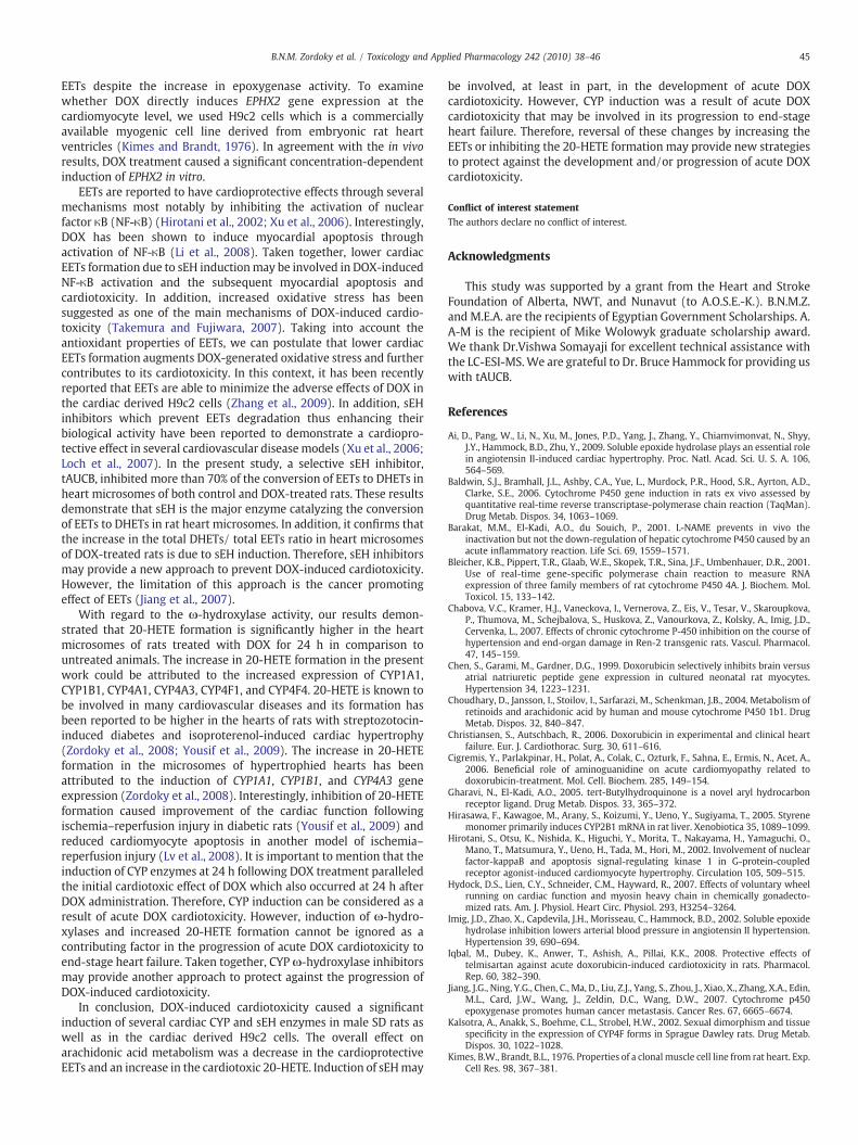
Cytotoxicity of DOX. The effect of DOX on cell viability wasdetermined by measuring the capacity of reducing enzymes presentin viable cells to convert 3-[4,5-dimethylthiazol-2-yl]-2,5-diphenyltetrazolium bromide (MTT) to formazan crystals as de-scribed previously (Korashy and El-Kadi, 2004). H9c2 cells weretreated for 2 h with various concentrations of DOX. After 24 h, thecolor intensity in each well was measured at wavelength of 550 nmusing EL 312e 96-well microplate readers, Bio-Tek Instruments Inc.(Winooski, VT). The percentage of cell viability was calculated relativeto control wells designated as 100% viable cells.
RNA extraction and cDNA synthesis. Total RNA from the frozen tissuesand from H9c2 cells was isolated using TRIzol reagent (Invitrogen)according to the manufacturer's instructions and quantified bymeasuring the absorbance at 260 nm. RNA quality was determinedby measuring the 260/280 ratio. Thereafter, first-strand cDNAsynthesis was performed by using the high-capacity cDNA reversetranscription kit (Applied Biosystems) according to the man-ufacturer's instructions. Briefly, 1.5 μg of total RNA from each samplewas added to a mix of 2.0 μl 10× RT buffer, 0.8 μl 25× dNTP mix(100 mM), 2.0 μl 10× RT random primers, 1.0 μl MultiScribe™ reversetranscriptase, and 3.2 μl nuclease-free water. The final reaction mixwas kept at 25 °C for 10 min, heated to 37 °C for 120 min, heated for85 °C for 5 s, and finally cooled to 4 °C.
Quantification by real-time PCR. Quantitative analysis of specificmRNA expression was performed by real-time PCR, by subjecting theresulting cDNA to PCR amplification using 96-well optical reactionplates in the ABI Prism 7500 System (Applied Biosystems). The 25-μlreaction mix contained 0.1 μl of 10 μM forward primer and 0.1 μl of10 μM reverse primer (40 nM final concentration of each primer),12.5 μl of SYBR Green Universal Mastermix, 11.05 μl of nuclease-freewater, and 1.25 μl of cDNA sample. The primers used in the currentstudy were chosen from previously published studies (Bleicher et al.,2001; Kalsotra et al., 2002; Wang et al., 2003; Hirasawa et al., 2005;Rollin et al., 2005; Baldwin et al., 2006) and are listed in Table 1. Assaycontrols were incorporated onto the same plate, namely, no-templatecontrols to test for the contamination of any assay reagents. Aftersealing the plate with an optical adhesive cover, the thermocyclingconditions were initiated at 95 °C for 10 min, followed by 40 PCRcycles of denaturation at 95 °C for 15 s, and annealing/extension at60 °C for 1 min. Melting curve (dissociation stage) was performed bythe end of each cycle to ascertain the specificity of the primers and thepurity of the final PCR product.
Real-time PCR data analysis. The real-time PCR data were analyzedusing the relative gene expression, i.e. (ΔΔCT) method as described in
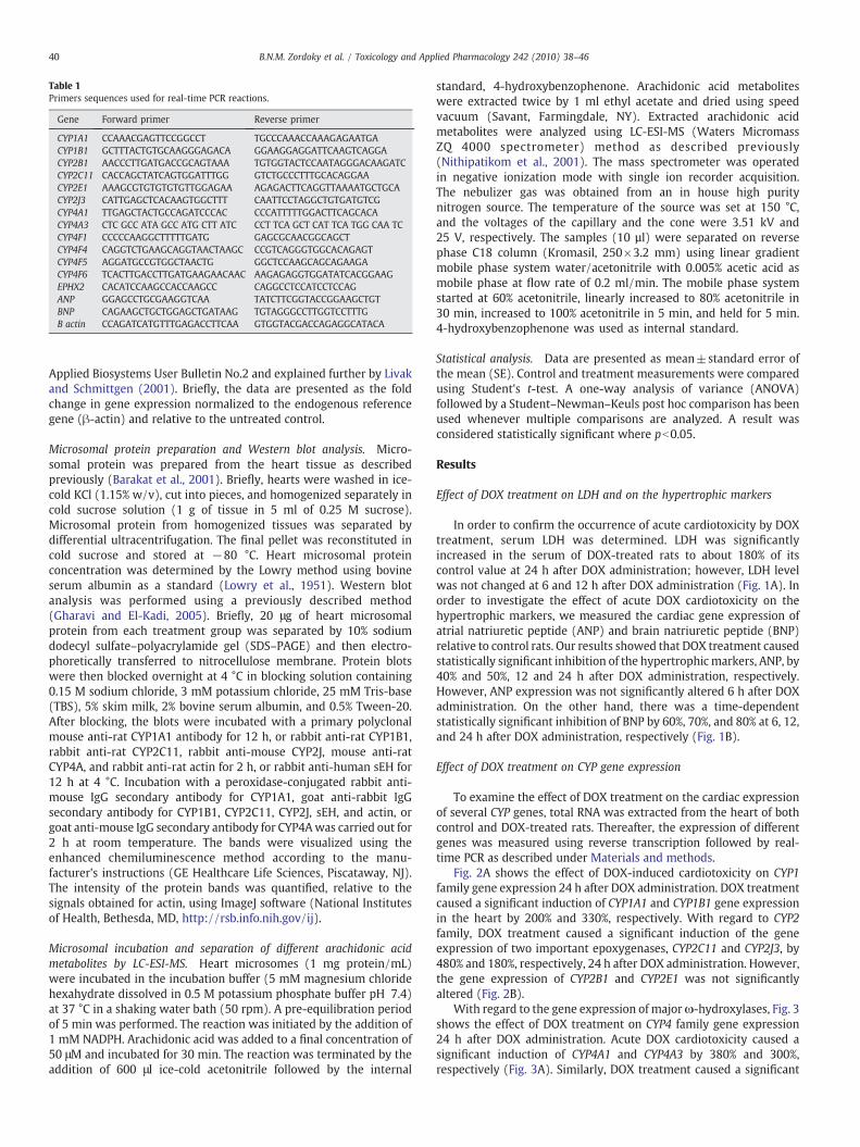
Table 1Primers sequences used for real-time PCR reactions.
Gene Forward primer Reverse primer
CYP1A1 CCAAACGAGTTCCGGCCT TGCCCAAACCAAAGAGAATGACYP1B1 GCTTTACTGTGCAAGGGAGACA GGAAGGAGGATTCAAGTCAGGACYP2B1 AACCCTTGATGACCGCAGTAAA TGTGGTACTCCAATAGGGACAAGATCCYP2C11 CACCAGCTATCAGTGGATTTGG GTCTGCCCTTTGCACAGGAACYP2E1 AAAGCGTGTGTGTGTTGGAGAA AGAGACTTCAGGTTAAAATGCTGCACYP2J3 CATTGAGCTCACAAGTGGCTTT CAATTCCTAGGCTGTGATGTCGCYP4A1 TTGAGCTACTGCCAGATCCCAC CCCATTTTTGGACTTCAGCACACYP4A3 CTC GCC ATA GCC ATG CTT ATC CCT TCA GCT CAT TCA TGG CAA TCCYP4F1 CCCCCAAGGCTTTTTGATG GAGCGCAACGGCAGCTCYP4F4 CAGGTCTGAAGCAGGTAACTAAGC CCGTCAGGGTGGCACAGAGTCYP4F5 AGGATGCCGTGGCTAACTG GGCTCCAAGCAGCAGAAGACYP4F6 TCACTTGACCTTGATGAAGAACAAC AAGAGAGGTGGATATCACGGAAGEPHX2 CACATCCAAGCCACCAAGCC CAGGCCTCCATCCTCCAGANP GGAGCCTGCGAAGGTCAA TATCTTCGGTACCGGAAGCTGTBNP CAGAAGCTGCTGGAGCTGATAAG TGTAGGGCCTTGGTCCTTTGΒ actin CCAGATCATGTTTGAGACCTTCAA GTGGTACGACCAGAGGCATACA
40 B.N.M. Zordoky et al. / Toxicology and Applied Pharmacology 242 (2010) 38–46
Applied Biosystems User Bulletin No.2 and explained further by Livakand Schmittgen (2001). Briefly, the data are presented as the foldchange in gene expression normalized to the endogenous referencegene (β-actin) and relative to the untreated control.
Microsomal protein preparation and Western blot analysis. Micro-somal protein was prepared from the heart tissue as describedpreviously (Barakat et al., 2001). Briefly, hearts were washed in ice-cold KCl (1.15% w/v), cut into pieces, and homogenized separately incold sucrose solution (1 g of tissue in 5 ml of 0.25 M sucrose).Microsomal protein from homogenized tissues was separated bydifferential ultracentrifugation. The final pellet was reconstituted incold sucrose and stored at −80 °C. Heart microsomal proteinconcentration was determined by the Lowry method using bovineserum albumin as a standard (Lowry et al., 1951). Western blotanalysis was performed using a previously described method(Gharavi and El-Kadi, 2005). Briefly, 20 μg of heart microsomalprotein from each treatment group was separated by 10% sodiumdodecyl sulfate–polyacrylamide gel (SDS–PAGE) and then electro-phoretically transferred to nitrocellulose membrane. Protein blotswere then blocked overnight at 4 °C in blocking solution containing0.15 M sodium chloride, 3 mM potassium chloride, 25 mM Tris-base(TBS), 5% skim milk, 2% bovine serum albumin, and 0.5% Tween-20.After blocking, the blots were incubated with a primary polyclonalmouse anti-rat CYP1A1 antibody for 12 h, or rabbit anti-rat CYP1B1,rabbit anti-rat CYP2C11, rabbit anti-mouse CYP2J, mouse anti-ratCYP4A, and rabbit anti-rat actin for 2 h, or rabbit anti-human sEH for12 h at 4 °C. Incubation with a peroxidase-conjugated rabbit anti-mouse IgG secondary antibody for CYP1A1, goat anti-rabbit IgGsecondary antibody for CYP1B1, CYP2C11, CYP2J, sEH, and actin, orgoat anti-mouse IgG secondary antibody for CYP4Awas carried out for2 h at room temperature. The bands were visualized using theenhanced chemiluminescence method according to the manu-facturer's instructions (GE Healthcare Life Sciences, Piscataway, NJ).The intensity of the protein bands was quantified, relative to thesignals obtained for actin, using ImageJ software (National Institutesof Health, Bethesda, MD, http://rsb.info.nih.gov/ij).
Microsomal incubation and separation of different arachidonic acidmetabolites by LC-ESI-MS. Heart microsomes (1 mg protein/mL)were incubated in the incubation buffer (5 mM magnesium chloridehexahydrate dissolved in 0.5 M potassium phosphate buffer pH 7.4)at 37 °C in a shaking water bath (50 rpm). A pre-equilibration periodof 5 min was performed. The reaction was initiated by the addition of1 mM NADPH. Arachidonic acid was added to a final concentration of50 μM and incubated for 30 min. The reaction was terminated by theaddition of 600 μl ice-cold acetonitrile followed by the internal
standard, 4-hydroxybenzophenone. Arachidonic acid metaboliteswere extracted twice by 1 ml ethyl acetate and dried using speedvacuum (Savant, Farmingdale, NY). Extracted arachidonic acidmetabolites were analyzed using LC-ESI-MS (Waters MicromassZQ 4000 spectrometer) method as described previously(Nithipatikom et al., 2001). The mass spectrometer was operatedin negative ionization mode with single ion recorder acquisition.The nebulizer gas was obtained from an in house high puritynitrogen source. The temperature of the source was set at 150 °C,and the voltages of the capillary and the cone were 3.51 kV and25 V, respectively. The samples (10 µl) were separated on reversephase C18 column (Kromasil, 250×3.2 mm) using linear gradientmobile phase system water/acetonitrile with 0.005% acetic acid asmobile phase at flow rate of 0.2 ml/min. The mobile phase systemstarted at 60% acetonitrile, linearly increased to 80% acetonitrile in30 min, increased to 100% acetonitrile in 5 min, and held for 5 min.4-hydroxybenzophenone was used as internal standard.
Statistical analysis. Data are presented as mean±standard error ofthe mean (SE). Control and treatment measurements were comparedusing Student's t-test. A one-way analysis of variance (ANOVA)followed by a Student–Newman–Keuls post hoc comparison has beenused whenever multiple comparisons are analyzed. A result wasconsidered statistically significant where pb0.05.
Results
Effect of DOX treatment on LDH and on the hypertrophic markers
In order to confirm the occurrence of acute cardiotoxicity by DOXtreatment, serum LDH was determined. LDH was significantlyincreased in the serum of DOX-treated rats to about 180% of itscontrol value at 24 h after DOX administration; however, LDH levelwas not changed at 6 and 12 h after DOX administration (Fig. 1A). Inorder to investigate the effect of acute DOX cardiotoxicity on thehypertrophic markers, we measured the cardiac gene expression ofatrial natriuretic peptide (ANP) and brain natriuretic peptide (BNP)relative to control rats. Our results showed that DOX treatment causedstatistically significant inhibition of the hypertrophicmarkers, ANP, by40% and 50%, 12 and 24 h after DOX administration, respectively.However, ANP expression was not significantly altered 6 h after DOXadministration. On the other hand, there was a time-dependentstatistically significant inhibition of BNP by 60%, 70%, and 80% at 6, 12,and 24 h after DOX administration, respectively (Fig. 1B).
Effect of DOX treatment on CYP gene expression
To examine the effect of DOX treatment on the cardiac expressionof several CYP genes, total RNA was extracted from the heart of bothcontrol and DOX-treated rats. Thereafter, the expression of differentgenes was measured using reverse transcription followed by real-time PCR as described under Materials and methods.
Fig. 2A shows the effect of DOX-induced cardiotoxicity on CYP1family gene expression 24 h after DOX administration. DOX treatmentcaused a significant induction of CYP1A1 and CYP1B1 gene expressionin the heart by 200% and 330%, respectively. With regard to CYP2family, DOX treatment caused a significant induction of the geneexpression of two important epoxygenases, CYP2C11 and CYP2J3, by480% and 180%, respectively, 24 h after DOX administration. However,the gene expression of CYP2B1 and CYP2E1 was not significantlyaltered (Fig. 2B).
With regard to the gene expression of majorω-hydroxylases, Fig. 3shows the effect of DOX treatment on CYP4 family gene expression24 h after DOX administration. Acute DOX cardiotoxicity caused asignificant induction of CYP4A1 and CYP4A3 by 380% and 300%,respectively (Fig. 3A). Similarly, DOX treatment caused a significant

Fig. 1. Effect of acute DOX cardiotoxicity on LDH (A) and on the hypertrophic markers(B). (A) LDH was estimated in the serum by commercially available kit as describedunder Materials and methods. The amount of LDH was calculated relative to the controland presented as percentage of control and DOX-treated animals for 6, 12, and 24 h. (B)Total RNA was isolated from the hearts of control and DOX-treated animals for 6, 12,and 24 h. ANP and BNP gene expressions were determined by real-time PCR. Results arepresented as mean±SE (n= 6). ⁎pb 0.05 compared with control.
Fig. 2. Effect of acute DOX cardiotoxicity on CYP1 (A) and CYP2 family gene expression(B). Total RNA was isolated from the hearts of control and animals treated with DOX for24 h. CYP1A1, CYP1B1, CYP2B1, CYP2C11, CYP2E1, and CYP2J3 gene expressions weredetermined by real-time PCR. Results are presented as mean±SE (n= 6). ⁎pb 0.05compared with control.
Fig. 3. Effect of acute DOX cardiotoxicity on CYP4A (A) and CYP4F (B) sub-family geneexpression. Total RNA was isolated from the hearts of control and animals treated withDOX for 24 h. CYP4A1, CYP4A3, CYP4F1, CYP4F4, CYP4F5 and CYP4F6 gene expressionswere determined by real-time PCR. Results are presented as mean±SE (n= 6).⁎pb 0.05 compared with control.
41B.N.M. Zordoky et al. / Toxicology and Applied Pharmacology 242 (2010) 38–46
induction of CYP4F1 and CYP4F4 gene expression by 200% and 250%,respectively. However, the expression of CYP4F5 and CYP4F6 was notsignificantly altered (Fig. 3B). Interestingly, all CYP gene expressionwas not significantly altered 6 and 12 h after DOX administration;however, there was a trend of induction of CYP1A1, CYP1B1, CYP2C11,CYP2J3, and CYP4A3 gene expression at 12 h after DOX administration(data not shown).
Effect of DOX treatment on CYP protein expression
To investigate whether the induction of CYP gene expression wasfurther translated into functional protein, microsomal protein wasprepared from hearts of control and rats treated with DOX for 24 h.Thereafter, CYP1A1, CYP1B1, CYP2C11, CYP2J3, and CYP4A proteinlevels were determined using Western blot analysis relative to actinas an endogenous control. DOX treatment caused a significantinduction of CYP1A1, CYP1B1, CYP2C11, CYP2J3, and CYP4A proteinexpression by 280%, 345%, 125%, 135%, and 250%, respectively (Fig. 4).
Separation of arachidonic acid metabolites using LC-ESI-MS selected ionchromatogram
LC-ESI-MS has been used in this study for the separation andquantification of CYP-derived metabolites of arachidonic acid. Themass spectrometer was operated in negative ionization mode withsingle ion recorder acquisition where the most abundant ion iscorresponding to the m/z=[M−1]−. All four regioisomeric epox-yeicosatrienoic acids (5,6-, 8,9-, 11,12-, and 14,15-EET) exhibited themost abundant ions corresponding m/z=319 ion, while theircorresponding DHETs has m/z=337 ion and 20-HETE has m/z=319 ion. The fact of having functional groups at different positions
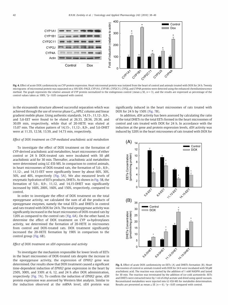
Fig. 4. Effect of acute DOX cardiotoxicity on CYP protein expression. Heart microsomal protein was isolated from the heart of control and animals treated with DOX for 24 h. Twentymicrograms of microsomal protein was separated on a 10% SDS–PAGE. CYP1A1, CYP1B1, CYP2C11, CYP2J, and CYP4A proteins were detected using the enhanced chemiluminescencemethod. The graph represents the relative amount of CYP protein normalized to the endogenous control (mean±SE, n= 3), and the results are expressed as percentage of thecontrol values taken as 100%. ⁎pb 0.05 compared with control.
Fig. 5. Effect of acute DOX cardiotoxicity on EETs (A) and DHETs formation (B). Heartmicrosomes of control or animals treated with DOX for 24 hwere incubated with 50 μMarachidonic acid. The reaction was started by the addition of 1 mM NADPH and lastedfor 30 min. The reaction was terminated by the addition of ice-cold acetonitrile. EETsand DHETs were extracted twice by 1ml of ethyl acetate and dried using speed vacuum.Reconstituted metabolites were injected into LC-ESI-MS for metabolite determination.Results are presented as mean±SE (n= 6). ⁎pb 0.05 compared with control.
42 B.N.M. Zordoky et al. / Toxicology and Applied Pharmacology 242 (2010) 38–46
in the eicosanoids structure allowed successful separation which wasachieved through the use of reverse phase C18 HPLC column and lineargradient mobile phase. Using authentic standards, 14,15-, 11,12-, 8,9-,and 5,6-EET were found to be eluted at 26.33, 28.56, 29.38, and30.09 min, respectively, while that of 20-HETE was eluted at15.07 min. The elution pattern of 14,15-, 11,12-, 8,9-, and 5,6-DHETwere at 11.35, 12.58, 13.59, and 14.73 min, respectively.
Effect of DOX treatment on CYP-mediated arachidonic acid metabolism
To investigate the effect of DOX treatment on the formation ofCYP-derived arachidonic acid metabolites, heart microsomes of eithercontrol or 24 h DOX-treated rats were incubated with 50 μMarachidonic acid for 30 min. Thereafter, arachidonic acid metaboliteswere determined using LC-ESI-MS. In comparison to control animals,in heart microsomes of DOX-treated rats, the formation of 5,6-, 8,9-,11,12-, and 14,15-EET were significantly lower by about 60%, 30%,50%, and 40%, respectively (Fig. 5A). We also measured levels ofenzymatic hydration of EETs products, DHETs. As shown in Fig. 5B, theformation of 5,6-, 8,9-, 11,12, and 14,15-DHET was significantlyincreased by 160%, 200%, 160%, and 150%, respectively, compared tocontrol.
In order to investigate the effect of DOX treatment on the totalepoxygenase activity, we calculated the sum of all the products ofepoxygenase enzymes, namely the total EETs and DHETs in controland rats treated with DOX for 24 h. The total epoxygenase activity wassignificantly increased in the heartmicrosomes of DOX-treated rats by120% as compared to the control rats (Fig. 6A). On the other hand, todetermine the effect of DOX treatment on CYP ω-hydroxylasesactivity, we determined the formation of 20-HETE in microsomesfrom control and DOX-treated rats. DOX treatment significantlyincreased the 20-HETE formation by 190% in comparison to thecontrol group (Fig. 6B).
Effect of DOX treatment on sEH expression and activity
To investigate the mechanism responsible for lower levels of EETsin the heart microsomes of DOX-treated rats despite the increase inthe epoxygenase activity, the expression of EPHX2 gene wasdetermined. Our results show that DOX treatment caused a significanttime-dependent induction of EPHX2 gene expression in the heart by250%, 300%, and 330% at 6, 12, and 24 h after DOX administration,respectively (Fig. 7A). To confirm the induction of EPHX2 gene, sEHprotein expression was assessed by Western blot analysis. Similar tothe induction observed at the mRNA level, sEH protein was
significantly induced in the heart microsomes of rats treated withDOX for 24 h by 150% (Fig. 7B).
In addition, sEH activity has been assessed by calculating the ratioof the total DHETs to the total EETs formed in the heart microsomes ofcontrol and rats treated with DOX for 24 h. In accordance with theinduction at the gene and protein expression levels, sEH activity wasinduced by 320% in the heart microsomes of rats treated with DOX for

Fig. 6. Effect of acute DOX cardiotoxicity on epoxygenase (A) and ω-hydroxylaseactivity (B). (A) Epoxygenase activity was determined from the sum of EETs and DHETsformation. (B) ω-hydroxylase activity was determined from the 20-HETE formation.Heart microsomes of control or animals treated with DOX for 24 h were incubated with50 μM arachidonic acid. The reaction was started by the addition of 1 mM NADPH andlasted for 30 min. The reaction was terminated by the addition of ice-cold acetonitrile.20-HETE was extracted twice by 1 ml of ethyl acetate and dried using speed vacuum.Reconstituted metabolite was injected into LC-ESI-MS for metabolite determination.Results are presented as mean±SE (n= 6). ⁎pb 0.05 compared with control.
Fig. 7. Effect of acute DOX cardiotoxicity on EPHX2 gene expression (A) and sEH proteinexpression (B). (A) Total RNA was isolated from the hearts of control and animalstreated with DOX for 6, 12, and 24 h. EPHX2 gene expression was determined by real-time PCR (n= 6). (B) Heart microsomal protein was isolated from the heart of controland animals treated with DOX for 24 h. Twenty micrograms of microsomal protein wasseparated on a 10% SDS–PAGE. sEH protein was detected using the enhancedchemiluminescence method. The graph represents the relative amount of sEH proteinnormalized to the endogenous control (n= 3). Results are presented as mean±SE.⁎pb 0.05 compared with control.
Fig. 8. Effect of acute DOX cardiotoxicity on sEH activity. Heart microsomes of control oranimals treated with DOX for 24 h were incubated with 100 nM tAUCB for 5 minfollowed by incubation with 50 μM arachidonic acid. The reaction was started by theaddition of 1 mM NADPH and lasted for 30 min then terminated by the addition of ice-cold acetonitrile. EETs and DHETs were extracted twice by 1 ml of ethyl acetate anddried using speed vacuum. Reconstituted metabolites were injected into LC-ESI-MS formetabolite determination. sEH activity was calculated as the ratio of total DHETs/ totalEETs. Results are presented as mean±SE (n= 6). ⁎pb 0.05 compared with control,#pb 0.05 compared with heart microsomes from animals treated with DOX for 24 h inthe absence of sEH inhibitor (sEHI).
43B.N.M. Zordoky et al. / Toxicology and Applied Pharmacology 242 (2010) 38–46
24 h (Fig. 8). In order to confirm that the increase in total DHETs/totalEETs ratio was due to sEH induction, heart microsomes from controland rats treated with DOX for 24 h were incubated with 100 nM of theselective sEH inhibitor, tAUCB, for 5 min followed by incubation witharachidonic acid as described under Materials and methods. Ourresults show that inhibition of sEH markedly decreased the totalDHETs/total EETs ratio from 1.2 in the control group to 0.3 afterincubationwith tAUCB. Similarly, the total DHETs/total EETs ratio wasdecreased from 3.2 in the DOX-treated group to 0.3 after incubationwith tAUCB (Fig. 8).
Effect of DOX on the EPHX2 gene expression in H9c2 cells
To investigate whether the induction of EPHX2 gene expression isdue to the direct effect of DOX on the cardiomyocytes, the cardiacderived H9c2 cells were treated with increasing concentrations ofDOX. Thereafter, the expression of EPHX2 was measured using real-time PCR as described under Materials and methods.
To determine the cytotoxic effect of DOX, H9c2 cells were incu-bated with increasing concentrations of DOX. Thereafter, cell viabilitywas measured by the MTT assay. Our results clearly demonstrate thatcells treated with DOX (1–5 μM) maintained more than 90% cellviability (Fig. 9A). Therefore, the observed changes in gene expressionare not due to decreased cell viability or toxicity. Our resultsdemonstrated that treatment of H9c2 cells with increasing concen-trations of DOX caused a significant concentration-dependentinduction of EPHX2. The induction was 180%, 210%, and 300% higherthan the control with DOX concentrations of 1, 2, and 5 μM, res-pectively (Fig. 9B).
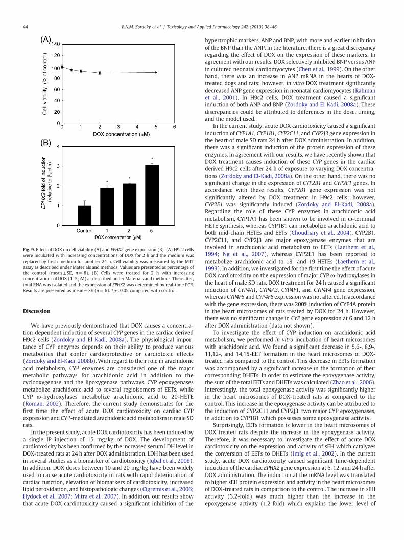
Fig. 9. Effect of DOX on cell viability (A) and EPHX2 gene expression (B). (A) H9c2 cellswere incubated with increasing concentrations of DOX for 2 h and the medium wasreplaced by fresh medium for another 24 h. Cell viability was measured by the MTTassay as described under Materials and methods. Values are presented as percentage ofthe control (mean±SE, n= 8). (B) Cells were treated for 2 h with increasingconcentrations of DOX (1–5 μM) as described under Materials and methods. Thereafter,total RNA was isolated and the expression of EPHX2 was determined by real-time PCR.Results are presented as mean±SE (n= 6). ⁎pb 0.05 compared with control.
44 B.N.M. Zordoky et al. / Toxicology and Applied Pharmacology 242 (2010) 38–46
Discussion
We have previously demonstrated that DOX causes a concentra-tion-dependent induction of several CYP genes in the cardiac derivedH9c2 cells (Zordoky and El-Kadi, 2008a). The physiological impor-tance of CYP enzymes depends on their ability to produce variousmetabolites that confer cardioprotective or cardiotoxic effects(Zordoky and El-Kadi, 2008b). With regard to their role in arachidonicacid metabolism, CYP enzymes are considered one of the majormetabolic pathways for arachidonic acid in addition to thecyclooxygenase and the lipoxygenase pathways. CYP epoxygenasesmetabolize arachidonic acid to several regioisomers of EETs, whileCYP ω-hydroxylases metabolize arachidonic acid to 20-HETE(Roman, 2002). Therefore, the current study demonstrates for thefirst time the effect of acute DOX cardiotoxicity on cardiac CYPexpression and CYP-mediated arachidonic acidmetabolism inmale SDrats.
In the present study, acute DOX cardiotoxicity has been induced bya single IP injection of 15 mg/kg of DOX. The development ofcardiotoxicity has been confirmed by the increased serum LDH level inDOX-treated rats at 24 h after DOX administration. LDH has been usedin several studies as a biomarker of cardiotoxicity (Iqbal et al., 2008).In addition, DOX doses between 10 and 20 mg/kg have been widelyused to cause acute cardiotoxicity in rats with rapid deterioration ofcardiac function, elevation of biomarkers of cardiotoxicity, increasedlipid peroxidation, and histopathologic changes (Cigremis et al., 2006;Hydock et al., 2007; Mitra et al., 2007). In addition, our results showthat acute DOX cardiotoxicity caused a significant inhibition of the
hypertrophic markers, ANP and BNP, with more and earlier inhibitionof the BNP than the ANP. In the literature, there is a great discrepancyregarding the effect of DOX on the expression of these markers. Inagreement with our results, DOX selectively inhibited BNP versus ANPin cultured neonatal cardiomyocytes (Chen et al., 1999). On the otherhand, there was an increase in ANP mRNA in the hearts of DOX-treated dogs and rats; however, in vitro DOX treatment significantlydecreased ANP gene expression in neonatal cardiomyocytes (Rahmanet al., 2001). In H9c2 cells, DOX treatment caused a significantinduction of both ANP and BNP (Zordoky and El-Kadi, 2008a). Thesediscrepancies could be attributed to differences in the dose, timing,and the model used.
In the current study, acute DOX cardiotoxicity caused a significantinduction of CYP1A1, CYP1B1, CYP2C11, and CYP2J3 gene expression inthe heart of male SD rats 24 h after DOX administration. In addition,there was a significant induction of the protein expression of theseenzymes. In agreement with our results, we have recently shown thatDOX treatment causes induction of these CYP genes in the cardiacderived H9c2 cells after 24 h of exposure to varying DOX concentra-tions (Zordoky and El-Kadi, 2008a). On the other hand, there was nosignificant change in the expression of CYP2B1 and CYP2E1 genes. Inaccordance with these results, CYP2B1 gene expression was notsignificantly altered by DOX treatment in H9c2 cells; however,CYP2E1 was significantly induced (Zordoky and El-Kadi, 2008a).Regarding the role of these CYP enzymes in arachidonic acidmetabolism, CYP1A1 has been shown to be involved in ω-terminalHETE synthesis, whereas CYP1B1 can metabolize arachidonic acid toboth mid-chain HETEs and EETs (Choudhary et al., 2004). CYP2B1,CYP2C11, and CYP2J3 are major epoxygenase enzymes that areinvolved in arachidonic acid metabolism to EETs (Laethem et al.,1994; Ng et al., 2007), whereas CYP2E1 has been reported tometabolize arachidonic acid to 18- and 19-HETEs (Laethem et al.,1993). In addition, we investigated for the first time the effect of acuteDOX cardiotoxicity on the expression of major CYP ω-hydroxylases inthe heart of male SD rats. DOX treatment for 24 h caused a significantinduction of CYP4A1, CYP4A3, CYP4F1, and CYP4F4 gene expression,whereas CYP4F5 and CYP4F6 expressionwas not altered. In accordancewith the gene expression, there was 200% induction of CYP4A proteinin the heart microsomes of rats treated by DOX for 24 h. However,there was no significant change in CYP gene expression at 6 and 12 hafter DOX administration (data not shown).
To investigate the effect of CYP induction on arachidonic acidmetabolism, we performed in vitro incubation of heart microsomeswith arachidonic acid. We found a significant decrease in 5,6-, 8,9-,11,12-, and 14,15-EET formation in the heart microsomes of DOX-treated rats compared to the control. This decrease in EETs formationwas accompanied by a significant increase in the formation of theircorresponding DHETs. In order to estimate the epoxygenase activity,the sum of the total EETs and DHETswas calculated (Zhao et al., 2006).Interestingly, the total epoxygenase activity was significantly higherin the heart microsomes of DOX-treated rats as compared to thecontrol. This increase in the epoxygenase activity can be attributed tothe induction of CYP2C11 and CYP2J3, two major CYP epoxygenases,in addition to CYP1B1 which possesses some epoxygenase activity.
Surprisingly, EETs formation is lower in the heart microsomes ofDOX-treated rats despite the increase in the epoxygenase activity.Therefore, it was necessary to investigate the effect of acute DOXcardiotoxicity on the expression and activity of sEH which catalyzesthe conversion of EETs to DHETs (Imig et al., 2002). In the currentstudy, acute DOX cardiotoxicity caused significant time-dependentinduction of the cardiac EPHX2 gene expression at 6, 12, and 24 h afterDOX administration. The induction at the mRNA level was translatedto higher sEH protein expression and activity in the heart microsomesof DOX-treated rats in comparison to the control. The increase in sEHactivity (3.2-fold) was much higher than the increase in theepoxygenase activity (1.2-fold) which explains the lower level of
45B.N.M. Zordoky et al. / Toxicology and Applied Pharmacology 242 (2010) 38–46
EETs despite the increase in epoxygenase activity. To examinewhether DOX directly induces EPHX2 gene expression at thecardiomyocyte level, we used H9c2 cells which is a commerciallyavailable myogenic cell line derived from embryonic rat heartventricles (Kimes and Brandt, 1976). In agreement with the in vivoresults, DOX treatment caused a significant concentration-dependentinduction of EPHX2 in vitro.
EETs are reported to have cardioprotective effects through severalmechanisms most notably by inhibiting the activation of nuclearfactor κB (NF-κB) (Hirotani et al., 2002; Xu et al., 2006). Interestingly,DOX has been shown to induce myocardial apoptosis throughactivation of NF-κB (Li et al., 2008). Taken together, lower cardiacEETs formation due to sEH inductionmay be involved in DOX-inducedNF-κB activation and the subsequent myocardial apoptosis andcardiotoxicity. In addition, increased oxidative stress has beensuggested as one of the main mechanisms of DOX-induced cardio-toxicity (Takemura and Fujiwara, 2007). Taking into account theantioxidant properties of EETs, we can postulate that lower cardiacEETs formation augments DOX-generated oxidative stress and furthercontributes to its cardiotoxicity. In this context, it has been recentlyreported that EETs are able to minimize the adverse effects of DOX inthe cardiac derived H9c2 cells (Zhang et al., 2009). In addition, sEHinhibitors which prevent EETs degradation thus enhancing theirbiological activity have been reported to demonstrate a cardiopro-tective effect in several cardiovascular disease models (Xu et al., 2006;Loch et al., 2007). In the present study, a selective sEH inhibitor,tAUCB, inhibited more than 70% of the conversion of EETs to DHETs inheart microsomes of both control and DOX-treated rats. These resultsdemonstrate that sEH is the major enzyme catalyzing the conversionof EETs to DHETs in rat heart microsomes. In addition, it confirms thatthe increase in the total DHETs/ total EETs ratio in heart microsomesof DOX-treated rats is due to sEH induction. Therefore, sEH inhibitorsmay provide a new approach to prevent DOX-induced cardiotoxicity.However, the limitation of this approach is the cancer promotingeffect of EETs (Jiang et al., 2007).
With regard to the ω-hydroxylase activity, our results demon-strated that 20-HETE formation is significantly higher in the heartmicrosomes of rats treated with DOX for 24 h in comparison tountreated animals. The increase in 20-HETE formation in the presentwork could be attributed to the increased expression of CYP1A1,CYP1B1, CYP4A1, CYP4A3, CYP4F1, and CYP4F4. 20-HETE is known tobe involved in many cardiovascular diseases and its formation hasbeen reported to be higher in the hearts of rats with streptozotocin-induced diabetes and isoproterenol-induced cardiac hypertrophy(Zordoky et al., 2008; Yousif et al., 2009). The increase in 20-HETEformation in the microsomes of hypertrophied hearts has beenattributed to the induction of CYP1A1, CYP1B1, and CYP4A3 geneexpression (Zordoky et al., 2008). Interestingly, inhibition of 20-HETEformation caused improvement of the cardiac function followingischemia–reperfusion injury in diabetic rats (Yousif et al., 2009) andreduced cardiomyocyte apoptosis in another model of ischemia–reperfusion injury (Lv et al., 2008). It is important to mention that theinduction of CYP enzymes at 24 h following DOX treatment paralleledthe initial cardiotoxic effect of DOX which also occurred at 24 h afterDOX administration. Therefore, CYP induction can be considered as aresult of acute DOX cardiotoxicity. However, induction of ω-hydro-xylases and increased 20-HETE formation cannot be ignored as acontributing factor in the progression of acute DOX cardiotoxicity toend-stage heart failure. Taken together, CYP ω-hydroxylase inhibitorsmay provide another approach to protect against the progression ofDOX-induced cardiotoxicity.
In conclusion, DOX-induced cardiotoxicity caused a significantinduction of several cardiac CYP and sEH enzymes in male SD rats aswell as in the cardiac derived H9c2 cells. The overall effect onarachidonic acid metabolism was a decrease in the cardioprotectiveEETs and an increase in the cardiotoxic 20-HETE. Induction of sEHmay
be involved, at least in part, in the development of acute DOXcardiotoxicity. However, CYP induction was a result of acute DOXcardiotoxicity that may be involved in its progression to end-stageheart failure. Therefore, reversal of these changes by increasing theEETs or inhibiting the 20-HETE formation may provide new strategiesto protect against the development and/or progression of acute DOXcardiotoxicity.
Conflict of interest statement
The authors declare no conflict of interest.
Acknowledgments
This study was supported by a grant from the Heart and StrokeFoundation of Alberta, NWT, and Nunavut (to A.O.S.E.-K.). B.N.M.Z.and M.E.A. are the recipients of Egyptian Government Scholarships. A.A-M is the recipient of Mike Wolowyk graduate scholarship award.We thank Dr.Vishwa Somayaji for excellent technical assistance withthe LC-ESI-MS.We are grateful to Dr. Bruce Hammock for providing uswith tAUCB.
References
Ai, D., Pang, W., Li, N., Xu, M., Jones, P.D., Yang, J., Zhang, Y., Chiamvimonvat, N., Shyy,J.Y., Hammock, B.D., Zhu, Y., 2009. Soluble epoxide hydrolase plays an essential rolein angiotensin II-induced cardiac hypertrophy. Proc. Natl. Acad. Sci. U. S. A. 106,564–569.
Baldwin, S.J., Bramhall, J.L., Ashby, C.A., Yue, L., Murdock, P.R., Hood, S.R., Ayrton, A.D.,Clarke, S.E., 2006. Cytochrome P450 gene induction in rats ex vivo assessed byquantitative real-time reverse transcriptase-polymerase chain reaction (TaqMan).Drug Metab. Dispos. 34, 1063–1069.
Barakat, M.M., El-Kadi, A.O., du Souich, P., 2001. L-NAME prevents in vivo theinactivation but not the down-regulation of hepatic cytochrome P450 caused by anacute inflammatory reaction. Life Sci. 69, 1559–1571.
Bleicher, K.B., Pippert, T.R., Glaab, W.E., Skopek, T.R., Sina, J.F., Umbenhauer, D.R., 2001.Use of real-time gene-specific polymerase chain reaction to measure RNAexpression of three family members of rat cytochrome P450 4A. J. Biochem. Mol.Toxicol. 15, 133–142.
Chabova, V.C., Kramer, H.J., Vaneckova, I., Vernerova, Z., Eis, V., Tesar, V., Skaroupkova,P., Thumova, M., Schejbalova, S., Huskova, Z., Vanourkova, Z., Kolsky, A., Imig, J.D.,Cervenka, L., 2007. Effects of chronic cytochrome P-450 inhibition on the course ofhypertension and end-organ damage in Ren-2 transgenic rats. Vascul. Pharmacol.47, 145–159.
Chen, S., Garami, M., Gardner, D.G., 1999. Doxorubicin selectively inhibits brain versusatrial natriuretic peptide gene expression in cultured neonatal rat myocytes.Hypertension 34, 1223–1231.
Choudhary, D., Jansson, I., Stoilov, I., Sarfarazi, M., Schenkman, J.B., 2004. Metabolism ofretinoids and arachidonic acid by human and mouse cytochrome P450 1b1. DrugMetab. Dispos. 32, 840–847.
Christiansen, S., Autschbach, R., 2006. Doxorubicin in experimental and clinical heartfailure. Eur. J. Cardiothorac. Surg. 30, 611–616.
Cigremis, Y., Parlakpinar, H., Polat, A., Colak, C., Ozturk, F., Sahna, E., Ermis, N., Acet, A.,2006. Beneficial role of aminoguanidine on acute cardiomyopathy related todoxorubicin-treatment. Mol. Cell. Biochem. 285, 149–154.
Gharavi, N., El-Kadi, A.O., 2005. tert-Butylhydroquinone is a novel aryl hydrocarbonreceptor ligand. Drug Metab. Dispos. 33, 365–372.
Hirasawa, F., Kawagoe, M., Arany, S., Koizumi, Y., Ueno, Y., Sugiyama, T., 2005. Styrenemonomer primarily induces CYP2B1 mRNA in rat liver. Xenobiotica 35, 1089–1099.
Hirotani, S., Otsu, K., Nishida, K., Higuchi, Y., Morita, T., Nakayama, H., Yamaguchi, O.,Mano, T., Matsumura, Y., Ueno, H., Tada, M., Hori, M., 2002. Involvement of nuclearfactor-kappaB and apoptosis signal-regulating kinase 1 in G-protein-coupledreceptor agonist-induced cardiomyocyte hypertrophy. Circulation 105, 509–515.
Hydock, D.S., Lien, C.Y., Schneider, C.M., Hayward, R., 2007. Effects of voluntary wheelrunning on cardiac function and myosin heavy chain in chemically gonadecto-mized rats. Am. J. Physiol. Heart Circ. Physiol. 293, H3254–3264.
Imig, J.D., Zhao, X., Capdevila, J.H., Morisseau, C., Hammock, B.D., 2002. Soluble epoxidehydrolase inhibition lowers arterial blood pressure in angiotensin II hypertension.Hypertension 39, 690–694.
Iqbal, M., Dubey, K., Anwer, T., Ashish, A., Pillai, K.K., 2008. Protective effects oftelmisartan against acute doxorubicin-induced cardiotoxicity in rats. Pharmacol.Rep. 60, 382–390.
Jiang, J.G., Ning, Y.G., Chen, C., Ma, D., Liu, Z.J., Yang, S., Zhou, J., Xiao, X., Zhang, X.A., Edin,M.L., Card, J.W., Wang, J., Zeldin, D.C., Wang, D.W., 2007. Cytochrome p450epoxygenase promotes human cancer metastasis. Cancer Res. 67, 6665–6674.
Kalsotra, A., Anakk, S., Boehme, C.L., Strobel, H.W., 2002. Sexual dimorphism and tissuespecificity in the expression of CYP4F forms in Sprague Dawley rats. Drug Metab.Dispos. 30, 1022–1028.
Kimes, B.W., Brandt, B.L., 1976. Properties of a clonal muscle cell line from rat heart. Exp.Cell Res. 98, 367–381.

46 B.N.M. Zordoky et al. / Toxicology and Applied Pharmacology 242 (2010) 38–46
Korashy, H.M., El-Kadi, A.O., 2004. Differential effects of mercury, lead and copper onthe constitutive and inducible expression of aryl hydrocarbon receptor (AHR)-regulated genes in cultured hepatoma Hepa 1c1c7 cells. Toxicology 201, 153–172.
Laethem, R.M., Balazy, M., Falck, J.R., Laethem, C.L., Koop, D.R., 1993. Formation of 19(S)-, 19(R)-, and 18(R)-hydroxyeicosatetraenoic acids by alcohol-inducible cyto-chrome P450 2E1. J. Biol. Chem. 268, 12912–12918.
Laethem, R.M., Halpert, J.R., Koop, D.R., 1994. Epoxidation of arachidonic acid as anactive-site probe of cytochrome P-450 2B isoforms. Biochim. Biophys. Acta. 1206,42–48.
Li, S., EM, Yu, B., 2008. Adriamycin induces myocardium apoptosis through activation ofnuclear factor kappaB in rat. Mol. Biol. Rep. 35, 489–494.
Livak, K.J., Schmittgen, T.D., 2001. Analysis of relative gene expression data usingreal-time quantitative PCR and the 2(-Delta Delta C(T)) method. Methods 25,402–408.
Loch, D., Hoey, A., Morisseau, C., Hammock, B.O., Brown, L., 2007. Prevention ofhypertension in DOCA-salt rats by an inhibitor of soluble epoxide hydrolase. CellBiochem. Biophys. 47, 87–98.
Lowry, O.H., Rosebrough, N.J., Farr, A.L., Randall, R.J., 1951. Protein measurement withthe Folin phenol reagent. J. Biol. Chem. 193, 265–275.
Lv, X., Wan, J., Yang, J., Cheng, H., Li, Y., Ao, Y., Peng, R., 2008. Cytochrome P450omega-hydroxylase inhibition reduces cardiomyocyte apoptosis via activation ofERK1/2 signaling in rat myocardial ischemia–reperfusion. Eur. J. Pharmacol. 596,118–126.
Minuz, P., Jiang, H., Fava, C., Turolo, L., Tacconelli, S., Ricci, M., Patrignani, P., Morganti, A.,Lechi, A., McGiff, J.C., 2008. Altered release of cytochrome p450 metabolites ofarachidonic acid in renovascular disease. Hypertension 51, 1379–1385.
Mitra, M.S., Donthamsetty, S., White, B., Latendresse, J.R., Mehendale, H.M., 2007.Mechanism of protection of moderately diet restricted rats against doxorubicin-induced acute cardiotoxicity. Toxicol. Appl. Pharmacol. 225, 90–101.
Monti, J., Fischer, J., Paskas, S., Heinig, M., Schulz, H., Gosele, C., Heuser, A., Fischer, R.,Schmidt, C., Schirdewan, A., Gross, V., Hummel, O., Maatz, H., Patone, G., Saar, K.,Vingron, M., Weldon, S.M., Lindpaintner, K., Hammock, B.D., Rohde, K., Dietz, R.,Cook, S.A., Schunck,W.H., Luft, F.C., Hubner, N., 2008. Soluble epoxide hydrolase is asusceptibility factor for heart failure in a rat model of human disease. Nat. Genet.40, 529–537.
Nakamura, T., Ueda, Y., Juan, Y., Katsuda, S., Takahashi, H., Koh, E., 2000. Fas-mediatedapoptosis in adriamycin-induced cardiomyopathy in rats: in vivo study. Circulation102, 572–578.
Ng, V.Y., Huang, Y., Reddy, L.M., Falck, J.R., Lin, E.T., Kroetz, D.L., 2007. Cytochrome P450eicosanoids are activators of peroxisome proliferator-activated receptor alpha.Drug Metab. Dispos. 35, 1126–1134.
Nithipatikom, K., Grall, A.J., Holmes, B.B., Harder, D.R., Falck, J.R., Campbell, W.B., 2001.Liquid chromatographic-electrospray ionization-mass spectrometric analysis ofcytochrome P450 metabolites of arachidonic acid. Anal. Biochem. 298, 327–336.
Outomuro, D., Grana, D.R., Azzato, F., Milei, J., 2007. Adriamycin-induced myocardialtoxicity: new solutions for an old problem? Int. J. Cardiol. 117, 6–15.
Rahman, A., Alam, M., Rao, S., Cai, L., Clark, L.T., Shafiq, S., Siddiqui, M.A., 2001.Differential effects of doxorubicin on atrial natriuretic peptide expression in vivoand in vitro. Biol. Res. 34, 195–206.
Rollin, R., Mediero, A., Fernandez-Cruz, A., Fernandez-Durango, R., 2005. Down-regulation of the atrial natriuretic peptide/natriuretic peptide receptor-C system inthe early stages of diabetic retinopathy in the rat. Mol. Vis. 11, 216–224.
Roman, R.J., 2002. P-450metabolites of arachidonic acid in the control of cardiovascularfunction. Physiol. Rev. 82, 131–185.
Seubert, J.M., Zeldin, D.C., Nithipatikom, K., Gross, G.J., 2007. Role of epoxyeicosatrienoicacids in protecting the myocardium following ischemia/reperfusion injury.Prostaglandins Other Lipid Mediat. 82, 50–59.
Takemura, G., Fujiwara, H., 2007. Doxorubicin-induced cardiomyopathy from thecardiotoxic mechanisms to management. Prog. Cardiovasc. Dis. 49, 330–352.
Ueno, M., Kakinuma, Y., Yuhki, K., Murakoshi, N., Iemitsu, M., Miyauchi, T., Yamaguchi,I., 2006. Doxorubicin induces apoptosis by activation of caspase-3 in culturedcardiomyocytes in vitro and rat cardiac ventricles in vivo. J. Pharmacol. Sci. 101,151–158.
Wang, S., Hartley, D.P., Ciccotto, S.L., Vincent, S.H., Franklin, R.B., Kim, M.S., 2003.Induction of hepatic phase II drug-metabolizing enzymes by 1,7-phenanthroline inrats is accompanied by induction of MRP3. Drug Metab. Dispos. 31, 773–775.
Xu, D., Li, N., He, Y., Timofeyev, V., Lu, L., Tsai, H.J., Kim, I.H., Tuteja, D., Mateo, R.K.,Singapuri, A., Davis, B.B., Low, R., Hammock, B.D., Chiamvimonvat, N., 2006.Prevention and reversal of cardiac hypertrophy by soluble epoxide hydrolaseinhibitors. Proc. Natl. Acad. Sci. U. S. A. 103, 18733–18738.
Yousif, M.H., Benter, I.F., Roman, R.J., 2009. Cytochrome P450metabolites of arachidonicacid play a role in the enhanced cardiac dysfunction in diabetic rats followingischaemic reperfusion injury. Auton. Autacoid. Pharmacol. 29, 33–41.
Zhang, Y., El-Sikhry, H., Chaudhary, K., Batchu, S.N., Shayeganpour, A., Jukar, T.O.,Bradbury, J.A., Graves, J.P., Degraff, L.M., Myers, P.H., Rouse, D.C., Foley, J., Nyska, A.,Zeldin, D.C., Seubert, J.M., 2009. Overexpression of CYP2J2 provides protectionagainst doxorubicin induced cardiotoxicity. Am. J. Physiol. Heart Circ. Physiol.
Zhao, X., Quigley, J.E., Yuan, J., Wang, M.H., Zhou, Y., Imig, J.D., 2006. PPAR-alphaactivator fenofibrate increases renal CYP-derived eicosanoid synthesis andimproves endothelial dilator function in obese Zucker rats. Am. J. Physiol. HeartCirc. Physiol. 290, H2187–2195.
Zordoky, B.N., Aboutabl, M.E., El-Kadi, A.O., 2008. Modulation of cytochrome P450 geneexpression and arachidonic acid metabolism during isoproterenol-induced cardiachypertrophy in rats. Drug Metab. Dispos. 36, 2277–2286.
Zordoky, B.N., El-Kadi, A.O., 2008a. Induction of several cytochrome P450 genes bydoxorubicin in H9c2 cells. Vascul. Pharmacol. 49, 166–172.
Zordoky, B.N., El-Kadi, A.O., 2008b. Modulation of cardiac and hepatic cytochrome P450enzymes during heart failure. Curr. Drug Metab. 9, 122–128.
Copyright © 2022 FDOKUMEN