Cardiovascular safety of non-aspirin non-steroidal - Oxford ...

2-Hydroxy Arachidonic Acid: A New Non-Steroidal Anti-Inflammatory DrugDaniel H. Lopez1, Maria A. Fiol-deRoque2, Maria A. Noguera-Salva2, Silvia Teres1, Federica Campana3,
Stefano Piotto3, Jose A. Castro4, Raheem J. Mohaibes2, Pablo V. Escriba5*, Xavier Busquets2
1 Lipopharma Therapeutics, Palma de Mallorca, Balearic Islands, Spain, 2 Laboratory of Cell Biology, Department of Biology-IUNICS, University of the Balearic Islands, Palma
de Mallorca, Balearic Islands, Spain, 3 Department of Pharmaceutical and Biomedical Sciences, University of Salerno, Fischiano, Salerno, Italy, 4 Laboratory of Genetics,
Department of Biology-IUNICS, University of the Balearic Islands, Palma de Mallorca, Balearic Islands, Spain, 5 Laboratory of Molecular and Cellular Biomedicine,
Department of Biology-IUNICS, University of the Balearic Islands, Palma de Mallorca, Balearic Islands, Spain
Abstract
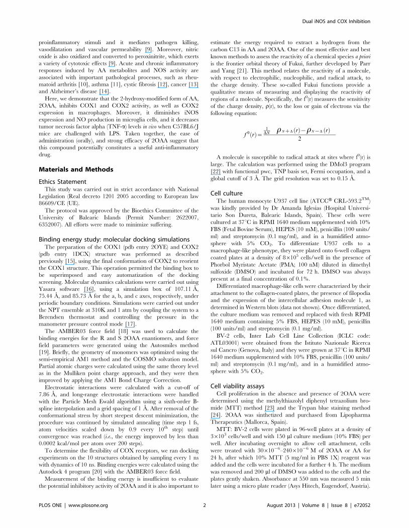
Background: Nonsteroidal anti-inflammatory drugs (NSAIDs) are a family of COX1 and COX2 inhibitors used to reduce thesynthesis of pro-inflammatory mediators. In addition, inflammation often leads to a harmful generation of nitric oxide.Efforts are being done in discovering safer NSAIDs molecules capable of inhibiting the synthesis of pro-inflammatory lipidmediators and nitric oxide to reduce the side effects associated with long term therapies.
Methodology/Principal Findings: The analogue of arachidonic acid (AA), 2-hydroxy-arachidonic acid (2OAA), was designedto inhibit the activities of COX1 and COX2 and it was predicted to have similar binding energies as AA for the catalytic sitesof COX1 and COX2. The interaction of AA and 2OAA with COX1 and COX2 was investigated calculating the free energy ofbinding and the Fukui function. Toxicity was determined in mouse microglial BV-2 cells. COX1 and COX2 (PGH2 production)activities were measured in vitro. COX1 and COX2 expression in human macrophage-like U937 cells were carried out byWestern blot, immunocytochemistry and RT-PCR analysis. NO production (Griess method) and iNOS (Western blot) weredetermined in mouse microglial BV-2 cells. The comparative efficacy of 2OAA, ibuprofen and cortisone in lowering TNF-aserum levels was determined in C57BL6/J mice challenged with LPS. We show that the presence of the –OH group reducesthe likelihood of 2OAA being subjected to H* abstraction in COX, without altering significantly the free energy of binding.The 2OAA inhibited COX1 and COX2 activities and the expression of COX2 in human U937 derived macrophages challengedwith LPS. In addition, 2OAA inhibited iNOS expression and the production of NO in BV-2 microglial cells. Finally, oraladministration of 2OAA decreased the plasma TNF-a levels in vivo.
Conclusion/Significance: These findings demonstrate the potential of 2OAA as a NSAID.
Citation: Lopez DH, Fiol-deRoque MA, Noguera-Salva MA, Teres S, Campana F, et al. (2013) 2-Hydroxy Arachidonic Acid: A New Non-Steroidal Anti-InflammatoryDrug. PLoS ONE 8(8): e72052. doi:10.1371/journal.pone.0072052
Editor: Matias A. Avila, University of Navarra School of Medicine and Center for Applied Medical Research (CIMA), Spain
Received May 8, 2013; Accepted July 7, 2013; Published August 27, 2013
Copyright: � 2013 Lopez et al. This is an open-access article distributed under the terms of the Creative Commons Attribution License, which permitsunrestricted use, distribution, and reproduction in any medium, provided the original author and source are credited.
Funding: This work was supported by grants BIO2010-21132 (PVE) and IPT-010000-2010-016 (XB) (Ministerio de Economıa y Competitividad, Spain), by Ajuts aGrups Competitius (Govern de les Illes Balears), and the Marathon Foundation. MAF-DR is supported by a fellowship from the Government of the Balearic Islands(Conselleria d’Educacio, Cultura i Universitat) and the European Social Fund. DHL and ST were supported by Torres Quevedo Contracts (Ministerio de Economıa yCompetitividad, Spain, and the European Social Fund). RJM is supported by a Marathon Foundation Fellowship. The funders had no role in study design, datacollection and analysis, decision to publish, or preparation of the manuscript.
Competing Interests: DHL and ST are employees of Lipopharma Therapeutics. This does not alter the authors’ adherence to all PLOS ONE policies on sharingdata and materials.
* E-mail: [email protected]
Introduction
Chemical modification of fatty acids is an experimental
approach used to inhibit cyclooxygenase 1 (COX1) and cycloox-
ygenase 2 (COX2) activity [1]. We rationally designed and
synthesized 2-hydroxy-arachidonic acid (2OAA), which contains a
hydroxyl group on the a-carbon of arachidonic acid (AA), a
modification that was designed to inhibit the AA pro-inflammatory
pathway by interacting with the active site of COX1 and COX2.
AA is the most abundant n-6 polyunsaturated fatty acid found in
the cell membrane [2], where it is stored. When phospholipase A2
(PLA2) is activated by different inflammatory stimuli, including
bacterial lipopolysaccharides (LPS), cytokines and allergens [3,4],
AA is released into the cytosol and then metabolized by
cyclooxygenases (COXs), lipoxygenases (LOXs) and cytochrome
P450 [2]. Two major COXs isoforms have been described, the
constitutive (COX1) and the inducible (COX2) [5]. When
metabolized by COX1 and COX2, AA is converted by a variety
of downstream enzymes (isomerases, reductases and synthases),
including the prostaglandins (PGs) and thromboxanes (TXs). The
LOX pathway metabolizes the AA to hydroxyacids and leukotri-
enes and the P450 pathway to epoxyeicosatrienoic acids or 20-
hydroxyeicosatetraenoic acid. Finally the AA-derived bioactive
products are released from activated cells to modulate the
inflammatory response [6,7].
An additional key process in inflammation is the synthesis of
nitric oxide (NO) by one of the three nitric oxide synthase (NOS)
isoenzymes: the constitutive, neuronal and inducible (iNOS)
isoforms [8]. The iNOS isoform is upregulated by a variety of
PLOS ONE | www.plosone.org 1 August 2013 | Volume 8 | Issue 8 | e72052
proinflammatory stimuli and it mediates pathogen killing,
vasodilatation and vascular permeability [9]. Moreover, nitric
oxide is also oxidized and converted to peroxinitrite, which exerts
a variety of cytotoxic effects [9]. Acute and chronic inflammatory
responses induced by AA metabolites and NOS activity are
associated with important pathological processes, such as rheu-
matoid arthritis [10], asthma [11], cystic fibrosis [12], cancer [13]
and Alzheimer’s disease [14].
Here, we demonstrate that the 2-hydroxy-modified form of AA,
2OAA, inhibits COX1 and COX2 activity, as well as COX2
expression in macrophages. Moreover, it diminishes iNOS
expression and NO production in microglia cells, and it decreases
tumor necrosis factor alpha (TNF-a) levels in vivo when C57BL6/J
mice are challenged with LPS. Taken together, the ease of
administration (orally), and strong efficacy of 2OAA suggest that
this compound potentially constitutes a useful anti-inflammatory
drug.
Materials and Methods
Ethics StatementThis study was carried out in strict accordance with National
Legislation (Real decreto 1201 2005 according to European law
86609/CE (UE).
The protocol was approved by the Bioethics Committee of the
University of Balearic Islands (Permit Number: 2622007,
6352007). All efforts were made to minimize suffering.
Binding energy study: molecular docking simulationsThe preparation of the COX1 (pdb entry 2OYE) and COX2
(pdb entry 1DCX) structure was performed as described
previously [15], using the final conformation of COX2 to reorient
the COX1 structure. This operation permitted the binding box to
be superimposed and easy automatization of the docking
screening. Molecular dynamics calculations were carried out using
Yasara software [16], using a simulation box of 107.11 A,
75.44 A, and 85.73 A for the a, b, and c axes, respectively, under
periodic boundary conditions. Simulations were carried out under
the NPT ensemble at 310K and 1 atm by coupling the system to a
Berendsen thermostat and controlling the pressure in the
manometer pressure control mode [17].
The AMBER03 force field [18] was used to calculate the
binding energies for the R and S 2OAA enantiomers, and force
field parameters were generated using the Autosmiles method
[19]. Briefly, the geometry of monomers was optimized using the
semi-empirical AM1 method and the COSMO solvation model.
Partial atomic charges were calculated using the same theory level
as in the Mulliken point charge approach, and they were then
improved by applying the AM1 Bond Charge Correction.
Electrostatic interactions were calculated with a cut-off of
7.86 A, and long-range electrostatic interactions were handled
with the Particle Mesh Ewald algorithm using a sixth-order B-
spline interpolation and a grid spacing of 1 A. After removal of the
conformational stress by short steepest descent minimization, the
procedure was continued by simulated annealing (time step 1 fs,
atom velocities scaled down by 0.9 every 10th step) until
convergence was reached (i.e., the energy improved by less than
0.0002 kcal/mol per atom over 200 steps).
To determine the flexibility of COX receptors, we ran docking
experiments on the 10 structures obtained by sampling every 1 ns
with dynamics of 10 ns. Binding energies were calculated using the
Autodock 4 program [20] with the AMBER03 force field.
Measurement of the binding energy is insufficient to evaluate
the potential inhibitory activity of 2OAA and it is also important to
estimate the energy required to extract a hydrogen from the
carbon C13 in AA and 2OAA. One of the most effective and best
known methods to assess the reactivity of a chemical species a priori
is the frontier orbital theory of Fukui, further developed by Parr
and Yang [21]. This method relates the reactivity of a molecule,
with respect to electrophilic, nucleophilic, and radical attack, to
the charge density. These so-called Fukui functions provide a
qualitative means of measuring and displaying the reactivity of
regions of a molecule. Specifically, the f0(r) measures the sensitivity
of the charge density, r(r), to the loss or gain of electrons via the
following equation:
f 0 rð Þ~1
DNrNzD rð Þ{rN{D rð Þ� �� �
2
A molecule is susceptible to radical attack at sites where f0(r) is
large. The calculation was performed using the DMol3 program
[22] with functional pwc, TNP basis set, Fermi occupation, and a
global cutoff of 3 A. The grid resolution was set to 0.15 A.
Cell cultureThe human monocyte U937 cell line (ATCCH CRL-593.2TM)
was kindly provided by Dr Amanda Iglesias (Hospital Universi-
tario Son Dureta, Balearic Islands, Spain). These cells were
cultured at 37uC in RPMI 1640 medium supplemented with 10%
FBS (Fetal Bovine Serum), HEPES (10 mM), penicillin (100 units/
ml) and streptomycin (0.1 mg/ml), and in a humidified atmo-
sphere with 5% CO2. To differentiate U937 cells to a
macrophage-like phenotype, they were plated onto 6-well collagen
coated plates at a density of 86105 cells/well in the presence of
Phorbol Myristate Acetate (PMA; 100 nM) diluted in dimethyl
sulfoxide (DMSO) and incubated for 72 h. DMSO was always
present at a final concentration of 0.1%.
Differentiated macrophage-like cells were characterized by their
attachment to the collagen-coated plates, the presence of filopodia
and the expression of the intercellular adhesion molecule 1, as
determined in Western blots (data not shown). Once differentiated,
the culture medium was removed and replaced with fresh RPMI
1640 medium containing 5% FBS, HEPES (10 mM), penicillin
(100 units/ml) and streptomycin (0.1 mg/ml).
BV-2 cells, Inter Lab Cell Line Collection (ICLC code:
ATL03001) were obtained from the Istituto Nazionale Ricerca
sul Cancro (Genova, Italy) and they were grown at 37uC in RPMI
1640 medium supplemented with 10% FBS, penicillin (100 units/
ml) and streptomycin (0.1 mg/ml), and in a humidified atmo-
sphere with 5% CO2.
Cell viability assaysCell proliferation in the absence and presence of 2OAA were
determined using the methylthiazolyl diphenyl tetrazolium bro-
mide (MTT) method [23] and the Trypan blue staining method
[24]. 2OAA was sinthetized and purchased from Lipopharma
Therapeutics (Mallorca, Spain).
MTT: BV-2 cells were plated in 96-well plates at a density of
36103 cells/well and with 150 ml culture medium (10% FBS) per
well. After incubating overnight to allow cell attachment, cells
were treated with 3061026–24061026 M of 2OAA or AA for
24 h, after which 10% MTT (5 mg/ml in PBS 1X) reagent was
added and the cells were incubated for a further 4 h. The medium
was removed and 200 ml of DMSO was added to the cells and the
plates gently shaken. Absorbance at 550 nm was measured 5 min
later using a micro plate reader (Asys Hitech, Eugendorf, Austria).
Dual iNOS and COX Inhibition
PLOS ONE | www.plosone.org 2 August 2013 | Volume 8 | Issue 8 | e72052
Trypan Blue staining: BV-2 cells were plated in 6-well plates at
densities of 36105 cells/well with 2 ml of culture medium (10%
FBS) per well. After incubating overnight to allow cell attachment,
cells were treated with 3061026–24061026 M of 2OAA or AA
for 24 h, Trypan blue staining was done as previously described
[24]. Briefly, 10 ml of sample (cell suspension) was mixed with
10 ml of trypan blue (Invitrogen), and pipetted into a CountessHchamber slide (Invitrogen) that was inserted in the CountessHAutomated Cell Counter (Invitrogen).
Assays of COX1 and COX2 inhibition in vitroThe effect of 2OAA and AA was assessed on purified COX1
and purified COX2 proteins using an in vitro cell free-system
inhibitor assay kit provided by Cayman Chemicals (Ann Arbor,
MI, USA). in accordance with the manufacturer directions. Both
assays quantifyed the levels of prostaglandin H2 (PGH2) produced
by purified COX1 or purified COX2 [25]. The results were
expressed as the percentage of COX1 or COX2 activity in the
presence or absence (control 100%) of 2OAA (25061026 M) or
AA (25061026 M).
Western blotting and protein quantificationTo quantify COX1 and COX2 protein, U937 cells were
differentiated in 6-well plates at a density of 86105 cells/well, and
incubated for 24 h in culture medium containing 5% FBS (6 ml/
well). Next, the culture medium was removed and replaced with
fresh medium containing 2OAA (12061026 M) for 1 h or
incubated with fresh culture medium alone (control). The cells
were then washed twice with PBS and the cells previously treated
with 2OAA were challenged for 6 h with LPS (62 ng/ml) plus
2OAA (12061026 M) in fresh medium, while untreated control
cells were challenged with LPS alone (62 ng/ml).
To study COX2 protein decay, U937 cells were differentiated
in 6-well plates as above and after removing the culture medium,
the cells were treated for 6 h with LPS (62 ng/ml) plus 2OAA
(12061026 M). After washing twice with PBS, the cells were then
treated for 2 h with cycloheximide (CHX, 5061026 M) and NS-
398 (2061026 M, a COX2-specific inhibitor) or MG-132
(2061026 M, a 26 S proteasome inhibitor) in the presence of
2OAA (12061026 M).
To quantify the iNOS protein, BV-2 cells were plated in 6-well
plates as described above, and the culture medium was then
removed and replaced with fresh medium containing 2OAA
(12061026 M) for 1 h (or with fresh culture medium alone,
control). The cells were then washed twice with PBS and 2OAA-
treated cells were challenged for 24 h with LPS (1 mg/ml) plus
2OAA (12061026 M) dissolved in fresh medium. Untreated
control cells were incubated for 24 h in the presence or absence of
LPS (1 mg/ml) dissolved in fresh medium.
At the end of the treatments, both U937 and BV-2 cells were
washed twice with PBS and harvested with a rubber policeman in
200 ml of protein extraction buffer (10 mM Tris-HCl buffer
[pH 7.4], 50 mM NaCl, 1 mM MgCl2, 2 mM EDTA, 1% SDS,
5 mM iodoacetamide and 1 mM PMSF). The cell suspensions
were sonicated for 10 s at 50 W using a Braun Labsonic U
sonicator and 20 ml aliquots were removed for protein quantifi-
cation (bicinchoninic acid method; Pierce-Thermo Fisher Scien-
tific Inc., Roskilde, Denmark). The remaining suspension
(,180 ml) was mixed with 20 ml of 106 electrophoresis loading
buffer (120 mM Tris-HCl [pH 6.8], 4% SDS, 50% glycerol, 0.1%
bromophenol blue, 10% b-mercaptoethanol) and boiled for 5 min.
The proteins were fractionated on 9.5% polyacrylamide gels
(SDS-PAGE: 15-well and 1.5 mm thick) and transferred to
nitrocellulose membranes (Whatman protranH, Dassel, Germany)
that were then blocked for 1 h at room temperature in PBS
containing 5% non-fat dry milk, 0.5% bovine serum albumin and
0.1% Tween 20 (blocking solution). The membranes were probed
overnight at 4uC with one of the following primary antibodies
diluted in blocking solution: monoclonal anti-COX1/anti-COX2
(1:800; Biotechnology Inc, CA, USA), anti-iNOS (1:4,000; BD
Biosciences, Franklin Lakes, NJ, USA), or anti-a-tubulin (1:10,000;
Sigma-Aldrich, St. Louis, MO, USA).
After removing the primary antibody, the membranes were
washed three times for 10 min with PBS and incubated for 1 h at
room temperature in fresh blocking solution containing horserad-
ish peroxidase-linked goat anti-mouse IgG (1:2000; Amersham
Pharmacia). Immunoreactivity was detected by Enhanced Chemi-
luminescence (ECL; Western Blot Detection system, Amersham
Pharmacia), followed by exposure to ECL hyperfilm (Amersham
Pharmacia). The films were scanned at 600 dpi and quantified
using Foto Look 32 software (Agfa Gevaert, Leverkusen,
Germany), analyzing the images with Quantity One software
(Bio-Rad, Hercules, CA, USA). The concentration of a given
protein was normalized to the a-tubulin content of the same
sample.
Quantitative Real Time-Polymerase Chain Reaction (qRT-PCR) and mRNA quantification
To quantify COX2 mRNA, U937 cells were differentiated in 6-
well plates as described above and the culture medium was then
replaced with fresh medium containing 2OAA (12061026 M) for
1 h, or incubated with fresh culture medium alone (control). The
cells were washed twice with PBS, and those previously treated
with 2OAA were challenged for 6 h with LPS (62 ng/ml) plus
2OAA (12061026 M) in fresh medium, while control cells were
challenged for 6 h with LPS alone (62 ng/ml). Total cellular RNA
was extracted from the cells using the RNeasy Mini kit in
combination with the RNase-Free DNase kit (Qiagen, Hilden
Germany), following the manufacturer’s instructions, and the total
amount and purity of RNA was measured using a Nanodrop 1000
spectrophotometer (ThermoFisher Scientific, Waltham, MA) at
260 and 280 nm. The integrity of the RNA was tested by
electrophoresis on 2% agarose gel and visualized by ethidium
bromide staining. Reverse transcription was carried out in a final
volume of 20 ml using the Transcriptor First Strand cDNA
Synthesis Kit (Roche, Mannheim, Germany) in a thermal cycler
(Eppendorf Master Cycler Gradient). The RNA samples (1 mg)
were mixed with oligonucleotides as primers (1 ml; 500 mg/ml) and
made up to a final volume of 13 ml with H2O. The samples were
then incubated at 65uC for 10 min and then the tubes were chilled
quickly on ice. Next, a reaction mix was added containing 56first-
strand buffer (4 ml), 10 mM dNTPmix (dGTP, dCTP, dATP, and
dTTP; 2.5 ml), Protector Rnase inhibitor (0.5 ml; 40 units/ml) and
the reverse transcriptase (0.5 ml; 20 units/ml). The reaction
mixtures were incubated at 25uC for 10 min, 55uC for 30 min,
and 85uC for 5 min, and the cDNA samples obtained were stored
at 220uC.
For PCR amplification, primers were designed based on specific
human COX2 sequences obtained from the SDSC Biology
Workbench Program: forward primer, 59-TGA GCA TCT
ACG GTT TGC TG-3; reverse primer, 59- TGC TTG TCT
GGA ACA ACT GC-39. As an endogenous control, the
expression of b-actin was quantified in parallel using the forward
primer 59-GCG GGA AAT CGT GCG TGA CAT T-39 and the
reverse primer 5-CTA CCT CAA CTT CCA TCA AAG CAC-
39. RT-qPCR amplifications were carried out in a Step One Plus v
2.0 thermal cycler (Applied Biosystems) using the SYBRH Premix
Ex Taq kit (Perfect Real Time, Takara), which contains TaKaRa
Dual iNOS and COX Inhibition
PLOS ONE | www.plosone.org 3 August 2013 | Volume 8 | Issue 8 | e72052
Ex TaqTM HS, dNTP mixture, Mg2+, SYBRH, Green I and
ROXTM Reference Dye. Thermal cycling was preceded by an
initial denaturation step at 95uC for 30 s. and DNA amplification
and fluorescence quantification was performed over 35 cycles with
a denaturation step at 95uC for 5 s, followed by an annealing and
extension step at 60uC for 34 s. Fluorescence quantification was
carried out after each DNA extension step, and StepOne software
(v2.0) was used to analyze the data, also producing a melting curve
analysis of the final products.
The ratio of COX2 expression to that of b-actin RNA (whose
expression is not modulated by 2OAA) was expressed as ddCT
values (as a percentage) using the following formula: ddCT = EX(Ctc-Ctx)/E Bact (Ctc-Ctx), where Efficiency (E) = 10 (21/m), and
(m) = slope of the graph formed by Ct values of mRNA vs the
logarithm (log) of its concentration (ng/ml). Finally, this value was
used to calculate the relative expression of COX2 in 2OAA-
treated cells with respect to the untreated control cells.
COX2 immunofluorescence detectionU937 cells were differentiated for 24 h in 8-well collagen-coated
plates containing 750 ml of culture medium (5% FBS) at a density
of 96104 cells/well. After incubating in the presence or absence
(control) of 2OAA (12061026 M) for 1 h, the cells were washed
twice with PBS and 2OAA-treated cells were challenged with LPS
(62 ng/ml) plus 2OAA (12061026 M) for 18 h, while control cells
were incubated for 18 h in the presence or absence of LPS alone
(62 ng/ml).
The cells were washed twice with PBS, twice with phosphate
buffer (PB; 0.1 M), and fixed with 4% paraformaldehyde for
30 min. Cells were then washed once with PB (0.1 M), twice with
PBS, and permeabilized with Triton X-100 (0.1%) for 5 min.
Subsequently the cells were washed with PBS three times and
incubated with 10% FBS in PBS for 3 h at room temperature,
followed by overnight incubation at 4uC in the presence of
monoclonal anti-COX2 (1:50; Biotechnology, INC, CA, USA)
diluted in PBS buffer supplemented with 10% FBS. The cells were
then washed three times with PBS and incubated for 1 h at room
temperature with Alexa Fluor 488-labelled goat anti-mouse IgG
antibody (1:200; Invitrogen, Molecular Probes; excitation at
488 nm and emission at 510–550 nm). Finally, cells were washed
twice with PBS, incubated with 500 nM propidium iodide for
4 min to stain the nuclei and washed once again with PBS. Images
were acquired using a Leica TCS SP2 spectral confocal
microscope, with 6306 optical magnification and 86 digital
magnification (,50006 total magnification).
Nitric oxide determinationBV-2 cells were plated in 96-well plates at a density of 26104
cells/well in 200 ml of culture medium (10% FBS) and after 24 h,
the culture medium was replaced with fresh medium alone
(control) or containing 2OAA (5061026, 12061026 and
24061026 M). After 1 h, the cells were washed twice with PBS
and 2OAA-treated cells were challenged for 24 h with LPS (1 mg/
ml) plus 2OAA (12061026 M), while control cells were incubated
for 24 h in the presence or absence of LPS (1 mg/ml) alone. The
NO produced by the cells was determined by assaying the amount
of nitrite generated using Griess reagent [26]. Briefly, aliquots of
the conditioned medium were mixed with an equal volume of 1%
sulfanilamide in 5% phosphoric acid and 0.1% N-1-naphthylene-
diamine-dihydrochloride in water. After incubating for 10 min at
room temperature, the absorbance of the reaction mixture at
540 nm was determined in a microtiter plate reader, using sodium
nitrite diluted in culture medium at concentrations of 1.5–25 mM
to generate a standard curve.
Determination of TNF-a serum levelsWe measured the effect of oral 2OAA administration on TNF-a
serum levels in a model of transient endotoxemia: C57BL6/J mice
(Charles River, Paris, France) challenged with LPS. The protocol
used in this study was approved by the Animal Ethics Committee
of the University of the Balearic Islands. C57BL6/J mice (15–16 g;
6–8 weeks old) were fed a standard laboratory diet with ad libitum
access to water and they were maintained on a 12-h light-dark
cycle at 22uC. The mice were randomly distributed into 5 groups
of 5 animals, of which three groups were administered 2OAA
dissolved in PBS (50, 200 or 500 mg/kg, p.o.), while the remaining
two groups received PBS (vehicle) alone (control). Doses of 2OAA
in mice were calculated following the guidelines of the Food and
Drug Administration of the U.S.A (FDA) [27]. After 90 min, the
mice were challenged with LPS dissolved in PBS (20 mg, i.p.),
while one of the control groups received an i.p. injection of PBS
alone. After establishing the optimal dose of 2OAA required to
decrease TNF-a serum level, we compared its efficacy with that of
cortisone and ibuprofen. Four groups of mice (5 mice per group)
were treated with 2OAA (500 mg/kg, p.o.), AA (500 mg/kg, p.o.),
or with therapeutic doses of oral ibuprofen (12.5 mg/kg, p.o.) or
cortisone (7.5 mg/kg, i.p.), while another two groups (5 mice per
group) received PBS alone (control groups). After 90 min the mice
were challenged with LPS in PBS (20 mg, i.p.), while one of the
control groups received an i.p. injection of PBS alone. Three hours
later the mice were sacrificed by decapitation, their blood was
collected and the serum separated by centrifugation, and the TNF-
a levels were determined by ELISA (Invitrogen, Barcelona, Spain)
according to the manufacturer’s instructions.
Statistical analysisThe results are expressed as the mean 6 SEM values of at least
three independent experiments. Statistical analyses were per-
formed using the Student’s t test (GraphPad Prism version 5.00 for
Windows) and the level of statistical significance was set at p,0.05.
Results
Computational simulations based on molecular dockingWe compared the binding energy of AA and 2OAA to COX1
and COX2 using computational simulations based on molecular
docking. For COX1, the binding energies of AA and 2OAA
enantiomers (R2OAA and S2OAA) were very similar (Table 1).
The two carboxyl oxygens (O1 and O2) of AA established
hydrogen bonds with Arg 120, and AA exhibited close hydropho-
bic contact with Phe 205, Val 344 and Tyr 348 (Fig. 1A). The
orientation of S2OAA (as well as R2OAA, which is not
represented in the figure) was very similar to that in AA, with
the oxygens O1 and O2 occupying the same positions (Fig. 1B).
The hydroxyl oxygen (O*) of R2OAA and S2OAA formed a
hydrogen bond with Glu 524 of COX1, although this favorable
interaction was counterbalanced by a distortion of the carbon
backbone (the RMSD between AA and S2OAA was 1.39 A). The
S enantiomer exhibited a better global interaction with the binding
site and the binding energy was slightly higher than that of AA. In
summary, the carboxyl groups of the inhibitors were essentially
superimposable, and only modest differences in the binding of AA,
R2OAA and S2OAA were detected (Table 1).
For COX2, the binding energies of the 2OAA enantiomers,
R2OAA (11.09 kcal/mol) and S2OAA (10.93 kcal/mol), were
higher than that of AA (10.25 kcal/mol: Table 1), with a higher
degree of hydrogen bonding between 2OAA and the receptor.
The carboxyl group of AA was coordinated with Arg 120 via one
hydrogen bond (Fig. 1C), while both R2OAA and S2OAA
Dual iNOS and COX Inhibition
PLOS ONE | www.plosone.org 4 August 2013 | Volume 8 | Issue 8 | e72052

Dual iNOS and COX Inhibition
PLOS ONE | www.plosone.org 5 August 2013 | Volume 8 | Issue 8 | e72052

exhibited 5 hydrogen bonds, two of which were severely distorted
(Fig. 1D).
Interestingly, in COX2 the O* oxygen of 2OAA occupied the
position of O1 of AA (Fig. 1C and 1D), while in an analogous
manner, O1 of R2OAA and S2OAA occupied the position of the
AA O2. Finally, O2 of R2OAA and S2OAA was free to form a
hydrogen bond with Tyr 355.
The map of the Fukui radical function for the total electron
density (Fig. 1E) clearly indicates the more favorable sites for
radical attack and the arrows highlight C13, the carbon involved
in H* abstraction in COX. Thus, it can be thus concluded that the
–OH group in C2 reduces the likelihood of 2OAA being subjected
to enzymatic alteration by COX isozymes.
ToxicityTo determine the toxicity of AA and 2OAA, we measured the
effect of both compounds on BV-2 microglial cell viability by the
MTT assay and by the Trypan blue staining method. Cells were
incubated for 24 h in the presence or absence (control) of AA or
2OAA (12061026 or 24061026 M). BV-2 microglial cell viability
was inhibited by AA, reaching ,25% (MTT, Fig. 2A) and ,24%
(Trypan blue, Fig. 2B) inhibition at 12061026 M and ,75%
(MTT, Fig. 2A) and ,72% (Trypan blue, Fig. 2B) inhibition at
24061026 M. By contrast, 2OAA had no negative effect on cell
viability at the same concentrations (Fig. 2C and 2D), indicating
that the hydroxyl group present in 2OAA attenuates the toxicity
exerted by the natural fatty acid AA and suggesting that 2OAA
has no toxic effects at therapeutic doses.
2OAA inhibits COX1 and COX2 activityWe determined the effect of AA and 2OAA on purified COX1
and purified COX2 activity by measuring PGH2 production in an
in vitro quantitative cell-free assay [25]. While 2OAA
(25061026 M) exerted a dramatic inhibitory effect on COX1
activity, and a marked and significant inhibition of COX2 activity
(Fig. 3A and 3B), AA did not significantly affect COX1 or COX2
activity at the same concentration (Fig. 3A and 3B).
2OAA downregulates COX2 expression in LPS-stimulatedU937 cells
To determine the effect of 2OAA administration on the
expression of both COX isoforms, differentiated U937 macro-
phage like cells were challenged with LPS (62 ng/ml; 6 h) in the
presence or absence of 2OAA (12061026 M; 6 h), and COX
isoform expression was evaluated in Western blots. As described
previously, LPS markedly increased the expression of the inducible
COX2 isoform without affecting that of the constitutive COX1
isoform (Fig. 4A) [28]. In LPS-challenged cells, treatment with
Figure 1. Computational simulations based on molecular docking. A. AA in the COX1 binding site. B. S2OAA in the COX1 binding site. Thetwo carboxyl oxygens (O1 and O2) of AA establish hydrogen bonds with Arg 120 and they have close hydrophobic contacts with Phe 205, Val 344and Tyr 348. The orientation of S2OAA is very similar to that of AA, with O1 and O2 occupying the same positions in both. The hydroxyl oxygen (O*)of S2OAA is hydrogen bound to Glu 524, although this favorable interaction is counterbalanced by a distortion of the carbon backbone. To facilitatevisual inspection of the interactions, the binding site is shaded in grey, the fatty acids are represented by sticks and balls, and only amino acids closerthan 3 A are shown. C. AA in the COX2 binding site. D. S2OAA in the COX2 binding site. The carboxylate group of AA is coordinated with Arg 2120 byone hydrogen bond, whereas R2OAA possesses five hydrogen bonds. The O* oxygen occupies the position of O1 of AA and in an analogous manner,O1 of S2OAA occupies the position of the AA O2. Finally, O2 of substituted arachidonic acid is free to hydrogen bond to Tyr 2355. The binding site isshaded in grey, fatty acids are represented by sticks and balls, and only amino acids closer than 3 A are shown. E. The Fukui function f0(r) is color-mapped onto the electron density isosurface with equal isovalues.doi:10.1371/journal.pone.0072052.g001
Table 1. Binding energy to COX isoforms.
Receptor Ligand Binding energy (kcal/mol)
COX1 AA 8.29
R2OAA 7.94
S2OAA 8.52
COX2 AA 10.25
R2OAA 11.09
S2OAA 10.93
doi:10.1371/journal.pone.0072052.t001
Figure 2. BV2 cell viability is inhibited by AA but not by 2OAA.BV2 mouse cells were exposed to 120 and 24061026 M of AA or 2OAAfor 24 h. Upper panel. Bar diagram showing the cell viability assayedwith A. MTT or B Trypan Blue by AA (120 and 24061026 M) ascompared with untreated control cells (100%; n = 6). Lower panel. Bardiagram showing no inhibitory effect on cell viability assayed by C MTTor D Typan Blue by 2OAA (120 and 24061026 M) as compared withuntreated control cells (100%: ** p,0.01, *** p,0.001; n = 6).doi:10.1371/journal.pone.0072052.g002
Dual iNOS and COX Inhibition
PLOS ONE | www.plosone.org 6 August 2013 | Volume 8 | Issue 8 | e72052

2OAA (12061026 M, 6 h) resulted in a marked reduction in
COX2 protein, while those of COX1 remained unchanged
(Fig. 4A). To further study the effect of 2OAA on COX2
expression, differentiated U937 cells were challenged for longer
with LPS (62 ng/ml; 18 h) in the presence or absence of 2OAA
(12061026 M; 18 h) and they were analyzed by confocal
microscopy using an anti-COX2 antibody. After stimulation with
LPS, the cells exhibited characteristic perinuclear localization of
the induced COX2, although in the presence of 2OAA there was a
marked reduction in COX2 expression (Fig. 4B).
We also evaluated the effect of 2OAA on COX2 mRNA
expression in differentiated U937 macrophages challenged with
LPS (62 ng/ml; 6 h). As expected, LPS markedly increased COX2
mRNA expression and despite its inhibitory effect on COX2
protein expression, exposure to 2OAA (12061026 M, 6 h) failed
to downregulate COX2 mRNA, suggesting the existence of a post-
translational regulatory mechanism (Fig. 5A). It was previously
demonstrated that AA downregulates COX2 expression by
inducing the degradation of COX2 protein via two distinct
mechanisms: ubiquitination and degradation through the ubiqui-
tin-proteasome system; and less well understood mechanism
triggered by the binding of AA to COX2, known as suicide
inactivation [29]. To study these proteolytic mechanisms, differ-
entiated U937 macrophage-like cells challenged with LPS (62 ng/
ml; 6 h) were treated for 2 h with the ribosome inhibitor CHX
(5061026 M), the selective COX2 inhibitor NS-398 (2061026 M)
or the 26S proteasome inhibitor MG-132 (2061026 M), both in
the presence or absence of 2OAA. LPS markedly induced the
expression of the inducible COX2 protein as evident in Western
blots (Fig. 5B), whereas treatment with 2OAA (12061026 M, 2 h)
provoked a marked reduction in COX2 protein. When 2OAA
(12061026 M, 2 h) was combined with either NS-398
(2061026 M) or MG-132 (2061026 M), COX2 protein expres-
sion recovered (Fig. 5B), indicating that both suicide inactivation
and proteasome-dependent proteolysis are triggered by 2OAA.
2OAA inhibits NO production in LPS-stimulated BV-2 cellsTo investigate the effect of 2OAA on the production of NO,
BV-2 cells were challenged with LPS (1 mg/ml; 24 h) in the
presence or absence of 2OAA at different concentrations
(5061026, 12061026 and 24061026 M; 24 h). NO production
was assessed by quantifying nitrite accumulation in the culture
medium (the final stable product of nitric oxide) using the Griess
reagent [26]. As expected, LPS induced a significant increase in
NO production, although 2OAA (5061026, 12061026 and
Figure 3. 2OAA inhibits COX1 and COX2 activity. Bar diagramshowing the inhibition of A. COX1 or B. COX2 activity determined bymeasuring PGH2 production in the presence of 2OAA (25061026 M)and AA (25061026 M), and compared with untreated control cells(100%: ** p,0.01, *** p,0.001 with respect to controls; { p,0.05, {{{p,0.001 with respect to AA; n = 6).doi:10.1371/journal.pone.0072052.g003
Figure 4. 2OAA decreases LPS-induced COX2 protein levels in differentiated human U937 cells challenged with LPS. A. Arepresentative immunoblot and bar diagram showing the effect of 2OAA (12061026 M; 6 h) on COX1/COX2 expression in differentiated U937 cellspreviously challenged with LPS (62 ng/ml; 6 h). Results are expressed as the COX2/COX1 ratio in untreated cells in the presence of LPS (100%: ***p,0.001; n = 6). Expression of the constitutive COX1 isoform was unaffected by either LPS or 2OAA treatment. B. Confocal micrographs showing theabsence of COX2 (green) expression (left panel) in unchallenged differentiated human U937 cells, the characteristic perinuclear induction of COX2(green) in LPS-challenged (62 ng/ml; 18 h) differentiated human U937 cells (middle panel), and the inhibition of COX2 expression produced byexposing these cells to 2OAA (12061026 M; 18 h). Cell nuclei were stained with propidium iodide (red).doi:10.1371/journal.pone.0072052.g004
Dual iNOS and COX Inhibition
PLOS ONE | www.plosone.org 7 August 2013 | Volume 8 | Issue 8 | e72052

24061026 M) diminished the LPS-induced production of NO in a
concentration-dependent manner (Fig. 6A).
2OAA downregulates iNOS expression in LPS-stimulatedBV-2 cells
We tested the effect of 2OAA on the expression of iNOS in BV-
2 cells challenged with LPS (1 mg/ml; 24 h) and in Western blots,
it was evident that exposure to LPS alone markedly increased the
expression of iNOS compared to that seen in unstimulated cells
(Fig. 6B). By contrast, the presence of 2OAA (120610-6 M; 24 h)
significantly dampened the expression of iNOS protein induced by
LPS stimulation (Fig. 6B).
2OAA reduced the serum TNF-a produced in LPS-challenged mice
To study the in vivo efficacy of 2OAA in a mouse model of
inflammation, we orally administered 2OAA (50 to 500 mg/kg) to
C57BL6/J mice 90 min before they received a LPS challenge
(20 mg/g, i.p.). LPS induced a marked and significant increase in
serum TNF-a levels, which was attenuated by 2OAA treatment in
a dose-dependent manner (Fig. 7A). This effect of 2OAA in vivo
was compared with that of cortisone and ibuprofen and we found
that the anti-inflammatory effect of 2OAA in mice (reflected in an
attenuation of the LPS-induced increase in TNF-a) was signifi-
cantly greater than that of ibuprofen and similar to that of
cortisone, a steroid compound at therapeutic doses (Fig. 7B).
Discussion
Understanding protein structure and structure-function rela-
tionships has provided significant insight into the molecular bases
of protein-ligand interactions [30,31], also forming the basis for
the rational design of drugs to treat human diseases with unmet
clinical needs [32]. The goal of the present study was to rationally
design a nonsteroidal anti-inflammatory drug (NSAID) with
similar potency to that of steroid compounds but that lacked the
significant side-effects of these drugs. For this purpose, we first
tested AA (the main COX substrate) analogues using computer
assisted tools, then comparing the potential toxicity and efficacy of
Figure 5. Proteolysis of COX2 by 2OAA. A. Bar diagram showingthe upregulation of COX2 mRNA in differentiated U937 cells challengedwith LPS (62 ng/ml). 2OAA (12061026 M; 6 h) failed to downregulateCOX2 mRNA levels despite its inhibitory effect on COX2 protein. B.Representative immunoblot showing COX2 protein in the presence orabsence of the general protein synthesis inhibitor CHX (5061026 M),the chemical blocker of the COX2 active site NS-398 (2061026 M), andthe 26S proteasome inhibitor MG132 (2061026 M) plus 2OAA(12061026 M). Constitutive COX1 protein expression is shown as aWestern blot loading control.doi:10.1371/journal.pone.0072052.g005
Figure 6. 2OAA attenuates the increase in iNOS protein levelsand NO production induced by LPS in BV2 murine microglialcells. A. Bar diagram showing the dose-dependent effect of 2OAA (50,120 and 24061026 M; 24 h) on NO production (measured by the Griessassay: see Materials and Methods) by BV2 mouse microglial cellspreviously challenged with LPS (1 mg/ml; 24 h). Results are expressedrelative to the NO production in untreated cells in the presence of LPS(100%: ** p,0.005, *** p,0.001; n = 6). B. A representative immunoblotand bar diagram showing the effect of 2OAA (12061026 M; 24 h) oniNOS expression in BV2 mouse microglial cells previously challengedwith LPS (1 mg/ml; 24 h). Results are expressed relative to the NOproduced by untreated cells maintained in the presence of LPS (100%:*** p,0.001; n = 6).doi:10.1371/journal.pone.0072052.g006
Figure 7. 2OAA attenuates the LPS-induced increase in serumTNF-a levels in C57BL6/J mice. A. Bar diagram showing the dose-dependent inhibition of serum TNF-a by 2OAA (50, 200 and 500 mg/kg)in LPS-challenged C57BL6/J mice (100%: *** p,0.001 with respect tothe LPS-challenged controls). B. Bar diagram showing the inhibitoryeffects of cortisone (12.5 mg/kg; Co), ibuprofen (7.5 mg/kg; Ib) and2OAA (500 mg/kg) on serum TNF-a in LPS-challenged C57BL6/J mice(*** p,0.001 with respect to controls; { p,0.05 with respect to Ib).doi:10.1371/journal.pone.0072052.g007
Dual iNOS and COX Inhibition
PLOS ONE | www.plosone.org 8 August 2013 | Volume 8 | Issue 8 | e72052
2OAA with that of AA in cellular and animal models. Our
molecular docking simulations predicted similar binding energies
for 2OAA and AA to COX1, and an enhanced interaction
between 2OAA and COX2 than that of AA. This suggests that
2OAA competes with AA to bind to COX isoforms and therefore,
that it interferes with their enzymatic activities to inhibit the
synthesis of pro-inflammatory mediators. In this context, the
development of COX inhibitors represents a landmark in the
evolution of NSAIDs [33]. However, AA derivatives are not
usually considered potential anti-inflammatory compounds, pos-
sibly because their structure is too similar to that of COX
substrates and products. In a cell, AA is metabolized by both
COX1 and COX2, is converted into pro-inflammatory PGs and
TXs [6,7]. By contrast, we propose that 2OAA is a potent anti-
inflammatory compound with low toxicity and acting as a COX
inhibitor, probably due to its similarity to the natural compound.
While AA (24061026 M) compromised microglial BV-2 cell
viability by 75% as shown by the MTT and Trypan blue staining
methods no toxic effects on cell growth were observed for 2OAA
under identical experimental conditions, indicating that the 2-
hydroxylation of AA attenuates its toxic effects.
We compared the effect of AA and this non-toxic analogue on
purified COX1 or purified COX2 activities by measuring PGH2
production in a cell-free system. Although the binding energies
predicted by the simulations of molecular docking to COX1 and
COX2 were very similar, in vitro 2OAA appeared to inhibit COX1
activity more strongly than AA. This might reflect the fact that the
binding of a ligand in a catalytically competent orientation is
necessary but not sufficient to induce enzymatic catalysis. Among
the initial steps in the formation of PGH2, a tyrosine is oxidized to
a free radical before catalysis can begin. The subsequent
abstraction of the AA 13-pro-S hydrogen by Tyr-385 results in a
pentadienyl radical, centered on C11, C13 and C15. In addition,
calculation of the Fukui function indicates that the presence of the
hydroxyl group reduces the potential reactivity of 2OAA. This
observation is consistent with the longer period over which 2OAA
remains at the active site of the COX enzymes and it is in
agreement with the inhibition observed.
To study the effect of 2OAA on the expression of the inducible
COX2, we used human U937 monocytes differentiated to
macrophage-like cells, which were stimulated with LPS to simulate
inflammation-derived COX2 overexpression. U937-derived mac-
rophages overexpress COX2 protein after LPS stimulation [28],
yet 2OAA administration decreases COX2 expression without
affecting constitutive COX1 expression. Thus 2OAA not only
inhibits the in vitro activity of both COX1 and COX2 but it also
significantly reduces COX2 levels while maintaining COX1
protein expression. Previous studies demonstrated that COX2
protein can be ubiquitinated and degraded by the 26 S
proteasome, a process that involves exit of the protein from the
ER via the ER-associated degradation system [29]. Another
mechanism that appears to be independent of the proteasome
system (suicide inactivation) involves the degradation of the
enzyme after the binding of its natural substrate [6]. In the
present study, we demonstrate that both 26S proteasome
inhibition (MG-132) and COX2 blockade at the active site (NS-
398) prevent proteolytic degradation of COX2. These results
suggest that both the proteasome and suicide inactivation
pathways are involved in 2OAA-induced COX2 degradation.
This anti-inflammatory mode of action, involving COX2 prote-
olysis, partially explains the potent effect of 2OAA. While this
work is focused on the effect of 2OAA on COXs inhibition, it is
conceivable that 2OAA could also have a variety of effects at other
different levels. For example, membrane AA could be displaced by
2OAA decreasing the concentration of AA available for PLA2. In
this context, 2OAA could also bind and interfere with the activity
of PLA2 affecting the release of stored AA which is the rate-
limiting step for icosanoid generation [2,4]. Another possibility not
explored in this work, is the possible effect of 2OAA on LOXs
activities and expression. Of particular interest would be the study
of the effect of 2OAA on 5LOX due to its important role on the
synthesis of hydroxyacids and leukotrienes implicated in inflam-
matory and allergic disorders.
Microglial cells are the resident macrophage-like cells of the
central nervous system and they are broadly implicated in
neuronal survival, innate immunity, microbial infection and brain
damage. Microglial over-activation (excessive production of PGs,
superoxide, NO and cytokines) can lead to inflammatory
neuropathologies [34]. Since iNOS expression can be inhibited
by natural lipids [35] and by modified lipids, such as oleanolic
acid-cyano-derivatives [36], we investigated whether 2OAA also
inhibited NO synthesis. Stimulation of microglial BV-2 cells with
LPS increased NO production and iNOS expression, as described
previously [37], yet these effects were attenuated by 2OAA in a
dose dependent manner. These results suggest that 2OAA inhibits
the production of NO derivatives such as peroxynitrites, minimiz-
ing the damage to proteins by oxidation. The AA, COX2 and
iNOS pathways are highly interconnected, with NO stimulating
both PLA-2 and COX2 activity, regulating AA release, and
promoting eicosanoid and PG synthesis [38]. Thus, 2OAA is an
anti-inflammatory AA derivative with a dual-mechanism of action,
simultaneously targeting excessive NO and PG synthesis. The
2OAA was originally designed to inhibit COX1 and COX2
activities. Our results showed that besides the inhibition of the
activity of both COXs isoforms, 2OAA induced COX2 protein
degradation and iNOS down-regulation. Therefore, further
studies are required to elucidate the cellular pathways and the
mechanisms that are affected by this new compound.
Finally we confirmed the in vivo efficacy of 2OAA by measuring
plasma of TNF-a levels. We selected a transient LPS-induced
endotoxemia model in C57BL6/J mice [39]. 2OAA readily
reduced the serum TNF-a levels in a dose-dependent manner,
producing a stronger effect than that of ibuprofen, similar to that
of cortisone at the therapeutic doses used. The FDA indicates that
the dose to be used in humans should be those used in mice
multiplied by 0.08 [27]. Thus, a dose of 500 mg/kg in mice would
correspond to 2.8 grams in humans (calculations made for a
weight of 70 kg), a dose that falls within the range of the daily
amount of NSAIDs currently prescribed (e.g., Ibuprofen). These
findings constitute a proof of relevance, demonstrating greater
efficacy of 2OAA than a commonly used NSAID like ibuprofen.
Furthermore, its comparable efficiency to cortisone and low
toxicity suggest that 2OAA may replace this steroidal anti-
inflammatory drug for certain treatments.
Author Contributions
Conceived and designed the experiments: DHL ST SP JAC PVE XB.
Performed the experiments: DHL MAFDR MANS ST FC SP JAC RJM.
Analyzed the data: DHL MANS SP PVE XB. Contributed reagents/
materials/analysis tools: DHL MAFDR MANS ST FC SP JAC. Wrote the
paper: DHL SP PVE XB.
Dual iNOS and COX Inhibition
PLOS ONE | www.plosone.org 9 August 2013 | Volume 8 | Issue 8 | e72052

References
1. Tronstad KJ, Berge K, Berge RK, Bruserud (2003) Modified fatty acids and
their possible therapeutic targets in malignant diseases. Expert Opin Ther
Targets 7: 663–667.
2. Brash AR (2001) Arachidonic acid as a bioactive molecule. J Clin Invest 107:
1339–1345.
3. Medzhitov R (2008) Origin and physiological roles of inflammation. Nature 454:
428–435.
4. Uozumi N, Kita Y, Shimizu T (2008) Modulation of Lipid and Protein
Mediators of Inflammation by Cytosolic Phospholipase A2a during Experimen-
tal Sepsis. J Immunol 181: 3558–3566.
5. Smith WL, DeWitt D, Garavito RM (2000) Cyclooxygenases: Structural,
Cellular, and Molecular Biology. Annu Rev Biochem 69: 145–182.
6. Fitzpatrick FA, Soberman R (2001) Regulated formation of eicosanoids. J Clin
Invest 107: 1347–1351.
7. Soberman RJ, Christmas P (2003) The organization and consequences of
eicosanoid signaling. J Clin Invest 111: 1107–1113.
8. Moncada S, Higgs A (1993) The L-arginine-nitric oxide pathway. N Engl J Med
329: 2002–2012.
9. Guzik TJ, Korbut R, Adamek-Guzik T (2003) Nitric oxide and superoxide in
inflammation and immune regulation. J Physiol Pharmacol 54: 469–487.
10. Scott DL, Wolfe F, Huizinga TWJ (2010) Rheumatoid arthritis. Lancet 376:
1094–1108.
11. Djukanovic R, Roche WR, Wilson JW, Beasley CR, Twentyman OP, et al.
(1990) Mucosal Inflammation in asthma. Am Rev Respir Dis 142: 434–457.
12. De Rose V (2002) Mechanisms and markers of airway inflammation in cystic
fibrosis. Eur Respir J 19: 333–340.
13. Mantovani A, Allavena P, Sica A, Balkwill F (2008) Cancer-related
inflammation. Nature 454: 436–444.
14. Eikelenboom P, Veerhuis R, Scheper W, Rozemuller AJM, vanGool WA, et al.
(2006) The significance of neuroinflammation in understanding Alzheimer’s
disease. J Neural Transm 113: 1685–1695.
15. Furse KE, Pratt DA, Porter NA, Lybrand TP (2006) Molecular dynamics
simulations of arachidonic acid complexes with COX-1 and COX-2: insights
into equilibrium behavior. Biochemistry 45: 3189–3205.
16. Krieger E, Darden T, Nabuurs S, Finkelstein A, Vriend G (2004) Making
optimal use of empirical energy functions: force-field parameterization in crystal
space. Proteins 57: 678–683.
17. Berendsen HJC, Postma JPM, van Gunsteren WF, Di Nola A, Haak JR (1984)
Molecular dynamics with coupling to an external bath. J Chem Phys 81: 3684–
3689.
18. Duan Y, Wu C, Chowdhury S, Lee MC, Xiong G, et al. (2003) A Point-Charge
Force Field for Molecular Mechanics Simulations of Proteins. J Comput Chem
24: 1999–2012.
19. Jakalian A, Jack DB, Bayly CI (2002) Fast, efficient generation of high-quality
atomic charges. AM1-BCC model: II. Parameterization and validation.
J Comput Chem 23: 1623–1641.
20. Morris GM, Goodsell DS, Halliday RS, Huey R, Hart WE, et al. (1998)
Automated Docking Using a Lamarckian Genetic Algorithm and Empirical
Binding Free Energy Function. J Comput Chem 19: 1639–1662.
21. Parr RG, Yang W (1989) Density-Functional Theory of Atoms and Molecules.
International series of monographs on chemistry 10. Oxford University Press.
New York.
22. Delley B (2000) From molecules to solids with the DMol3 approach. J Chem
Phys 113: 7756–7764.
23. Mosmann T (1983) Rapid colorimetric assay for cellular growth and survival:
application to proliferation and cytotoxicity assays. J Immunol Methods 65: 55–63.
24. Bowling SA, Clarke JD, Liu Y, Klessig DF, Dong X (1997) The cpr5 mutant ofArabidopsis expresses both NPR1-dependent and NPR1-independent resistance.
Plant Cell 9: 1573–84.
25. Gomes A, Fernandes E, Silva AMS, Pinto DCGA, Santos CMM, et al. (2009)Anti-inflammatory potential of 2-styrylchromones regarding their interference
with arachidonic acid metabolic pathways. Biochem Pharm 78: 171–177.26. Green LC, Wagner DA, Glogowski J, Skipper PL, Wishnok JS, et al. (1982)
Analysis of nitrate, nitrite, and [15N] nitrate in biological fluids. Anal Biochem
126: 131–138.27. Food & Drug Administration, USA website. Guidance for Industry. Estimating
the Maximum Safe Starting Dose in Initial Clinical Trials for Therapeutics inAdult Healthy Volunteers. Available: http://www.fda.gov/cder/guidance/
index.htm, http://www.fda.gov/downloads/Drugs/Guidances/UCM078932.pdf. Accessed 2013 Jul 16.
28. Barrios-Rodiles M, Tiraloche G, Chadee K (1999) Lipopolysaccharide
Modulates Cyclooxygenase-2 Transcriptionally and Posttranscriptionally inHuman Macrophages Independently from Endogenous IL-1ß and TNF-a.
J Immunol 163: 963–969.29. Mbonye UR, Yuan Ch, Harris CE, Sidhu RS, Song I, et al. (2008) Two Distinct
Pathways for Cyclooxygenase-2 protein degradation. J Biol Chem 283: 8611–
8623.30. Kendrew JC, Bodo G, Dintzis HM, Parrish RG, Wyckoff H, et al. (1958) A
three-dimensional model of the myoglobin molecule obtained by X-ray analysis.Nature 181: 662–666.
31. Perutz MF, Rossmann MG, Cullis AF, Muirhead H, Will G, et al. (1960)Structure of haemoglobin: a three-dimensional Fourier synthesis at 5.5-A
resolution obtained by X-ray analysis. Nature 185: 416–422.
32. Moore JM, Peattie DA, Fitzgibbon MJ, Thomson JA (1991) Solution structure ofthe major binding protein for the immunosuppressant FK506. Nature 351:248–
250.33. Rao P, Knaus EE (2008) Evolution of Nonsteroidal Anti-Inflammatory Drugs
(NSAIDs): Cyclooxygenase (COX) Inhibition and Beyond. J Pharm Pharmaceut
Sci 11: 81–110.34. Kim SU, de Vellis J (2005) Microglia in health and disease. J Neurosci Res 81:
302–313.35. Ohata T, Fukuda K, Takahashi M, Sugimura T, Wakabayashi K (1997)
Suppression of nitric oxide production in lipopolysaccharide-stimulatedmacrophage cells by omega 3 polyunsaturated fatty acids. Jpn J Cancer Res
88: 234–237.
36. Tran TA, McCoy MK, Sporn MB, Tansey MG (2008) The synthetictriterpenoid CDDO-methyl ester modulates microglial activities, inhibits TNF
production, and provides dopaminergic neuroprotection. J Neuroinflammation5: 14.
37. Wen J, Ribeiro R, Zhang YJ (2011) Specific PKC isoforms regulate LPS-
stimulated iNOS induction in murine microglial cells. J Neuroinflammation 8:38.
38. Xu L, Han C, Lim K, Wu T (2008) Activation of Cytosolic Phospholipase A2athrough Nitric Oxide-induced S-Nitrosylation. Involvement of inducible nitric
oxide synthase and cycloxygenase-2. J Biol Chem 283: 3077–3087.39. Eads D, Hansen RL, Oyegunwa AO, Cecil CE, Culver CA, et al. (2009)
Terameprocol, a methylated derivative of nordihydroguaiaretic acid, inhibits
production of prostaglandins and several key inflammatory cytokines andchemokines. J Inflamm (Lond) 6: 2.
Dual iNOS and COX Inhibition
PLOS ONE | www.plosone.org 10 August 2013 | Volume 8 | Issue 8 | e72052
Copyright © 2022 FDOKUMEN