Scattering of Stark-decelerated OH radicals with rare-gas atoms
Upload
independentCategory
view
1download
0
Structural comparisons of arachidonic acid-induced radicalsformed by prostaglandin H synthase-1 and -2
Ah-lim Tsaia,*, Gang Wua, Corina E. Roggeb, Jian-Ming Lüa, Sheng Pengc, Wilfred A. vander Donkc, Graham Palmerd, Gary J. Gerfene, and Richard J. KulmaczaaDepartment of Internal Medicine, University of Texas Health Science Center at Houston,Houston, TX 77030, United StatesbDepartment of Art Conservation, Buffalo State College, Buffalo, NY 14222, United StatescDepartment of Chemistry, University of Illinois at Urbana-Champaign, Urbana, IL 61801, UnitedStatesdDepartment of Biochemistry and Cell Biology, Rice University, Houston, TX 77005, United StateseDepartment of Physiology and Biophysics, Albert Einstein College of Medicine, Bronx, NY10461, United States
AbstractCyclooxygenase catalysis by prostaglandin H synthase (PGHS)-1 and -2 involves reaction of aperoxide-induced Tyr385 radical with arachidonic acid (AA) to form an AA radical that reactswith O2. The potential for isomeric AA radicals and formation of an alternate tyrosyl radical atTyr504 complicate analysis of radical intermediates. We compared the EPR spectra of PGHS-1and -2 reacted with peroxide and AA or specifically deuterated AA in anaerobic, single-turnoverexperiments. With peroxide-treated PGHS-2, the carbon-centered radical observed after AAaddition was consistently a pentadienyl radical; a variable wide-singlet (WS) contribution frommixture of Tyr385 and Tyr504 radicals was also present. Analogous reactions with PGHS-1produced EPR signals consistent with varying proportions of pentadienyl and tyrosyl radicals, andtwo additional EPR signals. One, insensitive to oxygen exposure, is the narrow singlet tyrosylradical with clear hyperfine features found previously in inhibitor-pretreated PGHS-1. The secondtype of EPR signal is a narrow singlet lacking detailed hyperfine features that disappeared uponoxygen exposure. This signal was previously ascribed to an allyl radical, but high field EPRanalysis indicated that ~90% of the signal originates from a novel tyrosyl radical, with a smallcontribution from a carbon-centered species. The radical kinetics could be resolved by globalanalysis of EPR spectra of samples trapped at various times during anaerobic reaction of PGHS-1with a mixture of peroxide and AA. The improved understanding of the dynamics of AA andtyrosyl radicals in PGHS-1 and -2 will be useful for elucidating details of the cyclooxygenasemechanism, particularly the H-transfer between tyrosyl radical and AA.
KeywordsPGHS radical; EPR; High-field EPR; Radical dynamics
© 2010 Elsevier Inc. All rights reserved.*Corresponding author. Division of Hematology, Department of Internal Medicine, University of Texas Health Science Center, 6431Fannin St., Houston, TX 77030, United States. Tel: +1 713 500 6771; fax: +1 713 500 6812. [email protected] (A.-L. Tsai).Appendix A. Supplementary dataSupplementary data to this article can be found online at doi:10.1016/j.jinorgbio.2010.11.012.
NIH Public AccessAuthor ManuscriptJ Inorg Biochem. Author manuscript; available in PMC 2012 March 1.
Published in final edited form as:J Inorg Biochem. 2011 March ; 105(3): 366–374. doi:10.1016/j.jinorgbio.2010.11.012.
NIH
-PA Author Manuscript
NIH
-PA Author Manuscript
NIH
-PA Author Manuscript

1. IntroductionThe two prostaglandin H synthase (PGHS) isozymes catalyze the first committed step ofprostanoid biosynthesis. Although the two isozymes of PGHS play very different roles inphysiology and pathology [1,2], they have quite similar structures: the cyclooxygenaseactive site residues of the two isozymes are almost superimposable, with a root mean squaredeviation of less than 0.4 Å [3–5].
Both isozymes have two catalytic activities: a cyclooxygenase that transforms AA to PGG2,and a peroxidase that converts PGG2 to PGH2 [6,7]. A branched-chain radical mechanismthat couples the two catalytic activities [8,9] has gained solid support from crystallographicand spectroscopic data [3–5,10,11]. The critical oxidant for cyclooxygenase catalysis in thismechanistic model is a peroxide-induced intermediate containing a tyrosyl radical.Mutagenic and crystallographic data identified Tyr385 (in ovine PGHS-1 numbering) as thelocation of the catalytic radical [3–5,10–12].
The EPR spectra of peroxide-treated PGHS-1 and -2 change over time, with wide-doubletsignals (WD1 and WD2 for PGHS-1 and -2, respectively) predominating initially and wide-singlet signals (WS1 and WS2 for PGHS-1 and -2, respectively) appearing later [13–15].The transition from WD to WS is much faster in PGHS-2 than in PGHS-1 [16]. These WSsignals are made up of the original WD and a narrow singlet spectrum (NS1 and NS2 forPGHS-1 and -2, respectively) [14]. Results from EPR studies of peroxide generated radicalsin PGHS-1 and -2 with mutated tyrosine residues indicate that the WD→WS spectraltransition involves formation of tyrosyl radicals at Tyr385 and Tyr504, with the Tyr385radical (WD) dominating in the early stages and the Tyr504 (NS) predominating later [17–19]. When PGHS is first complexed with some cyclooxygenase inhibitors, reaction withperoxide generates narrow singlet tyrosyl radicals, NS1a for PGHS-1 and NS2 for PGHS-2[13–15,20]. NS1a is similar to NS1b (generated when PGHS-1 is reacted with >20 foldexcess AA aerobically) in EPR lineshape but NS1a has better-defined hyperfine features[14,21,22].
EPR observations in single turnover experiments convincingly demonstrated that peroxide-generated tyrosyl radical in PGHS-1 and -2 oxidizes arachidonic acid (AA) to generate anAA-derived carbon-centered pentadienyl radical [23,24] that later reacts with oxygen andcyclizes to form PGG2. Studies using PGHS-2 and specifically-deuterated AAs providedfirm support for the formation of a pentadienyl radical delocalized over C11–C15 of thesubstrate [25,26]. However, these results did not address the structure of the substratederived radical(s) in PGHS-1, which has a considerably smaller, and potentially moreconstraining, cyclooxygenase active site [3–5]. We report here the results of a comparativestructural study on the AA-induced radical(s) of the two PGHS isoforms using the single-turnover EPR approach. We found five different overall EPR signatures in reactions ofperoxide-activated PGHS-1 with AA, each representing either a single species or a mixtureof radicals, whereas only two EPR signatures were observed with PGHS-2. Surprisingly twoof the five EPR signatures observed with PGHS-1 originate from a tyrosyl radical, oneidentical to the tyrosyl radical observed for inhibitor-treated PGHS-1 (NS1a) [14], and theother a novel species (NS1c). Moreover, a time-dependent EPR transition (WD1/WS1→pentadienyl radical→NS1c/NS1a) was observed with PGHS-1 in a rapid-freezequench experiment, with the intermediate spectra capable of being resolved by globalanalysis. These results will facilitate kinetic analyses of early events in cyclooxygenasecatalysis by PGHS-1 and -2.
Tsai et al. Page 2
J Inorg Biochem. Author manuscript; available in PMC 2012 March 1.
NIH
-PA Author Manuscript
NIH
-PA Author Manuscript
NIH
-PA Author Manuscript

2. Materials and methodsAA was purchased from NuChek Preps, Inc., Elysian, MN. 5,6,8,9,11,12,14,15-octadeuterated arachidonic acid (d8-AA) was a gift from Hoffman-La Roche, Nutley, NJ., orpurchased from Cayman Chemical, Ann Arbor, MI. Characterization of d8-AA has beendescribed previously [23,24]. 10,10-d2-AA, 11-d-AA, 13(R)-d-AA, 15-d-AA, 13(R),15-d2-AA and 16,16-d2-AA were prepared in high stereochemical and isotopic purity by totalsynthesis using a common precursor, 11-al-undeca-5,8-dienoic acid methyl ester [25–27].Hemin was obtained from Sigma. Ethyl hydroperoxide (EtOOH) was a product ofPolyscience Inc., Warrington, PA, or a gift from Dr. G. Barney Ellison, University ofColorado; procedures for determining EtOOH concentration and purity have been describedpreviously [28].
Ovine PGHS-1 was purified from sheep seminal vesicles [29] and human PGHS-2 waspurified from recombinant material expressed in a baculovirus system [30]. Theholoenzymes were prepared by replenishing with heme; excess heme was removed bypassage over DEAE-cellulose [31]. Purified PGHS-2 requires phenol (~50 µM) to protectagainst activity loss during reconstitution [16], whereas PGHS-1 appears to have sufficientendogenous reductant [32]. Cyclooxygenase activity was determined from the oxygenconsumption rate [29]; specific activities were 100–120 µmol O2/min/mg for PGHS-1 and20–40 µmol O2/min/mg for PGHS-2.
Single-turnover experiments using an anaerobic titrator were performed at 0 °C aspreviously described [23,24]. Rapid-freeze quench EPR experiments followed our publishedprocedure [16] but without oxygen present. Anaerobic solutions of PGHS-1 and the EtOOH/AA mixture were prepared in a tonometer on an anaerobic train, and the rapid-freezeapparatus (System 1000, Update Instrument, Madison, WI), was positioned inside ananaerobic chamber (Coy Laboratory Products, Grass Lake, MI). All glassware, mixers andreaction tubings were transferred to the anaerobic chamber one day prior to the experiment.The oxygen level in the chamber was monitored by an oxygen/hydrogen analyzer (Model10, Coy Laboratory Products) and remained <5 ppm (<0.01 µM in solution) during theentire experiment. Single or double push programs were used to obtain samples freeze-trapped at different reaction times. For D-band sample preparation, the quartz tube wasconnected directly to the nozzle outlet; the tube was removed after filling with reactionmixture, wiped quickly, and immersed in liquid nitrogen to freeze within 5 s. Samplesprepared in the X- or D-band tubes are stable in liquid nitrogen storage for several monthswithout obvious changes in their EPR.
X-band EPR was recorded at liquid nitrogen temperatures on a Varian E-6 or Bruker EMXspectrometer [23]. The routine EPR conditions were: modulation amplitude, 2.0 or 3.2 G;time constant, 1 s; power, 1 mW; and temperature, 96 K (Varian) or 110 K (Bruker). Radicalconcentrations were determined by double integration of the EPR signals, with reference toa copper standard (15). Most computer simulations of EPR spectra used a modification ofthe POWFUN program [33] kindly provided by Drs. Gerald T. Babcock and Curt Hoganson,Michigan State University. Data analysis and simulation for spectra obtained with 10,10-d2-AA and 16,16-d2-AA were carried out using the WIN-EPR and SimFonia software providedwith the Bruker EMX system. The accuracy of spectral simulations was evaluated bynormalizing to a spin concentration of 1 and calculating the standard error of estimate(S.E.E.):
(1)
Tsai et al. Page 3
J Inorg Biochem. Author manuscript; available in PMC 2012 March 1.
NIH
-PA Author Manuscript
NIH
-PA Author Manuscript
NIH
-PA Author Manuscript
where yi′ is the EPR signal amplitude, yi is the simulated amplitude at the same magneticfield and n is the number of data points. A smaller S.E.E. value indicates a more accuratesimulation.
Global analyses of EPR spectra obtained in the rapid freeze quench kinetic experiments inregard to putative reaction mechanisms were conducted using the Pro-K program (AppliedPhotophysics, Leatherhead, UK) by importing ASCII data for the EPR acquired at differenttime points. Pro-K is a multi-variable regression analysis based on Marquardt–Levenbergalgorithm to obtain the optimized values for nonlinear (rates) and linear (amplitude)parameters in a mechanistic model [34].
HFEPR spectra were collected on the D-band (130 GHz) spectrometer at Albert EinsteinCollege of Medicine, as previously described [35,36]. The spectrometer has a 7 T Magnexsuperconducting magnet with a 0.5 T sweep/active shielding coil. Field swept spectra wererecorded in the two-pulse, echo-detected mode with the following parameters: temperature,7 K; repetition rate, 10 Hz; 90 ° pulse, 40 ns; and time between pulses, 130 ns. The magneticfield was calibrated to an accuracy of ~3 G using a sample of manganese-doped MgO [37].The temperature of the sample was maintained within ±0.3 K using an Oxford Spectrostatcryostat and an ITC503 temperature controller. The D-band spectra were simulated asdescribed previously [38,39], with hyperfine interactions treated to first order and transitionprobabilities taken as unity. The distribution in gx was modeled as a Gaussian: spectra withthe appropriate g-values were calculated, weighted according to their position in thedistribution, and summed to generate the final simulation.
3. Results3.1. EPR signals produced during anaerobic reaction of peroxide-treated PGHS-1 and -2with AA
Reaction of PGHS-1 or -2 with 2–10 eq of EtOOH efficiently produced mixtures of Tyr385and Tyr504 radicals that equilibrated within ~20 s at 0 °C and gave rise to characteristicwide-singlet tyrosyl radical EPR signals (WS1 and WS2 for PGHS-1 and -2, respectively)[14]. The WS1 and WS2 tyrosyl radical mixtures maintained EPR lineshape and intensitylong enough that they could be reacted manually with AA under anaerobic conditions forEPR analysis of the early steps in cyclooxygenase catalysis. The initial task was todistinguish EPR signals contributed by AA radical intermediates from those due to tyrosylradicals, a task made easier by the use of specifically deuterated AA substrates.Representative examples of the types of EPR spectra obtained from >150 such manualsingle-turnover experiments with unlabeled and deuterated AA for the two PGHS isozymesare shown in the Supporting Information (Figs. S1–S6). The interpretation of each type ofspectrum in terms of the contributing radicals is summarized in Scheme 1. The experimentswith PGHS-2 produced overall EPR spectra consistent with a pentadienyl AA radical aloneor a mixture of pentadienyl AA and the WS2 tyrosyl radicals (Fig. S1). The results withPGHS-1 were more variable and complex. In addition to the pentadienyl AA radical aloneand mixtures of the pentadienyl andWS1 tyrosyl radicals, three other EPR signatures wereobserved in some of the single-turnover experiments. One was a narrow singlet tyrosylradical similar to the NS1a observed when inhibitor-pretreated PGHS-1 was reacted withperoxide (Fig. S3). The second additional type of EPR spectrum observed was similar to thatattributed previously to an allyl AA radical [40]. Further analysis by HFEPR (see later text)indicated that the latter spectrum was actually a novel tyrosyl radical signal that will bereferred to as NS1c. Although NS1c has an X-band EPR very similar to NS1a, NS1c formsonly in the presence of AA. The final type of EPR signal observed in single-turnoverexperiments with PGHS-1was a mixture of NS1c tyrosyl radical and pentadienyl AA radical(Fig. S6). No systematic correlation was apparent between the particular type of EPR
Tsai et al. Page 4
J Inorg Biochem. Author manuscript; available in PMC 2012 March 1.
NIH
-PA Author Manuscript
NIH
-PA Author Manuscript
NIH
-PA Author Manuscript

spectrum observed in a particular experiment with the cyclooxygenase specific activity, theEtOOH stoichiometry (1–10 eq), or the AA stoichiometry (1–5 eq). In at least one case,duplicate experiments with a single batch of PGHS-1 resulted in different types of EPRspectrum. The two EPR patterns for PGHS-2 occurred with almost equal frequency. ForPGHS-1, the NS1c or pentadienyl AA radical+NS1c types of spectrum occurred in >70% ofthe experiments.
We used both ‘dissection’ and ‘synthesis’ approaches for resolution of EPR patternspresumed to be composed of two spectral components. In the ‘dissection’ analysis, avariable proportion of an authentic EPR signal of one component (“A”) observed in aseparate experiment was subtracted from the observed composite EPR spectrum to generatea series of putative spectra that were evaluated as candidates for a pure second component(“B”) (See Figs. S5 and S6 in Supporting Information). In the ‘synthesis’ approach, we firstcalculated the theoretical EPR spectra for equal spin concentrations of the putative A and Bcomponents using published spectroscopic parameters [24,41]. Variable proportions of thetwo calculated spectra were combined to generate a series of composite simulated EPRspectra that were evaluated for their fit to the observed EPR spectrum (e.g., Fig. S5 inSupporting Information).
3.2. Time-dependent changes of tyrosyl and substrate radicals in PGHS-1One possible explanation for the variability in EPR line shape observed in the manualsingle-turnover experiments with peroxide-treated PGHS-1 and AA was that it reflectedvariability in the efficiency of mixing and trapping, thus producing samples at differentstages of reaction. We thus conducted rapid-freeze EPR kinetic measurements usingPGHS-1. It was shown previously that the sequence of EtOOH and AA addition had littleinfluence on the type of radical intermediates freeze-trapped [23], so we decided to use one-stage mixing to simplify the process and conserve enzyme. Anaerobic reactions of PGHS-1with a 1:1 mixture of EtOOH and AA were sampled at various times from 6 ms to 10 s andanalyzed by EPR (Fig. 1). Formation of a mixture of WD1 and WS1 tyrosyl radicals reacheda maximum as early as 20–50 ms (Fig. 1, 20 ms spectrum). At 100–500 ms the EPRappeared to be a composite of a pentadienyl AA radical signal and either WD1 or WS1components (Fig. 1, 500 ms spectrum), as indicated by comparison with arithmeticallygenerated spectra for mixtures of the individual species (Fig. S5, right panel). The presenceof pentadienyl radical was supported by the trough at ~3350 G and the low-field side bandof the main peak, signals that persisted for at least 2 s (arrows in Fig. 1, 2 s spectrum). Themain EPR changes between 0.5 and 2 s were the substantial decrease in overall line widthand the transformation to a less symmetric shape, with the trough much wider than the peak.The spectral lineshape at 2 s was very similar to the composite spectrum generated by thesummation of spectra from a pentadienyl radical and either an NS1a or NS1c tyrosyl radical(data not shown). The NS1b tyrosyl radical, observed under strongly inactivating conditions[14], seemed unlikely to be a major component here because the present samples retainedsubstantial cyclooxygenase activity (Lü et al., unpublished observation). Reaction samplestrapped after 4 s showed only a ~24 G narrow EPR without the satellite shoulders (Fig. 1,4.5 s spectrum). This narrow EPR appearing at long reaction times did not have the clearhyperfine features observed for the NS1a tyrosyl radical and was probably the NS1c tyrosylradical. Unfortunately, the presence of isopentane precluded thawing the rapid-freezesamples to test the narrow species for regeneration of the WD1/WS1 signature afterexposure to oxygen.
Tsai et al. Page 5
J Inorg Biochem. Author manuscript; available in PMC 2012 March 1.
NIH
-PA Author Manuscript
NIH
-PA Author Manuscript
NIH
-PA Author Manuscript

3.3. Global analysis of EPR kinetic data for species forming during reaction of PGHS-1with EtOOH and AA
We first validated the global analysis procedure with EPR spectra from fourteen time points(10 ms–50 s) during the simpler reaction of PGHS-1 with EtOOH alone, where the WD1signal formed, converted to WS1, and subsequently decayed (Fig. 2A). Using themechanism shown in Fig. 2B, the global analysis was able to resolve the WD1 and WS1 lineshapes (Fig. 2C) and accurately reconstructed the experimental spectra (Fig. 2D). Similarresults were obtained with EPR spectra from twelve time points in a second reaction ofPGHS-1 with EtOOH (data not shown). The rate constants for the mechanism in Fig. 2Bobtained from exhaustive global analysis of spectra from these two reactions were: k1=(2.5–4.0) × 105 M−1 s−1; k2=0.8–1.1 s−1; and k3=(2.5–9.0) × 10−3 s−1. The formation rate ofWD1 was comparable to the published rate for Intermediate II formation [16]. This ability toextract realistic spectra for the intermediate species and to closely replicate the experimentalspectra throughout the course of the reaction indicated that the deconvolution processworked well with real data obtained in an RFQ-EPR experiment.
Initial attempts at global analysis of RFQ-EPR spectra from reactions of PGHS-1 withEtOOH and AA (Fig. 3A), based on the two-branch mechanism (mechanism a in Fig. 3B)were unable to fully resolve a pentadienyl radical spectrum (data not shown). Thediscrepancies, particularly in the center region of the spectra, suggested that an additional,narrower species was present and that the two-branch mechanism was inadequate.
In the second round of global analysis for the PGHS-1/EtOOH/AA EPR data, themechanism was modified by adding another branch to the mechanism (mechanism b in Fig.3B) and the NS1c signal was specified for the narrow species. As before, AA binding wasassumed to be faster than the H transfer between AA and tyrosyl radicals, and thus not ratelimiting. These changes allowed accurate deconvolution of the WD1 and pentadienyl radicalspectra (Fig. 3C), and realistic reconstruction of experimental EPR spectra obtained duringthe reaction (Fig. 3D). The optimized k1 of 2.8 × 10⁵ M−1 s−1 obtained by global analysisfell within the range of (2.5–4.0) × 105 M−1 s−1 obtained above for reaction of PGHS-1 andEtOOH alone. The optimal rates of other steps were: k2=0.5 s−1; k3=0.1 s−1; k4=0.7 s−1;k5=0.1 s−1 and k6=0.002 s−1. The analysis predicted accumulation of significant pentadienylradical between 0.5 and 5 s, peaking at ~30% of total spin at 2 s. No distinction was madebetween NS1c and NS1a in the global analysis due to the subtle EPR line shape differencesbetween them. In any case, the global analysis provided an effective method to obtain arelatively simple reaction mechanism that can account for the RFQ-EPR kinetic data withboth peroxide and AA present.
3.4. Characterization of substrate radical structure in reactions of PGHS-1 and -2 with10,10-d2-AA
The contributions of most of the key protons of the AA pentadienyl radical to the hyperfineEPR features have been assessed by previous single-turnover EPR experiments usingvarious deuterated AA substrates [27]. We have now examined the contributions of the twoC10 protons using sequential addition of EtOOH and 10,10-d2-AA to PGHS-1 and -2. Withboth enzymes, reaction mixtures freeze-trapped ~30 s after 10,10-d2-AA addition had EPRspectra with a poorly-resolved 5-line hyperfine structure (Fig. 4, top two spectra). Deuteriumreplacement at C10 thus resulted in dramatic loss of resolution and decrease in overalllinewidth, indicating that both C10 hydrogens were strongly coupled to the pentadienylradical. As expected, a parallel PGHS-2 control experiment using d8-AA gave a well-resolved 5-line EPR (Fig. 4, bottom spectrum). Formation of substrate radical was quiteefficient in all cases, reaching ~0.3 spin/heme. Quite reasonable simulations of the spectra inFig. 4 were obtained using the same set of isotropic hyperfine coupling constants published
Tsai et al. Page 6
J Inorg Biochem. Author manuscript; available in PMC 2012 March 1.
NIH
-PA Author Manuscript
NIH
-PA Author Manuscript
NIH
-PA Author Manuscript

earlier for the pentadienyl radical [24,27], with 1/7 the coupling constants and I=1 for thedeuterated positions (red dashed lines in Fig. 4). The simulations of the 10,10-d2-AA radicalspectra visually appeared somewhat inferior to that for the d8-AA radical, but the S.E.E.values were comparable (0.21, 0.20 and 0.22 for PGHS-1 with 10,10-d2-AA, and PGHS-2with 10,10-d2-AA and d8-AA, respectively). Overall, our data indicated that there wereindeed two strongly coupled beta protons at C10 of the pentadienyl radical, as proposedearlier [24]. Loss of the two strongly coupled protons by deuterium replacement resulted in adramatic loss of resolution and decrease in overall linewidth.
3.5. D-band EPR analysis of radical intermediates in PGHS-1/AA complex reacted withEtOOH
D-band (130 GHz) EPR (HFEPR) has increased sensitivity to the g-value, facilitating theresolution and identification of multiple species. Fig. 5 shows the D-band spectrum ofPGHS-1 equilibrated anaerobically with AA first and then reacted with EtOOH. X-bandspectrum of this sample was dominated by the species of narrow EPR linewidth (data notshown) attributed to an “allyl” radical in earlier studies [40]. Surprisingly, the HFEPRspectrum of the PGHS-1/AA/EtOOH reaction mixture (Fig. 5, spectrum A) had g-values inthe range common for tyrosyl radicals, not fatty acid carbon-centered radicals. Attemptswere made to simulate the observed spectrum using parameters consistent with a puretyrosyl radical (Fig. 5, spectrum B), but the fit was unsatisfactory in the high field (low g-value) spectral region. This region (g=2.002–2.004) would be expected to containcontributions from π-type radicals with the spin localized primarily on carbon and withoutsubstantial spin delocalization on “heavy” atoms such as oxygen or sulfur. The AApentadienyl radical species fits this description. Accordingly, a 10% contribution of asimulated AA pentadienyl radical signal (Fig. 5, spectrum C) was combined with a 90%contribution from the simulated tyrosyl radical spectrum in Fig. 5B, and the compositespectrum was found to have an acceptable fit to the observed HFEPR spectrum for thePGHS-1/AA/EtOOH sample (Fig. 5, spectrum A). The small contribution of the AA radicalspecies and the lack of resolved hyperfine couplings precluded unambiguous estimates forthe hyperfine coupling constants for the AA radical, although its g-values must be in therange of 2.002–2.004. Thus, reaction of the AA complex of PGHS-1 with EtOOH underanaerobic conditions produced a 9:1 mixture of tyrosyl radical (NS1c) and a carbon-centeredradical, which may be a pentadienyl radical. The existence of such low concentration ofpentadienyl radical was obviously not detectable in X-band EPR spectra.
The simulation of the tyrosyl radical signal in the HFEPR spectrum of the PGHS-1/AA/EtOOH sample (Fig. 5A) required a distribution in the value of gx (the highest g-value), ashad been observed in other tyrosyl radical spectra, including WD [35]. This distribution wasattributed to site-to-site differences in the environment of the tyrosyl oxygen atom, such as adistribution in hydrogen bond lengths [42]. This explanation had been confirmed for the WDradical species using HFEPR and HFENDOR [43]. It is thus reasonable to assign the gxdistribution in the NS1c spectrum to a distribution in H-bond lengths of the parent tyrosylradical.
The NS1c tyrosyl radical was clearly different from WD1, as evident from differences inboth X- and D-band spectra [35,43]. The linewidths of the X-band spectra of NS1c andNS1a differed slightly but the latter shows more defined hyperfine features. The D-bandspectra of NS1c further revealed a smaller gx value, 2.0070 vs. 2.0078, but with much largerdistribution than that for NS1a radical (Fig. 5 and Ref. [35]). It was not possible to properlysimulate NS1c HFEPR data as a composite of NS1a plus a carbon-centered radical. Therequirement for AA in NS1c formation also distinguishes it from NS1a with respect to thechemical origin. Thus the radical species giving rise to the NS1c EPR spectrum is probably
Tsai et al. Page 7
J Inorg Biochem. Author manuscript; available in PMC 2012 March 1.
NIH
-PA Author Manuscript
NIH
-PA Author Manuscript
NIH
-PA Author Manuscript
different from that responsible for NS1a. However, independent methods will be needed toidentify the location of NS1c in the protein.
4. Discussion4.1. Substrate-derived radicals in PGHS-2
EPR characterization of substrate-derived radicals in anaerobic, single-turnover reactions ofPGHS-2 with several specifically-deuterated AAs (Fig. 4 and Refs. [24,26]) has solidlyconfirmed the formation of a carbon-centered pentadienyl AA radical upon reaction of AAwith the peroxide-induced Tyr385 radical. Monodeuterated AA labeled at the 11-, 13(R)-,and 15-positions, dideuterated AAs such as 13(R),15-d2-,16,16-d2- and 10,10-d2-AA, and5,6,8,9,11,12,14,15-octadeuterated AA (d8-AA) were also used in these manual freeze-trapping EPR studies. A pentadienyl radical formed between C11 and C15, with onestrongly-coupled proton at C16 and two strongly-coupled protons at C10, satisfactorilyexplains the observed EPR data [24–26] (Fig. 4). Conversion of tyrosyl radical to the AAradical was sometimes incomplete, as observed in this study (Figs. S4–S6 and Fig. 1).However, in PGHS-2 the interpretation was relatively straightforward as the observedsignals were consistent with either a pure pentadienyl radical or a mixture of pentadienylradical signal and WS2 signal that can be resolved arithmetically (Figs. S4, S5 and S6a). Itshould be noted that small proportions of WS signal may not alter the lineshape of thepentadienyl radical sufficiently to be detected. The variable presence of residual tyrosylradicals along with the substrate radical may reflect differences in substrate binding or H-abstraction kinetics. Recent steady-state oxygen kinetic isotope effect measurements withPGHS-1 have indicated that the H-abstraction step is reversible [44], and reaction conditionsmay affect the kinetics of the forward and reverse reactions in PGHS-2.
4.2. Substrate-derived radicals in PGHS-1Reactions of PGHS-1 with peroxide and AA produced a more complex variety of EPRspectra than observed with PGHS-2. Five different EPR patterns were observed in thePGHS-1 experiments. Two of these: full conversion from the WD1 or WS1 tyrosyl radicalto a pentadienyl substrate radical (Figs. S1 and S3), and partial conversion of the tyrosylradical to the pentadienyl radical (Fig. S4), are very similar to the observations withPGHS-2. The other three outcomes encountered in the PGHS-1/EtOOH/AA experimentswere more complicated and are discussed later.
4.2.1. NS1a alone—In several experiments, only an NS signal, without detectablepentadienyl radical signal was seen in the EPR following addition of AA to EtOOH-treatedPGHS-1 (Fig. S3). This NS was not altered when oxygen was introduced. The EPRlineshape of this NS radical was very similar to that of the NS1a species observed whenindomethacin-treated PGHS-1 was reacted with hydroperoxide [20]. NS1a appears to arisefrom the formation of radical at Tyr504 instead of Tyr385 [18,19]. The mechanism by whichAA binding would alter the distribution of tyrosyl radical from Tyr385 to Tyr504 is unclear.In contrast to the normal productive binding of AA (Fig. 6 A), the fatty acid substrate hasbeen observed in a reversed binding orientation in PGHS-2 (Fig. 6B and Ref. [45]) and asimilar non-productive orientation of AA may be possible in PGHS-1. AA bound in areverse orientation could plausibly behave similarly to an inhibitor and lead to peroxide-induced radical formation at Tyr504 rather than Tyr385. However, reversed binding shouldproduce marked substrate inhibition, which we have never observed. Furthermore, non-productive AA binding would likely be expected to be reversible, yet the observed NS1aspectrum in the PGHS-1/EtOOH/AA sample was stable, even upon exposure to air (Fig.S3).
Tsai et al. Page 8
J Inorg Biochem. Author manuscript; available in PMC 2012 March 1.
NIH
-PA Author Manuscript
NIH
-PA Author Manuscript
NIH
-PA Author Manuscript
4.2.2. NS1c alone—The other NS spectral intermediate found in PGHS-1/EtOOH/AAreactions (Fig. S2) was initially assigned as a sterically strained allyl radical, withconstraints imposed by the protein postulated to limit the delocalization of the unpairedelectron and the number of strongly interacting protons [40]. The 11d- and 15d-AAs weredesigned to determine if the putative allyl radical formed at C11–C13 or at C13–C15.Unfortunately, both of these labeled AAs led to formation of a radical EPR that is acomposite of pentadienyl radical and an NS1c/NS1a tyrosyl radical (Fig. S6). These resultsprovided little useful information about the radical structure other than suggesting it is notsubstrate based, prompting further structural examination by HFEPR. These HFEPR data(Fig. 5) clearly indicated that the dominant species in the spectrum was a tyrosyl radical,termed NS1c,with a small contribution from a carbon-based radical, likely an AApentadienyl radical.
Oxygen replenishment to samples whose EPR reflected a mixture of NS1c and pentadienylradicals caused only partial recovery of the WS1 EPR linewidth. This may reflectconversion of the minor pentadienyl radical component to PGG2, regenerating the mixtureof WD1 and NS1 radicals, or a sequential conversion of part of the NS1c first to apentadienyl radical and then to a mixture of WD1 and NS1.
4.2.3. Mixtures of pentadienyl radical with NS1a or NS1c tyrosyl radicals—TheEPR spectrum observed in some PGHS-1/EtOOH/AA experiments was consistent with amixture of pentadienyl radical and either NS1a or NS1c (e.g. Fig. S6B). Spectral synthesisusing the pentadienyl radical EPR and that of the WS1 tyrosyl radical spectrum failed toreproduce the observed EPR shown in Fig. S6B (data not shown). Whether this NS specieswas directly converted from the WD1/WS1 tyrosyl radical via phenyl ring rotation or wasdue to a proton-coupled electron transfer from nearby surrogate tyrosine to Tyr385 is notclear.
The NS1c tyrosyl radical EPR was very different fromtheWD1 signal, and exhibited subtledifferences at X-band from the NS1a signal we observed for inhibitor-treated oPGHS-1during peroxide reaction [14]. Although all three tyrosyl radicals had gx parametersindicative of H-bonding, the HFEPR lineshape ofWD1was very different in the gy and gzregions. The HFEPR of NS1a has a narrowly-distributed gx parameter which is notconsistent with the HFEPR data in Fig. 5. The X-band EPR spectrum was better simulatedas a combination of a pentadienyl radical signal and NS1c than treated as the sum of apentadienyl radical signal and NS1a (Fig. S6B). We also observed a temperature-dependentEPR lineshape transformation from that of a pentadienyl radical to that of an NS1c inPGHS-1; this interconversion apparently is reversible with a low energy barrier becausehigher temperature (25 °C vs. 0 °C) favored the NS1c species (Lü et al., accompanyingpaper). It is important to note that none of the above three EPR outcomes is observed forPGHS-1 reacted with peroxide alone. Instead, a WD1 to WS1 transition is the only observedevent in the same reaction time period [20,22–24]. Thus, the NS1a, NS1c and pentadienylradical components result directly or indirectly from AA binding to, or reacting with,peroxide-treated PGHS-1. Contributions from NS components are not obvious in EPRspectra recorded for anaerobic reactions of PGHS-2 with peroxide and AA[14,40]. It ispossible that PGHS-2may also exhibit NS EPR components in EtOOH/AA reactions underdifferent experimental conditions.
4.3. Structural characterization of radical intermediates in early PGHS reaction stepsThe present EPR and HFEPR characterizations of the various radical intermediates inanaerobic reactions of PGHS with peroxide and AA have resolved the long-standing puzzleof why experiments with deuterated and 13C-labeled substrates failed to establish the
Tsai et al. Page 9
J Inorg Biochem. Author manuscript; available in PMC 2012 March 1.
NIH
-PA Author Manuscript
NIH
-PA Author Manuscript
NIH
-PA Author Manuscript

structure of the putative “allyl” radical. Our data indicates that the EPR spectrum attributedto an allyl radical is actually a combination of a tyrosyl radical, NS1c, and pentadienylradical signals. The NS1c predominates when the reaction of PGHS-1/AA complex withEtOOH is freeze-trapped for EPR analysis (Fig. 5). On the other hand, nitric oxide (NO)-trapping experiments described in the accompanying paper suggest that pentadienyl radicalis the predominant species upon incubation of PGHS-1 with EtOOH and AA (Lü, et al.accompanying paper). These results suggest that an equilibrium is present between AApentadienyl and NS1c radicals and that NO, presumably acting as an O2 analog, shifts theequilibrium towards AA radical. The exact location of NS1c is not clear at this moment, butthe NS1c tyrosyl radical could serve as the temporary site for the oxidizing equivalent. Inthe presence of oxygen, formation of the AA peroxy radical would be expected to pull thereaction toward cyclooxygenase catalysis. However, if NS1c really equilibrates with the AApentadienyl radical, adding air to a sample dominated by NS1c should deplete NS1c andregenerate WD1. This seems in consistent with the actual data (Fig. S2) which showed anincrease of total spin but only limited increase of line width. One interpretation for thisoutcome is that the Tyr385 radical is regenerated upon oxygen replenishment, butexperiences a structural conversion to a conformation that produces an NS-type signal ratherthan the original WD1 signal. Support for this interpretation is provided by the observationthat, following the formation of NS1c, PGHS-1 is not inactive but retains the sameperoxidase and cyclooxygenase activity as the non-cycled enzyme (Lü et al., accompanyingpaper).
4.4. Resolution of overlapping EPR contributions of individual radical intermediatesThe arithmetic spectral “dissection” and “synthesis” approaches enabled resolution of theradical component EPR in our single-turnover manual freezing EPR experiments (Figs. S1–S6). Global analysis of the kinetic data obtained from rapid-freeze quench EPR alsoprovided the EPR of intermediate spectral components and estimates of the rate constantsfor individual reaction steps. However, this analysis is highly dependent on the mechanisticmodel chosen. We sought a “minimal model” that is able to account for the observed kineticdata (Figs. 2 and 3), accepting the possibility that the resolved intermediate species and theassociated rates may not be unique if the actual mechanism is more complex. Recent oxygenisotope effect studies suggest that the hydrogen atom-transfer step between the tyrosylradical and AA is reversible [44]. Allowing reversibility of the hydrogen atom-abstractionstep simply adds another variable in the mechanistic model for global analysis and shouldincrease the goodness of fit as the constraints become less rigid. In addition, testing a morecomplex model requires more time points with finer time increments.
Global analysis of the spectra obtained in rapid-freeze EPR experiments with PGHS-1 wasable to resolve the transition of the initial WD1 tyrosyl radical first to a mixture of WD1,NS1 and pentadienyl radicals, then to a mixture of NS1 and pentadienyl radical, and finallyto an NS1 radical that could be either an NS1a or an NS1c tyrosyl radical. Global analysis ofthe EPR spectral kinetics used in this study is a new approach that may be useful inmechanistic studies of other enzymes involving multiple radical intermediates. With thecomplicated radical dynamics observed for PGHS-1, global data analysis appears to beessential for isolating the key H-abstraction step and provides a useful tool to evaluatekinetic isotope effects with deuterated AA. A full-scale study of kinetic isotope effects onthe reaction of PGHS-1 is presented in another paper (Wu et al., accompanying paper).
Supplementary MaterialRefer to Web version on PubMed Central for supplementary material.
Tsai et al. Page 10
J Inorg Biochem. Author manuscript; available in PMC 2012 March 1.
NIH
-PA Author Manuscript
NIH
-PA Author Manuscript
NIH
-PA Author Manuscript

Abbreviations
PGHS-1 and PGHS-2 prostaglandin H synthase isoform-1 and isoform-2
AA arachidonic acid
EtOOH ethyl hydroperoxide
PGG2 prostaglandin G2
PGH2 prostaglandin H2
11-d-AA 11-deuterated AA
13(R)-d-AA 13(R)-deuterated AA
15-d-AA 15-deuterated AA
13(R),15-d2-AA 13(R),15-dideuterated AA
10,10-d2-AA 10,10-dideuterated AA
16,16-d2-AA 16,16-dideuterated AA
d8-AA 5,6,8,9,11,12,14,15-octadeuterated AA
WD wide-doublet tyrosyl radical
WS wide-singlet radical
NS narrow-singlet tyrosyl radical
NS1a NS generated by hydroperoxide in indomethacin or flurbiprofen-treated PGHS-1
NS1b NS generated under highly inactivated conditions
EPR electron paramagnetic resonance
ENDOR electron nuclear double resonance
HFEPR high-field EPR
S.E.E. standard error of estimate
AcknowledgmentsWe thank Drs. Bijan Bambai and Mei Du for their help in enzyme preparation. Supported by NIH GM44911 (A.-L.T), GM52170 (R.J.K), and GM21337 and The Welch Foundation C636 (G.P.), and GM075920 (G.J.G.).
References1. Smith WL, Garavito RM, DeWitt DL. J. Biol. Chem. 1996; 271:33157–33160. [PubMed: 8969167]2. Herschman HR. Biochim. Biophys. Acta. 1996; 1299:125–140. [PubMed: 8555245]3. Picot D, Loll PJ, Garavito RM. Nature. 1994; 367:243–249. [PubMed: 8121489]4. Kurumbail RG, Stevens AM, Gierse JK, McDonald JJ, Stegeman RA, Pak JY, Gildehaus D,
Miyashiro JM, Penning TD, Seibert K, Isakson PC, Stallings WC. Nature. 1996; 384:644–648.[PubMed: 8967954]
5. Luong C, Miller A, Barnett J, Chow J, Ramesha C, Browner MF. Nat. Struct. Biol. 1996; 3:927–933. [PubMed: 8901870]
6. Rouzer CA, Marnett LJ. Chem. Rev. 2003; 103:2239–2304. [PubMed: 12797830]7. van der Donk WA, Tsai A-L, Kulmacz RJ. Biochemistry. 2002; 41:15451–15458. [PubMed:
12501173]
Tsai et al. Page 11
J Inorg Biochem. Author manuscript; available in PMC 2012 March 1.
NIH
-PA Author Manuscript
NIH
-PA Author Manuscript
NIH
-PA Author Manuscript

8. Karthein R, Dietz R, Nastainczyk W, Ruf HH. Eur. J. Biochem. 1988; 171:313–320. [PubMed:2828053]
9. Dietz R, Nastainczyk W, Ruf HH. Eur. J. Biochem. 1988; 171:321–328. [PubMed: 3123232]10. Picot D, Garavito RM. FEBS Lett. 1994; 346:21–25. [PubMed: 8206152]11. Malkowski MG, Ginell SL, Smith WL, Garavito RM. Science. 2000; 289:1933–1937. [PubMed:
10988074]12. Shimokawa T, Kulmacz RJ, DeWitt DL, Smith WL. J. Biol. Chem. 1990; 265:20073–20076.
[PubMed: 2122967]13. Shi W, Hoganson CW, Espe M, Bender CJ, Babcock GT, Palmer G, Kulmacz RJ, Tsai A-L.
Biochemistry. 2000; 39:4112–4121. [PubMed: 10747802]14. Tsai A-L, Kulmacz RJ. Prostaglandins Other Lipid Mediat. 2000; 62:231–254. [PubMed:
10963792]15. Kulmacz RJ, Ren Y, Tsai A-L, Palmer G. Biochemistry. 1990; 29:8760–8771. [PubMed: 2176834]16. Tsai A-L, Wu G, Palmer G, Bambai B, Koehn JA, Marshall PJ, Kulmacz RJ. J. Biol. Chem. 1999;
274:21695–21700. [PubMed: 10419480]17. Tsai A-L, Hsi LC, Kulmacz RJ, Palmer G, Smith WL. J. Biol. Chem. 1994; 269:5085–5091.
[PubMed: 8106487]18. Rogge CE, Liu W, Wu G, Wang LH, Kulmacz RJ, Tsai A-L. Biochemistry. 2004; 43:1560–1568.
[PubMed: 14769032]19. Rogge CE, Liu W, Kulmacz RJ, Tsai A-L. J. Inorg. Biochem. 2009; 103:912–922. [PubMed:
19433337]20. Kulmacz RJ, Palmer G, Tsai A-L. Mol. Pharmacol. 1991; 40:833–837. [PubMed: 1658613]21. Tsai A-L, Palmer G, Kulmacz RJ. J. Biol. Chem. 1992; 267:17753–17759. [PubMed: 1325448]22. Kulmacz RJ, Ren Y, Tsai A-L, Palmer G. Adv Prostaglandin Thromboxane Leukot. Res. 1991;
21A:137–140. [PubMed: 1847761]23. Tsai A-L, Kulmacz RJ, Palmer G. J. Biol. Chem. 1995; 270:10503–10508. [PubMed: 7737984]24. Tsai A-L, Palmer G, Xiao G, Swinney DC, Kulmacz RJ. J. Biol. Chem. 1998; 273:3888–3894.
[PubMed: 9461572]25. Peng S, Okeley NM, Tsai A-L, Wu G, Kulmacz RJ, van der Donk WA. J. Am. Chem. Soc. 2001;
123:3609–3610. [PubMed: 11472139]26. Peng S, Okeley NM, Tsai A-L, Wu G, Kulmacz RJ, van der Donk WA. J. Am. Chem. Soc. 2002;
124:10785–10796. [PubMed: 12207535]27. Peng S, McGinley CM, van der Donk WA. Org. Lett. 2004; 6:349–352. [PubMed: 14748590]28. Rogge CE, Ho B, Liu W, Kulmacz RJ, Tsai A-L. Biochemistry. 2006; 45:523–532. [PubMed:
16401081]29. Kulmacz, RJ.; Lands, WEM. Prostaglandins and Related Substances: a Practical Approach.
Benedetto, C.; McDonald-Gibson, RG.; Nigam, S.; Slater, TF., editors. Washington, DC: IRLPress; 1987. p. 209-227.
30. Kulmacz RJ, Wang LH. J. Biol. Chem. 1995; 270:24019–24023. [PubMed: 7592599]31. Kulmacz RJ, Palmer G, Wei C, Tsai A-L. Biochemistry. 1994; 33:5428–5439. [PubMed: 8180166]32. Tsai A-L, Wu G, Kulmacz RJ. Biochemistry. 1997; 36:13085–13094. [PubMed: 9335571]33. Hoganson CW, Babcock GT. Biochemistry. 1992; 31:11874–11880. [PubMed: 1332777]34. Maeder M, Zuberbuhler A. Anal. Chem. 1990; 62:2220.35. Dorlet P, Seibold SA, Babcock GT, Gerfen GJ, Smith WL, Tsai A-L, Un S. Biochemistry. 2002;
41:6107–6114. [PubMed: 11994006]36. Ranguelova K, Girotto S, Gerfen GJ, Yu S, Suarez J, Metlitsky L, Magliozzo RS. J. Biol. Chem.
2007; 282:6255–6264. [PubMed: 17204474]37. Burghaus O, Rohrer M, Götzinger T, Plato M, Möbius K. Meas. Sci. Technol. 1992; 3:765–774.38. Gerfen GJ, Bellew BF, Griffin RG, Singel DJ, Ekberg CA, Whittaker JW. J. Phys. Chem. 1996;
100:16739–16748.39. Van der Donk WA, Stubbe J, Gerfen GJ, Bellew BF, Griffin RG. J. Am. Chem. Soc. 1995;
117:8908–8916.
Tsai et al. Page 12
J Inorg Biochem. Author manuscript; available in PMC 2012 March 1.
NIH
-PA Author Manuscript
NIH
-PA Author Manuscript
NIH
-PA Author Manuscript

40. Tsai A-L, Palmer G, Wu G, Peng S, Okeley NM, van der Donk WA, Kulmacz RJ. J. Biol. Chem.2002; 277:38311–38321. [PubMed: 12167656]
41. Xiao G, Tsai A-L, Palmer G, Boyar WC, Marshall PJ, Kulmacz RJ. Biochemistry. 1997; 36:1836–1845. [PubMed: 9048568]
42. Un S, Gerez C, Elleingand E, Fontecave M. J. Am. Chem. Soc. 2001; 123:3048–3054. [PubMed:11457015]
43. Wilson JC, Wu G, Tsai A-L, Gerfen GJ. J. Am. Chem. Soc. 2005; 127:1618–1619. [PubMed:15700978]
44. Mukherjee A, Brinkley DW, Chang KM, Roth JP. Biochemistry. 2007; 46:3975–3989. [PubMed:17355126]
45. Kiefer JR, Pawlitz JL, Moreland KT, Stegeman RA, Hood WF, Gierse JK, Stevens AM, GoodwinDC, Rowlinson SW, Marnett LJ, Stallings WC, Kurumbail RG. Nature. 2000; 405:97–101.[PubMed: 10811226]
Tsai et al. Page 13
J Inorg Biochem. Author manuscript; available in PMC 2012 March 1.
NIH
-PA Author Manuscript
NIH
-PA Author Manuscript
NIH
-PA Author Manuscript

Fig. 1.EPR spectra observed during anaerobic reaction of PGHS-1 with peroxide and AA. PGHS-1(0.1mM) in 0.1 M KPi, pH 7.3, containing 15% glycerol and 0.1% Tween-20 was mixed at23 °C with an equal volume of 0.1 MTris, pH 8.0 containing EtOOH (100 µM), AA (100µM) and 0.05% Tween-20. Samples were freeze-trapped after the indicated times at 117–123 K and the EPR spectra were recorded. The spectrometer conditions were: microwavefrequency, 9.29 GHz; power, 1 mW; modulation, 2 G; time constant, 0.33 s and temperature,114–118 K. The vertical lines indicate the peak-to-trough line width for the 20ms sample,and the arrows indicate EPR features associated with the pentadienyl AA radical. Overall
Tsai et al. Page 14
J Inorg Biochem. Author manuscript; available in PMC 2012 March 1.
NIH
-PA Author Manuscript
NIH
-PA Author Manuscript
NIH
-PA Author Manuscript

radical intensities (in spin/heme) are shown on the right side of each spectrum. Thisexperiment is one of three repetitions and all of them show similar results.
Tsai et al. Page 15
J Inorg Biochem. Author manuscript; available in PMC 2012 March 1.
NIH
-PA Author Manuscript
NIH
-PA Author Manuscript
NIH
-PA Author Manuscript

Fig. 2.Global analysis of EPR spectra obtained during reaction of PGHS-1 (17 µM) with EtOOH(85 µM). A) Experimental EPR spectra for samples quenched at the indicated reactiontimes; B) kinetic scheme used for global analysis; C) resolved spectra of WD1 and WS1obtained from global analysis (——) and the corresponding simulated spectra (······); and D)simulated spectra for the time points in Panel A based on the resolved WD1 and WS1spectra and their predicted concentrations.
Tsai et al. Page 16
J Inorg Biochem. Author manuscript; available in PMC 2012 March 1.
NIH
-PA Author Manuscript
NIH
-PA Author Manuscript
NIH
-PA Author Manuscript

Fig. 3.Global analysis of EPR spectra obtained during the reaction of PGHS-1 (100 µM) withEtOOH (100 µM) and 13, 13-d2-AA (100 µM). A) Experimental EPR spectra for samplesquenched at the indicated reaction times; B) kinetic schemes used for global analysis; C)resolved spectra of WD1 and pentadienyl radical obtained from global analysis (——) andthe corresponding simulated spectra (······); and D) simulated spectra for the time points inPanel A based on the resolved spectra and their predicted concentrations. The optimized rateconstants for mechanism (b) were: k1=2.8 × 10⁵ M−1 s−1; k2=0.5 s−1; k3=0.1 s−1; k4=0.7s−1; k5=0.1 s−1 and k6=0.002 s−1.
Tsai et al. Page 17
J Inorg Biochem. Author manuscript; available in PMC 2012 March 1.
NIH
-PA Author Manuscript
NIH
-PA Author Manuscript
NIH
-PA Author Manuscript
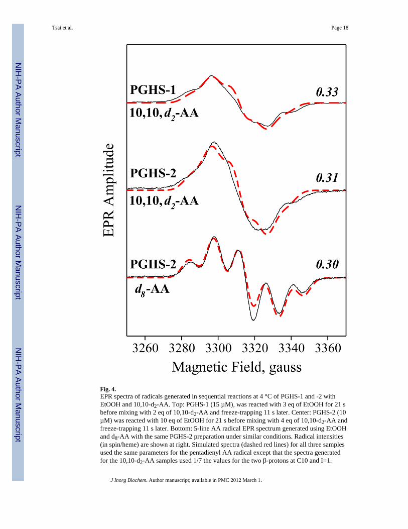
Fig. 4.EPR spectra of radicals generated in sequential reactions at 4 °C of PGHS-1 and -2 withEtOOH and 10,10-d2-AA. Top: PGHS-1 (15 µM), was reacted with 3 eq of EtOOH for 21 sbefore mixing with 2 eq of 10,10-d2-AA and freeze-trapping 11 s later. Center: PGHS-2 (10µM) was reacted with 10 eq of EtOOH for 21 s before mixing with 4 eq of 10,10-d2-AA andfreeze-trapping 11 s later. Bottom: 5-line AA radical EPR spectrum generated using EtOOHand d8-AA with the same PGHS-2 preparation under similar conditions. Radical intensities(in spin/heme) are shown at right. Simulated spectra (dashed red lines) for all three samplesused the same parameters for the pentadienyl AA radical except that the spectra generatedfor the 10,10-d2-AA samples used 1/7 the values for the two β-protons at C10 and I=1.
Tsai et al. Page 18
J Inorg Biochem. Author manuscript; available in PMC 2012 March 1.
NIH
-PA Author Manuscript
NIH
-PA Author Manuscript
NIH
-PA Author Manuscript

Fig. 5.HFEPR analysis of radicals in PGHS-1 reacted with AA and EtOOH. A) Solid line: fieldswept, echo-detected, 130 GHz EPR of PGHS-1 (80 µM) premixed with 5 eq of AA andreacted anaerobically with 10 eq of EtOOH for 5 s at 24 °C before freeze-trapping. Dashedlines: summation of simulated spectra for the tyrosyl and pentadienyl radicals (9:1 ratio)shown in B and C. B: Simulated spectrum for a tyrosyl radical using the followingpapameters: gx=2.0070, gy=2.0042, and gz=2.0020; full width half height Gaussiandistribution of g about gx=0.003. Principal hyperfine coupling values (in Gauss): twoprotons (9.5, 2.4, 6.2); two protons (1.0, 4.8, 0.8); one proton (6.7, isotropic); and one proton(7.8, isotropic). The Gaussian line-broadening function was set at 21 G full width halfheight. C) Simulated spectrum for a pentadienyl radical using parameters determinedpreviously [24]: gx=2.0032, gy=2.0032, and gz=2.0020. Principal hyperfine coupling values(in Gauss, all isotropic): two protons (10.4); two protons (3.3); two protons (16); one proton(11.5); one proton (9.8); and one proton (3.9). The Gaussian line-broadening function wasset at 21 G full width half height. The radical concentration for spectrum C is 12% that ofspectrum B.
Tsai et al. Page 19
J Inorg Biochem. Author manuscript; available in PMC 2012 March 1.
NIH
-PA Author Manuscript
NIH
-PA Author Manuscript
NIH
-PA Author Manuscript

Fig. 6.Cyclooxygenase active site structures for AA bound in a productive orientation to PGHS-1(panel A; 1DIY [11]) and in reversed orientation to PGHS-2 (panel B; 1CVU [45]). Keyresidues are labeled and hydrogen bonds are indicated by dashed lines. The phenoxyloxygen (orange) of Tyr385 (yellow) and the C13 (yellow) of AA (green) involved in H-transfer are highlighted to show the drastic distance change in the two panels.
Tsai et al. Page 20
J Inorg Biochem. Author manuscript; available in PMC 2012 March 1.
NIH
-PA Author Manuscript
NIH
-PA Author Manuscript
NIH
-PA Author Manuscript

Scheme 1.Different AA-induced radical EPR signatures found in PGHS-1 and -2 during anaerobicsingle-turnover manual freeze EPR experiments.
Tsai et al. Page 21
J Inorg Biochem. Author manuscript; available in PMC 2012 March 1.
NIH
-PA Author Manuscript
NIH
-PA Author Manuscript
NIH
-PA Author Manuscript
Copyright © 2022 FDOKUMEN