IAA stimulates pollen tube growth and mediates the modification of ...
Prostaglandin F2a stimulates motility and invasion in colorectal tumor cells
David Qualtrough1*, Abderrahmane Kaidi
1, Simon Chell
1, Henry N. Jabbour
2, Ann C. Williams
1and Christos Paraskeva
1
1Cancer Research UK Colorectal Tumour Biology Research Group, Department of Cellular and Molecular Medicine,School of Medical Sciences, University of Bristol, Bristol, United Kingdom2MRC Human Reproductive Sciences Unit, The Queen’s Medical Research Institute, Edinburgh, United Kingdom
Increased expression of cyclooxygenase-2 (COX-2) and subse-quent prostaglandin production is an important event in severalhuman malignancies, including colorectal cancer. COX-2 medi-ated prostanoid synthesis has been shown to play a key role intumor progression with prostaglandin E2 (PGE2) being shown topromote tumor growth, invasion and angiogenesis. The role of theother prostaglandins produced by COX-2 in tumors remainspoorly understood. We have shown that colorectal tumor cellsproduce prostaglandin F2a (PGF2a) and provide evidence thatPGF2a may play an important role in colorectal tumorigenesis.Our data show that PGF2a is secreted by both colorectal adenomaand carcinoma-derived cell lines at levels in excess of thosedetected for PGE2. These cell lines were also found to express thePGF2a receptor (FP) indicating potential autocrine effects ofPGF2a. This finding is further supported by an in vivo immunohis-tochemical study of FP expression in resected colon tissue. Thesedata show epithelial expression of FP in normal colorectal mucosaand also in colorectal adenomas and carcinomas. We comparedthe relative abilities of PGF2a and PGE2 to induce cell motility invitro in colorectal tumor cell lines and show the first evidence ofprostaglandin-induced cell motility in colorectal adenoma celllines. PGF2a induced cell motility with equivalent potency toPGE2 in all the cell lines tested and was also shown to increase theinvasion of carcinoma-derived cells into reconstituted basementmembrane. These data show that PGF2a may play an importantrole in the malignant progression of colorectal tumors.' 2007 Wiley-Liss, Inc.
Key words: colon; cancer; adenoma; prostaglandin; motility; invasion
Colorectal cancer remains a major cause of cancer deaths glob-ally. The inducible form of cyclooxygenase (COX-2), the keyenzyme in prostanoid biosynthesis, is overexpressed in over 80%of colorectal cancers.1 Furthermore, COX-2 is frequently expressedduring the premalignant adenoma stages of colorectal tumorigene-sis and shows a size dependent increase in expression indicating arole in tumor progression.2
The importance of COX-2 expression in colorectal tumor pro-gression has been highlighted by studies both in vitro and in vivo,and its effects are mediated through its prostanoid products.3
COX-2 catalyses the formation of prostaglandin H2 (PGH2) fromarachidonic acid and PGH2 is subsequently converted to severalstructurally related primary prostanoids, namely prostaglandin E2
(PGE2), PGD2, PGI2, Thromboxane A2 and prostaglandin F2a(PGF2a) by the action of specific synthases. Increased levels ofPGE2 have been observed in colorectal cancers when comparedwith histologically normal tissue.4–6 Subsequent studies usingin vitro models have shown that PGE2 is able to promote tumorcell growth,7,8 modulate apoptosis9 and increase cell motility.8
COX-2 expression and subsequent PGE2 production has also beenshown to enhance the production of angiogenic factors.10–12
Studies of prostaglandin production in colorectal tumors havealso identified the production of other primary prostanoids includ-ing PGF2a.
4–6 In 1 study on intestinal tissue from familial adeno-matous polyposis (FAP) patients, the detected levels of PGF2awere 30-fold higher than for PGE2.
13
Although widely studied in other tissues, the potential role ofPGF2a in colorectal cancer remains to be elucidated. The mostextensively studied area of PGF2a biology is its role in the regres-sion of the corpus luteum during pregnancy.14 Interestingly,
PGF2a stimulates the proliferation of Swiss mouse 3T3 cells withgreater efficacy than PGE2.
15,16
PGF2a is thought to largely act through the FP G-protein-coupled receptor, although the expression of this receptor has notbeen determined in colorectal cancer. Because of the importanceof COX-2 overexpression, the aim of the study presented here wasto investigate the potential role of PGF2a in colorectal tumorigene-sis. To this end, we determined the expression of the FP receptorin normal and neoplastic colorectal tissue, and a panel of adenomaand carcinoma-derived cell lines. The production of PGF2a wasexamined in parallel with that of PGE2 in the cell lines. Finally,we assessed the ability of both PGF2a and PGE2 to promote cellmotility and invasion which represent important hallmarks ofmalignant progression in these tumors.
Material and methods
Cell lines and culture conditions
AA/C1 is a clonogenic, adenoma cell line derived from a 3 to4 cm polyp from the descending colon of a familial adenomatouspolyposis (FAP) patient17 and is cultured in conditioned mediumas described by Williams et al.18 RG/C2 is a clonogenic cell linederived from a sporadic tubular adenoma of the sigmoid colon of1–2 cm in diameter and is cultured in DMEM supplemented with20% (vol/vol) FBS (Autogen Bioclear, Calne, UK).19 Both ofthese adenoma-derived cell lines are anchorage-dependent and arenontumorigenic in athymic nude mice. HCA7 was establishedfrom a moderately well differentiated mucinous carcinoma of thecolon20 and was a kind gift from Dr. Sue Kirkland (London, UK).HCA7-col29 was sub-cloned from the parental line21 and will behereafter referred to as HCA7. SW480 was derived from a spo-radic colonic adenocarcinoma.22 The carcinoma lines were cul-tured in DMEM supplemented with 10% (vol/vol) FBS, and allcell lines were cultured as adherent cells in 25 cm2 tissue cultureflasks.
For growth response assays, cells were seeded at 106 per25 cm2 flask and allowed to recover for 48 hr. The medium wasthen changed to DME-F12 (Invitrogen, Paisley, UK), supple-mented with 2% FBS, containing the appropriate amount of pros-taglandin F2a (PGF2a) (Sigma, Poole, UK) or vehicle control. Arange of prostaglandin concentrations from 0.1 to 10 lM wastested and compared with vehicle and untreated control. Treat-ments were made from prepared stocks so that a 1:1,000 dilutionof 95% ethanol was added to each treated flask. Following a 72 hrtreatment period, attached cell yield and the proportion of apopto-tic cells that had detached from the culture substrate were quanti-fied using a haemocytometer. The level of apoptosis in culturedcolonic cells can be assessed by measuring the proportion of cells
Grant sponsors: Cancer Research UK and Citrina Foundation.*Correspondence to: Cancer Research UK Colorectal Tumour Biology
Research Group, Department of Cellular and Molecular Medicine, Schoolof Medical Sciences, University of Bristol, Bristol, BS8 1TD, United King-dom. Fax:144-117-9287896.. E-mail: [email protected] 9 October 2006; Accepted after revision 13 February 2007DOI 10.1002/ijc.22755Published online 16 April 2007 in Wiley InterScience (www.interscience.
wiley.com).
Int. J. Cancer: 121, 734–740 (2007)' 2007 Wiley-Liss, Inc.
Publication of the International Union Against Cancer
that detach from the flask and float in the medium.23,24 Apoptosiswas confirmed in these �floating� cells by morphology (followingacridine orange staining).
Prostaglandin production assay
PGF2a and prostaglandin E2 (PGE2) enzyme immunoassay kits(Cayman Chemical Company, Ann Arbor, MI) were used to assaythe release of PGF2a and PGE2 into the growth medium by thecells. The lower limit of detection of the assays was 5.5 pg/ml forPGF2a and 36 pg/ml for PGE2, and the upper limit of detectionwas 64 pg/ml and 250 pg/ml for PGF2a and PGE2, respectively.Cells were grown to �70% confluency and the standard culturemedium replaced with serum-free DMEM-F12 (Invitrogen, Pais-ley, UK) for a period of 72 hr whereupon the medium wasremoved, centrifuged and decanted, snap-frozen in liquid nitrogenand stored at 270�C prior to analysis. These samples were dilutedappropriately so that readings fell within the detection limits ofthe assay. PGF2a and PGE2 production was normalized accordingto the number of adherent cells present in the particular culture atthe time of sampling. The results are expressed as picograms ofprostaglandin/106 cells and represent the average of 2 independentexperiments performed in duplicate.
In vitro motility and invasion assays
Cell motility assays were carried out using a transwell filter mo-tility assay as previously described.25 For quantitative analysis ofcell motility 8 lm pore size insert filters (Becton Dickinson,Oxford, UK) were coated with 10 lg ml21 Vitrogen Type I colla-gen (Cohesion, Palo Alto, CA). For invasion assays, 8 lm trans-well filters precoated with Matrigel (Becton Dickinson, Oxford,UK) were used. For both types of analysis 1 3 105 cells wereseeded per well in calcium-free DMEM containing 0.1% FBS(CF-DMEM) and allowed to adhere for 4 hr.
Initial experiments using a range of doses of prostaglandinsfrom 0.1 to 10 lM [PGF2a or PGE2 (Sigma, Poole, UK) diluted1:1,000 from a 1 mM stock made up in 95% ethanol, or vehicle(1:1,000 dilution of 95% ethanol)] were performed, 1 lM wasselected as it was found to stimulate significant motility.
Following a 24 hr incubation, cells were removed from theupper filter surface with a cotton swab. The filters were fixed andstained with haematoxylin. Cells that had moved to the lower filtersurface were counted in 10 fields at 203 magnification. Three in-dependent experiments were carried out in triplicate, and the dataare expressed as the mean 6 SEM. Statistical analysis of this datawas performed using the Student’s t test. Differences were consid-ered significance when the p value was <0.05.
Western blot analysis
Samples of 2 3 106 cells were prepared for Western blotting asdescribed previously.26 The FP receptor was detected using a rab-bit polyclonal antibody (1:1,000 dilution; Cayman Chemical Cay-man, Ann Arbor, MI) and the Lumiglo ECL detection system(Amersham Biosciences, Little Chalfont, UK). Ishikawa endome-trial carcinoma cells stably overexpressing the FP receptor wereincluded as a positive control.27 Anti a-tubulin (diluted 1:1,000;Sigma, Poole, UK) was used as a loading control.
Immunohistochemistry
Immunohistochemical analysis of FP receptor expression wascarried out as previously described.27 Following approval fromthe local research ethics committee, anonymised paraffin-embed-ded, formalin-fixed archival material was retrieved from the filesof the Department of Histopathology, Bristol Royal Infirmary foruse in this study. A total of 15 colorectal adenomas were exam-ined. These included 5 small (�5 mm) tumors, 5 medium-sized(5–10 mm) tumors and 5 large (�10 mm) tumors. We also exam-ined 10 invasive adenocarcinomas taken from colorectal resectionspecimens. Five samples of histologically normal mucosa werealso included in the study.
The FP receptor was detected using a FP-specific affinity puri-fied rabbit polyclonal antibody (diluted 1:500; Cayman, AnnArbor, MI) and a biotinylated swine anti-rabbit secondary anti-body (diluted 1:500; Dako Cytomation, Ely, UK). The specificityof the antibody was confirmed by prior overnight incubation at4�C with blocking peptide (diluted 1:50; Cayman, Ann Arbor,MI). This preblocking was used as a negative control as shown inFigure 2b. A standard avidin-biotin immunoperoxidase technique(Dako Cytomation, Ely, UK) was employed and the immunoreac-tion visualized by means of the diamino benzidine reaction withhaemotoxylin counterstaining. Antigen retrieval was performed bytreating sections for 5 min in a pressure cooker in 0.1% citratebuffer (pH 6.0). Sections were examined for FP immunoreactivity.
Results
Prostaglandin F2a is produced by colorectaladenoma and carcinoma cells
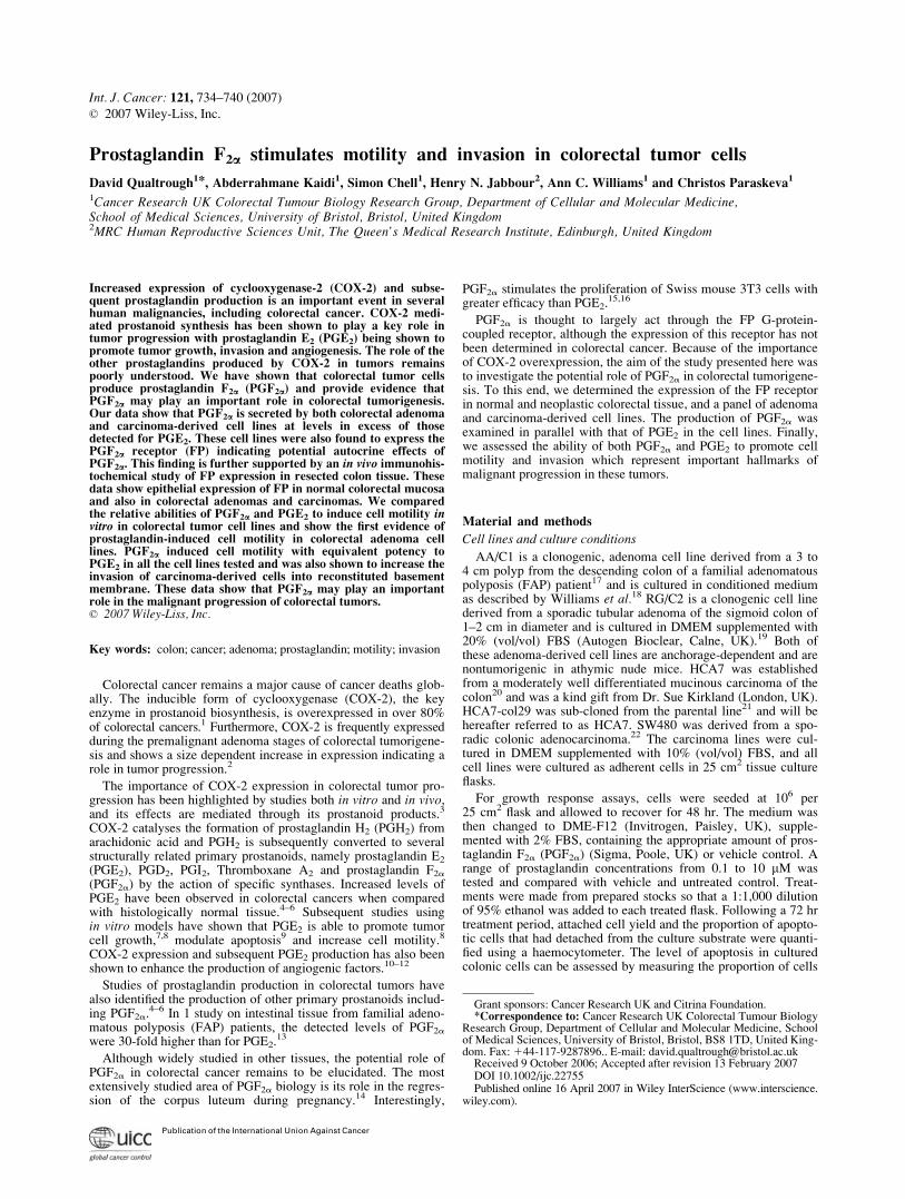
Samples of media were taken following 72 hr incubation of theadenoma-derived cell lines AA/C1 and RG/C2, and from the car-cinoma-derived cell lines HCA7 and SW480. For this culture pe-riod, serum-free media was used and these samples were assayedfor both PGE2 and PGF2a content using enzyme immunoassay.Prostaglandin concentrations were calculated in pg per millioncells. Figure 1 shows the mean value obtained, and Table I therange of results from 2 independent experiments performed induplicate.
Although relatively low amounts of PGE2 were found in the ad-enoma-derived cell lines AA/C1 and RG/C2 (6.69 and 7.49 pg/million cells, respectively), both produced PGF2a at a level com-parable with that detected in SW480 carcinoma cells. The HCA7carcinoma line produces very high levels of PGE2 due to the rela-tively high levels of cyclooxygenase-2 (COX-2) expression in
FIGURE 1 – Colorectal tumor cells produce PGF2a. Graphical rep-resentation of PGF2a and PGE2 produced by colorectal adenoma andcarcinoma cell lines. Enzyme immunoassays for PGF2a and PGE2
were performed on serum-free media from each cell line following72 hr incubation. AA/C1 and RG/C2 are adenoma-derived cell lines,whereas SW480 and HCA7 are carcinoma-derived. Prostaglandin pro-duction was calculated as picogrammes per million cells and thesedata represent the mean of 2 experiments performed in duplicate.
735PGF2� STIMULATES MOTILITY AND INVASION IN CELLS
these cells caused by stabilization of the COX-2 mRNA.28 It isinteresting to note that HCA7 also produced higher levels ofPGF2a than PGE2.
The FP prostaglandin receptor is expressed in colorectaltumors and in tumor-derived cell lines
The major receptor for PGF2a has been designated FP and hasbeen shown to be a Gq-linked G-protein coupled receptor whichmediates its effects through modulating intracellular calcium and
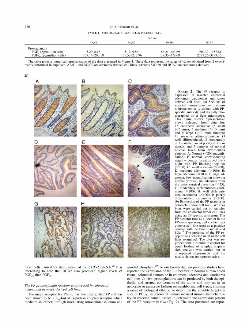
inositol phosphate.29 To our knowledge, no previous studies havereported the expression of the FP receptor in normal human colontissue, colorectal tumors or in colorectal adenoma and carcinomacell lines. In vivo, prostaglandins can be produced by both the epi-thelial and stromal components of the tumor and may act in anautocrine or paracrine fashion on neighboring cell types, elicitinga range of biological effects. To determine the possible target tis-sues of PGF2a in colorectal tumors we used immunohistochemis-try on resected human tissues to determine the expression patternof the FP receptor in vivo (Fig. 2). The data presented are repre-
FIGURE 2 – The FP receptor isexpressed in resected colorectaladenomas, carcinomas and tumorderived cell lines. (a) Sections ofresected human tissue were immu-nohistochemically stained with FP-specific antibody and digitally pho-tographed on a light microscope.This figure shows representativeviews selected from data for:15 colorectal adenomas [5 small(�5 mm), 5 medium (5–10 mm)and 5 large (�10 mm) tumors];10 invasive adenocarcinomas (3well differentiated, 3 moderatelydifferentiated and 4 poorly differen-tiated); and 5 samples of normalmucosa taken from diverticulitispatients. A: Normal (3200 magnifi-cation); B: normal—correspondingnegative control (preabsorbed over-night with FP blocking peptide)(3200); C: small adenoma (3200);D: medium adenoma (3100); E:large adenoma (3100); F: large ad-enoma, low magnification showingnormal (arrows) and adenoma fromthe same surgical resection (325);G: moderately differentiated carci-noma (3200); H: well differenti-ated carcinoma (3100); I: poorlydifferentiated carcinoma (3100).(b) Expression of the FP receptor incolorectal tumor cell lines. Westernblots were carried out on samplesfrom the colorectal tumor cell linesusing an FP-specific antiserum. TheFP receptor runs as a doublet in theFP-overexpressing endometrial car-cinoma cell line used as a positivecontrol, with the lower band at �64kDa.25 The presence of the FP re-ceptor was detected in all of the celllines examined. The blot was re-probed with a-tubulin to control forequal loading of samples. Expres-sion analysis was carried out in3 repeated experiments and theresults shown are representative.
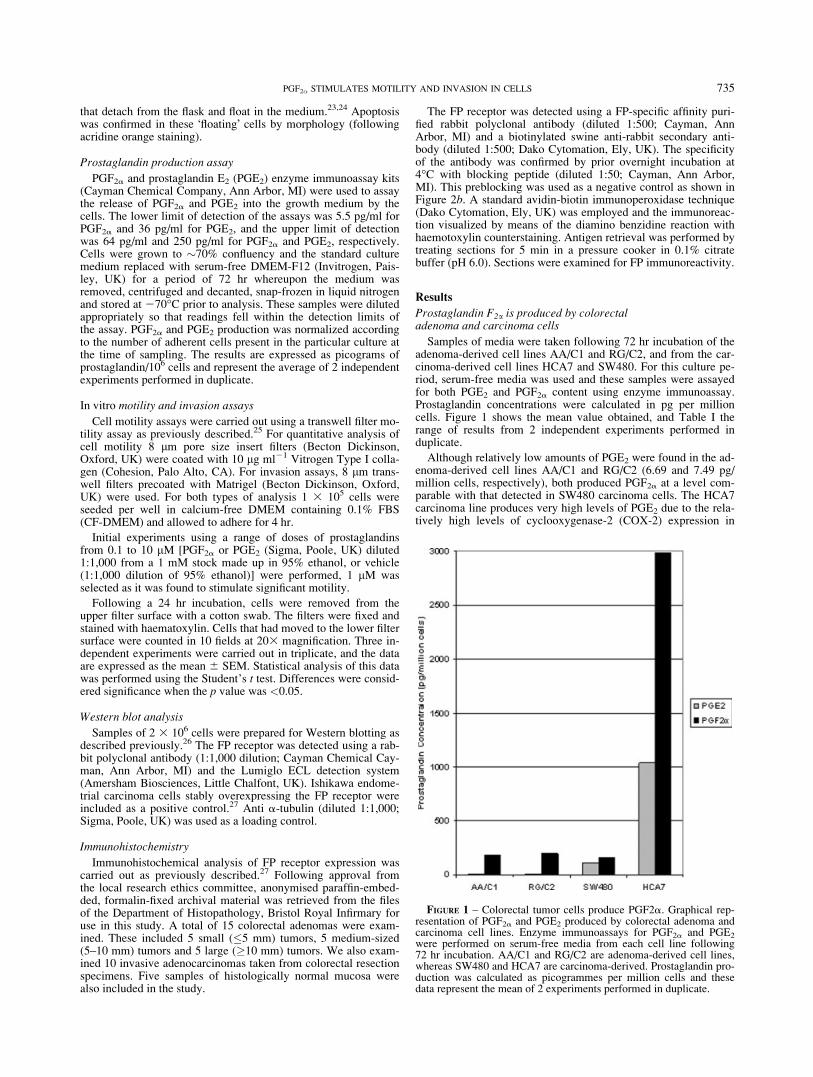
TABLE I – COLORECTAL TUMOR CELLS PRODUCE PGF2a
Cell line
AA/C1 RG/C2 SW480 HCA7
ProstaglandinPGE2 (pg/million cells) 5.20–8.18 5.12–9.86 86.21–123.69 910.39–1157.61PGF2a (pg/million cells) 157.14–203.10 175.52–217.96 139.35–178.69 2777.26–3193.16
The table gives a numerical representation of the data presented in Figure 1. These data represent the range of values obtained from 2 experi-ments performed in duplicate. AA/C1 and RG/C2 are adenoma-derived cell lines, whereas SW480 and HCA7 are carcinoma-derived.
736 QUALTROUGH ET AL.
sentative of 5 samples of normal colonic epithelium resected frompatients with diverticulitis; 15 samples of benign colonic adenomaand 10 samples of colorectal adenocarcinoma. The colorectaladenomas were divided according to size (small 5 <5 mm; me-dium 5 5–10 mm; large5 >10 mm).
Strong positive immunoreactivity to the FP receptor was seen inthe epithelial component of normal colonic mucosa (100% (n 55) Fig. 2a panel A) compared with the negative control (Fig. 2apanel B), yet no staining was detected in stromal cells. All of thecolorectal adenomas (15/15) displayed positive epithelial immu-noreactivity for FP but no expression was observed in any stromaltissues (Fig. 2a panels C–F).
The results in colorectal carcinomas were more varied. Carcino-mas 7/10 displayed positive immunoreactivity for FP in the epi-thelium with no staining detected in stromal constituents (Fig. 2apanels G–I). Positive immunoreactivity for the FP receptor wasdetected in all of the normal and adenoma samples, and the major-ity of the carcinomas, both the intensity and distribution of the FPstaining were observed to be similar when comparing normal withtumor tissue.
Having shown that the FP receptor was expressed in normal andtumor tissue in vivo we then studied the expression of the FP re-ceptor in adenoma and carcinoma-derived cell lines by Westernblotting. Representative results of 3 repeated experiments areshown in Figure 2b. Ishikawa endometrial carcinoma cells stablyoverexpressing the FP receptor were included as a positive con-trol.27 Both of the adenoma cell lines and all 3 carcinoma cell linesshow expression of the FP receptor. A doublet of bands was seenfor each cell line and it is not possible, using the reagents currentlyavailable, to determine whether these represent 2 distinct isoformsof the receptor or some form of post-translational modification.27
However, these data do show the expression of the FP receptor innormal and neoplastic colorectal epithelium, suggesting a possibleautocrine role for PGF2a secreted by the cells.
Prostaglandins F2a and E2 stimulate cell motility incolorectal adenoma-derived cell lines
Previous studies have shown that PGE2 confers broad rangingtumor-promoting effects in colorectal cancer yet nothing is cur-rently known about the role of PGF2a in this disease. Havingshown that colorectal tumor cells produce PGF2a and that the FPreceptor is expressed by colorectal epithelial cells in vitro andin vivo, we investigated the potential role of PGF2a in colorectaltumorigenesis using in vitro models.
Previous studies have reported that exogenous addition ofPGF2a stimulated cell growth in the colorectal carcinoma cell lineSW1116,7 but produced no significant effects on growth in HCT8and HT29.30 Studies in this laboratory have shown that cell linesderived from colorectal adenomas31 and carcinomas32 are growthstimulated by exogenous addition of PGE2. We conducted PGF2agrowth response experiments on the cell lines studied above(adenomas: AA/C1, RG/C2; carcinomas: SW480, HCA7) with arange of concentrations from 0.1 to 10 lM. No significant change
in cell growth or apoptosis was detected in any of the cell linesstudied over a 72 hr period (data not shown).
Previous work has shown that PGE2 has multiple effects on col-orectal carcinoma cells, including increasing cell motility.8,33
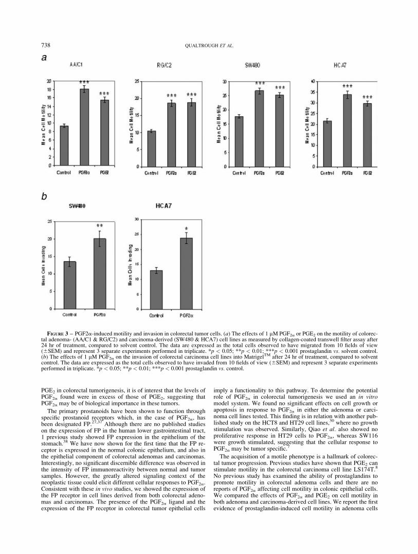
There are no previous reports of PGF2a inducing motility in tumorcells and, importantly, no studies of prostaglandin-induced motil-ity in colorectal adenomas. We compared the ability of exogenousPGF2a and PGE2 to influence the motility of colorectal adenoma(AA/C1, RG/C2) and carcinoma (SW480, HCA7) cells using aBoyden-chamber transwell-filter cell motility assay, compared tosolvent control. Initial experiments were performed using a rangeof doses of prostaglandins (0.1–10 lM) and 1 lM was selected asit was found to stimulate significant motility. Cells were treatedand incubated for 24 hr to allow movement through the collagen-coated filters. Figure 3a shows the average results of 3 independ-ent experiments performed in triplicate. As would be expected, theadenoma derived cell lines AA/C1 and RG/C2, which representearlier stages of colorectal tumor progression, were less motilethan their carcinoma counterparts, which display a �2-fold higherrate of motility (Fig. 3a).
Both PGF2a and PGE2 significantly increased cell motility inadenoma- (AA/C1 and RG/C2, p 5 <0.001) and carcinoma-derived (HCA7 and SW480, p 5 <0.001) cell lines. These are thefirst data showing the ability of prostaglandins to increase motilityin adenoma cells which represent the earlier, nonmalignant stagesof colorectal tumor development.
Prostaglandin F2a stimulates invasion in carcinoma cells
The process by which tumor cells break out from their site oforigin and metastasise to distant sites requires, in addition to mo-tility, an ability to invade through the basement membrane andunderlying mesenchymal cells. This process can be modeledin vitro by coating transwell filters (as used for the motility assayabove) with reconstituted basement membrane, in this instanceMatrigelTM. PGE2 has been shown previously to stimulate inva-sion by colorectal cancer cells in this type of assay.34 The ade-noma-derived cell lines have been shown previously to be nonin-vasive [Ref. 35 and Qualtrough and Paraskeva, unpublished obser-vations] in keeping with their premalignant nature. Adoption of aninvasive phenotype is a key distinction between benign and malig-nant colorectal tumors.
The data presented in Figure 3b clearly shows that 1 lM PGF2asignificantly stimulates invasion of HCA7 and SW480 colon carci-noma cells into a reconstituted basement membrane (HCA7, p 5<0.05; SW480, p 5 <0.01). The figures show a 1.8-fold stimula-tion of invasion in HCA7 and a 1.5-fold increase in SW480 withPGF2a compared with control.
Discussion
The overexpression of COX-2 is an important event in colorec-tal cancer. It has also become clear that it is the prostanoid prod-ucts of COX-2 activity that mediate major effects on tumor cellbehavior by promoting growth, survival, invasive behavior andinducing angiogenesis.36 Much of the focus of prostanoid researchin the area of colorectal cancer has fallen on PGE2, widelyaccepted to be the major prostanoid product of COX-2 in thesetumors.31 COX-2 overexpression also leads to the production ofother primary prostanoids such as PGD2, PGI2 and PGF2a,although the role of these in colorectal tumorigenesis remainspoorly understood.36 Several researchers have detected PGF2aproduction in colorectal tumor tissue, and in 1 study the levelsdetected were found to be in excess of those for PGE2.
13 The pur-pose of the study presented here was to determine the potentialrole of PGF2a in colorectal tumorigenesis.
The data presented here clearly shows readily detectable levelsof PGF2a produced by cell lines derived from both colorectaladenomas and carcinomas. Considering the known importance of
FIGURE 2 – CONTINUED
737PGF2� STIMULATES MOTILITY AND INVASION IN CELLS
PGE2 in colorectal tumorigenesis, it is of interest that the levels ofPGF2a found were in excess of those of PGE2, suggesting thatPGF2a may be of biological importance in these tumors.
The primary prostanoids have been shown to function throughspecific prostanoid receptors which, in the case of PGF2a, hasbeen designated FP.27,37 Although there are no published studieson the expression of FP in the human lower gastrointestinal tract,1 previous study showed FP expression in the epithelium of thestomach.38 We have now shown for the first time that the FP re-ceptor is expressed in the normal colonic epithelium, and also inthe epithelial component of colorectal adenomas and carcinomas.Interestingly, no significant discernible difference was observed inthe intensity of FP immunoreactivity between normal and tumorsamples. However, the greatly altered signaling context of theneoplastic tissue could elicit different cellular responses to PGF2a.Consistent with these in vivo studies, we showed the expression ofthe FP receptor in cell lines derived from both colorectal adeno-mas and carcinomas. The presence of the PGF2a ligand and theexpression of the FP receptor in colorectal tumor epithelial cells
imply a functionality to this pathway. To determine the potentialrole of PGF2a in colorectal tumorigenesis we used an in vitromodel system. We found no significant effects on cell growth orapoptosis in response to PGF2a in either the adenoma or carci-noma cell lines tested. This finding is in relation with another pub-lished study on the HCT8 and HT29 cell lines,30 where no growthstimulation was observed. Similarly, Qiao et al. also showed noproliferative response in HT29 cells to PGF2a, whereas SW116were growth stimulated, suggesting that the cellular response toPGF2a may be tumor specific.7
The acquisition of a motile phenotype is a hallmark of colorec-tal tumor progression. Previous studies have shown that PGE2 canstimulate motility in the colorectal carcinoma cell line LS174T.8
No previous study has examined the ability of prostaglandins topromote motility in colorectal adenoma cells and there are noreports of PGF2a affecting cell motility in colonic epithelial cells.We compared the effects of PGF2a and PGE2 on cell motility inboth adenoma and carcinoma-derived cell lines. We report the firstevidence of prostaglandin-induced cell motility in adenoma cells
FIGURE 3 – PGF2a-induced motility and invasion in colorectal tumor cells. (a) The effects of 1 lM PGF2a or PGE2 on the motility of colorec-tal adenoma- (AA/C1 & RG/C2) and carcinoma-derived (SW480 & HCA7) cell lines as measured by collagen-coated transwell filter assay after24 hr of treatment, compared to solvent control. The data are expressed as the total cells observed to have migrated from 10 fields of view(6SEM) and represent 3 separate experiments performed in triplicate. *p < 0.05; **p < 0.01; ***p < 0.001 prostaglandin vs. solvent control.(b) The effects of 1 lM PGF2a on the invasion of colorectal carcinoma cell lines into MatrigelTM after 24 hr of treatment, compared to solventcontrol. The data are expressed as the total cells observed to have invaded from 10 fields of view (6SEM) and represent 3 separate experimentsperformed in triplicate. *p < 0.05; **p < 0.01; ***p < 0.001 prostaglandin vs. control.
738 QUALTROUGH ET AL.
which responded to both PGF2a and PGE2. Our data also showthat PGF2a significantly stimulates cell motility in colorectal car-cinoma cell lines, as well as adenomas, and is comparable withPGE2 in its ability to do so. These data show an important biologi-cal effect of PGF2a in colonic tumor cells and also suggest thatprostaglandins may contribute to the progression of the adenomato carcinoma sequence by increasing cell motility.
The process of tumor invasion (and therefore malignancy)requires cell motility but also alterations in cell adhesion and thesecretion of enzymes to degrade basement membrane and matrixcomponents. Pai et al. have reported that PGE2 potentiates inva-siveness in the colorectal carcinoma cell lines SW480 and LoVoalthough the effects of PGF2a remain unreported.34 We have nowshown that PGF2a can also increase the invasiveness of theSW480 and HCA7 carcinoma cell lines in vitro, with the SW480data being comparable with that previously published for PGE2.
34
These increases were statistically significant in both of the celllines and demonstrate a potentially important and novel tumor-promoting effect of PGF2a in colorectal cancer. Because of thecomplexity of the invasion process, it is possible that PGF2a mayalso stimulate the production of matrix remodeling enzymes andalter cell adhesion complexes, in addition to stimulating cell mo-tility. PGE2 has been shown to stimulate colon cancer cell inva-sion through transactivation of the epidermal growth factor recep-
tor (EGFR).34 We have not seen EGFR activation by PGF2a in oursystem (data not shown), suggesting alternate mechanisms ofaction for this prostaglandin which represents an interesting areafor potential future study.
In conclusion, the data presented here show that PGF2a may bean important product of COX-2 overexpression in colorectaltumors, by promoting tumor progression and potentially metasta-sis. The use of high doses of NSAIDs in colorectal cancer preven-tion and therapy has recently come under close scrutiny due to theadverse cardiovascular effects associated with long term use ofRofecoxib and the subsequent withdrawal of this drug from clini-cal use.39 Although this does not preclude the use of this type ofdrug in an adjuvant setting, it does highlight the need for more tar-geted approaches.40,41 To facilitate this, it is of key importancethat we increase our understanding of the pleiotropic effects ofthe various prostanoid products of the COX-2 enzyme. The datapresented here show that PGF2a, as well as the widely-studiedPGE2 needs to be considered in our understanding of colorectalcancer.
Acknowledgements
We wish to thank Dr. Sharon Battersby (MRC Human Repro-ductive Sciences Unit, Edinburgh) for expert assistance with theimmunohistochemistry.
References
1. Eberhart CE, Coffey RJ, Radhika A, Giardiello FM, Ferrenbach S,DuBois RN. Up-regulation of cyclooxygenase 2 gene expression inhuman colorectal adenomas and adenocarcinomas. Gastroenterology1994;107:1183–8.
2. Elder DJ, Baker JA, Banu NA, Moorghen M, Paraskeva C. Humancolorectal adenomas demonstrate a size-dependent increase in epithe-lial cyclooxygenase-2 expression. J Pathol 2002;198:428–34.
3. Brown JR, DuBois RN. COX-2: a molecular target for colorectal can-cer prevention. J Clin Oncol 2005;23:2840–55.
4. Rigas B, Goldman IS, Levine L. Altered eicosanoid levels in humancolon cancer. J Lab Clin Med 1993;122:518–23.
5. Giardiello FM, Spannhake EW, DuBois RN, Hylind LM, RobinsonCR, Hubbard WC, Hamilton SR, Yang VW. Prostaglandin levels inhuman colorectal mucosa: effects of sulindac in patients with familialadenomatous polyposis. Dig Dis Sci 1998;43:311–16.
6. Yang VW, Shields JM, Hamilton SR, Spannhake EW, Hubbard WC,Hylind LM, Robinson CR, Giardiello FM. Size-dependent increase inprostanoid levels in adenomas of patients with familial adenomatouspolyposis. Cancer Res 1998;58:1750–3.
7. Qiao L, Kozoni V, Tsioulias GJ, Koutsos MI, Hanif R, Shiff SJ, RigasB. Selected eicosanoids increase the proliferation rate of human coloncarcinoma cell lines and mouse colonocytes in vivo. Biochim BiophysActa 1995;1258:215–23.
8. Sheng H, Shao J, Washington MK, DuBois RN. Prostaglandin E2increases growth and motility of colorectal carcinoma cells. J BiolChem 2001;276:18075–81.
9. Sheng H, Shao J, Morrow JD, Beauchamp RD, DuBois RN. Modula-tion of apoptosis and Bcl-2 expression by prostaglandin E2 in humancolon cancer cells. Cancer Res 1998;58:362–6.
10. Ben-Av P, Crofford LJ, Wilder RL, Hla T. Induction of vascular en-dothelial growth factor expression in synovial fibroblasts by prosta-glandin E and interleukin-1: a potential mechanism for inflammatoryangiogenesis. FEBS Lett 1995;372:83–7.
11. Tsujii M, Kawano S, Tsuji S, Sawaoka H, Hori M, DuBois RN. Cy-clooxygenase regulates angiogenesis induced by colon cancer cells.Cell 1998;93:705–16.
12. Cianchi F, Cortesini C, Bechi P, Fantappie O, Messerini L, VannacciA, Sardi I, Baroni G, Boddi V, Mazzanti R, Masini E. Up-regulationof cyclooxygenase 2 gene expression correlates with tumor angiogen-esis in human colorectal cancer. Gastroenterology 2001;121:1339–47.
13. Nugent KP, Spigelman AD, Phillips RK. Tissue prostaglandin levelsin familial adenomatous polyposis patients treated with sulindac. DisColon Rectum 1996;39:659–62.
14. Diaz FJ, Anderson LE, Wu YL, Rabot A, Tsai SJ, Wiltbank MC. Reg-ulation of progesterone and prostaglandin F2a production in the CL.Mol Cell Endocrinol 2002;191:65–80.
15. de Asua LJ, Clingan D, Rudland PS. Initiation of cell proliferation incultured mouse fibroblasts by prostaglandin F2a. Proc Natl Acad SciUSA 1975;72:2724–8.
16. Sauane M, Correa L, Rogers F, Krasnapolski M, Barraclough R, Rud-land PS, de Asua LJ. Prostaglandin F2a induces cyclin D1 expression
and DNA synthesis via early signalling mechanisms in swiss mouse3T3 cells. Biochem Biophys Res Commun 2000;270:11–16.
17. Paraskeva C, Buckle BG, Sheer D, Wigley CB. The isolation andcharacterisation of colorectal epithelial cell lines at different stages inmalignant transformation from familial polyposis coli patients. Int JCancer 1984;34:49–56.
18. Williams AC, Harper SJ, Paraskeva C. Neoplastic transformation of ahuman colonic epithelial cell line: in vitro evidence for the adenoma-to-carcinoma sequence. Cancer Res 1990;50:4724–30.
19. Paraskeva C, Finerty S, Mountford RA, Powell SC. Specific cytoge-netic abnormalities in two new human colorectal adenoma-derivedepithelial cell lines. Cancer Res 1989;49:1282–6.
20. Kirkland SC. Dome formation by a human colonic adenocarcinomacell line (HCA-7). Cancer Res 1985;45:3790–5.
21. Marsh KA, Stamp GWH, Kirkland SC. Isolation and characterisation ofmultiple cell types from a single human colonic carcinoma: tumorigenic-ity of these cell types in a xenograft system. J Pathol 1993;170:441–50.
22. Hague A, Manning AM, Hanlon KA, Huschtscha LI, Hart D, Para-skeva C. Sodium butyrate induces apoptosis in human colonic tumourcell lines in a p53-independent pathway: implications for the possi-ble role of dietary fibre in the prevention of large bowel cancer. Int JCancer 1993;55:498–505.
23. Hague A, Bracey TS, Hicks DJ, Reed JC, Paraskeva C. Decreasedlevels of p26-Bcl-2, but not p30 phosphorylated Bcl-2, precedeTGFb1-induced apoptosis in colorectal adenoma cells. Carcinogenesis1998;19:1691–5.
24. Leibovitz A, Stinson JC, McCombs WB 3rd, McCoy CE, Mazur KC,Mabry ND. Classification of human colorectal adenocarcinoma celllines. Cancer Res. 1976;36:4562–9.
25. Efstathiou JA, Liu D, Wheeler JM, Kim HC, Beck NE, Ilyas M, Kar-ayiannakis AJ, Mortensen NJ, Kmiot W, Playford RJ, Pignatelli M,Bodmer WF. Mutated epithelial cadherin is associated with increasedtumorigenicity and loss of adhesion and of responsiveness to themotogenic trefoil factor 2 in colon carcinoma cells. Proc Natl AcadSci USA 1999;96:2316–21.
26. Williams AC, Collard TJ, Paraskeva C. An acidic environment leadsto p53 dependent induction of apoptosis in human adenoma and carci-noma cell lines: Implications for clonal selection during colorectalcarcinogenesis. Oncogene 1999;18:3199–204.
27. Sales KJ, List T, Boddy SC, Williams ARW, Anderson RA, Naor Z &Jabbour HN. A novel angiogenic role for prostaglandin F2a-FP recep-tor interaction in human endometrial carcinomas. Cancer Res 2005;65:7707–16.
28. Shao J, Sheng H, Inoue H, Morrow JD, DuBois RN. Regulation ofconstitutive cyclooxygenase-2 expression in colon carcinoma cells. JBiol Chem 2000;275:33951–6.
29. Breyer RM, Bagdassarian CK,Myers SA, Breyer MD. Prostanoid receptors:subtypes and signalling. Ann Rev Pharmacol Toxicol 2001;41: 661–90.
30. Cassano G, Gasparre G, Susca F, LIppe C, Guanti G. Lack of effectby prostaglandin F2alpha on the proliferation of the HCT8 and HT29human adenocarcinoma cell lines. Oncol Rep 2000;7:183–6.
739PGF2� STIMULATES MOTILITY AND INVASION IN CELLS
31. Chell SD, Witherden IR, Dobson RR, Moorghen M, Herman AA,Qualtrough D, Williams AC, Paraskeva C. Increased EP4 receptorexpression in colorectal cancer progression promotes cell growth andanchorage independence. Cancer Res 2006;66:3106–13.
32. Hull MA, Ko SC, Hawcroft G. Prostaglandin EP receptors: targets fortreatment and prevention of colorectal cancer? Mol Cancer Ther2004;3:1031–9.
33. Buchanan FG, Wang D, Bargiacchi F, DuBois RN. ProstaglandinE2 regulates cell migration via the intracellular activation ofthe epidermal growth factor receptor. J Biol Chem 2003;278:35451–7.
34. Pai R, Nakamura T, Moon WS, Tarnawski AS. Prostaglandins pro-mote colon cancer cell invasion; signaling by cross-talk between twodistinct growth factor receptors. FASEB J 2003;17:1640–7.
35. Brunton VG, Ozanne BW, Paraskeva C, Frame MC. A role for epider-mal growth factor receptor, c-Src and focal adhesion kinase in an invitro model for the progression of colon cancer. Oncogene 1997;14:283–93.
36. Gupta RA, DuBois RN. Colorectal cancer prevention and treat-ment by inhibition of cyclooxygenase-2. Nat Rev Cancer 2001;1:11–21.
37. Bos CL, Richel DJ, Ritsema T, Peppelenbosch MP, Versteeg HH.Prostanoids and prostanoid receptors in signal transduction. Int J Bio-chem Cell Biol 2004;36:1187–205.
38. Hasumoto K, Sugimoto Y, Gotoh M, Segi E, Yamasaki A, YamaguchiM, Honda H, Hirai H, Negishi M, Kakizuka A, Ichikawa A. Charac-terisation of the mosue prostaglandin F receptor gene: a transgenicmouse study of a regulatory region that controls its expression in thestomach and kidney but not in the ovary. Genes Cells 1997;2:571–80.
39. Graham D, Campen D, Hui R, Spence M, Cheetham C, Levy G,Shoor S, Ray W. Risk of acute myocardial infarction and sudden car-diac death in patients treated with cyclo-oxygenase 2 selective andnon-selective non-steroidal anti-inflammatory drugs: nested case-con-trol study. Lancet 2005;365:475–81.
40. Chell S, Patsos HA, Qualtrough D, H-Zadeh AM, Hicks DJ, Kaidi A,Witherden IR, Williams AC, Paraskeva C. Prospects in NSAID-derived chemoprevention of colorectal cancer. Biochem Soc Trans2005;33:667–71.
41. Chell S, Kaidi A, Williams AC, Paraskeva C. Mediators of PGE2 syn-thesis and signalling downstream of COX-2 represent potential targetsfor the prevention/treatment of colorectal cancer. Biochim BiophysActa 2006;1766:104–19.
740 QUALTROUGH ET AL.
Copyright © 2022 FDOKUMEN