Pseudomonas aeruginosa dose response and bathing water infection

Microbiology (1999), 145, 2863–2873 Printed in Great Britain
A re-examination of twitching motility inPseudomonas aeruginosa
Annalese B. T. Semmler,1,2 Cynthia B. Whitchurch1 and John S. Mattick1,2
Author for correspondence: John S. Mattick. Tel : 61 7 3365 4446. Fax: 61 7 3365 4388.e-mail : j.mattick!cmcb.uq.edu.au
Centre for Molecular andCellular Biology1 andDepartment ofBiochemistry2, University ofQueensland, Brisbane,QLD 4072, Australia
Twitching motility is a form of solid surface translocation which occurs in awide range of bacteria and which is dependent on the presence of functionaltype IV fimbriae or pili. A detailed examination of twitching motility inPseudomonas aeruginosa under optimal conditions in vitro was carried out.Under these conditions (at the smooth surface formed between semi-solidgrowth media and plastic or glass surfaces) twitching motility is extremelyrapid, leading to an overall radial rate of colony expansion of 0<6 mm hN1 orgreater. The zones of colony expansion due to twitching motility are very thinand are best visualized by staining. These zones exhibit concentric rings inwhich there is a high density of microcolonies, which may reflect periods ofexpansion and consolidation/cell division. Video microscopic analysis showedthat twitching motility involves the initial formation of large projections orrafts of aggregated cells which move away from the colony edge. Behind therafts, individual cells move rapidly up and down trails which thin and branchout, ultimately forming a fine lattice-like network of cells. The bacteria in thelattice network then appear to settle and divide to fill out the colonized space.Our observations redefine twitching motility as a rapid, highly organizedmechanism of bacterial translocation by which P. aeruginosa can disperse itselfover large areas to colonize new territories. It is also now clear, bothmorphologically and genetically, that twitching motility and social glidingmotility, such as occurs in Myxococcus xanthus, are essentially the sameprocess.
Keywords : type IV fimbriae, pili, surface translocation, gliding motility
INTRODUCTION
The term twitching motility was first coined by Lautrop(1961) to describe flagellar-independent surface motilityin Acinetobacter calcoaceticus. Similar motility has beensubsequently described in a wide range of other bacteria,including Pseudomonas aeruginosa, Neisseria gonor-rhoeae, Neisseria meningitidis and other Neisseriaceae,various Moraxella species, Dichelobacter nodosus,Branhamella catarrhalis, Suttonella indologenes, Altero-monas putrefaciens, Pasteurella multocida, Xantho-monas maltophila, Kingella denitrificans and manyothers (Mattick et al., 1993).
Henrichsen (1972) defined twitching motility as a kindof surface translocation that produced spreading zones
.................................................................................................................................................
Abbreviations: Tc, tetracycline; TTC, 2,3,5-tetraphenyltetrazolium chlor-ide or tetrazolium red.
on solid media, in which the colony edge exhibitedvarying micromorphological patterns, with cell move-ment being ‘ intermittent and jerky’ and ‘pre-dominantly singly, although smaller moving aggregatesoccur’. In some cases, such as N. gonorrhoeae and N.meningitidis, the twitching zones on agar are so small asto be almost invisible (Henrichsen, 1975), suggestingthat the activity of twitching motility is variable indifferent species and}or that the culture conditions invitro are suboptimal for this process. He also concludedthat twitching motility on agar is dependent on a surfacefilm of liquid (and accompanying humidity), as well asthe presence of polar fimbriae, later termed type IVfimbriae (or pili) (Henrichsen & Blom, 1975; Henrich-sen, 1983; Ottow, 1975; Mattick et al., 1987).
Bradley carried out detailed microscopic studies of wild-type P. aeruginosa in comparison to non-fimbriated andhyperfimbriated mutants which lack twitching motility.He confirmed that fimbriae are required for twitching
0002-3373 # 1999 SGM 2863

A. B. T. SEMMLER, C. B. WHITCHURCH and J. S. MATTICK
motility and concluded that the mechanical basis of thisprocess is fimbrial extension and retraction (Bradley,1980), a hypothesis which has been broadly accepted inthe absence of further information. Type IV fimbriae arefilaments of about 6 nm in diameter which range upto several micrometres in length. They occur at one orboth poles of the bacterium and are primarily com-posed of a small structural subunit (pilin or PilA in P.aeruginosa) with a characteristic highly conserved andhighly hydrophobic amino-terminal region,which formsthe core of the helical structure, whose outer face iscomprised of the more hydrophilic and more variabledomains of the subunit (Dalrymple & Mattick, 1987;Parge et al., 1995; Forest & Tainer, 1997). Thesuperficial similarity of these structures to the peri-trichous type I fimbriae found in Escherichia coli ledmany investigators to assume that the (primary) functionof these organelles is attachment. There is substantialexperimental evidence for adhesive properties of type IVfimbriae and reduced cellular adhesion in their absence,both in P. aeruginosa and other species (Farinha et al.,1994; Rothbard et al., 1985; Ruehl et al., 1993; Chiet al., 1991). By and large, however, the role(s) of type IVfimbriae in adhesion and twitching motility have beenstudied independently.
In recent years P. aeruginosa has been used as theprimary model for studying fimbrial biology, due to itsease of culture and advanced genetics. Characterizationof P. aeruginosa mutants which lack twitching motilityhas so far identified over 30 genes involved in fimbrialbiogenesis and function, many of which have homologyto other gene}protein sets involved in protein secretionand DNA uptake in various bacteria (Hobbs & Mattick,1993; Alm & Mattick, 1997). This includes multiplesubunits with the characteristic type IV amino-terminalsequence, a specific prepeptidase, nucleotide-bindingproteins and inner- and outer-membrane proteins. It isnow evident that all of these systems share a commonassembly pathway and architecture, as well as in alllikelihood a common mechanism of action, based onrotation of a helical structure formed by subunits whichshare a common core but which have different externaldomains adapted to different functions. It has also beenshown that fimbrial production and twitching motilityin P. aeruginosa are controlled by at least three signaltransduction systems, one of which is analogous to, butsignificantly more complex than, the chemotactictransmitter–receiver system controlling flagellar rota-tion in E. coli (Hobbs et al., 1993; Darzins, 1993, 1994,1995; Whitchurch et al., 1996; Alm & Mattick, 1997).
Related genes encoding the type IV fimbrial subunit andother components have been found in a large number ofbacteria known to exhibit twitching motility, as well asin some not previously recognized to possess type IVfimbriae, including Myxococcus xanthus (Wu & Kaiser,1995), Aeromonas spp. (Barnett et al., 1997), Legionellapneumophila (Liles et al., 1998; Stone & Abu Kwaik,1998), Pseudomonas syringae (Roine et al., 1998) andAzoarcus spp. (Dorr et al., 1998), the latter indicatingthat type IV fimbriae are important in bacterial coloniz-
ation not only of animals but also of plants, fungi andprotozoa.
Despite considerable knowledge of the molecular gen-etics of type IV fimbriae, twitching motility as a processper se has not been carefully examined since the studiesof Henrichsen and Bradley some 15–25 years ago. Onthe surface of agar plates twitching zones are small,presenting as narrow ‘ground-glass ’ type edges aroundcolonies which also appear rough (Fig. 1a). However, inrecent years it has become clear that some species,notably Moraxella bovis (McMichael, 1992) and P.aeruginosa (Darzins, 1993; Alm & Mattick, 1995),exhibit very active twitching motility at the interstitialsurface between agar and plastic or glass, leading tospreading zones approaching 2 cm in diameter afterovernight growth (Fig. 1c). Here we have used videomicroscopic techniques to re-examine this process in P.aeruginosa under culture conditions which promotevery active twitching motility. The results show thattwitching motility is a highly organized process whichpermits rapid colonization of surfaces. Our observationsalso indicate that twitching motility and the socialcomponent of gliding motility (as occurs in the myxo-bacteria) are equivalent.
METHODS
Bacterial strains and media. The P. aeruginosa strains used inthis study were PAK (D. Bradley, Memorial University ofNewfoundland, St John’s, Canada) and PAKpilA : :TcR (for-mally strain AWK described by Watson et al., 1996b). P.aeruginosa was maintained on LB medium (Sambrook et al.,1989) solidified with 1±5% agar. Subsurface twitching assayswere performed using LB medium solidified with 1% agar.Light microscopy was performed using nutrient medium (gl−" : tryptone, 4 ; yeast extract, 2 ; NaCl, 2) solidified with 8 gGel-Gro l−" (ICN) for greater optical clarity. Tetrazolium red(2,3,5-tetraphenyltetrazolium chloride, TTC) was used at aconcentration of 0±05%.
Subsurface twitching assay. The P. aeruginosa colony to betested was stab-inoculated through the agar to the underlyingPetri dish. After incubation at 30 or 37 °C for the specifiedtimes, the zone of motility at the agar}Petri dish interface wasmeasured. To aid visualization, either the vital dye TTC wasadded to the medium prior to plate pouring, or the agar wascompressed after incubation and stained using a 0±05%Coomassie brilliant blue R250 stain (40% methanol, 10%acetic acid) as described previously (Alm & Mattick, 1995).
Light microscopy. Sterile microscope slides were submerged inmolten Gel-Gro medium to obtain a thin layer of mediumcoating the slide. The slides were allowed to set in a horizontalposition and air-dried briefly prior to use. The slides were theninoculated with a small loop-full of bacteria taken from anovernight plate culture. A sterile glass coverslip was placedover the point of inoculation and the slide transferred to alarge Petri dish containing a moist tissue and sealed withNescofilm (BANDO) to maintain humid conditions. Incu-bation times ranged from 2 to 6 h at 37 °C.
Slide cultures were examined using a Zeiss microscopeAxioskop 50 with Nomarski facilities at 200–400¬ mag-nification. Video microscopy was performed in a room heatedto 30 °C. Video images were recorded over a period of 2–4 h
2864

Twitching motility in P. aeruginosa
at speeds of either 1 field per 3±22 s, 1 field per 0±66 s or realtime (1 field per 1}50 s) using a JVC TK-CI38IEG videocamera connected to a Sanyo TLS-S2500P time-lapse videorecorder. Video images were edited and converted toQuicktime movies using Avid Videoshop version 3.0 andcan be viewed at http :}}www.cmcb.uq.edu.au}cmcb}PUBS}twitch.html
Western analysis. Colony Western analysis was performedusing the method described by McMichael (1992). An LB agarplate was stab-inoculated and incubated overnight at 37 °C.Following inoculation the agar was divided into quarters andone quarter was removed from the Petri dish, inverted andplaced onto Hybond-C nitrocellulose membrane (Amersham).Several layers of blotting paper were placed on top of themembrane with a small weight. After 10 min, the nitro-cellulose was removed and rinsed in PBS. The membrane wasblocked in 5% skim milk powder in PBS overnight and thenexposed to a 1:1000 dilution of rabbit anti-PilA antibody in1% skim milk powder in PBS. This was followed by exposureto a 1:10000 dilution of goat anti-rabbit immunoglobulin Gconjugated with horseradish peroxidase in 1% skim milkpowder in PBS. The proteins were detected using the Super-signal chemiluminescence kit (Pierce) and exposed to X-rayfilm.
RESULTS
Colony expansion due to twitching motility
Fig. 1. shows the appearance of colonies of wild-typeand a mutant of the fimbrial subunit gene (pilA) of P.aeruginosa on the surface of an agar plate cultured in ahumid environment, as well as at the interstitial surfacebetween the agar and the plastic at the bottom of theplate. Wild-type colonies on the surface form flat,spreading colonies with a characteristic ‘ rough’ ap-pearance and a small twitching zone consisting of a verythin layer of cells (Fig. 1a). In contrast strains which areincapable of twitching motility (e.g. PAKpilA : :TcR)produce smooth domed colonies (Fig. 1b).
When wild-type P. aeruginosa cells are stabbed throughan agar layer to the underlying plastic of the Petri dish,colony expansion due to twitching motility occurs veryrapidly and is seen as a faint halo at the interstitialsurface between the agar and the plastic (Fig. 1c). Thisdoes not occur in the PAKpilA : :TcR strain (Fig. 1d) orin any mutants lacking twitching motility (Darzins,1993; Alm & Mattick, 1997). This halo consists ofessentially a single layer of cells moving out from thecolony and is much more easily visualized by stainingwith Coomassie blue or the vital dye TTC (Fig. 2a, b).TTC has been used by others to examine swimmingmotility in E. coli and gliding motility in M. xanthus(Berg & Turner, 1995; Shi et al., 1996). TTC is a clearcompound which is reduced to form a large insolublered formazan crystal when taken up by the cell and hasthe advantage that, unlike Coomassie blue, it can beused to stain cells in situ.
Both Coomassie blue and TTC staining reveal a series ofconcentric rings in the twitching zone. Microscopicexamination of the TTC stained zones revealed that thedarker regions corresponded to areas of closely clustered
microcolonies, whereas the lighter regions are com-prised of a uniform layer of cells (data not shown).These microcolonies are reminiscent of those observedin the development of biofilms on plastic, which are alsodependent on the presence of functional type IV fimbriae(O’Toole & Kolter, 1998). The appearance in the P.aeruginosa colony expansion zone of regions of inter-spersed high and low cell densities suggests that colonyexpansion by twitching motility may involve some typeof periodicity, which was confirmed by examining thekinetics of the process (see below). The denser TTC-stained rings containing microcolonies do not appearuntil later in the incubation period, so we are unsureabout how their position precisely relates to the period-icity of twitching motility.
We have calculated the overall rate of radial colonyexpansion at the interstitial surface between the LB agarand the plastic Petri dish to be about 0±6 mm h−" bymeasuring the differences in the diameter of the coloniesover a 26 h period with incubation at 37 °C. We alsofollowed the kinetics of this expansion process moreclosely by measuring the diameter of the twitching zoneevery 30 min for a 12 h period with incubation at 30 °C(Fig. 2c). These data suggest that twitching involvesphases of motility interspersed with phases of cellulargrowth and division (consolidation), consistent with theperiodicity implied by the patterns observed in Coo-massie blue and TTC staining of the twitching zone (Fig.2a, b).
To determine whether the fimbriae are expressed only inthe twitching zone or across the colony as a whole, weperformed a Western blot of a mature colony at theagar}plastic interface using an antibody which detectsPilA (Fig. 2d, e). The results show that the fimbriae areexpressed only in the outermost part of the zone,wherein twitching motility is actively taking place. Thisis also evidence of differentiation within the colony as awhole, indicating that both the production of fimbriaeand presumably also the rate and direction of cellularmotion, are being regulated by local environmentalfactors.
Colony expansion due to twitching motility at thesurface of agar is significantly (about 10-fold) slowerthan that observed at the agar}plastic interface (cf. Fig.1a and c). To investigate if this might be due todifferences in oxygen tension experienced in theseenvironments we compared colony expansion both atthe surface and beneath the agar under aerobic andmicroaerophilic conditions (the latter using a sealedchamber from which the oxygen had been reduced byburning a candle until extinguished), ensuring highhumidity was maintained. We also assayed the in-terstitial twitching zone sizes at different volumes ofagar (5–40 ml). Neither approach demonstrated anysignificant effect on twitching motility (data not shown),indicating that differences in oxygen tension do notaccount for the observed rate differences. However, ahalo of twitching motility identical to that observed atthe interstitial surface between the agar and Petri dish
2865
A. B. T. SEMMLER, C. B. WHITCHURCH and J. S. MATTICK
(a) (b)
(c) (d)
.................................................................................................................................................
Fig. 1. Macroscopic views of P. aeruginosa colony morphologiesafter overnight incubation at 37 °C. (a) and (b) show surface-grown colonies of PAK (wild-type) and PAKpilA : :TcR, re-spectively. Bars, 2 mm. (c) and (d) show colonies obtained atthe interstitial surface between the agar and Petri dish of PAKand PAKpilA : :TcR, respectively. Bars, 5 mm. Note (d) has beenstained with Coomassie blue to aid visualization of the colony.
can be formed on the surface of the agar by placing aglass coverslip over the top of cells which have beeninoculated on an agar surface. This approach was usedto study the micromorphological details of twitchingmotility by video microscopy (see below). We thenexamined whether twitching motility requires an abioticsurface or simply any solid surface above and below thecells (such as an agar–agar interface). We repeated theexperiment with a thin agar slab in place of a glasscoverslip and found that these conditions also promotedtwitching motility, albeit not as actively as that observedat the agar–plastic interface at the bottom of a Petri dish.These observations suggest that twitching motility in P.aeruginosa is favoured at interstitial surfaces, includingagar–plastic}glass or agar–agar surfaces.
However, to our surprise, during the course of theseexperiments we also found that active twitching motilityoccurred (at the same rate as at an agar–plastic interface)over the surface of agar that had been set against theplastic on the bottom of the plate and then inverted andexposed to the air (i.e. without an agar or glass cover).These twitching zones at the agar–air interface wereidentical to those observed at the agar–plastic interface,including the formation of concentric rings and the veryfine outermost edge where twitching is actively takingplace (see Figs 1c and 2a, b, d). This observationindicates that the requirements for twitching motilityare not an interstitial surface per se but simply rather asmooth surface across which the bacteria can freely
(a) (b)
(c)
7
6
5
4
3
2
1
1 2 3 4 5 6 7 8 9 10 11 12
Twit
chin
g z
on
e ra
diu
s (m
m)
Time (h)
(d) (e)
.................................................................................................................................................
Fig. 2. Expansion profiles of twitching motility. (a) and (b) showinterstitial twitching zones which have been either stained withCoomassie blue or grown in the presence of TTC, respectively.(c) is a graph of the radial distances of colony expansionmeasured at 30 min intervals over 12 h. (d) and (e) show awhole-colony Western blot. (d) shows the macroscopictwitching zone of a PAK colony obtained at the interstitialsurface of the agar and Petri dish. (e) is an autoradiographof the same zone in (d) probed with anti-PilA antiserademonstrating that only the outermost periphery of the zone isexpressing type IV fimbriae.
move. Presumably the very limited twitching motilitynormally observed on the surface of agar plates relatesto the uneven or rough surface produced during thesetting of the agar at the air interface, whereas the agarsurface formed on the bottom against the plastic is veryeven. These observations indicate that active twitchingmotility can occur both at interstitial surfaces and on(smooth) biotic or abiotic surfaces exposed to air orliquid, consistent with the role of twitching motility inbiofilm formation and what might be expected to occurduring infection of epithelial surfaces. The difficulty inobtaining active twitching motility on the dried surfaceof conventional plates may also apply to other bacteria.
2866

Twitching motility in P. aeruginosa
(a)
c
m
(b)
c
m
.................................................................................................................................................................................................................................................................................................................
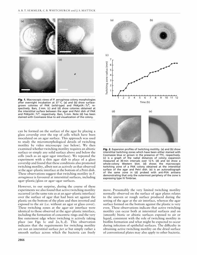
Fig. 3. Light microscopy of zones of twitching motility. (a) and (b) show typical colony expansion zones obtained at theinterstitial surface between the glass coverslip and Gel-Gro medium for PAK (twitching) and PAKpilA : :TcR (non-twitching) respectively. Micrographs were taken after 4–6 h incubation at 37 °C. Bars, 50 µm. ‘c’ indicates the colony; ‘m’indicates the uncolonized medium; the arrow indicates the radial direction of colony expansion.
On this basis we suggest that the use of smooth agarsurfaces (inverted after setting) may also allow twitchingmotility to be observed more easily in other type IVfimbriate species, such as N. gonorrhoeae and N.meningitidis, where thus far it has been very difficult toobserve this process in vitro. We are currently examiningthis possibility.
Microscopic analysis of the twitching zone
We have developed a system for light microscopicanalysis of twitching motility by adapting the methodoriginally developed for this purpose by Darzins (1993),wherein cells were point-inoculated onto an agar blockand covered by a glass slide, by replacing the agar witha thin layer of gellan gum (Gel-Gro) that gives muchgreater optical clarity. Under these conditions twitchingmotility is active and produces zones comparable to thatobserved on LB agar at the agar–plastic interface, asdiscussed above. Fig. 3(a) shows a typical micrograph ofa twitching zone under glass, in comparison with thatobserved with a pilA mutant (Fig. 3b). The micro-
morphological pattern of the twitching zone under glassdiffers substantially from that observed at the outermostedge of a surface-grown colony where, historically,twitching motility has been examined (see Bradley,1980; Henrichsen, 1972, 1983), but which we now knowto be a suboptimal environment for the process.
The twitching motility zone under optimal conditions iscomprised of three major micromorphological patterns(Fig. 3a). The leading edge of the twitching zone exhibitslarge rafts of 10–50 cells moving away from the colonyedge. These motile rafts are characteristic of twitchingmotility and have been described before (Darzins,1993; Bradley, 1980; Henrichsen, 1972). However, ourresults reveal that twitching motility can display fargreater complexity than has been previously described.Behind these rafts a strikingly intricate lattice-workpattern is formed comprised of structures consisting ofonly 1–5 cells in width. Closer to the colony, the spacesbetween the lattice are filled in, presumably due to celldivision. It is also worth noting that the twitching zoneis formed essentially of a single layer of cells. This is not
2867

A. B. T. SEMMLER, C. B. WHITCHURCH and J. S. MATTICK
.................................................................................................................................................................................................................................................................................................................
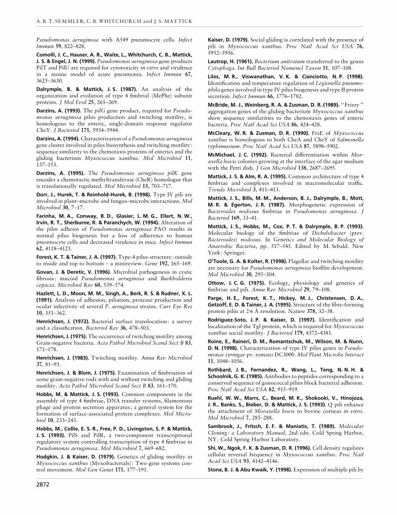
Fig. 4. PAK twitching motility filmed using time-lapse video microscopy. Captured frames shown here cover a 1 h periodof real time. A Quicktime video of this sequence (and others) can be found at http://www.cmcb.uq.edu.au/cmcb/PUBS/twitch.html
due to physical constraints as the centre of the colonyitself and the microcolonies within the twitching zoneare clearly capable of forming three-dimensional cellaggregates at the surface between agar or Gel-Gro andplastic or glass surfaces. Mutants which lack fimbriae donot exhibit raft or lattice formation (Fig. 3b). On aconventional agar surface (without a glass coverslip)(Fig. 1a) (see also Bradley, 1980; Henrichsen, 1972,1983), wild-type colonies do exhibit aggregates of cellsprotruding from the colony edge, but lack the highlystructured lattice-work observed under conditions
(smooth surfaces) which promote active twitchingmotility (see above).
Time-lapse video microscopy
Time-lapse video microscopy confirmed that the initialstages of twitching motility involve the formation oflarge rafts of aggregated cells moving away from thecolony edge (Fig. 4). These rafts move unidirectionally
2868

Twitching motility in P. aeruginosa
(a) (b).....................................................................................................
Fig. 5. Twitching motility. This sequencedemonstrates the twitching action oftenobserved when a cell snaps into alignmentwith neighbouring cells. The arrow indicatesa cell approaching a group of cells end onuntil it touches another cell with its pole(a) at which point it snaps rapidly intoalignment (b). This motility was filmed inreal time and consecutive frames are shown.
following the long axis of the cells which are in tightcell–cell contact and generally move radially outward,although they do meander. Occasionally within theserafts individual cells can be seen to reverse direction andmove back towards the colony, against the overallforward motion of the raft. Behind the rafts groups ofcells appear to stretch out and break up into smalleraggregates which move off in different directions andconnect with other groups (Fig. 4). This ultimately leadsto the formation of the fine lattice-like network observedin the twitching zone (Figs 3a and 4). The leadingaggregates appear to lay down ‘trails ’, along whichfollowing cells move rapidly up and down as individualunits or small groups, often reversing direction butalways following the long axis of the cell (Fig. 4). These‘ trails ’ are not visible but their presence is inferred bythe observation that following cells tend to followpathways that have been previously traversed by othercells. Once the lattice has been formed, the bacteriawithin mature sections appear to settle and divide to fillout the colonized space (Fig. 3a).
The movement of the cells within the twitching zone isinteresting. Individual cells appear to align with othersalong their long axis, apparently in intimate contact,and to slide by each other, either singly or in smallgroups. This is particularly noticeable and most activeduring the lattice formation, whereas the larger groupsof cells in the leading rafts tend to move more in unison,although individual cells do appear to move aroundwithin the raft. Nonetheless, individual cells movingwithin the trails}lattice network move more rapidlythan those in the rafts. In addition, reversal of movementalso occurs more often in these trails than in the rafts.
Careful examination of numerous time-lapse recordingsof twitching motility indicated that the movementinvolves cell–cell contact. Under the conditions usedhere, a single cell was never observed moving when wellseparated from other cells. Occasionally, an individualcell will move from one cluster to another but alwayswhen the clusters are in close proximity. Such a cell atfirst moves end on towards other cells until it touchesanother cell with its pole. It then immediately snaps intoan adjacent position alongside the other cell (Fig. 5).This ‘snapping’ behaviour is very rapid and accountsfor the jerky ‘ twitching’ which is characteristically seenwhen this motility is observed without time-lapse(Bradley, 1980; Henrichsen, 1972, 1983). Individual cells
are also seen frequently to move quickly along theoutside of an existing trail of cells. The cells in thenetwork appear to be in a generally constant state ofmovement, but individual cells can be stationary for aperiod and then suddenly move. All movement involvesat least one cell contacting another with its pole,consistent with the polar location of the fimbriae. Theimpression is that the cells are pulling and pushingagainst each other.
We have calculated the rates of cell movement duringtwitching motility by analysis of our time-lapse videorecordings. Under these conditions (Gel-Gro nutrientmedium at 30 °C) the leading edge rafts can move atspeeds of up to 6 µm min−", but can also temporarilystop, and normally move at a rate of about 3–4 µmmin−", consistent with macroscopic measurements ofthe radial expansion of the colony as a whole underthese same conditions, which was measured to be0±16 mm h−" over 16 h (about 2±6 µm min−" – see alsobelow). The rate of colony expansion due to cell divisionin the absence of twitching motility (calculated fromanalysis of time-lapse recordings of the pilA mutant)was only 0±1 µm min−", i.e. only a few percent of thatmediated by twitching motility in the wild-type. Withinthe developing lattice behind the outgoing rafts, in-dividual cells appeared to be more active and weremeasured to move up and down the trails at speeds ofabout 5–10 µm min−".
We have also observed that the rate of twitching motilityis dependent on growth conditions. Using LB agar wehave observed twitching-motility-mediated radial col-ony expansion rates of around 0±5–0±6 mm h−" (8–10 µmmin−") at 30–37 °C, which appears to be a consequenceof the higher nutrient levels in this agar than of the Gel-Gro nutrient medium used for microscopic studies.Twitching motility is stimulated (further) by higherlevels of tryptone and yeast extract, but not glucose(data not shown), and we are currently attempting toidentify the particular components which are respon-sible for this stimulation.
DISCUSSION
Our results indicate that under appropriate conditionstwitching motility in P. aeruginosa manifests as a rapid,highly organized mechanism of bacterial solid surfacetranslocation. This description differs significantly from
2869

A. B. T. SEMMLER, C. B. WHITCHURCH and J. S. MATTICK
the picture presented by earlier studies of twitchingmotility which essentially stated that twitching motilityis a relatively slow, disorganized mode of translocationin which cells move predominantly singly in an in-termittent and jerky fashion (Henrichsen, 1972). No-tably, these and other early studies examined twitchingmotility at the outer edge of the colony grown on an agarsurface solidified and dried in air, which is clearlysuboptimal for observing this process in vitro.
Conditions which appear to promote twitching motilityin P. aeruginosa are growth of the cells on smooth solidor semi-solid surfaces, such as agar set against plastic.Type IV fimbriae are expressed only in the outer activetwitching zone of expanding colonies (Fig. 2d, e),implying that type IV fimbriae are involved in bothadhesion to and movement across such surfaces, con-sistent with the known properties of these organelles.Type IV fimbriae and twitching motility have alsorecently been shown to be required for biofilm formationby P. aeruginosa on abiotic surfaces (O’Toole & Kolter,1998) and to be associated with the formation ofmicrocolonies on these surfaces. Our observation ofmicrocolonies within the twitching motility zone onagar culture is consistent with this and suggests that weare observing the rapid formation by twitching motilityof a biofilm on the agar or Petri dish surface. Biofilmformation is also important in P. aeruginosa infectionsparticularly in the lungs of cystic fibrosis patients wherethis pathogen grows as microcolonies embedded in theexopolysaccharide alginate (Govan & Deretic, 1996).Twitching motility and alginate production have beenshown to be coordinately (and probably reciprocally)regulated through AlgR (Whitchurch et al., 1996).Twitching motility therefore appears to be a keycomponent of biofilm formation on both abiotic andbiotic surfaces. In support of this, there is evidence tosuggest that twitching motility plays a significant role inthe colonization of epithelial surfaces in animals and thesubsequent spread of infection as mutants which retaintype IV fimbriae, but have lost twitching motility, havereduced infectivity (Hazlett et al., 1991; Comolli et al.,1999).
Our observations indicate that twitching motility sharesmany points of similarity with another mechanism ofbacterial translocation known as gliding motility and isalmost certainly synonymous with it. In Henrichsen’s1972 review, in which he surveyed and attempted toclassify the various forms of bacterial surface trans-location that had been reported in the literature up untilthat time, he states (in reference to gliding motility in arange of bacteria, notably fruiting and non-fruitingmyxobacteria, such as Myxococcus and Cytophagaspp.) that, ‘Under conditions optimal for gliding, thecolonies will be seen as ‘‘completely flat, rapidlyspreading almost invisible swarms ’’ or as a spreading,rhizoid growth with a honeycomb appearance. Move-ment takes place mainly in ‘‘ spearheads ’’ (i.e.spearhead-shaped cell aggregates at the edge of thecolony), single isolated cells very rarely being motile,and the picture is one of a ‘‘changing dispersed border ’’
with interlacing bands being continuously rearranged. ’A similar description is given elsewhere in the samereview: ‘The cells are arranged in a loose pattern ofinterlacing bands of rafts and cells…Groups of cellsresembling spearheads are seen projecting outwards.The locomotion, which is principally seen in the groupsof cells, i.e. rafts and spearheads, and takes the directionof the longitudinal axis of the bacteria, gives rise to aconstantly changing picture, steadily gliding groups ofcells uniting or dividing…’ (Henrichsen, 1972).
This is an exact description of what we have observed byvideo microscopy as type IV fimbriae-dependent twitch-ing motility in P. aeruginosa at the agar}Gel-Gro–plastic}glass interface. Henrichsen also reported thatgliding motility is confined to solid surfaces such as glassand agar, requires high humidity and that at least somespecies which exhibit gliding motility possess surfacefilaments of 4–8 nm, all of which are characteristic oftwitching motility or type IV fimbriae. It thereforeappears that Henrichsen’s conclusion that twitching andgliding motility are distinctive processes reflects arte-factual differences observed in vitro in different speciesdue to suboptimal environmental conditions, as well asperhaps in some cases species-specific idiosyncrasies inthe process or its regulation and directional control. Infact, prior to Henrichsen’s review there had been somedebate as to whether or not twitching motility andgliding motility were the same process (see Henrichsen,1972 and references therein).
The conclusion that twitching and gliding motility arealternative names for essentially the same process is alsosupported by microscopic and molecular genetic studiesof gliding motility in M. xanthus, wherein the processhas been extensively studied over the past decade.Gliding motility in this species has been separated intotwo facets, namely ‘adventurous motility ’, involving themovement of single cells away from the colony by anunknown mechanism, and ‘social motility ’, which hasmany micromorphological similarities with our obser-vations of twitching motility. Both twitching and socialgliding motility appear to involve trail formation. Inboth cases, cells moving within large rafts do not reverseas frequently as cells moving in trails (Shi et al., 1996).Our observation that in twitching motility isolated cellsare incapable of motility and that translocation onlyoccurs when cells are in close proximity suggests thatcell–cell interactions are required for motility, which isconsistent with observations of social motility in M.xanthus A−S+ mutants (Hodgkin & Kaiser, 1979).
Furthermore, social gliding motility in M. xanthus hasbeen recently shown to be dependent on type IVfimbriae, based on the discovery that several genesrequired for this process (pilA, pilS, pilR, pilB, pilC,pilD, pilT and pilQ) (Wu & Kaiser, 1995, 1997; Wu etal., 1997; Wall et al., 1999) are homologous to genespreviously identified in P. aeruginosa as being requiredfor fimbrial biogenesis and twitching motility (Alm &Mattick, 1997), consistent with earlier electron micro-scopic studies that indicated that pili or fimbriae were
2870

Twitching motility in P. aeruginosa
important in social motility (Kaiser, 1979). At the sametime, studies of twitching-impaired mutants of P.aeruginosa showed that this process was dependent on aset of genes (pilGHIJKL, chpAB) encoding a chemotacticsignal transduction system (Darzins, 1993, 1994,1995; Alm & Mattick, 1997), similar to that involved incontrolling flagellar-mediated swimming in enteric bac-teria, but which is more complex and most closelyrelated to an equivalent set of genes (frzABCDEFG)controlling social gliding motility in M. xanthus(McBride et al., 1989; McCleary & Zusman, 1990).
The mechanism of twitching}gliding motility remains amystery. The comparative molecular genetics, genomicsand biochemistry of twitching}gliding motility in P.aeruginosa and M. xanthus adds a powerful newdimension to dissecting this process. Clearly, in bothcases type IV fimbriae are involved, as is cell–cell contactinvolving at least one pole, where the fimbriae arelocated. In P. aeruginosa, in most cases (except whencells intersect at an angle), the cells are aligned alongtheir long axis and appear to slide by each other. Cellalignment also appears to be involved in M. xanthusgliding motility and to be important in a process termed‘stimulation’ whereby M. xanthus tgl− mutants whichlack type IV fimbriae and gliding motility can be rescuedphenotypically in both respects by physical contact withtgl+ cells (Rodriguez-Soto & Kaiser, 1997; Wall et al.,1998; Wall & Kaiser, 1998). Importantly, this stimu-lation involves Tgl-mediated stimulation of fimbrialassembly from pre-existing pilin and to lead to a rate offimbrial elongation in tgl− mutants that is similar towild-type, which is broadly consistent with the con-clusion from electron microscopic studies of P.aeruginosa that type IV fimbriae can extend and retract(Bradley, 1972a, b, 1974). Comparison of proteinsknown to be involved in twitching motility in P.aeruginosa to Tgl indicates that PilF is significantlysimilar to Tgl across the length of the protein and thusmay serve an equivalent function in twitching motilityas a trigger for contact-initiated fimbrial assembly}extension}retraction in this organism (Watson et al.,1996a; J. S. Mattick & C. B. Whitchurch, unpublishedobservations). This hypothesis is currently being tested.
The helical structure of the fimbrial strand stronglyimplies that the motive force for twitching}glidingmotility is transduced by fimbrial rotation, either as partof the extension}retraction of the fimbrial strand per se(Mattick & Alm, 1995) or by rotational forces appliedexternally to the cell or adjacent cells. In this context itis interesting to note that electron microscopic exam-ination of cells within the colony of the twitchingbacterium N. gonorrhoeae has demonstrated that thetype IV fimbriae of this bacterium wrap around the cellsfrom which they emanate as thin, individual structures,but also form thick bundles which branch and subdivideto interconnect cells within the colony (Todd et al.,1984). It is our impression that the cells may beswivelling during twitching in P. aeruginosa, but themicrographic resolution is insufficient to be confidentabout this. Whether twitching}gliding motility is
induced by the fimbriae acting in cis on their home cellor in trans by pushing or pulling adjacent cells remainsto be determined. In relation to the latter, our videomicroscopy of P. aeruginosa, especially of isolated cellsthat are being reconnected to existing trails, wouldsuggest that the cells are, if anything, being pulled ratherthan pushed.
Despite semantic differences and varying use of no-menclature, there now appears to be three systems ofbacterial motility that are genetically and morpho-logically well-defined: (1) swimming motility, whichoccurs in fluid media and is mediated by reversiblerotation of flagella ; (2) swarming motility, which occurson solid surfaces and is mediated by elongated andperitrichously hyperflagellated swarmer cells ; and (3)twitching motility}social gliding motility which alsooccurs on solid surfaces and is mediated by type IVfimbriae. Interestingly, we have recently found that P.aeruginosa is also capable of another form of motilitywhich resembles swarming, and that this activity canoccur in concert with twitching motility, although it isdistinct from it (C. B. Whitchurch & J. S. Mattick,unpublished observations). Thus P. aeruginosa appearsto have the capacity for all three of these forms ofmotility, under different circumstances, further evidenceof the versatility of this organism.
ACKNOWLEDGEMENTS
Annalese B. T. Semmler and Cynthia B. Whitchurch con-tributed equally to this work. This work was supported bygrants to C.B.W. and J.S.M. by the National Health andMedical Research Council of Australia. The Centre forMolecular and Cellular Biology is a Special Research Centreof the Australian Research Council.
REFERENCES
Alm, R. A. & Mattick, J. S. (1995). Identification of a gene, pilV,required for type 4 fimbrial biogenesis in Pseudomonas aeruginosawhose product possesses a prepilin-like leader sequence. MolMicrobiol 16, 485–496.
Alm, R. A. & Mattick, J. S. (1997). Genes involved in the biogenesisand function of type-4 fimbriae in Pseudomonas aeruginosa. Gene192, 89–98.
Barnett, T. C., Kirov, S. M., Strom, M. S. & Sanderson, K. (1997).Aeromonas spp. possess at least two distinct type IV pilusfamilies. Microb Pathog 23, 241–247.
Berg, H. C. & Turner, L. (1995). Cells of Escherichia coli swimeither end forward. Proc Natl Acad Sci USA 92, 477–479.
Bradley, D. E. (1972a). Evidence for the retraction of Pseudomonasaeruginosa RNA phage pili. Biochem Biophys Res Commun 47,142–149.
Bradley, D. E. (1972b). Shortening of Pseudomonas aeruginosapili after RNA-phage adsorption. J Gen Microbiol 72, 303–319.
Bradley, D. E. (1974). The adsorption of Pseudomonas aeruginosapilus-dependent bacteriophages to a host mutant with non-retractile pili. Virology 58, 149–163.
Bradley, D. E. (1980). A function of Pseudomonas aeruginosaPAO pili : twitching motility. Can J Microbiol 26, 146–154.
Chi, E., Mehl, T., Nunn, D. & Lory, S. (1991). Interaction of
2871
A. B. T. SEMMLER, C. B. WHITCHURCH and J. S. MATTICK
Pseudomonas aeruginosa with A549 pneumocyte cells. InfectImmun 59, 822–828.
Comolli, J. C., Hauser, A. R., Waite, L., Whitchurch, C. B., Mattick,J. S. & Engel, J. N. (1999). Pseudomonas aeruginosa gene productsPilT and PilU are required for cytotoxicity in vitro and virulencein a mouse model of acute pneumonia. Infect Immun 67,3625–3630.
Dalrymple, B. & Mattick, J. S. (1987). An analysis of theorganization and evolution of type 4 fimbrial (MePhe) subunitproteins. J Mol Evol 25, 261–269.
Darzins, A. (1993). The pilG gene product, required for Pseudo-monas aeruginosa pilus production and twitching motility, ishomologous to the enteric, single-domain response regulatorCheY. J Bacteriol 175, 5934–5944.
Darzins, A. (1994). Characterization of a Pseudomonas aeruginosagene cluster involved in pilus biosynthesis and twitching motility :sequence similarity to the chemotaxis proteins of enterics and thegliding bacterium Myxococcus xanthus. Mol Microbiol 11,137–153.
Darzins, A. (1995). The Pseudomonas aeruginosa pilK geneencodes a chemotactic methyltransferase (CheR) homologue thatis translationally regulated. Mol Microbiol 15, 703–717.
Dorr, J., Hurek, T. & Reinhold-Hurek, B. (1998). Type IV pili areinvolved in plant–microbe and fungus–microbe interactions. MolMicrobiol 30, 7–17.
Farinha, M. A., Conway, B. D., Glasier, L. M. G., Ellert, N. W.,Irvin, R. T., Sherburne, R. & Paranchych, W. (1994). Alteration ofthe pilin adhesin of Pseudomonas aeruginosa PAO results innormal pilus biogenesis but a loss of adherence to humanpneumocyte cells and decreased virulence in mice. Infect Immun62, 4118–4123.
Forest, K. T. & Tainer, J. A. (1997). Type-4 pilus-structure : outsideto inside and top to bottom – a minireview. Gene 192, 165–169.
Govan, J. & Deretic, V. (1996). Microbial pathogenesis in cysticfibrosis : mucoid Pseudomonas aeruginosa and Burkholderiacepacia. Microbiol Rev 60, 539–574.
Hazlett, L. D., Moon, M. M., Singh, A., Berk, R. S. & Rudner, X. L.(1991). Analysis of adhesion, piliation, protease production andocular infectivity of several P. aeruginosa strains. Curr Eye Res10, 351–362.
Henrichsen, J. (1972). Bacterial surface translocation: a surveyand a classification. Bacteriol Rev 36, 478–503.
Henrichsen, J. (1975). The occurrence of twitching motility amongGram-negative bacteria. Acta Pathol Microbiol Scand Sect B 83,171–178.
Henrichsen, J. (1983). Twitching motility. Annu Rev Microbiol37, 81–93.
Henrichsen, J. & Blom, J. (1975). Examination of fimbriation ofsome gram-negative rods with and without twitching and glidingmotility. Acta Pathol Microbiol Scand Sect B 83, 161–170.
Hobbs, M. & Mattick, J. S. (1993). Common components in theassembly of type 4 fimbriae, DNA transfer systems, filamentousphage and protein secretion apparatus ; a general system for theformation of surface-associated protein complexes. Mol Micro-biol 10, 233–243.
Hobbs, M., Collie, E. S. R., Free, P. D., Livingston, S. P. & Mattick,J. S. (1993). PilS and PilR, a two-component transcriptionalregulatory system controlling transcription of type 4 fimbriae inPseudomonas aeruginosa. Mol Microbiol 7, 669–682.
Hodgkin, J. & Kaiser, D. (1979). Genetics of gliding motility inMyxococcus xanthus (Myxobacterials) : Two gene systems con-trol movement. Mol Gen Genet 171, 177–191.
Kaiser, D. (1979). Social gliding is correlated with the presence ofpili in Myxococcus xanthus. Proc Natl Acad Sci USA 76,5952–5956.
Lautrop, H. (1961). Bacterium anitratum transferred to the genusCytophaga. Int Bull Bacteriol Nomencl Taxon 11, 107–108.
Liles, M. R., Viswanathan, V. K. & Cianciotto, N. P. (1998).Identification and temperature regulation of Legionella pneumo-phila genes involved in type IV pilus biogenesis and type II proteinsecretion. Infect Immun 66, 1776–1782.
McBride, M. J., Weinberg, R. A. & Zusman, D. R. (1989). ‘‘Frizzy ’’aggregation genes of the gliding bacterium Myxococcus xanthusshow sequence similarities to the chemotaxis genes of entericbacteria. Proc Natl Acad Sci USA 86, 424–428.
McCleary, W. R. & Zusman, D. R. (1990). FrzE of Myxococcusxanthus is homologous to both CheA and CheY of Salmonellatyphimurium. Proc Natl Acad Sci USA 87, 5898–5902.
McMichael, J. C. (1992). Bacterial differentiation within Mor-axella bovis colonies growing at the interface of the agar mediumwith the Petri dish. J Gen Microbiol 138, 2687–2695.
Mattick, J. S. & Alm, R. A. (1995). Common architecture of type 4fimbriae and complexes involved in macromolecular traffic.Trends Microbiol 3, 411–413.
Mattick, J. S., Bills, M. M., Anderson, B. J., Dalrymple, B., Mott,M. R. & Egerton, J. R. (1987). Morphogenetic expression ofBacteroides nodosus fimbriae in Pseudomonas aeruginosa. JBacteriol 169, 33–41.
Mattick, J. S., Hobbs, M., Cox, P. T. & Dalrymple, B. P. (1993).Molecular biology of the fimbriae of Dichelobacter (prev.Bacteroides) nodosus. In Genetics and Molecular Biology ofAnaerobic Bacteria, pp. 517–545. Edited by M. Sebald. NewYork: Springer.
O’Toole, G. A. & Kolter, R. (1998). Flagellar and twitching motilityare necessary for Pseudomonas aeruginosa biofilm development.Mol Microbiol 30, 295–304.
Ottow, J. C. G. (1975). Ecology, physiology and genetics offimbriae and pili. Annu Rev Microbiol 29, 79–108.
Parge, H. E., Forest, K. T., Hickey, M. J., Christensen, D. A.,Getzoff, E. D. & Tainer, J. A. (1995). Structure of the fibre-formingprotein pilin at 2±6 A/ resolution. Nature 378, 32–38.
Rodriguez-Soto, J. P. & Kaiser, D. (1997). Identification andlocalization of the Tgl protein, which is required for Myxococcusxanthus social motility. J Bacteriol 179, 4372–4381.
Roine, E., Raineri, D. M., Romantschuk, M., Wilson, M. & Nunn,D. N. (1998). Characterization of type IV pilus genes in Pseudo-monas syringae pv. tomato DC3000. Mol Plant Microbe Interact11, 1048–1056.
Rothbard, J. B., Fernandez, R., Wang, L., Teng, N. N. H. &Schoolnik, G. K. (1985). Antibodies to peptides corresponding to aconserved sequence of gonococcal pilins block bacterial adhesion.Proc Natl Acad Sci USA 82, 915–919.
Ruehl, W. W., Marrs, C., Beard, M. K., Shokooki, V., Hinojoza,J. R., Banks, S., Bieber, D. & Mattick, J. S. (1993). Q pili enhancethe attachment of Moraxella bovis to bovine corneas in vitro.Mol Microbiol 7, 285–288.
Sambrook, J., Fritsch, E. F. & Maniatis, T. (1989). MolecularCloning: a Laboratory Manual, 2nd edn. Cold Spring Harbor,NY: Cold Spring Harbor Laboratory.
Shi, W., Ngok, F. K. & Zusman, D. R. (1996). Cell density regulatescellular reversal frequency in Myxococcus xanthus. Proc NatlAcad Sci USA 93, 4142–4146.
Stone, B. J. & Abu Kwaik, Y. (1998). Expression of multiple pili by
2872

Twitching motility in P. aeruginosa
Legionella pneumophila : identification and characterization of atype IV pilin gene and its role in adherence to mammalian andprotozoan cells. Infect Immun 66, 1768–1775.
Todd, W. J., Wray, G. P. & Hitchcock, P. J. (1984). Arrangement ofpili in colonies of Neisseria gonorrhoeae. J Bacteriol 159, 312–320.
Wall, D. & Kaiser, D. (1998). Alignment enhances the cell-to-celltransfer of pilus phenotype. Proc Natl Acad Sci USA 95,3054–3058.
Wall, D., Wu, S. S. & Kaiser, D. (1998). Contact stimulation of Tgland type IV pili in Myxococcus xanthus. J Bacteriol 180, 759–761.
Wall, D., Kolenbrander, P. E. & Kaiser, D. (1999). The Myxococcusxanthus pilQ (sglA) gene encodes a secretin homolog required fortype IV pilus biogenesis, social motility, and development. JBacteriol 181, 24–33.
Watson, A. A., Alm, R. A. & Mattick, J. S. (1996a). Identification ofa gene, pilF, that is required for type 4 fimbrial biogenesis andtwitching motility in Pseudomonas aeruginosa. Gene 180, 49–56.
Watson, A. A., Mattick, J. S. & Alm, R. A. (1996b). Functionalexpression of heterologous type 4 fimbriae in Pseudomonasaeruginosa. Gene 175, 143–150.
Whitchurch, C. B., Alm, R. A. & Mattick, J. S. (1996). Co-ordinateregulation of fimbrial function and alginate production inPseudomonas aeruginosa. Proc Natl Acad Sci USA 93, 9839–9843.
Wu, S. S. & Kaiser, D. (1995). Genetic and functional evidence thattype IV pili are required for social gliding motility in Myxococcusxanthus. Mol Microbiol 18, 547–558.
Wu, S. S. & Kaiser, D. (1997). Regulation of expression of the pilAgene in Myxococcus xanthus. J Bacteriol 179, 7748–7758.
Wu, S. S., Wu, J. & Kaiser, D. (1997). The Myxococcus xanthuspilT locus is required for social gliding motility although pili arestill produced. Mol Microbiol 23, 109–121.
.................................................................................................................................................
Received 12 March 1999; revised 4 June 1999; accepted 9 June 1999.
2873
Copyright © 2022 FDOKUMEN