High-Throughput Phenotypic Characterization of Pseudomonas aeruginosa Membrane Transport Genes
Upload
independentCategory
view
3download
0
Microbiology (2000), 146, 2425–2434 Printed in Great Britain
Vanadium interferes with siderophore-mediated iron uptake in Pseudomonasaeruginosa
Christine Baysse,1 Daniel De Vos,1† Yann Naudet,1 Alain Vandermonde,1
Urs Ochsner,2 Jean-Marie Meyer,3 Herbert Budzikiewicz,4
Matthias Scha$ fer,4 Regine Fuchs4 and Pierre Cornelis1
Author for correspondence: Pierre Cornelis. Tel : 32 2 3590221. Fax: 32 2 3590390.e-mail : pcornel!vub.ac.be
1 Laboratory of MicrobialInteractions, Departmentof Immunology,Parasitology andUltrastructure, FlandersInteruniversity Institute ofBiotechnology and VrijeUniversiteit Brussel,Paardenstraat 65, B-1640Sint Genesius Rode,Belgium
2 University of ColoradoHealth Sciences Center,Microbiology, Box B-175,4200 E Ninth Avenue,Denver, CO 80202, USA
3 Laboratoire deMicrobiologie et deGe! ne! tique, Universite!Louis Pasteur, UPRES-A7010, F-67000 Strasbourg,France
4 Institut fu$ r OrganischeChemie der Universita$ tzu Ko$ ln, Greinstrasse 4,D-50939 Ko$ ln, Germany
Vanadium is a metal that under physiological conditions can exist in twooxidation states, V(IV) (vanadyl ion) and V(V) (vanadate ion). Here, it wasdemonstrated that both ions can form complexes with siderophores.Pseudomonas aeruginosa produces two siderophores under iron-limitingconditions, pyoverdine (PVD) and pyochelin (PCH). Vanadyl sulfate, at aconcentration of 1–2 mM, strongly inhibited growth of P. aeruginosa PAO1,especially under conditions of severe iron limitation imposed by the presenceof non-utilizable Fe(III) chelators. PVD-deficient mutants were more sensitiveto vanadium than the wild-type, but addition of PVD did not stimulate theirgrowth. Conversely, PCH-negative mutants were more resistant to vanadiumthan the wild-type strain. Both siderophores could bind and form complexeswith vanadium after incubation with vanadyl sulfate (1:1, in the case of PVD;2:1, in the case of PCH). Although only one complex with PVD, V(IV)–PVD, wasfound, both V(IV)– and V(V)–PCH were detected. V–PCH, but not V–PVD, causedstrong growth reduction, resulting in a prolonged lag phase. Exposure of PAO1cells to vanadium induced resistance to the superoxide-generating compoundparaquat, and conversely, exposure to paraquat increased resistance to V(IV).Superoxide dismutase (SOD) activity of cells grown in the presence of V(IV)was augmented by a factor of two. Mutants deficient in the production of Fe-SOD (SodB) were particularly sensitive to vanadium, whilst sodA mutantsdeficient for Mn-SOD were only marginally affected. In conclusion, it issuggested that V–PCH catalyses a Fenton-type reaction whereby the toxicsuperoxide anion ON2 is generated, and that vanadium compromises PVDutilization.
Keywords : Pseudomonas, vanadium, siderophores, oxidative stress
INTRODUCTION
Vanadium is a transition metal which, at neutral pH,can exist in two oxidation states, V(IV) (vanadylion; cationic species, VO+
#), and V(V) (vanadate ion;
anionic species, H#VO−
%) (Rehder, 1991, 1992). As a
.................................................................................................................................................
† Present address: Brandwonden Centrum, Militair Hospitaal KoninginAstrid, B-1120 Brussels, Belgium.
Abbreviations: CAS, chrome azurol ; EDDHA, ethylenediamine-di(o-hydroxyphenylacetic acid) ; ESI, electrospray ion; PCH, pyochelin; PVD,pyoverdine; SOD, superoxide dismutase.
result, vanadium can affect diverse biological processes.Vanadate resembles phosphate (HPO#−
%), and conse-
quently can take its place in phosphate-metabolizingenzymes (Rehder, 1991, 1992). Another important bio-logical characteristic of both vanadate and vanadyl ionsis that they can form complexes with carboxylate,catecholate and hydroxamate ligands, which are presentin different siderophores (Keller et al., 1991; Rehder,1991). Although this last characteristic is well known,surprisingly, there has been no study done so far onthe influence of vanadate or vanadyl ions on thesiderophore-mediated iron(III) uptake systems in Gram-negative bacteria. Such an investigation is justified if one
0002-4135 # 2000 SGM 2425

C. BAYSSE and OTHERS
Table 1. Bacterial strains and plasmids used in this study
Strain or plasmid Characteristics Reference
Strains
P. aeruginosa
ATCC 27853 Source of type II PVD Meyer et al. (1997)
PA6 Source of type III PVD Meyer et al. (1997)
PAO1 Wild-type; source of type I PVD ATCC 15692; Meyer et al. (1997)
PAO 6609 PVD− PCH+ Hohnadel et al. (1986)
PAO 66024 PVD− PCH+ Hohnadel et al. (1986)
PAO 6284 pchA (PVD+ PCH− Sal−) C. Reimmann (University of
Lausanne, Switzerland)
PAO 6285 pchD :Ω (PVD+ PCH− Sal+) Serino et al. (1997)
PAO 128-5 pvdB pchD (PVD− PCH− Sal+) C. Reimmann (University of
Lausanne, Switzerland)
PAO 128-6 pvdB pchA (PVD− PCH− Sal−) Serino et al. (1995)
PAO sodA : :Gmr Gmr sodA mutant of PAO1 This study
PAO sodA : :Gmr ∆sodB : :Tcr Gmr Tcr sodAsodB double mutant of PAO1 This study
FRD1 Wild-type cystic fibrosis isolate Goldberg & Ohman (1984)
FRD1 sodA sodA mutant of FRD1 Hassett et al. (1997b)
FRD1 sodB sodB mutant of FRD1 D. J. Hassett (University of
Cincinnati, USA)
FRD1 sodAsodB sodAsodB mutant of FRD1 Hassett et al. (1997b)
E. coli
DH5α-MCR Host for subcloning of plasmids F− lacZ∆M15
recA1 hsdR17 supE44 ∆(lacZYA argF)
Bethesda Research Laboratories
SM10 Kmr, mobilizer strain Simon et al. (1983)
Plasmids
pCRII-2.1 Apr Kmr TA cloning vector for PCR fragments Invitrogen
pEX100T Apr oriT mob sacB Schweizer & Hoang (1995)
pBR322 Apr Tcr, E. coli cloning vector Bethesda Research Laboratories
pUC19 Apr ColE1, E. coli cloning vector Yanisch-Perron et al. (1985)
considers the importance of siderophores for ironacquisition by pathogenic bacteria (Crosa, 1997). Fur-thermore, it is known that, at neutral pH, V(IV) can bereadily oxidized toV(V), generating the toxic superoxideanion (O−
#) (Liochev & Fridovich, 1987). It has also been
shown that complexation of vanadium by the sidero-phore desferrioxamine enhances its redox cycling ca-pacity in vitro (Stern et al., 1992). Despite this, theantibacterial activity of vanadium–siderophore com-plexes has never been investigated.
Under conditions of iron limitation, Pseudomonasaeruginosa produces two siderophores, pyochelin(PCH), a thiazolin derivative (Cox et al., 1981), andpyoverdine (PVD), a fluorescent siderophore, which iscomposed of a chromophore (a quinoline derivative)and a peptide arm (Budzikiewicz, 1993, 1997). PVD hasa much higher affinity for Fe(III) than PCH, which isconsistent with PVD-negative mutants being unableto grow in the presence of the strong iron chel-ator ethylenediamine-di(o-hydroxyphenylacetic acid)(EDDHA) and being avirulent in a mouse infectionmodel (Meyer et al., 1996). The production of sidero-phores and of the proteins required for their uptakeneeds to be tightly regulated in order to avoid wastage of
energy and accumulation of iron, which can be toxic forthe cell because free Fe+
#can generate toxic oxygen
radicals via the Fenton reaction.
Iron uptake in P. aeruginosa is controlled at differentlevels via a complex interaction of regulators (Vasil &Ochsner, 1999). The ferric uptake regulator (Fur) is ageneral regulator, which, together with Fe+
#as co-
repressor, represses the transcription of the pvdS gene,which encodes a sigma factor needed for the tran-scription of PVD biosynthetic genes. Fur also controlsthe transcription of pchR, a gene that encodes anactivator for the transcription of the PCH biosyntheticgenes. Using an elegant approach, Ochsner & Vasil(1996) found that Fur regulates a large array of genes inP. aeruginosa, including genes for putative siderophorereceptors, haem uptake, alternative sigma factors, two-component regulatory systems, regulators and otherunknown genes. Another gene repressed by Fur is sodA,which encodes the Mn-superoxide dismutase (SOD) ofP. aeruginosa (Hassett et al., 1997a). Two SODs areproduced by P. aeruginosa, a Mn-SOD induced underlow-iron conditions, and an Fe-SOD, which is thepredominant form (Hassett et al., 1993). These enzymescatalyse the dismutation of O−
#to H
#O
#(hydrogen
2426

P. aeruginosa and vanadium
peroxide) and O#; the peroxide, in turn, can be
converted to H#O and O
#by catalases. Both reactive
oxygen species are the product of the Fenton reactioncatalysed by free Fe+
#. Knowing that vanadium can
substitute for iron in siderophores and in some iron-proteins such as transferrin (Keller et al., 1991; Saponja& Vogel, 1996), we investigated whether vanadyl ionscould interfere with siderophore-mediated iron uptakesystems andor induce an oxidative stress in P.aeruginosa.
METHODS
Strains and growth conditions. The P. aeruginosa strains usedin this study are listed in Table 1. Cultures were grown inLuria–Bertani medium (LB) or in the iron-poor Casamino-acids medium (CAA) (Cornelis et al., 1992). Cultures weregrown in a Bioscreen apparatus (Life Technologies) using thefollowing parameters : shaking for 10 s every 3 min; readingevery 20 min; temperature, 37 °C; volume of culture, 300 µl.As inoculum, an overnight culture of PAO1 in CAA wasdiluted in order to achieve a final optical density at 600 nm of0±0001. Each culture was replicated three times and eachexperiment was performed in triplicate.
Vanadyl sulfate (VOSO%\5H
#O) was prepared as a 100 mM
stock solution and kept at 4 °C. Filter-sterilized FeCl$(0±1 M
in 0±1 M HCl) was added to the medium after autoclaving atthe concentrations indicated in the text. EDDHAwas preparedas a 100 mg ml−" solution (pH adjusted to 9) and filter-sterilized.
Detection and quantification of PVD in culture supernatants.PVD was detected in culture supernatants by measuring theabsorbance at 400 nm. The concentration of PVD wasestimated spectrophotometrically at 400 nm using a molarabsorption coefficient, ε
%!!, of 2 ¬ 10% M−" cm−" (Ho$ fte et al.,
1993) and normalized to the OD'!!
value of the culture.
To determine the effect of vanadium on PVD production,PAO1 cells were grown for 12 h in CAA containing 0, 0±5, 1±0or 1±5 mM VOSO
%and then diluted to start a new culture in
CAA for 48 h at 37 °C using the Bioscreen apparatus. Eachexperiment was performed in triplicate. The PVD content inthe supernatant was determined by measuring A
%!!.
Chrome azurol (CAS) detection of complex formation be-tween siderophores and vanadium. A vanadiumCAS sol-ution was prepared by a method adapted from the originalprotocol described by Schwyn & Neilands (1987). Six milli-litres of 10 mM hexadecyltrimethylammonium bromide(HDTMA) was first diluted to 20 ml with water, to which5±5 ml of 0±5 mM VOSO
%\5H
#O (in 10 mM HCl) was added.
To this solution, 7±5 ml of 2 mM CAS was added slowly underconstant agitation. Finally, 35 ml piperazine solution wereadded (4±3 g piperazine in 30 ml water and 5 ml pure HCl) and5-sulfosalicylic acid to a final concentration of 4 mM. Thefinal volume was adjusted to 100 ml. This purple vanadiumCAS solution could be poured as a gel after addition of 1%(wv) agarose.
Siderophore and vanadium–siderophore complex puri-fication. PVD (succinamide isoform) from P. aeruginosaPAO1 was isolated from CAA growth supernatants by thechloroformphenol method followed by chromatography onCM-Sephadex and elution with 0±1 M pyrimidineacetic acidbuffer pH 5, as previously described (Hohnadel & Meyer,1988). Eighty micromoles of the pure compound solubilized in
2 ml de-ionized water was supplemented with a slight excessof VOSO
%\5H
#O (100 µmol) and then filtered through a
Sephadex G25 column (2±5 ¬ 45 cm) eluted with de-ionizedwater. The major peak showing a brownish colour wascollected and lyophilized to obtain the dark brown compoundfurther analysed as a 1:1 V–PVD complex.
PCH was extracted with chloroform from pH-3-acidifiedgrowth supernatants and purified by Sephadex-LH20chromatography as described by Meyer et al. (1989). Com-plexation with vanadium was achieved by mixing 90 µmolPCH in methanol solution with 100 µmol VOSO
%\5H
#O. The
red-brown complex that formed instantaneously was purifiedby chromatography on Sephadex-LH20 (1±5 ¬ 30 cm column,elution with methanol).
Mass spectrometric analysis of vanadium–siderophore com-plexes. Mass spectra were obtained with a Finnigan MAT(Bremen) 900 ST instrument equipped with an electrosprayion (ESI) source, an electrostatic and a magnetic analyser, andan ion trap system.
Plate assay for sensitivity to paraquat. CAA or LB platescontaining 0, 0±5 or 1±0 mM VOSO
%\5H
#O were inoculated
with 10( cells (1 ml) from pre-cultures and dried for 1 h.Resistance to paraquat was analysed according to Hassett etal. (1995).
SOD activity measurements. Total SOD activity present in P.aeruginosa crude extracts was measured using the pyrogallolauto-oxidation inhibition assay described by Marklund &Marklund (1974). Briefly, 30 µl pyrogallol (20 mM), 30 µldiethylenetriaminepentaacetic acid (DTPA; 0±1 M) and 100 µlfreshly prepared crude cell extract (300 µg total protein) wereadded to 1±5 ml 0±1 M Triscacodylic acid pH 7±8; the finalvolume was adjusted to 3 ml with water. The change in A
%#!was monitored at 25 °C for 20 min. In the control auto-oxidation sample, the crude extract was replaced by 50 mMsodium phosphate buffer (cell lysis buffer ; pH 7). P.aeruginosa cell-free extracts were prepared from overnightcultures in CAA or in CAA containing 0±5 or 1±0 mM VOSO
%as described by Clare et al. (1984) except that, instead of usinga French press, cells were lysed by sonication.
Construction of sodA and sodAsodB mutants of P. aeru-ginosa PAO1. A 1150 bp fragment containing the sodA regionwas PCR-amplified using the primers 5«-GATGTGGCGCT-GGAAAACAC and 5«-GCCAGTCGATCACGTTGTAG.The PCR product was cloned into pCRII-2.1 (Invitrogen), anda 1±7 kb gentamicin resistance (Gmr) cassette was cloned intothe unique HincII site within the sodA gene. The 3±2 kbsodA : :Gmr construct was excised from the pCRII-2.1 poly-linker with PvuII and ligated into the SmaI site of the genereplacement vector pEX100T, which allows sacB counter-selection (Schweizer & Hoang, 1995). Escherichia coli SM10containing pEX100T-sodA : :Gmr was used as the donor strainin a bi-parental mating with P. aeruginosa PAO1. Trans-conjugants were selected on BHI agar containing gentamicin(75 µg ml−") and irgasan (50 µg ml−"), and subsequently platedon LB agar containing gentamicin (75 µg ml−") and 5% (wv)sucrose. Successful double-crossover events resulting insodA : :Gmr mutants were monitored by the loss of apEX100T-encoded carbenicillin resistance (Cbr) marker. Theinsertion of the Gmr cassette into sodA was confirmed by PCRacross the sodA : :Gmr region using the primers above. Thisyielded a 2±8 kb product for sodA : :Gmr mutants compared toa 1±15 kb product for PAO1 wild-type (data not shown).
A 1380 bp PCR product containing the sodB region wasamplified with primers 5«-TGATGGTGGCGGCCATGATG
2427

C. BAYSSE and OTHERS
and 5«-ATCGCCATTTCCCGGTCGAG and ligated intopCRII-2.1. The PCR product was excised with HindIII andXbaI and cloned into pUC19. A 540 bp NcoI–HincII fragmentcontaining most of the sodB coding region was excised, theends of the sodB flanking regions were filled in with Klenowenzyme and ligated to a 1±4 kb tetracycline resistance (Tcr)cassette which had been obtained by cutting pBR322 withEcoRI and StyI followed by end-polishing. The 2±6 kb∆sodB : :Tcr construct was excised from the pUC19 polylinkerwith PvuII and ligated into the SmaI site of pEX100T. E.coli SM10 harbouring the resulting plasmid, pEX100T-∆sodB : :Tcr, was used as the donor strain in a biparentalmating with P. aeruginosa sodA : :Gmr. The mating and thesubsequent isolation of sodAsodB mutants were performedunder anaerobic conditions using Campy Pak jars withpalladium catalyst (Becton-Dickinson) in the presence of 1%(wv) potassium nitrate as an alternative electron acceptorduring anaerobic growth. Candidate sodA : :Gmr sodB : :Tcr
double mutants were initially selected on BHI agar containing1% (wv) KNO
$, tetracycline (150 µg ml−") and irgasan (50 µg
ml−"), followed by sacB counter-selection on LB agar con-taining 5% sucrose, 1% KNO
$and tetracycline (150 µg ml−").
The successful replacement of the sodB gene by the Tcr
cassette was verified by PCR across the sodB region using theabove primers, resulting in a 2±2 kb PCR product for thesodA : :Gmr sodB : :Tcr double mutant compared to a 1±38 kbproduct for the sodA : :Gmr single mutant (data not shown).
PCR was performed using Taq polymerase and custom-madeprimers (Bethesda Research Laboratories) in a Perkin-ElmerCetus thermal cycler, with 30 cycles of denaturing (1 min,94 °C), annealing (1 min, 54 °C) and extending (1 min per kbof DNA, 72 °C). The PCR products were purified in low-melting-point agarose gels, routinely cloned into pCRII-2±1(Invitrogen) and sequenced with Sequenase 2±0 (United StatesBiochemical) and M13 primers or custom-made 18-meroligonucleotides.
RESULTS
Effect of iron on the growth of P. aeruginosa PAO1in the presence of vanadium
Table 2 shows the effect of the addition of increasingconcentrations of VOSO
%(1±0, 1±5 and 2 mM) on the
growth of PAO1 under iron-limiting or iron-sufficientconditions. The following parameters were analysed:
Table 2. Effect of vanadium on growth of P. aeruginosa PAO1 in CAA medium.................................................................................................................................................................................................................................................................................................................
CAA medium was supplemented () or not supplemented (®) with 10 µM FeCl$. Results shown are means of five experiments.
Cells were grown at 37 °C in a Bioscreen apparatus as described in Methods. All cultures received the same initial inoculum.
…VOSO4 (mM) 0 1±0 1±5 2±0
…FeCl3 w u w u w u w u
Lag phase (min) 230 300 350 700 1040 1490 2120
Slope (OD'!!
min−") 3±1¬10−% 3±1¬10−$ 1±4 10−% 3±8¬10−$ 0±8¬10−% 4±2¬10−$ 3±4¬10−$
OD'!!
max 0±43 1±56 0±38 1±36 0±25 1±35 0±09 1±29Time to OD
'!!max (min) 3010 1030 3240 1580 3240 2420 3020
OD'!!
after 48 h 0±43 0±95 0±38 1±16 0±23 1±25 0±09 1±25
, No growth.
the duration of the lag phase, the slope of the exponentialphase (OD
'!!min−"), themaximal optical density (OD
'!!max) reached, the time to reach the maximal OD
'!!, and
the OD'!!
after 48 h of growth. Addition of 10 µM FeCl$
to the CAA medium resulted, as expected, in strongstimulation of growth as judged by the maximal OD
'!!reached and the increase in the slope. When iron waspresent, the addition of increasing concentrations ofVOSO
%resulted in a concentration-dependent increase
in the lag phase (up to 35 h for the highest concentrationof 2 mM), but once growth resumed, both the slope andthe maximal OD
'!!reached were relatively unchanged.
In the absence of iron, increased concentrations ofVOSO
%not only caused a prolongation of the lag phase,
but also decreased the slope and decreased the maximalOD
'!!. In the presence of 2 mM VOSO
%, no growth was
observed in CAA medium without added iron, evenafter 72 h.
Effect of Fe(III) chelators on the growth ofP. aeruginosa in the presence of vanadium
The effects of adding the strong, non-metabolizableFe(III) chelator EDDHA, the cognate purified PVD, anda heterologous PVD from P. aeruginosa ATCC 27853for which PAO1 has no receptor (Hohnadel & Meyer,1988), on the growth of PAO1 in CAA medium wastested after 24 h. As expected, addition of the cognatePVD (50 µM) in CAA medium stimulated growth (0±49OD
'!!units compared with 0±36 OD
'!!units for the
CAA control), whilst addition of EDDHA (0±5 mg ml−")or the heterologous PVD (50 µM) had little effect ongrowth (0±29 and 0±40 OD
'!!units, respectively). In the
presence of a subinhibitory concentration of vanadium(1 mM), the OD
'!!after 24 h (0±33 OD
'!!units) was
reduced compared to the CAA control. Strikingly, thecombination of 1 mM vanadium and EDDHA resultedin complete suppression of growth (0±09 OD
'!!units),
an effect which persisted even when incubation wasextended to 48 h (results not shown). Interestingly,addition of the cognate PVD in the presence of 1 mMVOSO
%did not stimulate growth (0±22 OD
'!!units),
whilst addition of the non-cognate PVD, as in the case of
2428

P. aeruginosa and vanadium
1·0
0·1
0·011000 2000 3000 4000
OD
600
Time (min)
.................................................................................................................................................
Fig. 1. Growth of P. aeruginosa PAO1 wild-type (E), and PVD−
mutants PAO 6609 (^) and PAO 66024 (*) in CAA mediumcontaining 1 mM VOSO4. Growth was measured in a Bioscreenapparatus at 37 °C as described in Methods. Results shown aremean values from triplicate cultures.
1·0
0·1
0·011000 2000 3000 4000
OD
600
Time (min)
.................................................................................................................................................
Fig. 2. Growth of P. aeruginosa PAO1 wild-type (E), PVD+ PCH−
mutants PAO 6284 (_) and 6285 (+), and PVD− PCH− mutantsPAO 128-5 (*) and PAO 128-6 (^) in CAA medium and in thepresence of 1±5 mM VOSO4. Growth was measured in aBioscreen apparatus at 37 °C as described in Methods. Resultsshown are mean values from triplicate cultures.
EDDHA, resulted in complete growth suppression (0±09OD
'!!units).
Inhibition of growth of wild-type PAO1 andsiderophore-negative mutants by vanadium
Fig. 1 shows the growth curves of wild-type PAO1 andtwo PVD-negative mutants. These mutants are non-fluorescent under UV, do not produce PVD and areunable to grow in the presence of the Fe(III) chelatorEDDHA. These two mutants grew in the presence of1 mM vanadium only after a prolonged lag phase.Conversely, all mutants defective in the production ofPCH showed an increased resistance to vanadium,compared to the wild-type, although to different extents(Fig. 2). The two PVD+ PCH− mutants especially were
found to be completely unaffected by the presence of1±5 mM VOSO
%, a concentration that severely com-
promised the growth of the wild-type strain. The PCH−
Sal+ and the PCH− Sal− mutants showed an intermediatelevel of resistance to vanadium. These results led us toconclude that the production of PVD contributes toresistance to vanadium whilst, conversely, the pro-duction of PCH enhances the toxic effect of vanadium.
Effect of vanadium on PVD production
We wanted to investigate if pre-incubation of the cellswith vanadium would affect PVD production since wehad observed that the characteristic fluorescence due toPVD production disappeared when cells were grown inCAA medium containing 0±5 mM VOSO
%. Compared to
the control condition (CAA medium, no VOSO%, 100%
PVD production), pre-culturing in the presence of 0±5,1±0 and 1±5 mM VOSO
%for 12 h caused a reduction in
PVD production in CAA medium of 20, 40 and 42%,respectively, whilst cell growth, as determined by OD
'!!,
was identical under each condition used. Conversely,PCH production was not affected by pre-exposure of thecells to 0±5 mM VOSO
%. The PCH content of super-
natants from cultures of the PVD− mutant 6609 grownin CAA was determined by liquid CAS assay (Schwyn &Neilands, 1987) and was found to be identical whetherthe pre-culture was performed in the presence or in theabsence of 0±5 mM VOSO
%.
Vanadium complexation by PVD and PCH
Spent medium of PVD-producing strains grown in thepresence of vanadium showed a characteristic browncolour that was not observed in spent medium of PVD-negative mutants. Furthermore, the fluorescence underUV, characteristic of free PVD, could not be detected incultures of P. aeruginosa grown in the presence of0±5 mM VOSO
%. This prompted us to investigate
whether PVD and PCH could complex vanadium. Forboth purified siderophores, a very clear shift of the UV-visible peaks was observed after addition of 1 mMVOSO
%, an indication that a complex was formed. The
following peaks were observed: purified free PVD atpH 5±2, 365 and 385 nm; PVD plus VOSO
%, 405 nm;
purified PCH in methanol, 218 and 248 nm; PCH plusVOSO
%, 232 and 273 nm. Using another approach to
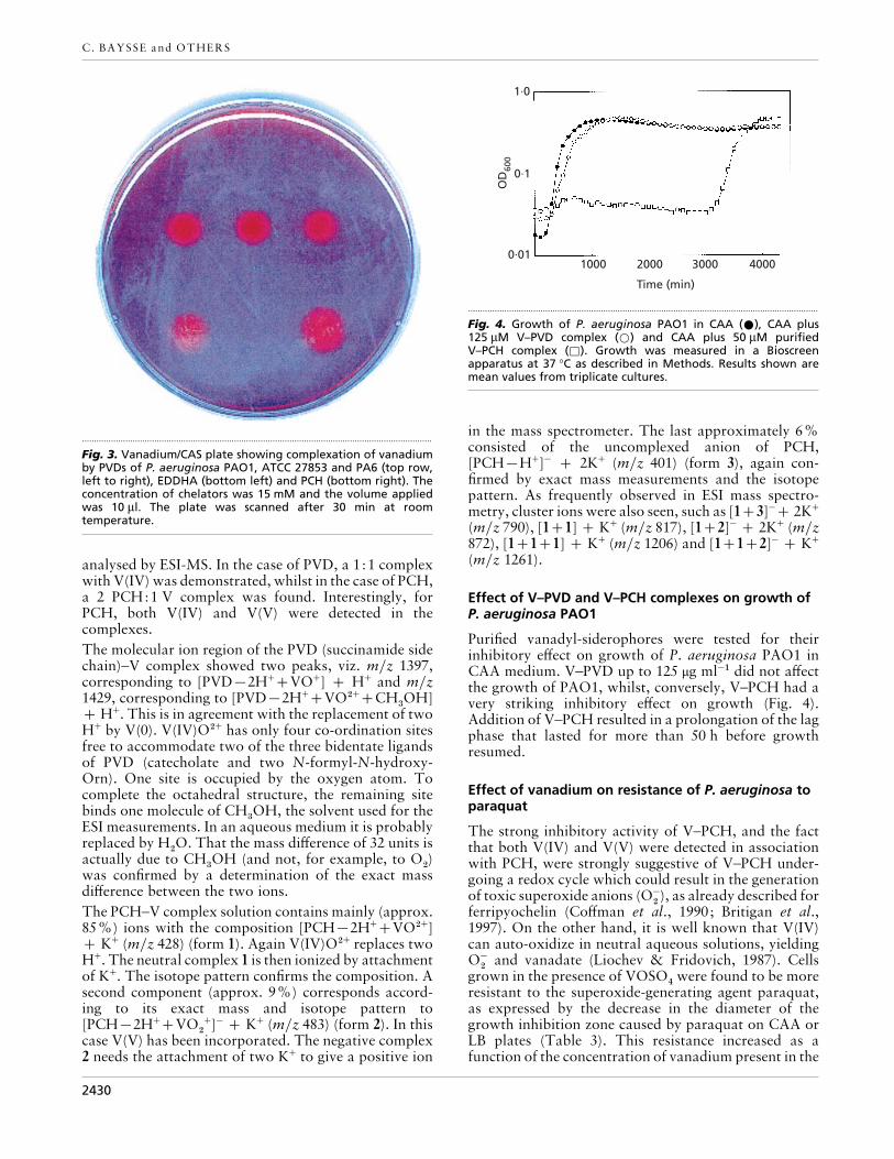
detect vanadium binding by both siderophores, avanadium-CAS assay was developed whereby iron wasreplaced by vanadium. Addition of both siderophores toa vanadiumCAS plate resulted in a rapid colour changedue to the de-complexation of vanadium–CAS (Fig. 3).
Mass spectrometric analysis of thevanadium–siderophore complexes
After binding with vanadium, the PCH– and PVD–Vcomplexes were purified as described in Methods and
2429
C. BAYSSE and OTHERS
.................................................................................................................................................
Fig. 3. Vanadium/CAS plate showing complexation of vanadiumby PVDs of P. aeruginosa PAO1, ATCC 27853 and PA6 (top row,left to right), EDDHA (bottom left) and PCH (bottom right). Theconcentration of chelators was 15 mM and the volume appliedwas 10 µl. The plate was scanned after 30 min at roomtemperature.
analysed by ESI-MS. In the case of PVD, a 1:1 complexwith V(IV) was demonstrated, whilst in the case of PCH,a 2 PCH:1 V complex was found. Interestingly, forPCH, both V(IV) and V(V) were detected in thecomplexes.
The molecular ion region of the PVD (succinamide sidechain)–V complex showed two peaks, viz. mz 1397,corresponding to [PVD®2H+VO+] H+ and mz1429, corresponding to [PVD®2H+VO#+CH
$OH]
H+. This is in agreement with the replacement of twoH+ by V(0). V(IV)O#+ has only four co-ordination sitesfree to accommodate two of the three bidentate ligandsof PVD (catecholate and two N-formyl-N-hydroxy-Orn). One site is occupied by the oxygen atom. Tocomplete the octahedral structure, the remaining sitebinds one molecule of CH
$OH, the solvent used for the
ESI measurements. In an aqueous medium it is probablyreplaced by H
#O. That the mass difference of 32 units is
actually due to CH$OH (and not, for example, to O
#)
was confirmed by a determination of the exact massdifference between the two ions.
The PCH–V complex solution contains mainly (approx.85%) ions with the composition [PCH®2H+VO#+] K+ (mz 428) (form 1). Again V(IV)O#+ replaces twoH+. The neutral complex 1 is then ionized by attachmentof K+. The isotope pattern confirms the composition. Asecond component (approx. 9%) corresponds accord-ing to its exact mass and isotope pattern to[PCH®2H+VO
#
+]− K+ (mz 483) (form 2). In thiscase V(V) has been incorporated. The negative complex2 needs the attachment of two K+ to give a positive ion
1·0
0·1
0·011000 2000 3000 4000
OD
600
Time (min)
.................................................................................................................................................
Fig. 4. Growth of P. aeruginosa PAO1 in CAA (E), CAA plus125 µM V–PVD complex (D) and CAA plus 50 µM purifiedV–PCH complex (*). Growth was measured in a Bioscreenapparatus at 37 °C as described in Methods. Results shown aremean values from triplicate cultures.
in the mass spectrometer. The last approximately 6%consisted of the uncomplexed anion of PCH,[PCH®H+]− 2K+ (mz 401) (form 3), again con-firmed by exact mass measurements and the isotopepattern. As frequently observed in ESI mass spectro-metry, cluster ions were also seen, such as [13]− 2K+
(mz 790), [11] K+ (mz 817), [12]− 2K+ (mz872), [111] K+ (mz 1206) and [112]− K+
(mz 1261).
Effect of V–PVD and V–PCH complexes on growth ofP. aeruginosa PAO1
Purified vanadyl-siderophores were tested for theirinhibitory effect on growth of P. aeruginosa PAO1 inCAA medium. V–PVD up to 125 µg ml−" did not affectthe growth of PAO1, whilst, conversely, V–PCH had avery striking inhibitory effect on growth (Fig. 4).Addition of V–PCH resulted in a prolongation of the lagphase that lasted for more than 50 h before growthresumed.
Effect of vanadium on resistance of P. aeruginosa toparaquat
The strong inhibitory activity of V–PCH, and the factthat both V(IV) and V(V) were detected in associationwith PCH, were strongly suggestive of V–PCH under-going a redox cycle which could result in the generationof toxic superoxide anions (O−
#), as already described for
ferripyochelin (Coffman et al., 1990; Britigan et al.,1997). On the other hand, it is well known that V(IV)can auto-oxidize in neutral aqueous solutions, yieldingO−
#and vanadate (Liochev & Fridovich, 1987). Cells
grown in the presence of VOSO%were found to be more
resistant to the superoxide-generating agent paraquat,as expressed by the decrease in the diameter of thegrowth inhibition zone caused by paraquat on CAA orLB plates (Table 3). This resistance increased as afunction of the concentration of vanadium present in the
2430

P. aeruginosa and vanadium
Table 3. Effect of pre-incubation with VOSO4 on the sensitivity of P. aeruginosa PAO1 toparaquat.....................................................................................................................................................................................................................................
The diameter of the growth inhibition zone around a drop of paraquat on a Petri plate (Hasset etal., 1995) was measured after 18 h (LB) or 48 h (CAA) incubation at 37 °C. Values are means (³0±2)of three separate experiments.
Medium Pre-incubation with
0±5 mM VOSO4
No VOSO4 VOSO4 (0±5 mM) VOSO4 (1 mM)
CAA – 2±4 2±0 1±3CAA 2±0 2±0 1±3LB – 1±9 1±6 1±3LB 1±4 1±2 1±3
Table 4. Effect of pre-incubation with VOSO4 on thegrowth of P. aeruginosa PAO1 in the presence of VOSO4.................................................................................................................................................
Cells were grown overnight in CAA containing 1 mM VOSO%
and 100 µM FeCl$at 37 °C and diluted to inoculate cultures in
the Bioscreen apparatus as described in Methods. Results areshown only for cultures grown in CAA plus FeCl
$(100 µM).
Values are means of five experiments.
VOSO4
(mM)
Induction with
1 mM VOSO4
Lag phase
(min)
OD600 min−1
0 – 220 4±3¬10−$
0 220 4±3¬10−$
1±0 – 240 2±1¬10−$
1±0 240 3±4¬10−$
1±5 – 280 1±2¬10−$
1±5 280 1±7¬10−$
medium. Pre-incubation of the cells with 0±5 mMVOSO
%also caused a decrease in the growth inhibition
caused by paraquat. In addition, we noticed that onCAA plates containing 1 mM VOSO
%a ring of increased
growth was visible at the limit of the paraquat growthinhibition zone (not shown). Altogether, these resultsstrongly suggest that vanadium can induce an oxidativeresponse and a defence mechanism in P. aeruginosa.Since both paraquat and vanadyl ions can generatesuperoxides, it is most likely that vanadyl ions caninduce a SOD activity in P. aeruginosa. To test thishypothesis, total SOD activity was measured by theinhibition of the auto-oxidation of pyrogallol in crudeextracts of P. aeruginosa PAO1 cells before and afterpre-incubation with 0±5 mM VOSO
%. The total SOD
activity in the conditions used was 10% before and 20%after exposure to VOSO
%(expressed as percentage of
inhibition of pyrogallol auto-oxidation). Such a doub-ling of SOD activity was consistently observed, con-firming that vanadyl ions indeed induce an oxidativestress and an adaptive response in P. aeruginosa.
Pre-exposure of bacterial cells to subinhibitoryvanadium concentrations induced a higher level ofresistance, resulting in an increased growth rate, but
1·0
0·1
0·011000 2000 3000 4000
OD
600
1·0
0·1
0·01500
Time (min)
1000 1500 2000 2500 3000
(a)
(b)
.................................................................................................................................................
Fig. 5. (a). Growth of P. aeruginosa PAO1 wild-type in CAA (E),sodA mutant in CAA (+), sodAsodB mutant in CAA (_), PAO1wild-type in CAA plus 1 mM VOSO4 (D), sodA in CAA plus 1 mMVOSO4 (*) and sodAsodB mutant in CAA plus 1 mM VOSO4(^). (b) Growth of wild-type strain FRD1 (E), FRD1 sodA (D),FRD1 sodB (^) and FRD1 sodAsodB (*) in CAA plus 1 mMVOSO4. Growth was measured in a Bioscreen apparatus at 37 °Cas described in Methods. Results shown are mean values fromtriplicate cultures.
only when iron was present during the pre-culture andthe culture (Table 4). Similar results were obtained in LBmedium (results not shown).
2431

C. BAYSSE and OTHERS
We also observed that PAO1 cells plated on P-agarcontaining 1 mM VOSO
%produced more pyocyanin
than on P-agar alone, an indication that the level of Fe-SOD might be elevated in the cells exposed to vanadium(Hassett et al., 1995).
Effect of mutations in sodA and sodB genes on theresistance of P. aeruginosa to vanadium
P. aeruginosa produces two SODs, one Mn-co-factored(SodA), and another Fe-co-factored (SodB) (Hassett etal., 1993). As already mentioned, resistance to vanadiumwas higher in the presence of iron (Table 2), indicatingthat SodB could be the major contributor to theresistance since the production of this haem-containingenzyme was shown to be increased under conditions ofiron sufficiency (Hassett et al., 1993). Furthermore, it isknown that SodB contributes more to resistance tosuperoxide induced by paraquat than SodA (Hasset etal., 1995).
SOD mutants from PAO1 and a cystic fibrosis strain,FRD1 (Hassett et al., 1997b), were grown in CAA or inCAA containing 1 mM VOSO
%. The results are shown
in Fig. 5(a) for strain PAO1 (wild-type, and sodA andsodAsodB mutants), and in Fig. 5(b) for strain FRD1(wild-type, and sodA, sodB and sodAsodB mutants).The growth of both sodA mutants (PAO1 and FRD1)was relatively unaffected by the presence of 1 mMVOSO
%compared to the respective wild-type strains.
However, the growth of the sodB (FRD1) mutant wasstrongly inhibited in the presence of vanadium. Thedouble sodAsodB mutants grew either very poorly (inthe case of FRD1) or not at all (PAO1). These resultsagain indicated that the resistance to vanadium-generated superoxides is largely due to SodB.
DISCUSSION
One of the main concerns for the treatment of bacterialinfections is the increasing proportion of antibiotic-resistant strains. Metals, such as silver and cerium, arealready used in topical applications because of theirantibacterial activity (Klasen, 2000). Vanadium salts areknown to exert, among others, anti-diabetic effects(Srivastava, 2000). Few reports mention the antibacterialeffects of vanadium salts. In the case of Streptococcuspneumoniae, both vanadate and vanadyl compoundswere found to have a strong antibacterial activity(Fukuda & Yamase, 1997). Although these authorsfound that vanadium salts had a negligible activitytowards other bacteria, including Staphylococcusaureus, E. coli and P. aeruginosa, it is important tomention the fact that they grew these bacteria only in theiron-rich Mueller–Hinton medium. In this study, wedemonstrate that vanadium salts inhibit the growth of P.aeruginosa, especially in an iron-limited medium. In aniron-replete medium, we observed that vanadium causeda prolongation of the lag phase without affecting theexponential phase. Conversely, under conditions of ironlimitation, the lag phase was less affected by the presenceof vanadium in the medium whilst the growth rate was
severely reduced. Although we do not at present have anexplanation for this phenomenon, it suggests thatvanadium can affect different functions depending onthe iron content of the cell.
The inhibitory effect of vanadium was strongly en-hanced by the presence of non-utilizable iron(III)chelators such as EDDHA or a heterologous, non-cognate PVD. This could mean that vanadium interfereswith siderophore-mediated iron uptake in P. aeruginosa.One way by which vanadium could affect iron uptake isvia the formation of stable vanadyl– or vanadate–siderophore complexes that, eventually, could be takenup by the cell, increasing the intracellular concentrationof vanadium. Alternatively, the vanadium–siderophorecomplexes could block the uptake of iron–siderophoresin a competitive way. The competition of vanadiumwith iron for binding by siderophores could explain whythe uptake of iron via the siderophores is compromised.However, we do not at this stage know whether thevanadyl–siderophores are actively taken up by the cells.
We demonstrate here that vanadium can indeed formcomplexes with the two siderophores of P. aeruginosa,PCH and PVD. This is not surprising since it is wellestablished that vanadium can be liganded by hydroxa-mates, carboxylates and catecholates (Keller et al., 1991;Rehder, 1991). Others have also demonstrated the factthat PVD, as well as PCH, can complex copper, zinc ormanganese (Poppe et al., 1987; Chen et al., 1994; Viscaet al., 1992; Bouby et al., 1999). Visca et al. (1992)suggested that PCH could play a role in the acquisitionof metals other than Fe(III), such as Co(II) and Mo(VI).Likewise, azotochelin, a catecholate siderophore, hasbeen suggested to participate in the uptake of mol-ybdenum by Azotobacter vinelandii (Duhme et al.,1998).
Here we show that the V–PCH complex, but not theV–PVD complex, has a strong antibacterial effect. Suchan effect was not observed by Visca et al. (1992) for PCHcomplexed with other metals, including Mo(VI) andCu(II). Interestingly, two forms of V–PCH complexeswere found, one with V(IV) and one with V(V). Thisresult suggests that V–PCH can undergo an oxidativecycle that can result in the generation of superoxideradicals (O−
#). It is well known that ferripyochelin can
undergo such a redox cycle, resulting in the productionof cell-damaging active oxygen species (Coffman et al.,1990; Britigan et al., 1997). Stern et al. (1992) alsodemonstrated that V(IV)–desferrioxamine has a redox-cycling activity and generates active oxygen species. Inanother study on A. vinelandii siderophores, Cornish &Page (1998) demonstrated that, among the threecatecholate siderophores produced by this bacterium,only aminochelin, which has the lowest affinity foriron(III), was unable to sequester iron and prevent aFenton reaction. Interestingly, the same authors alsoobserved that increased aeration resulted in a parallelincrease in the production all three catecholate sidero-phores of A. vinelandii, aminochelin, azotochelin and
2432

P. aeruginosa and vanadium
protochelin. Protochelin is a tri-catecholate with thehighest affinity for iron and is produced only underconditions of extreme oxygen stress. Like protochelin,PVD from P. aeruginosa can form a 1:1 complex withiron and protect the cells from oxidative damage due tothe Fenton reaction (Coffman et al., 1990). Indeed, theseauthors showed that O−
#could reduce and release iron
bound to Fe–PCH but not Fe–PVD.
The fact that PVD-negative mutants are more affectedby vanadium can be explained by their higher pro-duction of PCH (Ho$ fte et al., 1993), which results in anincrease in the toxicity of vanadium.
Another intriguing observation we report is the re-pression of PVD production when wild-type cells weregrown in the presence of vanadium. We can assume thatvanadium did not interfere with the normal Fur-mediated regulation since iron-repressed outer-mem-brane proteins are normally produced in CAA mediumcontaining vanadium and their production is repressedwhen iron is also present (results not shown).
We propose that V–PCH molecules undergo an oxi-dative cycle, resulting in the production of reactiveoxygen species that, in turn, induce a response in theform of increased SOD activity. We observed that a pre-incubation with vanadium salts increases the resistanceof PAO1 cells both to vanadium and to the redox-cycling agent paraquat, and causes an increase in SODactivity. Analysis of sod mutants indicated that Mn-SOD, encoded by sodA, only marginally participates inthe resistance towards vanadium in iron-limiting con-ditions, whereas SodB plays a major role. SodA isknown to be regulated by the Fur repressor (Hassett etal., 1996, 1997a), whilst not much is known about theregulation of SodB production in P. aeruginosa exceptthat its production is optimal under iron-sufficientconditions (Hassett et al., 1992). The pronouncedgrowth inhibitory effect of vanadium on iron-starvedcells could be the result of insufficient production of theiron-containing superoxide-detoxifying enzyme SodBunder these conditions. Further studies should includeexperiments designed to investigate whether vanadiumuptake by the cells is increased by siderophores. Also,the search for vanadium-resistant or vanadium-sus-ceptible mutants in P. aeruginosa should provide usefulinformation about the mechanisms involved in thehomeostasis of this metal, and the interaction betweensiderophores and oxidative stress in this bacterium.
ACKNOWLEDGEMENTS
We thank Dr Cornelia Reimman for sending us the PCHmutants, and Dr Dan Hassett for sending us the FRD1 strainand the different sod mutants as well as for interestingdiscussions. We also thank Dr Theresa Pattery for carefullyreading this manuscript. The Bioscreen apparatus wasacquired thanks to the FWO (krediet aan navorser). Thiswork was also supported by the Jean and Alphonse FortonFund.
REFERENCES
Bouby, M., Billard, I. & Maccordick, H. J. (1999). Selective behaviorof the siderophore pyoverdine A towards UO#+
#, Th%+, U%+ and
other cations. Czech J Phys 49, Suppl. 1, 147–150.
Britigan, B. E., Rasmussen, G. T. & Cox, C. D. (1997). Augmentationof oxidant injury to human pulmonary epithelial cells by thePseudomonas aeruginosa siderophore pyochelin. Infect Immun65, 1071–1076.
Budzikiewicz, H. (1993). Secondary metabolites from fluorescentpseudomonads. FEMS Microbiol Rev 104, 209–228.
Budzikiewicz, H. (1997). Siderophores of fluorescent pseudo-monads. Z Naturforsch 52C, 713–720.
Chen, Y., Jurkevitch, E., Bar-Ness, E. & Hadar, Y. (1994). Stabilityconstant of pseudobactin complexes with transition metals. SoilSci Soc Am J 58, 390–396.
Clare, D. A., Duong, M. N., Darr, D., Archibald, F. & Fridovich, I.(1984). Effects of molecular oxygen on detection of superoxideradical with nitroblue tetrazolium and on activity stains forcatalase. Anal Biochem 140, 532–537.
Coffman, T. J., Cox, C. D., Edeker, B. L. & Britigan, B. E. (1990).Possible role of bacterial siderophores in inflammation. J ClinInvest 86, 1030–1037.
Cornelis, P., Anjaiah, V., Koedam, N., Delfosse, P., Jacques, P.,Thonart, P. & Neirinckx, L. (1992). Stability, frequency andmultiplicity of transposon insertions in the pyoverdine region inthe chromosome of different fluorescent pseudomonads. J GenMicrobiol 138, 1337–1343.
Cornish, A. S. & Page, W. J. (1998). The catecholate siderophoresof Azotobacter vinelandii : their affinity for iron and role inoxygen stress management. Microbiology 144, 1747–1754.
Cox, C. D., Rinehart, K. L., Moore, M. L. & Cook, J. C. (1981).Pyochelin : novel structure of an iron chelating growth promoterfor Pseudomonas aeruginosa. Proc Natl Acad Sci U S A 78,4256–4260.
Crosa, J. H. (1997). Signal transduction and transcriptional andpostranscriptional control of iron-regulated genes in bacteria.Microbiol Mol Biol Rev 61, 319–336.
Duhme, A. K., Hider, R. C., Naldrett, M. J. & Pau, R. N. (1998). Thestability of the molybdenum–azotochelin complex and its effecton siderophore production in Azotobacter vinelandii. J Biol InorgChem 3, 520–526.
Fukuda, N. & Yamase, T. (1997). In vitro antibacterial activity ofvanadate and vanadyl compounds against Streptococcuspneumoniae. Biol Pharm Bull 20, 927–930.
Goldberg, J. B. & Ohman, D. E. (1984). Cloning and expression inPseudomonas aeruginosa of a gene involved in the production ofalginate. J Bacteriol 158, 1115–1121.
Hassett, D. J., Charniga, L., Bean, K. A., Ohman, D. E. & Cohen,M. S. (1992). Response of Pseudomonas aeruginosa to pyocyanin:mechanisms of resistance, antioxidant defenses, and demon-stration of a manganese-cofactored superoxide dismutase. InfectImmun 60, 328–336.
Hassett, D. J., Woodruff, W. A., Wozniak, D. J., Vasil, M. L., Cohen,M. S. & Ohman, D. E. (1993). Cloning and characterization of thePseudomonas aeruginosa sodA and sodB genes encodingmanganese- and iron-cofactored superoxide dismutase : dem-onstration of increased manganese-superoxide dismutase activityin alginate-producing bacteria. J Bacteriol 175, 7658–7665.
Hassett, D. J., Schweizer, H. P. & Ohman, D. E. (1995). Pseudo-monas aeruginosa sodA and sodB mutants defective in
2433

C. BAYSSE and OTHERS
manganese- and iron-cofactored superoxide dismutase activitydemonstrate the importance of the iron-cofactored form inaerobic metabolism. J Bacteriol 177, 6330–6337.
Hassett, D. J., Sokol, P. A., Howell, M. L., Ma, J. F., Schweizer,H. T., Ochsner, U. A. & Vasil, M. L. (1996). Ferric uptake regulator(Fur) mutants of Pseudomonas aeruginosa demonstrate defectivesiderophore-mediated iron uptake, altered aerobic growth, anddecreased superoxide dismutase and catalase activities. J Bacteriol178, 3996–4003.
Hassett, D. J., Howell, M. L., Ochsner, U. A., Vasil, M. L., Johnson,Z. & Dean, G. E. (1997a). An operon containing fumC and sodAencoding fumarase C and manganese superoxide dismutase iscontrolled by the ferric uptake regulator in Pseudomonasaeruginosa : fur mutants produce elevated alginate levels. JBacteriol 179, 1452–1459.
Hassett, D. J., Howell, M. L., Sokol, P. A., Vasil, M. L. & Dean,G. E. (1997b). Fumarase C activity is elevated in response to irondeprivation and in mucoid, alginate-producing Pseudomonasaeruginosa : cloning and characterization of fumC and puri-fication of native FumC. J Bacteriol 179, 1442–1451.
Ho$ fte, M., Buysens, S., Koedam, N. & Cornelis, P. (1993). Zincaffects siderophore-mediated high affinity iron uptake systems inthe rhizosphere Pseudomonas aeruginosa 7NSK2. Biometals 6,85–91.
Hohnadel, D. & Meyer, J. M. (1988). Specificity of pyoverdine-mediated iron uptake among fluorescent Pseudomonas. JBacteriol 170, 4865–4873.
Hohnadel, D., Haas, D. & Meyer, J. M. (1986). Mapping ofmutations affecting pyoverdine production in Pseudomonasaeruginosa. FEMS Microbiol Lett 36, 195–199.
Keller, R. J., Rush, J. D. & Grover, T. A. (1991). Spectrophotometricand ESR evidence for vanadium(IV) deferioxamine complexes.J Inorg Biochem 41, 269–276.
Klasen, H. J. (2000). A historical review of the use of silver in thetreatment of burns. II. Renewed interest for silver. Burns 26,131–138.
Liochev, S. & Fridovich, I. (1987). The oxidation of NADH bytetravalent vanadium. Arch Biochem Biophys 255, 274–278.
Marklund, S. & Marklund, G. (1974). Involvement of the super-oxide anion radical in the autooxidation of pyrogallol and aconvenient assay for superoxide dismutase. Eur J Biochem 47,469–474.
Meyer, J.-M., Hohnadel, D. & Halle! , F. (1989). Cepabactin fromPseudomonas cepacia, a new type of siderophore. J GenMicrobiol 135, 1479–1487.
Meyer, J.-M., Neely, A., Stintzi, A., Georges, C. & Holder, I. A.(1996). Pyoverdine is essential for virulence of Pseudomonasaeruginosa. Infect Immun 64, 518–523.
Meyer, J.-M., Stintzi, A., De Vos, D., Cornelis, P., Tappe, R., Taraz,K. & Budzikiewicz, H. (1997). Use of siderophores to typepseudomonads : the three Pseudomonas aeruginosa pyoverdinesystems. Microbiology 143, 35–43.
Ochsner, U. A. & Vasil, M. L. (1996). Gene repression by the ferricuptake regulator in Pseudomonas aeruginosa : cycle selection ofiron-regulated genes. Proc Natl Acad Sci U S A 93, 4409–4414.
Poppe, K., Taraz, K. & Budzikiewicz, H. (1987). Pyoverdine typesiderophores from Pseudomonas fluorescens. Tetrahedron 43,2261–2272.
Rehder, D. (1991). The bioorganic chemistry of vanadium. AngewChem 30, 148–167.
Rehder, D. (1992). Structure and function of vanadium com-pounds in living organisms. Biometals 5, 3–12.
Saponja, J. A. & Vogel, H. J. (1996). Metal-ion binding propertiesof the transferrins : a vanadium-51 NMR study. J Inorg Biochem62, 253–270.
Schweizer, H. P. & Hoang, T. T. (1995). An improved system forgene replacement and xylE fusion analysis in Pseudomonasaeruginosa. Gene 158, 15–22.
Schwyn, B. & Neilands, J. B. (1987). Universal chemical assay forthe detection and determination of siderophores. Anal Biochem160, 47–56.
Serino, L., Reimmann, C., Baur, H., Beyeler, M., Visca, P. & Haas,D. (1995). Structural genes for salicylate biosynthesis fromchorismate in Pseudomonas aeruginosa. Mol Gen Genet 249,217–228.
Serino, L., Reimmann, C., Visca, P., Beyeler, M., Della-Chiesa, V. &Haas, D. (1997). Biosynthesis of pyochelin and dihydroaeruginoicacid requires the iron-regulated pchDCBA operon in Pseudo-monas aeruginosa. J Bacteriol 179, 248–257.
Simon, R., Priefer, U. & Puehller, A. (1983). A broad host rangemobilization system for in vivo genetic engineering: transposonmutagenesis in gram negative bacteria. BioTechnology 1,784–791.
Srivastava, A. K. (2000). Anti-diabetic and toxic effects ofvanadium compounds. Mol Cell Biochem 206, 177–182.
Stern, A., Davison, A. J., Wu, Q. & Moon, J. (1992).Desferrioxamine enhances the reactivity of vanadium (IV) towardferri- and ferrocytochrome c. Free Radical Biol Med 12, 373–380.
Vasil, M. L. & Ochsner, U. A. (1999). The response of Pseudomonasaeruginosa to iron: genetics, biochemistry and virulence. MolMicrobiol 34, 399–413.
Visca, P., Colotti, G., Serino, L., Versili, D., Orsi, N. & Chiancone, E.(1992). Metal regulation of siderophore synthesis in Pseudomonasaeruginosa and functional effects of siderophore–metal com-plexes. Appl Environ Microbiol 58, 2886–2893.
Yanisch-Perron, C., Vieira, J. & Messing, J. (1985). Improved M13phage cloning vectors and host strains : nucleotide sequences ofthe M13mp18 and pUC19 vectors. Gene 33, 103–119.
.................................................................................................................................................
Received 23 March 2000; revised 20 June 2000; accepted 29 June 2000.
2434
Copyright © 2022 FDOKUMEN