Correction: Dynorphin Activates Quorum Sensing Quinolone Signaling in Pseudomonas aeruginosa
15
Dynorphin Activates Quorum Sensing Quinolone Signaling in Pseudomonas aeruginosa Olga Zaborina 1 , Francois Lepine 2 , Gaoping Xiao 3 , Vesta Valuckaite 4 , Yimei Chen 5 , Terry Li 6 , Mae Ciancio 4 , Alex Zaborin 1 , Elaine Petroff 4 , Jerrold R. Turner 7 , Laurence G. Rahme 3 , Eugene Chang 4 , John C. Alverdy 1* 1 Department of Surgery, Pritzker School of Medicine, University of Chicago, Chicago, Illinois, United States of America, 2 Institut National de la Recherche Scientifique (INRS)–Institut Armand-Frappier, Laval, Quebec, Canada, 3 Department of Surgery, Harvard Medical School, Massachusetts General Hospital, Boston, Massachusetts, United States of America, 4 Department of Medicine, Pritzker School of Medicine, University of Chicago, Chicago, Illinois, United States of America, 5 Department of Biochemistry and Molecular Biology, Pritzker School of Medicine, University of Chicago, Chicago, Illinois, United States of America, 6 Department of Immunohistochemistry, Pritzker School of Medicine, University of Chicago, Chicago, Illinois, United States of America, 7 Department of Pathology, Pritzker School of Medicine, University of Chicago, Chicago, Illinois, United States of America There is now substantial evidence that compounds released during host stress directly activate the virulence of certain opportunistic pathogens. Here, we considered that endogenous opioids might function as such compounds, given that they are among the first signals to be released at multiple tissue sites during host stress. We tested the ability of various opioid compounds to enhance the virulence of Pseudomonas aeruginosa using pyocyanin production as a biological readout, and demonstrated enhanced virulence when P. aeruginosa was exposed to synthetic (U-50,488) and endogenous (dynorphin) j-agonists. Using various mutants and reporter strains of P. aeruginosa, we identified involvement of key elements of the quorum sensing circuitry such as the global transcriptional regulator MvfR and the quorum sensing-related quinolone signaling molecules PQS, HHQ, and HQNO that respond to j-opioids. The in vivo significance of j-opioid signaling of P. aeruginosa was demonstrated in mice by showing that dynorphin is released from the intestinal mucosa following ischemia/reperfusion injury, activates quinolone signaling in P. aeruginosa, and enhances the virulence of P. aeruginosa against Lactobacillus spp. and Caenorhabditis elegans. Taken together, these data demonstrate that P. aeruginosa can intercept opioid compounds released during host stress and integrate them into core elements of quorum sensing circuitry leading to enhanced virulence. Citation: Zaborina O, Lepine F, Xiao G , Valuckaite V, Chen Y, et al. (2007) Dynorphin activates quorum sensing quinolone signaling in Pseudomonas aeruginosa. PLoS Pathog 3(3): e35. doi:10.1371/journal.ppat.0030035 Introduction It has been suggested for microbial pathogens that colonize the mucosal surface of a healthy host that symbiosis can be viewed as a form of molecular de ´ tente, a settled negotiation that is sustained by an ongoing chemical dialogue between the host and its flora [1]. Even for an opportunistic pathogen, virulence expression against its host presents a fundamental tradeoff in that it will provoke immune retaliation and deplete the host of critical resources, and, as such, bacteria are constantly assessing the costs versus benefits of express- ing virulence. Although bacteria use complex systems of communication like the quorum sensing signaling (QS) system to collect, process, and share information about the chemical composition of their environment [2], whether such events are influenced by specific host-derived signals that indicate a major change in host health status is less well defined. Our laboratory has been interested in host-derived bacterial signaling compounds that are proximate causes of microbial virulence activation during physiologic stress. To date, several host-derived bacterial signaling compounds have been identified that include adaptive elements of the immune system such as interferon c [3], tumor necrosis factor a [4], and interleukin-1 [5], as well as innate elements including adenosine [6], epinephrine [7–10], and antimicrobial peptides [11–13]. While in vitro exposure to various host compounds can activate the virulence of bacteria, much remains to be learned about how these compounds are collected, processed, and transduced within the various virulence regulatory systems of bacteria. One of the best studied systems of virulence regulation in bacteria is the QS system. The QS system functions via autoinducer molecules that are released and taken up by bacteria to provide a cell–cell communication network whereby complex assemblage behavior can be carried out by large populations of bacteria responding to local concen- trations of QS molecules [2]. In some cases, host-derived bacterial signaling molecules such as epinephrine have been shown to act as a surrogate QS autoinducer molecule [8], activating various virulence genes in intestinal bacteria such as Escherichia coli. In other cases, the QS system is activated by the binding of host-derived bacterial signaling molecules to specific membrane receptors on the bacteria, such as when Editor: Partho Ghosh, University of California San Diego, United States of America Received September 15, 2006; Accepted January 24, 2007; Published March 16, 2007 Copyright: Ó 2007 Zaborina et al. This is an open-access article distributed under the terms of the Creative Commons Attribution License, which permits unrestricted use, distribution, and reproduction in any medium, provided the original author and source are credited. Abbreviations: HHQ, 4-hydroxy-2-heptylquinoline; HQNO, 2-heptyl-4-hydroxyqui- noline N-oxide; I/R, ischemia/reperfusion; MvfR, multiple virulence factor regulator; PA-IL, PA-I lectin; PBS, phosphate buffered saline; PCN, pyocyanin; PQS, 3,4- dihydroxy-2-heptylquinoline; QS, quorum sensing signaling * To whom correspondence should be addressed. E-mail: [email protected]. uchicago.edu PLoS Pathogens | www.plospathogens.org March 2007 | Volume 3 | Issue 3 | e35 0001
Transcript of Correction: Dynorphin Activates Quorum Sensing Quinolone Signaling in Pseudomonas aeruginosa

Dynorphin Activates Quorum SensingQuinolone Signaling in Pseudomonas aeruginosaOlga Zaborina
1, Francois Lepine
2, Gaoping Xiao
3, Vesta Valuckaite
4, Yimei Chen
5, Terry Li
6, Mae Ciancio
4,
Alex Zaborin1
, Elaine Petroff4
, Jerrold R. Turner7
, Laurence G. Rahme3
, Eugene Chang4
, John C. Alverdy1*
1 Department of Surgery, Pritzker School of Medicine, University of Chicago, Chicago, Illinois, United States of America, 2 Institut National de la Recherche Scientifique
(INRS)–Institut Armand-Frappier, Laval, Quebec, Canada, 3 Department of Surgery, Harvard Medical School, Massachusetts General Hospital, Boston, Massachusetts, United
States of America, 4 Department of Medicine, Pritzker School of Medicine, University of Chicago, Chicago, Illinois, United States of America, 5 Department of Biochemistry
and Molecular Biology, Pritzker School of Medicine, University of Chicago, Chicago, Illinois, United States of America, 6 Department of Immunohistochemistry, Pritzker School
of Medicine, University of Chicago, Chicago, Illinois, United States of America, 7 Department of Pathology, Pritzker School of Medicine, University of Chicago, Chicago, Illinois,
United States of America
There is now substantial evidence that compounds released during host stress directly activate the virulence of certainopportunistic pathogens. Here, we considered that endogenous opioids might function as such compounds, given thatthey are among the first signals to be released at multiple tissue sites during host stress. We tested the ability ofvarious opioid compounds to enhance the virulence of Pseudomonas aeruginosa using pyocyanin production as abiological readout, and demonstrated enhanced virulence when P. aeruginosa was exposed to synthetic (U-50,488) andendogenous (dynorphin) j-agonists. Using various mutants and reporter strains of P. aeruginosa, we identifiedinvolvement of key elements of the quorum sensing circuitry such as the global transcriptional regulator MvfR and thequorum sensing-related quinolone signaling molecules PQS, HHQ, and HQNO that respond to j-opioids. The in vivosignificance of j-opioid signaling of P. aeruginosa was demonstrated in mice by showing that dynorphin is releasedfrom the intestinal mucosa following ischemia/reperfusion injury, activates quinolone signaling in P. aeruginosa, andenhances the virulence of P. aeruginosa against Lactobacillus spp. and Caenorhabditis elegans. Taken together, thesedata demonstrate that P. aeruginosa can intercept opioid compounds released during host stress and integrate theminto core elements of quorum sensing circuitry leading to enhanced virulence.
Citation: Zaborina O, Lepine F, Xiao G , Valuckaite V, Chen Y, et al. (2007) Dynorphin activates quorum sensing quinolone signaling in Pseudomonas aeruginosa. PLoS Pathog3(3): e35. doi:10.1371/journal.ppat.0030035
Introduction
It has been suggested for microbial pathogens that colonizethe mucosal surface of a healthy host that symbiosis can beviewed as a form of molecular detente, a settled negotiationthat is sustained by an ongoing chemical dialogue betweenthe host and its flora [1]. Even for an opportunistic pathogen,virulence expression against its host presents a fundamentaltradeoff in that it will provoke immune retaliation anddeplete the host of critical resources, and, as such, bacteriaare constantly assessing the costs versus benefits of express-ing virulence. Although bacteria use complex systems ofcommunication like the quorum sensing signaling (QS)system to collect, process, and share information about thechemical composition of their environment [2], whether suchevents are influenced by specific host-derived signals thatindicate a major change in host health status is less welldefined.
Our laboratory has been interested in host-derivedbacterial signaling compounds that are proximate causes ofmicrobial virulence activation during physiologic stress. Todate, several host-derived bacterial signaling compounds havebeen identified that include adaptive elements of the immunesystem such as interferon c [3], tumor necrosis factor a [4],and interleukin-1 [5], as well as innate elements includingadenosine [6], epinephrine [7–10], and antimicrobial peptides[11–13]. While in vitro exposure to various host compoundscan activate the virulence of bacteria, much remains to belearned about how these compounds are collected, processed,
and transduced within the various virulence regulatorysystems of bacteria.One of the best studied systems of virulence regulation in
bacteria is the QS system. The QS system functions viaautoinducer molecules that are released and taken up bybacteria to provide a cell–cell communication networkwhereby complex assemblage behavior can be carried outby large populations of bacteria responding to local concen-trations of QS molecules [2]. In some cases, host-derivedbacterial signaling molecules such as epinephrine have beenshown to act as a surrogate QS autoinducer molecule [8],activating various virulence genes in intestinal bacteria suchas Escherichia coli. In other cases, the QS system is activated bythe binding of host-derived bacterial signaling molecules tospecific membrane receptors on the bacteria, such as when
Editor: Partho Ghosh, University of California San Diego, United States of America
Received September 15, 2006; Accepted January 24, 2007; Published March 16,2007
Copyright: � 2007 Zaborina et al. This is an open-access article distributed underthe terms of the Creative Commons Attribution License, which permits unrestricteduse, distribution, and reproduction in any medium, provided the original authorand source are credited.
Abbreviations: HHQ, 4-hydroxy-2-heptylquinoline; HQNO, 2-heptyl-4-hydroxyqui-noline N-oxide; I/R, ischemia/reperfusion; MvfR, multiple virulence factor regulator;PA-IL, PA-I lectin; PBS, phosphate buffered saline; PCN, pyocyanin; PQS, 3,4-dihydroxy-2-heptylquinoline; QS, quorum sensing signaling
* To whom correspondence should be addressed. E-mail: [email protected]
PLoS Pathogens | www.plospathogens.org March 2007 | Volume 3 | Issue 3 | e350001

interferon c binds to the OprF outer membrane protein inPseudomonas aeruginosa [3].
We considered that opioids might function as host-derivedbacterial signaling molecules given that endogenous opioidsare broadly distributed within the richly innervated intestinalmucosa and exert multiple effects during stress in neuronal,immune, and intestinal epithelial cells [14–17]. The intestinaltract represents a unique intersection of opioids and bacteriagiven the high abundance of peripheral neurons, immunecells, and bacteria in this site. Three main families of opioidshave been identified based on their affinity to d-, l-, and j-opioid receptors [14] that include the endogenous opioids b-endorphin, enkephalin, and dynorphin [18–20]. Followingstress, endogenous opioids have been shown to act asparacrine and autocrine signals with high levels of functionalredundancy and pleiotropy [21]. These observations, coupledwith the findings that neutrophils themselves can synthesizeand release opioids at sites of inflammation, strongly suggestthat bacteria are exposed to opioids during the course ofinfection.
We have been interested in the mechanism by which thehuman opportunistic pathogen P. aeruginosa is activated toexpress a virulent phenotype during stress. We havepreviously shown in mice that stress results in the release ofsoluble compounds into the intestinal lumen that directlyactivate the virulence of P. aeruginosa to disrupt the intestinalepithelial barrier [22]. Given the abundance of neurons andimmune cells in the gut that could produce opioids, weexposed strains of P. aeruginosa to various opioids withspecificity to l-, k-, and j-opioid receptors and usedpyocyanin (PCN) production as a biologic readout forvirulence expression. Results demonstrated that only the j-opioid receptor agonist U-50,488 induced PCN production ina dose-dependent manner. Next, we examined the effect ofdynorphin, a naturally occurring j-opioid peptide known tobe present in the mammalian intestine, on its ability to
produce PCN in P. aeruginosa, and found that dynorphinpotently induced PCN production. In an in vivo stress modelin mice, we demonstrated that dynorphin is released into theintestinal lumen and binds to desquamated epithelia andintestinal P. aeruginosa. Dynorphin was found to penetrate thebacterial membrane and directly induce the expression of themultiple virulence factor regulator (MvfR)–regulated operonpqsABCDE, resulting in enhanced production of three knownintercellular QS-related signals, 2-heptyl-4-hydroxyquinolineN-oxide (HQNO), 4-hydroxy-2-heptylquinoline (HHQ), andPseudomonas quinolone signal (3,4-dihydroxy-2-heptylquino-line [PQS]) [23]. Exposure of P. aeruginosa strain PAO1 to j-opioid receptor agonists U-50,488 and dynorphin resulted inenhanced virulence as judged by suppressed growth of theprobiotic microorganisms Lactobacillus spp. and the nematodeCaenorhabditis elegans. Taken together, these studies providenovel insight into the mechanism by which P. aeruginosa isactivated to express virulence in response to host stress byprocessing the opioid peptide dynorphin into its QScircuitry.
Results
U-50,488 Stimulates P. aeruginosa PAO1 to ProducePyocyaninPreliminary work in our laboratory demonstrated that
exposure of P. aeruginosa to filtered intestinal contents fromstressed mice induced an intensely green color (unpublishedobservation), suggesting that the blue-green pigment PCN, aknown quorum sensing–dependent virulence factor [24], wasproduced by soluble compounds released into the intestinaltract during stress. In order to determine whether opioidcompounds might be among the factors responsible forbacterial virulence activation during stress, we exposed P.aeruginosa PAO1 to l-, j-, and d-opioid receptor agonists, andexamined bacteria for a change in color and PCN production.Studies were performed using morphine, a predominately l-opioid agonist previously shown to be synthesized in animals[25,26], U-50,488, a specific synthetic j-opioid, andBW373U86, a specific synthetic d-opioid agonist [14]. Figure1A shows that j-opioid U-50,488 induced an intensely brightgreen color in P. aeruginosa PAO1 that correlated to anincrease in PCN production in a dose-dependent manner(Figure 1B). The d-opioid BW373U86, on the other hand, hadan inhibitory effect on PCN production (Figure 1C), whereasexposure to the l-agonist morphine resulted in a bell-shapedtype dose response curve for PCN (Figure 1D). Althoughexposure to morphine did not increase PCN to the samedegree as with U-50,488 (;40% versus 300%), at lower celldensities when baseline levels of PCN were negligible, theeffect of morphine appeared to be pronounced at the 50-lMdose (;600%) (Figure 1E). None of the opioids tested resultedin significant changes in the growth of P. aeruginosa (Figure1F–1H).Since PCN production is a quorum sensing–dependent
virulence factor and is produced at high bacterial celldensities, we analyzed the effect of U-50,488 during bacterialgrowth. U-50,488 induced PCN production at earlier celldensity without affecting bacterial growth, suggesting aregulatory shift in the quorum sensing circuitry of P.aeruginosa (Figure 1I).
PLoS Pathogens | www.plospathogens.org March 2007 | Volume 3 | Issue 3 | e350002
Dynorphin Induces P. aeruginosa Virulence
Author Summary
Precisely how bacterial pathogens such as Pseudomonas aeruginosacause fatal infections in critically ill humans is unknown. Evidencesuggests that a major source of infection may be the patient’s ownintestinal microflora, which is subjected to unusual environmentalconditions during critical illness. Here, we show that intestinal P.aeruginosa can be alerted to the presence of a physiologicaldisturbance in its host by dynorphin, a human morphine-likechemical released during severe stress. Exposure of P. aeruginosa todynorphin activates its virulence machinery to produce harmfultoxins and to suppress the growth of probiotic bacteria, which areknown to promote intestinal health. The molecular mechanisms ofthese events involve the activation of highly regulated virulencemachinery in Pseudomonas, called quorum sensing, that allowsbacteria to sense host stress and respond with enhanced harmful-ness. These observations suggest that opportunistic pathogens likeP. aeruginosa are equipped with sophisticated surveillance systemsthat take advantage of a weakened host by intercepting andresponding to naturally occurring host chemicals that are normallyused as signaling molecules for immune activation and analgesia.Elucidation of the effect of dynorphin on Pseudomonas exposes amajor mechanism by which this organism behaves as a trueopportunist.

PLoS Pathogens | www.plospathogens.org March 2007 | Volume 3 | Issue 3 | e350003
Dynorphin Induces P. aeruginosa Virulence

Production of PCN in Response to U-50,488 Requires anIntact QS System: Role of Proximal QS Regulatory ProteinMvfR in Enhanced PCN Production in Response to U-50,488
Figure 2A outlines the critical pathways within the QSsystem that are involved in the regulation of PCN production.To verify that j-opioid mediated activation of PCN produc-tion requires an intact QS system, P. aeruginosa PAO1 mutantsdefective in key transcriptional regulators RhlR, LasR, GacA,and MvfR, as well as the autoinducer synthetases RhlI andLasI, were exposed to U-50,488 at concentrations of up to 1mM. All transcriptional regulator mutants failed to producePCN both in the presence and absence of U-50,488,
suggesting that an intact QS system is necessary for the j-opioid–mediated effect (unpublished data). PCN productionwas partially restored in DRhlI and DLasI mutants by addingexogenous C4-HSL (Figure 2B). That the addition of C4-HSLhad a minimal effect in the RhlI mutant can be explained bycompetitive binding of 3-oxo-C12-HSL to RhlR in theabsence of C4-HSL. In the double mutant DLasIDRhlI, PCNproduction was increased in response to C4-HSL, possibly asa result of the absence 3-oxo-C12-HS. In contrast to C4-HSL,the j-opioid agonist U-50,488 failed to restore PCN produc-tion under the same conditions, suggesting that j-agonistscannot function as surrogate QS molecules. We next focusedon two key proximal QS regulatory proteins, MvfR and GacA,
Figure 2. Role of Proximal QS Regulatory Protein MvfR in Enhanced PCN Production in Response to U-50,488
Error bars, mean 6 SD.(A) Schematic of PCN regulation in P. aeruginosa.(B) PCN production in DRhlI and DLasI mutants exposed to exogenous C4-HSL, 1 mM and U-50,488, 1 mM.(C) Production of PCN in DMvfR complemented with mvfR (DMvfR/mvfR) and DGacA complemented with gacA (DGacA/gacA) genes on a pUPC24plasmid, or transformed with blank plasmid (DMvfR/pUCP24, DGacA/pUCP24) in the absence (control) or presence of U-50,488, 1 mM.(D) Dynamic tracking of PCN production in complemented mutant DMvfR/mvfR grown in the presence of 200 lM U-50,488.doi:10.1371/journal.ppat.0030035.g002
Figure 1. U-50,488 Induces P. aeruginosa PAO1 to Produce PCN
Error bars are mean 6 SD.(A) Changes in cell culture color in PAO1 following overnight exposure to 1 mM of j- (U-50,488), d- (BW373U86), and l- (morphine) opioid receptoragonists.(B–D) Production of PCN in response to (B) j-agonist U-50,488, (C) d-agonist BW373U86, and (D) l-agonist morphine.(E) Dose response curve of PCN production in PAO1 exposed to morphine.(F–H) Effect of opioids on growth of P. aeruginosa PAO1.(I) Dynamic tracking of PCN production in PAO1 exposed to 200 lM U-50,488.doi:10.1371/journal.ppat.0030035.g001
PLoS Pathogens | www.plospathogens.org March 2007 | Volume 3 | Issue 3 | e350004
Dynorphin Induces P. aeruginosa Virulence

which are known to be critically important in PCN biosyn-thesis. The mutants were complemented with their respectivegenes, mvfR and gacA. While both complemented mutantsproduced PCN, responsiveness to the j-agonist U-50,488 wasonly observed in DMvfR/mvfR (Figure 2C). Dynamic trackingof PCN production in the DMvfR/mvfR during growth againdemonstrated enhanced PCN production in response to thej-agonist (Figure 2D). An inhibitory effect of U-50,488 onPCN production was found in the complemented DGacAmutant (Figure 2C). The mechanism for this paradoxicaleffect is unknown. It is possible that complex interactionsbetween the GacA and MvfR regulons that develop in thepresence of high copies of GacA in the complementedmutant and in MvfR when activated by U-50,488 produce thisdampening effect.
Exposure of P. aeruginosa PAO1 to U-50,488 Results inEnhanced Expression of pqsABCDE, Production of HQNO,HHQ, and PQS, and PA-I Lectin Expression
Because MvfR directs the transcription of the pqsABCDEoperon [27], which is responsible for the biosynthesis of 2-heptyl-4-hydroxyquinoline N-oxide (HQNO) as well as thedirect precursor of the Pseudomonas quinolone signal HHQ[23], we exposed PAO1 to U-50,488 and examined the effectof U-50,488 on the expression of pqsABCDE and theproduction of HQNO, HHQ, and PQS. The expression ofpqsABCDE was examined by measuring b-galactosidase activ-ity in strain PAO1 harboring the pGX5 plasmid containingpqsA’-lacZ construction [28]. Figure 3A shows that exposure ofPAO1 to U-50,488 resulted in enhanced expression ofpqsABCDE. Next, we examined the effect of U-50,488 on mvfRexpression in strain PAO1 harboring the pGX1 containingthe mvfR’-lacZ fusion gene [29] and found that U-50,488 hadno effect on mvfR expression (Figure 3B). The concentrationsof PQS, HHQ, and HQNO were found to be elevated in PAO1exposed to U-50,488 (Figure 3C). No differences wereobserved in the production of other important QS moleculesC4-HSL and 3-oxo-C12-HSL in PAO1 grown in the presenceor absence of U-50,488 (unpublished data). It has beenrecently reported that exposure of P. aeruginosa to PQSsignificantly increases PA-I lectin (PA-IL) expression [30].Since U-50,488 enhanced PQS biosynthesis, we considered itmight also stimulate PA-IL expression. PA-IL expression wasdynamically tracked in response to U-50,488 using the greenfluorescent PA-IL reporter strain 27853/PLL-EGFP previouslyconstructed in our laboratory [31]. Marked differences influorescence were observed in this strain during growth in theabsence and presence of U-50,488 (Figure 3D). Results wereconfirmed in strain PAO1 by real-time PCR (Figure 3E),demonstrating the increased expression of the lecA geneencoding PA-IL following exposure to U-50,488. Expressionof the housekeeping gene gltA encoding citrate synthase wasanalyzed under the same conditions, and no effect of U-50,488 on gltA expression was observed.
The Naturally Occurring j-Opioid Peptide DynorphinEnhances the Expression of pqsABCDE, Leading toIncreased Production of HQNO, HHQ, and PQS, theExpression of phzA1-G1, and Enhanced Biosynthesis ofPCN
Having established that opioid-induced PCN productionin P. aeruginosa is specific to j-receptor agonists, we next
sought to determine whether naturally occurring endoge-nous j-agonists could induce PCN production in P.aeruginosa. Among endogenous opioids, only dynorphin hasbeen shown to be specific to the j-receptor [32]. Therefore,we exposed PAO1 to varying concentrations of dynorphin A(1–17) (Sigma) and found a dose-dependent effect ofdynorphin on PCN production (Figure 4A). We nextdetermined if dynorphin increased the expression of mvfR,pqsABCDE, and phzA1-G1, key components involved in PCNregulation. The fusion constructs mvfR’-lacZ on pGX1 [23],pqsA’-lacZ on pGX5 [28], and phzABC-lacZ on MW303 [33]were introduced into strain PAO1. Similar to U-50,488,dynorphin did not increase MvfR expression in PAO1/mvfR’-lacZ (unpublished data); however, dynorphin increased b-galactosidase activity in both PAO1/phzABS-lacZ (Figure 4B)and PAO1/pqsA’-lacZ (Figure 4C). When PAO1/pqsA’-lacZ wasexposed to both dynorphin and PQS, b-galactosidase activitywas increased above that observed with either dynorphin orPQS alone (Figure 4D), suggesting a synergistic effect ofdynorphin and PQS on pqsABCDE expression.Next, we determined if dynorphin increased pqsABCDE
expression in the absence of PQS. We used a PAO1 derivativepqsC knockout mutant, strain MP603 [34]. Strain MP603/pqsA’-lacZ displayed only a baseline level of b-galactosidase activityboth in the presence and absence of dynorphin; however, b-galactosidase activity was significantly increased in responseto the combination of dynorphin and PQS compared to PQSalone (Figure 4E). The synergistic effect on pqsABCDEexpression in strain MP603 was dependent on the relativeconcentrations of dynorphin and PQS. For example, nosynergy was observed when the PQS concentration exceededthat of dynorphin, and similarly, an inhibitory effect wasobserved when the dynorphin concentration (.10-fold)exceeded that of PQS (unpublished data). Finally, similar toU-50,488, dynorphin increased HQNO, HHQ, and PQSproduction in PAO1 (Figure 4F).
Dynorphin Accumulates in Intestinal Tissues during Stress,Is Released into the Intestinal Lumen, and Is Transferred toBacteria within the Intestinal LumenWe hypothesized that bacteria might be exposed to
dynorphin in vivo in the intestinal tract under clinicallyrelevant pathophysiological conditions [35]. To test this, weexposed the mouse intestine to two conditions: 1) 30 min ofischemia followed by 30 min of reperfusion stress, and 2)ischemia/reperfusion (I/R) stress coupled with luminal inoc-ulation with P. aeruginosa (I/RþPa). Figure 5A–5C shows 4-lmintestinal sections isolated from (A) control, (B) I/R, and (C) I/R þ Pa mice, and stained with hematoxylin and eosin. Theblack arrows on Figure 5B and 5C show desquamatedepithelium that is a common feature of this injury. Figure5D–5G shows immunohistochemical staining of intestinalsegments for dynorphin. In control samples (Figure 5D and5G), dynorphin was found to be scarcely localized to thecrypts (Figure 5D, red arrow), whereas following I/R injury,dynorphin was found to be abundantly present on the villustips and within the intestinal lumen (Figure 5E and 5H, redarrows), a finding that appeared to be enhanced in thepresence of luminal P. aeruginosa (Figure 5F and 5I, redarrows). Examination of bacteria within the intestinal lumenand on the epithelial surface demonstrated positive dynor-
PLoS Pathogens | www.plospathogens.org March 2007 | Volume 3 | Issue 3 | e350005
Dynorphin Induces P. aeruginosa Virulence

phin staining bacteria at various sites, including bacteriaattached to intestinal epithelial cells (Figure 5J–5M).
To determine the concentration of dynorphin in theluminal contents of intestinal segments subjected to I/R andI/RþPa, 10-cm segments were flushed with 2 ml of phosphate
buffered saline (PBS) containing protease inhibitor cocktail(Roche), and samples assayed using competitive enzyme-linked immunosorbent assay (ELISA). Figure 5N shows asignificant increase in luminal dynorphin in mice subjectedto I/R injury that was further increased when I/R was coupled
Figure 3. U-50,488 Induces pqsABCDE Expression, Biosynthesis of HQNO, HHQ, and PQS, and Stimulates PA-IL Expression
Error bars, mean 6 SD.(A) Effect of U-50,488, 200 lM and PQS, 100 lM on pqsA’-lacZ expression in P. aeruginosa strain PAO1/pGX5 following 5 h of incubation.(B) Effect of U-50,488, 200 lM and PQS, 100 lM on mvfR’-lacZ expression in P. aeruginosa strain PAO1/pGX1 following 5 h of incubation.(C) Effect of U-50,488, 200 lM on HQNO, HHQ, and PQS production by P. aeruginosa PAO1. * p , 0.01.(D) Dynamic tracking of PA-IL expression using PA-IL reporter strain P. aeruginosa 27853/PLL-EGFP.(E) Real-time PCR of lecA encoding PA-IL and the housekeeping gene gltA encoding citrate synthase in P. aeruginosa PAO1 grown to OD600nm¼ 3.0 inthe presence of 200 lM of U-50,488. The graph was made based on the Ct levels for gltA, 20.2660.81 (control) versus 20.7860.26 (U-50,488); and forlecA, 29.5360.43 (control) versus 27.4260.97 (U-50,488). Ct levels for lecA blank control (no template) were ; 40.doi:10.1371/journal.ppat.0030035.g003
PLoS Pathogens | www.plospathogens.org March 2007 | Volume 3 | Issue 3 | e350006
Dynorphin Induces P. aeruginosa Virulence

with luminal inoculation with P. aeruginosa (I/R þ Pa). Todefine the putative role of dynorphin on PCN production invivo, PAO1 was exposed to filtered (0.22 lm) luminal contentsfrom each group of mice, and PCN production determined.Exposure of PAO1 to luminal flushings from intestinalsegments of the various groups of mice demonstrated asignificant correlation between dynorphin concentration inthe luminal samples and its ability to induce PCN production(R ¼ 0.7987, Figure 5O). Immunodepletion of dynorphin insamples using rabbit polyclonal anti-dynorphin antibodyattenuated the ability of samples to induce PCN productionin PAO1 (Figure 5P).
Dynorphin Binds to P. aeruginosa In Vitro and Enters theBacterial Cytoplasm
To confirm that dynorphin can bind to bacteria, weperformed in vitro staining of P. aeruginosa in the presence
of dynorphin. Dynorphin (100 lM) was added to P. aeruginosaat the early log phase, and incubated for 1 h. Cells werecollected, washed, and fixed on slide. Dynorphin was detectedby immunostaining using anti-dynorphin pAB. Figures 6Aand 6B show negative dynorphin staining in the negativecontrols when cells were cultivated without dynorphin (A),and when cells were cultivated with dynorphin but primaryantibodies were omitted and rabbit serum was used instead(B). Figure 6C demonstrates positive dynorphin staining incells cultivated with dynorphin followed by treatment withanti-dynorphin antibody. Structurally, dynorphin is similar toother cell-penetrating peptides in that its high content ofbasic and hydrophobic amino acid residues facilitates itspenetration through mammalian cell membranes [36]. There-fore, by using immunogold electron microscopy, we deter-mined the ability of dynorphin to traverse the bacterial
Figure 4. Dynorphin Activates MvfR-Dependent Pathway in P. aeruginosa PAO1
Error bars, mean 6 SD.(A) Dose-dependent effect of dynorphin on PCN production.(B) Expression of phzC1-lacZ in PAO1/pMW303 in the absence (control) or presence of 100 lM of dynorphin.(C) Dynamic tracking of expression of pqsA’-lacZ in PAO1/pGX5 grown in the presence of dynorphin, 100 lM, or PQS, 100 lM.(D) Expression of pqsA’-lacZ in PAO1/pGX5 in response to dynorphin, 100 lM, or PQS, 100 lM, or dynorphin plus PQS (100 lM each) determined after 5h of incubation.(E) Expression of pqsA’-lacZ in MP603/pGX5 in response to dynorphin, 100 lM, or PQS (20 and 80 lM), or sum of dynorphin (100 lM) and PQS (20 lM);or sum of dynorphin (100 lM) and PQS (80 lM) determined after 5 h of incubation.(F) Concentration of HQNO, HHQ, and PQS in P. aeruginosa PAO1 after 8 h of growth in the absence (control) or presence of dynorphin, 100 lM.doi:10.1371/journal.ppat.0030035.g004
PLoS Pathogens | www.plospathogens.org March 2007 | Volume 3 | Issue 3 | e350007
Dynorphin Induces P. aeruginosa Virulence
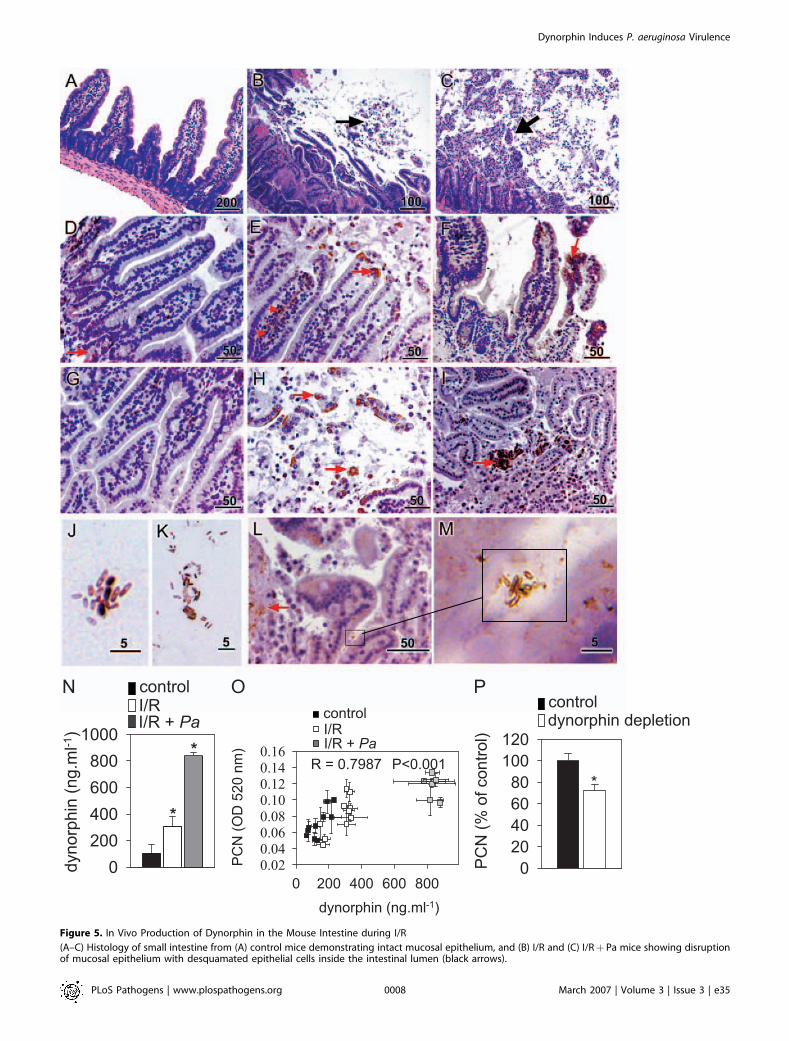
Figure 5. In Vivo Production of Dynorphin in the Mouse Intestine during I/R
(A–C) Histology of small intestine from (A) control mice demonstrating intact mucosal epithelium, and (B) I/R and (C) I/Rþ Pa mice showing disruptionof mucosal epithelium with desquamated epithelial cells inside the intestinal lumen (black arrows).
PLoS Pathogens | www.plospathogens.org March 2007 | Volume 3 | Issue 3 | e350008
Dynorphin Induces P. aeruginosa Virulence

plasma membrane and enter the bacterial cell interior. Figure6D shows an image of P. aeruginosa PAO1 cells incubated withdynorphin. Localization of dynorphin was identified by 10-nm gold particles (black arrows), which were found predom-inantly in the bacterial cytosol fraction close to the innermembrane.
j-Opioid Receptor Agonists U-50,488 and DynorphinEnhance the Virulence of P. aeruginosa PAO1 againstLactobacillus and C. elegansTo determine the clinical relevance of the above findings,
we examined the ability of j-opioid receptor agonists to shiftthe virulence of P. aeruginosa against the nematode C. elegans.
(D–F, G–I) Immunohistochemistry of the small intestine from (D and G) control mice demonstrating scarce dynorphin localized to the epithelial crypt(brown staining, red arrow), and following (E and H) I/R and (F and I) I/Rþ Pa showing dynorphin migration up the lamina propria (red arrows, [E]) andits accumulation on villi and within the lumen. Scale bars are in lm.(J–M) Images of luminal bacteria from mouse small intestine subjected to I/Rþ Pa demonstrating (J) transfer of dynorphin to bacteria (brown-coloredbacteria) and (K) positive dynorphin stained bacteria bound to desquamated epithelia; (I) abundant epithelial dynorphin staining (red arrow), and (M)co-localization of dynorphin stained luminal bacteria to sites of dynorphin accumulation at the epithelial surface. Scale bars are in lm.(N) Concentration of dynorphin in filtered luminal flushes isolated from intestine of control mice and mice subjected to I/R and I/RþPa. n¼10/group, *p ,0.001.(O) Correlation analysis between dynorphin concentration in luminal flushes and their ability to induce PCN production in PAO1.(P) Effect of dynorphin depletion with anti-dynorphin antibody on the ability of luminal flush samples to produce PCN in PAO1, n¼6/group, * p ,0.005.Error bars, mean 6 SD.doi:10.1371/journal.ppat.0030035.g005
Figure 6. Dynorphin Binds to P. aeruginosa In Vitro, and Enters the Bacterial Cell Cytoplasm
(A–C) Binding of dynorphin to P. aeruginosa; (A) negative control demonstrating no dynorphin staining when cells were not incubated with dynorphin;(B) negative control demonstrating no dynorphin staining when cell were incubated with dynorphin but primary anti-dynorphin antibodies wereomitted from staining procedure; and (C) positive staining (brown color) of P. aeruginosa incubated with dynorphin followed by whole procedure ofimmunostaining.(D) Immunoelectron microscopy of P. aeruginosa PAO1 cells incubated with dynorphin, 100 lM. Black arrows show 10-nm gold spots indicating thepresence of dynorphin.doi:10.1371/journal.ppat.0030035.g006
PLoS Pathogens | www.plospathogens.org March 2007 | Volume 3 | Issue 3 | e350009
Dynorphin Induces P. aeruginosa Virulence

In addition, we determined whether exposure P. aeruginosa toj-opioid receptor agonists could affect the growth of thecytoprotective probiotic organisms Lactobacillus plantarum andLactobacillus rhamnosus [37–40]. Media from P. aeruginosa PAO1grown in the presence of U-50,488 suppressed the growth ofL. plantarum and L. rhamnosus GG (Figure 7A and 7B), whereasU-50,488 alone had no effect (unpublished data). Similarly,media from P. aeruginosa PAO1 grown in the presence ofdynorphin suppressed the growth of Lactobacillus, whereasdynorphin alone had no effect (Figure 7C and 7D).Conditioned media from the PAO1 DMvfR mutant grown inthe presence or absence of dynorphin did not affect thegrowth of Lactobacillus spp., suggesting that the j-mediatedeffect is regulated via MvfR (Figure 7E). C. elegans feeding onlawns of PAO1 exposed to U-50,488 or dynorphin demon-strated suppressed production of new progeny, an indicator
of enhanced virulence (Figure 7F and 7G). In this assay, thePAO1 DMvfR mutant was observed to be significantly lessvirulent compared to the wild-type PAO1 (Figure 7F), and itsvirulence was not enhanced in the presence of j-agonists(Figure 7F and 7G).
Discussion
In animals exposed to physiologic or traumatic stress,subsequent bacterial challenge has been shown to result inincreased mortality [10,31] in association with impairedimmune function and bacterial clearance [17,41,42]. Datafrom the present study add to the small but expanding bodyof data showing that soluble compounds released by the hostduring stress and immune activation can directly interactwith pathways of bacterial virulence regulation in a highly
Figure 7. j-Opioid Receptor Agonists Activate Virulence of P. aeruginosa against Probiotic Bacteria and C. elegans
Error bars, mean 6 SD.(A and B) The exposure of P. aeruginosa PAO1 to U-50,488, 200 lM, increases the inhibiting effect of its extracellular milieu (conditioned media) on thegrowth of probiotic microorganisms (A) L. plantarum and (B) L. rhamnosus GG.(C and D) The exposure of P. aeruginosa PAO1 to dynorphin, 100 lM, increases the inhibiting effect of its extracellular milieu (conditioned media) on thegrowth of probiotic microorganisms (C) L. plantarum and (D) L. rhamnosus GG.(E) The extracellular milieu of P. aeruginosa PAO1 mutant DMvfR exposed to dynorphin, 100 lM, did not inhibit the growth of probiotic microorganismL. rhamnosus GG.(F and G) P. aeruginosa PAO1 but not mutant DMvfR exposed to (F) U-50,488, 200 lM, or (G) dynorphin, 100 lM, suppressed the production of newprogeny in C. elegans.doi:10.1371/journal.ppat.0030035.g007
PLoS Pathogens | www.plospathogens.org March 2007 | Volume 3 | Issue 3 | e350010
Dynorphin Induces P. aeruginosa Virulence

specific manner [3,8,11]. Opioids are ubiquitous neuro-transmitters within the enteric nervous system and encom-pass a wide variety of functions, including motility, secretion,immune modulation, and maintenance of epithelial barrierfunction. The abundance of the neural network within theintestinal tract is matched only by its microbial flora, wherebacterial cells outnumber the total number of cells in thebody [14]. In this study, we found j-opioid receptor agoniststo induce PCN production in P. aeruginosa. Althoughdynorphin has been shown to be present in a variety oftissues, whether dynorphin accumulates in intestinal tissuesfollowing host stress and/or bacterial infection has not beenpreviously addressed. Data from the present study show forthe first time to our knowledge, that dynorphin is releasedinto the intestinal lumen following I/R injury and accumulatesin regions of high dynorphin concentration, such as ondesquamated epithelium, where it binds to P. aeruginosa.Binding of dynorphin to P. aeruginosa was further confirmedin vitro by direct antibody staining. In addition, thepenetration of dynorphin into the cytosolic compartmentof bacterial cells was demonstrated by immunoelectronmicroscopy. These findings, coupled with the observationthat dynorphin can activate the virulence of P. aeruginosa, maybe of significant clinical relevance given that intestinalischemia invariably accompanies physiologic stress and hasbeen associated with fatal infection due to intestinal P.
aeruginosa [43,44]. However, dynorphin may enhance thevirulence of P. aeruginosa not only within the intestine butalso at other sites of tissue injury and inflammation. There isoverwhelming evidence that opioids are released andaccumulate at sites of inflammation primarily because all ofthese tissues sites are heavily innervated and highly populatedby macrophages and neutrophils. [45,46]. The ubiquitouspresence of opioid receptors on nerves throughout the bodysuggests that exposure of P. aeruginosa to opioids may be oneof the reasons it has evolved a mechanism to respond to thesecompounds [45,46]. Therefore, P. aeruginosa virulence couldbe activated by dynorphin in all infections associated withinflammation, including burns, implanted medical devices,and lung infections in patients with underlying lung disease.The precise mechanisms by which colonizing pathogens
important to human disease process host signals for thepurposes of virulence activation is a small and poorlyunderstood area of investigation. The pathways by whichbacteria gather, process, and become activated by host signalshave been shown to be both specific to bacteria and specificto the host signal involved, and gene activation through theQS system has been previously reported [3,8]. A majormechanism by which the j-opioid receptor agonists U-50,488 and dynorphin affect virulence gene activation in P.aeruginosa could be via expression of the pqsABCDE operon.The pqsABCDE operon directs the biosynthesis of 4-hydroxy-
Figure 8. Proposed Activation and Effectors Pathways of P. aeruginosa in Response to Host Stress (Intestinal I/R Injury)
(1) Dynorphin is released by intestinal tissues and accumulates in the lumen during ischemia/reperfusion and penetrates the plasma membrane of P.aeruginosa (dark green arrows).(2) Dynorphin synergizes with PQS via MvfR to increase the transcription of pqsABCDE leading to the production of HAQs, including HQNO and HHQ.(3) Increased HQNO production suppresses the growth of Lactobacillius spp., rendering the intestinal epithelium more vulnerable to invasion and theaction of cytotoxins of P. aeruginosa (red arrows).(4) HHQ is the immediate precursor of PQS [23], and both compounds play an important role in bacterial cell-to-cell communication [23] (yellow andblue arrows).(5) PQS induces the expression of pqsABCDE [63], and is required for phzA1-G1expression, the gene responsible for PCN production (blue arrows). 6.) Therelease of PCN can induce neutrophils apoptosis and damage epithelial cells [64] (green arrows) allowing for immuno-evasion and deeper penetrationof bacteria.doi:10.1371/journal.ppat.0030035.g008
PLoS Pathogens | www.plospathogens.org March 2007 | Volume 3 | Issue 3 | e350011
Dynorphin Induces P. aeruginosa Virulence

2-alkylquinolines (HAQs), among which HHQ and PQS arethemselves signaling molecules. Additionally, HHQ is a directprecursor of PQS [23]. PQS functions as a regulatory linkbetween the LasRI and RhlRI quorum sensing systems [30,47],and has been shown to play a critical role in the pathogenesisof P. aeruginosa in nematodes, plants, and mice [24,48,49]. Datafrom the present study suggest that dynorphin synergizeswith PQS to increase pqsABCDE expression. Further experi-ments are underway to clarify the precise mechanism ofdynorphin activation of pqsABCDE expression.
Among critically ill and immunocompromised patients,infection with P. aeruginosa carries the highest case fatalityrate of all nosocomial pathogens, approaching 60% [50,51].The primary site of colonization for P. aeruginosa in suchpatients is the gastrointestinal tract, where as many as 50% ofhospitalized patients harbor this organism [52,53]. Riskfactors for mortality due to P. aeruginosa infection suggestthat the degree of host stress is a major determinant of a fataloutcome from this pathogen [53,54]. In the present work weshow (Figure 8) that P. aeruginosa presents a ‘‘triple threat’’ toits host when exposed to dynorphin in that it can 1) activateQS circuits via enhanced PQS production, 2) reducepopulations of protective probiotic bacteria such as lactoba-cilli through the production of HQNO and PCN [55], and 3)increase the production of QS-dependent virulence geneproducts that affect host cell function such as PCN and thePA-I lectin/adhesin [30,56–58]. A clearer understanding of themechanisms by which P. aeruginosa is activated to expressvirulence in direct response to host stress has the potential tolead to preventive therapies that interdict in the process ofinfection at its most proximate point.
Materials and Methods
Bacterial strains and culture conditions. P. aeruginosa strain PAO1and its derivatives were routinely grown in tryptic soy broth (TSB)supplemented when necessary with tetracycline (Tc), 60 lg/ml,gentamicin (Gm), 100 lg/ml, or carbenicillin (Cb), 300 lg/ml. P.aeruginosa strains PAO1 wild-type, DRhlR (rhlR:: ISphoA/hah, ID44488), and DMvfR (mvfR:: ISlacZ/hah, ID 13375) were obtained fromthe P. aeruginosa mutant library [59]. Strains PAO-R1 (DLasR(lasR::Tc), PAO-JP-1 (DLasI (lasI::Tc), and PDO100 (DRhlI (rhlI::Tn501)were kindly provided by B. Iglewski. Strains PAO6281 (DGacA(gacA::Spr /Sm), PAO-MW1 (DRhlIDLasI (rhlI::Tn501 lasI::tetA), andMP603 (pqsC::Tc) were kindly provided by C. Reimmann, P. Green-berg, and C. Manoil, respectively.
PCN assay. P. aeruginosa cultures were grown at 37 8C, undershaking conditions at 220 rpm, in TSB supplemented with eithermorphine (Abbott Laboratories, http://www.abbott.com), U-50,488(Sigma, http://www.sigmaaldrich.com), or BW373U86 (Sigma) atvarying concentrations. Following overnight incubation, bacterialcells were spun down by centrifugation at 10,000g for 5 min, and 1 mlof supernatant was extracted using 500 ll of chloroform, re-extractedwith 150 ll of 0.2 M HCl, and then PCN was measured at OD 520 nmas described [60]. In experiments with dynorphin A (1–17) (Sigma),200 ll of bacterial culture was incubated in 2-ml wells of a 96-wellmicroplate (Whatman, http://www.whatman.com). Following over-night incubation, 200 ll of chloroform and 150 ll of 0.2 M HClwere used to extract PCN from cell-free culture media. In dynamicexperiments, PAO1 was grown in TSB supplemented with 200 lM U-50,488, and 1-ml aliquots were serially collected. Cell density wasmeasured at OD 600 nm, and samples were processed for PCN assay.In experiments in which strains complemented with mvfR or gacA onthe pUCP24 plasmid were used, experiments were performed with ahigher dose of U-50,488 (1 mM), as a baseline of PCN production wasalready high due to these strains containing multi-copy plasmidsencoding MvfR or GacA.
The b-galactosidase assay. Plasmid pGX5 [28] harboring the pqsA’-lacZ fusion construction was introduced in P. aeruginosa strains PAO1and MP603; and plasmids pGX1 harboring the mvfR’-lacZ fusion [23]
and pMW303 harboring the phzABC-lacZ fusion [33] were introducedin PAO1 by electroporation. Single colonies of PAO1/pGX5 or PAO1/pGX1, or PAO1/pMW303 were used to inoculate TSB supplementedwith carbenicillin (Cb), 300 lg/ml, for overnight growth at 37 8C, 220rpm. Overnight culture was diluted 1:100 in TSB, and 300 lg/ml Cbwas supplemented with U-50,488, 200 lM, or dynorphin, 100 lM, orPQS, 100 lM, or without supplementation (control). Aliquots of cellscultures were taken out dynamically; pellet was collected bycentrifugation at 6,000g, 5 min, and kept at�808C before processing.MP603/pGX5 was inoculated in TSB supplemented with Tc, 60 lg/mland Cb, 300 lg/ml. Overnight cultures were diluted 1:100 in TSB,subgrown for 1 h to a cell density of OD600nm 0.07–0.1, and aliquotedto create the following groups (in triplicates): 1) ‘‘control’’, 2)‘‘dynorphin’’, dynorphin added to a final concentration of 100 lM;3) ‘‘PQS’’, PQS added to final concentrations of 20 and 80 lM; and 4)‘‘dynorphinþPQS’’, dynorphin added to a final concentration of 100lM, incubated for 1 h, followed by the addition of PQS to a finalconcentration of 20 and 80 lM. All cell cultures were collected at thesame time (5 h), cell density of 1:10 dilutes samples was measured atOD 600 nm, and cells were collected by centrifugation at 6,000g, 5min, and kept at�808C before processing. For b-galactosidase activityanalysis, cells were treated with protein extraction reagent (Bug-Buster Master Mix; Novagen, http://www.emdbiosciences.com/html/NVG/home.html) for 20 min at room temperature, centrifuged for 30min at 4 8C, and cytosol fractions were used to measure b-galactosidase activity. In this case, 15 ll of cytosol fractions wereloaded on 96-well plates, followed by 135 ll of a master mixture of o-nitrophenyl-b-D-galactopyranoside (ONPG, Sigma) containing 100 llof 0.1 M Na-phosphate buffer (pH 7.5), 33 ll of ONPG, 4 mg/mldissolved in 0.1 M Na-phosphate buffer (pH 7.5), and 2 ll of Mg buffer(63 ll of H2O, 35 ll of 2-mercaptoethanol, and 2 ll of 5 M MgCl2).Absorbance at 410 nm was measured using Plate Reader, and resultswere expressed as Miller units (1,000 3 D410 nm 3 mg protein�1 3min�1).
RNA isolation and cDNA synthesis. For RNA isolation, 1 ml of P.aeruginosa PAO1 culture was grown in TSB with or without 200 lM U-50,488, to OD600 nm¼ 3.0. Next, 2 ml of RNA Protect Bacteria reagent(Qiagen, http://www1.qiagen.com) was added immediately at the endof the incubation period, and samples treated as recommended bythe Qiagen’s lysis protocol, followed by the addition of 3 ml of TRIzolLS reagent (Invitrogen, http://www.invitrogen.com). The RNA enrich-ment fraction was separated using Phase lock gel, heavy (Eppendorf,http://www.eppendorf.com). RNA was precipitated with isopropanol,dissolved in water, and the remaining DNA degraded using DNA-Freekit (Ambion, http://www.ambion.com). RNA integrity was monitoredby formaldehyde agarose gel electrophoresis, and the absence of DNAchecked by PCR using primers for 16S r RNA, forward 59-GGACGGGTGAGTAATGCCTA-39 and reverse 59-CGTAAGGGC-CATGATGACTT-39. The first-strand cDNA was prepared using 2lg of total RNA, Superscript II RNase H–RT (Invitrogen), andrandom primers as recommended by the manufacturer’s protocol.
Real-time reverse transcription (RT)–PCR. Real-time PCR wasperformed on the ABI Prizm 7300 Sequence Detection System usingSYBR Green qPCR SuperMix-UDG (Invitrogen), cDNA, and respec-tive primers: for gltA encoding citrate synthase (PA1580),5 9T C T A C C A C G A C T C C C T G G A C 3 9 a n d59TTTTCCGCGTAGTTCAGGTC39; for lecA encoding PA-IL( P A 2 5 7 0 ) , 5 9C G A T G T C A T T A C C A T C G T C G 3 9 a n d59ACCCTGGACATTATTGGGTG39. The integrity of the RT-PCRproducts was confirmed by melting-curve analysis. Expression levelswere calculated based on differences in Ct levels.
Protein concentration assay. Protein was measured using the BCAProtein Assay Reagent (Pierce, http://www.piercenet.com).
HQNO, HHQ, and PQS quantification. A single colony of PAO1was used to inoculate 5 ml of TSB. Overnight culture was diluted infresh TSB at 1:100, supplemented with either U-50,488 (200 lM) ordynorphin (100 lM). After 20 min of incubation under shakingconditions, cell cultures were aliquoted as 650 ll in 14-ml culturetubes. At designed time points, three tubes from each group wereremoved, and 650 ll of MeOH containing 2% acetic acid wasimmediately added, properly mixed, replaced into Eppendorf tubes,centrifuged at 13,000g, 30 min, 4 8C, and the supernatant used toquantify HQNO, HHQ, and PQS by HPLC/MS according to Lepine etal. [61]. Unlabeled PQS was obtained by the same synthetic routedescribed for deuterium-labeled PQS [61]. The final PQS-d4concentration was 20 mg/l, and the stock solutions were in methanol.
C4-HSL and 3-oxo-C12-HSL quantification. The homoserinelactones were quantitated by LC/MS/MS using a water/acetonitrilegradient containing 1% acetic acid. The analyses were performed inpositive electrospray ionization mode and the acquisitions were
PLoS Pathogens | www.plospathogens.org March 2007 | Volume 3 | Issue 3 | e350012
Dynorphin Induces P. aeruginosa Virulence

obtained in Multiple Reaction Mode (MRM). The transitionsmonitored were m/z 298 to 102 for the N-(3-oxododecyl)-homoserinelactone and m/z 172 to 102 for the N-(butyroyl)-homoserine lactone.Argon at 2.0 3 10�3 mtorr was the collision gas and the collisionenergy was 15 eV.
Complementation of MvfR mutant with mvfR gene. The mvfR genewas amplified using primers forward 59-AAGGAATAAGGGATGCC-TATTCA-39 and reverse 59-CTACTCTGGTGCGGCGCGCTGGC-39and cloned in pCR2.1 (Invitrogen). Plasmid pCR2.1/mvfR was digestedwith XbaI-HindIII restrictases, and mvfR was subcloned in pUCP24under the Plac promoter [62] to create pUCP24/mvfR. The plasmidspUCP24 and pUCP24/mvfR were electroporated in strain 13375defective in MvfR production to create strains DMvfR/pUCP24(control) and DMvfR/mvfR .
Complementation of GacA mutant with gacA gene. The gacA genewas amplified using primers forward 59-CGACGAGGTGCAGCGT-GATTAAGGT-39 and reverse 59-CTAGCTGGCGGCATCGAC-CATGC-39 and cloned in pCR2.1 (Invitrogen). Plasmid pCR2.1/gacAwas digested with XbaI-HindIII restrictases, and gacA was subclonedin pUCP24 under the Plac promoter to create pUCP24/gacA. Theplasmids pUCP24 and pUCP24/gacA were electroporated in strainPAO6281 defective in GacA production to create strains DGacA/pUCP24 and DGacA/gacA.
Anti-MvfR antibody. Polyclonal antiserum against 50–63 peptideLVRRDGYKVEPTEQ of PA1003 (MvfR) was produced in rabbits(ZYMED Laboratories, http://www.invitrogen.com). Anti-MvfR anti-bodies were affinity purified by AminoLink Plus Immobilization Kit(Pierce) using 50–63 peptide to create an affinity column.
Segmental intestinal I/R model. All experiments on mice wereperformed in accordance with University of Chicago guidelines andregulations, and mouse protocol number 71629 was approved by theAnimal Care and Use Committee of the University of Chicago. Male,wild-type C57Bl/6 mice (8- to 10–wk-old; Charles River, http://www.criver.com) were fasted overnight prior to use in the I/R studies. Forthese studies, mice were lightly anesthetized with sevoflurane prior toan i.p. bolus injection of Avertin (Sigma-Aldrich #T4, 840–2)prepared as a 1.2 % solution, at 0.2 ml 3 10 g�1 body weight. Micewere placed on a warmed heating pad under a warming light. Anabdominal midline incision was made and the small intestinesexposed. Sutures were placed so as to isolate a 20-cm segment ofsmall intestine. The intestine was divided proximally and distallyusing cautery. A clamp was placed on the superior mesenteric artery(SMA) to occlude blood flow to the jejunum and ileum. Ischemia wasconfirmed by visible blanching of the intestinal segment. During theischemic and reperfusion period, mice were hydrated via i.p.administration of warmed saline. After 30 min of ischemia, theSMA clamp was removed and the intestinal tract allowed to reperfusefor 30 min prior to removal of the isolated segment. This segment wasthen immediately flushed with 2 ml of PBS containing a proteaseinhibitor cocktail (Roche, http://www.roche.com). Mice were euthan-ized by an overdose of anesthesia, followed by cervical dislocation. Inselected experiments, P. aeruginosa (Pa) was injected into the isolatedintestinal segment prior to SMA clamping at a concentration of 107
CFU/ml in 2 ml of PBS to recapitulate the clinical circumstance of I/Roccurring in the presence of an opportunistic infection (I/Rþ Pa).
Histological and immunohistochemical staining. For histology,specimens were fixed in 10% buffered formalin and embedded inparaffin. Paraffin specimens were cut into 4-lm sections and weremounted on micro slides. Hematoxylin and eosin–stained slides werethen reviewed using an Olympus microscope. For immunohisto-chemical staining, sections were deparaffinized and re-hydratedthrough xylen and serial dilutions of ETOH to distilled water.Section were then incubated in antigen retrieval buffer (DAKO,S1699; http://www.dako.com) and heated in a steamer at 98 8C degreefor 20 min. After rinsing, slides were incubated in 3% hydrogenperoxide for 5 min and then 10% normal goat serum in 0.025%Triton X-100-PBS for 30 min. Anti-dynorphin A (1–17) antibody(EMD Biosciences, http://www.emdbiosciences.com) at 1:25 dilutionwas applied for overnight incubation at 4 8C in a humidity chamber.Following a TBS wash, slides were incubated with secondary antibody(DAKO, K4011) and the antigen–antibody binding was detected byDAB substrate chromogen system (DAKO, K3466). To ensure thatpositive staining was specific, control experiments were performed asoutlined above, eliminating the primary antibodies, and showed nostaining. Slides were briefly immersed in hematoxylin for counter-staining and evaluated under Zeiss Axioskop (http://www.zeiss.com),using oil 253Korr objective na 1.3 and oil 633 apochromat objectivena 1.4 with pixel sizes 1280 3 1024 regular and 3840 3 3072 highest.
In vitro immunostaining of P. aeruginosa PAO1 incubated withdynorphin. Cells of P. aeruginosa were grown overnight in TSB and
diluted 1:100 with 400 ll of fresh TSB media. After 2 h of incubation,cells were added by dynorphin, 100 lM (when needed), and incubatedfor one more hour. Cells were collected, washed three times with PBS,and fixed in 4% paraformaldehyde for 2 h, 4 8C, at rotation. Then,cells were kept on ice for 45 min followed by intensive washing withPBS. After last washing, cells were resuspended in 200 ll of PBS, and100 ll of cell suspension was poured on slide. The fixation of cells toslide was allowed overnight at room temperature. Cells on slides werethen incubated in antigen retrieval buffer (DAKO, K4011) andfollowed the above procedure of immunostaining (see ‘‘Histologicaland immunohistochemical staining’’). Two negative controls wereperformed: 1) omitting dynorphin, and 2) omitting primary anti-dynorphin antibodies.
Immunoelectron microscopy. P. aeruginosa PAO1 was grown for 10h in the presence of dynorphin, 100 lM, and then cells collected bycentrifugation, 5,000g, 5 min, washed with PBS, and fixed with 4%paraformaldehyde plus 0.1% glutaraldehyde in 0.1 M phosphatebuffer (pH 7.4) (PB) for 1 h. After fixation, cells were washed with 0.1M PB, and gradually dehydrated using ethanol at increasingconcentrations from 30% to 100%. Infiltration was performed using100% ethanol:LR White Medium (Electron Microscopy Scienceshttp://www.emsdiasum.com/microscopy) (1:1) for 1 h followed byovernight infiltration with LR White Medium. After infiltration, cellswere embedded in gelatin capsules. Polymerization was performed ina vacuum oven at 45 8C for 48 h. Then, 80-nm thickness sections werecut using a Reichert-Jung Ultracut E, and mounted on formvar-coated 200-mesh nickel grids. Sections on grids were re-hydrated withPBS for 30 min, blocked with 1% BSA in PBS for 30 min, and primedwith rabbit anti-dynorphin A 1–17 antibody (EMD Biosciences) at1:25 dilution in 1% BSA at humidified chamber for 3.5 h. Grids wereextensively washed with PBS, blocked with 0.1% BSA in PBS for 25min, and incubated in a humidified chamber for 1 h with goat anti-rabbit IgG conjugated with 10-nm gold particles (Ted Pella, http://www.tedpella.com) at 1:10 dilution in 0.1% BSA. Grids were washedwith PBS, fixed with 1% glutaraldehyde in PBS for 10 min, washedwith water, and stained briefly with uranyl acetate and lead citrate.Air dried grids were examined at 300 kV with an FEI Tecnai F30 (FEI,http://www.fei.com). Two negative controls were used: 1) grids withoutincubation with rabbit anti-dynorphin A 1–17 antibody, and 2) PAO1grown in the absence of dynorphin.
Biotinylation of dynorphin. Dynorphin A (1–17) (Sigma) wasbiotinylated using NHS-PEO4-biotin (Pierce), and purified by HPLC.
Competitive ELISA for detection dynorphin in luminal flushings.Luminal flushings were filtered with 0.22-lm filters (Millipore, http://www.millipore.com), aliquoted, and stored at�80 8C. Affinity purifiedF(ab)2 of Frag Donk anti-Rb IgG (Jackson Immunological ResearchLaboratories, http://www.jax.org) at a concentration of 10 lg/ml incarbonate-bicarbonate buffer (Sigma) were coated onto Maxisorpimmunomodules (Nunc, http://www.nuncbrand.com) for 2 h at 37 8C.Unbound sites were blocked with 3% bovine serum albumin in PBSfor 30 min at room temperature. After blocking, rabbit anti-dynorphin A (1–17) antibodies (EMD Biosciences) at 1:250 dilutionin PBST (PBS, Tween 0.05%) were added to wells for 1 h, at 37 8C.After washing with PBST, mixtures of biotinylated dynorphin, 50 ng/ml with varying concentrations of unlabeled dynorphin (0, 20, 50, 100ng/ml) or filtered luminal flushings diluted in buffer (20 mMphosphate buffer (pH 7.4), 150 mM NaCl, 10 mM EDTA, 0.5% BSA,0.02% Triton X-100) were loaded for 2 h at 37 8C. In this mannerbiotinylated dynorphin competes for antibody binding sites withunlabeled dynorphin or dynorphin in luminal flushes. Afterincubation, unbound biotinylated peptide was removed by washingwith PBST, and ImmunoPure streptavidin-conjugated horseradishperoxidase (Pierce) was added and allowed to bind to theimmobilized primary antibody-biotinylated peptide complex. Afterwashing, O-phenylaminediamine (Sigma) was allowed to react withthe bound HRP. The color intensity that develops is dependent onthe quantity of biotinylated peptide bound to the immobilizedantibody. When more non-biotinylated peptide competes for thelimited amount of antibody, less biotinylated peptide/SA-HRP can beimmobilized and less color is produced by the substrate
PCN assay in PAO1 induced by luminal flushings. PAO1 wasexposed to 20 ll of 0.22 lm-filtered luminal flushings included in 2 mlof TSB, and PCN was measured after overnight growth. Forimmunodepletion, samples were pre-incubated with anti-dynorphinantibody (EMD Biosciences) at 1:100 dilution for 2 h.
Effect of conditioned media from P. aeruginosa on the growth ofLactobacillus spp. Overnight cultures of P. aeruginosa PAO1 were usedto inoculate fresh TSB (1:100, vol/vol). After inoculation, the PAO1culture was subgrown for 1 h, then aliquoted, and U-50,488 ordynorphin were added to aliquots to create final concentrations of
PLoS Pathogens | www.plospathogens.org March 2007 | Volume 3 | Issue 3 | e350013
Dynorphin Induces P. aeruginosa Virulence

200 lM and 100 lM, respectively. After 23 h of growth at 37 8C, 180rpm, cultures were collected, centrifuged (5,000 rpm, 5 min) toremove cells, and supernatant was filtered using Millex-CV lowprotein binding membrane filters of 0.22-lm pore size (Millipore).The filtered supernatant, now termed conditioned media, was storedon ice before use. L. plantarum and L. rhamnosus GG grown overnightin MRS broth (Oxoid, http://www.oxoid.com) at 37 8C were used toinoculate fresh MRS (2:100, vol/vol). Freshly inoculated culture, 100 llwas put into 96-well plates (Nunc), followed by an addition of 100 llof conditioned media from P. aeruginosa. Control samples werecreated where 100 ll of TSB; TSBþU-50,488, 200 lM; TSBþdynor-phin, 100 lM were added to wells. Plates with Lactobacillus spp. wereincubated at 37 8C, unshaken, and growth was monitored dynamicallyby measuring OD 600 nm using Plate Reader.
Virulence assay of P. aeruginosa PAO1 using C. elegans. Wild-typeN2 C. elegans were kindly provided by M. Glotzer, University ofChicago. Culturing, cleaning, egg preparation for synchronization,and transferring were performed according to ‘‘Maintenance of C.elegans’’ (http://www.wormbook.org/chapters/www_strainmaintain/strainmaintain.html). For the experiment, two adult nematodes weretransferred to a lawn of P. aeruginosa on NGM agar prepared by thefollowing: P. aeruginosa PAO1 and PAO1 derivative DMvfR mutantwere grown for 6 h at 37 8C in TSB (control culture) or TSBsupplemented with U-50,488 (U-50,488 culture), 200 lM, or TSBsupplemented with dynorphin, 100 lM (dynorphin culture). Then, 10ll of TSB media (control), or TSB supplemented with U-50,488, 1mM, or dynorphin, 200 lM, was dropped at the center of NGM agarplate, allowed to dry for 10 min, and then 10 ll of bacterial culturewas dropped on a respective plate (control culture on TSB spot, U-50,488 culture on U-50,488 spot, and dynorphin culture ondynorphin spot). The plates were incubated at 37 8C overnight andallowed to equilibrate to 20 8C for two h before being seeded withnematodes. The plates with nematodes on P. aeruginosa lawn wereincubated for up to four days at 20 8C, and 43 10 ll of TSB, or TSB/U-50,488, 1 mM, or TSB/dynorphin, 200 lM, were droppedperipherally at the lawn ring, daily. Total numbers of nematodeswere counted daily.
Data analysis. Statistical analysis of the data was performed usingStudent’s t-test. Regression analysis was performed using Sigma plotsoftware.
Supporting InformationAccession Numbers
The Entrez Protein (http://www.ncbi.nlm.nih.gov/entrez/query.fcgi?db¼Protein) accession numbers for the P. aeruginosa PAO1 geneproducts discussed in this paper are citrate synthase (NP_250271),GacA (NP_251276), LasI (NP_250123), LasR (NP_250121), MvfR(NP_249694), PA-I galactophilic lectin (NP_251260), RhlI(NP_252166), and RhlR (NP_252167). The Entrez Nucleotide(http://www.ncbi.nlm.nih.gov/entrez/query.fcgi?db¼Nucleotide) acces-sion number for the P. aeruginosa PAO1 complete genome isNC_002516.
Acknowledgments
We thank O. Shevchenko for technical assistance; C. Reimmann forproviding GacA mutant; B. Iglewski for providing LasR, LasI, and RhlImutants; P. Greenberg for providing LasI/RhlI mutant; C. Manoil forproviding P. aeruginosa strain MP603; M. Whiteley for providingplasmid pMW303; H. P. Schweizer for providing pUCP24 plasmid; K.Winzer for providing PQS; M. Jacobs for providing RhlR and MvfRmutants; A. Khramtsov and M. Tretiakova for providing histologystaining and examining tissues; H. Auer for assistance in HPLCpurification of biotinylated dynorphin; V. Bindokas for assistancewith the preparation of Figure 5 and helpful discussion of electronand digital microscopy images; and S. Bond for her assistance in thedigital microscopy of intestinal samples.
Author contributions. OZ, TL, MC, AZ, EP, and JCA conceived anddesigned the experiments. OZ, FL, GX, VV, YC, TL, MC, AZ, EP, andJCA performed the experiments. OZ, FL, TL, MC, AZ, EP, JRT, LGR,and JCA analyzed the data. OZ, FL, GX, YC, EP, LGR, EC, and JCAcontributed reagents/materials/analysis tools. OZ, LGR, EC, and JCAwrote the paper.
Funding. This work was supported by National Institutes of Healthgrants RO1 GM62344–05 (JA) and DK47722 (EC), and DigestiveDisease Research Core Center grant DK42086 (EC).
Competing interests. The authors have declared that no competinginterests exist.
References1. Dawkins R (2006) The selfish gene. 3rd edition. New York: Oxford
University Press. 384 p.2. Camilli A, Bassler BL (2006) Bacterial small-molecule signaling pathways.
Science 311: 1113–1116.3. Wu L, Estrada O, Zaborina O, Bains M, Shen L, et al. (2005) Recognition of
host immune activation by Pseudomonas aeruginosa. Science 309: 774–777.4. Luo G, Niesel DW, Shaban RA, Grimm EA, Klimpel GR (1993) Tumor
necrosis factor alpha binding to bacteria: Evidence for a high-affinityreceptor and alteration of bacterial virulence properties. Infect Immun 61:830–835.
5. Porat R, Clark BD, Wolff SM, Dinarello CA (1991) Enhancement of growthof virulent strains of Escherichia coli by interleukin-1. Science 254: 430–432.
6. Kohler JE, Zaborina O, Wu L, Wang Y, Bethel C, et al. (2005) Componentsof intestinal epithelial hypoxia activate the virulence circuitry ofPseudomonas. Am J Physiol Gastrointest Liver Physiol 288: G1048–G1054.
7. Lacoste A, Jalabert F, Malham SK, Cueff A, Poulet SA (2001) Stress andstress-induced neuroendocrine changes increase the susceptibility ofjuvenile oysters (Crassostrea gigas) to Vibrio splendidus. Appl Environ Micro-biol 67: 2304–2309.
8. Sperandio V, Torres AG, Jarvis B, Nataro JP, Kaper JB (2003) Bacteria-hostcommunication: The language of hormones. Proc Natl Acad Sci U S A 100:8951–8956.
9. Lyte M (2004) Microbial endocrinology and infectious disease in the 21stcentury. Trends Microbiol 12: 14–20.
10. Alverdy J, Holbrook C, Rocha F, Seiden L, Wu RL, et al. (2000) Gut-derivedsepsis occurs when the right pathogen with the right virulence genes meetsthe right host: Evidence for in vivo virulence expression in Pseudomonasaeruginosa. Ann Surg 232: 480–489.
11. Bader MW, Sanowar S, Daley ME, Schneider AR, Cho U, et al. (2005)Recognition of antimicrobial peptides by a bacterial sensor kinase. Cell122: 461–472.
12. Bishop JL, Finlay BB (2006) Friend or foe? Antimicrobial peptides triggerpathogen virulence. Trends Mol Med 12: 3–6.
13. Hancock RE, McPhee JB (2005) Salmonella’s sensor for host defensemolecules. Cell 122: 320–322.
14. Sternini C, Patierno S, Selmer IS, Kirchgessner A (2004) The opioid systemin the gastrointestinal tract. Neurogastroenterol Motil 16 (Suppl 2): 3–16.
15. Neudeck BL, Loeb JM (2002) Endomorphin-1 alters interleukin-8 secretionin Caco-2 cells via a receptor mediated process. Immunol Lett 84: 217–221.
16. Neudeck BL, Loeb J, Buck J (2003) Activation of the kappa-opioid receptorin Caco-2 cells decreases interleukin-8 secretion. Eur J Pharmacol 467: 81–84.
17. Vallejo R, de Leon-Casasola O, Benyamin R (2004) Opioid therapy andimmunosuppression: A review. Am J Ther 11: 354–365.
18. Lawrence DM, el-Hamouly W, Archer S, Leary JF, Bidlack JM (1995)Identification of kappa opioid receptors in the immune system by indirectimmunofluorescence. Proc Natl Acad Sci U S A 92: 1062–1066.
19. Karaji AG, Khansari N, Ansary B, Dehpour A (2005) Detection of opioidreceptors on murine lymphocytes by indirect immunofluorescence: Maturenormal and tumor bearing mice lymphocytes. Int Immunopharmacol 5:1019–1027.
20. Sharp BM, Li MD, Matta SG, McAllen K, Shahabi NA (2000) Expression ofdelta opioid receptors and transcripts by splenic T cells. Ann N Y Acad Sci917: 764–770.
21. Peterson PK, Molitor TW, Chao CC (1998) The opioid-cytokine connection.J Neuroimmunol 83: 63–69.
22. Wu LR, Zaborina O, Zaborin A, Chang EB, Musch M, et al. (2005) Surgicalinjury and metabolic stress enhance the virulence of the humanopportunistic pathogen Pseudomonas aeruginosa. Surg Infect (Larchmt) 6:185–195.
23. Deziel E, Lepine F, Milot S, He J, Mindrinos MN, et al. (2004) Analysis ofPseudomonas aeruginosa 4-hydroxy-2-alkylquinolines (HAQs) reveals a rolefor 4-hydroxy-2-heptylquinoline in cell-to-cell communication. Proc NatlAcad Sci U S A 101: 1339–1344.
24. Lau GW, Ran H, Kong F, Hassett DJ, Mavrodi D (2004) Pseudomonasaeruginosa pyocyanin is critical for lung infection in mice. Infect Immun 72:4275–4278.
25. Stefano GB, Goumon Y, Casares F, Cadet P, Fricchione GL, et al. (2000)Endogenous morphine. Trends Neurosci 23: 436–442.
26. Boettcher C, Fellermeier M, Drager B, Zenk MH (2005) How humanneuroblastoma cells make morphine. Proc Natl Acad Sci U S A 102: 8495–8500.
27. McGrath S, Wade DS, Pesci EC (2004) Dueling quorum sensing systems inPseudomonas aeruginosa control the production of the Pseudomonas quinolonesignal (PQS). FEMS Microbiol Lett 230: 27–34.
28. Xiao G, He J, Rahme LG (2006) Mutation analysis of the Pseudomonasaeruginosa mvfR and pqsABCDE gene promoters demonstrates complexquorum-sensing circuitry. Microbiology 152: 1679–1686.
29. Deziel E, Gopalan S, Tampakaki AP, Lepine F, Padfield KE, et al. (2005) The
PLoS Pathogens | www.plospathogens.org March 2007 | Volume 3 | Issue 3 | e350014
Dynorphin Induces P. aeruginosa Virulence

contribution of MvfR to Pseudomonas aeruginosa pathogenesis and quorumsensing circuitry regulation: Multiple quorum sensing-regulated genes aremodulated without affecting lasRI, rhlRI or the production of N-acyl- l-homoserine lactones. Mol Microbiol 55: 998–1014.
30. Diggle SP, Winzer K, Chhabra SR, Worrall KE, Camara M, et al. (2003) ThePseudomonas aeruginosa quinolone signal molecule overcomes the celldensity-dependency of the quorum sensing hierarchy, regulates rhl-dependent genes at the onset of stationary phase and can be producedin the absence of LasR. Mol Microbiol 50: 29–43.
31. Wu L, Zaborina O, Zaborin A, Chang EB, Musch M, et al. (2004) High-molecular-weight polyethylene glycol prevents lethal sepsis due tointestinal Pseudomonas aeruginosa. Gastroenterology 126: 488–498.
32. Janecka A, Fichna J, Janecki T (2004) Opioid receptors and their ligands.Curr Top Med Chem 4: 1–17.
33. Whiteley M, Parsek MR, Greenberg EP (2000) Regulation of quorum sensingby RpoS in Pseudomonas aeruginosa. J Bacteriol 182: 4356–4360.
34. Gallagher LA, McKnight SL, Kuznetsova MS, Pesci EC, Manoil C (2002)Functions required for extracellular quinolone signaling by Pseudomonasaeruginosa. J Bacteriol 184: 6472–6480.
35. Schmidt H, Martindale R (2001) The gastrointestinal tract in critical illness.Curr Opin Clin Nutr Metab Care 4: 547–551.
36. Marinova Z, Vukojevic V, Surcheva S, Yakovleva T, Cebers G, et al. (2005)Translocation of dynorphin neuropeptides across the plasma membrane. Aputative mechanism of signal transmission. J Biol Chem 280: 26360–26370.
37. Luyer MD, Buurman WA, Hadfoune M, Speelmans G, Knol J, et al. (2005)Strain-specific effects of probiotics on gut barrier integrity followinghemorrhagic shock. Infect Immun 73: 3686–3692.
38. Valdez JC, Peral MC, Rachid M, Santana M, Perdigon G (2005) Interferenceof Lactobacillus plantarum with Pseudomonas aeruginosa in vitro and in infectedburns: The potential use of probiotics in wound treatment. Clin MicrobiolInfect 11: 472–479.
39. Mangell P, Nejdfors P, Wang M, Ahrne S, Westrom B, et al. (2002)Lactobacillus plantarum 299v inhibits Escherichia coli-induced intestinalpermeability. Dig Dis Sci 47: 511–516.
40. Ahrne S, Nobaek S, Jeppsson B, Adlerberth I, Wold AE, et al. (1998) Thenormal Lactobacillus flora of healthy human rectal and oral mucosa. J ApplMicrobiol 85: 88–94.
41. Eisenstein LK, MacFarland AS, Peng X, Hilburger ME, Rahim RT, et al.(2001) Effect of opioids on oral Salmonella infection and immune function.Adv Exp Med Biol 493: 169–176.
42. Eisenstein TK, Hilburger ME (1998) Opioid modulation of immuneresponses: Effects on phagocyte and lymphoid cell populations. J Neuro-immunol 83: 36–44.
43. Yale CE, Balish E (1972) The importance of six common bacteria inintestinal strangulation. Arch Surg 104: 438–442.
44. Koury J, Deitch EA, Homma H, Abungu B, Gangurde P, et al. (2004)Persistent HIF-1alpha activation in gut ischemia/reperfusion injury:Potential role of bacteria and lipopolysaccharide. Shock 22: 270–277.
45. Stein C, Hassan AH, Przewlocki R, Gramsch C, Peter K, et al. (1990) Opioidsfrom immunocytes interact with receptors on sensory nerves to inhibitnociception in inflammation. Proc Natl Acad Sci U S A 87: 5935–5939.
46. Cabot PJ, Carter L, Schafer M, Stein C (2001) Methionine-enkephalin-andDynorphin A-release from immune cells and control of inflammatory pain.Pain 93: 207–212.
47. McKnight SL, Iglewski BH, Pesci EC (2000) The Pseudomonas quinolone
signal regulates rhl quorum sensing in Pseudomonas aeruginosa. J Bacteriol182: 2702–2708.
48. Mahajan-Miklos S, Tan MW, Rahme LG, Ausubel FM (1999) Molecularmechanisms of bacterial virulence elucidated using a Pseudomonas aerugino-sa-Caenorhabditis elegans pathogenesis model. Cell 96: 47–56.
49. Rahme LG, Tan MW, Le L, Wong SM, Tompkins RG, et al. (1997) Use ofmodel plant hosts to identify Pseudomonas aeruginosa virulence factors. ProcNatl Acad Sci U S A 94: 13245–13250.
50. Aliaga L, Mediavilla JD, Cobo F (2002) A clinical index predicting mortalitywith Pseudomonas aeruginosa bacteraemia. J Med Microbiol 51: 615–619.
51. Kang CI, Kim SH, Kim HB, Park SW, Choe YJ, et al. (2003) Pseudomonasaeruginosa bacteremia: Risk factors for mortality and influence of delayedreceipt of effective antimicrobial therapy on clinical outcome. Clin InfectDis 37: 745–751.
52. Marshall JC, Christou NV, Meakins JL (1993) The gastrointestinal tract. The‘‘undrained abscess’’ of multiple organ failure. Ann Surg 218: 111–119.
53. Marshall JC (1991) The ecology and immunology of the gastrointestinaltract in health and critical illness. J Hosp Infect 19 (Suppl C): 7–17.
54. Blot S, Vandewoude K, Hoste E, Colardyn F (2003) Reappraisal ofattributable mortality in critically ill patients with nosocomial bacteraemiainvolving Pseudomonas aeruginosa. J Hosp Infect 53: 18–24.
55. Baron SS, Rowe JJ (1981) Antibiotic action of pyocyanin. AntimicrobAgents Chemother 20: 814–820.
56. Wu L, Holbrook C, Zaborina O, Ploplys E, Rocha F, et al. (2003) Pseudomonasaeruginosa expresses a lethal virulence determinant, the PA-I lectin/adhesin,in the intestinal tract of a stressed host: The role of epithelia cell contactand molecules of the Quorum Sensing Signaling System. Ann Surg 238:754–764.
57. Laughlin RS, Musch MW, Hollbrook CJ, Rocha FM, Chang EB, et al. (2000)The key role of Pseudomonas aeruginosa PA-I lectin on experimental gut-derived sepsis. Ann Surg 232: 133–142.
58. Bajolet-Laudinat O, Girod-de Bentzmann S, Tournier JM, Madoulet C,Plotkowski MC, et al. (1994) Cytotoxicity of Pseudomonas aeruginosa internallectin PA-I to respiratory epithelial cells in primary culture. Infect Immun62: 4481–4487.
59. Jacobs MA, Alwood A, Thaipisuttikul I, Spencer D, Haugen E, et al. (2003)Comprehensive transposon mutant library of Pseudomonas aeruginosa. ProcNatl Acad Sci U S A 100: 14339–14344.
60. Essar DW, Eberly L, Hadero A, Crawford IP (1990) Identification andcharacterization of genes for a second anthranilate synthase in Pseudomonasaeruginosa: Interchangeability of the two anthranilate synthases and evolu-tionary implications. J Bacteriol 172: 884–900.
61. Lepine F, Deziel E, Milot S, Rahme LG (2003) A stable isotope dilution assayfor the quantification of the Pseudomonas quinolone signal in Pseudomonasaeruginosa cultures. Biochim Biophys Acta 1622: 36–41.
62. West SE, Schweizer HP, Dall C, Sample AK, Runyen-Janecky LJ (1994)Construction of improved Escherichia-Pseudomonas shuttle vectors derivedfrom pUC18/19 and sequence of the region required for their replicationin Pseudomonas aeruginosa. Gene 148: 81–86.
63. Wade DS, Calfee MW, Rocha ER, Ling EA, Engstrom E, et al. (2005)Regulation of Pseudomonas quinolone signal synthesis in Pseudomonasaeruginosa. J Bacteriol 187: 4372–4380.
64. Lau GW, Hassett DJ, Ran H, Kong F (2004) The role of pyocyanin inPseudomonas aeruginosa infection. Trends Mol Med 10: 599–606.
PLoS Pathogens | www.plospathogens.org March 2007 | Volume 3 | Issue 3 | e350015
Dynorphin Induces P. aeruginosa Virulence